The work on which this book is based was begun in
November, 1901. From that date, systematic autopsies
were made on animals dying in the Philadelphia Zoological
Garden. Previously autopsies had been made
very rarely and only on animals of especial importance
and interest. Pathological conditions were occasionally
noted in animals subjected to anatomical study. The
work was strictly volunteer, for there was no one on whom
the Society had the right to call. Dr. Henry C. Chapman,
a former Director, whose interest was in physiology and
comparative anatomy, made nearly all the autopsies that
were made before the beginning of the present work. In
the annual reports from the foundation of the Garden in
1872, very few such examinations are recorded. In some
reports there are lists of important deaths, but no record
of the cause of death.
This state of things was not peculiar to the Philadelphia
Garden. It existed in every zoological garden in the
world. It exists in most of them to-day. When an animal
died it had no interest or value except for its hide and
bones. Rare specimens were sent to the Academy of
Natural Sciences from the Philadelphia Garden. The
great majority, however, were immediately destroyed,
and thus during the twenty-nine years from the foundation
of the Garden, preceding this work, there have been
lost many opportunities—some of which will never recur—of
increasing our knowledge of pathology.
This book gives results of the study of animals that
have not been subjected to experimental procedures and
conditions, and though their mode of life has not been
2that strictly natural to them, yet they have not been influenced
by any of the artificial procedures of the laboratory
which are usually followed in investigations on animals.
Though the object of the work was the pursuit of knowledge
for its own sake, yet results of practical value have
followed: hygiene has been improved; disinfection has
been made scientific; epidemics have been arrested; some
diseases, notably tuberculosis in monkeys, and spiropteriasis
in parrots, have been practically eliminated.
The value of the work is recognized by the keepers.
Their interest has increased and their morale has
improved. The frequent deaths in a large collection of
animals are discouraging, and a good animal man must
have courage and optimism—traits that are stimulated
by the knowledge that every animal that dies is
studied to determine the cause of death, and to prevent
its recurrence.
Some results of this study are to be considered as
“raw material,” while others permit cautious or tentative
conclusions. The first include—visceral weights, incidence
of certain lesions like anemia and hepatic cirrhoses,
occurrence of calculi and observations on weak hind
quarters, convulsions and constitutional diseases. The
second include—eradication of tuberculosis and spiropteriasis,
the response of the mammalian and avian heart
to strain and disease viruses, the origin of pneumonia in
birds, the genesis of bronchiectasis, the nature of osseous
degeneration with relation to diet and alimentary tract,
the comparative anatomy, physiology and pathology of
the female genitalia, the occurrence of lesions in the
thyroid comparable to those in man, the character of
kangaroo disease.
Great praise is due Dr. Herbert Fox and his assistants
Drs. F. D. Weidman and E. P. Corson-White, for
the splendid scientific spirit and thoroughness with which
they have done this work, with no adequate remuneration,
3except the intangible reward appreciated only by the real
research worker.
It is a mistake to think that all animals in the wild
state are healthy. The healthy wild animals that we see
are the survivors, the sick and the weaklings having died.
Undoubtedly diseases are fewer than in captive animals;
but on the other hand wild animals are exposed to dangers
to which captive animals are not exposed—such as lack of
care in sickness and old age, starvation, and attacks of
enemies that feed on them. The maximum longevities in
some species are probably in captive animals. It is doubtful
if a wild animal ever reaches the greatest age
possible to it.
Many kinds of parasites have been found in wild
animals captured only a short time before arriving at the
Garden, so that they must have existed in the wild. Thirteen
wild cats received from South Carolina within a few
days after capture were infested with intestinal, muscular,
and pulmonary parasites. Several years ago there
were received from the Island of Chincoteague, off the
coast of Virginia, a number of native birds recently captured,
all of which had mould disease of the air sacs.
This happened on several occasions, so that it became
necessary to reject all birds from this locality. Among
the birds were meadow larks and cardinals.
Hunters not infrequently kill animals with disease,
and if more hunters knew enough to recognize the disease
there would be more recorded cases. Sick animals are not
as likely to be killed by the hunter as healthy ones, as sick
animals seek seclusion and do not move about. In many
localities of the United States white-tailed deer are
infested with the liver-fluke. Trout and other fish in
remote mountain streams are found with tapeworms.
Round worms and other parasites infest the white
rhinoceros in the African forest.
4In many instances the parasites and the host, when in
health, get on very well together; but when the host weakens
the parasites may give him the finishing stroke.
Though some morbid conditions described in this book
are peculiar to captivity, yet it seems reasonable to
assume that many of the diseases found in captive animals
occur also in the wild. Some of the extinct races of
animals may have been wiped out by disease.
Captivity causes numerous physical and mental disarrangements.
Unaccustomed, unnatural and unvaried
food, change of climate and environment, physical and
mental degeneration from disuse of muscle and brain,
fear, ennui, nostalgia, lack of the exhilaration of chasing
and being chased, unsatisfied sexual feeling—all react
harmfully on the captive.
No captive animals get their natural food; and though
some, like the carnivora, may get approximately their
natural food, yet they do not get it in the natural way.
They have but little variety and may miss elements important
to their well-being. They get only certain cuts—muscle
and bone; they do not get blood, guts and glands.
The lion’s meat is handed to him. He does not tear down
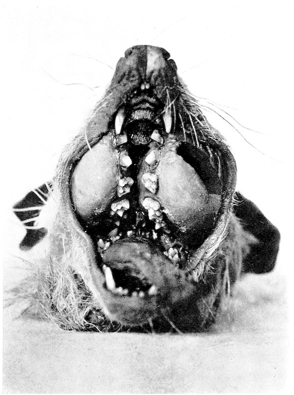
his prey; and one result is that the jaws of the captive-bred
lion differ decidedly in shape from the jaws of the
wild-bred lion. He gets his food regularly, with awful
monotony—twelve pounds of meat at 3.30 p.m. day after
day; there is no alternation of feasts and fasts, with
consequent change in the balance of the body reserves.
Some animals, such as caribou, the moose, the black
cock, the hoatzin, the koala, do not long survive captivity,
because it is impracticable to obtain their natural food.
Gastrointestinal disease is the commonest disease of
wild animals. Twenty-five per cent. of sick humans in
civilized communities also suffer from it. On the other
hand, many animals become accustomed to the new diet
and thrive on it.
5Animals also often thrive in a climate very different
from that of their natural habitat. Some animals from
warm countries, though kept outside without any artificial
heat, get on very well during the severe weather of Philadelphia
winters, when the temperature often drops to
zero Fahrenheit. For the past two years it has been the
custom to put outdoors all monkeys—of value—that are
suspected of having tuberculosis. The cheap monkeys are
killed. The outdoor monkeys are kept in fair sized cages—five
feet square and six feet high—usually singly, though
sometimes two are together. The vervet, the grivet, the
leonine macaque, the yellow baboon, the Hainan gibbon
and many others, have not only survived the climatic conditions
but have not succumbed to tuberculosis. South
American monkeys do not stand cold as well as Old World
monkeys, and cannot be kept out in severe winter weather.
The monkeys that are kept indoors during winter have
free access to the outside through swinging doors and
they often go out voluntarily in the coldest weather. The
same is true of other animals. A note made several years
ago (February 12, 1914) states: “Temperature last night
was 1° F. At 2 P.M. outside temperature 13° F., in carnivora
house 25° F., elephant house 38° F., giraffe house
39° F., monkey house 42° F. Two Bengal tigers were
voluntarily outside. Monkeys that have been out all
winter are: two Barbary apes, Hainan gibbon, lion-tailed
macaque, yellow baboon, grivet monkey, pigtailed
macaque, and eleven rhesus macaques. All the animals
appeared comfortable.”
The physical condition of the animal and the kind of
cold—damp or dry—have much to do with its ability to
stand low temperatures. Monkeys have passed through
very severe winters without damage, and subsequently in
a less severe winter have had frozen fingers, toes or tail.
The size of the cage or pen has not as much effect upon
the well-being of the animal as might be expected. Reptiles,
birds and mammals do as well in cages and pens of
6medium size as in very large ones. A deer or antelope in
a large enclosure does not use all its domain. It usually
has a favorite corner near the food, water and shelter,
and stays there. Nor has a large enclosure been found
perceptibly to diminish mortality from cage-mates. The
stronger will follow the weaker until he gets him, no
matter what the enclosure. Even in the large flying cage
for birds it is necessary to keep out those of a scrappy
disposition. From the point of view of the public small
enclosures are more satisfactory.
Mental disease in captive animals offers a fascinating
field for study—now chiefly speculative. Many
conditions are present to produce it—all the conditions
that cause prison psychoses in man. And
many if not all the insane and perverted acts
of animals have their counterpart in the human.
Masturbation is very common in mammals. Eating of
their own feces, coprophagy, is not infrequent, and is
especially notable in one of the highest types—the Chimpanzee.
Perhaps occasionally coprophagy may be due to
some defect in diet. There is no instinctive disgust at
excrement in the lower animals any more than there is in
the uneducated child. Nevertheless, eating feces cannot
be healthful, and probably does not occur in nature; and
occurs only in the human with mental disease.
Some of the insane acts of animals if prevalent in the
wild would probably cause the extinction of the race.
Such are killing of the young by the father and by the
mother; killing of the female by the male, usually during
rutting time, in some instances reminding one of Sadism
in man. This kind of sexual killing does not often occur
in the wild. The female has a better chance to escape,
and the male probably does not feel so inclined to damage
her when he chooses her himself as when she is chosen for
him by his gaoler. When the mother devours her young
it is usually shortly after birth. I have, however, the
record of a Jungle cat (Felis chaus) who ate two of her
7kittens when they were seven weeks old. Some of the
domestic animals devour their young; the sow often does
it, and occasionally the bitch.
Sucking, gnawing and eating parts of itself or of a
cage-mate is not uncommon in a variety of animals. Bears
lick their paws until they are sore; a monkey may gradually
gnaw away its tail from the tip to the body; an ocelot
(Felis chibigonazon) bit open his scrotum and devoured
his testicle; a Tasmanian devil (Sarcophilus ursinus) bit
off one of his front feet at the wrist; a monkey may
gradually gnaw away its fingers; and numerous other
self-inflicted mutilations occur. Often there is a local
irritating cause, as skin disease, lice, or freezing. But
in many cases no local cause can be found, as no local
cause can be found for thumb-sucking or nail-biting in
the human.
The surgery done by monkeys on their frozen fingers
and toes is interesting. After the flesh has sloughed the
monkey bites off the protruding phalanges, apparently
without pain, so that satisfactory well-covered stumps
are made.
Animals often mutilate their cage-mates in an
amicable way as distinguished from fighting. A bear may
lick its mate’s ears until the hair and skin are gone. A
monkey may eat its mate’s tail or patches of its skin, the
victim lying placidly while the process goes on. Many
animals are addicted to perverted acts on their own or
their mate’s sexual organs.
It is probable that the phenomena just mentioned are
due to confinement, idleness and ennui, and that they do
not often happen in nature where an animal is kept busy
seeking his food, fighting and avoiding his enemies,
attending to his mate or mates, and meeting the various
vicissitudes of his environment.
It should be remembered, however, that the members
of a wild species vary in intelligence and temperament, as
humans do. There are morons and perverts among
8animals in the wild; but not being coddled by the normal
members of the species, they have a poorer chance than
has the subnormal human of surviving and of transmitting
their peculiarities.
Fear, ennui, loneliness and nostalgia, by affecting the
minds of captive animals, react on their physical condition.
Some animals have the fear of man bred in them.
The young often show it from time of birth. This is
especially common in animals that have survived for generations
in proximity to man. It is one reason for their
survival. The mother and father may have become tame
and gentle in captivity and yet the young one may be a
wild thing from birth. Such fear is sometimes uncontrollable,
an apparently slight cause making the animal dash
itself against the fence of the enclosure. It is not mere
speculation to discuss the physical effects of the emotions
on the animal body. It has been shown that fear, anger,
and grief bring about distinct measurable physical
changes. Dr. Corson-White has found that the red corpuscles
are increased by over two million per cubic
centimetre in the blood of a cat frightened by a dog barking
at it. The amount of blood sugar is also increased.
Such observations are suggestive in a consideration
of the changes that may occur in a captive animal subjected
to acute and chronic fear.
The monotony of a captive animal’s life is broken only
by feeding, the sight and sounds of others in the same
building or nearby, and by visitors. Many animals show
their appreciation and pleasure when visitors approach,
and some of the more intelligent animals, bears and
monkeys and some birds, “show off” apparently to keep
the visitors there. When there is nothing doing, some
stand swaying their heads, like a weaving horse, or pacing
the cage, inanely tagging at each turn the side of the cage
with the head or other part of the body—often so persistently
that a sore is produced. Nearly all animals are
social and suffer from loneliness when kept by themselves.
9This is true even of the lowly forms. The keeper of the
reptile house reported that a giant tortoise became
despondent and refused to eat when his companion, a
leopard tortoise, was taken from him, and that he braced
up as soon as the leopard tortoise was returned. It is not
necessary that the companion be of the same species, or
even of the same family. A lion or a tiger may be satisfied
with a little dog for a companion, and there was an
African rhinoceros at the Philadelphia Garden that was
very discontented and unhappy when alone and became
perfectly satisfied when she was given a domestic goat as
a cage-mate; and the huge rhinoceros stood for a good
deal of butting and bullying from the goat without
retaliating. A sympathetic keeper may do much to relieve
the loneliness of the animals in his care.
Nostalgia, or homesickness, has been felt by all men.
Some have died of it. The tradition among writers is
that it affects young people and those who have been
living nearest to a state of nature. In this country the
American Indian and the negro are affected more than
the whites. Much was written of it after the Franco-Prussian
War and the American Civil War. It is a real
condition, capable in extreme cases of causing death and
of so weakening the sufferer as to make him more susceptible
to the invasion of other diseases. At the present day
we hear less of it among civilized people than formerly,
perhaps because the conception of home has been broadened
by modern methods of intercommunication. The
wild animal’s conception of home is narrow; he comes
directly from it into an environment where he may see
many other animals, but not one of his own kind. Predisposing
causes of nostalgia are stronger with him than
with the human. That home means a great deal to
animals is shown by the migration of birds—the return of
the carrier pigeon, and of the lost dog, and of the swallow,
which returns every year to the same nesting place.
10All animals long for the things of nature—open air,
earth, grass and water. They are thrilled when their feet
touch the sod. Even the hippopotamus gambols when he
leaves his concrete house and his feet touch earth
and grass.
The face and carriage of many animals cannot express
feelings as in the human, though it is not unreasonable to
assume that animals may indicate feelings by expression
understood by their mates, though not understood or even
noticed by man. When they can express it in a human
way their feelings may be read. The dejection of nostalgia
is especially shown by anthropoid apes. Gorillas
have been noted for it from the earliest writers. The
orang is prone to it, shows it by his attitude and expression,
and sometimes dies of it.
It is stated in A Handbook of the Management of
Animals in Captivity in Lower Bengal, p. 130, that
elephants have been observed to shed tears abundantly if
forced to leave their old home and surroundings. How
much other animals who cannot express homesickness
may feel it, and how often it is a cause of unhappiness,
depression and predisposition to disease, it is impossible
to say.
Most wild animals in captivity are sterile. The reason
is not known. It shows the profound effect of captivity.
It would be difficult to determine whether the sterility
of a mating is the fault of the male or the female.
There is no apparent rule for sterility. Some families
are always sterile in captivity, others are fertile, even
with very unfavorable surroundings. The deer, horses,
hippopotamuses, pigs, goats, sheep and oxen, are good
breeders; while antelopes, rhinoceroses, giraffes, elephants,
are poor breeders. Some members of a family
may be good, others poor. The lion and puma breed fairly
well; the tiger, leopard and jaguar, very poorly. Bears
breed well, but the mother usually destroys her young.
11We cannot mate wild animals and birds simply by
putting males and females together in the same cage.
Domestic mammals and birds usually mate under such
conditions, but wild ones often refuse. Many mammals
and probably all birds that are not polyandrous or
polygamous reserve the right to select their mates, and if
the sexes are put together by man they may view each
other with indifference or with animosity. There are
many males and females of the same species of mammals
at the Zoological Garden that will not consent to live
together. A male monkey in a cage with several females
will very often select one female for his mate and will
have nothing to do with the others.
Among monkeys fertility varies greatly. It is not
practical to determine the ratio among the various kinds,
as some kinds are much commoner in zoological collections
than others. I think that in general the Old World
monkeys (Cercopithecidæ) are better breeders in captivity
than those of the New World (Cebidæ). The
anthropoid apes are very poor breeders indeed; of the
many gibbons, orangs, and chimpanzees, that for years
have been captive in Europe and America, it is probable
that only the chimpanzee has bred, and that very rarely.
Refusal to mate, sterility, infanticide by father and
mother, and sexual killing keep down reproduction in
zoological gardens; and the number of young ones is a
good indication of the character of a garden and of the
provisions made for the happiness, comfort and health of
the animals.
With the birds in a zoological garden the conditions
for nesting and laying are not good. Caged birds have no
material for a nest, no privacy and rarely lay an egg.
The outdoor water birds and the outdoor upland birds
with natural surroundings, with secluded retreats, lay
and hatch very well. Birds like mammals apparently
are indifferent to publicity when copulating, but seek
12seclusion for laying and nesting—from maternal fear for
the safety of the young.
The sexual instinct in indoor caged birds in a zoological
garden is dormant. Very few copulate and very few
lay eggs; pigeons and Mexican conures (Conurus holochlorus)
are exceptions to this general rule.
Probably because the sexual instinct is dormant the
males never fight over the females among perching birds,
and very rarely kill each other. In some species of
finches, however, as the chestnut-eared (gn. Amadina),
the females fight among themselves if there are not
enough males.
Ovoviviparous reptiles breed more often in captivity
than one would expect; and egg-laying snakes often lay
eggs, which of course are only hatched artificially.
Birds suffer less than mammals from the psychological
effect of captivity. The mental development of a bird
is much lower. With few exceptions, like the ruffed
grouse, the bird accepts captivity easily and becomes
tame, though he will not stand being touched. He views
his keeper and visitors with indifference or friendliness.
If a bird house is bright, cheerful and sunny, all the
inmates thrive and appear to be happy. These conditions
undoubtedly affect the health of the bird, as is evidenced
by their plumage; bright colors that are lost in a dark
and gloomy house are retained and developed when the
house is cheerful and sunny. There are some birds, however,
that never retain their colors in the captivity of a
zoological garden. Among them are the scarlet ibis, the
American flamingo, and the roseate spoonbill. It has
been suggested that the loss of color is due to the lack of
something in the diet, mineral or organic, that the bird
gets in its natural habitat. Tame scarlet ibises living
with the chickens about the dwellings of natives in
Venezuela retain their brilliant color. The material of
the beautiful red color on the under surface of
the wings of the touracou contains copper, yet these
13birds retain this color very well in captivity, even after
several moultings.
The source of the copper has long been uncertain. In
nature the birds are fruiteaters, and their diet in captivity
consists of bone meal, zweibach, corn meal, white
potatoes, eggs and carrots—foods that are usually
assumed to contain no copper; and no copper utensils are
used in the preparation of the food. Dr. John Marshall,
however, writes me that all the common cereals contain
minute quantities of copper; and Dr. Leon A. Ryan,
University of Pennsylvania Medical Bulletin, June, 1907,
states that copper may be found in animal tissues. Dr. E.
P. Corson-White has found traces of copper in the bone
meal used at the Philadelphia Zoological Garden. The
copper in the red color of the touracou’s wing therefore
comes from the food.
It is probable that a bright and cheerful bird house
does not influence the color of birds by the direct action
of light on the color as much as indirectly by improving
the health and spirits of the birds. Coloration
in birds is a very complicated proposition. It depends
upon age, sex, season, health, light, heat, moisture, mode
of life, and food. No one bird house can combine all the
conditions necessary for the retention of natural colors
in every species. The desert species from a habitat of
intense light and dryness require for their color a different
environment from the forest species, from a
habitat of shade and moisture. The suppression of
sexual feeling in captive birds may influence color. In
nature the finest colors are attained by mating birds.
It may be said that all animals—except those of nocturnal
habit—have a feeling of joy and well-being in fine
weather and bright surroundings that reacts favorably on
the general health.
The variability of the breeding period induced by
captivity in many animals may be mentioned with
sterility. It was shown some years ago in the Philadelphia
14Garden by the European brown bear which in one
year gave birth on January 16th, and in the following
year on July 25th. It is another evidence of the profound
effect of captivity on the captive animal. I know of no
observations of the effect of captivity on the period
of gestation.
There is considerable mortality among captive
animals from killing of cage-mates. I do not refer to
sexual killing, already mentioned, or to fights over a
female. Often males, with no females near, cannot be
kept together; probably sexual jealousy is at the bottom
of it. Antelope and deer are especially inclined to scrap.
Even a large enclosure will not save the weaker male; the
stronger follows him with horrible persistency, sometimes
for days, around and around the enclosure, often
at a walk, but always on the offensive, at least during the
day; until, careless from weariness, the weaker is caught
unawares and finished by a horn-thrust in the side.
Both birds and mammals often kill their mates when
the mate is sick, or “down” from injury or disease. All
animals hate sickness and death, and show their dislike
by attacking or shunning it. Birds may get on happily
together for months until one becomes sick, and as he
crouches in a corner with ruffled feathers the others pick
on him and finish him. The same is true of mammals, the
sick one being horned or tramped to death by the mate
with whom he had formerly been on most friendly terms.
The keeper often reports an animal “killed by its mate,”
whereas the mate has only given the coup de grace.
This brutality is not universal. Rarely a parrakeet
will stand guard over his sick and dying mate; and we
have seen a ratel—of a ferocious family—stand guard
over and resist the removal of his sick companion.
The diagnosis of disease in wild animals is unsatisfactory;
usually impossible; clinical study as we know it
in the human is impossible. We know that the animal is
sick, but not why. A certain group of symptoms accompanies
15all diseases—dull, rough coat or feathers, refusal
to eat, weakness in the hind quarters, and finally getting
down. They rarely show symptoms of pain—or at least
we cannot read the symptoms. The pain of acute pancreatitis
in man is violent, yet many animals die with it
and we cannot tell that they suffer. Animals do not suffer
as much as the human, and they stand the ravages of
disease better than the human. At autopsy we often
wonder how the animal could have lived with the conditions
that are found. A monkey may be apparently well
until a few days before his death, though his lungs and
abdominal organs may be a mass of tubercle. A small
red howler monkey (Alonata seniculus) was in apparent
good health, playful and lively until twenty-four hours
before his death from acute pancreatitis, though his
stomach and intestines contained fifty-one nematode
worms, some of which were eight inches in length.
As diagnosis is unsatisfactory, so is treatment.
Usually all we can do is to treat symptoms; and by the
time disease has advanced to the point of becoming externally
noticeable, it has usually gone beyond the reach of
medical treatment. It must also be remembered that
drugs vary very much in their action in different families
of animals. Nux vomica will not kill the gallinaceous
birds of North America, and Tenant says that in Ceylon
the hornbill feeds on the fruit of strychnos nux vomica.
The pigeon is immune to opium. The Felidæ are said to
be unusually susceptible to carbolic acid; veratrum viride
is harmless to sheep and elk, but poisonous to the horse;
dogs can take with impunity large quantities of cyanide
of potassium. These statements are true when the drugs
are administered by mouth—the usual way of giving them
to wild animals. The action may be different if the
drugs are administered intravenously or subcutaneously.
Variations in effect when they are administered by mouth
are often due to chemical variations in the digestive secretions.
It is probable that the action of cyanide of potash
16on dogs depends on the amount of hydrochloric acid in
the digestive tract.
When worms or their eggs are found in the stools
vermifuges are used, and with some animals especially
liable to infestation by intestinal worms, periodic doses of
vermifuges are given as a prophylactic. Turpentine is
given to the zebra at fixed intervals for the round worm;
santonin, male fern and areca nut to the carnivora for the
various worms that are so common in the intestinal tracts
of these animals.
The work of the Laboratory of Pathology is throwing
light on the subject of diagnosis, and though from the
character of the clinical material diagnosis can never be
as satisfactory as in the human, yet we may fairly hope
for improvement. Prophylaxis is our chief reliance, and
always will be.
17
DISEASE IN CAPTIVE WILD
MAMMALS AND BIRDS
“We have also parks and enclosures of all sorts, of
beasts and birds; which we use not only for view and
rareness, but likewise for dissections and trials, that
thereby we may take light what may be wrought upon the
body of man.”
The purpose of a menagerie under the auspices of a
zoological society can scarcely be put into better words
than those found in this quotation from Sir Francis
Bacon’s New Atlantis. Apt as this description of the
mythical island’s collection may be, it is but a reflection
of the teachings of Plato’s original legend of a perfected
community, and the practical applications of these teachings
by Aristotle in his Anatomy and Physiology of
Animals. The history of human study shows a constant
investigation of lower forms of life, ever broadening in
its scope, ever more satisfying in its explanation of
biologic problems and ever increasing in value from an
economic standpoint.
If, however, all animals are to be subjected to “dissections
and trials” there inevitably will come under
observation many specimens presenting variations from
the accepted mean or standard or even from an average
for their kind and therefore approaching what may be
called pathological.
The desire to explain the abnormal has had the effect,
during the half century since Virchow defined cellular
pathology and Darwin systematized the world’s knowledge
of comparative biology, of directing attention to
comparative pathology and of stimulating the study of
18veterinary medicine. Moreover in the past twenty-five
years much work has been done and many isolated publications
have appeared upon the diseases of wild animals,
notably Bland-Sutton’s work, Evolution and Disease
(1895), a thoroughly scientific and most charmingly written
book, but rather elementary in its approach of the
subject of pathology, and Wood Hutchinson’s Diseases of
Animals, a more or less popularly presented treatise. I
am unaware, however, of any systematic monograph upon
the subject wherein we may find data showing the character
of pathology in the various animal groups or the
incidence of the various lesions. The reports of some
zoological gardens contain the result of medical and
pathological data collected for the report period. The
publication of greatest merit and value is that from the
Zoological Society of London, whose huge collection
studied by a large official personnel makes it possible to
present valuable data. The New York Zoological Park
uses its material in a similar manner and has been able to
explain some of the knotty problems so frequently met in
wild animal collections.
Here at Philadelphia it has been our practice now for
twenty years to perform an autopsy upon every mammal
and bird that dies, and upon all of the large or important
reptiles. There is no aquarium connected with the
Garden. The office of the society keeps a record of the
arrival and a general description of every animal so that
a brief history of the specimen is usually available. The
keepers are required to observe their charges regularly
and closely and to report any abnormalities to the officials
of the Garden. Somewhat detailed discussions upon signs
of sickness will be given at appropriate places, where also
a few remarks upon treatment will be included, but as this
work is not intended to be a treatise on therapy and since
this subject does not differ from that referable to domesticated
animals, little space will be devoted thereto.
Upon death a complete autopsy is performed and the
19findings are recorded upon a printed form from which,
when the histological, bacteriological and parasitological
studies have been completed, a set of cross index catalogue
cards are typed; these are divided into the principal
diagnoses and determinations. The report of the Zoological
Society, appearing at the end of their fiscal year,
February 28th, contains a résumé of the observations for
the year, together with notes of interesting cases and
experimental work.
There have accumulated the records of nearly six
thousand autopsies and upon them as a basis has been
founded the following report of the incidence and nature
of pathological manifestations in the various animal
groups, using also as additional data, published reports
from other gardens. The book might be described as a
collection of our studies, parts of which have appeared
as separate articles, but most of which are entirely new,
put together with as much connection as the subject
matter will permit. The zoological and pathological
literature has been consulted very extensively, but
except for the reports of zoological societies and
the publications of special students, it usually represents
isolated notes by travellers and veterinarians
so that many articles may have been overlooked.
Therefore no claim of perfect completeness of reference
is made, the statements resting chiefly upon our own
records. The subject will be approached from the standpoint
of description and incidence, but it is inevitable that
comparisons and contrasts must be noted.
Into the realm of evolution[1] I shall not venture
because I appreciate a lack of adequate preparation for
such an attempt, and because, even if such were not the
case, the material at hand is lacking in data upon fishes,
many kinds of reptiles and invertebrates.
20A direct and practicable application of these data will
be in the direction of explaining some of the pathological
states in domestic animals and man. There are indeed
many disease entities or syndromes in these groups for
which no useful hypothesis has been advanced, while for
others a partial explanation has been offered, usually,
however, inadequate wherewith to form the basis of
rational prevention or therapy. Thus, for example,
essential emphysema seems to be limited to the animals
of civilization; on the other hand, the anatomical basis of
exophthalmic goitre may be seen throughout nearly the
whole animal kingdom yet the clinical phenomena belong
characteristically to man, and are occasionally seen
atypically in the dog. While it may be impossible to
give a complete comparative anatomy and physiology for
each of the pathological states, the attempt will be
made to treat all subjects analogically through the zoological
orders.
The experimental pathologist may find the records of
the Garden useful in his work. For example, he can know
that rodents are not prominent among the orders showing
spontaneous arteriosclerosis, but that nephritis occurs
among them in about a quarter of natural deaths; or he
may learn that the Primates have a good cardiac
reserve while the Marsupialia have not. Too often
experimental work is not based upon facts including
natural probabilities.
A collection of pathological data such as is presented
in the following pages may be of assistance to veterinarians
and managers of zoological gardens in the
diagnosis of sickness in animals, both wild and domesticated.
We do not presume to offer a system of veterinary
medicine, but it is possible to introduce certain objective
findings of practical hygienic and therapeutic value. Such
observations are, however, limited and in our experience
at the Philadelphia Garden the diagnosis of disease in a
wild animal, excepting of course those which are perfectly
21self-evident, is more often speculation and
conjecture than at all well grounded. It is not uncommon
for animals to come to autopsy presenting a perfect
galaxy of abnormalities, yet the closest antemortem
observation failed to reveal unusual conduct or appearance.
On the other hand specimens are frequently opened
whose organs fail to contain any lesions discoverable
even by careful study. Dr. Henry Chapman, sometime
prosector to the Society, once made a remark in this connection—“Why
do they die or how can they live so long.”
Space is given to this phase of the observation of wild
animals in order to emphasize the difficulties of interpreting
their conditions, but of course it should be understood
that certain data of value may be gained by close attention
to the details of their normal behavior and to changes
which occur indicating that something is wrong.
The naturalist and the trained animal keeper are, in
our opinion, better judges of a wild animal’s condition
than is the veterinarian, unless he be at the same time a
zoologist and have long experience with a menagerie.
My own observation of dogs and horses leads me to think
that more acumen is needed to interpret the actions of
wild animals since they seem to have greater natural
reserve, and of course in regard to them there are many
more variables since we see fewer specimens of each
species than we know familiarly among domestic varieties.
The principal objects for observation are, as in
veterinary medicine, the eyes, the hair and skin, the
mucous surfaces, the droppings, the condition of the
abdomen, the appetite and the desire for water. Physical
examination is limited to tractable beasts and those which
can be caught and handled without danger to the personnel
or unusual fright and damage to themselves. In
the interpretations of physical signs in tractable animals,
such as many ungulates and some monkeys, the experience
of the trained veterinarian is of the greatest value,
but this fails amongst carnivores and birds. It might be
22said that anesthetics could be used for a thorough examination,
but this would be undesirable for a seriously sick
animal and it is, in our experience, none too safe a procedure
although often perfectly practicable. Animals
do not like to be molested much as they may seem to enjoy
attention, and when it is possible it is our practice to
avoid handling them.
It might be contended that observations upon diseased
states in captive animals would not represent natural
developments, in other words, not that which occurs in
the wild. Such indeed may be true in regard to the infectious
diseases, but since we are imperfectly informed as
to the pathology of the wild state, we are obliged to accept
and use the best substitute at hand. Moreover it seems
perfectly fair to consider as characteristic for an animal
or group, the physical and even physiological expressions
of morbid agencies as we know them, even though the
animals be at the time under conditions not natural to
them. It would be perhaps incorrect to say that cirrhosis
of the liver occurs in .6 per cent. of animals in the wild
as is the case for our autopsies, since incorrect food and
infections are potent in its causation; on the other hand,
our experience and some few data from naturalists and
pathologists make it conceivable that tumors occur to this
number in native states. The incidence of tumors in wild
rodents is quite well known. Degenerations and fibroses,
the result of parasitism, are known to exist throughout
the entire animal kingdom. Further to illustrate how
pathology is distributed in wild life, Plimmer’s
experience[2] with 500 rats (M. decumanus) might be
cited. He found the following: Tubercle 3 times, tape
worm cysts 10, Tryp. lewisi 49, empyema 2, tumor of jaw
from old injury 1, pleuritis and hydrothorax 1. Bacteria
were found in 71 rats, 40 times in the lungs, 31 times in
the spleen; saccharomyces were found 16 times in the
lungs. Dr. W. L. Abbott reports to us personally that he
23has repeatedly found coiled exproventricular worms in
the wild specimens he has collected. Not only are we
informed of some isolated and individual pathological
states but the existence of epizoötics of communicable
disease among wild life is well authenticated. The
simple citation of the extermination of deer in one section
of Colorado by pleuropneumonia will suffice to illustrate
this point. Other examples are, however, interesting.
The occurrence of changes in the jaw bone almost
certainly those of actinomycosis is reported by
Blair, the specimens being shot in the wild and believed
never to have been near civilization. The white-tailed
deer of the Swan River Valley in Montana, are known to
be constant carriers of liver-flukes.
It would seem therefore that it is not unfair to use
material gathered from animals under somewhat
unnatural conditions as representing the reaction of the
zoological orders to pathogenic agencies. Such conclusions
must however be made very guardedly, for
it is probable that not over ten per cent. of the
total number of mammalian and avian species are to
be observed in captivity. Because of the number
of orders and the great variety of genera included in
the present study it is probable nevertheless that
the lesions are fairly representative of the whole
animal kingdom.
However, the numbers and percentages given should
be read to indicate the probabilities and should not
be interpreted as implying the mortality relationships
since different varieties have differing powers of
resistance to the same pathological state. The margin
of safety in any given group for one or several different
disease entities cannot at present be stated with
any degree of precision but this factor is doubtless very
great. The work of physiologists suggests that there is a
reserve power in the human lung sufficient to sustain life
until five-sixths of the functionating organ is useless, and
24I shall quote a case of an opossum wherein only one-tenth
of the respirable surface seemed to have remained; we
have repeatedly seen both lungs of a monkey apparently
entirely solid. Such physical vital incompatibilities
might be exemplified by many other cases, but when one
reviews the physiological margin of safety, inexplicable
and contradictory instances are equally numerous. I
have seen a male deer run a doe against the fence and
butt her, without result, whereas in an apparently similar
occurrence the animal would be dead in a short time.
Numerous instances of slight enteritis of a short stretch
of duodenum or ileum have killed, with almost nothing to
be found microscopically, and on many occasions we have
been chagrined in being unable to discover the cause of
death. The capacity of self-healing is a variable one, but
seems in direct proportion to the quietness and seclusion
possible for the animal and inversely to the chance of
bacterial infection.
The effect of captivity has been the subject of much
speculation. For the preservation of health it would seem
that animals require periods of rest and activity, thorough
elimination, possibly a moderate exercise of their
procreative functions, but most of all, appropriate food
obtained by the physical effort we term chase. All but
the very last condition is supplied in a measure in well
managed collections. The degenerating effect of the
absence of chase must be admitted. An interesting and
suggestive example of this was noted by Mr. Jones at the
London Zoological Gardens. He observed the skull of a
lion that had been in captivity thirteen years, in which the
canine area of the face and the part of the skull acting as
the insertion for the seizing and holding muscles had
undergone atrophy while the chewing muscles with their
bony bases had remained normal. Numerous examples
of disease atrophy are on record and those of a
physical nature must have counterparts in the realm
of physiology. The size to which an animal will attain
25cannot be estimated by the examples seen in menageries.
Judging by the accounts of collectors and hunters and
upon the more reliable of the moving picture displays of
wild animals in their native haunts, it would seem probable
that under normal conditions of habitat the average
size of wild beasts is considerably in excess of that in
park specimens.
The effect of captivity may also be felt in the direction
of reduced resistance to infectious diseases. Brooks, of
the New York Park, expresses the view that captivity
increases susceptibility to bacteria and causes parenchymatous
degenerations. In the latter direction it is
interesting to learn that Seligman of London claims to
have seen sudden deaths in wading and struthious birds
from myocardial disease, without valvular or other
lesions, for which he holds the enervating effects of captivity
responsible. It is well recognized that a species
may be unusually susceptible to a disease that it has not
encountered in its phylogenic development. Man illustrates
this peculiarity very clearly. Europeans were
found exceedingly susceptible to sleeping sickness when
they went first to the part of Africa inhabited by the tsetse
fly, and the American Indians died in hordes when they
met the tubercle bacillus for the first time. Judging by
the ravages of tuberculosis in captive monkeys a similar
susceptibility probably explains the matter for there are
no entirely satisfactory records of this disease among
them in the wild state.
In so far as general susceptibility to infection is concerned,
it may be in part due to one of the artificial
conditions of captivity, that of inbreeding. This influence
is undoubtedly very great, both by chance in families, and
by intention on the part of dealers as well as the mating
which occurs in menageries. However, it is not known
how far inbreeding may go in the wild state so that one
must be very careful about drawing conclusions in this
particular. Several years ago, at the time we reported
26the neoplasms found at the Garden, discussion arose as
to the effect of inbreeding, and thereafter some observations
were made in this direction. With the exception of
the hyperplasias of the thyroid, not certainly of neoplastic
nature, in a much mixed-up family of wolves, we
could find no evidence that inbreeding was responsible
for tumors. Plimmer and Murray of London, seem to
imply that some of their inbred animals are likely to have
tumors; reference to this matter will be made later in this
book. In so far as diseases of the organic systems are
concerning those of the bones seem to be the only ones in
which inbreeding is significant.
The individual resistance will be reduced of course by
the unsanitary surroundings incident to trapping, shipping
and storage, but this need not affect the figures or
pathological tendencies of classes or orders.
The effect of captivity is felt in another way. A very
large percentage of wild life perishes during the first
weeks or months after its capture, and in gardens the
heaviest mortality occurs among the recent arrivals. The
London Garden figures that from thirty-three per cent. to
fifty per cent. of their total mortality is in animals that
have not been in the garden six months and that die
because they are not yet accustomed to their new surroundings.
It seems to us, both from an academic
and a practical standpoint, that this is a long
time and should afford ample opportunity for the
garden to study the specimen and for the specimen to
become acclimated. These early deaths are perhaps
to be ascribed in large part to failure of acclimatization
but many are doubtless the result of infection
acquired in the wild, in transit while in the hold of vessels,
at quarantine, or in trains, or at the establishments
of dealers. We have seen a few deaths which have followed
behavior that might be likened to homesickness.
Perhaps the age at arrival has an influence upon the
morbidity and mortality of wild animals, for it is easily
27conceivable that the young and the very old might adapt
themselves to new surroundings with much less readiness
than the sturdy middle-aged adult. The age of animals
upon arrival is very rarely known, and can only be
recorded as “young,” “fully developed,” and “old.”
This will have an effect upon statistics and when possible
is noted in the text, but this is not practicable to the
extent we desire. The meaning of “young,” “adult,”
and “old” is not the same throughout the animal orders
nor even within orders.
Mitchell[3] has attempted to gain concrete ideas
of the expectancy of life among animals by analyzing
the records of the London Gardens. This gentleman
bases his figures upon known ages and the length
of time in captivity, from a combination of which
data the specific viability and the potential longevity
may be estimated. Such results, he admits, can
only be approximate and they show within classes and
orders, a decided lack of uniformity. The terms “specific”
and “potential” longevity, coined by Sir Ray
Lankester, apply, for the first, to the average length of
life as it is affected by external conditions and those
incident to procreation, while, if an animal be under ideal
conditions it will attain the potential longevity which is
longer than the former. These considerations have a
biological and economic importance, while a knowledge
of the pathology shown by the various groups may help
to explain these durations of life. Contrariwise figures
of the expected longevity may assist us in evaluating
youth and senility in the causes of death but can hardly
affect the comparative nature of the lesion.
A résumé of Mitchell’s studies indicates that the
higher apes have a potential longevity and a hardihood
much less than man but still upwards of thirty years. As
one investigates lower in monkeys, life periods become
shorter, while in the next order, Lemures, the length of
28life rises. Carnivora have a reasonably good vitality,
their potential periods varying from ten years in the
foxes to thirty-three years in bears. Insect eating
animals are short lived, three years being a maximum.
The Bat family shows great variations, the greatest life
being not over seventeen years. The Rodentia have long
lives compared to their sizes—twenty years in porcupines,
fifteen years in squirrels, thirteen in marmots, nine in
agoutis and capybaras, and three in dormice (which is
also about the maximum for the rat). Hyraces live four
years on the average. Proboscidea, although reputed to
live to great age, probably rarely live a half century and
may be said to have an expectancy of twenty to thirty
years. Perissodactyla (horses, tapirs and rhinoceroses)
while they may live half a century, have an average life
of between fifteen and thirty years. The closely related
Artiodactyla fall into two groups, a first comprising
antelopes, sheep, goats and deer which rarely exceed
seventeen years, and a second consisting of cattle, camels
and giraffes, which vary in expectancy from eighteen to
thirty years. The smaller members of the Ungulata have
in relation to size a relatively greater viability, the
ruminants, however, having on the whole a low viability.
Marsupials vary from a maximum of seven years in the
opossum to eighteen in the wombats, but none of this
group has a good viability. The Aves as a class or if
compared according to dietary requirements, have longer
potential ages and better viability than mammals. Passerine
birds average twenty years and many live to sixty,
while the Picariæ approach the former figure but do not
have such good viability. Psittaci and Striges may live a
half century but the resistance of the latter is much
reduced by any unfavorable surroundings. The raptatory
birds live fifty years, but their viability is variable.
Herodiones have a maximum expectancy of thirty years
and good resistance, while their relatives, Steganopodes,
may live fifty years, and Odontoglossæ have a good
29viability, up to twenty years. Anserine birds may live to
be fifty, and, unless conditions are quite unfavorable,
have a good resistance. Columbæ may under good conditions
live to be fifty. Gallinaceous birds may only be
expected to survive twenty years, a figure also given for
Fulicariæ. Alectorides may live up to fifty years.
Limicolæ, though they do not thrive in captivity, may live
thirty years. Impennes live poorly under artificial conditions,
the greatest record being twelve years, a figure
also holding for Crypturi. Struthiones, if the conditions
be right, may live fifty years.
Because of the variable specific longevities, it is frequently
difficult to decide when an animal is senile. Man
is said to be as old as his arteries, and his span of life
nowadays is in the neighborhood of half a century. Parrots
exhibit lesions of the vascular system comparable
to the arteriocapillary fibrosis of human beings, and their
expected longevity is about the same or a little greater.
From a study of our cases of this lesion in parrots it can
be said to appear quite early in life and not to lead to
organic disease as it is alleged to do in man. It is, however,
interesting to note that in those animals which are
supposed to have the longest specific lives—elephants,
snakes, anserine and raptatory birds, parrots—there is
relatively low mortality and fewer infectious diseases
are encountered. The last part of this statement should
be qualified by stating that anserine birds and parrots
are quite susceptible to mycoses, in all probability from
musty food, which raises their death rate, but as
this is accidental and artificial, it can be excluded
from consideration.
In a rough way there is a direct relationship between
the size of an animal and its longevity, but this is not close
enough to be a reliable guide; whales and elephants live a
long time, but so do snakes and parrots. Within orders
this relation of size and expected longevity is more easily
seen but is not absolute. I cannot state, according to my
30present studies, that there is an unqualified relationship
between the size and expected longevity of an animal and
its pathological lesions.
The immediate surroundings and the management of
captive animals have a very direct and important bearing
upon the mortality and perhaps upon the incidence of
morbid processes but probably not upon the character of
the latter. A full knowledge on the part of the personnel of
a zoological society concerning the habits and habitat of
every animal in their keeping is essential, to which must
be added a group of interested keepers. In engaging the
last, it should not be forgotten that certain men
have “a way” with animals and that others cannot
manage themselves.
The enormous literature at the disposal of the
naturalists permits executive officers to formulate a plan
of housing and feeding with fair accuracy for each kind
of animal, but of course it is rarely possible to obtain in
sufficient quantity the natural food (e.g., ants for anteaters).
In so far as food is concerned it seems that with
a few exceptions like the one just mentioned, the substitutions
made at the zoological gardens are nearly
satisfactory. The elements in which the captive diet is
poor are the inorganic salts and vitamins since Dr.
Corson-White, some of whose work is included in a later
chapter, has shown that for those animals which our
statistics indicate as most prone to have rickets and osteomalacia,
the available phosphorus and calcium are low,
and one vitamin was also below the desired quantity. In
this regard, however, I am not at all convinced that diet
alone will suffice to explain these degenerative osseous
diseases; I shall take this up more fully later. Careful
inspection of all food should be made and cleanliness
(sifting of cereals, protection of meat from flies, etc.), is
indispensable. The mortality among our carnivora has
materially decreased since the horse meat after butchering
was placed in covered galvanized iron pans. There
31are many problems of feeding, too numerous to be covered
in a survey of this sort, which must be solved, and
it is a credit to superintendents that this they have
studied carefully.
There are two problems in the management of
animals upon which much difference of opinion exists,
namely the heating of houses and the material of which
cages are made. It seems to be the practice in many
gardens to keep animals very warm. Dr. Chalmers
Mitchell states unqualifiedly that adult animals do
not have to be kept warm, and that even an equable
temperature is not demanded, variations in temperature
having a distinctly stimulating effect. However he maintains
that they should be kept dry and must be supplied
with a shelter. This is in accord with the experience at
the Philadelphia Garden, since for many years we have
allowed access to the open air all winter to every animal
that could stand it. A large group of macaques has now
lived entirely in an open “band stand” cage for nine
years with a lower mortality than in the rest of the
monkey collection, which is permitted to go indoors some
of the time. Occasionally one in poor health is frozen to
death, and healthy ones may lose fingers, toes, or a part
of the tail, but the general condition is so much improved
that they present an attractive exhibit to visitors. Unless
a storm be of great severity, wild animals are usually
indifferent to it although they may seek their shelter.
Snow apparently is no source of fear to them, and many
enjoy playing in it. The general principles of the enclosure
should be proper lighting, free access of air, dryness and
shelter in time of storm, the last so arranged that the
sleeping place is well protected. Appropriate arrangements
should be made for nocturnal animals, regardless
of their visibility to visitors, if their preservation is
of importance.
The hygiene of communicable disease has influenced
everyone to use concrete and metal for cages. These substances
32are without doubt most simply kept clean, but
they are heat-conducting and remain cold or damp longer
than wood or the ground. It may be claimed that the
latter two cannot be disinfected so well, but this need not
militate against their use. Wood can be disinfected by
sunlight or by mechanical cleaning plus disinfectants, by
a blast lamp and by paint. The ground will disinfect
itself if allowed to lie fallow for a time, or it may be
turned over after sprinkling with lime. It is fair to note
that the New York Zoological Garden reduced their mortality,
especially from verminous pneumonia, by changing
some deer herds to concrete paved enclosures; if that
were the only change made the result would be very
significant, but it should not be forgotten that another
clean ground range might have served as well to a herd
from which the infected ones had died. My own observations
with guinea-pigs, rabbits, mice and dogs lead me to
believe that they thrive and breed better on wooden floors
than on metal or stone.
I have tried to work out figures to show that more
animals die when housed in enclosures of stone and metal
than when upon the earth or on wood, but the attempt has
been unsuccessful chiefly because of the presence of epidemics
and parasites, principally among the birds. The
attempt was further embarrassed because some members
of an order are housed on both floorings. However, there
was no great advantage for the metal and concrete floors
even after the epidemic had been discounted. This Garden
does not have a great number of pneumonias, a
disease said to be favored by dampness and cold, but
those that occur are chiefly among the small mammals, on
wooden floors and in the large bird house in cages of concrete
and metal. However, the construction of both these
houses permits the visitors to approach very close to the
cage, a factor that doubtless explains the disproportionate
incidence of inflammation of the lungs. In so far
as outdoor fowl and ungulate ranges are concerned, they
33should be changed frequently under the best conditions
since occasionally one will find groups doing badly until
moved. Moreover the ground becomes contaminated
with parasites such as esophagostomum and heterakis,
infestation with which while not very serious in itself,
may lead to fatal infection with bacteria.
The effect of animal parasites upon the morbidity and
mortality of wild beasts and birds in captivity is by no
means clear, and Doctor Weidman and I are inclined to
be sceptical, with certain reservations of course, of their
great importance in the death rate. Doctor Weidman has
kindly agreed to contribute a chapter upon the general
distribution of protozoal and metazoal parasites with a
summary of their probable pathogenic importance.
The groups known to have a decided pathological
power might be divided into the toxic, the tumor formers
and the mechanically obstructive; certain parasites have
properties placing them in two of these classes. The first
group comprises the hemosporidia and hemogregarines,
the uncinaria and some of the cestodes, forms which produce
hemolysis and hemorrhages with varying grades of
anemia. The importance of this group is shown chiefly
among the Aves, in which high grades of anemia are
occasionally met from malarial infections, but cats and
dogs or even herbivores also frequently suffer from
hookworm. The tumor-producers are chiefly echinococcus
worms, the cysts of which may grow large enough to
occupy nearly the entire abdomen. A certain grade of
anemia and general ill health accompany this hydatid
disease, partly the result of a toxin and partly by damage
to important viscera. Those parasites which obstruct
mechanically do so by their own bulk or by an accompanying
inflammation, incited by them as foreign bodies
or by bacteria which have gained entrance at the irritated
point. This is exemplified by the enormous collection of
nematodes sometimes found in reptiles (a pailful was
removed from a python) and by the tightly coiled or
34tangled thread and tape worms frequently found in birds.
The effect of swelling by the mucous membrane under the
influence of worms is illustrated by the infestation of the
proventricle in parrots. Here spiroptera penetrate into
and under the glandular layer which swells and pours out
mucus, the total mass of nematodes, mucus and tissue
obstructing the passage.
Very many animals show parasitic infestation at postmortem,
but the percentage in which they can be said to
be principal causes of death is quite small, while that in
which they play a rôle as activator of the terminal condition
is also small but indeterminate. The latter group
comprise, together with the anemias mentioned above,
certain forms of pneumonia, of hepatic and vascular
lesions. Inflammations of the lungs from ascaris and
paragonimus are fairly well known; fortunately we have
been troubled less with this than have most gardens, possibly
because we do not have such large herds of herbivora
susceptible to it. Hepatic diseases from flukes, from coccidia
and from amœbæ we have always with us in small
numbers, but they are unimportant excepting enterohepatitis,
a condition which appears in nearly all orders.
This last disease, be it purely amœbic as in dysentery of
man and monkeys, or like blackhead of turkeys and chickens
or in the forms of quail disease, arrests the attention
at once and evokes a desire to explain the association of
large intestines and liver. Parasitic vascular lesions are
relatively unimportant.
Taking parasitic infestations by and large, there are
close similarities throughout the entire animal kingdom,
and the effects produced by a given genus will be repeated
almost exactly in several others. The pathological pictures
of anemia, of hepatic degeneration, of cystic
degeneration, of colonic ulceration or of fibroses are similar
in different hosts, only slight variations in the type of
inflammation being noted, for instance in reptiles and
birds as against the mammals. We have made rather close
35observations upon the effect of parasites in the production
of neoplasms, incited by Fibiger’s discovery of
nematodes in the rat’s stomach cancer, but, with the possible
exception of a papillomatous growth in the stomach
of opossums from the action (?) of physaloptera, we have
been unable to establish such an etiological relationship.
A decision of the importance of parasites in any given
case is not without its difficulty, and we are inclined to
reserve judgment pending further analysis unless the
effect of the invaders is unequivocal. Leiper[4]
does not seem to credit animal parasites with a
great effect on the mortality after a specimen has
been in the collection six months since all the
intestinal varieties he studied came from animals dying
in that period. On the other hand the forms which
invaded the internal organs and tissues were, in his
series, from specimens resident several years in the
garden. He seems to think the conditions of life at the
garden favor the expulsion of intestinal worms. To what
extent some intestinal worms may be commensal remains
as uncertain as the value of certain bacteria in the gut
tract. In man considerable importance has been ascribed
to certain fermentative and putrefactive germs in the
maintenance of a reaction unfavorable to strict pathogens
and some observers have looked at them as possessing a
digestive power. In the digestive tract of the animals
eating large quantities of carbohydrate as cellulose,
nature provides for its use by rumination and by
supplying a large hind-gut, by which means secondary
mastication and bacterial decomposition of the cellulose
capsule insures its full use. Possibly a similar usefulness
may be finally ascribed to some animal microbes or even
larger metozoa.
The rôle of vegetable parasites in the causation of disease
among wild animals seems as undoubted as it is in
the human being and the pathologic results are usually as
36clear, at least for the entities of which we have exact data,
based upon comparisons with man and domestic animals.
There seems to be no essential difference among
mammals between the pathological pictures of infectious
septicemias, the mucous and serous membrane inflammations
and tuberculosis for example. They are
characterized by fibrinous, purulent or infiltrative
inflammations which may go on to necrosis or repair, by
fever, by leucocytosis and by evidences of resistance—all
of these things occurring in a similar way throughout the
class. Of course not all animals are receptive to all
infections since specific racial and generic immunities
exist, but the basic response in terms of pathology is
similar. There are no normal means of judging the susceptibility
of wild animals on their native heath to the
important pathogens of civilization, pneumococci, streptococci,
staphylococci, cholera bacilli, the typhocolon
group, the Friedlander group and others, but it is interesting
to note that in captive conditions they evince some
receptivity to these germs or their congeners. The pneumococcus
takes a fairly heavy toll in zoological collections
every year and the Friedlander bacillus, not a very common
cause of human pneumonitis, has been seen here and
at London.
Among the birds, however, quite distinct differences
in some pathological processes occur, not only from the
mammals but also within the class. As a whole birds
do not produce pus as we know it in man, probably
because of the absence from their leucocytes of a
protein-splitting ferment; their leucocyte-producing
organs do not seem to respond as readily to a virus,
the place of purulent exudate being taken by a coagulum
or necrosis. The former varies from a clear
gelatin-like material seen upon serous surfaces to a thick
mat or mass of coarse but short fibrinous strands.
Necrosis may succeed upon the latter or occur so
promptly as to appear like the original form of
37degeneration. It is usually rapid, accompanied by a
circumferential congestion but not associated with active
phagocytosis. Giant cell production is variable, but when
developed the appearance is like that of large syncytia.
Hemolysis is not marked in the simple infections but a
hyperplasia of the mononuclear nodes of the liver is the
rule. The function of this nodal increase is not quite
clear. It has been always thought that the scanty bone
marrow would supply the necessary erythrocytes, but we
have seen these mononuclear areas full of pale red cells
fitted with round nuclei and without pigment. The fibrin
mentioned above does not have the delicate interweaving
that we know in a fibrinous exudate in man. This is interesting
when we consider the composition of the blood
and its coagulation in the Aves. The cell upon which
human coagulation seems to depend, the platelet, is represented
in birds by the thrombocyte, which appears only
up to about 50,000 per cubic millimetre. Coagulation time
is relatively short and the resulting clot is firm and
irregular. Perhaps this may have something to do with
the nature of an inflammatory exudate.
The response to infection on the part of birds may to
some extent depend upon differences in anatomy, which
are quite distinct, not only from the mammals within
which class the anatomy is more uniform, but also from
one avian order to another. These differences among the
birds may be exemplified by the large foramina between
lungs and air sacs in the water birds, a passage which
permits infection, notably mycosis, to spread from
the first to the second. Again the close apposition of
the pancreas to the duodenum over a long stretch
permits easy infection of the former from the latter.
Still again the large renal-portal vein in the gallinaceous
birds explains some of the infections of the liver secondary
to intestinal disease. The position of the lungs,
deep in the thorax and fitted into recesses made by the
sharp anterior border of the ribs and overlaid anteriorly
38by a rather firm air sac wall, makes it difficult for these
organs to expand and therefore renders even a simple
congestion a dangerous thing. The position of the ovary
subjects the shell-less egg to much danger from the
intestinal area.
These and many other peculiarities of anatomy
affect the pathological picture in birds. To be sure
there are also noteworthy differences among the Mammalia,
notably in the intestinal and genital tracts,
but the pathologic response is not so varied as in the
birds. When due allowance is made for the kind of
stomach and absorptive area, apparent differences can
be reconciled. For example, there is little confusion
experienced in comparing acute erosive gastritis or the
follicular enteritis of an omnivorous intestinal tract (man
or pig), of a sacculated stomach and absorptive tract (the
marsupial), of a carnivorous gut (cat) or a herbivorous
compound stomach with its long digestive and water-absorbing
surface (cow or camel) and an expansive
muscular organ with a very extensive digestive area
(seal). The type of lesion seems the same, in that inflammation,
pus, necrosis, granulation tissue and cicatrices
are comparable throughout the series. The size of the
hind-gut has been taken by Metchnikoff as an indicator of
the possibility of intoxication by degradation products of
digestion. He believes that the capacious colon of herbivora
and the short small one in carnivora explain the
relatively greater life in the latter, because here less
stagnation and absorption can take place. A reference to
the expected lengths of life given before hardly substantiates
this, and in our later chapters there will be found
no strong indication that animals with large colons suffer
with degenerative visceral changes more than those with
small ones; nay even the reverse may be found true.
In regard to epizoötics the behavior of man and lower
animals is similar except perhaps that during an outbreak
a smaller percentage of the latter give evidence of individual
39immunity and whole groups are apt to be carried
off. Occasionally hygienic measures stay the ravages, at
other times nothing seems to avail. Fortunately it is
sometimes possible to sacrifice infective specimens and
remove contagion. We have had few serious outbreaks,
unless one might call our former heavy infection with
tuberculosis in monkeys an epizoötic. The principal ones
were an unexplained water fowl disease which carried
off one hundred and forty-six birds, an imported epizoötic
of quail disease which killed about the same
number, a few cases of blackhead among wild turkeys,
and a small group of cases of amœbic dysentery in
monkeys and of thrush in passerine birds, and a small
number of tuberculous pneumonias in snakes.
Pathology may be difficult upon an anatomical basis,
but when we engage to explain functional physiological
defects we are surely embarked, with a poor compass and
weak rudder, upon an uncharted sea. One knows, of
course, that all animals require the same amount of food
elements per kilo of body weight, that man eliminates his
nitrogen as urea and uric acid, that monkeys do the same,
that most other mammals destroy uric acid and excrete
allantoin, that birds and reptiles form uric acid but
chiefly urates, that there is an adaptation of alimentary
tract and diet, that herbivores have a high threshold for
carbohydrates, that there is a variable quantity of
enzyme present in different organs and in different animals,
that vitamins, whatever they may be, are necessary
for the growth of young animals, that hormones exist
whereby correlations of parts are kept normal—but these
things, rather than being learned thoroughly from animals,
have merely been substantiated by comparisons
with man. Constitutional diseases so-called, from which
the necessity to investigate much of this physiology originated,
are little known in the wild animal. Many cases
of so-called gout have been encountered and we have
seen an instance of diabetes in a fox, but more extensive
40experience is needed for definite practical comparisons.
This applies to thyroid and pituitary disorders and to the
vague conditions we have at times been obliged to call
marasmus or inanition.
Some attention has been given to the study of diets for
the wild specimens of our Garden, but no systematic
observations have been made or records kept upon purely
physiological subjects. Reference will be made at appropriate
places to accepted comparative physiological facts,
but our statistics permit additions to such knowledge only
in a limited manner and in isolated instances. Doctor
Corson-White has very ably summarized the diet, alimentary
tract and physiology of the zoological groups
with the pathology as found in our records.
A word might be added here as to the destruction of
animals by injury from fighting and harassment by
others in the cage. Fighting doubtless causes death,
especially when males are together, but it is our experience
that in cases of traumatic death search should
always be made to see if the resistance of the dead animal
had not been reduced by some disease. This is well illustrated
in birds. Very frequently a specimen will come to
autopsy with its head feathers plucked out, or with a billthrust
in the wing or pelvic region. Such birds are not
infrequently suffering from malaria, or heavy intestinal
parasitism or from organic disease whereby the resistance
and self-preservatory power has been decreased.
The foregoing survey of the approach to our subject
reveals the multiplicity of factors which affect the study
of comparative pathology. No one of them can be entirely
omitted, no one is without some effect upon the origin and
expression of disease, and no one is fully understood.
Yet it is to be hoped that a study of our material, accumulated
under routine conditions and uninfluenced by any
experimental procedures, will demonstrate the natural
response of various zoological groups to morbific
agencies. Perhaps reactively some of the modifying
41conditions may thus be understood. It is also not
unreasonable to expect that alterations observed as
natural responses in a large number of specimens in
nearly normal surroundings would serve as more reliable
guides to investigative speculation than would changes
in a few animals under artificial technical experimentation.
We hope that the few facts we have been able to
record may afford someone a basis for further biological
studies. It is also to be hoped that something has been
learned which in the end will afford an explanation of the
diseases of man. Too great optimism in this direction
should be guarded against because the human being is
indeed an animal sui generis and, from the standpoint of
normal conditions of nature, a wild animal.
The zoological classification found on pages 43–46
was compiled in 1903 by Dr. A. E. Brown on the basis of
the British System. With a few exceptions the computations
in the text are made on the basis of zoological
orders since the number of specimens in families is often
too small and the complications of so many different
figures would be confusing. The tables will be found to
correspond to the sequence of the classification.
Dr. Corson-White has, however, used for her analysis
the dietary groupings. A carnivore in her chapter
implies strictly a meat-eater, in the rest of the book one of
the zoological group Carnivora.
The Laboratory of Comparative Pathology at this
Garden speaks for the earnest desire on the part of the
Directors to use the material to its fullest extent, and I,
acting for myself and my associates, wish to record our
appreciation of the facilities offered to us for study, and
for the broad-minded, scientific coöperation the Board has
always displayed. The President, Charles B. Penrose,
M.D., Ph.D., LL.D., was the active originator of the plan
whereby this department was started, and he has given to
it continuously the support of his rich experience. I wish
to express for myself the deepest appreciation of his
42personal interest in my studies, and assistance which has
been constructive and stimulating. Whether or not this
present work prove useful to the extent that is hoped, the
results from the Laboratory are such as to make the
scientific world debtor to this gentleman.
It is a duty, and a pleasant one, to record, though
unfortunately in memoriam, my association with Arthur
Erwin Brown, A.M., Sc.D., Ph.D., C.M.L.Z.S., for many
years the Secretary of the Society and Executive Officer
of the Garden. Doctor Brown as teacher was ever ready
to help in the broad subject of biology, and I am proud to
recall that he guided me also as a friend.
The first director of the Laboratory was Courtland Y.
White, A.M., M.D., who served from 1901 to 1906, retiring
then to accept a position in the City Laboratory. The
foundation of the recording system is still in use essentially
unchanged from his plan, and is a credit to his foresight.
Our clerk and technician, Miss Harriet M. Phelps,
has served the Garden faithfully and well since 1906. The
condition of the museum is very much due to her interest
and watchfulness. Thanks and appreciation for her work
are felt by every one, the author most of all. Dr. F. D.
Weidman has been our first assistant since 1911, and his
work on parasitology has been of the greatest value,
practically and scientifically. It is to be hoped that we
shall be able to retain him indefinitely. Dr. E. P. Corson-White
has in recent years taken an assistant position
with us, armed for the work with a thorough knowledge of
applied organic chemistry and immunology, and has
already obtained useful results.
43
ZOOLOGICAL CLASSIFICATION
MAMMALIA
PLACENTALIA
Primates
Simiadæ
Anthropoid apes
Cercopithecidæ
Old World monkeys (macaques, baboons).
Cebidæ
New World monkeys (capuchins, howlers, spiders).
Hapalidæ
New World monkeys (marmosets).
Lemures
Lemuridæ
Lemurs, Loris, Galagos.
Carnivora
Felidæ
Cats
Viverridæ
Civets, Genets, Paradoxures, Ichneumons.
Hyænidæ
Hyena.
Canidæ
Dogs, Wolves, Foxes, Jackalls, Etc.
Mustelidæ
Marten, Skunk, Weasel, Otter, Badger, Etc.
Procyonidæ
Raccoon, Bassaris, Coati, Kinkajou.
Ursidæ
Bear.
Otariidæ
Eared Seal, Sea Lion.
These are grouped separately as suborder, PINNIPEDIA, illustrating water carnivores.
Phocidæ
Common Seal, Walrus.
Insectivora
Tenrecidæ
Tenrec.
Solenodontidæ
Solenodon.
Talpidæ
Moles, Shrews.
Erinaceidæ
Hedgehog.
Chiroptera
Pteropodidæ
Fruit Bats, “flying foxes.”
Vespertilionidæ
Common bats.
Emballonuridæ
Snouty Bats, Free-tailed Bats.
Rodentia
Sciuridæ
Squirrels, Spermophiles, Marmots.
Castoridæ
Beaver.
Muridæ
Rats, Mice.
Geomyidæ
Pouched Rats, “Gophers.”
Dipodidæ
Jumping Mice, Jerboas.
Heteromyidæ
Kangaroo Rats.
Octodontidæ
Capromys, Coypu.
Hystricidæ
Porcupines.
Chinchillidæ
Viscacha, Chinchilla.
Dasyproctidæ
Agouti, Spotted Cavy.
Caviidæ
Guinea-pig, Capybara.
Leporidæ
Rabbits, Hare.
Proboscidea
Elephant.
Hydracoidea
Cape Hyrax.
44
Ungulata
Perissodactyla (odd toed)
Rhinocerotidæ
Rhinoceros.
Tapiridæ
Tapir.
Equidæ
Horse, Ass.
Artiodactyla (even toed)
Bovidæ
Oxen, Antelopes, Sheep, Goats.
Cervidæ
Deer, Moose, Elk.
Antilocapridæ
Prong-horned Antelope.
Giraffidæ
Giraffe.
Tragulidæ
Chevrotains, Muis Deer.
Camelidæ
Camels, Llama.
Hippopotamidæ
Hippopotamus.
Suidæ
Swine, Warthogs.
Tayassuidæ
Peccaries.
Sirenia
Sea-cow, Manatee, Durong.
Cetacea
Whales, Porpoises.
Edentata
Bradypodidæ
Sloths.
Dasypodidæ
Armadillo.
Myrmecophagidæ
Anteaters.
MARSUPIALIA
Marsupialia
Didelphyidæ
Opossums.
Dasyuridæ
Dasyures, Tasmanian “Devils.”
Peramelidæ
Bandicoots.
Phascolomyidæ
Wombat.
Phalangeridæ
Phalangers.
Macropodidæ
Kangaroo, Wallabies.
MONOTREMATA
Monotremata
Echidnidæ
Echidna, Ornithorhynchus.
AVES
Passeres
Turdidæ
Thrushes, Robins, Etc.
Sylviidæ
Warblers, Kinglets.
Paridæ
Titmouse.
Troglodytidæ
Wrens, Mockingbirds, Catbird, Etc.
Pycnonotidæ
Bulbul.
Crateropodidæ
Babblers, Jay-thrushes.
Oriolidæ
Oriole.
Motacillidæ
Wagtails.
Dicruridæ
Drongos.
Mniotiltidæ
Chats, Warblers, “Woodwarblers,” Etc.
Cœrebidæ
Sugarbirds.
Vireonidæ
Vireos.
Laniidæ
Shrikes.
Ampelidæ
Waxwing.
Hirundinidæ
Martins, Swallows.
Meliphagidæ
Honeyeaters.
Tanagridæ
Tanagers.
Ploceidæ
Weavers, Whydah birds, Waxbills, Finches, Etc.
45
Fringillidæ
Finches, Sparrows, Buntings, Grosbeaks, Etc.
Icteridæ
Hangnests, Troupials, Grackles, “Blackbird,” Etc.
Sturnidæ
Starlings, Mynahs.
Corvidæ
Crows, Jays, Magpies, Jackdaws.
Alaudidæ
Larks.
Tyrannidæ
Tyrans.
Cotingidæ
Bellbird, Cock-of-the-rock, Etc.
Picariæ
Upupæ
Hoopæ.
Trochilidæ
Hummingbirds.
Cypselidæ
Swifts, “Chimney Swallow.”
Caprimulgidæ
Night hawk, Whip-poor-will.
Coraciidæ
Roller.
Halcyones
Alcedinidæ
Kingfisher.
Momotidæ
Motmots.
Bucerotes
Bucerotidæ
Hornbill.
Trogones
Trogonidæ
Trogons.
Scansores
Picidæ
Woodpeckers.
Rhamphastidæ
Toucans.
Capitonidæ
Barbets.
Coccyges
Cuculidæ
Cuckoos.
Musophagidæ
Touracous.
Psittaci
Loriidæ
Lories, Lorikeets.
Cacatuidæ
Cockatoos.
Psittacidæ
Macaws, Conures, Amazons, Parrots, Parrakeets.
Striges
Strigidæ
Barn owl.
Bubonidæ
All other owls.
Accipitres
Falconidæ
Buzzards, Hawks, Falcons, Eagles, Etc.
Serpentaridæ
Secretary Vulture.
Catharidæ
Vultures.
Columbæ
Treronidæ
Fruit pigeons.
Columbidæ
All other pigeons and doves.
Pterocletes
Pteroclidæ
Sand grouse.
Galli
Tetraonidæ
Grouse, Ptarmigans.
Phasianidæ
Pheasants, Fowls, Turkeys, Quail, Etc.
Cracidæ
Curassows, Guans, Etc.
Megapodidæ
Brush turkey.
46
Hemipodii
Turnicidæ
Hemipodes.
Fulicariæ
Rallidæ
Rails, Porphyrios, Gallinules, Coots, Etc.
Alectorides
Aramidæ
Courlan.
Eurypygidæ
Sun bittern.
Gruidæ
Cranes.
Cariamidæ
Cariama “Crane.”
Psophiidæ
Trumpeters.
Limicolæ
œdicnomidæ
Thicknees.
Charadriidæ
Plovers, Sandpipers, Curlews, Woodcocks, Etc.
Chionidæ
Sheathbills.
Gaviæ
Lariidæ
Gulls, Terns.
Stercorariidæ
Jaeger Gull.
Pygopodes
Colymbidæ
Loons, Grebs.
Alcidæ
Auks, Murrs, Puffins.
Impennes
Spheniscidæ
Penguins.
Steganopodes
Sulidæ
Gannets.
Pelicanidæ
Pelicans.
Phalacrocoracidæ
Cormorants.
Anhingidæ
Darter “Water turkeys.”
Tubinares
Procellariidæ
Petrels, Fulmars.
Herodiones
Ardeidæ
Herons, Bitterns, Egrets.
Ciconiidæ
Storks, Ibises.
Plataleiidæ
Spoonbills.
Odontoglossæ
Phœnicopteridæ
Flamingoes.
Palamedeæ
Palamedeidæ
Screamers.
Anseres
Anatidæ
Swans, Geese, Ducks.
Struthiones
Apterygidæ
Kiwis, Apteryx.
Casuariidæ
Cassowaries.
Struthionidæ
Ostriches.
Rheidæ
Rheas.
Crypturi
Tinamidæ
Tinamous.
47
List of Animals subjected to Autopsy giving the number of each. These Figures are used to obtain the percentages quoted in the Tables and Text.
MAMMALIA
Primates
498
Lemures
86
Carnivora
481
Pinnipedia
20
Insectivora
6
Chiroptera
5
Rodentia
198
Ungulata
365
Proboscidea
3
Hyracoidea
7
Edentata
16
Marsupialia
175
Monotremata
0
1860
AVES
Passeres
1355
Picariæ
87
Striges
133
Psittaci
689
Accipitres
196
Columbæ
157
Pterocletes
0
Galli
299
Hemipodii
2
Fulicariæ
35
Alectorides
37
Limicolæ
6
Gaviæ
20
Pygopodes
0
Impennes
5
Steganopodes
21
Tubinares
0
Herodiones
98
Odontoglossæ
6
Palamedes
5
Anseres
317
Struthiones
32
Crypturi
5
3505
5365
SECTION II DISEASES OF THE HEART — Expression of Cardiac Disease. — Coronary Artery Disease. — Kinds of Pathological Change. — Degenerations and Inflammations. — Endocarditis, Myocarditis. — Unusual Pericardial Changes. — Hypertrophy and Dilatation. — Summary of Lesions Associated with Hypertrophy and Dilatation. — Comparison of Mammalia and Aves. — Avian Hypertrophy. — Summary.
48
The heart is an organ whose duty, throughout the two
classes considered in this study, remains entirely identic,
purely a physical one in driving the blood through the
corresponding vascular system. The physics involved
naturally differs between mammals and birds, but energy
is derived from the automatic power lodged in the cardiac
musculature. Whether this be neuromyogenic, as seems
to be the case in all mammals, or purely myogenic, as is
probably the case for the birds in which MacKenzie and
Robertson[5] say there is no atrioventricular bundle, the
result is the same, since in both classes there is some
continuity of muscle fibres from auricle to ventricle. The
gross anatomy varies little if any more than the physiology,
albeit there is proportionately greater auricular
capacity in the mammals than in the birds, and indeed
there are differences within the classes which cannot now
be readily explained; certain minor variations of valvular
arrangement exist, such as the absence of the
membranous light tricuspid in Aves.
When, however, one considers the cardiac power
available for various animals, the subject becomes one of
greater breadth and complexity, for no consistency
obtains even within families, since the demand for
cardiac strength will vary more with habits than with
zoological relationships. Thus for example the domestic
rabbit has a small heart volume while the wild hare has a
great one. Although, of course, the size of an organ may
not be an absolute measure of its efficiency (a flea’s leg
muscle has relatively greater power than a man’s), yet
size is the only physical gauge one has for estimating
nature’s preparation for expected demand. Perhaps this
49will be shown later when after discussing the pathological
anatomy of the heart in the wild mammals and birds, we
can study these changes in light of statistics upon the
relative size of the heart.
The diseases of this organ are known only by their
physical effects, chiefly by causing physical or functional
defects in other organs and to a minor degree by purely
physiological irregularities in the heart itself (tachycardia,
arrhythmia). All the latter and most of the
former are subjects discovered by observation during
life and unfortunately cannot be included in the study
at hand. Both states are well known to the veterinarian
who diagnoses them with reasonable ease in animals that
can be handled; I saw one case of arrhythmia in a monkey
for which no adequate gross morbid explanation was
found postmortem. Some of these functional abnormalities
are certainly caused by myocardial disease and
cardiac failure has occurred among many orders. An
interesting observation was made by Plimmer[6] on several
large birds (ostriches, storks, cassowaries) which
apparently died from this condition; at autopsy he found
myocardial degeneration, or epicardial edema or only a
flabby heart. Lack of exercise was held responsible by
this observer. Such cardiac deaths have probably been
encountered at this Garden but we have accounted them to
shock, or gastrointestinal disease; this matter will be
discussed on a later page.
If a degenerative sclerosis of coronary vessels be the
cause of angina pectoris then perhaps paroxysms of this
kind occur, for we have seen such anatomical changes in
the heart of three widely separated varieties of animals,
a Nylghaie, a Hamadryas Baboon, a Macaque, and a
Brown Pelican. The history of these animals does not
50register anything resembling the clinical picture of
angina pectoris in man, and they did not come to their
death from the arterial changes in the heart alone since
sufficient other pathology also existed.
As an introduction to the strict pathology of the heart
it might be well to outline the headings of the scheme
upon which it seems desirable to study the subject. It is
hardly profitable to take up seriatim the ordinary general
pathological processes as discussed in systems of pathology
for it is our purpose to show the distribution of basic
aberrations from the normal in terms of zoological position.
To this end one must consider the response of the heart
(a) to damaging influences and (b) to a demand for
increased work. In the first group come degenerations
and inflammations, upon which may succeed an incompetency
in the form of dilatation. The response of the
normal heart to any physical demand greater than
customary has usually been thought to lie in the direction
of hypertrophy, but on occasion it has seemed to be in the
form of dilatation, especially if the strain has been sudden
and severe. Starling thinks that the primary and
normal reaction of the heart to physical strain is always
dilatation. The idea of hypertrophy must not be confused
with an understanding of the relatively large
hearts in animals whose habits demand great cardiac
power, for then it is their norm and might be called
“physiological cardiac hyperplasia.” I shall use the
terms “increased muscle bulk” and “increased chamber
space” as preferable to hypertrophy and dilatation; this
also focuses attention upon the two features of an
enlarged heart.
Degenerative changes in disease are recorded in our
system as amyloid, hyaline, fatty metamorphosis, granular
and cloudy degeneration. While there is perhaps
between some of these conditions and true myocarditis a
matter only of degree, the records have been analyzed as
filed and perhaps some lessons can be learned from the
responses of the various zoological orders. In the
accompanying Table 1 will be found the distribution of
degenerative and inflammatory lesions through the zoological
orders. The percentages speak for themselves but
deserve as well some consideration from the standpoint
of normal heart value; this will be taken up later after the
other lesions have been discussed.
51
Table 1.
Table Showing Incidence, in the Orders, of Degenerations and Inflammations, or in Other Words the Response to Infectious and Toxic Agencies.
In this and subsequent tables, figures in italics are for small groups
of animals coming to autopsy, usually less than one hundred, and from
which percentages may be misleading. The number of autopsies upon such
groups may be found by consulting the list given on page 47.
52
Romberg said in his classical work on the heart that
there is always some form of myocardial disease with
endocarditis. The 44 cases of valvular disease detected at
this Garden are 15 of chronic nature, 29 of acute or subacute
character. In the former, the chronic, 9 showed
some grade of muscular involvement, while 21 of the 29
acute cases of valvular inflammation were accompanied
by myocardial damage; the percentages are 60 for chronic
and 72 for acute, a relation that would be expected if one
credit the theory that many valvular inflammations start
at the root of the valves, but, at all events, they indicate
that after the acute stages have passed the myocardial
damage may be repaired.
Fig. 1.—VEGETATIVE AND ULCERATIVE ENDOCARDITIS OF AORTIC VALVE. OSTRICH (STRUTHIO AUSTRALIS). ORGANISMS ISOLATED CORRESPONDED CLOSELY TO BAC. AFANASIEFFICHESTER.
53The large number of cases of endocarditis among the
Carnivora, Accipitres, Anseres and Marsupialia is noteworthy
and can hardly be explained by other argument
than a special vulnerability of this organ in these groups.
However, the unusual number of cases in our only native
marsupial, the opossum, seems worthy of a special note
since ten of the twelve instances in the order Marsupialia
affected this particular animal. When seen these ten cases
were acute in five instances, subacute in three and chronic
in two. The Streptococcus pyogenes was isolated in three
of the five acute cases; bacteriology of the others was
negative or not done. All of the acute and one of the subacute
cases were combined with some evidence of general
septicemia. The type of lesion was in no way peculiar,
unless the facts that all were vegetative when acute and
markedly deforming when chronic, be noteworthy. In two
chronic aortic cases the valvular orifice was almost closed,
yet the left ventricle could not be considered as greatly
hypertrophied and no dilatation existed. In one acute
aortic and mitral case, general dilatation existed.
The mitral was involved nine times, three times alone,
four times with the aortic, once each with the tricuspid
and pulmonary. Once the vegetations were limited to
the mural endocardium. Nine of these animals came from
one enclosure over a period of two and a half years, during
which time other opossums died from similar bacterial
infections (pneumonia) despite repeated cleansing of the
place. No unusual number of cases of this or similar
kinds occurred elsewhere in the Garden at this time, but
it would seem that we had in this cage a continued bacterial
infection.
The appearance of a stiff gelatinous exudate in the
pericardial sac has attracted our attention on ten
occasions (8 birds, 2 mammals). The substance seems
quite homogeneous and almost entirely acellular. One
specimen became solid on heating and another became
turbid when put into Kaiserling’s fluid. In two cases bacterial
cultures were made; nothing grew. It has not been
associated with tuberculosis or tumors nor has any one
pathological lesion more than another appeared to
accompany it.
A peculiar lesion occasionally seen in birds is “uratic
pericarditis” a process not infectious at all, according to
Plimmer, but due to renal disease. It has been seen here
in association with retention of urates in the kidney, with
gout of birds, and apparently quite independent of any
renal or constitutional disease. Both layers of the sac
54are pearl gray or irregularly salted with a whitish granular
material so that they are entirely opaque; occasionally
the distribution is spotty. The deposit does not seem to
penetrate the myocardium. There is at times some
involvement of other serosæ, but this is usually much less
marked than around the heart. It does not seem that this
of itself should be fatal, but it has been the most decided
pathological factor in some of the autopsies.
Aside from pericarditic exudates, twenty-one instances
of pericardial effusion have been encountered. They offer
little that is peculiar in etiology, chemistry or cytology,
but as there has been some question of the position of the
accumulation of the fluid in human beings, it might be
well to note the position in our material. It is recognized
in veterinary medicine that the cardiac dullness is
increased especially to the right, and that most of the
fluid will be on that side and posteriorly. At our
autopsies on mammals this is the position usually occupied
by the fluid, the apex being covered by pericardium,
unless the quantity be great enough to make the sac taut,
and this position is retained whether the animal be laid
upon the one side or the other; nor does the fluid all leave
the base of the heart when the body is placed prone. The
crown of the heart is nearly always well covered. In birds,
on the other hand, the fluid occupies the apical part of the
sac, probably due to the fact that this membrane is
attached by its tip to the transverse air sac wall which
takes the place of a diaphragm, so that the tip of the heart
is always free and the pericardium of the base fairly
closely applied to the epicardium. This holds good even
for the birds whose cardiac apex is normally attached to
the pericardium by a fibrous band.
The response of the heart to a continued demand upon
its working capacity will, as already indicated, lead to
increased muscle bulk or to larger chamber size. Whether
55hypertrophy be purely the building of a bigger engine or
be accompanied by, or due to, muscular disease as had
been suggested by certain authors, was a question to
which an answer was hoped, but it would seem that the
solution is no nearer than can be obtained in human
pathology. Fifteen of the 34 cases showed some degree
of myocardial damage. Some of the other cases may have
been instances of so-called essential hypertrophy,
enlargements due to hard work or to low grade hidden
infection. Aubertin[8] ascribes such cardiac muscle
increase to overwork under the stimulus of intoxication
from intestinal sources or from irregular
constitutional functions. For information concerning
this and pathological enlargements one may consult the
Table (2) of Hypertrophies and Dilatations; in advance
the method of charting must be known. Since it is
usually impossible to decide what may be the single
important factor in the cardiac disease, all of the
accredited factors have been listed with the hope that the
resulting figures would be significant. Thus an animal
may have recorded pericarditis, nephritis and arteriosclerosis—who
shall say which was primary or most
potent in the cardiac change.
Essential hypertrophy is limited to those cases for
which there was no concomitant pathology that might
have been responsible for the overgrowth. There was one
in a carnivore (fox) and one in a raptatory bird (buzzard).
Idiopathic dilatations on the other hand are much
more common, but they still bear a relation to the
apparent vulnerability of the heart. Their distribution is
as follows: Primates 1, Carnivora 1, Pinnipedia 1, Ungulata
1, Marsupialia 4, Anseres 1. These may be cases such
as Plimmer described, of cardiac failure, indicated by
dilatation, the result of inactivity. Besides these special
instances and the ones accounted for in the list, there
were three acute dilatations apparently due to shock,
56two ungulates and one marsupial, probably incidental to
fright when being caught by the keepers.
The association of secondary dilatation with hypertrophy
is only evident in three instances. One case and
perhaps the most interesting, is that in which the principal
antecedent pathology was thyroid hyperplasia and
nephritis; the dilatation was perhaps agonal or shortly
before the last struggles. It would seem that all of the
dilatations occurred shortly before death because long
standing passive congestions and dropsies of cardiac
origin are exceedingly rare; only one certain case is
recorded (carnivore).
Let us now examine the Table (2) according to orders
and then as to causation. The Primates’ heart is apparently
well able to increase in size in response to increased
work, a demand most often made by pulmonary, pleural
and pericardial diseases. Two of these cases occurred in
animals suffering with pulmonary tuberculosis sufficiently
extensive to impede cardiac action while in
another case the tuberculous lesion was mild but a pericarditis
existed. When the right hand columns are
inspected it would seem that on occasion dilatation may
occur; one of the tuberculous pulmonary cases had a
dilated heart. The slothful lemurs apparently have no
call upon their cardiac mechanism.
Carnivora with their large organ, which, it would
seem, should be prepared for excess work either simply
as a reserve or as an inherent ability to grow,
present in about equal numbers, hypertrophy and dilatation.
It is admitted that there are within this order,
genera of differing habits, but analysis of the canidæ,
felidæ and ursidæ for examples, in the first place, offer too
few specimens for conclusions and, secondly, have upon
trial actually shown nothing definite, so that we are
forced to use the larger group, the order. It is interesting
to note that long continued infection is in this order
the most potent factor in enlargements of the heart. Four
of the ten cases show myocarditis. Nephritis does not
seem very important in relation to cardiac muscular
increase, but occurs with great frequency in association
with dilatation. Three of the cases of hypertrophy were
associated with thyroid disease and two of these showed
dilatation as well. The general causes of chamber
distention are more diverse, and we see associations
that do not appear with hypertrophy, namely arteriosclerosis
and diseases of the chest.
57
Table 2.
Table Showing Incidence per Order of Hypertrophy and Dilatation, and the Principal Associated Lesions Believed to Have Etiological Importance.
Heart of
Hypertrophy
Dilatation
Total
Percent. for Order
Valvular Disease
Arteriosclerosis
Diseases of Thoracic Serosæ
Pulmonary Disease
Renal Disease
Chronic Infection
Acute Infection
Thyroid Disease
Myocardial Disease
Total
Percent. for Order
Valvular Disease
Arteriosclerosis
Diseases of Thoracic Serosæ
Pulmonary Disease
Renal Disease
Chronic Infection
Acute Infection
Thyroid Disease
Myocardial Disease
Primates
4
.8
1
3
3
1
1
1
4
.8
1
1
1
1
Lemures
Carnivora
10
2.1
1
2
6
1
3
4
11
2.2
1
2
2
4
1
2
3
2
Pinnipedia
Rodentia
8
4.2
3
2
2
4
Insectivora
Chiroptera
Proboscidea
Hyracoidea
Ungulata
4
1.2
1
4
2
11
3.
4
1
2
2
2
1
Edentata
2
12.5
1
1
1
Marsupialia
1
.6
1
1
1
8
4.5
1
3
2
2
1
Monotremata
Total Mammals
19
2
1
4
3
8
7
2
3
7
44
0
2
7
8
12
7
11
3
10
Passeres
1
1
Picariæ
1
1.1
1
1
Striges
Psittaci
1
.1
1
Accipitres
8
4.1
4
1
3
1
1
5
1
.5
1
1
1
Columbæ
Pterocletes
Galli
2
.7
1
1
1
1
2
.7
1
1
1
1
Hemipodii
Fulicariæ
Limicolæ
Gaviæ
Pygopodes
Impennes
Steganopodes
Tubinares
Herodiones
Odonotoglossæ
Palamedes
Anseres
2
.6
1
1
1
5
1.5
1
2
1
2
1
Struthiones
2
6.2
2
1
1
1
Crypturi
Total Birds
15
0
6
3
0
5
3
4
0
8
10
0
1
3
1
2
1
5
0
3
Total
34
2
7
7
3
13
10
6
3
15
54
0
3
10
9
14
8
16
3
13
For meaning of italics see foot note Table 1.
58The rodents seem to have no power to increase muscle
bulk, but a sufficient number of cases of dilatation occur
to make one conclude that this is their method of response
to unusual strain. Pulmonary disease, mostly of infectious
nature, and myocardial degenerations are the
principal causes.
The next order to show cardiac enlargement is the
Ungulata where nephritis is the most frequent association
with hypertrophy and disease of the pleura and pericardium
with dilatation, or the reverse of the factor value
in the Carnivora. These animals, fairly well prepared
for flight, with moderately large hearts, seem more often
to show dilatation than hypertrophy.
Two Edentata (armadillo) showed dilatation but
no hypertrophy.
Marsupials behave somewhat like rodents in that the
heart does not seem to increase muscle bulk, but our
records do not explain this clearly. As already mentioned
four cases had no sufficient internal reason for
dilatation, but as one was probably the result of shock
three only remain to be accounted for. Nephritis seemed
to exist in all three, but two of them had kangaroo mycosis
of the jaw and a general chronic infection.
If now our attention be given to the Aves we find the
highly specialized Passeres and Striges not represented
and their closely related well-organized orders Picariæ
and Psittaci with only an isolated single case. This is the
more interesting since the last order suffers reasonably
59often with arteriosclerosis. Accipitres, the birds of pugnacious
habit and carnivorous diet, seem well able to
increase their muscle upon demand, but do not often
suffer dilatation. Vascular and renal diseases stand out
most prominently in the etiology, and one-half of them
show myocardial change. The Galli, which includes both
ground and flying birds, are represented but fail to
exhibit any unusual accompanying disease. Anserine
birds apparently have a low power to increase the size
of the heart, but most often allow it to dilate. Struthiones,
large stalking and rapidly travelling birds,
apparently have a good margin of safety in their
cardiac mechanism.
Analysis of the associated pathology will reveal that
among the mammals, renal disease, chronic infections
and diseases of the thoracic serosa are most often responsible
for hypertrophy, and that something over one-third
of the hearts showed myocardial damage. Among the
Aves arteriosclerosis and renal disease are most important
in enlarging the heart; half of the cases had
myocarditis. In so far as dilatation in mammals is
concerned, renal disease and acute infections are decidedly
more important than other influences, even than the
next in order—chronic infections and pulmonary diseases;
only one-fifth of the cases had myocardial disease.
Acute infectious disease is the most potent cause of dilatation
in birds; only two of the eight cases had degeneration
of the heart muscle.
If a comparison of the incidence of increased muscle
bulk in the two classes be made[9] it will be found to
occur two and one-half times more often in mammals,
60while dilatation occurs nearly ten times more often
among the mammals than among the birds. Hypertrophy
is accompanied by myocardial change in 44 per cent. of
the cases, whereas muscular degeneration was only seen
in 24 per cent. of the dilatations; this change is conspicuously
lacking in the Primates, Ungulates and Marsupials.
The usual teaching has been that dilatation, which means
enlargement of chambers and thinning of walls or at least
no thickening thereof, implied an inability on the part of
the heart to keep up with increased demand—a decompensation.
If Starling be correct that dilatation is not a
degeneration of pump value but merely one of adaptations
to increased demand, then this method is more
characteristic of mammals than of birds. There is,
however, the reserve power to increase the muscle
bulk inherent in the mammalian, not possessed or
needed by the avian heart. The large-hearted class
Aves certainly dilate their blood pump less frequently
than mammals and indeed have less cardiac disease.
An analysis of the incidence of hypertrophy versus
dilatation shows that hypertrophying power resides
in the Primates, Accipitres and Struthiones, their
hearts relatively seldom dilatating. Lack of such power
and consequent dilatation resides in Rodentia, Ungulata,
Marsupialia and Anseres. Hypertrophying power lies
therefore chiefly in the heart of average size for its class,
dilatation occurring in the small heart. (See page 63.)
There is little to be learned from the nature and
anatomy of the hypertrophies and dilatations except perhaps
their character among the birds, in which the
physics of the circulation is somewhat peculiar. In this
class both the hypertrophy and distention are predominatingly
left-sided, a state probably explained by the
pressure against which the pump must work in flight
because then the lungs and the viscera are somewhat
61compressed by the pressure of an excess of air in the
pneumatic sacs. At all events while concentric hypertrophy
was mentioned once, it is difficult to estimate the
degree of increase in the right chambers because they are
not uncommonly well filled when diastole occurs at death.
Grober[10] asserts that the normally large heart (or what
I have called “physiological hyperplasia”) shows a
“hypertrophy” of the right ventricle because of the extra
work entailed in flying. This is certainly not the case in
the material we have seen under pathological conditions.
Right sided increase might be expected if pulmonary or
serous membrane affections were prominent, but left-sided
increase, following arteriosclerosis and nephritis is
the actual finding. The best examples of concentric
hypertrophy are in the dogs with thyroid disease and the
best examples of concentric dilatation in ungulates
suffering shock.
The foregoing pathological data can now be summarized
by grouping the facts under the headings of
absolute and relative vulnerability of the heart. By the
former is meant the actual number and quality of lesions
in the various orders, but here at once one comes upon the
irregularity of examples of zoological and pathological
character, and if one trust entirely to the percentages,
fallacious conclusions might be reached. Basing judgment
upon the incidence of pathological lesions in
mammals and birds, it is evident that the former has
greater vulnerability, as 13 is to 6.2. This is noteworthy
as we shall learn that the bird has a larger and apparently
better prepared heart than the mammal. Attempts
to discover the order or kind of animal having the greatest
or lowest vulnerability are difficult for the reason
given above. Thus, for instance, Pinnipedia, Proboscidea,
Edentata, Gaviæ, Impennes, Steganopodes, and
62Struthiones present the highest percentages of cardiac
lesions, but the total specimens examined are so few that
these figures may well be misleading. (See Tables 1
and 2.) If, however, figures mean anything in such small
groups, these are the animals which have the greatest
cardiac vulnerability. They have little in common in
regard to zoological relationships and habits; four of the
seven orders are rather slothful and three are active. It
is much better to limit our observations to those orders
from which sufficient examples have been subjected to
autopsy and upon which we have some standards for
comparison in the heart-body weight ratio. It so happens
that in the above seven orders I was unable to obtain any
reliable figures of heart weight. Table 3 is a combination
of data from Tables 1 and 2 for the principal orders
from which we have enough material (at least one hundred
autopsies) and for which it is possible to obtain as
comparative standards figures indicating the weight of
the normal heart in kilograms of body weight;
Table 4 gives these ratios for normal hearts. The
information about the weights was obtained from
some of our own figures and the references given in
the footnote.[11] There are no extensive data
upon weights and measures in exact terms, such as
body weight, so that we are limited to the numbers
quoted in parentheses besides the orders in the table.
The ratios might be modified slightly by a greater
number of examples, but they show certain things by
comparison of the classes; in a rough manner the heart
ratios correspond to the pathology.
63
Table 3.
Table Containing a Condensation of the Two Foregoing Tables and Showing Figures for Degenerations, Hypertrophy and Dilatations for Orders Having the Largest Number of Autopsies.
Degenerations, &c.
Hypertrophy
Dilatation
Primates
7.6
.8
.8
Carnivora
11.
2.1
2.2
Rodentia
8.
0.
4.2
Ungulata
5.4
1.2
3.
Marsupialia
19.
.6
5.
Passeres
1.4
0.
0.
Picariæ
6.7
1.1
0.
Striges
.8
0.
0.
Psittaci
3.4
0.
.1
Accipitres
18.3
4.3
.5
Columbæ
2.
0.
0.
Galli
9.3
.7
.7
Herodiones
4.1
0.
0.
Anseres
15.7
.6
1.5
Table 4.
Table Showing Weight of Normal Heart in Relation to Body Weight. Number of Specimens used to Determine Weight Quoted in Parenthesis.
However, there are many reasons why great caution
should be used in evaluating the relative size of the heart.
Welcher showed in his work that the proportion is
64greater in small and young animals than in large and
adult ones. All the authors quoted agree that in birds
and to less degree but still clearly in mammals, there is a
direct relationship between the bodily activity of an
animal and its cardiac bulk. This is fairly well shown
in the list of avian heart ratios, but not so clearly in the
mammals. What shall be considered the most active
mammals—the monkey, perhaps, with his tendency to be
occupied constantly, yet we find the greatest heart bulk
among the Carnivora, animals prepared for travel and
struggle, and the smallest among the Rodentia, quiet and
timid animals. The avian order showing the greatest
cardiac ratio, the Fulicariæ, shore birds, is made up of
some quiet hiding varieties, and of some capable of very
prolonged flight; the most constantly active fliers (Passeres)
also have a high cardiac weight proportion. The
inactive owls have the smallest heart bulk.
The contrast between the average heart-to-body
weights of mammals and birds is striking, the latter having
two and one-half times as much as the former,
5.8 vs. 13.8. Since this is the most prominent and best
supported statement in the table of weights, it may be
used to compare with the incidence of the pathology as
seen in the two classes.
Degenerations and inflammations occur in mammals
and birds as 9.5 is to 5.5.[13]
Hypertrophies occur in mammals and birds as 10.3 is
to 4.3.[13]
Dilatations occur in mammals and birds as 2.4 is
to .28.[13]
In other words, mammals are much more susceptible
than birds to degenerative and inflammatory processes,
show an ability to increase the muscle bulk two
and a half times as great and are liable to chamber distention
nearly ten times as often. It might also be put
65that birds cannot or do not need to increase their muscle,
and that the chamber and muscle balance is more
perfectly arranged.
While in the preceding pages hypertrophy has been
discussed rather from the standpoint of its value as a
compensating and reserve capacity, and dilatation as a
degenerative or decompensatory process on the part of
the cardiac mechanism, it may be that dilatation of the
mammalian heart is the usual method employed by the
class in response to increased demand. It seems certain,
however, that the originally and normally larger heart,
both mammalian and avian, more often uses an increase
of its muscle to this purpose.
Hypertrophy was accompanied by myocardial disease
in 44 per cent. of the cases, while dilatation showed this
change in only 24 per cent. This supports the theory that
dilatation is a normal response of the myocardium under
strain and the belief held in many quarters that the muscle
increases its bulk because some of it is damaged.
The differences between classes are not so conspicuous
between orders. However, the large heart of the carnivores
increases both its muscle and chambers, while the
small heart of the rodents and marsupials more often
dilates. Analysis of the avian orders is inconclusive and
somewhat contradictory. Let it suffice to say that the
birds which fly most, with exception of the ducks, have a
relatively low vulnerability, and the soaring carnivorous
Accipitres and the largest birds, Struthiones, apparently
have a high susceptibility to damaging influences and
enlarge their muscle bulk in response to increased work.
Aneurysms of the heart are quite rare; only two have
been seen. They were both located at the apex of the
left ventricle in birds; they did not rupture. Myocardial
damage is evident in both cases but the cause is not clear;
parasites could not be demonstrated. Plimmer reports a
case of cardiac aneurysm at the apex from infestation
of the heart muscle by sarcosporidia.
SECTION III DISEASES OF THE BLOOD VESSELS — Thromboses. — Arteritis. — Degenerative Arteritis or Arteriosclerosis. — Summary on Arteriosclerosis. — Aneurysms.
66
The gross anatomy of the blood vascular system is
constructed upon the same general scheme throughout
mammals and upon a comparable basis in birds. Microscopically
there is little variation throughout the orders
unless it be in the relative proportion of muscular and
connective tissues. The origin of the great vessels at
their cardiac base and their distribution to the pulmonary
and to the greater circulations in no way differ in
these two classes in that it always consists of an efferent
pathway to the lung and a root vessel above the aortic
orifice. The former has usually quite a distinct origin
on the right side, but in some birds the posterior wall of
the pulmonary artery may overlie the entire aortic base;
this, however, is not the rule for birds. The aorta in most
mammals remains a separate and distinct vessel for some
distance, after which it gives off the innominate and subclavians.
In the birds on the other hand, the stretch
immediately above the aortic valve is usually ballooned
out somewhat, into a sort of sac or ampulla from which
the subclavians and descending aorta arise. This forms
a structure of rather trident shape, the lateral prongs
being the subclavians, the middle and posterior being the
aorta proper. In some birds the aorta may have the
length of a centimetre or more then dividing into the left
subclavian and right aorta from which the right subclavian
comes off.
There is definitely more support to the heart and
vascular roots in mammals than in birds, in the latter
class these structures lying quite free between the lateral
air sacs and well in front of the lungs. Nor is there the
richness of mediastinal areolar and fatty tissue in the
winged creatures.
67The vessels of mammalia retain a considerable wall
throughout nearly their entire length. At first the wall
is thin compared to the calibre of the vessel while the
arteries smaller in calibre, have a heavy wall. In birds
the arterial stalk at the heart is supplied with very heavy
walls, but after the second branching the relation of wall
to calibre seems to continue about the same. In this class
the stalk vessels have wall to calibre relation of 1 to 3
(measurements in 2 Passeres, 1 Psittaci, 1 Accipitres)
whereas in mammals the relation varies from 1 to 5 to 1
to 7 (observations on 2 carnivores, 1 ungulate, 2 rodents).
In mammals the consistency of a normal artery wall
remains much the same, a firm, resilient, yellow-white
tissue, quite opaque and standing open upon cross
section. In birds this description covers the main stalk,
the aorta in the abdomen and the first part of the carotid
and iliacs. When these characters are lost, the arteries
become semitranslucent bluish strands so that they are
difficult to follow in the muscles of the neck and extremities.
This is particularly true in the Passeres, Picariæ,
Galli and Columbæ while in the Psittaci, Accipitres, and
Anseres the arteries are distinctly whiter than the veins
but yet quite soft. In the Herodiones and Struthiones,
thick walled vessels may be followed as far as the second
joint in both extremities. These differences depend in
part upon the grosser construction of the central arteries
in Aves and in part upon the larger amount of elastic
tissue in them than in the secondaries and smaller vessels,
and than in comparable mammalian vessels.
It is impracticable to go into the minutiæ of histology
in the different orders, which indeed varies but little,
although attention might be directed to the facts that in
all central vessels the relative amount of elastica is
greater than in smaller ones and that muscular tissue
seems to exceed in the latter. Considerable work has been
done upon the amount and arrangement of muscle bands
in isolated genera, but no comprehensive data are at hand
68upon orders. The strands of muscle do not seem
arranged so regularly as in mammals; the pulmonary
artery of the cat, for example, has a muscle arranged like
an oblique band in waves or festoons along the length.
The mammals as a class seem more richly supplied with
arteries and veins than do the birds, and the square area
of the vascular system is likewise larger. This is distinctly
different from the amount of heart bulk as given
in the discussion of kilogram-heart ratios so that one
might say that the birds are “overhearted and
undervesseled.”
In so far as the physiology of the two classes is concerned
it is obvious that a different regulatory system is
necessary because, aside from the variations of pressure
incidental to pulmonary, muscular and visceral work,
there remains the altering pressure within the air sacs of
Aves, a force different under states of rest, of running, of
deep water swimming and of flying with or against the
wind. Part of the internal air pressure variation is cared
for by the ability a bird has to respire the air in its sacs
and bones, but in prolonged exposure to the pressure
under water or during protracted flight some compensatory
mechanism doubtless exists. This seems to reside
in part in the heavy elastic quality of the arterial stalk
and the very rich venous supply of the abdomen, including
the renal-portal system and the distensible pelvic
veins. Just where the governing power for this mechanism
resides is as much a matter of debate as in the case
of the human being, but certain researches would place it
in the caudate lobe and pituitary body.
Having discussed these general comparative data we
can now pass to a consideration of the pathology seen at
this Garden. The subject will be studied from the standpoint
of the vessels as a system and the changes peculiar
to it. Naturally the most important lesions affect the
great stalks and the principal trunks, from which the
processes may continue into the smaller vessels. The
69essential alterations are inflammatory and degenerative,
of which the latter are by all odds the more important.
The former are either involvements of the vessel walls
by frankly infectious processes, or less easily proved to
be bacterial in origin, as is the case with periarteritis
nodosa. Acute arteritis and phlebitis are constantly
encountered and present nothing unusual. General nodal
periarteritis has been seen in the lower animals, Lupke
having reported[14] before the German Pathological
Society a big outbreak in cows, but it is less common than
among men; we have not discovered it here.
Thrombosis is practically always a parasitic or an
infectious process although at times considerable difficulty
is encountered in explaining the source of the
worms or bacteria. Thus, for example, the iliac or femoral
thromboses which are at the bottom of intermittent
claudication, are frequently quite vague in origin. We
have had one such case in a deer in which a partly occluding
thrombangeitis existed in both femoral arteries and
veins. Mesenteric thrombosis, a serious condition in
cattle and horses from infestation with sclerostomum or
strongylus, has not been proven at the Garden, but we
have seen one case of numerous thromboses of the venous
radicals in the jejunal wall apparently due to some nematode
larvæ; the specimens were so soft by decomposition
that determination was not attempted. There occurred
a thrombosis of the cava and aorta originating from a
necrotizing cloacitis, apparently streptococcal in nature,
in a Demoiselle crane. The clot, while not totally occlusive,
extended nearly as far as the heart in the vein and
the abdominal aorta. There is also on record a thrombosis
of the vena cava and right pulmonary vein in an
American beaver, harboring Hepaticola hepatica in the
liver, with a fibrosing pneumonia due to this parasite.
70Another case in which parasites seemed to take a hand
concerned a common raccoon with tapeworms (sp.?) in
the small intestine and microscopically discoverable
parasitic parts in the lungs; these organs were the seat
of extensive congestion and venous thrombosis, the latter
containing really enormous numbers of diplococci. The
parasites probably paved the way for bacterial invasion.
A frank case of septic thrombotic aortitis was noted in a
Rice Grackle, the infectious focus apparently being a
vegetative “tricuspid” valvulitis.
In addition to these cases, productive inflammatory
changes were discovered five times affecting vessels in or
near frank inflammatory processes. The animals affected
with this productive process were three birds, a rodent
and an elephant. In the case of two birds and the rodent
the process was associated with chronic intestinal lesions,
while in the elephant it was found as an endarteritis obliterans
in large vessels of the lung of chronic pulmonary
tuberculosis occurring in this animal. These instances
serve as examples of the truly productive inflammatory
processes affecting vessels and illustrate the distribution
through the animal kingdom. Pathogenetically there are
no essential differences, and histologically they correspond
to the forms seen in man. Had every piece of
tissue been subjected to microscopy wherein such lesions
might have existed, more examples might have been
discovered, but these processes excite no peculiar
secondary effects so that attention is not drawn to them
directly. The only noteworthy difference between mammals
and birds is the fragile character of the clots in the
latter class. This is peculiar because the principal
response of this class to an infectious irritant is coagulation
necrosis, liquefying enzymes apparently being
absent or small in quantity.
71Fatty deposits in the aortic intima are by no means
uncommon in the human subject and are encountered at
all ages, even in youth at a time when progressive
arteriosclerosis does not accompany them. There is a
belief in many quarters that this fat may be laid down
and then removed. Such deposits are exceedingly rare
in wild animals; when they occur it is in small indefinite
patches and not the bands or rows as found in man.
Table 5.
Table Showing the Incidence of Degenerative Arterial Disease, the Percentage in Animals Subjected to Autopsy and the Principal Associated Pathology.
Whether or not it be exact to speak of the more protracted
forms of vascular disease usually called arteriosclerosis
or atheroma as degenerative, such changes
form the most pronounced features of the lesions, and
72we have made such a separation at this laboratory. Here
is not the place to engage in the academic discussion of
the nature of the process, but I wish to state that collectively
the changes as seen in such lesions in the lower
animals are more degenerative than productive, and that
we have never seen true ulcerative atheroma as it not
uncommonly appears at the autopsy table in any large
hospital. This disease of the vascular walls has long
been attributed to alcohol, gout, syphilis and other such
prolonged intoxications to which we might apply the light
term of “toxins of civilizations.” Too little credit, or discredit
has been given to chronic intestinal disorders,
overeating, and overdrinking of ordinary fluids, to
entirely incorrect diets, and to chronic bacterial diseases.
Even though the exact counterpart of the disease in man
does not occur in lower animals, we shall see the probable
association with food and with habits, in a manner
discordant with former teaching of the causation of
the disease.
The group to which the name degenerative arteritis
has been applied is, as has already been indicated, more
productive than the analogues seen in the human being,
but indeed it is questionable whether the lesions even in
the lower animals are not more degenerative than productive.
Since, however, chronic arteritis is always associated
with damage to the elastic and muscular fibres of
the media as well as with fatty change and overgrowth
of the intima, all the deforming and degenerative cases
will be classed together.
The general picture in mammals is one of diffuse
rather than of plaque-like thickening, but well outlined
raised or depressed areas are encountered. In the aorta
and larger branches one may find irregular streaking and
loss of elasticity with fairly clear, pale yellow or gray,
flat sections of distinct opacity. Rarely these may contain
calcareous matter, a change most often seen in the
carnivores. The lesions are very largely limited to the
aorta; 26 or 76 per cent. of the 35 cases had this distribution
alone. The arch seemed never to be affected alone,
and indeed it is rather commoner to find opaque patches
stretching along the thoracic or even abdominal portion;
this is especially true of the Ungulata.
Fig. 2.—ARTERIOSCLEROSIS AND ATHEROMA. THORACIC AORTA. JACKAL (CANIS AUREUS). THIS WAS CONTINUED TO THE MESENTERIC AND ILIAC VESSELS.
73There have been also in mammals five cases of mesial
change which have given rise to the picture described by
Mönckeberg and usually entitled by his name. However,
the noteworthy differences between the wild animal and
the human cases are the absence of advanced calcification
in the media under the concavities and the prominence of
the changes in the aorta near the heart to be found in
the former. These few cases do not permit an association
of the arterial disease with any particular pathology
in other parts.
Considered minutely, the outstanding lesion in the
class Mammalia is the separation of the elastic fibres by
fluid and debris, apparently derived from the degenerated
muscle fibres, associated with a decrease of round
and elliptical nuclei. Globules and hyaline pink staining
material are often collected between split-up elastic
strands, which fibres in some cases seem quite numerous,
in others reduced. In the intima heaping-up of cells and
fibres is very moderate in degree while usually one finds
only subendothelial edema. When the process has
advanced far, the microscopy is like that of well
developed human lesions. Arterial degeneration due to
parasites gives a different picture in that medial degeneration
is far advanced and some fibrinocellular activity
is seen upon the intima when this tissue remains. When,
however, the infestation has proceeded to weaken the wall
sufficient for it to give way into an aneurysm, little or no
vestige of the true arterial wall is left.
In the Aves the distribution and anatomy of this
process present some differences. The aorta is as usual
most conspicuously the seat of change, but it is noteworthy
that the dilatation or ampulla immediately above
74the aortic valves and from which the main vessels spring,
is practically always free of lesions which are on the other
hand most marked in the thoracic and abdominal sections.
One’s attention is usually attracted to the aortic surface
by its roughness although visibly there may be no plaques,
but upon close inspection a mottled opacity may be
detected. This all seems due in the few cases subjected to
tissue section, to hyperplasia of endothelia, with or without
fibre increase. The media may show muscular granularity
or no change at all. At the stage when plaques are
formed, fairly well outlined, firm but rather brittle,
raised areas are detected, seated upon a distinctly opaque
gray wall. The remainder of the vessel may be smooth
and elastic but sometimes, in the Accipitres for instance,
a general resistance to pressure and tension is found.
Microscopically such a vessel will show a media the seat
of ruptured muscle fibres, split-up or broken elastica and
some debris, while the intima is covered with active and
distinct fibrocellular exudate.
I have for comparison divided the cases into those in
which the superficial productive character was prominent
and those seemingly entirely a degeneration of the media,
that is with inactive intima. In mammals 77 per cent. of
the cases were of the degenerative type while in birds
50 per cent. were of this kind. The exact importance of
this difference is not easy to evaluate, but with
the facts that the bird has a greater elastic supply
for its large vessels and a greater wall to calibre ratio,
it is interesting. The aorta alone was affected in half of
the birds, the remainder showing lesions in the carotids
and femorals.
The tendency for the media to degenerate would lay
the basis for concavities on the intimal surfaces after the
type seen in Mönckeberg’s sclerosis. A number of cases
of this variety have been encountered, but instead of
being better developed in the vessels of extremities as in
man they have presented more definite pictures in the
aorta and pulmonary vessels. Examples will be quoted
under the discussion of the various orders.
Fig. 3.—ARTERIOSCLEROSIS, ATHEROMA AND ANEURYSMAL DILATATIONS IN THORACIC AORTA. WILD CAT (FELIS RUFFUS).
75Primates are not often affected with degenerative
arterial disease, two of the instances observed showing
this change confined to the coronary vessels. As might be
expected the myocardium in both was affected, and in one
animal had a definite concentric hypertrophy. A very
interesting case was encountered in a Lion-tailed
Macaque (Macacus silensis). His heart showed distinct
fibrosis of the conducting pathways from auricle to
ventricle and of the papillary muscles. No atheroma
was present in the aorta, but in the pulmonary distinct
sacculations of the Mönckeberg type were found. Mesial
degeneration was apparently responsible, but no calcification
had occurred. Death was due to acute gastritis.
Carnivora present about half the cases seen in
mammals and 16 per cent. of the total. Five of the sixteen
cases owe their origin to parasitic arteritis and were
combined with aneurysms. As will be noted by consultation
of the list there is no outstanding accompanying
pathology, a fact which makes parasites more important.
One case of mesial degeneration, resembling the
Mönckeberg type was observed in the thoracic aorta of
a bear.
Ungulata are generously represented, thirteen cases
being recorded distributed rather unevenly between the
odd-toed (1 or 8.5 per cent.) and even-toed groups (12 or
2.9 per cent.). The single case in the first group occurred
in a Zebra (Equus burchelli) wherein was found about
the middle of the thoracic aorta a diffuse thickening of
media and intima in a circular plaque approximately two
centimetres in diameter; it was by no means so well
developed as similar lesions in the even-toed ungulates.
This recalls the expression of doubt by Zinserling as to
the occurrence in the horse of arteriosclerosis similar to
that in human beings.
76The lesions in the Artiodactyla are both in plaques
and diffuse, the aorta and its branches sometimes being
quite wrinkled but beset with firm elevations with and
without calcification. These changes are fairly definite
and, although they never attain the development seen in
man, resemble the stage of wrinkling and roughness in
the preulcerative stage of the human analogue. Two of
the cases were associated with dilatations of the mesenteric
vessels and with periarteritis, a picture strongly
suggesting parasitic infestation; in one case ineffectual
search was made, in the other no record is made of the
parasites. Renal disease occurred only twice and myocardial
damage only once. Chronic pulmonary disease,
present thrice, took the form of tuberculosis twice and
pleural adhesions with atelectasis once.
Marsupialia present three quite interesting cases, a
Tasmanian Devil (Sarcophilus ursinus) and two Kangaroos
(Macropus). The first showed distinct sacculations
in the ascending aorta, suggestive of Mönckeberg’s
sclerosis but equally resembling several small or incomplete
aneurysms. The underlying vessel was opaque and
stiff, continuing so to the middle of the thoracic portion.
The other marsupials showed distinct mesial damage
with early calcification and a roughened intima; once the
pulmonary artery was involved.
It will be noticed that the orders Rodentia and
Lemures are missing from those showing arterial disease
although we have a reasonable number of autopsies
upon them.
Passeres are hardly good exponents of vascular
disease, an interesting thing in view of their large heart,
heavy vessels, and flying habits. It is, however, striking
that two of the three birds of this group had aneurysms of
the aorta, one of which ruptured just above the origin,
partly into and partly outside the pericardium. Despite
several microscopical sections we were unable to find the
tiny ends of the breach and any evidence of parasites. In
the other case a vegetative growth occurred on the intima
near the dilatation.
Fig. 4.—ARTERIOSCLEROSIS IN AORTA. OTTER (LUTRA CANADENSIS). THERE IS RELATIVELY LITTLE INTIMAL CHANGE, THE MEDIA BEING FIBROTIC AND ALLOWING THE INTIMA TO BE DEPRESSED IN SMALL CONCAVITIES.
77The Picariæ were represented by a Hornbill and a
Toucan. The former presented roughened yellowish elevations
for two centimetres above the aortic valve; this
seemed the only involvement. The Toucan had a
few small scattered but deep yellow plaques in the
same location.
Psittaci, although failing to be accredited with a high
percentage of arterial disease, nevertheless present some
striking and interesting changes. In the first place, the
central vessels are not so conspicuously the principal seat
of atheroma as is the case in many other orders, and the
lesions are not so productive. It is usual to find flat areas
of opacity, perceptible as easily by section through the
wall as by holding up the opened vessel to the light,
the seat of the density being in the media. This can be
followed into the wing arteries and, upon microscopic
section, these smaller vessels will show mesial degeneration,
thus being comparable to arteriocapillary fibrosis
of man. However frequent this picture may be, there are
also instances of overgrowth in the intima, prominences
over opacities or raised plaques at the points of branching,
lesions which correspond to the activity of the inner coat.
It is interesting that renal disease, chronic pulmonary
and general infections occur in a goodly proportion of
these birds.
Striges is an order of little importance. The lesions
in the two cases consisted of rather prominent plaques in
aorta and large branches.
Accipitres stand out as giving the greatest percentage
of any order of which we have had a fair number upon
which to make comparisons. Their arterial lesions are
frequently accompanied by renal, myocardial and valvular
disease. Mesial and intimal alterations are about
equal in degree, irregular patches going on to softening
without ulceration, and early calcification being quite
prominent. The sickle at the branching of the renals is a
78favorite site of deposit. Like the parrots their arterial
damage is not confined to the aorta, but may be found in
the carotids, femorals or small wing arteries. The most
common situation is, however, in the lower thoracic and
renal regions.
Galli, represented by five specimens, seem to have
their vascular disease accompanied very often by myocardial
and infectious disease. Their arterial pathology
consists of raised gray patches in the aorta only.
Steganopodes give the highest percentage among
avian orders, but this must be held sub judice because of
the small number of specimens subjected to autopsy.
Renal disease occurred in three cases. Their lesions are
in the form of yellow opaque streakings in the aorta and
its branches, in one case following the carotid half way up
the neck. Plaques are not common, and when they occur
are streaky and illy outlined.
Herodiones, represented only by a heron, are negligible.
This bird showed a diffuse thickening with early
thrombotic deposits attached to the intima.
Palamedes are represented by a Screamer which
showed around the orifices of the renal arteries an early
fatty deposit and mesial opacity.
Anseres present a considerable number of cases which
are accompanied by cardiac, renal and general pathology.
The character of the lesions is like that of the Accipitres
and the distribution differs in only one particular. In
four of these eleven cases the elevations or opaque areas
were limited entirely to the stretch of aorta which might
be compared to the arch in mammalia, that is the part
with which the right subclavian is in closest contact and
which bends almost directly backward to become the
thoracic aorta. This excludes the ampulla just above
the aortic valves. There seems no real reason for this
and it may be accidental.
Fig. 5.—AORTIC ARTERIOSCLEROSIS. SARUS CRANE (GRUS ANTIGONE). THE LESIONS ARE ELEVATED AND IRREGULAR.
79Struthiones, with seven specimens having arterial
disease in the great vessels alone, seem to have no
especial characters unless these be in the heavy furrowing
and stiffness of the wall, with opaque, elevated,
indefinite patches, seen mostly in the descending aorta;
once a long tough and partly brittle stretch was found in
the carotid.
Alectorides, with a relatively high percentage, present
irregularly outlined fatty and finely granular patches
in the intima of the lower aorta and abdominal vessels,
and, in two cases, as far as the vessels of the lower
extremities could be followed.
The orders Columbæ and Fulicariæ are missing from
the list of Aves having arterial disease, yet a reasonable
number of autopsy records are at hand.
Having discussed the orders separately, a review of
the whole situation is desirable. Chronic arteritis, or as
it is usually called arteriosclerosis, is common to very
many zoological orders, and its principal lesions are
comparable throughout the two classes under consideration.
A statement as to its incidence would best be made
by adopting an arbitrary number of observations as the
desired minimum upon which to draw conclusions, and I
shall adopt one hundred as such a figure. Accepting this
as reasonable, a review of the table indicates that the order
of percentage incidence is: Accipitres 6.6, Ungulata 3.5,
Anseres 3.4, Carnivora 3.3, Striges 2.2, Psittaci 1.8, Marsupialia
1.8, Galli 1.6, Primates 0.6, Passeres 0.22; the
other orders have less than one hundred specimens each.
There is no doubt that carnivorous birds have the highest
incidence of chronic arterial disease. Next in order come
three varieties with nearly equal incidence, the ungulates,
anserine birds and carnivorous mammals. These orders
have little in common unless it be that in nature they are
often engaged in prolonged or strenuous effort, as in
fight or flight. We possess no measurement of their
vascular supply but by consultation of the table giving
80heart weights (page 63) it will be found that three of
them have values below that of the class in which they
belong; the Carnivora alone have a greater heart-to-body
ratio than the average for its class Mammalia. Nor do
these orders have any direct dietetic relationship. The
expected longevity of these groups does not permit one
to discover any reason for arterial changes except perhaps
that they have a reasonably good viability under
park conditions, and therefore many have a longer opportunity
to develop vascular disease. It so happens,
however, that the first four groups are the most likely to
suffer from gastrointestinal inflammation, of dietetic or
bacterial origin.
It is interesting, but not easily explicable that the
orders of great activity, Primates and Passeres, are at
the end of the list; their food is very largely carbohydrate
in character. Just why Lemures, Rodentia and Columbæ
should be missing is not quite clear, because orders of
comparative habits and food are included.
A review of the concomitant pathology reveals the
fact that nephritis more often accompanies these processes
than any other single condition. Among the chronic
infectious disease in the table is included chronic enteritis;
this group falls well behind the renal diseases. The
relatively small number of cases of valvulitis speaks
rather against an active infectious origin of the vascular
lesions.
Aneurysms have been observed all over the world and
in nearly all the larger orders. The London Garden has
had an unusually large number to report, the most
striking being that described by Seligman in the 1906
Report of the Society, in a tiger thirteen years in captivity
which had fourteen sacculations from pea to plum
size scattered along the aorta. Even with the number of
cases on record and those collected here it would be
unwise to draw deductions as to their incidence or as to
the possibilities of vascular dilatation in any given order.
Horses have aneurysms occasionally, cows and dogs
rarely, according to Rievel. I can, however, state that
there has not occurred in our experience a large growing
pulsating aneurysm in the aortic arch region comparable
to the condition so well known in man. The literature to
which we have had access gives a definite impression that
parasitism of vessel walls is the most important factor
in the causation of ectasia, and that simple non-parasitic
arteriosclerosis is relatively unimportant. Two of our
seven cases seem to have been free of parasites but the
notes cannot entirely assure one of this. The distribution
of cases at this Garden is found in Table 5.
Fig. 6.—PARASITIC ANEURYSM IN THORACIC AORTA. PARADOXURE (PARADOXURUS LEUCOMYSTAX). PARTLY SACCULAR, PARTLY DISSECTING ANEURYSM WITH OPEN THROMBOSIS AS INDICATED BY THE GLASS ROD. PIECES OF WORM FOUND IN WALL. COULD NOT OBTAIN WHOLE SPECIMEN, SPECIES UNDETERMINED.
81A dilatation of the first part of the arch in a seal to a
size which might be described as an aneurysm caused us
to make such a diagnosis, correctly enough from the size
and shape of the vessel but possibly worthy of reconsideration
in light of the fact that no damage to the wall was
found. At the heart and in the descending arch the
diameter measured 4–5 cm., while the first part of the
aorta measured 7.5 cm. This great irregularity in width
could not be found in other seals albeit this section of the
arch is usually a trifle larger than its origin and descending
portion. The cava in seals is also large, but in this
particular animal it measured 6 cm. across at the liver
where there is a normal dilatation. These two spaces are
looked upon as normal reservoirs for blood during
diving, but the case in question seemed to have excessive
“aneurysmoid” enlargements without mural disease.
London reports an aneurysm of the aorta in a seal.[17]
Aneurysms are not so common in birds, the incidence
being in comparison with mammals as 1 to 2.2. Two seats
are prominent for their development: the sinuses above
the aortic valves and the first part of the subclavian vessels;
two of the six cases occupied the first position, two
82the second, while one other lay in the arch of the aorta,
the last in its descending thoracic portion. Those
developing over the valves seem to arise from simple
degenerative arteritis; those that occupy the other locations
are apt to be surrounded by plaques on the intima.
Aneurysms in birds reveal by microscopy some trace of
all the vascular coats and seem not to construct an adventitia
from surrounding areolar tissue. The veins have
presented no peculiar pathology, except in tumors which
will be taken up later. A Derby’s Tyran showed a phlebitis
and periphlebitis of the left subclavian vein, of
mycotic nature, which led to death by rupture
and hemorrhage.
SECTION IV DISEASES OF THE BLOOD AND BONE MARROW — Anemia. — Secondary Anemia. — In Mammalia. — In Aves. — Summary of Secondary Anemia. — Summary of Primary Anemias. — Leucemia. — Avian Leucemia. — The Bone Marrow. — Myeloma.
83
The production and physiology of the circulating blood
seem closely similar in the two classes under consideration,
although the anatomy is not the same in birds and
mammals, variations also occurring within the latter
group. Pathological responses follow comparable lines in
that hemolyzing agencies, be they hemosporidia, absorptions
from metazoan parasites or bacterial toxins, produce
a reaction in erythropoietic centres, and positively chemotactic
viruses call forth increases in the colorless
elements. We have also observed a decrease of leucocytes
in an Orang Utan suffering from influenza, a finding
analogous to that in the human attack. There is, however,
a much less ready response on the part of birds to
any leucocyte-stimulating influence, in this class the
mononuclears seeming to bear much of the burden
assumed by the myeloid cells of Mammalia or at least
appearing on the stage very quickly so that any increase
of the latter is overshadowed by them. Perhaps this
apparent difference may be further explained by the
greater number of colorless blood cells, structures which
might be called the principal secondary defences of the
body and constantly at the disposal of the organism,
normally present in the birds’ blood; they amount to
25,000 per cubic millimetre in birds, while in the mammals
very few varieties have half this number. On the accompanying
Table (6) will be found a few differential leucocyte
counts now known to us.
84
Table 6.Differential Percentages of Leucocytes.
The Figures are based upon Counts of Two Hundred Cells upon Two Slides unless Otherwise Specified. Blood Films were taken from Apparently Healthy Animals in the Exhibition Cages.
Animal
Polymorphonuclears. Per cent.
Small Lymphocytes. Per cent.
Large Mononuclears. Per cent.
Eosinophiles. Per cent.
Special
Notes
Potto Perodicticus potto
20.
67.
7.1
5.9
Rhesus Macaque Macacus rhesus.
30.2
61.4
5.6
2.8
(Av. 5 counts)
Raccoon-like Dog Canis procyonoides.
62.2
32.
2.
3.8
Swift Fox Canis velox.
65.
18.
13.
1.
3 per cent. Baso
Dingo Canis dingo.
77.
15.4
3.8
3.8
Timber Wolf Canis mexicanus.
76.5
15.5
3.5
4.5
Wild Cat Felis ruffus.
55.8
34.
7.
3.8
Gray Ichneumon Herpestes mungo.
52.6
44.4
2.3
.7
Indian Paradoxure Paradoxurus niger.
66.
20.
14.
0.
Large spotted Civet Viverra megaspila.
60.
31.2
1.2
7.6
Ocelot Felis pardalis.
79.
15.6
3.5
1.9
85Texas Skunk Mephitis mesomelas.
44.1
46.3
4.3
5.3
a. There are several grades of eosinophilic granulations, ranging from very fine to very coarse. They are entirely discrete, however, and the eosinophile cells are quite distinct from the homogeneous neutrophiles.
Tayra Felis tayra.
75.6
19.2
4.1
1.1
White nosed Coati Nasua narica.
60.
20.
18.
2.
b. The protoplasm of the leucocytes shows practically no stain. Mitotic figures are frequent among the polymorphonuclear cells. The small lymphocytes show basophilic granules.
Crab eating Raccoon. Procyon cancrivorous.
45.
42.
2.5
10.5
Ring tailed Bassaris Bassariscus astutus.
39.2
54.7
4.3
1.8
Kinkajou Potos caudivolvulus.
47.4
42.5
6.
4.1
Common Raccoon Procyon lotor.
46.
42.9
7.4
3.7
Texas White footed Mouse Peromyscus leucopus.
67.
19.
11.
3.
Kangaroo Rat Perodipus richardsoni.
55.
33.
6.
5 per cent. x cells.
c. Many leucocytes were noted, with deeply staining nuclei filling up most of the cells. The protoplasm was colored a deep brown. These were called x cells pending investigation.
Polecat Mustela putorius.
42.4
54.6
2.1
.9
Common Opossum Didelphys virginiana.
44.
39.
7.
9.
1 per cent. Mast cells.
d. Polynuclears quite large, with deeply staining nuclei which are in many instances entirely separate and distinct.
Bridled Wallaby Onychogalea frenata.
58.3
38.8
2.
4.2
86Six banded Armadillo Dasypus sexcinctus
57.1
23.2
6.3
13.4
Elephant Elephas indicus.
15.
47.4
7.6
5.8
(Bilobed 23.8) (Basophile .4)
e. The ordinary polymorphonuclears are very few in number, and those seen have nearly all a faint acidophilic or basophilic character. The eosinophiles are quite distinct and their granulations are large and globular. The cells called bilobed are unusual and can probably best be accounted for as directly dividing small lymphocytes. The staining properties and shape of the nuclei of the bilobed cells are most closely related to the lymphocytes. They are not always regular, however, but may be almost as irregular as the polynuclears. In practically every instance, however, a connecting isthmus may be found between the spherical nuclear portions. No mitotic figures seen. The protoplasm is homogeneous and pale blue or lilac in relatively large amount. No granules were ever seen.
87It would seem, from a general observation of simple
and infected wounds and from a few blood counts, that
the response of leucocytes in the lower animals is greater
than in monkeys and man. The ease with which animals
endure a wound and the rapid local pus formation about
an infection speak for an easy mobilization of their
cellular defenders; their connective tissue elements seem
equally well brought into play. In so far as birds are concerned
perhaps the normally large number of leucocytes
and the participation of local tissue cells in response to
irritation is a preparatory protective mechanism because
of their relatively small amount of bone marrow which
may not be able to mobilize new cells rapidly; many of the
birds, notably those prepared for long flight, have much of
their osseous system given over to air space. The number
of red blood cells is also greater in Mammalia, which
show a variation from 4,000,000 per cubic millimetre in
some small genera to 12,000,000 per cubic millimetre in
some ungulates, while birds vary from 2–5,000,000 per
cubic millimetre. Despite these fundamental differences
in the classes, pathological changes of anemia, leucocytosis
and leucemia are comparable; polycythemia in
lower animals is unknown to me but may of course occur.
As in human pathology this condition may be
divided into the group that follows some disease which
damages the red blood cells or their source, called secondary,
and those cases not preceded by such a condition,
called primary. Formerly this latter group, known as
progressive pernicious anemia, was copiously represented,
but study has discovered that worms, inorganic
poisons and infections can produce a picture of grave
anemia so that the formerly large group has dwindled.
We now conceive a primary anemia to be one without
discoverable responsible antecedent pathology, therefore
a disease of the bone marrow itself. There is one variety,
hemolytic anemia, which seems to be an intoxication of
the bone marrow with solution of red cells, but the
affected tissue puts up some struggle against the poison.
In another primary, the so-called aplastic anemia, no
88activity at all is shown by the marrow, no young cells
appearing in the circulation. Clorosis, or green sickness
of young persons, is a primary anemia and presents itself
as a moderate cell reduction with a disproportionately
low hemoglobin percentage. As a disease entity this does
not occur in the lower animals, so far as I am aware, but a
very few hemoglobin estimations and a reference to the
literature would indicate that well marked hemoglobin anemia
does occur.
In so far as the pathology of anemia is concerned we are
obliged usually to judge by the appearance of the blood
and tissues, the yellowish pallor of the mucous membranes,
the condition of the marrow and the amount of pigment;
severe rapid cases show hemorrhages and prolonged
cases have fatty degeneration of the parenchymatous
organs. For a decision of the primary or secondary
nature we must judge the accompanying pathology and
the condition of the bone marrow.
Secondary anemia can be laid in general to insanitary
housing or inappropriate diet over a long period, to
chronic bacterial infection of low grade, to the action of
blood parasites or those of the bowel which either suck
blood or elaborate an absorbable toxin, or to single great
or repeated small hemorrhages. We shall now consider
the cases at the Garden. Perhaps many other animals
have had a substandard blood, but these are the cases in
which the gross appearance attracted close scrutiny in
this direction. London has had much anemia, probably
from their reported heavy infestation with parasites, but
this factor has with us apparently played a small rôle in
the production of anemia.
In so far as the Primates are concerned the one outstanding
cause of anemia is degenerative disease of the
osseous system. In both rickets and osteomalacia there
89is an irregular hyperplasia of the marrow, which is
usually more marked in the latter. In osteomalacia one
finds irregular areas of congestion or even hemorrhage
besides masses of a gelatinous fatty tissue while scattered
about are pink spots where the marrow is better preserved.
In rickets, on the other hand, the tissue is more
uniformly congested and less sharply separated from the
endosteal osseoid material or the irregular epiphyseal
spongiosa. The fibroid or osteoid growth of osteomalacia
seems to be fairly well differentiated from the marrow
tissue although it may send strands into the canal and
across the spongy area. Histologically there is not a distinct
difference in the appearances nor do they differ
from the human analogue. In those cases which develop
late in life the red cell centres are very few in number but
usually active. In the blood, one finds a few nucleated
and stippled cells, but not much change in size and shape
of the erythrocytes.
It does not seem that the anemia can be the cause of
death, for among our thirty-nine cases of osteomalacia
and rickets, the prosectors have thought it of sufficient
importance to record in the diagnoses but eleven times.
There are usually complications of pneumonitis or enteritis
to finish the animal before the poverty of the blood
will do so, and our records show only a Black Spider
Monkey (Ateles ater), a Silky Marmoset (Leontocebus
rosalia) and a macaque (sp.?) with osteomalacia and
grave secondary anemia. It would seem, however, that
hemoglobin anemia must exist, for, despite one record of
40 per cent., Fleischl, no excess of pigment deposit is
noted in the spleen, liver or marrow.
Carnivora have shown a moderate number of diseases
of the skeleton but the occurrence of a marrow involvement
seems less frank, although the anatomical changes
are similar. However, there are three grave secondary
anemias recorded in eleven carnivores suffering from
osteomalacia and rickets. Another prime cause of low
90blood value in this order is gastrointestinal inflammation;
in seventeen cases of anemia, of secondary nature, five
showed gastroenteritis of severe grade or protracted
character. Perhaps the most prolific single cause of this
blood change is parasitism, six of the seventeen cases
showing infestation, five of which are nematodes and two
cestodes, one showing both. The details of these are
worth recording. Two young Jungle Kittens (Felis
chaus) from the same litter died of enteritis with a noticeable
anemia; they harbored in their upper small intestine
ascarids, and one of them had a few hookworms (sp.?).
A Kinkajou (Potos caudivolvulvus) died from a general
mild infection, emanating from a pneumonia perhaps,
and showed a heavy infestation with tænia (sp.?). An
American Wild Cat (Felis ruffus) died from acute catarrhal
enteritis and anemia; the parasitological findings
included Filaria fasciata, adults in abdominal and gluteal
muscles, larvæ in the blood; Dibothriocephalis felis,
Ascaris mystax and Uncinaria canina in the small intestines.
While the blood was thin and pale and some
pigmentation existed, the condition could not be called a
picture of grave anemia. A noteworthy finding was the
deep pigmentation of almost the entire intestinal wall.
Ascaris mystax was found in an under-sized inbred gray
wolf, killed because of poor coat; there was a marked
anemia and atrophy of the skin. An Ocelot (Felis chibigonazon)
gave a picture of anemia due to uncinariasis, but is
not so instructive as the following. A Swift Fox (Canis
velox) exhibited clearly a case of progressive secondary
anemia from uncinaria, and the history is worthy of a
brief recital.
Muscles atrophic, greenish black over abdomen. Fat absent. Lung
is blotched by darker red markings where parenchyma contains distinct
excess of frothy pink watery fluid. No fluid or adhesions in pericardium.
Heart is contracted, and muscle is pale yellow and firm. The only
abnormality consists of slight yellowing of musculature. The abdomen
shows great omentum firmly adherent to fundus of bladder. No fluid
or other adhesions in abdomen. The liver is normal in size, smooth in
91surface and has sharp edges, is friable and bright orange yellow. The
section surface is glistening, smooth and dry. Organ is poor in blood
content, greasy and breaks easily. Bladder is large, contents fluid green
bile; duct patulous. Spleen normal. Kidney is small and smooth,
diminished in bulk, normal location, smooth surface, and pale yellowish
gray, consistency, soft, flabby. Ureters normal. Mouth and teeth
normal. Stomach contains scanty brown fluid. Duodenum—Mucosa bile-stained,
contained a solitary nematode worm. In its lower portion it
becomes filled with a blackish red fluid, and its mucosa becomes studded
by heavily outlined punctate hemorrhages whose positions are best seen
through serosa. Jejunum similar in condition to duodenum and contains
four small nematode worms. At one point, i.e., where the worm is located
at beginning of ileum the blood staining of mucosa is strictly in neighborhood
of the worm. Capillaries nowhere congested. Ileum contains slight
amount of brownish black material, mucosa normal. Large intestine and
rectum normal. Pancreas normal. Lymphatics normal. This is a case of
death by anemia as result of bites of hookworms. There were certainly
more than four worms present antemortem since no males were found,
and this may be explained by a possible diarrhœa which has flushed them
out. This idea is borne out by empty condition of gastrointestinal tract.
Animal Parasites—Uncinaria canina. The four small nematode worms
above mentioned conform in all respects to the given anatomical points
of uncinaria. All four specimens are perfectly formed females. The location
of the hooks was easily determined and established as being in the
most dorsal portion of buccal cavity, and as projecting forward and
ventrally in two groups of three each in same manner as described heretofore
in similar infestations in foxes. The large nematode worm found
in duodenum is, from its possession of three lips and its general form, an
ascaris. The mustache, however, which is so commonly seen in ascaris
worms from this region is absent. Postmortem blood of heart shows red
cells granulated and almost worthless for histological study. Nucleated
reds, however, are absent but other points valuable in settling the question
of anemia cannot be determined.
Lung.—There are no abnormalities in supporting tissue. Alveolar
walls are markedly congested. Bronchi normal and show no trace of
larval hookworm infestation. Air sacs contain many red blood cells
together with a few heart-failure-cells. Congestion of lung.
Liver.—No excess of fibrous tissue. Finer details of structure cannot
be made out owing to advanced autolytic changes. The only possible
pathological changes consist of localized areas where liver cells have
fused to form notable masses of pink granular material suggesting
local necroses. Autolysis.
Intestines.—Four sections are present and all show essentially the
same character of changes. Interstitial tissue between muscular tissue
and submucosa loosely arranged as though separated by edema.
On luminal side of muscular mucosa is a distinct zone of striking
tawny yellow color with hematoxylon-eosin combination. This zone is
92smooth homogeneous and contains a few spindle and stellate cells
with no capillaries and with little or no fibrillation. It abuts
upon the fundi of the crypts. Interstitial tissue of mucosa is loose
and infiltrated with round and spindle cells in its deeper portions.
Here it is also congested but congestion is most marked toward lumen
where masses of free blood cells occur in interstitial tissue, within
lumina of crypts and within lumen of intestine itself; no parasites or
ova are found. Epithelium of crypts has granular cytoplasm; goblet
cell formation frequently seen; cilia well preserved. Edema; Subacute
catarrhal enteritis Hemorrhage.
Six instances of nephritis, four parenchymatous and
two diffuse, are recorded in the total of seventeen cases
of secondary anemia in carnivores. Except in the skeletal
disease the marrow is very mildly affected, some edema
and reddening grossly and moderate hyperplasia
minutely, being the only noteworthy changes.
The next order is that of the Ungulata wherein we
have found but two cases of frank anemia, an Isabelline
Gazelle (Gazella isabella) and an Aoudad (Ovis tragelaphus).
The former had several lesions of different
etiology and nature so that an impoverishment of the
blood is not astonishing: parasitic (?) cyst in lung,
chronic infective arthritis, calcareous tuberculosis,
congestion and edema of lungs, osteomalacia, and
osteofibroma of maxilla. The condition of the aoudad
was too indefinite to permit conclusions.
The marsupials are represented by two common
Opossums (Didelphys virginiana) and a Rufous Rat
Kangaroo (Aepyprymnus rufescens), two of which suffered
also from rickets. The condition of the bone
marrow was unfortunately not recorded, but in other
rachitic marsupials this tissue follows the changes seen in
other orders. One opossum had a hypertrophic gastritis
with numerous Physaloptera turgida, a worm frequently
associated with chronic thickening of the mucosa; there
was also an early portal hepatic cirrhosis with enlargement
of the spleen.
93Among the Rodentia we have had anemias in a
Beechy’s Gopher (Citellus grammurus beecheyi) and a
Southern (Sciurus niger niger) and Western Fox Squirrel
(Sciurus rufiventer). The first had a myeloma also,
and will be discussed later. One of the squirrels had
osteomalacia, while the other suffered with diarrhœa and
showed hydropic degeneration of the kidneys, conditions
probably due to acute intoxication.
The class Aves is represented by the orders Passeres,
Psittaci, Columbæ, Herodiones, Gaviæ, Picariæ, Striges,
Galli and Accipitres; the first four are well represented,
but in the other orders only one or two cases have occurred.
The causes of anemia in birds are essentially those
discussed for mammals with the provision that greater
attention must be paid to parasites, particularly those of
the blood. Several slightly varying protozoa inhabit the
blood corpuscles of birds, and numerous embryos may
circulate after they escape from a parent lying in some
organ or tissue. The rôle of blood parasites, intra- or
extracorpuscular, in the cause of death or of anemia is,
however, somewhat paradoxical. Plimmer seems to
credit a heavy infestation with great value in the cause
of death. In the human being an infestation of one cell
in a hundred is a fair grade of malaria; such a relation is
apparently common in birds, and we have repeatedly
seen a much heavier seeding while Plimmer reports as
many as 70 per cent. of the erythrocytes to be carriers of
hemogregarines (he has seen 92 per cent. in reptiles).
Can then the effect upon hemic function and vital resistance
be great? It has been our practice to interpret the
finding of circulating protozoa or of larval metazoa as
merely reducing the resistance of the birds so that they
succumb more readily to incorrect food, strange environment
or infection.
94It is perhaps well to show the state of our records in
the Passeres by a table.
Passeres—Secondary
anemia associated with intestinal or visceral parasites
11
blood parasites
9
intestinal inflammation
6
skeletal diseases and chronic infection (osseous)
3
nephritis
4
miscellaneous and unassociated anemias
8
(entries in this line not included under any other heading)
Total cases
37
In the first group, two of the birds showed cestodes,
one a tænia, the other not examined for identification;
three showed coiled filaria in the air sacs, two, tropidocerca
in the proventricular wall and five had coiled
filaria in the serosa of the stomach. The second group
was infested five times with Halteridium and five times
with embryo filariæ. Anemia was associated with gastrointestinal
inflammation alone only once, the remaining
five cases having other finding of greater significance. The
three birds of the next group concerned one with tuberculosis,
one with mycosis and one with a long continued
abscess. It seemed worthwhile to separate four cases of
anemia in which nephritis was a prominent association,
in three indeed being the only other diagnosis. While
it is impossible to state that either is dependent upon the
other, and they may of course be coincidental, it is nevertheless
noteworthy that such an obscure relation occurs
here as well as in human pathology. In one of these birds
seen recently there was in all probability a distinct hemoglobin
anemia suggested by jaundice, pallor of the tissues,
absence of pigmentations and the finding of large pale
erythrocytes in the heart blood. The last group is a
mixed one including some birds in which only anemia was
diagnosed, others with prolonged hemorrhages, two
tumors, intestinal sand, congestion of the lungs and
the like.
The parrots and their relatives are represented by
nine specimens, among which two had proventricular
95spiroptera, two had long standing tuberculosis and two
had osteomalacia. The notes of the other three are not
sufficient to warrant deductions.
Herodiones showed eight cases of anemia, five herons,
one bittern and two storks. Parasites are noted in only
three examples, herons, and it is noteworthy that these
all had flukes in the proventricle or intestine; one also
had ascarids in the proventricle. Two of this order suffered
with long standing inflammation following bone
injuries. Perhaps the outstanding features of this order
are the erythrocytic picture and the condition of the
spleen. The red blood cells seem very fragile or soft, for
one often encounters in their fresh or stained preparation
vacuoles or rifts in the protoplasm surrounding the
nucleus. At first we thought these were hemosporidia,
but repeated attempts at their coloration and the absence
of pigment granules seem to warrant an assumption that
they are artefacts. In five of the seven instances there is
very definite evidence of present or past activity of the
spleen. We have not always considered it sufficiently
prominent to call it a splenitis, but follicular activity is
commonly discoverable, and two cases of definite fibrosis
are recorded. The sun bittern (Eurypyga helias) showed
a chronic interstitial nephritis in the atrophic stage. No
other of the wading birds showed secondary anemia.
There are seven cases among the pigeons (Columbæ)
where anemic tissues attracted our attention. Three were
associated with osteomalacia, in one of which the marrow
picture was that of an aplastic form being everywhere
pale and flabby without cells under the microscope; it is
further interesting in this case that there was a distinct
but ineffectual attempt at bony regeneration by the periosteum.
In another case, this time brought to death by an
enteritis and cloudy swelling of the viscera, the marrow
was hyperplastic and red, there being activity in the basic
staining areas of the head and in the shafts. (Notes of
the third case scanty.) None of the seven cases seems to
96have been associated with animal parasitism; one had
tuberculosis. The other cases are obscure and not
definitely connected with other pathology.
Ten more cases of anemia were scattered among seven
orders. There is nothing striking or even individual
about them worthy of special mention.
A review of our records shows that among 5365
animal autopsies we have recorded anemia of probable
secondary character in 122 instances, 53 (2.8 per cent.)
mammals and 69 (1.9 per cent.) birds. The orders represented,
with the percentage for the order, are Primates,
25 or 5 per cent.; Carnivora, 18 or 3.7 per cent.; Ungulata,
3 or .8 per cent.; Marsupialia, 4 or 2.2 per cent.;
Rodentia, 3 or 1.5 per cent.; Passeres, 37 or 2.7 per cent.;
Psittaci, 9 or 1.3 per cent.; Herodiones, 8 or 8. per cent.;
Columbæ, 7 or 4.7 per cent.; Picariæ, 2 or 2.3 per cent.;
Striges, Galli, Gaviæ and Accipitres, each one case. A
consideration of their associated pathology reveals the
fact that four changes are prominently associated with
secondary anemia, to wit: gastrointestinal inflammation,
26 times (15 mammals and 11 birds); parasitism, 29 times
(7 mammals and 22 birds); osteomalacia, 24 times (18
mammals and 6 birds), and nephritis, 18 times (12 mammals
and 6 birds); a few of these cases overlap, but this is
rather the exception than the rule, and this does not
militate against the importance of the connection with
anemia. It will be noted that practically all the important
orders of animals are represented, including species
from all over the globe. There is, however, no especial
relation of anemia to the kind of diet or digestive tract.
Conclusions as to the meaning of these figures of
incidence are hardly justifiable. In so far as the blood
picture is concerned we can only record the qualitative
appearance and the effect upon tissues. It cannot be
stated that to external observation a secondary anemia
97presents any distinguishing features that a specimen in
poor condition may not exhibit. The monkeys formerly
dying of tuberculosis had not infrequently pale buccal
mucosa and skin around the eyes, but upon examination
of their viscera, blood or marrow the quality of
their blood could not be called greatly substandard. Slide
smears of secondary anemia in many specimens would
occasionally show stippling or a moderate number of
nucleated cells with anisocytosis and poikilocytosis.
This is much more frankly exhibited in the Aves, wherein
displaced karyolytic or pyknotic nuclei are very common.
Mention has been made of the rifts in the protoplasm,
seen in Herodiones, and this has been observed in other
orders. Perhaps the most striking change is the increase
of young erythrocytes and of thrombocytes in the winged
creatures. The nucleus of the former reminds one of that
of the human plasma cell.
The condition of the bone marrow corresponds with
fair accuracy to that which one is accustomed to see in
the human being. Certainly this holds good for the
mammals, while among the birds, the few observations
upon which we feel like relying indicate a nodular
erythropoiesis of rather striking character. In the areas
of reddening as seen grossly there will be found under
the microscope an orderly arrangement of large red cells
with loose chromatic nuclei about a very much larger cell
of the same type, apparently the primary erythroblast.
Outside of this group, red cells such as appear in the
circulating fluid, are rather irregularly distributed in a
marginal zone. I have seen small areas like this in
apparently normal marrow, but the central grouping
was not so large as in the anemic cases; it thus appears
that we probably have the anatomy of erythropoiesis.
The deposition of pigment in the birds is in much
coarser granules than among the mammals, in the former
case large masses sometimes obscuring several liver cells
98or apparently blocking a lymphatic sinus; the Kupffer
cells do not seem to be heavily laden.
The extramedullary formation of blood cells has been
a matter of considerable interest and study in the human
being, and as far as it concerns the circulating mononuclears,
the general opinion seems to be that such a histogenesis
exists. A decision in the negative is perhaps
reached by the majority in the case of erythropoiesis, and
as far as my observations go, this holds for all mammals.
It seems worth while, however, to record an occasional
finding in some birds, especially anemic ones, which may
be of importance in their erythropoiesis. The adult red
cell is a clearly formed ellipse with a distinct, deeply
stained, sharply outlined nucleus of a shape corresponding
to that of the whole cell. Young red cells have a more
nearly circular outline but almost truly circular nucleus,
the short diameter being at least proportionately greater
than is the corresponding short diameter of the whole
cell; this is also the nucleus whose internal structure
resembles that of the human plasma cell. Groups of such
cells have been seen in the interstices of the liver, sometimes
as many as twelve, in a rather orderly formation.
An excess seems at times visible in the spleen but not in
orderly arrangement. Observations are under way
toward determining the relation of this finding to the
amount of marrow, the condition of the blood and the
habits of the bird.
Primary Anemia.
As already specified primary anemia is apparently
causeless, aside from the assumption that it is a disease
of the marrow itself. Since there are only four cases,
representing three orders and they cannot be grouped as
could the secondary variety, the individual instances will
be discussed separately.
Ring tailed Bassaris (Bassariscus astutus). Adult died after two
weeks’ stay in the Garden with a history of general failure of condition.
99The diagnosis at autopsy was primary anemia, fatty degeneration
of the liver, hemorrhages in intestines and spleen, hyperplastic bone
marrow. The external appearance is of general good condition, fair
skin, mucous membranes pale. Lungs collapsed, and gray-red. There
are several small hemorrhages scattered irregularly throughout respiratory
tissue. Lymph Nodes—small, soft mottled gray-red. Pericardium
had slight excess clear fluid, and no adhesions. Heart normal in size,
and of pale brown color. The liver of normal size, smooth surface
and sharp edges, of a pale brown color, soft and friable. Has indistinct
markings like yellow brown mottlings on section surface. gall-bladder
contains some viscid brown bile. Spleen, normal or slightly
less in size, consistency firm, capsule pale pearl gray, apparently not
thickened. There are numerous small hemorrhagic spots on section
surface. Interlying pulp is homogeneous deep red. Follicles not visible.
Kidney, normal in size and shape. Trabeculæ faint. Capsule smooth,
strips easily, smooth surface, and brown. Consistency soft. Medulla
prominently striated, cortex homogeneous salmon pink. Stomach contains
a little glistening mucus. Mucosa pale, flat yellowish, slightly
opaque. There is a recent clot lying in some mucus just above pyloric
valve. There is, however, no open vessel nearby. From pylorus to
anus lumen contains some rather fresh smeared out or slightly clotted
blood and mixed in with mucus. Mucosa is flat translucent, submucosa
slightly injected in a mosaic fashion, otherwise gut wall is negative.
No recognizable food present. Follicles not visible. Mesentery glands
small, soft, pale yellow. Bones seem entirely normal. Marrow of
long bones is firm, bloody. Marrow of ribs also deep red. Blood
in intestinal tract is probably a recent slow oozing from intestinal
walls, and was probably the last straw. Cause of this anemia could
not be determined. Blood preparation not made because it was too
long after death. Liver shows moderate fatty infiltration of marginal
areas. Pigment is scarce, only a few granules being present in the
Kupffer cells, not more than is often seen without marked anemia.
There is a slight increase in interstitial nuclei but not in fibres. No
obstruction or increase of bile ducts.
Kidney.—Very mild swelling of tubular epithelium but no exudative
processes. Glomeruli show a few vacuoles but capsular space is
negative to pigment. Bone marrow (Femur) fairly cellular in construction,
but fat well mixed. Cellular areas well arranged, active,
most of cells are small members of the larger mononuclear variety.
Small lymphocytes abundantly represented. Most of the larger cells
are non-granular, with centrally placed nucleus. Megakaryocytes fairly
numerous, nuclei seem closely jammed into centre. No recognizable
certain nucleated red blood cells, moderately number stippled cells,
few adult red cells. Eosinophiles and basophiles quite few. Pigment
small quantity.
This is a case of primary anemia of moderate severity
and short duration, and probably of hemolytic character
100if one may judge by the bone marrow, although excessive
pigmentation of the liver and kidneys was not found.
Unfortunately the spleen was not minutely studied, nor
was the central nervous system investigated. Atrophy of
the intestinal tract did not exist.
Two cases occurred among the monkeys, but one
example will answer, since the two were essentially
the same.
The case to be cited was that of a Japanese Macaque (Macacus
fuscatus). ♀ Young, weight three pounds two ounces, exhibited in the
Garden about four months, and apparently in good shape until two
weeks before death when it rapidly became emaciated.
Diagnosis.—Aplastic anemia, chronic atrophic gastritis. Atrophy
of heart muscle, fibrosis of liver, slight local cloudy swelling of liver.
Perilobular diffuse nephritis (subcapsular type). Congestion of spleen.
Fibrillar fibrosis of spleen. Hemosiderin pigmentation of spleen. Local
amyloid infiltration of spleen. Calcareous infiltration in medulla
of adrenal.
Coat only fair, body emaciated. Pale muscles, fat scanty. Respiratory
tract normal throughout save for slight emphysema. The Pericardium
showed no fluid or adhesions. Epicardium glistening and slightly
thickened. Heart pale in color. Abdomen shows no fluid or adhesions.
Liver slightly decreased in bulk, smooth surface and sharp edges, hard,
and rusty brown. Gall-bladder distended, contained green fluid. Spleen
firm and normal in size. Capsule smooth, shape normal. Section surface,
dark reddish brown, trabeculæ distinctly visible. Kidney, normal
in shape, capsule smooth, strips easily, smooth surface, glistening, pinkish
gray, consistency hard. Section surface, poor demarkation between
cortex and medulla. R. Adrenal, thick orange yellow cortex, solid
small brown medulla. Mouth and teeth normal. Stomach distended,
contains gas and small quantity yellowish mucus. Mucosa everywhere
normal. Postmortem blood examined, stained by Romanowsky, but red
cells were disintegrated possibly by laking so examination is not satisfactory.
Histological Sections: Heart shows normal epicardium quite
free of fat. Myocardium peculiar in that fibres immediately under epicardium
show marked broadening in a very narrow rather sharply
indicated zone where nuclei are extremely large although not especially
chromatic. Transverse markings here easily, although faintly recognized,
have very indefinite borders, their longitudinal fibrillæ being ranged in
form of a coarse reticulum. This comes about from frequent and extensive
lateral anastomoses with fellow fibres giving appearance of a
syncytium. In deeper parts, fibres are of more normal size but nuclei
are still large and fibres, now cut in transverse section do not appear to
anastomose so freely; there appears to be a slight excess of fibrous
101tissue in their deeper parts. Arteries quite normal. Atrophy
with regeneration.
Liver.—Capsule and interstitial parts on whole normal. Perilobular
fibrous tissues largely missing, but where remaining show an overgrowth
occurring in peculiar zonal arrangement and of old adult almost
hyaline type. Its fibres are often arranged strikingly in whorls. Bile
ducts, arteries and veins quite normal. Parenchymal cells of normal
size, finely granular, prominent normal nuclei and contain small
quantities of finely granular, golden brown pigment not really as
abundant as commonly seen in severe anemias. Blood capillaries
narrow, contain small quantities R.B.C., and Kupffer’s cells very frequently
contain fine granules like those of parenchymal cells but of a
greener tint. In a few isolated areas parenchymal cells distinctly more
swollen than others and many show disintegration of nucleus. Hemosiderin
pigmentation. Perilobular fibrosis. Slight local cloudy swelling.
Kidney.—Capsule smooth, interstitial fibrous tissue of organ proper
highly fibrosed in peripheral parts, but slightly in deeper. No
lymphocytic infiltrations anywhere or sclerosis of vessels. Tubular
epithelium highly atrophic in subcapsular regions where tubules are
narrow. In deeper parts epithelium is at times so swollen as to occlude
lumina, where they are coarsely granular and occasionally show some
karyolysis, a pink hyaline or finely granular material. Tufts never
show fibrosis, normal size. Bowman’s capsule heavily thickened. Chronic
diffuse nephritis (subcapsular type).
Spleen.—Slightly hyalinized capsule, normal thickness. General
reticulum of pulp slightly fibrosed and poor in lymphocytes. Sinuses
broad, crowded with red blood cells, but only small numbers of lymphocytes.
Coarse granular blood pigment abundant, showing greenish
cast on focusing. Malpighian follicles normal size, slightly fibrosed,
and in several instances show a deposit of smooth pink material
between cells. Congestion. Fibrillar fibrosis. Hemosiderin pigmentation.
Local amyloid infiltration.
Adrenal.—Organ appears normal in all respects save for presence
of a few small irregular areas of calcification in medulla. These occur
apart from any recognizable necrotic or fibrous areas. In one place one
appears to lie within lumen of blood vessel. No fibroses or special
congestions anywhere in organ, and cells show normal details and
normal numbers of vacuoles. Calcareous infiltration of medulla.
Stomach.—Muscular tunic normal. Submucosa thin, has densely
arranged bundles of smooth, pink character. Mucosa distinctly thinned,
shows comparatively few regions holding acid cells, consisting for most
part of peptic type of gland. These are short and of broader calibre
towards lumen than deeper, suggesting a hyperplasia of luminal portions;
stroma richly infiltrated with lymphocytes, not fibrosed or congested.
Epithelium of crypts has rarefied appearance, shows no special
degenerative changes. Chronic atrophic gastritis.
102Bone marrow appears as widely separated large, fat globules with
intervening granular edematous material and no hematopoietic elements.
Blood capillaries numerous and highly congested.
Although the notes fail to discuss the gross appearance
of the bone marrow, the amount of alteration in its
microscopy and the relatively small output of pigment
in the liver, seem to substantiate the determination of
aplastic anemia; it is unfortunate that the blood smears
could not be used in the decision. At all events the condition
of the intestinal tract, of the heart, liver, spleen,
and adrenal, justify us in classing the case as one of
primary anemia. The next and last instance is of the
same type, although I am inclined now to differ from the
diagnoses made at the autopsy table, that of aplastic
anemia, and to place it in the hemolytic variety. The
rapidity of the fatal attack, the redness of the marrow,
the excessive pigmentation, and the prominence of recent
degenerative lesions in the organs are much more like the
changes of a primary hemolytic intoxication than of an
aplastic anemia.
Gray Fox (Canis cinereo). ♂ Weight four pounds, adult, was in the
exhibition two years, but in good condition until two weeks before
death, when it stopped eating and rapidly fell away.
Diagnosis.—(Aplastic) Primary anemia. Zenker’s Hyaline of heart
and skeletal muscles. Mucoid degeneration of bone marrow. Congestion
of bone marrow. Atrophy of hemopoietic elements in bone marrow.
Hemosiderin pigmentation of liver. Atrophy of liver. Congestion of
liver. Congestion and fatty infiltration of kidney. Patulous lumina
in adrenal and absence of pars glomerulosa.
External appearance of coat good. Decomposition advanced in intestines.
Skin and subcutaneous tissue faintly yellow. Poorly developed,
dark muscles and fat. Respiratory tract normal throughout. Pericardium
glistening, transparent, and pale, with no adhesions. Heart a little
too pale, consistency slightly soft. Normal or slightly increased size
of liver, with smooth surface and sharp edges, consistency friable,
and of a brownish red with rusty coloring. Spleen normal. Kidney
normal in size, shape, location, and consistency. Capsule strips easily,
and of a faintly yellow, under general red, coloring. Adrenal normal.
Mouth and teeth normal. Stomach, serosa and wall normal. Mucosa
shiny, autolytic, muddy red. Ileum, agminated follicles swollen. Feces
from colon examined microscopically. Pancreas normal. Bone marrow
103tibia and femur gelatinous and red, not slightest trace of yellow. Blood
films from heart’s blood show poikilocytosis and anisocytosis; only one
nucleated red.
Microscopic Notes.—Heart has torn but normal pericardium. No
abnormalities of interstitial tissue or vessels. Fibres of normal width
but show transverse markings irregularly since cytoplasm becomes
hyaline and swollen in many places along its course. Nuclei prominent,
slightly pyknotic. Zenker’s Hyaline.
Bone marrow consists of a matrix of granular or fibrillar mucoid tissue
within the delicate reticulum of which highly developed capillaries are
placed, together with stellate spindle and sealring cells. In a few
places only are myelocytes recognizable and then in decreased numbers.
Plasma cells sometimes found containing much blood pigment.
Liver.—Capsule normal; perilobular fibrous tissue only slightly
overgrown, moderately infiltrated with lymphocytes and heavily with
blood pigment. Arteries, ducts, veins, normal. Parenchymal cell a
little smaller than normal with nuclei of normal type, and crowded with
fine granules of blood pigment. Latter lie in usual pericanalicular position.
Blood capillaries narrow, moderately congested and Kupffer’s cells
also contain abundant pigment granules.
Thyroid.—Interstitial framework shows no fibrosis or cellular infiltrates.
Blood vessels normal. Acini fairly uniform in size, none ever
attaining large proportions, but some being distinctly below normal.
They are uniformly filled with a very pale pink hyaline material which
in some way gradually increases in color intensity toward one side,
attaining in a few examples usual intensity of colloid. Lining epithelium
is low cuboidal, shows no special hyperplastic features or atrophy.
Kidney.—Capsule normal. Interstitial tissue normal. Blood vessels
slightly congested. Tubular epithelium granular, disintegrated and frequently
contains numerous fat globules and obscured nuclei. Lumina
of about normal size containing variable quantities of pink granular
detritus. Glomerular tufts normal in size and appearance. Subcapsular
space and Bowman’s capsule normal.
Adrenal.—Capsule and pericapsular tissue normal. Parenchymal
cells throughout poor or practically free of vacuoles, such appearing
in only limited portion of pars vesicularis. Interstitial framework
and vessels normal. Structure of columns in pars vesicularis is peculiar
in that they extend quite to capsule with no intervening pars glomerulosa,
and again in that most peripheral parts are expanded at times showing
a lumen, while deeper parts show broad cells extending fully across the
column. Pars reticularis contains no pigment and medullary cells
quite normal. Skeletal muscles show comparatively few fibres with
transverse markings. Most are swollen, hyaline, lumpy, and have
pyknotic nuclei. Interstitial parts show no inflammatory change.
Tissues treated by Prussian blue test for iron. Kidney, adrenal, heart
found to contain none. Spleen, liver contain abundance. That in spleen
responds to test showing that it is all iron containing. Two kinds of
104pigment found in liver. In periphery of lobule as much contains iron
as that which does not, while in deeper parts iron predominates. Many
times both kinds are recognized in one cell. On the whole it is the finest
granules which contain more iron (are bluer) while iron free pigment
occurs in bile canaliculæ. That in Kupffer’s cells stains strongly blue.
A review of these instances of grave anemia brings
one to the conclusion that there is a strong similarity to
the disease in man. Perhaps we have constructed a picture
that is too narrow for the animal kingdom in general,
but surely these few instances deserve to be distinguished
from the secondary cases already presented if for no
other reason than that no associated etiological condition
was exposed. It was hoped in studying the anemias of
lower animals, and this hope extends over all the subject
of this book, to be able to throw some light upon causation.
The thought of incorrect diet came at once, but we
are confronted with the paucity of cases among our
records. Moreover, secondary anemia from digestive
and dietetic troubles is clear, but how we can use this
argument for an essential change in hematopoiesis and
natural hematolysis, is far from evident. It will be
noticed that I have studiously avoided grouping any case
with parasites among the primary cases, nor will there
be found any evidence of generalized infectious disease.
Most of the reported instances of pernicious anemia in
the lower animals have been associated with one or other
of these factors, although certain authors (Kitt, Hutyra
and Marek) maintain that a causeless variety probably
exists.
This condition is fairly well recognized by veterinarians
as occurring among domesticated animals, but
in the records of this Garden it has occurred rarely,
indeed only once in a mammal and but five times in birds.
It is interesting that, in the wealth of material at the disposal
105of Plimmer and his associates, only one case, a polecat
with lymphatic leucemia, is noted, and but very few
avian instances. Herewith is submitted the protocol of
our single mammalian case; perhaps we have missed
others of a mild grade dying during the early stages
because their resistance to infection was reduced.
Unfortunately, perhaps because of the postmortem
changes, but more likely because it appeared at first as if
we had to do with a case of generalized tuberculosis,
the bone marrow in this case was not examined. Nevertheless
the infiltrative character of the lesions, the absence
of distinct tumors and the numerous mononuclears in the
blood as seen in sections seem to justify a diagnosis of
leucemia, in all probability of the lymphatic type. There
follows this case one with similar gross and microscopic
picture which has no visible increase of leucocytes, but a
very distinct myeloid picture in many places.
Common Opossum (Didelphys virginianus). ♂ Adult. No
evidence of illness. Found dead. Lymphatic leucemia (involving
all viscera and lymph nodes). Diffuse nephritis. Both lungs have
become entirely involved in a firm, gray-yellow mass not adherent to
any serous surface. Practically no normal lung tissue is left. This
seems like tuberculosis but no tubercle bacilli could be found in a good
smear. Estimation of the normal cubic capacity of an opossum lung
was made to be about five cubic inches. In this case not over one-half
cubic inch remained respirable. Bronchial lymphatic glands were enlarged,
firm, yellow gray, with no recognizable lymphatic tissue. Heart
muscle was firm and flaccid, pale and striated. Liver very large, firm
and tough, with smooth surface and sharp edges. Color pale brown.
Section surface glistening, dry, smooth, opaque. Common bile duct
patulous. Spleen, slightly increased, firm, tough consistency, capsule
smooth. Section surface, smooth, firm, brown-red, pale pulp, prominent
follicles, and trabeculæ faintly visible. Right kidney, slightly decreased,
normal in shape. Capsule smooth, strips with difficulty, tears surface.
Surface, granular, color brown, consistency firm. Thickness of cortex,
narrow irregular, markings irregular and obscure. Small mass of
fibrous material in cortex about 3 × 3 mm. like those in lungs. There
are also numerous pale yellow gray areas in cortex and outer medulla,
round and streaky, distorting the striated architecture. Right adrenal
converted to a yellow gray mass like lungs. The mesenteric and retroperitoneal
lymph nodes are firm, gray-yellow. This includes those under
diaphragm and around cœliac axis.
106The histology of the organs may be described together. The infiltrate
described is a densely packed mass of large cells with large, well
staining nuclei and a very narrow rim of protoplasm. It is not limited
by any definite wall or septa. It has no interstitial tissue. There is no
blood supply in the densest masses but the walls of the blood vessels
remain intact wherever the mass surrounds them. In the lungs it has
involved all structures indiscriminately, and has destroyed practically
all of the respiratory surface. It seems to follow by preference the
peribronchial space. A few glands may be seen in the centre of this
mass, but they are rapidly undergoing degeneration. In the liver the
infiltrate is chiefly beneath the capsule extending inward but a very
short distance. There are no large masses as in other organs but small
infiltrates are seen at the portal areas. The spleen shows a diffuse
excess of pulp cells and many of the cells above described, the difference
being only in the size of the nucleus which is smaller in the pulp cells.
There are very small round cells relatively. Follicles are absent, connective
tissue not altered. Note states follicles prominent; this is due to
nodal hyperplasia of the large mononuclears above described. The
parenchyma in the kidney is anemic, the epithelium is slightly pigmented
but this is probably not abnormal. Between the tubules especially of
the outer layer of the medulla and medullary ray but also in the cortex
and around the glomeruli are diffuse, irregular, infiltrating masses of
the cells as described above. In some places in the kidney hyaline casts
are being formed probably due to the degeneration of the epithelium
by pressure. There are a few distentions of the tubules. The capsular
space is free. In the neighborhood of the collections the capsules are
a trifle thicker than normal. Lymph nodes, similar to spleen in that
most of the bodies are thoroughly overrun with the large mononuclears.
The sinuses, both marginal and internal, are practically obliterated by
these cells. In the blood vessels of the lungs and liver there are many
large mononuclears, perhaps not as large a number as might be seen
in leucemia, but decidedly in excess of normal.
Common Marmoset (Callithrix jacchus). ♂ Adult. Had cage
paralysis for two months before death and declined gradually from
that time.
Diagnosis.—Bronchopneumonia. Myeloid hyperplasia of bone marrow.
Myeloma in pancreas. Fatty degeneration of liver. Constipation.
Nematodes in cecum. Animal is thin, skin bare in spots. Both lungs are
pale pink with large areas of deep red consolidation. Heart is dilated,
increased in size with firm, red-brown muscle. Liver is firm, red-brown,
with smooth surface and sharp edges. Section surface is glistening,
smooth and moist. Lobular outlines are clear by reason of pale lines.
The gall-bladder is normal in size and contains fluid pale green bile;
duct patulous. Spleen is normal in size, smooth capsule, soft, purple
pulp, follicles small and faint, trabeculæ fairly prominent. Kidneys
normal in size and shape. Capsule smooth. Section surface smooth and
brown and firm. The glistening section surface has a narrow cortex,
107swells slightly, with prominent striæ. Intestines throughout are pale
on serosa. Wall thin. Mucosa flat, pale pink. Contents creamy mucus
in the upper intestine. Large intestine contains large masses of very
firm feces. Cecum is distended with feces and a great quantity of
nematode worms. They are not attached to mucosa nor does mucosa
seem altered because of their presence. Skeleton and muscles.—Long
bones of extremities break easily, but with snap. Skull can be dented
with fingers. Bone marrow of femur bright red.
Microscopical Notes.—Liver shows moderate degree of fatty degeneration
with capillary congestion. Kidneys negative. Some postmortem
change in last two organs. Spleen, marked congestion. Hyperplasia of
large lymph cell type, particularly in follicular centres. Blood destruction
moderate. Bone marrow seen in condition of marked activity of
myeloid type. Aside from enormous crowding of strands there does not
seem to be any atypical cell. Intestines show practically no change.
Same condition holds in pancreas. In several places in pancreatic ducts
cross sections of nematodes may be found. In among lobules of pancreas
is a well encapsulated cellular mass without particular architecture.
It consists of cells of large lymphocyte or endothelioid series. There are
numerous cells of size and staining characters of small lymphocytes.
There are no megalocytes but there are some indistinguishable from
myelocytes. This may be an intrapancreatic lymph node. One small
lymph node found in section; it shows a picture quite like the marrow
except for megalocytes. Blood vessels do not show an excess of leucocytes
in free or coagulated blood.
Perhaps this latter case belongs to the aleucemic
leucemias or pseudoleucemias. These two conditions are
recognized by the difference in circulating leucocytes, a
piece of information not at our disposal. The whole subject
of hemato-lymphatic affection must remain unsettled
in so far as a diagnostic name is concerned, for in very
few cases has the blood of our animals at autopsy been
in a state permitting reliable observations upon stained
smears, because of coagulation, lysis or decomposition.
After considering a few more of the diseases of the blood
and marrow, the lymphatic apparatus will be considered.
But there is a borderland to which a word might be
devoted at this time, that group to which various names—Hodgkin’s
disease, pseudoleucemia, general adenopathy,
adenie, aleucemic leucemia—have been applied
and which has been accepted as occurring in the domesticated
animals. Since I have been occupied for several
108years in a study of this clinicopathological complex in
the human being, such cases have been searched for most
diligently, but without success. The New York Zoological
Park records a case of Hodgkin’s disease, without specifications,
in 1901, and at the London Garden a pseudoleucemia
was found. The paucity of leucemia and
of the aleucemic adenopathies in lower animals and
their relative frequency in man excite speculation as
to their interdependence; but more of this under
the lymphatics.
The class Aves is rather better represented in the
group of leucemias, but here the well known infectious
disease may confuse the picture. The birds affected were
Psittaci 3 (1 parrot, 1 parrakeet, and 1 amazon), Herodiones
(stork) 1, and Galli (Gambel’s quail), 1. There was
no close association of these cases either in time or housing.
One of the parrots and the stork had a picture
suggesting that given by Warthin for avian leucemia
while the remainder presented greater evidence of a
generalized infection, such as Moore described, associated
with the finding of the B. sanguinarium; this organism
was isolated once, but no secondary cases succeeded upon
the death of this bird. It seems hardly profitable to quote
protocols of this relatively unimportant condition, especially
since it is fairly well known.
The separation of the two groups just specified might
be discussed, however, for it is by no means certain that
they are or are not different. When a pathological
picture of leucemia gives a decided impression of an
acute infection there are very prominent involvements
of the viscera but no lymph nodal masses. On the other
hand, in the cases with nodular masses corresponding to
the scanty lymph tissue of birds, there is much less infiltrative
involvement of viscera and less parenchymatous
degeneration. This suggests that they are different
109processes, but an analogous contrast may be found in the
pathologic anatomy of acute and chronic leucemia in man,
and I am inclined to view them as stages of the same disease.
In one of our infectious cases noted above the lesion
was certainly myelogenic for the infiltrate in the organs
and the cells in blood smears showed an enormous number
of eosinophilic and basophilic polynuclears greatly in
excess of normal. The study of two of our cases confirms
the picture as given for lymphatic and myeloic leucosis
by Ellermann[18], but material corresponding to his
lymphoidocytes or erythroleucotic group has not come to
our attention. Cells with deeply staining basophilic
protoplasm and a lymphoid nucleus are certainly to be
found with reasonable ease in the avian marrow normally
and, more than this, can be detected by careful search in
nearly all cellular infiltrates of organs not leucemic in
nature. Perhaps, as Ellermann states, they are collateral
stages in normal erythrogenesis.
Since the foregoing conditions so vitally concern the
bone marrow, it is but natural to give to this structure
a separate consideration. From what is known of the
origin, physiology, anatomy and regeneration of the
marrow from the work of Ponfick, of Neusser, Bunting,
Selling, Werigo and many others, it seems highly
probable that the principal conclusions reached in the
study of human medicine and experimental pathology,
apply to the whole group of animals here under discussion.
The peculiar arrangement already mentioned as
encountered in the marrow of birds differs little if any
from the erythropoietic centres seen in man after experimental
anemia, although it may be somewhat more
orderly. Myeloblasts or megakaryocytes are not numerously
present in any order, but seem more prominent in
the mammals than in birds. In so far as the mononuclear
110groups are concerned, one can state with reasonable
certainty that they differ little throughout the animal
kingdom. They occur in islands, strands, or infiltrate—like
groups, are mixed granular and non-granular in
character and, with exception of the frankly oxyphilic
cells, are distinctly basic in tinctorial affinity.
In a case probably myeloma, soon to be discussed,
there is not a single acidophilic or multinucleated
cell to be found in two sections. As might be expected
from the greater eosinophilic content of the circulating
blood in the Aves, greater numbers of such cells are to be
found in the marrow and they are, understandingly
enough, sometimes found in distinct nodes and groups
containing mono- and polynuclear varieties. Concerning
the platelets, no sufficient data are at our disposal to warrant
a general statement. In the avian marrow they can
be made out quite clearly, as in the blood, and have a
greater diameter and a sharper, more chromatic nucleus
than in the higher mammalian blood. Mast cells are quite
common in lower mammals and birds, in whose blood they
maintain an appreciable percentage, while in the marrow
they stand out clearly. It is noteworthy, in the light of
Graham’s statement that the hemic basophile is but a
degeneration form of the eosinophile, that in the bird’s
marrow, large mono- and polynuclear cells with both
kinds of granules easily may be found by Romanowsky
stain. An increase of eosinophiles, seen in avian as well
as in human parasitism, is not necessarily accompanied
by basophilia.
Hyperplasia and atrophy of marrow in the lower
mammals follow much the same conditions as in higher
groups. During acute general infections, as by the paracolon
bacillus in carnivores, it is common to find a distinct
increase in the mononuclear centres, while in suppurative
lesions a polynucleosis results. The bird,
however, responds less readily with leucocytes, judged
by cross sections of blood vessels and the activity of the
111marrow. The latter may show a myeloid picture, but
mononuclears without granules, with deeply staining
nucleus and protoplasm, are usually more numerous; two
cases recently studied, one of tuberculosis and one of
pneumonia with general congestion, had similar bone
marrow—pale homogeneous red with distinct mononucleosis,
more outspoken, however, in the former case.
Pigment is not common in the avian marrow.
The relation of the marrow to general conditions has
been mentioned in the foregoing pages, but perhaps the
following diagnoses will illustrate other connections seen
among our records:
Bactrian Camel (Camelus bactrianus).—Hydatid disease
of lung and liver. Hemorrhagic enteritis. Atrophic
bone marrow. Calcified areas in thyroid.
American Gray Wolf (Canis mexicanus).—Cretinoid.
Hemorrhagic external pachymeningitis with craniotabes.
Secondary hyperplasia of thyroid with colloid.
Chronic lymphatic hyperplasia. Chronic interstitial
nephritis. Chronic enteritis. Osteogenesis imperfecta.
Hemorrhagic bone marrow. Concentric hypertrophy and
dilatation of heart.
Perhaps no pathological condition has given rise to
more varied opinions than the tumor-like hyperplasias of
the bone marrow, growths resembling bone sarcoma with
and without giant cells—myeloma, chloroma, pseudoleucemia
ossium and many others. In brief only two
cases occur in our series which could be admitted to this
category. There have been osteomata, but they were so
clearly local tumors that they cannot be included in
myeloid neoplasms that are assumed to be systemic in
nature. Here is not the place to engage in a discussion of
the correct classification and nomenclature since there
are included only the aleucemic newgrowths usually
assumed to originate from blood-making cells. The first
112case seems to be a myeloma because of the involvement of
many bones and the infiltrates in the liver. Judging by
the cross section of blood vessels there is no leucemia, but
of course this is not final, although somewhat supported
by the normal size of the spleen and lymph nodes;
lymphatic structures need not be enlarged in myeloid
leucemia although they usually are.
Beechy’s Gopher (Citellus grammurus beecheyi). Adult ♂ .
Gradual loss of power in limbs beginning about two weeks ago. No
other symptoms.
External Appearance.—General condition fair, hair in good
condition. Fully developed animal. Muscles contained no fat.
Respiratory Tract.—Thymus, large soft gray homogeneous. Both
lungs are distended, pale, homogeneous, yellow pink, boggy, do not
crepitate, but contain no edema. Lymph nodes, small soft anthracotic.
Pericardium contained no fluid.
Abdomen.—No adhesions. Size of liver normal or slightly small,
firm and pale brown-red. Architecture irregular showing areas of
perfect homogeneity and others where lobules are clear outlined by
paler interstices. Gall-bladder distended, contains brown fluid. Common
bile duct, patulous. Spleen, normal or slightly small, consistency
soft, capsule smooth, location normal; section surface, homogeneous
pale pulp, faint trabeculæ. Kidney normal in size and shape, capsule
smooth, strips easily, smooth brown surface; section surface glistening,
opaque; consistency firm; thickness of cortex normal and of medulla
normal; homogeneous cortex, glomeruli not visible.
Adrenal.—Narrow, brown, opaque cortex; pale brown, opaque
medulla. Bladder, small quantity of cloudy urine showing albumin,
bile and many granular casts, few hyaline casts. Teeth carious broken;
mouth pale. Stomach distended; serosa and wall pale, contained gas;
mucosa pale, flat, translucent except in pyloric segment where it is
slightly thickened, irregular, opaque and there are several saucer shaped
depressions covered with black, shiny material; these seem like sluggish
ulcers. Intestinal tract throughout seems normal save for its pallor.
Few natural fecal masses in lower gut.
Skeleton and Muscles.—All bones are irregular in thickness, very
brittle and show in their length irregular swellings made up of periosteal
growth and probably increase in marrow. The latter is pale yellow
with punctate hemorrhages. Skeleton seems too soft to give support,
but there is little deformity.
Microscopy.—Bone section shows a myelomatous growth of costal
marrow, new myelocytes predominating. The cells are packed in disorderly
fashion through the marrow. They are chiefly lightly granular
113but a few distinct promyelocytes are found. The hyperplasia is invading
cartilaginous bone with absorption. In some places cartilage
is of fetal type. Perichondrium is active but there is no round cell
infiltration. Ossification is imperfect at costochondral junctions. Kidney
shows granular and vacuolar degeneration of epithelium with flattening
of tubular lining. Nuclei are for most part normal. Epithelium of
tufts and Bowman’s capsule is likewise granular and vacuolated. Distal
and discharging tubules seem to be most affected. No well defined
casts are found.
Liver.—The fine markings mentioned in the notes correspond to
areas of infiltration of large pale mononuclears without granules. There
is no fibroblastic or polynuclear increase around them. Very small
necroses seen in hepatic lobules. Very slight increase of connective
tissue is noted.
This seems to be a tumor of true myelocytic origin;
none of the cells was of the plasma type as usually
depicted. The second case stands in the files as a myeloma,
yet the full description and slides have been mislaid.
It is cited briefly for record.
Samoli Ostrich (Struthio molybdophanes). Adult ♂ . Would not
eat for three weeks.
Diagnosis.—Tuberculosis of lung, liver, spleen, kidney, mesenteric
and cervical lymph glands. Myeloma of periosteum of pelvis.
Skeleton and Muscles.—Large tumor mass lying on inside of pelvis
measuring about ten inches by twelve inches with a thickness of about
four inches. It is sharply defined and separated from adjacent muscle
by a capsule. Inner border is directly below the peritoneum, and outer
border lies directly on bony pelvis. Traced to its origin it seems to
come from pelvis yet tumor peels off bone easily, leaving a rough surface.
The tumor is soft and succulent, of a red gray color and contains
some fat and much irregularly placed masses of bony tissue.
SECTION V DISEASES OF THE LYMPHATIC TISSUES INCLUDING SPLEEN — Hyperplasia. — Lymphomatosis. — Local Hyperplasias. — Thymus. — Tuberculosis. — Tumors. — The Spleen. — Enlargements of Spleen. — Reason for Congestive Enlargements. — Inflammations. — Special Subjects—Amyloid. — Necroses. — Spleen in Anemia. — Spleen in Hepatic Fibroses. — Perisplenitis. — Tuberculosis.
114
Since we have followed diseases of the blood from the
simple anemias to a place where mononucleosis in the
circulatory system and in the fixed tissues is the prominent
feature, another step reaches the area whence most
of these cells emanate—the lymphatic system. The
anatomical and physiological position of the lymphatic
circulatory apparatus is closely analogous in the classes
under discussion, and it stands in an anatomico-clinical
sense, closer to the hematopoietic system than to any
other structures, throughout the whole animal creation.
This anatomical division of the circulation is closely comparable,
for pathological purposes, in the mammals
whereas in the birds one finds noteworthy variations.
In the class Aves lymphatic radicals are extremely
numerous, the plexuses in the extremities and thorax
perhaps being complemental for the rather scantily supplied
blood vessels. About the fibulo-tibial and femoral
muscles the tiny lymph vessels form an extraordinarily
dense and intricate lacework, a replica of which may be
found in muscles of the upper limb, while in the pelvis
and thorax a rich plexus is distributed around both kinds
of blood vessels and also lies within the walls of air sacs.
The air sac walls in the chest display lymphatic lines
very well in birds that have been for many years exposed
to railway dust, the natural pearl gray glistening membrane
looking as if black pepper had been evenly dusted
over it. The lymph glands or compound nodes so easily
discovered in mammals are practically absent in birds.
Along the large thoracic vessels and in the pelvis of some
anserine and struthious varieties, small illy defined
masses of lymphatic tissue may be discovered by careful
115search but they do not possess nodal arrangement and
capsule. Lymph follicles on the other hand, are
quite numerous in the respiratory, and especially, the
intestinal tract. Groups of follicles may also be found
in the lining of the upper ends of these tracts in such
situations that the names faucial, pharyngeal, or even
tubal tonsils are justly applied. I do not find any reference
to a lingual tonsil in birds, but this structure is
found, with of course many modifications, down as far as
the monotremes. The lymphoid tissue of the nasopharyngeal
region is so placed in animals as to be
exposed to aerial and food infections, just as it is in man.
It is, however, noteworthy that chronic inflammations
leading to hypertrophy or to obstruction have not come
to our attention. There is only one diagnosis of chronic
tonsilitis in our records, a determination based upon the
nodular red-brown prominence of the tonsillar region, but
there were no true hypertrophy and areas of fibrosis combined
with necroses as seen in human medicine; this case
concerned a Chacma baboon.
In so far as the reaction to infectious or toxic agents
is concerned there seems to be a fairly uniform character
through the mammalian groups, but in the Aves there
are a few differences worthy of mention. It is at once
admitted that these observations upon birds are based
upon a very few sections of isolated lymphatic tissue,
but they are supported by records of the changes in nodes
in mucous membranes and viscera, and by those in the
spleen. The first and perhaps most noteworthy difference
is in the paucity in birds of large mononuclears of the
endothelial type. Their position in chords, sinuses and
germinal centres is taken by deeply staining mononuclears,
of the size and general character of large
lymphocytes as seen in the blood. The hyperplasia in the
follicles is much more dense but it is outdone by that in
116the chords. Since the nodal tissue of birds is not so
sharply delimited by some sort of capsule, it is but
natural that the hyperplasia should be diffuse; in the
intestinal wall it may extend laterally twice or thrice the
width of the normal follicle. Necrosis, unless the disease
be mycotic, tuberculous or parasitic, is uncommon.
It may be well to discuss for a space the reaction to
infection of the thoracic and intestinal lymphatics in
mammals. The amount of lymphadenoid tissue in the
mediastinum is very great in some mammals, notably the
Ungulates, while in others, the Rodents and Primates, for
example, it is not so plentiful. Nevertheless the gross
and minute changes are usually of the edemato-exudative
type—large, pink, soft, moist glands. In the abdomen,
on the other hand, one usually finds well outlined, firm,
yellow nodes in the mesentery and behind the peritoneum.
This is not only to be discovered in various chronic
diseases of the intestine but even in acute, so-called toxic
enteritis seen in carnivores from food poisonings. One
must therefore ask if the local nodes abundant in the
intestinal mucosa do not take up the poisons which cause
the acute hyperplasia and are in turn backed up by the
stalk glands. Even in so acute and overwhelming a
disease of the intestine as hog cholera (which we have not
had here) the glands retain their general structure,
although hemorrhagic, while in late stages they become
firm, sharply outlined and pigmented. In birds there may
be swelling of the omental bursæ, but as there are no
lymph glands no masses are found.
The response on the part of the lymphatic tissues as a
system, or some large section of this system, is shown in
the following figures. It is our practice to include in the
diagnosis general acute or chronic lymphadenitis or
inflammation of a large drainage area. Acute changes
have been mentioned 103 times, in which the important
orders are represented as follows: Primates 21, Carnivora
46, Ungulata 15, Rodentia 3, Marsupialia 13,
117Pinnepedia 4. Chronic changes are mentioned 43 times
as follows: Primates 7, Carnivora 19, Ungulata 14, Marsupialia
1, and Pinnepedia 2. This great proportion
among the Carnivora does not indicate that they have
more lymphatic structures for such an advantage is probably
possessed by the Ungulata, but perhaps should be
interpreted as an evidence for this order of the ready
response to irritation on the part of the tissues in question.
They probably suffer more, as we shall see later, with
inflammation affecting drainage tracts. The hyperplasias
or inflammations included in the figures above most often
accompany gastroenteritis, pulmonary diseases or long
standing infectious processes such as arthritis, while
there are also lymphatic enlargements both local and
general, associated with skeletal degenerations (rachitis-osteomalacia)
and with thyroid disease. The former may
be described as lymphadenitis, the latter as lymphatic
hyperplasia.
Unlike lymphadenitis, a condition associated with
some definite infectious or toxic cause, systemic hyperplasia
of the lymphatic tissue may be apparently primary
and causeless. In a pathological and clinical sense alike
these hyperplasias are protean in their manifestation,
making a satisfactory classification extremely difficult.
For our purposes they are divisible into acute and
chronic, associated with an increased number of circulating
lymphocytes and without such a lymphocytosis.
The first, acute systemic lymphatic hyperplasia, is
known in man as status thymicolymphaticus, a well
recognized condition chiefly encountered in youthful
males having some of the stigmata of the opposite sex.
There is no record, nor have I any recollection of a pathological
state in a wild animal comparable to this condition.
If acute generalized lymph node increase be associated
with lymphocytosis, acute lymphatic leucemia exists;
there is no case in our records. Chronic enlargement of
lymph nodes with increase of circulating mononuclears is
118chronic lymphatic leucemia; a case of this has been cited
under leucemia.
Chronic enlargements of the lymphatic tissues without
leucemic blood present a bewildering number of varieties,
the best known names of which are Hodgkin’s disease,
aleucemic leucemia, pseudoleucemia and lymphomatosis.
On occasion they are at first localized tumors, being
generalized only late in the course; under these circumstances
they are usually classed with neoplasms in the
form of leucosarcoma and lymphosarcoma. Examples
of lymphatic disease answering the above description are
limited to two, but even these must be explained in certain
particulars. There has already been quoted under
leucemia a case of a common Marmoset (Callithrix jacchus)
which was probably aleucemic leucemia of the
myeloid variety; the enlargement of lymphatic nodes was
trifling. The case to be cited answers in most respects to
the descriptions of systemic lymphomatosis, but there
were found two conditions, enteritis and parasitism,
which might be responsible for sufficient general toxemia
to stimulate lymph nodes and follicles to a state of active
growth. Even accepting these two cases as examples of
this group, it is very plain that chronic systemic primary
lymphatic hyperplasia is an exceedingly rare entity in
wild animals. There has been no case resembling
Hodgkin’s disease of man.
California Hair Seal (Zalophus californianus). Young ♂ four
months old. Appeared to be in good health, no loss of flesh or activity.
Diagnosis.—Lymphomatosis. Fatty degeneration of kidneys. Emphysema.
Chronic follicular and catarrhal enteritis. Nematodes in intestines.
External appearance good. Both lungs distended and are tense;
it seems almost like a spastic dilatation—a simple emphysema—beneath
pleural surface are many minute petechiæ. Lung mottled red and gray.
This color is present on section. Lobules clearly outlined and separable
with fingers. Bronchi and vessels widely open, the latter containing
small amount of fluid blood. Lymph nodes—Mediastinal, tracheal and
those visible in neck are variously but definitely enlarged, firm, irregular
119and roughly nodular, both in appearance and to touch. On section
they are red and gray, follicular border not preserved and merging
with medulla; connective tissue visible; gray juice expressed. Pericardium
normal. Epicardium, glistening, transparent and pale. Heart
position, size and interior normal. Abdomen contained no adhesions or
fluid. Pale brown-red, firm liver with smooth surface and sharp edges;
markings not clear; section surface smooth; watery blood expressed
from section. Gall-bladder normal, contents limpid, brown. Common
bile duct patulous. Somewhat enlarged spleen, soft, yet resilient;
capsule smooth; section surface pulp homogeneous brown-purple; follicles
gray, slightly enlarged, clear, sharply outlined; trabeculæ faintly
visible, more prominent where they are near follicles. Kidney normal
in size, shape and location, with smooth, pale brown surface; capsule
smooth, strips easily, section surface glistening, consistency soft yet
resilient; thickness of cortex slightly wide, of medulla normal; individual
lobules clear yet no increase of connective tissue between; pyramids
quite pale; top of medulla a little darker; cortices, pale, striæ quite
faint and seem irregular; glomeruli not visible. R. Adrenal—In upper
pole are two cysts about 1.5 and .8 cm. across; no scar at this point. L.
Adrenal—Wide pale purple, regular homogeneous cortex under very
dense capsule; medulla reddish brown, homogeneous, quite bloody. In
left adrenal is a pale gray, fairly well outlined area between medulla
and cortex at upper pole about 3 mm. across.
Stomach.—Contains glass and stones; mucosa, soft, smooth, flat,
yellow and pink, translucent rugæ; cardia about normal; pylorus, valve
prominent but probably not hypertrophied. Duodenum—Beginning at
pylorus and extending through to ileum where follicles commence, intestine
contains small amount white, creamy material, serosa negative,
mucosa smooth flat, pale pink, translucent. There are small nematodes,
probably uncinaria, some of which are attached quite firmly. Here and
there throughout the gut are bits of thickened mucosa or submucosa, 2–5
mm. across. Some of these have a tiny opening from which clear fluid
can be expressed. Ileum shows smooth flat translucent mucous membrane;
empty; individual follicles faint but discernible. Colon reddened
mucous membrane, nematodes and little mucus. Pancreas, soft, normal
in size and position, color pink gray. The lymphatics of peritoneal cavity
are all enlarged; most of retroperitoneal are also. Regional lymph
glands are also enlarged. The description given for mediastinal answers
here. The glands of mesenteric stalk present chain of sausage-like
masses. Glands in mesentery are but slightly affected. Glands within
and without peritoneum at kidney are especially enlarged. Large gland
behind stomach and pancreas and in front of upper end of spleen is
much enlarged, thick and roughly circular; it shows much fibrosis. The
regional glands are distinctly enlarged but proportionately not so much
as the internal ones. Smear of juice expressed from peritoneal glands
shows large and small lymph cells in about proper proportions. There
are a few plasma cells. No granular cells.
120Smear from bone marrow shows enormous number of premyelocytes
and mast cells. Polynuclears in their early stages are not numerous.
Small groups of lymphocytes, lightly packed together, noted here and
there. Blood smears unsatisfactory.
Microscopical.—Lung shows distended vesicles with considerable
congestion of septa and in some places red blood cells in alveoli.
Kidney.—Glomeruli contracted, relatively anemic; capsule negative;
epithelium throughout in state of granular degeneration probably fatty;
connective tissue not increased.
Intestines.—Show slight hyaline change in muscle fibres and nodular
degeneration and disappearance of protoplasm, leaving bare nuclei
and outline of the cells; submucosa loose; mucosa shows infiltration of
round and plasma cells, diapedesis, degeneration and desquamation of
epithelium. Section of ileum shows a hyperplasia of lymph
follicles with active centre in which large endothelial cells predominate.
Small swellings in submucosa consist of chronic granulation
tissue, but there is no foreign body and no evidence of preëxistence
of a lymph follicle.
Lymph nodes loosely arranged but in places solidly small lymphoid;
chords and sinuses contain chiefly small lymph cells in which are some
mast and plasma cells; follicles small and loose, and centres contain
chiefly large lymph and plasma cells; connective tissue not increased
yet there are some fibroblasts in follicles.
Spleen.—Shows almost entirely hemorrhagic pulp; blood destruction
not active; follicles large and made up of about equal mixture
of small lymph, plasma and large lymph cells; connective tissue prominent
probably because of excess of blood around it.
In this group and touching the purely hyperplastic,
come the hypertrophies of the lymphadenoid tissue of
the nasopharynx; this can be disposed of by stating that
no true chronic hypertrophies have been seen. With most
cases of gastroenteritis in ungulates, and many in carnivores,
there is a swelling and redness of the tonsillar
region but an exudative or necrotizing process does
not occur.
The thymus is rarely visible in our specimens but
when found has usually been normal. An increase in size
and a decrease of consistency of this organ is noted in
marasmus (inanition) from failure of care of the young
121by the parent, but so far as can be determined this has
not been adequate to cause tracheal compression. In a
few cases of rachitis in the canines, the organ is large and
pale along with the rest of the lymphatic system. In one
of the tumors of the mediastinum to be discussed, the
suspicion arose that the growth originated in the thymus;
adenomata and sarcomata have been described in the
lower animals.
Tuberculosis of the superficial lymph glands is rare
as an independent lesion in the lower animals. Dr. C. Y.
White was fortunate in seeing a monkey with a chain of
fibrocaseous nodes in the cervical region, upon which
before death he offered the suggestion that it was of
tuberculous nature. In Primates almost all drainage
glands exhibit some miliary or caseous process. One
monkey rejected upon the tuberculin test had what was
apparently a primary lesion in the glands at the tracheal
bifurcation. In the Ungulata, lymphatic tuberculosis
assumes two forms, the caseous and the cellular. The
former is generally understood while the latter is more
uncommon. It is occasionally seen in the “fungous
tubercle” of cows, but we have seen it in deer and in
another order, Carnivora. In the latter, tuberculosis being
uncommon, examples in the lymph nodes were noted but
twice, once caseous and once solid; this latter was made
up of firm, homogeneous yellow pink masses of glands in
the mediastinum, showing under the microscope solidly
packed epithelioid and giant cells.
Lymphatic tuberculosis in the birds is rare; only one
case is recalled (unfortunately record cannot be found)
as small yellow, discrete firm nodules in the mediastinum
and neck. The minute picture was of a solid arrangement
of large vacuolated mono- and polynucleated cells which
were so packed with bacilli that the preparation could
not be decolorized.
122
The neoplastic enlargements are represented by a
lymphosarcoma of the mediastinum in a Dorcas Goat;
there were secondary growths in the liver, the kidney and
several isolated lymph glands. Secondary growths from
original tumors not in the lymphatic system are curiously
rare, certainly much less frequent than is found in the
human being. In ninety-two tumors which are known to
give metastases, only three involved the lymphatic glands
and only one of these could be considered as involving
nodes not in the ordinary drainage pathway.
The spleen is an organ whose exact position in the
scheme of things remains mystifying through the whole
series of animals. Its functions have been arrived at
largely by exclusion, somewhat by conjecture and speculation,
while the acceptably proven duties are indeed few.
Although this is not the place to enter into a discussion of
all the points at issue concerning the anatomy and function
of the organ, a few facts might be mentioned of
comparative and perhaps pathological value. While the
spleen has been looked upon always as the origin of blood
cells in the embryo, cases in man are on record where no
spleen was discovered, a condition suggesting that its
absence is not incompatible with life; that such is the case
is obvious for experimental or therapeutic removal of
the organ is well known.
It was suggested by Virchow that at times the spleen
assumes the duty of the bone marrow and the swelling of
the viscus in certain anemias seems to support this statement.
However, there is no physical relationship between
the size of the animal and that of the spleen, nor between
the available quantity of bone marrow, the obvious richness
of circulating blood and amount of lymphatic tissue.
This is well shown in the water mammals which have a
great deal of blood in vessels and viscera yet their spleen
123is relatively smaller than birds of somewhat comparable
size (the ostrich, for example). The Carnivora and
Rodentia among the mammals, have quite large spleens
proportionately, while comparable birds, Accipitres and
Striges, have relatively small ones, and an absolutely
small amount of marrow.
Another anomalous example of the function of this
tissue is found in the hematopoiesis of birds. While there
seems to be some evidence that in the spleen and liver
red blood cells may be formed, there is perhaps at most
times sufficient bone marrow to keep the blood cells at a
proper number. There is no inverse relation between the
amount of bone marrow and that of splenic bulk; that is
in diving birds with their hollow bones the latter is no
greater than in gallinaceous birds which have abundant
marrow in all the skeleton.
It has been suggested that the spleen supplies an
activator for pancreatic ferments. This receives a sort of
support from the greater size of the organ in carnivores
and smaller relative size in ungulates, since in the former
concentrations of digestive enzymes are more often
needed. Because of the greater excretion of iron in
splenectomized animals or those being starved, Fischer
has thought that the spleen has some power to metabolize
this element; this receives some support from the statements
concerning the size of this organ in the carnivorous
orders. From experimental studies it appears that some
relation exists between the lymphocytes and resistance to
implanted tumors. If one apply this idea to the amount
of lymphatic tissue and the incidence of spontaneous new
growths, it is found that no definite relationship is discoverable
either in terms of size of spleen or richness in
lymphatic nodes. Carnivores, rodents and marsupials
show a high tumor incidence and have a good lymphatic
supply. Aves, on the other hand, with a poor nodal apparatus
and a variable splenic bulk, show many fewer
124tumors than do the mammals. The mononuclears of avian
blood rise normally as high as 60 per cent., of which
40–45 per cent. are of the small size.
The size of the spleen is subject to great variation
not only within orders but actually within genera; it even
seems that one sometimes sees two or three members of
a species kept in the same enclosure, maybe with the same
disease, maybe without any obvious disease, yet with
definite variation in the size of the organ. This irregularity
is recognized by veterinarians (Hutyra and Marek)
who ascribe it to some unknown disease, past or present,
and to normal variation. The lack of uniformity is seen
more clearly in birds than mammals. In the former it
might be due at times to low grade or inactive parasitism
or, conjecturally, to toxins from incorrect diet; we shall
see later that infection and intoxication seem to have a
different effect. Some writers have mentioned the possibility
of an idiopathic splenomegaly (?), a condition
associated in youthful human beings, with anemia and
lymphadenopathy, and occasionally going over into a
sort of leucemia. The existence of such a condition is
difficult to admit or deny; we have met nothing which
could not be aligned in some fairly well defined group.
Birds have a relatively larger spleen than mammals and
in addition the organ seems to respond more actively
in infections or intoxications, since it may reach, under
active stimulation, a size ten times that of the normal
organ.
Acute enlargements of the spleen, be they of congestive
or “inflammatory” nature are quite common
among the lower animals, a fact that is recognized in
veterinary medicine. Congestions of the spleen are most
often seen during acute infections and diseases of the
heart and liver. The diagnosis has been made among
125mammals proportionately more than in birds, 27 or 1.5
per cent. to 35 to 1 per cent.
Perhaps this is partly due to the circulatory anatomy
since in the former the arterial and venous supply is
usually by one large vessel of each kind, whereas in birds
the splenic branch of the cœliac axis breaks up into several
small arteries and the venous return is accomplished by
numerous venules some of which reënter the posterior
cava almost directly, others joining with the mesenteric
to form the portal; by this arrangement a more elastic
system is assured. The same condition is found when
analyzing the records of chronic passive congestion; in
eighteen recorded cases, fourteen were mammals and four
birds. In these cases the principal associated pathological
lesions were pleural and pulmonary in seven, cardiac
in three, renal in three and hepatic in four. While the
anatomy of the splenic blood supply may help to
explain the small number of congestions in Aves, it will
not answer for the inflammations which occur in large
numbers in this class; this will be discussed in the
following pages.
Hemorrhage and infarction of the spleen are not
very common, there being eleven of the former and ten of
the latter and all occurring with the same indication or
history of infectious disease; in one case, an opossum
(Didelphys virginiana) an injury probably caused a massive
hemorrhage shaped like an infarct. It is, however,
curious that of the twenty-one cases only five occurred in
birds, of which only one hemorrhage was in the shape to
which the term infarction is best applied. There have
been, as one might expect, a few cases of infected infarction,
with abscess. It is perhaps worth noting that no
case of hemorrhagic cyst or inspissated coagulum has
been seen. Only one case of rupture is recorded secondary
to acute splenitis during an acute septicemia.
126
It has been the practice at this laboratory to classify
splenitis in three ways, (1) acute diffuse splenitis—general
congestion with perhaps small hemorrhages,
swelling but retention of general architecture and with no
undue prominence of the follicles; (2) follicular hyperplasia—where
this is the prominent gross and minute
finding, the pulp being less pronouncedly involved;
(3) acute splenic tumor—where the organ is greatly
enlarged but with loss of the usual markings. Perhaps
this separation is not warranted upon a strict etiological
basis, and yet as we shall see it holds true fairly well in
the toxic and infectious lesions. Moreover, from the following
list it is evident how the various orders call upon
the splenic tissue in disease.
Table 7.
This Shows the Percentage of Various Forms of Splenitis in the Animals Coming to Autopsy.
Acute Diffuse Splenitis. Per cent.
Follicular Splenitis. Per cent.
Acute Splenic Tumor. Per cent.
Carnivora
7.
3.
1.3
Primates
2.5
4.9
2.
Ungulata
2.4
.3
1.2
Rodentia
2.6
4.7
1.
Marsupialia
9.2
3.
3.7
Pinnipedia
33.
Proboscidea.
33.
Edentata
12.5
6.2
Passeres
3.
3.
3.8
Psittaci
6.5
2.3
4.1
Anseres
5.2
7.2
5.2
Herodiones
1.
6.
2.
Galli
2.6
1.5
4.7
Struthiones
10.
10.
1.
Accipitres
7.5
3.7
1.6
Gaviæ
5.
Picariæ
4.
1.3
Striges
6.8
3.4
Fulicariæ
3.
6.
Steganopodes
10.
Columbæ
.7
.7
For the meaning of italics see foot note Table 1.
The points to draw from this chart are first the
greater variety of lesions seen in the birds, which apparently
127make greater use of the organ in defence, and the
preponderance of acute diffuse hyperplasias in mammals.
It is noteworthy that the Primates and Marsupialia show
more of the acute hypertrophy of the spleen going under
the name of tumor. It would be interesting and valuable
to be able to discover exactly what determines and constitutes
the conditions usually termed hyperplasia and
inflammation of the spleen and an attempt was made in
this direction by tabulating the data from Table 7 in
terms of each diagnosis, classifying these latter also as
infectious and toxic. The results of this effort are not
conclusive, and while they permit of some discussion of
the lesions, do not allow finished conclusions. The figures
obtained by study are not illuminating. One can state,
however, that in infections, either specific or not specific,
more elements of the spleen were engaged in the process
than when the condition did not resemble a communicable
disease, but might be called toxic. Under the former
condition the diagnosis of splenitis or acute tumor predominated,
while under the latter follicular hyperplasia
is more often recorded. Anatomical alterations in these
two groups are described in the definition given in a
previous paragraph and deserve no special discussion
except in so far as they concern the changes in the avian
splenic tumor when under magnification.
Microscopically there is a total loss of the relations in
the acute splenic tumor of birds, the chords and follicles
being replaced by a rather regularly arranged mass of
small and large mononuclears, granular cells of the circulating
types and red blood cells; pigment always seems
increased. Endothelial cells do not take part in the
general mass of the organ but along lymph and blood
vessels their bulk and number are increased so that if the
packing be not too dense one may find double lines of these
cells passing through the hyperplastic tissue. There is,
however, a group of seven cases (six birds) to which the
term endothelial hyperplasia in the spleen has been
128applied. All of them show an unusual prominence of the
vascular linings and of the follicular centres and perivascular
areas; besides this there is a moderate general
hyperplasia. An examination of the history and autopsy
results in the birds indicated that they had all suffered
with some rather protracted intoxication and
showed a moderate anemia (four of the spleens were
well pigmented).
The more chronic changes of this organ, be they
moderate or of a grade to which one must apply the term
fibrosis, are rather uncommon when one considers the
number of animals with prolonged infection, anemia,
skeletal diseases and hepatic cirrhoses; these are the conditions
that take a prominent place in the associated
pathology and history. There is no essential difference in
the organ throughout the animal kingdom, and one may
find as far down as the struthious birds analogies to the
processes of domestic and human animals.
Amyloid changes in the spleen have occurred in
the following orders: Primates, Carnivora, Rodentia,
Marsupialia, Passeres, Galli, Gaviæ, and Anseres,
fourteen cases in all. This infiltration is usually
ascribed to long continued suppuration or chronic
infection, every one admitting, however, that once in a
great while a case is seen wherein no cause can be discovered.
In this laboratory we have a high percentage
of cases without adequate accompanying pathology
so that we have called six of the above cases
primary or idiopathic. The gross appearance of
the spleen is in all these cases that of an enlarged,
firm, homogeneous body without the sago spots usually
described for this change. Microscopically the infiltration
occurs first in the vessel walls, thence spreading to
the sinus walls and reticulum. Joest in discussing this
129infiltration[19], states that it is comparatively rare among
the domestic animals, where it may appear under the
usual conditions or as a primary affection; it seems at
times to follow unsuitable feeding. According to this
author, the “sago” type is more common than the
“bacony;” this has not been our experience. The most
conspicuous example is given here.
European Badger (Meles meles) Adult ♀ . Gradual failure for two
weeks.
Diagnosis.—Amyloidosis. Chronic parenchymatous nephritis.
Emphysema of lungs.
Muscles atrophic; fat scanty. Heart is dilated and muscle is
red-brown. Aorta is jaundiced and there is a small patch of atheroma
near the anterior leaflet of aortic valve and about 5 mm. above it.
Abdomen contains a slight amount of clear fluid, no adhesions. Liver
is normal in size, smooth surface, sharp edges, firm consistency, yellow
color. The section surface is glistening, smooth and moist. The spleen
is very hard, greatly enlarged (25 × 6 × 2 cm.) and lies across the abdomen
over the intestines. It is pale red and very bacony. In iodin the
tissue stains a mahogany brown. The kidney is normal in size. (5½ x 3
cm.). The capsule is smooth and strips easily leaving a smooth, yellow
surface. Organ is firm. The section surface is glistening, has a relatively
narrowed cortex and relatively wide medulla. The glomeruli are barely
visible but stain a mahogany brown in iodin. The adrenals are 12 × 10 × 4
mm. The cortex is wide, dull yellow and regular. The zone beneath
fades into the cortex although rather abruptly. The centre of both
glands is occupied by an irregular pale yellow, sharply outlined nodule
suggestive of solid medulla, tubercle or tumor. The zone between cortex
and this is gray, irregular and firm. This is practically a normal figuration.
The stomach is empty, mucosa apparently normal. Intestines
were not opened but serosa seems normal; when opened after Kaiserling
fixation they seem normal. The pancreas is normal in size, firm and
pale. Lymphatics of the mesentery are slightly enlarged, soft, homogeneous
pale yellow.
Histological Notes.—Lung seems somewhat atrophic and there are
some vesicular ruptures. One small patch of amyloid found in blood
vessel wall. Liver shows slight capillary congestion and granularity
of parenchyma. There is a marked blood vessel amyloid deposit not
only in interlobular spaces but in intralobular capillaries. Spleen, no
trace of splenic tissue recognizable in section. It is composed of more
or less eosin-staining material surrounding single, or small collections
of round or plasma cells. Kidney shows marked amyloidosis of glomeruli
and slight deposit in blood vessel walls and in the increased connective
tissue. There is a general moderate fibrosis; irregular tubules; low
130epithelium and hyaline casts. Heart muscle fibres are small and stain
deeply. No amyloid in section. No pigmentation although section is
suggestive of brown atrophy. Adrenal is practically negative. There
seems to be slightly more connective tissue than normal but parenchyma
may be considered normal. At one place in the cortex there are some
structures of deep layer included in vesicular layer. This seems like
a structural malformation.
Focal necroses of the spleen affecting chiefly follicular
centres but also chords, are not at all uncommon in avian
spleens, especially where parasitism occurs, not only with
hemic protozoa and embryos, but also with intestinal or
visceral nematodes and trematodes.
The spleen in the anemias shows much less definite
change than one would expect. In the secondary form
of anemia among mammals one finds a slight excess of
pigmentation and an occasional fibrosis but often the size
of the organ is recorded as normal. When the impoverished
blood seems secondary to skeletal degenerations
there is a diffuse or follicular enlargement. In the birds,
on the other hand, there is nearly always some grade of
enlargement which is due in the well studied examples to
a richness of blood cells. There may be a slight increase
in follicles, but these bodies are usually small and solid.
In two instances a prominence of large endothelial cells
was discovered. Pigment is seldom increased, but it
may be very excessive.
In the primary anemias little more than the above is
to be found. Fibrosis is more evident and perhaps pigmentation
less so, but the variations are more of degree
than kind. The spleen in leucemia can only be discussed
upon the case already reported (q. v.).
Fig. 7.—EUROPEAN BADGER (MELES MELES). PRIMARY AMYLOIDOSIS. THE ENORMOUSLY ENLARGED SPLEEN IS SEEN LYING ACROSS THE STOMACH BELOW THE LIVER, WHICH ORGAN IS EXCEEDED IN SIZE BY IT.
131
The fibroses of the liver are not infrequently associated
with some enlargement of the organ under discussion.
At this laboratory hepatic cirrhoses are divided
into Portal, Biliary, Fatty, Perilobular and Vascular.
By a study of the spleens in these cases a few facts have
been obtained. In mammalian portal cirrhoses there is
usually a very moderate but definite increase in the size
of the spleen due to connective tissue increase in the
trabeculæ and reticulum, with very small compact follicles.
Among the birds the organ seldom shows more
that a moderate congestion and diffuse hyperplasia. In
the mammalian biliary cirrhoses there is almost without
exception a definite enlargement due to fibrosis and follicular
hyperplasia. Among the birds the process is very
far from uniform, there being just as often no change, as
a congestion and hyperplasia, or as a mild fibrosis; it is
notable, however, that pigmentation is commonly met in
this class. No noteworthy change is met with in the
spleen of fatty hepatic cirrhoses. We have no record
of cyanotic induration of the spleen accompanying a
similar condition in the liver. When the condition of
perilobular hepatic fibrosis has been met a distinct
increase of the splenic pigment is usually found. In
reviewing the facts in this paragraph one is forced to the
conclusion that, with the exception of the frank infective
cases, there is no definite relation of the splenic changes
to those in hepatic cirrhosis, a fact made perhaps the
more significant in view of the idea held in some quarters
that the primary change in this pathological process
occurs in the spleen.
Perisplenitis of an acute or fibrosing variety as a part
of peritonitis or as the result of an injury to the splenic
region, has been encountered on numerous occasions and
offers nothing worthy of comment, but the so-called
“sugar-icing” spleen, associated with perihepatitis and
general peritoneal thickening, and with an obscure relation
to tuberculosis has not been seen. As we shall learn
later peritoneal or massive intestinal tuberculosis is not
132common in the lower animals. A diffuse fibrous thickening
of a pearl gray glistening appearance, has occurred
in five monkeys harboring the Filaria gracilis in
the peritoneum.
In tuberculosis of this organ the capsule is almost
invariably thickened over the nodules, but not over the
whole organ and rarely in the diffuse variety. There is
almost always, however, in this prolonged infectious disease
some reaction on the part of the spleen, particularly
when several other viscera are involved. In such cases
a low grade of general hyperplasia and fibrous tissue
increase is found. This is especially true in the Primates
and Carnivora in long continued pulmonary tuberculosis,
and it is in this form that one does not often see tuberculous
lesions proper in the spleen itself. Tuberculosis of
the spleen is not very common in adult human beings and
domesticated animals, while in the young it is seen in a
fairly high percentage of cases. In the wild animals this
organ seems much more susceptible to the settling of
tubercle bacilli as will be attested by the facts to be cited,
and yet there are some conspicuous exceptions to this
statement. Just why certain groups, or orders should
show splenic tuberculosis very frequently while others
fail to do so is far from clear.
The appearance of the lesion is fairly comparable
throughout the animal kingdom, variations among the
Aves being chiefly in the more frequent occurrence of
the diffuse tuberculous splenitis. Miliary, caseous and
conglomerate masses occur separately or together and
without very distinct relation to other pathological
involvements.
The relative incidence of tuberculosis in the spleen is
set forth in the section devoted to this infection, but may
be mentioned here in a general way. The organ is about
equally susceptible to the disease in mammals and birds
133according to the figures, but the high percentage for the
former is due to the frequency with which tuberculosis is
met in the Primates. With this order deducted there is
no doubt that the avian spleen has a greater vulnerability
for the tubercle bacillus than has the mammalian organ.
Rodentia as a mammalian order stand next to the
Primates, whereas the Carnivora and Ungulata relatively
seldom show splenic tuberculosis. Among the Aves one
finds that Columbæ, Galli and Accipitres have the highest
splenic susceptibility, but beyond this one hardly dare
venture because of the irregularity in the number of
specimens seen at autopsy. Suffice it to say that a greater
number of cases of tuberculosis are seen in the spleen
of the class for which the intestinal origin of tuberculosis
appears most important.
In the cases of actinomycosis and its congeners, such as
the Kangaroo maxillary mycosis, which we have seen at
the Garden, no specific changes have been seen in the
spleen, unless a single or double nodule of the same variety
as the original focus may be considered specific. Such was
found in two cases, one a tapir with low grade inactive
actinomycosis, the other a kangaroo with maxillary streptothricosis.
Two deer came to autopsy with a gross
picture suggestive of anthrax; no bacilli could be found
in the semifluid spleen, but a member of the hemorrhagic
septicemia group was found. The latter group of infections
has been fairly well represented, although not in
epizoötic form, and the almost invariable splenic change
has been that already discussed as diffuse splenitis.
Three secondary tumors are recorded, two cancers and
one renal adenoma.
SECTION VI THE RESPIRATORY SYSTEM AND ITS RELATED STRUCTURES — Rhinitis, Sinusitis. — Mycosis. — Larynx. — Laryngitis. — The Bronchi. — Bronchitis. — Bronchiectasis. — The Lungs. — Congestion of Lungs. — Pneumonia. — Production of Insular Pneumonia in Birds. — Abscess and Gangrene of Lung. — Pneumonokoniosis. — Infarction of Lung. — Emphysema. — Tumors. — The Pleura.
134
It is customary to divide the descriptions of normal
and diseased conditions of the upper entrance to the body
into respiratory and alimentary parts, the nose, nasopharynx
and larynx belonging to the former, the mouth,
buccal cavity and pharynx to the latter. As a matter of
fact they can for most purposes be considered as the
structures contained in the anterior head and furthermore
their pathological states are more often followed by extensions
into or implications of the respiratory organs
proper than of the alimentary tract. From a comparative
standpoint the incidence of specific infectious diseases and
of the involvement of accessory nasal sinuses present the
most interesting subjects. There are several infections,
believed to be specific, observed among domestic mammals
and birds but their actual individuality has hardly been
unexceptionally proved. This refers to the communicable
rhinitis of cows, pigs, rabbits, and birds, especially
parrots, the follicular catarrh of horses, and croupous
nasopharyngitis, all of which have been ascribed to a particular
virus, without finished evidence in many instances.
Some of these diagnoses doubtless cover or are confused
with the early symptoms and signs of the disease of protean
manifestations, distemper, and indeed the Bact.
septicus and relatives of the bird cholera organisms are
reported as being responsible for them. No intention of
excluding well recognized entities like bird diphtheria,
foot-and-mouth disease or influenza, exists. I shall refer
below to small groups of epizoötics which do not conform
strictly with word pictures drawn by Hutyra and Marek,
Moore, or Ward and Gallagher.
135
The nature of inflammations of the nasopharynx suggests
at once that there may be some anatomical reason
for their distribution and character. A general review of
the anatomy of the mammalian and avian nasopharynx
reveals the relatively greater space in the former, especially
in the passage from the nose to the pharynx, and
emphasizes the exposure of the opening of the upper
larynx in the bird, lying as it does in the posterior part of
the tongue and surrounded by the constrictores glottidis.
A dissection of the accessory nasal sinuses exposes the
relatively large size of these spaces in the lower mammals,
and the capacious openings into the nasal cavities.[20] In
the Primates and Lemures the anatomy more closely
resembles that of man, the sinuses being relatively smaller
and the communicating passages narrower. In the bird
on the other hand, while the sinuses may be extensive in
some they are usually small, yet in all the communication
with the turbinate area is by a narrower slit or tortuous
canal, frequently, as in Galli, running from below upward
into the maxillary sinuses. The extent of the turbinate
and the richness in mucosa is probably greater in all
mammals than in birds; certainly this seems true of
Carnivora and Ungulata versus Accipitres and Galli.
If the seriousness of a rhinitis be dependent upon the
extent of involvement of the sinuses and the blocking up
of their outlets it would be expected that the variety of
animal having the smallest drainage channels would show
the greatest evidence of these diseases. Our records
would indicate that 32 birds (.96 per cent. of the autopsies
upon Aves) had rhinitis whereas only 7 mammals (.39
per cent. of autopsies on this class) presented the condition.
Extension to the sinuses occurred in only one-third
of each of these figures, a complication which
in turn produced generalized infection more often in
136mammals than in birds as 4 is to 3. These figures are
perhaps too small for conclusions but it would seem that
rhinitis occurs more often in birds with their small sinuses
and channels while sinusitis and general infection occur
more often in mammals with their large sinuses and extensive
turbinate apparatus. The most conspicuous orders
represented are, in line of numbers Anseres 12, Psittaci
7, Accipitres 5, Carnivora 3. Nine of the twelve waterfowl
were part of an epizoötic which will be discussed
under specific diseases.
Bacteriologically the mammalian cases that have been
worked out were due to Streptococcus pyogenes in several
instances, including the generalized cases, and to a
mixture of streptococci, golden staphylococci and members
of the colon-aerogenes group. In one case in a
tapir a member of the B. septicus group was found.
Moulds were discovered in three avian cases and filaria in
one. No pentastomum or œstrus has been discovered. In
thirteen instances the lungs have been involved, apparently
secondary to the nasopharyngeal disease.
There have been two small outbreaks of an acute nonspecific
infection—that is not suggestive of cholera, psittacosis
or infectious enteritis—among the parrots in
which during a short time 4 and 6 parrots died with nasopharyngosinusitis
as the prominent lesion. One outbreak
was studied bacteriologically without definite result.
There was no uniform internal pathology unless, in one
outbreak, congestion of the cerebellum may be mentioned.
Fowl diphtheria and its associated condition from which
a satisfactory separation has not been accomplished,
epithelioma contagiosum, has happily given us little concern,
so that it is not possible to record any instructive
facts upon its cause or differential diagnosis. There was
recorded in the 1911 Report of the Society the occurrence
of two fatal cases in cassowaries from which it was possible
to isolate the B. columbarum and one bird with the
same clinical appearances whose recovery seemed to be
137due to the use of human diphtheria antitoxin. In light of
more information and consideration of the accepted variability
of this disease, it is possible that this bird may have
recovered without the injections or with the use of normal
serum. Nowadays it is possible to obtain antiroup serum
which is stated by Blair of New York to be efficacious.
The disease has been observed in a wild turkey and an
Abyssinian Ground Hornbill, beside the two struthious
birds mentioned above.
Mycotic disease of the nasopharynx seldom restricts
itself to this cavity, usually extending by continuity to the
esophagus, or by inspiration to the lungs whence it
spreads to the air sacs. This condition of the upper
passages has occurred here only in Psittaci and Accipitres
although it is reported by veterinarians as occurring in
Anseres and Struthiones. In the first order four birds
were affected, two showing extension to the esophagus
and a like number having pulmonary and serous membrane
involvement. These cases were all due to aspergillus
whereas those next to be mentioned were caused
by an oidium close to the “albicans” variety. In four
Mississippi kites the prominent changes were found in the
pharynx and esophagus down as far as the proventricle
with only a few rather trifling lesions in the nasal area.
Infiltrative and necrotizing processes characterized the
action of the oidium while that exerted by the aspergillus
was more superficial and extensive. In one case of a
parrot the whole nasal cavity was completely filled with a
yellow gray exudate whereas the esophageal wall of the
kites was thoroughly infiltrated by a gray-brown, friable,
necrotic mass. Attempts at treatment were made in the
case of the latter, using potassium chlorate and saline
solution on cotton swabs. The result was entirely negative
and the applications seemed to have no effect upon the
course of the infection.
138There are on our records in addition to the above,
several cases of necrotizing processes about the head
seeming to emanate from wounds to the mucosa by
foreign bodies, by decomposition of pieces of food in
crevices or by damage by masses too large to be swallowed.
In the few instances where we have tried bacteriology,
no definite result has been obtained unless the
frequent occurrence of organisms bearing a resemblance
to Bact. necrophorus be important. This organism however
may be found in many necrotic processes in animals;
I do not look upon it as specific in the locations just cited.
Mammals as a class do not present many inflammatory
conditions around the anterior head, aside from the
specific diseases like distemper (?), actinomycosis, Kangaroo
disease and the like. Monkeys occasionally have
acute coryza, which may indeed seem transmissible to
others but it seldom leads to any serious consequences
and is untreated, except by segregation. Tuberculous
lesions are not recorded. There has been no glossitis aside
from lesions involving the pharynx. The tonsils have been
discussed under the lymphatic apparatus and it only need
be repeated here that inflammation and hypertrophy of
these organs are exceedingly rare. Specific or individual
diseases of the salivary glands are also rare although
these organs may be involved by extension. This general
region is not often affected with tumor, unless the
jaw be included which bone is the seat of several
tumors in antelopes and opossums. Aside from these
we have seen an epithelioma of the tongue in a black bear
(Ursus americanus).
The larynx is an organ of fairly uniform construction
through the mammalian orders but is conspicuously different
in the Aves where it is double. The upper end of
the trachea in the latter class is surmounted by a cartilaginous
box lying beneath the root of the tongue through
139which an anteroposterior slit-like opening forms the
glottis; there is no epiglottis. This is only an air passage,
the voice being made in the syrinx or lower box which lies
at the bifurcation of the trachea. The structure of the
upper box is quite simple with its lateral plates controlled
by the glossal muscles and two external retractors but
the syrinx is very complicated and variable in the different
orders and even in the same family. It possesses
an internal and external set of muscles and in some birds
can be opened at one point to permit air to pass to the
cervical or thoracic air sacs. Detailed discussion of its
anatomy is hardly profitable since there is nothing
peculiar about its diseases. On one occasion only have
we seen distinct pathological change—what was probably
an extension of mould disease from it to the cervical air
sac. It is involved in true tracheitis and bronchitis but
even these are rare in birds.
The larynx on the other hand is constantly reddened
in cases of pharyngitis and may be the seat of mould
colonies. Edema of this structure is, however, not very
common, it being recorded but twice in birds in association
with nearby inflammation and five times in mammals;
in the latter cases three were of acute infectious nature,
one was a tumor and the other osteomalacia. It is
common to find the laryngeal and tracheal mucosa swollen
and wet in chronic bone degenerations without the condition
being severe enough to call it edema.
Acute laryngitis of active catarrhal or purulent nature
has been met five times in mammals and twice in birds
while more chronic lesions have occurred only in the
former, four times. Tuberculous laryngitis has been
observed in a cockatoo and a lemur; they are interesting
enough to cite. There are no cases recorded among
monkeys despite the large number dying from the disease;
this implies of course that no suspicion of its existence
140was had at postmortem but perhaps some would have
been detected had every larynx been subjected to microscopic
section. A citron-crested cockatoo was found
when posted to have general miliary tuberculosis. The
bright red rim of the glottis attracted attention and upon
slitting open the organ, pinhead size, sharply outlined
yellow tubercles were found on both sides. A black and
white lemur was killed because of a positive tuberculin
test. He was in good condition and exhibited as his only
lesions retropharyngeal lymph nodes with precaseous
miliary nodules and small miliary tubercles on the epiglottis,
true and false vocal chords and in the mucosa of
the main ventricle, each lesion being surrounded by a
narrow sharply injected zone. This seems like a recent
double implantation since the retropharyngeal glands
probably do not drain toward or from the larynx. The
larynx has been the seat of only one tumor, a squamous
cell cancer in an Azara’s agouti. The tumor caused
ulceration and edema of the whole mucosa sufficient to
produce fatal asphyxia.
The trachea is of relatively little comparative or
pathological interest aside from its inflammations which
however are so closely associated with bronchitis that
they will be included under that heading. Perhaps the
most important condition of this tube is its infestation
with Syngamus trachealis since this leads to inflammations
not only of the related mucosa but predisposes to
pulmonary infection. The occurrence in the Galli is well
known but perhaps it is not so well recognized that this
worm occurs also in crows (Passeres) and swans and
geese (Anseres). For the diagnosis of this condition it is
customarily stated that a frothy mucus in the mouth is
very suggestive; this is true in the cases seen here but
in addition a mucopurulent stomatitis is exceedingly
common and when the two are combined the picture is
almost confirmatory. Although worms are credited with
considerable weight in the production of pneumonia in
141Ungulata, they have only been seen once within the
tracheal tube.
The bronchi will be discussed as a separate part of the
respiratory system in so far as possible since they present
a very decided difference in anatomy between mammals
and birds and because the incidence of their disease is
other than will be found for the lungs. However, distinction
has been made between changes in the grosser tubes
and those in the finer bronchioles, especially because
capillary bronchitis so-called is really a pneumonitis in
which the mucosa of the larger passages need not participate.
The mammalian tubes are not really greatly different
in their construction, passing through ever smaller
branchings which give an increasing square area of tube
capacity and more extensive mucous surface. The avian
main bronchus breaks up very shortly after entering the
lung into a varying number of spaces lined with low
epithelium lying upon a fibrous support and without
cartilage. These spaces then open into secondary air
spaces of a size visible to the naked eye which are in turn
surrounded by microscopic alveoli. The largest spaces,
first mentioned, continue to grow smaller toward the
lower part of the lung where they usually communicate
with one or other of the various ostia of air sacs. Bronchial
diseases in birds must therefore be limited at the
place where the bronchi lose the cartilaginous rings since
below this the surface functionates as pulmonary tissue.
142
The accompanying list, Table 8, will show the distribution
of bronchitis not accompanying pneumonia or due
to mycosis. It is striking that carnivorous animals are
more prone to bronchitis than any other order (the
struthious birds are too few to be important). There is a
very decided preponderance of mammalian cases over
avian, there being not only more cases but proportionately
more orders affected. The character of lesions in the
mammals is nearly always catarrhopurulent or freely
purulent while ulcerative changes are not uncommon.
Peribronchial infiltrates are seldom found without some
evidence of pneumonia; nor is it common to meet the pale
lines extending from bronchi between the lobules, such as
are seen in human streptococcal disease. Avian bronchitis
is usually hemorrhagic or catarrhal and with
exceeding rarity becoming purulent; when this occurs the
cause is frequently found to be tuberculosis or mycosis.
Inflammation of the larger passages is nearly always
accompanied by pulmonary congestion, a serious condition
in birds as will be seen later. A few of these cases
have been studied bacteriologically with no definite result,
nor have these cases occurred in such groups that an
epizoötic was suggested. Bact. avisepticum, Bact. canisepticum,
Bact. coli, Bact. aerogenes mucosum, and Ps.
pyocyaneus, Streptococcus hemolyticus and non-hemolyticus
and staphylococci have been found.
Table 8.
Showing the Percentage Incidence of Bronchitis and of Parasites in the Autopsies upon the Various Orders.
Orders
Simple Bronchitis
Verminous Bronchitis and Pneumonitis
Inactive Parasites of Lung (Encysted) &c.
Cases
Per cent.
Cases
Per cent.
Cases
Per cent.
Primates
7
1.4
3
.6
5
1.
Lemures
1
1.2
Carnivora
14
2.9
8
1.6
8
1.6
Insectivora
Chiroptera
Rodentia
2
1.
Ungulata
5
1.3
2
.5
10
2.7
Proboscidea
Hyracoidea
Edentata
2
12.5
Marsupialia
3
1.7
2
1.1
3
1.7
Monotremata
Passeres
4
.29
7
.5
2
.16
Picariæ
Striges
Psittaci
4
.58
Accipitres
1
.5
Columbæ
Galli
1
.3
Hemipodii
Fulicariæ
Alectorides
Limicolæ
Gaviæ
Impennes
Steganopodes
Herodiones
1
1.
1
1.
Odontoglossæ
Palamedes
Anseres
2
.67
3
1.
Struthiones
1
3.3
For meaning of italics see footnote Table 1.
143A review of the active verminous lesions of the bronchi
and the pneumonitis to which parasites lead, shows again
the highest number among the Carnivora, with negligible
percentages among the Aves. The forms concerned are,
when determined, ascarides, strongylus, hepaticola, fasciolopsis,
cytoleichus, pneumonyssus and paragonimus;
these will be discussed later. The lesions in the bronchi
are mucocatarrhal or hypertrophic; occasionally actual
ulcerations are seen. What is more important however is
the peribronchitis leading to interstitial pneumonitis and
to bronchiectasis of the smaller bronchi, or to areas of
atelectasis by total occlusion of some small air passage.
This pathology is fairly well recognized among veterinary
pathologists but there are two points which seem worthy
of special emphasis, namely, the relative mildness of the
changes in the larger bronchi and the importance of the
worms as causes of pneumonia. In regard to the first
it can be pointed out that the trachea and its branches
need not be altered at all while the middle sized bronchi
present a mottling of small recent congestion with pigmentations
from old hemorrhages, together with slight
unevennesses of the surface. Small bronchi on the other
hand are the seat of ulcerative internal processes and
quite marked peribronchitis, as indicated by round and
connected tissue nuclei or perhaps polynuclear increase
under active acute inflammation; it is in the latter case
that acute pneumonitis is present.
144To what extent do the parasites predispose to pneumonia?
Compare for this purpose the two columns of
Table 8 showing active and inactive pulmonary parasitism.
In Rodentia and Galli alone do we note that active
parasitism is effective, there being no passive cases. In
all the other orders, animal invaders of the lung are more
often encountered as quiescent or encapsulated bodies,
therefore as findings incidental to the autopsy and perhaps
not concerned in the cause of death. In Ungulata
the inactive parasitisms are five times as frequent as the
active inflammatory lesions. It might be added that the
list is made up of cases wherein we saw parasites whether
determined or not, an explanation of the apparently small
number of cases; there were many more in which such
invaders were suspected but not found and therefore
excluded.
Bronchiectasis affecting the smallest tubes, or bronchiolectasis,
is not at all uncommon in verminous pneumonitis
and is explained as due to the degeneration of the
wall, the surrounding progressive ulceration, to accumulation
of inspired air and its retention by the obstruction.
There is described a generalized bronchiolectasis, chiefly
in young human beings, due to a destructive bronchiolitis;
this has not been seen.
Non-verminous bronchiectasis of the middle sized
bronchi such as is seen in human chronic bronchitis,
simple or tuberculous, is quite uncommon. Widening of
the bronchial lumen may be divided, as I see its pathogenesis,
into (a) that due to congenital weakness of the
walls, (b) that due to obstruction permitting air to pass
into but not out of a bronchus because of a ball-valve
obstruction or weakness of expiratory power, (c) that due
to external pressure by tumors or distortion by fibrous
tissue either within the lung or pleura and (d) that due to
inflammatory weakening of walls, augmented by loss of
145supporting pulmonary tension, accummulation of secretion
and the dilating effect of inspiration preparatory to
and incidental to coughing. How important the last
three auxiliary factors may be in the cases explicable
under a, b, c, can easily be speculated upon and may vary
in different cases.
Bronchiectasis is reasonably common with pulmonary
diseases of man, particularly of chronic character, but
is certainly not frequent among animals. Under the first
group (a) we can record one case, a Siberian tiger which
died of enteritis and its complications to which were added
a mild inactive bronchitis and a bronchiectasis of diffuse
distribution. The lungs were irregular in shape, dull,
gray-red in color and gave a variable boggy and vesicular
sensation to the fingers. On opening the lung, dilatations
of the bronchi were found, affecting chiefly the larger
secondaries but apparently not the bronchioles. Parasites
were not found nor were inflammatory reactions
apparently adequate to explain the distentions, so that we
looked upon this case as congenital. Cases coming under
the headings b and c are not recorded. Inflammation
almost certainly represents the most important single
factor in the pathogenesis of this lesion and could be
demonstrated in two cases, a Clouded Leopard (Felis
nebulosa) and a Red River Hog (Potamochærus porcus).
While I feel that parasites probably laid the foundation
for the dilatations in these cases, none were found after,
in one case at least, a very thorough search, although in
the second animal a single cyst of Cysticercus tenuicollis
was found in the peritoneum. In both animals there was
a low grade interstitial pneumonitis and peribronchitis
with dilatations of the middle sized and end bronchi, these
being supplied with thick walls but containing very
scanty secretion.
We have on record chronic ulcerative pulmonary
tuberculosis in six primates, two carnivores and nine
ungulates. It is highly probable that among this number
146some cases of ulcerative bronchiectasis occurred but if so
they were not conspicuous enough to mention in the diagnosis
and in only two protocols do I find a discussion
thereof, once in a monkey and once in a carnivore. All the
cases of our records were diffuse ectasias, fusiform, or
irregular and none of the distinct saccular variety.
From the foregoing facts it would seem that in human
cases more weight should be laid to the effect of the
dilating power of coughing and its preparatory exertions.
While I am aware that the comparative incidence of
human and lower animal bronchiectasis cannot be based
upon the meager figures at our command, these dilatations
certainly can be expected in a general autopsy
service more in man than in lower animals. Chronic
bronchitis is relatively rare, aside from the verminous
varieties. I have seen little retained exudate in the
bronchi, probably because quadrupeds seem with ease to
raise and swallow the secretions. Nor do animals give
vent to paroxysms of coughing such as the human being
feels forced to do. Suggestive deductions from these
points are that inflammation is the principal factor in
acquired bronchiectasis and that the retention of secretion
with violent inspiratory efforts are potent in man for the
dilatation of the tubes.
The essential respiratory organ of the animal body,
the lung, is all through this kingdom a structure intended
to expose the blood to free or combined atmosphere in
order to permit gaseous interchange, therefore being
arranged so that there is a close apposition of the two
factors, separated only by such cells and membranes as
may be necessary to protect the circulation; perhaps these
anatomical elements possess at the same time some vital
force to further the exchange of useful and useless matter.
In the two classes under discussion there is no difference
whatsoever in the primary factors of respiration albeit
147some variations exist in reference to oxygen and carbon
dioxide interchange, moisture of the air, and the physics
of inspiration and expiration. The chemical variables
have in our limited knowledge of comparative physiology
apparently little effect upon morbid anatomy but it is
probable that some pathology may be in part explained
on physical grounds.
The mammalian respiratory box is a relatively elastic
affair, but collapsed at the end of expiration which is
largely a passive or recoil process. The avian thorax is
believed to be normally a tensely distended space from
which air is expelled by pressure of the pectoral contraction
upon the broad sternum driving the latter back upon
the air sacs which in turn drives it from the lungs. Also
by this means, air is distributed through the bones and
air spaces, a measure necessary in flight, especially in a
head wind when tracheal inspiration is said to be suspended
at times. The communications of the lungs, air
sacs and bones make it possible for birds to breathe
internally when the trachea is closed and externally as
well if a bone be opened. The balance of air pressure in
the lungs and related spaces is dependent upon the
patency of the ostia communicating between the bronchial
ends and the air sacs, from which the bony cavities obtain
their supply. Should all these be closed there is first a
standstill of current and a limitation of the respiration of
the lungs. Fortunately it is extremely rare that this
occurs for it is obvious that it is incompatible with flight,
and with life indeed. The principal effect upon the lungs
of obstruction to the passages seems to be expressed in
congestion but in how far this is due actually to the
closure of foramina and how far to the cause of obstruction
is sometimes difficult to evaluate. It should be
remembered that the air sacs are usually looked upon as
mucous surfaces continuous with the bronchial wall, there
being a deep layer to each membrane possibly continuous
with the serous membranes. In mould disease of the
148lungs there is very commonly a colony lying in the ostium
supplying the anterior, lateral and posterolateral cavities.
The lungs in birds are not free as in mammals, being
fitted into the troughs made by the anterior ridges of the
ribs, to the serous covering of which they are lightly
attached by delicate fibres running between the two. This
more or less definite fixation, together with the pressure of
the air in the sacs give the free play of the lungs a limited
excursion. They are naturally very elastic by reason of
a good supply of elastic fibers and large air spaces, a condition
aided by their attachments to the supports of the
diaphragm and to the insertions of the air sac walls. Notwithstanding
this elasticity and the great capacity of the
organ for blood, it seems as if congestion of the lungs is
a very serious matter, since from the foregoing review
of anatomy, accommodation of excess blood and any consolidation
must be difficult. As a matter of fact the mere
excess of blood known as active congestion seems able to
kill small varieties.
Birds of flight seem to have little resistance to this condition
and often it is the only diagnosis one can make at
autopsy. The causes of this condition include exposure,
dust, gorging (?), indigestion, enteritis and infection in
birds while in mammals acute gastrointestinal disease
stands out as the most prominent accompaniment. To
what extent dust and exposure operate I do not see,
although they are frequently mentioned as causes. The
overfilling of the crop, esophagus and proventricle, the
turgescence incident to gastric indigestion or the pressure
of foreign bodies in large amount are supposed to operate
by exerting pressure on the anterolateral air sacs with
closure of their ostia and also by right lateral torsion of
the heart with twisting of the very delicate pulmonary
veins.
149I have sought to show that protozoa or embryo
nematodes in the blood might embarrass the lungs to a
state of congestion, a thought suggested by some findings
in the London Gardens, but only about ten per cent. of our
cases of hemic parasitism are accompanied by it.
The incidence of congestion of the lungs not due to
stasis as from cardiac diseases, is 2.4 per cent. in mammalian
autopsies, in only 7 per cent. of which figure did it
represent the principal morbid anatomy, whereas in birds
it occurred to the extent of 7.6 per cent. of postmortems,
in 17 per cent. of which it was the sole or principal cause
of death. This seems to bear out the feature of delicacy
of the pulmonary vascular mechanism in these latter
animals. This condition seems to be indicated by simple
dyspnœa in birds, relief for which has occasionally been
afforded by removal from the exhibition cages and protection
separately in a warm dry room; this is partly
hypothetical of course and congestion is to be looked upon
as serious, particularly in passerine birds.
Pneumonia as a clinical disease is a relatively uncommon,
although quite serious sporadic condition in
animals. However accompanying the specific, more or
less epizoötic diseases such as influenza, distemper and
the choleras it may be a frequent and quite pronounced
complicating feature of the case. Pneumonia per se has
exacted a reasonable toll in this Garden but unfortunately
recognition being impracticable, diagnosis and treatment
have not progressed. Nor has it been practicable to group
our cases pathologically because of the lack of history
and the difficulty of making bacteriological observations
at many autopsies. Fortunately we have had practically
no epizoötic pneumonias, an experience shared with other
gardens judging by their published reports. Etiologically,
and of course this applies to non-verminous, non-mycotic
and non-tuberculous cases, the pneumococcus has
150stood out prominently as a cause with a few additional
cases due to the streptococcus and the Bact. aerogenes
mucosum group; London reports four cases in monkeys
due to the Friedlander bacillus. Some time ago Doctor
Weidman subjected our pneumonias to an analysis and
was able to show that there is no parallelism between the
seasonal incidence of pneumonia in man and animals,
rather indeed that the Garden is more apt to have a
greater number of cases in the summer, a sort of “closed
season” for man. This I am inclined to interpret as connected
with the larger number of visitors during that
season. Doctor Weidman was further able to show that
the only real examples of lobar fibrinous pneumonia
strictly comparable to the human infection occurred in the
Primates. I have uncovered one in a lemur and one in a
carnivore. The pneumococcus has been far and away the
greatest producer of our pneumonias, in two typed cases
being of the IV variety. There will be given below a
summary of the pathological types of pneumonia encountered,
to be followed by some notes upon the principal
gross and minute anatomy in special orders. Table
9 will show the numerical distribution of types among
the orders. All the principal mammalian orders are
represented while the birds seem relatively less susceptible
to the disease and, except the Passeres, show a
trifling incidence.
Table 9.
Showing the Number of Cases of the Various Forms of Pneumonia Found in Each of the Orders.
Fibrinous Lobar
Catarrhal
Acute Interstitial
Septic
Hypostatic
Chronic Interstitial
Pleurogenic
Primates
4
22
2
1
1
2
2
Lemures
1
3
Carnivora
1
34
4
1
Insectivora
Chiroptera
Rodentia
6
3
Ungulata
14
5
1
1
Proboscidea
1
Hyracoidea
Edentata
1
Marsupialia
13
1
1
Monotremata
Passeres
1
59
2
2
Picariæ
1
Striges
3
1
Psittaci
1
15
1
Accipitres
1
Columbæ
2
Galli
2
Hemopodii
Fulicariæ
1
Alectorides
Limicolæ
Gaviæ
Impennes
Steganopodes
Herodiones
1
Odontoglossæ
Palamedes
Anseres
1
Struthiones
151Primates present a definite group of variations from
the other orders, notably in having four clear cases of
lobar fibrinous pneumonia, and in certain histological
findings. In reference to the lobar cases, a review of their
history does not indicate that any might have been surely
diagnosed by their symptoms, and only possibly by signs
in one case during the stage of red hepatization; unfortunately
no temperature records are at hand. In one case
it was possible to see a group of alveoli with the fibrin
collected in a strand which, according to classical description,
passes through the septum to the adjoining alveolus.
There were two cases, a Chimpanzee (Pan niger) and
a Galago (Galago maholi) with a microscopical picture
suggestive of those we met in the influenza epidemic, and
indeed the lung of the former resembles grossly the lung
of influenza pneumonia. The spotty areas of watery
purple color correspond under magnification to celluloedematous
semisolid sections showing a sanguineous
exudate, few polynuclear cells and many swollen epithelia.
The microscopic picture of the bronchocatarrhal pneumonias
152shows conspicuously thickened septa decidedly
wider than one is accustomed to see in human cases and
apparently due more to round cell infiltration than to congestion
or polynuclear increase.
Bronchopneumonia or capillary bronchitis with zones
of cellular edema in the vicinity is a rather usual picture
in the deaths from degenerative bone disease. It cannot
be said that there is anything very peculiar about it,
although a frequent note met in the autopsies describes
spotty areas of hemorrhage and nearby atelectasis.
The case of lobar pneumonia found in a ring tailed
lemur (Lemur catta) showed very delicate fibrinous reticulum
and relatively few cells in the exudate, a picture
apparently due in part to beginning resolution since the
whole upper left lobe was in a stage of gray hepatization.
The peculiarity of the Carnivora seems to lie in the
reaction of the epithelia, these cells being quite large,
swollen and occasionally much vacuolated. Such a picture
was most pronounced in the terminal bronchitic pneumonias
in cases which might be called distemper. Many
instances of pseudolobar catarrhal or bronchopneumonia
are recorded but we also observed the fibrinous lobar
form at the stage of red hepatization in a Texas skunk
(Mephitis mesomelas). Concerning the orders Rodentia
and Edentata no especial notes seem necessary for their
inflammatory reactions are essentially like the others in
that epithelial cells are much swollen and prominent.
Pneumonias of Ungulata are well known to pathology
and offer in causation and microscopy little that is
peculiar. It might be emphasized however that the gross
appearance of the bronchocatarrhal variety closely simulates
that of lobar pneumonia, therefore to be called a
pseudolobar form, in that extension to various parts of
a lobe seems to occur. Moreover in the bronchitic
varieties associated with enteritis, with or without infectious
foci in the pharynx or larynx, there may be two or
even three stages of the pneumonitic process in one lung
or lobe. It seems that this pseudolobar appearance
occurs definitely more often in ungulates than in the
other orders.
Fig. 8.—NORMAL AVIAN PRIMARY AND SECONDARY ALVEOLI. NOTE THE DELICACY OF THE SEPTAL PROLONGATIONS THAT BOUND THE PRIMARY ALVEOLI, ALL OF WHICH ARE WIDELY OPEN.
Fig. 9.—EARLY BRONCHOPNEUMONIA OF SUPERFICIAL ORIGIN. NOTE SOME LITTLE EXUDATE IN SECONDARY ALVEOLUS. WIDE SWOLLEN SEPTA AND BOTH ALVEOLI REDUCED IN SIZE.
153Marsupialia offer two rather easily grouped classes
of bronchopneumonia—one associated with enteritis and
one secondary to “Kangaroo disease” of the jaw; they
differ in microscopy correspondingly. The simple bronchitic
and peribronchitic infiltrate and superficial exudate
occurring with enteritis or with a general infection is
relatively diffuse, giving in some instances the impression
of an interstitial process and showing notably swollen
septa; there may be fibrin but this is exceptional and
scanty. When mycosis of the jaw has been the origin or
occasion of the infection the picture is that of frank aspiration
pneumonia, therefore more like a septic infarct.
However the amount of fibrin is sometimes very great and
whole alveoli will be filled with it, perhaps accompanied
by red cells, polynuclears and epithelia. Epithelial cells
however play a small part in the minute anatomy. Hemorrhage
and edema are prominent but true abscess formation
and gangrene are not. Possibly the animals die too
soon for the latter to develop.
Pneumonia in Aves aside from that due to moulds is
apparently much less common than among the Mammalia,
one order only, the Passeres, showing an incidence comparable
to the important orders of the latter class. The
other orders, and this applies particularly to those of
which we have an adequate number, are quite insusceptible
to simple pneumonia, none of them showing over
two per cent. There are listed for Aves three instances
of lobar fibrinous pneumonia. These cases can be
described together since in all the findings were about the
same. A whole lung or goodly portion thereof was uniformly
involved in a red or gray consolidation of rather
fine granular character which on section study seemed to
be made up of the same lesion all over, with fibrin a prominent
part of the exudate. The coagula were largely
154within the secondary alveoli but the primaries also
contained it. The microscopic section may not have represented
the process at all places, and since the arrangement
of fibrin is similar in definitely catarrhal lesions,
these may of course have been instances of pseudolobar
pneumonia.
Our data are too few to draw any conclusions as to the
behavior of the various orders but one note may be permitted.
The passerine birds have a great tendency to
dense cellular infiltrates while parrots show more coagulative
or fluid exudates.
Insular consolidations in which catarrhal and infiltrative
processes are prominent, the bronchopneumonias,
seem to arise in two ways. One course of events apparently
follows infection via the bronchial mucosa, the other
via the blood stream and a study of the resulting lesions
may help toward an understanding of the development of
pneumonia in man.
When infection unquestionably has been superficial,
that is via the bronchus, the first thing to happen is a
swelling of the septal prolongations dividing the primary
alveoli and an extension of their ends farther into the
secondary alveoli with the result that the inlet to the
primary air sacs is narrowed and the space in the secondaries
is reduced. Upon the surfaces there then develops
the usual catarrhal exudate while in the deeper parts
marked congestion makes its appearance. Fibrin may
develop and be mixed with the cells both in the larger and
smaller alveoli but it is more evident in the former.
(Figs. 8, 9, 10.)
Fig. 10.—LATER BRONCHOPNEUMONIA OF SUPERFICIAL ORIGIN. NOTE GREATER EXUDATE, GREATER SWELLING OF SEPTA. PRIMARY ALVEOLI PRACTICALLY ALL CLOSED. MUCH OF LUNG HAS BECOME CONSOLIDATED.
Fig. 11.—INSULAR PNEUMONIA, BEGINNING AS CELLULAR INFILTRATION OF DEEPER PARTS OF SEPTA AND OF INTERSTITIAL TISSUE. FOUR AREAS OF DENSE AIRLESS CONSOLIDATION. ALL SECONDARY AND MANY PRIMARY ALVEOLI WIDELY OPEN.
155The other process by which insular pneumonia develops
seems to begin in the septa of the smaller alveoli
and in the perivascular areas. This has been looked upon
as hematogenic or pleurogenic. The first change occurs in
the surroundings of the primary alveoli where there
appears a richness of nuclei, of round, moderately well
stained character, among which one may see a few granular
and red blood cells. Soon the epithelia of adjacent
alveoli increase in number and a fibrinocellular exudate
appears, at first probably in the smaller sacs. However
when the lesion is intensive the course of events must be
rapid for the identity of a group of primary alveoli is soon
lost and the exudate may extend to the larger air space.
(Fig. 11) In severe or late cases a decision as to the
course of origin is often impossible. The most instructive
point of this part of the study is the closing of primary
alveoli by the swelling of their septal ends and the early
occlusion of the secondary alveolus by a catarrhofibrinous
or even pus-like material. It is quite possible
that a similar course of events transpires in the pathogenesis
of human pneumonia, the superficial avian form
being comparable to the aspiration form, the interstitial
form comparable to the septicemic variety.
Abscess and gangrene of the lung are degenerative
processes dependent upon embolism, or inspiration of
infective matter and it is usually assumed that gangrene
succeeds upon abscess when the blood or air supply of a
part of the pulmonary tissue has been obstructed
mechanically or by inflammation. A review of our
material adds little to the etiology or pathogenesis of
these two lesions, well recognized as they are by veterinarians.
As opposed to human beings, lower animals probably
suffer more from them, for an explanation of which
one can probably look to the B. necrosis or necrophorus,
an organism quite common in feed, and acknowledged to
be of great importance as a secondary invader during
specific infectious diseases. It has been found in embolic
abscesses and in the organs in calf diphtheria and similar
other conditions. It has been cultivated here twice, once
from a lung abscess, once from Kangaroo disease. It
doubtless occurs in human necrotizing processes but is
seldom emphasized or even heard about; possibly none is
due to it or its congeners.
156
Table 10.
Table giving Analysis of 20 Mammalian and 3 Avian Cases of Abscess and Gangrene of the Lung
Animal
Causative condition upper respiratory tract
Pneumonia, septic or other
Abscess or Gangrene
Single or Multiple
Position
Bacteria
Sooty Mangabey Cercocebus fuliginosus
Negative
Catarrhal pneumonia
Gangrene
Massive single
Upper part lower lobe and adherent part upper lobes left side
Streptothrix, necrosis bacillus.
Rhesus Macaque Macacus rhesus
Negative
Septic from suppurating gland
Abscess
Single
Occupied nearly all right middle lobe
Am. Wild Cat Felis ruffus
Aspiration vomitus from violent gastroenteritis
Gangrene
Bilateral
Right middle lobe, left lower lobe, left bronchus ruptured, right middle lobe ruptured
Puma Felis concolor
Acute purulent nasopharyngitis
No
Abscess and gangrene
Bilateral
Right upper and left lower lobe abscesses biggest, right middle also
Streptococcus pyogenes.
Ichneumon Herpestes mungo
Negative
Sepsis, scalp wound
Abscess
Bilateral
Scattered small abscesses, under pleura especially
Raccoon Procyon lotor
Negative
Pneumonia followed by sepsis
Abscess
Bilateral
Probably all lobes, right middle contains largest
Skunk Mephitis mephitica
Cellulitis face and neck
Sepsis, very mild
Abscess
Single
Left lower lobe, single small
Puma Felis concolor
Perforating abscess around jaw
Secondary terminal pneumonia, sepsis
Abscess
Bilateral
Numerous small abscesses, irregularly scattered through both lungs
Porcupine Erethizon dorsatus
Negative
Enteritis, no special sepsis
Abscess
Bilateral
Multiple small scattered. Parasitic? No notes parasites or bacteria
Kangaroo Rat Perodipus richardsoni
Negative
Right sided bronchopneumonia
Abscess
Single
Left lower lobe, small abscess, record scanty
Squirrel Sciurus p. carolinensis
Abscess under eye and in masseter muscle
No
Abscess
Multiple
Left lung scattered, small abscesses, parasites not seen
157Mule Deer. Mazama hemionus
Actinomycosis? nasopharynx
Arthritis. Myositis Tenosynovitis
Abscess and gangrene
Bilateral
Scattered both lobes under pleura, upper right inferior tip gangrenous
Axis Deer. Cervus axis
Negative
Negative
Abscess
Single
Apparently primary streptothrix, abscess in cardiac tip of upper lobe with extension toward hilum
Streptothrix.
Tapir. Tapirus indicus
Abscess of parotid
Died from enteritis
Abscess
Bilateral
Multiple subpleural pus pockets, surrounded by catarrhal pneumonia
Staphylococci.
Gazelle. Gazella isabella
Negative
Sepsis from infected wound
Abscess
Bilateral
Multiple subpleural and internal, both lungs about the same
Kangaroo. Macropus rufus
Negative
Catarrhal pneumonia
Abscess and gangrene
Double left
Lower middle left lobes, seat of a ruptured gangrene surrounded by pneumonia, right lung pneumonic
Pneumococci streptothrix.
Kangaroo. Macropus rufus
Kangaroo mycosis of jaw
Sepsis
Abscess
Bilateral
More on right side, sharply outlined abscesses
Streptothrix, cocci.
Kangaroo. Macropus giganteus
Kangaroo mycosis of jaw
Abscess
Single
Abscess and atelectasis, right middle lobe
Streptothrix
Kangaroo. Macropus robustus
Negative
Sepsis from infected wound
Abscess
Single
Lower right lobe
Devil. Sarcophilus ursinus
Negative
Negative sepsis
Abscess
Multiple
Largest middle of right lung, many small scattered abscesses.
Probably streptothrix.
Crow. Gymnorhina leuconota
Negative
Filaria in blood?
Abscess
Multiple
Scattered tiny abscesses, no worms seen
Heron. Ardea tricolor ruficollis
Negative
Negative
Abscess
Single
Upper pole, right lung, no apparent antecedent cause
Goose. Anser fabalis
Syngamus in trachea
Negative
Abscess
Single
Lower half right lung occupied by abscess, which has penetrated air sac
158The distribution of abscess and gangrene in the lungs
in terms of the antecedent disease, therefore its causation,
may however be of interest. The lower animals move
more in the horizontal position, they seldom cough, they
are subject to several different diseases with principal
lesions in the anterior head (diphtheria, actinomycosis,
etc.) but not to chronic lymphatic infection, they push
their snouts into all kinds of filth thereby probably taking
into the nose and throat many objects which can find their
way to the bronchi, and finally they are not subjected to
various instrumental operative procedures when they
chance to have a focus of pathology in the nasopharynx.
For these reasons the position of abscess and its sequels
may be instructive. It has recently been stated that abscesses
of the lung in human beings following anesthesia
for infections of the upper respiratory tract, occur most
often in the upper parts of the lung whereas those following
pneumonia develop more in the lower lobes. For the
animals of this series, these facts are not borne out. In
the accompanying list will be found our acceptable cases
of abscess and gangrene. Little can be said about
incidence save the fact that the highest percentage and
number occur in the Carnivora. It will be found that
the right lung is affected nine times predominatingly
while the left lung is affected seven times. The lobe most
often singled out for an isolated lesion is the lower left,
the right middle being the next most often affected. These
figures concern the mammals alone, the three birds being
considered too few to discuss. It cannot be said from
these figures that there is in animals any definite distribution
of pulmonary abscess and gangrene.
Nor do these figures correspond to those appearing in
literature of human pathology. In man inspiration of
foreign bodies, including bacteria-laden mucus, usually
159carries them into the right lung because of the larger
and more directly vertical bronchus to that side. Emboli
go as a rule also to the right lung more than to the left
because of the greater size of the pulmonary artery and
more direct blood current to that side. In these animals
right side lesions are more numerous but the left lower
lobe is the principal segment of the lung to be affected.
The cases are too few and the anatomy too variable to
permit any deductions. There is in the affected animals
no uniform anatomical peculiarity which would explain
the predominance of the right lung as a whole or the left
lower lobe as a unit.
Pneumonokoniosis, because of its importance in industrial
diseases, has been subjected during recent years to
considerable intensive study in human medicine, from
which activity some interesting and useful information
has been obtained as to its genesis and effect upon the
function of the lung. This condition is of course a purely
environmental one, the degree and particular kind of
“dusting” being dependent upon the duration and
nature of exposure of the particular individual. This
Garden is situated beside an active railroad trunk line so
that the opportunity for coal dust inhalation is continuous.
The degree of anthracotic pigmentation of the
lungs and related serous membranes is really negligible
and with one questionable exception, we have not seen
fibrosis due to this cause in any animal. The one exception,
an amazon presented and living in the Garden but
three months, at necropsy showed an interstitial chronic
bronchitis and pneumonitis stretching out from the hilum,
all of the affected area being deeply pigmented. The
picture was comparable to what might be expected from
a second degree anthracotic fibrosis of Landis and Pancoast.
Many specimens come to autopsy with some grade
160of pigmentation, but none, except the one above, with
resultant fibrosis. The degree of anthracosis is usually
so slight that it has been considered important enough to
include in the diagnosis but eighteen times and curiously
enough seventeen of these were observed in birds. Were
there more cases it might be profitable to plot their exhibition
period but the use of this small number might lead
to error; the average length of exhibition of the birds was
about a year. It is common to observe some black specklings
of the air sacs, as if pepper were dusted on them as
has been said before, but even this is rarely marked. It is
most often seen in the Anseres, Psittaci and Struthiones
but a goodly number of cases occur in the long-lived
Passeres.
The distribution of the pigment is essentially the same
throughout Mammalia—peribronchial, submucous and in
the lymph nodes at the root of the lung. In the birds it is
first seen in the subepithelial spaces of the septa of the
small alveoli where they project into the secondaries,
later accumulating in the connective tissue of the main
septa. Collections under the pleura and at the root of
the lung are rare, the dust usually spreading out along the
air passages into the air sacs.
Other forms of pneumonokoniosis are unknown.
Although animals must inspire much dust from dry feed
and from floors it must be caught early and removed by
snorting or by the lymphatic drainage. It seems fairly
well accepted that dusts are dangerous to the degree that
they contain inorganic substance and as these animals are
not exposed to concentrated mineral or metallic dusts, no
effects are seen.
Infarctions of the lung, while not at all common, are
interesting because of their incidence in the Carnivora
and in the distribution. The figures concern the mammals
161only since the decision for or against infarct in the birds is
very difficult because of the frequency in this class of
hemorrhage with pulmonary congestion. There were
eleven single or double non-septic infarcts, of which seven
occurred in Carnivora, one in an ungulate, two in Primates
and one in a rodent. The existence of parasites was
excluded in most of the cases but could not be entirely in
all. Eight of these infarcts were on the left side, five of
these being in the lower lobe.
Emphysema of the atrophic and chronic vesicular
types with the soft, fluffy, pigmented or pale pink organ
has not occurred in the animals under observation. Acute
vesicular emphysema, such as is seen in chronic bronchial
and cardiac diseases, has been encountered several times.
Cardiac lesions were found four times, nephritis eleven
times, acute enteric conditions seven times, hepatic diseases
seven times. Two cases of wide spread amyloid
disease showed a deposit of this substance in the alveolar
walls. It is quite common to find some grade of
emphysema in monkeys dying from osteomalacia and
rickets. The process is then most prominent in the upper
lobes and along the free anterior margins. The incidence
in the orders is Primates 5, Lemures 2, Carnivora 2,
Pinnipedia (drowning) 1, Rodentia 1, Ungulata 4, Marsupialia
4. The best example was found in a Skunk
(Mephitis mesomelas) having a general infection, emanating
from the cranial sinuses, and cardiac dilatation.
Emphysema does not seem to occur in birds for only one
was seen which seemed to present this condition. This
was a Bald Eagle (Haliæetus leucocephalus) with chronic
renal and enteric disease and cardiac hypertrophy. The
lungs were tensely distended under their serous covering
and showed a few small bullæ anteriorly. Unfortunately
a histological preparation is not at hand.
162
Tumors of the lung are moderately common, both of
primary and secondary origin. Thus we have seen three
primary and six metastatic growths in mammals and one
of each kind in birds. The primaries were: carcinomata
in a civet (Viverra tangalunga), a bandicoot (Thylacomys
lagotis), a kangaroo (Macropus rufus), and a lorikeet
(Glossopsittacus concinnus). The secondaries were: carcinomata
in a black bear (Ursus americanus), a polar bear
(Ursus maritimus), a lion (Felis leo), and a dasyure
(Dasyurus maculatus); sarcomata in a prairie wolf
(Canis latrans), and a raccoon-like dog (Canis procyonoides);
adenocarcinoma in a chestnut-eared finch
(Amadina castanotis).
The histological character of the primary cancers
would place them in group of the nodular and infiltrative
types of Kauffman. They all seem to have taken their
origin from the smaller bronchi, the usual starting point.
The growths were small in the civet and bandicoot and
strongly suggest that the tumors arose in bronchi occupied
by parasites; such bodies could not be demonstrated.
It is the usual thing to find in cases of parasitism of the
bronchi that if there be no ulcerative destruction of tissue
the epithelium undergoes some form of hyperplasia, and
even structural metaplasia in the air tubes supplied with
cuboidal or cylindrical cells. Epithelia many layers deep
have been encountered, usually arranged in orderly
fashion but frequently “papillomatoid,” suggesting the
epidermal layers yet not so far as to show protoplasmic
bridges. Distention of various degrees, affected by the
contents of the tube and the surrounding inflammation,
are common. Such a picture naturally resembles epithelioma
and indeed growths of this nature are reported
as due to verminous pneumonitis.
There are, especially in cats and dogs, small scattered
adenomatoid growths[21] under the pleura and in the
163pulmonary substance, thought to originate in the alveolar
epithelium and occasionally growing to large size; the
case in the kangaroo may have had this origin. It was
the only primary tumor to give metastasis (to the spleen
and gastric wall), the secondaries being decidedly adenomatous
in character.
Metastatic growths come from the following originals:
two from the thyroid, well known to give pulmonary
embolism in dogs; one each from the breast, uterus,
adrenal, intestine and kidney. The form assumed is a
gray and red mass lying under the pleura or an isolated
nodule in the substance. Sarcomatosis, the form apparently
spreading out from the hilum and growing in
isolated grayish tubercular masses, has not been seen.
The pleura is a tissue apparently quite susceptible to
infection in mammals and so closely associated with the
air sacs in birds as to be a part of the same membrane,
therefore the two being affected together. Throughout
the higher class all orders give copious examples of the
involvement of the pleura, principally of course as an
accompaniment or a sequel to pneumonitic or bronchitic
processes but also as a part of acute infectious diseases,
such as hemorrhagic septicemia, pleuropneumonia and
the like. However two orders present such a number of
instances of pleuritis that they deserve notice. The seals,
Pinnipedia, of which we have twenty autopsy records,
showed inflammation of this membrane four times, three
of which were dependent upon pulmonary infection and
one apparently due to general septicemia with trifling
damage to the lung proper. One of the first cases had
gone on to empyema of the classical type, a shrivelled dry
almost carnified lung with a thick fibrinopurulent covering.
The lung of the seal is well divided into lobules, the
external surface being generously supplied with lymphatic
channels under the pleura, an arrangement which
164should carry away infection one would think. Perhaps
this high percentage of pleurisy in our Pinnipedia is but
accidental. The marsupials, while having a notable percentage
of pleurisy both among all the cases and in relation
to the number of postmortems, are not so striking
from the etiological standpoint since practically all of
these have suffered with Kangaroo mycosis or pneumonia.
In over half the cases of this infectious disease some grade
of pleuritic exudate has been observed, only one, however,
going to the stage of empyema.
One cannot speak so definitely of pleuritis in birds
since this tissue merely represents in them the covering
of the lung and is firmly attached posteriorly to the ribs
and anteriorly to the air sacs. Exudates show as collections
upon the air sac side of the combined membrane,
pleuritis proper in birds being an infiltrative affair coming
through the pulmonary tissue and therefore being a
part of pneumonitis. I notice a tendency in a few articles
to write of pleuritis when the process is confined to the
thorax but this gives the impression that the disease is
peculiar. There seems no difference in the gross and
minute appearance between thoracic serositis and panserositis.
The course of procedure seems to be from the
anterior or mesial pulmonary ostia into respectively the
cervical and thoracic air sacs and this seems to hold good
whether the infection be mycosis or fowl cholera or fowl
pest. There are records of 104 cases of serositis in birds
of which 45 were among parrots, the remainder being
well distributed among the various orders; only two each
occurred in Galli and Anseres, orders prominently
affected under domestication. This high percentage of
pleuroperitonitis among parrots and their congeners can
only be explained upon the ground of a continued infection
of our stock by the virus of fowl cholera and by
mould. One case of undoubted fowl cholera occurred
recently and as the records are reviewed a few are discovered
where the organism was found. The virus must
be of low grade for we have had no severe and devasting
epizoötic. Mycosis is constantly with us no matter what
we do in hygienic measures. There was a small group
of cases of pulmonary and serous membrane mycosis combined
with staphylococcus infection which carried off six
birds. The pathology of this group was interesting
because one could follow the infection of the bacterium.
The anterior pulmonary ostium was surrounded or
covered by a mycotic mass and spreading downward
from this was a grayish yellow turbidity of the air sac
walls with a delicate sticky or almost mucilaginous
exudate extending into the lateral abdominal and posterior
sacs.
Fig. 12.—ENDOTHELIOMA OF PLEURA. LEOPARD (FELIS NEBULOSA).
Fig. 13.—ENDOTHELIOMA OF PLEURA IN FIG. 12. DETAIL OF ONE OF THE WARTY EXCRESCENCES.
165There is on record one tumor of the pleura, an endothelioma,
in a Clouded Leopard (Felis nebulosa). It was
the usual plate-like thickening with warty excrescences.
No metastases occurred. (Figs. 12 and 13).
SECTION VII THE ALIMENTARY TRACT. PART 1.—PHARYNX, ESOPHAGUS, STOMACH AND INTESTINES — Pharynx. — Dilatations of Esophagus. — The Proventricle. — The Stomach. — Gastric Ulcers. — Tumors. — The Intestine. — Inflammation. — The Table. — Mammalia. — Carnivora. — Marsupialia. — Aves. — Gastritis. — Enteritis. — Constipation. — Mechanical Obstruction. — Ileus. — Hernia. — Rectal Prolapse. — Diverticula. — Tumors.
166
The portion of the anatomy that we now approach
varies in construction almost as much through the animal
kingdom as do the external shape and covering of the
various orders and much more than do the other systems.
The reason for this is obvious, an arrangement accommodative
to the differing food chiefly, but not a little to the
ease with which animals obtain and assimilate their nutriment.
It would be impracticable to describe all the variations
of the orders discussed in this study, but since
certain gross and minute differences are of importance in
comparative pathology they will be discussed at the appropriate
places. It is my purpose to present in a
subsequent section a discussion of food in captivity from
the standpoint of its quality and quantity in relation
to pathology.
Doubtless the quality of food is the largest factor in
the production of disease both of the alimentary tract and
elsewhere, but I am not at all sure that the quantity may
not be equally important, in certain groups at least. Thus,
for example, the ungulate has nearly always available in
bedding a substance that he can and will eat, and the
prevalent idea that an animal will eat only as much as is
good for him seems not to hold at all times, since overfilled
rumens are only too common. It might be thought,
however, that captivity creates a sort of pica, or that
enforced idleness is conducive to gorging. The use a few
lines above of the word “doubtless” may have arrested
the attention of some, yet when the whole subject is
reviewed it seems entirely justified. Plimmer puts incorrect
food at the head of the list of the causes of enteritis,
167Brooks emphasizes the importance of certain grasses and
musty fodder, systematic writers detail among the principal
causes of gastritis spoiled food, and in zoological
gardens specific disease like hog cholera and enterohepatitis
are relatively uncommon while nonspecific
gastroenteritis is the most frequent diagnosis in causes
of death.
The other factors to which enteritis is ascribed are
animal and vegetable parasites and mechanically operative
foreign bodies, the last being unimportant. Just
how important the first mentioned are is a matter of some
question which must be subjected to considerable study
before any solution can be expected.
If for no other reason than that the gross and minute
pathological anatomy of gastroenterocolitis is the same
through the mammals and birds (aside from a few
specific lesions like enterohepatitis, typhoid fever, etc.),
while the food and bacteria vary, it would seem probable
that the ultimate cause is the same, a poison which can be
formed alike in the carnivorous and herbivorous gut, and
not dependent upon bacteria, but upon the chemistry of
the food or of the intestinal mucosa. To put the matter
more simply, the lesions being the same under nearly all
conditions is not the cause the same, and is it not a
poisonous product from food or the intestinal lining. It
is profitable here only to mention the marked similarity
of enteric lesions under the differing conditions and in
different orders. We shall study chartwise, the various
forms of inflammation from the cardia to the anus in
terms of their anatomical diagnosis and most probable
etiology in an attempt to throw light upon the matter, and
later present the physiology. System requires, however,
some attention first to anatomical order so that a brief
review of the esophageal and pharyngeal conditions
is indicated.
168
The buccal-pharyngeal cavity in mammals is used
chiefly as a passage way for food and as the place where
some of them triturate and insalivate the bolus. Certain
orders, Primates, rodents and marsupials, use this cavity
thoroughly at the first mastication, others use it little at
first but may ruminate, the ungulates, while strict carnivores
use it very little. The Aves use their pharynx
almost exclusively as a passage, and, despite the presence
of a certain amount of salivary gland tissue, probably do
not digest any substance in this cavity. The crop or
ingluvies is a sac to permit of salivary digestion but is
really a storehouse to allow rapid feeding without overfilling
of the proventricle. The esophagus extends from
the pharynx to the cardiac opening of the stomach in
mammals and widens into the glandular stomach or
proventricle in birds, the upper end of which lies in front
of the lower third of the left lung behind the heart.
Inflammations of the buccal, pharyngeal and esophageal
walls are relatively common in certain orders especially
ground birds and grazing ungulates. This would
seem to be explained on the basis of injury to the mucosa
by sharp or pointed objects picked up while feeding. The
character is usually necrotizing, but need not be, and the
bacteriology is not specific. Definite infectious diseases
like diphtheria and actinomycosis are not included here,
but it might be mentioned that the second disease cited
is believed to be started by the penetration of the organisms
into wounds made by sharp grasses. Certain orders,
notably Ungulata, Passeres, Psittaci and Struthiones, are
quite susceptible to mycotic infestation and we have seen
an outbreak of thrush in Kites (Accipitres). It is, however,
interesting and possibly significant of peculiar
protective powers in the upper alimentary tract, that
strict carnivores have failed to show ulcerative, purulent
or necrotizing inflammations of the mucosa from the
169mouth to the cardia. There has been no important data
upon ingluveal indigestion or esophageal obstruction.
Birds especially, and occasionally mammals, gorge themselves
or take too large a bolus, but it seems as if this is
only fatal where some distinct important pathology is
present which has reduced their resistance. In the lower
esophagus one has to deal with worms in connection with
the proventricle in birds, but no mammals seem to have
suffered with temporary or permanent strictures. “Crop-binding”
has occurred in the following orders: Psittaci,
Galli; and overfilling of the esophageal dilatation was
seen in Accipitres and Herodiones; Columbidæ with their
double crop were not affected by this abnormal collection
of food in the esophagus.
The mammals have shown three dilatations of the
esophagus interesting enough to detail briefly:
Mongoose Lemur (Lemur mongoz) ♂ . Sacculo-fusiform dilatation
of esophagus, probably congenital, with adjacent fibrosis of lung. In
poor condition for several years but recovered satisfactorily from a
bad cut inflicted by cage-mates. At autopsy the general condition is
poor, hair missing in spots, all skin dry and atrophic with patches of
keratotic dermatitis. All tissues anemic, muscles lusterless. Right
lung collapsed, brown and pink, spotted with anthracosis. Left lung
pushed forward and to left by a mass in the posterior mediastinum.
Lower lobe in its posterior portion is adherent to esophageal mass.
Lower half of this lobe beginning where bronchus ends and extending
over anterior-posterior surfaces shows marked fibrotic processes and
at one point in tissue between end of bronchus and adherent esophagus
there is no lung tissue remaining. No recent consolidations. Bronchial
lymph nodes, small, firm homogeneous pale brown with specks of anthracosis.
Heart contracted, normal in size, firm red-brown. Aorta is
firmly adherent to esophageal mass where bronchus crosses it. The
lower half of the esophageal from the hilum of the lung to the cardia
is the seat of a dilatation, fusiform for the most part, but with a saccular
portion anteriorly. This latter presses the left bronchus upward and
heart forward. The wall of the tube is slightly irregularly thickened
but there is no cicatrix and mucosa shows slight hypertrophic condition.
A large mass of food occupies the dilatation. Stomach is empty
save for gas. Mucosa is soft, smooth, pale pink. Duodenum shows
170slight swelling of the rather pale yellow submucosa and mucosa, but
the tips of the villi are injected. Intestine contains only a little slimy
mucus. Large intestine contains a mass of constipated feces. The
esophageal dilatation seems to have been congenital although it is barely
possible that the fibrosing pneumonitis and pleuritis may have aided
and caused it by traction. It has been doubtless the cause of the animal’s
inanition.
Black Bear ♂ (Ursus americanus). Sacculate dilatation of esophagus
with chronic esophagitis. Chronic hypertrophic gastritis. Chronic lymphadenitis.
Fatty degeneration of liver. Acute catarrhal enteritis. Had
been vomiting more or less, nearly every day for two months and
did not eat for six days before death. Mouth, pharynx and esophagus
are full of macerated, unrecognizable food. Pharynx seems normal.
Larynx is yellow, mucosa rough and slightly thickened in places especially
just above the vocal chords. No excess of mucus. Esophagus
in neck is dilated. Mucosa is rough, irregular yellow brown. This
dilatation proceeds downward so that at thoracic opening tube is twice
normal size. Upon entering thorax this dilatation turns to right and
in irregular saccular form extends to diaphragm compressing heart and
lungs to left. The main course of it then recrosses the midline, aorta
being slightly twisted as it regains position in front of vertebræ. The
wall is irregularly thickened from pseudomembranous patches and
some hypertrophy of mucous and submucous layers. Muscle and serous
coats are considerably thinned. The sac is full of macerated food and
gas. The right lung is compressed small resilient red gray. No adhesions
in either pleura. On section the lung tissue is found to be
slightly edematous, red gray, compact and while not atelectatic yet
crepitates much less than normal. Left lung is the seat of passive congestion
in lower lobe and lower half of upper lobe. The upper half of
lobe is compressed and subcrepitant like the right lung. Lymph glands of
neck and thorax are much enlarged firm with large irregular, clear outlined
follicles and brown firm homogeneous pulp. The aorta shows
slight roughening, the intima being smooth and homogeneous.
Lion ♂ (Felis leo). Ten and one-half months old. Cystic parenchymatous
goitre, dilatation of esophagus, ulcerative enteritis. Acute
glomerular nephritis. Chronic hyperplasia of spleen. Fatty infiltration
of liver. Bronchopneumonia (from pressure of goitre). Ascaris in intestines.
Had lump on neck for several weeks, ate very little and
seemed to have hard work to get anything down. Stopped eating toward
the last and vomited water and foam. There is a small ulcer with
everted lips just below left incisor on lower lip which the keeper says
is of several months’ duration. The thyroid is much enlarged and forms
a large mass in the upper chest and extends far up in the neck. Because
of this mass the lungs are pushed far down in the chest. Heart also
lies very low. The thyroids are enormously enlarged and cystic, the
right measuring 13 × 8.5 × 5 cm. and the left 19 × 9 × 5 cm. The lungs
are pale pinkish white. Air content increased in places, decreased in
others. No hypostatic congestion. The lungs seem normal except at
the apex where they are collapsed probably from pressure on lung by
enlarged thyroid which dips down into the chest for at least three
inches filling entirely the apex of the chest. One bronchial lymph gland
was about the size of a walnut, the rest were normal. The heart seems
normal except for its slightly low position. The abdomen contains about
300 c.c. of deep yellow highly albuminous fluid. No adhesions. The
liver is softer than normal, glistening, smooth, moist and very yellow
particularly at the edges. The gall-bladder contains a green mucoid
bile and the duct is patulous. Spleen and kidneys are normal. Mouth
and pharynx are normal. The esophagus is much dilated above the
thyroid. The enlarged thyroid pressing upon it, has acted as a distinct
obstruction. In this pouch was a large amount of food probably (from
the history) eaten two days before. The esophagus below this point
was normal except for the presence of thin mucus. Stomach empty
save for two small bits of meat. The duodenal walls are much thickened,
mucosa covered with small ulcers many with a hemorrhagic base; there
were present also a few small, round worms.
Fig. 14.—DILATATION OF ESOPHAGUS. LION (FELIS LEO). DILATATION PROBABLY DUE TO OBSTRUCTION BY ENLARGED THYROID BODY. IN ILLUSTRATION DISTENTION OF ESOPHAGUS CAN BE SEEN IN THE FORK OF THE THYROID LOBES.
171Here are presented three different dilatations, the
first probably congenitally started and aided by pulmonary
fibrosis, therefore secondarily a traction
diverticulum, the second probably entirely congenital,
and the unusual third case due to obstruction by an
enlarged thyroid. In this connection might be mentioned
small saccular diverticula in the proventricle of a Fire
Finch (Lagonosticta senegala), and at the pylorus in a
Puma (Felis concolor). Neither of these seemed of any
significance and played no part in the death of the
animals; they did not seem to be artificially produced, by
worms, for example.
The proventricle or forestomach of birds, is the seat
of active secretion of the gastric juice in nearly all orders,
although Jobert believes that the mucosa of the gizzard
may contribute some digestive fluid, and there are active
glands in this tissue in a few orders. The proventricle
does not act as a reservoir during digestion, but as soon
as the juices are well mixed with the bolus the food is
172passed on to the gizzard. The organ has a rather free
position, at least as far as its left lateral and downward
movements are concerned for the left lateral abdominal
air sac is free on that side of the gastric complex and the
left lateral thoracic also extends down the side of the
proventricle. Upward and anteriorly is the heart. Some
of the cases of proventricular spiropteriasis have shown
very marked congestion of the left lung, possibly due to
the closure of the anteroinferior air sac aperture on that
side. Aside from parasitism, affections of this organ are
not very numerous.
Infestation with spiroptera and with less dangerous
worms was quite serious at one time, but since routine
examinations of all suspicious birds has been practiced
this parasitism has been under control. This is a subject
of especial importance to collections, and will be described
in a special section. The susceptibility of the proventricle
to damage seems from our figures to be very distinctly a
matter of zoological order. In so far as parasites are
concerned, the parrot group stands away ahead of all
others having an incidence among autopsies upon Psittaci
of 16 per cent.; after them come the Picariæ with 9 per
cent., and Columbæ and Passeres each with 4 per cent.
When, however, non-verminous conditions are reviewed
the anserine birds are found the most susceptible, 3 per
cent. of the autopsies upon this order revealing proventricular
lesions; after them come the Columbæ, 2 per
cent., and Psittaci, 1.5 per cent. In this group are
included inflammations of all kinds, dilatations and distentions
and some lesser matters.
This part of the stomach has been involved in our cases
of fowl diphtheria, showing a distinct mucopurulent
inflammation with penetration into the depths of the
glands; no separation of the mucosa occurred, but ulceration
was seen. Perforation was observed thrice, twice by
ulceration around a worm while it was boring into the
173muscular layer and once, in an ostrich, by the penetration
of a nail. Obstruction of this division of the stomach by
impaction of food and stones is not common, but does
occur. The reason for such obstruction is usually very
obscure. Sometimes it seems due to the feeding of
seeds and the like in too finely divided form, whereby food
and pebbles are taken up together. Some of the smaller
birds have had in times past too many small pebbles in the
cages, while others have had too large seeds, thus apparently
trying to use the pebbles to crush them. It would
seem also that the birds had really eaten too much and
could not accommodate it in the gullet and gizzard; this
seems surely true in three or four Accipitres. Most often,
however, we have had to fall back upon the inadequate
explanation of pica or perverted appetite.
Impacted proventricles and gizzards have been
observed thirty-four times, in eight of which it seemed
the sole cause of death, and therefore probably entirely
due to foreign bodies in food. The theory is accepted
that dilatation and obstruction will not occur if the motor
power of the gastric wall be normal and no inflammation
exist. In this regard we can only discover five birds (the
mammals will be discussed later) with any distinct inflammatory
or degenerative disease of this part of the
anatomy and two with lesions elsewhere which might
affect the musculature; this leaves the vast majority of
gastric obstruction in birds unexplained on basis of
defective motor power, therefore probably dependent
upon the character of material consumed. The anserine
birds and parrots are most often affected by this form
of obstruction.
Acute or chronic dilatation of the forestomach and
gizzard is very rare in birds, it having occurred only
thrice in our records, a finch, a parrot and an owl; the
causes were entirely unknown since the cavities were not
overfilled with food.
174
Impaction of the mammalian stomach is a diagnosis
made but once in our records, an Indian Antelope (Antilope
cervicapra), and this is viewed with suspicion. The
rumen was undoubtedly tightly packed, being distended to
its fullest capacity with rather dry and not properly
softened grain. However, postmortem changes had
advanced and therefore observations in the whole body
were not dependable. The bulk of food which can be
accommodated by the rumen is very large, and had this
not been dry and firm the condition might not have been
interpreted so seriously. Many animals come to autopsy
with a well filled, indeed even with a well packed stomach,
but there is usually sufficient reason for this or there is
distinct pathology to account for death.
All this of course implies a stomach of normal or
approximately normal dimensions since distention
beyond this, or dilatation of the stomach, is more definite.
In veterinary medicine, gastric tympanites (rumen alone
or all stomachs, or the simple stomach) is ascribed to
food that ferments easily when taken in excess or in the
presence of defective motor power, to constrictions by
scars and to obstructive tumors; excessive feeding is
sometimes mentioned but given a subordinate rôle. Our
records throw very little light upon the subject since only
five cases were observed. Four of these five seem to be
due to acute fermentation independent of gross physical
obstruction, while one, a Cape Hunting Dog (Lycaon
pictus) showed an old chronic ulcerative gastritis with
both healed and active ulcers distorting the pyloric end of
the organ. Three of the first mentioned four were
monkeys and one was an ocelot. The stomach of the
voracious monkey is at time of dissection usually well
filled, but in these cases there seems no doubt that gas and
excess fluid had distended the cavity enormously, in one
instance apparently assisting in acute cardiac dilatation.
There were no obvious reasons for assuming any damage
to the gastric motor mechanism.
Fig. 15.—ROUND ULCERS IN STOMACH WALL. COMMON OPOSSUM (DIDELPHYS VIRGINIANUS). THERE IS GENERAL MODERATE CHRONIC GASTRITIS WITH ROUND ULCERS NEAR THE CARDIA.
175
Gastric ulcer, so-called peptic or round ulcer of the
stomach, having a chronic course and leading to radiating
scars of the mucosa is not common in the lower animals,
but frequent enough in the human being. The form of
ulcer in question has at present no adequate explanation,
or at least there is no one cause which will answer for all
cases. Local injuries from within or without the stomach,
bacterial embolism, entrance of bile through the open
pylorus and many other factors have been named in the
causation but can seldom be used in any given case. In the
lower animals with their relatively frequent parasitic
infestation, another factor is added. In analysis of our
statistics I have separated ulcerative gastritis from parasitic
and mycotic ulcerations and from peptic ulcers; the
first is discussed in later paragraphs. Parasitic ulcers of
the stomach occur chiefly in our native marsupial, the
opossum, and in some Carnivora; physaloptera, strongylus,
ascaris, and gastrophilus have been found. The
kangaroos are frequently affected (8 cases) with an
acute or subacute ulceration of the gastric wall, without
much general gastritis. The lesion is peculiar in appearance.
The youngest ulcers are black or dark gray, flat
necroses of the mucosa alone and indeed the process very
frequently penetrates no deeper. Older lesions spread
laterally and may be preceded by a very narrow congested
line but there is no raised edge nor does there seem
to be submucous infiltration. If the process be rapid
a loose dirty slough may form. Certain of the advanced
cases of Kangaroo mycosis will present more infiltrative
lesions of the gastric wall leading to large and well
defined necrotic areas; they may at times penetrate the
whole wall outward. (See page 580.)
176True peptic ulcers have been found in Primates, 4;
Carnivora, 5; Pinnipedia, 2; Insectivora, Ungulata and
Hyraces each one. The London Garden reports that gastric
ulcerations occur most often in Carnivora and
Marsupialia. Those in the last three orders of our list
were small, usually multiple and relatively superficial.
The ulcers found in Primates and Carnivora present the
usual pictures seen in man. In one example in each of
these orders radiating scars of healed defects are mentioned
in the notes. None of them seems to have led to
cancer, and in only one, a wolf (Canis lupus), was the scar
tissue sufficient to cause definite impediment to the motility
of the stomach. Six of the fourteen examples
appeared on the greater curvature, the remainder on the
lesser. Ten ulcers were in the pyloric division, the other
four being scattered. No other pathology is found common
to these cases which might be drawn into etiological
association.
Tumors of the gastric complex are not at all common,
there being only the following to report: Primates,
Hamadryas Baboon (Papio hamadryas), diffuse adenoma;
(none in Carnivora with the most ulcers); Marsupialia,
Red Kangaroo (Macropus rufus), malignant
papilloma with metastases. The former tumor, shown in
Fig. 17, was a diffuse soft excrescence beginning near the
pylorus and stretching along the lesser curvature toward
the cardia. Histologically it was made up of glandular
acini growing in all directions but always maintaining
normal relations of cells and basement membrane. There
were no metastases and other reasons for death existed.
The tumor of the kangaroo stomach was a true epitheliomatous
cancer with metastases to liver, spleen, and kidney.
Only one secondary tumor was observed, from a
carcinoma of the lung in a Red Kangaroo (Macropus
rufus).
Fig. 16.—MULTIPLE GASTRIC ULCERS. COMMON WOLF (CANIS LUPUS). CHRONIC GASTRITIS WITH NUMEROUS IRREGULAR ULCERS OF THE PEPTIC TYPE.
Fig. 17.—PAPILLOMA OF STOMACH. HAMADRYAS BABOON (PAPIO HAMADRYAS).
177
Inflammation of the gastrointestinocolic tube is the
most important single condition with which handlers of
animals have to deal, and unfortunately it can seldom
be diagnosed clinically, early and accurately enough, to
make treatment useful. At this Garden some evidence of
acute or chronic disease of the tube has been present in
31 per cent. of our autopsies. The reports of other
gardens would indicate that their figures might be quite
close to this. What is the cause of this high mortality?
Incorrect feeding, qualitatively or quantitatively has been
put at the top of the list by Plimmer, but he adds other
less important factors: Bacteria of infectious power,
protozoa, foreign bodies and parasites or their mural
cysts. In order to evaluate approximately how each of
these acts let us review the causes as they are generally
known and later discuss the pathology as seen in the
various orders.
(1) Overloading of the stomach by too much food or
by rapid eating of a hungry animal is of importance under
certain domestic circumstances where times of feeding
are irregular or intervals are too long, but this cannot
occur in any well regulated menagerie. It is possible,
however, that overfilling might occur in certain Ungulata,
which have hay and straw nearly always available, if the
food in their reach happens to be particularly agreeable
or tasty to them.
(2) Insufficient mastication would seem to be important
only in those orders which depend upon this action to
triturate, insalivate and macerate their food, of which
Homo, Primates, Ungulata and Marsupialia are the
principal ones.
(3) Disturbance during and after feeding has always
been believed to affect digestion unfavorably, and it may
be that visitors to a collection exert such an effect; this
factor is probably negligible.
178(4) The appropriateness of the food is a very important
factor in the health of an animal under captive
conditions. Diet lists are made up by officials largely
according to the known habits and general physiology of
an animal, but the food offered can at best only approximate
what the wild beast obtains for himself. It does not
follow because a selected diet may seem to provide all the
elements contained in the food available under natural
conditions that it actually does so, especially since we are
aware that some essential food factors, known under the
term vitamins, are necessary to best development. These
substances vary in closely similar foods, and seem to be
higher in simple natural foods than in prepared diets.
We have seen in this Garden that the inorganic constituents
must be correctly represented in the food, else
degenerative osseous condition may develop. Inappropriate
diet may express itself at once after the receipt of
an animal, by its sickness or death, or after some time in
the development of chronic tympanites, chronic intestinal
catarrh or bony deformities.
(5) The physical condition of food is a matter of no
small moment. The taking of soft food in large quantities
especially by herbivorous animals, permits too short
a sojourn in the gastric fundus and is often followed by
pyloric and duodenal disease. Too firm food may pack
the rumen, fundus or proventricle as the case may be, and
be succeeded by distention of these parts and catarrh of
the pyloric and intestinal area. The effect of foreign
bodies mixed with food is difficult to evaluate unless of
course they be of such a nature (pointed metal and the
like) as directly to traumatize the mucosa. Many birds
and mammals come to autopsy with a relatively large
number of stones and small sticks in the stomach without
any distinct evidence that they have been hurt thereby.
In the bird the stones may be so large and numerous as to
leave little room for food, or small enough to pass out into
the intestine where they undoubtedly may pave the way
179for bacterial action. Smoothly polished pebbles in small
quantity seem to have little effect in mammals. Hair balls
are not common and unless of large size are apparently
unimportant. Considerable sand mixed with food has a
distinctly irritating effect. It is perhaps best known as a
chronic gastric disorder of horses; we have seen it
in zebra.
(6) Spoiled food is obviously a very prime factor in
inflammations of the gastrointestinal tract. Its operations
are illy understood except perhaps when products
of fermentation or putrefaction prevent digestive action
or are absorbed. If in small quantities not sufficient to
cause acute fermentative inflammation or intoxication,
such substances frequently taken may doubtless produce
chronic catarrhs. Many animals are fed upon vegetable
mashes, or stews which can decompose, while bad meat
may occasionally be fed. We had a rather serious outbreak
of enteritis in small Carnivora from the use of fowl
heads obtained at hotels; some of these cases were shown
to be due to B. paracoli, thus to be looked upon as infections.
Dirty food while not spoiled may carry with it
organisms of decomposition, or of infective qualities, or
the dirt may act as an irritative foreign body. We have
found that for delicate ungulates (antelopes) it is highly
desirable to screen grain, and that the grade of hay
should be of the best.
(7) Infectious conditions are of great importance
under certain circumstances but with the exception of hog
and fowl cholera, the dysenteries and a few other diseases,
do not as a rule play a great part in mortality as
specific diseases unless of course an epizoötic appear. The
greater problem is to understand bacterial action in the
face of other factors. Are infectious germs introduced
with food and drink in every case of gastroenteritis or
do some other factors activate those already present in
the gut tract? Unfortunately these questions cannot be
answered directly. We can, however, point out which
180groups of bacteria are most common in some of the
orders, which orders are most susceptible to bacterial
invasion and which to local lesions with intoxication. The
greatest problem in the field is the interrelation of germs
of various sorts in the intestinal tract. Certain varieties
are known to develop intoxicating aromatic substances,
others to elaborate or excrete fatty acids, still others to
form antiferments but the conditions existing in the
various kinds of intestinal tracts are too little understood
to help very much in this study.
(8) Animal parasites have long been considered as
one of the causes of gastrointestinal inflammation, a condition
largely due to copying from book to book of a few
facts and more impressions. The sum of reliable information
to-day would seem to indicate that a few parasites—uncinaria
being the most conspicuous example of this
type—draw considerable blood from the mucosa, that a
few, like uncinaria and dibothriocephalus, elaborate an
absorbable toxin, that some, notably ascarids, produce
an irritating substance, and that many possess the power
in themselves or by some excretion to act as antiferments.
These factors, were they all combined in one worm, might
probably irritate the mucosa sufficiently to produce
inflammation, but it is not easy to imagine that they would
cause an acute specific condition. It is much more easily
conceived that with tiny hemorrhages or ulcerations of
mucosæ, bacteria might get in their work or if considerable
ferment were neutralized, maldigestion, flatulence or
indigestive irritation would ensue. With certain worms
like esophagostomum there is considerable evidence to
show that a chronic fibrous disease of the intestinal wall
arises, but in this case the parasite resides in the mucosa
and acts as a foreign body. It would seem, however, that
the most important influence that animal parasites exert
is to be found in the preparation of the mucosa for the
action of bacteria. Masses of parasites may of course
181physically obstruct the lumen and lead to intestinal stasis
and dilatation.
In analyzing the cause of a gastroenteritis and its
consequent effect upon the wall of the tube and upon the
viscera, certain physical, chemical and physiological factors
must be considered. Whether this may be directly
the effect of bacteria or poisons from worms or some
other factor seems of little moment since in any fully
developed case, symptoms and effects are comparable.
Moreover it seems that pathological anatomy, both gross
and minute, is essentially the same from Primates to
struthious birds, the highest and lowest of the two classes
here considered. By this is meant that the acute congestive
condition of the gut tract with solution of the
surface, to which we have applied the name of toxic
enteritis, seems to be met with in this form throughout
all the orders. So too catarrhal inflammations are the
same to the naked eye and under the microscope, due
allowance being made for the fact that mammals use polynuclear
cells for exudative purposes while birds employ
mononuclears. Concomitantly with these conditions, a
degenerative process may be going on in the liver and
kidney, and hyperplasias, especially in the true infective
processes, will be found in the related lymphatic
structures.
The majority of students to-day place responsibility
for gastroenteritis upon the bacteria known to be present
in the various intestinal tracts, mentioning especially
colon and proteus groups, streptococci, the necrosis bacillus
and anaerobes of the Welch class. In a few of our
studies of intestinal bacteria in cases of enteritis one thing
has been very definite and that is that in the intestinal
content of animals whose food is largely meat, Gram-negative
bacilli have predominated, whereas in herbivorous
animals Gram-positive organisms have been most numerous.
From the observations of Kitt, Strassberger
and some others, the normal flora of domesticated animals
is subject to wide variations so that our observations
must receive confirmation before they are finally
acceptable. We have on several occasions isolated from
carnivorous intestines Bact. paracoli, Bact. suipestifer
and other members of this group. We have no reliable
cultural data upon the herbivorous intestine and can only
quote the Gram pictures as mentioned above. On two
occasions, an eland and an elk, a very large number of
forms corresponding to necrosis bacillus were seen; to
this organism Kitt gives considerable power in the
production of necrotizing processes.
182
Table 11.
This table shows an analysis of all cases of gastroenterocolitis. The left half of the table is an analysis of orders upon which one hundred or more autopsies have been done, the right half of orders with fewer than that number. The left half is expressed in percentages, the right half in number of cases only since percentages might be misleading. Left hand table: First column is percentage of gastroenterocolitis per order; next five columns the percentages in which each of the factors in the headings was believed responsible; the last three columns show the participation of each of the divisions of the intestinal tract; thus Primates had all told 24.8 per cent. of inflammations of which 7.2 per cent. were in stomach, 18.3 per cent. in the intestines and 8 per cent. in the colon; obviously many had all three sections affected. Right hand table is constructed on a similar basis except that number of cases is quoted, not percentages, and the total is put in the last column.
Percentages in orders with sufficient autopsies.
Number of cases in orders with under 100 autopsies each.
Per cent. of Autopsies
Food
Bacteria
Animal Parasites
Physical Objects
Undetermined
Stomach
Intestines
Colon
Food
Bacteria
Animal Parasites
Physical Objects
Undetermined
Stomach
Intestines
Colon
Total Animals Showing Gastroenteritis
Primates
24.8
1.8
4.4
3.6
15.
7.2
18.3
8.
Lemures
8
2
13
4
19
3
23
Carnivora
46.1
2.9
7.2
5.8
.2
30.
21.
41.
6.6
Pinnipedia
1
1
1
8
7
8
1
11
Insectivora
Chiroptera
2
1
1
2
Rodentia
25.
5.
2.
2.
16.
7.
20.5
3.5
Ungulata
24.8
6.6
2.2
2.
.8
13.2
9.1
20.
2.5
Proboscidea
1
1
1
Hyraces
2
1
1
2
Edentata
2
1
1
2
Marsupialia
33.1
10.
10.1
13.
22.
21.
3.5
183Passeres
23.6
.1
4.5
1.8
.15
17.
1.2
22.
3.
Picariæ
1
10
4
24
5
36
39
Striges
41.5
1.5
5.3
.7
34.
6.
40.
.7
Psittaci
36.6
.3
10.7
3.7
.15
21.8
1.7
35.4
1.3
Accipitres
40.7
4.5
.5
35.7
5.2
39.
1.
Columbæ
17.8
.6
3.
.6
13.5
4.5
13.5
.7
Galli
38.6
5.3
19.6
13.7
2.6
21.6
19.6
Hemipodii
Fulicariæ
6
1
8
1
15
15
Alectorides
1
1
2
10
2
13
3
14
Limicolæ
1
1
1
Gaviæ
1
1
2
2
Impennes
3
1
3
3
Steganopodes
1
1
8
1
10
10
Herodiones
1
3
25
1
24
6
29
Odontoglossæ
Palamedes
1
1
1
Anseres
29.6
.3
4.4
2.3
.6
22.
2.5
27.7
6.6
Struthiones
7
14
8
20
3
21
Crypturi
1
1
1
184
Our records have been analyzed from the standpoint
of diagnosis and the most probable cause. The first will
be taken up in discussing each of the orders. The probable
causes are divided into food, bacteria, animal
parasites, physical objects and undecided, in other words
a classification based upon the most prominent or definite
evidences as seen at postmortem combined when possible
with antemortem observations. When findings were
inconclusive or contradictory, cases were called undecided,
naturally a very large group. Fermentative
processes in the presence of obviously undigestible
material, are classified with food as a cause. When evidences
of septicemia existed in absence of the other
factors, it is held that bacteria were responsible. Cases
were grouped under animal parasites when these were the
most definite findings. Physical objects are relatively
unimportant and self-explanatory. To the etiological
chart there are appended columns intended to show the
percentage or case incidence of the disease of the grosser
subdivisions of the gastrointestinal tract which indicate
in a general manner what part of the tube in the various
orders is most susceptible to disease. While of course
185conclusions must be drawn with great caution, there can
be little doubt, for example, that carnivores and marsupials
have more gastric disease than any other order,
and that the high place for the colon is held by the gallinaceous
birds. This charting was suggested by the work
of Dr. Raymond Pearl upon statistics, wherein he takes
as a basis of classification the part of the body which
succumbs to disease-producing organisms or from which
a disease starts. It cannot be stated that there is a clear
cut relationship between enteritis and the expectancy
of life.
The Primates as an order have their share of inflammations
of the gastrointestinal tract and present
points of interest. Acute digestive disorders succeeded by
acute dilatation of the stomach, or in less fermentative
cases by acute catarrh of the intestine, are not at all uncommon.
The reason for this is not discovered by reviewing
the diet and manner of feeding. The buccal pouches,
distensible esophagus, the freely movable stomach, and
relatively elastic gastric wall would seem to permit of very
considerable dilatation to accommodate the large quantities
which the monkey sometimes crams into himself.
Nine fairly acceptable records of gastric overfilling exist
and two of them seem to have been followed by tympanites
sufficient to embarrass respiration, in one case there
occurring an acute cardiac dilatation with myocardial
degeneration. The animals give no symptoms of this
condition and in the last case cited the beast, while old,
ate well and was not distended the evening before death.
When acute gastritis exists (twenty cases) the animal
seems uneasy but does not vomit. On one occasion I was
called to see a monkey which was retching and seemed in
pain. Lime juice was offered and taken, followed by
gentian and cardamon, which seemed to give some benefit.
186Somewhat later this was repeated in another case,
but observations where this might be useful are rare.
The pathology of gastric conditions offers little to
contrast with that of man. The enormous distensibility
of the fundal pouch often suggests to the observer the
rumen of ungulates. Acute gastritis of one kind or
another and acute catarrhal enteritis are the most common
lesions noted in the Primates. Involvement of the
intestine or colon need not carry with it an increase of
signs of illness, although at times one will see an evidently
sick animal with diarrhœa. Anatomically the
lesions are commonly restricted to the stretches of gut
above the ileum, it being rather rare that this division or
the colon is affected. Pathologically the lesions are
catarrhal with definite involvement of the follicles in
about one-third of the cases. In this order toxic and
pseudomembranous forms are quite rare and ulcerative
lesions uncommon. Colonic disease as a sequel to inflammation
higher up is sometimes seen in the follicular
varieties, but takes a minor place compared to amœbic
dysentery of which we have had several cases; this will
be discussed under a separate heading. Degenerative
disease of the skeleton is almost always accompanied by
a low grade of enteritis but not necessarily gastritis or
colitis. The pallor of the mucosa, while at times striking,
may be relieved by follicular spots and petechia or pigmentation.
Often, however, animals suffering from
osteomalacia and rickets come to their end by an acute
inflammation of the gut tract.
The bacteriology at our disposal allows no conclusions.
Aside from a case probably due to Ps. fluorescens and
one with colon bacillus abscesses in the liver, no reliable
data are at hand.
Reference to Table 11 reveals the fact that among
orders with sufficient autopsies to permit percentages, the
alimentary tract in monkeys is in the group of low figures,
that the intestinal section is relatively more often
187affected, and that the colon is more often diseased than
in other mammalian orders, and is exceeded only by the
gallinaceous birds.
The Lemures, of which we have eighty-six autopsies,
do not differ much in anatomy from the Primates,
however greatly they disagree in habits and outward
appearance; their diet is the same. Clinically the slothful
behavior of a normal lemur probably obscures
symptoms and signs of illness, for our antemortem notes
with the exception of a few observations of loose stools,
fail to offer a lead as to diagnosis. This order has a large
incidence (twenty-three cases) of gastroenteric conditions
as shown in Table 11, but some explanation of the
figures is deserved. In the first place, only one case of
acute gastritis occurred, and this was apparently a part
of a general infection, and if induced at all by food this
was only secondary. Indeed as one reviews the records
it does not seem that the lemurs are easily disturbed in
their gastric digestion. Acute and subacute inflammations
from bacterial action seem definitely more prominent
since they take the catarrhal, follicular and deep
submucous form and are frequently associated with generalized
infectious processes. One amœbic case was
observed and there was another in which a heavy cestode
and nematode infestation seemed to have paved the way
for bacteria.
The food of this order is received into the fundal
part of the stomach, the distensible but normally capacious
left and superior two-thirds of the organ. The
general shape of the viscus, that of a gourd, permits a
fairly sharp separation of the fundal and pyloric sections,
so definite indeed that the pathology of the two parts was
studied. The intestines vary in length, but in the
land carnivores are relatively short, narrow in lumen
and rich in wall. A cecum, or at least a blind end of the
188large gut made by the insertion of the small intestine
above the tip of the colon, is suggested in all families,
although, as in the bears, it may be quite insignificant or
rudimentary. Theoretically no stasis should occur at this
point. The colon is short in all carnivores and, like the
small gut, with a heavy wall. The comparative simplicity
of the carnivorous gut tract, the ability of many of these
animals to disgorge, the suggestion of high resistance of
the upper end of the tract to infection and the ease with
which diarrhœa can clear out the tube, would seem to
warrant the expectation that inflammation would not be
serious. Such, however, is not the fact for, on the contrary,
they have shown a higher incidence than any other
order for which we have adequate comparison. Anatomically
considered their stomach occupies the second place
in vulnerability, next to the marsupials, and their
intestines the highest place; this indicates of course that
combined gastric and intestinal disease has often occurred.
Involvement of the colon occupies the second place,
in ordinate susceptibility, being exceeded only by the
monkeys, due to heavy parasitic infestation, but would
occupy the first place were the eleven amœbic dysenteries
in monkeys subtracted from their total, a subtraction
which might be allowed since it represented an epizoötic
outbreak.
Etiologically considered, it would seem as if the
influence of incorrect feeding were of little importance,
and from one standpoint this is probably the case. Acute
fermentative or irritative processes are not common at
all, while more inflammatory pictures, catarrhal, erosive
or ulcerative, are the rule. There is another phase to the
term incorrect food, that is incorrect in its cleanness.
During 1912–15 there was an increasing mortality among
the cats and dogs fed upon horse meat, mutton and fowl
heads. Early in 1916 the butcher shop was reconstructed
and thoroughly cleaned and covered galvanized pans
supplied in which to transport the food; these pans were
189scrubbed and scalded after use. Since that time, infections
inflammations of the stomach and intestines have
shown an ever increasing downward incidence, which
result, there having been no material changes in other
directions, I do not hesitate to ascribe to the improvement
of butchering and dispensing engineered by Dr.
W. B. Cadwalader.
Helminths seem to be of importance in this order both
by reason of the percentage of autopsies in which they
presented the most probable or at least most suggestive
cause and because uncinaria and strongylus have been
seen attached to the wall and a large bulk of known irritative
cestodes have occupied the lumen. Physical
objects, stones, bones, wire, may cause irritation enough
to activate bacterial action or may actually penetrate the
wall; the latter action is well known. In so far as practical
application of this is concerned, it teaches to feed
whole, unsplintered or ground bone.
The distribution and character of pathological lesions
according to the region of the stomach is what might be
expected from the shape and physiology of its parts.
True inflammatory processes are best, and in some cases
only seen in the pyloric half of the viscus, while the
changes in those few cases believed to be fermentative or
irritative in nature were largely confined to the fundus.
Dilatation of the latter part may be understood because
there the muscular coats are about equal to the mucous in
thickness and one-half the width of those at the pylorus,
but why inflammatory processes should not be so
developed in the fundus is not clear unless the greater
availability of mucus protects the secreting wall. Not
only does acute inflammation reach its most definite
form in the second part of the stomach, but the irregular
pigmentation, mammillated overgrowth and atrophy or
ulceration of chronic disease are likewise best seen in
this part.
190Acute enteritis, of all varieties, is seen more beautifully
in carnivores than in any other order of mammals,
and nowhere can it be studied better. Its gross appearance
is that of the text-book and its minute character even
more instructive. I have used a slide of acute catarrhal
enteritis in a lion for the illustration of this lesion for the
Text-book of Pathology by Doctor Stengel and myself.
However, as is known to all who have paid any attention
to enteritis, the postmortem findings are usually much
less definite than clinical observations would warrant one
to expect. The Carnivora not uncommonly show intestinal
congestion, mucous membrane swelling without edema
or opacity, congestion of the spleen, cloudy swelling of
the liver and kidneys and perhaps mesenteric lymph node
edema. This picture we have viewed as a toxic affair of
some sort or a bacterial infection not yet far enough
advanced to produce catarrhal or ulcerative enteritis
and septicemia. In such cases the carnivorous intestinal
mucosa offers instruction. The epithelium is vacuolated
or fringed on the free edge or may be missing altogether.
In the depths mucus formation is very active, and where
it is going on, round cells seem attracted, collecting in
groups in the villus or in the subjacent submucosa. Perivascular
round cell increase may be noted. Plasma cells
and granular eosinophiles are common, but I cannot state
how important the latter are in the general picture
because of the frequency of parasites in carnivores. The
central vessel of the villus and the arterioles of the submucosa
are injected. Lymph follicles may or may not be
enlarged, but if so usually fail to show a germ centre.
Colitis alone is not common in this order, but as an
extension process or involvement at the same time as the
upper levels it occurs occasionally. The only fact I wish
to record and one which I would emphasize because of
having seen it recently in a human case of chronic colitis,
and since it does not appear important to systematic
writers, is superficial blood supply. The capillary network
191of the colonic villi, while rich, is in the form of a
fine plexus just under the epithelium. In the cases studied
these vessels become quite distinct and possess much
more definite walls, often bordered by mononuclears, while
connective tissue is more evident at the bases of the villi
and deeper. This may help in deciding the existence of
a colitis.
Bacteriologically the most instructive experience to
report is the discovery that a small outbreak of enteritis
among small Carnivora, chiefly cats, fed upon fowl heads
was due to Bact. paracoli, or at least this organism was
found in the intestinal mucosa, spleen, and heart’s blood
of three cases. The type of enteritis was hemorrhagic and
follicular. There was also a case of septicemia apparently
emanating from enteritis due to Bact. suipestifer in
a lion (Felis leo). These facts bring strongly to attention
the modern teaching that meat poisonings of the Gärtner
type are to be considered as infectious and not of the
so-called ptomaine group.
Pinnipedia, while related closely to the Carnivora,
are grouped in a suborder in our classification and because
of their restricted diet are treated here in a separate
paragraph. The tract is peculiar in the strong tubular
stomach sharply bent upon itself, the great length of the
small gut (upwards of a hundred feet in some genera),
and the practical absence of a cecum. Pathologically
speaking, the most striking lesion of these animals is
ulcerative gastritis, a process usually most marked along
the posterior-superior surface, but not confined thereto.
Upon inspection the gastric mucosa, normally supplied
with low regular rugæ, is much distorted by swellings
upon the top of which are irregular ragged ulcers with
rounded elevated but not frayed margins. The density
of the edges indicates much infiltration of the deep
mucosa and submucosa; this can be confirmed by microscopical
examination. One attempt to study this gastritis
bacteriologically was fruitless. Sections of one case
192showed streptothrix-like masses while in another case
bacterial colonies and yeast-like bodies were found in
adjacent lymph nodes. The genesis of this condition
might lie in injury by fish fins or by foreign bodies, of
which large numbers are found at times (a pint and a half
of stones, marbles, and sticks were found in one stomach).
Gastritis has been the starting point of septicemia on two
occasions, and three times an acute exacerbation or new
implantation of infection occurred, with extension into
the intestine. It is interesting that all the deaths of
Pinnipedia with gastroenteric conditions occurred in the
winter months.
Insectivora are represented by two common European
Hedgehogs. In one there were three shallow
but shelving ulcers in the stomach which had bled sufficiently
to weaken the animal; free blood was found in the
intestine. The other specimen was diagnosed at postmortem
as having catarrhal enteritis involving nearly
the whole small gut, but histological section did not confirm
this.
As one descends the zoological scale the first gastrointestinal
tract prepared for the nutritional care of bulky
food is to be found in the Rodentia. This order presents
a great variety of shapes and arrangements of the
stomach, but the outstanding feature, with very few
exceptions (cf. spermophiles), is the development of the
cardiac and fundal divisions ostensibly for the reception
of a large bulk of coarse food to be digested at leisure.
Some genera like the hamster (Cricetus) have a stomach
closely resembling the ruminants, while that of the
spermophile suggests the equine stomach. The pyloric
end, variable in many ways, greatly resembles the
abomasum. So too the duodenum is large, loose and distensible
while the copious small gut ends in a very large
cecum, shaped at times in a manner which has led to the
term “colonic stomach.” The colon is variable and not
always supplied with longitudinal bands and sacculations.
193In regard to incidence of gastrointestinal disease,
rodents occupy a middle position in the table. The
stomach seems a vulnerable section of the tract. Dilatation
of the left hand section is common, due, to all appearances,
to fermentative processes which have as a result
the softening of the mucosa so that even immediately
after death it will separate almost entirely. In these
cases the pyloric part need not participate but may
remain flat, smooth, soft and pink. This condition is
slightly more common in the compound than in the simple
stomachs. In some of this order, especially rats and
cavies, there is a fermentative gastroenteritis expressed
by injection and edema of the pylorus and duodenum, and
much frothy mucus. It was at first thought that some
relation might exist between this condition and the
absence of the gall-bladder, but it occurs in varieties
possessing this structure. The reaction of the intestine
to irritation in this order is peculiar in two ways, the
occurrence of mucus and the activity of the lymphatics.
In all the inflammations from and including the stomach
to the cecum, mucus is conspicuous. At times it is thin
or loose and mixed with contents, while at others it forms
a relatively close covering for the mucosa almost like a
false membrane. Rodentia are peculiar in the promptness
and clearness with which the follicles of the intestinal
wall and mesentery enlarge in inflammation. They
appear as pale, well outlined or diffuse opacities in the
wall or as distinct plaques prominent on the surface.
The Proboscidea are represented by one Elephant
(Elephas indicus), in which a mild catarrhal change
was seen in the middle stretches of the small intestine.
This was of little importance as a cause of death, there
being several other diagnoses, and was probably a terminal
affair.
Hyraces, of which we have a total of seven examples,
present two mild involvements of the intestine but
none of the stomach. It would appear from the records
194that the intestinal condition had little to do with the death
of the animals, and unfortunately no microscopic slides
were made. Because of the curious formation of the large
gut, notes of both ceca were made in one case, and can be
condensed as follows: “The upper or anterior cecum
presented a shaggy pearl gray mucous covering, closely
attached to the mucosa. This cavity and the posterior
ceca were packed with dry crumbling feces. Small thin-walled
cysts were seen in the tips of the lower ceca. Duodenum
was congested and mucosa swollen. Stomach
contained dry, poorly digested food. No parasites
were found.”
The Edentata are represented by an Armadillo
(Tatu novemcinctus) and an Anteater (Myrmecophaga
tridactyla.) The former had a prolapse of the rectum
accompanied by colitis but it is impossible to decide the
priority of the two conditions since the former is known
to have existed long enough to have permitted the latter
to develop. The Anteater had a distinct mucocatarrhal
enteritis in which bacteria played a part since involvement
of the liver, spleen, kidney and lymph nodes also
existed. The beast was in good condition upon arrival,
but did not become accustomed to the proffered diet, and
was distinctly anemic at death.
The Ungulata, so-called for their hoofed and
horned character, are also associated anatomically by the
construction of their gastrointestinal tract. However, the
order of the list as given on page 44 does not represent
their historical development nor does it accord with
anatomical arrangement of the tube under discussion.
The last three families of Artiodactyla are perhaps the
simplest in the construction of this tube, or at least take
an equal place with the Perissodactyla, while the remaining
families of the former suborder have a complex tract
of generally similar architecture. This whole order has,
however, an alimentary tract anatomically suited for the
consumption of bulky vegetable stuffs and shows an
195attempt at adjustment between the food, the methods of
mastication, the area of digestive surface, and the bulk
necessary for nutrition. The number of factors opened
up by the many variations between this complex tract and
that of the carnivorous simple tract is so great that I shall
attempt only to contrast the anatomy and pathology of
the simpler ungulate tracts and the complex ruminant
apparatus.
The simpler gastrointestinal tract is possessed by the
Perissodactyla, and by the following families of Artiodactyla,
the Phachocœridæ, Suidæ and Tayassuidæ. This
consists of a stomach with a single cavity (some Peccaries
have partitions but no true septa with strict histological
differences) divided into esophageal, cardiac, fundal and
pyloric areas, dependent upon the nature of the lining
epithelia and the absence or presence of glands, as well as
the nature of the tubules. The duodenal section is ample
and may be sacculated while the intestines are small in
calibre and rather sturdy in wall. The cecum is relatively
very large, well supplied by longitudinal bands and
sacculations; the colon is relatively short but quite
capacious and sacculated.
The ruminants and other remaining members of the
Artiodactyla have a compound stomach suited to the
separation of coarse and fluid foods and the retention of
water, and so arranged that boluses of different densities
are distributed as needed. These divisions are histologically
as well as grossly different. The first three,
comparable to the esophageal section of the simpler
stomach, are reservoirs or channels, while the fourth or
true digestive section, is divided into areas corresponding
to fundus and pylorus, possessing the appropriate type
of gland. The duodenum in this group is narrow, as is
the rest of the small gut, and has delicate walls. The
cecum proper is short and of variable width, but never as
great as in the group first discussed, while the colon, an
intricately wound tube, is narrow and very long. Certain
196of the first group (Peccaries) have a colon of this type,
but it is not so complicated as in true ruminants.
These complications seem designed to permit of a long
retention of coarse food of low nutritive value per bulk
for a time sufficient for full digestion; reverse adaptation
of large size of stomach and colon may be explained on the
same basis. Let us now examine these two groups to discover
their pathological reactions and the nature of
the lesions.
Table 12.
Showing the Percentage Incidence of Various Forms of Inflammation in the Alimentary Tract of Ungulata.
Lesion
Group A Perissodactyla and Swine, Wart Hogs, Peccaries Per cent.
Group B Ruminants and Relatives Per cent.
Acute fermentative gastritis
18.
2.1
All other forms of gastritis
16.3
5.
Acute toxic or fermentative enteritis
1.6
2.3
All other forms of acute enteritis
1.6
8.4
Chronic enteritis
0.
1.9
Colitis and typhlitis
0.
3.1
Ungulates as a class stand in a position equivalent to
the Primates in the incidence of gastrointestinal diseases.
They show a conspicuous percentage of cases apparently
due to incorrect food and, coupled with this, a high percentage
of gastric involvement, being exceeded in this
respect by the marsupials only (there is an adequate
reason for this—see below). Cases ascribed to bacterial
or parasitic agents are not numerous. Analysis of
the records of the two groups discussed above brings out
some interesting facts. Consultation of Table 12 shows
percentage comparisons, based on the number of
autopsies, of lesions in the various sections of the tract.
Group A, that with the simple stomach and the short
capacious colon, is represented by forty-four specimens,
while Group B embraces 321 animals. Perhaps the use of
these two widely differing figures for comparison is open
197to some objection which might be final and decisive were
not the figures so definite. At a glance one can see that
Group A has involvement more marked high in the tract
while Group B has more disease in the intestine and colon.
Certainly gastritis is more common (five times) in the A
than in the B group, while enteritis is more common in B
(over three times). No case of chronic enteritis or of
involvement of the colon is recorded in animals with a
simple stomach and a wide hind-gut. This may be read
either in terms of vulnerability of the stomach or in the
degree of resistance of the respective groups.
Consideration of the local factors of the stomach
brings to light at once the fact that incorrect food entering
the simple stomach could attack the softer, less
resistant glandular section of the fundal and pyloric
areas whereas the rumen and psalter of the compound
organ, with their stratified epithelium devoid of glands,
act as barriers or as places where detoxication of irritants
might take place. In both groups bulky food is
packed to the left, the esophageal and cardiac section
in the simple form, the rumen in the compound. Soft or
liquid food may pass into the psalter and abomasum of
the ruminant stomach almost directly since it has not the
force or bulk to push aside the valve-like fold of wall at
the junction of esophagus, rumen and reticulum. For
this reason, if for no other, the character of soft food
supplied to this order must be unexceptionable.
It has not been possible to follow out the layering of
diets as Scheunert did when showing the course of various
foods before they are mixed at the beginning of the
pyloric compartments. We have seen two cases in
ruminants which seem to indicate that soft food had
passed into the right side cavities of the stomach, there to
cause irritation, while the rumen remained quite normal.
It seems, however, accepted by veterinarians that excessive
soft food may be followed by trouble in the digestive
stomachs, while excessive dry food may cause distention
198of the left hand sections. So far as I know, the relative
incidence of gastric disorders in the above outlined
groups has not been pointed out before.
The pathological types of gastroenteritis do not present
many unusual features. Simple injection of vessels
during digestion seems more evident in this than any
other order, while mucus production seems less marked.
The mucosa of the reservoir portions may, in simple overfilling
and fermentative distention (gastric tympanites),
be quite normal or dull red; when active fermentation has
occurred it may be digested and peel off. More or less
true inflammation as seen in the digestive stomachs is
definitely better expressed in the compound ruminant
organ than in the simple equine variety where congestion
and edematous swelling with hemorrhage form the usual
picture. True catarrhal changes both gross and minute,
are often well seen and ulcerative lesions are not
uncommon; ulcers are unusual in the simple stomach.
These statements hold good also for the small intestine.
Enteritis affects the duodenum and jejunum more in the
Perissodactyla and swine than in the ruminants.
Ungulata do not react with hyperplasia of the mural
lymphatics as do many other orders, but the swelling of
the mesenteric nodes is often noteworthy. As might be
expected this is more definite with catarrhal changes and
therefore best seen in the ruminants. In the colon, however,
the solitary follicles are often quite prominent in
simple inflammation. Histologically the greatest changes
seen in this order are superficial degenerations with
mononuclear increase in the deep submucosa, mostly
arranged in perivascular fashion. The villi do not contain
the large number of cells seen in the Carnivora.
Consultation of the figures for this order in
Table 11 arrests attention at once because of the
peculiar percentages found under the detailed factors.
199Thus food is of no moment as a disturber of the alimentary
tract, while bacteria and parasites are high in
importance. The gastric segment is more often attacked
than any other portion and slightly more often than in the
nearest order, Carnivora. These unusual figures deserve
explanation, to which purpose it will be necessary to consider
the anatomy of the organ and to discuss why
bacteria and parasites stand so high in etiology.
Marsupials are divided into six families in our classification
(see page 44) which in regard to their diet,
range from largely carnivorous (the first two) through
those choosing mixed insects, fruits and vegetables (the
second two) to those eating vegetables and grain (the last
two). The stomach of these animals does not vary exactly
according to their diet, the first four, opossums, dasyures,
bandicoots and wombats, possessing an organ closely
similar in outline and construction and resembling the
carnivorous variety, while the phalangers and kangaroos
have a stomach entirely different from the first four
although somewhat similar to one another. The first
group has a round or irregularly elliptical organ with the
esophagus and pylorus close together along the lesser
curvature. The wombats have a bank of glands surrounded
by a capsule, near the cardia. The stomach of
the first four animals is divided into cardiac, fundal and
pyloric parts by the construction of their mucosa, the
first mentioned division being a high, rounded pouch,
rumen-like, well to the left. The phalanger’s stomach is
more elongated, the two openings well separated and a
fissure is found in the right end of the lesser curvature
which serves to separate the pyloric part from the rest.
The Macropodidæ all have a stomach resembling the
human colon in being elongated, with longitudinal bands
gathering it into sacculations. There is a distinct esophageal
section to the left with a blind sac suggesting an
ungulate rumen, a long tubular fundal, and a sacculate
pyloric division.
200The small intestine of the order starts with the opossums
as a stout muscular and mucous tube fitted for meat
eating, but as one proceeds to study the families downward
in the list this tube becomes more delicate and
longer. In the first two families the cecum is rudimentary
and the colon very short as in cats, but the length and
capacity of these parts increase through the bandicoots
and wombats until in the strict fruit, vegetable and
grain eaters, phalangers and kangaroos, the cecum is
long and capacious and the colon relatively long
and roomy.
Table 13.
Showing the Incidence of Gastroenteric Disease in the Two Forms of Marsupial Intestinal Tracts.
Group A Carnivorous Stomach and Intestines 103 Specimens Per cent.
Group B Herbivorous Stomach and Intestines 73 Specimens Per cent.
Acute gastritis
9.6
15.
Bacteria
Verminous
Bacteria
Verminous
Chronic gastritis
0.
13.6
5.4
0.
Acute enteritis
2.9
1.5
Colitis incl. typhlitis
1.9
4.3
Table 13 contains an analysis of the forms of gastroenteritis
as they were described in the two groups of
tracts, that resembling the carnivorous, that similar to
the herbivorous intestinal construction, and according to
the factor believed to be responsible. In the first group
gastritis of an acute nature occurred in 9.6 per cent. of
the 103 specimens. They were chiefly catarrhal in character
and seem for the most part secondary to verminous
infestation; at least six of the ten cases were associated
with parasites. The process microscopically is catarrhal
and deeply infiltrative. Group B has a high incidence of
gastritis and here the evidence of bacteria or toxins is
quite plain. Several of the cases were in animals showing
also Kangaroo disease of the jaw with pneumonia or
septicemia; the gastric lesion of streptothricosis will be
described under that heading. The character of gastritis
201without jaw disease is somewhat different from that with
it. Pathologically the process is a congestive and superficially
necrotizing affair, forming upon the tips of the
folds, small gray erosions or flat shallow irregular
ulcers, which upon histological study consist of loss of
tissue of the mucosa and some deep congestion with round
cell groups but no reaction deep in mucosa or submucosa.
True catarrhal inflammation has occurred, but not like in
the opossums.
Chronic gastritis in the simple stomachs is almost
exclusively in opossums harboring Physaloptera turgida,
a worm which fastens itself more or less firmly in the
mucosa and probably, with the assistance of bacteria,
causes sufficient irritation to produce a hypertrophic
change in the deeper layers and a destruction of
the glands where it holds and a distortion of those nearby.
One is reminded that Fibiger found spiroptera to be
responsible for adenocarcinoma in rats; no tumor formation
has been found in these animals, although one
opossum with such a stomach had an adenocarcinoma
mammæ. Small hemorrhagic spots may occur in the
deeper layers, possibly where the worms have bitten.
The rugæ are irregular or interrupted by knobs
and papillæ.
Group B, stomachs showing chronic change, were all
kangaroos. The three cases resembled the infiltrating
necrotizing lesions as discussed under ulcers (page 175).
The process showed an infiltration of the subsurface tissues
with a gray slough over the densest part. The
mucosa as a whole was irregularly rugous and spotted
with red gray areas.
Altogether one gets the impression that in the simpler
stomach, reactive inflammation is most prominent, while
in the colonoid stomach degeneration is greater
than reaction.
Intestinal lesions in marsupials are not common and
not peculiar except in that they carry out the pathological
202reaction as seen in the stomach. The carnivorous intestine
shows frank catarrhal changes, the herbivorous
presents congestions and superficial necroses. These two
groups then follow the descriptions as already given for
carnivores and ruminants.
The colon presented in the first group, simple
catarrhal or follicular change. In the second division
the lesions resembled those in the stomach; they were
only once of the three instances associated with
streptothricosis.
The class Aves has been treated in the chart precisely
as have the mammals, but it is not possible to make the
accurate diagnoses or to separate groups of lesions according
to anatomy as was done for the latter class. Upon
the average there is more gastroenteritis among birds
than mammals, but when looking for an explanation of
this, it was unfortunately necessary to enter in the “undecided”
column of Table 11 a very great number of cases.
This column would be greater relatively were it not for the
parasites in parrots and gallinaceous birds, the entries for
which are high. Besides this fact and that the owl stands
at the top, the pigeons at the bottom of the list numerically,
no further general statements are allowable. There
are several orders with high values among those of which
insufficient autopsies are at hand to cast percentages.
The avian alimentary tract seems to have developed
according to the character of food the different varieties
of birds consume if one may judge by the construction of
the bill, the gastric musculature and the intestinal wall.
Zoological classification has only secondarily considered
this point, it being made incidental to the habits and
habitats of birds. For this reason the orders as outlined
on pages 44–46, placed as they are in historical evolutionary
position, represent with few exceptions groups which
have differing diets and, by the same token, differing gastrointestinal
203tracts. Classifications based upon habits of
life (Raptores, Cantores, Natores), prove likewise too
broad or too heterogeneous, while systems making
character of food the chief criterion though apparently
correct in reasoning and helpful in certain orders,
(Accipitres, Galli) are found to present copious exceptions;
moreover we are imperfectly informed of the exact
diet that many families require or resort to in absence of
their preferred food. I shall therefore discuss the chief
diseases and distributions according to our classification,
preceding the discussion by a brief résumé of the anatomical
peculiarities of the avian alimentary tube.
The first digestive burden falls upon the proventricle
where the principal juices are secreted while the muscular
stomach or gizzard assumes the duty of gastric
mastication. The lateral muscular bellies of its heavy
wall grind the food and mix well the gastric juices. Its
mucosa probably supplies only lubricant. In birds whose
food is hard, corn and the like, this grinder is supplied
with a dry horny internal layer, while a thick, moist, soft,
epithelial surface is sufficient for carnivorous birds. All
kinds of gradations exist between these extremes. The
mucosa of the proventricle is always soft, but quite deep
to permit the placement of compound tubular glands.
The relation of size of these two parts is subject to
many variations.[22] The proventricle is larger proportionately
in meat eating, fish-eating and fruit eating
birds, the gizzard having the greater size in granivora
and insectivora. In certain birds the mucosa of the two
is separated by a very soft thin zone, an important fact in
Psittaci since at this place spiroptera seem to penetrate
to the glandular layer of both organs.
The duodenum begins in practically all birds, from a
spherical cavity at the pyloric end of the gizzard, to be
accredited anatomically to both sections. It passes downward,
204forms a long loop enclosing the pancreas, its distal
end lying under the liver and near the gall-bladder. Near
its end it receives the major bile and pancreatic ducts;
smaller ducts from the liver and pancreas may enter near
the pylorus or elsewhere along the loop. The small
intestine is usually simple in its coils, but in the birds
that eat grain, grass and greens, may be long and complicated.
So too the colon, usually a very short segment,
may be increased in the just mentioned group while the
ceca are only of any considerable length in herbivorous
birds. The length of the ceca is, according to Owen,
related to the availability of food and the need the bird
may have for exhausting the nutritive value of it. In
carnivorous birds as in similar mammals, the whole gut,
but especially the hind-gut, is very short and the ceca
small or absent. But so they are in picarian birds which
are chiefly herbivorous, but may eat meat.
The double-muscle stomach, that with the two lateral
plates and tough epidermal internal coating, is seldom
the seat of disease. An excess of greens in the diet sometimes
seems to soften or macerate the lining, while an
excess of pebbles may cause erosions. Upon severe irritation
this internal layer assumes the appearance of
tanned leather and may crack. The proventricle of such
a gizzard seems rather resistant to disease, particularly
one would say, to infective processes, for catarrhal or
ulcerative inflammation is uncommon. The saccular
stomach with uniform muscular walls continuous with
those of the proventricle, such as is seen in raptatory
birds and parrots, offers a somewhat different picture.
The internal membranes of these organs are definitely
softer, seeming to swell with great ease, and the glands
themselves are smaller both at the fundus and outlet, a
construction which may favor their closure by swelling
from simple congestion and edema. Catarrhal and ulcerative
205processes are definitely more common in such
organs than in the first type or true gizzard.
Enteritis of all orders is most outspoken in the duodenal
loop, but a determination of the lesion must be
made with some care. All the signs of intestinal inflammation—congestion,
swelling and opacity, excess mucus or
mucopurulent covering—must be present to justify a
gross diagnosis of enteritis and even when these exist one
fails at times to confirm the finding by microscopical section.
These changes may be simulated by digestive
activity so that it is but reasonable to demand them all in
a clear cut fashion before applying the term enteritis.
However, it is well known that cases in both human and
veterinary medicine giving a satisfactory clinical picture
of this disease may fail to show to the naked eye and
under magnification the changes expected.
In the human being, the carnivore and the ruminant,
the ileum presents the most definite picture of enteritis.
In the bird, the duodenum shows the prominent lesions,
and with the exception of specific diseases like blackhead,
is always involved when the smaller coil of small intestine
is affected. This is true whether the enteritis be nonspecific
or be associated with cholera of chickens, ducks
or parrots.
The colon presents peculiar lesions in but few birds.
Ulcerative and necrotizing processes have been encountered
in three orders, Psittaci, Anseres and Herodiones,
suggestively like the specific forms seen in the ceca.
Microscopy and one unsuccessful culture failed to reveal
a mould or protozoön. The condition appears as a gray
white plaque in the cloacal wall or it may spread up to the
colon and around the urethral orifices. At times it is
superficial upon the mucosa and may be covered by
a pseudomembrane.
206The chief interest in the ceca centres about enterohepatitis
either of heterakis and amœbic origin or that
supposed to be due to coccidia or Bac. scoticus. This
specific form has been encountered only in Galli (three of
the four families). It has been so well described by
Hadley, Smith, Morse and Cushman that it is unnecessary
to discuss it since we have nothing to add to its
pathogenesis or pathology. At a later time some attention
will be given to our experience with Quail disease.
What is more interesting from a comparative standpoint,
besides having a bearing upon blackhead, is
the discovery of heterakis in the ceca, and hemorrhage
and fatty change in the liver without amœbæ or coccidia
in either place (unfortunately no bacteriology was done),
in a Sebastopol Goose (Anser domesticus), a bird which
has ceca not unlike the gallinaceous varieties. This is a
single observation and must be treated expectantly.
Microscopically the avian digestive tract in its various
inflammatory states presents a few noteworthy features.
The primary reaction, sometimes the only one, to irritation
is injection of the vessels in the villi or deeper
mucosa. To this, however, is nearly always added a
granularity of the epithelium, without much evident mucus
(goblet cell) formation. When the epithelial degeneration
is marked there appears a round cell increase in
the deep mucosa shortly followed by a similar infiltration
into the villi. True catarrhal enteritis as described for
the cats is not as common as some combination of the
changes just detailed, but when it occurs is best developed
in the carnivorous avian tract. The most striking cellular
finding is the round cell of the infiltrate. It is of the
middle lymphoid size with clear protoplasm, or, when late
in the disease, may be small and so-called adult. Polynuclears,
unless eosinophilic, and endothelioid cells
are rare.
The foregoing are general remarks concerning the
pathology of the avian tract, and we are now ready to
207discuss briefly the reactions of the orders. I shall, however,
omit mention of those in the right half of Table 11.
Passerine birds are represented better than any other
order because they are more numerous in nature and
therefore exhibited more generously in collections. This
order is not especially vulnerable as to its alimentary
tract, but this tube is often affected by tiny stones, bits
of rust from cages and by acute general nonspecific infections
to which these birds are quite susceptible. Upon
many occasions intense congestion with and without tiny
hemorrhages in the duodenum are all that can be found in
the little birds and the diagnosis of enteritis is put down.
Only about one-tenth of these birds have shown more or
less definite catarrhal changes. Many birds have heavy
infestation with worms which may activate bacteria.
The Striges and the Accipitres will be discussed
together because of the similarity of their tracts and their
diets. The reaction of their gastric complex has already
been mentioned and what was written there can be
extended to the intestine. The type of lesion is catarrhal
and seems to be “meat bred” although this cannot be
proven. They never have given positive heart’s blood
cultures so that the disease seemed not to be septicemic.
Liver and spleen have harbored colon and paracolon
bacilli. It will be noticed that they have the highest
incidence of gastric disease.
Psittaci, birds with a tract similar to the last two but
with a captive diet of seeds, fruit and vegetables
although they may eat small animals and insects in the
wild, present figures under bacteria and parasites which
explain the involvement of the alimentary organs. We
have had two acute outbreaks of what seemed to be fowl
cholera, judging by the pathology and the isolation of
Bact. gallinarum and we have frequent deaths with the
same gross anatomy from which bacterial isolation has
not been tried or was unsuccessful. At all events the
enteritis of parrots is often a definitely infectious affair.
208We have also isolated Bact. psittacosis on two occasions.
Separation of the birds and cleansing of the exhibition
spaces seemed to stop the disease. The lesions are
hemorrhagic and superficially necrotizing in this group
whether or not they are septicemic. Some have also
shown a follicular appearance both grossly and minutely,
one of which cases was associated with Bact. psittacosis.
The effect of animal parasites is chiefly exerted, for
this group, in the proventricle where the Spiroptera
incerta occupies the lumen and penetrates the mucosa or
burrows under the inner lining of the gizzard. Enteritis
is not especially associated with this infestation, death
resulting more from inanition than infection; some few
cases have had enteritis, others pulmonary disease.
Columbæ are not susceptible to disease in the parts
under consideration. This order seems to have some
tissue resistance, for their lesions are quite frankly
catarrhal, more so than in most grain-eating birds.
The figures set against the Galli are swollen by the
number of cases of enterohepatitis of one sort or
another. Extracting these from the total leaves the order
among the lowest. Their lesions are congestive and
hemorrhagic, although they may show catarrhal cases.
They seem to be able to summon mucus more readily than
many other birds.
Anserine birds, though not very high in figures, present
three conditions worthy of note. In the first place,
acute simple gastritis occurs often, sometimes associated
with foul green food, sometimes without any apparent
cause. From the number of times that foreign bodies are
present it seems probable that they contribute in some
way. Excessive stones and sand, bits of glass, collar
buttons and the like are sometimes found. Then the form
of acute enteritis has always a hemorrhagic tendency, at
least in the submucosa, while the mucosa may be swollen,
opaque and covered with mucus. Upon histological study
these intestines show intense swelling by cellular infiltrate
209and disappearance of the tips of the villi. The third
observation concerns what is apparently a subacute or
chronic process although this is not supported by microscopy.
Certain birds will have a cast of mucus and
epithelial detritus rather closely adherent to the wall.
Under the microscope there may be slight evidence of
chronic inflammation or there may be little amiss. These
birds have usually been large ones, and several have come
from the separate goose pens, not from the open lake
where many birds are kept.
The struthious birds deserve a word. They have had
a great deal of enteritis and mostly of infectious nature.
Two instances have arisen from bird diphtheria, one from
cholera and six from what later seemed to have been
anthrax but was not diagnosed at the time. The character
of the lesions in the struthious intestine tends to be
hypertrophic and superficially erosive if not ulcerative.
The changes are found with greatest clearness in the
lower duodenum and small coil.
Having discussed the inflammatory conditions of the
gastrointestinal tract we now come to the more or less
definitely mechanical abnormalities, whether or not they
depend upon preëxisting inflammation, and the subject of
constipation will claim first attention. In the human
being this condition is the result of bad habits more than
any other one thing or all things together, I think it will
be admitted. In the lower animals perhaps no such thing
as habit of defecation exists so that one can with more
certainty hold incorrect food, chronic catarrhs or physical
obstruction as responsible. Veterinarians look upon
excess of dry food and irregularity of work and food
periods as the principal causes of constipation. These
factors do not hold in zoological collections. As a matter
of fact constipation is of minor importance in this
menagerie, but a certain few cases are worthy of note. It
210has been mentioned in the diagnoses in only a little over
1 per cent. of the total, and of these the records indicate
its importance only ten times (.2 per cent.); a few notes
of these cases are appended. The first place of incidence
is taken by marsupials (six kangaroos and one opossum),
the second by ungulates (largely ruminants) and the
third by Primates. It will be noted that with exception
of the opossum, herbivorous mammals occupy the first
places of incidence, carnivores falling well behind the
orders named. This condition is quite infrequent in
birds and is usually associated with the presence of seeds
or parasites or with impaction in the ceca.
Primates, almost exclusively feeders upon carbohydrate
and soft protein food, have shown as causes of
constipation two outstanding conditions. A low grade
of colonic catarrh with excessive pouchings of this tube
has had constipation associated with it three times. One
of these cases had small coproliths in the diverticula, one
other a fecal concrement in the cecum. Another group of
these cases with evidence of delayed passage of feces
shows chronic peritonitis with adhesions, one of which
seems certainly due to filaria in the peritoneal sac.
The seat of constipation in monkeys is practically
always the colon. The carnivores while occasionally
showing hard fecal masses packed into the colon, more
often exhibit a constipation in the ileum. One case presented
a nearly empty colon with a long scybalum just
above the cecal valve. There is no peculiar associated
pathology in the notes at my command.
Ungulata, showing next to the highest incidence, has
its stoppage chiefly in the colon, but the lowest stretch of
the ileum may contain balls of feces. In nearly every case
one finds some grade of colonic catarrh. In two instances,
there being a proctitis, it seemed as if the animal voluntarily
restrained from defecating because of pain. The
caput coli is the seat of stoppage in the Rodentia.
211Marsupials give such a high relative incidence that
especial search of their records was made, without, however,
very definite result. In three of the seven cases an
acute general infection existed, in one an acute peritonitis
which seemed to emanate from a small ulcer in the ileum,
in one an injury to the anal region was found while in the
remaining two the notes would suggest that the lower
intestine was atonic, judging by its distention, translucency
and pallor. In five the stoppage took place
in the large bowel alone, in the others both divisions
being affected.
It is often difficult to establish a diagnosis of constipation
in birds because many varieties form a long rather
dry mass in the lower small intestine, to be moistened in
the cloaca for discharge. Still again the groups with
capacious ceca are apt to have them filled normally with
firm casts. Diagnoses of fecal inspissation and stoppage
in the smaller tube have been made seldom, but one must
consider also the obstruction offered by excessive urate
collections either in the cloaca or lower ileum which will
amount to a constipation if the cloaca be over-dilated
and dried urates mixed with dirt or feathers cover the
anal opening.
The causes of this condition in birds are usually
mechanical, inflammation being found in a small minority
of cases. In the small passerine birds, seeds, sand, or
parasites form the commonest findings. This is also true
of parrots, while excessive urate collections are noted for
both these groups. The gallinaceous birds present two
reasons for fecal stoppage—disease of the ceca (see
pages 205–6) and cloacitis probably secondary to anal
closure by excessive urate collection. Uratic stones,
varying from one to five millimetres in diameter, have
been found in the cloaca in several orders. In only one
case, a pheasant, did they cause ulceration and cloacitis.
Sand, rust, grains and the like are found frequently, and
sometimes in groups of birds, indicating that the specimens
212had not been put upon the correct flooring or
caging. Unbroken seeds may obstruct the lumen.
Although the following is not constipation it is well to
cite at this place an experience which amounted to
mechanical intestinal obstruction. A number of finches
were subjected to postmortem and found to have whole
white millet seeds in their intestines, this being the only
discoverable cause of death. Investigation revealed that
during the night mice ate the canary seed in the pans,
leaving only the millet, which the hungry birds consumed
whole. Small birds can take a few millet and crack them
when eating leisurely, but apparently not when hungry.
When the food was removed at night the trouble ceased.
Obstruction by sand is well illustrated by a peculiar
form of pica, in a goose, which is worth citing, and calls to
mind the sand disease of horses:
Canada Goose ♂ (Branta canadensis canadensis).
Diagnosis.—Masses of sand in entire intestinal tract. The general
condition externally and internally is good. The crop is distended like
a sausage, quite firm and the overfilling is obviously due to sand in
which very few stones, which could be called pebbles, are found. This
mass continues into the esophagus making the whole tract impassable
for food. The mucosa is a little pink and dirt-stained in places but is
not visibly inflamed. The gizzard is contracted over a mass of sand
but no food. Sand in more or less definitely packed condition is found
all along the gut tract, in one place in the small coil it being quite as
tight as in the crop and no lumen remaining. Sand and bits of shale
are found in ceca. The organs are apparently healthy, slightly pale
perhaps, but certainly not distinctly anemic. No infection exists. The
aorta, just above renals, has a 15 mm. × 2 mm. pale opacity of same
consistency as the rest of the vessel, just perceptibly higher than surrounding
surface.
“Sand disease” has occurred in a Persian Wild Ass
(Equus onager) causing in this case ulceration, perforation
and peritonitis, a Common deer (Mazama virginiana)
and a Chapman’s zebra (Equus burchelli chapmani).
The collection of sand is always greatest in the caput coli,
but may coat the large bowel to the anus.
213Larger and more definitely obstructive physical
objects are found in both mammals and birds. We have
on record a lion (Felis leo) and a tiger (Felis tigris),
which swallowed pieces of bone large enough to be stuck
in the small intestine and completely occlude it. Smaller
objects like buttons have been found even in the passerine
tract. Worm masses may occupy such a large part of the
lumen of the tube as to constitute a physical obstruction.
This is definitely less important in mammals than in
birds, especially in the passerine order of the latter class.
Dilatation of the intestine aside from that occurring
in connection with fermentation, constipation or ileus, in
other words chronic atonic dilatation, has not been
encountered. Acute dilatation has been found in several
orders under the picture known for domesticated animals.
Its pathology and incidence have already been discussed.
Ileus or acute intestinal obstruction may be divided
for our purposes into intussusception, volvulus, strangulation
and paralysis from interruption of mesenteric
circulation. Examples of all these varieties have been
encountered and illustrative cases will be cited. In so far
as incidence is concerned, the Ungulata and Carnivora
greatly outnumber all other orders, showing seven cases
each; the sum total in all other orders is but eight. Upon
re-reading some of the protocols I have, however,
excluded three invaginations in the carnivores, one each
in the ungulates and rodents, as probably being postmortem
or shortly antemortem occurrences; two had very
early peritonitis but other things, sufficient to account for
death, were present. These deductions bring the total
cases of ileus in mammals to seventeen. Five cases in
birds will be discussed briefly.
Primates present one case of volvulus, one of intussusception
and one of internal strangulation. The first
displayed the entrance of four inches of ileum into the
214colon with such swelling of the wall as to prevent reduction.
The exciting cause seemed to be an enteritis, the
cause of death a peritonitis. A white-collared mangabey
(Cercocebus collaris) was the victim of volvulus probably
favored by an anomalous position of the transverse and
descending colon which lay to the right, the latter
traversing the abdomen obliquely from right to left to
reach the pelvis. The volvulus occurred in the ileum just
above the cecum, the twisted part being found adherent
by the peritonitis. The third case is a strangulation due
to peritonitis from filaria and adhesions between stomach
and colon from a colitis and pericolitis due to cestodes,
one of which was found deeply implanted in the
colonic wall.
Two cases of intussusception are noted (after deductions
above) for the Carnivora. They both occurred in
the ileum, one restricted thereto, the other extending into
the colon. In both a vague history of being “off their
feed” or giving evidence of intestinal trouble could be
obtained from the keeper. The three excluded cases had
invaginations in the middle and lower small intestines
but not at the cecal valve. Volvulus did not occur in
the Carnivora.
A paradoxure (Paradoxurus hermophroditus) died as
the result of a strangulation of a six-inch knuckle of gut
which had passed through a hole in the omentum. The
animal had not been eating well for a month but gave no
signs by which this ileus could have been diagnosed. Perhaps
it had existed for sometime but only shortly before
death had swollen sufficiently to cause obstruction.
Having excluded a doubtful invagination in a small
rodent there remains an interesting though somewhat
obscure case in a porcupine (Erethizon dorsatus dorsatus).
This animal suffered with an acute hemorrhagic
and catarrhal enteritis while the colon seemed free of
change until the rectum was reached. Here was a stretch
of a foot with the purple, lusterless but translucent
215appearance of a strangulated intestine although no involution
or twisting remained. This was looked upon as
a volvulus which had untwisted a few hours before death.
Intussusception was seen only once in the Ungulata,
a tapir (Tapirus terrestris) with chronic enteritis. Here
the ileum had passed into the colon for a distance of nine
inches, it being much swollen and congested but not
gangrenous. Its condition warranted the idea that the
process was antemortem but a peritonitis had not arisen,
death having occurred from the slight extra shock in an
animal suffering with chronic malnutrition. Volvulus
was encountered three times, two deer and a zebra. The
last was the animal already described that carried such a
heavy load of sand in the gut tract, a factor in the production
of the twist probably although this might have been
aided by a fibromyoma of the colonic wall. The location
of the volvulus in this order was twice in the dilated
descending colon, the third in the jejunal area. This last
was a twist which resembled an internal strangulation
because of the intricate knot-like windings of the
small bowel.
The marsupials present two interesting cases. A rock
kangaroo (Petrogale pencillata) had chronic gastric
ulcerations with local peritoneal adhesions which apparently
obstructed nearby coils of intestine so that they
became inflated and twisted over. An opossum had a volvulus
of the stomach which performed one and a half
turns from left to right; its protocol follows.
Common Opossum ♂ (Didelphys virginiana). Ileus. One and one-half
complete volvulus turns of stomach on duodenum. General condition
fairly good. Abdomen quite prominent, a condition found to be due
to great dilatation of the stomach which occupied the whole anterior
part of the abdominal cavity. The organ is blue and the vessels stand
out. Postmortem changes are occurring everywhere favored by the
obstruction to the circulation. The dilated stomach has undergone a
volvulus upon the third part of the duodenum making one and a half
turns. The spleen lies upon the right side well below the liver; it is
swollen, soft and deep purple. The duodenum in its upper half takes
part in the dilatation and beginning gangrene. The pedicle of the
216twist is made of the duodenum, esophagus, edge of the mesentery and
the middle part of the pancreas; the end of the tail of the last is gangrenous.
There is no apparent obstruction lower down to explain the
twist of the stomach.
Among the Aves the following cases only are worthy
of report. A parrot (Melopsittacus undulatus) was found
to have a tightly packed mass of worms in the end of the
duodenum above which the bowel was distended, elongated,
doubled on itself and of a deep red color; below this
the small intestine was empty. A closely similar condition
was found in a Screech Owl (Otus asio asio) the
obstruction occurring just above the end of the small gut.
A Sparrow Hawk (Falco sparverius) had an invagination
two cm. in length, a short distance above the end of the
small intestine. No peritonitis existed but the presence
of an acute enteritis helps to explain the intussusception.
Hernia is not a common occurrence among the lower
animals but our experience is instructive in two particulars,
to wit, its absence in the orders preceding the
Rodentia and the frequency of the traumatic variety.
There being no general remarks to be made upon the subject,
it seems well to give a summary of the findings in
each of the seven cases.
A Western Fox Squirrel (Sciurus rufiventer) showed
a diaphragmatic defect on the right side, a rounded opening
with smooth edges, through which a loop of intestine
had passed, entering behind the liver and reaching into the
pleura as high as the pulmonary apex where it was
adherent; this was probably of long standing. Two more
loops were found wedged in the diaphragmatic hole, one
of which was gangrenous.
An Indian Antelope (Black Buck) (Antilope cervicapra)
presented an irreducible incarcerated but not
strangulated umbilical hernia. The peritoneum was fused
217with the aponeurosis at the ring but the gut was not
adherent at this point while it was attached within the
sac outside the muscle, thus forming the incarceration.
Apparently the sac had dissected between the muscular
layers for it could be followed for several centimetres in
some directions. A Hog Deer (Cervus porcinus) had
apparently suffered an injury in the flank for at one point
the muscles were irregularly cicatrized and a rent was
present through which several loops of intestine and a
band of omentum had escaped, being adherent to fascia.
No injury to the skin was apparent.
Another Indian Antelope showed a clean traumatic
rupture of the muscle and peritoneum in the right
inguinal region without penetration of skin. An acute
hernia had occurred which was lightly adherent to fascia
and an acute peritonitis was beginning. The bowel was
however not strangulated.
An aoudad (Ovis tragelaphus) seems to have suffered
an injury by a pointed object (horn?) just to the right of
the ensiform cartilage for at this position there is a
circular hole, with smooth healed edges, in the aponeurosis,
permitting the emersion of a peritoneal sac containing
omentum. All parts were adherent but no acute
inflammation existed.
What may have been a hernia or a relaxation of the
transversus perinei was observed in an Undulated Grass
Parrakeet (Melopsittacus undulatus). A bulge about the
size of the finger end was seen externally, beside and
behind the anus. This proved to contain several loops of
bowel and a mass of fat.
A lateral abdominal hernia was seen in a Barbary
Turtle Dove (Turtur risorius). It consisted of a peritoneal
sac and two loops of intestine. This protrusion,
while firmly fixed in its unnatural position, was in no
way constricted.
218
Prolapse of the rectum may in a sense be looked upon
as a hernia or at least as a relaxation of the anal and
perineal muscles with protrusion of parts normally situated
intracorporeally. Although not frequent it has been
incurable in the animal, as it frequently is in man without
operation, a measure we have not adopted. Just what
determines weakness in the pelvic outlet is entirely
obscure for indeed we have seen here wounds and inflammations
of the perineal area without prolapse of the
rectum and in none of the cases of prolapse did the pelvic
floor seem injured or diseased. It is but speculation to
blame the annular muscles of the anus. Tenesmus, or at
least reasons for this straining action, have been sought,
with the result that in our cases lesions of the egg-laying
apparatus in birds and enteritis in mammals have stood
out most prominently. In no case have hemorrhoids been
encountered nor has a tumor pendant from the colonic
mucosa, drawn the bowel toward the anal opening. It
might be added parenthetically here that hemorrhoids are
practically unknown for quadrupeds, Hutyra and Marek
failing to mention them independently and only one reference
being found in the Jahresbericht fur Veterinär
Medizin (Schmidt 1914–169); this case is more like
angioma than hemorrhoids. If tenesmus be active in the
production of rectal prolapse then it would have to be
assumed that this straining effort can be induced by enteritis
since eversion of the rectum has occurred with this
disease in the absence of colitis, the condition usually
expected in the presence of tenesmus. The thirteen cases
have been seen in Mammalia, 8, (Carnivora, 2, Rodentia, 1,
Ungulata, 3, Edentata, 1, Marsupialia, 1) and Aves, 5,
(Passeres, Picariæ, Striges, Psittaci and Galli each one).
Three mammals had enteritis, one had foreign bodies in
the bowel and one had many ascarids; three had no
demonstrable or suggestive causes. Two of the five birds
219had enteritis high in the tract, one had uratic calculi in
the cloaca, and three had trouble in the egg-laying
apparatus: one too large an egg, one a broken egg and
one a salpingitis.
It is almost certain that in a human pathological
service of fifty-five hundred autopsies, one or more
diverticula of the Meckel variety would be encountered
and perhaps several of other kinds. In our material
only pouchings or false diverticula of the colonic wall
are recorded, and our personnel has often spoken of
the absence of these gross abnormalities of the alimentary
tract. The two cases, notes of which are given, are
instances of hernial pouchings of the colonic mucosa and
serosa, a condition which is well known in human medicine.
It may be said to occur in two varieties, one in which
the pouchings have heavy walls formed by a thickened
mucosa, muscularis and peritoneum and one in which the
bulgings have delicate walls, then being small herniæ of
the inner coats through rifts in the outer. Such a division
is probably unnecessary or misleading since the latter
may be only a forerunner of the former. However the
clinical evidence of the simple variety is scanty and may be
little more than constipation while the peritonitic variety
gives a clinical picture of pain, constipation and a mass in
the left abdominal area, then known as diverticulitis or
pericolitis sinistra. In these cases the colon is much distorted
by the irregularity of its mucosa and by inflammatory
thickening of the muscularis and serosa. Diverticula
arise from defects of the muscular coat, or secondarily
after inflammation or prolonged constipation, by
weakness of muscle, or as hernial protrusions around the
entrance of blood vessels where the muscle is thin. Such
sacculations permit feces to collect and continue the
inflammation, thus further weakening the gut and producing
constipation, the whole vicious cycle being favorable
220to the formation of more sacculations; coproliths
may form in the diverticula. The two monkeys now
reported seem to have varying grades of the same condition,
a long standing colitis with diverticula, constipation
and the collection of inspissated feces in the
sacculations. These animals did not have hemorrhoids.
Black Ape ♀ (Cynopithecus niger). Coprostasis. Coproliths in
diverticulum. Chronic colitis. Cor bifida. The large intestine is of
the same calibre as the small intestine should be when not distended.
The sacculations as seen before opening the organ are salient, forming
distinct pouches. In one or two cases they are so pronounced as to
constitute diverticula 7 cm. long. In two instances the serosa at the
fundi of these diverticula is markedly hyperemic and very thin. In
many cases the sacculations contain coproliths. The wall of the organ
is distinctly thickened, puckered, inelastic and opaque. Mucosa is
thrown up into coarse rugæ.
Japanese Macaque ♂ (Macacus fuscatus). Chronic hypertrophic
colitis. False diverticula of colon. The large intestine contains a
moderate quantity of quite constipated feces. The serosa is smooth.
The wall shows at several stretches enlargements of the normal sacculations,
forming false diverticula. The wall of the gut in these herniæ
is thinner than in the surrounding parts; no ulcers exist; no local
peritonitis is present. The mucosa everywhere is irregular in thickness,
less translucent than normal and thrown into irregular rugæ; tenacious
mucus covers it. No ulcers.
Only one tumor was observed in the mammalian intestinal
tract proper. Dasyure (Dasyurus maculatus)
Adenocarcinoma of the intestines. On postmortem there
was a pale diffuse thickening of the coats of the small gut
over a large area; numerous soft, light yellow, sharply
circumscribed, elevated (like secondary tumors) nodules
in the liver and spleen, and a pea-size whitish nodule
around a bronchus in the right lung. Histological section
of primary growth not made but a cross section of the
intestine in the vicinity shows an adenomatous change
with considerable increase in the connective tissue. The
nodules in the liver, spleen and lung and the appearances
of the abdominal lymph nodes, found microscopically, are
precisely similar. They consist of irregularly arranged
221epithelial nests and distorted acini, around which are
sharply outlined spaces, filled with the remains of degenerated
blood or a granular material. The metastases
are always sharply outlined.
Aves supply three papillomata which are interesting
in that one occurred in the proventricle, and two grew in
the duodenum in the vicinity of the upper biliary opening
and presented within the lumen soft masses which, while
not occluding the passage, offered some little obstruction
as indicated by a slight distention above their location. In
two, carefully studied, no indications of parasites or of
cancer could be found. The birds concerned were an
amazon, an owl, and a rhea.
SECTION VII THE ALIMENTARY TRACT, PART 2. THE LIVER — Fat Deposits. — Amyloid Deposits. — Degenerations. — Acute Atrophy. — Hepatitis. — Necroses. — Abscess. — Cirrhosis. — Gall Stones. — Inflammation of the Biliary System. — TUMORS.
222
A consideration of the liver is anatomically and
physiologically the next step in the discussion of diseases
of the alimentary tract. While this organ may participate
in most of the pathological states of the tubal part of
the system, it is comparatively seldom the primary seat
of change and when damaged seems to be possessed of
great accommodative and reconstructive power. This
must be true, and fortunately so, since we ascribe to it
the major detoxicating function of the body. Nevertheless
it is noteworthy that the largest solid organ of
the animal body shows a relatively low percentage of
changes threatening to life. In the sense of Pearl’s
method of statistics, it does not “break down” easily.
In pathology it is the custom to list with great care all the
changes, gross and minute, in the liver, but with a few
exceptions they are secondary or incidental. They do
however reflect many things, especially referable to diet
and to chronic infection from the intestinal drainage
area. It is in these directions that the organ will be
studied in the following pages.
Anatomically the liver is situated in the right upper
part of the abdomen subjacent to the diaphragm in both
mammals and birds, being held in position by attachment
to this transverse partition, by ligaments or folds of
peritoneum, and by the other abdominal viscera. Its
general relationships do not offer great variations since
in all animals means are afforded for a dual blood supply
and an outlet for the hepatic secretion, the bile, into the
higher intestines. Naturally variations in the size of the
lobes are observed and there has been considerable speculation
as to their independence and association. From
the standpoint of comparative pathology, little can be
223ascertained to assist in this matter unless the position of
abscesses and hepatitis relative to cholecystitis have a
bearing; some discussion of this will appear later. We
have not observed any peculiar pathology of the lobes of
Spigelius and Riedel. In so far as the size and arrangement
of the organ is concerned a few general facts of
significance may be mentioned.
It was formerly thought that the liver varied inversely
as the size of the animal but Magnan[23] and others have
shown that the matter is not so simple. In the first place
if there be an actual mathematical formula it is that the
liver varies in size inversely as the surface area of the
body, but this is not the whole story. It seems that the
relation of size of the organ to its weight is not constant
and that it is better to judge of the organic capacity by
the latter. In herbivorous animals, both birds and mammals,
the liver is lightest per kilo of body weight; next in
weight are in order, fisheaters, meateaters, insectivora,
seedeaters, fruiteaters and omnivora. There is besides
this a roughly inverse ratio between the size of the liver
and the length of the intestine and in the class Aves
inversely as the size of the lungs also. From the immediately
foregoing statements it is apparent that a
bewildering variation occurs and that only rough
measurements of the relative volume of the liver are available.
An attempt was made in the Marsupialia, which
present all the variations given, to discover if any peculiar
pathology corresponded with the above groups; as it was
fruitless, no change from our zoological treatment will
be made.
Lobar arrangement varies from the relatively simple
double avian type to the manifold lobulations of the seal
or the marsupial but I can find no literature to indicate
that lobes or lobulations have a direct effect upon functions.
There must be a difference of blood supply for in
224certain infectious diseases like enterohepatitis and amœbiasis,
the cystic and extreme right lobes are more affected
than the left parts of the organ. In the bird this is not
so difficult to follow since the three divisions of the portal
vein, while they combine at times in an ampulla within the
hilum of the liver, seem directed to certain lobes, that
from the left portal seeming to point toward the right
side. The avian portal system differs from the mammalian
in having a large branch from the renal area, the so-called
renal-portal system, pass to the liver, and by having a
free anastomosis between the portal area and the caudal
vena cava whereby blood from the pelvic district may
pass into the general circulation without going through
the liver. There is no unanimity of opinion as to the function
or importance of this connection[24] and from
the data collected here there is no peculiar renohepatic
pathology.
The gall-bladder is not a constant organ in either mammals
or birds and indeed it may be absent or present in
very closely related species (Two-toed Sloth present,
Three-toed Sloth absent). When present in mammals it
is usually a dependent bag while in birds it commonly lies
upon the cystohepatic duct, between the liver and the
last curve of the duodenum, in some varieties filling from
the bottom, the inlet being guarded by a valve. This
cystic duct in nearly all birds, comes exclusively from the
right lobe while the hepatic duct, with which the cystic
has no connection, is formed by combination within the
liver of radicles from both sides. It passes to the duodenum
well in advance of the cystic duct, in some birds,
e.g., the Struthiones, very near the pylorus, that is on
the descending limb of the duodenal loop. By this means
obstruction to the biliary stream is rendered difficult. The
common duct combines with one of the pancreatic outlets
in most mammals but the abdominal salivary gland in
225lower animals has more often patent separate ducts or
multiple ducts than it does in man. Birds have one to
four pancreatic ducts separate from the biliary openings.
The gall-bladder is missing in most varieties of the
following groups: pigeons, parrots, wrens, ostriches,
rheas, cuckoos, toucans among the birds; most odd-toed
ungulates, hyraces, Indian elephants, all deer, peccaries,
three-toed sloth, and many rodents. The varieties lacking
this reservoir are herbivorous in the main, true
carnivores seeming always to be possessed of such a
structure. Among the important herbivorous ungulates,
Bovidæ, Tragulidæ, Camelidæ and Suidæ have this bile
reservoir almost without exception. Because of the
interest now being shown in the pathology of the gall-bladder
and its passages and of the pancreas, it was hoped
that evidence of definite practical value for human pathology
would be at hand in our study if we divided the
animals into groups with and without a bile reservoir.
The result is not unequivocal but worthy of note; it is
discussed on pages 238 and 255.
Microscopically the well known lobular arrangement
of the liver is rather faithfully carried out among the
mammals albeit the most systematic and complete architecture
is to be found in the pig while the marsupial seems
the most disorderly, thus resembling the avian organ. In
the latter class all the parts are indistinct, the cells having
an unclear outline, the tubules being intricately wound
and the interlobular connective tissue being scanty and
not anastomosing in a definite framework. The intralobular
reticulum is especially difficult to detect. Groups
of cells are often found at portal spaces; these are large
and small mononuclears and granular cells, probably of
the hematopoietic system. It is possible that blood
formation is performed in the liver and spleen in some
adult birds but such a function is denied for the
mammal except under very unusual conditions of bone
marrow atrophy.
226Glycogenic and fatty conservation is a function possessed
by both zoological classes as are the detoxicating
and bile-producing powers. However it is highly probable
that urea and creatin in metabolism is not cared for
by the avian liver as it is by the mammalian, judging by
the researches of Paton and of Richet.
The care of fat by the liver is very well shown by
examining the incidence of fatty metamorphoses through
the various orders. In the first place Mammalia show a
slightly higher percentage of fatty change than do Aves
and should show a greater difference were it not for the
large number of cases in two orders of the latter. Among
mammals, lemurs, rodents and marsupials store fat in the
liver more than other orders but in the second and third,
it is chiefly the carnivorous varieties that have this
property. Just why the slothful herbivorous lemurs
should be first on the list is not evident especially since
the grain-eating Ungulata are least apt to present fatty
livers. With this exception, mammals with plentifully
available hydrocarbons in their diet are most apt to show
its deposit in the organ under discussion. Among the
birds the gallinaceous varieties stand far ahead of all
others, the passerines following next. Galli show the condition
in association with acute infections, chronic diseases
and in health. Unless there be distinct reason for
it at autopsy, it may almost always be said to be normal.
Passeres, especially the smaller forms, frequently come
to autopsy with such excessively large livers, and
indeed with a very large pad of abdominal fat, and
nothing else, that one is compelled to look upon this overburdened
organ as incapacitated by the deposit. These
two orders increase the percentage value for the birds.
Striges, Anseres, and Accipitres also show a good number
of cases but there is among the Aves no such clear relationship
227between food fat and fat infiltration as may be
found in the Mammalia.
Amyloid deposit is reported with reasonable frequency
in domesticated animals, causing in them a fairly definite
entity, being as usual related to the effects of long continued
or repeated infectious disease. Wild animals suffer
from this condition but rarely and therefore to our
few cases will be given a short discussion separately. An
Indian Paradoxure (Paradoxurus niger) had patches of
amyloid irregularly distributed through the organ. The
animal had a carcinoma of the head of the pancreas, an
obstructive biliary cirrhosis in a state of atrophy and
a chronic nephritis with arteriosclerosis. There was
nothing peculiar about the distribution of the deposit as
there was in the next case, a Badger (Meles meles) where
amyloid was found around the interlobular vessels and
extending in the lobules along their canaliculi. This
latter case seemed without cause and we have considered
it a primary amyloidosis, the spleen, heart muscle, kidneys,
intestines and other structures being affected. (See
Fig. 7.) A third mammalian case concerned a Dasyure
(Dasyurus viverrinus) which showed distinct intralobular
collections. Its cause was a chronic suppurative
process in the jaw bone.
Avian livers are somewhat more prone to show amyloid
deposits, eight cases being on record. Four occurred
in the Passeres, one each in Columbæ and Impennes and
two in Anseres. Three were associated with chronic
infectious disease and two with well established nematode
parasitism. The remaining three, classed as primary,
were not related to any other lesions, in two the amyloid
liver being the only finding.
The next abnormal deposition related to the physiology
of the organ is blood pigmentation. Normally hemic pigment
is dispensed with very rapidly but under unnatural
228conditions it accumulates. In only one order is there any
noteworthy percentage of hemosiderosis, the carnivores,
the remainder showing a very trifling incidence.
Going further into the physicochemical alterations of
the liver brings us to consideration of those changes
known as degenerations—parenchymatous, fatty, hydropic,
hyaline, all of which we shall group under one
heading. They occur in a great variety of conditions and
do not appear to be specific, nor as the records are
analyzed do they appear to occur preëminently in any one
disease of the lower animals. The percentages are however
higher for orders and families whose diet contains
relatively more protein, carnivores, the higher marsupials,
accipitrine, and wading birds.
A very important degenerative disease of the liver is
acute yellow atrophy or, better expressed, acute degenerative
atrophy for it is a total destruction of the whole or
large parts of the parenchyma. It is apparently toxic in
origin being related to the toxemias of pregnancy, to
certain organic and inorganic soluble poisons; some cases
arise without discoverable cause. We have seen no cases
in the mammal but two in birds. Both were females, one in
active ovulation, while the other had no related pathology
and the condition of the ovaries could not be determined
since they had been destroyed after death by rats.
The macroscopic and minute anatomy offers nothing new.
Jaundice was present but not intense.
True inflammatory lesions are to be defined as some
form of parenchymatous change to which are added congestion,
infiltration of round or polynuclear cells, stagnation
in the bile ducts or perhaps actual degeneration of
229their lining cells. It seems necessary to stipulate these
things because in the chronic forms, usually called cirrhosis,
it is necessary to have all of them, plus efforts at
regeneration, in order to determine it as a chronic progressive
process. Acute hepatitis is a rare condition in
mammals except when it is combined with septicemia or
severe enteritis. In birds on the other hand the liver is,
aside from the intestinal wall, perhaps the most frequent
seat of pathology in the abdomen. This is because of its
almost constant involvement in infective enteritis, and in
such conditions as fowl cholera, fowl typhoid, coccidiosis
and cecal amœbiasis, all of which we have sporadically.
When one searches for special distribution among the
orders, only one of them stands out as having a high percentage,
the Galli, an order which seems to have a very
vulnerable liver.
The macroscopic anatomy of hepatitis in birds is
peculiar in showing a definite swelling with spots of gray
or yellow color, sometimes coalescing to form irregular
areas. These are much more definite than in the mammalian
organ where swelling and hemorrhage are the
commoner findings. These pale spots are of two origins.
They may be focal necroses of the hepatic cells, with or
without circumferential congestion or hemorrhage to
make them stand out. In amœbic, coccidial and typhoid
livers such is the type of change. In septicemia and
cholera, the mottlings are made up of increased interstitial
mononuclear areas, with blood cells and shadow
cells numerously present. I have seen what was in all
probability a stage of repair after both these kinds of
change. In the former, regeneration seemed to take place
from adjoining liver cells, there being in the section no
evidence of increased bile ducts to make new hepatic cells.
It seemed also that phagocytes were derived from blood
cells and not from Kupffer’s cells. In the infiltrative
lesion disappearance of the liver cells from the groups
leaving compressed and deeply granular remnants was all
230that could be determined. Regeneration seemed to be
progressing in the manner just outlined.
The degenerative and infiltrative areas of acute hepatitis
are simulated by focal necroses in livers not the seat
of a general hepatitis from which they can be differentiated
only by the microscope. These small areas of
local tissue death are quite common in all pathological
processes but are most common in the liver, possibly
because of its exposure to toxins from the intestine. Their
exact origin is not determined, various explanations
being given. The somewhat distinct distribution in mammals
versus that in birds may help in the final decision.
In the former, focal necroses are more often encountered
midway in the anatomic lobule and around the central
vein whereas a perivascular location seems the usual
position in the bird.
Massive necroses of the liver may be of considerable
importance in veterinary medicine. They take their origin
in several different ways. The commonest in our records
are those due to cecal coccidiosis and amœbiasis (quail
disease and blackhead) while from the primary seat of
these two infectious diseases, the cecum, may originate
the virus of nonspecific hepatic necroses. We have
observed several birds, passerine, psittacine and gallinaceous,
which at autopsy showed a distention of the
cloaca, ceca, and lower small intestine with urates and
slime but no mural inflammation and a large area of
necrosis in the liver. This suggests perhaps a “white
diarrhœa” but it did not occur in epizoötics and other
morbid anatomy of this specific disease was absent. These
frequent instances of association between the colonic area
and the liver seem to suggest the transfer of necrotizing
organisms, just as amœbæ travel, and to indicate measures
to clean out the tract when birds become “plastered.”
Massive necroses also arise from mould disease, and from
231infection with the necrosis bacillus, emanating from
nearby infectious foci, or via the normal passageways
from the intestine. Massive areas of degeneration may
form by the coalescing of numerous foci, in any septicemic
disease.
In man, amœbæ, flukes, cestodes and biliary tract infection
are the commonest causes of purulent collections
within the liver. In the lower mammals parasites play
practically a solitary rôle at least as the major influence
in localizing the collection, bacteria from the intestine
doing the rest. We have one case of massive abscess in
a porcupine suffering with septic pneumonia, the suppuration
in the liver being due to the colon bacillus, the
general septicemia probably being from distemper.
Monkeys have shown more abscesses than any other
order, three being observed. One was due to infestation
with trichocephalus which had apparently penetrated
from the colonic wall into the liver through adhesions
formed between these two structures. Another seems
certainly amœbic but these protozoa could not be found,
while the third followed an ulcerative enterocolitis of
unknown cause. Two cats were seen with parasitic
abscesses; one harbored Distoma or Clonorchis sinensis,
the other a nematode of ascaris type.
The topographic distribution of these six hepatic
abscesses was interesting. The position of the abscess is
not mentioned in one case but of the remaining five three
were entirely in the right lobe, one had the major lesion on
the right side and smaller separate abscesses spread over
the organ, and one with about equal distribution in all
lobes. All three confined to the right side were solitary.
Abscesses of considerable size are not met with in the
bird as in the mammal perhaps because the former does
not form real pus, necroses developing instead.
232Congestion of the liver is a matter of small importance
from the standpoint of pathology unless it be of sufficient
duration to cause cyanotic atrophy and induration. However
the facts that congestion of this organ occurs three
times as often in the mammal as in the bird and that
vascular cirrhosis has not been seen in the latter class,
are interesting and noteworthy. In addition ascites of
hepatic origin has not been seen in the birds. The explanation
for this lies in the rich anastomosis between the
intestinal area and the caudal vena cava so that the blood
does not have to pass through the liver to reach the heart.
This arrangement would reduce the back pressure in
passive congestion and relieve the liver in the congestion
due to toxic or inflammatory distention of small vessels.
The chronic inflammations or so-called cirrhoses of the
liver have been subjected to a great deal of study and
many theories have been expounded as to their cause and
classification. Here is not the place to discuss the academic
question of nomenclature but rather to adopt an
acceptable working classification and to analyze our
material thereon. A cirrhosis is a chronic inflammation
of the liver indicated by increased connective tissue with
evidences of degeneration and attempts at regeneration
on the part of the hepatic cells. Certain cases of increased
connective framework fail to show the last two features
and, since they must be grouped near the cirrhoses because
of the prominence of connective tissue, they are called
fibroses, perilobular in type. Among the instances carrying
out the full stipulations are livers with evidence
of a perivascular fibrosis and obstruction, to which are
added degeneration and regeneration of the lobular
margins; such are PORTAL CIRRHOSES in human medicine
associated with passive congestion in the intestinal area,
and ascites. In a second variety, fibrosis seems to succeed
upon obstruction to the biliary lumina or upon peribiliary
233inflammation, BILIARY CIRRHOSES. The effects of this are to
dam back bile with the production of varying degrees of
jaundice and for the inflammation to spread into the
lobules, thus distorting their internal architecture; this
form is therefore unlike portal cirrhosis which alters the
size and shape of lobules as a whole. Fatty change is
very prominent in certain cases and it has been a custom,
perhaps without warrant, to put such livers into a
separate group. It may be that they represent a different
chemical process. When there exists for a long time a
venous stasis in the liver, necrosis is apt to occur in the
cells subjected to pressure and the absence of fresh blood.
This gives rise to a “nutmeg” liver upon which may
succeed a definite perivenous fibrosis.
This then is a working classification of the hepatic
cirrhoses. Perhaps many slightly differing varieties
might be constructed but this grouping will permit comparison
and contrast with human cases, and with instances
in the various orders. Because of the relatively small
total, thirty-two, it is perhaps unwise to attempt any conclusions
as to distribution but it is certainly noteworthy
that twenty-six occurred in mammals. This means 1.6
per cent. in mammalian autopsies against .2 per cent. in
avian. Among the former class the carnivores stand at
the head of the list, followed in order by the marsupials,
ungulates, primates, and rodents.
Carnivora have shown a few typical portal cirrhoses
from a pathological standpoint but only one, in a badger
(Taxidea taxus), was combined with the classical picture
of intestinal hyperemia and ascites. Two of the cases were
combined with chronic enteritis which may, of course,
have been secondary but there was also a hyperplasia of
the spleen which bespoke some grade of infection. None
of the four showed involvement of the biliary tract. One
animal, a skunk (Mephitis mesomelas), was jaundiced;
it had anemia, nephritis and enlarged spleen but no
intestinal inflammation; perhaps the associated anemia
234may have been responsible for the pigmentation. Biliary
cirrhosis occurred in two Carnivora, in both associated
with enlarged spleen and nephritis. One showed jaundice
and the other, with a huge liver from congestion and
interstitial infiltration, had a small ascites. Fatty cirrhosis
was diagnosed in a raccoon but this is viewed with
some reservation because this animal easily stores fat
and in this case it may not have been a part of the process.
In none of the foregoing cases did parasitism enter into
the causation of the change and I shall always specify
when such a factor was probable. The only vascular
cirrhosis in our records occurred in a Gray Wolf incident
to a longstanding myocarditis (Gray Wolf, Canis lupus
mexicanus, Myocarditis, Adenomatoid goitre, Chronic
gastroenteritis, Vascular cirrhosis of liver, Subacute diffuse
nephritis, Edema of lungs, pericardium, and peritoneum).
Two examples of perilobular fibrosis appeared
in this order, a raccoon (Procyon lotor) and a paradoxure
(Trichosurus vulpecular vulpecular). The only noteworthy
feature was, in the former, a very marked biliary
stasis on the lobular margins and in the connective tissue;
this animal was not jaundiced.
Ungulata are normally well supplied with definite
interlobular strands which, in a few varieties, completely
encircle the lobule but always show as clear fibrous septa
going out from the portal areas. This richness of connective
tissue renders more difficult a decision of increase
so that unequivocal degenerations and regenerations
with inflammatory changes have been demanded as criteria
for cirrhosis. It has been recognized that cattle get
a definite increase in their interstitial tissue without
serious reaction in the parenchyma. With the knowledge
of these facts in mind it has been possible to detect two
distinct portal cirrhoses, two biliary cirrhoses and three
perilobular fibroses. It is however evident by examining
the rest of the autopsy notes that the chronic inflammations
have had with one exception, little influence on
235the animal’s life and death and the associated pathology
is not instructive in etiology. One old deer with the
definite portal type had ascites and intestinal hyperemia
which hastened his end.
The type of cirrhosis in the marsupial is progressively
inflammatory and of the biliary variety. In two of the
three cases there was active infection somewhere in the
body, one a long continued streptothricosis, the other and
more important a choledochitis with involvement of the
pancreatic head. The third case showed a nephritis and
a pericholedochitis and pericholecystitis. In all three
there was definite evidence of biliary obstruction within
the liver and in the occurrence of general jaundice.
Monkeys have presented one portal, two biliary, and
one perilobular cirrhoses. The London Garden reports a
cirrhosis with gall stones in a Chimpanzee. The case of
the Barbary Ape is so good that it is quoted in brief.
Barbary Ape ♀ (Macacus innuus). Found dead. Never known to
be sick. On exhibition nine years. Acute dilatation of stomach. Acute
gastritis. Portal cirrhosis of liver. Acute parenchymatous nephritis.
Chronic passive congestion of lungs. Chronic splenitis and perisplenitis.
Ascites. Mild passive congestion of abdominal circulation. On opening
the abdomen a dilated stomach occupies most of the anterior part,
displacing the intestines downward and backward. The upper lobes
of both lungs are uniformly deep red, soft, collapsed, subcrepitant. Subclavian
vessels—veins distended with red clot, arteries with small amount
of chicken fat clot. The heart is dilated on the right side, filled with
currant jelly clot. The liver is small, surface hobnailed, edges rough,
consistency tough, color brown. Section surface glistening, moist,
granular and opaque, mottled by irregular brown areas separated by
paler brown intercommunicating bands. Gall-bladder is small, contains
viscid yellow bile and duct is patulous. Areolar tissue about the bile
ducts is thick and opaque, the duct wall itself is thick and yellow. Gall-bladder
tightly attached to capsule of liver. Spleen is slightly enlarged,
soft and tough. Capsule is smooth, opaque and thickened on gastric
surface. The trabeculæ are prominent, pulp mottled gray-red, few
recent hemorrhages. Capsule of the kidneys is smooth, strips easily
leaving a smooth brown surface with dilated vessels. Organ is soft.
Section surface is glistening, striæ wide and indistinct, glomeruli faintly
visible. Microscopic section of liver shows high grade of fibrosis
almost entirely confined to portal areas with a marked increase in bile
ducts although no place is found where these bile ducts are running
236into lobules suggesting attempt at regeneration. Liver cells show high grade
of fatty degeneration in some places, whole lobules being necrotic.
There is no pigmentation and connective tissue is fairly rich in cells.
Fibrosis quite well advanced. Cells about equally fibroblasts, round
cells and polynuclears. Bile ducts very well preserved and cellular
infiltrate rather less directly around them than at other parts of connective
tissue. The section of kidney shows moderate congestion, granular
and vacuolar degeneration of epithelium generally distributed except
in proximal tubules where there is swelling and desquamation. Detritus
present in tubules and capsular spaces. Tufts swollen.
The biliary forms of Primates were associated in one
case with an undetermined parasite in the bile channels, in
the other with tuberculosis and chronic enteritis. In all
the cases the relative inconspicuousness of bile in ducts
or in cells is worthy of mention. The perilobular fibrosis
in a small cebus was trifling in extent but was associated
with considerable round cell infiltration in isolated areas;
there was also nephritis, splenitis, and enteritis.
The only representative of the rodents is a capybara
(Hydrochœrus hydrochœrus), their largest variety. This
case was originally described as a typical Lænnec or
Pictou cirrhosis but I now class it as a portal form. The
distinct insular arrangement of the lobules, the failure of
involvement of the bile channels and the ascites are
reasons for the present decision. The animal suffered
also from tuberculosis (not in liver) and myocarditis.
The Indian Elephant, “Bolivar” (Elephas indicus),
an old specimen, is the only member of his order to show
cirrhosis. It may be considered as a senile process
in part but the extreme distortion and compression of
the lobules press the conclusion that it was a progressive
inflammation.
Fig. 18.—PORTAL CIRRHOSIS OF LIVER IN ATROPHIC STAGE. BARBARY APE (MACACUS INNUUS). THE DILATATION OF THE STOMACH ALSO SHOWS IN PHOTOGRAPH.
Fig. 19.—ATROPHIC PORTAL CIRRHOSIS OF LIVER. INDIAN ELEPHANT (ELEPHAS INDICUS).
237Aves fail to show lesions which could be called portal
cirrhosis, five of their six cases being biliary and one fatty
with signs of continued infection. The macroscopic
anatomy of the avian liver with chronic fibrosing hepatitis
is fairly uniform and suggestive. In the first place it is
grossly nodular, lumpy, not finely granular or “hobnailed.”
The sensation to the finger is resilient rather
than tough. The color is variable but green and dull
purple are the usual shades. On section no peculiarities
present themselves unless it be that one can find pale spots
on a dark background, which may correspond to the mammalian
connective tissue strands. Microscopically the
increase of cellular groups at portal spaces and the extensive
growth of connective tissue between the liver columns
are the noteworthy features. There is nothing in mammalian
cirrhoses to compare with the intralobular growth
of fibres in birds. There is of course no regularity so
that the degree of replacement or necrosis of parenchyma
is hard to estimate. Bile ducts do not proliferate but
seem, once obstructed and surrounded, to succumb to the
inflammation. The six cases in birds are: Psittaci, 3, Galli,
Anseres, Struthiones each one. The cases in the last two
orders were associated with parasites, to which bacteria
or toxin may have been added. It is interesting to note
that the two frankly progressive obstructive biliary cases
in the parrots showed general jaundice.
It was formerly customary in many quarters to speak
of atrophic and hypertrophic cirrhosis. Now it is generally
thought that any form will be large or small as
growth and regeneration on the one hand, or contraction,
atrophy and degeneration on the other, may be predominant
at the time the organ is seen. It is perhaps misleading
to judge by our notes of what happens, but it is
curious that in the thirty-two cases, the pathologist could
state only in seventeen instances that the liver was larger
or smaller than normal. This means therefore that the
liver of cirrhosis need not deviate greatly from its
customary size. Nine of the seventeen times the organ
was considered smaller than normal, eight times it was
greater. These variations did not strictly correspond to
type, but the portal form, frequently called atrophic, was
more often small than was the biliary form.
238Gastrointestinal disease accompanied cirrhosis in
fourteen instances. Nephritis was present nineteen times.
The spleen was enlarged six times, in all of which definite
evidence of infection existed in the body. Choledochitis
existed four times, twice with biliary cirrhosis, twice with
perilobular fibrosis; cholecystitis existed twice, once in a
monkey, and once in a bird with parasites. Pancreatitis
was seen in three biliary cirrhoses and once in a perilobular
fibrosis.
The relation of the existence of cirrhosis to the
presence of a gall-bladder is interesting. Among the
thirty-two animals twenty-one have gall-bladders, eleven
have not. The exact number of animals in our whole list
with and without this structure, unfortunately cannot be
given with exactness. As nearly as I can figure it out, sixteen
per cent. of our animal posts have been on varieties
without a gall-bladder, eighty-four per cent. with it. This
would make the absence of this reservoir a factor favoring
the development of cirrhosis since one-third of the cirrhoses
are in groups devoid of this bag, yet these same
groups supplied only one-sixth of the total postmortems.
Our experience with concrements in the biliary system
is limited to six cases which can be detailed in brief.
American Beaver ♀ (Castor canadensis) showed a soft purplish
liver with groups of tortuous yellow lines; these prove to be groups
of hepaticola with fatty degeneration around them, but successful
regeneration is going on; bile ducts are not seriously involved over any
great part of the organ; the bladder is distended greatly with thin, yellow-green
fluid; duct is not patulous; common duct narrowed at middle
and above this constriction lies a small concrement; bladder contains
two large and several small pale yellow-green friable stones; mucosa
injected and covered with mucopus; the pancreas is not affected.
American Beaver ♂ (Castor canadensis) shows a slight bile obstruction
and pigmentation through the liver but no pus or cirrhosis; bladder
is collapsed containing only a little limpid brown fluid; wall is slightly
roughened but not opaque; there is a blue-black stone 1.5 × 1 cm. free
in the cavity; duct patulous; pancreas and intestine not affected.
Brant Goose ♂ (Branta bernicla glaucogastra) liver shows slight
fatty change; bladder much distended, contains twenty-six small, quite
239hard, greenish stones; one is impacted in the cystic duct which is
not patulous.
Pigtailed Macaque ♂ (Macacus nemestrinus) shows a normal liver;
bladder contains a small black concrement, very hard, no cystitis.
Polar Bear ♀ (Ursus maritimus) showed a chronic cholecystitis and
cholangitis, the stone (?) in this case consisting of a solitary, black,
friable mass, six mm. in diameter.
Mongoose Lemur ♂ (Lemur mongoz) showed a normal liver; bladder
of about normal size but the duct can be forced only by considerable
pressure; there is a small stone and a granule in the tortuous cystic duct;
no cholecystitis.
The specimens that are preserved show these to be
chiefly inspissated bile, those from the first beaver and
the goose being the only ones to rise to the dignity of gall
stones; it would seem that there was plenty of opportunity
for calculi to form in the bladder of this beaver. In
no case is there a cholangitis or cirrhosis dependent
upon cholelithiasis.
While stones have been shown as infrequent there is
a condition of the bile which may be quite important. In
Passeres, Accipitres, and Striges one frequently sees a
very dense inspissation of the bile both in the cystic area
and in the lesser independent bile duct. This need not
be, indeed usually is not, associated with hepatitis or
cholecystitis. There is no one thing more common than
another in relation with it but the diagnoses most often
made are enteritis, distention of the proventricle and
gizzard, and constipation.
The biliary tract from its origin in fine intrahepatic
radicles to the bladder and to the end of the common or
intestinal ducts is the seat of many inflammations both
acute and chronic, but since they are supposed to lead to
damage to the liver and pancreas and to the production of
gall stones, it is well to consider the system as a whole.
As a matter of fact separate analyses of cholangitis,
choledochitis and cholecystitis do not reveal different
figures for each or for different orders. The vulnerability
240of this tract is found to be directly as the percentage of
cirrhosis, to wit, the carnivores stand first, then the marsupials,
ungulates, Primates and rodents; among the birds
the order is Accipitres, Anseres, Struthiones, Psittaci, and
Galli. It is difficult in most instances to evaluate the
various possible etiological factors, but, due caution being
exercised, gastrointestinal inflammation could be held responsible
in seventeen of the total of fifty cases. In twelve
of the seventeen this process was wholly or largely in
the duodenum. The next factor was general infection, at
the head of which pneumonia and “distemper” occupied
about equal places. In marsupials, the streptococcal and
streptothrical infections to which these animals are susceptible,
was the prime factor. This group almost always
has definite signs of stasis both in the liver and, as indicated
by jaundice, in the general tissues. Pancreatitis
was present in seven of the fifty cases and in five of the
seven, enteritis was also found. Common duct stones
were not observed. I shall have something to say
about pericholangitis and pericystitis under the head
of pancreatitis.
The liver presents a good share of the tumors appearing
in solid viscera but, with the exception of a few points,
they offer little of interest. In the first place three angiomata
have been seen and while they may not be tumors in
the accepted sense of the word, may be considered briefly.
A single cavernous angioma was seen in a goose. It
occupied a large part of the right lobe but did not seem
to affect mechanically the function of the organ since conditions
wholly foreign to the liver were the cause of death.
A leopard presented several small groups of telangiectatic
angiomata lying mostly at portal spaces, a few also
under the capsule. The liver of a thrush was likewise
scatteringly beset with small angiomata. The original
241notes and recent examination do not reveal parasites or
perivascular sarcomatous change.
Simple adenomata were observed in a woodchuck
(Arctomys monax). This diagnosis is made with the
appreciation that nodular regeneration of the liver after
damage and in cirrhosis sometimes suggests tumor, but
with adenomata an increase of supporting framework
may occur. The liver of this animal presented numerous
.3 to 10. cm. irregularly spherical, encapsulated, firm or
slightly resilient, brown masses which under the microscope
consisted of large pale vacuolated cells in columns
or strands not connected with bile ducts. The last feature
speaks in favor of the diagnosis of adenoma. The damage
to the organ was probably considerable and the portal
circulation must have been impeded since passive congestion
and ascites were present. Enteritis and nephritis
seemed the causes of death.
Adenomata or fibroadenomata of bile duct origin were
seen in four animals, a Red Fox (Canis vulpes pennsylvanicus),
a Gray Fox (Canis cinereo argenteus), a
Jaguar (Felis onca) and a Common Deer (Mazama virginiana).
The first two present similar pictures, pinpoint
to 8. mm., gray, well outlined areas some of which
are clearly cystic, others opaque and more solid. In the
first fox a larger mass was found near the hilum. Careful
study and consultation has failed to discover parasites in
these cases, although their presence was strongly suspected,
so that we were forced to conclude, in view of the
rather typical microscopic picture, that they are adenomata
of bile duct origin. Their scattered distribution,
but with a tendency to be more numerous beneath the
capsule, corresponds with a human case just brought to
my notice. The mass in the liver of the deer was single
and resembled an infarct, with cysts exposed by cross
section. This tumor was found on the diaphragmatic
surface of the right lobe.
242Tumors of an atypical, therefore malignant, epithelial
variety were found four times, in an Alpaca (Lama pacos)
and three parrakeets; these birds are very prone to have
all kinds of tumors. The records of the first animal could
not be as satisfactory as might be desired because of an
advanced state of decomposition but there was a carcinoma-like
growth of the gall-bladder area and a large
hard alveolated tumor occupying one-half of the liver.
The colon had been involved by the former, with perforation.
Two of the parrakeets showed a simple carcinoma
with well developed fibrous tissue bands running in all
directions through the large mass. The whole growth
was comparable to the usual picture of these massive
tumors when they are primary in the liver. All these
three cancers seem to take their origin in the liver cells
but the third had such an interesting involvement of the
connective tissue that its minute anatomy will be given;
it was denominated adenocarcinoma sarcomatodes.
Undulated Grass Parrakeet ♂ (Melopsittacus undulatus). Section
of liver shows organic capsule normal. Nothing remains of the original
structure by which it might be recognized, suggestion in places of
granular cells resembling liver cells being only occasional occurrences
and in small numbers. Where liver cells do occur they are highly
granular in various degrees of atrophy and show various grades of
nuclear retrogression. Greatest part of section consists of dense, white
fibrous tissue in which lymphocytes are rather diffusely placed together
with large numbers of epithelium-lined spaces. These spaces are often
elongated after manner of imperfect ducts but are of irregular form,
have single layer of low cuboidal epithelium and richly staining nuclei.
Upon search certain acini are found to have especially hyperchromatic
nuclei and penetration of basement membrane. In such localities collections
of epithelial cells are to be seen in plug form in lymphatics
and acini of imperfect development of lumen are found. In addition
to these epithelial lesions connective tissue ones are seen, occurring
generally in restricted localities. The interstitial framework is seen to
consist of closely placed spindle cells, some of which are especially
elongated after manner of imperfect ducts but are of irregular form,
directed in a definite, purposeful manner, but interlace in the whorling
manner noted in fibromas. Nuclei are, however, entirely too chromatic
for a connective tissue tumor. Whenever a vessel occurs in these regions
its lining endothelium is always swollen and nuclei in its wall
243will be proliferated and of embryonic type. This latter condition is
apt to occur in patchy manner, part of wall appearing normal and
other parts containing these peripherated elongated nuclei.
Secondary tumors were observed in the liver seven
times as follows: Red Kangaroo (Macropus rufus) from
malignant papilloma of the stomach; Spotted tailed
Dasyure (Dasyurus maculatus) from cancer in the small
intestine; Dorcas Goat (Capra hircus) from sarcoma in
lymph nodes in mediastinum; Raccoon-like Dog (Canis
procyonoides) from mixed tumor of thyroid; Undulated
Grass Parrakeet (Melopsittacus undulatus) from a brain
tumor probably glioma; another of same species from a
sarcoma of pectoral muscle; European Robin (Erithacus
rubeculus) adenoma of adrenal (hypernephroma).
SECTION VII THE ALIMENTARY TRACT, PART 3. THE PANCREAS — Pancreatitis. — Tumors.
244
The pancreas, an organ functionating as a gland with
an internal secretion and by pouring a digestive juice into
the duodenum, remains a structure of constant anatomy
throughout the zoological classes under discussion in that
it is composed of compound racemose lobules whose
outlets join to form large discharging ducts, and of
interstitial bodies, the islands of Langerhans, without
connection with the secreting acini but having some relation
with the blood and lymph vessels. The organ
originates embryologically by sprouts from the side of
the primitive gut just below the part destined to be
stomach, and from an outbudding of the common biliary
duct. These two sprouts or pouches combine to form one
organ, but this does not necessarily effect a union between
their lumina. In some birds and mammals (Accipitres
and some Ungulata) the lobes of the pancreas remain
distinct during life, and the discharging tubules seem to
empty only their respective lobes. However, there is no
uniformity in the matter, and indeed the anatomy of the
ducts is subject to very great variation despite the rather
similar beginnings of the organ. Those who are interested
in this point may consult Beddard,[25] Letulle and
Nathan-Larrier,[26] and Opie[27]; there will be given in
the following pages the average findings of anatomy of
the gland body and of its ducts.
The region of the pancreas in lower animals, especially
those which travel constantly on four feet, is one of
great activity, and the organs are more freely movable
than in the human being. The only exception to the latter
245part of this statement may possibly be found in the cats
and dogs, in which there are firmer attachments of the
duodenum and pancreas to the vertebral column and the
liver; this is brought about by the short gastrohepatic
omentum and the abrupt curvature of the duodenum
toward the back, under the mesenteric stalk. In the
Ungulata and Marsupialia and in some Rodentia, the
pyloric, duodenal, and pancreatic attachments are relatively
loose, and torsion of the pylorus seems to be
allowed for, since in these animals great distention of the
stomach is the rule. Among the Aves the anatomy is
wholly different. The birds have no attachment of the
duodenum and pancreas to the posterior abdominal wall,
except indirectly through a narrow strip comparable to
the gastrohepatic omentum, one division of which passes
to the beginning of the duodenum, the other to its end, and
by a thin tail of pancreas which goes toward the spleen.
The bulk of the pancreas lies in the U made by the long
free duodenal loop, the two organs being covered by the
serous membranes forming the middle abdominal sac. It
will be seen from the foregoing that the movability of the
pancreas is considerable—a highly necessary provision,
because the stomach and duodenum are also movable and
subject to distention by food and alteration of position
during flight.
In the class Mammalia there are usually two ducts,
one entering the duodenum in combination with the bile
duct, the other variously above or below this common
opening. As will be seen in Table 14, however, there are
several exceptions to this statement, there being but one
duct opening independently of the bile duct. The general
anatomy is closely similar throughout this class, so I shall
confine my notes to the exceptions from the general rule,
especially where they seem to be of importance in the
etiology of pancreatic lesions.
In the class Aves the pancreas consists usually of two
or three distinct lobes lying one in front and two behind
246the cleft between the limbs of the duodenal loop, and it
discharges its secretion into the duodenum by two or
three ducts separately, and almost invariably above the
bile duct openings. One duct always opens near the top
of the distal end of the duodenal loop, near the bile duct.
In the gallinaceous birds that have a bile duct opening
into the duodenum near the pylorus, there is usually a
pancreatic duct opening there also. In some birds a third
duct passes from the body of the pancreas to the duodenum
at different places along the loop. It does not
seem probable that dislocation of the duodenal loop would
seriously interfere with the passage of the pancreatic
secretions, since the gland is so intimately related with
the duodenal serosa, but obstruction to the biliary flow
due to changes in position of the intestine is easier
because the bile duct is separate and loose and arises
from the end of the gall-bladder. The ducts of both these
structures pass very obliquely through the duodenal wall
a matter of importance, as will be seen when discussing
the infiltrative forms of enteritis. The gall-bladder is
not present in all birds, but this is probably of no
importance, as the hepatic ducts are wide and run
directly from the liver to the duodenum. The pancreatic
ducts are short and are closely bound around by glandular
tissue up to a place quite close to their entrance into
the intestine.
The musculature of the gall-bladder and the ducts
seems comparable in mammals and birds, and a constrictor
or sphincter usually called the muscle of Oddi,
is present in all but pigeons (Oddi). There may be found
also muscular fibres in the major ducts of the pancreas,
but they are not so heavy nor distributed so definitely as
similar tissue in the bile duct walls. The mucosa of
the pancreatic duct is much more folded in birds than in
mammals, seemingly, therefore, more adapted to obstruction
by swelling from any cause.
247Passerine birds have two pancreatic ducts usually on
the ascending loop of the duodenum, or there may be one
ahead of the pyloric biliary duct. The picarian varieties
possess three ducts as a rule, one near the beginning of
the pylorus, one near its end and a third of inconstant
location. Owls have a system like Passeres, but the relation
between the organ and the intestinal loop is looser
and the ducts are wider. Columbæ have two pancreatic
ducts in the ascending limb of the duodenum. Gallinaceous
varieties have a double biliopancreatic system, a duct of
each kind entering the descending and the ascending
duodenal reaches, with the biliary placed after the pancreatic
in each instance. Accipitres have always two
and oftentimes three ducts as do Anseres, both orders
frequently having the third duct opening at the bottom
of the duodenal loop where stagnation can and does occur.
Fulicariæ have usually three ducts.
The foregoing are the orders presenting pancreatitis
and therefore those whose anatomy concerns this study
directly. The irregularity in number and arrangement of
ducts continues through all the avian orders which show
a greater aberration from standards than do the mammals.
Theoretically the birds should cast some light upon
the unsettled question of the causes of pancreatitis, and
as a matter of fact such a result seems to have been
realized. In 1915 I published an article upon a study of
this subject which indicated that acute inflammations of
this organ may arise via the lumen of the duodenum and
pancreatic ducts, while chronic processes were the result
of periductal passage of pathogenic agents. Further
study would seem to indicate that disease of the biliary
tract is of importance in lesions of the pancreas since a
decidedly large number of cases is found in mammals,
where the relation of ducts is definitely more intimate
than in birds. The work of Archibald,[28] Deaver and
248Sweet,[29] and Judd[30] seem to agree with the findings
upon our material. This need not be, however, in discord
with the idea that acute inflammation is superficial in
origin, chronic lesions deep or lymphogenic. The discussion
will be resumed in a subsequent paragraph.
The amount of pancreas to be found in birds is greater
than that in mammals. According to our figures the organ
represents ¹⁄₄₀₀th of the body weight in the former and
¹⁄₆₀₀th in the latter. These figures are averages of a small
number of instances and are not final. It is, however,
obvious to casual daily observation that birds as a class
have a large pancreas.
The minute structure of the organ is governed by the
same general rules throughout the two classes under consideration.
Birds do not have as many interstitial islands
as do mammals, but they are more compact and seem
more definitely constructed of coiled tubules. In so far
as the internal structure of the organ is concerned there
has not developed in our study pathology peculiar to any
animal. The importance of the ducts and position of the
organ will be discussed later.
Recognition of pancreatic disease during life is practically
impossible. In human medicine the signs and
symptoms are vague and inconstant,[31] diagnosis often
being a matter of exclusion. Veterinarians, except under
the best hospital conditions make no attempt to diagnose
pancreatic lesions but, since the improvement of surgical
practice, at times operate upon cases of evident pain and
distention which prove to be pancreatitis. These things
were evident in a deer that I saw and that died on the
following day from acute hemorrhagic pancreatitis; I
made no attempt at this diagnosis, believing it to be acute
tympanites. The feces were normal, according to the
judgment of persons qualified to give an opinion.
249The condition of the pancreas at autopsy on animals
not dying with lesions of this organ deserves some attention
since it may confuse the uninitiated. If the organ be
seen in its normal resting stage shortly after death, it is
not difficult to recognize the condition as normal for the
species. Activity is indicated by a darker or redder color
and an increase of consistency. In carnivorous or
omnivorous animals and birds the pancreas in this state
is a body with a distinct bulky character, whereas in
strictly herbivorous varieties, especially ungulates, the
structure is diffusely pink and doughy. This is important
since the early stages of self-digestion and decomposition
assume this same character in all varieties, while
later stages present a deep red, swollen, wet organ. These
appearances must be differentiated from acute hemorrhages
or inflammations, a distinction based upon actual
local blood collections or extravasations and areas of
degeneration in true disease. Oftentimes differentiation
must be made under the microscope and in advanced
decomposition, determination is impossible. When there
is torsion of the stomach, notably in ungulates, the pancreas
is often found decidedly congested. This, it seems,
is due to a twist of the duodenum and passive congestion
of it and the pancreas—the only simple explanation
despite the apparent provision for a high degree of
mobility, as already explained. The organ is nearly
always mildly congested in severe grades of acute duodenitis,
although it need not be pathologically involved.
It is, however, noteworthy that the pancreas is an organ
with a low morbidity index, especially when one considers
its proximity to a structure showing the highest disease
index in the body, the intestine. The succeeding paragraphs
will reveal in comparison to other organs only a
small number of cases of degeneration, inflammation and
tumors. This has been ascribed to the freedom of blood
supply and the power of tryptic digestion.
250An expression of this relative immunity to pathologic
change is met in analyzing the data upon the simplest
lesions, degenerations, to be expected in many states of
disease. Only a small number of cases present themselves,
and they are under expected conditions, namely
in association with acute general infection, sometimes
definitely septicemic in nature. About half of them were
discovered microscopically, affecting the islands of
Langerhans in vacuolization or granular disintegration.
Focal necroses of the organ were met four times,
three turkeys and a cockatoo. It is noteworthy that
all these birds had some involvement of the liver,
twice a complete acute hepatitis and twice a cholangitis.
This is the more interesting since we shall learn
that the liver is less often involved in avian than in
mammalian pancreatitis. Hemorrhages occur occasionally
in the pancreas in acute general infections and are
seen in acute inflammations of the intestines; the percentage
incidence with the latter is, however, very small.
Pancreatic apoplexy proper has not occurred, for all the
instances of large hemorrhage into the organ have been
combined with changes forcing a classification of
acute pancreatitis.
Pancreatitis in the acute form is divided by many
writers into exudative, hemorrhagic and necrotizing,
while for the chronic variety an inter- and intra-acinus
form has been described. It is questionable whether it is
fair in acute cases to focus attention by special nomenclature
on different macroscopic pictures, unless it be for
descriptive purposes solely, since there is nothing at hand
to indicate that differing agents cause one kind every
time. The physical findings seem to depend rather upon
the speed of operation of the causation than upon its
essence. Sudden obstruction of the pancreatic duct is
believed to produce necrotizing processes to which hemorrhage
251may be added by digesting of blood vessels.
Exudative cases seem due to extension of ulcerative
inflammation, from a perforated gastric ulcer for
example, to which digestive pancreatitis may be added.
The interacinus chronic inflammations are usually considered
as due to obstruction or infection through the
biliary or pancreatic ducts whereas vascular disease produces
intra-acinus connective tissue overgrowth. Analysis
of the records of this laboratory would seem to indicate
that necrotizing and hemorrhagic processes belong
together, exudative in a class by themselves, and that
chronic disease may be either interlobular or intra-acinar
without regard to associated pathology. I have therefore
studied our cases from this standpoint.
Pancreatitis has occurred in thirty-eight mammals
and birds among the 5365 autopsies, an incidence of
0.7 per cent.; class incidence in mammals twenty-seven or
1.5 per cent.; birds eleven or .3 per cent. (Table 14.)
Among the higher class all the important orders are represented,
but by no means in equal degree, whereas in the
birds, less than half of the orders are listed, with the
important Psittaci missing, despite a high death rate.
It is perhaps well to be guarded in stating the relative
vulnerability of the pancreas in various orders, but one
cannot avoid the observation that Carnivora stand well
in advance of the others (3. per cent. of autopsies), to be
followed by Ungulata (1.9 per cent.) and Rodentia
(1.7 per cent.). Nor can one fail to see that mammals
have inflammations of this organ five times as often as
do birds.
252
Table 14.
Showing Cases of Pancreatitis, their Pathological Nature, the Character of Ducts of the Particular Animal, the Associated Pathology, All of Which Data are Collected at the Bottom into Totals for Zoological Classes and Orders.
Order
Acute Hemorrhagic or Necrotizing Pancreatitis
Chronic Pancreatitis
Number of Ducts
Open with Bile Ducts
Cholecystitis
Cholangitis or Choledochitis
Hepatic Cirrhosis
Inflammation outside Pancreas
Acute Enteritis
Chronic Enteritis
Fat Necrosis
Primates:
Marmoset
Chr. Dif.
2
1
0
0
0
Lymphadenitis adjacent glands
0
0
0
Carnivora:
Wolf
Ac. Hem.
1
1
?
+
0
0
+
0
0
Fox
Ac. Hem.
1?
1
0
0
0
0
?
0
0
Bear
Chr.
1
1
0
0
0
Areolar tissue and glands
+
+
Raccoon
Chr. sl.
1?
1
0
0
0
0
0
0
0
Bear
Ac. Nec.
1
1
0
0
0
Slight
+
0
0
Coati
Ac. Hem.
2
1
0
+
0
0
0
0
+
Badger
Hem. and
Chr.
2?
1
?
+
Atr.
0
0
0
Ocelot
Ac. Hem.
2
1
+?
+?
0
0
+
0
+
Bear
Ac. Hem.
1
1
0
+?
0
0
+
0
0
Wild Cat
Chr.
2?
1
0
0
0
0
+
+?
0
Lion
Ac. Hem.
2?
1
0
0
0
0
+
+
+
Skunk
Ac. Nec.
2?
1
?
+
Infective
+ Lymphnodes
0
0
0
Raccoon
Ac. Nec.
1?
1
0
0
0
0
0
0
small
Rodentia:
Beaver
Ac. Hem.
1
0
+
0
0
0
+
0 0
Porcupine
Ac. Hem.
1
0
0
0
Lymphnodes
+
0
0
253Ungulata:
Antelope
Ac. Hem.
1
0
0
0
0
0
0
0
?
Deer
Ac. Hem.
1
0
?
0
+
+
0
0?
Deer
Hem.
1
0
0
0
+
0
0
0
Deer
Hem.
1
0
0
+
0?
0
0
0
Elk
Ac. Nec.
1
0
0
0
Lymphnodes
+
0
0
Nylghaie
Ac. Hem.
1
0
0
0
0
0
+
0
0
Peccary
Ac. Hem.
2
1
0
0
0
0
+
0
+
Marsupialia:
Phalanger
Ac. Nec.
1
1
0
0
0
0
+
0
+
Opossum
Ac. Nec.
1
0
0
0
0
0
+
+
+
Opossum
Ac. Nec.
1
1
0
0
0
0
+
0
+
Devil
Chr.
2?
1
+
+
0
+
0
+
+
Passeres:
Finch
Chr.
3
0
0
0
0
0
?
?
0
Thrush
Nec. and Hem.
3
0
0
0
+
0
0
0
0
Chaffinch
Chr.
3
0
0
+
0
0
0
0
0
Picariæ:
Hornbill
Acute
3
0
0
+
0
+
+
Striges:
Owl
Chr.
3?
0
0
+?
0
0
0
+
+
Accipitres:
Kestrel
Chr.
3?
0?
0
0
0
+
0
+
0
Columbæ:
Dove
Chr.
2
0
0
0
0
0
0
+
0
Galli:
Pheasant
Chr.
2
0
0
+
0
0
0
+
0
Fulicariæ:
Gallinule
Ac. Hem.
3?
0
0
+
0
0
+
0
0
Anseres:
Gadwall
Ac. Hem.
3?
0?
0
0
0
0
+
0
0
Struthines:
Ostrich
Ac. Hem.
3?
0?
0
0
0
0
+
0
0
254=
Totals
Primates
0
1
1
Carnivora
10
4
4?
4
2
3
7
2
5
Rodentia
2
1
1
2
Ungulata
7
1
1
3
5
2
Marsupialia
3
1
1
1
1
3
2
4
Mammalia
22 or 1.1%
6 or .32%
6
6
3
8
16
6
11
Passeres
1
2
1
1
Picariæ
1
1
1
1
Striges
1
1?
1
1
Accipitres
1
1
1
Columbæ
1
1
Galli
1
1
1
Fulicariæ
1
1
1
Anseres
1
1
Struthiones
1
1
Aves
5 or .14%
6 or .17%
4
1
2
4
4
1
Grand Total
27 or .5%
12 or .22%
6
10
4
10
20
10
12
255Further analysis of the data leads into a consideration
of the anatomy of the viscus in terms of the acceptable
theories of the origin of the lesion. It is commonly
believed that infection of the gland occurs by passage of
organisms through the duct opening in the intestines,
especially when there is swelling of the mucosa of both.
For the human being the idea is current that infection or
obstruction of the common bile duct may spread to the
pancreatic duct, and that mechanical or inflammatory
obstruction of the papilla of Vater may permit the bile to
pass up the pancreatic duct. This theory is based upon
certain observations, notably those of Opie, in cases, where
a gall stone obstructed the ampulla, bile entered the
pancreas and acute pancreatitis arose, partly by the activating
action of the bile upon the pancreatic juice and
partly by bacteria introduced at the same time. This
method of origin is perhaps accepted in most quarters,
but there are some who believe that infection of the gland
may occur by the infiltration of lymphatics around the
pancreas by disease of adjacent parts—gall-bladder,
stomach or lymph nodes. Quotation has already been
given to reference literature, and I shall not go further
into theory except in pointing out how our material may
help to answer the question.
In the first place it seems perfectly obvious that infection
might in any animal travel from the intestine to the
pancreas via its ducts, the main question to be settled
being the relative importance of the infection via the bile
duct. Let us now see if the variations in anatomy will
cast any light upon the matter.
Among the 1860 mammals, there are as far as I can
determine 1275 which have one pancreatic duct opening
in conjunction with the bile duct, 585 in which the former
has an intestinal opening independent of the latter.
Among this 1275 there are nineteen cases of pancreatitis,
while among the 585 there are eight cases, or as 14.9 to
13.6. All Aves have separate biliary and pancreatic ducts
and relatively little pancreatitis, although frequently
suffering with its most common accompaniment, namely
duodenitis. There is therefore some evidence that more
pancreatitis occurs when there is a physical proximity
or combination of bile and pancreatic ducts.
Active infections of the biliary system in relation to
pancreatitis are, however, not as conspicuous as might be
256expected. In the mammals twelve of the twenty-seven
cases showed cholecystitis or cholangitis; in every
instance the form of pancreatitis was acute. Among the
eleven avian cases four showed inflammation of the
biliary channels, but not of the bladder. Hepatic cirrhosis
was observed four times. In a thrush and a skunk
obvious infectious cirrhosis existed, and in both a necrotizing
pancreatitis was found. A badger suffered with
atrophic cirrhosis of the liver and a chronic pancreatitis
with acute exacerbation. A deer showed marked perilobular
fibrosis with a recent hemorrhagic pancreatitis
probably due to duodenal torsion. Nothing very distinctive
is to be found in these cases, but they merely make the
total of involvements of the liver and its adnexa up to
twenty. It is to be emphasized that pancreatitis was not
associated with lithiasis in ducts or bladder as described
on page 240. Peripheral cholecystitis and plastic inflammations
about the pylorus and lesser omentum are
exceedingly rare in wild animals, while they are not common
in human surgical practice. They did not occur at
all in mammals in this series, the only external inflammations
being in lymph nodes in cases of frank infectious
character. There were distinct adhesions between liver,
duodenum and pancreas in two birds, one with acute, the
other with chronic pancreatitis.
In so far as the kind of pancreatitis is concerned mammals
had twenty-two acute and six chronic forms, one
animal having the former implanted on the latter, while
birds had five acute and six chronic. The preponderance
of acute over chronic lesions in mammals again recalls
the association of the biliary and pancreatic ducts, but
if one expect that such a relation establishes acute inflammation,
the relatively high figures for Rodentia and
Ungulata, with a single duct removed from the bile duct
conflict with the data for orders having two ducts such
as the Carnivora. Every case in the former orders was
of acute nature; only two had any hepatic disease, four
257had lymph gland hyperplasia in the pancreatic region,
and seven had acute enteritis. The preponderance of
acute over the chronic cases in mammals and the nearly
equal number in birds is, however, apparent.
The collateral pathology with the most definite relationship
to pancreatitis is enteritis and one may say that
the former occurs in proportion to the incidence of the
latter. Acute forms, twenty-seven, were associated with
acute enteritis nineteen times. Chronic enteritis was
found with chronic pancreatitis in six of eleven cases.
There is a rough relationship between the type of
pancreas and the nature of the lesions. The organ may
be divided for this purpose into the compact organ firmly
held in place by attachments to the lesser omentum,
spleen and duodenum, and the velamentous organ which
spreads a considerable distance along the duodenum and
sends out digitations into the mesentery and thin
processes toward the spleen. The first type is seen in
primates and carnivores and birds, while the second
is characteristic of rodents, ungulates and marsupials.
The compact variety showed all but one of the chronic
cases while the loose organ was affected by the acute pancreatitis
in twelve of thirteen cases.
The microanatomy of the cases may throw a little
light upon our subject. I was able to see the duct in one
case of acute pancreatitis (bear). It showed a simple
catarrhal inflammation with a very moderate circumferential
round cell increase. The destruction of glandular
areas by edema, hemorrhage and necrosis offers nothing
of importance except in a few birds. In these the necrosis
is more definite about cross sections of ducts, and the
islands of Langerhans are frequently spared until
necrosis is locally complete. In a case of chronic pancreatitis
in a bird, a cross section of pancreatic duct was
found in the intestinal wall; a chronic catarrhal and infiltrative
enteritis existed in this specimen. A very definite
mantle of round cells was found about the duct while the
258mucosa showed no change, although the lumen seemed
large. Interlobular fibrosis was the rule, only one case
of intra-acinar pancreatitis being encountered; this specimen,
a bird, showed great distortion of the acini and of
the islets.
The study of comparative pathology of pancreatitis
does not settle its etiology, but some very suggestive
facts may be learned. The association of hepatic and
biliary disease and of enteritis in the causation of pancreatitis
seems amply confirmed, and the latter factor is
in our series numerically the greater. It is suggestively
shown that inflammations of the pancreas occur more
frequently in the zoological class in which the ducts of the
organ and of the liver empty into the duodenum together
or in close association. Moreover, infections of the liver
and adnexa are very important in the mammals, more so
than birds in which enteritis, notably chronic in type,
usually accompanied the involvement of the pancreas.
This is consistent with the incidence of bile tract disease
as already discussed under that subject, and it is interesting
to note that the mammals showing the greatest
number of cases of choledochitis and cholangitis also
show the high case incidence of pancreatitis. The birds
that have bile tract disease have little pancreatic disease.
These facts when considered in connection with the free
biliary supply of the avian duodenum, the disassociation
of the ducts of the two glands and the close apposition of
the pancreas to the duodenal wall, suggest strongly that
direct infection of the pancreas can occur from the
intestinal wall along the walls of the ducts perhaps via
the lymphatics. This is supported by the observation of
at least one case in which there was a definite inflammation
under the adventitia of the pancreatic duct, its
mucosa being normal. The study also suggests that acute
pancreatitis is more often associated with acute lesions
in the intestines and with hepatic or gall-bladder disease,
and that chronic pancreatitis seems more often the result
of chronic or repeated intestinal inflammation. Peripheral
inflammation such as occurs in gastric or duodenal
ulcers, has not been encountered in a distinct character so
that its value cannot be estimated.
Fig. 20.—ADENOMA OF PANCREATIC DUCTS. CORSAC FOX (CANIS CORSAC).
259Cystic change in the pancreas has been observed a few
times, but never a large visceral collection or the so-called
extra-pancreatic cysts of the omentum. One acinus cyst
was seen in a drake, one congenital cyst in a lark, and
multiple ductal cysts were seen in a baboon and a duck.
The parenchyma in all cases seemed entirely capable
of functionating.
Tumors of the pancreas have been three in number,
two being of academic interest only. One of these concerned
an apparent adenoma of the ducts within the
organ, discovered microscopically in the sections from a
Corsac Fox (Canis corsac). This is the only specimen
we have had, and I can find no description of the normal
microanatomy of this species, so that with a knowledge
that certain carnivores have convoluted ducts, the
determination is made tentatively; it corresponds microscopically
to a ductal adenoma. The pancreas of a
raccoon (Procyon lotor) showed a true adenoma of
glandular acini as two separate but closely applied firm
nodules surrounded by a capsule, and with distorted acini
as seen under magnification. The most important tumor
was found in an Indian Paradoxure (Paradoxurus niger),
an adenocarcinoma involving the head of the organ,
enlarging it to twice its normal size; there were no metastases.
The animal suffered also with an infective hepatic
cirrhosis of recent origin, chronic nephritis of the arteriosclerotic
type, chronic fibroid splenitis. No obstruction
to the biliary channels existed.
SECTION VII THE ALIMENTARY TRACT, PART 4. THE PERITONEUM
260
This visceral envelope is principally important
because of the fatal character of its acute inflammations.
In man peritonitis of acute origin and type is commonly
secondary to a focus of progressive inflammation in some
abdominal organ and usually speaks for the virulence of
the primary disease and for the low resistance of the
serous membrane. Because of this vulnerability, greater
foresight is attempted to prevent the extension of acute
intra-abdominal inflammations and under operative conditions
punctilious care is used to avoid contamination of
the general peritoneal cavity. Involvement of the peritoneum
in septicemic states is relatively uncommon in
man, but seemingly more frequent in the lower mammal.
This surface seems more resistant to infection at operation
in the lower animals since post-operative peritonitis
after castration and experimental procedure is certainly
infrequent; our data will permit no percentage figures of
vulnerability under such conditions. Judging, however,
from the number of times at which the diagnosis of acute
peritonitis has been made, the lower animal has a
decidedly low percentage resistance although its pathological
states are primary or secondary to conditions
unusual in man. Among the 5365 autopsies acute peritonitis
appears in the diagnoses 137 times or 2.4 per cent.;
mammals, 57 or 3 per cent.; birds, 80 or 2.3 per cent.
The exact causes are usually obvious, practically
always so in human medicine, but a number of cases
escape adequate explanation. From a practical standpoint
two origins are important to zoological collections,
261trauma and intestinal perforations by sharp objects.
Ungulata frequently suffer abdominal injuries in fighting,
as do rodents. Peritonitis sometimes supervenes even in
the absence of penetrating wounds, probably by reason of
damage to the intestine whereby its permeability is
increased. Pointed objects are frequently swallowed by
animals and perforation occurs. The danger of feeding
split bone to carnivores is well known; some years ago
we lost two tigers and a lion in this manner.
Ileus, in one of the several forms, has been an
occasional cause of peritonitis in primates and ungulates.
The extension of purulent inflammation, abscesses
and the like is easy to understand, but we have seen
several cases of apparent extension from enteritis without
perforation. The reason for this is probably in the
kind of enteritis. Monkeys with amœbiasis and gallinaceous
birds with enterohepatitis have supplied most of
the cases, these infestations of the gut wall being deep
and spreading so that a chance is afforded to penetrate
the serosa along blood and lymph vessels. One case in a
deer seemed to originate from a simple catarrhal colitis;
trichocephalus in the colon may have helped. Parasites
are not very potent in causing an acute peritonitis, but
aggravate the action of other agents. Septicemic states
are at the bottom of 24 per cent. of our cases of peritonitis.
This is particularly true of birds, it being
recognized that their acute general infections frequently
have such an effect, but the primates and carnivores also
have a vulnerable peritoneum when septicemia exists.
The principal outstanding visceral lesions in the mammalian
cases is pneumonia; in birds it is cholera and
plague. The rupture of eggs in birds lays the foundation
of a peritonitis, while bacteria from the oviduct or cloaca
complete the process. Chronic peritonitis is not common.
It is usually due to parasites or to tuberculosis. There
262has been observed, however, no complete general
involvement of the peritoneal cavity including the liver
and spleen, sometimes called “sugar-icing,” and believed
to be tuberculous in origin. The only tumor found in very
close association with the peritoneum occurred in a
Chapman’s Zebra (Equus burchelli chapmani) in which
animal a fibromyoma seemed to spring from a loop of
intestine and grow away from the gut wall. It was undergoing
myxoid change.
SECTION VIII THE URINARY TRACT — The Kidney. — Kidney Weights. — Absence of Abnormalities. — Hypertrophy. — Infiltrations. — Degenerations. — Hemorrhages. — Nephritis. — Ascending Nephritis. — Histology of Nephritis. — General Effects of Nephritis. — Calculi. — Tumors.
263
The kidneys, ureters, bladder and urethra remain comparable
in all mammals excepting the monotremes where
there is no urinary passage through genital openings,
the urine being ejected through the vesicoanal pouch, a
sort of cloaca. In the bird the first two parts remain as
in mammals while the ureters terminate in a hernia-like
pouch of the rear wall of the cloaca. It would seem
from this arrangement that ureteral transmission of
infection from the anal area to the kidneys would be
facilitated in the lowest mammals and in the Aves. Variations
in size, shape and position exist to a minor extent in
the higher orders but in all forms, the system remains a
post-peritoneal structure.
Differences in construction are to be seen for example,
in the single pyramidal kidneys of marsupials and certain
rodents, in the lobulated organ of bears, cattle and seals, in
the twisted viscus of horses, but these gross appearances
do not destroy the uniform scheme upon which the functionating
unit is built. The single-lobed kidney discharges
all its collecting tubules into one calyx while the multiple
pyramids of the lobulated organ are fitted with individual
calices which in turn empty into the pelvis proper; this
is true whether the lobulations are retained, as in the
bear, or are smoothed out in the course of development as
in man. The secretory tubule remains in essentially the
same form in all kidneys; the modern idea of its anatomy
may be found in the work of Huber[32].
The most decided example of the lobulated kidney is
to be found in the class Aves, wherein the organ consists
264of two or three large lobes lying in concavities of the
sacrum, each lobe being made up of tiny lobules. The
latter appear to the unaided eye as fairly distinct
divisions whether viewed on the exterior or by cross
section. There is a cortex and a medulla to each, the
separation being clear in a large specimen, vague in a
small one. These lobulations are quite well observed
when the kidney is full of urates, a common finding in
birds. Magnification of the avian kidney reveals an
apparently simpler tubular arrangement than is found
for mammals[33][34], yet the relation of vascular plexuses
and secreting tubules remains similar.
From the standpoint of comparative pathology attention
can be drawn to the glomerulus, to the interstitial tissue,
and to the character of the epithelium. The tuft of
intricately wound capillaries called the glomerulus has
always been viewed as the part of the secretory unit
chiefly concerned in urine production whether one accept
the older idea that it excretes only fluid or the modern
belief of many observers that all parts of the urine go
out through it. In the mammal the tuft is closely wound,
is surrounded by a distinct space and a limiting membrane
of appreciable width called Bowman’s capsule; all this
so-called Malpighian body has a breadth varying from
120 to 300 micra. There is however great variation in
the size of this body when seen in the peripheral and
deeper zones of the same organ, amounting at times to
seventy per cent. of the diameter. The capillary congeries
forming the avian tuft is by no means so delicate
and one can see individual capillaries with more ease. It
may be impossible to discover a space between the tuft
and its exceedingly delicate capsule, the latter being
usually applied closely to the vascular corpuscle. The
whole breadth varies from 70 to 140 micra with an average
265of 110. There is more uniformity in size than in the
mammalian organ.
Supporting tissues between the tubules seem less
definitely nuclear in the bird than in the mammal, at least
in so far as connective tissue is concerned, there being in
the former only a few groups of mononuclears to be seen
in the cortex. Perivascular tissues are reasonably rich.
The epithelium of proximal and distal tubules is not
easily fixed by our customary laboratory techniques, the
best results being obtained by Zenker’s fluid. As seen in
a routine specimen of a normal organ it is vacuolated or
very palely stained. The individual cells stand out clearly
and many present a pointed end to the tubular lumen.
According to the work of Mangan[35] and of Alezais[36],
the bird has an average kidney-to-body weight of 6.9
grams per kilo while man has a ratio of 4.3 grams, dog 5.9
grams and guinea-pig 8.5 grams. The first author would
show that the fish-eating birds have the heaviest and vegetarian
birds the lightest organ. Our own figures are
limited to the weights of apparently normal organs in
thirty-one mammals and five birds; they are as follows:
per kilo of body weight
Primates
(5)
7.7 grams
Carnivora
(6)
7.6 grams
Rodentia
(2)
15. grams
Hyracoidea
(1)
7.5 grams
Ungulata
(9)
3.5 grams
Edentata
(1)
5.6 grams
Marsupialia
(7)
7.6 grams
Monotremata
(1)
11.2 grams
Average 7.
Steganopodes
(1)
9.1 grams
Anseres
(1)
3.9 grams
Struthiones
(3)
7. grams
Average 6.7
This to be sure is not a very exhaustive list but is the
result of our routine observations and subject to all limitations
266of such work. Grossly diseased organs are
naturally excluded. More avian weights are not available
because of the difficulty of removing the organ from its
bed, in a manner assuring us of completeness. I am
inclined to view our mammalian records as fairly representative.
Figures to be found in text-books of human
and veterinary anatomy correspond to those given by the
authors just quoted and in our own list. The values for
rodents, hyraces, edentates and monotremes may be modified
by more figures.
There is however one point which does not appear in
the list. Small animals have relatively larger kidneys
than large animals. This is perhaps most strikingly
illustrated among the ungulates which have the lowest
value quoted. A small deer had a kidney-to-body index
of 5.9 grams per kilo while a camel had only 2.8. Judging
by the work of Magnan the avian kidney should be larger
than the mammalian, a conclusion with which I am
inclined to coincide, even though the weights cited do not
bear this out.
In so far as the function and chemistry of the kidney
and its excretion are concerned this study can supply
little. The general metabolism is known for most
animals, it being dependent upon diet and gastrointestinal
discharge of excrement. What lessons can be learned
will be discussed by Dr. Corson-White in the section on
diet. Our observations upon the ability of the kidney to
excrete normal urine are limited to the examination of
vesical contents at death or of the occasional specimen
obtained in cages in the quarantine room. Renal disease
was formerly considered of little or no importance in
veterinary medicine or at least was studied only as a
specific separate and occasional occurrence. Kitt[37]
systematized the knowledge of the subject at the time he
wrote but it remained for Hutyra and Marek in their
text-book to emphasize its general importance and to
267clarify diagnostic measures. Breindl[38] pointed out that
nephritis occurs more often in acute general diseases,
notably the specific infections, than was customarily
thought, thus placing the subject for the lower animal
where it is in human medicine.
Renal disease is quite common among wild animals
albeit there are certain orders in which the lesions are
less conspicuous. Clinical diagnoses of nephritis, and
this is the only diagnosis attempted, have been made on
monkeys by examination of urine which shows the same
characters as in the human disease. In ungulates more
attention is to be placed upon the cellular contents of the
urine since renal epithelium is apparently shed more
readily and casts less often formed. Signs and symptoms
of renal disease are limited to edema and uremia; cases
of the latter are rare enough to discuss separately at the
proper place.
Abnormalities of size, shape and position of the kidney
are frequently reported in literature of veterinary medicine
and aplasia has been described. Our material has
failed to present cases of horse-shoe kidney well known to
occur in horses, cows, sheep and dogs. Wandering
kidneys are also known but have not been seen in our wild
animals. Shall these abnormalities be considered as due
to degenerative changes in cross bred animals or as the
result of the strain of domestication? To such a speculative
question our material affords no answer.
That the kidney has the power of hypertrophy in a
compensatory manner is illustrated by two cases. A
Japanese Macaque (Macacus fuscatus) ♂ apparently had
suffered with a unilateral nephritis which had gone into a
contracted stage. At all events much functionating tissue
268was gone, the organ irregular and small, being half or less
of the size of the other organ which was larger than is
considered normal for the species. Histologically the
large organ was practically normal. A common opossum
(Didelphys virginiana) suffered with a complete suppurative
nephritis of the right side which completely destroyed
the organ; the origin of this is not clear as no ascending
disease could be determined and no certain acute infection
had existed; decomposition precluded satisfactory bacteriology.
The left kidney was nearly twice its normal
size and involved in an early diffuse nephritis, with
miliary abscesses, in which the glomeruli did not participate.
There were in these sections evidences of regeneration,
swollen reduplicated epithelial coverings presenting
a picture similar to those seen in so-called chronic
nephritis secondary to interstitial change.
Pathological infiltrations of the renal structures are
exceedingly uncommon. Early in our experience we were
often perplexed at the appearance of certain organs,
notably in carnivores and marsupials to which we were
inclined to apply the term fat infiltration. However the
absence of reasons for considering this picture pathological
seemed sufficient cause to ignore the finding, and later
Pfeiffer[39] called attention to the apparent inability of
these kidneys to emulsify fat or at least to combine it in
an invisible form, an ability possessed by the herbivorous
organ. A monkey and a passerine bird only showed sufficient
fat visible in the renal epithelium to warrant a
denomination of fatty infiltration; these were both obese
specimens. Amyloid infiltration occurred in four mammals
and six birds, being a sequel of its usual causes,
tuberculosis, chronic suppuration and osseous system
disease. It is perhaps well to emphasize the fact that
every organ the seat of amyloid deposit need not be
269enlarged. This teaching is common but I have seen human
cases without enlargement and only two of the ten cases
in these animals are noted as bulkier than normal.
Degenerations represent the reaction of the kidney to
toxic or infectious agents and might be considered as indicating
the vulnerability of the organ. Their incidence
does not coincide with that of nephritis as we shall see
later. Any discussion of degenerative phenomena, and
especially in the kidney, should be limited by a definition
of what they are believed to be and their separation from
inflammations. Degenerations are swellings, granularities,
vacuolizations or infiltrations of tubular epithelium,
changes which destroy the outline and internal structure,
perhaps including the nucleus. No changes of the
glomerular tuft or interstitial tissue are necessary for
this conception since when these occur the picture
becomes that of nephritis. In border-line cases it is
safer to include the case under the latter heading since
then the physiology is apt to be disturbed, albumen and
casts appearing in the urine. Degenerations appear in
various pathological states—toxemia, infection, prolonged
congestion and others. The first named cause seems to be
the most important in our records and the seat of the
toxine production seems to be the intestine. Enteritis
stands very high in the list of accompanying factors, especially
in Carnivora, Primates and in Aves. Perhaps the
most instructive cases are to be found in the Ungulata
with toxic duodenitis. The kidney in these animals is
deep red or purple, with a spanned capsule. The section
surface bulges slightly, is of an opaque, dull purple color
and shows a congested zone between cortex and medulla.
Tubular epithelium may be found, under the microscope,
sufficiently swollen to fill the lumina, in places being like
ground glass, in others distinctly vacuolated. It cannot
270be stated absolutely which part of the tubule is usually
affected; it seems more often the distal convoluted portions
than other subdivisions. Glomeruli may be full of
blood but there is no increase in cells nor any material in
the capsular space. The urine is dark and may or may
not show albumen. The kidney of kangaroos with streptothricosis
is similar to the picture just given.
Mammals have shown a percentage incidence of renal
degenerations of 4.8 per cent. while Aves show only 3. per
cent. In order of incidence the carnivores head the list
followed by Lemures, Accipitres, Rodentia, Primates and
Marsupialia; the remaining groups show but a few cases.
A form of degeneration is sometimes seen in the avian
kidney the seat of excess urate collections, especially
when these are arranged as so-called uratic infarcts. This
last term has been applied to the streaking and mottling
of human kidneys by the accumulations of these salts in
a manner believed by some to be related to the formation
of uratic calculi. The epithelium of such a kidney may
show granularity and collections of acids and salts have
been found in the lumina. In the bird on the other hand
one frequently sees masses of urates in one lobule, or a
part thereof, arranged to simulate closely the common
infarct shape. Secretory cells in the affected area are
hydropic, with absent or dislocated nucleus, or again they
present a densely basic staining protoplasm. This form
of kidney is well seen in what has been called here an
uratic serositis, a coating of all somatic free membranes
with a thin, white, granular film. We have tried with
many techniques to preserve one of these cases but the
deposit either dissolves or the whole specimen becomes
opaque. Although the term infarct is applied to these
lesions, they are of course not infarcts in the customary
use of the term. True infarcts are exceedingly uncommon
and, with the exception of one case which became infected
and suppurated, have been negligible in our material.
271
Hemorrhages into the kidney are found in acute infections
and certain diseases like leucemia; they are of little
moment. Perirenal hemorrhage is a somewhat striking
and unusual affair. Recently I saw at a human autopsy
of a young subject a subcapsular hemorrhage from the
renal substance probably due to vascular rupture in an
acute nephritis; there was no history of injury. There
have been three cases of subcapsular hemorrhage in our
records and as two of them represented the immediate
cause of death, are interesting enough to record. An
armadillo suffered an acute diffuse nephritis with much
congestion but not enough to call it hemorrhagic. There
was a large hemorrhage around the left organ, probably
from a vessel near the hilum, sufficient to compress the
kidney and cause it to atrophy. A lion presented an
acute vegetative endocarditis with all its usual complications.
The right renal capsule was distended with
recent clot to a size which reached to the pelvic brim.
Presumably an embolism caused thrombosis, ulceration
and rupture of some middle size vessel. A dormouse suffering
with an acute general infection probably emanating
from the intestine, had several small recent clots
separating the kidney from its capsule.
Nephritis, whether one begin its conception with the
clinicopathological picture originally given by Bright,
with the purely pathological classification of Weigert and
Virchow or the modern tendency to subordinate all physical
changes to clinical phenomena, is nevertheless a
process of degeneration and inflammation affecting the
secreting and supporting structures of the kidney and
leading to some degree of impaired function. The disease
is bilateral in so nearly every case that for practical
purposes unilateral cases may be ignored. This implies
that for some reason the renal tissues are generally susceptible
272to etiological agents so that when one side is
affected its fellow seems always to participate or to
follow. It seems desirable in studying nephritis to evaluate
fully the mutual relations of functionating and supporting
tissues and of the various sections of the first
named. It is taught in many places that inflammations
of one or another of these parts may occur independently,
as for example a tubular nephritis, a glomerulonephritis
and an interstitial nephritis. If however one reflect upon
the dependence of the tubular function upon the glomerulus
and vice versa or upon the effect of inflammatory
exudates in the supporting tissues upon the blood supply
of the tubule, it becomes evident that only the most
trivial or evanescent pathological changes in one can be
without effect upon the others. It is difficult to see how,
for examples, a glomerulus could remain normal if its
associated tubule were destroyed or how if round cell
infiltration or pus surround a capsule for any length of
time, this structure could fail to be doomed. All this is
by way of directing attention to the progress of physical
damage in a kidney which has received injury sufficient
to cause nephritis, but of course it does not explain
the cause.
In classification of nephritis different commentators
have employed different standards according as they
viewed the acuteness or chronicity of the process, or as
the principal functionating structures, glomerulus, tubular
epithelium, blood vessels, or supporting connective
tissues, presented the most conspicuous changes. To
these, clinicians have added phenomena of constitutional
complication or of direct renal insufficiency. These latter
being unavailable for us, we must fall back upon a
classification based upon physical changes and to this end
we have always used a slight modification of the Weigert
method. This classification offers little in the direction
of etiology except that toxins are believed to cause
tubular changes, bacteria to produce glomerular lesions
273and vascular deficiencies to lie at the root of chronic
interstitial nephritis.
The origin of acute nephritis of chiefly degenerative
character seems best explained by reference to some form
of toxemia, whereas exudative processes, be they in
glomerulus or supporting structures, seem to depend
upon the direct action of bacteria. The origin of a chronic
nephritis cannot be explained quite so readily. No one
has answered with complete satisfaction whether a
chronic process always begins with and proceeds from a
single attack of acute disease, whether many acute
attacks succeed upon one another or whether many small
crops of agents successively attack the organ over a long
time. Nor has an adequate explanation of the rôle of
damaged blood vessels been given. It is reasonably easy
in man to discover the existence of nephritis and of a
possible cause; this is only true of acute cases in wild
animals. Focal infections, those which might be the point
of mobilization for bacteria sent to the kidneys, are frequently
found in man but with exception of an occasional
carious tooth, or a chronic osteitis are to be localized with
difficulty in lower animals. In so far as the rôle of a single
acute attack in the causation of chronic disease is concerned
our material offers nothing, but some collateral or
presumptive evidence may be mustered in regard to
multiple infections.
Wild animals do not give evidence of repeated attacks
of acute disease and indeed it would seem that they more
often die of an acute infection than live to have it
repeated. Evidences of chronic infection, not focal, are
reasonably definite in forty-eight per cent. of the cases
of chronic nephritis encountered here. This suggests
strongly that in this material protracted infectious states
offer opportunities for renal damage of progressive character.
Vascular disease has been found twenty-eight
times (see also section on arteries), in twenty-six of which
the nephritis seemed due to or advanced by the damage to
274the vessels. This means further that only 14.3 per cent. of
the chronic forms and 4 per cent. of the total seem closely
related to disease of blood vessels.
Nephritis has been found in 12.2 per cent. of our total
autopsies. Mammals show an incidence of 20.6 per cent.,
birds 7.7 per cent. Only the orders upon which more than
one hundred autopsies have been held are subjected to
separate analysis. Some of the remaining orders give
very high figures which may indicate great renal vulnerability
but it is deemed unfair to make statements upon
them. Carnivorous mammals and birds lead their respective
classes, the succeeding order of renal vulnerability
being marsupials, ungulates, rodents, Primates, Galli,
Striges, anserine birds, parrots, and doves. The leaders
of this list, Carnivora and Accipitres, occupy a definite
position in the analysis of acute and chronic lesions.
Their kidneys show the smallest percentages of acute
lesions and the highest percentages of chronic lesions.
This would seem to indicate a resistance to acute injuries
but susceptibility to prolongated or repeated infections or
intoxications. The relation of chronic infection of some
sort to chronic renal disease is not as clear as the influence
of acute infection to acute nephritis. Taking Carnivora
for example with 34.2 per cent. of chronic nephritis we
find 22.4 per cent. with evident chronic inflammation while
in the 55 per cent. of acute forms 40 per cent. are of acute
infectious origin—the relation is as 64 is to 74.
While the relation of infection to nephritis is a consistent
and perfectly acceptable one, the frequency of this
disease in the carnivorous orders obliges one to think of
high protein diet as a favoring factor. Renal disease is
common enough in other orders, some strictly herbivorous,
and it is fair only to emphasize which are the
leaders in incidence. In so far as anatomy or habits are
concerned no generalizations seem permissible. There is
no relation of nephritis to the size of the kidney as given
275on a previous page, to the length of the alimentary tract,
or to the expected longevity.
Toxic nephritis is a term applied when the kidney is
the seat of epithelial degeneration, much congestion, perhaps
leading to tiny hemorrhages, and definite swelling of
the tuft without exudation into the capsular space. It is
a severe grade of the degenerations already mentioned
and is exemplified by the organ in cases of acute duodenitis
of ungulates and in some monkeys dying after
tuberculin injection. It seems especially to follow gastrointestinal
diseases believed to be due to food intoxications.
It seems important in monkeys and wild rodents. No
adequate explanation is at hand for the latter.
As has already been stated vascular disease was
present in twenty-six cases in a manner suggesting some
relation to the cardiorenal complex but the only organ to
which the term renal sclerosis of arteriosclerotic origin
could be applied is that of an eagle; the autopsy is cited.
Bald Eagle (Haliæetus leucocephalus). ♀ General obliterating endoarteritis.
Chronic interstitial nephritis. Passive congestion of liver.
Chronic localized myocarditis. Near the apex of the heart the muscle
shows a slight opacity. The kidney is enlarged, firm, section surface
glistening. Both section and surface show a mottled brown and white
appearance, following particularly on section the division into cortices
and medullæ. Digestive system apparently normal. Microscopic section
of heart muscle from the wall of the ventricle shows well preserved
muscle fibres with a slightly unusual degree of pigmentation. Section
from valve base shows a definite interfascicular and intrafascicular fibrosis
which is co-extensive with a similar thickening of the endo- and
pericardium. The new tissue under the latter is edematous. The valve
itself is thickened the fibres swollen and hyaline. There is no reduplication
of the endothelium. One artery in the muscle is obliterated. This
is not associated with any degeneration of the muscle in the section.
Section of kidney shows the pale areas noted grossly to be made up
of groups of arteries with their extensive coalescing adventitiæ. The
changes in the arteries are precisely the same as those seen in the liver
but are more extensive. Connective tissue goes out from the arteries
into the parenchyma distorting the tubules and enclosing the glomeruli
so that the capsule of the latter is much thickened. Epithelium is
granular, in some places absent, nearly always low. Section of liver
shows general parenchyma practically normal with slight granularity
in places and moderate passive congestion. Veins are negative but
276arteries show a general arteritis. The picture varies somewhat in different
arteries ranging from a simple thickening of the adventitia to a
change involving all three layers. There is hyaline change in the media in
many sections. Lumen is in all cases reduced and in some there is
active intimal proliferation in excess of what would be expected in
connection with the medial change. A few of the arteries have their
lumen completely obliterated.
This is meant to illustrate the picture of vascular
disease in the kidney in the absence of satisfactory evidence
that nephritis per se antedated or accompanied
changes in the vessels. In such cases vascular disease
dominates, renal parenchymatous damage being relatively
inconspicuous. Two old carnivores, a paradoxure
and a skunk, presented shrunken kidneys with prominent
wide-walled vessels but in these some definite evidence of
old nephritis was at hand.
In so far as the relation of senility to nephritis is concerned
the data at hand are not conclusive. In many old
animals some degree of fibrosis is present without the
existence of truly destructive changes in the parenchyma.
Plimmer of London writes that there is increased nephritis
in old age but from our material I would be inclined
to put in that in many cases the nephritis was the reason
for old age rather than that old age brought on a
nephritis. However the exact length of life and of
captivity is known in too few specimens to make a conclusion
justified.
There is some difference of opinion as to the definition
of the term ascending nephritis, a confusion arising partly
from the intended meaning of the participial adjective,
partly from the frequency with which infections or
obstructions of the urinary outlets antedate or accompany
suppurative nephritis. Perhaps our records may help to
straighten out this matter.
Ascending nephritis means for our study an infection
which passes from the pelvic surfaces of the pyramids
277outward toward the renal capsule. Thus it is immaterial
whether there be or be not an obstruction lower down.
Such forms of nephritis are infiltrative, frequently purulent
and are dependent upon pyelitis or the settling of
bacteria in the deepest parts of the medulla.
Three explanations are given in human medicine for
the origin of this lesion. Some observers assume a direct
transmission of bacteria up the ureter from an infected
bladder or urethra, in a direction contrary to the urinary
current. This, it is believed by some can occur only in the
presence of physical obstruction, stone, kink, or pressure
of adjacent masses upon the ureter, whereby its blood
supply is damaged and infection facilitated. Others
would explain the path of infection as the lymphatics of
the ureteral wall which are infected at the opening in the
bladder by a deep seated cystitis or by infection from a
periproctitis or from the female genitalia. Still another
explanation is offered by those who do not credit ascending
infections. They would have it that pyelitis arises
from bacteria in the blood stream and only in the presence
of injury (calculus), ureteral dilation (slowly progressive
stenosis, or kinks, floating kidney, pressure by pregnant
uterus) and similarly operative factors. There should
be excluded from this category cases of chronic nephrolithiasis,
and of stone only in the pelvis. Under such conditions
it is inevitable that a low grade of fibrosis with
damage to the secretory structures should exist, even in
the absence of active bacterial invasion. The cases are
only important for our present subject when active
bacterial infection is implanted upon them.
Among our autopsies there have been found fourteen
cases of ascending nephritis and seven cases of pyelitis;
the most instructive examples are mentioned briefly as
follows: Five of this twenty-one were associated with
general infectious diseases (three septicemias) and presented
no evidence of ascending obstruction. Two of this
five were a bear and a fox, the former suffering with
278streptothricosis septicemia, the latter with distemper;
one was a fox whose pelvic and renal lesion seemed
entirely primary for no apparent focus was detected; two
were birds with acute general infection. These cases seem
therefore to be instances of primary pyelitis. The following
group includes cases with inflammation low in the
urinary tract. A fox had a cystitis, urethritis and colitis,
a distinct edema and congestion being found in the pelvis
around the rectum. Two opossums had cystitis, one due
to a traumatic urethritis, the other secondary to a prostatitis
of undetermined causation. A raccoon had a
chronic cystitis with swelling and edema of the first parts
of ureter. A parrakeet showed ureteritis, pyelitis and
nephritis from simple cloacitis.
Where obstruction was more definite the following
cases were observed. Suppurative nephritis succeeded
upon gangrenous cystitis after uterine prolapse in
a deer. The following cases of pyelitis and nephritis
were associated with calculus, only in the
renal pelvis—an armadillo, Tasmanian devil, a
deer and a goose. A cockatoo had a stone in the
cloaca which seemed to cause a definite obstruction to
both ureters and a catarrhal inflammation of the wall.
Pyelitis and interstitial nephritis can also follow excessive
urate collections in birds. At a later time this will be
discussed more fully, but at this place two cases of distinct
abscess formation in a renal lobule based upon
urate collections may be mentioned since in a measure
the lesions were dependent upon obstruction.
It is therefore evident that all the theories of the
causation of pyelitis with resultant nephritis seem acceptable.
It has been claimed that bacteria may be found in
the blood stream before evidences of pyelitis present
themselves. Concerning this our records offer no information,
but it is worthy of note that five of twenty-one
instances gave a picture of septicemia. It is, however,
fair to state that, while mild cases of pyelitis occurred
279where there was, judging from the protocol and histological
sections, obvious opportunity for its ascent, in ten
other cases of cystitis and urethritis no pelvic or renal
disease is recorded; two of these were acute exudative
cystitis and one was a tumor. It seems that wild animals
seldom live long enough to have obstructions exert back
pressure of urine to the extent which one is accustomed
to see in human medicine. Hydroureter and hydronephrosis
have not been seen.
Abscess of the kidney has occurred occasionally in the
metastatic form and only twice as the large destructive
process such as is seen in the human being (surgical
kidney). One massive abscess was seen involving about
one-third of the organ, and this seems to have had a
tuberculous basis. Another destructive purulent nephritis
was quoted on page 268 when illustrating
compensatory hypertrophy.
Examination of records and preserved specimens of
nephritis reveals few striking differences which might be
considered characteristic for the various orders. This
is possibly due to the fact that the lesions have been
classified under the same system, a method which has
proven convenient and consistent. In support of this
one might refer to that form of nephritis which gives the
most definite clinical and pathological picture of renal
disease, namely the chronic parenchymatous. This is
fairly well represented in Primates, Carnivora and Ungulata.
In one-fourth of the cases one finds distinct edema,
especially in the body cavities, cervical and mediastinal
tissues, and in one-fifth an appreciable grade of anemia;
uremia was the terminal picture in one animal of
each order.
An attempt to discover minute lesions peculiar to the
various groups gives results that are far from satisfactory.
With reserve it may be said that carnivorous
280animals show a tendency to greater interstitial and
glomerular lesions than do herbivorous ones, and that
casts are more often found, in all kinds of nephritis, in
the former varieties. Rodents are conspicuous exceptions
to this statement, since they frequently have
glomerular lesions and casts in abundance; this exception
exemplifies the unwisdom of drawing definite conclusions
in this respect.
Birds as contrasted with mammals show very prominent
tubular and inconspicuous glomerular lesions. The
principal alterations seen in the avian kidney are round
cell infiltrations of the deep cortical and outer medullary
zones, and cloudy or hydropic swelling of the convoluted
tubules. In chronic cases intertubular fibrosis is clear
but not so definite as the perivascular, while the glomerular
tufts are occasionally wholly normal. When these
are destroyed it seems to have occurred by compression
rather than by inflammation. A form of nephritis peculiar
to birds might be termed local necrotizing. It seems to
be due to local urate deposits and to have its origin like
that form already mentioned under acute interstitial
nephritis. It has been seen in avian gout, a condition in
which the kidneys may or may not have visible masses of
hardened urates in them. The gross picture is of a spotty
pale organ of a gray-brown color. Minutely studied the
medulla, adjacent cortical tubules and perhaps the pelvic
tissues will present an opaque condition taking a diffuse
basic dye. Crystals have not been seen. This form is
especially common in Columbæ, Psittaci and Herodiones.
The results of nephritis generally speaking are edema,
anemia, cardiac hypertrophy, inflammations of the serous
surfaces and uremia. Aside from the cases of chronic
parenchymatous nephritis cited above, edema has been
decidedly inconspicuous. It may be found in avian cases
of acute and subacute nephritis, about the flanks and in
281the thoracic areolar tissues, but is only exceptionally perceptible
before the body is opened. Anemia is almost
never extreme. The bone marrow although mentioned in
but few histories, seems unchanged. Cardiac hypertrophy
was observed ten times in 652 cases of nephritis, twice in
460 acute and subacute cases, eight in 192 chronic cases.
In the former no myocarditis was found, in six of the
latter it was found. Clinical and pathological experience
teaches that serous surfaces are frequently inflamed
during a nephritis. Observations on our material coincide
with this statement, but do not offer an explanation of it.
The figures should be examined for mammals and for
birds separately, since the serous cavities of the former
are closed, separated, and protected, whereas a close
apposition exists between the serous membranes and the
lungs in birds, an arrangement facilitating infection from
without. Nevertheless the bird has definitely less serositis
accompanying nephritis than does the mammal—4.4
per cent., versus 8.6 per cent. In the former class
70 per cent. of these accompany acute nephritis associated
with acute general disease while only 45 per cent. of
the 8.6 per cent. of mammals had serositis, acute
nephritis and general infection. This indicates clearly
that mammalian renal disease has some effect upon serous
membranes other than the simple participating coincidence
of the two types of changes during an acute general
infectious disease. No one kind of nephritis was especially
characterized by this complication.
Uremia, except under the best clinical conditions, is a
term to use with caution. I have seen several monkeys,
a few marsupials and carnivores and an occasional bird
in a dazed ataxic condition, sometimes exhibiting an
atypical clonic or tonic convulsion, with fixed, rather
small pupils. To this picture I have applied the name
uremia, and upon several occasions have found a severe
grade of nephritis. I must admit having failed to find
renal change, however, with this clinical picture, especially
282in the carnivores and parrots, animals which
“throw fits” at times without apparent good reason. In
so far as the monkey is concerned, I am satisfied that the
picture is similar to that seen in the human being. The
London Garden reports in 1917 the occurrence of uremia
in a Mandrill (Papio maimon).
Calculi are well known in the renal system of the
domesticated animals so that it is not surprising to find
them well represented all through the lower orders.
Their structure, composition and effects do not differ,
however, and it remains only to point out their distribution.
One of the frequent results of renal and pelvic
lithiasis is, however, missing in our records, namely
hydronephrosis, and indeed a pressure dilatation of the
ureter and pelvis has not been observed in all our experience.
This would seem to be explained first upon the
infrequency of stone, of obstructive new growths and
inflammatory strictures and second by the fact that lower
animals do not long survive conditions which would
occasion back pressure upon the kidneys.
Definite renal and pelvic calculi have occurred in five
Ungulata, one each in Edentata and Steganopodes. To
these might be added cases of uratic sand in one Carnivora,
one Marsupialia, and uratic stones of large size
in the cloaca of two Passeres and one Accipitres. Uratic
collections within the avian kidney have been mentioned
and will be discussed later.
According to written descriptions and three preserved
examples the calculi in four of the ungulates are
mixed urates and carbonates, although one in a deer was
said to be “mulberry” in surface. The specimens at hand
are moderately hard with rough irregularly crystallized
outer shells like carbonate deposits. Incomplete moulding
to the calyx is found. The pelvic cavity while seeming to
be enlarged is not distended nor do the stones assume the
283“antler” character and distort the pyramid. All the
animals showing these stones have but a single pyramid
in the kidney. The fifth example in the ungulate had
early stones forming in the apex of the pyramid and not
yet discharged into the pelvis. Four of these cases
showed stones on both sides, one only in the left kidney.
The case among the Edentata concerned an Armadillo
(Tatu novemcinctus), in the kidney of which there were
definite smooth, round, hard pebbles in the right pyramid
and several fine grains in the pelvis. There was a recent
hemorrhage in the pelvis which, from local appearances
and autopsy survey, was due to the lithiasis.
A Gannet (Sula bassana) represents the only avian
true calculus. In this case many small, hard, yellow
stones occupied the right pelves, which contained also
loose urates. The lobules were much distorted and
showed a mild interstitial change. This accumulation
was confined to the left side. These well developed cases
having been mentioned the next most important may
be quoted:
Tasmanian Devil (Sarcophilus ursinus). Inactive and rather on the
decline for several months. Chronic diffuse nephritis with acute interstitial
exacerbation. Calculi in kidney pelvis. The kidney size is
normal, shape irregular, capsule smooth, strips easily leaving a rough
mottled green-brown surface. Consistency is soft, tough, resilient. Cortex
slightly wide, medulla normal. On removal of capsule the surface
is found very irregular and elevations from the surface are pale red gray-green
color. These elevations do not correspond with any change
in cortex on section. Section is smooth, solid, markings not clear but
glomeruli are distincter than striæ. Line between cortex and medulla
obscure. The left kidney contains sand-like calculi, possibly uratic,
as there are some pale areas near point of pyramid which are firmer
than rest of tissue and rather gritty. Microscopic section of kidney
shows the architecture much disturbed by connective tissue overgrowth
in outer layers of medulla and inner layers of cortex and following the
medullary rays to the capsule. Tubules are compressed and distorted
in the vicinity mentioned. Epithelium is elsewhere low, opaque and
granular. The connective tissue about the glomeruli is thickened and
hyaline as it is in most other places. Tufts are not yet compressed.
Following medullary rays there is a recent round and polynuclear infiltrate
both around and in tubules.
284A Golden Cat (Felis temminicki) presented sand in
the urethra, which had caused a traumatic urethritis and
distention of the bladder. There was no evidence of renal
urate collections, but a low grade prostatitis existed so
that the bladder might have been distended before the
urethra became inflamed, thereby giving opportunity for
sand to form or to have arisen in the prostatic ducts.
Three birds, a Bunting (Passerina ciris), a Bulbul
(Chloropsis aurifrons), and a Buzzard (Buteo albicaudatus),
had large cloacal urate calculi which could
obstruct the ureter but had failed to do so; one had an
acute ascending pyelonephritis, however.
Excessive urate collections in ureters and kidneys
occur all through the avian orders and in about the same
percentages; meat and fish-eating birds have practically
no cases, however. The condition seems at times the only
finding at autopsy, or it may be associated with uratic
serositis. Gout of birds is commonly accompanied by it,
but need not be since two of the best examples of this
disease had practically normal kidneys.
From these records it would seem that renal and
pelvic calculi occur almost exclusively in herbivorous
animals. At least true stones forming in the renal
pyramid and pelvis are found most characteristically
developed in the Ungulata, the typically herbivorous
mammal. Judging by the bilateral distribution of stones
and uratic collections, local processes, inflammation
especially, have less to do with their production than the
availability of precipitable inorganic salts in the urine.
Tumors of the kidney have been observed sixteen times,
fourteen of which were primary and two secondary. The
latter two concerned an epitheliomatous metastasis from
a malignant papilloma in the stomach of a Kangaroo
(Macropus rufus) and a sarcoma growing like an infarct
secondary to a mediastinal tumor in a Dorcas Goat
(Capra hircus). The only important primary tumor of
the kidney in a mammal was found in a Gray Squirrel
(Sciurus carolinensis pennsylvanicus), a solid gray
nodule composed histologically of large and small deeply
staining cells, many containing large vacuoles and fitted
with a small dark nucleus. The arrangement of the elements
was in irregular acini or bundles and thereby
suggested the tumor known as hypernephroma. A small
nodular adenoma was found at the upper end of the right
kidney in a common opossum (Didelphys virginiana)
and seemed to be purely of renal construction in that an
attempt to retain tubular arrangement was evident.
Fig. 21.—CALCULI FROM RENAL PELVIS TO END OF URETHRA. COMMON RACCOON (PROCYON LOTOR). THESE STONES WERE PALE YELLOW-GRAY. THEY CONSISTED OF A URATIC BASE, BUT SOME PHOSPHATES AND CARBONATES WERE FOUND. THE RIGHT KIDNEY WAS NOT AFFECTED. THERE WAS ONE IRREGULAR CALCULUS AND THIRTY-SEVEN SMOOTH MASSES FROM BLADDER TO END OF PENIS. THIS CASE IS NOT INCLUDED IN STATISTICS. OCCURRING AFTER THEIR COLLECTION HAD CEASED.
285Twelve primary tumors occurred in birds, and of these
five were found among parrakeets, they being curiously
enough all of the same type. These cases were all discovered
in the undulated grass variety (Melopsittacus
undulatus ♂ ♂ ♂ ♂ ♂ ) and, because of this fact and their
histological similarity, have excited interest. Grossly
they are irregularly nodular or lobulated tumors usually
springing distinctly from one lobe, but sometimes
destroying the whole organ; they are soft, resilient and
hold their place well during manipulation. Sometimes
one may detect the topography of the renal lobes on cross
section while at other times the mass is homogeneous.
Microscopically one finds the structure of papillary
adenoma with cystic formations or the production of
atypical solid nests of epithelia which would have to be
called cancerous, for they certainly make no attempts to
retain acinus or duct groupings. Carcinomatous areas
have been discovered in two of these cases, not in the
other three, which have been called papillary adenoma.
One of the tumors was subjected to many sectionings and
different stainings techniques to discover, if possible,
animal and vegetable parasites; this search failed. One
of these tumors produced hemorrhage by rupture of a
pyramid but extension to adjacent tissue and metastases
have not been seen. Pathologically these must be classified
with the tumors but because of the number of closely
similar growths in the same avian species housed in the
286same enclosure, the possibility of a parasitic cause will
not be forgotten.
The remaining seven renal neoplasms are made up of
two adenomata in a Jungle Babbler, (Crateropus canorus)
and a red headed duck (Fulligula ferina americana),
two adenocarcinomata in a saffron finch ♀ (Sycalis
flaveola), and a chestnut-eared finch, ♂ (Amadina castanotis),
two hypernephromata in an American robin, ♀
(Planesticus migratorius) and a European blackbird, ♀
(Merula merula), and a spindle celled sarcoma on a scaly
ground dove ♀ (Scardapella squamosa). One adenocarcinoma
sent out metastases to the lung, one hypernephroma
had secondary growths in the lungs, the other
in the liver. The sarcoma case presented a metastasis
in the tibia.
Diseases of the lower parts of the urinary tract are
not numerous and of incidental interest only; many are
associated with or due to lesions in the genital organs, and
will be referred to later. Cystitis is uncommon, only
being observed some four times unrelated to prostatitis
and vesiculitis. Two of these cases were secondary to a
traumatic urethritis. No stones have been seen. A mixed
cell sarcoma was found springing from the bladder wall in
a Richardson’s Kangaroo Rat (Perodipus richardsoni).
The written record has unfortunately been lost, but the
preserved slide confirms the original diagnosis. Rupture
of the bladder occurred in a Gray Fox with stenosis of the
end of the penile urethra causing retention and secondary
cystitis. Another stricture of the urethra in an Ocelot
(Felis chibigonazon) caused great dilatation of the bladder.
This animal is thought to have chewed off all the
external genitalia because of lice, with the result that the
stump of the urethra became involved in a contracting
cicatrix. Opossums, raccoons and wild dogs have shown
light cases of urethritis seemingly traumatic in origin,
and two dogs had cystitis and urethritis associated with
what was believed to be distemper.
SECTION IX THE FEMALE REPRODUCTIVE ORGANS — Comparative Anatomy in Explanation of Human Uterine Anomalies. — Anatomy of Labor. — Dystocia. — The Pelvis. — Inflammations. — Obstructions to Continuity of Birth Canal. — Cysts. — Tumors. — The Mammary Gland.
287
The mechanism and organs of reproduction differ so
widely in the classes under discussion that it will be
necessary to describe separately the alterations in mammals
and in birds. Examples of abnormality and
disease are not very numerous and I shall cite cases for
many of the conditions rather than prepare comparative
lists as has been possible in many foregoing sections.
Some years ago Dr. Edward A. Schumann[40] studied the
comparative anatomy and physiology of the mammalian
female generative organs, and I shall condense and paraphrase
his work. (The complete articles may be found
as in the references below.) This gentleman, because of
his gynecological experience and broad interest in comparative
biology, has been consulted whenever unusual
material from this tract has presented itself so that many
of the descriptions that I shall employ are due to him. I
take this opportunity to acknowledge with thanks his
interest and helpfulness.
The development of the genital tract seems to be
essentially the same through all orders in that the genital
ridge forms the ovary and its attachments while the
Mullerian ducts supply the tubes, uterus and vagina.
Early in fetal life these two longitudinal ducts begin to
approach one another, and by the end of the third month
should be in the position which they are to retain for the
full development of their end result. In the human being
288this position is complete union and fusion, with the production
of a single tube from the uterine fundus to the
exterior, while in the lowest mammals, edentates and
marsupials, the two Mullerian ducts retain their lateral
position, and upon completion of embryonal life a double
tube from the ovaries to the exterior is found. If the normal
fusion of the ducts does not take place, and if for any
reason their proper relation is not reached, an abnormality
will result varying according to the stage of
development that has been reached. Thus in man instead
of a single uterus of triangular shape and a single cervix,
a double set of tubes may be found. It can be shown that
the abnormalities of the human uterus are of definite and
fixed types corresponding to an arrest of fusion or completion
of the developmental cycle as given for the
Mullerian ducts and further that these very deformities
are comparable to normal organs of lower orders. In
other words abnormalities in arrest of development in
the human uterus represent normal types of lower uteri
at various evolutionary stages. In monotremes there are
two ovaries, tubes, uteri, cervices, a urogenital passage
and a clitoris. This corresponds with Uterus didelphys
with a single vagina. This abnormality while occurring
in the lowest mammal, does not represent the most
marked deformity known for the human being—that in
which the double tube remains to the vaginal outlet, a
condition found in the marsupials. In this order the
uteri are entirely separate, and each is fitted with
its own vagina. The next higher order, Edentata, seems
somewhat out of place if it be judged by its female
genitalia since it is possessed of a triangular uterus
and single vagina but without distinct cervical segment;
the tract is very similar to that of the Primates.
Rodents, in the various families, present no uniform
uterine construction, there being four kinds corresponding
to as many degrees of Mullerian duct fusion
and differentiation. The lowest forms simulate the
289marsupials, another group is like the monotremes, a
third shows a complete fusion with a single cordiform
uterine body, while the last resembles somewhat the
first, but the uterine divisions are bound together and
the vagina is divided only half way down. The Insectivora
occupy a transitional position resembling higher
and lower groups in having a long uterovaginal canal,
without distinct cervix, extending upward into long
curved cornua. Cetacea (whales) have a highly rugous
single vagina, a distinct, short uterine segment divided
into two separate horns. The genitalia of Sirenia
resemble those of the last group, but the cervix is better
developed. In the last three orders the clitoris begins
to be well developed and to present externally. Proboscidea
have a single vagina separated from the short
uterine body by three transverse folds corresponding to
the cervix; the two cornua are long and wide.
“The foregoing orders present in their uteri all the
essential characteristics of uterus bicornis unicollis with
single vagina and are therefore the homologues of this
anomaly in man.”
In the Perissodactyla, the bicornate uterus has a
body of a little less than half the whole length; there is
a sphincter at the lower end of the body but no projecting
cervix. The cornua are longer still in the
Artiodactyla and are coiled in a manner suggesting
spiral sheep’s horns; there is a differentiated projecting
cervix. In cats the length of the uterine body and of the
cornua are almost equal and both are flat tubes; the
cervix is prominent and the vagina long and rather
smooth. The dog’s uterus is similar but the two cornua
are bound together or fused before the point at which
their termini enter that of the uterine body; the cervix
is not very prominent, but well formed and the vagina
is rugous. These types correspond to the uterus cordiformis.
Lemurs have a common uterovaginal cavity
like the Edentata. The lower monkeys possess a long
290slender uterus with definite superior lateral angles, the
last vestiges of the cornua, a prominent cervix and a
short rugous vagina. The higher apes have a uterine
construction almost identical with that of man at the
stage of infantile development.
“From the foregoing study it is clearly shown that
every anomaly of the female genitalia in Man is in
reality the result of atavism and hence, a degenerative
change, and inasmuch as every special form of anomaly
finds its counterpart in the normal anatomical arrangement
of the analogous structures in one or another of
the great mammalian groups, one additional item of
proof is offered in support of that greatest of biological
doctrines, the descent of Man from the lower forms
of life.”
In a second article Schumann reviews the comparative
anatomy of labor, demonstrating that the basic
principles are essentially the same, alterations only
being in the direction of accommodation to the pelvic
construction. In order to make the analysis comparable
with human conditions the pelvis is studied as if the
animal were standing erect upon the hind limbs.
The salient points of difference between the quadruped
pelvis and the biped, human type may be
epitomized as follows: (a) The entire pelvis lies (with
the animal in its normal station) in a generally horizontal
position with a slight slope downward anteriorly.
(b) The false pelvis is almost entirely wanting, there
being practically no bony structures above the brim with
the exception of the small upper portions of the ilia.
(c) The pelvis in quadrupeds is never basin-shaped, the
lateral walls from the iliac crests to the tuberosities of
the ischia lying roughly parallel to each other and
enclosing a pelvic cavity rectangular in outline. Only
in the highest apes does the basin-shaped pelvis appear.
291(d) The angle of the axis from the promontory of the
sacrum to the symphysis is always greater in the quadrupeds
than in man, averaging in the former from 70 to
80 degrees, in the latter about 55 degrees, (e) The
symphysis pubis is an extremely long joint, being frequently
greater than half the length of the entire pelvis,
(f) The sacroiliac joint is more or less movable in all
quadrupeds, especially in young animals. The rotation
of the sacrum on the ilia increases the anteroposterior
(dorsoventral) diameter of the outlet and at the same
time wedges apart the ilia, thus increasing the lateral
diameter.
In regard to the forces of labor it is to be pointed out
that in the lower mammals the pregnant uterus hangs below
the pubic arch so that the fetus must rise at an angle
of about 45 degrees to pass over the pelvic brim. In so
doing it meets the narrowest part of the triangular bony
pelvic inlet, the anterior pubic angle. Since the sacrum
is above and out of the way, the lateral diameter is the
one which must be suitable to the passage of the presenting
part. This is the head in homo, the largest part
of the fetus, but in lower animals either head or breech
often accompanied by one or more extremities, may
present; the head is not the largest part in lower mammalian
fetuses. The uterine contraction proceeds as in
man, the fundus and cornua acting alone until the cervix
contains the fetus, at which time all parts contract. In
the bicornate organ both sides must contract or the fetus
might be forced from the gravid to the empty side. In
multiparous animals with both uterine horns occupied,
the fetuses lie head to head, breech to breech and are
expelled alternately from each side.
In uniparous animals rotation is in the nature of an
accommodation of the greatest diameter of the fetal
body in cross section to greater axis of the mother—the
dorsoventral. Uterine contractions cause the fetus to
unfold from its elliptical form and to assume an
292extended position, permitting head or breech to enter
the lower pelvis. The pubic angle having been passed
and the extension of the presenting part being successfully
accomplished, there is no striking difference in the
manner of external expulsion of the fetus.
Dystocia in domesticated animals is a well studied
subject, and its general clinical phases are fairly well
known. Several cases have been observed, details of
which are worthy of note since some of them are
entirely complete.
Inertia uteri as a single non-obstructive condition
seems not to have been observed here, although well
enough known to veterinarians. Nor has a case of
dystocia been seen as the result of excessive expulsive
force. Obstruction to natural passage by bony
deformities or malformations is a common occurrence in
man but not so in lower animals. There may also be
dystocia by reason of a normally formed but too small
pelvis when the female has been impregnated by a much
larger male, or if the female conceived before the pelvis
has achieved its full growth.
Deformities may be due to irregularities of bony
development, exostoses, fracture or diseases of the
osseous system. A most interesting case of this type
occurred in a Barbary ape (Macacus innuus) which had
been in the collection for two years and was apparently
in good health when discovered in labor.
No progress being made and the animal becoming shocked, an ineffectual
attempt was made to deliver by version, the monkey dying during
the operation. Upon autopsy the uterus contained a fetus apparently
at term. The head was extended so that the face presented, but the
head was not engaged. The cervix was fully dilated but the uterine
muscle was relaxed and flaccid. The fetus was dead when the animal
was first examined. The uterus contained two placentæ as is normal
for these apes, the left placenta being the place of attachment of the
fetus while the right one was somewhat smaller and presented no umbilical
293cord. The fetus was normal in size and form, the face was extended
and its lower portion far advanced in a caput succedaneum. The measurements
of the fetal were as follows: bitemporal 5.5 cm., biparietal 6
cm., occipitomental 8.5 cm., occipitofrontal 7.5 cm. The pelvis (dried specimen)
presents a most interesting condition. The sacrum is bent sharply
forward, carrying with it the border of the ilia, which are bent upon
themselves forward and downward. The lateral walls of the pelvis are
greatly narrowed, the ischia drawn inward. The pubes and the symphysis
are fairly normal. The pelvic measurements are: diagonal conjugate
6 cm., true conjugate 4 cm., greatest transverse 4 cm.
It is apparent at a glance that here was an impossible
labor, since the head of the fetus could not possibly
enter the pelvis, the size of which, intra vitam, must have
been less than the above measurements by reason of the
soft parts. “This is in the experience of the writer a
unique case of a complete obstetric history, plus the
specimens, of labor with an osteomalacic pelvis in a
wild animal.”
Another case may be added to those already reported
by Dr. Schumann, as follows:
Hairy rumped Agouti (Dasyprocta prymnolopha). Dystocia. An
apparently normal fetus occupies the left uterine horn. The nose was
engaged in the pelvis and has been moulded in a curve pointing to the
right. The fetus measures—bitemporal 30 mm., cervical-coronal 31
mm., length of fetus 17 cm. Pelvic inlet in the fresh state measures 18
mm. transversely and about 17 mm. anteroposteriorly. The umbilical cord is
10 cm. long and appears normal as do the membranes. Placenta presents
as a spherical mass of hard dense consistency, 35 mm. in diameter
and with apparently normal placental tissue occupying the lower border
of this spherical mass. On section the mass shows areas of alternating
soft red tissue separated by communicating trabeculæ of dense white
fibroid tissue. This mass is distinctly encapsulated, but the nature of
the enclosing membrane is indeterminate. The pelvis in dry state shows
evidences of malformation due to trauma. The right ileum is pushed
forward and inward carrying the acetabulum a short distance inward
and backward. There is a marked thickening about the right acetabulum.
At the upper portion of the symphysis there is marked bending
backward toward the sacrum with thickening of the bone. The last
sacral vertebra is sharply bent and anchylosed, forming an angle of
sixty degrees. The pelvic measurements in the dry state—at superior
strait-transverse 24 mm., right oblique 23 mm., left oblique 26 mm., true
conjugate 22 mm., outlet 17 mm., between the ischiatic spines.
294This is obviously a traumatic malformation and
forms a relatively contracted pelvis. The delivery of an
adult fetus is impossible.
Cape Hyrax (Hyrax capensis). Impossible labor due to malformation
of the pelvis. This animal died as the result of shock and exhaustion
of labor. She was pregnant of two fetuses, one of which was
extracted manually by the keeper but was dead at birth. Twenty-four
hours later the animal was found dead. On autopsy there was present
a fully developed fetus in the right cornu, the head just above the pelvic
inlet. The left cornu was large and boggy. The myometrium of the
right side was so thin as to be almost transparent. On examining the
bony pelvis the reason for the dystocia is at once apparent. The sacrum
is tilted to the right, and the body of the left ileum is bent sharply to
the right, the pelvic inlet being obliquely contracted, the right oblique
diameter being 16 mm. while the left is 23 mm.
The pelvic obliquity made the birth of a full sized
fetus impossible, the one delivered being under
developed. The myometrium was evidently stretched
almost to the point of rupture when death occurred.
Obstruction to the birth canal by abnormalities in
the soft parts, such as muscular rigidity, edema, or
inflammation, tumors, atresia or developmental defects
are occasionally seen by veterinarians but have not been
encountered here.
Dystocia from uterine displacements are uncommon
except such as may depend upon the failure of support
by the abdominal wall, since this is the principal support
of the organ especially when gravid. Hernia or hysterocele
is known and anteflexion has been seen.
Torsion, a rare human condition, is not uncommon in
lower animals probably due to the loose dependent
position of the pregnant cornua, attached only to the
pelvic walls by slender inactive suspensory ligaments.
A case may be reported, not originally described.
Canada Porcupine (Erethizon dorsatus dorsatus). Obstructed labor.
This animal died from exhaustion due to an impossible labor. On
autopsy the abdomen contained a moderate amount of clear serous
fluid with one small blood clot. The right uterine cornu was distended
with a fetus to about the same diameter as the uterine body. This cornu
295was congested and edematous and its walls very thin. The cornu was
twisted one-half full turn from right to left, so that the pregnant portion
overlaid the uterine body anteriorly. The torsion produced a compression
of the vessels on the right side to the point of violent
congestion of the cornu. There was no apparent rupture of the uterine
walls, death having occurred from exhaustion. The fetus and its membranes
were normal.
Many forms of abnormalities in position are recognized
for domesticated animals, but since we know so
little of the early stage in the wild specimens no data
can be given.
Complicated labor in lower varieties of animals will
follow lines similar to those for man and domesticated
animals. Hemorrhage from trauma is not common at
term, but several cases of abortion following injury have
been seen. Postpartum hemorrhage might be expected
in the higher apes which have a large discoid placenta
similar to the human form, but when the placenta is
more loosely attached and is subdivided as in lower
forms, such bleeding is of no danger; when a cotyledon
is torn from the ungulate uterine wall, a free hemorrhage
sometimes occurs.
Geoffroy’s Marmoset (Leontocebus geoffroyi). Puerperal relaxation
of the uterus with fatal hemorrhage. The uterus is 4 cm. long, 15 mm.
wide at intertubal line. Uterine wall averages 2 mm. in thickness. Peritoneal
surface is smooth, glistening and intact. Uterine musculature is soft
and relaxed. Entire organ is intensely congested and on section uterine
cavity contains a large firm blood clot completely filling it. Mucosa
is of deep purple color, shows many fragments of decidua and is the
seat of profuse hemorrhage.
Placenta previa is very rare. A row of cotyledons
may form near the internal os, therefore like a placenta
previa, but it appears to be of no consequence. Premature
separation of the placenta is known to
veterinarians, and is exemplified by the following case
in our records:
Black Lemur (Lemur macaco) was found dead in its cage. Upon
autopsy the uterus contained a small fetus with one leg and the tail
296protruding from the vulva. The fetus was normal in size. There was
a large amount of free blood in the uterine cavity and extensive extravasation
into the myometrium. The placenta was completely detached.
Rupture of the uterus and cervix have not been seen,
but a traumatism of the vagina gave occasion for the
following death:
Bactrian Camel (Camelus bactrianus) died of shock in labor. Upon
autopsy the animal was found to have hydatid disease of the liver, lungs,
and spleen, cirrhosis of the liver, and nephrolithiasis. Protruding from
the vulva was a portion of the fetal membranes, the whole vaginal wall
and several coils of intestine which had escaped through a large rent
in the posterior vaginal wall. The anterior wall was swollen and edematous,
the whole region surrounded by clotted blood. The cervix was
obliterated, the membranes unruptured, the fetus in the normal extended
head presentation. The veil-like placenta was somewhat injected but
otherwise normal. There was some hemorrhage about the rectum but
none in the free peritoneum.
This animal had broken her hind leg just above the
fetlock three weeks before falling into labor, and was
unable to stand. It is evident that the difficulty of
delivery associated with an unnatural and forced
posture due to the fractured leg was sufficient cause for
the rupture of the vagina where the tissues were degenerated
as a result of the coincident general disease.
Inversion of the uterus is one of the common accidents
of labor among all animals, most frequently seen
in ruminants. It is a condition more to be expected in
lower animals than in man because of the long slender
relaxed suspensory ligaments, the length of the uterus
and the rigor of the contractions. The immediate causes
are those operative for human beings. Three cases are
recorded—one in an axis deer, one in an opossum, one
in a mouse, the last being detailed in the following notes:
A Japanese Waltzing Mouse (Mus wagnerii rotans) died a few
hours after an uneventful labor. On autopsy the entire uterus was
found inverted and prolapsed, the organ the seat of a violent congestion,
the animal having died of shock.
Fig. 22.—INVERTED AND PROLAPSED UTERUS. JAPANESE WALTZING MOUSE (MUS WAGNERII ROTANS). UTERUS SHOWN LYING ON CARD.
297
A study of the dynamics of the female pelvis from an
evolutionary standpoint may explain some of the difficulties
attending parturition. Starting from the biological
law that morphology follows function and that the
anatomy of a part alters to suit a changed physiology
with such modifications as are necessary to fit each part
properly to interact with other structures comprising
the entire animal, it is evident that two great changes
have occurred in the evolution of homo—the assumption
of the upright posture and an increase of intellectual
power necessitating a larger cranium of modified form.
To this end also the pelvis would have to change both
for support and to allow the passage of the enlarged
head. The functions of the pelvis are (1) to attach the
legs or hinder limbs to the trunk; (2) to furnish points
of attachment and fulcra for the great muscles which
move the limbs, and in the case of man, hold the trunk
erect; (3) to provide egress and support for the terminal
canals of the intestinal and urinary systems; (4) to
provide for a birth passage; (5) to act as a shelf and
support for the abdominal viscera. In quadrupeds the
first four functions being perfectly served, there are
fewer abnormalities of reproduction (and in the positions
of viscera as well—Ed.). In man natural selection
weeded out narrow pelves, but the present product is as
yet an imperfect structure for one of its main uses,
parturition. It should be a funnel-shaped basin of the
shape of the fetal head and of the same height at all
points—that is not oblique, there should be no promontory,
the pelvic symphysis should be short and the
sacrum of the same height. This would obviate internal
rotation now necessitated by the oblique pelvis; this does
not occur in quadrupeds.
Tracing the evolution of the pelvis, it is to be found
first in fishes where it is a loose disjointed, variable
structure not attached to the spine; in some it consists
298merely of ischia. In reptiles the box assumes a form
suggesting higher types. Passing from the toads to
turtles and to the crocodiles, the elements which go to
make up the pelvis assume a more and more osseous
character and become more and more definitely articulated
with the spine or with the differentiated sacrum.
Its purpose in these low forms is mostly as a support
for the muscles of the legs and back. Birds present an
advance in pelvic construction but with a great preponderance
of the vertebral column since thirteen to
seventeen bones may fuse to form a sacral “roof.” The
ilia, ischia and pubis are firmly combined. The box is
long and narrow with a heavy part for the acetabulum
and broad surfaces for muscular insertions. The pelvis
of birds differs from that of cold-blooded vertebrates
in the greater number of vertebral segments entering
into its composition, and in their bony confluence. It
differs from that of mammals by being unclosed by an
anterior symphysis and by a widely perforate acetabulum.
The ossification of the pelvic bones is to afford
a support for the legs, and the open pelvis allows passage
of the large brittle egg. The shape of the pelvis is of
little importance in parturition in the foregoing animals,
except for birds which bear large eggs when the pubis
anteriorly is open for that purpose.
In monotremes one finds the reptilian type of pelvis
with the three divisions of the innominate bone remaining
separate. The pelvis is short, heavy and flat and is
fitted with marsupial bones. The marsupial pelvis,
possessing parallel walls made by the ilia and ischia
and a long symphysis, resembles roughly a triangular
prism. The sacrum is wedge-shaped, without a promontory
and has a considerable movability. The marsupial
bones are quite long. The triangular outlet is many
times the size necessary for the passage of the fetus.
The rodent pelvis is difficult to summarize morphologically
by reason of the great diversity of form
299occurring in the many genera of the order, but it may be
said that here the pelvis is usually of a type rather
higher in the scale than the other structural characteristics
of the order would indicate. The outlet is more
commonly ovate than rectangular, the ischia and ilia
lie at a more marked angle to each other, and the true
conjugate forms a lesser angle than is common in quadrupeds.
The sacroiliac joint is, in general, freely movable.
The female insectivorous pelvis is relatively large,
the sacroiliac junction usually being long and well knit
while the pubes are slender, wide of angle as to their
descending rami and there may be no symphysis, as in
bats. Edentates have distinct bony unions of the elements
of the innominate bone and of this to the sacrum
which increases in width downward permitting a long
synostosis with ischia and pubes; these joints are not
movable. The pubes are slender and the symphysis
short. The sacrosymphyseal angle is 80 degrees in the
armadillo. The inlet is roughly triangular to almost
round. Cetacea have no clearly developed pelvis, its
place being represented by two long bones, larger in
males, which seem to be the insertion of the genital
erector muscles. There is no junction to form a pelvis
nor is there an acetabulum. A pelvic box is absent in
the Sirenia, but lateral processes from the lumbar vertebræ
form a sort of ileum between which an ischium
is located.
Proboscidea have a massive pelvis lying vertical to
the spine. The iliac alæ are wide and deeply concave;
the ischia are short, heavy and parallel to the ilia; the
pubes are short and combined in a heavy symphysis; the
sacroiliac joint is short, heavy and slightly movable;
the outlet is ovate. In one specimen examined the length
was 4 feet, symphysis 18 inches, true conjugate 19
inches, transverse diameter 17.5 inches; crests of iliac
were 28 inches long.
300Perissodactylic animals throughout this suborder
have similar pelves except in so far as the obliquity is
concerned. In the rhinoceros it is 90° with the spinal
column, in the horse 135°; the angle of the true conjugate
is 10° in the former, 50° in the latter. The lateral
halves of the box are heavy and parallel; the symphysis
is short in the rhinoceros, large in the horse; the outlet
is ovoid. Artiodactyla, including pachyderms and ruminants,
show a variety of shapes and constructions.
Hippopotami have a short massive box tilted at about
150° from the spine, with widely flaring ilia and ischia;
the pubes are slender but not combined in a strong symphysis.
In ruminants the sacrum consists of four fused
units; the sacroiliac joint is fairly movable; the obliquity
is about 145°; ilia are long and slender and flaring; the
ischia are broad and parallel with the ilia; pubes are
slender but form a symphysis about two-fifths the total
pelvic length; the infrapelvic angle is wide; the outlet of
the pelvis is almost rectangular.
The general characteristics of the carnivorous pelvis
may be summarized as consisting of a long strong
symphysis, parallel lateral pelvic walls, a great sacrosymphyseal
angle, and a marked separation of the bodies
of the ischia. The sacroiliac joint is in general moderately
movable.
The pelvis of lemurs is narrow, attached lightly to
the slender sacrum, tilted at an angle of 140° and is
possessed of a short weak symphysis; it resembles the
structure in bats. In macaques the box is long, the
sacrum wide, with a short iliac synostosis, the ilia long,
narrow and curved out sharply, the ischia are continuous
with the ilia and widely separated; the symphysis is
short, about one-sixth the pelvic length; the angle of
the superior strait is about 60°; the outlet is oval, the
transverse diameter being short. The chimpanzee pelvis
is made up of a wedge-shaped sacrum composed of three
vertebræ, wide, flaring, concave ilia, stout well separated
301ischia with flattened tuberosities and a short symphysis
parallel to the sacrum; the outlet is ovoid; true conjugate
angle is 65°; the sacroiliac junction has little motility.
In the gorillas the following points differ from the last
described structure. Five vertebræ comprise the sacrum
and the anterior surface is distinctly concave; the pelvic
contour while still ovoid, has the two diameters more
nearly equal; the pelvic angle is obtuse; the true conjugate
is at an angle of 70°.
In man the salient features of the pelvis are—a
broad, wedge-shaped sacrum, concave anteriorly, with
wide articular surfaces and a limited motility; widely
flaring ilia including the concave curvature of the body
of the bone whereby the lateral diameter of the pelvic
inlet becomes wider than in lower orders; short stout
pubes with a narrowed angle beneath them; heavy blunt
ischia with large tuberosities; true conjugate is at an
angle of 55°. The human fetal pelvis resembles that
of quadrupeds.
Study of these data indicates that the quadruped
pelvis retains many things in common through all the
orders especially in being a roughly rectangular structure
lying chiefly horizontally, with a poorly developed
false pelvis, straight ischia and a long symphysis pubis;
the angle of the true conjugate is greater than in man and
may be up to 80°. The long pubic synostosis changes
the relation of the true and diagonal conjugate, but the
former is no indicator of pelvic capacity, since in lower
mammals the promontory of the sacrum lies anterior to
the symphysis; the vertical diameter is a better measure
of pelvic size and form. Sacral movement seems
greater in lower animals especially in youth. The shape
of the pelvic inlet is triangular in the lowest forms, the
posterior base of this becoming wide as one ascends in a
zoological line; the concavity of the ilia also increases so
that the higher the animal the more curved are the lateral
borders. In quadrupeds the anteroposterior
302diameter is greater than the lateral; the reverse is true
in man. The long straight-sided quadruped pelvis is
retained because of the direction of the forces from the
legs, which is as much or more upon the anterior or
pubic arch as upon the sacroiliac junction. In the semiupright
position of the monkey the force is directed
backward and downward upon the sacrum, this aiming
to widen the pelvis by forcing the ilia apart. The effect
of the upward force from the femora is to throw the
pelvis upward and anteriorly by directing the line of
action through the acetabula more toward the ventral
surface. The sacroiliac ligaments hold the ilia firmly,
their alæ being spread outward by the force from above.
Elevation of the pubes shortens their symphysis and the
true conjugate. By these changes the birth canal is
shorter, entirely bony, with the upper inlet on the same
plane, and promontory and symphysis are near enough
the same level to be met at the same time by the engaging
head. In these pelves the anteroposterior diameter is
still long and superior rotation is not necessary.
In Man the force exerted on the pelvis from above is
greatest among all animals and is greater when he is in
active motion. The force is directed from above to the
sacroiliac joint, the iliac bodies and the acetabular
region while from below the pressure is directly exerted
upon the last named. The force from above rotates the
sacrum downward at its upper end, the attached sacroiliac
ligaments at the same time pulling the alæ inward
and throwing outward the lower end, the acetabular
part. This tends to widen the pelvic box and to reduce
its anteroposterior diameter. But the force acting from
the legs and the adductors of the thigh push the lower
parts of the ilia and the ischia and pubes apart, thus
counterbalancing the effort of the force from above. The
combined forces tend to bend the iliac bodies, thereby
producing the curved lateral margins of the superior
303strait and making the lateral axis long. As between
these two forces that from above is certainly the greater.
The effect of these forces can be followed by comparing
a quadruped pelvis, a human fetal pelvis and an
adult human one. The first two are similar chiefly in
the shape of the inlet and the flat character of the ilia.
This is strongly corroborative of the evolutionary
development of the pelvis to meet the demand of the
upright position. The effects of this evolution are as
follows: First, to develop a forward inclination of the
sacrum and a concavity in its surface anteriorly, second
to increase greatly the iliopubic and ilioischiatic angles,
third to cause the acetabula to move forward of the lumbosacral
axis, fourth to shorten the bodies of the ilia and
to develop in them a regularly curved surface, the concavity
of which faces forward and inward, fifth to
decrease the interpubic angle and accordingly to remove
the triangular quality of the pelvic contour, sixth to
increase the transverse diameter at the expense of the
anteroposterior, and seventh to decrease greatly the
sacrosymphyseal angle with the result that the entire
pelvic cavity lies in one plane.
The effect of forces in alterations of the pelvic architecture
may be seen in their several stages by the
observation of the mammalian pelvis in the course of its
evolution and development. The transitions in form are
very gradual, but their gradations are well shown in the
characteristic forms which have been described; the quadruped,
monkey, anthropoid ape, human fetal and adult
human type.
By an examination of the adult pelvis and fetal skulls
it will be seen that the shape of the quadruped birth canal
accommodates the fetal head nicely when in extension
since there is no large posterior cranial development, the
head and neck being of nearly the same thickness. In the
monkey, where the facial angle increases, the head does
not advance as a pointed presentation but as an irregular
304surface—the chin, forehead or occiput. It is only when
the head has assumed a high facial angle and a well
developed occipital lobe and the pelvic inlet has become
transversely wide, that internal rotation is necessary.
Resuming a discussion of our records I shall at this
place introduce additional cases of pathology in the
parturient genital organ and then discuss the general
system. Beside the cases of dystocia discussed in preceding
pages there occurred in a deer a complete eversion
of the uterus, which was incompletely involuted, accompanying
prolapse of the rectum and bladder due to
straining after severe injuries, probably inflicted by a
male. The animal lived long enough to develop a purulent
cystitis which spread to the vagina, uterus and pelvic soft
parts. The left uterine cornu of an American bison was
found to contain a decomposing fetus at about half term;
general sepsis had supervened which led to the animal’s
death in about a week, according to the keeper’s observations.
The history of a monkey, followed through two
pregnancies and finally dying of tuberculosis and sepsis
is interesting.
Pigtailed Macaque (Macacus nemestrinus). Received March 5, 1903,
died October 29, 1904. This animal gave birth on December 28, 1903 to
a young one which appeared feet first; the arms appeared to have penetrated
the septum between the vagina and rectum and protruded through
the anus. Had to be cut off before delivery. She had a second young
one October 21, 1904, which was properly delivered but was weak and
lived two days, having received little attention from the mother. The
mother since has been weak in the hind legs, eaten almost nothing and
several times has passed a little blood in the stool. Lungs are partially
collapsed; contain scattered small pinhead tubercles; right lower lobe
contains a caseous nodule about the size of a cherry. Both pleuræ contain
clear fluid. Abdomen contains a half pint of cloudy fluid. Omentum
adherent along lower border and region of spleen and contains
pinhead tubercles. Surface of the liver is studded with numerous pinpoint,
pinhead and (old) pea sized tubercles. Some places in the liver
seem to be the result of two invasions. Spleen presents small cherry
sized tubercles thickly grouped together leaving small amount of splenic
tissue to be seen. Kidneys contain pinhead to barley grain tubercles.
Uterus is somewhat enlarged. External surface smooth. Section shows
wall to be about one-quarter inch thick. Uterine cavity enlarged. Considerable
305bloody pus in uterine cavity. No communication can be found
between the bladder and the vaginal wall or between the vaginal wall
and the rectum. The intestines are negative. Mesenteric glands enlarged
and caseous.
Abortion and miscarriage are occasionally seen in the
monkeys, rodents and ungulates. The underlying reason
for this can seldom be determined. A few cases seem to
depend upon annoyance or abuse by cage-mates, a few to
immaturity of the mother and some others seem the
result of bone diseases such as osteomalacia. It does not
occur regularly in any group or enclosure and appears to
have no comparative pathological value.
Injuries of the external genitalia of the nature of
lacerated wounds are occasionally seen in ungulates and
carnivores, but never in monkeys. Acute vaginitis, sometimes
localized into an abscess of the wall has been known
to follow these traumata. Chronic changes have been
met but once, which example will be detailed under
another heading.
Endometritis has been observed sixteen times, as follows:
Carnivora 10, Rodentia 3, Ungulata 2, Edentata 1.
In searching for causes it was found that the association
with a recent delivery of young, an abortion or the retention
of a fetus, was responsible five times. Association
with tumors of the uterus was noted four times. Injury
preceded the condition on two occasions, while one
instance seemed to be hematogenic, being secondary to a
septic pneumonia. On four occasions the actual cause
could not be established with satisfaction. Pathologically
the traumatic and parturient cases were purulent while
tumors seemed to produce a more exfoliative or hypertrophic
inflammation.
Inflammation of the Fallopian tube has been observed
but five times, and only in one of these did the uterine
wall fail to participate in the disease. The animals
306affected were three carnivores, one rodent and
one marsupial.
No particularly important association of this salpingitis
and other pathology was noted, and indeed the
only noteworthy lesion of the organ under discussion is
now to be cited.
Nylghaie (Boselaphus tragocamelus). Chronic vaginitis, fibroma
uteri, chronic tuberculous salpingitis. The vulva and lower half of the
vagina are covered by a thin coating of yellowish mucus. Mucosa is
mottled purple, irregular, in some places smooth and flat, and in others
showing clear cysts with yellowish fluid contents. The upper part of
the vagina shows a pedunculated fibroid extending from the cervix.
This tumor measures 32 × 20 mm., and is attached to the posterior
cervical wall by a broad pedicle and is of smooth surface. The right
uterine wall shows a large fibroid which twists the uterine lumen to the
left. The tumor in the right uterine cornu is nodular, measures 11 × 9
× 7 cm., shows many dilated veins coursing over the surface and one
section shows a soft central necrotic area. The left uterine cornu
shows a small fibroid at the lower end. There is a chronic endometritis
present. The outer half of the right tube is the seat of firm nodular
swellings, one of which proves to be a purulent salpingitis (tuberculous),
the others calcareocaseous salpingitis. Both ovaries are fibroid
and cystic. Microscopic section shows some exfoliation of the epithelium
of the vagina. The uterine cornu is thickened and the fibrous
tissue is greatly increased. The tube is the seat of a purulent salpingitis
with an occasional broken down tubercle present. The ovaries show a
mild degree of oöphoritis (Fig. 23).
Obstruction to the lumen of the genital canal was
observed in three mammals.
An American Bison (Bison bison), five years old and known to have
been in captivity three years died of a chronic gastroenteritis and
pulmonary parasites after many weeks of failing health. At autopsy
a double hydrosalpinx, and probably unilateral hydrometra, with low
grade cystic oöphoritis were found. A picture is reproduced. The
notes are not perfectly clear as to the anatomy but from the gross specimen
in preservative it would seem that the left uterine cornu was
involved in the dilatation (Fig. 24).
Fig. 23.—PEDUNCULATED FIBROMA OF CERVIX; INTRAMURAL FIBROMA OF UTERINE BODY; FIBROMA OF LEFT UTERINE CORNU; TUBERCULOUS SALPINGITIS AND MURAL METRITIS RIGHT SIDE. NYLGHAIE (BOSELAPHUS TRAGOCAMELUS).
Fig. 24.—DOUBLE HYDROSALPINX AND UNILATERAL HYDROMETRA. BISON (BISON BISON).
307Lion cub (Felis leo) had congenital cystic uterus. The only abnormality
found is in connection with the internal genitalia. The vagina
is 50 mm. long, uterus 23 mm. long, horns of uterus each 90 mm. long.
The diameter of the uterus measures 10 mm. in both body and cornua,
the latter being distended by clear watery fluid. They are symmetrically
curved downward. They show no adhesions or obliteration of the os
uteri to account for fluid retention. The probe is readily passed from
the vagina to the end of the uterine horns. Vagina is patulous through
its whole course. Ovary and tube normal.
An Axis Deer (Cervus axis) showed congenital hydrometra. This
specimen is from a day old animal, consists of the genitalia in which
the entire body of the uterus and both uterine cornua have been transformed
into a thin-walled translucent cystic cavity containing clear
fluid. The body of the uterus measures 15 mm. diameter, each cornu
reaching the diameter of 5 mm. The cornua are curled not unlike ram’s
horns, and are united by a line of adhesions above the body in the midline.
From the ends of the convoluted and cystic uterus and cornua
spring the normal tubes each with its ovary.
Cysts have not been noticed in the lower genital canal,
but it is true that this region is not exhaustively studied
in routine autopsies; however, no large cysts have occurred
there. Cysts of the ovary have been limited to the
so-called cystic disease of this organ; one parovarian cyst
has been found. These animals, two ungulates, two carnivores
and a rodent, are noted as being young adults,
only one of which was known to have borne young. In
only one, the rodent, was there evidence of chronic peritoneal
disease. The notes of the parovarian cyst case
are as follows:
An Aoudad (Ovis tragelaphus) showed a ruptured parovarian cyst
with normal right side pregnancy. The fetus, its placenta and membranes
show no change. The right broad ligament and ovary are negative,
the latter containing a red and gray corpus luteum. The left ovary
is fibrotic and the left ligament is the seat of a large hematoma, which
on section is found to contain thin sheets of gray translucent membrane,
like walls of a cyst. The vessels are all distended. Arteries have stiff
walls and are empty. Veins have well formed clot. The whole uterus
and adnexa were slightly turned to the right, but the twist does not
seem to have been sufficient to cause rupture of a broad ligament vessel.
Microscopic section of the broad ligament shows a parovarian cyst into
which there has been hemorrhage. The cyst is separated from the ovary
proper by a short band of tissue which apparently consists of thinnedout
ovarian cortex.
308
Tumors of this tract in the mammals have been confined
to the uterus proper, none having been found in the
vagina, tubes or ovaries; in birds one oviductal growth
and two ovarian tumors are recorded. These tumors are
reported briefly according to their histological structures,
a brief summary being added at the end.
A Black Lemur (Lemur macaco) showed a local leiomyofibromatous
nodule on the lateral aspect of the uterine body near the cornu.
A nine banded Armadillo (Tatu novemcinctus) gave an interesting
specimen which can be described as follows: The uterus is enlarged
so that it measures 90 mm. from external os to fundus. Tubes and
ovaries apparently normal. There is considerable grumous blood in the
vagina and cervix; the former is normal. The cervix is pale and opaque
in its lower half; upper half is slightly congested and mucosa decidedly
rugous. The uterus itself shows an attenuated muscular wall with a
thickened irregular mucosa which is the seat of pseudomembranous tabs
of a dull red color, while the mucous membrane itself is irregularly red
and yellow; also some grumous blood in the cavity. The size of the
uterus is due to a large fibroma attached to the left lateral wall near
the cornu. The mucous membrane of this is irregularly disturbed in
some places, the tumor being partly bare. Here and there the mucous
membrane shows the same degenerating hypertrophic character as seen
on uterine wall. The tumor is attached to the wall by a narrow peduncle
(Fig. 25).
A nylghaie with multiple fibromata has already been described.
A Jaguar (Felis onca) presented in the middle of the right uterine
cornu a cystic resilient tumor 3 × 4 cm., which proved to be a fibroadenoma.
A lioness (Felis leo) presented a penetrating malignant adenoma of
the uterine cervix upon which an active endometritis was implanted.
The tumor penetrated the uterine wall, which gave way, an acute fatal
peritonitis resulting. Metastases had occurred to the lung.
A Wild Boar (Sus scrofa) had a generalized ulcerating carcinoma
of the uterine body.
An instance of chorionepithelioma in a Canada porcupine is
worthy of separate description.
Fig. 25.—PEDUNCULATED FIBROMA UTERI. NINE BANDED ARMADILLO (TATU NOVEMCINCTUS).
Fig. 26.—FIBROMYOMA OF UTERUS. CORNUA AND TUBES. INDIAN ELEPHANT (ELEPHAS INDICUS). WHOLE MASS AS MOUNTED ON BOARD. IT MEASURED WHEN FRESH ABOUT SIX FEET ACROSS.
Fig. 27.—CROSS SECTION OF CORNU OF Fig. 26 AT HIGHER POWER. A PIECE CUT OFF WHERE THE PALE AREA SHOWS ON THE RIGHT CORNU OF THE OTHER FIGURE.
309Canada Porcupine (Erethizon dorsatus dorsatus). Acute suppurative
catarrhal endometritis, hemorrhage in myometrium, chorionepithelioma.
In anterior abdominal wall a short quill was found imbedded.
A very small quill was imbedded in the retrocervical muscles. A quill
about 2 cm. long lies free in the peritoneal cavity attached by recent
plastic adhesions to peritoneum over left pubic ramus. A fourth quill
was adherent by recent fibrous lesions to anterior wall of cecum. Peritoneum
contains a moderate excess of thin watery fluid. Liver and
spleen are negative. Kidney is large, greenish yellow, firm, smooth and
glistening. The right uterine cornu is subinvoluted (the animal was
delivered of a fetus before arrival at the Garden, which was sixteen
days before death). Its walls are thick and distinctly congested, the
congestion being of inflammatory type. Microscopic section of uterus
shows a regular, not ulcerated serosa. Subjacent fibrous tissue is loose
and contains a granular precipitate together with a few red blood cells.
Muscular bundles under this are widely separated evidently partly by
trauma, but certainly also by edematous interstitial tissue in which
fibrillæ are widely separated and between which free red blood cells
and plasma cells are seen. Capillaries ramifying through muscular
bundles are greatly distended and congested. Numerous large arteries
are present in addition. Some of these contain pink granular material
within their walls together with diffuse collection of red blood cells.
Fibroblasts extend into this necrotic mass from other sections of the
walls. Lumen of such arteries is diminished and in places quite obliterated
by recent organization tissue. At many places in muscularis are
large cells of irregular rounded form and some cytoplasms incline toward
the basic tint with one or several large hyperchromatic nuclei.
They are especially likely to occur close to a capillary. There is an especially
large accumulation of these cells at that point of section
farthest from fundus. Here these cells occur in chord-like masses
which infiltrate the muscularis both internally and externally. This
particular mass lies in the muscularis internal to great arteries and
well removed from mucosa. In this mass are giant cells with multiple
nuclei scattered through the whole cytoplasm together with smaller
cells with exceptionally large hyperchromatic nuclei. Subepithelial
tissue is especially congested and contains numerous fibroblasts together
with a few well formed glandular acini. Lining epithelium is discontinuous,
of simple tall columnar type, in places becoming flattened or
even lost. Lumen of organ is practically completely occupied by pus.
The Indian Elephant “Empress” (Elephas indicus) showed calcified
fibroids of the fimbriæ; gross and microscopic notes and a photograph
are given. The uterus is bicornate in type. In its body there are
numerous fibrous nodules 1 to 4 cm. diameter. They can be traced from
the cervix to the ends of both horns and tubes. At the end of each
tube there is a great mass of calcified partly conglomerate tumors some
of which are partly, others quite, pedunculated. One specimen measuring
2 × 1 × 1½ cm. has a peduncle 15 cm. long. The mass on the right side
weighs 3,926 grams, that on the left side about the same. Section of
tumor from uterine cornu shows the classical appearance of a leiomyoma
with usual whorling and interlacing bundles of involuntary muscle
fibres. Degenerative and vascular changes not seen nor is there any
notable addition of fibrous tissue. This latter tissue is shown only in
small amounts at one end of section.
The preceding data record the discovery of three
fibromata of the uterus and one of the fimbriæ; one fibroadenomata
310of the cystic type, one malignant adenoma,
one adenocarcinoma, and one chorionepithelioma. They
were found in Lemures 1, Carnivora 2, Ungulata 2, Proboscidea
1, Rodentia 1, and Edentata 1. Metastases
occurred but once, to the lung. No secondary tumors were
found in the uterus.
While upon the subject of neoplasms of the female
genitalia, it may be well to describe the three instances
found in the avian organs. A very definite case of adenocarcinoma
occurred in the oviduct of an Undulated Grass
Parrakeet (Melopsittacus undulatus), a variety of bird
very susceptible to neoplasms. The notes in an abbreviated
form are given:
Immediately under the ovary is an irregular mass measuring 2 cm.
long, 1 cm. wide, 1 cm. deep. The lower part of the tumor thus comes
to press against the cloaca. It is adherent anteriorly to the peritoneum.
It apparently consists of two parts, an upper rounded larger, and
a lower spherical smaller. Both parts are well encapsulated
and separated from each other by a well defined constriction.
The upper part has a pale opalescent appearance. It cut easily with
moderate resistance. The lower portion externally has an egg-yellow
color streaked with red. Upon section it has the same general appearance
but contains in addition numerous small, irregular, yellow areas
which mask the general opalescent appearance. The centre of this node
contains an empty space (cyst) 1 × 2 mm. Microscopic section consists
of an oval or elliptical mass showing over one convexity a depression
simulating a constriction. A thin fibrous capsule extends over most of
the section which is extra thick at the point of constriction. Constriction
roughly divides the section into two. The upper portion consists of
one or two coarse septa of fibrous tissue. From these central areas a
delicate connective tissue framework extends peripherally. In this
framework are great numbers of irregular gland spaces. These gland
spaces are so closely placed in most cases that room is afforded for but
one nucleus of the bundle. The gland spaces vary in size, some large,
some small, and show grotesque shapes. The larger gland spaces here
contain granular debris and pyknotic nuclei. Compound granule cells
suggesting colostrum corpuscles may be seen in this debris. The epithelium
of the gland spaces consists of a single layer of columnar
epithelium of low cuboidal type. In places it is heaped up so as to
present several layers. In places too it is not applied in a regular
manner to the basement membrane but breaks through and then the
cells extend in most disorderly fashion into the lymphatics of the
stroma. At these points the nuclei are hyperchromatic. The lower
portion follows closely the description given above save that the glandular
spaces are much larger. They contain pink glandular material with
admixture of compound granule cells. At the convexity of the tumor
the acini are especially large. Here they contain a pink granular material
which stains more intensely than the other granular contents and,
too, inside of this intense pink material are sharply circumscribed
areas of yet more intensely pink staining material. This latter substance
has a streaming appearance under the high power. This streaming
appearance is due to elongated areas of less dense material which
are placed with their long axes parallel. This lower portion shows,
furthermore, even with the naked eye, two large cysts which are lined
by epithelium and contain a very small number of compound granule
cells. The capsule at the lower pole is worthy of note from the extreme
dilatation of its capillaries.
Fig. 28.—PAPILLARY ADENOMA OF OVARY. WILD TURKEY (MELLEAGRIS GALLOPAVO).
311A papillary adenoma was found in a wild turkey
(Fig. 26) and a mixed cell sarcoma in a King parrakeet
(Apromictus cyanopygius). None of these tumors sent
out metastases.
Interest in the avian reproductive tract from a pathological
standpoint centres around the tumors as already
given, and abnormalities in egg-bearing. Among our
specimens there have been many cases of soft shelled eggs
apparently blocked in the oviduct, of “egg-binding” and
of the inspissated-egg-remains in the abdominal cavity.
These conditions are well known to veterinarians and are
explained on the basis of improper food, immaturity of
the bearing fowl, injury and inflammations of the cloaca
and oviduct. I made an attempt to associate these conditions
with infectious disease incidence and with the
normal egg size. The results are not harmonious. No
relation existed between general or local infection and
any of these conditions. Gallinaceous birds with their
large eggs show the highest percentage (2.3 per cent.),
but Anseres with a somewhat larger relative egg size
show 1.6 per cent. Passeres, with eggs of very variable
size but relatively large pelves, have an incidence of .6 per
cent. Struthiones’ eggs are relatively small; their incidence
is 1.5 per cent.
312Salpingitis occurred in seven cases, but there have
been no evidences of an acute infectious disease such as is
responsible for gleet.
This structure has been the seat of three inflammatory
conditions and four tumors. The former occurred twice
in nursing animals, one of which seemed to be suffering
with distemper, another from puerperal sepsis, while the
third case was probably traumatic. The animals were in
order, a raccoon, a coati and a skunk. Four cancers of the
breast have been observed, all richly cellular or glandular
in type; none of the scirrhous variety has been
encountered. Two of them were ulcerating and one was
about to become so. One gave extension to the axilla and
lung, one had penetrated the abdominal wall and grown
around the kidney; the remainder had not yet extended.
The animals were Black Bear (Ursus americanus)
(thoracic mamma with extension), Common Opossum
(Didelphys virginiana) (two sections of breast in pouch),
two White footed Mice (Peromyscus leucopus) (posterior
abdominal right gland and whole side). These animals
were all adult but not old.
Fig. 29.—ADENOCARCINOMA OF MAMMARY GLAND (TWO SECTIONS WITHIN POUCH). COMMON OPOSSUM (DIDELPHYS VIRGINIANA).
SECTION X THE MALE GENITALIA
313
Affections of the penis are limited to phimosis and
occasional inflammations on a traumatic basis. The latter
is best seen in carnivorous animals like raccoons and
coatis, but has little pathological interest. Two cases of
phimosis have been seen at the autopsy table and one in
an animal (hyena) still living. The last was operated
upon some years ago and has given no trouble since. The
prepuce of this beast is seldom retracted, but no swelling
or retention of urine has occurred. A Red River Hog
(Potamochœrus porous) dying of a variety of lesions,
was found to have contracted preputial opening, the
edges of which were tight and adherent to the glans penis
at various spots. The prepuce had been dilated with urine
to a large size from which collection the fluid could be
pressed dropwise only by considerable pressure. Opening
the sac revealed forty to fifty gray white sand granules
about the size of millet seeds. A gray fox (Canis cinereo
argenteus) had a mild grade of phimosis; in this case
probably traumatism had some etiological relation, for it
is the one referred to before in which a terminal urethral
stricture was followed by rupture of the bladder.
The testes have been peculiarly free of disease, only a
small number of lesions having been found. A raccoon had
an acute inflammation, traumatic in origin, and a few
passerine birds were noted as showing involvement of this
organ in the presence of some general infectious diseases.
Two tumors were found in birds, none in mammals. The
avian cases both occurred in Red shouldered Parrakeets
(Palæornis eupatrius) and were round cell sarcomata,
without metastases.
Acute inflammations of the prostate and Cowper’s
glands occurred only once as secondary to pelvic infection.
314Chronic change was observed in these bodies on two
occasions, and prostatic hyperplasia, commonly called
hypertrophy, was seen thrice. The mammalian prostatic
area is known to become overfilled with secretion and to
be affected by inflammation when it is not discharged.
This occurred in a wood rat (Neotoma pennsylvanica)
and a wild boar (Sus scrofa), the former having the
condition so marked that prostate and seminal vesicles
were swollen backward into the pelvis like a tumor. Histologically
one finds in these conditions an engorgement
of the glandular and ductal spaces with a mucoid substance
and a mild round and plasma cell infiltrate in the
connective tissue; there are no marked evidences of
active inflammation. The testes were not abnormal in
these cases.
The three cases of “hypertrophy” of the prostate are
worthy of separate record.
Indian Paradoxure (Paradoxurus niger) the prostate is greatly
enlarged, of pale orange color, soft and does not exude pus on section
or pressure.
Common Opossum (Didelphys virginiana) The prostate is
enlarged, soft, gray-yellow. Urethra contains a little gray mucus but
seems patulous. Seminal vesicles negative.
Rhesus Macaque (Macacus rhesus). Glandular hyperplasia of
prostate. The bladder is collapsed. The opening of the urethra is
occupied by a firm, friable yellow-white cast of matter apparently inspissated
semen. Urethral mucous membrane normal. Prostate is 7 ×
4.5 × 3 cm. firm, resilient, dull purple-gray, capsule negative. Section
shows normal lobulations containing apparently normal secretion. Vasa
negative. Testes negative. Microscopic section of prostate shows acini
of various sizes lined by a single layer of tall vacuolated cells with
nucleus at the bottom. Cells probably nowhere reduplicated. Acini
nowhere grossly atypical but everywhere hyperplastic and dilated irregularly.
Mass is adenomatous in general increase but no part is truly
neoplastic. Interstitial tissue rather less than normal proportionately.
No “amyloid” bodies.
Fig. 30.—PAPILLARY ADENOMATOUS HYPERPLASIA OF THE PROSTATE GLAND. RING TAILED LEMUR (LEMUR CATTA).
315Still another case of accessory sex gland enlargement
was found in a Ring tailed Lemur (Lemur catta), this
time, however, with more suggestion of a neoplastic
change. The prostate of the lemur is normally large,
pale gray-pink and rather firm. Histologically it is about
equally glandular and fibrous. In this case the cellular
activity is undoubted, and one must consider it adenomatous.
The cause of death was enteritis, being perhaps
more serious in the presence of the urethral obstruction.
The seminal vesicles are distended to 7 × 2 cm. with a thick boiled-starch-like
material. Wall and mucosa are negative. Prostate large,
tense and injected. Its cut surface is pale purple, homogeneous;
character of fluid is normal although excessive. Urethra is occupied
by a cast of rather tenacious starch-like matter which begins at neck of
bladder and runs almost to meatus. Openings of excretory ducts are
prominent. Mucosa and submucosa of urethra are deep purple and the
former seems to be slightly opaque as if covered with desquamated
epithelium. Testes and epididymes seem normal. Vasa deferentia are
slightly distended with excess of normally turbid fluid. Microscopical
section shows hyperplastic epithelial condition with accumulation of
droplets of hyaline matter but there is no amyloid deposit. In places
it is possible to see a hyperplastic and loosened epithelium with nuclei
becoming vacuolated, and the whole being cast off. Less granular free
globules suggest that this is the method of origin of the hyaline globules
free in the acini. The picture is one of papillary adenoma. In some
places there is surely reduplication of the lining cells. Connective
tissue is deeply staining, compact and with adult nuclei. Growth is not
very vascular. There are no corpora amylacea.
A case of tuberculous prostatitis and seminal vesiculitis
was seen in a Japanese Macaque (Macacus fuscatus).
Judging by the advanced stage of these lesions and
their more recent character in other viscera, the disease
was suspected as pelvic in origin, possibly due to infection
by a thermometer. Whether or not such be the case cannot
be established, but at all events, separate thermometers
kept in carbolated vaseline were employed after this
death. The females caged with this animal did not develop
tuberculosis of the pelvic organs.
SECTION XI THE DUCTLESS GLANDS — The Thyroid Bodies. — Anatomy. — Physiology. — Pathological Anatomy. Classification. — Hyperplasias. — Atrophies. — Tumors.
316
The ductless glands occupying the anterior cervical
regions, known as the thyroids and parathyroids are
structures to be found in some form in all vertebrates,
but increase in distinctness of outline and construction
upward in the zoological scale. In the bird they present
themselves as discrete rounded bodies lying well to the
side of the midline resting usually upon the carotid artery
or jugular vein or both. As one removes the skin reddish
brown globular masses will be exposed to view, sometimes
showing an irregular lobular outline, a variation due to
separate but attached masses of parathyroid glandules;
the latter may be yellowish or even white. For the most
part, however, distinct thyroid and parathyroid bodies
are separated with difficulty, and one must discover the
latter by microscopical section. In the mammal these
organs are by no means so readily found when removing
the cervical integument, for they are usually buried
beside the trachea and covered by sternohyoid and
sternothyroid muscles. Their position, relative to the
larynx and upper end of the sternum, varies considerably
but this seems to have little importance in the enlargements
to which the gland is liable. It is, however, lower,
that is more posterior, than in the human being, rarely
rising as high as the lateral thyroid cartilages. The
principal lobes are elliptical or roughly triangular masses
with their long axis corresponding to that of the
animal’s body and apposed mesially to the trachea. When
enlargement occurs it develops in the anterior or ventral
direction, pushing through the cleft between the muscles
and the trachea to present under the cervical skin. In
317quadrupeds it may become dependent and the swelling is
nearer the sternum than is the case in man. The isthmus
is a very variable structure, and its presence or absence
cannot be said to be a constant character in any order, or
indeed in any family. I have seen in old animals a fibrous
band extending over the face of the trachea connecting
the capsule of the lateral lobes, which might have been
an isthmus at one time. From these few observations the
idea of atrophic fibrosis occurred to me. Such may be
the reason for the absence of this transverse link in some
adult specimens.
The amount of thyroid tissue possessed by an animal
might be judged by measurement or weight. The former
is misleading since the density might vary, as it certainly
does in the two classes and between certain orders in
mammals. Actual weights would afford little comparison,
whereas the weight in terms of total body weight may
supply a guide to the amount of gland normal to an
animal. There are given in Table 15 the grams-per-kilogram-body-weights
of the thyroid bodies (thyroid and
parathyroids both sides combined) of twenty animals
whose gland seemed entirely normal at autopsy. They
are all adult specimens, free of cretinoid characters and
of bone or heart diseases, conditions which might reflect
abnormalities to these glands. The list is too small to
warrant any conclusions, but in one respect confirms
Murray’s[41] observation and certain experimental work,
notably of Vincent and Jolly[42] and Carlson, Rooks and
McKie[43]. The carnivores have more thyroid than
ungulates (averages .55 gm. vs. .18 gm.), but the marsupials
on our list have nearly as high an average as the
former, namely .44 gm. To these figures might be added
others which I have worked out from the list given by
Murray; it is only possible to compute the gram-perkilogram
318value for a few of his examples since the body
weights are not given in all. Using 2.2 pounds as equal to
one kilogram, the seal of 432 kilos had .03 gm. of thyroid,
lion had .18 gm., leopard had .46 gm., a serval .36 gm.,
a skunk 2.35 gm., (was this normal?) while a single
herbivore with the weight given was a porcupine having
.25 gm. per kilo. The average of Murray’s carnivores
is therefore .67 gm. per kilo of body weight, whereas our
figure is .55 gm. That the incidence of thyroid abnormalities
stands in direct relation to carnivorous character
has been recognized before, and is abundantly borne out
by our statistics, as will appear at a later place.
Table 15
Showing Weights of Animals, of Their Thyroid Bodies and the Relation of These Weights Per Kilogram.
Grams of Body
Grams of Thyroid
Grams of Thyroid per Kilo of Body
Primates:
Woolly Monkey
2,370
2.
.84
Lagothrix lagotricha
Carnivora:
Wild Cat
8,180
2.
.24
Felis ruffus
Silver Fox
3,325
4.
1.2
Canis chama
Kamchatkan Bear
31,800
22.
.63
Ursus beringiana
Lynx
9,500
1.
.1
Felis canadensis
Jaguar
29,500
16.
.54
Felis onca
Rodentia:
Ground Squirrel
550
2.
3.6
Xerus capensis
Ungulata:
Zebra
340,000
64.
.19
Equus burchelli
Giraffe
384,000
45.
.12
Giraffa camelopardalis
Barasingha Deer
56,800
8.
.14
Cervus duvanceli
Kashmir Deer
56,800
20.
.35
Cervus cashmirianus
Urial
22,700
2.5
.11
Ovis vignei
Marsupialia:
Kangaroo
18,000
10.
.55
Macropus robustus (?)
Tasmanian Devil
3,120
4.
.13
Sarcophilus ursinus
Wombat
26,000
7.
.27
Phascolomys mitchelli
Wallaby
3,360
3.
.9
Genus and species (?)
Edentata:
Anteater
3,300
2.
.6
Myrmecophaga tetradactyla
Accipitres:
Wedgetailed Eagle
2,300
1.
.43
Aquila audax
Struthiones:
Rhea
18,000
5.
.27
Rhea americana
Emu
36,300
23.
.63
Dromæus novæ-hollandiæ
319
The minute anatomy of the normal thyroid is fully
given in text-books, and is doubtless pictured in the minds
of all but students of the subject as a fixed and definite
affair. Such is not the case. There is certainly a variation
in gross size under conditions of seasonal and sexual
activity, and it would seem acceptably demonstrated that
changes in diet, especially where meat is concerned, are
associated with swellings or shrinkage of the glands.
These gross changes must be due to alterations in histology.
In youth also the bulk is larger, a condition due
to cellular activity, while as middle age advances the
thyroid becomes smaller and more balanced in its colloid
and cellular proportions. I need not detail the ultimate
constituents of the gland, but it is well to emphasize a few
points which must be taken into consideration in microscopical
diagnosis. In the first place, the lobules or acini
are not all of the same size in normal organs. This is
especially true in the normal adult gland but may be so in
youth. The cells which form the inner lining of the acinus
are low cuboidal in shape but the elements which lie under
them are oval and do not change with hyperplasia of the
former. The colloid which fills the glandular spaces is
very susceptible to mechanical and chemical agencies. In
material preserved in alcohol it may be shrunken away
from the cells or heavily vacuolated, while in tissue
320preserved a long while in any liquid it may be found contracted
or broken. Large vacuolated or vesicular cells
are characteristic of the human parathyroid; this type is
by no means so prominent in the lower animals, and in the
few examples studied closely the arrangement is less
definitely glandular than in man.
The avian thyroid is distinct from the mammalian in
the delicacy of its fibrous framework and the flatter
character of its epithelia. Acini are usually of more
uniform size. Capsular vessels are prominent but
internal vascularity is less in birds than in mammals and
their goitres are not solid.
The physiological value of the thyroid-parathyroid
complex has been the subject of extensive study and
voluminous literature without exhaustion of the possibilities,
but with the result that we are possessed of
knowledge explaining certain phases of abnormality, even
if the normal functions be not unexceptionally demonstrated.
The accepted alterations of functions are
hypothyroidism—inadequate physiology, and hyperthyroidism—excessive
activity. Absence or atrophy of the
thyroid bodies is usual in hypothyroidism, while enlargements,
collectively called goitre, commonly accompany
excessive function. Exemplifying the former, cretinism
is the result of failure of normal function and development
during fetal life while myxedema is the expression
of the disappearance of thyroid secretion after it has once
been operative; the latter may occur in infancy after
nursing has ceased, or at any time that the thyroid may
atrophy, during some of the forms of goitre for example.
Hyperthyroidism may express itself, with or without
visible enlargement of the thyroid body, in nervousness,
gastrointestinal disturbances, tachycardia, loss of weight
and exophthalmos. The first group, which might be called
athyroidism, is often associated with alterations in the
321bony skeleton in the form of chondrodystrophy or of
rickets, while some degree of osseous change is observed
with myxedema. Goitre, be it simple or exophthalmic, may
be followed by cardiac enlargement or by myocarditis.
The character of the thyroid secretion is not known
except that it is influenced by the availability of iodine in
the diet, but there are some other as yet unexplained
features. The normal thyroid fed to cretins or persons
suffering with myxedema, has the power to improve the
condition apparently by supplying iodine and the other
essential elements. Iodine is an important constituent
of the gland, being present in combination with protein.
Its quality varies indirectly with the amount of colloid
and of hyperplasia (Marine). The administration of this
element is beneficial in colloid goitre but is harmful in the
toxic variety. Even though the administration of thyroid
extract may relieve athyroidism, this procedure in normal
animals fails to produce typical pictures of hyperthyroidism.
Carnivora fed thyroid gland do not show toxic
symptoms until excessive amounts are given, whereas
herbivorous varieties are much more sensitive to this
feeding.[44] Tachycardia, nervousness and exophthalmos
are not produced by these experiments, an interesting
observation since these signs are not recorded in wild
animals, and only vaguely reported by veterinarians.
Man is apparently very sensitive to thyroid dysfunction.
On the other hand, meat-eating animals are more
sensitive to excision of the thyroid body than are grain
eaters.[45] Chemical studies have shown that the thyroid
is concerned in basal metabolism since this is increased
in hyperthyroidism and decreased in myxedema; nitrogen
output is much elevated.
Thyroid physiology stands probably in some relation
to the cardiac mechanism since in simple colloid or simple
hyperplastic goitre if of long duration, cardiac enlargement
322and disease may be greater than the excess work
occasioned by the mere physical bulk of the enlarged
gland in the neck would seem to warrant. A detoxicating
function has been ascribed to the gland, but Murray discredits
this on the ground that congestion occurs in
infectious disease of warm blooded animals but not in
snakes. He thinks the gland more likely stands in some
connection with the thermoregulatory mechanism.
It is evident from the foregoing that the thyroid is
closely related to protein metabolism, and that this is in
some way connected with the ability the body possesses to
use iodine in the food if it can get it. Feeding of meat to
fish was found by Marine to increase the size of the
thyroid. With all this in mind it is not astonishing that
two-thirds of our cases of thyroid abnormalities were
found in the order Carnivora.
The anatomical changes of the human thyroid that
precede or accompany the various clinical pictures cannot
be said to be uniform to a degree that one can even
approximately predict in every case what will be found
at operation, at autopsy or by the microscope. Furthermore,
much discussion has existed upon the importance
of the several changes, the association with clinical phenomena
and the nomenclature. I shall not enter the
academic discussion with our material because so much
has depended, in human medicine, upon symptoms, signs
and chemistry—data that we cannot adduce. Upon many
occasions I have seen animals with very evident goitres,
but have not been able to detect bulging of the eyes or
especial nervousness. One striped hyena carried his
mass for several years. It swelled up occasionally and
seemed to cause dyspnœa. At one examination of the
beast, to see if anything could be done for him, a large
cyst broke under the examining hand, whereupon a deep
inspiration was heard and relief was apparent. This
handling was repeated twice, these times with the purpose
of breaking cysts and when this was successful
323disappearance of the dyspnœa was observed. However,
a similar attempt upon another hyena and a wolf failed
possibly because no large thin-walled cyst was present.
These and the case of the lion cub (page 170) are the
only instances in which the enlarged thyroid seemed to
have given serious difficulty, and the symptoms were
probably due to pressure. Many, indeed most, enlarged
thyroids have been found at autopsy, when the Garden
personnel was unaware of their existence. Interesting
notes of familial cretinism will be found under the
appropriate heading.
And now to return to the question of morbid anatomy
of the thyroid gland, I shall begin by outlining briefly the
classification to be used in analyzing our cases, a system
which combines those of many pathologists, yet which I
believe contains the essentials of all. The changes in the
gland being hyperplastic and recessive, at times to a stage
of atrophy, no clear cut definite line of demarcation
separates all these pictures; instead they must be thought
of as merging into one another. When the thyroid
enlarges more or less continuously with a maintenance
of considerable colloid, the picture is that of COLLOID
GOITRE. The gland is pale, gelatinous, tense but resilient
and may show large cystic areas with fluid contents.
Microscopically studied the acini are overfilled with colloid
yet the lining cells are retained but flattened. The
cysts may show the ruptured septa of the original acini.
Enlarged soft reddish glands are found at times to contain
much colloid, nearly every acinus being distended
with it, but in such organs the epithelia are high or even
reduplicated; the amount of contents is the striking
feature. These are termed HYPERPLASIA WITH COLLOID.
Hyperplasia may go on with the absorption of colloid,
HYPERPLASIA WITHOUT COLLOID. The gland is then a darker
body of more solid character, red, dull purple or uniformly
324pale pink, somewhat dependent upon the blood
content. By magnification one sees smaller acini with
prominent high cuboidal or cylindrical epithelia and little
or no colloid. The increase of cells seems to be due both
to an increase in their size and number. As the proliferation
increases the lining layer must be accommodated
so that it bulges out into the lumen as a bud or
papilla which, if it be extensive or universal in the
thyroid, gives rise to the ADENOMATOID GOITRE OF PAPILLOMATOUS
type. This growth is accompanied by much
congestion and small or large hemorrhages may occur,
forming cysts containing a blood stained fluid. Grossly
such a gland has solid and cystic areas, is mottled red and
gray or brown from old pigmentation and is usually of
very irregular shape. These forms are more or less
uniform and general, but in certain instances the hyperplasia
tends to remain in isolated areas or nodules, and
in these develop solid masses of thyroid epithelium, sometimes
with a small lumen usually devoid of colloid, and a
rather rich but loose fibrous supporting tissue, the whole
picture resembling the microanatomy of the fetal gland;
to these the name fetal adenoma has been given, but since
they are not fetal in origin and do not behave like tumors
I have called them NODULAR ADENOMATOID HYPERPLASIAS.
The next step in hyperplasia would remove it from
benign to malignant in pathological character, and the
term NEOPLASTIC HYPERPLASIA is used; this must of course
be limited to the epithelial growths, since sarcomata,
while they occur in the gland, come from other cells.
The changes in atrophy consist in irregular distortion
of the gland by fibrous tissue to which may be added large
colloid or fluid cysts. There is no uniform finding in the
thyroid for the diseases believed to be due to its atrophy,
functionally at least, namely cretinism and myxedema.
In the former there may be no thyroid, or it may rather
closely resemble the normal organ while in the latter
definite scarring and distortion is the rule. The gland
325acini are compressed, the cells vacuolated or crushed out
of existence or there may be colloid cysts.
Inflammations occur as swellings of the interstitial
tissues and of the acinus cells during many acute infections.
Repetitions of this may leave a definite increase
of connective tissue with large cells in the acini, a lesion
which many observers have looked upon as underlying
certain goitres and myxedema.
The cause of progressive hyperplasias has been
ascribed to infection, to chemical substances in water and
food, endogenous toxins, heredity and many other factors.
While we can add nothing definite in this matter it
is worthy of notice that all our animals are exposed to the
same general climatic conditions, receive the same water,
are fed from the same stocks and many varieties may be
in charge of the same keeper. The influence of preëxisting
infections cannot of course be measured. Inbreeding or
captive breeding seems to have a very definite effect upon
thyroid insufficiency as is well known and so sharply
emphasized by McCarrison in his reference to intermarriage
among certain Moslems; I shall cite the history of
a wolf bitch which gave birth to three cretin litters while
apparently well but mated to a goitrous male. These facts
concerning the etiology are given merely to emphasize the
high degree of probability that the distribution of the
lesions of the thyroid gland among our specimens indicates
the susceptibility of the various orders. This perhaps
needs no emphasis for the carnivores, but it does for
the marsupials. The literature contains many references
to goitre in domesticated ungulates; this would give the
impression that they are common among them, and so
they may be, but this is not the case for wild ungulates.
There being no doubt that the Carnivora have the highest
incidence of thyroid enlargement, man being especially
prone to it, and since goitre may be induced in fish by
326feeding meat, the inference is direct that high protein
diet stands in some relation to this condition. Thirty-nine
of our sixty thyroid lesions occurred in the order
Carnivora; all the families of land varieties are represented;
8.1 per cent. of the specimens coming to autopsy
showed definite thyroid alterations. However, marsupials
have also a decided thyroid vulnerability as
indicated by 4 per cent. of the specimens presenting
abnormalities at death. Four of the seven cases were
among the carnivorous opossums and “devils,” the
remaining three being in herbivorous kangaroos. The
influence of high protein diet is not evident in birds.
A discussion of the essential pathology can be based
upon the accompanying table. In making a diagnosis
care was used to exclude mild swelling of the gland seen
in acute infectious disease and under conditions of sexual
activity. The gross diagnosis was checked by microscopical
section, and all but a very few have been
reëxamined for the purpose of making the table.
Table 16.
Showing Distribution of Lesions in the Thyroid Body by Giving the Number of Cases Met in the Autopsies upon the Various Orders, According to the Classification Given in the Text.
Order
Cases of Thyroid Disease
Colloid Goitre
Hyperplasia with Colloid
Hyperplasia without Colloid
Papillary Adenomatoid Hyperplasia
Nodular Adenomatoid Hyperplasia
Malignant Hyperplasia
Mixed Tumors
Sarcoma
Atrophy
Carnivora
39
7
6
5
4
9
1
1
1
7
Rodentia
1
1
Ungulata
2
1
1
Marsupialia
7
1
1
1
1
3
Passeres
2
2
Psittaci
2
1
1
Accipitres
1
1
Galli
2
1
1
Alectorides
1
1
Anseres
3
2
Total
60
12
11
7
6
12
2
1
2
8
Fig. 31.—HYPERPLASIA WITH COLLOID. AMERICAN BADGER (TAXIDEA TAXUS). MEASUREMENTS; 3 × 1.5 CM.; 3 × 1.5 × 1½ CM.
Fig. 32.—ADENOMATOID GOITRE. RACCOON-LIKE DOG (CANIS PROCYONOIDES).
327This rather diversified group of pathological lesions
would warrant one to expect a notable number of
instances of disease observed during life, suggesting that
the thyroid was at fault; such, however, is not the case.
In the first place, no case of exophthalmic goitre, as the
symptom complex is known in man, has been observed, yet
the anatomical alterations, hyperplasia without colloid,
and with papillary or solid adenomatoid character, are
abundantly represented. For the pathologist to accept
a case as toxic goitre I would ask evidence of enlargement
of the heart and perhaps in addition degeneration of the
myocardium. The animal showing the closest resemblance
to the disease in man was a Raccoon-like Dog
(Canis procyonoides) whose history and notes are given
in brief.
Raccoon-like dog (Canis procyonoides) ♀ . Acute hemorrhagic
splenitis. Acute fermentative gastritis. Subacute catarrhal enteritis.
Acute general infection. Hypertrophic cirrhosis of liver. Chronic
interstitial nephritis. Hypertrophy of heart with acute myocarditis-infiltrative
and parenchymatous. Chronic lymphadenitis with acute
exacerbation. Epigastritic and gastric venous stasis. Nodular adenomatous
goitre. The right thyroid is lower, both measure 4 × 3 × 2 cm., are
soft, resilient with dense gray capsule. Section shows cysts filled with
blood separated by pale septa of soft tissue of varying thickness. One
similar mass under angle of jaw seemed like a lymph node but on section
is like thyroid. Parathyroids not found. Pleuræ negative. Lungs
are gray, collapsed except lower half of lower lobes which are slightly
emphysematous and edematous. No consolidations. Pericardium contains
about 3 cc. clear, colorless fluid. Epicardium is glistening, transparent
and pale. Heart muscle is soft flaccid, pale mottled gray-brown.
All chambers are distended with mixed clot. The coronary muscles and
columnæ are mottled brown and gray. The tips at insertion of chordæ
are pale. Streaks of gray run through muscles. One area 1 × 2 cm.
of softening found in middle of left auricular muscle. Aorta negative.
The liver is enlarged, surface rough and irregular, edges rounded
and uneven, consistency firm and tough, color mottled deep red-brown.
Section surface glistening, moist, granular, opaque. Lobular markings
not lost but obscured. Connective tissue lines not clear but surely
diffuse in lobules. Scars on surface leading to definite connective tissue
strands about vessels and irregularly placed. Gall-duct patulous
with limpid bile. Spleen is well forward in front and below stomach.
It is much enlarged, soft, tough, has a smooth, tense capsule. Section
328surface shows homogeneous purple pulp with faint, narrow but tough
trabeculæ. Follicles distinctly outlined, slightly large but merely of
a slightly paler purple than pulp. On surface are many round 2–5 mm.
sharply outlined gray thickenings of the capsule and immediately subjacent
pulp. The right kidney’s lower half has been replaced by a
thin-walled clear cyst 3 × 2.5 × 2 cm. The left kidney is small, capsule
strips with difficulty tearing surface slightly. Surface and section are
mottled pink and gray, glistening and opaque. It is firm, dense and
tough, cortex narrow, medulla wide. Cortex has obscure, irregular markings
with few small cysts, striæ and glomeruli, faintly visible, margin
between layers irregular. Veins are distended over surfaces of stomach,
under surface of diaphragm, in peritoneum over liver but not in abdominal
wall. Stomach contains sour gas and water. Mucosa especially
near cardia is deep purple. Rugæ are large and permanent but mucosa
and submucosa are soft and on section congestion does not extend deep.
The tips of the rugæ near pylorus are infiltrated and the infiltration is
surrounded by a zone of congestion. Mucosa seems about to slough
but has not separated. At pylorus mucosa becomes deep brown-red,
dense, swollen, opaque and covered with a slimy, soft brown mucus.
Folds are prominent but temporary. In jejunum and ileum mucosa is
still swollen and opaque and rugæ are still larger than normal and temporary
with a dense sensation on compressing them. The color is not
brown but deep pink and yellow with areas of submucous injection.
Follicles not visible. Colon is negative except for slight thickening of
mucosa unaccompanied by congestion or opacity. Lymph glands of
small omentum are small, firm, yellow, homogeneous; those of the mesentery
are large, edematous, yellow brown and tense with lymph which
escapes on section. Lymph channels up to mesenteric stalk can be
traced.
Histological Notes.—Liver architecture much altered by passively
dilated hepatic capillaries chiefly toward the centre of the distorted
lobules. This distortion is in the form of irregular liver columns separated
by irregular vessels and interlobular connective tissue. This latter
is increased everywhere but is abnormal in distribution within the
lobules. The connective tissue at the portal spaces is not so much
increased but it shows most around arteries. Bile ducts seem not
increased in numbers. Much bile pigment in large, coarse, dark brown
masses chiefly settled within portal spaces. The cells show slight fatty
infiltration. No multinucleation. Organ is not seriously robbed of
functionating tissue. Alterations are not equally distributed over
section. Spleen shows enormous congestion with edema of the few
chords and perifollicular tissue left unengorged with blood. Follicles
are negative. No connective tissue increase. Blood destruction not
now active but there are many hemosiderotic masses irregularly scattered.
The subcapsular areas are loose edematous follicles. Heart muscle
fibres have lost all transverse striations, some are hyaline while others
are fibrillar. The nuclei are decreased in number but there is no increase
329of connective tissue nuclei. No pigmentation. There are several
areas of round and polynuclear cell infiltration and one distinct abscess
in section. The perivascular tissues are edematous. Muscle fibres are
large and wide. Thyroid made up largely of slightly enlarged acini in
most of which a slightly eosin-stained hyaline collection is found.
There are a few cysts containing a thrombus and hemorrhage. There
are no typical colloid cysts. Some scars from old hemorrhages may be
seen. There is much free blood in and between acini. Blood pigment
free and in granule cells is abundant. Some acinus cells show fat droplets.
Lung shows old interstitial tissue increase especially about vessels
and a few scars, some of which are forming cartilage. These are deeply
encapsulated. Mesenteric lymph nodes show trabecular thickening with
active connective tissue formation which is also present about follicles
and along edges of chords. Follicles lack germ centres, solidly lymphatic.
About them and in and along chords are many tissue cells some of
Maximov type and a few eosinophiles. Many of these and endothelial
cells are phagocytic of red blood cells. (Fig. 32.)
Just at the time of completing this book another case
strongly resembling exophthalmic goitre in man was
encountered in a Gray wolf (Canis lupus). This animal
had a history of enlarged neck and enlarging abdomen for
about six months. His appetite and discharges remained
about normal but weight was lost and activity reduced to
a minimum. Attempts at removing the fluid believed to
be in the peritoneum, by the use of diuretics, failing and
the beast being in such poor shape, he was killed. An
enormous adenomatoid goitre, concentric hypertrophy of
the heart, passive dilatation of all cervical and thoracic
veins, passive congestion of the liver and congestion of
the portal area were autopsy diagnoses. It will be
noted that no exophthalmos and nervousness were
observed during life.
Bone disease and atheroma are at times associated
with thyroid insufficiency in man. The former, aside from
osteogenesis imperfecta of cretinism, occurred only once
in a carnivore and once in a marsupial. There is but
one case of atheroma among the sixty cases of thyroid
disease.
The reaction of the avian thyroid in its hyperplasias is
somewhat different from that of the mammalian. The
330delicacy of the septa and the relative paucity of vessels is
perhaps the reason that the gross and microscopic pictures
differ from those found in mammals. It should be
emphasized, however, that while one can perceive a hyperplasia
of the gland of both classes when the testes or
ovaries are active, there appears less participation of the
thyroid in birds in infectious diseases than is the case for
mammals. In simple functional hyperplasia the capsular
vessels are prominent, but the cross section need show
no change. In the continued hyperplasias the organ
remains more solid, being less apt to develop cysts; large
cysts are occasionally seen, however, and in one case the
entire gland was composed of them. Microscopically the
differences are largely of degree in that the process is less
frank in development, but the essential changes of swollen
epithelia and condensed colloid remain the same.
The thyroid gland in its functional capacity, may be
considered to undergo hyperplasia and then atrophy of
the parenchyma cells. Normally this would leave the colloid,
the epithelia and the supporting tissue in proper
balance, but in the presence of low grade inflammation or
where an abnormally hyperplastic process retrogresses,
the connective tissues may exceed their norm, the
epithelia may be shed or remain high and the colloid be
irregular in distribution. Such a state of atrophy may
exist in fetal life, arise from unknown cause during a
course of toxic goitre, or perhaps insidiously in chronic
toxic conditions. When this occurs in fetal life cretinism
or myxedema arises, when in later life, only the latter
appears. Judged entirely by microscopic findings, eight
instances of atrophic changes in the thyroid have been
found. Three of these were in Carnivora and were
secondary to definite goitres, but were not followed by
myxedema; one of these three was a cretin. A brother
of this cretin but not himself a cretin, died at the age of
five months from acute dilatation of the heart, and with a
decidedly atrophic thyroid gland. A lion showed a distorted
gland, the result of chronic inflammation, a
condition also present in a leopard, in the latter possibly
in association with a general infection of the heart, vessels
and kidneys of long standing. A case in a bear can
only be explained on the basis of chronic intestinal
toxemia. A camel is the only other variety of animal to
show this regressive change. The beast suffered with a
marked anemia with marrow atrophy and hydatid
disease; calcification was found in the thyroid.
Fig. 33.—COLLOID GOITRE WITH HEMORRHAGE FROM LEFT GLAND WHICH KILLED THE BIRD. BLACK AREA IS CLOTTED BLOOD. MUTE SWAN (CYGNUS OLOR ♂ ).
Fig. 34.—AT THE LEFT, THE INSIDE OF THE CALVARIUM SHOWING HEMORRHAGIC PACHYMENINGITIS. AT THE RIGHT IS A FEMUR, SHORT AND HEAVY BUT WITH THIN CORTEX; CONSTRUCTION IS ORDERLY. CRETIN GRAY WOLF PUP (CANIS MEXICANUS).
331As has been repeatedly stated, myxedema has not been
seen, but hypothyroidism has expressed itself in these
animals as cretinism. The most interesting pathological
fact concerning the relation of the thyroid to this maldevelopment
is that there is absolutely no uniform gross
or microscopical change constantly present in the typical
cases. This will become more evident as the following
records of our cases are perused. In 1914 an apparently
normal Gray Wolf bitch threw two normal young ones
which died of lack of maternal care; they were not posted.
The father of this litter died shortly and was found to
have a sarcomatous hyperplasia of the thyroid. A year
after the first lot a second litter was born of an apparently
perfect father which still lives. This animal was
purchased in the same lot with the mother, and the two
could be related. The first father was not related to the
female. This litter consisted of seven, two dying almost
at once and burned, the other five not being especially
good specimens. They died at ages ranging from two to
five months and were all cretins or cretinoid. Two showed
hemorrhagic pachymeningitis, one external, one internal,
and the usual bulky skeleton of cretinism (Fig. 34).
The bones were constructed in a rather orderly and somewhat
graceful manner, the uncalcified epiphyses being
only occasionally distorted. The same two animals,
mated again in 1916, had as offspring seven pups. One
332evident cretin was killed while another runt was sacrificed
and found to have fractures of both femora around which
no trace of callus was discoverable (Fig. 35). Two other
cubs were apparently normal, while the remaining three
did not develop and soon showed the cretin characters.
When this group was about three months old they were
fed chopped horse thyroid; one improved decidedly, one
slightly, the third not at all, but it might have been too
weak to get its share. These animals lived from eight
months to three years; the two good ones remain alive.
In 1919 the mother was killed by her cubs, probably
because she was weakened by long sickness. A papillary
adenomatoid goitre, endocarditis, nephritis and chronic
enteritis were found. The pathological changes in the
thyroids of the cretins were as follows: In the second
litter two cubs had hyperplasia with colloid, one had
nodular adenomatoid change and the fourth showed distinct
atrophy secondary to colloid increase. In the third
group two had distinct colloid changes, once pure and
once as a secondary process with some evidence of
atrophy to alter the fibrous tissue and shape of the acini.
The remaining four seem to be all colloid in character, but
I am not satisfied with the description or sections so that
I shall not offer an unqualified diagnosis. The adrenals
of these animals all showed some medullary congestion
but no change in the chromaffin or lipoidal content.
The deformative lesions of bones are frequently associated
with lesions in the pituitary body. Several of our
thyroid cases have been studied for such changes without
their discovery. Indeed no gross alterations have been
noted among many hundreds of hypophyses seen in
removing the brain nor in a small number studied
histologically. Those examined under the microscope
have seemed to correspond to the descriptions given by
Stendell in Oppel’s Handbook of Comparative Microscopic
Anatomy.
Fig. 35.—PATHOLOGICAL FRACTURE OF FEMUR. CRETIN GRAY WOLF PUP.
True malignant hyperplasias of the thyroid epithelium
in man are being more thoroughly studied in recent times
so that similar lesions in the lower animals gain interest.
The notes given below are of value as individual observations
only, but since three were in carnivores, another
indication is at hand of the vulnerability of this order.
One of the cases is admitted upon diagnosis alone, the
slides and records having been lost, but since the determination
was made by Dr. C. Y. White, I am satisfied to
accept it. The four of which notes are at hand are
as follows:
Raccoon-like Dog (Canis procyonoides) ♂ . Adenocarcinoma sarcomatodes.
Metastases to liver and lungs. Fatty degeneration of liver
and kidney. Acute diffuse splenitis. Submucous hemorrhages in
stomach. At level of thyroid cartilage on each side and removed 1 cm.
from same is a rounded encapsulated nodule measuring 2.5 × 2 × 1 cm.
Portions are hard, others fluctuate suggesting cystic degeneration. Below
these nodules are two bodies also bilateral, evidently lobes of thyroid,
each measuring 5 × 2.5 × 2.5 cm. They are firm with some foci of
cystic softening. From a ruptured cyst of the right lobe grumous, red,
malodorous material exudes. Peripheries of such cyst show greenish
discoloration. Bodies as a whole are greenish black in color. They are
well encapsulated, do not meet in midline but are joined at lower pole
by firm, apparently colloid, material. All through lung especially under
pleura there are dark red, rounded, firm, well circumscribed foci measuring
2–8 mm. diameter. They project markedly on pleural surface. No
capsule can be made out. Upon incising they have lighter red centres
and deeper peripheries. They cut with resistance and have no inclination
toward a wedge shape. Surface is for most part smooth except where
tumors are present. Organ is soft and distinctly yellow. All portions
of liver contain rounded and irregular nodules, some deep, others superficial.
They vary in size from 2 mm. to 3.5 cm. diameter. No capsule
can be made out, yet they are circumscribed. The central portions of
larger nodules are dirty gray and friable. Peripheral parts dark red.
Smaller lesions are solid red and of fleshy consistency. Spleen is deep
dark red, homogeneous. Histological section of thyroid shows firm,
old, dense capsule very irregular in thickness seemingly on account of
the penetration of the enclosed tumor cells. Such infiltration gives the
inner outline of capsule a very irregular, bizarre appearance, and at
times thins the capsule until it is reduced to nil. In one place the
tumor elements appear outside the capsule at a point where a large
vessel is apposed to outside capsule. The appearances within this capsule
334vary; in places the picture is that of a carcinoma. Small, round,
interspersed with larger irregularly shaped acini are seen lined by a
single layer of low cuboidal epithelium. Very frequently indeed the
lining cells contain fine granules of golden brown pigment even where
their lumina contain no blood. Some of the larger acini contain altered
blood cells and a smooth, pink material, knife streaked and vacuolated
peripherally. In some parts of section these acini are regular and
well formed, in others they are very irregular and appear to be eroding
the capsule. A second appearance concerns the connective tissue. Appearing
in almost any part of the section and bearing no regular relation
to the epithelial elements or the section in general are areas of
closely packed large spindle cells with hyperchromatic nuclei. In
another place such spindle cells are arranged purposefully to form
irregular capillaries containing blood. A third appearance results from
a combination of the first two. Here there are acini, between which
run blood capillaries with remarkably rich and numerous embryonic
lining cells. A section stained by Van Gieson stain proves that part
of the pink intra-alveolar material is colloid. Every gradation can be
made out in tint of this material from pink to salmon to orange. It
is often very difficult or impossible to state whether a given blood-filled
space is a blood vessel or an acinus with hemorrhage. In both
structures the lining consists of flattened cells. In one there is the
possibility of colloid, in the other of hematogeneous hyaline, both
with peripheral vacuolization. Lung shows walls of alveoli thickened
by young type of cells. Nuclei of cells lining bronchi are prominent,
in good condition. Much coal pigment through whole section. Air
sacs empty. There are several rounded nodes through section consisting
of closely packed spindle and round cells. Blood is abundant
in such nodules both in small lined spaces like capillaries and in larger
necrotic foci where there is abundant blood pigment. In one place
an irregular, large acinus is seen containing a smooth pink material.
A large part of the interstitial tissue is diffusely infiltrated by the large,
round cells with hyperchromatic nuclei (Figs. 36 and 37).
Prairie Wolf (Canis latrans) ♂ . Mixed tumor of thyroid. Metastases
to lungs. The neck of the animal is enormously enlarged, the
diameter exceeding that of the body. Thyroid is enormously enlarged
to about the size of a child’s head, rather firm before incision. When
incised about 300 cc. blood stained fluid drained. It is rather soft
and quite friable looking as if made up of fatty and hemorrhagic
matter. The lung is of mottled deep red color with here and there on
surface small hemispherical areas about the color of the surrounding
tissue but of slightly increased resistance. They are raised above the
surface and measure 2–7 mm. in diameter. Histological section
of thyroid shows a mixed tumor. It is not possible to say that it is a
pure thyroid gland tumor. It is largely sarcomatous, the round cell
alveolar arrangement dominant at one place, at another the short
spindle cell but not typical, so-called spindle celled type. There are
many areas of small and a few of large hemorrhage. Cartilaginous
deposit is occurring at some places in the field showing the latter type
of sarcoma. Section of lung contains a large sarcoma nodule. The
cells consist of round cells without the large cartilage-like cells found
in the original tumor mass.
Fig. 37.—ADENOCARCINOMA SARCOMATODES. SARCOMATOUS PORTION. RACCOON-LIKE DOG (CANIS PROCYONOIDES).
335Coypu (Myocastor coypus) ♂ . Sarcoma of right thyroid. The
thyroids occupy a position deep in the neck upon the anterior vertebral
muscles, the left higher than right, being up to level of top of thyroid
cartilage. Only a half-inch of lower pole of right remains and it is
like the left which is soft, deep brown-red, delicately lobulated, closely
bound to trachea but movable in fascia. It is 30 × 10 × 3 mm. The
upper part of the right organ is occupied by, or at least within the
same capsule as a 25 × 15 × 10 mm. encapsulated, pink mass with many
small vessels on its exterior. It is soft and on section the surface is
mushy, of gray-pink-yellow, and seems to have an exceedingly delicate
trabecular network. Posterior and superior to this, lying near the
salivary glands but back of them is a similar mass 15 × 12 × 5 mm. Still
another lies anterior to what remains of the right thyroid and is about
8 × 5 × 4 mm. The adrenals are 30 × 13 × 8 mm. slightly hard and not
unlike a long kidney in arrangement. The cortex is wide, regular,
brown or tawny, the medulla rich in vessels and deep brown. Histological
section of thyroid is an almost completely cellular mass with
here and there delicate and incomplete trabeculations. Small blood
vessels are numerous and consist of a delicate line with a cell nucleus
here and there, that is no true wall. It seems as if the blood channels
were simply regular spaces through the cell mass. The cell type is
mononuclear with definitely acidophilic “granuloid” somewhat vacuolated
protoplasm. The nucleus is almost without exception eccentric,
rather poor in chromatin but in places diffusely staining. Mostly,
however, the nuclear skein is in spots or threads and fairly dense
around margins, therefore not unlike a thyroid cell and a plasma cell.
Here and there one finds compressed remains of thyroid acini. There
is decided irregularity of size and shape in these cells. Its origin is not
clear but this seems like a sarcoma of the thyroid.
Undulated Grass Parrakeet (Melopsittacus undulatus) ♂ . Medullary
carcinoma of thyroid. On opening the body a mass 10 × 6 × 4 mm.
is found in the upper thoracic region on the right side. A similar
mass measuring 5 × 3 × 2 mm. lies in similar position on the left side.
They are identified as thyroid glands only from their position and
from the numerous large vessels which radiate from them. An especially
large vessel leads directly to the heart. Long axes extend anteroposteriorly.
The masses are of a firm gelatinous consistency, the color of carpenters
glue in lower portions, shading off to a dirty canary yellow above.
They have a translucent appearance in lower portions. The surface
is fairly smooth, adherent latterly and posteriorly. They are well
circumscribed. At one end of the histological section thyroid tissue
336is easily identified. It varies from normal in that its spaces are often
very large, contain villus projections or may be completely filled by
large compound granule cells with no colloid. Other acini are atypical
and contain typical colloid. Continuous to such thyroid tissue is a
very large, rounded tumor. It consists of round cells with round
nuclei in which many mitotic figures may be seen. An arrangement
into acini cannot be made out nor is colloid material abundant. In
one or two places an irregular collection of such material may be seen
with peripheral vacuolization but its confines are always indefinite.
As far as section goes the mass is well encapsulated but the lymphatics
are infiltrated by the tumor cells. The tumor, too, is sharply separated
from the relatively normal thyroid. Irregularly scattered through
section are remarkable cells with nuclei three or four times the size
of other nuclei. They may be hyperchromatic or normally staining.
(Figs. 38 and 39.)
The THYMUS BODY is a structure encountered in our
specimens with greater regularity than is the case in
human autopsy experience. However, no great size of the
gland is observed, and there is no record or recollection of
anything which could resemble an enlargement suggesting
status thymicolymphaticus nor has a tumor with this
organ as its origin been observed. In one case only did
the thymus present what was believed to be an unusual
size. An adult Gray Lagothrix (Lagothrix lagotricha)
died with an acute intestinal infection. Its thymus was a
large, soft, deep pink body lying in the anterior mediastinum,
running up to the clavicular joints and down along
the sternum. The death had ample explanation without
any state of this organ. The thymus body has not been
found enlarged in association with thyroid disease.
337The SUPRARENAL or ADRENAL BODY is an organ of essentially
the same general construction in the two classes
here studied except that in birds the cortical portion may
be imperfectly developed and in some of the lower groups
is decidedly narrow. This outer zone may indeed be
entirely missing since tissue comparable to it is distributed
elsewhere in the body, notably with ganglia along
the vertebral column. The organ is infrequently the seat
of alterations, detectable either grossly or microscopically.
Congestion and small hemorrhages are rather
common in acute infectious disease especially when the
respiratory system is involved, but these rarely destroy
tissue or materially reduce the chromophilic cells. These
circulatory disturbances have, however, been predominatingly
among the mammals although birds have suffered
with infections to a high percentage. The medulla
is much more often the seat of congestion while, when
hemorrhage has occurred, the cortex is apparently always
involved. Lipoidal reduction has been seen in a few
mammals, Primates and Carnivora, once to a state of
complete exhaustion. More serious lesions have occurred
eleven times, and since the cause and meaning of disease
in this body are so vague it seems well to recite briefly
each one.
A Weeper Cebus (Cebus capucinus) suffered for
several months with constantly but slowly increasing
skeletal deformity of the osteomalacic variety. He died
after moving him to a new cage, his end being hurried by
a scalp wound. At autopsy the skeletal condition was
determined to be of the above named kind. The organs
were in good condition. The adrenals were decidedly
enlarged for a monkey of this size, measuring 1.8 cm. in
length. The medulla was a solid, brownish, homogeneous
portion covered with a very narrow, barely discernible
cortical zone. This was apparently due to a uniform
hypertrophy of the cells of the medulla. The testes were
slightly atrophied and fibrotic. A Black Spider Monkey
(Ateles ater) had a history of stiffness of legs for six
months. This was probably a sign of osteomalacia since
at autopsy this condition was found together with a
secondary anemia, chronic gastritis, acute enteritis and
brown atrophy of heart. The adrenal was knob-shaped,
the cortex was wide, brown, regular, the medulla small
gray-purple. “Histologically the capsule of the adrenal
is thicker than is commonly seen in Primates and connective
tissue bands between the units of the zona glomerulosa
338are somewhat stouter than common. The cells of
this layer take the stain a little more deeply than usual,
but are otherwise negative. The layer separating cortex
and medulla is occupied by a band of well formed connective
tissue which is not proceeding inward but
outward and so encroaching upon the zona reticularis as
to remove it completely in places, in others to make
isolated islands of its cell groups. Fine lines of connective
tissue are penetrating from this into the middle layer but
not disturbing it as yet. The connective tissue septa penetrating
the medulla are somewhat wider than one would
expect but show no activity in their growth. The medulla
is somewhat broken up, vacuolated and the chromophilic
cells are not especially prominent, indeed some of them
seeming to have undergone necrosis.” A puma (Felis
concolor) died after a sickness of two weeks from acute
gastroenteritis with its usual visceral associations, including
acute nephritis, and with calcifications in the adrenals.
These structures were quite firm and nodular, on section
tough and resilient. “The cortex is irregular, brown,
with paler brown medulla. Areas of calcifications appear
as small dots, as linear formations and in some places
seemingly around blood vessels. Histological section
shows marked vacuolization of cells, particularly of cortex.
There is a diffuse overgrowth of connective tissue
which has become hyaline. Here and there small calcareous
deposits may be seen but no massive areas as
mentioned above.”
A Himalayan Thar (Hemitragus jemlaicus) came
to his end, after a history of convulsions, from nephritis,
which had resulted in general edema including the
serous sacs, and an associated cardiac dilatation.
His adrenal was egg-shape, of normal size, with a
wide, irregular dull brown cortex and a homogeneous
opaque, darker brown medulla. “Histologically the cellular
structure of the cortex is partly destroyed, partly
339dropped out and partly disturbed by overgrowth of connective
tissue. This connective tissue is quite prominent
in the medulla where it is surely increased although it is
made more prominent by absence of cells, some of which
have been degenerated and some dislodged.” A Japanese
Macaque (Macacus fuscatus) after drooping three weeks
presented at autopsy the following numerous lesions:
anemia, chronic atrophic gastritis, atrophy of heart muscle
with regeneration, hemosiderin pigmentation of liver,
perilobular fibrosis of liver, chronic diffuse nephritis
(subcapsular type), congestion of spleen, fibrillar fibrosis
of spleen, hemosiderin pigmentation of spleen, local
amyloid infiltration of spleen, calcareous infiltration in
medulla of adrenal. Grossly the adrenal showed a thick,
orange yellow cortex and small solid, brown medulla.
“Histologically the organ appears normal in all respects
save for the presence of a few small irregular areas of
calcification in the medulla. These occur apart from any
recognizable necrotic or fibrous areas. In one place one
appears to lie within the lumen of a blood vessel. No
fibroses or special congestions found anywhere in the
organ and cells show normal details and normal numbers
of vacuoles.”
A California hair seal (Zalophus californianus)
which had been refusing food and having loose stools
for about ten days presented after death the following
diagnosis: Hypernephroma of adrenal, chronic
hypertrophic enteritis with acute exacerbation, hemorrhagic
splenitis, passive congestion of liver, congestion
and edema of lungs with catarrhal pneumonia, acute
fibrinous pericarditis, chronic lymphadenitis, chronic
interstitial nephritis. His right adrenal seemed about
normal, being 5 × 2 × 1 cm. with a narrow, dull
yellow cortex and a large mottled gray-brown medulla.
The left one was 5 × 3 × 1 cm. The upper pole is
swollen and contains in its centre a spherical tumefaction
340which is red, mottled, sharply outlined, with a
suggestion of a capsule and slightly firmer than surrounding
organ. “Histological section shows a capsule of very
noticeable thickness but possibly not much in excess of
normal. The cortex particularly in its deeper layers is
much injected and in some places there has been hemorrhagic
diffusion. In many places in the zona fasciculata,
more especially near the periphery, there is breaking up
of the cell tubes with an infiltration of large round cells
and some chromatophilic cells. In other places this seems
to have gone on to fibrous tissue increase and necrosis of
the cortical fibrous cell types. The connective tissue layer
below the cortex is wide and the spaces filled with blood.
This connective tissue also surrounds islets of medullary
cells which are not specially chromatophilic. The mass
in the medulla is made up of varying sized alveoli surrounded
by rather rare, highly vascularized connective
tissue and enclosing islets of medullary cells. These
alveoli may be subdivided by septa. Hemorrhage has
occurred into many of them. The individual cell masses
are made up of groups of rather large cells with illy
defined margin, a granular, opaque but not vacuolated
protoplasm. They have a bladder-like nucleus in which
the centrosome is large and prominent. Definite mitotic
figures could not be found, but mitosis is probably
present. A few cells with double nuclei were seen and
one with four. In many of these large islands the centre
has gone to pieces from hemorrhage or necrosis. Some of
the vessels are thrombotic and one shows a very pronounced
periarteritis.” A brown cebus (Cebus fatuellus)
was killed because of a poor tuberculin test chart. His
organs were negative except the right adrenal body which
was 3 × 1.5 cm., or four times the size of its fellow. It
was a tense body with a smooth, mottled, deep yellow
surface. On section there were deep yellow islands
separated by pale brownish septa; the structure did not
341resemble adrenal. Histological section showed a hypernephroma
of vacuolated cell type, roughly alveolar. Two
other cases, which because of their microanatomy are to
be called hypernephroma can be added to those just cited.
One occurred in an undulated grass parrakeet (Melopsittacus
undulatus), the other in a black duck (Anas
obscura). As illustrative of this tumor the former will be
cited in brief. “Hypernephroma of adrenal with hemorrhage
into body cavity. A tumor approximating in size
the head of the host extends from the region of the
internal genitalia and adrenals lying more on the right
than on the left side extending fully to cloaca and shoving
all abdominal viscera forward. It has a pedicle springing
from between the two upper lobes of the kidney where
adrenals and internal genitalia are not distinguishable.
Tumor is coarsely lobulated, well encapsulated, nowhere
adherent. It has a pale, dirty yellow color, richly marked
by red lines of congested vessels. It is fairly soft, i.e.,
about consistency of normal liver. Upon incising, the cut
surface bulges markedly, is a dirty gray-yellow, blotched
with darker gray areas, shows no internal hemorrhages
or markings of special import. No metastases noted to
any other organ. Microscopic section shows a light capsule
surrounding the tissue of the tumor. The latter has a
very scanty fibrous reticular framework showing no
orderly or purposeful arrangement. Upon and between
the reticulum, irregular and for the most part elongated
collections of cells are placed. At times these present an
elongated fascicular form, but this is not by any means a
prominent feature. The cells themselves are large,
rounded or polygonal, have coarsely granular cytoplasm
which only in rare cases contains vacuoles. Nuclei of
these cells stain very poorly, but it can be
made out that they are of large epithelial type and of
vacuolar appearance.”
A somewhat unusual tumor was encountered in a
Polar bear (Ursus maritimus), an adenocarcinoma of the
342adrenal, when judged purely by its histology but a secondary
tumor in the lung displayed the more familiar picture
of large vacuolated cells as seen in hypernephroma. The
diagnosis follows: “Scirrhous adenocarcinoma of
adrenals, secondary carcinoma of lymph glands, secondary
hypernephroma of lung, secondary carcinoma of
diaphragm, acute mucopurulent bronchitis, acute catarrhal
enteritis, chronic diffuse fibrous cholecystitis, cholelithiasis,
slight acute interstitial pancreatitis, follicular
hyperplasia of spleen with fibrosis, hydrothorax,
hydropericardium, chronic hypertrophic osteoperiostitis,
encysted trichina in diaphragm, fatty infiltration of
diaphragm, chronic diffuse nephritis, chronic productive
lymphadenitis, pigmentation of lymph gland. Both
adrenals are smaller than normal, of woodeny consistency,
the pale cortex and medulla are poorly separated
from each other. The cut surface shows gray white and
tawny mottling and occasional calcareous points. Histological
section shows an extra capsule of fibrous tissue
containing highly distended veins; the lining contains
masses of tumor cells, many of which are necrotic. It is
distinctly denser in type than normal and more abundant
in places showing a proceeding fibrosis. Parenchyma
shows but few irregular islands containing non-neoplastic
cells, some of which are highly vacuolated, others are not.
Interstitial tissues in peripheral parts are often grown
together with the deeper portion where are intermixed
tumor areas. The latter consist of small round acini of
variable size lined by cells of active type. Nuclei are
large and hyperchromatic, cytoplasm broad and disintegrating.
Parts show necrosis and hemorrhage. Upon
search transitionals from non-neoplastic to neoplastic
cells can be discovered in same fasciculus.”
SECTION XII THE SKELETON AND ITS JOINTS — Effects of Trauma. — Inflammations. — Degenerative Skeletal Diseases. — Rickets. — Osteomalacia. — Osteitis Deformans. — Tumors. — The Muscles.
343
The bones with their articulations have been the
subject of extensive study and research by zoologists in
the direction of classification and evolution. Adaptation
of the osseous construction to the needs of the animal is
well appreciated biological knowledge. For example, the
keel of the sternum in birds affords broad origins for the
flying muscles, the pectorals, which also insert on the
alæ of this bone and on the clavicle, and in addition use
these latter formations as fulcra. So too the extremities
of quadrupeds are angular in their upper two segments
for the purpose of supplying a direct action of the flexors
employed in running and leaping. The thick masseter
muscle of carnivores is accommodated in the deep zygomatic
fossa. Many other examples might be cited, but
these serve to direct attention to the adaptation of
function and construction. Pathological changes in
our materials are however too few to permit conclusions
as to possible relation of zoological position
and development except such as may refer to deformity
incident to the degenerative processes—rickets, osteomalacia
and osteogenesis imperfecta, and in these
conditions the alterations are merely passive accommodations
to weakened support in order to obtain comfort. To
put the matter in other words, it would seem that, aside
from the diseases just named, there is no outstanding
change in the skeletal tissues peculiar to zoological orders
that might indicate vulnerability of the system or the
methods of response to injury or disease.
There must be considerable reserve or reconstructive
power in the bones of animals since it is a common thing
at autopsy to see unmistakable evidences of repair of
344fractures, dislocations and inflammations. Some illustrations
are introduced to exemplify this healing ability,
one of which was found in an animal shot by a hunter,
the other an incidental autopsy discovery. Even though
there be no definite relationship between the zoological
order and osseous disease, it is interesting to record a
very simple observation. Animals with long extremities,
especially when the bones are quite near the skin, have a
rather high incidence of fractures and inflammations.
Thus the ungulates have of all orders the highest percentage
of these traumatic and infective lesions; herons
and gallinaceous birds follow the ungulates. Marsupials,
primates and carnivores, in this order, are susceptible to
inflammations but not to fractures. Bones are often
broken, among the Cervidæ, Bovidæ and Camelidæ, when
as they are chased by mates, they fall upon the slippery
floor of the cages; or again the mounting of a small
animal by a large buck may crush the former to the earth.
Two cases of fractured pelvis have been seen in antelopes
from a fall with extended hind legs.
It would seem that repair is usually satisfactory if the
animal has a quiet retreat where callus may form and
union occur. A heron is known to have broken both bones
of the leg; at autopsy a very insignificant circumferential
callus remained, the member being as straight and strong
as normal. Figure 40 shows the femur of a deer shot by
a hunter; the shortening was considerable, but function
was doubtless good because the hunter could perceive no
limping as the animal ran. The most interesting fracture
among our records was an intracapsular fracture of the
hip in a Huanaco (Lama huanacos) shown in Figure 41.
This animal slipped on the ice in December and was
thought to have broken something near the hip, but it
limped around without any great show of pain until the
following May, when it died of meningitis secondary to an
otitis media et interna. At autopsy an unhealed, complete
fracture of the neck of the right femur was found,
apparently separating the head from the neck, the former
being dislocated to the upper angle of the obturator foramen.
Everywhere about the joint callus had been thrown
out, but not in a manner to effect a junction of the broken
ends nor to seal the edge of the acetabulum to the femoral
neck. This was probably due in part to the irregularity
of the line of fracture and to the interposition of the
upper part of the dislocated head between the lower rim
of the acetabulum and the surgical neck of the bone.
When the specimen was fresh traces of capsule were
found over the upper half of the acetabulum. While it is
usually difficult to decide the manner in which these
injuries effect their damage and deformity, it might be
ventured to explain this case as due to extreme posterolateral
extension of the leg driving the head of the femur
downward and inward, rupturing the capsule and the
ligament bridging the acetabular notch, to rest on the
pubis at the upper edge of the obturator foramen where
it could find a sort of joint cavity made by the pubic and
ischial segments of the old acetabulum, but about an inch
and a half below its normal location.
Fig. 40.—HEALED FRACTURE OF FEMUR. FROM A DEER SHOT BY A HUNTER.
Fig. 41.—PARTIALLY HEALED INTRACAPSULAR FRACTURE OF HEAD OF RIGHT FEMUR. HUANACO (LAMA HUANACOS).
345Another injury to the hip joint was noted in a Livingstone’s
eland (Taurotragus oryx livingstonii). This
beast was not positively known to have fallen, although it
was suspected that such an accident had occurred by
reason of sudden inability to rise. At autopsy, death
having succeeded on signs of shock, a complete upward
and backward dislocation of the right femoral head was
found; there was also an intracapsular rupture of the left
round ligament, but on this side the femoral head had not
left the acetabular cavity.
Many other fractures have been observed but generally
without interesting features. The conclusions
which may be drawn from our experience are that
animals with long bones, and liable to chase have the
greatest liability to fractures, and that the healthy beast,
346given seclusion and quietude, possesses great ability to
heal its broken bones. Pathological fractures are
occasionally seen. (Consult notes on cretin wolves.)
Before entering upon a discussion of the most important
of osseous lesions, rickets and osteomalacia, certain
inflammatory states may be appropriately described.
Hypertrophic osteoperiosteitis: A male lion (Felis
leo) at the Garden three years died, after being out of
condition for a long time, from chronic ulcerative pulmonary
tuberculosis with terminal pneumonia, nephritis
and enteritis. Both hind feet had been observed as
enlarged and apparently painful for some weeks before
death. Upon dissection the bones of both hind feet are
the seat of extensive hypertrophy, and the periosseous
fibrous tissues are thickened. A large mass about the size
of a small orange lies attached to the outer side of each
ankle. The hypertrophic periosteitis extends up the
tibia a distance of about three inches and the fibula for
about the same distance. These two bones are adherent
to each other for about 1½ inches. The joint between
them and the tarsal bones is apparently perfectly free.
The calcaneum is the bone most severely involved; on this
is a large rounded mass which extends on the bone for a
distance of about 2½ inches. The small bones of the foot
are more or less severely involved but are not bound
together, the joints being practically free. The terminal
and next phalanges are entirely free from disease while
the metatarsals are severely involved and grown together
into one large mass. On section this appears as a mass of
spongy bone lying on top of the cortex. In the dried
specimen this looks very like old pumice stone. Histological
section shows the periosteum raised from the bone
by mononuclear infiltration. The bone marrow spaces are
filled by a very delicate gelatinous material. The lamellæ
are thickened. A photograph of the foot with a normal
example is given. (Fig. 42.) (See also Tuberculosis
section—Carnivora.)
Fig. 42.—HYPERTROPHIC PERIOSTEITIS. RIGHT HIND FOOT WITH A NORMAL LEFT. LION (FELIS LEO). THIS CONDITION WAS ASSOCIATED WITH CHRONIC PULMONARY TUBERCULOSIS.
Fig. 43.—MARKED SCOLIOSIS IN A COCKATOO.
347A cockatoo died from acute miliary tuberculosis; the
upper thoracic and lower cervical vertebræ are involved
in an S-shaped scoliosis which reduces the height of the
thorax by perhaps a centimetre. Thorough dissection
was not made, the trunk being kept as a museum specimen
and for study in event another avian scoliosis occurred;
but from palpation, separation of the muscles and
stretching of the spinal column it does not appear that a
tuberculous osteitis of the vertebra existed. It seems
that this may be due to congenital deformity or old injury.
A white-nosed coati (Nasua narica) suffered with generalized
tuberculosis which also affected the wrist joint
with a caseous and ulcerative arthritis.
Gouty arthritis has been recorded but three times,
although on several occasions small uratic deposits in
tendon sheaths have been observed in birds; gout has not
been seen in mammals. An illustrative case in a Boat-billed
Heron (Cancroma cochlearia) will be given in the
section on gout.
Arthritis as an acute infectious disease such as
rheumatism of the human being, has not been observed,
but copious examples of acute, subacute or chronic monoarticular
inflammation are recorded. Nearly all of these
have a definite explanation—traumatism or acute general
disease, and there are a few cases of polyarthritis with
chronic disease. Notable among the last are two instances
of chronic dry ossifying arthritis and synovitis, one with
tuberculosis, the other with actinomycosis, both occurring
in ungulates. A third case similar in character deserves
special mention. The Indian elephant “Bolivar”
(Elephas indicus) died from pulmonary tuberculosis,
myocarditis, nephritis and hepatic cirrhosis. The joints
of all extremities showed atrophic arthritis with fluid, the
synovial membranes being ulcerated or retracted and
fibrotic. The articulating surfaces where not roughened
348by erosion, were flattened. It is perhaps worthy of
mention that this old and familiar animal was the occupant
of the same enclosure, floored with cement, for over
thirty years, conditions which might be partly instrumental
in the arthritic changes as well as in the flattening
of articular surfaces.
The Ungulata frequently suffer with wounds, ulcers
and abscesses about the lips, nose, and soft tissues of the
jaws which may at times be confusingly like actinomycosis.
This disease we have seen in gazelles and tapirs
but have had to exclude it in several other members of this
order. A number have come to autopsy with osteitis of
the lower mandible, some evidently traumatic in origin,
others probably due to infection via the teeth. Figure 44
represents the jaw bone of an Isabelline gazelle (Gazella
isabella) suffering with a rarefying osteitis from a root
abscess, and illustrates well the possibility of focal infection
from this source.
While the foregoing instances of disease in the osseous
system are interesting examples of individual pathological
lesions, they are insignificant in comparison with
the forms of bony change known under the names of rickets,
osteomalacia, osteogenesis imperfecta and the like—systemic
conditions which are chiefly degenerative but
have certain evidences of inflammation in addition. The
modern knowledge of the first two named is so far from
complete that it cannot be said that there is any certainty
of their identity. Indeed there seem to be some reasons to
think that there is more than one variety of rickets, that all
cases are not dependent upon the same cause, and that in
essence it is the same process as osteomalacia, the latter,
however, occurring at a later age. We shall show that in
the same order, Primates, both diseases may occur in
animals fed upon the same diet, and that one family tends
to have one disease, another family the other.
Fig. 44.—DENTAL ROOT ABSCESS AND OSTEITIS OF JAW BONE. ISABELLINE GAZELLE (GAZELLA ISABELLA).
349
Since the two conditions are diagnosed separately in
veterinary practice and each seems to have a distinct
place in medical ideas, it may be well to outline upon what
criteria the two diagnoses have been made in this Garden.
Rickets is essentially a disease of early life. The animal
is noted as having a large head, squatty station, heavy
extremities and a prominent belly. Death occurs as the
result of enteritis or pneumonia. Occasionally such a
young specimen seems to recover from the disease but
retains the distortion of his skeleton; this is important,
for we believe that osteomalacia, except the variety confined
to periods of pregnancy, rarely ends in recovery
when once thoroughly established. At autopsy the cranial
bones are the seat of osteotabes, the face is broad, the
epiphyseal junctions are swollen by irregular osteogenesis
and granulation tissue, the periosteum shows
an irregular fibrous tissue overgrowth—the last two
processes producing bones of irregular contour and
thickness. Section through the osteogenetic ends of the
long bones shows actively congested marrow up to the
articular cartilages with very tortuous strands of spongy
bone or cartilage, and when considered transversely,
there is a bone-forming layer of many times the normal
thickness but bloody red instead of pink.
Osteomalacia appears in mature animals or at least
those well able to care for their own nourishment. The
earliest observations are not referable to the skeleton but
to the change in the activity of the beast. He will be noted
as less active in running, jumping or searching for his
food. The customary position is a sitting or lying one.
No change is noted in the head or face. As the disease
progresses, the animal becomes quite inactive, seeks solitude
but will eat well if the food be conveniently available
and he does not have to fight for it. The movements are
350stiff and seem painful. About this time definite alteration
in the shape of the chest is perceptible, and in some cases
there is anterior curvature of the legs. Movement
becomes so difficult, probably from weakness and pain,
that it seems as if paraplegia actually existed. The
inability of affected monkeys to climb has given rise to the
term “cage paralysis,” but this term should not be
restricted to weakness, the result of osteomalacia since it
is used by dealers and keepers to imply the cramped
station and gait of an animal long housed in quarters too
small for it, an appropriate application because it suggests
cause and effect. However, the appellation is widely
and loosely used insuring its employment in diagnosis
for entirely different conditions such as degenerative
bone disease and hind-quarter laming from enteric intoxications;
for these affections one might use the term in an
adjectival or descriptive sense.
Our Primate collection has suffered considerably with
osteomalacia, and we have devoted much time to the study
of its cause and treatment. However, the Garden is not
alone in this experience, for wherever certain species are
kept the disease appears. The description of cases in the
New York Garden by Blair and Brooks[46] is excellent, and
with the exception of data concerning the nervous system,
almost exactly parallels our own observations. They lay
much stress upon the changes in the brain, cord and
ganglia as constant in well developed cases but as probably
secondary to the osseous, hemic and metabolic
disturbances. We have been unable to find any pathological
lesions in four thoroughly studied brains and
cords from well developed cases. As will appear later,
our most satisfactory findings were in the dietary and
metabolic chemistry and in the osseous pathology. The
cases recorded by Campbell and Cleland[47] would seem
351to be undoubted instances of myelitis, but the osseous
changes are not sufficiently discussed. In many cases it
would seem, therefore, that there is some change in the
nervous system, but there may be some examples without
this and with predominant osseous lesions. We are inclined
to think that these two groups differ qualitatively,
and we look upon the confusion as demanding for its ultimate
solution the use of exact nomenclature, especially the
exclusion of “cage paralysis” as a diagnostic term. The
only division we can understand at the present time
depends upon the gross changes in the bones, those with
and those without definite irregularities in contour due
to periosteal overgrowth. Certain of the former may
show no unevennesses at all, the deformity being due to
softness of the skeleton. The other group has shafts of
irregular thickness, swellings around the joints and much
beading of the middle of the ribs.
Whether or not there be true paralysis is difficult to
settle, but in our cases we have decided always in the
negative because of the ability of the monkeys to grasp
firmly with the hind digits. The animals tend to lie in one
position, determined probably by comfort, the result
being that they develop sores at the points of contact with
their cage floor, and deformities of the skeleton (see
Fig. 45). These deformities are especially well exhibited
by the chest, the vertebræ and the pelvis and are referable
to the almost constant squatting of the animal; the long
bones may be bowed but not as much as in rickets, nor is
the epiphyseal junction so knobby as in that disease.
The foregoing description is based chiefly upon observations
on monkeys but may be closely paralleled in
carnivores and rodents. These latter, however, lie rather
than sit during the development of the disease, so that
thoracic and pelvic deformity is relatively less than in
monkeys. Death is due to enteritis, anemia, shock from
fractures and respiratory inflammation.
352In so far as the deformities of the skeleton may serve
to distinguish between rickets and osteomalacia, I can
only point to the preponderance of changes in the skull
and extremities in the former and of the trunk bones in
the latter. Deformity of the chest, barrel-shape shortening
and pigeon breast, is due more to posture than to
the essentially osseous changes. “Rachitic rosary” may
occur in both, but it is always better exhibited in rickets;
in this disease the swellings occur at the costochondral
junction, while in osteomalacia rosary-like nodules may
develop anywhere along the ribs.
Examination of the anatomical lesions is, however,
somewhat more helpful, and the following description for
osteomalacia may be contrasted with that already given
for rickets. The peculiar change is a thinning of the shaft
of long bones and reduction of the subperiosteal plates
of flat bones.
In mammals the long bones are more affected than in
birds whose sternum, ribs and beak show the severest
changes. The skull is frequently not affected to a serious
degree, but may, however, show advanced lesions, the
cranial plates being thinned in places so that they may
be bent in, or occasionally a periosteal thickening may
be found; the head as a whole is not misshapen. The ribs
are softened and may be of paper thickness although
there may be found a periosteal overgrowth, perhaps a
kind of splinting, which makes the diameter variable. At
costochondral junctions, beading may be found, but without
the active congestion seen in rickets. Similar
alterations may be found in the long bones, here in
characteristic degree in that the shaft walls are thin, by
removal of the endosteal and periosteal layers sometimes
with definite retraction of the marrow. Occasionally subperiosteal
thickenings, made of osteofibrous tissue are
encountered. At the epiphyses there are strands of
gelatinous tissue, fibrous and cartilaginous, separating
pink or blood-red areas of marrow. These strands may
contain calcareous matter and are probably the remains of
the cancellated tissue. Despite all this activity at the
ends of the long bones there is not the extreme prominence
of articulations so characteristic of rickets.
Gelatinous or cartilaginous islands may be seen in the
deep red shaft marrow.
Fig. 45.—OSTEOMALACIA. MODERATELY ADVANCED CASE WITH HOWEVER WELL ESTABLISHED DEFORMITY OF THORAX AND PELVIS. THIS POSITION WAS CONSTANT FOR THREE MONTHS BEFORE DEATH. BLACK HANDED SPIDER MONKEY (ATELES GEOFFROYI).
353Fractures may be found and around them may form a
blood clot or loose fibrous tissue entirely devoid of bone
salts. If a break has existed for some time a very pronounced
fibrous overgrowth from the periosteum is apt
to occur, indeed an excessive fibrosis may exist, but this is
ineffective for healing of the fracture or splinting of the
shaft. Certain cases, notably in Carnivora, seem to have
especial activity in and around joints so that when the
member is dissected one gets the impression of osteoarthritis.
In such cases the synovia may be fibrotic and the
articular surfaces dry. The pelvic deformities are similar
to those in the human being—lateral contraction with
bending in of the superior rami of the pubis with the production
of a beak, to which the name “duckbill” has
been given. The anterior curvature of the lumbar spine
makes an acute angle at the upper end of the sacrum.
Histological examination of a number of our cases of
osteomalacia and rickets have failed to show any lesion
different from those known for the human being and for
domesticated animals. It is noteworthy that not all bones
of a given case will show the changes to the same degree
even though grossly they may seem comparably affected.
So too there is no certain relation between the degree of
deformity as shown by the body as a whole and the
advancement of osteoporosis as seen under the microscope.
These observations are in accord with those of
Brooks and Blair. Just why this is cannot be stated, but
as the cause of these two bone diseases may not always
be the same, variations in gross and minute anatomy are
not remarkable.
354Analysis of the bones shows a loss of calcium and an
excess of sulphur and magnesium. The loss of the first
is chiefly via the intestinal discharges but also via the
urine. The metabolism of one monkey showed a high
calcium and phosphorus loss with moderate retention of
sulphur and magnesium.
Because of the importance of osteomalacia and
rickets in cebus monkeys and certain other animals, Dr.
E. P. Corson-White has been investigating its etiology.
I shall refer briefly to her results as they affect our present
subject but shall leave for discussion in the chapter
on diet, which she has written, the broader question of
food and systemic disease.
It must be understood that the instances included in
this general discussion of degenerative osseous disease
are cases of definite character and development. There
may have been, in addition to the numbers cited in the
list on page 357, many more animals at autopsy with
early or unrecognized constructive or destructive abnormalities,
and we are thoroughly familiar with the
imperfect skeletal development of specimens, inbred or
reared in captivity or even those adult when caught yet
under Park conditions for many years. In these latter
groups the changes vary from incomplete construction
(an example of atrophy was quoted on page 24) to actual
degeneration as in osteomalacia. Inbreeding seems to be
a potent factor in many cases, a well known fact in human
and veterinary medicine. The importance of inactivity
in the causation of degenerative bone disease, the unused
muscles giving the bones nothing to do, is certainly admitted
but it is immeasurable. It is probably not great in a
cage of mixed varieties of monkeys. The effect of the
absence of sunlight in osseous degeneration is no factor
in our material. The exhibition house is well lighted and
many animals are out of doors all year around.
The ductless glands have repeatedly been accused of
responsibility for these disorders. In our seventy-nine
355cases of osteomalacia and thirty-four of rickets, no
abnormality has been observed in ovary, testes or
adrenals except for moderate congestions. Two cases
were associated with pancreatic disease, once acute, once
chronic. The thyroid body has been found to have been
definitely abnormal only once—secondary hyperplasia
with colloid in a carnivore. In the Primates this body was
frequently congested and has shown small colloid cysts
but was not uniformly enlarged or atrophic. As a therapeutic
measure I have administered adrenalin to two
monkeys, one for a few weeks, one for nearly six months;
this treatment was without any perceptible effect upon
the process.
Dr. Corson-White has, by the study of some cases
during the life of the monkey, confirmed the decreased
alkalinity of the blood in connection with the increased
output of calcium in the feces and urine.
Since the explanation of the disease by blaming the
ductless glands has failed, Dr. Corson-White has undertaken
a study of the diet given our monkeys to see if any
fault in it were a part of the etiology. Analysis of this
diet (see list page 426) computed from Atwater’s table,
and by actual analysis of the amounts of food consumed
by the animal in four four-day periods, gave:
1. Protein—low in quantity and poor in quality; especially
low in phosphorus content.
2. Fat—very low.
3. Carbohydrate—very high, almost eleven times the
value of all other ingredients.
4. Ash—decidedly low and predominatingly acid.
Further analysis of this ash showed a trace only of calcium
and phosphorus and iron and only a small amount
of sodium; potassium, sulphur and magnesium were
slightly higher.
5. Vitamines A, B, C, were present in extremely small
amounts—A was exceptionally deficient, and in the
rations of some days was entirely lacking.
356There are in this monkey diet several factors of
importance. 1. Low vitamine contents—especially Vitamine
A—factors which are essential for life and growth.
2. A high carbohydrate diet—which in oxidation yields
an acid ash and which favors the growth of intestinal
bacteria producing acid and gas. The acid from these two
sources must be neutralized either by the alkali derived
from food, or from the body storage. This diet, however,
is abnormally low in ash and especially in the alkaline
salts of the ash, therefore making it an ideal diet for the
production of osteomalacia.
The following table shows the additions necessary for
corrections of the separate ingredients of the diet:
Monkey diet
Corrected by
Rice
Casein, Salt mixture, Carrots, or Lettuce.
Bread
Casein, Butter fat, Salt mixture, especially Phosphorus.
Potato
Salt mixture, especially NaCl and CaCO3.
Raw peanuts
Salt mixture.
Bananas
Casein, Yeast, or Carrots.
Corn
Casein, Tryptophan, Lacto-albumin.
Apple
Casein, Gelatin, Butter fat.
Onion
Casein, Gelatin, Butter fat.
The complete diet may therefore be rendered adequate by the addition of fresh, whole milk and leafy vegetables, or by butter fat, salt mixtures and leafy vegetables.
It would seem from these data that in this inefficient
diet we have, if not the cause of osteomalacia, at least a
very potent factor in its production. The disturbance
of the calcium and phosphorus metabolism may be due
primarily to the deprivation of the alkaline salts from
the diet (famine osteomalacia) or to a drain from the
alkaline storage of the body, associated with a deficient
diet (as in the cases of osteomalacia of pregnancy and lactation)
or in the combined action of a diet faulty in more
than its salt content, which by the production of acid in
its oxidation and by favoring the development of acid-forming
bacteria, causes the drain of the body alkali for
357the neutralization of this acid, or it is due to the combination
of all these factors acting through their influence on
the ductless glands.
It is important also that while this disease is very
common among the Cebidæ it has never been found among
the macaques. This may be due to the fact that, owing to
the storage sacs in the mouth of the macaques, more food
proportional to body weight is consumed, or there may
be an essential difference in the basal metabolism of the
families and individuals. All the factors enumerated do
tax the metabolic resources of the body and depress the
functions of the endocrine glands. Only detailed and
accurate quantitative studies of normal metabolism and
the effect of alterations of it on the ductless glands will
give a more definite answer to the problem.
This work indicates clearly the alterations to be made
in the diets to meet the requirements of the Cebidæ and
is to be followed by investigations along similar lines
for other families.
Table 17.
A List of the Orders Exhibiting Definite Lesions of Osteomalacia and Rachitis.
Osteomalacia
Rachitis
Primates
29
10
Lemures
4
2
Carnivora
3
8
Hyracoidea
2
1
Rodentia
5
3
Marsupialia
1
10
44
34
Passeres
3
Psittaci
9
Accipitres
2
Columbæ
12
Galli
9
35
79
34 = 113
Having discussed the nature of these diseases and
some of the factors in their causation, analysis of their
distribution may be appropriately added. The accompanying
list (Table 17) illustrates the orders in which the
358two diseases have been found. Veterinarians are familiar
with systemic osseous diseases in all the domesticated
herbivores, but Hutyra and Marek note them as uncommon
in dogs and birds. Among the Primates, osteomalacia
occurs almost exclusively in New World monkeys,
Cebidæ and Hapalidæ, whereas rickets is much more
common among macaques (Cercopithecidæ). Eight of
the ten cases of rickets in monkeys seem to have arrived
at the Garden with evidences of this disease. Half of the
cases were arrested, or at least not florid, when the beast
came to autopsy. All of the osteomalacic lemurs belonged
to the ring tailed species, born in the Garden and dying
at ages from three to seven years. The cases of rickets
among the Carnivora were four Felidæ, three Canidæ
and one Procyonidæ while all the osteomalacia cases
were in the last family. Six of the eight cases among
the rodents affected squirrels. The large number of cases
of rickets among the marsupials is due to a litter of
small opossums thrown by an apparently healthy mother
and dying in from six weeks to three months.
The avian varieties which show the most definite
osteomalacic changes are the pigeons and pheasants, with
the parrakeets presenting nearly as characteristic lesions.
Birds when affected with this disease, may come to
autopsy in fairly good plumage and without any very
marked emaciation. This is remarkable, for when the
cresta sterni is palpated this ridge may sometimes be
bent enough laterally to touch the alæ sterni. How the
bird can sit upon a perch when it is possible to bend the
femora almost double, is difficult to understand. Deformity
is by no means so frank as in mammals although
periosteal overgrowth may be quite marked at times.
Anemia is undoubted in nearly every instance, the pallor
of the muscles seeming to be as great as if the specimen
were intentionally bled to death.
359
Dr. Corson-White was fortunate enough, during the
course of her work upon osteomalacia of monkeys, to
detect a specimen which did not show the usual excessive
excretion of calcium but on the other hand retained this
element and evinced alkali hunger. The general appearance
of the specimen was similar to that of monkeys
having osteomalacia but at autopsy a definite picture of
Paget’s disease or osteitis deformans was discovered.
This led to a search for cases in the literature and to the
following study, which I paraphrase and condense from
Doctor Corson-White’s notes.
Osteitis deformans is a chronic constitutional affection
characterized by the absorption of compact bone, chiefly
in the cranium and long bones, and the laying down of
fibro-osteoid tissue in such an excess as to enlarge the
affected bones. This material, which is soft and cuts
with reasonable ease, has calcareous matter in it as shown
by Röntgen-ray examination. Paget described it in a
classical article in 1876[48] since which time the reported
cases have mounted to three hundred and fifty. Because
it has only been recognized in its best developed stages,
it may be that early mild or arrested cases have been
overlooked. Judging by the instances claimed to have
been found in museum collections of bones, it is probably
an affection dating to antiquity. So far these remarks
apply only to man but in lower animals the reports are
very few and those are not available in the original. The
abstracts and references show considerable confusion.
The names osteitis deformans, osteoporosis, osteitis
fibrocystica, osteodystrophia deformans and osteosarcoma,
leontiasis ossei, etc., are used almost interchangeably.
In 1901 Barthelemy[49] described a condition
(Maladie du Son) in horses in which there was a marked
enlargement of the head and of the epiphyses of the long
360bones. His cases were more allied to osteitis fibrosa
cystica. Paget’s disease always attacks the diaphyses
of the bones and not the epiphyses. Goldman[50] described
typical examples of this condition in fowls. Jöst[51],
in one communication, described a case in a horse
which he says was identical with that condition described
by Paget as osteitis deformans and by Virchow as leontiasis
ossei; he also refers to similar cases in goats and monkeys.
Rossweg[52] found it in goats. In wild animals the
only suggestive article found was by Jöst but the description
was probably of an osteoporosis and a craniosclerosis
which occurred in a young lion and a monkey. All the
communications deal with either domesticated animals or
those in captivity.
The etiology of this condition is as obscure to-day as
it was at the time of Paget’s first description. Prince
thought it might be due to a defect in some peripheral
nerve or nerve centre or to a tract degeneration. Cases
have been reported in conjunction with a myelitis. There
has been however little on which to base these suppositions.
Paget felt that the process was at least upon an
inflammatory basis and deduced this from the enlargement
and the excessive production of an imperfectly
developed structure with increased blood supply. Many
felt that rickets, osteomalacia and osteitis deformans
were all manifestations of the same disease. A bacterial
cause was proposed by Arcangelli who claimed the discovery
of diplococci and improvement from a vaccine.
Lancereaux[53] and Richards felt that focal infection
played a profound rôle in the etiology. However all other
observers fail to isolate an organism from the bones or
to get improvement from removal of infectious foci.
361Heredity has been held responsible in seven per cent. of
the cases in human beings.
That some inflammatory factor is partly responsible
seems plausible when one considers the active growth of
fibrocellular tissue in the endo- and periosteum. The
more interesting theories go back to perversions of
internal secretions, pituitary, parathyroid etc., (Macallum
& Vogtlein). Higbee and Ellis[54] say in relation to the
neurotrophic theory that if the neurotrophic mechanism
governs metabolism and is influenced by the activity of the
ductless glands, there is considerable likelihood that its
disturbance may possibly be found to be the cause.
Da Costa[55] believed the disease to be a disorder of
bone metabolism probably dependent on the absence or
perversion of some internal secretion. There is much
evidence on hand to indicate that disorders of the ductless
glands do influence bone metabolism, and changes in these
glands have been reported in cases of Paget’s disease,
although the findings and lesions have been far from uniform
or distinctive or even confined to one gland. Eight
cases were reported as possibly due to a hypothyroid condition;
pituitary changes were found in three; adrenal
changes in one; parathyroid reported missing in two;
three had sclerotic thyroids. Many case reports make no
mention whatever of the glands of internal secretion.
Da Costa interprets the retention of calcium, phosphorus
and magnesium, with the sulphur loss found in
these cases, as indicating a stimulated osseous or osseoid
formation accompanying the resorption of a highly sulphurized
organic matrix. In the course of this calcification
procedure we suppose a certain quota of the sulphur
of the matrix is replaced by other elements, a process
which must entail retention of calcium, phosphorus and
magnesium and increased elimination of sulphur. He
shows the close parallelism between the mineral metabolism
362of a growing boy, a case after parathyroidectomy and
a case of osteitis deformans, and suggests that this depends
in some way either on the absence or perversion of
some internal secretion, possibly of the parathyroids,
which controls calcium exchange in the body. Substances
from some cause arise which have the power to abstract
calcium from the body tissues, the abstraction of these
salts being the first step in the production of the disease.
The example which is reported in full was the first to
be encountered in our 5,365 autopsies but shortly after
this series was concluded two more came to autopsy and
Dr. Corson-White’s studies were made to embrace these.
A reddish woolly monkey (Lagothrix infumatus)
received November 25, 1919, was a particularly active
specimen and as far as we could determine a perfectly
healthy adult animal. He passed the tuberculin test and
was placed on exhibition. In April, 1920, he was first
reported as crippled and was removed to the laboratory in
June, 1920. At that time the long bones of the legs and
arms were bowed anteriorly and laterally, the degree of
curvature making the hands and feet seem disconnected.
The monkey could stand but made no voluntary effort to
do so. There was evidently some pain although it could
not have been at all severe. He resented handling,
especially of his arms and legs. The head was rounded,
resembling that of a baby, and the eyes were protuberant
suggesting an exophthalmos. The maxillary bones were
so excessively thick that the mouth could not close and
the monkey drooled saliva. His blood on admission to the
infirmary in May was—Hg eighty-nine per cent., R. B. C.
4,370,000, W. B. C. 5,800; one week before death it was
Hg fifty-four per cent., R. B. C. 2,860,000, W. B. C. 6,000.
Routine urine examination showed a constant trace of
albumin, hyaline and granular casts. There was at no
time a Bence-Jones protein reaction, excess of indican,
indol, or diacetic acid. He had a constant slight diarrhœa
with some flatulence, the semifluid, constantly acid feces
presenting a preponderance of Gram-positive coccoid
organisms. His appetite was fair and he showed marked
craving for lime, eating plaster from the walls when he
could get it. Because of this desire for lime he was given
a salt mixture to see if it would have any effect on the
bone condition. The mixture used was the following:
Fig. 46.—OSTEITIS DEFORMANS. SKELETON SHOWING GENERAL THICKENING OF ALL BONES, BUT ESPECIALLY OF THE SKULL, JAWS AND LONG BONES. NOTE THICKNESS OF CALVARIUM, 10 mm.; NORMAL IS ABOUT 3–4 mm. THE HUMERUS IS TWICE NORMAL SIZE. THE WIDTH OF THE ULNAR CORTEX IS SHOWN BY A TRANSVERSE SECTION NEAR THE ELBOW JOINT. REDDISH WOOLLY MONKEY (LAGOTHRIX INFUMATUS).
363
NaCl
0.874 grams
KCl
0.548 grams
CaH(PO) H2O
3.608 grams
Ca lactate
0.386 grams
Mg citrate
0.848 grams
K citrate
1.953 grams
This mixture the animal ate with avidity and seemed more
comfortable; other than that no change was noted.
An inorganic metabolism examination was attempted
on the ordinary diet and the diet plus the salt mixture.
Under the circumstances at our disposal this was not
entirely accurate but showed such marked variation from
the control animals examined—normal and osteomalacic—and
such marked correspondence on the four
separate four-day periods of each intake that it seemed
acceptable. The result of this investigation on the first
series of four four-day periods was:
Intake
Output
Total
Feces
Urine
Calcium
0.0280
0.014
0.0022
0.0162
= 0.0118
retention
Magnesium
0.0640
0.034
0.0123
0.0463
= 0.0177
retention
Phosphorus
0.1540
0.027
0.0430
0.07
= 0.084
retention
Sulphur
0.1440
0.002
0.1680
0.17
= 0.026
loss
The result on the higher salt content was practically the
same, the retention being in proportion slightly less. The
diet for these small monkeys is two apples, two bananas,
six small sweet potatoes, with a lump of boiled rice about
the size of an egg. The content of this diet has been
found very low in calcium, phosphorus, sodium, chlorine
and iron, while potassium, magnesium and sulphur were
high. To this diet lime water was added to increase its
inorganic content.
364This Reddish Woolly Monkey died August 20, 1920,
and was immediately posted. There was marked thickening
of the frontal, occipital and parietal bones, upper and
lower jaws. The increase in the size of the alveolar
margins prevented the closing of the mouth; only the last
four teeth could be brought into apposition. The enlargement
of the mandibles reduced the capacity of the mouth
cavity. The skull while decidedly thickened did not enlarge
at the expense of the cranial cavity. There was a
cervical and dorsal kyphosis. The chest was increased
anteroposteriorly and contracted laterally. The long
bones were thick, bulky and deformed. (Fig. 46.)
A Black Spider Monkey (Ateles ater) showed a general
hyperplasia of the whole shaft of the long bones. She
was much deformed by curvatures and swellings of the
skeleton—head enlarged, face deformed by the swelling of
the upper and lower alveolar processes, jaws do not close
and the palatal bones were flattened, skull irregularly
thickened, elastic but not soft, slight subperiosteal growth.
Thickening of the long bones was largely due to subperiosteal
growth; section of the ulna showed a subperiosteal
osseoid layer surrounding the old shaft. This tissue
seemed to be very poor in lime salts, cutting without any
grit. Marrow cavity was filled with a fairly firm, deep
red marrow which did not bleed on section. The third
monkey, a Brown cebus (Cebus fatuellus) showed exactly
the same general picture but was less severely affected
than the other two.
“The more minute study of the bones of these
monkeys shows a variety of pictures while preserving one
general form. The skull was smooth, mottled by irregularly
placed areas of congestion; it was asymmetrically
thickened; differentiation between cortex and diploe,
internal and external tables was lost; calcareous matter
was absorbed and the resultant bone was soft, elastic and
porous; lacunæ enlarged and lined with bone corpuscles
and giant cells. Other areas show more dense bone, the
365reparative processes being more active in that the lamellæ
are wide and the vascular spaces narrower. As a
rule the compact bone is absorbed, the Haversian canals
are more or less confluent and there is generally a marked
increase of newly formed osseoid tissue. The ossifying
periosteitis obliterates the depressions for the cranial
arteries and the sutures. The skull cap becomes finely
porous, cancellous and even cavernous. The spaces are
filled with a soft, red marrow-like material. The bones at
the base of the skull are much less involved; the pericranium,
dura and brain are normal.
“The long bones show interlacing narrow strands
which are in some regions wide, in others narrow or thin
as in spongy bone. Large irregular cavities are present
and there is a disappearance of the compact bone and an
encroachment on the medullary canal of a relatively dense
new bone with small irregular trabeculæ surrounded by
osteoblasts and a fibrous connective tissue which fills the
outer trabecular spaces. The new bone is often both
subperiosteal and subendosteal, the latter often gaining
on the former. It is always soft and irregularly calcified.
The general arrangement of the strands in the deep layers
is longitudinal but in the subperiosteal bone they are very
irregular and almost at right angles with the central
strands. In this new osseoid tissue cysts are frequent,
varying in size from very small to rather large cavities
filled with a cloudy gelatinous material. The picture here
is very like osteitis fibrosa cystica. Endosteal cells proliferate
and may fill up the marrow spaces so that solid
masses of fibrous tissue result. Frequently the osteoid
material shows fibrillæ. The compact bone may be irregular
with well marked Haversian systems. Toward the
periosteum the cells may become scanty, the bone dense in
structure while toward the interior the cells are more
numerous and in the more cancellous portions, the
trabeculæ become slender and far apart; here they may
366be covered by a single row of osteoblastic cells. The
intertrabecular spaces are large, irregular and filled with
a delicate alveolar tissue containing only a minimal
number of normal bone marrow cells, large capillaries and
no giant cells. Periosteum may be of usual thickness;
the bone immediately beneath is spongy. The sclerosis
of the bone in its densest areas is entirely due to the ossification
of spindle cells which have remained in the place
of the original marrow of the bone. As a whole the bone
is nowhere normal in amount or proportion but the small
Haversian systems are properly made, the abnormality
being chiefly due to cellular and fibrous growth around the
large lamellæ which at times is normal in amount but
usually much in excess. In places this consists wholly of
fibroblasts, at others of giant and round cells very suggestive
of sarcoma. All histologists apparently agree
that Paget’s disease starts as a resorption of already
calcified bone.”
These data seem to supply ample evidence that the
autopsy diagnosis of Paget’s osteitis deformans was correct.
While the anatomy and course and chemical changes
presented by these monkeys do not settle the causation of
the disease, they offer very definite suggestions which
Dr. Corson-White summarizes in the following cautious
conclusions.
“Many of the cases of Paget’s disease first came
under the observation for fractures, accidents common in
osteomalacia but very rare in developed cases of osteitis
deformans. Early cases all presented diarrhœa, which
was present in all the early human cases seen, and in all
the cases reported in monkeys. This symptom was mentioned
in fourteen of the cases from the literature. It was
also a constant symptom in primate osteomalacia. The
diet of these monkeys was exceedingly low in those substances
essential to bone development, and Sherman has
shown that the calcium balance is regulated to a certain
367extent by the calcium ingested, and that when the diet was
poor in this element, the output materially exceeds the
intake, a fact which is immediately changed where the
animal is put on a diet high in calcium. So far as we could
find there are no studies on the mineral metabolism of
beginning cases of Paget’s disease. It seems possible
from the osteomalacic animals previously studied, that the
low mineral and otherwise faulty content of the diet might
so disturb the chemical equilibrium directly, through the
neurotrophic mechanism or through the perversion of the
ductless glands, that the mere addition of lime water
might entirely change the pathological picture. This is in
accord with the histology. The initial histological picture
is always resorption of bone, a general decalcification
which later presents an irregular proliferation. The
disease then progresses along different lines ending as
osteitis fibrosa cystica, Paget’s or Von Recklinghausen’s
diseases, etc., dependent upon the strength of the reparative
stimulus and the organism upon which it acts.
“These cases are of interest (1) because they are typical
examples of Paget’s disease as it has been described
in man both clinically and pathologically, (2) because the
disease shows the same general type of inorganic metabolism
that was exhibited in man, (3) because of the alkali
hunger shown by one monkey, and by two human cases, a
hunger which was severe, which preceded the deformity
and disappeared after the deformity was established,
(4) because the disease developed in animals fed on a diet
insufficient in its inorganic and vitamine content to which
an excess of calcium was added.
“From this study it seems possible that Paget’s
disease may be just one stage in a deficiency disease, a
reparative response through a neurotrophic mechanism
or through the perversion of the glands governing calcium
metabolism which has been perverted by an improperly
balanced diet.”
368
Neoplastic diseases of the bones have yet to be classified
to everyone’s satisfaction. All gradations of hyperplasia
of osteogenetic cells and fibres and of the marrow
elements, from simple inflammation to true sarcoma, are
recognized. As one reviews a large series of lesions,
clearly defined types may be found, but there are transition
stages to which an exact name is difficult or
impossible to apply. Nor does the pathological diagnosis
always fit with the clinical course. Thus, for example, the
giant cell tumor of bones looks malignant, and is not, and
its structure may be simulated in such diseases as fibrous
osteitis and Paget’s disease. As we have seen in the discussion
of the latter of these two, abnormalities of fibre
and cell growth simulate neoplasms very closely. In
addition it might be mentioned here that actinomycosis
may produce bony growths resembling sarcoma. When
osteitis deformans affects the facial bones especially, it
has been called leontiasis ossium, and it is then a more
nodular, tumor-forming process, the enlargement consisting
of fleshy masses occupying the whole bone, but
especially the marrow cavity. Histologically the lesion is
fibrocellular, frequently with numerous giant cells;
accumulations of small sarcoma-like alveoli may be found.
We have encountered four cases among common opossums
(Didelphys virginiana) and one in an Isabelline
Gazelle (Gazella isabella) and, because of the localization
and fleshy consistency of the tumor, we have called them
osteosarcomata for descriptive purpose, but not for
classification among neoplasms (where they will not be
found). Two of the opossums had osteoporosis and
gelatinous marrow in the ribs. All these animals had been
in the exhibition under a year, and as far as known are
not related. Figure 47 shows the gross character, while
the following is the description from one protocol.
Fig. 47.—EXAMPLES OF LOCAL OSTEOMATA RESEMBLING OSTEOSARCOMA AND FIBROUS OSTEITIS; THEY PROBABLY BELONG TO THE DISEASE KNOWN AS LEONTIASIS OSSIUM.
Fig. 47B. A AND B, OPOSSUMS (DIDELPHYS VIRGINIANA).
Fig. 48.—FIBRO-OSTEOMA, A LOCAL SINGLE TUMOR OF THE UPPER JAW. THIS DID NOT RESEMBLE THE CHANGES IN ACTINOMYCOSIS, BUT THE INFECTION WAS NEVERTHELESS EXCLUDED BY BACTERIOLOGICAL SEARCH. ISABELLINE GAZELLE (GAZELLA ISABELLA).
369Common Opossum (Didelphys virginiana) ♀ . Osteosarcoma of alveolus,
rarefying osteitis deformans of skull, hypertrophy of thyroid,
acute catarrhal enteritis, acute hyperplasia of spleen. About the middle
of both lower rami and involving the posterior half of each upper
maxilla is a uniform elliptical growth apparently emanating from
alveolus. Teeth not loose, but can be moved in tumor to be described.
On section a white glistening homogeneous growth is seen apparently
originating in the body of the alveolus and around the teeth. The shaft
of bone is soft and easily broken. What remains of marrow is irregularly
injected. In upper jaw there is a distinct porosis of facial bones;
they and the enclosed sinuses are deeply injected. Lower four ribs on
both sides show distinct nodulations of pale color along a bluish
bone. All ribs are very soft and section shows osteoporosis of shaft
with injected marrow and distinct cartilaginous periosteal bone formation.
The skull is everywhere soft and the bone is apparently increased
in thickness, rich in blood, but porotic. Rest of skeleton seems
well calcified. Microscopic section of tumor shows practically the
same picture. Bone is nowhere normal in amount and proportion but
the Haversian systems seem properly made, the abnormality consisting
chiefly of cellular and fibrous growth around larger lamellæ, which at
times is normal in amount but usually much in excess. In places this
consists wholly of fibroblasts, at others of giant and round cells very
suggestive of sarcoma; indeed all areas must be called giant cell sarcoma.
There is an attempt to lay down osteoid tissue at places particularly
beneath periosteum. The giant cells are in great numbers
and some seem osteoclastic. As the lamellæ disappear young connective
tissue seems to take their place but giant cells do not remain numerous
at such places. Despite its atypical nature it must probably be looked
upon as an osteosarcoma. The tooth socket is not much involved save
for hyalinization of root matrix immediately about dentinal zone.
In another Isabelline Gazelle (Gazella isabella) there
was a fibro-osteoma localized to one side of the superior
maxilla (Fig. 48); this has been included among the
tumors, while the above mentioned cases have not been
so grouped.
True osteosarcoma seems not to have occurred. One
tumor was seen upon the wing of a Cuvier’s Toucan
(Rhamphastos cuvieri) which was formerly diagnosed as
sarcoma, but later examination reveals some giant cells
in arrangement suggestive of tuberculous osteitis; since
this is the only case and not unequivocally a tumor its
record is hardly warranted. Osteomata of the hard
variety have been seen on the ribs of a pigeon and a
370pheasant as small rounded compact well outlined tumors.
It is thought that they represent products of healing after
osteomalacia or rickets. An osteochondroma growing
from the nasal cartilage was found in a caracal (Felis
caracal), a fibro-osteoma was found on the vertebra
and clavicle of a Beechy’s spermophile (Citellus grammurus
beecheyi) and a fibroma occurred on the clavicle
of a lesser snow goose (Chen hyperboreus hyperboreus).
The only other tumor from a bone was an endothelioma
from the periosteum of the clavicle in a moorhen (Gallinula
chloropus). It corresponds to the usual idea of this
tumor. It probably caused death by cachexia, and by its
size, interference with respiration. The only secondary
tumor was a metastasis in the tibia from a spindle cell
sarcoma of the kidney in a scaly ground dove (Scardapella
squamosa).
The skeletal muscles of the wild animals of our collection
have been quite free of pathological lesions such as
atrophies and dystrophies and indeed seem relatively
seldom affected by disease. Occasionally hyalinization
will accompany infectious disease or local suppurations
will spread into the muscles. Much more often filaria,
sarcocystis, flukes and larval insects will be found resident
within or between muscle bundles; this will be
discussed at a later time. Six tumors have been found,
three of which certainly developed in a muscle, while for
a fourth case no primary growth was discovered. This
last one, to dispose of it at once because of its peculiarity,
was an adenocarcinoma found as a firm, conglomerate,
encapsulated mass in the sheath of the gluteal muscles of
a waltzing mouse (Mus wagneri rotans). No other growth
was discovered although it must be admitted the body was
not exhaustively searched for some tiny nodule to which
primary focus this muscle mass could have been secondary.
That metastases may be larger than original
371growths is well known. The gross diagnosis was sarcoma.
If this be an original tumor it might be explained as
arising from ectopic mammary tissue.
The five other tumors were sarcomata, one of large
cells almost syncytial in size, shape and number of nuclei,
two definite spindle cell growths and two of fibrosarcoma
type. The first occurred in an all green parrakeet
(Brotogerys tirica), the second in an undulated grass
parrakeet (Melopsittacus undulatus), the third in a larger
Egyptian gerbille (Gerbillus pyramidum), the fourth in
a white-footed mouse (Peromyscus leucopus), and the
last in a bean goose (Anser fabalis).
SECTION XIII THE CENTRAL NERVOUS SYSTEM AND THE SPECIAL SENSES — Convulsions. — Ataxia. — Meningitis. — Tuberculosis. — Encephalomyelitis, Poliomyelitis. — Brain Weights. — Principal References to the Relative Weight of the Brain
372
Diseases of this system and its specialized end organs
of sense in the eye, ear and nose are recognized by
veterinarians as occurring among domesticated animals,
in which however they are by no means so common as in
human beings. Because of the natural reserve of wild
beasts and because their habits and manners are not so
familiar to the observer, it would seem that clinical evidences
of disease of the nervous system are rarest among
them. The brain, cord and nerves of our specimens have
not been studied with the minuteness accorded to the
examination of other viscera since we have not had the
personnel to devote the time to this really colossal undertaking,
our work with these tissues being directed toward
the accumulation and preservation of apparently normal
brains from every species; there are now some nine
hundred brains on the shelves. However, whenever the
history of the animal before death has suggested that
disturbance in the nerve organs might exist, they have
been dissected grossly and examined microscopically.
Had we subjected all our material to microscopic study
it is quite possible that we might have discovered more
lesions. This is still possible by reason of our gross
material and autopsy protocols.
We have been fortunate in having Dr. W. B.
Cadwalader, Secretary of the Society, and Dr. J. H. W.
Rhein with their broad experience in neurology, take
interest in this phase of the subject and study our
material. The number of cases in which definite lesions
have been found is surprisingly small, so that no generalizations
can be attempted except perhaps in a negative
sense. After the study of thirty-nine brains and cords
373from animals in whose history some suggestion of
nervous system disease existed, Doctor Cadwalader could
find abnormalities in only twenty-two cases, mostly however
of a very indefinite character. This observer
further points out that in his experience with our
material, his observations of our animals and a review of
the literature, he is unable to find satisfactory instances
of the so-called system or tract diseases such as tabes
and lateral sclerosis. This he suggests is due to the
absence of arterial sclerosis in the lower types of brain,
intimating further that perhaps the relation of syphilis
to human arteriosclerosis and degenerative nervous system
disease is thereby strengthened. These facts being
true it is not astonishing that massive cerebral apoplexy
and cerebral softening do not occur[56]. The lesions that
have been found were either definitely infectious, as
encephalitis or acute septicemia, tuberculosis, poliomyelitis,
meningitis, or the minute hemorrhages and vague
granularities or vacuolizations of nerve cells in toxemic
conditions. The material being limited and broad statements
being impossible, the important cases will be cited
individually or in small groups.
Before entering upon the special subjects, a word
might be added concerning the clinical evidences of
neurologic conditions as seen in the Garden. Perhaps the
most common and certainly the most definite clinical sign
of nervous disturbance is the convulsion. General spasms
or fits are fairly common among the carnivores and
monkeys, in the former most often associated with
intestinal parasitism, in the latter with no especial
relationship unless it be renal disease. The convulsions
have been general, with and without the preservation of
374consciousness. No cases of focal or Jacksonian spasms
are recorded although we shall cite two instances of
tuberculoma reasonably near the motor area in the
cerebrum. The exact cause of many cases cannot be set
down since intracranial disease does not often exist.
Intoxication from chemical products of disturbed
digestion or from worms themselves is the usual explanation
of spasms associated with intestinal parasitism;
if this be true, intoxication in the absence of demonstrable
parasites may also be the cause in certain cases
of enteritis. However we have seen fits when enteritis,
parasites and renal disease were not found. If these
were true epilepsy, they are instances of perhaps the
rarest disease of animals, which I do not presume
to diagnose.
Convulsive seizures in herbivorous animals are exceedingly
rare although I have seen clonic movements
of a spasmodic character in antelopes and deer shortly
before death from gastroenteritis. Ataxia and incoördination
are much more common. Birds, notably parrots
and soft-billed insectivorous varieties, are not uncommonly
afflicted with fits but as they are rarely observed
except by the keeper the exact nature is difficult to
describe. Those seen by the writer have been of
two kinds.
The first and more common consists of falling from
the perch in a dazed and stiff condition, with dilated fixed
eyes, stiffened and spread-out legs and wings. Recovery
follows shortly and the bird resumes its perch either in
excitement, or slowly and uncertainly, perhaps to have
another attack in a few minutes. These cases, in the
few instances in which they could be followed, were due
to faulty feeding and enteritis and showed either nothing
or a mild congestion of the brain. The other variety of
fit is epileptiform, a rapidly developing clonic spasm of
all parts of the body with a tendency to opisthotonos. In
one case of this character, a parrot, no lesions were found
375in the brain, an enteritis existing however. Another
case concerned a pet Indian Shama I had at my home.
He had been doing well and singing loudly, until one
evening he was allowed to remain in a tobacco-smoke-filled
room whereupon next morning he stopped eating
and singing. Later that day the clonic form of convulsions
appeared, growing worse for thirty-six hours or
until death. At autopsy no food was found in the
alimentary tract. The brain and cord were congested
grossly, while minutely, perivascular hemorrhages and
marked vacuolization and diffuseness of staining were
found in ganglion cells of the bulb, pons, anterior spinal
horns and in the pyramidal cells of the cerebellum. Happening
so promptly after exposure to tobacco smoke,
when the bird was doing well, I venture to associate
the two.
Incoördination and ataxia are so often observed and
under so many conditions that it is well nigh impossible in
any individual case to give an adequate explanation
before death. They are in all probability the expression
of sickness and nothing more in the vast majority of
instances. When they are observed in such cases as the
tyromata of the cerebrum or in certain of the ungulates,
they may mean something definite. In this latter order
and to a less extent in carnivores, one frequently sees
weakness and uncertainty of gait in the hind quarters, the
legs being usually coördinate but tending to give way
under the weight of the body.
From a study of veterinary literature and our own
material it would seem that this may have many explanations.
In the first place, it may simply indicate weakness
expressing itself in the heaviest part of the body, the
animal inclining its femora forward to assist in supporting
the heavy abdomen. It may be an expression of
abdominal pain, the recti becoming rigid and the quadriceps
376of the thigh participating in the protection of the
belly. Almost any of the intra-abdominal conditions,
gastroenteritis, mesenteric thrombosis, peritonitis, or
diseases of the psoas muscle and lumbar vertebra, might
occasion this attempt at support. Disease of gluteal
muscles, as hemoglobinuric fever, may produce a palsy of
the whole pelvic girdle with weakness of the hind legs.
There may be associated with the weakness of the hind
legs a humped-up condition of the lumbar spine and
retraction of the abdomen, sometimes called “tucked in;”
in two definite cases of this last sort we have found renal
pelvic stones and once intestinal sand. Some instances
are undoubtedly due to meningomyelitis or to poliomyelitis
and at the place for this subject a few cases will be
discussed. Meningitis has not been found in the ungulates
showing this weakness. There have been however
cases of ataxia in the hind legs of deer and antelopes,
which did not have a ready explanation fitting in with the
foregoing. Two of these we thought might be due to
certain grasses in the enclosures and have changed the
exhibition spaces. No conclusion can be drawn from this
as yet. No enterocolic disease could be found nor any
lesion of the sciatic nerve and lumbar enlargement of the
cord. We have however discovered sciatic neuritis in a
case like hemoglobinuric fever in a Burchell’s zebra. The
history of the animal is similar to that of this disease in
domestic animals in so far as symptomatology is concerned;
in so far as confinement in a stall is concerned no
data is at hand but death occurred on December 26th in
the zebra house whereas he had been accustomed to go
out into the yard all summer and autumn.
The coverings of the brain and cord have not been the
seat of the well known acute inflammations seen in
domestic horses and cattle. Eleven instances of disease
in the meninges are recorded but, with very few exceptions,
377have been accompanied by other lesions offering a
ready etiological explanation. These cases are however
not very instructive except perhaps three in monkeys
where the meningitis seemed to be secondary to gastroenterocolitis.
In one case a colon bacillus was apparently
responsible, in a second no bacteriology was undertaken
and a third was too rotten for the results to be dependable.
A focus of infection aside from the intestinal area could
not be found. The only noteworthy finding was the
scantiness of the cerebrospinal fluid and the almost
exclusive subpial exudate; these facts would seem to
strengthen the thought that the virus came through the
blood stream. Another case was due to extension, through
the temporal bone to the lateral sinus, of a necrotizing
process beginning in the buccal muscles or parotid gland;
the necrosis bacillus and a host of Gram-positive cocci
were found. A Canadian porcupine suffered with a
mucopurulent nasopharyngitis which involved the deep
sinuses, the middle ear and the temporal bone; smears
from the pus over the corresponding cerebral hemisphere
and from the nasal pus showed pneumococcus forms; the
lungs were not affected. The llama which showed the
intracapsular fracture of the femur (page 344) had also
hemorrhage into a fibrinous exudate in the mastoid cells
with deep opaque congestion and edema of the pia above
the petrous portion of the temporal bone. Decomposition
precluded satisfactory bacteriology but it is suggested
that probably injury in falling started a hemorrhage in
the ear upon which a secondary infection was implanted.
What seems a true meningitis secondary to otitis media
and mastoid suppuration was seen in a marmoset.
A case of the well known but obscure condition known
as chronic productive pachymeningitis was observed in
a badger. Although it cannot be explained it is cited as
a matter of record and interest.
American Badger (Taxidea taxus). Pachymeningitis externa. The
dura is fast to the skull and cannot be removed. Scattered irregularly
378over the entire inner surface of the skull are pale pinhead sized hard
nodules. It is impossible to tell if they are in the dura or the bone.
The brain shows engorged vessels but is otherwise negative.
Two instances of hemorrhagic pachymeningitis associated
with cretinism were seen in wolf cubs. The following
notes illustrate both cases.
American Gray Wolf (Canis mexicanus). Cretinism. Hemorrhagic
external pachymeningitis with craniotabes. Upon removing the calvarium
a marked craniotabes of the under surface is found and with
it a deep red and purple staining of outside of dura and inner table
of skull. These changes are most marked along the longitudinal sinus
at internal occipital protuberance and along left parietal region. Dura
on left side is distinctly congested. This is also true of pia. The
brain and its base seem normal.
Cyst of the brain. A sooty mangabey (Cercocebus
fuliginosus) had been in the Garden for about four
months and was apparently an adult normal animal. It
died rather suddenly after a distinct convulsion with semiconsciousness.
At autopsy in addition to a nephritis, a
large cyst was found to occupy the posterior third of the
left hemisphere. Its walls were composed of a thin (one-sixteenth
inch) rim of cerebral substance and the
meninges; its contents were clear. The notes do not
record any examination for parasites. This monkey
showed no localizing signs.
Gross tuberculous lesions have been found in the brain
in several specimens with generalized disease but only
two cases are of special interest. A Rhesus macaque
(Macacus rhesus) suffering with generalized but chiefly
lymphatic tuberculosis, showed a large plaque on the
external surface of the dura over the vertex where it was
adherent to a yellow, fairly firm nodule about ten mm.
across. This nodule was deeply imbedded in the brain
substance, barely projecting above the surface, generally
spherical and not encapsulated. There was no peripheral
reactive zone. The meninges were not altered anywhere
except as above. The blood vessels were not especially
379congested. The pia arachnoid contained no excess of
fluid but the summits of the convolutions were flattened.
The mass was located in the posterior frontal convolution,
near the longitudinal fissure, occupying nearly its whole
breadth and penetrating about one centimetre. It did
not enter the fornicate gyrus. There was a completely
degenerated core about two mm. across. The adjacent
bone was beginning to erode. No localizing signs
were reported.
The other case, that of a young Drill baboon (Papio
leucophæus), was studied with Dr. J. H. W. Rhein and
can be reported in the following condensed notes.
The baboon appeared to be perfectly well until October 18, 1906,
when some lameness in the anterior and posterior extremities on the
right side was observed. This gradually increased, and was associated
with general convulsions. On November 30th, I made an examination
and found the following condition: The right upper and lower extremities
were weaker than on the left side. On the left side the power seemed
to be fair. He was able to hold on to an iron bar with the fingers of
the upper and lower extremities on the right side but in withdrawing
the bar it was not difficult to overcome his grasp and the power on this
side was distinctly less than on the left. The movements of the right
arm were somewhat ataxic, as observed when he made efforts to grasp
the bar. The knee jerks were increased on both sides and appeared
to be equally so. There was no evidence of facial palsy. He moved
both sides of the face equally well at times when he expressed anger
or fear in the facial expression. The tongue seemed to be retracted
equally well on both sides. Tests for hemianopsia were, of course, unsatisfactory,
but he seemed to recognize readily the approach of the
iron bar from both sides. There was no disturbance of the rectal or
bladder functions, although at autopsy the bladder was full. Death
occurred on December 2, 1906. At the autopsy the brain and spinal
cord, with the other organs were examined. The dura was adherent
to the left side of the brain, in the prefrontal region, in the upper
third and when the brain was removed it was observed that an area
of softening lay beneath this point. A small caseous mass was also
observed at the base of the right lung, and beneath the diaphragm on
the right side was a large abscess, partly involving the liver. The
tubercle bacillus was found in the pus removed from the area of softening
beneath the left cortex. A study of the brain revealed the presence
of three foci of softening. The largest one was situated in the left
hemisphere in the prefrontal region, and extended from just beneath
the cortex in the upper third of the region, downward almost to the
base of the brain. This area was cylindrical in shape and measured
3802.5 cm. in its greatest diameter. The area of softening consisted of
caseated material and pus, in which the tubercle bacillus was found
present. The second area of softening was found on the right side
of the brain, much smaller in extent and measuring 1.5 cm. diameter.
There was no pus present in this area, but it consisted of a circumscribed
mass of caseous material. The apex of this area of softening was just
beneath the cortex in the prefrontal region on the right side, and in
the removal of the brain the cortex was torn just above this area of
caseation. This point was .5 cm. in front of the central fissure and
about 1 cm. below the superior surface of the brain. A third area of
softening was observed posteriorly, in the white substance, in the
parieto-occipital region, and measured about 6 mm. in diameter. A
study of the sections of the brain shows beautifully the extent of the
destruction of the brain tissue. The optic thalamus and the lenticular
nucleus, and the posterior limb of the internal capsule, on the left
side, are destroyed in part. The anterior limb of the internal capsule
in one section, is preserved, notwithstanding the fact that the optic
thalamus on the same side has been in large part destroyed. In spite
of the fact that the posterior limb of the internal capsule has been
destroyed at some levels, it is interesting to note that the degeneration
of the pyramidal tracts of the pons and medulla on the opposite side
and the lateral columns of the spinal cord are not intensely—although
distinctly—degenerated. It is very interesting to note that in view
of the severe damage to the posterior limb of the internal capsule on
the left side, there was not more paralysis, for it will be remembered
that there was considerable power of prehension in the right upper
and lower extremities.
While, as has been stated, no well defined cases of the
recognized meningocephalic infectious diseases have been
observed in the Garden, there have been several animals
in whose cord and brain changes were found comparable
to the infections disease known in man as poliomyelitis.
On several occasions since this disease was recognized as
occurring in epidemic form it has been observed that
domestic mammals and fowls suffered from a similar
condition. The general vicinity of Philadelphia had a low
grade epidemic among children during the years from
1907 to 1912 and it is during this time that most of the
cases of a comparable character were observed among
our animals. It is to be emphasized that attacks were
entirely sporadic and the cases did not appear to bear a
381relation to one another. This character is quite in accord
with certain of the outbreaks in man. However we cannot
state that the disease is exactly the same as seen in
the human being for, as will be noted in the cited examples,
all the pathological features were not fulfilled.
It is not always possible to differentiate between
myelitic disease and polyneuritis of man or animals.
Studied symptomatically the cases in our records which
proved to have degenerative and infiltrative lesions comparable
to poliomyelitis showed gradual but progressive
paralysis expressed by inability to move rather than disinclination—in
other words loss of power rather than
restriction because of pain. None of the animals in
which poliomyelitis was demonstrated have exhibited the
ataxia of the hind legs discussed on a previous page nor
have we found myelitic lesions in the few cords from
animals suffering with this weakness. The nearest approach
to a cerebrospinal explanation for weakness and
palsy was in a zebra which died with constipation, acute
nephritis, and hepatic perilobular fibrosis. In this animal
a pronounced subpial mononuclear infiltrate was observed,
in places involving the superficial parts of the
cerebral gray matter, especially about the congested
vessels of this area. This condition was present to a
slight degree in the cord. There was then a low grade
meningoencephalitis but no nerve cell changes. So far as
is known to me no animal showing a definite local paresis
or paraplegia recovered from the attack; had this
occurred we might have observed residual palsies.
In so far as lesions are concerned they are perhaps
best illustrated by the appended cases, but since even in
them there is a lack of uniformity, it may be well to
discuss the basic changes of all. The outstanding
abnormality in the microscopic anatomy is the richness
of small mononuclear cells beneath the pia, both spinal
and cerebral, around the smaller blood vessels and to a
lesser extent around the multipolar cells of the gray
382matter. These do not present the dense colonization often
seen in the acute cases of infantile paralysis in man but
are prominent in comparison to normal nervous tissue.
Hemorrhages or at least small groups of erythrocytes
outside of blood vessels are seen here and there. Vacuolization
of ganglion cells is variable, being prominent in
some, trifling or absent in others. Glial proliferation is
often quite marked, and in one case to be cited seems the
prominent lesion.
The animals in which meningopoliomyelitis has been
found were three monkeys, two Canadian lynx, a bear and
a raccoon; about a score of cords from other animals
with some kind of palsy have been studied microscopically
without discovering it. The following cases illustrate our
material. The only instance of two cases in close relation
concerns the lynx (Felis canadensis). They occupied the
same cage and died twelve days apart. No symptoms
were recorded until a few days before death when a
general paralysis appeared, deepening to completeness on
the day of death. No case occurred in neighboring cages.
Doctor Rhein studied all these cases, and his notes are
used for these records. Portions of the lumbar and cervical
enlargements and of the thoracic regions of the cord
were stained with hemalum and acid fuchsin and with
thionin. The pia was slightly infiltrated. There was
some cellular infiltration of the anterior septum, and the
vessels here showed an increase in the nuclei of the walls
and a slight perivascular infiltration. The pial infiltration
seemed to be equally distributed in the entire circumference
of the cord, although perhaps a little more
marked over the anterior and posterior septa. The vessels
of the gray matter were congested and the walls of
the vessels in most part showed a proliferation of the
nuclei. There were a few small hemorrhages into the
gray matter, probably agonal. As compared with the
human cord and the cords of monkeys, antelopes and
dogs, there was an unusually large number of glia nuclei,
383which, if found in the human cord, would be looked upon
as a proliferation process. In some cases these nuclei
were heaped together in masses, and were evidently
pathological. There was also, about the ganglion cells,
some pericellular round cell infiltration, and this was
more marked around a few cells which were almost
entirely destroyed. The ganglion cells themselves were
swollen. Some showed eccentric nuclei, and many of them
stained poorly, while one or two showed distinct vacuolization.
In one field a ganglion cell was partly destroyed
by a recent hemorrhage. There were, however, a number
of cells which appeared normal. This process seemed to
be fairly distinct in the lumbar and cervical enlargements,
but was not clearly demonstrated in the sections from
the dorsal region. The cellular infiltration of the horns
was evidently not leucocytic, but presented the appearance
of a connective tissue proliferation. Although these
are not the exact lesions found in poliomyelitis in the
human animal, they are at least suggestive of the same
process since the infiltrating cells are of the lymphatic or
connective tissue types. There is no acute inflammatory
leucocytic infiltrate.
A weeper cebus (Cebus capucinus) ever since he was
received acted in such a peculiar manner, seeming to have
only partial control of his movements, that he was known
as the “Crazy Monkey.” There was no history of illness
before death. Pathological diagnosis: Chronic enterocolitis,
chronic adhesive pericarditis, early interstitial
change in kidney, edema of lungs, meningitis and poliomyelitis.
The pia of the paracentral cortex was
thickened and was the seat of a round cell infiltration of
moderate degree, the cells being of the mononuclear type.
The blood vessels of the cortex were congested and the
nuclei of the walls were increased. The round cell infiltration
of the pia had in some places extended into
the cortical layers. The pia surrounding the medulla
oblongata was also the seat of a slight round cell infiltration.
384The pia of the spinal cord, however, did not
show any cellular infiltration. The cells of the anterior
horns of the spinal gray matter were extensively diseased,
being swollen in places, some surrounded by a glia proliferation
and many with marked vacuolization.
A common raccoon (Procyon lotor) was observed in
the laboratory to have complete paralysis of the anterior
and partial paralysis of the posterior extremities. This
latter was almost complete in muscles controlling the
feet, while the thigh and hip muscles showed some irregular
incoördinate movements. Respiration shallow but
regular. History shows that the power of the extremities
began to fail about a month before death and was absolutely
lost in the fore extremities three days before the
animal was killed. Diagnosis: Poliomyelitis. Examination
of the central nervous system showed the presence of
marked round cell infiltration of pia of cortex and of
spinal cord, more particularly in the lower thoracic and
lumbar regions. Ganglion cells in the lumbar region were
markedly diseased. There were numerous old and fresh
hemorrhages and a moderate degree of round cell infiltration
in the anterior horns. Two young of this animal,
born three months before its death, showed weakness and
gradual increasing paralysis of their extremities beginning
when three months old (that is at the time of the
death of their mother), and lasting until their death, one
in the seventh and one in the eighth month of life. These
were found not to have changes in the central nervous
system, but there was sufficient rachitis to account for
this paralysis.
The only tumor of the central nervous system found
among these animals occurred in an Undulated Grass
Parrakeet (Melopsittacus undulatus). The gross notes
are very vague but the microscopy is suggestive of a
glioma. The growth in the brain consists of large irregular
masses of large cells with vesicular nuclei and pale
homogeneous protoplasm. “Scattered between these
385accumulations are irregular strands of spindle cells, with
spindle-shaped nuclei, taking the hematoxylin very
deeply. The supporting tissue is almost without cells,
taking the eosin faintly, and is quite loosely arranged.
No fibrils are seen among the cells. The blood vessels are
congested, and at one place there is a small hemorrhage.
The vessel walls are the same as the rest of the connective
tissue. There is a slightly atypical metastasis in
the liver.”
A case, the identity of which is still undecided, was
observed in a Green Monkey (Cercopithecus callitrichus);
it may belong among the gliomata or glioses.
There was in the middle of this monkey’s cerebrum a gray
area about 3 × 2 × 1 cm. with a softened centre, the more
solid parts being found under the microscope to consist
of glia tissue, blood vessels and degenerated cells. No
true gliomatous formations could be discovered. Because
of the indefiniteness of the lesion, it is not included in
the tumors or inflammations. Clinically the effect of the
change was to cause blindness and ataxia but motor
power was not greatly impaired.
A very small number of tumors of the brain in wild
animals is on record in the English and German literature,
perhaps the most interesting being what resembles
in description a subdural neurocytoma reported by
Wilson in the Proceedings of the London Zoological
Society, 1908. The mass was separate from the cerebellum,
but had hollowed out a place for itself in this part
of the hind-brain.
The policy of preserving the brain of all species
enables us to record in the accompanying list the weights
of a large number of specimens. In order that the figures
may have a representative and comparative value only
those are given where the total body weight of the animal
is also known. The specimens were removed by the laboratory
386staff, most of them by one person, and by the
same technique. All brains were weighed immediately
upon removal from the body, no preservative being near
the organ. The brains were themselves externally normal.
Our technician is skillful in removing the organ,
practically always getting the pituitary body, and cutting
off the brain stem at the foramen magnum, the pia
remaining but the dura removed. Because the specimens
were taken, in practically all cases, from animals
that died in the Park, and because of the shortness of the
list (196), it seems wise not to attempt conclusions referable
to comparative weights of the different orders and
families. However, the data seem worthy of record
because it is doubtful if anywhere one can find so many
weights taken under comparable conditions by the same
personnel. One can find a considerable list of brain
weights and values in many publications throughout the
literature, notably in an article by Ziehen in Bardeleben’s
Handbuch der Anatomie (Vol. IV, Abt. III 363), but from
no single source are there so many varieties or so long a
list. Ziehen’s tables are compiled from the literature
and therefore represent data collected under different
conditions, many of which were probably pathological.
The appended figures are to be considered as raw
material collected under uniform conditions.
Examination of the figures bears out in a measure
some of the remarks made by Ziehen, notably those
which indicate that between large and small varieties
of the same general group, the smaller has the greater
brain weight value and that the youthful animal has more
brain than the adult.
The brains at the museum are fixed in saline-formaldehyde—sufficient
strength of the former to suspend the
organ in the container and four percentage of the latter.
When fixation is complete, as indicated by density, preservation
is done in one per cent. formaldehyde, the organ,
usually bound in gauze, being laid in cotton. A list of
387important references is added to the weight tables—some
antedating Ziehen’s articles, but principally those that
have appeared since the publication of his monograph.
Ziehen: In Bardeleben’s Handbuch der Anatomie des Menschen, Vol. 4, pt. 3.
Ziehen: Handbuch der Anatomie des Nervensystems, Jena, 1903.
Girard: Bulletin de l’Institut Gen. Psychologie, Vol. 7, p. 53.
von Buschau: Real Encyclopedie der Gesamten Heilkunde 3. Aufl.
von Buschau: Neurologisches Zentralbl., 1897, March.
Brandt: Bull. de la Soc. Imperial des Naturalistes de Moscow, 1867, 40, pt. 2, 525.
Mies: Verhandl. der Gesellsch. deutscher Naturforsch und Arzte, 1898, 353.
Dhere et Lapicque: Archives de la Physiologie, October, 1898.
Lapicque et Girard: C. R. des Sceances de l’Academie de Science, Paris, 1905, 140, 1057.
Lapicque: Bulletin de Museum d’Histoire Naturelle, 1909, No. 7, 408.
Lapicque: Revue du Mois, Paris, April, 1908, 445.
Lapicque: Bulletin et Memoires de la Société d’Anthropologie de Paris, 1907, 5, Vol. 8, No. 3, 261.
Lapicque: Biologica, Vol. 2, 1912, p. 257.
Funk: Inaug. Dissert. Wurzburg, 1911.
Poynter: Cerebral Anthropology, Lincoln, 1913.
Mollison: Arch. für Anthropologie, 1914, XIII, 388.
Hultgren: Das Hirngewicht des Menschen, Upsala, 1912.
Weber: Festschrift für Karl Gegenbauer, 1898.
Dubois: Bulletin de la Soc. Anthropologie, Paris, 1897, 337.
Dubois: Archiv. für Anthropologie, Vol. 25, 1898.
Dubois: Proc. Sci. K. Acad. Wet., Amsterdam, 1914, 16, 647.
Dubois: Zeitschrift für Morph. und Anthropologie, 1914, Vol. 18, 323.
Marchand: Hirngewicht des Menschen, Leipsig, 1902.
Rudolph: Beiträge zur Path. Anatomie, Jena, 58, 1914, 48.
Kraemer: Mitt. der Deutsch. Landwehrgesell, 29, 1914, 55.
388
Table 18.—Giving the Actual Weight of the Brain and the Relation of this to that of the Body in 196 Animals.
Order Family
Sex
Known Age at Death
Development
Time in Garden
Condition
Body Weight in Grams
Brain Weight in Grams
Grams Brain Per Kilo of Body Weight
Genus Species
Common Name
Primatesa:
Simiadæ:
Simia satyrus,
Orang utan
♂
Immature
4 yrs. 6 mo.
Good
15,500.
405.
26.1
Orang utan
♀
Immature
4 yrs. 2 mo.
Thin
22,178.
300.
13.4
Hylobates hainanus,
Hainan Gibbon
♂
Mature
4 yrs. 3 mo.
Good
5,900.
115.
19.4
Hylobates leuciscus,
Silver Gibbon
♀
Mature
14 days
Very thin
3,030.
75.
24.7
Cercopithecidæ:
Presbytis cephalopterus,
Ceylon Entellus
♂
Mature
1 mo.
Good
4,080.
58.
14.2
Cercopithecus sabæus,
Grivet Monkey
♀
Mature
14 yrs.
Good
3,530.
65.
18.4
Cercopithecus patas,
Red Monkey
♂
Mature
2 yrs. 9 mo.
Good
5,060.
105.
20.7
Cercocebus fuliginosus,
Sooty Mangabey
♀
3 mo.
Good
3,342.
105.
31.4
Macacus arctoides,
Brown Macaque
♂
3 yrs. 6 mo.
Good
3,161.
100.
31.7
Macacus nemestrinus,
Pigtailed Macaque
♀
4 yrs 3 mo.
Very Thin
4,560.
100.
21.9
Pigtailed Macaque
♀
10 mo.
Baby
10 mo.
Good
1,390.
67.
48.2
Macacus rhesus,
Rhesus Macaque
♂
6 mo.
Baby
6 mo.
Good
462.
67.
145.
Papio porcarius,
Chacma Baboon
♂
Mature
1 wk.
Thin
12,300.
180.
14.6
Papio cynocephalus,
Yellow Baboon
♂
Mature
4 yrs. 6 mo.
8,942.
140.
15.6
389
Cebidæ:
Ateles ater,
Black Spider Monkey
♀
1 mo.
Good
1,790.
125.
70.
Black Spider Monkey
♀
2 mo.
Good
2,475.
105.
42.5
Black Spider Monkey
♂
2 mo.
Good
3,630.
125.
34.4
Lagothrix lagotricha,
Woolly Monkey
♀
9 mo.
Thin
1,634.
82.
50.3
Cebus fatuellus,
Brown Cebus
♀
2 yrs.
Thin
1,500.
60.
40.
Cebus albifrons,
White fronted Cebus
♂
1,750.
61.
34.8
Aotes vociferans,
Noisy Douroucouli
♂
2 wks.
Good
480.
20.
41.7
Callitrichidæ:
Callithrix pencillata,
Black eared Marmoset
♂
1 yr.
Good
135.
10.
73.
Black eared Marmoset
♂
6 mo.
Good
250.
15.
60.
Leontocebus œdipus,
Pinche Marmoset
♂
1 wk.
Good
167.
14.
84.
Lemures:
Lemuridæ:
Lemur catta,
Ring tailed Lemur
♀
Mature
7 yrs.
Good
1,470.
30.
20.4
Ring tailed Lemur
♀
1 yr. 3 mo.
Good
1,240.
20.
16.
Lemur mongoz,
Mongoose Lemur
♀
2 yrs.
Good
1,457.
20.
13.8
Galago maholi,
Maholi Galago
♂
5 yrs. 3 mo.
Mature
5 yrs. 3 mo.
Good
100.
4.
40.
Carnivora:
Felidæ:
Felis viverrina,
Fishing Cat
♂
10 mo.
Good
7,000.
60.
8.5
Felis chaus,
Jungle Cat
♀
Mature
5 yrs. 3 mo.
Good
3,640.
45.
12.2
390
Felis ruffus,
American Wild Cat
♀
4 yrs.
Thin
4,402.
50.
11.1
Felis onca,
Jaguar
♀
20 yrs.
Mature
15 yrs.
Very thin
29,500.
170.
5.7
Felis eyra,
Eyra
♂
3 mo.
Good
5,000.
44.
8.8
Felis canadensis,
Canada Lynx
♀
Mature
4 yrs. 6 mo.
Very thin
7,270.
80.
11.
Felis chibigonazon,
Brazilian Ocelot
♀
Young
1 mo.
Thin
1,350.
35.
26.
Felis pardalis,
Common Ocelot
♀
10 mo.
Good
5,800.
50.
8.6
Common Ocelot
♂
Young
2 mo.
Thin
2,700.
45.
16.6
Felis concolor,
Puma
♂
Good
52,700.
150.
2.8
Puma
♀
Mature
8 yrs. 6 mo.
Very thin
29,500.
50.
1.69
Puma
♂
Baby
Good
1,067.
55.
51.5
Puma
♀
Baby
Good
998.
55.
55.
Felis tigris,
Bengal Tiger
♀
Mature
12 yrs.
Fair
66,000.
250.
3.8
Bengal Tiger
♂
Mature
12 yrs.
Very thin
91,000.
240.
2.6
Felis uncia,
Snow Leopard
♀
Mature
8 yrs.
Good
22,700.
120.
5.3
Hyænidæ:
Hyæna hyæna,
Striped Hyæna
♂
Mature
4 yrs. 3 mo.
Poor
27,200.
92.
3.3
Striped Hyæna
♂
Mature
8 yrs. 10 mo.
Good
36,300.
90.
2.4
Canidæ:
Canis procyonoides,
Raccoon-like Dog
♀
Mature
3 yrs.
Good
4,770.
25.
5.2
Raccoon-like Dog
♀
Good
4,900.
35.
7.1
391
Canis familiaris,
Eskimo Dog
♂
Mature
Very thin
36,300.
100.
2.7
Canis cinereo argenteus,
Gray Fox
♀
Mature
3 yrs.
Good
2,652.
40.
15.
Canis cinereus argenteus scotti,
Scott’s Gray Fox
♀
Mature
2 yrs. 6 mo.
Good
2,200.
53.
25.
Canis chama,
Silver Fox
♂
7 mo.
Good
3,325.
40.
12.
Canis mesomelas,
Blackbacked Jackal.
♂
2 yrs. 6 mo.
Good
6,000.
60.
10.
Blackbacked Jackal
♀
3,500.
70.
20.
Mustelidæ:
Mustela foina,
Beech Marten
♀
Mature
3 yrs.
Good
810.
15.
18.3
Mustela americana,
Pine Marten
♂
Mature
4 yrs.
Good
680.
13.
19.
Gulo gulo,
Wolverine
♀
3 yrs. 3 mo.
Good
5,700.
75.
13.1
Galictis barbara,
Tayra.
♂
7 mo.
Good
1,480.
55.
37.
Mephitis mephitica,
Common Skunk
♀
6 mo.
Good
1,755.
6.
3.4
Common Skunk
♀
2 wks.
Good
1,700.
7.
4.1
Meles meles,
European Badger
♂
3 yrs.
Good
7,473.
46.
6.
Procyonidæ:
Procyon lotor,
Common Raccoon
♂
Mature
7 yrs. 3 mo.
Good
9,000.
42.
4.6
Common Raccoon
♀
Mature
9 yrs. 2 mo.
Good
5,450.
37.
6.9
Procyon lotor hernandezi,
Mexican Raccoon
♀
2 yrs.
Good
6,130.
40.
6.5
392
Nasua nasua,
Ring tailed Coati
♂
1 yr. 4 mo.
Good
1,600.
40.
25.
Ring tailed Coati
♂
10 mo.
Good
1,600.
35.
24.3
Nasua narica,
White nosed Coati
♂
Mature
8 yrs.
Very thin
5,000.
44.
8.8
Potos caudivolvulus,
Kinkajou
Mature
3 yrs. 2 mo.
Good
1,440.
35.
24.2
Ursidæ:
Ursus malayanus,
Sun Bear
1 yr. 9 mo.
Good
41,000.
125.
3.5
Ursus beringiana,
Kamchatkan Bear
♀
Very thin
31,800.
315.
9.9
Pinnipedia:
Otariidæ:
Eumetopias stelleri,
Steller’s Sea Lion
♂
4 yrs.
Very thin
120,400.
515.
4.27
Zalophus californianus,
California Hair Seal
♂
Mature
6 yrs. 9 mo.
Very good
94,000.
420.
4.46
Rodentia:
Muridæ:
Peromyscus leucopus,
White footed Mouse
♀
5 yrs.
Mature
5 yrs.
Good
39.
1.
25.6
Castoridæ:
Castor canadensis,
American Beaver
♀
8 yrs. 2 mo.
Good
10,000.
40.
4.
Octodontidæ:
Myocastor coypus,
Coypu
♀
Good
2,000.
20.
10.
393
Hystricidæ:
Hystrix longicauda,
Malaccan Porcupine
♀
Mature
18 yrs.
Thin
6,000.
26.
4.3
Erethizon dorsatus dorsatus,
Canada Porcupine
♀
2 mo.
Good
4,065.
20.
4.9
Coendon prehensilis,
Brazilian Tree Porcupine
♀
1 wk.
Good
480.
14.
29.1
Caviidæ:
Cavia porcella,
Wild Guinea-Pig
♂
Mature
6 yrs. 2 mo.
Good
320.
4.
12.5
Hydrochœrus hydrochœrus,
Capybara
♂
1 mo.
Good
19,000.
50.
2.63
Proboscidea:
Elephas maximus,
Asiatic Elephant
♂
3 yrs.
Young
1 mo.
Good
241,000.
3,432.
14.1
Hyracoidea:
Hyracidæ:
Procaria capensis,
Cape Hyrax
♂
8 mo.
Good
1,800.
15.
8.3
Cape Hyrax
♀
1 yr. 9 mo.
Good
2,170.
20.
9.2
Ungulata:
Tapiridæ:
Tapirus indicus,
Malayan Tapir
♀
Mature
7 yrs. 4 mo.
Thin
250,000.
300.
1.2
Malayan Tapir
♂
Young
3 mo.
Good
80,000.
225.
2.8
Equidæ:
Equus prjivalskii,
Wild Horse
♀
1 wk.
Baby
1 wk.
Good
22,700.
330.
14.
Equus burchelli,
Burchell’s Zebra
♂
Mature
6 yrs.
Good
341,000.
645.
1.9
394
BovidÆ:
Strepsiceros capensis,
Greater Kudu
♂
4 mo.
Thin
285,000.
290.
2.2
Cobus leche,
Leche Antelope
♀
Mature
10 yrs. 6 mo.
Good
52,700.
200.
3.8
Damaliscus albifrons,
Blessbok
♂
Mature
11 yrs.
Good
58,100.
245.
4.2
Boselaphus tragocamelus,
Nylghaie
♀
2 yrs. 2 mo.
Good
136,300.
310.
2.3
Poephagus grunniens,
Yak
♂
3 yrs. 6 mo.
Young runt
3 yrs. 6 mo.
Runt
114,000.
290.
2.5
Yak
♀
Mature
5 yrs.
Thin
177,000.
385.
2.2
Taurotragus oryx livingstonii,
Livingstone’s Eland
♀
Old
13 yrs.
Good
327,000.
415.
1.3
Hemitragus jemlaicus,
Himalayan Thar
♂
2 yrs. 2 mo.
Good
31,800.
165.
5.2
Himalayan Thar
♂
9 mo.
Young
9 mo.
Good
9,100.
130.
16.
Ovis tragelaphus,
Aoudad
♂
3 yrs. 2 mo.
Good
77,200.
233.
3.
Aoudad
♀
Old
3 yrs. 6 mo.
Good
41,000.
195.
4.8
GiraffidÆ:
Giraffa camelopardalis,
Giraffe
♀
5 yrs. 8 mo.
Good
500,000.
630.
1.2
Giraffa capensis,
Giraffe (Southern)
♂
Old
7 mo.
383,000.
670.
1.7
CervidÆ:
Cervus duvanceli,
Barasingha Deer
♀
1 mo.
Baby
1 mo.
Good
9,500.
121.
13.
Cervus eldi,
Eld’s Deer
♂
Good
59,000.
225.
3.8
Cervus elaphus,
Red Deer
♀
Good
72,700.
310.
4.4
395
Cervus porcinus,
Hog Deer
♂
11 mo.
Young
11 mo.
Good
25,000.
130.
5.2
Hog Deer
♂
3 yrs. 4 mo.
Good
41,000.
125.
3.
Cervus sika manchuricus,
Manchurian Sika Deer
♂
Mature
6 yrs. 3 mo.
Good
56,800.
265.
4.6
Cervus cashmirianus,
Kashmir Deer
♂
3 yrs. 4 mo.
Fair
56,800.
275.
4.6
CamelidÆ:
Camelus bactrianus,
Bactrian Camel
♂
Mature
11 yrs.
Fair
430,000.
610.
1.4
Camelus dromidarius,
Common Camel
♀
Mature
5 yrs.
Fair
472,700.
465.
.98
HippopotamidÆ:
Hippopotamus amphibius,
Hippopotamus
♂
1 mo.
Baby
1 mo.
Good
40,000.
195.
4.87
SuidÆ:
Macrocephalus africanus,
Wart Hog
♀
Mature
7 yrs. 9 mo.
Good
59,000.
151.
2.5
Wart Hog
♂
2 yrs. 9 mo.
Good
82,700.
150.
1.8
Wart Hog.
♂
Good
90,000.
158.
1.7
TayassuidÆ:
Tayassu tajacu,
Peccary
♀
Mature
6 yrs. 6 mo.
Good
19,650.
95.
4.8
Peccary
♂
3 yrs. 6 mo.
Good
22,700.
75.
3.3
Endentata:
DasypodidÆ:
Tatu novemcinctus,
Nine banded Armadillo
♀
2 yrs.
Good
1,840.
10.
5.4
Myrmecophagidæ:
Myrmecophaga tetradactyla,
Tamandua Anteater
♀
1 wk.
Good
3,300.
24.
7.3
396
Marsupialia:
DidelphyidÆ:
Didelphys virginiana,
Common Opossum
♂
2 wks.
Good
2,500.
6.5
2.6
Common Opossum
Young
3 mo.
Good
351.
6.
17.
DasyuridÆ:
Sarcophilus ursinus,
Tasmanian Devil
♂
Mature
4 yrs. 6 mo.
Thin
2,950.
8.
2.7
Dasyurus viverrinus,
Common Dasyure
♀
2 yrs. 2 mo.
Good
680.
4.5
6.6
Common Dasyure
♂
8 mo.
Good
1,130.
5.5
4.8
Common Dasyure
♂
1 yr. 6 mo.
Good
1,430.
7.
4.9
Phascolomyidæ:
Phascolomys mitchelli,
Wombat.
♂
Mature
9 yrs. 6 mo.
Excellent
25,900.
70.
2.7
MacropodidÆ:
Macropus giganteus,
Great Gray Kangaroo
♂
Mature
3 yrs. 2 mo.
Good
34,000.
65.
1.9
Macropus rufus,
Red Kangaroo
♂
Mature
5 yrs. 8 mo.
Good
63,600.
70.
1.1
Macropus unguifer,
Nailtailed Wallaby
♂
2 wks.
Good
4,600.
12.
2.6
Macropus thetidis,
Thigh striped Wallaby
♂
2 wks.
Good
5,150.
25.
4.8
Macropus ualabatus,
Black Wallaby
♀
1 yr. 6 mo.
Good
8,170.
35.
4.2
Aepyprymnus rufescens,
Rufous Rat Kangaroo
♀
3 yrs. 1 mo.
Mature
3 yrs.
Good
1,130.
15.
13.3
Rufous Rat Kangaroo
♀
6 yrs. 9 mo.
Mature
6 yrs. 9 mo.
Good
1,485.
15.
10.+
397
Passeres:
Icteridæ:
Quisculus quiscula,
Purple Grackle
♂
2 yrs. 7 mo.
Good
115.
2.7
23.4
Icterus icterus,
Common Troupial
♂
2 yrs. 7 mo.
Good
53.
2.
27.7
Common Troupial
♂
4 yrs. 7 mo.
Thin
68.
2.
29.4
Ploceidæ:
Amadina erythrocephala,
Red headed Finch
♀
4 yrs.
Good
25.
1.2
48.
Munia malacca,
Black headed Finch
♂
3 yrs.
Good
13.
.7
52.6
Tanagridæ:
Euphonia violacea,
Violet Tanager
♀
2 days
Good
15.
1.
66.6
Violet Tanager
♂
2 days
Good
16.
1.
62.5
Tanagra cana,
Silver blue Tanager
♂
6 mo.
Good
29.
1.
34.5
Silver blue Tanager
♀
1 yr. 2 mo.
Good
30.
1.
33.3
Fringillidæ:
Zonotrichia albicollis,
White throated Sparrow
♂
1 yr. 6 mo.
Good
35.
1.5
42.8
White throated Sparrow
♂
6 mo.
Good
32.
1.5
46.9
White throated Sparrow
♂
1 yr. 8 mo.
Good
23.
1.3
56.5
Eophona melanura,
Black tailed Hawfinch
♂
2 mo.
Good
31.
2.
64.5
Black tailed Hawfinch
♂
1 mo.
Good
30.
1.7
56.6
Java Sparrow
♂
Good
28.
1.2
42.8
Passerina ciris,
Nonpareil Bunting
♂
9 mo.
Good
15.
.5
33.3
Pipilo erythropthalmus,
Towhee
♀
1 yr. 2 mo.
Good
16.
1.2
75.
398
Turdidæ:
Turdus iliacus,
Red winged Thrush
♀
5 yrs. 6 mo.
Good
55.
1.5
27.2
Pittidæ:
Pitta strepitans,
Noisy Pitta.
1 yr. 8 mo.
Good
92.
2.5
27.
Crateropodidæ:
Garrulax leucolophus,
White crested Jay Thrush
♀
2 yrs. 6 mo.
Good
105.
2.7
25.7
Picariæ:
Rhamphastidæ:
Rhamphastos cuvieri,
Cuvier’s Toucan
♀
11 yrs. 9 mo.
Thin
356.
8.
22.4
Bucerotidæ:
Lephoceros flavirostris,
Yellow billed Hornbill
♀
1 yr. 9 mo.
Good
177.
6.
33.8
Striges:
Aluconidæ:
Aluco pratincola,
Am. Barn Owl
♂
1 mo.
Good
470.
11.
23.4
Bubonidæ:
Bubo virginianus,
Great Horned Owl
♀
1 yr.
Good
1,415.
12.
8.4
Otus asio asio,
Screech Owl
♂
1 day
Good
122.
6.
49.
Strix varia varia,
Barred Owl
♀
11 yrs.
Good
1,022.
12.
11.6
Psittaci:
Psittacidæ:
Conurus cactorum,
Cactus conure
♀
1 yr. 6 mo.
Good
64.
4.
62.5
Chrysotis leucocephala,
White fronted Amazon
1 yr.
Good
251.
8.5
33.8
399
Chrysotis levaillanti,
Levaillant’s Amazon
1 yr.
Good
300.
12.
40.
Accipitres:
Serpentariidæ:
Serpentarius serpentarius,
Secretary Vulture
♂
2 wks.
Good
3,768.
15.
4.
Falconidæ:
Haliæetus leucocephalus,
Bald Eagle
♀
1 yr. 3 mo.
Good
2,860.
15.
5.2
Columbæ:
Columbidæ:
Ocyphaps lophotes,
Crested Pigeon
3 mo.
Thin
142.
2.
14.
Lophophaps leucogaster,
Plumed Pigeon
♂
Good
52.
1.2
23.
Phaps chalcoptera,
Bronze winged Pigeon
♀
4 yrs. 2 mo.
Good
219.
1.5
6.8
Columba phæonata,
Dark backed Pigeon
♂
2 yrs. 9 mo.
Good
360.
2.
5.05
Scardapella squamosa,
Ground Dove
♀
2 yrs. 9 mo.
Good
23.
1.7
64.
Zenaidura macroura carolinensis,
Carolina Dove
♂
2 yrs. 9 mo.
Thin
92.
1.
10.8
Galli:
Phasianidæ:
Coturnix pectoralis,
Stubble Quail
♀
2 yrs.
Good
70.
.7
10.
Lophortyx californica californica,
California Quail
♀
1 yr. 4 mo.
Good
180.
1.5
8.33
Arboricola atrogularis,
Black throated Hill Partridge
♀
1 yr. 2 mo.
Good
151.
2.5
16.
Meleagris gallopavo silvestris,
Eastern Wild Turkey
♂
Good
6,340.
12.
1.9
400
Cracidæ:
Ortalis vetula,
Mexican Guan
♀
2 yrs. 2 mo.
Thin
442.
5.
11.3
Megapodidæ:
Catheturus lathami,
Brush Turkey
♂
3 mo.
Good
1,487.
5.
3.37
Alectorides:
Gruidæ:
Grus lilfordi,
Lilford’s Crane
♂
1 yr. 4 mo.
Good
3,790.
18.
4.7
Tetrapteryx paradisea,
Stanley Crane
♂
3 yrs. 3 mo.
Good
4,450.
13.
2.9
Steganopodes:
Anhingidæ:
Anhinga anhinga,
Darter
♂
3 yrs.
Good
251.
8.5
33.8
Darter
♀
7 yrs. 6 mo.
Good
998.
6.
6.
Herodiones:
Ardeidæ:
Nycticorax nyctanassa,
Yellow crowned Night Heron
♂
1 yr.
Good
620.
7.
11.3
Ciconiidæ:
Leptoptilus dubius,
Adjutant Stork
7,873.
36.
4.5
Leptoptilus crumeniferus,
Marabou Stork
4,580.
24.
5.2
Plataleidæ:
Carphibis spinicollis,
Straw necked Ibis
1,665.
89.
4.8
Palamedes:
Palmadea derbiana,
Derbian Screamer
♂
3 yrs.
Good
2,730.
10.
3.6
Derbian Screamer
♂
1 wk.
Thin
1,700.
8.
4.7
401
Anseres:
Anatidæ:
Cygnopsis cygnoides,
Chinese Goose.
♂
2 yrs. 6 mo.
Good
2,740.
15.
5.5
Plectropterus niger,
Black Spurwinged Goose
♂
9 yrs.
Good
2,735.
13.
4.7
Cereopsis novæ-hollandiæ,
Cereopsis Goose
♀
2 yrs. 3 mo.
Good
2,370.
9.
3.6
Cereopsis Goose
♂
1 yr.
Good
2,748.
11.
4.
Anser domesticus,
Sebastopol Goose
♂
3 yrs. 2 mo.
Good
3,106.
19.
6.1
Anser fabalis,
Bean Goose
♂
12 yrs. 6 mo.
Good
2,640.
13.
4.9
Chloephaga magellanica,
Upland Goose
♂
2 wks.
Thin
1,822.
8.
4.4
Coscoroba coscoroba,
Coscoroba Swan
♂
1 mo.
Good
1,915.
9.
4.7
Anas platyrhynchos,
Mallard Duck
♀
3 yrs.
Good
1,471.
8.
5.4
Mallard Duck.
♀
3 yrs.
Good
525.
6.
11.4
Dafila acuta,
Pintailed Duck.
3 yrs.
Good
489.
6.
12.2
Fuligula ferina,
Pochard
♂
4 yrs. 6 mo.
Good
600.
6.
10.
Struthiones:
Struthionidæ:
Struthio molybdophanes,
Samoli Ostrich
♀
9 yrs. 3 mo.
Thin
60,000.
37.
.65
Casuariidæ:
Casuarius papuanus,
Papuan Cassowary
♂
8 yrs.
Good
24,000.
30.
1.3
SECTION XIII-PART II THE EYE — Inflammations. — Cataract. — Amblyopia. — Moon Blindness.
402
Conjunctivitis is rather rare but does occur in all
varieties of animals, seldom however, to the extent that
the specimen has to be sacrificed. So far as the daily
reports can be relied upon we have had no epidemic
inflammations of the eye. The parrots and monkeys are
the only animals that can be handled enough for treatment,
and in them the applications have not seemed very
efficacious. Two cases of conjunctivitis and iritis have
had a tuberculous basis and two other specimens have
had tuberculosis in the eye. Two parrots had, in association
with generalized tuberculosis, semisolid masses in
the orbit which dislocated the bulb, thickened the lids and
presented as yellowish granulating tumors under the
conjunctiva; a Swainson’s long-tailed jay (Calocitta formosa)
had this lesion on both sides. The only case in
a mammal concerned an Anubis baboon (Papio anubis) in
which one eye had been enucleated, two weeks before
death, for purulent ophthalmitis. The other eye became
affected shortly after the extraction and the animal was
killed; it was found to have caseous pneumonia. The
tuberculous process had probably begun in the region of
the optic nerve and involved the whole bulb. Secondary
infection with pyogenic cocci had been superimposed
upon the original process. Keratomalacia, encountered
on a few occasions, will be discussed by Doctor White.
Iridocyclitis was found in a white browed guan
(Penelope superciliaris) the notes of which are condensed
as follows:
The internal organs show nothing of value pathologically. The
right eye shows moderate conjunctivitis and a marked thickening
of the cornea with complete opacity. The lens is destroyed and the
403retina infiltrated by gelatinous material. Humors are watery, non-suppurative,
but the fundal portion of the retina shows several poorly circumscribed,
yellowish white, gelatinous collections. Microscopical section
of cornea shows replacement of normal bundles by wavy ones intermixed
with small numbers of nuclei. These are never of inflammatory
type, but always of connective tissue type. Conjunctival mucosa defective
in centre, puckered but shows no subjacent inflammatory features.
Ciliary body richly infiltrated by lymphocytes and vessels distinctly
congested. This condition extends over whole anterior surface of iris
and for a short distance over posterior. Sclera shows much bone formation.
Chronic interstitial keratitis, subacute interstitial iritis and
cyclitis.
Opacities of the cornea are quite common among our
specimens, most often due we believe, to local trauma;
ungulates exhibit them more than other varieties. Cataractous
opacities of the lens are frequently observed in
senile animals but, while I have no figures for the statement,
I believe they are not as common among our
specimens as can be observed in domestic horses and
dogs. No record has been made of streaky clouds or spots
in the lens but only of complete opacities. There are three
only, an aoudad (Ovis tragelaphus) a macaw (Ara macao)
and a summer duck (Aix sponsa) the last of which alone
is interesting. This bird, a fully developed adult female,
was killed because of total blindness and found to have a
low grade chronic pancreatitis and a bilateral Morgagnian
cataract, the lens capsule containing a thin cloudy
fluid with the nucleus quite freely movable in it.
A very interesting case of amblyopia in a young
monkey was studied and reported by Dr. H. M. Langdon
and Doctor Cadwalader in the Journal of Comparative
Pathology and Therapeutics, Vol. XXVIII, Part 4.
Because of its unusual character and careful investigation,
the report is reproduced here:
Pigtailed macaque (Macacus nemestrinus) was born June 9, 1913,
in the monkey house, a well developed baby. He thrived and was as
404good as any for his age. He was never known to have anything wrong
with him until on the morning of June 3, 1914, when he was found on
the bottom of the cage in the monkey house. He had clenched hands
and feet, jaws tightly closed, lips drawn back, eyes staring and glassy,
with convulsive shaking of the extremities. At intervals he would
become limp, with fists still clenched, and with only occasional jerks in
the extremities. This would last about a minute, and then convulsive
movements would be resumed. The entire “fit” lasted about ten
minutes. He was immediately removed from the large cage in the
monkey house to a small one in the back room of the laboratory. When
put in the small cage he staggered as if dazed, and groped about
apparently blind. He never recovered his sight entirely, but at times
seemed to see better than at others. He was not seen in a “fit” in the
laboratory. On June 24th, a small piece of banana was offered by a
person who stood directly in the sunlight. The monkey came to the
front of the cage, reached out and grasped very firmly the thumb of
the hand holding the banana but did not take the banana although he
very plainly wanted it. The banana was thrown into the cage, hitting
the monkey on the back. He turned very quickly, then smelled over
the floor of the cage until he found the banana. On June 30th, he
was examined by Doctor Langdon and the following condition was noted:
“Pupils react to the light of the ophthalmoscope. Optic discs are
normal. Arteries possibly a little small. No other fundus changes.”
On July 1, a cloudy day, he was laid facing a window. A coat sleeve
was laid over his eyes for a minute and then quickly removed. His
pupils were seen to react slowly but distinctly to the light. His gaze
would not follow a finger moved in front of his eyes. When put back
in the cage he climbed up on the wire at the back and then tried to
climb the plain sheet-iron side. He groped and felt for a support and
then fell. This he did several times. About August 1, when the eyes
were examined, there seemed to be more visual perception and very distinctly
prompter pupillary reflex, which condition remained about the
same when examined October 1. He died October 10, 1914, of a compound
fracture of the right femur inflicted by a monkey in the adjoining
cage.
At autopsy the viscera appeared normal throughout. The animal
was fairly well nourished. There was about 5 cc. clear, pale yellow fluid
under the dura. It escaped upon removal of the brain. There were
adhesions of the dura over the temporal lobe (inferior surface), posterior
and external to the optic tract, so firm as to remove some periosteum
and superficial bone. Rest of dura seemed normal.
Examination of the brain. Sections were made from different parts
of the cerebral cortex, all of which were more or less alike. There was
swelling of the endothelium of the pial lymph spaces, with some separation
of the fibres of the pia itself which extended into the sulci. The
perivascular lymph spaces of the larger arteries of the cortex were
dilated, and the adjacent cerebral tissue was edematous. A well marked
endothelial swelling and hyperplasia affected a number of the arteries
405and capillaries producing marked general or nodular thickening in
some places. Accompanying these hyperplastic changes there was a
marked calcification of some of the arteries. This was not confined
to one tunic, but in some instances it extended almost completely through
the vessel wall, and here and there the lumen of a vessel was nearly
obliterated. The main features were endothelial hyperplasia, edema of
the pia and of the subpial cortex with some calcification of the vessels.
It was perhaps less well marked in the occipital lobes than in other
parts. The optic nerve and other portions of the brain appeared to
be normal.
It seems also profitable to repeat here a report Dr. H.
M. Langdon and I made in 1911 upon a horse with periodic
ophthalmia or “moon blindness,” a widespread condition
and one upon which there is even to-day little known
and much contradictory theorizing. It is worthy of
record that Dr. J. H. W. Eyre of Guy’s Hospital, had a
case to study at the same time as ours. He did not find
the protozoön-like body discussed below, but laid weight
upon the isolation of St. aureus, an organism often mentioned
in the literature about this disease. I cite the
whole report since our publication in the 1911 Report of
this Garden seems not to have been quoted in any of the
reference articles on “Moon blindness.” Those who are
interested in the clinical and pathological sides of the
question will find a good summary in Veröff. aus der-Jahres.
Vet. Berichten der beamt. Tierärzte Preussens,
1908, and the bacteriology of the equine eye by Karsten,
Inaug. Disser. Giessen, 1909.
“During the latter part of 1909 and first part of 1910
we had a horse referred to us suffering with recurrent
ophthalmia or moon blindness. This affection, suggested
by its name, is supposed to have some relation to the lunar
periods. Some points in our work showed that such may
be the case. Attacks appear not infrequently at the time
of the full moon, and in our only experimental infection
twenty-eight days elapsed between inoculation and a
general ocular inflammation.
406“This affection manifests itself as a conjunctivitis early
in the attack, but rapidly progresses to an iridocyclitis and
lastly to a panophthalmitis. After each attack the ball
is smaller until it is so shrunken as to be sightless from
chronic thickening and opacities. The causation is not
known. The disease behaves not unlike an infectious one,
remaining in a stud for years at a time. Not every
horse may be affected. It has been connected with dampness,
bad fodder, overwork and the like. Again others
have connected it with malaria or rheumatism.
Potapenke, Vigezzi, Koch and others have found various
microörganisms, no two of which seem to be the same.
Even an animal organism like malaria has been described.
(Whether or not malaria has anything to do with the
disease, it must be said that our horse was favorably
affected in regard to temperature as well as to the eye
condition by repeated subcutaneous injections of Quinine
Bisulphate, Grain xx daily.) The attacks last five to nine
days. One or both eyes may be attacked and not uncommonly
do they alternate. One eye may cease to have
attacks while the other continues. The experiments here
recorded were made with the idea of transmitting the
disease to other horses. They were only partially successful.
During eight months the affected animal referred
to us had six attacks of ophthalmia. The attack was
observed for study on the first occasion, but during the
second his anterior chamber was entered by a needle
attached to a syringe, the exudate aspirated and injected
into the eye of a horse with apparently healthy eyes. The
history of this second horse will be given later. The
attacks of the first horse ranged from six to twelve days.
Five of the six affected the left eye and one the right. In
January, 1910, the left eye was used for further inoculation,
and following this traumatism complete recovery
never took place. The corneal scar left by the needle tract
almost disappeared, but an inferior anterior synechia
formed and was followed by a spreading opacity of the
407cornea, much wrinkling of the iris and opacity of the
depths. After the fourth attack in this eye it was completely
blind. Material was obtained from this eye during
its last attack, but it was merely serous fluid containing
a few blood cells and epithelium, but no bacteria.
“In transferring the affection from this animal, the
conjunctival sac was washed with 1–5000 bichloride of
mercury solution and well rinsed with salt solution. The
anterior chamber was then entered with an aspirating
needle and the exudate removed. This consisted of 0.4 cc.
slightly turbid straw colored fluid containing a few
shreds of lymph. Bacteriological cultures, moist and dry
preparations were made from a part of this, while the
remainder was introduced into the anterior chamber of
the second horse. This animal’s eye showed the effects
of the traumatism for eight days, and then was normal
save for a small opaque spot in the cornea left from
needle puncture. After twenty-three days a small patch
of lymph collected in the pupil. This increased slowly
accompanied by lacrymation until the twenty-seventh
day, when a sudden and violent conjunctivitis arose. The
lymph in the anterior chamber likewise suddenly
increased and rapidly became pus, forming a hypopyon.
The conjunctivitis became purulent. The violent stage
lasted five days and slowly subsided, leaving an ectropion
with a densely injected bulbar conjunctiva, almost complete
corneal opacity and an irregular contraction of iris,
apparently due to several small synechiæ. The depths
could not be seen because of the corneal condition. This
stage of affairs remained during the rest of the animal’s
life, two months. He was permitted to live to see if an
exacerbation of this chronic process or involvement of
the other eye would appear. Such not occurring in two
months, he was killed and the eyes removed. Fluid
removed from the left eye of the first horse when killed
during the last attack was injected into the anterior
chamber of a third horse. This animal’s eye received
408the operation well and the trauma had entirely disappeared
when the animal died on the eighth day.
“Laboratory Examinations.—From fluid removed
from horse eye (No. 1) anaerobic cultures made on milk
and blood serum, blood agar, glycerine agar; cultures were
made directly from the fluid, while the coagula were dried
upon slides and stained as follows: Loeffler’s, Gram’s,
Giemsa. In all there are very few recognizable bodies.
They are red blood cells, polynuclears and a very few
small mononuclear cells. In regard to microörganisms
three structures present themselves. A well staining
Gram-positive, rounded end rod of fairly uniform size but
tending to grow in pairs and stain rather irregularly with
Loeffler and Giemsa. These forms are sometimes called
‘dumbbell’ in that they are bipolar, or even seem to have
a constriction in their centre. Another form is peculiar
and cannot be said to be recognized as a bacterium. It is
circular, of fairly regular size and contour and in many
places looks like a very large coccus. In Loeffler’s stain
it is colored deeply in the centre with a paler marginal
zone and an unstained halo about it, which, however, is
not like a capsule. In the Gram and Giemsa method it is
deeply blue or purple with a retractile centre and very
sharply outlined contour. These forms varied from 3
to 5 microns. The third form is a wavy delicate short
mycelium-like thread. Smears from the cultures as made
above showed chiefly a Gram-positive, rounded end rod
but which did not grow on planting out. It grew on
aerobic media, but was not found on anaerobic. The
Gram-positive organism would not grow beyond the
fourth generation. It was not identified with any known
species by the characters manifested during the short
time we were able to keep it alive but could be placed in
the Hog Cholera group. The mycelium was found to be
an aspergillus. In regard to the large coccus-like body,
little can be added to the above description. Further
examination did not reveal characters permitting us to
409place it among the protozoa. No evidences of division
were seen. The body is quite uniform in appearance,
varying only in size. Whatever this is it seems to be an
organized body.”
“Cultures from pus in the anterior chamber of the
second horse showed the palely staining rod, an aspergillus
and Micrococcus aquatilis. The first was planted on
horse serum bouillon, but did not grow after the first generation.
These cultures were made after death, but the
cultures made during the acute attack direct from conjunctival
sac contained such a host of organisms that no
judgment could be formed of their relative importance.
The polar staining rod was found in smears. No large
coccus-like bodies were observed in the second horse.
Fluid taken from the first horse’s eye at death was sterile.”
“These observations are at variance with those of
others but such results are not unique in this respect. It
seems as if the polar staining rod deserves some consideration,
and we expect to devote some attention to it if
another horse suffering from recurrent ophthalmia come
to our notice. The large coccus-like bodies are very interesting
and may be protozoa. The finding of the amœba
in the cases of Potapenke, increases their importance.
Before, during and after the fourth attack of the first
horse twenty grains of quinine bisulphate were given
hypodermically daily for twenty days. The attack was
very mild. Before the drug was given his temperature
had ranged from 99° to 101° F. Immediately after the
first dose the temperature fell to below 99° F., and
remained at a very regular level during the entire twenty
days. No malarial organisms were found in the blood.”
The ear is without special interest except as a place
of localization of sarcoptes, demodex and fly larvæ. A
few cases of acute catarrhal otitis media have been found
in association with nasopharyngitis both of the nonspecific
variety and that which resembles distemper. One
case which led to meningitis has been mentioned.
SECTION XIV CONSTITUTIONAL DISEASES — Gout. — Diabetes.
410
There is a long list of diseases including among others
such conditions as hyperthyroidism, osteodystrophies,
diabetes and gout which are spoken of as constitutional
but which in reality are usually dependent upon some
lesion peculiar to a definite organ. Several have been
discussed under systemic diseases so that there remain
for consideration in this section only two, gout
and diabetes.
Constitutional diseases are recognized in wild animals
either not at all or by some happy chance which permits of
examination direct enough to elicit diagnostic criteria.
Gout has been discovered for example in some parrots
and herons because of their swollen feet and their movements.
In veterinary practice fairly accurate diagnoses
are possible but in wild collections they are nearly always
hit or miss. Therapeutics naturally follow this rule.
Gout in mammals has been observed in the London
Zoological Garden but has not been encountered here or
we have overlooked it. Avian gout on the other hand in
one of its forms comes to our attention not infrequently.
It occurs most often in parrots, gallinaceous and anserine
birds and herons; occasionally accipitrine birds will suffer
with it, an observation more often recorded in European
collections than with us. The figures show no predominance
of percentage for any order but the records
indicate that the most beautiful examples of internal
uratic deposits occur in the anserine birds and parrots,
while the best specimen of general gout, including the
joints, was found in a boat-billed heron (Cancroma
cochlearia) quoted below.
411In so far as etiology of this disease is concerned in
domestic stock, too rich food, especially in protein, and
restriction of activity seem to be credited with the
greatest influence. These factors, while doubtless of
importance for birds as they are believed to be for man,
do not seem to fill all the requirements since all our
specimens are confined and, because of their lack of exercise,
possibly receive too much food. Judging by our
observations and by publications from other gardens,
carnivorous birds are not conspicuous for the incidence
of gout whereas grain- seed- and fish-eaters suffer more
often. This suggests that these varieties cannot dispose
of dietary protein which might be excessive for their
metabolism while in captivity, whereas carnivorous
species have a digestive and chemical reserve to take care
of excess protein. Some such accommodative power must
exist in human beings since not every large meat-eater
develops gout. Heredity, often blamed for the human
disease cannot help us with these birds. Examination of
the diet list at the Garden does not reveal a great percentage
of concentrated protein in the feed of the grain-
and seed-eaters. The disease occurs too seldom to
disturb the accepted dietary for its possible elimination.
Studies now going on may indicate appropriate changes
in the dietaries that might be responsible.
Arthritic gout appears usually in the pedal joints
but may be found in the wings. Irregular, sometimes
very deforming swellings appear which must be tender
judging by the quietness of the bird and by its behavior
if the joints be touched. Most often the swelling seems
greater upon the flexor (palmar) surfaces of the toes or
in the end of the tarsal articulation. Aside from these
few observations there is nothing peculiar about the attack
or the specimen during its sickness. Chronicity seems
to be the rule and little emaciation may be found.
Appetite is normal or excessive, provided the food can
be reached.
412Internal or serous membrane gout cannot be recognized
during life so far as I know. The bird may seem in
its usual condition of feather, activity, appetite and
elimination, when suddenly it will fade in a day or so and
die. At autopsy the serous surfaces of the heart and
peritoneum will be white with uric acid crystals and the
kidneys a pale yellow brown with markings indicating that
the pelves and tubules are choked with urates.
The boat-billed heron (Cancroma cochlearia) had had bad feet
for three months. The general condition is poor as to plumage and
flesh. The tarsal and metatarsal joint areas of both legs are surrounded
by firm tough swellings involving skin and periarticular tissue.
That on left foot has ulcerated and bled. On section the swelling is
found to consist of reddened fibrous tissue around tendons, the latter
apparently running through smooth sheaths. At both ankles are urate
deposits clearly seen in this inflammatory tissue but at the lower end
of the tarsus there are no distinct deposits. The joint surfaces do not
seem to be involved. Knee, hip, and wing joints seem uninvolved.
Internally all surfaces are opaque by sprinkling of whitish or yellowish
dots like urates; this is especially marked over heart. Pleuræ aside from
urates are negative. Lungs very slightly uniformly congested throughout.
Aorta and branches are stiff, intima smooth. The liver is soft,
deep brown color, architecture seems normal. The kidney has a smooth
capsule and a smooth pale yellow surface. Organ is firm. Section surface
is glistening and opaque, every lobule clear, pelves filled with
pale yellow material, cortical areas irregular. Alimentary tract negative.
Microscopical section of kidney shows general topography retained,
vessels very much injected, some showing thrombosis. Cortex
slightly irregular probably by swelling of medulla. Tubular epithelium
swollen and granular or desquamating and degenerating. Glomeruli
vary in size and shape, mostly fill out the capsule. Capillary cells show
some vacuoles. Some urate collections in tubules; practically all pelvic
tubules have some urates. Interstitial tissue not increased. Blood
vessel walls somewhat loose. Endothelium prominent. No areas of
degeneration seen.
Diabetes is an infrequent but well recognized disease
among domestic animals. Its detection depends on a
rather vague chain of symptoms confirmed by the discovery
of sugar in the urine. For the suspicion that a
wild animal was suffering with diabetes one would have
413to rely upon great thirst, loss of flesh, depression, excessive
urination and possibly cataractous opacity of the
eye. Such a chain of symptoms has not been detected.
At every occasion at postmortem that the bladder is full
of urine, a routine examination is made. In this way we
detected one case which seems to have been diabetes, the
diagnosis being based upon the glucosuria and the
lipemia. For some unknown reason a section of the pancreas
was not made, a regrettable matter since a definite
purulent gingivitis existed and may have lain at the basis
of an infective pancreatitis, well known to be the cause
of certain cases of diabetes. The case is recorded in full
since it is unique, no other case in a wild animal being
fully reported.
The arctic fox (Canis lagopus) ate and appeared well the day
before it was found dead. Diagnosis—Diabetes mellitus. The animal
was in good condition. The left conjunctiva was reddened, congested,
edematous, with slight mucopurulent discharge in canthus. Muscles
have a cloudy appearance. Fat lacks rich yellow color. The general
impression of anemia is present. Lungs and pleura are normal throughout.
Heart muscle is pale, firm and tough. The tricuspid shows
thickening of the edge of posterior leaflet, the mitral shows slight sclerosis
of edge of mesial leaflet. The auricles are distended with clot.
Left ventricular wall is greatly thickened. Upon incising the heart
the surface of blood shows fine fat globules. Peritoneum is normal.
Liver is slightly increased, surface smooth, edges rounded, consistency
soft, color brownish red with yellow mottlings which are without definite
boundaries; the section surface is moist, granular and opaque. The
bile is fluid, green-yellow and the duct is patulous. The spleen is
slightly enlarged and soft. The kidney is slightly enlarged, capsule
strips easily leaving a smooth, purplish red surface; section surface
is glistening, moist and exudes blood; consistency is slightly softened;
cortical striæ very distinct. The bulging cut surface and poor demarkation
of cortex and medulla characteristic of acute nephritis are present.
The organ shows fat globules in the expressed blood. The adrenals
are very small, firm, brown, bean-shaped bodies with a brownish medulla.
The bladder is slightly distended with turbid urine. Urine shows dark
granular casts, compound granule cells, spermatozoa and a positive
Fehling’s test. Prostate is large and firm and a turbid material exudes
from external meatus. The mouth shows several decayed teeth. In
the neighborhood of last molars on left side of upper jaw a bead of pus
exudes; further pressure results in no greater flow. The stomach is
414distended with a great quantity of undigested food and gas; no worms.
Serosa and wall normal but anemic. Duodenum normal. Jejunum contains
numerous worms about 1 to 1.5 cm. long; it is distended with gas.
The pancreas is large, soft, like fat, white; it extends between the layers
of mesentery along the course of the duodenum; at first the pancreas
was mistaken for fat.
Histological Notes.—Spleen shows a distinct overgrowth of trabeculæ.
Beyond this there is nothing pathological. Liver shows distended
portal venules in which there are chains of bacilli. There is no especial
fibrous overgrowth of capsule of Glisson; capillaries are choked
with shadow corpuscles and here too, long chains of bacilli may be seen;
parenchyma cells show postmortem change. Adrenal is the seat of
postmortem degeneration, not congested, nor is there any evidence of
bacterial invasion. The kidney shows no interstitial changes, in fact
the section seems to be entirely normal save for moderate congestion.
Vessels show no bacteria.
SECTION XV THE RELATION OF DIET TO DISEASE — Relation of Food to Alimentary Tract. — The Bacterial Flora. — Relation of Dietary Groups to Autopsy Diagnoses. — Malnutrition. — Starvation. — Irregularities of Inorganic Metabolism. — Deficiencies of Vitamines. — Irregularities of Carbohydrate Metabolism. — Irregularities of Fat Metabolism. — Irregularities of Protein Metabolism. — The Carnivorous Diet. — The Herbivorous Diet. — Soft Herbivorous Diet. — Seed Diet. — Grain and Grass Diet. — Food Poisoning.
415
BY
Dr. E. P. Corson-White
Food in the widest acceptation of the term, means
every thing ingested that goes, directly or indirectly, to
growth, repair of the body, or production of energy, all of
which phenomena must continue when food is withheld or
supplied in insufficient quantities. Under the latter condition
the processes go on at the expense of the body
tissues as these are protected only when the diet is adequate
in every way. A proper diet, therefore, must be
one on which an animal will attain maximum development,
maintain a normal weight curve, show a minimum susceptibility
to disease, live out a full term of life, breed
normally, and rear healthy offspring, capable of normal
independent life after they are weaned. It must fulfill
the caloric needs of the body, and in young animals it must
also supply the growth impulse. In its physical properties
it must fit the morphological demands of each type
of gastrointestinal tract. In its chemical content it must
supply all the elements found in the body in usable form,
and in amounts sufficient to cover the needs of the body
for growth, repair and waste. To evaluate fully the
influence of food on the individual animal it is necessary
to study its relation: (1) to the type of alimentary tract,
(2) to the type of bacterial flora and their metabolic
processes, (3) to the chemical needs of the body, (4) to
the changes arising in the catabolism and anabolism of
all types of food, (5) to exercise or its lack, keeping in
mind always the constant interdependence of all factors.
Our knowledge of nutrition has to a very large extent
416paralleled the advances in chemistry, especially the researches
into the structural makeup of living cells, the
intermediate stages in their upbuilding and degradation
and the products resulting from their physiological
activities.
Incorrect feeding both qualitative and quantitative undoubtedly
plays an important rôle in producing disease.
In the early works on nutrition, the proportion of fats,
carbohydrates and proteins was regarded as the essential
point of a normal diet. The researches on the composition
of foods marked the first real epoch in this history
and Fischer’s[57] studies on the variation in the composition
of proteins from different sources first introduced
the idea of quality. Later Mendel and Osborne investigated
the biological values of purified proteins, while at
the same time McCollum and others were studying the
value of the groups of proteins occurring in a single
natural food stuff, were calling attention to the so-called
vitamines, and were emphasizing the need of balanced
inorganic materials. These studies have practically
revolutionized our knowledge, particularly of the effects
of badly balanced foods. They have clearly demonstrated
that dietary values can, in all probability, be discovered
only by careful biological study of feeding experiments
together with the finer analysis of the components of the
diet, especially of the protein and fat radicles. At the
same time a definite appreciation of the rôle of each element
in metabolism must be kept in mind.
These varied studies on nutrition have shown that the
chemical requirements of a diet are in their ultimate
analysis essentially the same for all species of the higher
animals—that is all require approximately the same
amount of protein, fat, carbohydrate, etc., per kilo of body
weight, while the morphology of the tract decides the
physical properties of the diet.
417
Food derived from animal sources is high in protein,
readily digested, and highly putrefactive. This type of
diet is suited to an alimentary tract which permits rapid
passage through its length, and is fitted with sturdy walls.
The gastric section is simple, the intestine short and
narrow with ill-defined separation of its parts into small
gut, cecum and colon. This type is found in all land
Carnivora. The fish-eating carnivores have a strong
tubular stomach and an enormous length of intestine, but
no cecum. The omnivores occupy a middle place. In
them the alimentary tract consists of a simple stomach, a
short wide intestinal tube, and a more complex, although
still comparatively simple, cecum which is generally
longer than that found in the carnivores. This tract is
too small to manipulate the bulky vegetable masses necessary
to provide their minimum protein requirement, and
too long and complicated to dispose quickly of the putrefactive
animal tissue. Among these animals colitis is
common, due to the fact that the shape and position of this
part of the tract favors stasis, or at least a sluggish
movement of its contents at a point in the digestive
scheme where the food residue is rich in protein by-products,
ready for bacterial growth.
The herbivores with food derived from plants which
requires a long period of time for its digestion, have, on
the other hand, voluminous stomachs, or large ceca or
both; and very long small intestine. In this tract the
concentrated food of the carnivores would provide an
enormously excessive protein intake or if only the protein
requirement is supplied would leave the tract so empty
that it would be unable to functionate.
All studies in comparative anatomy demonstrate the
fact that while neither a complex stomach nor a large
cecum is essential to the digestion of vegetable food, a
capacious and complex alimentary canal, as a whole, bears
a relation to vegetable diet, particularly in the mammals.
418Either a highly developed concentrated glandular apparatus
is added to the stomach, as in the wombats, beavers
and dormice, or the stomach is subdivided, sacculated, or
otherwise amplified as in the ruminants and herbivorous
marsupials. Sometimes both complexities are combined
as in the case of the sloths. If the simple stomach is
retained, it is supplemented by a large sacculated colon or
cecum, as in the horse. In birds, the proventricle is larger
in meat- and fish-eaters, while the gizzard is more muscular
in grain- and insect-feeders, and the intestines are
longer in those devouring coarse green grass and leaves.
The length of the ceca is related entirely to the diet, the
long ones corresponding to the diet which needs protracted
periods of time to exhaust its nutriment.
The bacterial flora harbored in the intestinal tract is
closely related to the type of food and to the character of
the alimentary tract. Levin[58] found sterile intestinal
tracts in white bears, seals, reindeer, eider ducks and
penguins when in the Arctic regions; but these same
animals when they are brought to a temperate climate
rapidly acquire intestinal bacteria. The function of the
normal inhabitants of the tract is, probably, to protect
the body against invasions of obnoxious species. Herter
found in man that a few species adapt themselves to the
digestive tract and control the growth of newcomers
capable of doing injury. These common varieties become
a source of danger only when present in large numbers.
Bacteria which produce decomposition of food in the
digestive tract are of three types: (1) Pure putrefactive
anaerobes, (2) organisms both fermentative and putrefactive,
but tending generally to antagonize the putrefactive
anaerobes, and (3) fermentative organisms. In the
stomach, fermentation of carbohydrates with the
419production of organic acids is a frequent occurrence.
Putrefactive types are very rare except with pyloric
stenosis, a condition which favors excessive fermentation
by diminishing the tone and motility of the stomach and
the amount of hydrochloric acid. This condition is
further increased by excessive carbohydrate food. In
general the products of fermentation tend to restrict
putrefaction, yet both may be operative. In the small
intestines, bacteria are always present because of the protein
richness of secretions, the rapid digestion of food and
the slight or ineffectual antiseptic properties of intestinal
juice, bile and pancreatic secretions. The putrefactive
bacteria rapidly increase and decompose any protein that
is unabsorbed—a process most marked in the colon
because its shape and position favor stasis or slow movement
of its contents. In general the greater the amount
of unabsorbed and digestible protein and the longer the
material stays in the intestinal tract, the greater the
putrefaction. The meat-eating animals develop Gram-negative
bacilli, while the carbohydrate-eaters show a
predominance of Gram-positive types.
Ingested food never contains the enormous amount of
bacteria found in the feces. The alimentary tract with its
contents forms a most efficiently combined incubator and
culture medium, in which bacterial growth exceeds that of
any known location both in intensity and complexity. The
range of reaction and composition of nutritive substances
at different levels of the intestinal tract is such that a
great variety of bacteria capable of growth at body temperature
develop. The prominent types that appear in
the flora of each order of mammals are fairly constant
in their occurrence. They depend primarily on food
ingested, and show well marked seasonal variations,
dependent again on changes in food. Faulty feeding may
itself give rise to a toxic condition of the gastrointestinal
tube, and thus often prepares this soil for the development
of organisms.
420The intestinal flora also changes along rather definite
lines as the diet of the host changes from the monotony of
the infant to the variety of the adult. At birth the tract
is sterile, but bacteria soon make their entry through
the mouth in food and water. The majority of these
organisms pass to the stomach where many are destroyed,
but a number travel to the intestines where they may gain
a foothold. There is always a mechanical transportation
of intestinal bacteria from higher to lower levels. A continued
preponderance of protein in the diet of all animals
leads to a partial or complete suppression of the Gram-positive
acid-forming groups and an increase of the proteolytic
Gram-negative types; while on the other hand an
excess of carbohydrate leads to diminution or suppression
of proteolytic activity and an increase in the fermentative
organisms. Therefore the most important normal factor
in determining the intestinal flora in health is the chemical
composition of the ingested foods.
The nature of the dominant organisms which develop
in diets rich in carbohydrates varies with the carbohydrate
itself. In all ordinary diets there are (1) starches—forms
not readily fermentable, and (2) sugars—which
are largely absorbed from the higher levels of the small
intestine, leaving residual starches and proteins in
relatively great concentration in the lower levels. Therefore
the obligate fermentative organisms are prominent
only in the higher levels, the facultative appear in the
intermediate places, and the obligate proteolytic organisms
in the lower intestines. This accounts in a measure
for the great increase of lower intestinal disturbances in
omnivores. Complete proteins resist putrefaction, but
the products of protein digestion and of the intestinal
secretions constitute the main substrata for putrefactive
bacteria. Animal protein develops more active proteolytic
bacteria than vegetable protein, which accounts for
the greater predominance of putrefactive infections in
carnivores than in omnivores.
421There are two important factors to consider in discussing
the influence of diet on intestinal bacteria:
(1) The substitution of types, which frequently follows a
monotonous diet, and (2) the change in metabolism of
existing types of bacteria when dietary conditions are
such that the intestinal medium at one or another level
fluctuates in its content of usable carbohydrate and other
nutrient. The nature and extent of these modifications
and their effects upon the host vary greatly, not only
qualitatively but quantitatively. An invasion of the tract
by exogenous bacteria, as the dysentery bacillus, cholera,
typhoid, etc., in food or water may lead to a more or less
pronounced replacement of some of the normal intestinal
types by these alien organisms, and to the production
of disease.
The importance of all the foregoing facts concerning
the changes in the food, in the intestinal cultural substrata
and in the advent of new kinds of organisms was emphatically
demonstrated in the marked fall in gastrointestinal
diseases in carnivores after proper screening of meats.
The simple protection of the food given to these animals
eliminated the air bacteria which, entering from dust and
flies, alter the chemistry of the meat before consumption
or change the flora of the intestine after consumption.
Normal organisms, or types indistinguishable from them,
may multiply, through unusual conditions, extend their
normal habitat, and eventually lead to abnormal reactions
detrimental to the host. These facts throw considerable
light on the site and character of gastrointestinal lesions
found in various orders, a subject to be discussed more
fully later.
There are many intestinal disturbances of unknown
causation, in some of which bacteria presumably play a
secondary part. The primary disturbance is due to the
products resulting from the action of bacteria upon food.
Many toxic bodies are produced either before or after
ingestion by the bacterial decomposition of carbohydrate,
422fat or protein, independent of any actual infection. The
symptoms arising from bacterial decomposition of foods
depend largely on the organism concerned and vary from
a mild intoxication to a severe toxemia.
Analysis of the autopsies on file from sole point of
view of dietary habits of the animals gives rather interesting
groupings of disease states, which apparently and,
in some cases definitely, emphasize the relationship
between food, metabolism and disease. (Table 19.)
From this table a few facts stand out prominently.
It is definitely shown that both birds and mammals on a
diet of mixed animal and plant tissue show a low percentage
of disease in the gastrointestinal tube, liver,
pancreas and kidney. The mammals on this diet give the
highest figures for anemias and degenerative osseous
conditions. Birds on this diet show very little osteomalacia,
but a fair amount of anemia. Possibly this may
be accounted for by the fact that all of them pick gravel
and may be able from this to supply some of the inorganic
deficiency. Carnivorous birds and mammals, on the
other hand, show an exceedingly large assortment of
gastrointestinal disorders, diseases of the accessory
glands of digestion, and of the kidneys. Disorders of the
thyroid gland are almost entirely confined to carnivorous
mammals—7.5 per cent., compared to 0.25 per cent. in all
other orders. Gout, while common among birds, was not
present in any mammalian autopsy, while arthritis in
mammals reached its highest record among grass- and
grain-eating herbivora. The percentage of rickets was
highest in the young carnivores (2.6 carnivores as against
.4 per cent. in all other mammals), and was very rare
among all birds.
The succulent vegetable diet was lowest in its relation
to degenerative visceral disorders and highest in acute
gastritis; the latter fact was probably due to the fermentation
423of soft moist food that requires rather a long time
for its primary digestion. This type of food has also a
high and easily available sugar content which makes it a
very favorable medium for many of the fermentative
types of bacteria. Most of the lesions in this group were
around the pylorus and upper duodenum.
Table 19.
An Analysis of the Pathological Findings Described in the 5,365 Autopsies from the Point of View only of the Dietary Habits of the Animals. The Percentage Results Represent the Proportionate Number of Cases of Each Pathological Lesion Found in the Entire Group of Animals on Each Special Diet without Reference to Zoological Orders.
Disease states
Mammalia 1860
Aves 3505
Omnivora
Carnivora
Herbivora
Omnivora
Carnivora
Herbivora
Succulent Vegetables
Grain
Seeds
Succulent Vegetables
Malnutrition
.1
1.6
.6
2.2
.05
.4
.1
Food Poisoning
.3
2.5
.05
.2
.08
Acute Gastritis
3.2
6.3
9.3
3.1
.9
2.
1.3
13.5
Acute Duodenitis
.5
.3
.5
.1
1.4
1.2
5.4
Acute Enteritis
2.5
3.4
3.
3.1
7.
1.
8.
5.4
Acute Gastroenteritis
26.3
53.2
19.9
29.2
25.3
38.6
35.6
64.8
Chronic Gastritis
1.1
6.
2.
.8
.2
1.4
.3
5.4
Chronic Enteritis
2.
5.6
3.
2.2
1.1
3.3
1.3
13.5
Colitis
1.9
Acute Pancreatitis
.1
2.2
1.
3.1
.4
.6
.08
Chronic Pancreatitis
.5
1.7
.2
1.2
.5
Acute Liver Disease
.8
1.3
.3
1.4
4.2
2.8
2.5
2.7
Chronic Liver Disease
3.
6.3
3.3
6.
1.1
2.5
1.6
13.5
Acute Nephritis
9.1
12.2
12.7
12.4
5.1
6.7
4.1
8.1
Chronic Nephritis
4.5
11.6
6.7
7.8
2.9
6.7
2.1
13.5
Myocardial Degeneration
.1
.34
1.1
.3
2.
.4
8.1
Arterial Disease
.1
3.1
.3
2.2
.3
3.1
.66
1.8
Anemia pernicious
.3
.32
Anemia secondary
4.2
.32
1.2
1.5
1.1
2.5
1.5
Thyroid Disease
7.5
.3
.7
.3
.2
.3
Adrenal Disease
1.6
1.3
.3
1.5
.08
Diabetes
.2
Osteomalacia
5.2
.4
2.3
.2
.1
.6
2.8
Osteitis deformans
.6
Arthritis
.3
.3
2.2
.2
.08
Rickets
.1
2.6
.6
.7
.08
Gout
.4
.08
Sore Eyes
.3
.2
.1
.3
Malignancy
.05
.9
.6
.05
.6
Tuberculosis
32.6
3.5
4.5
9.6
12.
1.7
17.2
5.7
424Overeating is a factor that must be borne in mind when
considering the hay- and grass-eating herbivora. Packing
of the rumen is a not infrequent discovery. This condition
is also found in certain seed-eating birds. As a
supply of food is constantly at the disposal of these animals
and exercise is prevented by captivity, continuous
eating becomes their principal diversion. In this group
also food poisoning was highest, a condition which may
be due to (1) spoiled food, (2) poisonous substances in the
foods, (3) fermentation of grass foods (spoiled hay or
musty fodder). Malnutrition also, is higher than with any
other diet, due probably to the somewhat meagre nutritious
value of the food. This group also shows a high percentage
of acute pancreatitis, degeneration of the liver,
myocardium and arteries. Arthritis was present in this
group 2.2 per cent., against 0.2 per cent. in all
other groups.
A study of Table 19 demands a constant recollection
of the morphology of the tract involved and its main
points of vulnerability, the bacteria capable of living on
the particular type of food or its constituents and the
by-products produced during the digestion and absorption
of these foods. Not one of these factors can be
ignored in evaluating the influence of diet, which to be
correct must supply elements in proportions that are
chemically available for body needs (for instance, Von
Wendt[59] found that more iron was required if the diet
was deficient in calcium). These proportions must be
worked out by carefully combined chemical and biological
experiments.
There was one omnivorous beast, a Hamadryas
Baboon, which represented the only true case of starvation,
probably induced by nostalgia, as it never ate after
coming into the Garden. Thirty cases of partial starvation
or malnutrition are listed in our records, the
425majority among the rarer specimens, ten carnivorous,
seven herbivorous and one omnivorous mammals, ten
carnivorous and two seed-eating birds, due possibly to
inappropriate diet or to some unknown factor that rendered
the diet inadequate. At the autopsy nothing was
found to account for death except the draining of all
storage supplies.
The reports of studies conducted during long laboratory
fasts have been among the most valuable records for
the understanding of the chemical requirements of diet
and of the close chemical interrelationship existing
between the different food factors. In absolute starvation
life is very short, primarily because water is
necessary for respiration, for dissolving products of
metabolism and for preventing changes in digestive intestinal
secretions. The amount of water needed varies with
different species of animals. If the water is supplied, the
organism is enabled to maintain its energy for continued
existence from the destruction of its own tissues. The
length of life depends upon the amount of protein
ingested before the fast commenced, and the amount of
stored fat and glycogen, especially that stored in the liver.
The mechanism of the results is similar. The animal
body uses first its available glucose, and when this is
partially exhausted burns its stored fat and protein.
The fat combustion is usually defective, ketone bodies
appearing in the urine in large quantities. The change
from fat to protein metabolism accounts for the premortal
rise in metabolism which occurs usually a few days
before death. The chemical composition and corpuscular
richness of the blood is tenaciously preserved; glucose
and protein concentration are practically normal up to
the day of death. There is at times a slight increase in
globulins and always an increase in fat due to its transportation
from storage depots. The cause of death is
426primarily due to loss of substance in organs necessary
to life and to an acid intoxication.
Wasting occurs first in stored substances, fat, glycogen,
etc., then in the least used organs. The bones usually
show some rarefication. The animal, as a rule, dies from
acid intoxication before atrophy of the organs is marked.
In the wild, when animals are forced to seek their food
with the expenditure of much energy and where feasts are
often followed by fasts, this using up of storage supplies
cannot help being a factor in preserving the integrity of
the storage and eliminative organs. In captivity this
cannot occur. Food is supplied regularly, exercise is
lacking, consequently overloading and disease of storage
and eliminative organs is more or less constant—a situation
very marked in the Carnivora.
Table 20.
Detailed Analysis of the Various Diets Used at the Philadelphia Garden on Basis of 100 Grams of Mixed Food.
Omnivora
Carnivora
Herbivora Succulent Vegetables
Herbivora Coarse Food
Mammals
Birds
Meat
Fish
Mammals
Birds
Hay Food
Seed Food
Protein
14.3
11.5
15.6
17.2
6.1
3.2
6.4
7.1
Fat
9.5
7.2
18.8
.3
2.6
.5
2.2
1.3
Carbohydrate
26.7
41.2
18.5
25.7
35.9
51.2
Calcium
.034
.068
.058
.109
.067
.025
.071
.044
Magnesium
.058
.093
.118
.133
.164
.119
.289
.16
Potassium
.497
.713
1.694
1.671
.538
.242
.644
.324
Sodium
.103
.284
.421
.373
.08
.291
.089
.261
Phosphorus
.263
.484
1.078
1.148
.556
.342
.692
.458
Chlorine
.117
.377
.378
.528
.038
.044
.073
.063
Sulphur
.338
.486
1.146
1.119
.134
.125
.217
.163
Iron
.0032
.0063
.015
.0055
.0018
.0012
.0022
.0012
A further study of Table 19 in the light of the finer
analysis of the ingredients of the diets, shown in Table 20,
explains, at least in part, the high percentage of certain
types of disease in relation to particular diets.
In the food of the first group, the omnivorous mammals,
there is a moderately increased carbohydrate content
and an unevenly balanced inorganic content, the
427last being the factor most at fault. The calcium and
phosphorus are both so low that at the best the animal
could only be in equilibrium, while any drain of the fixed
bases would sooner or later have to be replenished from
the calcium and phosphorus storage depots, the bones.
Osteomalacia is most marked in the Cebidæ, monkeys
whose diet is even lower in these same elements: calcium
.025, phosphorus .116, and iron .0008 per 100 grams of
food. The inorganic composition of all animals is grossly
similar; the typical digestion developed from the habitual
diet of the animal explains the more apparent changes
and variations in their reactions to certain deprivations.
Twelve essential elements are present in the body,
namely: carbon, nitrogen, hydrogen, oxygen, phosphorus,
calcium, sulphur, sodium, chlorine, potassium,
iron, magnesium. Of these, five are furnished
by the protein molecule and three of the five are
duplicated in the fats and carbohydrates; the remaining
seven must be present in the mineral ash. These
elements functionate in three ways, (1) as constituents
of bone, (2) as essential elements of organic compounds,
(3) as soluble salts in body fluids. Chlorine,
sodium, sulphur are supplied in sufficient quantity with
most diets. In the case of chlorine, marked differences
exist between the herbivores and carnivores. The meat-eating
mammals easily acquire sufficient sodium chloride
from the flesh and blood of their victims, while the
herbivores on the other hand, find in their vegetable food
large amounts of potassium and very little sodium or
chlorine which must therefore be acquired separately.
Both omnivores and herbivores crave salt, probably
because this large potassium content of vegetable food
tends to increase the sodium elimination. A deprivation
of salt always leads to a distaste for foods rich in potassium.
So far as is known excessive sodium stimulates
428protein catabolism, and through the overstimulation of
the digestive tract, may interfere with the absorption
of food.
Sulphur is largely taken into the body in organic
combination with the protein, (a very little inorganic
sulphur appears in the drinking water) therefore if the
protein requirements are adequate the sulphur will
usually be adequate.
Magnesium is abundant in meat and most plant
tissues; so that except in diets of highly refined foods, it
is more often excessive than deficient.
The other elements, calcium, phosphorus and iron are
frequently insufficient, especially for animals on omnivorous
diet (cf. Table 20). Phosphorus enters into
every living cell, and in cases of starvation is excreted
up to the last. It is involved in practically all the cell
functions. In the body it is present (1) as an inorganic
compound in the bone tissues and blood where it helps to
maintain neutrality, (2) as phosphorus-containing protein,
phosphatids and phosphoric esters of a carbohydrate,
all closely associated with the cell and its nucleus. In
foods, phosphorus occurs in the same positions, that is,
inorganically or combined with protein, fat or carbohydrate.
It is not entirely proved but is very probable that
the phosphorus in organic combination has the greater
metabolic value, inasmuch as there is greater storage of
nitrogen and stimulation of tissue growth on foods containing
phosphorized proteins, fats, etc. It has been
shown, however, that the animal body can satisfactorily
supply its phosphorus requirements by inorganic phosphates.
The omnivorous diet, even the widely varied diet
of man, is very often deficient in phosphorus, a fact which
becomes very important when we consider that the
omnivorous diet produces many acid residues which must
be neutralized, and that phosphorus is largely responsible
for the maintenance of tissue neutrality. Voit showed
that the phosphates excreted during starvation were
429withdrawn from the bones; and there is much proof that
during the daily metabolism a certain slight movement of
phosphorus takes place. The metabolized phosphorus is
excreted by carnivores practically from the kidney alone;
by herbivores almost entirely through the intestinal wall,
while in the omnivores it is excreted by kidney and intestinal
tract. Whether these facts have any real influence
on the phosphorus need of different types is not altogether
determined.
Calcium also enters into many of the essential functions
of life, coagulation of the blood, contractility of the
heart, etc. Omnivorous diet is usually deficient in this
element, which is very irregularly distributed both in
animal bodies and plants. Insufficient amounts lead to
deprivation of body tissues and to the production of
osteomalacia-like conditions. Voit produced marked thinning
of the skull bones and sternum by a diet poor in
calcium. Steenbok and his associates had the same
results in cattle by feeding “shorts” a diet rich in magnesium.
Etienne[60] showed that an excess of magnesium
in an otherwise well balanced food caused a continual
loss of calcium. Adults stand a deprivation of calcium
much better than children or young animals. They often
show no symptoms and retain a normal blood content as
the losses from the blood and soft tissues are promptly
replaced from the bones. Sooner or later all these
animals show weakness and flexibility of the bones.
Osteomalacia occurred in 5.2 per cent. of the animals on
an omnivorous diet, that is this number showed gross
evidence of absorption of bone salts. This condition
occurring in man and the lower animals is a generalized
softening of adult bones that were at one time normally
calcified. Three clinical varieties are recognized in man:
a mild form seen in pregnant, puerperal and lactating
women, a senile form in which the lesions are usually
limited to the pelvis, and a severe progressive form
430encountered in both sexes and at any age. This last form
ends in marasmus. Its chemical characteristic is a loss
of calcium and phosphorus with retention of sulphur
and magnesium.
The progressive type has occurred very frequently
among the Cebidæ whose diet on careful examination,
showed a protein content low in quantity, poor in quality,
and especially deficient in the phosphorus-containing proteins
and total fat. The carbohydrate was high. The ash
was small in amount and predominatingly acid. The daily
ration often showed only an unweighable trace of calcium,
phosphorus or iron. Sodium, potassium, sulphur and
magnesium, on the contrary, were present in amounts
sufficient for equilibrium or in excess. The Vitamines
A.B.C. were present but were not always correctly proportioned.
The fat soluble A was low and in some daily
rations was entirely lacking.
Diet has at various times been proposed as at least
one factor in the production of this condition, a premise
that has gained considerable weight through the increase
in the number of cases, both in man and in the domesticated
animals, reported from the war-famine district of
Central Europe where the dietary was restricted and
unbalanced. It has been shown that when calcium is low
in the diet, the amount excreted materially exceeds the
intake. Benedict[61] has further shown that even during
absolute fasts calcium is excreted. The requirements of
this element for man have been fairly well worked out,
but for animals we have no standards. Still it seems
certain from the foregoing observations that storage
supplies are called upon very early in cases of deprivation,
while in pregnancy and lactation when the calcium
requirements are greatly increased, a reason is found for
a higher incidence of osteomalacia, Steenbok and Hart[62]
have shown that the skeletons of cows and goats gave
431evidence of a drain of inorganic salts during the production
of milk unless the calcium and phosphorus of the
diet were liberally supplied. In osteomalacia it would
seem that inefficient diet, if not the cause, was at least a
very potent factor in pathogenesis. The disturbance
of the calcium-phosphorus-metabolism may be due to
the deprivation of the alkaline salts as in the famine
osteomalacia, to a drain from the alkaline storage of the
body associated with an inefficient diet as in the osteomalacia
of pregnancy and lactation or to the combined
action of a diet faulty in more than its salt content, which
by the production of acid in its oxidation and by favoring
the development of acid-forming bacteria, causes a drain
of the body alkali for neutralization of the acid; or it may
possibly be due to a combination of all these factors acting
through their influence on the ductless glands.
Paget’s disease or Osteitis Deformans is a chronic
constitutional process which usually involves all the
bones of the adult skeleton. DaCosta[63] believed it to be
a disorder of bone metabolism probably dependent upon
absence or perversion of some internal secretion. We
have had the unique opportunity of observing three cases
of this disease in Cebidæ, the family of monkeys which has
presented the highest incidence of osteomalacia. The
experience is all the more interesting because of the
typical picture presented by the specimens, and of the
absence of references in the literature on the subject, to
the occurrence of the malady in wild animals. The interesting
point about these cases lies in the fact that the
disease appeared in all three only after lime water was
added to the diet to supply the deficiency of calcium.
Search for literary record of the disease brought to
light a case in a horse that Barthelemy[64] described, but
this involved the epiphyses of the bone while osteitis
432deformans is confined as a rule to the diaphyses. This
case was probably more closely allied to osteitis fibrosa
cystica. Goldman[65] described examples in fowls and
Rossweg[66] refers to specimens in domestic goats and
monkeys. Many of these cases first come under observation
through fractures, an accident common to osteomalacia,
but very rare in well developed osteitis
deformans. The diet of our monkeys was exceeding low
in those substances essential to bone development.
Sherman[67] has shown that the calcium balance is regulated
to a certain extent by the calcium ingested, and
that when the diet is poor in this element, the output
materially exceeds the intake, a condition which is
definitely changed when the animal is put on a diet high
in calcium.
So far as we could find there are no recorded studies
of the mineral metabolism of beginning cases of Paget’s
disease. It seems possible from the study of osteomalacia
that the low mineral and otherwise faulty diet, added to
the symptoms produced by that diet might so disturb the
chemical equilibrium, directly through the neurotrophic
mechanism or through the perversion of the ductless
glands, that the mere addition of the lime water might
entirely change the pathological picture. This is in
accord with the histology where the initial lesion is
resorption of bone followed by irregular proliferation.
It is also in accord with the probable chemistry of calcification.
These animals all showed a lowered carbon-dioxide-carrying-power
of the blood, and therefore
lowered ability to carry calcium in solution. It is possible
that Paget’s disease is but a stage in a deficiency disease,
a faulty reparative response through a disordered neurotrophic
mechanism, or through a perversion of the glands
governing calcium metabolism. Such perversion could
433be caused by an improperly balanced diet, or by the
addition of an excess of calcium to the diet of an animal
whose body fluids were unable by reason of previous
faulty diet or other disorder, to hold it in solution. In
young animals the calcium demands are much higher than
in adults, a need met in the high calcium content of breast
milk, a content in excess of almost every other food, but
apparently just sufficient to maintain calcium equilibrium.
After it is weaned the young animal frequently shows
disorders of its inorganic metabolism. Herter estimated
that a child should store at least 0.1 gram of calcium daily
and he described many cases of arrested bone development
occurring during infancy and early childhood,
because of an inefficient assimilation of calcium. One
case, probably of this character, was found in a Hamadryas
Baboon (Papio hamadryas) a typical example of
infantilism. The animal was an adult male about half
the size of an adult female. His skin was fine and more
delicate than normal, the bones were small and slender,
contour of body was that of a young animal, genitalia
were imperfectly developed, thyroid gland apparently
normal, gastrointestinal tract atrophic, associated was
a slight arthritis, portal cirrhosis of liver and
diffuse nephritis.
First among the results of inorganic insufficiency in
youth stands Rickets. This disease occurs in children
starting usually at about the sixth month and continuing
with irregular remissions for several years. The bone
changes, which are the most prominent, are always
associated with more or less severe anemia, a general
lowered resistance and flabby musculature. The excretion
of calcium is very high in the feces and low in the
urine. There is a frequent negative calcium balance
dependent upon the great loss in the feces. Healing is
preceded by a hyperretention of calcium and a relative
increase in the urinary calcium. The excessive loss of
calcium in the feces is not brought about through the
434agency of fats because fat could only remove calcium as
insoluble soaps and these are not at all increased. This
fact contradicts the idea of fat starvation as a cause of
rickets. Howland and Kramer found that the blood in
active rickets had a normal or slightly lowered calcium
content, but a regularly reduced phosphorus content.
The latter deficiency was extreme at times. They ascribe
to this deficiency the failure of the bones to calcify. It
can be readily understood that a decrease of phosphorus
in the blood would render difficult the precipitation of
calcium phosphate.
Recently two series of studies, the first by Pappenheimer,
Zucher and McCann and the second by Shipley,
McCollum, Park and Simonds have shown that rats fed
on a diet low in calcium but with a sufficient amount of
fat soluble vitamine and phosphorus develop a bone condition
with many fundamental resemblances to rickets.
They were also able to produce the condition with an
excess of calcium and deficiency of phosphorus. On the
first diet, the condition differs from rickets in that the
arrangement of the proliferating zone of cartilage cells
is maintained and the evidence of bone resorption in the
diaphyses is excessive. A diet deficient in both calcium
and phosphorus leads to an atypical rickets.
In the animals autopsied at this Garden rickets
occurred very much more frequently in the flesh-eaters
than in any of the other dietary groups. On closer
analysis it was found that rickets in almost every case
appeared in the carnivores which did not receive bones
as a part of the food. Rickets occurred frequently in the
omnivorous macaques which however did not show osteomalacia,
although they belong to the same dietary group
as the Cebidæ. The reason they did not suffer the latter
disease while adult but had rachitic young is probably
due to the fact that this monkey group, which breeds best
in our Garden, receives in addition to the diet given to
Cebidæ one raw egg. This increased the calcium content
435of their food more nearly to the requirements of these
mammals. These monkeys also have mouth sacs, which
enable them to acquire more food per kilo of body weight
than the smaller Cebidæ which are not so advantageously
equipped. The food even in the amounts consumed by
the macaques is low in calcium, phosphorus and iron. It
is very possible that there are enough of these ingredients
present as a rule, to maintain the animal in organic
equilibrium, during normal life, and possibly even enough
to supply the needs of the embryo but not sufficient to
maintain the young during the period of lactation. A
few macaques dying during the delivery of young showed
slight osteomalacic changes in the pelvis. This was
notably present in one described in detail by
E. A. Schumann.
The calcium requirements of the female are always
much increased during pregnancy and lactation due to
the withdrawal from the mother to meet the needs of the
embryo and nursling. Forbes and Beegle[68] found
that lactating animals made heavy drains on their storage
calcium even when the diet was liberal and the animal
was storing nitrogen.
Iron is the essential element of hemoglobin and
chromatin—the body constituent most directly concerned
with the process of oxidation, secretion, reproduction
and development. The iron of the food is absorbed from
the small intestines, enters the circulation through the
lymphatics, is deposited in the liver, spleen, and bone
marrow and eliminated through the intestinal walls.
There is very little iron reserve in the adult body; and as
a result any failure of the intake to equal the output
causes an immediate reduction of the hemoglobin. Voit
found that the iron eliminated in the feces of starving
dogs, or dogs on a diet low in iron comes from the body
through the intestinal walls. Medicinal iron stimulates
the production of hemoglobin and red blood cells but
436whether it is directly employed in the production of hemoglobins
has not been proved. Undoubtedly most of the
extra iron given with the food passes through the alimentary
tract without being absorbed or metabolized.
The greater the amount of iron in the food, the greater the
influence of the inorganic iron. Anemia occurred in all
the animals we examined at least four times as frequently
in omnivorous as in all the other dietary groups, a fact
probably explained by the low content of iron and calcium
in this diet. Both Von Wendt[69] and Sherman[70]
demonstrated that larger amounts of iron were required
to maintain the iron equilibrium when the amount of calcium
was low.
Herter has shown that many anemias are associated
with intestinal putrefaction. The carnivores, however, on
a diet that putrefies very easily and on one in which the
iron content is apparently of distinctly lower nutritive
value than that of the iron found in milk, eggs and vegetables,
presented an anemic incidence of only 0.32 per
cent. This is probably due to the excellent hygienic care
of the meat foods and to the morphology of the carnivorous
intestinal tract, which is short, straight and
fashioned for quick elimination. The cases of anemia
steadily increase among the animals as the conformation
of the tract approaches the omnivorous type with the
longer and wider hind-gut.
Herbivora, obtaining their iron from vegetable
sources, are much less liable to blood disorders. The
iron needs of the female are greater than those of the
male because of the drains of pregnancy and lactation.
Young animals demand more iron than adults. All
exclusively breast-feeding animals have a considerable
storage of iron in the body at birth, while those that eat
food immediately have no such supply. Bunge’s[71]437experiments showed that breast-fed animals contained
about six times as much iron as the milk that nourished
them. The iron content of all these animals is highest
at birth, remains constant during the suckling period and
then rapidly decreases to the adult standard. After this
level is reached the iron metabolized must be supplied
from the food if the hemoglobin is to be spared.
The functions of all these inorganic substances are
intimately interrelated and in places interchangeable.
Calcium is capable of correcting disturbances of inorganic
equilibrium in the animal body whatever the direction
of the deviation from the normal may be. These
interrelationships are most involved in the maintenance
of body neutrality. The normal processes of metabolism
involve a continual production of carbonic, phosphoric
and sulphuric acid which must be immediately disposed
of if the neutrality of the body is to be permanent.
The factors involved in this are carbonates, phosphates,
ammonia and proteins. Carbon dioxide is the
chief excretory product but is at the same time a normal
constituent of the blood and as such, is an important
factor in this physicochemical regulation. There is a
tendency for the respiratory mechanism to hold its carbon
dioxide tension nearly constant. Late investigations have
shown that lowering of this tension is an early sign of
beginning acidosis. When food such as protein, is taken
in excess the strongly acid residues are neutralized by
the sodium and potassium carbonates which are eliminated
with a corresponding loss of sodium and potassium.
The carbon dioxide tension diminishes, 37.2 per cent. on a
high protein as against 43.3 per cent. on a vegetable diet.
If this excess is long continued, the result may be, and
often is, an increased elimination of the base-forming elements
which if not made good tends to diminish the body’s
reserve alkalinity. A diet with a preponderance of basic
elements leads to an alkaline urine with an increased uric
acid solvency and an increased carbon dioxide tension
438and reserve alkalinity. A diet with a preponderance in
the acid-forming elements, on the contrary, leads to an
increased urinary acidity and urinary ammonia,
decreased ability to dissolve uric acid and lowered carbon
dioxide tension and alkaline reserve.
Recent investigations have shown that diets furnishing
sufficient amounts of protein, fat, carbohydrate and
inorganic salts may yet prove inadequate for growth or
even for maintenance. Hopkins,[72] feeding rats on purified
food mixture was unable to obtain any growth until
he added small quantities of milk or of the ether-soluble
portion of milk but with this addition growth progressed
in the normal manner, but it was out of all proportion
to the energy or protein value of the addition. Five
substances of this character, called by Funk[73] Vitamines,
have been described, two of which have definitely
established a place as essential food factors. According
to him, pellagra, rickets, scurvy and beriberi are the
result of a lack of these unidentified but specific
and indispensable food complexes.
The first vitamine isolated was the fat soluble A, an
adequate supply of which is necessary, not only because
of its stimulating growth properties, but because its
absence produces a serious condition of the eyes and, at
times, marasmus leading to death. Xerophthalmia is a
common condition in animals on experimental diets. The
eyes are swollen, the cornea inflamed and often opaque
while blindness and death invariably occur unless the dietary
error is corrected. McCollum[74] rescued animals
almost at the point of death by butter or other fat rich in
this vitamine. Opacities of the cornea are often seen in the
animals in this and other gardens among ungulates—hay-eating
439mammals; four advanced cases were found, three
in seed-eating birds and one in a fox on a diet made up
solely of horse muscle. The quantity of vitamine A
present in muscle, hay and seeds is very small. It is
supplied in largest amounts in milk, eggs, glandular
organs and leaves, substances which were very low or
absent in the diet of all the affected animals. This
xerophthalmia has been reported in man on several occasions,
especially by Hrdlicka[75] in American Indians,
by Mori[76] in 1400 Japanese during a period of food
shortage (this epidemic was cured by the addition of
chicken livers to the diet), by Bloch[77] in forty-seven
children of Copenhagen fed on a fat free milk who were
cured by the administration of cod liver oil. The disease
is not however a fat starvation, as it is entirely uninfluenced
by vegetable fats which do not contain
this vitamine.
Beriberi is an established deficiency disease, frequently
seen among the poorer classes of the Orient
whose diet is limited to polished rice and fish. It has
appeared in Labrador coincident with the excessive use
of bolted flour. A similar condition has been induced in
pigs and cattle by a diet made up of an excess of cotton
seed meal and tankage. Two forms of the disease are
described: (1) acute or wet, characterized by marked
edema, ascites, hydropericardium, hydrothorax, edema of
the lungs, and a congestion of the spleen, liver, kidney,
and heart muscle, (2) chronic or dry, characterized by
polyneuritis. The disease was first produced experimentally
in pigeons by Eijkman[78] in 1897 by means of a
diet of polished rice. The paralysis appeared in 2–3
weeks after the diet was initiated. Fraser and
Stanton[79] in 1907, found that it could be cured by an
440alcoholic extract of rice polishings. Funk[80] later
determined the vitamine character of this extract. In
pigeons and fowls experimental feeding usually results
in the chronic or polyneuritic form, expressed by a typical
degenerative inflammatory condition of the peripheral
nerves. In pigs, on the contrary, Rommel and
Vedder[81] produced both types, though the acute or
wet beriberi appeared more frequently. In rats the same
deficiency causes multiple hemorrhages in the cerebellum
and midbrain followed by a degeneration of the associated
nervous structures. It is possible that the pathology
following a lack of the vitamine B or in fact any of
the vitamines will vary with the different species or
with varying demands of different individuals. This
antineuritic vitamine affects more than the nervous
system, and it is possible that all vitamines may have
wider effects than are at present described.
Scurvy was the first condition to call attention to diet
as a cause of disease. It occurs in man when deprived
of fresh vegetables. That faulty diet was in some way
the cause of scurvy has been known for many years,
but only since 1905 has there been any systematic attempt
to determine the peculiar value of the curative foods. At
this time Theobald Smith[82] called attention to a
disease suggestive of scurvy which developed in guinea-pigs
fed on a diet of oatmeal. This observation was confirmed
by Holst and Frölich[83] who stated that the
disease could be prevented by the addition of fresh milk
or cabbage, because in these foods there was present an
antiscorbutic or C vitamine. This unidentified substance
was easily destroyed or diminished by heat or an alkaline
medium. It was found in rather large amounts in
succulent vegetables and fruits. McCollum[84] and his
441coworkers showed that the oat kernel was low in inorganic
salts and vitamine A and poor in the quality of its
protein; but with these faults corrected it proved to be
a complete food for rats. McCollum also found that
scurvy developed more readily in animals if the physical
properties of the diet favored constipation. He was able
to delay the onset of the disease in guinea-pigs for a
considerable period by the addition of mineral oil which
has no food value, or phenolphthalein, a cathartic. At
the same time, Jackson and Moore,[85] found the cecum
of all guinea-pigs dying of scurvy, packed with putrefying
feces. They were able to produce a mild type of the disease
by the injection of the diplococci isolated from the
swollen joints.
From these observations it seems safe to conclude that
scurvy may not be purely a deficiency disease, or even a
simple dietary one, although the presence of a vitamine
influence is not excluded; but it is probably the result of
a bacterial invasion of tissues debilitated by a faulty diet
and by the toxins produced by the putrefactive bacteria
developing in a diet unsuited to the anatomical demands
of the alimentary tract. This theory receives support
from the fact that pasteurization destroys all aciduric
bacteria, allowing only the spore-forming putrefactors
to develop; and from the fact that scurvy develops more
frequently in children on stale pasteurized than on stale
raw or boiled milk. In this Garden no suggestion of scurvy
has been noted.
Pellagra is very definitely a disease of poverty endemic
for years among the poor, especially in the mountains
of Northern Italy. It has been under observation
in the United States since 1907. So far as is known no
cases have been observed among animals. Opinions differ
as to the rôle of diet in the etiology but the results
of recent studies seem to show that uncomplicated cases
of average severity clear up entirely on a diet rich in
442animal protein. No vitamine deficiency has so far been
determined. Wilson’s careful studies of the diets known
to have produced the condition show that the etiological
factor lies in a deficiency of the protein molecule. The
results of Goldberger[86] corroborate this fact, and he
concludes from his latest studies that “the dominating
rôle of diet in the prevention and causation of pellagra
is referable primarily to the character of the protein supply
or to the specific quality of the aminoacid makeup
of the protein supply.” Just what aminoacid or combination
of aminoacids it is, has not been determined, nor has
the possibility of a vitamine alone or in combination with
the aminoacid factor been absolutely excluded.
The principal influence of the omnivorous diet is
toward those degenerations arising primarily from imbalances
in the inorganic makeup, or to insufficiencies of
certain necessary factors. The vitamine deficiencies are
markedly less prevalent in animals than in man whose
food is less often consumed in its natural state. It is
now known that much of the injury and loss of nutritive
value in foods is produced by the processes involved in
preparation, preservation, refinement and storage.
Whenever the choice of food is not restricted, vitamine
deficiencies do not occur. The vitamine requirements
probably differ in different species and in individuals
from the same species according to their environmental
and individual variations. It is very possible that if the
diet is low in vitamine content there may arise conditions
of relative deficiencies; and McCarrison has shown that
a vitamine deficiency associated with a high fat or carbohydrate
content may disturb the balance of the endocrine
glands. It is however to the inorganic content of
the omnivorous food that most of the disturbances peculiar
to this diet are to be assigned.
With the flesh eating animals and birds the records
present a very different picture. Disorders of the digestive
443tube, of the storage organs, of the organs of elimination
and of the endocrine glands predominate. Their
diet is low in carbohydrates and, at times, in fats and very
high in protein. Bone supplies the inorganic salts, which
in this Garden is fed only to the larger mammals. The
carnivorous birds get their inorganic supply from mice
which are eaten entire. The carnivores are as a rule large
and are given to active fighting or to long flights. In the
wild, very probably there are long periods between feasts,
while in captivity the food is always plentiful and regularly
supplied. This regularity added to the lack of exercise,
particularly among the larger animals, must lead to
excessive demands upon the storage and eliminating
organs. Storage is always promoted by rest and liberal
diet, and cleared away by exercise and starvation. The
life of these birds and mammals, moreover favors inactivity
of the bowels, which, together with the highly putrefactive
diet adds another serious factor to a problem
which in gardens is almost insurmountable.
The carbohydrates are derived from the glucose and
glycogen of the meat and from the protein molecule. They
are absolutely less than in the diet of herbivores but become
a factor in the disorders of this group because of
the lack of exercise and the regularity of feeding. In
digestion the carbohydrate becomes available for absorption
and bacterial growth in the upper small intestine
and appears on the other side of the intestinal wall as
blood glucose in which form it is burned for energy or
stored as glycogen for the future maintenance of the
blood glucose.
The blood of different animals has a glucose concentration
between 0.05–0.1 which for each species is quite constant,
as it is regulated by the coadaptation of four
factors: combustion, fermentation of glycogen, formation
of fat, and elimination from the kidney. In excessive
444feeding the amount needed for energy is burned, the
remainder is stored in the liver up to its capacity, then
in the muscles and other cells, after which fat is formed
and all further excess is eliminated by the kidney. Overfeeding
causes an immediate overloading of the oxidative
mechanism with symptoms of gastric disorder, achylia,
and at times acid fermentation with irritation of the
stomach walls and the development of bacteria in the
organ. This is frequently followed by glycosuria, several
types of which are described: (1) associated with an
increased concentration of glucose following excessive
ingestion exceeding the normal glycogenic function of the
liver, a form common among the Herbivora, (2) that due
to a reduction of the glycogenic function of the liver, (3)
that associated with disease of the ductless glands in
which the resulting glycosuria probably depends upon the
influence of these glands upon the pancreas, (4) that
dependent upon the defect of glycolysis or to an overstocked
liver seen in gout, obesity or hypertrophic cirrhosis,
and (5) renal glycosuria due to a lowering of the
renal threshold and usually associated with gout, arteriosclerosis
or chronic nephritis; this last is best explained
on the ground of increased renal permeability. Normally
when the blood sugar concentration rises above a certain
level the elimination via the kidney begins and continues
until the blood has again reached its normal concentration.
The relation of the kidney to glucose concentration is
not constant and variation is always toward the side of
lesser elimination while the kidneys become accustomed
to the higher level.
Diabetes, a disease of the islands of Langerhans in
the pancreas, is essentially a disturbance of sugar metabolism
always associated with an exaggerated and defective
fat and protein combustion. It is not only that the
diabetic has lost the faculty of combustion but these
abnormalities all establish states of intoxication to which
the diabetic must sooner or later succumb. Among lower
445animals the disease is rare. Dogs are most frequently
affected (about 1 in 12,000 deaths). It has also been
described in horses, cattle and monkeys. In our records
there was one case an Arctic fox (Canis lagopus) presenting
a typical picture. Degeneration of the islands of
Langerhans was seen in three other animals, but there
was no other evidence of diabetes. This disease is not
due to diet but to the absence of a normal ferment (pancreaticozymo-excitor)
for one particular type of food.
Disorders of fat metabolism are very rare among
lower animals notwithstanding the fact that fat even in
the carnivorous diet, represents about 13 per cent. of the
whole intake. It plays two important rôles in the body,
storage for energy reserve, and as a most essential
structure in cellular protoplasm, in which position it joins
with protein in complex combinations of still unknown
composition which present to a striking degree the
phenomenon of absorption. Very marked biological
differences exist in the value of fats from different
sources, due to the presence or absence of vitamines.
The body fat is derived from the fat of the diet or is
synthesized from glucose. The former is specific to the
fat consumed while the latter is specific to the animal. In
omnivores the type depends upon the varying extent to
which animal fats enter the diet, in carnivores it depends
almost entirely on the fat intake, while in the herbivores
practically all the fat is synthesized from the carbohydrate.
On digestion, fat splits, yielding a glycerol and
fatty acid which are collected in the lymph spaces of the
intestinal mucosa, there changing to some complex combination
which is not only soluble but diffusible.
Fatty infiltration and fatty degeneration are conditions
of much pathological interest and of great frequency
in captive animals. The researches of Mansfield[87] have
446thrown considerable light upon these conditions. He
found that the total fat content in cases of most marked
degeneration was normal or reduced. The proportion of
fat free from protein was increased and the firmly bound
fat decreased. This increase is due to neutral fat brought
from without the organ by the blood when for any cause
the oxidative powers are decreased, and setting free of
the previously invisible intercellular fat and lipoids,
which are normally present in the cells, by autolytic or
physicochemical changes. This condition is pretty evenly
distributed among the dietary groups, the liver being
most commonly involved. The hepatic cells are easily
degenerated by the toxins or other harmful substances
passing through the organ and become passive and unable
to throw off or to utilize the deposited fat. In all probability
the same general situation occurs in the
atheromatous changes in arteriosclerosis which on this
diet shows a high incidence. The causative agent is probably
some poisonous substance, possibly a protein
degradation product, indol, pressor substance, acting on
the intima over long periods, or at irregular but often
repeated periods causing first destruction then fat accumulation.
It is also possible that it may be caused by
repeated absorption of some sensitizing protein.
Arteriosclerosis in these animals is often closely associated
with nephritis.
Obesity may result from excessive ingestion of food
in individuals whose habits are sedentary and whose
digestions are active or it may come from an inherent
abnormality of metabolism dependent upon ductless
gland disease. It is very common in castrated animals.
The obesity of overeating is always of milder type than
that associated with endocrine disturbance.
So far as is known there are two main disorders of fat
metabolism—the failure of the diabetic to form fat from
glucose, and acidosis, the inability of the organism to burn
fat beyond betaoxybutyric acid, acetoacetic acid, or acetone.
447The symptoms are unsteadiness of gait, stupor,
coma, air hunger, in all of which the essential features are
due to the impoverishment of the body in available bases.
In infants this frequently follows an excessive fat diet.
It is also common in starvation due to the deprivation of
sugar. It is associated with phosphorus poisoning, narcosis,
carcinoma, liver disease, inanition, etc. It has been
produced experimentally by the administration of acids
or by foods deprived of their bases. The excess of acid
in the body whether produced in the body or introduced
from without must be neutralized in part by the ammonia
manufactured in the ultimate metabolic transformation
of the protein and by the alkaline salts of the blood and
tissues. When alkali is reduced the carbon dioxide accumulates
in the tissues, blocking oxidation. The urine
immediately shows an increase of ammonium salts, a
decrease of the urea and an increase in the output of
sodium, potassium, calcium and magnesium, which last
two are drawn from the bones.
Symptoms do not arise until the fixed alkalies are
exhausted; and they are immediately relieved by the
administration of alkalies, except in those cases of starvation
where the administration of sugar and the subsequent
sparing of the fats relieves the situation. In herbivores,
acidosis does not follow starvation, but, on the other hand,
it is markedly easier to excite it in herbivores than in
carnivores whose heavy protein diet produces more
ammonia, which better enables the animals to protect
their fixed alkalies. The acid intoxication of infections
arises from different causes and is dependent on the
intensity of the type of infection; but ultimately it also
depends upon the depletion of the fixed alkalies.
Fat and carbohydrate disturbances are not infrequent
in carnivores, but it is with the protein fraction of the diet
that most of the trouble is connected. Natural foods
448contain several proteins or groups of proteins, whose
biological adequacy depends upon their yield of aminoacid.
Experiment has shown that many proteins are
entirely lacking in one or more of these essential radicles;
and no food can be adequate unless it contains at least
all the aminoacids that the individual animal is unable to
manufacture for itself. So far as is known, no animal can
produce in itself either lysin or tryptophane. Gliadin, the
principal protein of wheat and lacking in lysin, is unable
to support growth even when given in amounts sufficient
to insure the storage of nitrogen, and is associated with a
diet adequate in all other factors. Absence of tryptophane
prevents not only growth but maintenance. Any of the
aminoacids, whose radicles are contained in tissue proteins,
may contribute to the maintenance of adult
equilibrium; but no growth occurs unless all the necessary
groups are present. Except in laboratories, diets are
never made up of isolated proteins, but they are often
composed of proteins derived from one plant and are
often deficient. McCollum and his associates in their
studies showed that while there were pronounced differences
in the composition of many foods used by men and
animals not only in their protein content but in water,
fats, carbohydrates, etc., yet in the combinations found
even in rather restricted diets, the errors, as a rule, corrected
each other.
During digestion the protein molecule is broken down
into the component aminoacids which are absorbed and
synthesized in the intestinal walls, and appear on the
other side as the specific blood protein, which serves as
the substrate for the anabolism of all the special tissue
proteins. Excessive protein is stored to a slight extent as
aminoacid for the future maintenance of the blood protein,
the integrity of which is tenaciously protected during
hibernations, sexual migrations, and even during starvation.
The animal body tends to adjust its nitrogen
449metabolism to its nitrogen supply; the adjustment
requires an appreciable amount of time. A diet changed
to a lower nitrogen level results in a continued loss of
nitrogen, increased combustion of fats and carbohydrates.
The animal makes no apparent effort to reëstablish
equilibrium, and sooner or later digestive disturbances
and loss of strength occur.
If, on the contrary, the protein is steadily increased
after an animal has established equilibrium, the nitrogen
metabolism increases and the level of nitrogen equilibrium
rises to higher and higher levels. There is, at the same time,
a lowering of the fat combustion, an increase in the respiratory
quotient and in the heat production. The excess
protein must be split, deaminated, burned and eliminated.
Fifty-five per cent. of the intake is converted into glucose
which is burned and the excess stored as glycogen. The
sulphuric acid formed during the protein cleavage is
neutralized by the body alkalies. In these cases the liver
is often congested and enlarged. The urine shows excess
of urea and ammonia. At times the excess, being so great
that it cannot be absorbed, undergoes chemical and bacterial
decomposition which causes digestive disturbances,
torpor and constipation.
The organisms associated with protein food are
usually the putrefactive types which break the protein
molecule into the aromatic bodies, phenols, indolacetic
acid, indolpropionic acid, skatol, etc. These bodies on
absorption are believed to give rise to hypertrophy of the
adrenal, interstitial changes in the kidney, and arteriosclerosis.
Another group of substances, pressor bases
and amines, are manufactured by certain anaerobes
acting on proteins. These, when fed by mouth, are detoxicated
by the liver cells, but when formed below the portal
circulation, give rise to anaphylactic phenomena—urticaria,
etc. Certain other organisms give rise to
soluble toxins as in botulism and thyrotoxicon poisoning.
450All these types of toxins will destroy if they act acutely in
sufficient concentration; or as is more common, if they act
persistently over long periods, or at oft recurring intervals
they will cause serious injury to the tissues coming
in contact with them, and have a part in the production
of cirrhosis of the liver, chronic nephritis, myocarditis,
arteriosclerosis, etc.
All foods have a limit beyond which they are excreted
untouched or imperfectly oxidized. Many of these partial
oxidation products of protein are in themselves toxic and
may also be a source of these degenerative organ conditions.
The pathological material studied by us showed a
marked decrease in gastrointestinal diseases in close association
with the more hygienic care of the meat foods.
Always associated with the protein foods are the
nucleoprotein complexes, which are split by both bacteria
and digestive juices into globulins and nucleic acid, and
then through the agency of a special enzyme, into purin
bases and uric acid, in which forms they are excreted in
the urine and feces. The oxidation of purins is
never complete.
Gout, representing the pathology of purin metabolism,
is a paroxysmal inflammatory disturbance, due to the
deposition of sodium urates in the joints or in the internal
organs, usually accompanied by a fibrosis especially in
the liver, kidney, arteries, etc. The disease occurs almost
exclusively in birds. Isolated cases have been described
in dogs, horses and hogs, but among lower animals it is
undoubtedly very rare. In birds it is most frequent in the
carnivores—4 per cent., as against 0.02 per cent. in all
other groups. It is higher in fish-eating birds than among
the flesh-eaters. The avian gout is usually of the visceral
type and was most marked in its distribution over the
organs in the Anseres and Psittaci, birds whose diet
apparently is not unduly heavy in nucleoproteins, but
whose tract approaches the carnivorous type. The only
451arthritic cases occurred in Boat-billed herons (Cancroma
cochlearia), fish-eaters. Our records show examples in
Accipitres, Galli and Columbæ, although the number of
cases in the last order were few and slight in extent. This
disease stands in close relation to diet, as it develops on
generous protein food, high in nucleoprotein or hypoxanthin,
especially if this be associated with restricted
activity.
The carnivorous mammals lead in the disease of the
thyroid glands. Thyroid disease occurs among the birds,
but is equally distributed among the dietary groups.
Thyroid activity has a marked influence on metabolism
probably through the influence of the iodine-containing
protein of its secretion. There are some experimental
evidences in favor of a detoxicating function of the thyroid,
of which the following are quoted: (1) The effects of
thyroidectomy are most marked in the carnivores;
Herbivora are often capable of several years of life without
thyroid tissue; (2) administration of meat to
thyroidectomized omnivores or herbivores caused a
marked increase in all symptoms. The importance of
the relation of the meat diet, detoxication and thyroid
disease receives considerable confirmation from the fact
that among the 1,860 mammalian postmortems thyroid
disease occurred in 2.6 per cent. of all mammals, 94.9 per
cent. of which were found in flesh eating varieties.
Wells[88] suggested that possibly this could be interpreted
as an indication that toxic materials found in the
meat in the intestinal tract were, under normal conditions,
detoxicated by the thyroid. Against a local neutralization,
however, is the improvement following the
administration of dried thyroid substance. The function
is either neutralization of toxic substances or the stimulating
action on intracellular metabolism, both of which
might be called into play by an excessive protein diet.
452
The pathology of the more prominent diseases
developed in carnivores points at least to diet as a predisposing
or determining factor. This diet is very high
in a distinctly putrefactive protein and yields products,
chemical and bacterial, which are toxic and which give
rise to acute or more often chronic diseases of the alimentary
tract and its adnexa. By reason of the amount
ingested, excessive because of lack of exercise, there is
a severe tax on the storage organs and on the detoxicating
glands, as the liver and thyroid. The constant
absorption of these toxic substances gives rise to chronic
degenerative or fibrotic changes in the organs through
which they pass: liver, kidneys, arteries, heart. In birds
the degenerative diseases are even more marked than in
mammals on the same diet. The ultimate fault of this
diet, especially for mammals and birds with restricted
activity, lies in the production of toxic bodies, produced
either in the incomplete degradation or oxidation of the
protein molecule or as the result of bacterial action on the
protein molecule, a poisonous quality which is probably
enhanced by the chemical changes occurring while the
digested protein is passing through the intestinal mucosa.
Garden conditions are such that these factors are almost
unsurmountable unless the substitution of vegetable protein
could be accomplished. Failure is often caused by
limited feeding to carnivores of muscle and bones,
whereas they should be supplied with glandular organs
and blood.
Herbivorous diet must be divided into two groups,
(1) that composed of succulent vegetables, and (2) of
grasses, grains and seeds. In the first group there is an
apparent variation in the results found in mammals and
birds. In both there is a marked decrease in the chronic
degenerative pathology. In both, acute gastritis is more
453prominent, far outstripping the incidence of this condition
in other classes.
This diet yields a large and quickly available amount of
carbohydrate which in conjunction with the moisture, heat
and bacteria which are unavoidably associated with raw
vegetables, makes an ideal situation for infection. These
foods carry many saprophytic bacteria, moulds, etc. In
birds the conditions are aggravated by the injuries that
may occur from the sharp objects picked up with the
gravel. The incidence of acute infection is higher among
birds than among mammals of this group, and often there
is involvement of the whole tract. The explanation of the
other pathological findings occurring among birds must
be found in the frequently repeated low grade infections
which result finally in the production of chronic lesions
in the digestive tract, liver, pancreas and kidney. Toxins
as an etiological factor cannot be altogether excluded, but
as a rule they are not important because the by-products
of vegetables are distinctly less toxic than those derived
from animal sources. Arteriosclerosis is much less frequent
and less intensive in herbivorous birds than among
the carnivorous, probably because of differences in the
concentration and character of toxins in the two groups.
The diet of succulent vegetables is composed of tubers,
edible roots and leaves. The tubers and edible roots are
high in water and carbohydrate and poor in the amount
and quality of the protein, most of which is not even a true
protein but a mixture of aminoacids. The leaves, on the
contrary, are rich in organic ash, especially calcium,
sodium, chlorine, and fat soluble A vitamine, and as a rule
contain a good quality of protein. They often, however,
contain injurious substances. This diet, while measurably
less nutritious than that of the carnivores, can satisfactorily
nourish many animals with an extensive intestinal
tract during growth and even throughout their entire life,
454but proves entirely inadequate when fed to an omnivorous
tract.
Closely allied in general character to the diet of succulent
vegetables are the seed diets, eaten only by birds
and having no parallel among mammalian foods. All
seeds, in contradistinction to tubers, contain true proteins
which, however, are of poor quality because of the deficiencies
in the aminoacid content. They are as a rule low
in the fat vitamines and in the amount of calcium, sodium
and chlorine carried. In three pathological conditions
only do these birds show any oversusceptibility: (1) Sore
eyes, (2) acute enteritis, (3) osteomalacia. Sore eyes
were frequently noted in this group. The lesions were
very like those described in animals deprived of the fat
vitamine, which was present in this food in very small
amounts or entirely absent, thus giving a very plausible
explanation of this condition, especially as in some of the
cases no other cause could be found. Gastric disease of
any type is rare in this group because the food at the
gastric stage is highly resistant to bacterial action. In
the duodenum, however, the conditions are early changed
because the bacteria carried with the food through the
stomach become active in the presence of available carbohydrate
and protein decomposition products.
Osteomalacia is confined almost as exclusively to the
seed-eating birds as it was to the omnivorous mammals,
and it is also associated with the same deficiencies, calcium
and phosphorus (cf. Tables 19 and 20). It is also
interesting to note that these two diets, the omnivorous
and seeds, yield the greatest number of cases of tuberculosis.
Mammals showed 32.6 per cent., as against 5.8 per
cent. in all the other dietary groups, an observation which
becomes more striking when man is added to the omnivorous
group. Seed-eating birds showed 17.2 per cent., as
against 6.4 per cent. in other groups. In both diets the
455fat, fat vitamine and inorganic salts, especially the
calcium, are deficient in amount. In the wild, birds vary
their diet of seeds with insects, worms, soft fruits and the
tender shoots of plants, and at the same time they increase
their inorganic intake by the minerals picked up with the
gravel and from the water which has penetrated the soil.
The hay-eating animals constitute a large and well
studied group—including practically all the domestic
varieties. Table 19 shows that these animals yield the
greatest number of cases of malnutrition, food poisoning,
acute pancreatitis, acute degenerative conditions of liver
and myocardium.
Recent literature describes many cases of osteomalacia,
especially among horses and cows, in the famine
districts of Europe. In our collection of 1,860 postmortems
only one case was found, that of an Isabelline
gazelle (Gazella isabella), a hay-eating animal, and in this
case it was secondary to infection.
Arthritis, occurring in 3.4 per cent. of all the autopsies,
was almost entirely confined to the hay-eating
animals. The literature describes many cases of arthritis
almost entirely confined to ungulates, of which
many were associated with calving and subsequent infection.
Bacteriological researches have found it most often
associated with streptococci, staphylococci, or Bact. perfringens,
organisms that require a certain amount of
carbohydrate for their proper development. The relation
of diet to this condition probably lies only in the fact that
it provides an excessive carbohydrate substrate suitable
for the optimum development of these organisms. Folin
and Bergland, noting glycoresis in Herbivora, thought that
it represented the absorption and excretion of unusable
carbohydrate, present in grains, vegetables, fruits, etc.,
and that it was sharply separated from the main carbohydrate
metabolism. These products were absorbed from
456the blood exactly as they were ingested like lactose,
dextrose, etc., are absorbed, but do not enter into the
economy although they might cause disorders, especially
forms of arthritis.
The grain foods are composed largely of carbohydrates
(principally in the form of cellulose and starch)
small amounts of protein and little or no fat. They have
a very low nutritive index so that large amounts must be
consumed to supply adequate calories. This food is
constantly present, and during the enforced idleness of
captivity is almost continuously eaten. Despite these facts,
however, malnutrition is present in 2.2 per cent. of the
animals on this food. Associated with the plentiful food
and lack of exercise are overeating and pica. Overfilled
stomachs occurred thirty-four times. They were limited
to these mammals and to the seed-eating birds whose
environmental conditions are practically the same. Pica
or excessive appetite for abnormal food, is also more frequent
in these groups, but is usually associated with
badly balanced diets, and thus represents an effort on the
part of the animal to supply its own deficiency. It is
present in osteomalacic monkeys and has been reported
in cattle from regions where osteomalacia is common and
following crop failures where the rations are restricted.
In cattle it very often accompanies food poisoning, especially
that produced by ingestion of peat hay.
Disturbance of the alimentary tract and its adnexa
occurs in two forms: (1) Infection which is quite common
and involves the duodenum, pancreas and liver, and
(2) toxic. Compared with other diets alimentary disorders
are not frequent among grain feeders, despite
the ease with which grass foods ferment and the great
variety of organisms found in them such as moulds
(aspergillus), Bact. coli, paratyphosus, enteritidis,
suipestifer, oidium lactis, etc. Few bacteria can
attack whole protein, cellulose or starch, and the decomposition
products, peptone, glucose, etc., are not available
457in any quantity until the lower stomach and duodenum
are reached. The inflammation of the alimentary tracts
of these animals is confined to the fourth stomach and
duodenum, with, in many cases, extension to liver
and pancreas.
Acute and chronic degenerative changes occur very
frequently, and as a rule are the result primarily of
absorbed toxins. After ingestion of new hay this often
appears. The toxic substance probably is a terpinol ester,
cumarin, which is produced by an enzyme in the cut grass.
The result is a gastroenteritis with jaundice, thirst and
marked flatulence. It is very probable that many of the
gastrointestinal and degenerative lesions are the result
of the combined action of toxin and bacteria.
Food poisoning occurs in all diets, but especially
among the grass-eating mammals. To-day under the
general heading of food poisoning are included those
cases due to (1) some injurious substance inherent in the
food itself, true food poisoning, (2) those due to toxic
substances liberated or produced in food contaminated by
parasites or bacteria, (3) those due to bacteria that are
carried by food and develop into true infection after
ingestion. Most of the cases of meat poisoning described
in literature undoubtedly belong to this third class, i.e.,
flesh is infected during the life of the animal or during
its preparation for food and the virus develops in the
host after ingestion. A fourth and more rare class of
food poisoning is due to the condition of the individual
consuming the food-protein sensitization.
Injurious constituents of normal flesh foods are very
uncommon. There are a few poisonous fish, notably the
balloon, puffer, and Fuga fish of Japan, which when
eaten give rise to cholera-like conditions ending in convulsions
and paralysis. A marked intoxication has been
described in dogs which have fed upon the Greenland
458shark. Some fish are poisonous at certain periods as
spawning season, the poison then being confined to the roe.
Still others are harmless unless rendered toxic by some
injurious food. This poisoning of muscle meats is seen
in quail and partridges fed on mountain laurel, in some
fish after consuming certain marine plants, and in cattle
poisoned by amanita.
The most common sources of poisoning are spoiled
meat and flesh of diseased animals, both of which are
serious factors in the production of the gastrointestinal
disorders of omnivores and carnivores. Practically all
the reports of meat poisoning from the literature have
been traced to the use of raw or insufficiently cooked
flesh, and have yielded on bacteriologic examination Bact.
paratyphosus, Bact. enteritidis, Bact. suipestifer, Bact.
coli, or Bact. proteus.
The bacteria may produce toxin in the food previous
to ingestion causing in the host only a severe intoxication.
This is the situation developed after eating sturgeon
infected with Bact. piscidus agilis, an organism which
manufactures a highly poisonous alkaloid. A similar
intoxication follows the ingestion of potatoes infected
with Bact. proteus or containing the poisonous alkaloid,
solanin, which is produced in diseased and sprouting
potatoes. Other examples of this are (1) ergotism—due
to an infection of rye and wild grasses with Claviceps
purpurea which produces three poisonous bodies, ergotinic
acid, which is not poisonous when taken into the
stomach, sphacetinic acid and cornutin which act on the
nervous system, brain, cord, vagus and vasomotor centre
giving rise to toxic polyneuritis, and (2) favus, an acute
febrile anemia with jaundice and hemoglobinuria probably
due to a bacterial infection or fungus growth of the
bean. Infected food may also produce soluble heatresisting
toxins that produce immediate symptoms and
increase the animal’s susceptibility to infection. This is
the more common finding in cases of poisoning with milk
459and milk products. Non-pathogenic saprophytes carried
in milk produce (1) a poison closely allied to tyrotoxicon,
(2) a toxalbumin which in itself causes serious disturbances.
Botulism, also probably of this group, is a disease
initiated by a toxin elaborated by Bact. botulinus acting
on a protein. There is, however, some evidence that Bact.
botulinus can also establish a real infection.
The toxemias from food infected with bacteria may not
occur until the food is ingested or the bacteria implanted.
This result occurs in infections with Bact. bovis morbificans,
Gärtner’s bacillus, etc., or after the feeding of
meat from animals infected with Bact. paratyphosus
and enteritidis.
The plant poisons are more frequently due to inherent
injurious substances, although even among them, bacterial
and fungus diseases play an important rôle.
Among the 16,673 plants indigenous to North America,
almost 500 are more or less poisonous and about 30
are of great economic importance. The toxic factor may
be confined to the leaf, seed or root, but more often it is
associated with all parts of the plant. Through the efforts
of the Department of Agriculture a more or less complete
list of the plants implicated in the poisoning of stock has
been compiled. This list includes the following: Amanita
muscaria; A. phalloides; Veratrum viride; Phytolacca
decandra; Agrostemma githago; Delphinium, 25 varieties;
Astragalus mollissimus; Aragallus lambertii;
Crotalaria sagittalis; Euphorbia lathyris; E. marginata;
Rhus radicans; R. diversiloba; R. vernix; Aesculus pavia;
A. hippocastanum; A. glabra; A. Californica; Cicuta
maculata; C. vagans; Conium maculatum; Kalmia latifolia;
K. augustifolia; Leucothöe catesbaei; Rhododendron
maximum; Pieris mariana; Datura stramonium;
Solanum nigrum; S. dulcamara; Helenium autumnale;
Asclepias pumila; A. verticullata; A. galoides; A. mexicana;
A. eriocarpa; A. speciosa; A. fremonti; Eupatorium
agertoides; E. urticarfolium; Isocoma wrightii;
460Daubentonia longifolia; Senecio jacobia burchelli latifolius.[89]
Some of these as the Amanita are only occasional
sources of disaster, but as they frequently involve man
they are important. The Amanita muscaria symptoms
appear very soon after eating the fungus and consist of
a deepening stupor. A. phalloides, on the contrary, starts
with severe abdominal pain, cramps, discharges of blood
and mucus and later convulsions. The meat of animals
dying from fungus poisoning is distinctly poisonous.
This transfer of poison to the muscles of the animal partaking
of these plants occurs also in poisoning
with Kalmia.
The other plants of this list are closely associated
with the grass foods and are consumed usually when the
food on a range is scarce. Some groups as the Asclepias
contain a distinct neurotoxin and give rise to a condition
known as trembles or staggers. It affects mostly cows
and sheep, causing staggering, trembling gait, bloating
and salivation and death with convulsions. There is
marked congestion of alimentary tract, liver and kidney.
In the cerebrospinal axis there are marked changes in the
nerve cells of the medulla and spinal cord. The Purkinje
cells show the effect of extreme fatigue. Other plants
causing stiffness or weakness of the extremities, show
on microscopic examination no definite lesions in the
cerebrospinal axis. Loco weed—Astragalus mollissimus
and Aragallus lambertii—causes maniacal disturbances
but no gross lesions. This weed in Colorado costs the
state enormous amounts of money yearly.
Helenium poisons domestic animals by means of a
toxic glucoside, dugaldin, which produces stiffness, salivation
and nausea with mild depression (“spewing
sickness”). The alimentary tract shows severe inflammation
of the rumen and reticulum which may at times be
hemorrhagic. The liver usually presents an interstitial
461hepatitis. This toxin is decidedly hemolytic. The effects
of this plant are always permanent, total recovery being
very rare.
The larkspur (25 different varieties), on the contrary,
shows prompt recovery after treatment, but no establishment
of toleration. These plants give rise to nausea,
vomiting and great agitation and destroy many animals
yearly. The poisons are included in four alkaloids, all
spinal cord depressants resembling aconite in general
character.
These poisonous plants all produce more or less gastrointestinal
inflammation and practically all are
destructive in their action on the liver, pancreas and
kidney. It is impossible to form even approximate estimates
of the damage done by them because of the general
ignorance of the subject. The Division of Botany has
been collecting for the past few years specific information
concerning these plants, but the individual plants are not
equally poisonous, and all animals do not show the same
susceptibility to the poison. Veratrum viride, for
instance, is eaten with relish by sheep and elk and is
decidedly toxic for the horse. In many the toxic factor
has not been isolated. Some, as Euphorbia, are poisonous
only when fed in honey derived from its flowers.
The influence of diet on the general health of animals
is very far reaching and very inclusive. Metabolic disturbances
are undoubtedly at times the result of
unbalance—deficiencies on the one side, excesses on the
other, at times are probably much more the results of
bacterial invasions aided and abetted by the food administered,
at still other times are poisonous either in their
own content or from the degradation products resulting
from digestion or bacterial decomposition.
SECTION XVI NEOPLASMS — Incidence of Tumors. — Special Tumors.
462
The occurrence of true neoplasms in domesticated
animals has always been well known and thoroughly
studied while for beasts in the wild the data has been
fragmentary. That tumors exist in natural environment
has been accepted upon the testimony of hunters but
there is an impression, and nothing more, of their
extreme scarcity probably because only younger vigorous
animals come to the attention of the sportsman
or collector. This matter will of course not be
settled until some natural historian with a knowledge
of pathology, makes a survey of a large number of specimens
taken during a collecting expedition. Observations
in menageries are valuable to the extent that they show
what tumors may occur, the orders most commonly
affected and the incidence under captive conditions. It
is unfortunate that too seldom do we know the history of
our specimens in regard to the age, manner of capture
or breeding, data which if at our disposal would permit
of a very fair idea of the probable incidence in the wild
and of the effect of captivity. Some observations in this
direction are however possible by using the figures of
known captivity and breeding.
The facts gleaned from a study of neoplasms under
captive conditions may be of interest to the experimental
pathologist, especially when considering the relation of
the origin from the embryological layers. I have tabulated
this with great care, using Jordan’s[90] table for
the source of the various tissues, and further have studied
the destination of metastatic emboli in terms of
the blastoderm.
The following observations are based entirely upon
our own data for while it might be valuable to include the
cases in the literature their descriptions are often so
463meagre that they would not combine readily with our
records. Plimmer, Seligmann, and Murray have published
in the Proceedings of the London Zoological
Society since 1903, their annual report of the pathological
service in which they have recorded very many interesting
tumors. So too from time to time Harlow Brooks
and W. R. Blair in the Annual Report of the New York
Zoological Park, have presented cases occurring in their
service. Joest [91] discusses tumors in the lower animals
in a broad way and analyzes their incidence and
characters. C. Y. White and I [92] have already published
articles on this subject. Numerous single references may
be found in the Jahresber. der Veterinär-Medicin.
In so far as the incidence of tumors in wild animals is
concerned this literature can scarcely give an adequate
measurement but it would seem that they are less than
in domestic varieties. Exact figures for the occurrence
of tumors in the latter seem not available in the literature,
but one can find that in the Prussian army horses about
one hundred are observed each year. In our 5,365 specimens
collected during nineteen years, 94 tumors in 92
animals have been found, 1.7 per cent. or about one in
every sixty specimens, not at all a low figure. If one were
to include all fibromata of the feet and the blood collections
to which the name angioma might be applied, this
incidence would be greater; they are excluded because
few in number and vague in history; only one true
angioma was seen.
The gross and microscopical appearances of tumors in
the lower animals are essentially the same as one
encounters in human beings or at least it is possible to
apply the pathological nomenclature used in human
medicine to all the neoplasms we have discovered. There
is appended a list of all the animals and their tumors, a
table of zoological orders, tumors and organs (Table 21)
and an analytical table of the histological data. (Table 22)
464
Table 21.
Table of Orders and Families Showing Type of Tumor and Principal Organ of Origin.
Order
Family
Fibroma
Osteoma
Chondroma
Lipoma
Myoma and Fibromyoma
Glioma
Angioma
Endothelioma
Sarcoma
Papilloma
Epithelioma
Adenoma
Carcinoma
Chorionepithelioma
Hypernephroma
Mixed Tumors
Organic Source of Tumor
Notes Extra cases not tabulated.
Lung
Muscle
Thyroid
Uterus
Kidney
Bone and Cartilage
Gastrointestinal Tract
Liver, &c.
Peritoneum
Lymph Tissue
Mamma
Ovary
Pancreas
Skin
Testes
Adrenal
Unknown
Primates, Cercopithecidæ
1
1
Cebidæ
1
1
Adenoma of prostrate
suggesting cancer in places.
Lemures, Lemuridæ
1
Carnivora, Felidæ
1
1
1
2
1
2
1
1
1
Endothelioma of pleura.
Viverridæ
2
1
1
1
Canidæ
1
3
1
1
3
2
1
Procyonidæ
1
1
Ursidæ
1
2
1
1
Epithelioma of tongue.
Phocidæ
1
1
Rodentia, Sciuridæ
1
1
1
1
1
1
Muridæ
2
3
3
2
465
Heteromyidæ
1
Sarcoma of bladder.
Octodontidæ
1
1
Hystricidæ
1
1
Dasyproctidæ
1
Squamous carcinoma of larynx.
Proboscidea
1
1
Ungulata, Equidæ
1
1
Bovidæ
1
1
1
1
1
Cervidæ
1
1
Camelidæ
1
1
Suidæ
1
1
Edentata, Dasypodidæ
1
1
Marsupialia, Didelphyidæ
1
1
1
1
Dasyuridæ
1
1
1
1
Peramelidæ
1
1
Macropodidæ
2
1
1
Passeres, Turdidæ
1
1
Crateropodidæ
1
1
Tanagridæ
1
1
Fringillidæ
1
2
2
1
Icteridæ
1
1
Striges, Bubonidæ
1
1
Psittaci, Loridæ
1
1
Cacatuidæ
2
2
Psittacidæ
3
1
7
1
5
4
1
1
2
1
1
5
1
3
1
2
1
5
Glioma of brain.
Accipitres, Falconidæ
1
1
1
1
Columbæ, Columbidæ
1
1
Galli, Phasianidæ
1
1
Fulicariæ, Rallidæ
1
1
Anseres, Anatidæ
1
1
1
1
1
1
1
1
Struthiones, Rheidæ
1
1
Total
2
1
1
8
3
1
1
2
16
1
3
19
25
1
7
2
4
6
5
8
14
5
6
9
2
2
4
2
3
1
2
5
10
466
Table 22.
Analytical Table Showing Data of Incidence, Sex, Breeding, Duration of Captivity, Metastases and Embryological Origins and Distributions According to Order.
Examination of the table, (21) from the standpoint
of differential percentage reveals that mammals have 48
tumors giving an incidence of 2.58 per cent. whereas birds
have 44 new growths equivalent to 1.23 per cent. Were
it not for the high figures for one single variety of bird
(Undulated Grass Parrakeet) this value for Aves would
be still lower. At all events our figures would indicate
that the mammal is at least twice as productive of neoplasms
as is the bird. In our material the latter class has
had a better chance than Mammalia to show its
susceptibility since there have been nearly twice as
many autopsies.
Within the classes the comparative figures have less
value because of the smaller and varying numbers. Such
high percentages as are shown by the elephants and
armadillos cannot be taken as indicators for their orders
since too few specimens were examined. Judging by
orders with more than one hundred autopsies the rodents
stand at the head of the list followed by the marsupials
and carnivores. It is interesting that the animal nearest
to man, the monkey, and with greatest number of autopsies
in its zoological class, has the lowest tumor incidence.
Psittaci lead the avian orders, followed by the Fulicariæ,
but as there are but thirty-five autopsies upon these, the
second place rightly belongs to the Anseres. All the
principal orders are represented but the only one of
importance is the leader. The Psittaci are very prone to
have tumors in the renal area, sometimes of the kidney,
at others of the adrenal and occasionally of the sex
glands. Some remarks have already been given to this
matter in the sections devoted to the kidney and genitalia
but it will be discussed again under tumor morphology.
Among these ninety-two animals, one bore multiple
tumors, a Jaguar (Felis onca) with adenomata of the
liver and uterus and angiomata of the mesentery. Careful
469study failed to reveal any parasitism as the cause of
the growths and since the first two were of slightly varying
structure it is not believed that one is a metastasis
from the other.
The sex incidence stands in direct relation to the proportion
of total males and females posted or in other
words it is the same for the two. The figures might be
somewhat affected were the gender of all the parrakeets
available but the tumors growing in the upper renal area
frequently destroy the sex gland.
Definite statements concerning the importance of
breeding in the causation of neoplasms cannot be made
since we cannot quote figures for the percentages of wild-
and captive-born of our entire autopsy list. The data
are confused by scanty information concerning the
twenty-six parrots, the history of which is vague and I
am perhaps too severe in accrediting the birth of sixteen
of them to captivity. This was done because of a lack of
exact information concerning these specimens and, because
their variety is known to breed when captive by the
residents of their habitat[94], the distribution into wild-
and captive-born is based upon what information we have.
If the order Psittaci be subtracted entirely, it leaves a
total of 62 tumor-bearing animals of known breeding, 49
of which were born in the wild, thirteen in captivity, a
fact which strengthens the thought that unnatural breeding
increases the chance of neoplasms.
The known length of captivity has also a direct bearing
on this point. The figures given in the columns “known
captivity” and “average for tumor bearers” were
compared with figures obtained by averaging the lives of
fifty others (when possible) of the same order or of at
least three times as many as bore tumors. Animals
dying from injury were excluded. With one exception
the average for “tumor bearers” exceeded that for
“non-tumor bearers”; the exception, the Ungulata, had
470the same average for both groups. It seems then
that tumors occur in animals in captivity longer
than the average for their order, or in specimens
that have the power to live under confined conditions until
neoplasms develop. In this respect I recall the statements
made by Harlow Brooks[95] that tumors will probably
be found more commonly in animals when they live
in a manner comparable to that of urban man and that
racial degeneracy will favor their development. There
is adduced here perhaps the first definite evidence that
long captivity allows tumor tendency to express itself
but it does not prove that confinement increases tumors.
Nor does the expectation of life, average or potential,
stand in any direct and definite relationship to the frequency
of neoplasms. The only clear case of long life
and high tumor incidence is to be found in Parrots; we feel
however that some unknown factor increases tumors of
the renal-adrenal region in these birds and that unqualified
statements about age and tumor growth are not permissible.
Since tumors grow in many wild-born
specimens, a high percentage of which become known in
the first few years of captivity, is it not highly probable
that tumors are reasonably common in the wild and that
we do not observe enough purely natural specimens to
assume an immunity on the part of free living beasts.
One of the undesirable features of captive breeding is
consanguinity of parents and there is good reason to
believe that tumor susceptibility can be bred into or out
of a line of animals by mating tumor bearers and non-tumor
bearers, the tendency following the rules of
Mendelian inheritance (Slye). Is there any proof that
inbreeding does not occur in the wild and if it do, it is
perfectly possible that tumor tendency may be transmitted
as a dominant character; the effect of artificial or
intentional inbreeding in captivity would only offer an
opportunity for a summation of these influences.
471If injury and animal parasitism have any importance
in neoplasmata then this opportunity certainly occurs
under natural conditions. Fibiger observed gastric
tumors in rats arising under the influence of nematodes
while Slye and Wells report facial neoplasms in mice
apparently arising at points of old injuries. It seems to
me that we have no right to assume an immunity of wild
animals, in their native environment, to tumors; the
incidence is another matter but it may be considerable.
It was thought possible that there might be some light
shed upon the matter by an analysis of our sarcomatous
and epitheliomatous tumors in wild- and captive-born
animals. In our second paper[96] upon this subject I
ventured the statement that sarcomatous growths
occurred more frequently in captive-born, epitheliomatous
in wild-born specimens. Greater data have not borne
out this conclusion and information was sought as to the
embryonal derivation of tumor-bearing tissue. Analyzing
the cases in which all the factors could be obtained, it
seems that among seven tumors of captive-bred animals,
five came from the entoderm, two from the mesoderm,
whereas in wild-bred animals, of the fifty-seven tumors,
five came from the ectoderm, thirty-two from the
mesoderm and fourteen from the entoderm. These
figures do not include the parrots. The sex values have
no significance.
It is interesting and noteworthy, that, as in the human
being, the majority of the tumors came from tissues arising
in the mesoderm and that the entodermic derivatives
received the largest number of metastases; no ectodermic
tissues were sites of secondary tumors. The visceral
seats of metastases are probably of little value for comparison
in so small a number; the lung and liver however
occupy the prominent places.
Interesting as the foregoing facts may be, they do not
shed light upon the question of breeding and degeneracy
472in the causation of neoplasms. Attention is arrested however
by the paucity of tumors in derivatives of the ectoderm
since in man new growths are common in the breast,
at the rectal and labial mucocutaneous junctions and on
the skin. The immunity of the ectodermic tissues to
secondary growths is very distinct; this holds true in man.
The diagnosis of fibroma offers the same difficulty in
the zoological material as it does in man and even more
care must be exercised for solid tumors in certain localities.
The bird often presents hard nodular masses on the
palmar and lateral aspects of the feet, sometimes surmounted
by callosities, to which the term fibroma or fibromatous
corns might be applied. Section of some of these
will reveal areas of granulation tissue about points of
inflammation so that we have considered them as infectious
or the result of incorrect perches and excluded them
from the tumors. True fibromata have been encountered
thrice but in combination with muscle tissue as a fibromyoma
thrice in addition. The “fibroids” seen in the
elephants and armadillo have already been described.
The nodular growth sometimes accompanying degenerative
disease of the osseous system followed by
attempts at repair as discussed under osteitis deformans,
leontiasis ossium and actinomycosis, are often productive
of masses to which it is easy to apply the term
osteoma. If one demand that an osteoma shall be a distinct
neoplastic, localized bony growth of unnatural or
greatly exaggerated structure, then the tumor is quite
rare. We have seen one growing from the vertebræ and
clavicle of a gerbille and a fibro-osteoma on one jaw of an
Isabelline gazelle. The chondromata have been limited
to one case, a unilateral mass growing from the nasal
cartilage of a caracal.
Lipomata are localized collections of fat consisting of
cells with greater fat capacity than normally, sometimes
473surrounded by an indefinite capsule. Judging by the
observations of Joest and Johne they are reasonably
common in horses and cows. We have not seen a single
case in mammals but eight cases appeared in the birds.
These were with one exception disposed under the skin
mostly over the abdomen and chest and once under the
scalp. In a hawk the tumor grew as a pelvic mass surrounding
the cloaca and apparently caused decided
obstruction to the lumen. The lipomata of the Psittaci
usually grow as pendulous masses on the abdominal wall
covered by thin, featherless, delicate skin, often showing
dilated veins. Upon section they are rather rich in
blood supply, “angiolipoma,” but fail to show any
angiomatous or solid cellular areas under the microscope.
The frequency of the growths in one variety
(Roseate cockatoo—Cacatua roseicapilla) led to an
attempt to transplant the tumor. The plant seemed to
thrive in the recipient for a while but soon disappeared.
Breeding experiments on the tumor bearers are now
under way.
Angiomata of lymph channels were observed in the
omentum and mesentery of a jaguar (Felis onca); this
is the animal with three apparently separate and distinct
tumors. “The omentum is normally fatty and
slightly congested. In its meshes are myriads of tiny
cysts containing gray fluid. The main peritoneal area
is negative but in the pelvic region on anterior rectal
wall, in the superior edge of the broad ligament and in
Douglas’ pouch, are cysts from a few millimetres to
several centimetres, with clear contents. The microscopic
section of omentum shows the multiple cysts as cavities of
varying size, from that of an arteriole to the diameter of
a two-third lens field. They are lined with flat, closely
placed pavement cells with well stained but vesicular
nucleus. The septa are adult connective tissue. No contents
or granular eosin-staining material. No swollen
cells like in adenomata. No parasites seen.”
474Two endotheliomata have been found, one of the flat
variety with warty excrescences common on serous surfaces,
located in the pleura of a leopard (Felis nebulosa),
and one of the nodular variety, growing from the clavicle
of a Moorhen (Gallinula chloropus).
The sarcomata present their usual morphology
grossly and minutely and with the exception of the cases
arising from the pectoral muscle and from the genital
area offer little of interest. Two instances in the former
location, observed in parrakeets, presented several puzzling
features. The component cells were spindle in shape,
similar to a muscle cell but were fitted with the round or
elliptical nuclei of embryonal cells. In a few places they
were exceedingly large and had shadowy outlines like a
syncytium or they would be so arranged as to suggest a
glandular structure. The dominant type of cell was, however,
everywhere the spindle as it is seen in sarcoma. The
sarcomata when they occur in the genital area usually
assume the alveolar arrangement and are of the round or
mixed cell variety. Only three of the sixteen sarcomata
gave metastases.
Papillomata of minor character appear occasionally
on the skin of animals as warts, but only one instance of
any greater importance has been found. The duodenal
mucosa of an owl (Bubo virginianus) presented a soft
growth which partly obstructed the intestinal lumen.
Papillary adenomata, on the other hand, have been
observed several times, but since they have more
importance as irregular hyperplasias of glandular origin
have been included in the next group. An interesting
case was seen in a baboon (Papio hamadryas) in which a
large part of the gastric wall was the seat of adenomata,
presenting in addition several distinct papillary outgrowths.
A similar picture was found in the duodenum
of the rhea (Rhea americana).
The greatest interest in the adenomata centres around
these growths in the renal area in parrakeets, and as they
475have much in common with all the glandular tumors of
this region, a general discussion of this subject may be
introduced here. We have observed seven tumors constructed
on a glandular basis of renal or adrenal
character. Grossly these tumors develop as irregular
masses usually of distinct brown color, constructed on a
lobular plan, delicate barely visible septa dividing the
growth. They seem devoid of large vessels, a gross
observation confirmed microscopically. There is no criterion
to the naked eye, which will distinguish the variety
of epithelial hyperplasia or permit separation of these
neoplasms from some sarcomata; the latter are usually
gray but need not be so. Minutely studied, three of these
tumors proved to be adenomata, all papillary, one cystic
as well. Three had to be denominated carcinoma because
of their distinct separate crowded nests and incomplete
acini. The cells comprising these growths are comparable
to the lining elements of the collecting tubules of the renal
lobule in that they have relatively large nuclei and a
tendency to basic staining protoplasm. The adenomatous
picture is, however, more comparable to the cortex than to
the medulla. The remaining tumor was a hypernephroma
of the usual large cell, acinus-forming type and seemed
to originate in the adrenal. None of these tumors in the
parrakeets sent out metastases. Other hypernephromata
have been diagnosed, to the number of six. Upon review
of their descriptions and sections, the determinations are
to be confirmed. However, it must be recorded here that
none of the three in mammals gave metastases, while two
of the three in birds did so. They are all of the usual type
with large vacuolated cells in glandular groups or strands.
Three rather interesting examples of epithelioma have
been observed. The first and most important was a basocellular
growth of the tongue in a black bear (Ursus
americanus). The local damage—ulceration and infiltration—and
swelling sufficient to interfere with deglutition,
were quite considerable. The basal cell nests had penetrated
476deeply into the muscle, but extension had taken
place only to a single submaxillary gland. A squamous
epithelioma was found on the skin of the thigh of a Tasmanian
devil (Sarcophilus ursinus). The construction
was somewhat unusual in that it was cystic but lined with
squamous and keratinized plates. It could not be decided
that it originated from glands like a trichoepithelioma;
it was not like a basal cell cancer. No metastases had
occurred. The third case was that of a tumor within the
abdomen of an Amazon (Chrysotis leucocephala). It consisted
of an illy defined basement membrane upon which
were irregular stratified squamous epithelial cells. Upon
the surface were wavy bands of horny material, very
much like dried and cast-off epithelial scales, except
more compact and extensive. These latter seemed to
form the bulk of the mass. Beneath the membrane a few
irregular accumulations of cells bearing a similarity to
those on the surface could be found, but they were probably
large plasma cells. The epithelial layer dipped down
like in epithelioma. No pearls or separate nests were
found. While this mass was not localized, it was doubtless
an epithelioma, and should be included in this
series. Its possible origin in the small intestine has
been considered.
The question of the occurrence of tumors in wild
animals seems fairly well settled when twenty-five
examples of malignant epithelial neoplasms can be discovered
in fifty-three hundred autopsies. It is interesting
to note the incidence of these tumors in wild- and park-bred
animals. Exclusive of the parrakeets there are
twenty-one cancers, seventeen in known wild-bred, two in
known park-bred specimens, and two with breeding uncertain.
The average known duration of captivity of the
wild-bred animals is about four years, while the two park-bred
animals lived eight and eighteen years. Thirteen of
the twenty-one cases were males, eight females. Adenocarcinoma
was discovered twelve times, simplex nine
times, medullary and squamous each twice. Three tumors
of the pancreas and mammary gland were seen in which
fibrotic or scirrhus areas were found, but in no case was
there detected that hard cicatrizing cancer so commonly
found in the human breast. All the interesting cases of
carcinoma have been recorded in the discussion of organs
from which they took origin. The only case of chorionepithelioma
has been reported in detail on page 308. The
two cases of mixed tumors are as follows: Mixed tumor of
the thyroid and adenocarcinoma sarcomatodes in the
liver; they have been discussed in detail on pages 334
and 242 respectively.
Fig. 49.—BASAL CELL CARCINOMA OF TONGUE. BLACK BEAR (URSUS AMERICANUS). NOTE ULCERATION WHERE PIECE HAS BEEN EXCISED, AND ALSO NODULAR THICKENING OF WHOLE BASE OF TONGUE.
Fig. 50.—MICROSCOPICAL APPEARANCE OF TUMOR IN FIG. 49.
477Analysis of the incidence of tumors according to
organs is disturbed by the large number of cases in
Psittaci. Including this order the first place is taken by
the kidney, followed by the liver, uterus, muscle, gastrointestinal
tract, bone and cartilage, thyroid, adrenal and
lung in this order. Curiously enough, if these birds be
subtracted the degree of organ susceptibility to new
growths is not greatly altered. The lead is still held by
the kidney, the uterus occupying the second place and
then in sequence the liver, gastrointestinal tract, muscle,
thyroid and adrenal. Examination of the figures for
mammals shows the uterus to lead in numbers, followed
by the liver, thyroid, and mammary gland. For the birds
the kidney takes the undisputed head of the column with a
total of twelve tumors (27 per cent. of all avian tumors);
the next figures are shown by the liver, gastrointestinal
tract and muscle.
ZOOLOGICAL AND PATHOLOGICAL LIST OF TUMORS
MAMMALIA
Primates (2)
Cercopithecidæ—Hamadryas Baboon (Papio hamadryas)
Papillary adenoma of gastric mucosa
Cebidæ—Brown Cebus (Cebus fatuellus)
Hypernephroma of right adrenal
478Lemures (1)
Lemuridæ—Ring tailed Lemur (Lemur catta)
Papillary adenoma of prostate
Carnivora (17)
Felidæ—Clouded Leopard (Felis nebulosa)
Endothelioma of pleura
Caracal (Felis caracal)
Osteochondroma of nose
Lion (Felis leo)
Malignant adenoma of cervix uteri
Metastases to lung
Jaguar (Felis onca)
Fibroadenoma of uterus
Fibroadenoma of bile ducts
Lymphangioma of mesentery
Viverridæ—Indian Paradoxure (Paradoxurus niger)
Adenocarcinoma of pancreas
Malayan Civet (Viverra tangalunga)
Carcinoma of lung
Canidæ—Corsac Fox (Canis corsac)
Adenoma of pancreatic ducts
Red Fox (Canis vulpes pennsylvanicus)
Cystic adenoma of bile ducts
Raccoon-like Dog (Canis procyonoides)
Adenocarcinoma sarcomatodes of thyroid
Gray Fox (Canis cinereo argenteus)
Papillary cyst adenoma of bile ducts
Prairie Wolf (Canis latrans)
Sarcoma of thyroid region
Metastases to lungs
Prairie Wolf (Canis latrans)
Sarcoma of thyroid region
Procyonidæ—Common Raccoon (Procyon lotor)
Adenoma of pancreas
Ursidæ—Polar Bear (Ursus maritimus)
Adenocarcinoma of adrenals
Metastases to lungs, lymph nodes, diaphragm
Black Bear (Ursus americanus)
Medullary carcinoma of breast
Metastases to lungs
Black Bear (Ursus americanus)
Epithelioma of tongue
Phocidæ—California Hair Seal (Zalophus californianus)
SECTION XVII THE COMMUNICABLE DISEASES—PART I — TUBERCULOSIS. — The Table. — Total and Class Incidence. — Pathological Types. — Visceral Distribution. — Ordinate Characteristics. — Individual Features of Avian Tuberculosis. — Histology of the Tuberculous Lesions. — Types of Bacillary Infection. — Discovery of Tuberculosis During Life. — Importance of Transmission and Known Susceptibility. — The Tuberculin Test. — The Temperature of Monkeys. — Conditions Which Modify the Temperature. — The Test. — The Dosage. — The Temperature Taking. — The Reaction. — Results. — The Skin and Eye Tests with Tuberculin. — Pathological Effects of the Injection of Tuberculin. — The Tuberculin Test in Other Animals.
483
Nearly all infectious diseases have either a limited
natural zoological distribution or are encountered chiefly
in one order or division. Thus typhoid fever is peculiar
to man, hog cholera to swine, foot-and-mouth disease to
cows. A second group, including for example anthrax,
variola, hemorrhagic septicemia and malignant edema, is
somewhat less specific, and may occur in several varieties.
There is no more widespread, important infection,
zoologically, economically and hygienically, than tuberculosis,
and it would seem that all kinds of vertebrates are
subject to it. Its manifestations too, are sufficiently
similar to support the idea that there must have been
originally a common ancestor of the viruses which we now
designate separately by a term to indicate their immediate
source (human, avian, etc.), and moreover it has been
shown that any of the artificially separated varieties or
subspecies may under certain circumstances infect all
zoological families.
There is, however, a varying resistance to the tubercle
bacillus, certain zoological groups standing out preëminently
as more or less susceptible to it. There is also a
tendency for each animal group to present features more
or less peculiar to itself, but nevertheless the characteristics,
both gross and minute, of the disease caused by the
Bacillus tuberculosis are sufficiently similar to permit
close analogy and to establish a diagnosis when the bacteria
are found.
The data collected at this Garden are well suited to
elucidate the susceptibility of wild animals under captive
conditions and to illustrate the nature of lesions in them.
484Perhaps they do not offer a perfect cross section of
zoological distribution of tuberculosis because of the predominance
in the display of birds, of monkeys and of
ungulates, but the figures will be found significant in certain
respects. Such records cannot be compared with
those obtained for domesticated animals in farms or
breeding places, nor can our material be used to show
the incidence for individual species, such as cows, dogs
and the like, in a manner commonly used in veterinary
literature. Those who are interested in this phase of the
problem can find in Lubarsch-Ostertag’s Ergebnisse,
1917–18, No. 2, a summary by Eber of all recent literature,
covering nearly 1,700 references, which really amounts to
a review of all modern knowledge of tuberculosis in the
lower animals. The article does not, however, attempt
to compare or contrast the incidence per order or family
in wild varieties since little information is available on
these points. There are no reliable data concerning the
existence of tuberculosis in the wild. It is noted in the
report of the British Tuberculosis Commission that
monkeys were received with this disease; Eber mentions
that wild swine from a private preserve presented
undoubted evidence of tuberculosis and another similar
experience with pheasants, but these and other citations
can give no proper estimate of exact conditions nor allow
a decision that the infection exists at all under natural
environment for in all cases the association with human
beings or domesticated animals cannot be excluded.
Tuberculosis is usually described as a disease of
civilization and its incidence surely varies directly
with crowding.
Description of Table 23. The study of our autopsy
records was begun by the preparation of the accompanying
table, which is based first upon the zoological classification
per order with subdivisions for families where
485there are sufficient examples within important orders,
and second, upon percentage of total cases. For the
analysis of the pathological types, headings of probable
origin, gross anatomical lesions and visceral distribution
were then made. The first vertical column shows the total
autopsies per order, and where families are given, for
each of them. In three instances Primates, Ungulata
and Galli, not all families are represented, so that the
total for the order is greater than that for the subdivisions
quoted. It is noteworthy that we have had no
tuberculosis among nine families of ungulates; this will
be discussed later. The second column gives the total
cases of tuberculosis for the order and family, respectively,
followed by a third line of percentages. For the
analysis of the pathology in these animals all the protocols
were reviewed. Forty-eight of them being found
unsuitable, deductions were made according to the natural
group, thereby leaving the number shown in the fifth
column for separation according to origin and type.
Analyses in the succeeding columns are made upon
number of cases and not upon percentages, because of the
confusion arising from small decimals. The actual relationships
will be pointed out in the notes. Percentage is
not so necessary because comparisons and contrasts are
usually made with classes or orders where the figures
are comparable.
486
Table 23.
Analytical Table Showing Incidence of Tuberculosis per Order and for Some of the Principal Families, to Which are Added Columns Showing Probable Origin of Infection, Pathological Type of the Disease and the Visceral Distribution of the Morbid Process.
These and figures set opposite them are cases not percentages.
488It is generally conceded that the principal and only
significant routes of origin for tuberculosis are via the
respiratory and alimentary tracts. The criteria upon
which to decide the route that has been followed are by no
means definite and may not be for any given case
unexceptionable. In birds the alimentary tract is conceded
to be the important one, while in mammals an
aerogenic route is believed to be the rule. However, since
feeding experiments have shown that tubercle bacilli can
gain the lungs by passing through the intestinal wall and
abdominal lymphatics without leaving gross traces, the
decision that one or the other route has been taken may be
erroneous, and statistics therefore can often be fallacious.
It is usually the rule to assume that the oldest or best
developed lesions occur where the originally settled
organisms exerted their maximum effect. The questions
of infection-path and of original lesion not having been
settled it is obvious that decision as to the route must be
in the nature of an estimate. With these limitations in
mind I have divided the cases into probable respiratory
and alimentary origins according to the following criteria.
Where the lesions were wholly respiratory or within the
lymph glands of the trachea and bronchi the decision was
not so difficult. Predominance of the pulmonary disease
with recent lesions in other organs was taken to indicate
an aerogenic origin. The chronic ulcerative or fibrous
pulmonary lesions were also ascribed to those beginning
in the lungs. The alimentary tract was considered for
this purpose as beginning in the tonsillar area and ending
at the anus. This is as I understand the customary teaching.
When the lymph nodes of the alimentary area were
advanced in the process, the intestinal method was held
responsible. It is of course not to be forgotten that
organisms coughed up from the lungs and swallowed may
be responsible for lesions within the alimentary system.
However, a predominance of intestinal, splenic, hepatic
and lymphatic lesions caused me to place the case with
those originating from the alimentary tract. Granting
the limitations of our knowledge, of the criteria and of my
own judgment, it is noteworthy that the results of this
division of the table are not contradictory to the usual
teaching, the most conspicuous being the predominance of
the alimentary infection of Aves and in the order
Primates, whereas the pulmonary route has the highest
figures for the Ungulata.
The next subdivision of the table concerns the gross
pathological type. Beginning with the most acute form,
the acute miliary, progression is made in terms of
489chronicity—then following in order the massive caseous
form including caseous pneumonia, the caseous miliary or
nodular form so well represented by the monkey, then the
fibroulcerative type such as one encounters in human
consumption, including also forms in which fibrosis predominates,
and lastly the rather uncommon pearl disease.
This classification has been relatively easy to follow and
can be readily imagined by the reader. There are of
course intermediate cases or transition forms and there
have been instances partaking of more than one character.
The groupings present only gross appearances and, with
few exceptions, are not to be taken as direct indications
of type incidence in special groups.
Visceral distribution is shown in the last gross section
of the table; single cases or unusual locations are not
tabulated but will be separately discussed. The visceral
distribution is made upon evident gross lesions or their
discovery in organs whose condition suggested the need of
microscopic study for confirmation. The figures in the
table will be reviewed first upon the incidence as a whole
and then between classes and orders. This will be succeeded
by an analysis of the particulars for each order
and then for each of the pathological headings.
The autopsies upon 5,365 animals have revealed the
existence of tuberculosis in 492, a percentage of 9.1. This
means that lesions due to the Bacillus tuberculosis were
present, but they were not always the cause of death, since
many specimens have been executed and others have had
sufficient pathology to kill, aside from the tuberculous
changes. Deaths due to the disease alone are difficult to
estimate, but seem to be about 325 or 6 per cent. These
figures, while they represent the total incidence, lose considerably
in significance when the factors are analyzed.
Mammalian incidence is 14.7 per cent., Avian 6.2 per cent.,
but the former is based upon figures obtained from six of
490twelve orders numbering 1,860 animals, whereas the latter
represent the cases in fourteen of twenty orders numbering
3,505 birds. The percentages are considerably
increased by high figures for a few orders, Primates,
Lemures, Columbæ for examples. There are missing
from the list very few orders of which we have any
notable number of autopsies, Marsupialia and Herodiones
being the only important ones; it would seem that these
orders have a high resistance to the disease.
Investigation into the origin of the disease in mammals
and birds shows with definiteness the preponderance
of the alimentary route influence in the latter, but for the
former the figures cannot be said to be conclusive. The
bird excretes large numbers of bacilli with the feces
thereby soiling the feed and the ground. This is due to
the frequency of intestinal open lesions and to the really
enormous numbers of bacilli which are in the morbid tissue.
I think it can be said with safety that, other things
being equal, the bird excretes bacilli constantly and in
greater numbers than does the mammal, and that
in physically comparable lesions there are more bacilli in
the avian than in the mammalian.
The inconclusive figures for the origin of the disease
in the mammal can be clarified very little by the subtraction
from the tables of the figures for the very susceptible
Primates. By doing this it would seem that the respiratory
route dominates as 43 to 29, whereas if the reasonably
susceptible Lemures be also deducted the ratio becomes
as 38 to 15. It would seem that the evidence favors the
aerogenic route in the mammal.
An inquiry into the gross pathological types reveals
at once the frequency with which the nodular and massive
caseous forms appear. If the number of cases be reduced
to percentage it will be found that 59 per cent. of all
specimens presented the nodular variety and 26.6 per
491cent. the massive caseous form. These large figures
(equaling when combined 85 per cent.) coupled with the
fact that only 12 per cent. of the total were fibroulcerative
and 1.8 per cent. of the pearl type, would seem to
indicate that the nodular and massive caseous processes
are the lesions to be expected in wild animals. Furthermore,
if these nodular and caseous forms speak for recent
infection or acuteness of the morbid process, it would
seem that wild animals have a low tissue resistance
to tuberculosis. It is a widespread belief, in some degree
well supported, that a disease new to an animal species is
highly fatal and that the survival of the race depends
upon an active self-immunization or the survival of the
pathologically least susceptible. If tuberculosis be a
disease of civilization, these figures would suggest that
it is absent in nature. As a further support of this idea
it can be said that with the exception of two cases in
ungulates, no fibroid tuberculosis, approaching the
quiescent type as seen in man and rarely in domestic
animals, was encountered. Very rarely calcareous
deposits will be found in both simian and ungulate
lesions but these need not indicate a tendency to general
healing although at that place the process may be inactive.
The bird uses considerable fibrous tissue in the
construction of its tuberculous mass but fibrosis never
masters the situation with the formation of scar tissue
sufficient to wall off the process. Pearl disease, a fibrocaseous
condition, is not a healing fibroid procedure and
is, in our material, of no numerical significance.
The distribution of the morbid lesions in the viscera
presents some interesting features. In the first place the
data leave no doubt that the most susceptible tissue in
the wild animal body is, as in the case of human and
domestic animals, the lung. The susceptibility of this
organ in the two classes is however a different matter
492since in the mammal 91.4 per cent. show pulmonary lesions
while only 53.7 per cent. of birds are so affected. Part of
the reason for this appears in the figures for the principal
abdominal organs, of which the liver and spleen occupy
the prominent places. The mammalian livers show 58.2
per cent., the avian 80.3 per cent.; 67.5 per cent. of mammalian
spleens, 68.0 per cent. of avian spleens have tuberculous
lesions. The figure for the mammalian spleen is
distorted because of the peculiar susceptibility of this
organ in the monkey, it being conspicuously free of
lesions in most mammalian orders. The intestines presented
discernible lesions in practically 40 per cent. of
birds but only in 13.5 per cent. of mammals. Renal
involvement was found in 38.4 per cent. of mammals and
22.9 per cent. of birds.
A study of the changes in the serous surfaces is complicated
by the difference of anatomy in the two classes.
The mammal has separate closed serous sacs well guarded
against invasion from mucous surfaces whereas in the
bird the air sacs and serous cavities are closely related,
the latter being loosely applied to viscera they are
intended to cover. Moreover in Aves direct infection of
the air sacs seems a definite possibility. Notwithstanding
the fact that the bird’s sacs and serous surfaces appear
so open to infection there is no great preponderance of
lesions within them—Aves 39.3 per cent. Mammalia 32
per cent. The lymph nodes were tabulated as a tissue
rather than according to location, the latter method being
found profitable for discussion in a few orders only.
As might be expected the abundant lymph nodes of the
mammal were affected out of all proportion to those in
the bird. These matters will be discussed later. The
remaining figures on the table have no comparative value.
The Primates as an order have shown low resistance
to tuberculosis, a fact well known to general observation.
How much this is due to the unsanitary surroundings to
493which these naturally free active beasts are subjected, in
catching, transporting and storing for sale, must at present
remain conjectural, but they are probably infected
with ease as our experience in this laboratory suggests.
Desiring a tuberculous monkey for certain tuberculin
tests, I injected one hundredth of a milligram of a human
culture, known to produce definite lesions in rabbits; the
animal died in three months with advanced general tuberculosis.
The unexpected and interesting feature of our
figures is the susceptibility of different families within
the order. The Old World monkeys, Simiadæ and Cercopithecidæ
have a combined incidence two and one-half
times as great as the New World Cebidæ, and the marmosets
had no tuberculosis at all in the thirty-two specimens.
Possibly this is a matter of transportation and
handling, which reduces the resistance and offers chance
to infect apes and baboons. The New World capucin monkeys
have their exposure too since many of them are
household pets before the Garden receives them. Their
usual life in captivity is however shorter than that of
Old World varieties, they therefore being exposed to infection
for a shorter time. It would seem however that
American Primates are more resistant to the disease than
African and Australasian.
The form of tuberculosis to which this order is liable
is well described in text-books, it being so characteristic
that the term “monkey tuberculosis” is used to distinguish
it. The purpose of the term is to compare the
lesions with certain cases of generalized tuberculosis in
children. It is characterized by a nodular involvement of
the liver and of the spleen particularly, sometimes also
of the lungs but in fatal cases the last organ is commonly
the seat of massive caseation or caseous pneumonia. The
prominence of the pulmonary lesions often makes a decision
of origin difficult since important changes may be
found in the liver and abdominal nodes. Blair at New
York, and Rabinowitsch at Berlin are of the impression
that many cases of monkey tuberculosis start by pharyngeal
494and tonsillar infection because they found cervical
adenitis so commonly. Our records and specimens would
support this idea in only fourteen instances and I am of
the impression that the lower intestinal route is more
often responsible, even to a higher figure than is recorded
in the table. This view is based upon the frequent occurrence
of enlarged glands in the mesentery, retroperitoneum
and posterior mediastinum, in the latter location
being quite as prominent if not more so than in the
bronchial and tracheal area. Occasionally deposits of
calcareous matter will be found in old caseous glands
but in such animals there has always been some other
spot of activity of tuberculosis. The frequency with
which the liver and spleen are affected gives opportunity
for hematogenic spread, a method of no small importance
in the opinion of Eber. The chronic ulcerative form is
quite well displayed in monkeys, interestingly enough to
cite an illustrative case at the end of this division of the
discussion. Five cases of distinct chronic cavitation
were encountered; several small recent cavities were
found in the massive caseous pneumonic cases. The two
acute miliary cases and one of the pearl type will be discussed
briefly on a later page.
Despite the prominence of the liver, spleen and lymph
nodes, the lungs stand ahead of all others by a safe margin
of visceral incidence. The spleen stands in the third
place in this order and in the next, Lemures, but in no
other mammalian group does this organ occupy so prominent
a position. The susceptibility of lymphatic tissue in
the monkey is further illustrated by the large number
of cases showing lesions in lymph nodes. It is rather
striking however that our material showed very few
active ulcerations in the lymphoid plaques of the intestinal
wall nor indeed do the intestines present a large
numerical involvement. Serous membrane tuberculosis is
chiefly that of the pleura, upon which early precaseous
tubercles are frequently found, usually in conjunction
495with pulmonary disease. Tuberculous peritonitis of the
plastic and nodular variety occurred only six times,
though light adhesions to nodules in the liver and spleen
were quite common. Pericarditis was found five times,
in three of which there was myocardial disease; which of
the two was primary was not indicated in the notes but
from present reading it would seem that the heart muscle
was involved first.
The next order, Lemures, has a susceptibility of about
half that of the monkeys judging by the percentage incidence.
Analytically the members of this group react quite
like the preceding order in having the same types of
origin, pathology and organic distribution. Since they
are so close zoologically, present similar lesions and are
tested in the same manner as the monkeys, we group them
together and shall proceed to discuss special cases of
interest in both orders.
Special Cases in Primates and Lemures. Acute general
miliary tuberculosis occurred thrice in Old World
and once in New World monkeys. The first case took its
origin in a caseous gland in the bronchotracheal area,
the lung showing a minor degree of involvement with
milia but no older process. The second took its origin in
the mesenteric area and the organs of this section of the
body were most affected. The only case in the Cebidæ
seemed to be of intestinal origin since an acute plastic
peritonitis with fluid exudate accompanied the generally
miliary disease.
Pearl disease of the bovine type has been encountered
on four occasions but it cannot be said to have developed
to the state of perfection seen in the cow. There is lacking
the masses of nodules growing together in a fungoid
character usually seen on the pleura and peritoneum. The
monkey form is in isolated nodules of gray-yellow color
which may show caseous centres. The visceral lesions
are in firm separate areas not tending to soften or coalesce.
Two of these monkeys died from recent pulmonary
496exacerbations. From one of the cases a bovine bacillus,
judging from culture and rabbit virulence, was isolated.
As a good example of monkey tuberculosis, illustrating
at the same time a chronic ulcerative pneumonitis with
cavitation, the following case is cited:
Green Monkey (Cercopithecus callitrichus) ♀ . Was coughing and
drooping for two days before death. Chronic ulcerative tuberculosis
of lungs with cavity formation; early conglomerate tuberculosis of liver;
conglomerate caseous tuberculosis of spleen; early conglomerate tuberculosis
of ileum (Peyer’s patches); acute catarrhal enteritis; miliary
tuberculosis of right kidney. The animal is well preserved, sleek, with
a moderate amount of fat. The left pleura is largely obliterated by
adhesions in the lower portion. As lung is freed it is torn, showing a
cavity measuring 3 × 3 × 4 cm., which is filled with a curdy gray material.
Cavity has well defined walls. Rest of lower lobe in which this cavity lies
is solidified, red and edematous and contains numerous conglomerate
tubercles. Upper lobe practically free of tubercles; shows compensatory
emphysema. The right lung closely resembles the left but lacks
the cavity. The liver is enlarged, soft and friable, of red color, spotted
yellow. Serous and section surfaces show closely packed early conglomerate
tubercles. The spleen is of normal size, soft, has red pulp
with large conglomerate tubercles which project slightly on the capsule.
The kidneys are apparently normal except for the presence of two or
three subcapsular large, solitary tubercles in the right organ. The
duodenum has thickened walls, mucosa bright, brilliant scarlet hue. In
the ileum the walls are thickened, mucosa bright red, agminated follicles
hyperplastic elevated and display several (4–12) miliary tubercles. These
may be seen shining through on the serous surface but there is no
peritoneal tuberculosis. No ulceration of Peyer’s patches. Contents
of large intestine is rather dry and here the mucosa shows exaggerated
rugæ which cannot be smoothed out. Walls are thickened, and ulceration,
while suggested, cannot surely be determined.
An interesting case of primary tuberculosis in the
larynx detected at postmortem after a tuberculin injection
is as follows:
Fig. 51.—BOVINE TUBERCULOSIS IN THE MONKEY. THIS SPECIMEN SHOWS THE BOVINE PEARL DISEASE ON THE COSTAL PLEURA, AND SERVES AS WELL TO ILLUSTRATE THE NODULAR TUBERCLES COMMONLY FOUND IN THE SPLEEN OF ALL PRIMATE TUBERCULOSIS.
497Black and White Lemur (Lemur varius) ♂ . Miliary tuberculosis
of larynx; perilaryngitis and retropharyngeal lymphadenitis. Killed
because of unsatisfactory chart after injection of tuberculin. The only
tuberculous lesion to be found in the body, which is in excellent shape,
is in and about the larynx. The lesions within are on the epiglottis,
false and true vocal cords and the main ventricle. On either side of the
root of the epiglottis, there are a few recent tubercles. The lesion
in the retropharyngeal lymphatics is recent and diffuse. This is
probably primary as it is not known that the retropharyngeal glands
drain to or from the larynx. About the lesions on the laryngeal mucosa
there is an area of congestion probably due to the tuberculin injection.
Local lymphatic tuberculosis of comparative interest
was encountered a few times. Three cases of cervical
adenitis, large enough to be visible, were seen, of which
one broke down about two weeks before death, and discharged.
The others did not ulcerate through the skin
but, contrary to the usual rule for the human being,
remained as isolated glands only lightly adherent to one
another where they lay adjacent. There was also seen
an ulcerating tuberculous lymph node in the groin of one
monkey, the animal having rather pronounced abdominal
and pelvic tuberculosis. Two instances of tonsillar tuberculosis
are recorded, in both of which the lesion was of
some duration and associated with caseation in the lymph
node lying immediately behind and below it. A Guinea
Baboon (Papio sphinx) had as an unusual part of his general
tuberculosis, an active caseopurulent collection in the
antrum of Highmore, which attacked the upper maxilla
and immediately adjacent muscle; tubercle bacilli could be
demonstrated.
One of the cases of nodular or massive peritoneal
tuberculosis is quite like the tumor-forming variety of
human adolescents; it is as follows:
Reddish Macaque (Macacus rufescens). Caseous tuberculosis of
mesentery and spleen; miliary tuberculosis of lungs, pleura and liver;
chronic myocarditis. The lymphatic glands of the posterior and superior
mediastinum and bronchi are slightly enlarged, soft and anthracotic
but do not show any tuberculous change. Both lungs are riddled with
small, firm, gray miliary tubercles, some surrounded by a clear mantle
of connective tissue. The intervening lung is practically normal. Pleura
over base of right lung on both surfaces shows small, pale miliary
tubercles. The liver contains various sized miliary tubercles. There
is a large caseous mass in posterior end of spleen with adhesions to
kidney, stomach and colon. Retroperitoneal glands are much enlarged,
firm, homogeneous—probably tuberculosis of a different type. Lesser
omentum contains one caseous gland. Few caseous glands in great
omentum. In the right iliac region there is a large mass involving many
coils of intestine. It is found to arise probably from the ileocecal
498glands and can be traced along the mesentery to the central lymphatic
stalk. The mass involved the tissues of the mesentery and surrounds
many coils of intestine. Cecum and first part of colon can be traced
over its right side. Rectum is free except on right side where it is
lightly attached to the mass. Epicardium is gray and irregularly
thickened and the muscle just beneath serous membrane is pale and
streaked with red lines.
There have been three cases of tuberculosis of the
internal male genital area, one of which was suspected of
having been the primary seat of the disease; it was described
on page 315. The other two could have been secondary
since other points of morbid change were as old
or older. One of these cases formed a tumor as large as a
goose egg at the vesical neck, obstructing the flow of urine
and blocking up the seminal vesical, in consequence of
which paralytic distention occurred in the bladder while
the vesicles were tightly filled with inspissated semen. A
case of Fallopian salpingitis has also been cited. Two
instances of cerebral tuberculomata have already been
described.
Carnivora. This order has the reputation of being
quite resistant to the tubercle bacillus, based upon the
relative infrequency among cats and dogs in contrast to
cows and swine. Some veterinary statistics cite the
incidence up to 5 per cent., and occasional references
may be found to tuberculosis in circus lions and tigers.
Our records would suggest that in gardens the wild
varieties of this order have about as much of the infection
as the domestic carnivores, 3.5 per cent. The group is
made of six Felidæ, one Viverridæ, three Canidæ, six
Procyonidæ, and one Ursidæ. The first family includes a
lion, tiger, a jaguar and three smaller cats. The Canidæ
are all small foxes. The Procyonidæ are all coatis. The
features of this order are the occurrence of the fibroulcerative
variety with cavitation in the Felidæ and the
caseous nodular abdominal and glandular disease in the
coatis. All these animals, even those of the last named
variety and pathological type, tend to show some tissue
499resistance to the tuberculous disease. Connective tissue
activity is characteristic of the process, considerable distortion
being produced by the fibrosis. This feature is
borne out where the tissues are studied microscopically.
Definite milia are sometimes found, but they consist
of epithelioid and round cells with imperfect caseation,
giant cells being often missing. About the miliary
tubercles a diffuse and not essentially specific tuberculous
granulation tissue is found, mixed with which is much
connective tissue growth. The fibrotic adhesion-forming
serous membrane tuberculosis of carnivores seems
worthy of emphasis by the citing of a case in point.
White nosed coati (Nasua narica), was received in poor
condition and died in a few days. Upon dissection a
slightly turbid yellowish fluid was found to occupy what
remained of the peritoneal cavity which was reduced in
size by dense adhesions of the intestines into an inflammatory
mass. The omentum was a diffuse thickened
apron, also beset with fine tubercles, lying over the mass.
Fine young tubercles could also be found upon the
intestines and liver while the mesenteric lymph nodes
were early in caseation; thoracic organs not infected.
One of the most interesting cases concerned hypertrophic
osteitis in a chronically tuberculous lion; the feet
are discussed on page 346. This process was described
by Marie for human beings many years ago, and was
reported in dogs by Cadiot[100] in 1912. This beast was
one of five large cats which have died from tuberculosis
in its chronic ulcerative form. Three of the cats, one fox
and the bear showed definite cavitations of a ragged loculated
form. The cavities were usually of the multiple
variety and were found in the posterior, that is lower lobe.
Rodentia. The paucity of cases in this order permits
little information to be drawn from the form of tuberculosis.
The total seems to have been swollen by a group of
three beavers, all of which came in one shipment. The
500remainder were a Capybara and an Agouti. The general
type is that of much caseation with little or no surrounding
fibrosis. One illustrative case is cited:
American Beaver (Castor canadensis). General tuberculosis. The
animal presents generalized tuberculosis. The regional lymph nodes
show caseous nodules. The right hip joint shows caseous material
about the acetabulum with necrosis and pathological fractures in
the os innominatum immediately above the acetabulum and including
its cavity. Lungs show almost no normal respiratory tissue, the process
being a diffuse precaseous, partly gelatinous pneumonic phthisis.
The superior and posterior mediastina show caseous glands. There is
miliary tuberculosis of the liver. Nodular caseous tuberculosis of the
spleen with small tubercles and some cirrhosis of the intervening tissue.
There are caseous nodules in all perirenal glands and in the kidney
cortices. The psoas muscle glands are densely caseous. The pelvic
organs except about the right acetabulum escape involvement. Adrenals
not opened but probably not involved.
Ungulata. This order shows the most definite figures
among those for the order of mammals. Nearly one-tenth
of the whole number of specimens have had some
form of tuberculosis and of a very definite character. It
is well at first to mention, however, that only four of
thirteen families are represented, from which four came
328 of the total 365 autopsies. The remaining thirty-seven
were such animals as tapirs, giraffes, swine, and
peccaries, in all of which tuberculosis has been reported
from elsewhere.
Pulmonary disease with less prominent lesions in
other organs, especially the intestines and their related
glands, speaks in favor of the aerogenic route being the
common one. This of course has been a bone of contention
among veterinarians, and I do not presume to settle
the matter with these figures.
This order resists tuberculosis to a certain degree as
attested by the fibroulcerative character of the majority
of the cases. Two instances, one in a buffalo and one in
a deer, showed very highly fibrotic pulmonary lesions
with a partial attempt to surround and wall off numerous
areas of caseation. So too in this order there is a greater
501tendency to calcification, both in the intra- and extrapulmonary
nodes. It is to be emphasized that in our
material the thoracic lymph nodes are affected more
than the abdominal and regional as 3 to 1. The apparent
immunity of the spleen of this order is well illustrated.
The paucity of serous surface involvements in the
wild Bovidæ and their prominence in the Cervidæ cannot
be ignored in the figures, but it seems misleading since
pleural growths and adhesions are quite common in the
domestic Bovidæ. The case in the Equidæ was that of a
Zebra with a large tuberculous abscess in the retroperitoneal
glands forming a tumor in the left renal region.
It was quite well surrounded by fibrosis, and the infection
had not extended; it seemed quite recent. Analysis of the
figures for the remaining three families of ungulates
offers little for contrast and much for comparison; it is
the usual picture as seen in the domestic cow. Some
special cases are worthy of review.
An interesting specimen of softened glands chiefly on
one side of the neck was found in a Fallow deer (Cervus
dama). It resembled the juvenile human cases that
require surgical attention. Although palpable lymph
nodes can be found in practically all cases of generalized
tuberculosis in the Ungulata, this is the only case in our
records in which they have presented a large tumefaction
and broken down. Pulmonary cavitation is recorded but
thrice, one for each of the last three families. Fibrocaseous
tuberculosis of the testes was discovered in a
Nylghaie (Boselaphus tragocamelus), but there is no
knowledge of mating or offspring. An ischiorectal
abscess was found in an American Bison (Bison bison)
showing nodular precaseous tubercles of the lung. The
former was the cause of death. No tubercle bacilli could
be found in the abscess contents, so that the tuberculous
basis is inferred, not proven. Tuberculous salpingitis
in a Nylghaie was discussed on page 306.
502Proboscidea. Eber mentions in the article already
referred to that there are three reports in the literature of
tuberculosis in elephants. When looking for an explanation
of tuberculosis in this animal it must be remembered
that it is one of the most attractive objects in a zoological
garden and receives perhaps more attention, including
feeding, from visitors than any other specimen. The beast
while possessing some tissue resistance to tuberculosis,
is by no means immune thereto, as has been thought by
some persons on account of its reputed longevity, and
therefore he is to be protected from infection just as
much as other animals. It would appear that he may
present caseous pneumonia or nodular caseous disseminated
lesions. Our two cases, in animals at the Garden
twenty and thirty-eight years respectively, were both of
the fibrocaseous variety; the lesion was confined to the
lungs. A brief description of their lesions is as follows:
Indian Elephant (Elephas indicus) ♂ . Chronic polyarthritis.
Chronic myocarditis. Chronic hepatitis (cirrhosis). Parenchymatous
nephritis. Chronic tuberculosis of the lungs, partly encapsulated. Pigmentation
of the spleen. The pleuræ are very fat but the surfaces are
smooth and devoid of adhesions. The lymph nodes of the mediastinum
are about 10 × 20 cm. for the largest while the smaller ones vary around
2 × 4 cm. They are firm, deep red-brown without clear divisions into
medulla and follicular cortex. There are several large, firm, pale rather
cheesy follicles in all the large ones and a few of the small. These do
not appear like tuberculosis. The lungs are flaccid and soft; gray
and red mottled. The bronchi are firm and stand open. Around one
in the upper lobe of the right lung, there is a large area of cheesy
degeneration around which a zone of connective tissue has formed.
This extends about the bronchus about halfway in a sheath-like manner.
There is also a separate nodule the size of a cherry with a cheesy
centre. The trachea appears normal. Tubercle bacilli could be demonstrated
in the cheesy material. Microscopic section of lung around the
cheesy area shows a low grade chronic granulation tissue in some places
enclosing cheesy masses with giant cells on the margin. The neighboring
septa are slightly thickened and in some places broken, forming
emphysematous cavities. Some of these cavities are edematous.
Indian Elephant (Elephas indicus) ♀ . Miliary and conglomerate
caseous tuberculosis of lung. Edema of lungs. Endarteritis deformans
of lung. Cloudy swelling of liver. Chronic passive congestion of liver.
503Hemosiderin pigmentation of liver. Acute parenchymatous nephritis.
Chronic passive congestion of spleen. Chronic hyaline perisplenitis.
Multiple calcified fibroid tumors of uterus. Leiomyoma of uterine cornu.
Senile atrophy of ovaries. Acute catarrhal enteritis. There are some
adhesions of the upper lobes of the lung to the ribs. The lungs are
large, increased in weight, color pink and mottled red, air content
diminished. There are several masses of tubercles, each as large as
a cocoanut, in both lobes. In one such the tubercles are yellow and
caseous; some are fibroid but none are liquefied or calcified. The fibrous
tissue of the lung parenchyma here is much overgrown. In one instance
the terminus of a bronchus is solidly plugged by caseous material.
Mucosa of bronchi is reddened, markedly ulcerated, ulcers overlaid
by mucopus.
The avian form of tuberculosis is somewhat peculiar
in its physical appearance as well as in its distribution.
The isolated nodular type is far and away more common
by more than 100 per cent. than all the other types combined.
These nodules are usually well circumscribed,
and to the naked eye suggest that they have a restraining
fibroid wall. This is, however, not the case, the impression
being due to the dense but actively growing fibrocellular
cortical zone of the tubercle. The centre of the
nodule, instead of having the soft character like Camembert
cheese, resembles the firm but brittle American dairy
cheese. Upon opening such an area the central necrotic
mass may split away from its cortex and even shell out,
leaving a cavity lined by a gray-yellow membrane. These
characters are best displayed in nodules of moderate size,
the small ones being like the yellow mammalian analogue,
the large being like indefinite cheesy masses. In the surrounding
tissue evidences of inflammatory processes
seem decidedly greater in our material than I am accustomed
to see in human and veterinary pathology. This,
it seems, should be emphasized since secondary infection
with pus cocci and other pathogenic germs appears less
often in birds than in mammals.
The difference speaks, therefore for a difference
either in the tubercle bacillus of birds or the avian physiology.
504Judging by the limited morbid processes produced
by injecting avian bacilli into rabbits and guinea-pigs
the reaction of the bird itself would not seem wholly
responsible for the difference. The local tissue reaction
in all avian lesions is mononuclear and fibrous, softening
and pus being rare. It would seem from this and similar
operations that the bird expresses its resistance to the
bacteria by a fibrocellular reaction which goes on to
fibrosis without softening; perhaps this means also that
their polynuclears are not sufficiently active, but the
pathogenic power of the bacillus itself doubtless
is individualistic.
The character of the cheesy degeneration is likewise
different from the mammalian. It seems like an abrupt
hyaline necrosis of a large central mass and not the slower
cell death seen in the other types of tubercle. At times
the degenerated area, instead of having the yellowish
color of caseation, will present what we have designated
“gelatinous tuberculosis,” the whole infiltrated area
resembling boiled sago or tapioca. This seems to be a
complete homogeneous coagulation or hyaline necrosis
of the whole mass out to the delicate fibrous mantle supplied
by the tissue in which the tubercle lies.
The organic distribution of tuberculous lesions has
already received some attention and is to be discussed
with the orders. There are, however, some localities
affected conspicuously in the bird. The skin lesions often
attract attention during life. They occur around the eye, at
wing joints, on the cresta sterni and on the legs. Parrots
and jays have shown nodular or diffuse growths around
the eye, originating both in the lids and orbit, which on
section have proved to be tuberculous. These seldom
ulcerate, but those upon the skin of the breast and wings
tend to have superficial erosions or deep ulcers. The latter
lesions are more common upon pigeons but have been
seen in Psittaci and Galli. Toucans and pigeons when
pinioned, have on three occasions shown a tuberculous
mass on the stump.
Fig. 52.—MASSIVE TUBERCULOSIS IN LIVER AND SEVERAL MURAL TUBERCLES OF INTESTINE. COMMON PEA FOWL (PAVO CRISTATA).
Fig. 53.—NODULAR HEPATIC LESIONS IN THE LIVER OF A DOVE.
505Two parrots with hyperkeratosis of the beak and of
the skin of the feet, have also had tuberculosis. These
have been mentioned in literature as of tuberculous
origin. One case well studied failed to show tubercle
bacilli in the corns. In the absence of tubercle bacilli, one
is inclined to think that this might be explained on the
basis of a circulating toxin such as is assumed to be
responsible for hypertrophic periosteitis. This latter
condition has not been seen in birds.
Still another type of occasional occurrence deserves
mention. While most of the lesions in birds correspond
to the description given in the preceding pages,
some lesions fail to degenerate in the centre, retaining
instead a solid homogeneous fleshy character of dull gray-yellow
color. Upon section these have been found wholly
cellular in construction. To distinguish them from the
ordinary nodules they have been designated tuberculomata.
Lesions of this kind may occur along the lymphatic
paths, indeed seem more common in the lateral cervical
and thoracic chain, and upon bones and nerves. When
they are numerous the nodular caseous type is inconspicuous.
They suggest the bovine infection (Pearl disease),
but one attempt to prove this failed. We are of
the opinion that this is the avian lymphatic form, as
our examples correspond to the literary descriptions of
cervical tuberculous lymphatics in birds. No especial
variety of bird is more often affected by this process.
Intestinal tuberculosis among the Aves may be said to
assume three forms. The best known, indeed the form
usually spoken of as representing the common picture, is
that which produces varying sized nodules upon the
serosa, sometimes associated with adhesions to neighboring
intestines. Just how this type develops is not
known. In some quarters it is believed to originate by
the penetration of the tuberculous granulation tissue
506from the mucosa through the intestinal wall by following
lymphatic channels and that irregular contractions of the
musculature squeeze the exudate outward under the
serosa. Other observers think that the bacilli are carried
via the lymphatics to the superitoneal tissue,
there starting the tubercle. The truth of the matter will
probably be that both methods are operative although we
have seen more cases suggestive of the second than of the
first explanation. When these peritoneal nodules are
numerous and prominent, mucosal ulcers are uncommon
and vice versa.
The second form is the ulcerative, flat ragged or
crateriform defect situated in a diffusely thickened wall.
This was well illustrated in cases of pulmonary infection
in doves and guans, suggesting reinfection of the gut
tract from swallowed tubercle bacilli or a backward
development of the disease after the lungs were
nearly solid.
The third form of tuberculous enteritis is quite interesting
and striking. It is best seen in the duodenal loop
but may occur anywhere. Diffuse thickening of the
enteric wall is noted, and when palpation is practiced a
resilient but leathery sensation is obtained. Careful
inspection reveals the mucous surface to be velvety, a condition
due to a swelling, that is widening, of the villi which
retain their erect position and, when washed in flowing
water, will be seen to move like a field of grain in a
breeze. The serosa may be, usually is, negative. Studied
microscopically the peculiarity of this form is in the
development of tubercles and diffuse cellular exudation in
the villus stalk, sometimes extending into the submucosa
also. Round cell masses like lymph follicles are sometimes
prominent. This form is not associated with any
peculiar organic distribution so far as my studies go.
Fig. 54.—TUBERCULOUS MASSES OF INTESTINAL WALL SHOWING OUTWARD GROWTH. SOMETIMES THESE MASSES OBSTRUCT THE LUMEN.
507Passeres. The peculiarity of this order seems to be in
the predominance of the pulmonary route as origin of
tuberculosis. Perhaps in no other order has there been
such extensive and advanced lesions as in these little
birds. Sometimes one whole lung will be solid while its
fellow will be half occupied by caseous material. The
doves alone seem to approximate the Passeres in ability
to live with so much tuberculous exudate.
Picariæ. Specimens from this order illustrate well the
intestinal origin and distribution of tuberculosis. There
was, among these birds, one case showing tuberculomata
which was, because of its gross anatomy, listed as the
pearl type. Its description is as follows:
Lesson’s Motmot (Momotus lessoni). The region above and behind
the right clavicle in front of the brachial plexus on the internal surface
of the thorax, exterior to the first and second ribs, and on the
internal surface of the ribs at the junctions of ribs with the alæ of
the sternum, there are many small, irregular, smooth, firm, yellowish
white nodules varying in shape from spherical to sweet potato and
in size from 3 × 3 mm. to 3 × 7 mm. These are found quite homogeneous
on cross section. They do not resemble tubercle or mould infection
but make one think of neuromata. There are also a few present in
the left lateral air sacs, close to but not joining the intestine. The
lungs are apparently normal. Histological section of the masses described
as distributed along the nerves consist of sharply outlined but
not well encapsulated masses made up of irregularly disposed bunches
of large cells with vesicular nuclei in a stroma of loose connective tissue
very inconspicuous in amount. There is also quite a number of
small round cells and a few leucocytes. The large cells first described
have the nucleus eccentric for the most part. Many of them have
two nuclei and a few three and occasionally a giant cell is observed.
Blood vessels have a very delicate wall and are frequently encountered
in the centre of these masses. Atypical mitoses can be found. Here
and there a seal ring placement of the nucleus can be found. A few
eosinophiles are present not definitely placed. Necroses, with large
quantities of nuclear fragments, are scattered irregularly through the
mass. The diagnosis rests between an infectious granuloma, false
neuroma and sarcoma. Tubercle bacilli were found by stain in great
numbers both within and without the cells.
Psittaci. Tuberculosis occurs in this order somewhat
more frequently in the varieties whose habitat is the
Eastern world, although South American birds also suffer
from it in the characteristic manner. There seems to be
no difference in the pathology of these two groups. Parrots
present very beautifully the separate solid or semisolid
508nodules of avian tuberculosis, whether they be in
the lungs, liver, or spleen. When the lung becomes riddled
with masses, coalescence occurs and the whole mass
turns into a cast of the hemithorax. Lesions in the liver
are mostly isolated, but the spleen often appears like one
large pink tuberculous nodule. The liver occupies as
usual the first place in organic incidence.
Striges. Owls (and Struthiones—see below) present
the interesting exception to the rule of intestinal origin
of tuberculosis in birds. Perhaps the platting is incorrect
but the birds in the order under discussion had older and
much more advanced lesions in the lungs and thoracic
serosa than they did in the abdominal organs. That this
was true in all three examples is in itself noteworthy.
Perhaps they possess less pulmonary and more intestinal
resistance. One of these birds showed a small recent
cavitation in the posteroinferior angle of one lung.
Accipitres. With one exception the cases of this
order occurred among the Falconidæ, that is in hawks,
buzzards, and eagles. Their lesions are usually generalized
as indicated by the figures for visceral distribution,
but that half the number should have the oldest,
most prominent lesions in the lungs is curious. Their
intestinal tuberculosis seems mostly of the diffuse infiltrative
type.
Columbæ. These birds are obviously the most susceptible
of all the varieties of which there are sufficient
autopsies to make a comparison. Generalized nodular
lesions emanating from the intestinal tract comprise
their usual form, while most of the hepatic lesions are
small miliary and nodular; occasionally one sees caseous
masses destroying large sections of the organ. Their
intestinal lesions may assume any of the three
forms described.
Galli. This is an order of something over the average
percentage incidence for the birds but containing families
that seem very susceptible to tuberculosis. The small
509number of Brush Turkeys (Catheturus lathami) had 60
per cent. of the disease, while South American Cracidæ
had 44 per cent. These two groups raise the incidence for
the order. Galli as a group have generalized nodular
tuberculosis originating by the intestinal route. This is
especially seen in the Phasianidæ, while the very susceptible
Cracidæ have much more prominent lesions in
the lungs, often of a massive caseous type. It is really
astonishing at times how much of the pulmonary tissue
is occupied by infiltrate before death has supervened.
Fulicariæ are represented by a special contingent
of rails and gallinules. Avian characters are well illustrated
in the order. So too the succeeding order, Alectorides,
another variety of shore birds, run true to the avian
form. It is interesting to note that in the two cases
from each of these orders tuberculosis and aspergillosis
have been combined. The former has assumed the firm
nodular type, while the mycosis has been of the air sac
variety. The following case is worth citing as possibly
illustrating infection per cloacam. There is, however,
no trace of this bird having been with a male with
the disease.
Demoiselle Crane (Anthropoides virgo) ♀ . General tuberculosis
including the oviduct. All organs are thickly beset by caseous tuberculous
nodules except the lungs which have only a few scattered ones.
The oviduct is, for its lower two-thirds, much enlarged, firm, tough, pale
yellow, thickly beset with caseous nodules; upper parts uninvolved.
The kidneys are definitely enlarged, irregular, almost mulberry-like,
brownish yellow, firm and tough. On section the lobules are irregular,
connective tissue increased, urates in pelves. Tubercles in intestines
seem to be wholly peritoneal. This seems like a tuberculosis of genital
origin judging from condensation of tubercles in the lower abdomen
(mass around cloaca). The ovaries are not involved. Lungs and thoracic
air sacs relatively free. The pericardium shows a whitish thickening
of both layers due to the presence of whitish granules like urates.
Histological section of kidney shows the capsule not greatly altered.
Glomeruli largely negative but a few show hyaline capsular thickening
of vacuoles in tufts or fibrosis in tufts or obliteration of whole structure.
Tubules largely degenerated, distended or distorted. Interstitial tissue
between the tubules definitely but irregularly increased. No real attempt
510at regeneration. Few vessels show perivascular fibrous change. One
tubercle seen.
Anseres. These birds present no especial features so
far as percentage or organic incidence are concerned.
The individuals are mostly geese and swans, ducks being
somewhat more often affected by mycosis than by tuberculosis.
However, both these diseases tend to assume the
nodular type in Anseres so that the diagnosis should be
supported by bacteriological discovery of the respective
organisms.
Struthiones. The marked feature of this order is
the prominence of the isolated and confluent nodules
in the lungs, of apparently greater age, certainly of great
size, than similar lesions in the abdominal viscera. Caseation
of the avian variety is well illustrated in these
birds. The thyroid body was involved in two of the three
cases, the ovary in one. The representatives of the
Crypturi, two tinamous, came at the same time and lived
only a few months. Miliary tuberculosis of the small precaseous
variety was the form exhibited by both specimens.
The initial and characteristic unit of tuberculosis, the
miliary tubercle, seems to be constructed upon the same
general principles in all cases of the disease and in
all members of the zoological groups in our study and in
a manner entirely comparable to that well known for man
and for the domestic animals. There are, however, certain
minute differences which are interesting and may at
some time become important. It is customary to speak
of the bovine tubercle and of the human variety, but there
are also slight variations of the microanatomy of each of
these, while one may find on occasion a tubercle of the
human type in a cow and vice versa. Not all the domestic
animals show the bovine form, although in sheep and
swine it is approximated very closely. In the horse there
is much greater tendency to a central softening and
fibrosis is not so common as in the bovine tubercle. I
have attempted to study the histological anatomy of each
of the zoological orders, but it has not resulted in any
profitable discovery. It is, however, possible to contrast
the type commonly found in monkeys with that characteristic
for man and the ungulates and also to emphasize the
construction of the avian tubercle that it may be distinguished
from mammalian tuberculosis and from
avian mycosis.
Fig. 55. DIFFERENT VARIETIES OF THE MILIARY TUBERCLE.
A.—THE BOVINE FORM SHOWING THE NUMEROUS LANGHANS’ GIANT CELLS, THE ABUNDANT SMALL EPITHELIOID CELLS. THE MODERATE NUMBER OF SMALL ROUND CELLS, AND THE ACCOMPANYING CONNECTIVE TISSUE INCREASE. THERE IS MODERATE CASEATION.
Fig. 55. DIFFERENT VARIETIES OF THE MILIARY TUBERCLE.
B.—THE HUMAN TUBERCLE WITH CENTRAL COMPLETE NECROSIS. TYPICAL GIANT CELLS, ABUNDANT EPITHELIOID CELLS AND THE RELATIVELY NARROW SMALL ROUND CELL MANTLE.
Fig. 55. DIFFERENT VARIETIES OF THE MILIARY TUBERCLE.
C.—THE TUBERCLE FREQUENTLY FOUND IN MONKEY TUBERCULOSIS. WITH RAPIDLY ADVANCING CENTRAL NECROSIS ENCLOSING MUCH CHROMATIN DEBRIS. THE ABSENCE OF LANGHANS’ GIANT CELLS. THE PRESENCE OF LARGE, PALELY STAINING EPITHELIOID CELLS OF LANGHANS’ TYPE AND THE VERY SLIGHT CIRCUMFERENTIAL REACTION.
Fig. 55. DIFFERENT VARIETIES OF THE MILIARY TUBERCLE.
D.—AN AVIAN TUBERCLE WITH CENTRAL SHARPLY MARGINATED NECROSIS CONTAINING MUCH CHROMATIN DEBRIS. THE IRREGULARLY ARRANGED POLYNUCLEAR CELLS TYPICAL OF AVIAN TUBERCLES. THE SMALL NUMBER OF REGULARLY ARRANGED EPITHELIOID CELLS. THE PAUCITY OF SMALL ROUND CELLS AND THE PRONOUNCED CONNECTIVE TISSUE MANTLE.
511The tubercle of the Primates is a loosely constructed
affair lacking the fibrous mixture of the bovine and the
close cellular packing of the human form. Studied from
the periphery to the centre, there will be found very little
fibrocellular reaction in the immediately surrounding
organ, while the mantle of round cells, rather prominent
in the human tubercle, is often quite inconspicuous. The
principal cellular component of the miliary granuloma is
the large pale endo- or epithelioid cell, which is abundant,
loosely arranged and without apparent purpose. In the
centre is an irregular necrosis usually retaining some
chromatic matter, probably the remains of recently
destroyed nuclei, but this caseous midpoint does not
assume the dense acid staining common for many milia.
Giant cells of the Langhan’s or foreign body type are
often entirely missing, and when present are scanty.
There may be large cells, resembling the aforementioned
epithelioid cells, with two or even three large palely staining
nuclei, but these latter are arranged irregularly and
not like the spokes of a wheel near the cell wall.
This picture suggests a rapidly growing inflammatory
mass and indeed this is the type that tuberculosis follows
in monkeys. In a few cases gross evidence of fibrosis in
the serous surfaces and in the lungs has been observed,
but they are too rare to permit one to think that connective
tissue activity is an important part of the reaction
of this beast to the disease.
512The avian tubercle as it rests in the tissue seems like a
sharply outlined almost encapsulated body. This is in
part due to the homogeneity of its structure and in part
to the fibrocellular condensation around the caseous part
of the growths. Examined from without inward, there is
a round cell mantle, between the elements of which course
fine but easily perceptible fibrils; elastic tissue has been
seen among them. The small cells continue more deeply
than the fibres, to be succeeded in prominence by epithelioid
cells of rather dense character, the nuclei especially
seeming quite rich in chromatin and round. The
rotundity of the nuclei remains not only in the single
separate cells forming the middle zone of the cellular
cortex, but can be found in the nuclei of the compound or
giant cells which comprise the internal layer lying upon
the necrotic centre. These giant cells are characteristic
for the avian tubercle in assuming a form like syncytia
with nuclei arranged in irregular radiating columns. This
internal large cell area may completely surround the
central necrosis or it may be interrupted by the large
single cells. Tubercle bacilli are more common in and
between single cells than multinuclear ones. Within the
cellular zone lies the necrotic centre, often, indeed usually,
full of chromatic debris. This centre is commonly quite
amorphous but occasionally one will see what is probably
the remains of a coarse coagulum. Between the necrosis
and the cells one usually finds a split, a sort of separation
of the gangrenous from the living part. Old tubercles
with denser fibrous capsule retain this microanatomy
in part, but the cellular zone gradually becomes thinner
and thinner until all that remains is a narrow cortex of
round cells and imperfectly retained multinuclear cells.
Tuberculous granulation tissue without definite milia
consists entirely of the round cells with small vacuoles
and a fine but definite fibrosis. Tuberculomata consist of
cells of varying sizes with small round nuclei. Interstitial
fibrosis is delicate and barely visible unless especially
sought. Giant cells may be encountered but are not so
large as in milia. Necrosis occurs but not in an orderly
manner in relation to cells as in an isolated tubercle.
Tubercle bacilli are very numerous.
Fig. 56.—PHOTOGRAPH OF YOUNGEST AVIAN TUBERCLE AFTER COMPLETE FORMATION.
Fig. 57.—TUBERCULOMA, A SOLID TUMOR-LIKE MASS, CONSISTING OF CLOSELY PACKED LARGE CELLS FITTED WITH RELATIVELY SMALL ROUND LOOSE NUCLEI. THESE CELLS ARE CROWDED WITH BACILLI.
513
According to experimental and statistical research,
all the tubercle bacilli of the higher vertebrate classes can
be infective for any member of these classes. Thus, for
example, human bacilli have been found in many orders
of mammalia and in birds. The bovine form has been
found in swine. The lesson from this is that while the
special predilection of a variety of the tubercle bacillus
may be for one kind of animal, it is potentially a virus for
other kinds. Hygienic principles have therefore been laid
down at the Garden which aim at the protection of all
specimens from every variety of tubercle bacillus. For
this reason and because the laboratory has not attempted
extensive research on bacteriology, few type determinations
have been made and those at hand offer nothing new
or unusual; they are noted here as a matter of record.
Bovine bacilli have been judged by their slow growth
and infectivity for rabbits, human bacilli by the reverse
of these characters. Avian tubercle bacilli can be cultivated
with reasonable ease directly from lesions not
bearing a mixed bacterial flora, and grow in a yellow,
moist, even, spreading colonization. In our two attempts
at infection of guinea-pigs, no success was had, although
Rabinowitsch and others had no difficulty in so doing; this
strain may vary in virulence as do other tubercle bacilli.
No avian culture was obtained from a mammal, but a
bovine was found in a parrot and a human in a duck.
Bovine bacilli were isolated once from a monkey (see
page 496) and in another case of lymphatic type, bacilli
of the short heavy blunt shape, supposed to be characteristic
of this variety of the germ, could be stained. Monkey
tuberculosis in our experience is usually due to the human
514tubercle bacillus, judging by the staining characters and
two successful cultures.
Fully developed chronic tuberculosis may be recognized
with reasonable ease in the human being and some
domestic animals. The diagnosis rests largely upon the
history and symptoms and partly upon the appearance of
the individual and upon signs elicited by physical examination.
There is good reason to believe that these latter
methods are entirely applicable to certain wild animals,
notably those that can be caught and held quiet, but
because of their naturally great reserve many specimens
offer little reason for suspicion as to their tuberculous
condition until near death. Certain ungulates with
chronic pulmonary disease get thin and weak but remain
on their feet with good appetite and satisfactory discharges
for many months. Primates, Carnivora, Rodentia
and Aves not uncommonly come to autopsy with very
good coats and without great emaciation and yet are
heavily infected. It can be stated with fair positiveness
that no chain of historical data or gross observations are
certainly known to us as indicative of tuberculosis in the
wild beast. Coughing is not necessarily characteristic of
chronic pulmonary infection, although when continuous it
rouses considerable suspicion, especially in the Ungulata.
It is to be interpreted with care in all animals that have
loose bedding as bits of straw or seeds get into the throat
causing irritation; the dust of hay may cause coughing
in horses.
However much chronic or fatal tuberculosis may be
interesting from the standpoint of pathology or of
zoological or visceral incidence, the most important factors
in our knowledge of the disease are its early recognition
and treatment, either for curative or hygienic
purposes. Since we have learned that advanced lesions
may exist in an animal without materially affecting its
515external appearance and behavior, it naturally follows
that early cases, possibly of an “open” or infectious
character are still less likely to give evidence of their
existence. This is well recognized by veterinarians as
being true of cattle, but is perhaps less well known, or
possibly admitted, by those who handle the very susceptible
monkey.
Upon a visit to a foreign garden I was told that
experience alone is sufficient to enable an observer to
detect tuberculosis, and that the disturbance entailed in
physical examination and tuberculin tests is prejudicial
to the well-being of all varieties, but especially the
delicate ones. I learned later that they had the disease in
their exhibition cages all the time but decided to put their
method to the test. Shortly after my return from abroad
a splendid specimen of Grivet Monkey (Cercopithecus
sabæus) was condemned by the tuberculin test. He was
well studied by the superintendent and two very experienced
keepers, all of whom pronounced him one of the
finest specimens they had ever seen, and stated that he
was behaving quite normally. Despite their protests he
was sacrificed, tuberculosis with early cavitation being
found in the upper lobe of the left lung. Incidentally
vague physical signs were found by auscultation, but as
the monkey was unruly and had long pectoral hairs little
weight was placed on the observation. However, it is
frequently possible to make very thorough physical
examination of the lungs of the more tractable specimens,
diagnoses of pneumonia and bronchitis being frequently
made in this and other parks, so that treatment may
be instituted.
Some light upon possible reasons for the poor condition
of an individual animal is of course shed by a
knowledge of the disease to which that particular variety
is most susceptible, to which may be added the data
516obtained from previous deaths in the same group or
enclosure. Thus, for example, a sickly monkey would be
suspected of having tuberculosis or early osteomalacia,
whereas no suspicion of these diseases would fall upon
the marsupials. The same position would be assumed if a
dove and a heron were out of condition.
In so far as enclosures are concerned, the matter is
somewhat different. Whenever a case of tuberculosis
occurs in a cage, the remaining specimens if any are
removed and the place cleaned by soap and water and
disinfectant and paint. The naked flame from a blast
lamp is used when possible. Out-of-doors enclosures are
vacated, spread with lime and allowed to lie fallow for as
long a time as practicable. These methods have been in the
main successful in clearing a cage of the disease, and all
our experience demonstrates the effect of cage hygiene
and the selection of non-infective replacements. A few
places such as those occupied by doves and guans have
not been freed of infection, if one judge by its appearance
when new specimens are placed in them, but they may of
course be due to the infection from elsewhere. The history
of seven years in the new bird house where the
hygienic conditions are excellent, seems to indicate that
a cage thoroughly cleaned is no longer a source of danger,
and that a repetition of tuberculosis in such an enclosure
is due to its importation with new exhibits.
The spread of the disease to nearby cages seems to
depend upon two factors. If the number of cases has
been large and the infection virulent, immediately adjoining
cages are involved, but the tendency to spread is
directly proportional to the proximity of orders or
families that have a high susceptibility for tuberculosis.
This second factor seems to be the more important and
is illustrated by our experience in one corner of the new
bird house. In this area are exhibited certain doves and
pheasants, among which are many cases, while the passerine
varieties nearby are little affected. So too in the
517flying cage the disease has occurred in varieties with high
general susceptibility. There are at the present writing
ten orders on exhibition in this large enclosure and there
have been more. An occasional case of the disease occurs,
but only in the orders which show it elsewhere. The
Herodiones, of which we have had nearly one hundred
autopsies and many now are on exhibition, are always
well represented in this cage and yet show no tuberculosis.
In the ten orders mentioned above three show no
cases of the disease.
These observations illustrate the spread of
tuberculosis, especially to the most susceptible varieties,
and how non-susceptibles under good hygienic conditions
fail to become infected even when infected animals are
near them. The freedom of activity in the large
enclosure is doubtless an important factor.
The history of the past three years with regard to the
control of tuberculosis in the small cages shows that
twenty-nine were infected, but by the measures employed
nineteen have remained free of the disease for one year;
three of the remaining ten are known to have received
newly arrived and possibly infected specimens.
The accredited method of transmission in birds, the
swallowing of material soiled with the feces richly laden
with germs, is the principal reason why infected enclosures
and their immediate environment are the principal
breeding places for tuberculosis. To be sure air
currents may blow the virus around, allowing it to light
upon food in other cages but this cannot be a great menace
if for no other reason than that we have had no epizoötic
outbreak of the disease, when there were groups of deaths
in doves and guans.
Evidences with which to trace transmissions are much
clearer in the birds than in the mammals with the exception
of monkeys and some ungulates. Of course cases
are perhaps too few in the carnivores and rodents to
permit correct deductions but it is very rare that more
518than one case occurs in the same enclosure containing
groups of these varieties. Nor do animals in adjoining
cages seem to “catch” the infection. This observation
does not suggest that any relaxation of hygiene need be
allowed but probably it implies that not many bacilli are
excreted by these animals; they cough very rarely.
Groups of ungulates (bison and deer) are often known
to be infected but just how it has arisen is seldom clear.
Transmission from monkey to monkey has been observed
so frequently that it cannot be doubted, nor will anyone
wonder at it if reflection is given to the close personal
contact of these animals during their natural behavior.
They huddle, pluck lice from one another, take food
from the mouth of another, bite and perform many other
actions greatly facilitating the transfer of any virus.
Bacilli may also be disseminated by coughing, drooling
and with the fecal discharges, for which latter there seems
ample opportunity since a notable percentage of cases
have intestinal lesions. Monkeys do not seem to raise
sputum and expectorate it but they do eject saliva from
their lips.
Contraction of the disease from infected cages is believed
to have occurred at least once in our experience
but the lesson of complete sanitary cleaning of the
enclosure learned from that happening, seems to have
enabled us to forestall its repetition.
Tuberculosis presents the greatest single problem
among the specific infectious diseases which the director
of a menagerie must attempt to solve. Even though one
may possess a knowledge of its zoological distribution,
clinical characters and pathological effects, these are insufficient
criteria for its detection at a stage when the
animal might be saved by treatment or, what is most
important, removed from its companions that they might
be protected. To this end there remains but a single
519procedure for the discovery of the existence of tuberculosis—the
use of tuberculin in one of its forms by one
of its methods of application. The use of this test
in veterinary medicine needs no commentary, having
made its place in clinical and hygienic practice for a
quarter century or more. Armed with the knowledge of
the satisfactory use of the toxins of tubercle bacillus in
cows, Dr. Penrose, Dr. C. Y. White, Dr. A. E. Brown and
Dr. Leonard Pearson began in 1901 a series of experiments
with old tuberculin of Koch which have led to the
development of a technique for its use in the detection
of infected monkeys. These interesting and instructive
animals, being known as highly susceptible since most
of the collection died of the disease in those days, and
being handled with reasonable ease by experienced men,
were investigated as the most important specimens upon
which to perfect the method. Other varieties have been
studied since and I shall refer to them individually. The
greatest amount of work and the most conspicuous success
attended the observations upon monkeys and the
results of this study are now in daily use in this Garden.
The work, conclusions and results, originated by
Doctor Penrose, Doctor White, and Doctor Brown
can be described as one of the most completely satisfactory
series of observations in scientific medicine.
Applying the principle that a tuberculous animal reacts
to the injection of tuberculin by a temperature rise,
the normal temperature curve of the monkey was studied,
that of the tuberculous monkey determined by killing
many specimens. This enabled them to state which
animal was infected, which was not and to place on exhibition
only healthy specimens. Added to this, strict hygienic
principles in the housing and handling of the animals
have resulted in the elimination of the disease from our
exhibition house. Occasionally a case may develop, perhaps
from feeding by visitors, but the matter is no longer
a problem. I know of no more complete and satisfactory
520experiment and its practical application than this work,
which is condensed in the succeeding paragraphs.[101]
The success of the tuberculin test in the lower animals
as in man depends chiefly upon the alterations in temperature
following the injection of the toxin. It is generally
admitted to-day that a healthy animal’s temperature will
not be affected by the introduction of this material. There
are in addition changes in the pulse and respiration rate
and in the physical signs but these are detected with
difficulty and are much less definite than thermometric
records. The first essential was therefore a thorough
familiarity with the normal temperature of the monkey,
a requirement which met with considerable difficulty from
the beginning since the earliest observations revealed puzzling
irregularities. This necessitated the establishment
of certain regulations of technique which, after the preliminary
tests, have been found satisfactory enough to
continue until the present day. All monkeys are received
in the quarantine rooms of the laboratory where they are
observed by the officials of the Garden and of the laboratory
and there they remain in separate cages until passed,
as free from tuberculosis, to the exhibition house.
The handling of monkeys for the purpose of taking
temperatures is a matter of no small importance since
excitement will quite definitely increase the registration.
We have been fortunate enough to have in charge of this
work since its inception the same man, Keeper McCrosson,
who is thoroughly experienced in the care of these beasts
and who can catch and hold them with a minimum of disturbance.
To him and to the interested laboratory helpers
much credit is due. Small specimens like capucins and
spider monkeys are caught with the gloved hand or with
the protection of a piece of heavy cloth. Larger specimens
may be caught in a net while strong monkeys are fitted
521with a collar and chain by which they are pulled into the
corner of the cage and held, while the door is opened to
permit a helper to catch the feet and arms. Two experienced
men can take the temperature of any monkey that
can be handled at all safely. The knowledge of how
to do such work reduces the excitement of the animal
and renders more accurate the observation of its temperature.
During the period of temperature-taking food is
given in small quantities and only after the record is
made.
Temperatures are taken in all animals by rectum,[102]
the thermometer, a separate instrument but always the
same for each animal, well greased with plain vaseline,
being passed along the anterior rectal wall and allowed
to register for twice its indicated speed. During the preliminary
work, special instruments of officially standardized
accuracy were obtained by Doctor Brown but once
the normals were obtained, ordinary good thermometers
registering from 94°F. to 108°F. have been employed.
If the record vary very much from the expected, such
as the figures obtained at the same time on the preceding
day, or if the rectum be crowded with feces, the instrument
is shaken down and reintroduced. In order to
facilitate timing of exposures we use sand glasses of
three minute run.
After some experimentation by taking records at various
times of day it was found that monkeys as a group
do not have a uniform temperature during twenty-four
hours but register a higher figure during daylight than
during darkness. This is probably due, as I shall discuss,
to the period of activity, not to the time of day.
Figures obtained at various hours indicated that the
highest and lowest temperature would be obtained if
records were made at four-hour intervals at three, seven
and eleven o’clock AM. and PM. To give the normal temperature
522of a monkey, the kind and the time of day are
necessary adjuncts. Reference to forty-eight hour charts
which are used for the illustration of normal records, and
for contrast with tuberculin reactions later, will convey to
the reader a better idea of the normal daily rhythm
of the simian heat regulating system than would
verbal description.
Chart A. ORANG UTAN (Simia satyrus). Non-tuberculous at death.
Note.—In the temperature charts degrees indicated by circles and connected
by dashes are from records made after diagnostic tests by injecting
tuberculin.
The anthropoid apes (Curves A and B) have on the
whole a mean temperature nearer the human being than
do the lower monkeys, but they too present daily variables
far greater than man. The high point of their curve, at
three PM., is in the neighborhood of 100°F. the lower
523point around 97.5°F. From these charts and other records
it can be said that while the higher apes have a daily
temperature curve with its high point at three PM. and its
low point at three AM., there is in them not by any means
the regularity of curve to be found in Cercopithecidæ and
Cebidæ. Our records of temperatures in the Hylobates
(Gibbons) are not extensive enough to quote but what we
have approach those of the lower monkeys.
Chart B. CHIMPANZEE (Pan niger). Non-tuberculous at death.
Graphic curves of the normal temperatures of the
various genera of Cercopithecidæ and Cebidæ present
striking similarities in the regularity with which the
daily rhythm is performed. In the seven genera of which
we have accurate records the normal high points fall
between 102°–103°F. and the low points between
99°–100°F. while the curve of the four-hourly steps is
524closely comparable. The curves D to J are composites
from charts of animals that have been tested with tuberculin,
which thereafter died or were killed and found free
from tuberculosis. Not every individual chart that may
come to hand necessarily follows the exact course detailed
in these illustrative curves but these latter offer a guide
as to what is to be expected of the different varieties.
They show unequivocally the V-shaped curve of the
temperature of the monkey during twenty-four hours.
Chart C. Composite chart of twenty-two non-tuberculous Lemures.
The Callitrichidæ or Hapalidæ have failed to show
tuberculosis in our Garden and little has been done upon
them. As a matter of record there is reproduced the
only satisfactory chart at hand (K) taken very early in
525the researches. It shows a similarity to those of the
higher monkeys; because of its very high afternoon record
the animal was killed; no tuberculosis was found.
Chart D. Composite chart of eighteen non-tuberculous Cercopithecus.
The Lemures, being close to the Primates zoologically
and presenting a high incidence of tuberculosis, were
included in this study. Observations upon their normal
temperature were hampered more than upon that of
monkeys and even to-day we cannot feel the same confidence
in the records. Irregularity is most marked and
they seem easily disturbed by handling. Chart C shows
a composite temperature for forty-eight hours of twenty-two
proven non-tuberculous Lemures. The tendency for
the “night drop” is certainly existent but with much
less definiteness than in the Primates.
526
Chart E. Composite chart of seventeen non-tuberculous Macacus.
Observations by A. E. Brown[103] and by Simpson and
Galbraith[104] would seem to indicate that the diurnal variation
in monkeys is due to periodicity of activity. Doctor
Brown found that the temperature of a night monkey is
reversed, that it is higher during the dark than the daylight
hours. See chart of Potto (Perodictus potto) chart
L. The Scotch observers report that if the activity of
day monkeys were reversed, daytime being made artificially
dark and activity forced during the night, the temperature
curves were likewise reversed.
527
Chart F. Composite chart of eleven non-tuberculous Papio.
Perhaps the most important discoveries of these investigators
concerned the influence of excitement upon the
temperature records. These observers indicate definitely
that the greater the physical activity and nervous excitement
the higher the thermometric record. We have noted
that the substitution of a strange keeper who may not
be gentle and tactful with the monkeys can serve to raise
the temperature above the records obtained by an experienced
man with whose methods the animals are familiar.
A knowledge of these facts dictates at least two important
precautions on our part—our specimens must be
kept under identic conditions peculiar to their kind, and
surroundings must be established offering comfort with
a minimum of annoyance in transfer and handling. To
528this end all specimens upon receipt are put into separate
cages suitable to their size and allowed to become accustomed
to their surroundings for several days before
attempt at temperature-taking is made. Mention has
already been made of the experience and interest of the
principal keeper; the regularity of records is an attest
to his work. Daily three o’clock afternoon temperatures
are taken first to accustom the animal to the matter, before
test records or tuberculin injections are made.
The existence of pathological states undoubtedly
affects normal temperature curves and tuberculin reactions.
Gastroenteritis has the effect of increasing the
whole level and of making irregular the midday and
afternoon records. Respiratory tract disease cannot be
said to have a very definite effect; its most frequent influence
seems to be to drive the night records lower so
that there is a long fall between seven and eleven PM. and
a long rise between seven and eleven AM.
The preliminary rest of the new arrivals having passed
daily three o’clock afternoon temperatures are taken until
an even level is obtained; this requires usually four
days but in very nervous specimens it may be much
longer. The afternoon temperature course provides not
only a means of teaching the monkey what is coming but
supplies us with a high point record for comparison.
When a new variety is received, a full normal twenty-four
hour record is usually made. This preparatory routine
being fulfilled, the animal is injected under the skin of the
thigh or flank with freshly diluted mixed bovine and
human tuberculin.
Early trials with this substance revealed the fact that
a dosage based upon the weight in comparison to man
failed to elicit a definite response whereas if based upon
529relative weight of cow was too large. The finally determined
quantity was arrived at, as was the case in early
human and bovine work, by experiment and trial and
was as follows: A monkey of five to ten pounds (2.3
to 4.5 kilos) received an initial dose of 1. milligram
and for each additional five pounds (2.3 kilos) 0.5
mg.; this is 0.2 to 0.4 mg. per kilo. The amount given
to man varies from 2. to 5. mg.; if the body weigh 60 kilos
this is 0.03 to 0.08 mg. per kilo. Cows are given usually
in this country 400 mg. or, for a cow of 250 kilos, 1.6 mg.
per kilo. In the early work, doses comparable to the
figure for man failed, whereas at least two animals died
very quickly after 1.+ mg. per kilo; 5. mg. was the original
high dose. While the death of a monkey after a large
dose was of no moment and was perhaps desirable, it
would only be the heavily diseased specimens and this
would give no criterion upon which to judge the appropriate
dose for all. Experience seems to warrant us in
continuing with our present figures since all tuberculous
monkeys have reacted to it. Subsequent cases for retest
are increased from 50 to 100 per cent. depending upon the
size of the monkey, the very robust and vigorous ones
receiving an increase represented by the higher figure.
One monkey injected eight times has risen from 1. to 24.
mg. with constantly a negative response over a period
of nine years.
Doses for Lemures are relatively higher, averaging
1.5 mg. or about 0.5 mg. per kilo; they are increased in
the same manner as above.
Injections are usually made in the late forenoon, temperature
records being started at the usual three PM.
hour and continued at four-hour intervals for forty-eight
hours, giving thirteen records over two days, a time period
presenting two complete cycles of diurnal variation. This
was found necessary because certain cases do not react
530during the first day. Explanation of this was sought
in the nature of the lesion but could not be found further
than that mild early lesions may give it but it cannot be
read as indicative of low activity since one case of laryngeal
tuberculosis had this “delayed reaction.” At times
it has seemed to occur when the injection fluid formed a
blister under the skin, a pocket in the areolar subcutaneous
tissue, whence absorption would be slow. Whatever
the correct explanation, experience has justified the recording
of temperatures for full forty-eight hours.
Chart G. Composite chart of five non-tuberculous Cynopithecus.
531
Chart H. Composite chart of eleven non-tuberculous Ateles.
The experience gained with these monkeys supported
definitely the general opinion that tuberculin injected into
healthy animals will not disturb the temperature but will
produce decided changes in that of tuberculous animals.
The reaction in the tuberculous animals may assume
several characters, of which usually two are combined
in a chart. The commonest and most convincing is a
definite rise in the first twelve hours, amounting to one
degree or more; rarely it may be three degrees (W). This
is followed either by a maintenance of a high level or an
attempt to perform the night drop. It may be said that
in general there is an abortive attempt in nearly all tuberculin
reactions to simulate the V of the normal cycle; this
can be seen in charts M, N, O and P. Another rise may
be attempted during a similar period of the second twenty-four
hours or the whole course may at that time approximate
the normal. A modification of this type of reaction
532is the performance of the whole daily rhythm on a high
level, set, as it were, by the initial three PM. record. This
form is confusing at times and has been responsible for at
least one of our mistakes. Combined with this high level
of curve is a tendency for the second twenty-four hours
to be higher than the first day (See Variegated Cebus Q
and Sooty Mangabey R and Chacma Baboon W). The
second type of reaction, illustrated by chart S, fails to
resemble the normal daily cycle of the monkey temperature
but has sudden rises and falls as its characteristic
feature. We have learned to look with suspicion on all
charts with sudden marked changes of record even if they
follow in the main a rhythmic course. The sudden fall
exhibited by a very sick monkey illustrated by Grivet
Monkey (T) and Campbell’s Monkey (U) is a bad
sign. It has been met more often in advanced caseous
pulmonary tuberculosis than in any other tuberculous
lesion. On two occasions it has been seen in the absence
of tuberculosis so that retest is indicated if the specimen
be valuable; such animals however rarely survive the disturbance
incident to the test as they are usually suffering
with some serious disease. Illustrative charts of several
positive reactions serve to elucidate their character better
than description. If comparison and contrast of the normal
and post-injectional temperature be made, the conclusions
are definite.
Chart I. Composite chart of eight non-tuberculous Cercocebus.
533
Chart J. Composite chart of twenty non-tuberculous Cebus.
It cannot be said that any type of reaction indicates
a particular form of disease although the last type, the
534falling of the temperature beyond the thermometric registration
point, usually means advanced lesions especially
of the caseous pneumonic form. A very small lesion may
give a definite reaction as in Cebus (V).
Chart K. GEOFFROY’S MARMOSET (Leontocebus geoffroyi). Non-tuberculous.
The examples given are those of a definite character
but there are many charts that vary from the normal
upon which a decision is extremely difficult to make. Such
animals are held in quarantine to be retested after the
lapse of three months. Early in the work a suspected
specimen was reinjected after two weeks, failed to give a
reaction but died in about two months of tuberculosis. The
nullification of the test by previous injections of tuberculin
is well known. Three months’ interval permits a disappearance
535of the non-sensitivity and allows any latent
tuberculosis, possibly stimulated by the toxin, to develop.
Repetition upon the same monkey has occurred as high
as ten times without apparent harm.
Chart L. POTTO (Perodicticus potto). Healthy.
There are sometimes in human beings local reactions
at the point of injections. These have been entirely lacking
from our monkey specimens. Nor have we ever seen
secondary tuberculous lesions appear at the point of the
needle-stick. Aseptic syringes and generally cleanly technique
have also protected against local abscesses. When
an animal is injected he may scratch or pick at the spot
for a minute or two but thereafter seems to ignore it
entirely.
536
Chart M. MONGOOSE LEMUR (Lemur mongoz). Tuberculous.
537
Chart N. BLACK HANDED SPIDER MONKEY (Ateles geoffroyi). Tuberculous.
The value of the test can best be estimated by a recital
of the mortality of monkeys, from tuberculosis, since its
inception. Before the test was started practically every
monkey in the collection for sufficient length of time to
be exposed died from the disease. The average duration
of exhibition life of all specimens up to 1903 did not
exceed eleven months. The time has risen almost uninterruptedly
until now it is thirty-five months. There are,
at time of writing, sixty-eight specimens in the cages
which have been on view from one to one hundred and
eighty-five months with an average of fifty-four months.
These figures speak for themselves as evidence of the reduction
of infection. The average mortality from enteritis
and degenerative bone disease has remained about the
same through all these years. Percentage figures such as
are recorded in our yearly report are misleading because
all monkeys written into the property record of the Garden
are listed and since some of these specimens remain in
quarantine, they do not properly belong to the exhibition
collection. Up to 1906 when the test technique was perfected
nearly all deaths were due to tuberculosis, the figure
for 1906 (including experimental animals) being 78 per
cent. However from February 1906 to October 1907 and
from then until May 1910 no case of tuberculosis occurred
in the exhibition cages and both deaths at these given
times seem like infection from visitors. During the next
three years thirteen monkeys died of the disease in the exhibition
538and many more in quarantine. By 1913 the outbreak
was stamped out. Its explanation is not so very far
to seek. In the fall of 1910 we obtained some suspected
monkeys which were kept in one of the quarantine rooms.
After repeated testing two were passed. From them five
cases are known to have originated and it was not until in
1912 when the whole exhibition house was cleaned of specimens,
thoroughly disinfected and fumigated and until
every specimen was retested, that the infection passed.
In 1914 no cases occurred, while in 1915 a case either
slipped through undetected or was a visitor infection;
1916 two cases, 1917 one case, 1918 one case (see orangutan
charts), 1919 and 1920 none and 1921 one case, 1922
no cases. Since 1912 the whole monkey collection has been
tested every two years, a method which enabled us to catch
a small group in 1916 and has protected the collection
since then. Three of the six monkeys specified above were
never placed free in the general cages of the exhibition
house, they being segregated in smaller cages. One, the
orang, was with its mate in an isolated cage. The other
two were in larger cages and their history suggests visitor
infections.
Chart O. RHESUS MACAQUE (Macacus rhesus). Tuberculous.
539
Chart P. ORANG UTAN (Simia satyrus). Tuberculous.
We have never underestimated the possibility that
an occasional very early case might evade detection by
this test but we believe the history just outlined warrants
us in depending upon it for the protection of the exhibition.
By the tuberculin test we have detected the existence
of the disease in twenty-three per cent. of specimens.
540Every condemned specimen, forty-one, showing tuberculosis,
gave a positive test. Fifteen monkeys condemned
on their temperature charts failed to show the disease.
Eight per cent. of the tests resulted in suspicious charts,
and the animals finally died of the disease in quarantine.
Fifty-six tuberculous monkeys died on exhibition, of
which thirty-one were original there and twenty-five their
contacts. Twelve of the thirty-one were in the early
stages of the work, thirteen due to our misadventure of
1910 and the remainder, six, scattered over nine years.
Chart R. SOOTY MANGABEY (Cercocebus fuliginosus). Tuberculous.
Another interesting experience concerns the exhibition
of a group of Rhesus Macaques in an open “band
stand” cage. The idea arose in an attempt to find a
separate exhibition space for some good specimens that
gave unsatisfactory charts, with the purpose of applying
at the same time the “open air” treatment if tuberculosis
existed. The experiment has been entirely successful
since in the eleven years during which this enclosure
has been used there has been but a single case of tuberculosis
among twenty-six monkeys. Curiously enough this
exception gave a good chart and we suspect it was a
visitor infection; no secondary case arose from it. The
animals housed in this cage keep in excellent condition,
their coats responding to our severe winter by increasing
in thickness and glossiness. Frozen toes, fingers and
tails are sometimes seen but these monkeys seem just as
happy as the others. Breeding is active and the young
are lusty and husky. Practically the only deaths are due
542to accident, or to abuse of old and less vigorous members
of the colony. We are unable to give comparative exhibition
periods and death rates for monkeys in the large
house and this open cage because some specimens have
been changed from one to the other but it is certain that
the appearance of the “band stand” monkeys is better
than those in the house and there are four of eleven animals
in the former which have been there eleven years
and only four among the seventy in the exhibition house
for that length of time.
Chart S. VERVET MONKEY (Cercopithecus lalandii). Tuberculous.
543
Chart T. GRIVET MONKEY (Cercopithecus sabæus). Tuberculous.
The results of the foregoing work seem to demonstrate
that the tuberculin test permits the separation of tuberculous
and non-tuberculous monkeys and that its employment
serves the purpose of maintaining a healthy
exhibition by excluding infected specimens. These experiences
form further corroboration of the facts that tuberculosis
begets tuberculosis, that a healthy individual is not
a source of infection. It follows that an obviously tuberculous
animal should not, need not, be a source of danger;
the hidden or unrecognized case is the menace. There
is little or no problem when an unequivocally good or
bad temperature record is obtained; it is when there are
slight variations from the standard for the group that
decision as to the disposition of the specimen must be
made. Nearly always such specimens are retested until
the records are definite. If they be constantly irregular
the animal is either sacrificed or exhibited in a separate
cage far from other monkeys. It is by the sacrificing of
544infected specimens or the segregation of suspected ones
that our collection is kept clear of disease.
Chart U. CAMPBELL’S MONKEY (Cercopithecus campbelli). Non-tuberculous. (See page 533).
Hygiene of a general character must be maintained
also. Our quarantine rooms are disinfected by formaldehyde
and mechanical cleansing after every case detected
as tuberculosis, and painted every two years. Monkeys
associated with infected ones, are retested and then given
a bath of carbolized water before being put on exhibition.
The exhibition house is mechanically and chemically disinfected
at the injection time each two years. All keepers
are examined for tuberculosis upon beginning their employment
and those handling monkeys, periodically thereafter.
When a case of tuberculosis dies, all animals in
545the same and adjoining cages are removed for retest
and the enclosure scrubbed and disinfected.
Chart V. WEEPER CEBUS (Cebus capucinus). Tuberculous.
There is a source of tuberculosis upon the importance
of which we can only speculate—the visitor. There were
two isolated cases in animals which had passed the test
with unexceptionable charts, three and four months on exhibition;
curiously enough no other cases occurred in their
cages. These we have laid to visitor infection since no
previous exposure can be traced for the specimens and
no secondary cases occurred.
I can conclude this discussion of the tuberculin test
and of the control of tuberculosis by its use, by mentioning
the possibilities for the solution of the problem in man.
While the eradication of the disease cannot be accomplished
546as easily as if a potential source could be eliminated
by sacrifice, it will come in direct relation to the
earliness of detection of infection and isolation of the
sources of danger. Not so much the cough-racked consumptive
but the unrecognized early lesion whose bearer
hawks and spits in public places or at home, unaware of
his malign power!
Chart W. CHACMA BABOON (Papio porcarius). Tuberculous.
The first of these can be dismissed briefly, for in a few
cases it was absolutely of no value. A known tuberculous
monkey was injected into the skin of the chest with 0.5
mg. of old tuberculin. The small bleb disappeared in a few
hours and was followed by no reaction whatsoever. Other
547attempts likewise failed, some of them I believe due to the
technical difficulty of injecting into the skin. This tissue
is very thin, delicate and loose at the less hairy places
where a reaction might be read—arm, chest, abdomen.
The hairless parts of the rump might be used, but are so
often scratched and soiled with dirt that readings might
be misleading. The Von Pirquet test was done on the
first mentioned specimen and was likewise negative. His
tuberculin test was afterward positive.
Chart X. BLACK APE (Cynopithecus niger). Tuberculous.
The ophthalmic reaction is highly spoken of in the
New York Zoological Park and has been used elsewhere.
It was tried by me at the time Doctor Blair first discussed
it, but with variable results. One set of two monkeys
was treated with Calmette’s purified tuberculin into the
548conjunctival sac and given a subcutaneous dose of old
tuberculin. Another set received 1 per cent. old tuberculin
into the conjunctivæ and the usual subcutaneous dose.
Although all these monkeys gave a temperature reaction
only one gave a conjunctival reaction. Fearing that the
two tests simultaneously might be an unfair trial, another
poor specimen was given an eye test which resulted
negatively; a later subcutaneous test and autopsy
revealed the disease. Because of these experiences and
the fear that any reacting conjunctivæ might become secondarily
infected from the uncertain personal hygiene of
the beast, we decided to omit this method and rely upon
the temperature test.
It is generally believed that tuberculin injected into
tuberculous animals, in doses large enough to produce a
marked reaction at the site of disease, may stimulate the
process to growth and spread and that certain parenchymatous
organs in such bodies undergo degenerative
changes. We can give little information concerning the
first point because known infected animals have not been
sacrificed during the test and we are not informed of the
degree of morbid lesion in those dying, since we had no
previous knowledge of its existence.
Fig. 58.—EFFECT OF TUBERCULIN ON THE KIDNEY. AN UNUSUALLY SEVERE REACTION IN THE RENAL TUBULES AND INTERSTITIAL TISSUE FOLLOWING A TEST DOSE OF TUBERCULIN. SUCH A MARKED CHANGE SUGGESTS THAT IN THIS CASE PRE-EXISTING RENAL DAMAGE WAS AGGRAVATED. THE ANIMAL WAS TUBERCULOUS BUT HAD NO LESIONS IN THE KIDNEY.
549An interesting and practically useful observation has,
however, been made upon the kidneys of several monkeys
dying shortly after tuberculin injection. It consists in a
marked cloudy swelling of the renal epithelium and a congestion
or even thrombosis of the glomerular capillaries,
accompanied sometimes by increase of nuclei in the tuft
and by amorphous material in the space of Bowman.
Grossly such kidneys are but little changed, albeit the
cortical zone may be dull and opaque and swell out
slightly on section; very occasionally bloody streaks may
separate the cortical and medullary striæ. In a few kidneys
there have been suggestions of preëxisting nephritis
but usually the findings are confined to those given above.
At all events true glomerulonephritis is not often found.
Monkeys which have this condition may or may not
exhibit a behavior suggesting its existence. Sometimes
it will be noted that the animal is dull and eats little, at
other times the keeper will report that the cage is seldom
wetted and we know of cases in which only an ounce or
two of urine has been passed in a day. Two monkeys were
distinctly ataxic and incoördinate and one of these had a
convulsion. From one a specimen of urine showed
albumen but no casts.
These signs of renal affection are not always alone nor
are the kidneys necessarily the only part diseased since
postmortem records show a variety of accompanying
lesions, bronchitis and enteritis, for example. There are,
however, several cases dying in a few days after tuberculin
injection, both with and without tuberculosis, in
which the renal changes were quite prominent; two
examples, without tuberculosis, exhibited the damage to
the kidneys very well and with no other evident visceral
pathology. The relation of cause and effect may not be
unequivocal, but these findings suggest that the condition
of the kidneys deserves attention when tuberculin is to
be injected. My associate, Dr. Corson-White, is firmly
convinced that the substance whips up a preëxistent
parenchymatous disease and wants to see a urinalysis
from every monkey that is in any way abnormal.
Two cases of tuberculosis occurred in White nosed
Coatis (Nasua narica) so that it was decided to test their
neighbors in the next cage. There is reproduced a composite
(Y) of the temperature record of three of these
animals after receiving 2 mg. of tuberculin under the skin.
No tuberculosis was found in them at death, all dying
550within two years. The similarity to the primate type of
temperature curve is striking.
Chart Z shows the course of temperature before and
after tuberculin injection in a Bactrian Camel (Camelus
bactrianus) ♀ which lived for some months and showed
no infection at autopsy.
Charts AA and BB show the temperature ranges of
respectively a healthy and a tuberculous Bison (Bison
bison.)
Chart CC is that of a Malayan Sambur Deer (Cervus
equinus) which died a week after injection, showing fibrocaseous
tuberculosis. The failure to make an initial rise
is noteworthy, but the fall in temperature may be
explained by the severity of the lesions and the
approaching death.
Chart DD represents daily and post-injection records
of a Virginia Deer (Cervus virginianus) which at death
was found free of the disease. Chart EE is that of a
healthy American Elk (Cervus canadensis).
551
Chart Y. Composite chart of three non-tuberculous coatis.
552
Chart Z. BACTRIAN CAMEL (Camelus bactrianus). Non-tuberculous.
553
Chart AA. AMERICAN BISON (Bison bison). Non-tuberculous.
554
Chart BB. AMERICAN BISON (Bison bison). Tuberculous.
555
Chart CC. SAMBUR DEER (Cervus equinus). Tuberculous.
556
Chart DD. VIRGINIA DEER (Mazama virginiana). Non-tuberculous.
557
Chart EE. AMERICAN ELK (Cervus canadensis). Non-tuberculous.
SECTION XVII—PART 2 MYCOSIS — Types of Mycosis. — Incidence in Avian Orders.
558
This is a general term applied to the infections with
Hyphomycetes, but in the zoological material discussed
here it refers chiefly to the growth of aspergillus in the
air sacs and viscera of birds. A few cases of cutaneous
mould growth have been seen in mammals but are of
trifling importance; brief mention will be made of them
on a later page.
Under the names of Aspergillosis and brooder pneumonia,
the infection with Aspergillus fumigatus, A. glaucus
and others is well known to breeders of chickens,
ducks and ostriches. Literary references to its occurrence
in zoological collections are numerous, but there seems to
be no record indicative of its frequency in the various
orders nor discussion of the pathological types best
exhibited by different birds. There has indeed been
some discussion of the actual entity, mould disease, the
picture found at autopsy being referred to secondary contamination
with fungi in the presence of bacterial infection.
Experiments by DeLong and others have been
indifferently successful in the production of the disease
by inhalation of mould spores. The appearance of groups
of cases in breeding places, apparently all exhibiting the
same organism, in the absence of other varieties of avian
epizoötics seems to warrant the conclusion that the
hyphomycetes can at least be associated with a fatal
morbid lesion of quite uniform character whether or not
they be the original invaders.
Judging by our experience it would seem probable that
the aspergillus can, under conditions not fully explained,
cause inflammation of the avian air sac and tubercles in
viscera, in the absence of other evident causes of illness
and death. Over three-fourths of our cases have no other
559diagnosis than “mould disease”; this may be in part
due to overlooking other things, but to a much greater
extent to the very extensive mould growth which obscures
all other changes. That unexplained preparatory conditions
may exist is indicated by general observation and
some experimental work. Whereas in breeding establishments
this disease occurs in epizoötics, or in groups
of cases, with us it is enzoötic, constantly present, never,
however, bursting forth in virulent form with high
mortality. Nor is the condition highly contagious.
In an attempt to explain its source I examined over
forty varieties of feed and found therein several strains
of aspergillus and of mucor; the latter occurs occasionally
in the avian air sac. The infective material is therefore
constantly present, and it would seem that if it were
capable of initiating a fatal disease many more cases
should come to our attention. Perhaps these moulds do
gain access to the avian air sac and are killed off, or only
assume a pathogenic rôle when they are in large numbers
or a preëxisting disease assists them. Since our
records do not support the idea that a preceding condition
must exist for a growth of mould to be successful,
and yet pathological and experimental observations suggest
that something helps its colonization in the air sac,
what are such conditions? Moulds grow on feed and litter
in which birds pick; from this it is quite possible for
a piece of grain or even inorganic matter laden with
spores to be inhaled and lodged in a secondary alveolus
near the air sac whence extension into the air spaces could
occur. In addition I think it quite conceivable that a
whole colony of mould might be inspired with the same
result, the mechanical obstruction being sufficient physical
damage to incite inflammation. The continuous moist
surfaces of the bronchial passages and air sacs afford
conditions favorable to the growth of mould and as
inflammation is not vigorous, little resistance is presented
to its spread.
560The mode of operation of these hyphomycetes has
usually been assumed to be a mechanical one, local
colonization replacing healthy tissue or spreading along
surfaces so that function is physically impossible. A
support of this idea is to be found in the fact that inflammation,
as produced by schizomyces, is trifling or absent;
the necrosis that occurs is due to choking off of tissue by
the intricately tangled masses of mycelia and blocking
off of air or blood supply. The existence of an infiltrating
and necrotizing form in some parrots and gallinaceous
birds, suggested to me that a toxin might be responsible
for some part of mould action. Proof for this speculation
was sought by injecting into the pectoral muscles of
pigeons an emulsion of a dead mould and a filtered
broth culture. Necroses occurred but only to an extent
which I interpreted as due to the physical destruction of
muscle by the injected material; they were larger with the
dead mould than with broth filtrate. I concluded therefore
that aspergillus perhaps has no toxin as usually
described for bacteria.
Avian mycosis occurs in three different forms, two of
which are probably of similar nature and two are frequently
combined. The first variety, most often seen in
gallinaceous and anserine birds, consists of thickening
and opacity of the air sac walls, upon the surface of which
either a curd-like pseudocoagulum or a velvety or fluffy
mould growth appears. This variety usually begins
about the anteroinferior pulmonary stoma on the right
side extending thence to the related sac, upward toward
the wing and downward to the abdominal spaces.
Occasionally the middle thoracocervical space is involved,
probably via the opening in the syrinx. Extension takes
place by the way of normal passages, but when the growth
is dense it also seems to occur by continuity through tissue.
This variety may or may not be associated with the
second, an infiltrative type of lesion best seen in the lung.
Under what seems to be a true picture of mycotic pneumonia,
dirty gray consolidated areas will be found around
the bronchial space, infiltrating in all directions and without
definite boundaries. A similar lesion has been seen
also in the liver on rare occasions, but the lung is its usual
seat. Judging from microscopic appearances this is a
process complicated by the addition of bacteria.
Fig. 59.—NODULAR OR TUBERCULAR MYCOSIS IN THE LUNGS OF A DUCK. ONE-HALF OF THE LUNG IS CUT AWAY AND LAID ON THE INTESTINES TO CONTRAST THE PLEURAL AND SECTION SURFACES.
561The third variety is nodular or tubercular mycosis, a
process of probably more chronic nature since around the
isolated lesions connective tissue is perceptible, it being
absent or inconspicuous in the other types. The
formation of gray or yellow-white nodules from a few
millimetres to a centimetre in cross section, is the characteristic
production in this variety. Lungs, liver, spleen,
intestines and air sacs are involved in about this order.
Attempts at explanation of this peculiarity of growth
were made in the direction of identification of the species
of mould, kind of bird and probably degree of resistance.
No conclusions could be drawn since the same variety of
mould was found in this as in other types; no bird showed
a special susceptibility or resistance to it. It is quite
difficult to obtain a culture from nodular mycosis, it being
necessary to crush or grind the solid masses before
making cultural implants. This variety should always
be differentiated from tuberculosis by staining for
the organisms.
Histologically studied these three types are not as
easily separated as the gross appearances would warrant
one to expect. The original mould nodule begins in
essentially the same manner in all, a small colonization
of mycelia and spores which grow centrifugally, but ever
becoming more intricately wound in their first location.
As the tissue is invaded, total necrosis takes place, no
recognizable cellular architecture being left. When fully
formed the mycotic tubercle consists from the centre
outward of a necrotic mass, in which spores and mycelia
562stain indifferently well, surrounded by a dense zone of
fully formed mould beyond which mononuclear cells and
a few loose fibrils may be found. The circumferential
tissue of the viscus supplies a mild congestion and perhaps
a moderate connective tissue capsule. Giant cells,
as known for tuberculosis, do not appear.
Where the process involves loose tissue like the lung,
especially when growing rapidly, the sporulating heads
of the mycelia stretch out in advance of the main mass
and resemble rays. So too in a rapidly growing nodule
radiating mycelia are sometimes seen but never with the
regularity of arrangement typical for actinomycosis.
Diffuse and irregular mycelial spread is characteristic
of the second or infiltrative type of lesion and between the
mould stalks one may discover well preserved tissue cells
and at times bacterial forms like cocci or bacilli. This
picture, suggesting as it does bacterial admixture and
more active inflammation, leads one to the conclusion that
the morbid process in which it is found does not represent
mould disease per se, but a mixed infection. Whether or
not the bacterial disease exists first and paves the way for
the mould I am unprepared to say; I am inclined to the
view that mycosis can start by itself.
Mycosis may be said to occur in all birds although the
appended list fails to show cases in a few of the orders
included in this study; the sum total of autopsies from
missing orders is only 45, so that they may be ignored.
per cent.
Passeres
3.7
Picariæ
1.2
Psittaci
3.9
Striges
6.7
Accipitres
5.1
Columbæ
.6
Galli
2.7
Fulicariæ
8.6
Alectorides
2.7
Gaviæ
10.
Impennes
40.
Herodiones
2.
Anseres
6.
Struthiones
9.4
For meaning of italics see foot note Table 1.
Fig. 60.—MICROSCOPIC APPEARANCE OF ONE OF THE NODULES. NOTE THE CENTRAL NECROSIS AND THE CLOSE FIBROCELLULAR PACKING IN THE RESTRAINING CAPSULE.
Fig. 61.—MYCELIAL GROWTH IN AN ALVEOLUS IN THE RAPIDLY SPREADING VARIETY OF PULMONARY MYCOSIS. SEVERAL FRUIT-HEADS ARE VISIBLE.
563These figures hardly permit conclusions as to relative
vulnerability unless the large percentage of cases for
the small number of Impennes, Gaviæ and Struthiones be
permitted to stand. Judging from orders upon which
there are at least one hundred autopsies, owls, ducks and
eagles are most likely to suffer with mycosis. The percentages
are, however, not very convincing, and it would
seem better with these data to conclude for the present
that any variety of bird is susceptible to mould. Ostriches
have long had the reputation of succumbing to this affection,
so that their outstanding position in the list is more
easily credited.
It was to be hoped that the various susceptibilities
would assist in an explanation of the genesis of mould
disease, but the result of the analysis is suggestive only
in one direction which can be stated quite briefly.
Ostriches, owls, shore and swimming birds have large
stomata between the lungs and the lateral air sacs and
show a high mould disease incidence. The natural thought
is that access of mycelia and spores to the air sacs is
facilitated. More comparative data is being assembled
upon this point. Water birds are more susceptible to
mould than land birds; the percentages based upon cases
and totals for orders is 5.4 to 3.7.
The hygiene of mycosis is that of scrupulous cleanliness.
Being hampered by incomplete knowledge as to
its genesis one can only apply common sense measures.
The germs having been found upon all the vegetable
feeds, it naturally follows that they cannot be eradicated,
but their colonization in large numbers can be prevented
by repeated cleansing or sterilization of bins and pans
so that no mouldy or musty material is given to the
animals. At times of serious outbreaks sterilization by
burning all old feed, starting fresh with good material in
bins painted or saturated with disinfectant and then
deodorized, is the only salvation. Where the blast lamp
can be applied, it is the safest procedure. Autoclave
564sterilization should be thorough if practiced. By constant
vigilance we believe that our large exhibition house
is protected, but the sanitation of the pond, where many
ducks have the disease, can never be so satisfactory.
Mycosis among mammals as an organic pathological
entity is certainly a rare condition, indeed almost always
to be considered an accidental or secondary one. In so
far as human pulmonary disease is concerned it is among
the pathological curiosities deserving of individual
report. Moulds of several varieties have been found in
intestinal ulcers, in cranial sinuses and in the ear, while
generalized mycosis from thrush and favus are reported
(cases of thrush in kites with fatal outcome has been
described on page 168). Cutaneous infestation with
mould is quite another matter for ring worms and similar
conditions are now believed to be due solely to the penetration
by spores and mycelia into the superficial dermis
and into hair follicles. Numerous small lesions have been
detected on dogs and cats that correspond to the ring
worms described for them; to these we have devoted little
study, because when discovered the animal is removed,
treated or killed to protect others. Doctor Weidman has
discovered several varieties of hyphomycetes which he
will report upon at a later time. No case of generalized
mould disease has been found in mammals, but Doctor
Weidman has discovered a hitherto undescribed mould
in ulcers and cutaneous abscesses of seals; no extension
beyond subcutaneous areolar tissue occurred in
these cases.
Botryomycosis, while not strictly belonging to the
foregoing group, may be mentioned here because of its
nodular tumor-forming superficial growths. The case to
be cited certainly belongs to this illy defined group of
diseases even if the organism was not isolated. It cannot
be accepted as wholly demonstrated that the disease
described under this name is always the same or that it
has a single cause.
565California Hair Seal (Zalophus californianus) ♀ . Had sore spots
on side for several months. Ate well up to three days before death
but ate something to time of death.
Diagnosis.—Chronic enteritis with acute hemorrhagic exacerbation.
Low grade chronic diffuse nephritis. Botryomycosis. General condition
poor, subcutaneous fat practically absent. On the left side of the
thorax there is a warty and nodular thickening of the skin overlying
diffuse and flat thickening of subcutaneous tissues. There are warty,
pustular, fistulous communications in four places between surface and
deep mass. They are covered over with light crust. On dissection
mass is found to be in subcutaneous tissue well outlined and encapsulated
and consisting on section of dense, white, firm trabeculæ forming a
mesh around yellow, soft areas which can be squeezed out. An adjoining
lymph node is much enlarged, dense, tough, resilient, on section
showing great connective tissue increase and solid brown medulla. The
thyroids are solid, brown, 3. x 2. x .6 cm. and 3.7 × 1.8 x .6 cm. Trachea
and bronchi contain pink froth but mucosa is negative. Anterior edges
of lungs are distinctly emphysematous. Remainder of lung is uniformly
congested and lobules are quite prominent. No consolidations.
Bronchial lymph nodes are slightly large, anthracotic and wet. The
heart is dilated, filled with mixed clot, muscle firm and deep brown
color. The liver surface is smooth, edges sharp, size normal, color
dull brown with greenish cast, consistency firm and tough. Section
surface is glistening, smooth, moist, lobules indistinct but probably
normal. The gall-bladder is full of fluid yellow bile. The common
duct is patulous. The spleen is of normal size and shape, capsule opaque
gray. The trabeculæ are prominent, the pulp stippled rusty brown.
The kidney is of normal size, capsule is smooth, strips easily leaving a
smooth purple surface. The organ is firm. The lobules and lobular
markings are distinct. The adrenal has a narrow regular brown cortex
and gray homogeneous medulla. The stomach contains whole fish.
The mucosa shows digestion, congestion and mucus formation. Beginning
at the pylorus and extending to the colon the mucosa is swollen
and edematous, yellow brown. In the lower part it is quite firm and
opaque. In the upper part it is more translucent except where there
are diffuse hemorrhagic mottlings of the submucosa. Here and there
are shallow erosions but no ulcers. The lymphatics of the mesentery
are definitely enlarged, white, pale and very firm. Smear from the
surface of the growth in side fails to show any definite yeasts or moulds
by Loeffler’s or Gram’s stains. It is largely made up of polynuclear
cells with many large mononuclears, many of which are phagocyting
polynuclears and nondescript bodies. Cultures failed to grow. Microscopic
section of liver shows marked congestion with slight hydropic
degeneration of the epithelium. The kidney capsule is not thickened.
Interstitial tissue not grossly exaggerated but connective tissue nuclei
fairly numerous. Some tufts have decidedly more elongated nuclei
566than others and connective tissue around the stalk vessels seems hyaline.
Capsule for most part not thickened but space contains cells and
detritus in many instances. Epithelium of the tubules for the most
part swollen, loosened and without nuclei. Some imperfectly formed
casts. The intestine shows distinct congestion of the whole mucosa
with here and there definite small hemorrhages well out in the villi.
Slight round cell increase but no definite fibrosis. In submucosa around
vessels connective tissue is hyaline in many places. The tumor on side
consists of dense strands of connective tissue forming alveoli of varying
size containing an exudate of fibrin and cells, about three-fourths
of the latter being mononuclears. Neither connective tissue nor cells
are arranged in a characteristic or peculiar manner so the observer
is forced to conclude that this is one of the conditions of the group
called Botryomycosis.
SECTION XVII—PART 3 THE STREPTOTHRICOSES — Actinomycosis. — Streptothricosis or Nocardiosis of Kangaroos. — The Course of the Attack. — The Incidence of the Disease. — The Pathology. — Bacteriology. — Biology of N. macropodidarum. — Experiments at the Reproduction of the Disease. — Specific Prevention and Treatment.
567
The organisms belonging to the genera Streptothrix,
Actinomyces, Discomyces and Nocardia as named by
various authors have in common the power to produce
local chronic inflammation of gradually spreading character
and chronic course. Their most conspicuous representative,
the ray fungus, is best known as the producer
of lumpy jaw in cattle and as an occasional pathogen in
man. Other members of the group cause certain lymph-channel
disease in domestic animals and pulmonary disease
in man. Pathogenic power, it is believed, lies in
the ability of these organisms to colonize and irritate,
thus producing continuously enlarging tumefactions, no
evidence being at hand that any of them produce a toxin
either in their surroundings or within their own bodies.
Because of their constant irritation, bacterial mixed infection
often ensues so that purulent degeneration may
occur at the original site of disease and thence may spread
via the blood vessels, or by continuity of tissues or, if the
mucous membrane of the pharynx be diseased, by the air
passages.
The study of the genesis of actinomycosis is by no
means a closed one. While it is believed that pastures
and fodder carry the organism and that it gains access
to the tissues by passing into small wounds that are made
by sharp sticks or grain beards, the exact origin of the
disease is not understood. The original lesion is certainly
trifling and the fully developed one may not be
discoverable until it is well under way and causes external
deformity. Even when sloughing has occurred, the disease
is not very communicable. The method of contraction
of lymphatic streptothricosis in cattle is believed to
be from other cases via skin wounds or if abrasions be
568soiled by infective dirt. Just how human beings contract
these infections, in the absence of infected cattle is unknown,
but for the pulmonary form the route usually
followed in tuberculosis is probably taken.
The material of our zoological collection permits few
observations of value upon “lumpy jaw” but we have
encountered a streptothricosis of kangaroos which may
throw some light upon the whole subject and to these cases
I shall devote considerable space since no description of
it occurs in the literature.
This disease has been diagnosed with certainty in two
American Tapirs and with reasonable satisfaction in
three deer. Two Malayan Tapirs have also had lumpy
jaw clinically but the organisms were not found. It is interesting
and noteworthy that other zoological collections
have observed the disease in this same animal, a fact
which suggests the high susceptibility of the tapir to
actinomycosis. There are recorded in the protocols
a few times sluggish ulcers on the tongue in other ungulates
but I am not prepared to label them as actinomycotic
since on one occasion smears and sections were studied
with great care and nothing found to justify such a diagnosis;
nor were there maxillary or pulmonary lesions.
Before passing to a discussion of the diagnosis and morbid
characters it seems worthy of emphasis that our cases
of this disease should appear in one family of Perissodactyla
and in one family of Artiodactyla, in the latter
not affecting Bovidæ, the family to which domestic
cattle belong.
The diagnosis of lumpy jaw depends upon the growth
of tumors in the neck and maxillary regions which tend
to break down and discharge a thick pus containing
“sulphur granules,” little masses of necrotic matter surrounding
colonies of the ray fungus. When these conditions
are fulfilled, the matter is easily enough settled.
569This was possible with the tapirs but in the deer the conclusion
was not so easily reached and the diagnosis had
to be made partly by exclusion. Anatomically the gross
and microscopic appearances of lumpy jaw in the tapirs
follows the text-book descriptions but our cases in the
deer deserve separate comment. Whether or not these
differences mean a peculiarity of resistance on the part
of the animal or a new variety of streptothrix only further
study can settle.
The beginning of the lesion in the deer was in the jaw
bone as circumscribed or fusiform swellings appearing on
the under surface. Growth usually progressed into the
pharyngeal cavity and backward under the ear, but a large
tumor stretching down the neck was only observed once.
Suppuration and ulceration occurred twice but only once
were bacteriological observations possible before death,
and then they were negative. The fatal outcome seemed
to be due to inanition, possibly because the animal could
not eat, for respiratory tract involvement was only present
once and then to a trifling degree. At autopsy, actinomyces
in ray form were found in one animal only, the
diagnosis resting upon histology in the other two. Nor
did the degenerated centre of the swelling contain the sulphur
granules in any case.
The microscopic characters of the tumors resembled
those of giant cell sarcoma and chronic rarefying osteoperiosteitis
with areas of round cell infiltration but no
granulomata as are occasionally seen in lumpy jaw. I
have always felt that a “giant cell sarcoma” with inflammation
when seated in the jaw of a lower animal
should be looked upon with grave suspicion and be
searched diligently for fungi. The organisms could not
be found in sections of any of these cases, although present
in the pus from a pocket in one. Reference has been
made in discussing tumors of the bones in gazelles and
opossums to their resemblances to osteofibroma and actinomycosis.
The diagnoses were made after long study
570of the notes and sections. Actinomycosis is usually unilateral
while leontiasis ossium is commonly bilateral;
the tumors have not broken down nor spread into the
neck. In one macerated jaw bone the osteoporosis and
hypertrophic periosteitis were comparable to those of the
bovine form but the masses were not so extensive as
is common for domestic cattle.
Treatment of this disease was attempted in the tapirs
but not in the deer; the latter are too nervous to be
handled repeatedly with safety to themselves. Following
the usual method, potassium iodide was administered
in saturated solution on bread, beginning at twenty grains
thrice daily and rising in two cases to sixty grains thrice
daily. It cannot be stated that any material improvement
followed this heavy dosage although in one case
the disease was very protracted—some six months, so
that it may have modified the progress of the lesion.
However, other things were done for the beast so that
the effect of any one kind of treatment is difficult to
evaluate. It was noted that iodide served to keep the
stools quite loose and that its withdrawal was followed
by constipation; upon resumption of the drug normal
bowel movements appeared. Every soft spot was opened
surgically to allow the pus to drain away. A vaccine of
Act. bovis was prepared and injected under the hide beginning
at 0.5 mg. and rising to 2.5 mg. in five doses after
which the animal became so unruly that the injections had
to be discontinued. On the whole we are not impressed
with the probability of success in the treatment of actinomycosis
in tapirs. In the future we propose to try operation
and the use of Dakins solution or Dichloramin T.
A fatal disease of Australian marsupials characterized
by swellings and ulcerations about the lips, teeth,
tongue and cervical tissues is known apparently all over
the world by observers of these animals in collections.
571From commercial shippers of animals, from zoologists
and naturalists we have reports that wherever kangaroos
and wallabies are exhibited this disease makes its appearance
and carries off a considerable percentage of
the collection. A fully developed case bears a noteworthy
resemblance to lumpy jaw, being called “jaw disease”
by non-medical observers. However, it is highly probable
that, while the most conspicuous morbid changes occur
around the jaw, the agent provocative of the disease is
capable of causing different pathological effects and that
certain cases of septicemia and gastroenteritis are due
to it; Doctor Blair of New York concurs in this opinion.
Our study of the problem would inculpate a variety of
Nocardia, possibly assisted by certain schizomyces. I
have seen in the literature, but unfortunately cannot locate,
a reference to an article by a Russian who observed
the disease and was convinced that its cause is to be found
in a streptothrix[105] obtainable from the necroses in the
soft tissues of the jaw, a view entirely in accord with
our findings.
The disease is not very communicable because its
appearance in a pen need not be followed by secondaries
in the mates of the sick beast. It appears chiefly in newly
acquired specimens but may develop sporadically in those
exhibited a long time and apparently not associated with
recent acquisitions. This suggests two or three possibilities.
It may be imported by new arrivals, or newcomers
may meet a germ to which they are unaccustomed
and therefore less resistant, the strange surroundings
reducing their opposition to it. Old specimens may have
enough resistance to withstand infection entirely or only
succumb to large doses. Lastly one comes to the explanation
commonly employed for actinomycosis, the presence
of the organisms in fodder or pasture, perhaps all the
572time, but gaining entrance to the animal’s body via
wounds made by sharp sticks or the beards of grain.
While circumstantial evidence offers some support to
this general idea, it cannot be accepted as proven. I
have not made studies of the feed for the purpose of
isolation of the streptothrix but cultures from the lips and
gingival margin of healthy and infected animals were
made for its cultivation. These attempts were fruitless,
and without wonder since the germ when isolated from
a fully developed case is quite finical in its manner of
growth; there are so many kinds of bacterial life that
they may easily overgrow the one in quest. So, too, cultures
made directly from subcutaneous necrotic areas may
not always give a positive growth although smears from
the same material may reveal numerous threads under
the microscope.
The idea that sharp grasses are responsible for the
origin of Kangaroo disease finds a protagonist in Dr. A.
S. LeSouef, Director of the Zoological Garden at Sidney,
Australia, a gentleman whose judgment carries weight.
He writes: “We have found that it is entirely due to getting
spear or barley grass in their food; owing to the
formation of the mouth, this grass gets wedged in between
the teeth and the cheeks, penetrates the flesh and
allows the bacteria to get a footing, this in time heaps up
on the inside and forms an abscess that bursts exteriorly.
Formerly all the Australian Zoos lost animals through
this cause, but now, through being very careful not to
give any rough spined grasses, we never have the
trouble.” Since receiving this letter we have removed
straw bedding, and feed only soft alfalfa which is
carefully inspected for foreign substances. During this
time we have had two cases but the period of observation
is too short for final judgment as to the value of feeding
grasses without sharp beards and spines. The appearances
of cases sporadically without reference to the arrival
of new specimens, the low communicability of the
573disease, its beginning in the jaw in most cases and the
prominence of pulmonary and gastric lesions, all seem
to support the thought that the virus is received with the
fodder or drink. The anatomy of the kangaroo’s buccal
cavity favors the collection of material between the gums
and cheeks and between the root of the tongue and the
molars, while the “hare lip” also affords a crevice in
which food particles or foreign bodies may accumulate.
These three places seem to be the starting points of
most of the cases.
Despite careful watching of the exhibition specimens
it is often difficult to detect the beginning stages of the
disease. Since our last outbreak it has been the practice
to examine all kangaroos thrice yearly by catching them,
inspecting the buccal membranes, teeth, tongue and nose
and by palpating the jugular and sublingual regions. This
procedure succeeded in catching one very early case from
which the original changes can be described.
The animal appeared in generally good condition but
close inspection revealed a “running nose,” a purplish
mottling along the gingival margin of one lower jaw below
which was a doughy swelling; no internal ulceration
had appeared nor was there a visible change in the external
contour of the jaw. Within a few days a small fusiform
lump appeared along the body of the lower maxilla
which spread gradually backward, the nearby soft parts
becoming involved very shortly. This particular animal
died without ulceration but with evidences of septicemia.
Usually at the time that the lump is noticeable the animal
loses appetite, becomes inactive and seems depressed; no
especial change in the coat need be perceptible although
it may be lusterless or at times ruffled. In the cases with
great involvement of the cervical tissues, dyspnœa is an
early sign but I lay this more to pulmonary disease than
to mechanical obstruction of the upper air passages. The
574loss of appetite is in large part due no doubt to the discomfort
of chewing and swallowing in the presence of an
inflammatory mass in the neck. The eyes usually remain
normal until quite late. No change in the character of
the droppings is recorded.
A slightly different course is followed by the cases
that have the primary lesion in the “hare lip” and nose;
from these the masses along the jaw may be entirely
missing. After an initial stage of “running nose” with
or without swelling of the upper lips and alæ of the
nose, the animal rapidly goes down hill, with dyspnœa,
loss of flesh, perhaps loose stools, lusterless eyes and a
“dead” coat, a series of signs indicative of a septicemic
state not pronounced in the first variety; any form of this
infection may however present course and pathology
of a septicemic character. If the beast live long enough
ulceration may appear on the upper lips or a large area of
necrosis between them may be discovered.
There have been in our series two cases, believed to be
due to the same virus, which gave a picture of septicemia
with pulmonary localization; they will be discussed
in detail later but are of interest here because they were
not known to be sick until the day before their death. A
similar failure to evince signs of sickness is found in
the gastrointestinal cases, those with ulcerations in the
stomach and perhaps an accompanying catarrhal intestinal
inflammation. At most the report will be that the
specimen was “off its feed.”
Fig. 62.—KANGAROO STREPTOTHRICOSIS. ULCERATIVE AND NECROTIZING PROCESS IN “HARE LIP” AND IN MUCOSA AND BONE OF ANTERIOR PORTION OF HARD PALATE; TEETH HAVE FALLEN OUT ON LEFT SIDE.
575The signs of Kangaroo disease with exception of those
applying directly to the nose and jaw are therefore very
vague and one is limited to observation of the contour
of the head and of the discharge from the nostrils. Because
of the indefinite nature of the earliest changes,
the duration of the disease cannot be stated with accuracy
but from the time that the swellings are perceptible it is
not very protracted if no treatment be given. Some cases
die in four or five days while others may last up to three
weeks and we believe that two of our cases may have been
existent longer than that. It is impossible to estimate the
duration of the septicemic and gastric forms although
the latter, judging by the appearance of the ulcers, are
believed to be chronic. We believe that frequent inspection
and the precautions as to the character of fodder are
the only special hygienic measures indicated.
Not the least puzzling character of the disease is the
variability of its appearance. There have been groups
of cases in our records; for example the following periods
showed several while the intervening years lacked them
entirely—1905, 1907–8, 1911–2, and 1920–1. The second
and fourth outbreaks were definitely related to a new
arrival but the records do not show that such was the
case for the other two. Mr. Joseph who supplied us with
many specimens, tells us that he has had an experience
of fifty-four cases in 200 kangaroos and then failed to
encounter the disease for years. Perhaps this irregularity
of appearance has something to do with the character
of food supplied to the animals.
Among seventy deaths of Macropodidæ we have had
thirty-three cases of the varieties which I have included
in this infection, made up of the following forms: cases
limited to the jaws, pharynx and neck, six; cases of this
sort with extension to lungs and stomach, ten; cases of
this sort with general spread suggesting septicemia, five;
gastrointestinal and hepatic, eight; nasal and sinus infection
without necrosis in the jaw and with general
spread including the lungs, four. The total incidence in
Kangaroos is therefore 47 per cent., the necrotizing forms
being 30 per cent., the gastrointestinal 11.4 per cent.
The essential features of the necrotizing variety of
this disease are similar to those of actinomycosis—an inflammation
giving rise to much fibrous tissue overgrowth
576enclosing pockets of softening, the whole process causing
a deforming tumefaction. While primarily developing
in the soft parts, this streptothricosis behaves like the ray
fungus in that it spreads not only along clefts of tissue
but directly through muscles and organs and even bones.
Rarefying osteitis with irregular attempt at repair in the
form of productive periostitis may be found in both infections.
There is however a greater tendency to ulceration
and general disease in the marsupial form, variations
which seem referable to secondary invaders. When however
the massive tumorous, necrotic and ulcerative characters
of this streptothrix disease are insignificant or
absent, the pathology is modified to the extent of obvious
bacterial mixed infection, there then being catarrhal and
fibrinous inflammations with degenerations of the viscera.
Since the pathology varies with the bacteriology as
seen at this laboratory, it is well to pause at this time
in a discussion of the former subject to introduce a brief
statement of our findings in the latter, leaving however a
full description thereof for later paragraphs. It is relatively
easy to find in smears from necrotic masses threads
of streptothrix, straight or curved with heavy blunt, but
not bulbous, ends and never branched. Similar forms may
be found in the necrotic tissue, both free in the softened
area and near the margin of the healthy tissue, as irregular
colonies growing in a tangled mass from the edge of
which radiating threads may be seen. It has not been
possible to find a “ray” growth with anything like the
regularity so characteristic of the actinomyces nor do
the ends present the bulb distinctive of that organism.
Branching has been found once only, it being very uncommon
in tissues although beautifully developed in cultures.
While not especially sought, cocci and bacilli have
not been seen, by Gram stains, within or immediately
around the streptothrix colonies. Surrounding the mycelial
groups is a necrotic zone about which is a loose
connective tissue full of mononuclears and a few polynuclears.
The centre of the colony is made up of tangled
mycelial threads and necrotic debris. From uncontaminated
necrotizing masses we have obtained cultures three
times out of very many attempts.
Fig. 63.—KANGAROO STREPTOTHRICOSIS. ANTEROPOSTERIOR SECTION OF HEAD, SHOWING MASSIVE TUMEFYING PROCESS IN LOWER JAW BONE.
577When ulceration or suppurative softening has taken
place mixed infection with lower bacterial forms naturally
occurs and the whole picture changes. Pulmonary
complications, with or without evident ulceration in the
pharynx, also admit other bacteria. Streptothrical forms
are often easy to detect in stained smears and in cultures
but the very extensive bacterial flora soon overgrows
them and attempts at isolation are fruitless. Under the
best of conditions their colonial development is slow
and tiny until they are well accustomed to saprophytic
life. The complicating bacteria that have been identified
are Streptococcus pyogenes, pneumococcus, pyocyaneus
and colon bacilli to which may be added moulds of the
Aspergillus group but these all have been variable in
numbers and appearance; the most frequent and therefore
probably most important secondary invader is an
organism we have not been able to identify.
This germ, a tiny, Gram-negative, non-motile rod with
a tendency to bipolar staining, will appear in smears
from an ulcerated necrotic mass, from the nasopharyngeal
exudate and from pulmonary lesions and may develop
upon agar or blood media for the first generation but
refuses to grow after that despite our best efforts. At
present we hope to have it by growing material a long
time in blood broth. Microscopical examination has not
revealed it in the tumor-forming variety but on one
occasion it was found in the lung; its Gram-negative characters
make its detection in tissue very difficult. For
obvious reasons the importance of this germ cannot be
estimated but it seems from the frequency with which
it is encountered that in some manner the streptothrix
578may be aided by this unidentified bacillus especially
in the ulcerative and septicemic varieties of Kangaroo
disease.
In so far as the diagnosis of this infection in the
uncomplicated form, like lumpy jaw, is concerned the finding
of streptothrix by stain seems adequate and its presence
in the gastric ulcers and hepatic necroses identifies
this variety. The most difficult question to decide is the
identity of the cases without one or the other of these distinctive
features but with mucocatarrhal or purulent
nasosinusitis followed by pneumonia or septicemia, and
of cases of primary pulmonary involvement. These instances
have been diagnosed as belonging to the same
category because of the presence of streptothrix in the
exudate at the site of the important lesions and because
the type of lesion is similar to that which complicates
accepted characteristic cases. Inability to reproduce experimentally
any of these infections limits our criteria
for judgment in the matter. I am inclined to view these
septicemic cases therefore as initiated by the streptothrix,
growing in the nose and sinuses or inhaled into the
lungs, aided by lower bacteria, an unidentified Gram-negative
bacillus being the most important.
Having reviewed briefly the bacteriology of Kangaroo
disease, its strict pathology may be discussed more definitely
in terms of the type of infection. Reference has
already been made to the method of pathogenesis
employed by the streptothrix and its congeners. Whether
or not a toxin is elaborated by these organisms is an
unsettled question, especially for the marsupial variety
because as yet it cannot be made to produce lesions in
other animals. It is highly probable that all these organisms
find colonization easy in the animal’s body once they
get well settled, and that they act mechanically, producing
necroses by their growth and by attracting leucocytes in
such large numbers that digestion of devitalized tissue
occurs, to an extent that resembles pus. The inflammatory
tissue is not distinctive, except in so far that fibrosis
enclosing pus pockets is peculiar to it. In softer tissue,
like the liver, fibrosis is not so prominent, whereas diffuse
and irregular spread is more pronounced. At the margin
with the healthy tissue, reactive, that is resistant, inflammation
is no more in evidence than within the tumor
growth itself and as a matter of fact the tissues do not
seem to put up a good fight against the spread of
the inflammation.
Fig. 64.—KANGAROO STREPTOTHRICOSIS. STOMACH, SHOWING TWO ULCERATIONS AND DEEP INFILTRATIONS OF THE WALLS.
579Histologically, aside from the finding of the streptothrix
colonies, there is nothing distinctive, the peculiar
expressions of the disease being most manifest in their
gross characters. For the purpose of describing the
pathological features, the cases have been divided into
the necrotizing form around the jaw, a similar process
in the stomach and liver, necrotic cervical cases followed
by lung involvement, the nasal variety upon which pneumonia
succeeds and a septicemic form arising from any
locality. Illustrative cases will be cited for each of these
forms, a method of presenting the pathology thought to
be superior to a general discussion.
The first illustrative case is one localized in the
tongue and pharyngeal wall; it is quoted because of its
strict localization.
Great Gray Kangaroo (Macropus giganteus). Sick four days, tongue
swollen so he could not eat.
Diagnosis.—Necrotizing process of floor of mouth and pharyngeal
wall, dilatation of heart, passive congestion of liver, acute diffuse
nephritis, inflammatory edema of lungs. General condition good. Jaws
and teeth negative. Floor of mouth firm in places, boggy in others,
but generally infiltrated. Anterior two-thirds of tongue purple and
green as if gangrenous. Root of tongue and adjacent floor of mouth
yellowish, wet as if from recent coagulation necrosis. In the muscle
of the tongue a line of demarkation is shown at end of hemorrhagic
zone behind which muscle is fairly good. Sides of pharynx, palate,
tonsillar region show superficial pseudomembranous inflammation and
yellowish gray, wet infiltration of muscles. Epiglottis purple and
swollen to twice normal size. Laryngeal mucosa deeply injected, swollen
and covered with tenacious gray mucus. Trachea and bronchi deeply
injected and slimy. The lungs are uniformly deeply injected and along
580course of bronchi in lower lobe, lung tissue is distinctly more boggy than
elsewhere. On section this area is slightly paler and more granular than
the rest of the lung. Lung is everywhere slightly edematous. The
bronchial lymphatics are swollen, pale pink and edematous. The heart
is dilated acutely judging from the left ventricle wall which is nowhere
over 1 cm. The liver is slightly enlarged, surface smooth, edges sharp,
color deep purple, section surface very bloody. The spleen is soft,
capsule smooth, pulp homogeneous purple, follicles not visible, trabeculæ
normal. The kidney is slightly large, capsule smooth, strips easily
leaving purple surface. The cut surface swells out, has irregular striæ,
congested lines between, glomeruli visible and large. Smear from centre
of tongue muscle shows staphylococci in some places in colonies, and
long, slender rods.
The following case is one of gastric, intestinal and
hepatic involvement, apparently primary, the last possibly
arising by a hematogenic or lymphogenic route.
Judging by the slides of the gastric wall the process
started deeply and broke through the mucosa. This cannot
be asserted definitely since kangaroos are susceptible
to gastritis so that the streptothrix may have been
implanted upon a preëxisting inflammation.
Black Wallaby (Macropus ualabatus). Congestion and edema of
lungs, abscess of stomach and liver (streptothrix), ulcerative enteritis,
necroses of spleen and lymph nodes, congestion of kidney. The animal
is thin, hair loose. The mouth and nose seem to be absolutely healthy.
The weight of the lungs is increased by congestion, they are solid,
homogeneously red, with no air in any lobe except at edges. A piece
cut from centre of lung sinks quickly in water. The trachea contains
frothy blood. The heart muscle is soft, flabby and lustreless, chambers
dilated, valves normal. The liver is of normal size, firm, smooth surface,
sharp edges, red-brown color. The small sublobe of the liver
which lies between the gall-bladder and the pyloric end of the stomach
shows a large abscess 4 × 3 cm., apparently starting in the substance
of the liver via the bile ducts. This is certainly not extension from
the stomach abscess as the liver lying against the stomach is nearly
normal. The abscess is sharply circumscribed with a zone of congestion
about it. Aside from congestion the rest of the liver is normal.
The common bile duct is large and freely patulous. The capsule of the
spleen is thick, consistency firm, pulp deep red, irregularly mottled by
pale areas of necrosis. The kidney capsule is smooth, strips easily
leaving a smooth, brown surface. The organ is firm. The section surface
is glistening, the cortex wide and congested, the medulla normal.
The adrenal medulla is deep purple with congested line between it and
the pale cortex. Most of the gastric mucosa seems good. At about
581the middle of the lesser curvature is an ulcer about 4 cm. across. The
shelving edges are covered with apparently normal mucosa. The centre
contains bloody pus and nodular masses of the submucosa extending
in finger-like projections through the pus. At one point on the greater
curvature there is a small pocket of pus on the serous side which has
not ulcerated through to the mucosa nor broken into the peritoneum.
The large intestine is deep red and the follicles appear from the serosa
as darker areas. On the mucus side the follicles have ulcerated, having
a necrotic centre and shelving edges. The rest of the mucosa in the
neighborhood is swollen and deep red. The colon mucosa is dry and
the contents are hard, dry “baked” feces. The main pancreatic duct
and the common bile duct form a thick, firm, cord-like mass running
through the pancreas and enlarging the papilla of Vater into the duodenum.
All abdominal lymph nodes are large, firm and on section
mottled with red areas. Culture from the liver abscess failed to grow.
Histological section of lung shows moderate congestion, collapse of
alveoli or their filling by edema, epithelial and small round cells. There
seems to be no fibrin. This could be an early stage of pneumonia.
Bronchi are for the most part negative, little peribronchial round cell
infiltration. No streptothrix in two areas of round cell infiltration or
in bronchi. Liver section shows a part of the liver destroyed by hemorrhage,
degeneration and necrosis. The abscess consists of necrotic matter
surrounded by a zone of about equal numbers of mono- and polynuclears
and around this a loose fibrocellular zone. Streptothrix abundant
in the abscess. Lymph nodes show chronic inflammation and coagulation
necrosis without abscess formation. No streptothrix in areas of
necrosis. Kidney is very much congested with little or no damage to
secreting parts. Spleen shows enormous congestion, moderate amount
of pigmentation, connective tissue both trabeculæ and through pulp
increased, no areas of necrosis. In the stomach the mucous membrane
shows slight cellular activity and some degeneration—this amounts to
a true catarrhal gastritis especially in view of the submucous cellular
infiltration and the granulation tissue which has separated the muscularis
and involved most of the connective tissue. The edge of the
necrotic part begins abruptly, the mass of necrosis lying on an active
fibrocellular submucous and muscular layer. Streptothrix can be seen
at edge and in necrosis.
Pneumonia originating either by inhalation or via the
blood stream, is illustrated in two stages by the succeeding
cases. The first history illustrates the pulmonary
involvement as secondary to necrotic streptothricosis
around the jaw and tongue while the second animal’s
disease began in the nose and related sinuses. These
two protocols provide material for a discussion of two
phases of the subject.
582The character of the early bronchopneumonia in the
first is peribronchial, and there is distinct indication of a
generalized process suggesting a hematogenic origin,
whereas there is but one area of bronchopneumonia in the
second—a necrotizing lesion beginning in the bronchus.
Streptothrices are rare in the first case but reasonably
easy to find in the second. This latter is one of the cases
which seem to support the idea that nasosinusitis may
have a streptothrix as its basis in the absence of the usual
picture of necrotizing “lumpy jaw.” These cases also
indicate that pneumonia may originate either by inhalation
or by the blood stream, and that perhaps the hepatic
lesion may have the latter origin. There have been two
instances of necrotizing periarthritis, in one of which the
threads could be found. This also suggests that spread
through the blood stream can occur, possibly in this
respect to places where previous injury prepares for the
reception of the organisms.
Fig. 65.—KANGAROO STREPTOTHRICOSIS. ULCERATION IN GASTRIC WALL AND MASSIVE NECROSES IN LIVER.
Fig. 66.—KANGAROO STREPTOTHRICOSIS. SECTION OF LUNG SHOWING EARLY ABSCESSES AND NECROSES, ONE WITHIN A BRONCHUS, ONE IN SEMICONSOLIDATED PULMONARY TISSUE. NOCARDIAL STRANDS COULD BE FOUND IN BOTH AREAS.
583Thigh striped Wallaby (Macropus thetidis). Streptothricosis of soft
tissues of jaw. Early bronchopneumonia. Acute fermentative gastritis.
Acute general infiltrative enteritis. Cloudy swelling of myocardium.
The general condition of coat and of nutrition is good. The jaws
are wide and the maxillocervical region full, both due to an indurative
inflammation of the gums, tongue, floor of mouth and upper cervical
tissues. At either side of the tongue and running around body of
maxilla both sides, the inflammatory tissue becomes softer and there
is an area about one inch long where it is soft, gray and contains yellow
gray bodies in a grumous matrix. The teeth seem sound as do the external
buccal tissues. The nasopharynx is free from induration. The
bone on the left side shows a periosteitis with involvement of the superficial
layers of bone, while on the right side the periosteum is swollen
and opaque but the bone is free. The thyroid is imbedded in the edematous
infiltration of the lower cervical tissues. The pleuræ are free of
fluid and adhesions. Lungs are collapsed, uniformly pink somewhat
emphysematous at places but give the impression of being lumpy. On
palpation numerous nodular areas are detected. These prove to be
peribronchial areas of gray-red solidity which swell out on section.
The bronchus contains a gray and bloody thick mucoid matter. There
is distention of the mesenteric vessels especially near the enteric insertion.
The liver surface is smooth, edges very sharp, consistency firm,
tough, resilient, color deep red, the section surface is glistening, moist,
opaque, architecture probably normal. The gall-bladder is distended
with viscid green bile; the common duct is patulous. The spleen has
a rough, thin capsule, consistency tough and resilient, the section surface
is mottled red with purple points; on section two small, pale objects
seem to be squeezed out. The kidney capsule is smooth, strips easily
leaving a smooth, deep red surface, the consistency is soft,
the cortex is deep red, then a purple line between it and the red
medulla, striæ invisible. The stomach contains frothy grayish mush.
The mucosa is finely mammillated, deep pink until the last third when
it becomes deep red, deeply injected and somewhat thickened. The
pylorus is closed. Externally the gut is congested, in places translucent
but for most part seems thickened by reddish swelling of both
external layers and mucosa. The mucosa is granular or pebbly with
here and there a small bloody suffusion. The histological section of
lung shows alveoli open, septa relatively thin but somewhat congested,
bronchi mostly open and connective tissue not increased. Some few
bronchi, especially the larger, show a slight catarrhal bronchitis but
mostly an infiltrative peribronchitis. The nearby veins and arteries
show the most striking change, there being in nearly all of them a
distinct thrombosis without circumferential pneumonia. In one place a
distinct peribronchial pneumonitis was found. The kidney shows very
marked congestion of all parts, causing compression, cloudiness and
granularity of the epithelium. Glomeruli and connective tissue about
normal. The intestinal serosa is negative save for congestion. Submucosa
is densely infiltrated with mononuclears, some in definite groups.
Section does not show areas mentioned in notes but these could be accumulations
of cells with congestion. No streptothrix forms. Section
from the infectious focus of face consists of active granulation tissue,
densely injected and filled with mononuclears of two types, one the
lymphoid cell, the other of the young connective tissue type. Areas of
grouping like abscesses are seen and some necroses. Streptothrix in
small numbers in the cellular collections.
Nail tailed Wallaby (Macropus unguifer). Kangaroo disease of
nasal region. Necrotizing bronchopneumonia (Aspergillus fumigatus
and Micrococcus albus). Acute diffuse splenitis. Congestion of liver
and kidney. The general condition of coat and nutrition is good. The
face is wide just below the eyes. About the “hare lip” and the nose
the soft tissues are soft, gray, necrotic. All the internal nasal tissues
seem swollen, gray-red. There is subcutaneous edema, bloody in places,
around the right face, eye and jugular angle. Tissues of nasopharynx
swollen, deeply injected and covered by a thick mucus. Pharyngeal and
buccal cavities negative. Tonsillar areas pink and flat. Larynx and
trachea slightly swollen but pale on mucosa. Salivary glands and cervical
glands normal in size and pale pink. Pleuræ pale and empty. Lungs
swollen out uniformly, quite cottony except at lower right base where
there is a nodule about 3 × 5 cm. firm and doughy. On section it is
found to be a peribronchial consolidation of pale reddish gray color
584and indefinite outline. The bronchus itself is deeply congested and contains
a grumous mass. The peribronchial lymph nodes are small, soft,
pink, homogeneous. The heart is negative. The liver is large, surface
smooth, edges sharp, color deep purple, consistency soft. Section surface
is glistening, smooth, moist, very dark purple with obscure markings.
The gall-bladder contains fluid brown bile; common duct is patulous.
The spleen is soft, tough, capsule pebbly, section surface is
mottled, light and deep pink, follicles and trabeculæ not distinguished.
The kidney capsule is smooth, strips easily leaving a smooth purple
surface, section surface is glistening, deeply congested, striæ obscure
but seem normal, glomeruli not visible, organ is soft. The gums and
teeth are not involved in the mycosis. The stomach contains mushy
digesting food. The mucosa is mottled pink, soft, digesting, at lower
half submucosa is deep pink, a few small ecchymoses. From pylorus to
ileum, serosa is deeply injected, edematous, mucosa swollen and edematous,
deep pink, loosened in places, but translucent. Below this the
mucous membrane becomes smooth, flat, pink-yellow. Lower ileum and
colon contain rather firm fecal balls. Follicles nowhere prominent.
The pancreas is small, soft, yellow pink. The follicles of the mesentery
are small, pink gray and homogeneous. Smears from the bronchopneumonia
show a threadlike Gram-positive form and a few Gram-negative
rods. Cultures from lung show Aspergillus fumigatus and
Micrococcus albus. Nose too foul for culture. Histological section of
lung shows the alveoli mostly open but the septa widened by congestion.
Blood vessels are open and contain recent clots; one vessel near lesion
below is thrombotic. The two large bronchi in section show catarrhal
bronchitis and infiltrative peribronchitis of which the latter is more
severe and advanced. Beside the larger is a necrotizing pneumonitis
from which nearly all the architecture has disappeared. The exudate
is chiefly mononuclear around the edges; centre no cellular identity.
Another mononuclear process not connected with bronchus in section
is found with an early necrosis. Streptothrix strands may be found in
the bronchial exudate and near the margin of the necrotic patch. They
do not grow in colonies however. The spleen shows general congestion
without pigmentation. Follicles large, solidly lymphoid. Connective
tissue about normal. The kidneys show marked congestion everywhere.
Capsule and intrarenal fibrous tissues about normal. Very severe congestion
which seems to have caused compression and granularity of
the epithelium.
The last case, judging by stained smears, is one of
pure nasosinusitis from streptococci and streptothrices.
Cultures were not tried because of the enormous bacterial
flora.
Fig. 67.—KANGAROO STREPTOTHRICOSIS. LOW POWER PHOTOMICROGRAPH OF A NOCARDIA COLONY WITH NECROSIS WITHIN AND AROUND IT. THIS WAS FOUND IN A SECTION FROM THE LIP OF THE SPECIMEN SHOWN IN Fig. 62. THE BLACK BORDER CONSISTS OF PARALLEL THREADS SO CLOSELY PLACED THAT THEIR SEPARATION UNDER THE CAMERA IS PRACTICALLY IMPOSSIBLE. THIS TYPE OF COLONY RESEMBLES THE “RAY” COLONY OF ACTINOMYCES.
585Robust Kangaroo (Macropus robustus). Acute purulent ethmoiditis.
General acute purulent anterior cranial sinusitis. Acute necrotizing
glossitis and pharyngitis. Cloudy swelling of kidney. The face seems
a little full and the subcutaneous tissues slightly edematous. The nasopharynx
contains a thick tenacious mucopus. Ethmoid and frontal
sinuses and turbinate spaces contain a thick purulent matter, the mucosa
being densely injected, swollen and velvety. Pharyngeal wall and right
half of posterior half of tongue are involved in a dull brown and necrotizing
process, quite sharply outlined by zone of congestion. This
process is comparable to the necrotizing gingivitis seen in front of jaw
in kangaroos. Larynx, trachea and lungs seem uninvolved save for
slight generalized congestion. Cervical lymph nodes especially those
about the larynx are definitely enlarged, soft, moist and brown. Mediastinal
nodes slightly enlarged, soft and pink. The heart is negative.
Liver normal. Spleen is soft, homogeneous dull red. The capsule of
the kidney is smooth, strips easily leaving a purple surface. The
glistening section surface swells slightly, vasa recta are congested, striæ
wide and pale, glomeruli not visible; consistency is resilient. The mouth
and teeth are not involved in the process mentioned above. There is
a small quantity of properly digesting food in the stomach. Stomach
and intestines negative. Brain not involved. No extension from anterior
cranial sinusitis. Smears from the mucopus confirm the gross appearance
and contain short chains of streptococci and large diplococci.
Smear from cut surface of tongue shows innumerable small bacilli and
diplococci but especially mycelia with rather heavy clubbed ends but
without true branching. One group was found arranged like ray fungus.
It is noteworthy that there is no aspiration pneumonia and very
slight evidences of septicemia.
The original discovery of the streptothrical forms was
made in stained smears from necrotizing lesions. They
were considered as secondary invaders until repeated
observations of a similar character aroused the suspicion
that they stood in some important relationship to the
lesion. Early attempts at their cultivation were made
under anaerobic precautions, a method now known to be
almost certainly doomed to failure because a strain long
under cultivation requires two to three weeks to make an
appreciable growth in the absence of air. Finally in 1911
a successful cultivation occurred by searing the surface of
an unopened mass in a freshly dead animal and planting
bits of the interior upon aerobic blood serum plates.
Colonies grew after three or four days and from them the
first strain was started. It grew for several generations,
586long enough for the preparation of a vaccine, which will
be described later, when by mischance it was lost. In
1920 another successful cultivation occurred, this time by
incising a mass in the soft sublingual tissue and plating
in the same manner; upon this culture the biology is
described. Smear preparations offer no more than has
already been mentioned.
Colonies develop upon blood serum plates as opaque,
pale yellow, circular, discrete masses with a slightly
depressed uneven centre, but without umbilication.
They remain smooth and slightly glistening for several
days, then become slightly wrinkled and twisted with a
more definitely raised edge and a tendency to an uneven
sinking in of the centre. Transfers to agar slants show
wrinkled continuous opaque, dull yellow, sharply outlined
growths which soon wrinkle, fold, and twist like certain
tubercle bacillus cultures. Spreading occurs, but is slow
after forty-eight hours. As medium becomes drier it is
possible to see a thin, colorless, wrinkled film stretching
out from the main growth. If the medium be dry or old
or if only a small portion of seed material be used and
this scattered over the surface of the slant, discrete
colonies arise. These are circular, seldom exceeding
3 mm., dirty yellow-white, distinctly umbilicated and
without clear film of spreading around them.
In nearly all quite old cultures, a white chalky efflorescence appears
over the surface.
The morphology of the young agar culture is chiefly mycelial or
filamentous, whereas from a culture on dried media and those showing
efflorescence, the organisms are short, heavy, deeply granular and
of the mycobacterial type.
Glycerine agar.—Corresponds to agar.
Blood agar.—Similar to agar but much less luxuriant.
Blood serum.—Limited dirty yellow, raised, dull, wrinkled and
granular, tightly adherent to the medium.
Potato.—Spreading, dirty yellow, much wrinkled, friable, tightly
adherent.
Gelatine.—Limited growth as a wrinkled, tough scum only
on surface.
Fig. 68.—KANGAROO STREPTOTHRICOSIS. HIGHER MAGNIFICATION OF EDGE OF STREPTOTHRIX COLONY, Fig. 67. IT SHOWS THE DEEPLY STAINING MYCELIA SEPARATING MUSCLE FIBRES WHICH ARE DEGENERATING.
587Litmus milk.—No change for six days, then beginning slight alkalinity
which increases very little, shows digestion of the caseinogen,
slight, thin filmy growth on surface.
On media such as litmus lactose agar and old Endo it grows slowly
on surface and assumes the color of the medium.
Broth.—Only surface growth appearing during early generations,
after 3–6 days as a wrinkled, pale yellow scum very much like the
tubercle bacillus growth; later generations grow perceptibly in one to
three days. Medium perfectly clear. If a large mass be seeded into
neutral broth there is a perceptible increase in the growth after ten
days. The medium thereafter tends to a faint turbidity. Titration
of broth growth after twelve days shows alkalinity requiring 0.3 cc.
decinormal acid, while the control tube incubated same length of time
showed an acidity requiring 0.57 cc. of decinormal NaOH.
On the following sugars there is a slight surface growth without
change in the color, Andrade indicator—dextrose, lactose, saccharose,
maltose, mannite, dextrin, galactose, salicin.
Cultures observed on two per cent. neutral agar.
A.—Stained by Loeffler’s stain.
Twenty-four hours.—Shows threads growing out from a central
amorphous mass, but the whole does not retain the regularity or
parallelism of actinomyces. Threads are poorly stained and rather
disconnected but not jointed. Small number of metachromatic bodies
apparently in older individuals, certainly in the better formed ones.
No intercalary spores, unless the metachromatic bodies be so considered.
Individual threads measure from one-third to one micron in
width. Metachromatic bodies measure on the average one micron.
The threads in the forty-eight hour preparation seem distinctly
wider, up to one micron and possibly become heavier toward the end,
but do not have a distinct bulbous extremity.
In three days the threads are much longer, show distinct branching
and a tendency to transverse segmentation. More than one metachromatic
body may be present in one segment.
Four days.—Still coarser, short segments have appeared separately.
Metachromatic body is coarser and blacker; some of the masses have
gone to pieces and show only a diffusely staining smudge of metachromatic
bodies. The short segments show a tendency to grow out
into threads.
Fifth day.—Condition is much the same plus many young, delicate,
poorly staining threads.
Sixth day.—The same but all seem to be somewhat wider and
diffusely staining.
Seventh day.—More diffuse staining and decidedly fewer metachromatic
bodies.
A.—Stained by Gram’s stain.
Twenty-four hours.—All forms are light purplish. The threads
stain much more clearly than by Loeffler’s and show distinct transverse
588segmentation of rather uniformly long bacilliform shape. Metachromatic
bodies not so distinct but seem larger where found. Coarser
threads have swellings in some of the areas which are not segmented and
this type seems to have more branching and metachromatic bodies; in
other words it would seem that this is a form that reproduces by budding
or intercalary spore formation.
Forty-eight hours.—Much the same, more long threads with transverse
division, somewhat more delicate, generally fewer coarse threads
with swellings and spores. Still pale purple and not distinctly Gram-positive.
Three days.—Condition much the same.
Four days.—Two forms present—definitely Gram-negative delicate
slender threads, nearly Gram-positive, and heavier, curved and twisted
long bacillary forms, some streptococcoid threads and a few bulbous
short threads. Very few metachromatic bodies.
Five days.—Condition much the same except that the delicate
threads are inconspicuous and the darker purple bacilli have increased.
Metachromatic bodies increased as have swellings in coarser threads.
Six days.—Much the same but for the appearance of young, delicate
definitely Gram-negative threads. There are fewer metachromatic
bodies and internal spores.
Seven days.—The same.
B.—Grown on Loeffler’s blood serum.—Loeffler’s stain.
Twenty-four hours.—Delicate, poorly stained short threads, few
tiny metachromatic bodies.
Two days.—Not well stained, relatively short threads show numerous
metachromatic bodies varying from exceedingly tiny dots to coarse
granules wider than the thread. These may be numerous in the same
segment and form a row from six to ten. Many short bacillary forms.
Three days.—Poorly stained, metachromatic bodies apparently more
numerous but much smaller.
Four days.—Almost entirely short, heavy bacillary forms, some
of which are very like diphtheria bacillus in the irregularity of width;
many metachromatic bodies, distinct branching, some of the small
heavy ones have fusiform swellings; practically no long, heavy threads.
Five days.—Essentially the same, individual elements slightly larger,
fewer but coarser metachromatic bodies, more numerous round forms
suggesting large pale cocci.
Six days.—Much the same but elements shorter, smaller and some
more segmented.
Seven days.—More long forms of uniform staining but still a
majority of coccoid or short bacillary forms with irregular staining
and metachromatic bodies; no long threads.
B.—Gram’s stain.
Twenty-four hours.—Pale purple, almost Gram-negative, long, slender
but well outlined threads, a few coccoid forms, practically no
granules.
Fig. 69.—KANGAROO STREPTOTHRICOSIS. PHOTOMICROGRAPH SHOWING THE SEPARATE THREADS OF NOCARDIA IN A SOFT NECROTIC LESION.
589Two days.—Very pale, almost Gram-negative threads, very many
coccoid forms and short rods, considerable segmentation of the
longer threads.
Three days.—Increase in short, heavy bacillary forms with bulbous
ends, deeply stained ones and the granules being lightly Gram-positive;
long, slender threads are disappearing.
Four days.—Almost exclusively short, heavy forms with bulbous
ends with coccoid forms, heavier forms almost definitely Gram-positive,
granules Gram-positive.
Five days.—Much the same but more segmentation in the bacillary
forms, coccoid forms become more numerous.
Six days.—Individuals are somewhat longer but there are many
rods with fusiform swellings containing granules; coccoid forms present
in chains sometimes.
Seven days.—More long rods or short threads, pure coccoid and
bacillary forms.
The morphology upon bouillon depends somewhat on age and upon
the location. Upon the surface the long branching mycelial type appears
early and persists until the whole surface is covered whereupon the
segments divide into coccoid elements with metachromatic bodies. If
heaping-up develop the coarse grains on the mass consist of granular
or coccoid rods. When growing in the depth the coccoid form is the
predominant one, only a few delicate mycelia, usually Gram-negative,
being found.
The Gram character of the organism should be emphasized. The
young, delicate mycelia are negative or take a very feeble blue stain.
The heavy bacillary forms are Gram-positive. Like the ray fungus
the heavy ends are sharply Gram-positive, but unlike it, there has
never been seen a Gram-negative bulbous capsule around this end.
The determination of this organism was undertaken
from the classifications of Petruschky (Kolle-Wassermann),
of Castellani in Castellani and Chalmers’
Tropical Medicine, and of the Society of American Bacteriologists.
In the first classification it corresponds in
some ways with Streptothrix hominis, and in some ways
with Streptothrix capræ. As for the second authority it
falls into the Nocardiaceæ, section parasitica, subsection
I, in that a distinct earthy odor is absent and that
there is no liquefaction of coagulated protein. It
resembles several of the species given in this subsection,
but does not correspond exactly with any of them. Consultation
590of the classification of the American Bacteriologists
would place it among Mycobacteriaceæ. The
facts that it is strongly aerobic, produces whitish efflorescence
which may possibly be aerial hyphæ and breaks
up into short segments, place it in the genus Nocardia.
It seems, however, to belong to a division of Nocardia
which is close to the Mycobacterium since the short elements
are swollen, cuneate and usually heavy, which is
unusual in the more typical Nocardia. It is not, however,
acid fast and therefore cannot be classified among the
Mycobacteria. This culture seems to be a variety not
heretofore described, and since its association with the
disease is so definite, whether or not it be the cause, the
name Nocardia macropodidarum is proposed, because
the kangaroos belong to the order Marsupialia,
family Macropodidæ.
The discovery of these organisms within tissues is by
no means easy even though the larger colonies may be
located by staining. If Loeffler’s method be used the
central mass stains quite diffusely and the spreading
mycelia around the edge stain faintly. For study purposes
this stain is preferable to Gram-Weigert, since
despite the positivity of the cultures, the blue dye can be
removed very easily from sections and only with great
care will enough remain to permit tracing of the separate
threads; with Gram stain no detail can be made out in the
centre of the colony, it being a diffuse blue. Careful
search near the edge of these necroses will usually succeed
in the discovery of a few mycelia stretching in between
the mono- and polynuclears of the low grade inflammation.
This is best seen in the margin of gastric ulcers, but may
also be found in the cervical masses. When searching in
the pulmonary tissues the organisms are to be found in
the bronchial exudate or at the edge of pneumonias. In
one nasal mucosa the mycelia were dispersed, not growing
in colonies as in localized inflammations.
591
When the first culture was isolated it was injected into
guinea-pigs; its loss stopped further work because it
could not be regained from the animals. The present
culture had been injected into guinea-pigs, rabbits, opossums—all
with negative results; such an experience is
not unknown for actinomyces. Intraperitoneal, intravenous
methods having failed, inoculation was made into
the gums of rabbits and of opossums with no result, even
after traumatizing the mucous membrane. The injection
of about 5. mg. of a twenty-four-hour agar culture was
made directly into the masseter muscle of an opossum
without producing even a lump at the site. Atomizing a
culture into the nose and throat of an opossum seemed
also without effect. Injection of cultures into the nose,
gums and labial tissues of a wallaby have been negative;
nor has any perceptible effect followed the atomizing of
a heavy nocardial suspension in broth into the trachea of
this animal.
The results of these experiments are in accord with
those of many similar attempts to reproduce actinomycosis.
Perhaps in Kangaroo disease the small Gram-negative
bacillus is a necessary factor.
Encouragement that we were upon the right track was,
however, found in another direction. Improvement in
human and bovine actinomycosis having followed the use
of vaccines, it occurred to me to try this method as treatment
and prophylaxis. The first culture to be isolated
was just at hand, so that it could be used at once. Five
injections were given under the skin of the thigh to a
recently developed case of the ulcerative gingival variety,
a noticeable improvement occurring almost at once, and
at death there was an apparent cure of the local lesion.
However, the accompanying protocol made at the time
592tells the whole story, no adequate explanation being
at hand.
Red Kangaroo (Macropus rufus). Disease of the mouth first
noticed March 31, 1912, died September 13, 1912. Necrotizing osteitis,
arthritis and periarthritis of left ankle, subacute fibrinous right pleuritis,
hemorrhagic bronchitis with atelectasis in right middle lobe, abscess
of right middle lobe; passive congestion of lungs, liver, kidney, chronic
splenitis, chronic general lymphadenitis. The animal is in general
good condition except for a fusiform swelling about the left heel with
evidence of fracture. The necrotic process in the hare lip, nose and
palate has entirely disappeared. One front incisor has gone and the
other is loose. There is a scar on the under part of the soft palate
in a small healed channel between palate and floor of nose. There
is no evidence of pyorrhœa. Cervical and axillary lymph nodes are
much enlarged, pale yellow, firm and of the appearance like early
stages of Hodgkin’s disease. Fascias of cavities congested. The lungs
are mottled purple, air content decreased, section surface purple,
exuding frothy blood. The whole right lung is covered with a thick
fibrinous exudate, most intense over middle lobe at site of atelectasis.
There are light scattered adhesions. The anterior margin of the lung
is adherent to the pericardium which is covered in the front by exudate.
Upper and lower lobes show hypostatic congestion. Middle lobe has
separate bronchus filled with necrotizing blood clot extending into a
smaller bronchus with complete occlusion. The alveoli supplied by the
last show atelectasis like hemorrhagic infarct. There is a small subpleural
abscess near the margin of this atelectatic area. The bronchial
lymph nodes are slightly enlarged, mottled yellow and pink, firm with
large, diffuse follicles. The pericardium contains 2–3 cc. clear fluid.
The heart muscle is pale, purple and soft. All the vessels are full of
currant jelly clot. On the posterior surface of the aorta internally about
an inch above the valves there is a patch of roughening with a suggestion
of thickening and opacity. It is comparable to the early stages
of syphilitic aortitis. The liver is normal in size, surface smooth, edges
sharp, consistency firm and friable, color purple. The section surface
is glistening, smooth, moist, and shows passive congestion. The gall-bladder
contains fluid brown bile and the common duct is patulous.
The spleen is slightly enlarged, firm and tough, capsule wrinkled. Section
surface is mottled red and purple with irregular gray trabeculæ
and faint scattered follicles with diffuse margins. The kidney capsule
is smooth, strips easily leaving a smooth brown surface. Organ is
firm. The section surface is glistening with a line of passive congestion
with distended vessels between the cortex and medulla which are of
normal widths. Intestines seem normal throughout. The pancreas is
firm, pale pink, slightly edematous. The mesenteric lymph glands are
moderately enlarged, yellow, firm, homogeneous with congested centres.
About the left ankle joint there is a necrotizing infection which has
593involved the bone causing a pathological fracture of the lower end of
the tibia. Smears from the periarthritis, pleuritis and blood clot in
the bronchus show streptothrix, a short colon-like rod and a coccus in
fours—a picture precisely like that obtained from the jaw bone cases.
In addition to the above there is a very distinct encapsulated pneumococcus
form in smears from the blood clot in the bronchus. This is
the animal which was vaccinated with a culture made from the depths
of a necrotic mass, upon which treatment she rapidly improved and
as seen from the above notes recovered from the palate condition. Why
she should have a second infection apparently with the same organism
is difficult to determine. Possibly the second batch of vaccine was not
sterile, it not having been controlled because the first batch of vaccine
was sterile after one hour at 60° C. Possibly the animal was sensitized
and a few bacteria settled in the leg. It was along this leg that the
inoculations were made.
We permit ourselves the facetious observation that
that vaccine prevented the labial and cervical variety for
five years, because during that period it stood in the icebox,
and there was no case of that particular form to
which to give it, although a few of the nasal and gastric
varieties occurred. It was recontrolled and did not show
living organisms. That it should have cured the disease
in the jaw and apparently later permitted a lighting up
of a septicemic and pulmonary form with necroses in the
leg is difficult to explain.
Just recently we have used a vaccine from the current
culture upon another case beginning in the gums and jaw
bones. This case was detected early and was treated with
ascending doses beginning at 0.3 mg. and running up to
10. mg. At first there was some improvement, but the
animal finally died from pulmonary complications. The
course of the disease, however, instead of being three
weeks, as is the customary duration, lasted two months,
an extension of the course which has made us hopeful.
These two experiments, indefinite though they be, have
offered encouragement and seem to supply a little additional
support to the idea that the organisms stand in
etiological relationship to the disease.
The employment of the vaccine has been extended to
its use as a prophylactic in animals exposed to the disease
594or specimens that have slight reddenings or erosions on
the buccal mucosæ suggesting possible early stages of
streptothricosis. Five animals have now had a course of
vaccine injections, ranging in number from 5 to 10 and in
quantity from 0.3 to 2.4 mg. over a period of a month.
Fourteen months have elapsed at the time of writing and
only one case has developed, but this of course cannot
settle the efficacy of the method; perhaps it would be safer
to demand that no case should ever appear in a treated
animal, while the disease did appear in the untreated.
The preparation of the vaccine is by no means a simple
matter, since the surface growth upon solid media is so
tenacious. Methods such as are employed for the
tubercle bacillus have to be used. The first two vaccines
were made by scraping off surface colonies from agar and
grinding with glass balls. One successful batch was made
recently by simply triturating the colony directly on the
agar slant, but the latest method seems to offer the
simplest and most generally satisfactory way. Neutral
broth is placed in flasks containing glass beads and
sterilized in the incubator. This is seeded with the
Nocardia, incubated at 37° C. until the surface is covered,
heated to 60° C. in a steam sterilizer and tested for
sterility. If growth occur it is reheated until dead,
whereupon the broth is syphoned off, the growth emulsified
by whirling the flask, thus grinding the bacterial mass
by the glass beads. Sufficient saline is added to make a
workable emulsion, and the fluid then poured into bottles.
Control by reculturing is again done, and if the fluid be
found sterile, 0.5 per cent. trikresol is added to keep it so.
These organisms cannot be counted accurately because
of the variation in length, their budding and coccoid
forms. Standardization is done by weight. A definite
equal quantity of the suspension and of the saline used to
make it are evaporated to dryness in weighed vessels and
the whole then weighed. The difference is the weight of
the organisms suspended in the saline. Such a fluid can
595be diluted so that a given bulk will contain a convenient
weight of germs. The one now in use contains 8. mg.
per cubic centimetre. Dosage as indicated above usually
begins at 0.5 mg., a quantity which does not produce any
local inflammation at the site of injection. It is perhaps
well to adopt a quantity of 0.1 mg. per kilo as the
initial quantity.
The Garden has encountered no case of the remaining
important chronic infections, glanders, lymphangitis, and
infectious abortion.
SECTION XVII—PART 4 ACUTE DISEASES RESEMBLING THE SPECIFIC INFECTIONS OF DOMESTIC ANIMALS — Diphtheria.
596
Specific communicable diseases are sometimes divided
into those most often encountered as “herd diseases” and
those which appear as single cases or in small groups.
This would seem to imply that the first behave as easily
disseminated epizoötics, their virus passing from animal
to animal simply by proximity or by casual contact whereas
the transfer of infective material is less readily accomplished
by the second group, often demanding special
assistance. Foot-and-mouth disease, pleural pneumonia,
cattle plague, and influenza illustrate the epizoötics while
tetanus, rabies, quarter-ill, malignant edema, and infectious
vaginitis are examples of less easily transferred
processes.
It is not intended that these remarks shall cover all
possible means of transmission but instead they are intended
to focus attention upon the sources of viruses
whereby animals become infected. An original case must
always be present in order for spread to occur. Where
animals are being added to a herd a new comer may be
diseased or the carrier of a virus; when animals are
transported for sale or other reason, infection may be
met in a new stall, conveyance or pasture; contaminated
food may be offered. In menageries, with specimens,
single or in small groups, and arrivals always quarantined
before other animals are exposed, acute specific infections
seldom appear. It is also improbable that a wild animal,
infected at its source or in some dealer’s place, would
survive the journey and arrive in an infective condition.
Consultation with the reports of other gardens fails
to discover records of any serious outbreaks of epizoötic
disease except for fowl cholera and distemper, examples
597of infection with the bipolar organisms of the Pasteurella
group, believed responsible for the hemorrhagic septicemias;
instances of the occurrence of the group specified
secondly—anthrax and the like—are also reported. This
represents fairly well our own experience.
The bacteria variously named Bac. avisepticus, ovisepticus,
bovisepticus, canisepticus, etc., grouped by Ligniere
under the name Pasteurella, are doubtless of considerable
importance and are probably quite widespread in natural
surroundings. The viruses of the epizoötic conditions
like cattle plague and influenza are apparently more
definitely parasitic, requiring for their persistence ever
renewed transfer from host to host. The former infections
we have met in repeated single isolated cases and
in small groups, whereas no cases of the specific epizoötics
have been diagnosed.
Hemorrhagic septicemia, a denomination very descriptive
of its pathological picture, has been encountered in
many varieties, carnivores, ungulates, primates, rodents,
and birds. The diagnosis depends upon the presence of
hemorrhages with edema, degenerations of the parenchymatous
organs, more or less respiratory catarrh to which
may be added relatively mild gastrointestinal inflammation;
the bacteria are found in the circulating blood and
in exudates. A description of these organisms is not
profitable, they being well known in veterinary pathology.
What is more important, significant and supportive of
the opinion expressed above concerning the widespread
distribution of the virus, is the incidence of the infection.
Exclusive of the condition known as fowl cholera, it has
appeared among mammals and birds as single cases with
one exception—that of two Barbary apes which had been
in separate cages side by side. The total of cases with
determined bacteriology is eleven, with undecided bacteriology
but suggestive pathology nine additional. No pertinent
history in common can be found in the records
of the determined cases, except perhaps that they were
598all animals which had been in the collection at least three
months, a period which would seem to exclude the probability
of an imported infection. Because of the isolated
character of the cases and impossibility of making a clinical
diagnosis, no attempt at specific nomenclature as used
in veterinary medicine has been made, hemorrhagic septicemia
seeming to cover its identity and nature.
The disease known as fowl cholera is practically always
associated with the bacteriological discovery of a
member of the hemorrhagic septicemia group while its
pathology corresponds with that of mammalian infection
with these germs. Enteritis is a prominent feature. This
disease has appeared thrice among our parrots carrying
off from six to ten birds before hygienic measures became
effective. In all three our cultures showed the bipolar
organisms. Besides these specific outbreaks numerous
isolated cases of acute general infection have occurred
among small passerine and picarian birds which could
not be determined as hemorrhagic septicemia by bacteriological
methods although superficially resembling it in
gross pathology; they yielded to the same hygienic measures.
Perhaps we were dealing with fowl plague, a
disease believed to be due to a filterable virus. That this
is the case is strongly suggested by an outbreak of fowl
typhoid in the parrots, from some fatal cases of which
we were able to isolate B. sanguinarium, and by a group
of deaths in small parrots from which no specific organism
could be recovered.
The identification of these supposedly specific diseases—plague,
typhoid, septicemia, leucemia—by pathological
criteria is by no means simple even if we have at hand the
complete description of Moore, of Hutyra and Marek, of
Ellermann and of Ward and Gallagher. Bacteriology
must decide and cultures should be made upon bodies
recently dead. In addition to the above infections we have
had two small outbreaks of psittacosis in parrots from
which it was possible to isolate the specific organism. On
599both occasions there was more than one death before the
specific nature of the disease was identified yet, noteworthily,
no spread to the other birds in the same exhibition
house occurred.
Distemper, a disease variously held as due to cocci, to
influenza-like organisms and to a filterable virus, may appear
in sporadic or epizoötic form. The diagnosis during
life is not so easy unless all the cardinal features are
present, while after death the same thing holds good. I
am inclined to think that from the standpoint of diagnostic
accuracy, the term is used much too loosely, a ready
excuse for such laxity however being that it stimulates
to greater care in hygiene. Whether or not B. bronchi- or
canisepticus be the cause of the disease, organisms corresponding
to it can be found in stained smears from
nearly every case in which the respiratory, cutaneous,
nervous and internal signs suggest the disease. To make
a diagnosis of distemper it is my practice to require at
least three of the cardinal clinicopathological features,
whereupon, if the bacterial findings be as described, the
denomination is permitted. This was dictated because
during the period, now happily well in the past, when the
cats and dogs suffered frequently with enteritis, nasopharyngeal
signs occasionally presented themselves or
spasms were reported, but no skin eruptions appeared,
yet seldom were all of these signs combined nor could
we find the bipolar organisms. I note that in 1915 Doctor
Blair of New York observed a toxic enteritis resembling
but not identical with distemper. As with our cases
he failed to find that the condition was communicable.
We ascribed our cases to spoiled food—fowl heads or
dirty horse meat (see page 179). Our acceptable examples
of distemper number three, two ferrets and a lynx, but
very suggestive cases were found in foxes, wolves and
raccoons. Since writing the above notes, sixteen wolves,
foxes and wild dogs died in an outbreak of distemper
imported by a newly arrived specimen admitted to the
600colony by mistake. When we were aware that the disease
had appeared antiserum was administered therapeutically
to all that were sick and prophylactically to all the rest—large
doses, 25–35 cc., were given for treatment, smaller
quantities, 10–20 cc., being used as a preventive.
Seven sick animals recovered and no animal (8) given
serum prophylactically became sick. This experience encourages
us to think that with antiserum and rigidly
enforced quarantine rules, distemper will not be a serious
matter to handle.
The hygiene of the foregoing conditions is of a general
character—removal of the specimens when known to be
sick, thorough cleansing of the cages, segregation of
mates or of neighbors when this is practicable, burning
of refuse, liming of the ground and such other measures
as the local conditions may indicate.
Although no cases of mammalian diphtheria have been
observed, three and possibly four birds have suffered
with this disease. The three acceptable cases were in
cassowaries (Casuarius occipitalis) occupying adjoining
cages and sickening within a few weeks of one another.
Just how the infection was brought to them must remain
a mystery since no additions had been made to the group
for some time previously. All three birds were observed
during life, and from the first case the Bac. diphtheriæ
avium was isolated; in smears from the other two similar
bacteria were seen but isolation was unsuccessful. The
two acutely fatal cases showed large pseudomembranous
collections on the nasopharyngeal mucosa and beneath
the tongue while the nares were occluded by the same
material. Plaques of membrane were also found on the
surface of the esophagus and proventricle. The exudate
ran out of the mouth and formed dried crusts upon the
cervical skin. Pseudomembranes of a continuous character
were lacking in the third bird, their place being taken
601by small yellow or yellow pink nodular elevations, apparently
just beneath the surface, here and there upon the
reddened, slimy buccal, lingual and pharyngeal mucosæ.
Crusts upon the skin of the neck also formed in this case.
These cases are of interest not only because of their
appearance without satisfactory explanation but because
one improved very much after injections of human diphtheria
antitoxin, this remedy being used because we were
then unaware of the existence of an avian diphtheria antitoxin.
No claim can be made post hoc ergo propter hoc
that the human antitoxin helped the attack—it may have
been mild—but the experience is worth recording. Dosage
was as follows: December 3, 3,000 units; December 8,
1,500 units; December 21, 5,000 units; December 27, 5,000
units. Shortly after the inception of the treatment the bird
was noticed to eat better and to be more lively; this was
followed by a reduction in the mucous strings in the mouth
and the crusts upon the skin. This improvement continued
and the bird seemed well in about two months but,
after the lapse of three months more, a mucous nasopharyngitis
was again observed. Despite two injections
of 5,000 units human diphtheria antitoxin the bird succumbed
five days after the beginning of this attack.
Autopsy revealed much the same condition as was found
in the first birds and from the larynx the Bac. avium was
isolated. Another case suggestive of diphtheria was seen
in a hornbill but antemortem observation being impracticable
and postmortem decomposition being advanced
when autopsy was performed, the diagnosis could not
be confirmed.
An unusually well developed case of molluscum contagiosum
was seen in the Wild Turkey (Meleagris gallopavo)
recorded here by photograph and in the form of
notes upon the histology made by Doctor Weidman.
The bird’s head was affected universally from beak
to ears by horny nodules up to the size of a pea. They
were so large and numerous around the eyes as to completely
602close them. There were no lesions elsewhere on
the body, none of the other turkeys were similarly affected
and though watched, none have since developed a similar
condition. Histological examination shows a keratosis,
many of the cells showing characteristic “molluscum
bodies” which appear the same and behave the same
tinctorially as the human examples. This turkey case
differs from the human, however, in that there are none
of the pocket-like epithelial extensions deep down into
the corium and this turkey case may be very useful in the
further study which is contemplated to show that such
things as molluscum bodies are not sufficient of themselves
to stamp a dermatosis as a pathological entity, but
that they are general pathological processes which may
occur in a number of different diseases. The disease has
been reported in sparrows, pigeons, but never so far as
I can find, in turkeys.
A few isolated cases of infectious disease are included
here as a matter of record although they may not be especially
significant or important. Rabies was found in a
pair of deer which had been bitten by a stray dog. The
period of excitement was relatively long, while the paralytic
stage was only a few hours. Negri bodies were
found. Tetanus killed a Persian Wild Ass (Equus
onager) the infection wound seeming to be a bruised and
abraded area on the rump. From the contused muscle
tetanus bacilli were isolated. A gas-bacillus infection,
emanating from the vagina which was protuberant and
lacerated because of injury by mates, was seen in a pregnant
llama (Llama lama.) On two occasions nodular
masses have been found under the skin of seals, not unlike
the one studied by Doctor Wiedman and thought by him
to be due to moulds. These two have, however, failed to
show mycelia or yeast-like bodies, and one thinks only of
placing them in the group of botryomycosis. I have never
seen a case of this disease, so that I am forced to rely upon
literature, a method that inspires no especial confidence
in the diagnosis. The bacteria usually held responsible
for botryomycosis could not be isolated. Just what can
be done for the condition is difficult to state, since seals
are scarcely tractable animals.
603The following case has some features like paralytic
hemoglobinuric fever and is reported as a matter of
record. The long standing gastroenteritis may have been
the basis for the intoxication which led to the paralysis
and muscular degeneration. This laboratory has now
under way studies upon the laming of ungulates, accompanied
by weakness of the hind quarters, but no conclusions
have been reached. It is interesting to note that
Hutyra and Marek quote Johne as having seen a case of
hemoglobinuric paralysis in a zebra in 1879.
Burchell’s Zebra (Equus burchelli burchelli). The only symptom
observed in this animal was gradually increasing lassitude which was
first noticed about three months ago; toward the end he habitually
stood with tucked tail and nose to the ground as if asleep. He ate well
and digestion appeared good, but he became very weak as shown by his
inability to rise when he got down on the third and second day before
he died, although on both occasions he was able to stand when lifted.
Injury, hemorrhage in thigh muscles, chronic gastritis, sciatic neuritis.
Œstrus larva in stomach, ascaris in intestine. Both lungs are widely
distended and the caudal half of both is the seat of passive congestion.
Upper lobes are slightly edematous. No consolidations. Heart normal.
Abdomen contains about two quarts of clear straw colored fluid. Liver
is of normal size, smooth surface, sharp edges, firm, friable. On section
it is very bloody, veins distended, some with clot. Architecture normal.
Spleen is of normal size, soft, tough, capsule rough. Section surface is
homogeneous, pulp purple, trabeculæ normal, follicles not visible. The
kidney capsule is smooth, strips easily leaving a smooth brown surface,
firm. Striæ normal, rather wide, glomeruli not visible. Stomach is
filled but not distended with partly digested straw. Mucosa of cardia
dry, roughly irregular, some irregular mammillations. Two flat papillary
growths. Œstrus larva attached to a smaller elevation. The
mucous membrane of the fundus is soft, moist, irregular, in some places,
translucent, in others opaque; near pylorus mucous membrane is swollen
edematous, pink, slightly eroded at pyloric valve. Small intestine has
smooth, flat, pale yellow translucent mucosa. Lumen filled with mucopurulent
matter like mixed egg. Ileum slightly congested but mucosa
firm and translucent. Pancreas is soft, slightly uniformly congested.
All mesenteric lymph glands are slightly enlarged and edematous but
with normal architecture. In the posterior thigh muscles beside the
604sciatic nerve, most marked on the right side, is a large hemorrhagic
infiltration. There is edema of muscles and intermuscular septa all
about this area extending upward as well as to pelvis and psoas muscle.
This latter within the abdomen shows slight blood stained edema. No
other muscle shows this hemorrhage. Microscopic section of liver and
kidney are negative aside from congestion. The stomach shows very
irregular epithelial covering, in some places wholly desquamated. Where
this is most marked there is a dense round cell infiltration in the villi
with some increase in the connective tissue cells. This chronic inflammatory
reaction is present in all fields, most marked, of course, in upper
layers of mucosa. Glands are distorted and upper epithelium of them
is polychromatophilic. The intestine shows similar changes in less intense
manner.
Waterfowl Epizoötic. There is reproduced here an
account of an unexplained epizoötic among ducks and
geese from the Annual Report of the Zoological Society
for 1916. Nothing additional has been learned and no
repetition has occurred since the drainage and cleaning
of the lake.
There began on August 27 a series of deaths among the waterfowl
and in one month there were lost forty-one specimens including both
ducks and geese. Four additional cases were scattered through the next
four months, the last case dying January 11, 1916. All of these came
from the lake, none being from the adjacent stream for rare waterfowl
or from the more distant stream into which the lake drains. The symptoms
were most marked and striking. In the early stages the wings
drooped, then the legs became weak followed by inability to raise the
head. In the latest cases the voice (ducks) lost its normal character and
became hissing. The mind appeared clear for the eyes were bright,
feathers unruffled and the bird attempted to escape when approached.
Diarrhœa was present, dejecta thin, watery white, no admixture of
mucus. Autopsy findings were not frank. At most some swelling of the
spleen and a little pale thickening of the intestinal wall constituted the
picture. Smears from intestine and nasal mucosa showed no protozoa.
The blood taken from the living sick ducks showed no parasites or anemic
changes in either raw or variously stained preparations. From the
spinal cords of three ducks a 50 per cent. glycerine emulsion was prepared
and was injected into the cerebral substance and abdomen of
domestic ducks with negative results. A variety of different bacterial
cultures was obtained from the liver, spleen, blood and congested nasal
mucosa of several birds dead with the disease and injected into domestic
ducks with negative results. Histological sections were cut from the
important organs of thirteen birds. The kidneys, lungs and pancreas
showed no abnormalities. The heart muscle in some cases and also
605some of the skeletal muscles showed Zenker’s hyaline degeneration
together with minor hemorrhages and edema. Several of the proventricles
showed low grade inflammatory signs toward the gizzard. The
intestines regularly showed lymphatic infiltrations of the villi most
marked toward the tips but without congestion. The lumen showed no
parasites, bacteria or protozoa. Liver showed in almost every case
pigmentation by hemosiderin at times as heavy as that seen in pernicious
anemia. The finer bile ducts here showed peripheral round cell
infiltrate, which was not continued into the major ducts as determined
by serial sections. Parenchymal cells were cloudy and swollen. Spleen
showed in early cases polymorphonuclear infiltrate of the follicles, in
later cases atrophy of follicular splenocytes and more or less pigment
occurred in both stages. The spinal cord and various peripheral nerves
showed no inflammation or degeneration as determined by the appropriate
special nerve stains. The above clinical, histological, protozoological,
and bacteriological examinations having failed to detect the cause
and the epizoötic now being over, its nature becomes a matter of
deduction. The only constant features of any importance were the
paralysis, the intestinal round cell infiltrate and thickening, the pigmentation
of the liver and degeneration of skeletal muscles. Of the various
possibilities, beriberi was early considered. This is not possible because
the food of the birds was a varied one and furthermore none of the
nerve degenerations of beriberi were noted. Second, acute bacterial or
protozoal infections are unlikely because no constant primary lesions
were discovered at autopsy, the numerous cultures failed to produce the
disease and other birds living on the stream draining the lake were not
similarly affected. Third, a food poisoning. This is possible first because
paralytic symptoms were present such as are seen in vetch and mussel-poisoning
and secondly because the epizoötic ceased when the birds were
taken from the lake and placed upon the grass. If this be the case the
toxic material produced the paralysis by direct action upon the muscle
fibres just as that of typhoid fever does and must have caused hemolysis
as shown by the hepatic pigmentation. The source of this food poisoning
is conjectural. Perhaps a dead fish decomposed in the water or
there were some algæ with poisonous properties present. The outbreak
has a resemblance, but only a superficial one, to infection with one of
the group of botulism bacilli. The cause of the trouble must be considered
as undetermined.
Enterohepatic Disease. Since the normal drainage
from the intestinal tract passes so largely through the
liver, there is little to wonder at in morbid lesions of the
latter organ consequent upon disease in the former. Not
only does this succeed upon bacterial infection of the
digestive tube but also upon infestation with animal
parasites, under the latter condition forming changes of
606much more considerable extent, at least in gross bulk,
than in the former. Changes in the liver secondary to
enteric disease from bacterial infection take the form of
cholangitis, thrombosis, degenerations and probably
cirrhosis while abscesses and necroses succeed upon protozoal
or metazoal parasitic involvement. The latter is
exemplified by amebic abscess in man and other mammals
and by “blackhead” and “quail disease” in birds;
it is to the latter conditions that attention is now directed.
The chapter upon the cause of these diseases has yet to
be completed, although many reams have been written
about it, while the transmission is fairly well understood
and the pathology well described. My purpose here is to
discuss our experience with the two above mentioned
diseases which, while far from conclusive, may assist
somewhat in explaining their etiology. There is also
reproduced our original report upon quail disease from
the Society’s Report of 1915, giving data and figures.
Blackhead has been found in five wild turkeys. An
unusual case in a Berwick’s Swan is recorded since it
bears a striking resemblance to the disease.
The points at issue in the determination of the etiology
of blackhead are the importance of Heterakis papillosa
in the ceca and the frequency and activity of ameba or
histomonas. In three of the five cases of the disease in
turkeys the nematode was found macroscopically in
the ceca, in two it was not; in one its absence
was confirmed microscopically. In two of the turkey
cases, forms corresponding to the ameba or histomonas
were discovered while the descriptions of the hepatic
lesions in two birds use the term coccidia which,
from a revision of the slides, is probably incorrect
although some of the parasites seem to be possessed of a
doubly contoured refractile margin. The larger, more
diffuse and ameba-like forms in the intestinal wall suggest
that the hepatic inclusions belong to the same group.
In only one case was exhaustive search made for coccidia,
607and without success; the material was not preserved. In
two turkeys entirely free of lesions distinctive of blackhead,
cecal nematodes (one heterakis, one unknown) are
recorded, and in the intestinal wall of another, also free
from the disease, forms indistinguishable from ameba
could be discovered.[106]
The protocol of the Berwick’s Swan is interesting
because the full fledged disease is not known in this bird.
While this case is not by any means typical, the chronic
cecitis and ameba-bearing necroses in the liver stamp it
as of a kind with the true infection of turkeys. Perhaps
the resistance offered by the swan effected a modification
of the disease, preventing the usual necrotizing enteritis
and turning it into a chronic interstitial variety.
Berwick’s Swan (Cygnus berwicki). About a month before death
passed several large clots of blood. Acute catarrhal enteritis, mural
endocarditis, chronic colitis, chronic nephritis, passive congestion and
necroses in liver, acute follicular splenitis, edema of lungs, chronic pericarditis,
chronic salpingitis, hydrothorax, hydropericardium, hydroperitoneum.
Tissues generally are slightly yellow. In serous cavities
of thorax is about three ounces of clear fluid. Lungs are distended,
subcrepitant, pale red and gray, highly edematous. The pericardium
contains about one-half ounce of clear watery fluid. Epicardium is
glistening, congested, irregularly thickened especially near the blood
vessels. The heart is contracted, slightly large, pale brown-red muscle.
On the posterior surface of the right ventricle extending from the auricular
opening to the pulmonary valve is an irregularly curved line of
grouped, recent red vegetative granulations. Valves negative, they and
chambers competent. Aorta negative except heavily blood stained.
Liver is slightly large. What of the liver remains undamaged is homogeneous
deep purple. Major portion of right lobe badly contused; this
seems to have been partly antemortem because there is blood staining
and mottling under capsule. In view of colon finding and history of
possible injury it is probably the result of degenerations in the liver plus
slight trauma. There are several small, pale gray, well outlined, homogeneous
areas probably necroses in the liver. The spleen is slightly
large, soft, egg-shape, capsule smooth. Section surface shows bright
red homogeneous pulp with clearly cut, large follicles. The kidney
capsule is smooth, surface smooth brown, consistency firm and tough.
The section surface gives a dull gray-brown appearance, seemingly from
608connective tissue. Markings indistinct. Oviduct is negative except over
a distance of an inch near the cloacal opening. Here there is a compound
curve with constriction to almost obliteration of lumen. This
does not seem to be connected with the colonic trouble. The stomach is
negative containing only a few small pebbles. Beginning at the pylorus
and extending through the whole of the small gut is a recent, moderately
severe catarrhal enteritis with so much exudate as to form almost a cast
of the tube. Colon and cloaca show an infiltration of submucosa with
areas of hemorrhage. Mucosa swollen as if by edema, glistening and
covered by bloody mucus. Ceca negative except that they seem to have
been closed as their contents are scanty and firm. Histological section
of cloaca shows it to be the seat of a chronic inflammation which has
constricted and distorted the tubules into simple masses of nuclei.
Marked polynuclear and round cell infiltration of mucosa and submucosa.
This is apparently due to ameba-like bodies—a large vacuole with a
delicate limiting membrane and a piece of diffuse chromatin in the
centre—a few of which may be found deep in the mucosa. Liver shows
marked passive congestion, here and there areas of necrosis with some
fatty infiltration. Small groups of ameba-like bodies can be found
apparently lying in sinusoids of liver and in neighborhood of necroses.
Quail disease, since the careful work of Morse in 1907,
has been thought by most observers to be due to an organism
of the colon group, but I am informed recently by the
Pennsylvania State Board of Animal Industry that
coccidia have been found often enough in the droppings
and in the morbid lesions to warrant a suspicion of their
etiological importance. Although they were not especially
sought in the work about to be reported, their presence
probably would not have escaped detection during
that investigation. I have recently had occasion to
examine three birds with lesions identical with those
accepted as characteristic of quail disease, one of which
was subjected to the proverbial “fine tooth comb”
methods; no coccidia were found in the liver or intestinal
lesions.
The idea that quail disease, with its ulcerative typhlitis
and necrotizing hepatitis, is identical with blackhead
or at least that if the latter be due to protozoa, the former
is also, requires no special stretch of imagination to one
familiar with the morbid lesions. A decision is the more
difficult because of one’s inability to reproduce quail
609disease at will and the none too great certainty of the
intentional production of blackhead. At all events the
transmission is potentially the same, ground or food
soiled with droppings, indicating that hygienic measures
should take the form of segregation and disinfection.
Here follows the report of our original observation:
“An epizoötic disease has decimated three newly imported
lots of quail, Scaled quail (Callipepla squamata),
Gambel’s quail (Lophortyx gambeli) and Texas bobwhite
(Colinus texasus virginianus). On January 5, 1915,
the first lot of twenty-four quail arrived from northern
New Mexico via Kansas City; on January 11th a second lot
of twelve bobwhite arrived from Brownsville, Texas, via
Kansas City; the first of this lot died the day after arrival
with lesions of this infection. From this lot of birds the
first lot was probably infected, the first death occurring
on January 20th, no other deaths having occurred in the
first lot since arrival. On January 21st the third lot of
twelve quail arrived direct from Mexico. The first of this
lot died of the disease on January 24th. Some birds were
also sent at the time of the arrival of the third consignment,
to Doctor Kalbfus of the State Game Commission.
It is to be emphasized that to date no cases of infectious
enteritis have occurred in the lot sent to Doctor Kalbfus.
The first case appeared at this Garden on January 12th,
more than a week before the third lot arrived. It would
seem that the disease was brought to the Garden by the
second lot of birds, and that they picked it up on the way
from Texas to Kansas City to Philadelphia. The birds
made a stop at Kansas City. The birds died at long
intervals for the first two weeks, but late in January and
early in February several died each day. The last death
with characteristic lesions occurred February 11th. After
the epidemic reached its height it subsided very quickly.
“During the illness the birds exhibited very few symptoms,
indeed some of them were not known to be sick. A
few sat huddled in a corner with ruffled feathers and
610drooping head; the stools were little if any altered as far
as could be determined among so many in the enclosure.
At death the birds were in good condition, feathers fairly
smooth, skin clear, body plump and fat in good amount—not
abundant, nor were the animals emaciated. The
principal lesions were enteritis, degenerative necroses
and abscesses in the liver, congestion of all the viscera
and plastic peritonitis in a few. A small number showed
congestion of the lungs and two had patches of pneumonia.
Many but not all of the birds had Heterakis in the
ceca. The process seemed to start as a focal necrotizing
lesion in the mucosa or submucosa of the ileum just above
the ceca and colon; many had lesions in the ceca and as
far down in the colon as the cloacal dilatation. Among the
animals dying late in the epidemic several showed lesions
involving the whole small intestine, a few indeed with
greater involvement of the duodenum than of the
lower parts.
“Judging from the gross and microscopical appearances
it seems that the virus causes at first a cellular infiltrate
in the mucosa or submucosa upon which necrosis
shortly supervenes. The overlying mucosa soon degenerates,
and the surface is covered with an indefinite
slough. In other cases, especially early in the epidemic,
the process extended outward and appeared as muscular
or subperitoneal necrotic areas before the mucosa was
much involved. At all events necrosis was an early
change in every case. The blood vessels were usually
thrombotic. In the cases that spread toward the peritoneum
a plastic peritonitis of varying severity was
present. The focal liver lesions were not present in every
case. They took the form of focal necroses or abscesses.
Some fatty or parenchymatous degeneration was always
present. The liver lesions probably started as inflammations
of the veins from which necrotizing or infiltrative
lesions spread. The splenic lesions were those of
lymphoid hyperplasia, only distinctive in the enormous
611number of large lymph cells. Typical microscopical
changes are as follows, quoted from one of the autopsy
protocols: The lung showed moderate congestion with
here and there a little epithelial swelling and a mild bronchitis
and peribronchitis. The type of bronchitis is
infiltrative rather than catarrhal. The heart muscle
showed granular degeneration of the fibres with breaking
up or irregularity of the striæ. Some increase in interfibrillar
nuclei and especially those of the capillaries.
There is moderate congestion. Epi- and endocardia are
slightly raised as if by edema. Here and there slight
fragmentation of fibres. The liver cells are granular
and some show fat droplets. There is moderate congestion
and more than the normal number of round nuclei
between the columns. Here and there are focal necroses
of varying sizes without circumferential reaction. Here
and there are also some small collections of round cells
near to which the liver nuclei are large and show attempts
at regeneration. In these collections but not in the
necroses, bacillary forms may be found. There is no
reaction on the part of the bile ducts. The larger vessels
are thrombotic, and in one section a thromboangiitis was
found. One stretch of early plastic perihepatitis was
found. The kidney showed slight granularity with slight
cloudy swelling of the epithelium. The nuclei of the
glomeruli are prominent. There is moderate congestion.
The spleen showed distinct large lymph cell hyperplasia
with relative inconspicuousness of small round cells.
The follicles are very diffuse, their centres filled with
large lymph cells. The cords are hyperplastic and the
sinuses compressed. Moderate congestion; no unusual
blood destruction; one area of hyaline necroses found.
The proventricle and gizzard are negative with the probable
exception of active desquamation on the surface of
the former. The outer coats of the duodenum are negative
except for slight richness in nuclei. The deep mucosa
is very rich in nuclei and red blood cells. The outer parts
612of the villi are either swollen with a cellular infiltrate or
by an area of granular necrosis, or have disappeared. It
would seem that the surface of the mucosa rapidly degenerates
and desquamates. Bacteria are very numerous.
The adjacent pancreas is negative. The ileum showed
round cell infiltration of the deep mucosa, swelling of the
villi and a desquamation of the surface. One ulcer was
found having its base on the swollen muscularis and
being covered with necrotic slough. Adjacent peritoneum
is slightly infiltrated, but chiefly congested and edematous.
This ileum lesion seems to be the characteristic one
of the disease. Bacteriological observations were made
upon cultures obtained from the intestinal mural lesions,
the peritoneal exudate, the liver necroses, and the heart’s
blood in eleven cases. In seven cases I was able to isolate
a motile rod like the B. coli communis and in four cases
a non-motile rod of the Bact. aerogenes type. The
former is quite similar to the B. scoticus (Migula)
reported in Grouse disease.
“We obtained from Doctor Kalbfus of the Pennsylvania
State Game Commission, four perfectly healthy
birds for experimentation. A culture of the isolated germ
was injected into two of them and mixed with the food of
the remaining two. It does not seem profitable to cite the
details of the work as the results were entirely negative,
no lesions resulting that bore the slightest resemblance to
the spontaneous disease. The birds either lived indefinitely
or succumbed to wholly foreign conditions. This
negative experiment is of course no proof that the organism
is not the cause of quail disease, for the methods
employed might not be the correct ones to propagate the
virus or the germ may have lost its virulence during the
laboratory culture work. However, as some observers
have not reported this bacillus in the disease this germ
loses something in importance by the negative inoculation
experiment.
613“Judging from reports and based upon the observations
of Morse upon Grouse disease it would seem that the
incubation period of the disease is about eight to ten
days. However, one of the third lot of our birds died
within three days of its arrival at this Garden, and therefore
within three days of its exposure to the second
arrivals; if it be correct that this second lot brought the
disease and the third lot did not have it, it would seem
that the incubation period can be as short as three days;
how long it may be is only suggested by the fact that some
of the third lot did not die for three weeks after arrival
and exposure. All the Gambel’s and scaled quail succumbed
to the disease, but two of the twelve bobwhite
survived. It would seem that although these last birds
probably introduced the disease, they still possessed more
resistance than the others, for the second death among
them occurred seventeen days after the first death. The
epidemic as we have seen it here seems to be the same
as Grouse disease of Scotland and as the Grouse disease
in this country as reported by Morse (Bureau of Animal
Industry Report 109, May 18, 1907).
“The means of transmission of the disease is not
exactly known, but is in all probability by a pollution of
the food, the water supply or the ground. Since the lesions
are so marked in the lower ileum, cecum and colon, a
possible transmission by cohabitation must not be entirely
overlooked. There does not seem to be any means of
limiting the epidemic in a flock by segregation or sacrifice
of the infected birds, because symptoms are few and do
not appear until shortly before death. Each bird would
have to be put into a separate cage until proved infected.
Scrupulous cleansing of the enclosure is desirable, but its
efficiency is difficult to estimate.”
SECTION XVIII THE ANIMAL PARASITES, THEIR INCIDENCE AND SIGNIFICANCE — The Value of Parasitological Studies in Zoological Gardens. — Pathogenicity of Animal Parasites in General. — Modes of Disease Production (Pathogenesis). — Results of Review and Tabulations. — Incidence According to Parasitic Groups. — Analysis of Table 26. — Visceral Distribution. — Special Parasitologic Considerations. — Avian Spiropteriasis. — Hepaticola (Trichosoma) Hepatica in Prairie Dogs. — Hookworms. — Transmission of Animal Parasitism From Wild Animals to Man. — Treatment.
614
Fred D. Weidman, M. D.
It is quite to be expected that animal parasites would
be found in the animals of zoological gardens, garnered
as these beasts are from all parts of the world, tropical
and otherwise. It inevitably follows that many of the
forms should be strange and new, enticing one to the
fascinating determination of their identity, life history
and hygienic importance; and, developing from all this,
one can easily imagine how limitless the opportunities
are for scientific work in parasitology in a laboratory
like ours.
As in other biological fields, the taxonomic range of
parasites here is wide. It extends from the lowly protozoa
to the insecta, and, dropping to the smaller subdivisions,
includes not only most of the genera familiar to
human parasitology but many known only among the
lower animals. From the standpoint of the host, the biologic
state of parasitism extends from the lowest protozoa
to homo.
The above will suffice to indicate the wide range of
parasitism in animals, but the extent of work actually
done thus far in wild animal material is a different story.
Collated, consistent studies, so far as I am aware, have
been undertaken only at the London Garden, here at
Philadelphia, and at Washington, D. C., by Dr. Charles
W. Stiles and Albert Hassal. The data, collected by the
last mentioned workers are incidental to the Index Catalogue
of Veterinary and Medical Zoology, and embrace
only the (index) phase indicated by the title, but it is so
valuable, and withal so altruistic, that it must be credited.
615What other work there is is scattered where—not in literature—general
biological, medical and veterinary. That
at London has been conspicuous through the observations
of Plimmer and of Beddard on filariæ and cestodes
respectively, while the work of Nicoll must not fail
of mention.
That the reader may the better appraise the
sections of our own work which are to follow I wish
at once to indicate their material basis. Ordinarily
only the larger parasites are looked for at the autopsy
table and there must be special indications to demand
search for the finer ones. Those of microscopic size,
or so minute as to be overlooked in the guise of
seeds, vegetable fibres, etc., have not, both here and
elsewhere, been routinely studied as have macroscopic
ones.[107] From our autopsies there have accumulated
records of nearly 900 parasites—some determined generically,
others but as to order. The parasites have in
greatest part been preserved and are available for further
study; in the past, special groups have been culled
out from time to time and examined. Where conditions
have been pressing, as in certain epizoötics, investigations
have amounted to more than observations and descriptions,
and received detailed laboratory examinations with
more or less animal experimentation as the occasion
demanded.
The foregoing may suffice to apprise the reader that
the subject of wild animal parasites has been but broached
so that data are especially incomplete on life histories—a
phase most important in relation to hygiene; but in
spite of this and although the statistics are only approximate,
as is the case in most parasitological work, these
data have attained to sufficient proportions to justify at
least a beginning in the matter of collating and generalization.
616At any rate the time has arrived to establish
at least a nucleus for the accretion of data, which can be
later subjected to confirmation or correction. We draw
just a grain of comfort from the knowledge that the more
fully worked field of human parasitology is also vulnerable
to criticism of very much the same order.
The foregoing chapters have made clear two fields
of practical usefulness of any study in such gardens.
These—hygiene in relation to the animals and comparison
in relation to human beings—need therefore only to be
mentioned at present since it is obvious that both benefit
by our parasitological work. But there is yet a third—a
scientific phase of parasitology which may be considered
purely academic. It consists in morphological and other
studies necessary for the identification of the parasite,
the determination of its life history, etc. These last
studies may still in a restricted sense include a modicum
of the practical in so far as they have a bearing on the
disease with which they are associated. But on the whole
they are a source of danger for us since such things
as studies on the finer structures of worms, taxonomic
arrangements, descriptions of new species of commensals,
etc., being alluring, are likely to lead one so far afield that
eventually an attitude of stubborn resistance will have to
be assumed in order to conserve that precious, volatile
laboratory asset—time—for the more crying, practical
problems ever reaching out to us.
However, in parasitological investigations as in other
scientific work, immediate abstract information may at
some time prove to be of greatest practical value. Thus
for example if we can discover the exact facts concerning
one phase of the life history of a certain parasite, it may
be possible by hygienic measures, to break the cycle of
development of the parasite at one point thereby preventing
617its completion. This information is perhaps obtained
most readily in experimentation upon the rôle of lower
animal forms in the pathogenesis of disease but where
reliable evidence is lacking, help may be had by comparison
with others in the same taxonomic group. Undoubtedly
systematic classification will go far to help solve
many of these riddles.
The first question which arises in this connection concerns
the actual ability of animal parasites to produce
disease in wild animals. At once it will be seen that this
must be a relative matter, for no one on one side would
contend that every symbiont in an animal is harmful—parasites
sensu stricto—nor on the other that none could
possibly be, i.e., that all are always commensals. It is
evident that the issue boils down to questions as to the
extent to which they are harmful. Before attempting
the answer let us consider the means by which the parasites
may conceivably produce disease.
The medical reader is familiar enough with the pathogenic
powers of some animal parasites, but may be sufficiently
interested to glance over specific wild animal
instances illustrating them while they are being listed for
those less familiar with this subject.
1. Mechanical Obstruction.
I refer here particularly to simple blockage of normal
body passages as the result of bulk or mass. This occurs
more commonly in the intestines than elsewhere on account
of the greater frequency, greater numbers and
larger size, in general, of parasites inhabiting this tract.
Thus, we have recorded a liothrix (Liothrix luteus)[108]
where the combination of a small host and consequently
618narrow gut and comparatively large parasite induced
obstruction. Plimmer[109] records microfilaria clogging
the brain capillaries. Shipley[110] mentions two specimens
of Ascaris lumbricoides obstructing the nares of a chimpanzee
(Pan niger). Blockage may also be produced secondarily
to the presence of the parasite, even in the
absence of notable numbers of them, and quite apart from
the element of verminous bulk. This occurs through inflammatory
swellings which the worms excite. We saw
many serious grades of this in our spiroptera epizoötic,
the lumen of the proventricle being narrowed by swelling
of the mucosa and more or less occluded by exudate and
necrotic mucous membrane.
Yet another direction wherein a mechanical rationale
pure and simple obtains is by the production of diverticula.
Worms encysted in the gut wall may, by weight
alone or by excitation of peristalsis, cause the wall to
bulge outwards (or inwards even) like a pocket. Such a
diverticulum has been noted in the gut of a Pale Cebus
(Cebus flavescens)[111] parasitized by acanthocephalus,
but in this case there were adhesions to the nearby
stomach, and it is possible that in this individual case the
diverticulum was a traction one, i.e., pulled out by the
anchorage of adhesions externally.
Fig. 71.—ACANTHOCEPHALUS (THREE SPECIMENS) PROJECTING FROM THE INCISED INTESTINES OF A PIGMY MARMOSET. COMPARE THE SIZE OF THE PARASITES, WHICH MAY BE DISTINGUISHED BY THEIR ANNULATIONS, WITH THAT OF THE INTESTINES.
Fig. 72.—BLOOD-RED NEMATODES PROTRUDING FROM FRONTAL SINUSES OF COMMON OPOSSUM (DIDELPHYS VIRGINIANA). THE SKULLCAP HAS BEEN LIFTED OFF AND THE POSTERIOR WALLS OF THE SINUSES BROKEN.
6192. Mechanical Irritation.—In those instances where
inflammation is the manifestation which reflects the
simple mechanical effects of parasites it will be difficult
indeed to prove, in the present state of our knowledge,
that it is not rather the effect of associated toxic substances
or excreta elaborated by the parasite. But
instances of a purely mechanical irritation there must
be, although one can scarcely put the finger upon them
and say that this or that individual inflamed mucosa did
not become so from a toxic cause. Omitting these then,
the more certain, purer, more unequivocal examples will
be those where physiological processes become exalted as
the result of the parasitic irritation. An example in
point is a case of volvulus in a Screech Owl (Otus asio
asio).[112] Here it is probable that the parasites excited
the gut to undue peristaltic action, and that during this
process it became twisted. Worms in such passages as
the nose and nasal sinuses (I have seen blood-red filariæ
in the frontal sinuses of an opossum) undoubtedly produce
nervous effects by their presence and movements.
Those in the subcutaneous tissue (filariæ of wild cats)
probably also do so. It is difficult to judge those cases
where doubtfully sensitive parts are the ones affected.
Probably the intestinal and intraperitoneal worms, and
less certainly the generally-migrating ones analogous to
Filaria loa, produce no nervous effects mechanically.
3. Production of Hemorrhages.—Hemorrhages large
enough to kill suddenly are theoretically possible, since
worms occasionally produce aneurysms which may rupture;
we have seen such an accident in a Paradoxure
(Paradoxurus leucomystax). But certainly it is the long continued,
wasteful small hemorrhages that are important,
inducing an anemia often of severe and fatal grade.
The hookworms are the shining offenders here, yet we
have seen very much the same effect from Acanthostoma
in the intestine of monkeys. Œsophagostomum has also
been incriminated at the London Garden in young Rhesus
Macaques (Macacus rhesus)[113] where the young forms
of the parasite did the damage as they burrowed into the
wall of the gut.
4. Opening up Avenues of Infection.—This may be
accomplished either by passage of parasites from one
position normally containing bacteria to another which
is susceptible to infection, or by devitalizing a tissue
which is ordinarily resistant to infection; i.e., creating a
620locus resistentiæ minoris. The intestinal tract is the
most common organ concerned, but the illustrations to
follow will give variety. Thus, the mature examples of
œsophagostoma in young rhesuses just referred to above
burrowed into the gut wall and led to both local and general
peritonitis. In one of our “spiroptera” parrots the
worm had passed through the proventricular wall and a
chronic fibrosis resulted around it. At the autopsy on a
Rhesus Macaque Doctor Fox found a localized abscess
adjacent to the gut wall, and in it a whipworm was
imbedded. Passing from these examples of intestinal
worms, I can mention the loss of a valuable Philippine
Spotted Deer (Cervus alfredi) as the result of secondary
infection of a cysticercus cyst of the lesser omentum
which led to a nearby peritonitis. Lung infections
are not uncommon. Murray[114] records that forty-four
out of eighty-five young rhesus monkeys dying from pneumonia
showed an acarian, and he ascribed the pulmonary
irritation to certain crystals in the excreta of the mite. I
have studied a case of bronchopneumonia in a prairie dog
where great numbers of an arachnid were present. The
reports of the London Zoological Society are replete with
notes of round worm pneumonias of reptiles. These pulmonary
cases must result from decreasing of tissue
resistance by the presence of the worms, and are easy to
understand, much more so than the intestinal infections
when one recalls how sensitive lung tissue is to foreign
bodies, and that there seems to be no indication that this
tissue becomes accustomed to infestation such as may be
argued for the gut. All these citations must convince us
that parasites are most important predisposing agents
to infection, and that this is one of the most sinister
phases of animal parasitism.
5. Destruction of Tissue.—This heading does not
refer to the comparatively trivial effects that accompany
the more acute inflammations secondary to parasites,
621albeit certainly the absorption of their disintegrative
tissue products has some effect on the economy; but our
ideas of such are so vague as to justify their being disregarded
here. What I refer to is the more massive
destruction such as may occur in the blood, for instance,
from the action of protozoa. There is also loss of mucosa
in those chronic infestments of the stomach where we find
excessive fibrous tissue overgrowth. The most striking
example of tissue destruction we have seen was in the
cirrhotic livers of prairie dogs affected by Hepaticola
hepatica, where in extreme cases, the amount of functionating
liver substance was reduced to a very small fraction
of its normal bulk.[115]
6. Toxins.—We have no direct evidence to offer that
noxious products of parasites are concerned in producing
disease in wild animals. The local effects of such toxins
are not distinctive enough—individual enough to toxins
or to the animal body—to separate them from the effects
of such accompanying factors as bacterial inflammations;
nor can we separate the general effects of
these toxins from what might have been, for instance,
the effects of an accompanying anemia of hemorrhagic or
other origin. From a knowledge of what happens in
human prototypes though, there is scant doubt that some
one of the multitudinous species must be capable of producing
toxins, but just which varieties are concerned
cannot be listed by anyone. By analogy we can at most
only suspect the hookworms and the dibothriocephalidæ.
Under this same category of the toxins come the worm-products
which are reputed to have a destructive effect
upon the digestive enzymes in the gastrointestinal tract
of the host, and which would thereby interfere with the
proper assimilation of pabulum, resulting in malnutrition.
For the same reasons as above indicated for the
toxins one is unable to speak for or against these
“anti-enzymes.”
6227. Preclusion of Nutrition.—This must be a very
unimportant phase of the activity of intestinal parasites,
when one compares the bulk of food which passes through
the bowel and the average number of worms present; and
the same holds good for some interstitial parasites like
the adult filariæ. Even in amazingly heavy infestments
of the intestines one will be constrained to dismiss this
idea when he compares the bulk of parasites with that of
the host, and recalls what the physiologist terms the
“factor of safety” inherent in this tract as elsewhere.
But in the case of blood parasites the matter may be
different. Here we are concerned with the withdrawal of
refined foodstuffs—those which have been worked over
and over by subtle internal metabolic processes; and we
are not so sure, especially on recalling the enormous
numbers of parasites usual to blood infestments, that
there is the capacity on the part of these internal
processes to meet increased demands that we count upon
for the intestinal functions. It is much more serious to
be deprived of the finished product than of the crude
because it means the undoing of “digestive” work all
along the line, from gut to tissue cell. Furthermore, a
blood infestment guarantees that the parasite has been
feeding upon and depriving the animal of the precise
foodstuffs the cells require, and not by any chance upon,
even in part, intestinal substances that were wastes or
residues. If we except the blood parasites, then, it seems
safe to conclude on the whole that the amount of pabulum
used by parasites is unimportant to the animal.
Having reviewed the manner in which parasites may
conceivably be harmful, it is time to return to the question
of the actual exercise of these powers.
The older appraisal of parasites in animals, namely
that they were rather innocent of disease production, was
suggested by and borrowed from the veterinarian, probably
being engendered in him by their frequency in what
appeared to be normal domestic specimens. Yet it is only
623proper to add that one of our former pathologists, and
sometime professor of veterinary pathology, Dr. C. Y.
White, is a medical man and is of much the same opinion.
Older writers regarded worms even as “guardian angels”
of children. Very recently Schwartz[116] reviews some
work in this connection showing that, in vitro, some
cestode extracts were inhibitory to certain bacteria (B.
anthracis, B. pyocyaneus and B. dysenteriæ Shiga).
This relationship is so different from natural conditions
as to need no further comment.
At the London Garden the view appears to be different.
In the 1910 report they charge five deaths against
perforation by worms of the stomach and intestines; in
the 1911 report they record giant toads dead from lung
infestment; in 1912 “eighteen cases of enteritis were due
to worms”; and in 1917 they mention pneumonia in a
toad and perforation of the stomach of a puma. These
reports represented evidently the more striking, unequivocal
examples of death from parasites which had
outspoken anatomical expressions, and omitted those in
which the more subtle agencies of parasitic pathogenesis
were concerned. Their experience has apparently been
much the same as ours.
The ideal approach to a decision in reference to the
importance of parasites would appear to be a mathematical
one, something as follows: First, to determine
what species infest animals and how commonly, then to
decide which ones are pathogenic and thirdly to estimate
the severity of the disease induced; so that finally, by an
analysis and comparison of the three results—a comparison
and analysis judicial in the broadest sense—we
might hope to come to an opinion. Let us consider the
three avenues in order. At the first glance it must be
evident that a list of all possible parasitic varieties does
not exist and may never be compiled. The most that can
be done is to tabulate the findings in scattered laboratories,
624data usually recorded in terms of the individual
observer’s studies and often inadequate to give the compiler
all the facts desired. The same remarks apply to
the percentage incidence of parasitism. Not to prolong
the academic discussion, suffice it to say that very much
the same obstacles present in the second avenue—that of
pathogenicity of the individual species. Our own data
referring to this second heading will be presented later,
but after the failure of the first avenue, the second and
third lose greatly in value. At best, statistics can be
only suggestive. Unless critically and suspiciously
interpreted, and with a full appreciation of their limitations
from a foreknowledge of the way in which they were
compiled, they would only delude the reader and offend
science, and so we abandon this line of reasoning.
At present the best results of the study of pathogenesis
by animal parasites will probably be reached by
a combination of methods, as follows:
1. Direct. How commonly do we see clinical symptoms
and morbid anatomical changes that are incontrovertibly
due to the parasite? We restrict ourselves here to
a narrow group of infestments indeed, and think of such
diseases as trichosomiasis in prairie dogs and spiroteriasis
in parrots.
2. By comparison with analogous infestments of
domestic animals and man—more thoroughly studied and
therefore more accurately appraised, in general, as to
pathogenicity; a comparison from the standpoint of disease
production rather than natural habits of the parasite.
Example, coccidiosis and hookworm disease in
foxes and dogs.
3. By inference through deduction. This is the most
unsatisfactory consideration of all, and should be well
checked up and discounted. Here we would evaluate the
known propensities of the parasite first, such as its size,
motility, anatomic position in the host and the general
pathological traits of the genus and family to which it
625belongs, etc., and then compare these verminous properties
with those of the host—its size, temperament, physical
stamina, etc. This third consideration must necessarily
overlap with or be supplementary to the first two. For
example, this consideration would have to be resorted to
in many cases of ascaris infestment where anatomical
changes are generally not demonstrable.
Acting on these three considerations, and after twelve
years of observation on parasites here in the Garden, a
fresh review of our records, and a recent review of the
accessible relevant parasitological literature I have come
to the conclusion that, considering wild animal collections
the world over, there is no justification for an unqualified,
definite answer to the question of pathogenic parasitism
that will meet all conditions. We lack data on too many
species that are not sufficiently represented in collections
or indeed not represented at all. It is the liability to
infestment of each order or family of beasts that will have
to be determined, and, depending on the assortment each
garden has on exhibition, will the importance of parasites
to the garden as a whole vary.
Speaking for the Philadelphia Garden, I have come to
the conclusion that on the whole parasitism does play an
important part of our annual losses. The financial loss
which could be charged against spiroptera alone is in the
four figures, to say nothing of the difficulty of replacement
of rare species. And while touching the financial
phase let it be added that scientific work done now, it must
be remembered, is not restricted to the present time or
place, but is to be measured in dollars and cents with the
yard stick applied to the future, and in other places than
that where the initial work is done. Even if we cannot
answer the question of the matter of importance the world
over we can guarantee that it is sufficiently so in the
Philadelphia and London Gardens to warrant a rigid
supervision for parasitism; and since the other larger
collections are probably made up of similar animals,
626albeit in different proportions, we surmise at least that
it is likewise so with them.
Importance of Parasites in Other Fields.—In addition
to their importance to exhibitions, animal parasites
of wild animals are important first to man. The animal
hosts may serve as porters of infestation, and interfere
with attempts at eradication of the disease. The experience
of the European with African sleeping sickness
attests to this. Not to go farther than immediate examples
I wish to note in this connection the occurrence in this
Garden of scabies in an orang which was transmitted to a
keeper, and of amebic dysentery in monkeys. Leiper[117]
has called attention to a guinea-worm in a leopard.
Parasites are important to certain wild animal industries.
The ones that have come to my attention are the
fur seal (Otoes alaskanus) industry of the Pribiloff
Islands and fox-farming in Newfoundland. In both of
these instances the hookworm was concerned and entailed
losses of thousands of dollars. Lucas, who conducted a
United States Government commission to the seal
grounds and after whom Stiles named the parasite, has
left very full notes of the former disease. I have identified
the same infestment in a young California hair seal
(Zalophus californianus) which was born and died
in this Garden. This indicates that the parasite might
perhaps be found farther down the Pacific coast than
hitherto suspected.
To hunters parasitism of animals must be important,
but to an unknown and undoubtedly unimagined extent.
The grouse plague of Scotland[118] is an example to point.
Who knows but that the disappearance of some of our
game animals, particularly birds, was not due more to
disease than to the ravages of man? There is at least
food for thought here.
627Occurrence of Animal Parasites in the Wild.—It
would be unbelievable that parasitism did not exist in the
wild. It seems proper, however, to record some evidence.
Diesing’s Systema Helminthum is replete with references
to Natterer’s Brazilian expedition. Nicoll speaks
of a German expedition to Spitzbergen in 1898, and
a Swedish one to Egypt in 1901, in both of which large
numbers of parasitic forms were collected. Nicoll[119]
found Trichosoma hepaticum in a hare shot in the
wild, and liver-flukes[120] in a kestrel shot on the coast
of Scotland. Leiper[121] found nine species of worms in
hippopotami during an expedition to Uganda, and[122]
states that thirty-seven species of helminths were collected
on an Antarctic voyage by Surgeon Atkinson. In
an investigation of Grouse disease in Scotland, Fantham
found many different blood and intestinal parasites.
Dr. Charles B. Penrose tells me that all of the white-tailed
deer he shot in the valley of the Swan River, Montana,
were infested with liver-flukes, so much so that the
liver was literally riddled by the disease, and yet the deer
were fat. The black-tail deer of the same valley were not
thus parasitized and were not as fat. In our own Garden
we have found many tapeworms in wild cats[123] which
had been too recently captured for the worms to have
developed in captivity. Such instances might be still
further multiplied.
A more important consideration is the fate of the
parasites thence introduced into our Garden. Do they
disappear of themselves? Naturally we can never make
sweeping predictions, for future events will depend upon
the life history of the individual parasite concerned. But
by and large, once introduced it is better to assume the
attitude of pessimism, and resign oneself against spontaneous
628disappearance and, what is worse, realize that the
parasitism is likely to become indigenous. We have several
pieces of evidence, however, that the infestment may
occasionally quite disappear. Thus, I have seen Coccidium
bigeminum spontaneously disappear from a Swift
Fox (Canis velox) and Spiroptera incerta from a Macaw
as proven at autopsy. Nicoll[124] remarks that certain
trematode infestations were heavier in newly arrived
animals than in ones long resident in the Garden. This is
conceivable on the basis of individual worms dying out,
i.e., fulfilling their life spans without the host becoming
reinfested with fresh parasites. Precise information on
the subject is supplied by Ackert[125] who found that
cestodes disappeared from chickens in six to eight months
when the birds were confined, i.e., protected from reinfestment.
Moreover, it is known that worms can escape
during acute infections, the infectious state of the economy
producing conditions obnoxious to the parasite. We hear
of many instances of their expulsion in human feces and
vomitus during malaria and the exanthemata of childhood
and know of similar discharge from animals during the
death agony. I cite these data largely because they explain
the scarcity or absence of parasites at autopsy in animals
which were known to have been clinically infested.
FREQUENCY OF PARASITISM IN WILD ANIMALS
There can be little doubt that wild animals are more
frequently infested than man, and furthermore with a
larger number of parasites. I have no statistical basis
for these opinions—they rest on personal observations
of human and animal autopsies, and reports of findings
in the tropics and elsewhere. They have therefore but the
value of an individual opinion. I should estimate rather
cautiously that wild animals are infested at least two or
three times as frequently as man and much more heavily.
629The first step in the discussion of the incidence of
parasites must be that respecting the (host) classes and
smaller taxonomic divisions—of course as they have been
studied in this Garden. Certain statistical limitations
were experienced and can be summarized as follows:
Data are not available on a sufficiently large number
of animals to justify conclusions as far down as genera
and species, except for such commonly and generously
exhibited forms as monkeys and parrots. I have therefore
in tabulating and reviewing our records, distributed
the animals only as far as families—not into genera and
species. The table (24) to follow will be found not to
contain every family because to do so would needlessly
enlarge it. Accordingly I have followed the policy of
only indicating those genera and species showing either
frequent or important infestment. I shall refer to those
groups later as “susceptible” groups. If no family
is recorded in the table it means that we have had no
important numbers of infestments in it. The “remarks”
column shows the individual parasite that has been particularly
frequent or otherwise important. If there are
no remarks it means that the species of parasites found
have been scattering.
We now pass to an analysis and discussion of the findings
brought out in the previously mentioned review of
our records and in Table 24. Viewed broadly we find
that there is a wide variation in the susceptibility of different
families to infestment. Those that are susceptible
may be located by consulting the table, and each will
therefore not be separately culled out and subjected
to needless repetition. A few points are however worthy
of separate mention. While there is a familial or generic
susceptibility within certain orders it is unwise to generalize
too broadly. Thus for example the Corvidæ have
a high percentage in incidence for tropidocerca, syngamus
and periproventricular worms, many families of
Ungulata harbor echinococcus, and Carnivora are prone
to show ascarids. On the other hand, among the copious
exceptions to this may be cited the irregular liability to
infestment exhibited by the Galli. Four varieties of these
birds are represented but there are missing such important
kinds as curassows, guans, guinea fowl and peafowl.
Physaloptera, 38; oxyuris, 5; cestodes, 5; nematodes in lungs, 3; cysts in peritoneal areolar tissue, 2; trematodes in ileum, 1.
Didelphys virginiana
Macropodidæ
Kangaroos and wallabies
70
0
0.
Aves
Passeres
Corvidæ
Common Crow
16
7
44. }
Tropidocerca and occasional intestinal cestodes. Syngamus in crows. Few filaria.
Corvus brachyrhynchos
}
brachyrhynchos
}
Magpies
28
18
64. }
Jays
41
22
55.
Periproventricular filaria, strongylus.
Pies, choughs, etc.
35
12
33.
There is a striking consistency of infestment in the different members of Corvidæ both as regards degree of infestment and species of parasite present.
Sturnidæ
Starlings
63
19
30.
Periproventricular filaria largely.
Turdidæ
25
8
33.
Periproventricular filaria largely.
Thrushes and Robins. None in American thrushes, one in a robin.
Finches. Not examined closely at autopsy, but there is a scattering of periproventricular filaria and intestinal cestodes through most of the species.
No significant groupings. Parasites scattering. Few intestinal cestodes.
Geese
83
13
15.6
Ducks
188
8
4.
Struthiones
36
1
2.8
Nor do all members of a genus necessarily show the
same susceptibility, and the heterakis infestment in the
pheasants illustrates this matter very well. It was limited
almost entirely to two species—Amherst’s and Golden,
whereas several frequently exhibited species showed none.
The following table brings this out in more detail:
635
Table 25.
Heterakis in Pheasants.
Species
Total
Infested
Per cent. infested
Golden Pheasant (Chrysolaphus amherstiæ)
18
12
67
Amherst’s Pheasant (Chrysolaphus pictus)
16
5
31
Silver Pheasant (Gennæus nycthemerus)
19
1
5
Reeves’ Pheasant (Phasianus reevesi)
16
1
6
Ringnecked Pheasant (Phasianus torquatus)
12
0
0
Swinhoe’s Pheasant (Gennæus swinhoii)
10
0
0
Enzoötics and environment played no part in the
above figures. We have had no real heterakis enzoötics,
for in but two instances did three heterakis deaths occur
in a year, and two deaths per year have occurred in but
four instances in the past twenty years. During this time
there have been sufficient animals on exhibition and subjected
to autopsy to indicate definitely that the two species
named—Amherst’s and Golden, must be considered as
more susceptible than the other varieties. Nearly all of
the heterakis in quail likewise occurred in one species—seven
of the ten cases occurred in a total of twenty-three
Scaled Quail—but in these birds the infestment appeared
in enzoötic form and cannot be viewed as indicating a
preference for a species.
Psittaci are on the whole, not susceptible to worms.
It is true that we suffered a serious outbreak of spiropteriasis
a few years ago, but if we consider this a
closed chapter we can accept the above generality as
stated. Among 774 parrots autopsied we have encountered
but one cestode and three intestinal round worms.
The deer, likewise, are singularly free from intestinal
parasites. I gave the detailed records of these animals
in Table 24 to emphasize the scarcity of parasites even
when fairly numerous specimens had been available for
examination.
Other interesting features in the table are the outstanding
infestments of squirrel monkeys and marmosets
among the monkeys, of gastric and intestinal worms in
the wild cats, and intestinal worms in the zebras.
636The foregoing has had to do with parasitism from
the standpoint of the host. The next phase, that of the
individual parasite itself, interests more the strict parasitologist
than the general zoologist; however, both will
see how it may have a very practical value.
Table 26.
Distribution of Parasitic Cases According to Parasitic Groups.
Inasmuch as it has been physically impossible to determine
specifically and classify efficiently the accumulations
of verminous material from our autopsies I will
not be able to tabulate parasitic groups even as closely as
I did in the “animal host” table. Nevertheless sufficient
has been done to illuminate in part certain phases of
parasitism and to prevent a summary dismissal of the
subject. Reviewing our cross index I have distributed
the data into the following Table 26, the parasites being
637listed in the order of their frequency. It may serve only
as a panorama of the situation, inasmuch as determinative
study of a group amounts to a research in itself, and the
multiplicity of them precludes a consistent study of every
one. The data are based upon “cases of parasitism.”
That is, each and every worm species occurrence has been
counted, regardless of whether it was the same species
that has been concerned over and over again, or in different
anatomical positions (of different individual hosts,
of course) or whether it was in association with other
parasites.
There is a grand total of 890 cases of animal parasitism
embraced in the above table, which is a sufficiently
large number to give representative value to some phases
of the analysis.
In the first place nematode worms occur about three
times as frequently as all other forms of parasites. In
gardens where spiroptera has not figured so largely the
proportion might be reduced to about two to one. Cestodes
rank a poor second, trematodes a worse third, and
acanthocephali a very bad last. This order agrees with
our figures of 1913[130] and with the small series of
Nicoll.[131] The latter worker found that the order was
not changed when pains were taken to include also such
smaller worms as could only be obtained from the host by
using sieves, etc. Cestodes were not likely to be overlooked,
but very small trematodes and nematodes were
easily passed over.
As to the individual organs which are most commonly
parasitized our records show that with Aves as well as
Mammalia the intestines are the parts most commonly
affected. The stomach ranks second for both—the proventricle
638rather than the gizzard of birds corresponding,
parasitologically speaking, to the stomach of mammals.
We have found but one parasitic species in the gizzard
of birds, i.e., immature forms of Spiroptera incerta lying
under the chitinous lining of the gizzard and only discoverable
after the lining has been peeled off. The peritoneum
comes third (air sacs of birds) due to the presence
of filaridæ, and the blood fourth for the same reason. It
is to be emphasized that, in our data, identical organs
of mammals and birds should be about equally liable to
infestment with the possible exception of the lungs. But
in view of the small number of cases available there is no
justification for speculating about the reason for this
last difference, albeit the radical difference in the anatomy
of the two classes is very inviting.
Now that our spiroptera enzoötic has subsided, the
order above given will be changed, and in view of like
disturbing factors other gardens should not expect the
same order to hold invariably for their collection, since
their enzoötics will depend somewhat on the preponderance
of animals of one or another family which are likely
to compose their exhibits. A single such enzoötic may
suffice to disarrange the whole fabric, and if two or three
are taken into account the order of organ involvement
can be quite disrupted. To attempt to construct statistically
an “order of frequency involved” which would
stand for every garden would only lead to interminable
adjustments on the basis of animals exhibited and of
parasitic enzoötics, so that I have finally been reduced to
a combination of our Garden statistics and the bloodparasitic
ones of the London Garden. Doing this I have
arranged in Table 27 the frequency of organ involvement
as follows and estimated the percentage of animals infested.
These figures are computed upon a different basis
from that of Table 24. They naturally cover all animals
and not the “susceptible” ones as in Table 24.
Fig. 73.—HUGELY DISTENDED PROVENTRICLE OF PARROT DYING WITH SPIROPTERIASIS. COMPARE ITS SIZE WITH THAT OF THE HEART WHICH IS ABOVE AND TO THE LEFT, AND THAT OF THE GIZZARD BELOW AND TO THE LEFT.
639
Table 27.
Mammalia
Aves
per cent.
per cent.
Intestines
9.0
Blood
6.5
Stomach
3.7
Intestines
3.5
Peritoneum
2.3
Proventricle
1.7
Blood
1.5
Air sacs
1.3
Lungs
1.0
Liver
0.3
Muscles
1.0
Gizzard
0.3
Liver
0.5
Scattering
0.4
Total
20.0
Total
14.0
The effect of this is at first sight startling in that it
places the blood parasites of birds so far in the fore, but it
must be at once recalled that the inquiries upon the blood
parasites were much more searching—microscopic, than
in the case of the other organs. If similar methods were
applied to the others their percentage of parasitism might
be notably raised—particularly that of the intestines.
At this point the statistical considerations of parasitism
will give way to descriptions of certain specific infestments
that have given us more or less concern.
The occurrence of single parasitic varieties or of well
known species in an isolated host may occasionally be of
practical importance, but usually they amount to little
more than an academic study, whereas the repeated discovery
of single parasitic kinds, or infestment of similar
hosts, especially when grouped, raises the matter to a
very practical level demanding attention. Such findings
being not infrequent in our experience, it has been possible
to study our material in a manner designed to show
the frequency of various parasites in a certain host, the
susceptibility of certain animals to parasites in general
and the infestment of dissimilar hosts by the same parasite.
The more important of these now follow.
640
This disease concerned parrots particularly but toucans,
pigeons, and such widely separated species of birds
as the starling, quail, thicknee and barbet have been occasionally
affected. To the naked eye the parasite
resembles the human hookworm, but differs in location,
being a resident of the proventricle where it produces
a swelling of the mucosa which interferes with the passage
of food. Up to a hundred worms may be present in the
one bird, and immature forms are occasionally found
under the chitinous lining of the gizzard. The parasite
burrows into the mucous membranes, occasionally penetrates
quite through the wall into the air sacs, and on
one occasion induced an adenomatous hyperplasia of the
mucous membrane, and an adjacent “peritonitis.” Mucus
is sometimes present in the droppings. Death may occur
either acutely, or with emaciation. Spiroptera incerta
Smith[132] is the common parasitic species of parrots, but
I have found at least one other as yet unidentified species
in the toucan, and there are probably more. In the
eight year period 1906–1913 from 25 to 50 per cent.
of our dead parrots showed this parasite every year,
the total loss being 113 birds for this period—a most
important infestment.
Fig. 74.—HISTOLOGIC SECTION THROUGH PROVENTRICULAR WALL OF PARROT, SHOWING SECTIONS OF SPIROPTERA IN THE LUMEN AND MUCOSA. THERE IS SOME GLANDULAR HYPERPLASIA (ADENOMATOID) AND NECROSIS OF THE LUMINAL PORTIONS OF THE MUCOSA.
Fig. 75.—INFLAMMATORY ROUND CELL INFILTRATION AROUND NERVE TRUNK IN WALL OF PROVENTRICLE. PARROT DEAD WITH SPIROPTERIASIS.
641We approached the problem by diagnosing and isolating
the infested birds through a microscopic examination
of droppings, finding that by boiling the droppings in 5
per cent. NaOH solution we clarified them and made examination
easier and more certain without at the same
time destroying the parasitic ova. The result of the examination
of all our parrots was the isolation of 14 per
cent. of the parrot population; and as these died off the
diagnosis of infestment was found confirmed at autopsy
in every case. The parrot house was thoroughly renovated
and no newly arrived parrots were admitted until
after quarantining and examining droppings for ova.
The toucans and other species, being housed elsewhere,
were not quarantined. Following this, we were gratified
to experience no more spiroptera deaths in parrots for
seven years. Then, in 1920 and 1921, a new outbreak
occurred in four toucans and several other scattering
species, including two parrots; but none of these came
from the main parrot house and probably represented a
fresh importation. We attempted to cure the isolated
verminous birds by medication but were unsuccessful.
Likewise attempts at determining the life cycle of the
parasite brought us no farther than that the ova developed
larvæ in moist sand in six days. Feeding of ova,
freshly passed and larvated did not produce infestment
in parrots or pigeons. On the whole we can quote our
experience with spiroptera as a most satisfactory example
of the value of hygiene and as a result which could
never have been accomplished by medication.
Bancroft[133] and Hall[134] have given us details concerning
this parasite and the disease it causes. It is
threadlike, several inches long, and permeates the livers of
the gray rat, white rat and wild hare.[135] We first saw it
in the more or less cirrhotic livers of several prairie dogs;
later we observed it in a beaver and the gray rats of
the Garden. In the prairie dogs and beaver the liver
resembled that of fatty cirrhosis and was so considered
on naked eye examination at our first autopsy. We
were only set right when we came to the histological
examination. It was remarkable how well conditioned
some of the prairie dogs were in in the face of very extensive
liver destruction; but on the other hand some were
emaciated and a few of the spontaneously diseased showed
at autopsy an enormous ascites. The outstanding features
at autopsy were the large size of the liver and its pallor
642and hardness; and fine yellow lines could sometimes be
made out twisting over the surface.
The disease affects wild rats differently from prairie
dogs. In both the spontaneous and experimental disease
the infestment was insignificant, amounting to perhaps
three or four foci the size of a split pea near the anterior
margins of the liver. Diagnosis may be easily confirmed
by crushing the yellow infested portions of the liver
between glass slides and examining microscopically
for ova.
We have seen such a small number of cases of this
disease because so few prairie dogs reach the autopsy
table, yet there must be some important mortal factor
in our prairie dog enclosure, for the Superintendent
states that the population there does not increase in spite
of the frequent births and additions from dealers. The
animals almost invariably die under ground and their
bodies are not recovered.
In order to test out the origin of the infestment we
trapped two of our exhibition specimens, and the liver of
both was found infested on surgical examination whereas
six newly purchased ones had normal livers. The latter
were secured fresh from their native habitat in the West,
and their livers were examined through long surgical
incisions and found free of infestment. Later we fed the
ova (embryophores) from rat livers to these prairie dogs
and on destroying them found them infested. We were
also successful in transmitting the disease in the opposite
direction, i.e., from prairie dog liver to white
rat. From all this we feel sure that the prairie dog
disease in our Garden was transmitted from the rat and
that here is another reason for rat extermination in a
zoological garden.
Fig. 76.—OVA OF HEPATICOLA HEPATICA IN LIVER OF PRAIRIE DOG. THEY HAVE BIPOLAR OPENINGS. THERE IS DESTRUCTION OF LIVER TISSUE AND A LITTLE INFLAMMATORY REACTION OF CELLULAR CHARACTER, BUT NO IMPORTANT FIBROSIS.
Fig. 77.—UNCINARIA SMITHI COILED IN INTRAHEPATIC BILE DUCTS OF GIRAFFE. NOTE MARKED PERIDUCTAL FIBROSIS IN THE NEIGHBORHOOD OF THE PARASITES.
643The adult Hepaticola hepatica of prairie dogs I have
not seen in sufficient entirety to compare with the rat species
and therefore cannot affirm that the two are identical
species. It is presumably like that of the rat, being threadlike
and most difficult to separate from the liver substance
through which it ramifies. At maturity it dies and disintegrates,
leaving the ova distributed more or less
in tracts through the liver substance, so that we are
limited to a certain period wherein to obtain the mature
form. The ova are not passed into the intestine, but remain
in situ, just as in the case of hydatid disease, and
therefore diagnosis cannot be achieved by examination
of feces. For the disease to be transmitted the host must
die and its carcass be eaten or otherwise so disintegrated
that the ova are distributed abroad. Another interesting
observation is the long incubation period of the
ova. Confirming Bancroft, we found that the ova only
became larvated after they had lain in water at least
three months.
These important parasites have been taken from
several foxes: Gray Fox (Canis cinero argenteus), Arctic
Fox (Canis lagopus), Swift Fox (Canis velox), Red
Fox (Canis vulpes pennsylvanicus), a Gray Wolf (Canis
mexicanus), divers members of the Felidæ-Eyra (Felis
eyra), Jaguarundi (Felis jaguarundi), American Wild
Cat (Felis ruffus), Spotted Wild Cat (Felis ruffus
texensis), Ocelot (Felis pardalis), from two Giraffes
(Giraffa Camelopardalis, Giraffa capensis), a Malayan
Tapir (Tapirus indicus), and a young California Hair
Seal (Zalophus californianus). It has been a most serious
infestment in American wild cats (Felis ruffus and
Felis ruffus texensis)—animals which generally also harbor
other species of worms. In view of the petechial
hemorrhages of the intestines and analogous circumstances
in dogs and human beings, it must be conceded that
this worm is pathogenic.
At this point it is fitting to note the infestment as it
affects hair seals. The parasite concerned, Uncinaria
lucasi, has long been a scourge among the fur seals
644(Otoes alaskanus) of the Pribiloff Islands. Its punctures
are bloodless, being signalized instead by small edematous
plaques in the intestinal mucosa, The animal we
autopsied was a young California Hair Seal born in the
Garden, and is singularly the only hair seal in which we
have seen it. The natural habitat of the hair seal is the
coast of California which means that the range of U.
lucasi may extend farther southward than at first suspected.
We have none of the northern variety.
I point out two giraffe cases only because they are
unique as to the organ (liver) affected. So far as I know,
mature hookworms have never been reported from other
organs than the intestines.
From the prophylactic standpoint it will be advisable
to have as little moist earth as possible, particularly
sandy ground, in and around the enclosures for the above
mentioned susceptible animals because it is in such soil
that the earlier stages of the life cycle of the parasite
are passed.
We have never found any of the human hookworm
species in our animals, but it must be recognized that
transmission is possible to a certain degree. Anchylostoma
ceylanicum Lane[136] was found in man, cats, dogs, and
a lion; Leiper[137] reports A. duodenale in a dog, and
von Linstow[138] states that the latter parasite also
occurs in the chimpanzee.
Amebic Dysentery in Monkeys.—We recently lost
six monkeys in a small outbreak of this disease—four
black spider monkeys (Ateles ater), a Pinche marmoset
(Leontocebus edipus), and a woolly monkey (Lagothrix
lagotricha). Except for non-characteristic looseness of
stools, there were no symptoms until the usual terminal
lethargy set in. Living amebæ were found in feces. At
autopsy only the colon was found to be anatomically
affected. It was hugely distended, fully an inch in
diameter, and there were numerous confluent ulcers of the
mucosa covered by a thick slough. The liver showed no
abscesses. In the histological sections amebæ were found
in the interstices of vital gut tissue just as they are in
corresponding human lesions. I have not diagnosed the
species yet, but can vouch that it is not Endameba histolytica
or coli.
Fig. 78.—MICROSCOPIC SECTION OF LIVER OF GIRAFFE, SHOWING SECTIONS OF UNCINARIA SMITHI IN BILE DUCT AND MARKED FIBROSIS AROUND THE DUCT.
Fig. 79.—COLON OF MONKEY DYING WITH AMŒBIC COLITIS. HIGHLY ELEVATED SLOUGHS COVER THE ULCERS.
645According to Leidy’s recommendation, grated nutmeg
was administered and was followed by an improvement
in symptoms. The animals became brighter and the stools
firmer, but the amebæ were not eradicated. Emetin
hypodermically and by mouth had no obvious effects on
the disease or the amebæ. One monkey thus treated with
nutmeg recovered, but died the next year of another affection
and disclosed the scars of the old ulcers in the colon.
Our experience with this disease, however, is not unique.
At Washington, D. C.,[139] eight spider monkeys were
affected, and sporadic cases come to light from the West
Coast[140], Manila, Khartoum and Ceylon. Prowazek’s
report concerned a young orang[141]. Liver abscesses in
addition to the intestinal lesions were found by three
different observers.
As to the transmissibility of monkey amebiasis to
man, reporters are divided. Both sides are probably
right, in as much as Endameba histolytica was concerned
in some cases and non-human species in others. It is
an infestment to be feared, and calls for examination of
stools from such newly arrived animals as are known to
be susceptible (spider and woolly monkeys, orangs).
Parasites of Marmosets and Squirrel Monkeys.—I
give a special place to this subject because Table 24 shows
that these monkeys are so commonly infested and because
they are so commonly used as household pets. In this
646connection the questions arising are, first, whether the
infestment is a menace to life, and second, whether it is
existent outside the Garden or only acquired here. The
following lists set forth the parasitic status as shown at
autopsy. The figures indicate how long the animal lived
in the Garden:
Marmosets
Squirrel Monkeys
Infested
Not infested
Infested
Not infested
1 day
6–15 days
( 4 animals)
2 days
3 months
1 day
2 months
1 month
( 6 animals)
14 days
3 months
6 months
3–5 months
( 9 animals)
26 months
5 months
12 months
6 months
( 2 animals)
14 months
12 months
7 months
( 2 animals)
15 months
12 months
8 months
( 1 animal)
13 months
9 months
( 2 animals)
10 months
( 1 animal)
12 months
( 1 animal)
14 months
( 1 animal)
15 months
( 1 animal)
17 months
( 1 animal)
18 months
( 2 animals)
20 months
( 1 animal)
21 months
( 1 animal)
Totals 8 animals
35 animals
3 animals
5 animals
Reverting to the questions above raised, the data show
that some of the animals were certainly infested on
arrival here, and that others probably were; but since
these animals were not examined for parasites on arrival
in the Garden the duration of infestment remains
unknown, and accordingly we are not justified in going
farther in our conclusions. In the case of the marmosets,
though, if we confine ourselves to the non-parasitized
animals, it would appear that the “acclimatization”
period is within the first six months. I have attempted
to arrive at a conclusion on this basis, but the average
lifetime of the four parasitized marmosets which survived
this period is the same as that of the sixteen non-parasitized
survivors, and we do not know at what time the
parasitized ones contracted the disease.
Fig. 80.—ARACHNID (PNEUMONYSSUS FOXI) IN LUNG OF ADULT MONKEY (MACACUS RHESUS). IT OCCUPIES THE CENTRE OF A CYST WHICH IMMEDIATELY UNDERLIES THE PLEURA SEEN AT UPPERMOST PART OF THE ILLUSTRATION.
647Cysticercus tenuicollis.—We have noted this bladder
worm in the Aoudad (Ovis tragelaphus), Red River
Hog (Potamochœrus porcus), domesticated Angora
Goats and several deer (Cervus alfredi, Capreolus capreolus,
Mazama mexicana, M. hemionus) located with one
exception in the peritoneal cavity or membrane. One of
the mule deer (Mazama hemionus) exhibited the parasite
also in the lung and liver. This parasite is discussed
because the very valuable Philippine spotted deer
(Cervus alfredi) died from a peritonitis secondary to an
infected cyst in the lesser omentum, and because the
parasitism (Tænia marginatum) is contractible from
canidæ which are also on exhibition in the Garden. It
happens that the spotted deer did not become infested
from the dogs, but it is quite probable that the goats did,
since they passed many times daily in front of the wolf
cages, drawing the children’s carriages over the walks,
and were stabled nearby. We have not discovered any of
the other tapeworm cysts in deer which might be transmitted
to them from the canidæ. Camels which are parked
directly opposite them have only exhibited echinococcus
cysts, yet we have never found its adult form (Tænia
echinococcus) or its ova in the canine feces. The danger
of fatal disease from C. tenuicollis, even though the infestment
be present, is remote; but we feel that it is better, if
possible, not to exhibit the canidæ adjacent to susceptible
animals.
Pulmonary Acariasis in Monkeys.—We have seen but
two instances of this affection in the Philadelphia Garden.
The offending parasite in our animals was a new species,
Pneumonyssus foxi Weidman[142]. It occurred sparingly
in small cysts under the pleura and was certainly benign
in our cases. The importance of the infestment consists
in part in that these lesions may be mistaken for tubercles.
648At the London Gardens[143] acariasis was found in
forty-four young rhesuses dying of pneumonia, and the
observers ascribed the inflammation to irritation of certain
doubly refractile crystals which occurred in the
excreta of the mite. There are four other recorded
instances of such disease in monkeys, all caused by different
species of parasites.
As to pathogenesis of these arachnids, the London
experience is most illuminating in that it was young
rhesuses that were affected. Our specimens were mature,
and nothing was stated to the contrary in the other
reported cases from various parts of the world. The
parasites are perhaps inhaled from the straw used as
bedding, since such vegetable material is a common
habitat for mites. If the resultant acute pneumonia is
weathered the relics might remain only in the form of
the subpleural and parabronchial cysts such as we have
seen at the Philadelphia Garden.
I am the more willing to accept the possibility that the
simian arachnids can induce an acute pneumonia after
studying a very definite case of bronchopneumonia in a
prairie dog, which was induced by Cytoleichus penrosei
Weidman 1916.[144]
Periproventricular Filaridæ of Birds.—Every year
we report a number of cases (up to twenty-three) of these
worms, probably several species, coiled under the serosa
of the air sacs and most commonly around the proventricle.
Tentatively we have recognized two forms, a
shorter (an inch or so long) and a longer (three to four
inches). The latter is most inextricably coiled, but the
former may be teased out. Microfilaria occur in the blood
of the latter cases, but not in that of the former. The
adults have been observed to penetrate from their position
in the air sac serosa into the lumen of the proventricle
(goose), to have caused rupture of the inferior cava
(bulbul), to be associated with subserous cysts of the
intestine (weaver) and with profound anemia (liothrix).
The birds affected are mostly small, inexpensive ones, but
the infestment is important because of its frequency and
deserves study of the means of transmission.
Fig. 81.—ARACHNID (CYTOLEICHUS PENROSEI) IN A BRONCHOPNEUMONIC FOCUS IN THE LUNG OF A PRAIRIE DOG (CYNOMYS LUDOVICIANUS).
Fig. 82.—FILARIAL WORM COILED NEAR PROVENTRICLE OF A FINCH.
649Physaloptera in Opossums and Badgers.—These
worms were frequent findings for a period of years and
were particularly impressive on account of the large
number of parasites present. The stomach often contained
scores, more or less securely attached to the
mucosa by the head. The worms average an inch or two
in length and perhaps an eighth of an inch in thickness.
P. turgida is the only species we have identified (three
examinations). As to pathogenicity we have not observed
that definitely constant lesions are induced by the parasites.
In several instances the gastric mucosa has shown
the mosaic appearance indicative of chronic gastritis, a
condition not necessarily incited by, but certainly aggravated
by, these worms; at least significant is the habit of
the worm to imbed its head in the gastric mucosa. In one
instance the microscope has revealed a most severe
fibrosis of the submucosa. The fibrosis was not so much
diffuse as it was local or nodular, and in favorable places
the ova of physaloptera could be discovered in the centres
of the nodules, and thus betrayed the previous presence
of the adult worm there. In this individual animal the
case against the physaloptera is clinched by direct evidence.
In other cases we have circumstantial evidence.
Whereas it is not a deeply burrowing parasite, it is still a
penetrative one, and this is sufficient to compromise the
all important “integrity of the mucosa.” It should therefore
be considered pathogenic in all cases, because open
to suspicion in several directions—abstraction of tissue
juices, irritation by its products or movements and by
opening up an avenue for bacterial infection.
Tropidocerca in Birds.—This is a blood-red nematode
of the size of a mustard seed to that of a peppercorn which
650inhabits the depths of the proventricular mucosa. At first
sight its spheroidal form suggests that of a fluke, but
under the microscope it is found to be a nematode
hugely ballooned out by ova, and coiled up into a ball. In
spite of its dangerous appearance—being red—it is most
likely quite innocuous, for microscopic sections show no
sign of inflammation around the worm. Moreover, we
know that a Concave Casqued Hornbill (Dichoceros
bicornis) now on exhibition has harbored the worms, as
indicated by ova in the droppings, for eight years and yet
seems perfectly well. I have made wax reconstructions
of three of the worms and find that the coils are not very
intricate and that they assume no regular or constant
arrangement.
Syngamus Trachealis.—Our worst experience with
this picturesque parasite was in common crows (Corvus
b. brachyrhynchos). In 1914 and 1915 alone we lost five
such birds. Some geese, swans and a pheasant complete
the short list of birds affected in addition to the crows. In
no case was it a young bird that was affected. Shipley[145]
reports this parasite in two grouse at the London
Gardens, and Plimmer’s tables show that three deaths
were directly charged against them in one year[146].
Extra-intestinal Tapeworms.—This discovery is
worthy of record because it is rare for cestodes to appear
anywhere save in the intestines. We have observed three
instances where they had backed up into the bile duct—twice
in the Cape Hyrax (Procaria capensis) and once in
a Livingston’s Eland (Taurotragus oryx livingstonii).
At the London Gardens they were mentioned in the gall-bladder
of a wallaby and in Cape Hyraces. Beddard[147]
carefully describes four new species of these cestodes
from the hyrax.
Fig. 83.—PHYSALOPTERA IN STOMACH OF COMMON OPOSSUM (DIDELPHYS VIRGINIANA). THIS IS NOT AN EXCEPTIONAL DEGREE OF INVOLVEMENT.
Fig. 84.—ONE OF THE FIBROUS NODULES IN THE GASTRIC SUBMUCOSA OF AN OPOSSUM. AN OVUM OF PHYSALOPTERA IS SEEN PRECISELY IN THE MIDDLE OF THIS ILLUSTRATION.
Filariasis in Wild Cats (Felis ruffus).—This parasite
was named Filaria fasciata because it coils in the
fascia between the muscles—generally those of the thigh
and abdomen. The worms are easily detected on skinning
the animal and separating thigh and other muscles.
Microfilaria were always present in the blood. The grade
of pathogenicity is only conjectural.
Peritoneal Filaria in Monkeys.—Thread worms
have been encountered eleven times, largely in Cebidæ. In
652several instances F. gracilis has been the species identified,
always inhabiting the peritoneal cavity, and in one
instance also the lung. Microfilaria were always present
in the blood. We have never seen lymphangitis or
elephantiasis in our filarial cases.
Blood Parasites.—I justify this paragraph on the
basis of the usefulness it might have in the clinical direction,
for while the taking of blood specimens is not as
easy as with man it can still be done with some animals.
From time to time we have encountered blood parasites
in this Garden, but the large numbers occurring in the
experience of special searchers in the London Garden
and Plimmer’s particular interest in this direction make
their data much the more valuable. In one report of 6,430
animals examined he found 7 per cent. infested with blood
parasites of one sort or another. I have constructed the
foregoing table (28) from his various reports to show
which animal classes were affected by the several
blood parasites.
This table (28) brings out that considering them as a
whole and without respect to host, just as the animals
come day in and day out to the autopsy table, blood
parasites will be met in 6.7 per cent. of all cases. They
are seen most commonly in the form of hemogregarines
of reptiles (2.5 per cent. of all animals and 12 per cent.
of all reptiles) while microfilaria run a close second, being
found in 2 per cent. of all animals but much more
commonly in birds. Hemoproteus of birds while ranking
third, should be emphasized on account of its acknowledged
blood-destructive properties. The remaining infestations
were too infrequent to be useful statistically.
Turning to individual groups of blood parasites, microfilariæ
of birds deserve special comment. They occurred
four times more often in birds than in other animals, or,
put in another way, one out of every twenty-two birds
was affected, and only one out of every ninety other
animals. The high figure for birds is significant in relation
to what we have already said about periproventricular
filaridæ in our Garden, indicating that the same
infestment probably also exists in London.
Fig. 85.—ADAPTATION FROM RECONSTRUCTION OF TROPIDOCERCA CONTORTA. THE WORM LAY IN THE WALL OF THE PROVENTRICLE OF A LOUISIANA HERON (ARDEA TRICOLOR RUFICOLLIS).
Fig. 86.—CESTODES (THREE) PROJECTING FROM THE SEVERED END OF THE BILE DUCT OF A CAPE HYRAX (PROCARIA CAPENSIS).
653A point brought out by Plimmer is to the effect that,
of the several blood parasites, the microfilariæ were the
least harmful, and that of these the adult forms were the
only ones to produce symptoms; yet in one place[149] he
records microfilaria as plugging the cerebral capillaries
of birds. This is a very important lesion if permanent,
and especially so when affecting cerebral capillaries as
do the organisms and pigment of malaria. The adult
forms were found in one-fourth of the cases where microfilaria
were demonstrated.
As to the pathogenicity of these blood parasites in
general, it will be unsafe to arrive at a definite conclusion,
recalling the pitfalls that I have already outlined in discussing
pathogenicity of parasites in general. Keeping
in mind the wonderful adaptability on occasion of animals
to unfavorable circumstances we must hesitate to declare
unqualifiedly the importance of even blood parasites as
morbid agents. Where the parasite is known to destroy
the blood cells of birds and mammals it is otherwise, but
even here experimental work would be necessary to settle
the question. The element of “racial” immunity and of
phylogeny is the fly in the ointment of our deductions.
Examples of direct transmission will be only occasional,
due to the relatively infrequent contacts between
the two hosts. Pets threaten the most. Several such
examples have been touched upon in the preceding pages
and it but remains to gather them into one place. There
is one concrete instance in the form of clear cut simian
scabies being transmitted to a keeper in this Garden[150]654and a similar lot fell to the keeper of a wombat at the
Paris Garden[151] as well as to the taxidermist who preserved
its skin. We know that the skin and feathers of
our parrots and pigeons harbor mites[152] (plumicoles
of Megnin) and, recalling the occasional cases of poultrymen’s
itch, a transient affection might be conceded from
pet parrots and other birds. Pediculi are not as numerous
on monkeys as popularly supposed—we see very
few at the autopsy table. We have seen Trichinella spiralis
in the polar bear (Ursus maritimus)—an animal whose
flesh is edible. The hydatid cysts in the camel appear unimportant,
but in the livers of deer it is otherwise.
Neither of these infestments is dangerous if the meat is
sufficiently cooked before eating.
Hookworm disease points thus far only to Anchylostoma
duodenale in the chimpanzee and Uncinaria ceylanicum
in the lion and tiger. Both serve as reservoirs
of the disease, the ova being discharged by way of the
feces. Similarly the Strongyloides intestinalis infestment
which we have seen in the orang might be transferred
to man. Indirectly, Europeans traveling in Africa
have made the crucial test that certain ungulates and
other wild animals of Africa are the reservoirs of Trypanosoma
gambiense, the parasite of the well known
African sleeping sickness; for this example the blood
stream of the beast is the reservoir and a biting insect
the means of transmission.
The above examples are cited to emphasize the possibility
that parasites of wild animals may have a pathogenic
significance for man. They do not exhaust the
subject. Many more instances might be cited but the foregoing
bring out the important ones which have come to
our attention.
655
The recognition of the existence of parasites during
the life of an animal, especially those of the skin and
intestinal tract whose discovery is easiest, suggests that
some means of combating them should be employed. But
we are by now quite satisfied that medicinal and disinfective
therapeutic procedures, while they have their
field of usefulness, are much less to be depended upon
for the protection of exhibits than are preventive measures
of general hygienic nature. Under the latter heading
come the prompt removal of excreta, frequent changes
of drinking water, routine examinations of feces of certain
varieties, autopsy examinations and incineration of
autopsy remains—all of which are part of the requirements
of common cleanliness and general disease prevention.
I wish to amplify the matter of disposal of feces
and general cage-police. Our ideas as to what constitutes
thoroughness in this work have changed considerably
since Fulleborn’s recent demonstration that ascarid ova[153]
could live in formaldehyde for four or five years,
and the older one of Galli-Valerio[154] that those of
Hepaticola hepatica lived one month in 2 per cent. formaldehyde
solution. Evidently the same substances which
disinfect do not invariably disinfest; and if the occasion
should arise for the most exacting control in this respect,
a special investigation of the susceptibility of the individual
ova in question would have to be undertaken.
In addition to these general measures we have put
up certain special safeguards against parasites. Thus,
each specimen of the large Carnivora (lions, tigers, leopards,
etc.), has received routinely a dose of santonin
every month over a period of several years. We have
no figures on which to base comparison with previous
periods, but an examination of feces of all the inmates
656of the Carnivora house in 1916[155] showed that less than
one-third of the animals were infested, and of these
all save the jaguars showed either small numbers of ova
in the feces or relatively non-pathogenic forms. The
jaguars had been badly infested for many years with
dibothriocephalus. Prior to this examination we had
been under the impression that nearly every one of the
felidæ ordinarily was infested and if this impression
was well founded, due credit must be given, in company
with general hygienic precautions, to the routine santonin
dosages. It goes without saying that where animals
are detected at autopsy with unequivocal transmissible
and dangerous parasites (coccidia, amebæ, etc.), the contacts
are isolated, examined and if necessary treated for
the affection or even sacrificed.
To continue the preventive measures, it would be most
desirable to examine at least the blood and feces of all
newly arrived animals, but at present this is not practicable
on account of the labor involved in the laboratory
and in collecting the material, and because all animals
do not stand the restraint involved when blood specimens
are being taken. At present we are limiting special examinations
to the droppings of newly arrived parrots and
toucans for Spiroptera incerta and to the feces of certain
monkeys for amebæ.
Further preventive measures will depend on the
nature of individual infestments as they crop up. Food
inspection, screening, sulphur dips, etc., are but a few
examples of what might be found necessary hygienically
after investigating or establishing the life cycle of our
numerous parasitic groups. However we cannot forbear
to emphasize again the value of the blast lamp and of
paint in the hygiene of animal enclosures—means we
believe to be much more potent and quite as practicable as
chemical disinfectants.
Fig. 87.—TRICHINELLA SPIRALIS IN MUSCLES OF POLAR BEAR (URSUS MARITIMUS). THIS WAS AN OLD INFESTMENT, AS INDICATED BY THE THICK AND HYALOID CHARACTER OF THE CAPSULE.
657Turning now to the active curative side of the subject,
what medical means we have against parasites appertain
for the most part to the intestinal ones. The
treatment of tapeworms is very hazy and unsatisfactory—areca
nut is perhaps more useful in animals than any
one other drug. For round worms santonin is most to
be depended on although turpentine is useful against the
round worm of the Equidæ. The dosage of santonin per
month has been—for large bears, ten grains; for lions,
tigers, large pumas, six grains; for jaguars, leopards,
hyenas, four grains; for wild cats, etc., two grains. The
dose of areca nut recommended for Carnivora is two
grains per pound of body weight. Since ungulates do not
stand areca nut well, iron sulphate may be used. For
animals the size of a horse the dosage is two drams, and
to this one or two grains of arsenic trioxide may be added.
On the basis of very carefully controlled experiments on
dogs, Hall recommends carbon tetrachloride for hookworms
in these animals—0.3 mils per kilo of body weight,
without purging. Its efficacy has been confirmed lately
but we have not had the occasion to test it.
From time to time we have broached other lines of
medication against worms which may be worth while relating
if for nothing more than to illustrate the uncertain
ways of our vermifuges when applied to wild animals.
I can speak first of thymol as employed on parrots
parasitized by Spiroptera incerta. The first thing that
impressed us was the large dosage which birds could endure.
The lethal dose for pigeons was four grains, suspended
in mucilage of acacia. After we had established
that certain parrots withstood fourteen grains in mucilage,
we administered on one occasion twelve grains and
on another sixteen grains, suspended in glycerin. The
drug is reputed to be absorbed when exhibited in the
latter vehicle and we hoped to get a certain anthelmintic
effect on the parasites from the blood side as well as
from the lumen of the gut. The bird itself, a very heavily
658infested cockatoo, showed no ill effects and passed two
dead female spiroptera and enormous numbers of ova.
But thereafter it passed even greater numbers of ova
than before (we estimated 182,000 per day for this bird
over a five day period and 288,000 on a single subsequent
day), and was obviously unimproved by the treatment.
The explanation of failure was clear, for the worms can
retire into the protecting mucus or mucous membrane
lining the proventricle until the thymol has passed by,
and even though paralysed may not be flushed out. In
a later test on a toucan which died twenty minutes
after thymol administration we found at the autopsy
that worms deeply imbedded in the proventricle were
translucent and motionless from the effects of the
thymol-glycerin mixture, i.e., saturated with the medicament
and apparently dead. Twenty minutes later
they were placed in normal salt solution in the incubator,
and next morning were found actively motile.
Thymol evidently does not kill—it only stupefies, and in
the absence of means for flushing the parasites out, as we
do in human hookworm cases, this class of vermifuge will
have to be abandoned in work against this parasite.
Not with any serious hope of success, but feeling that
arsenic was the most promising drug available for
parenteral use, we tried atoxyl hypodermically and
arsphenamine intravenously but without success. The
only positive results were to emphasize the tolerance of
some lower animals to arsenic. Thus in preliminary work
pigeons received sixty drops of Fowler’s solution by
mouth without embarrassment, but five minims killed a
pigeon when administered hypodermically. The organic
arsenical, arsphenamine, was withstood intravenously by
pigeons in six times the proportional human dosage.
One of our drug trials was instructive in that it
worked quite a different effect from that in man, besides
being most amusing. In earlier diagnostic work on
spiroptera we tested the practicability of examining the
659vomitus for the worms, hoping thereby to get a greater
concentration of ova, which would facilitate the microscopic
examination. Hypodermic injections of apomorphine
(0.1 grain) into an amazon did not induce vomiting
from the gizzard as hoped—only a regurgitation from the
crop, but it did cause some dizziness and most ludicrous
talking and laughter.
To illustrate further the difficulties of animal medication
I quote our experience with four red howling
monkeys (Alonatta seniculus). One of these died of intestinal
obstruction from large ascarids—the case which
has been already cited. Ova were found in the stools of
the remaining three, and one of the monkeys was treated
twice with santonin. It died in thirty hours after the
second dose—not of santonin poisoning, for none of the
clinical symptoms were present, but most likely from absorption
of toxic substances originating in the decomposing
ascarids which crowded the gut. It profits not
to destroy these parasites, then, unless we feel assured
that they may thereafter be removed immediately.
If, for the sake of brevity, I were asked to state in a
single sentence the practical status of animal parasitic
disease in this Zoological Garden I would put it thus:
Since there are various animal parasitic diseases continuously
present here of which we know, and since fresh
ones are from time to time cropping out, and since these
are on the whole of economic importance, it behooves us
to continue and extend our efforts against an issue extant—somewhat
through therapeutic means, but far more
through clinical laboratory examinations, careful autopsy
searches, and by rigid general hygienic measures
such as cage-police, new quarters, isolation, or if necessary,
destruction of the exhibit.
A LIST OF THE PUBLICATIONS FROM THE LABORATORY OF COMPARATIVE PATHOLOGY OF THE PHILADELPHIA ZOOLOGICAL SOCIETY 1909–1923
665
1.
Results of Tuberculin Tests in Monkeys at the Philadelphia Zoological Garden, by C. Y.
White, M.D. and Herbert Fox, M.D. The Archives of Internal Medicine,
December, 1909, Vol. 4, pp. 517–527, Chicago, Illinois.
2.
Note on the Occurrence of a Ciliate (Opalinopsis nucleolobata, n.s.) in the
Liver of a Mammal (Canis latrans), by Allen J. Smith, M.D. and Herbert Fox, M.D.
University of Pennsylvania Medical Bulletin, February, 1909, Philadelphia,
Pennsylvania.
3.
The Tuberculin Test in Monkeys: with Notes on the Temperature of Mammals, by Arthur Erwin
Brown, D.Sc., C.M.Z.S., Sec. Zool. Soc., Phila. Proceedings of the Zoological
Society of London, 1909, pp. 81–90.
4.
Observations on the Occurrence of Neoplasms in Wild Animals, by C. Y. White, M.D. and
Herbert Fox, M.D. Proceedings of the Pathological Society of Philadelphia,
February, 1910.
5.
Observations on the Comparative Anatomy of the Female Genitalia, by Edward A. Schumann,
M.D. American Journal of Obstetrics and Diseases of Women and Children, Vol.
LXIV, No. 4, 1911, New York.
6.
Observations Upon Neoplasms in Wild Animals in the Philadelphia Zoological Garden, by
Herbert Fox, M.D. The Journal of Pathology and Bacteriology, Vol. XVII.
(1912), pp. 217–231. England.
7.
A Study of Metazoan Parasites Found in the Philadelphia Zoological Garden, by Fred D.
Weidman, M.D. Proceedings of the Academy of Natural Sciences of
Philadelphia, March, 1913, pp. 126 to 151, Philadelphia, Penna.
8.
The Pathology of the Thyroid Gland in Wild Animals, by Herbert Fox, M.D. Journal of
Comparative Pathology and Therapeutics, Vol. 27, p. 23. Edinburgh, Scotland.
666
9.
The Mechanism of Labor From the Standpoint of Comparative Anatomy, With a Report of Cases
of Dystocia in Wild Animals, by Edward A. Schumann, M.D. American Journal of
Obstetrics and Diseases of Women and Children, Vol. LXIX, No. 3, 1914, New York.
10.
Cirrhosis of the Liver in Wild Animals, by Herbert Fox, M.D. New York Medical
Journal, December 19, 1914.
11.
The Dynamics of the Female Pelvis; Its Evolution and Architecture with Respect to
Function, by Edward A. Schumann, M.D. American Journal of Obstetrics and Diseases
of Women and Children, Vol. LXXI, No. 1, 1915, New York.
12.
Pneumonyssus foxi, Nov. Sp. An Arachnid Parasitic in the Lung of a Monkey
(Macacus rhesus), by Fred D. Weidman, M.D. Journal of Parasitology,
September, 1915, Vol. II, pp. 27–45, Urbana, Illinois.
13.
Cytoleichus penrosei, A New Arachnid Parasite Found in the Diseased Lungs of
a Prairie Dog, (Cynomys ludovicianus). Journal of Parasitology,
December, 1916, Vol. III, pp. 82–89, Urbana, Illinois. Fred D. Weidman, M.D.
14.
A Method of Obtaining Duplicate Reconstructions from the One Series of Wax Plates, by
Fred D. Weidman, M.D. New York Medical Journal, March 3, 1917, New York.
15.
Papers: Read at the Meeting of the Pathological Society at the Philadelphia Zoological
Garden.
Pancreatitis in Wild Animals, by Herbert Fox, M.D.
Report of an Enzootic of Parasitic Proventricular Worms (Spiroptera incerta,
Smith) of Parrots, with Control of Same, by Fred D. Weidman, M.D.
Coccidium bigeminum, Stiles, in Swift Foxes (habitat Western U. S.), by Fred D.
Weidman, M.D.
Distribution of Uncinaria Among the Lower Animals, by Fred D. Weidman, M.D.
An Arachnoid (Pneumotuber macaci, Landois and Hœpke?) Parasitic in the Lungs of
a Monkey (Macacus rhesus), by Fred D. Weidman, M.D.
A Note Upon the Lesions of the Female Genitalia in Wild Animals, by Edward A. Shumann,
M.D.
667Amblyopia in a Young Monkey (Macacus nemestrinus), by H. M. Langdon, M.D.
and W. B. Cadawalder, M.D.
Remarks on Examinations of a Series of Brains, by W. B. Cadawalder, M.D.
Journal of Comparative Pathology and Therapeutics, December, 1915, Vol.
XXVIII, Part 4, pp. 298–336, Edinburgh, Scotland.
16.
Reversionary Pseudobile Canaliculi Formation in the Cirrhotic Liver of a Vulpine
Phalanger, by Fred D. Weidman, M.D. New York Medical Journal, March 9, 1918,
New York.
17.
A Contribution to the Anatomy and Embryology of Cladorchis
(Stichorchis) Subtriquestrus, Rudolphi, 1814 (Fischoeder, 1901), by
Fred D. Weidman, M.D. Parasitology, Vol. X, No. 2, January 22, 1918,
Cambridge University Press, London, England.
18.
Nutritive and Blood Changes in Rats on Cancer-Inhibiting and Cancer-Stimulating Diets, by
E. P. Corson-White, M.D. Pennsylvania Medical Journal, March, 1919, Vol.
XXII, p. 348, Athens, Penna.
19.
Pemphigus in an Orang Utan Infested with Strongyloides (intestinalis?) and Dying from
Advanced Tuberculosis, by Fred D. Weidman, M.D. Journal of Cutaneous
Diseases, March, 1919, Vol. XXXVII, pp. 169–173, Chicago, Ill.
20.
Arteriosclerosis in Wild Animals, by Herbert Fox, M.D. American Journal of Medical
Sciences, June, 1920, No. 6, Vol. CLIX, p. 821, Philadelphia, Penna.
21.
Osteomalacia in Wild Animals, by E. P. Corson-White, M.D. Archives of Internal
Medicine, November, 1922, Vol. 30, pp. 620–628, Chicago, Illinois.
22.
Osteitis Deformans in Monkeys, by E. P. Corson-White, M.D. Archives of Internal
Medicine, December, 1922, Vol. 30, pp. 790–796, Chicago, Illinois.
23.
Certain Dermatoses of Monkeys and an Ape, by Fred D. Weidman, M.D. Archives of
Dermatology and Syphilology, March, 1923, Vol. 7, pp. 289–302, Chicago, Illinois.
24.
Acute Papular and Desquamative Exanthem in an Orang Utan, by Herbert Fox, M.D., and Fred
D. Weidman, M.D. Archives of Dermatology and Syphilology, April, 1923, Vol.
7, pp. 462–464, Chicago, Illinois.
1. Those who are interested in the subject of disease in its effect on
evolution are referred to Morley Roberts, Proceedings, Zoological Society,
London, 1918, p. 247.
11. dal Piaz: Papers from the Department of Anatomy, University of
California, 1912. Bergmann: Dissertation, Munich, 1884. Loer: Arch. f.
die gesamte Physiologie, 1911, V. 140–293. Grober: Arch. f. die gesamte
Physiologie, 1908, V. 125–507. Grober: Deutsch Archiv f. Klin. Med., 1907,
V. 91, 502. Welcher and Brandt: Arch. für Anthropologie, 1903, V. 28.
Vierordt: Tabellen, 1906. Parrot: Zoologischer Jahresbericht, 1893.
Hasenfeld and Romberg: Arch. f. Exp. Path. und Pharmacol., 1897, V.
39–333. Joseph: Jour. Exp. Med., 1908, V. 10–521.
12. These values are obtained in part from the literature, in part from our own specimens.
At this laboratory the hearts of animals killed or dying from a short illness, organs showing
no pathological change, were removed by cutting at the base of the vessels, washed free of
blood and weighed. The weight of the whole animal was obtained after death.
13. These figures are obtained by determining mathematically the
percentage of each feature in each class and then reducing the numbers
to their lowest value.
40. Comparative Anatomy of the Female Genitalia, Am. Jour. of
Obstet., Vol. LXIV, No. 4, 1914. Mechanism of Labor from a Comparative
Standpoint, Ibid., Vol. LXIX, No. 4, 1914. Dynamics of the Female Pelvis,
its Evolution, etc., Ibid., Vol. LXXI, No. 1, 1915.
55. Publ. Jefferson Med. College, Vol. 6, 1, 1915.
56. We have observed later, however, one case with very definite
degenerative arterial disease; it will be discussed under the affections of
the eye since the most definite and indeed only lasting sign of trouble was
amblyopia, the attack being ushered in by a nondescript fit.
101. White and Fox, Archives of Internal Medicine, 1909, Vol. IV,
p. 517.
102. The temperature in the axilla is often .5°F. higher than by
rectum, but the difficulties of the axillary method render it impracticable.
103. A. E. Brown, Proc. London Zool. Soc., June, 1909, p. 81.
104. Simpson and Galbraith, Trans. Royal Soc., Edinburgh, XIV, p. 1,
65, 1906.
105. This term will be used in the following pages to mention the
organism since by many persons it is better known than Nocardia and
moreover describes the form better. I believe genus Nocardia is the correct
nomenclature for reasons given on a subsequent page.
106. Those interested in the investigation of the cause of Blackhead
are referred to the recent literature by Tyzzer and by Smith, in the Jour.
of Exp. Med. and Jour. of Med. Research, 1918–1922.
107. There are certain exceptions to this, as with Nicoll’s (Proc. Zool.
Soc. London, 1912, p. 858) careful search for trematodes with sieves, but
this means a separate research, and is incompatible with the all-round,
general policies of present routine laboratory organization.
125. Jour. Parasit., June, 1921, Vol. VII, p. 198.
126. This figure and a number of others in the tables do not correspond with those in other
sections of this book because certain injured, decomposed and newly arrived animals were
available and accepted for my purposes, but were objectionable for the general medical
statistics and therefore excluded.