We will next consider what of late years has become the most important
of the lines of evidence, not only in favour of the general fact of
evolution, but also of its history: I mean the evidence which has been
yielded by the newest of the sciences, the science of Embryology. But
here, as in the analogous case of adult morphology, in order to do
justice to the mass of evidence which has now been accumulated, a whole
volume would be necessary. As in that previous case, therefore, I must
restrict myself to giving an outline sketch of the main facts.
First I will display what in the language of Paley we may call
“the state of the argument.”
It is an observable fact that there is often a close correspondence
between developmental changes as revealed by any chronological series of
fossils which may happen to have been preserved, and developmental
changes which may be observed during the life-history of now existing
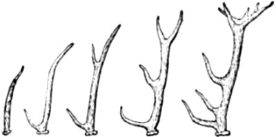
individuals belonging to the same group of animals. For instance, the
successive development of prongs in the horns of deer-like animals,
which is so clearly shown in the geological history of this tribe, is
closely reproduced
in the life-history of existing deer. Or, in other words, the antlers
of an existing deer furnish in their development a kind of
résumé, or recapitulation, of the successive phases
whereby the primitive horn was gradually superseded by horns presenting
a greater and greater number of prongs in successive species of extinct
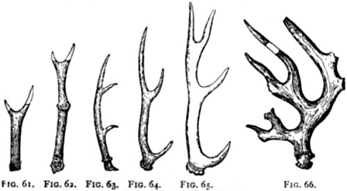
deer (Fig. 26). Now it must be obvious that such a recapitulation in the
life-history of an existing animal of developmental changes successively
distinctive of sundry allied, though now extinct species, speaks
strongly in favour of evolution. For as it is of the essence of this
theory that new forms arise from older forms by way of hereditary
descent, we should antecedently expect, if the theory is true, that the
phases of development presented by the individual organism would follow,
in their main outlines, those phases of development through which their
long line of ancestors had passed. The only alternative view is that as
species of deer, for instance, were separately created, additional
prongs were successively added to their antlers; and yet that, in order
to be so added to successive species every individual deer belonging to
later species was required to repeat in his own lifetime the process of
successive additions which had previously taken place in a remote series
of extinct species. Now I do not deny that this view is a possible view;
but I do deny that it is a probable one. According to the evolutionary
interpretation of such facts, we can see a very good reason why the
life-history of the individual is thus a condensed
résumé of the life-history of its ancestral species. But
according to the opposite view no reason can be assigned why such should
be the case. In a previous chapter—the
chapter on Classification—we have seen that if each species were
created separately, no reason can be assigned why they should all have
been turned out upon structural patterns so strongly suggestive of
hereditary descent with gradual modifications, or slow
divergence—the result being group subordinated to group, with the
most generalized (or least developed) forms at the bottom, and the
highest products of organization at the top. And now we see—or
shall immediately see—that this consideration admits of being
greatly fortified by a study of the developmental history of every
individual organism. If it would be an unaccountable fact that every
separately created species should have been created with close
structural resemblances to a certain limited number of other species,
less close resemblances to certain further species, and so backwards;
assuredly it would be a still more unaccountable fact that every
individual of every species should exhibit in its own person a history
of developmental change, every term of which corresponds with the
structural peculiarities of its now extinct predecessors—and this
in the exact historical order of their succession in geological time.
The more that we think about this antithesis between the naturalistic
and the non-naturalistic interpretations, the greater must we feel the
contrast in respect of rationality to become; and, therefore, I need not
spend time by saying anything further upon the antecedent standing of
the two theories in this respect. The evidence, then, which I am about
to adduce from the study of development in the life-histories of
individual organisms, will be regarded by me as so much unquestionable
evidence in favour of
similar processes of development in the life-histories of their
respective species—in so far, I mean, as the two sets of changes
admit of being proved parallel.
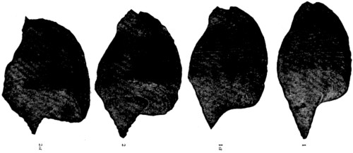
[100]
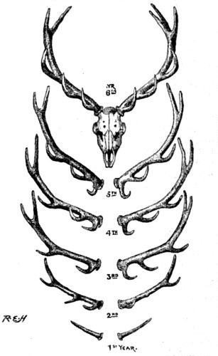
Fig. 26.—Antlers of Stag, showing successive addition of
branches in successive years. Drawn from nature (Brit. Mus.).
In the only illustration hitherto adduced—viz. that of deers’
horns—the series of changes from a one-pronged horn to a fully
developed arborescent antler, is a series which takes place during the
adult life of the animal; for it is only when the breeding age has been
attained that horns are required to appear. But seeing that every animal
passes through most of the phases of its development, not only before
the breeding age has been attained, but even before the time of its own
birth, clearly the largest field for the study of individual development
is furnished by embryology. For instance, there is a salamander which
differs from most other salamanders in being exclusively terrestrial in
its habits. Now, the young of this salamander before their birth are
found to be furnished with gills, which, however, they are never
destined to use. Yet these gills are so perfectly formed, that if the
young salamanders be removed from the body of their mother shortly
before birth, and be then immediately placed in water, the little
animals show themselves quite capable of aquatic respiration, and will
merrily swim about in a medium which would quickly drown their own
parent. Here, then, we have both morphological and physiological
evidence pointing to the possession of gills by the ancestors of the
land salamander.
It would be easy to devote the whole of the present chapter to an
enumeration of special instances of the kinds thus chosen for purposes
of illustration; but as it is desirable to take a deeper, and therefore
a more general view of the whole subject, I will begin at the
foundation, and gradually work up from the earliest stages of
development to the latest. Before starting, however, I ask the reader to
bear in mind one consideration, which must reasonably prevent our
anticipating that in every case the life-history of an individual
organism should present a full recapitulation of the life-history of
its ancestral line of species. Supposing the theory of evolution to be
true, it must follow that in many cases it would have been more or less
disadvantageous to a developing type that it should have been obliged to
reproduce in its individual representatives all the phases of
development previously undergone by its ancestry—even within the
limits of the same family. We can easily understand, for example, that
the waste of material required for building up the useless gills of the
embryonic salamanders is a waste which, sooner or later, is likely to be
done away with; so that the fact of its occurring at all is in itself
enough to show that the change from aquatic to terrestrial habits on the
part of this species must have been one of comparatively recent
occurrence. Now, in as far as it is detrimental to a developing type
that it should pass through any particular ancestral phases of
development, we may be sure that natural selection—or whatever
other adjustive causes we may suppose to have been at work in the
adaptation of organisms to their surroundings—will constantly seek
to get rid of this necessity, with the result, when successful, of
dropping out the detrimental phases. Thus the foreshortening of
developmental history which takes place in the individual lifetime may
be expected often to take place, not only in the way of
condensation, but also in the way of excision. Many pages of ancestral
history may be recapitulated in the paragraphs of embryonic development,
while others may not be so much as mentioned. And that this is the true
explanation of what embryologists term “direct”
development—or of a more or less sudden leap from one phase to
another, without any appearance of intermediate phases—is proved
by the fact that in some cases both direct and indirect development
occur within the same group of organisms, some genera or families having
dropped out the intermediate phases which other genera or families
retain.
The argument from embryology must be taken to begin with the first
beginning of individual life in the ovum. And, in order to understand
the bearings of the argument in this its first stage, we must consider
the phenomena of reproduction in the simplest form which these phenomena
are known to present.
The whole of the animal kingdom is divided into two great groups, which
are called the Protozoa and the Metazoa. Similarly, the whole of the
vegetable kingdom is divided into the Protophyta and the Metaphyta. The
characteristic feature of all the Protozoa and Protophyta is that the
organism consists of a single physiological cell, while the
characteristic of all the Metazoa and Metaphyta is that the organism
consists of a plurality of physiological cells, variously modified to
subserve different functions in the economy of the animal or plant, as
the case may be. For the sake of brevity, I shall hereafter deal only
with the case of animals (Protozoa and Metazoa); but it may throughout
be understood that everything
which is said applies also to the case of plants (Protophyta and
Metaphyta).
A Protozoön (like a Protophyton) is a solitary cell, or a
“unicellular organism,” while a Metazoön (like a Metaphyton)
is a society of cells, or a “multicellular organism.” Now,
it is only in the multicellular organisms that there is any observable
distinction of sex. In all the unicellular organisms the phenomena of
reproduction appear to be more or less identical with those of growth.
Nevertheless, as these phenomena are here in some cases suggestively
peculiar, I will consider them more in detail.
A Protozoön is a single corpuscle of protoplasm which in different
species of Protozoa varies in size from more than one inch to less than
1/1000 of an inch in diameter. In some species there is an enveloping
cortical substance; in other species no such substance can be detected.
Again, in most species there is a nucleus, while in other species no
such differentiation of structure has hitherto been observed.
Nevertheless, from the fact that the nucleus occurs in the majority of
Protozoa, coupled with the fact that the demonstration of this body is
often a matter of extreme difficulty, not only in some of the Protozoa
where it has been but recently detected, but also in the case of certain
physiological cells elsewhere,—from these facts it is not unreasonable
to suppose that all the Protozoa possess a nucleus, whether or not it
admits of being rendered visible by histological methods thus far at our
disposal. If this is the case, we should be justified in saying, as I
have said, that a Protozoön is an isolated physiological cell, and, like
cells in general, multiplies by means of what Spencer and Häckel
have aptly called a process of discontinuous growth. That is to say,
when a cell reaches maturity, further growth takes place in the
direction of a severance of its substance—the separated portion
thus starting anew as a distinct physiological unit. But,
notwithstanding the complex changes which have been more recently
observed to take place in the nucleus of some Protozoa prior to their
division, the process of multiplication by division may still be
regarded as a process of growth, which differs from the previous growth
of the individual cell in being attended by a severance of continuity.
If we take a suspended drop of gum, and gradually add to its size by
allowing more and more gum to flow into it, a point will eventually be
reached at which the force of gravity will overcome that of cohesion,
and a portion of the drop will fall away from the remainder. Here we
have a rough physical simile, although of course no true analogy. In
virtue of a continuous assimilation of nutriment, the protoplasm of a
cell increases in mass, until it reaches the size at which the forces of
disruption overcome those of cohesion—or, in other words, the
point at which increase of size is no longer compatible with continuity
of substance. Nevertheless, it must not be supposed that the process is
thus merely a physical one. The phenomena which occur even in the
simplest—or so-called “direct"—cell-division, are of
themselves enough to prove that the process is vital, or physiological;
and this in a high degree of specialization. But so, likewise, are all
processes of growth in organic structures; and therefore the simile of
the drop of gum is not to be regarded as a true analogy: it serves only
to
indicate the fact that when cell-growth proceeds beyond a certain point
cell-division ensues. The size to which cells may grow before they thus
divide is very variable in different kinds of cells; for while some may
normally attain a length of ten or twelve inches, others divide before
they measure 1/1000 of an inch. This, however, is a matter of detail,
and does not affect the general physiological principles on which we are
at present engaged.
Now, as we have seen, a Protozoön is a single cell; for even although in
some of the higher forms of protozoal life a colony of cells may be
bound together in organic connexion, each of these cells is in itself an
“individual,” capable of self-nourishment, reproduction,
and, generally, of independent existence. Consequently, when the growth
of a Protozoön ends in a division of its substance, the two parts wander
away from each other as separate organisms. (Fig. 27.)
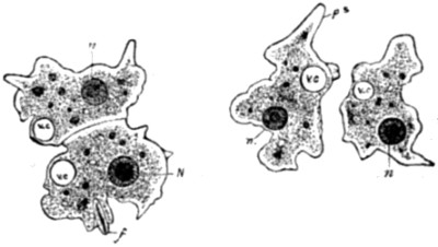
Fig. 27.—Fission of a Protozoön. In the left-hand
drawing the process is represented as having advanced sufficiently far
to have caused a division and segregation both of the nucleus and the
vesicle. In the right-hand drawing the process is represented as
complete. n, N, severed nucleus; vc, severed vesicle; ps,
pseudopodia; f, ingested food.
The next point we have to observe is, that in all cases where a cell or
a Protozoön multiplies by way of fissiparous division, the process
begins in the nucleus. If the nucleus divides into two parts, the whole
cell will eventually divide into two parts, each of which retains a
portion of the original nucleus, as represented in the above figure. If
the nucleus divides into three, four, or even, as happens in the
development of some embryonic tissues, into as many as six parts, the
cell will subdivide into a corresponding number, each retaining a
portion of the nucleus. Therefore, in all cases of fissiparous division,
the seat or origin of the process is the nucleus.
Thus far, then, the phenomena of multiplication are identical in all the
lowest or unicellular organisms, and in the constituent cells of all the
higher or multicellular. And this is the first point which I desire to
make apparent. For where the object is to prove a continuity between the
phenomena of growth and reproduction, it is of primary importance to
show—1st, that there is such a continuity in the case of all the
unicellular organisms, and, 2nd, that there are all the above points of
resemblance between the multiplication of cells in the unicellular and
in the multicellular organisms.
It remains to consider the points of difference, and, if possible, to
show that these do not go to disprove the doctrine of continuity which
the points of resemblance so forcibly indicate.
The first point of difference obviously is, that in the case of all the
multicellular organisms the two or more “daughter-cells,”
which are produced by division of the “mother-cell,” do not
wander away from one
another; but, as a rule, they continue to be held in more or less close
apposition by means of other cells and binding membranes,—with the
result of giving rise to those various “tissues,” which in
turn go to constitute the material of “organs.” I cannot
suppose, however, that any advocate of discontinuity will care to take
his stand at this point. But, if any one were so foolish as to do so, it
would be easy to dislodge him by describing the state of matters in some
of the Protozoa where a number of unicellular “individuals”
are organically united so as to form a “colony.” These cases
serve to bridge this distinction between Protozoa and Metazoa, of which
therefore we may now take leave.
In the second place, there is the no less obvious distinction that the
result of cell-division in the Metazoa is not merely to multiply cells
all of the same kind: on the contrary, the process here gives rise to as
many different kinds of cells as there are different kinds of tissue
composing the adult organism. But no one, I should think, is likely to
oppose the doctrine of continuity on the ground of this distinction. For
the distinction is clearly one which must necessarily arise, if the
doctrine of continuity between unicellular and multicellular organisms
be true. In other words, it is a distinction which the theory of
evolution itself must necessarily pre-suppose, and therefore it is no
objection to the theory that its pre-supposition is realized. Moreover,
as we shall see better presently, there is no difficulty in
understanding why this distinction should have arisen, so soon as it
became necessary (or desirable) that individual cells, when composing a
“colony,” should
conform to the economic principle of the division of labour—a
principle, indeed, which is already foreshadowed in the constituent
parts of a single cell, since the nucleus has one set of functions and
its surrounding protoplasm another.
But now, in the third place, we arrive at a more important distinction,
and one which lies at the root of the others still remaining to be
considered. I refer to sexual propagation. For it is a peculiarity of
the multicellular organisms that, although many of them may likewise
propagate themselves by other means (Fig. 28), they all propagate
themselves by means of sexual congress. Now, in its essence, sexual
congress consists in the fusion of two specialized cells (or, as now
seems almost certain, of the nuclei thereof), so that it is out of such
a combination that the new individual arises by means of successive
cell-divisions, which, beginning in the fertilized ovum, eventually
build up all the tissues and organs of the body.
[111]
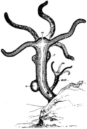
Fig. 28.—Hydra viridis, partly in section. M, mouth; O,
ovary, or bud containing female reproductive cells; T, testis, or bud
containing male reproductive cells. In addition to these buds containing
germinal elements alone, there is another which illustrates the process
of “gemmation"—i. e. the direct out-growth of a fully formed
offspring.
This process clearly indicates very high specialization on the part of
germ-cells. For we see by it that although these cells when young
resemble all other cells in being capable of self-multiplication by
binary division (thus reproducing cells exactly like themselves), when
older they lose this power; but, at the same time, they acquire an
entirely new and very remarkable power of giving rise to a vast
succession of many different kinds of cells, all of which are mutually
correlated as to their several functions, so as to constitute a
hierarchy of cells—or, to speak literally, a multicellular
co-organization. Here it is that we touch the really important
distinction between the Protozoa and the Metazoa; for although I have
said that some of the higher Protozoa foreshadow this state of matters
in forming cell-colonies, it must now be noted that the cells composing
such colonies are all of the same kind; and, therefore, that the
principle of producing different kinds of cells which, by mutual
co-adaptation of functions, shall be capable of constructing a
multicellular Metazoön,—this great principle of co-organization is
but dimly nascent in the cell-colonies
of Protozoa. And its marvellous development in the Metazoa appears
ultimately to depend upon the highly specialized character of
germ-cells. Even in cases where multicellular organisms are capable of
reproducing their kind without the need of any preceding process of
fertilization (parthenogenesis), and even in the still more numerous
cases where complete organisms are budded forth from any part of their
parent organism (gemmation, Fig. 28), there is now very good reason to
conclude that these powers of a-sexual reproduction on the part of
multicellular organisms are all ultimately due to the specialized
character of their germ-cells. For in all these cases the tissues of the
parent, from which the budding takes place, were ultimately derived from
germ-cells—no matter how many generations of budded organisms may
have intervened. And that propagation by budding, &c., in
multicellular organisms is thus ultimately due to their propagation by
sexual methods, seems to be further shown by certain facts which will
have to be discussed at some length in my next volume. Here, therefore,
I will mention only one of them—and this because it furnishes what
appears to be another important distinction between the Protozoa and the
Metazoa.
In nearly all cases where a Protozoön multiplies itself by fission, the
process begins by a simple division of the nucleus. But when a Metazoön
is developed from a germ-cell, although the process likewise begins by a
division of the nucleus, this division is not a simple or direct one; on
the contrary, it is inaugurated by a series of processes going on within
the nucleus, which are so enormously complex, and withal so
beautifully ordered, that to my mind they constitute the most
wonderful—if not also the most suggestive—which have ever
been revealed by microscopical research. It is needless to say that I
refer to the phenomena of karyokinesis. A few pages further on they will
be described more fully. For our present purposes it is sufficient to
give merely a pictorial illustration of their successive phases; for a
glance at such a representation serves to reveal the only point to which
attention has now to be drawn—namely, the immense complexity of
the processes in question, and therefore the contrast which they furnish
to the simple (or “direct") division of the nucleus preparatory to
cell-division in the unicellular organisms. Here, then
(Fig. 29), we see the complex processes of karyokinesis in the first
two stages of egg-cell division. But similar processes continue to
repeat themselves in subsequent stages; and this, there is now good
reason to believe, throughout all the stages of cell-division, whereby
the original egg-cell eventually constructs an entire organism. In other
words, all the cells composing all the tissues of a multicellular
organism, at all stages of its development, are probably originated by
these complex processes, which differ so much from the simple process of
direct division in the unicellular organisms[9].
In this important respect, therefore, it does at first sight appear that
we have a distinction between the Protozoa and the Metazoa of so
pronounced a character, as fairly to raise the question whether
cell-division is fundamentally identical in unicellular and in
multicellular organisms.
[113i.]
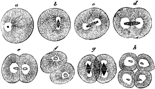
Fig. 29.—Successive stages in the division of the ovum,
or egg-cell, of a worm. (After Strasburger.) a to d show the changes
taking place in the nucleus and surrounding cell-contents, which result
in the first segmentation of the ovum at e; f and g show a
repetition of these changes in each of the two resulting cells, leading
to the second segmentation stage at h.
Lastly, the only other distinction of a physiologically significant kind
between a single cell when it occurs as a Protozoön and when it does so
as the unfertilized ovum of a Metazoön is, that in the latter case the
nucleus discharges from its own substance two minute protoplasmic masses
("polar bodies"), which are then eliminated from the cell altogether.
This process, which will be more fully described later on, appears to be
of invariable occurrence in the case of all egg-cells,
while nothing resembling it has ever been observed in any of the
Protozoa.
We must now consider these several points of difference seriatim.
First, with regard to sexual propagation, we have already seen that this
is by no means the only method of propagation among the multicellular
organisms; and it now remains to add that, on the other hand, there is,
to say the least, a suggestive foreshadowing of sexual propagation among
the unicellular organisms. For although simple binary fission is here
the more usual mode of multiplication, very frequently two (rarely three
or more) Protozoa of the same species come together, fuse into a single
mass, and thus become very literally “one flesh.” This
process of “conjugation” is usually (though by no means
invariably) followed by a period of quiescent “encystation"; after
which the contents of the cyst escape in the form of a number of minute
particles, or “spores,” and these severally develope into
the parent type. Obviously this process of conjugation, when it is thus
a preliminary to multiplication, appears to be in its essence the same
as fertilization. And if it be objected that encystation and
spore-formation in the Protozoa are not always preceded by conjugation,
the answer would be that neither is oviparous propagation in the Metazoa
invariably preceded by fertilization.
Nevertheless, that there are great distinctions between true sexual
propagation and this foreshadowing of it in conjugation I do not deny.
The question, however, is whether they be so great as to justify any
argument against an historical continuity between them. What, then, are
these remaining
distinctions? Briefly, as we have seen, they are the extrusion from
egg-cells of polar bodies, and the occurrence, both in egg-cells and
their products (tissue-cells), of the process of karyokinesis. But, as
regards the polar bodies, it is surely not difficult to suppose that,
whatever their significance may be, it is probably in some way or
another connected with the high specialization of the functions which an
egg-cell has to discharge. Nor is there any difficulty in further
supposing that, whatever purpose is served by getting rid of polar
bodies, the process whereby they are got rid of was originally one of
utilitarian development—i. e. a process which at its commencement
did not betoken any difference of kind, or breach of continuity, between
egg-cells and cells of simpler constitution.
Lastly, with respect to karyokinesis, although it is true that the
microscope has in comparatively recent years displayed this apparently
important distinction between unicellular and multicellular organisms,
two considerations have here to be supplied. The first is, that in some
of the Protozoa processes very much resembling those of karyokinesis
have already been observed taking place in the nucleus preparatory to
its division. And although such processes do not present quite the same
appearances as are to be met with in egg-cells, neither do the
karyokinetic processes in tissue-cells, which in their sundry kinds
exhibit great variations in this respect. Moreover, even if such were
not the case, the bare fact that nuclear division is not invariably of
the simple or direct character in the case of all Protozoa, is
sufficient to show that the distinction now before us—like the one last
dealt with—is by no means
absolute. As in the case of sexual propagation, so in that of
karyokinesis, processes which are common to all the Metazoa are not
wholly without their foreshadowings in the Protozoa. And seeing how
greatly exalted is the office of egg-cells—and even of
tissue-cells—as compared with that of their supposed ancestry in
protozoal cells, it seems to me scarcely to be wondered at if their
specializations of function should be associated with corresponding
peculiarities of structure—a general fact which would in no way
militate against the doctrine of evolution. Could we know the whole
truth, we should probably find that in order to endow the most primitive
of egg-cells with its powers of marshalling its products into a living
army of cell-battalions, such an egg-cell must have been passed through
a course of developmental specialization of so elaborate a kind, that
even the complex processes of karyokinesis are but a very inadequate
expression thereof.
Probably I have now said enough to show that, remarkable and altogether
exceptional as the properties of germ-cells of the multicellular
organisms unquestionably show themselves to be, yet when these
properties are traced back to their simplest beginnings in the
unicellular organisms, they may fairly be regarded as fundamentally
identical with the properties of living cells in general. Thus viewed,
no line of real demarcation can be drawn between growth and
reproduction, even of the sexual kind. The one process is, so to speak,
physiologically continuous with the other; and hence, so far as the
pre-embryonic stage of life-history is concerned, the facts cannot
fairly be regarded as out of keeping with the theory of evolution.
I will now pass on to consider the embryogeny of the Metazoa, beginning
at its earliest stage in the fertilization of the ovum. And here it is
that the constructive argument in favour of evolution which is derived
from embryology may be said properly to commence. For it is surely in
itself a most suggestive fact that all the Metazoa begin their life in
the same way, or under the same form and conditions. Omne vivum ex
ovo. This is a formula which has now been found to apply throughout the
whole range of the multicellular organisms. And seeing, as we have just
seen, that the ovum is everywhere a single cell, the formula amounts to
saying that, physiologically speaking, every Metazoön begins its life as
a Protozoön, and every Metaphyton as a Protophyton[10].
Now, if the theory of evolution is true, what should we expect to happen
when these germ-cells are fertilized, and so enter upon their severally
distinct processes of development? Assuredly we should expect to find
that the higher organisms pass through the same phases of development as
the lower organisms, up to the time when their higher characters begin
to become apparent. If in the life-history of species these higher
characters were gained by gradual improvement upon lower characters, and
if the development of the higher individual is now a general
recapitulation of that of its ancestral species, in studying this
recapitulation we should expect to find the higher organism successively
unfolding its higher characters from the lower ones through which its
ancestral species had previously passed. And this is just what we do
find. Take, for example, the case of the highest organism, Man. Like
that of all other organisms, unicellular or multicellular, his
development starts from the nucleus of a single cell. Again, like that
of all the Metazoa and Metaphyta, his development starts from the
specially elaborated nucleus of an egg-cell, or a nucleus which has been
formed by the fusion of a male with a female element[11].
When his animality becomes established, he exhibits the fundamental
anatomical qualities which characterize such lowly animals as polyps and
jelly-fish. And even when he is marked off as a Vertebrate, it cannot be
said whether he is to be a fish, a reptile, a bird, or a beast. Later on
it becomes evident that he is to be a Mammal; but not till later still
can it be said to which order of mammals he belongs.
Here, however, we must guard against an error which is frequently met
with in popular expositions of this subject. It is not true that the
embryonic phases in the development of a higher form always resemble so
many adult stages of lower forms. This may or may not be the case; but
what always is the case
is, that the embryonic phases of the higher form resemble the
corresponding phases of the lower forms. Thus, for example, it would be
wrong to suppose that at any stage of his development a man resembles a
jelly-fish. What he does resemble at an early stage of his development
is the essential or groundplan of the jelly-fish, which that animal
presents in its embryonic condition, or before it begins to assume its
more specialized characters fitting it for its own particular sphere of
life. The similarities, therefore, which it is the function of
comparative embryology to reveal are the similarities of type or
morphological plan: not similarities of specific detail. Specific
details may have been added to this, that, and the other species for
their own special requirements, after they had severally branched off
from the common ancestral stem; and so could not be expected to recur in
the life-history of an independent specific branch. The comparison
therefore must be a comparison of embryo with embryo; not of embryos
with adult forms.
In order to give a general idea of the results thus far yielded by a
study of comparative embryology in the present connexion, I will devote
the rest of this chapter to giving an outline sketch of the most
important and best established of these results.
Histologically the ovum, or egg-cell, is nearly identical in all
animals, whether vertebrate or invertebrate. Considered as a cell it is
of large size, but actually it is not more than 1/100, and may be less
than 1/200 of an inch in diameter. In man, as in most mammals, it is
about 1/120. It is a more or less spherical body, presenting a thin
transparent envelope, called
the zona pellucida, which contains—first, the protoplasmic
cell-substance or “yolk,” within which lies, second, the
nucleus or germinal vesicle, within which again lies, third, the
nucleolus or germinal spot. This description is true of the egg-cells of
all animals, if we add that in the case of the lowest animals—such
as sponges, &c.—there is no enveloping membrane: the egg-cell
is here a naked cell, and its constituent protoplasm, being thus
unconfined, is free to perform protoplasmic movements, which it does
after the manner, and with all the activity, of an amœba. But even
with respect to this matter of an enveloping membrane, there is no
essential difference between an ovum of the lowest and an ovum of the
highest animals. For in their early stages of development within the
ovary the ova of the highest animals are likewise in the condition of
naked cells, exhibiting amœbiform movements; the enveloping
membrane of an ovum being the product of a later development.
Moreover this membrane, when present, is usually provided with one or
more minute apertures, through which the spermatozoön passes when
fertilizing the ovum. It is remarkable that the spermatozoa know, so to
speak, of the existence of these gate-ways,—their snake-like
movements being directed towards them, presumably by a stimulus due to
some emanation therefrom[12].
In the mammalian ovum, however, these apertures are exceedingly minute,
and distributed
all round the circumference of the pellucid envelope, as represented in
this illustration (Fig. 32).
[121i.]
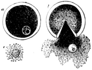
Fig. 30.—Ovarian ovum of a Mammal, (a) magnified and
viewed under pressure, (b) burst by increased pressure, with yolk and
nucleus escaping: (c) the nucleus more freed from yolk-substance.
(From Quain’s Anatomy, after Allen Thomson.)
[122]
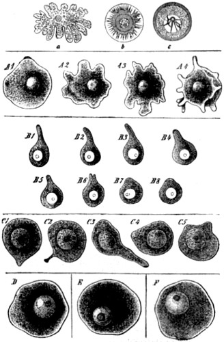
Fig. 31.—Amœboid movements of young egg-cells, a,
Amœboid ovum of Hydra (from Balfour, after Kleitnenberg); b, early
ovum of Toxopneustes variegatus, with pseudopodia-like processes (from
Balfour, after Selenka); c, ovum of Toxopneustes lividus, more nearly
ripe (from Balfour, Hertwig). A1 to A4, the primitive egg-cell of a
Chalk-Sponge (Leuculmis echinus), in four successive conditions of
motion. B1 to B8, ditto of a Hermit-Crab (Chondracanthus cornutus), in
eight successive stages (after E. von Beneden). C1 to C5, ditto of a
Cat, in five successive stages (after Pflüger). D, ditto of Trout; E, of
a Hen; F, of Man. The first series is taken from the Encycl. Brit.;
the second from Häckel’s Evolution of Man.
[123i.]
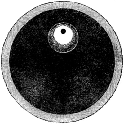
Fig. 32.—Human ovum, mature and greatly magnified.
(After Häckel.)
In thus saying that the ova of all animals are, so far as microscopes
can reveal, substantially similar, I am of course speaking of the
egg-cell proper, and not of what is popularly known as the egg. The egg
of a bird, for example, is the egg-cell, plus an enormous aggregation
of nutritive material, an egg-shell, and sundry other structures suited
to the subsequent development of the egg-cell when separated from the
parent’s body. But all these accessories are, from our present
point of view, accidental or adventitious. What we have now to
understand by the ovum, the egg, or the egg-cell, is the microscopical
germ which I have just described. So far then as this germ is concerned,
we find that all multicellular organisms begin their existence in the
same kind of structure, and that this structure is anatomically
indistinguishable from that of the permanent form presented by the
lowest, or unicellular organisms. But although anatomically
indistinguishable, physiologically they present the sundry peculiarities
already mentioned.
Now I have endeavoured to show that none of these peculiarities are such
as to exclude—or even so much as to invalidate—the supposition of
developmental continuity between the lowest egg-cells and the highest
protozoal cells. It remains to show in this place, and on the other
hand, that there is no breach of continuity between the lowest and the
highest egg-cells; but, on the contrary, that the remarkable uniformity
of the complex processes whereby their peculiar characters are exhibited
to the histologist, is such as of itself to sustain the doctrine of
continuity
in a singularly forcible manner. On this account, therefore, and also
because the facts will again have to be considered in another connexion
when we come to deal with Weismann’s theory of heredity, I will
here briefly describe the processes in question.
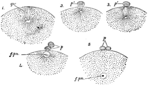
Fig. 33.—Stages in the formation of the polar bodies in
the ovum of a star-fish. (After Hertwig.) g.v., germinal vesicle
transformed into a spindle-shaped system of fibres; p.′, the first
polar body becoming extruded; p., p., both polar bodies fully
extruded; f. pn., female pronucleus, or residue of the germinal
vesicle.
We have already seen that the young egg-cell multiplies itself by simple
binary division, after the manner of unicellular organisms in
general—thereby indicating, as also by its amœbiform movements, its
fundamental identity with such organisms in kind. But, as we have
likewise seen, when the ovum ceases to resemble these organisms, by
taking on its higher degree of functional capacity, it is no longer able
to multiply itself in this manner. On the contrary, its cell-divisions
are now of an endogenous character,
and result in the formation of many different kinds of cells, in the
order required for constructing the multicellular organism to which the
whole series of processes eventually give rise. We have now to consider
these processes seriatim.
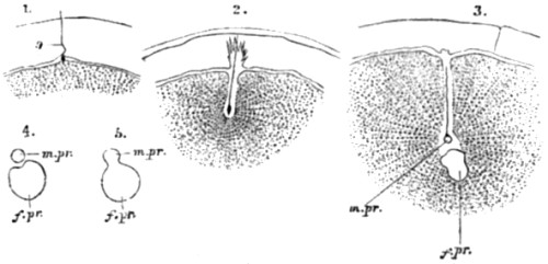
Fig. 34.—Fertilization of the ovum of an echinoderm.
(From Quain’s Anatomy, after Selenka.) S, spermatozoön; m.
pr., male pronucleus; f. pr., female pronucleus. 1 to 4 correspond to
D to G in the next figure.
First of all the nucleus discharges its polar bodies, as previously
mentioned, and in the manner here depicted on the previous page. (Fig.
33.) It will be observed that the nucleus of the ovum, or the germinal
vesicle as it is called, gets rid first of one and afterwards of the
other polar body by an “indirect,” or karyokinetic, process
of division. (Fig. 33.) Extrusion of these bodies from the ovum (or it
may be only from the nucleus) having been accomplished, what remains of
the nucleus retires from the circumference of the ovum, and is called
the female pronucleus. (Fig. 33. f. pn.) The ovum is now ready for
fertilization. A similar emission of nuclear substance is said by some
good
observers to take place also from the male germ-cell, or
spermatozoön, at or about the close of its development. The
theories to which these facts have given rise will be considered in
future chapters on Heredity.
Turning now to the mechanism of fertilization, the diagrams (Figs. 34,
35) represent what happens in the case of star-fish.
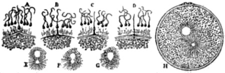
Fig. 35.—Fertilization of the ovum of a star-fish. (From
the Encycl. Brit. after Fol.) A, spermatozoa in the mucilaginous coat
of the ovum; a prominence is rising from the surface of the ovum towards
a spermatozoön; B, they have almost met; C, they have met; D, the
spermatozoön enters the ovum through a distinct opening; H, the entire
ovum, showing extruded polar bodies on its upper surface, and the moving
together of the male and female pronuclei; E, F, G, meeting and
coalescence of the pronuclei.
The sperm-cell, or spermatozoön, is seen in the act of penetrating the
ovum. In the first figure it has already pierced the mucilaginous coat
of the ovum, the limit of which is represented by a line through which
the tail of the spermatozoön is passing: the head of the spermatozoön is
just entering the ovum proper. It may be noted that, in the case of many
animals, the general protoplasm of the ovum becomes aware, so to speak,
of the approach of a spermatozoön, and sends up a process to meet it.
(Fig. 35, A, B, C.) Several—or even many—spermatozoa may thus enter
the coat of the ovum; but normally only one proceeds further, or right
into the substance of the ovum, for the
purpose of effecting fertilization. This spermatozoön, as soon as
it enters the periphery of the yolk, or cell-substance proper, sets up a
series of remarkable phenomena. First, its own head rapidly increases in
size, and takes on the appearance of a cell-nucleus: this is called the
male pronucleus. At the same time its tail begins to disappear, and the
enlarged head proceeds to make its way directly towards the nucleus of
the ovum which, as before stated, is now called the female pronucleus.
The latter in its turn moves towards the former, and when the two meet
they fuse into one mass, forming a new nucleus. Before the two actually
meet, the spermatozoön has lost its tail altogether; and it is
noteworthy that during its passage through the protoplasmic
cell-contents of the ovum, it appears to exercise upon this protoplasm
an attractive influence; for the granules of the latter in its vicinity
dispose themselves around it in radiating lines. All these various
phenomena are depicted in the above wood-cuts. (Figs. 34, 35.)
Fertilization having been thus effected by fusion of the male and female
pronuclei into a single (or new) nucleus, this latter body proceeds to
exhibit complicated processes of karyokinesis, which, as before shown,
are preliminary to nuclear division in the case of egg-cells. Indeed the
karyokinetic process may begin in both the pronuclei before their
junction is effected; and, even when their junction is effected, it does
not appear that complete fusion of the so-called chromatin elements of
the two pronuclei takes place. For the purpose of explaining what this
means, and still more for the purpose of giving a general idea of the
karyokinetic processes as a whole,
I will quote the following description of them, because, for terseness
combined with lucidity, it is unsurpassable.
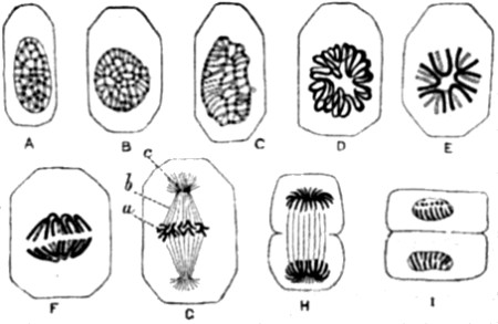
Fig. 36.—Karyokinesis of a typical tissue-cell
(epithelium of Salamander). (After Flemming and Klein.) The series from
A to I represents the successive stages in the movement of the chromatin
fibres during division, excepting G, which represents the
“nucleus-spindle” of an egg-cell. A, resting nucleus; D,
wreath-form; E, single star, the loops of the wreath being broken; F,
separation of the star into two groups of U-shaped fibres; H, diaster or
double star; I, completion of the cell-division and formation of two
resting nuclei. In G the chromatin fibres are marked a, and correspond
to the “equatorial plate"; b, achromatin fibres forming the
nucleus-spindle; c, granules of the cell-protoplasm forming a
“polar star.” Such a polar star is seen at each end of the
nucleus-spindle, and is not to be confused with the diaster H, the two
ends of which are composed of chromatin.
Researches, chiefly due to Flemming, have shown that the nucleus in
very many tissues of higher plants and animals consists of a
capsule containing a plasma of “achromatin,” not deeply
stained by re-agents, ramifying in which is a reticulum of
“chromatin” consisting of fibres which readily take a
deep stain. (Fig. 36, A). Further it is demonstrated that, when the
cell is about to divide into two, definite and very remarkable
movements take place in the nucleus, resulting in the disappearance
of the capsule and in the arrangement of its fibres first in the
form of a wreath (D), and subsequently (by the breaking of the loops
formed by the fibres) in the form of a star (E). A further movement
within the nucleus leads to an arrangement of the broken loops in two
groups (F), the position of the open ends of the broken loops being
reversed as compared with what previously obtained. Now the two groups
diverge, and in many cases a striated appearance of the achromatin
substance between the two groups of chromatin loops is observable (H).
In some cases (especially egg-cells) this striated arrangement of the
achromatin is then termed a “nucleus-spindle,” and the group
of chromatin loops (G, a) is known as “the equatorial
plate.” At each end of the nucleus-spindle in these cases there is
often seen a star consisting of granules belonging to the general
protoplasm of the cell (G, c). These are known as “polar
stars.” After the separation of the two sets of loops (H) the
protoplasm of the general substance of the cell becomes constricted, and
division occurs, so as to include a group of chromatin loops in each of
the two fission products. Each of these then rearranges itself together
with the associated chromatin into a nucleus such as was present in the
mother cell to commence with (I)[13].
Since the above was published, however, further progress has been made.
In particular it has been found that the chromatin fibres pass from
phase D to phase F by a process of longitudinal splitting (Fig. 37 g,
h; Fig. 38, VI, VII)—which is a point of great importance for
Weismann’s theory of heredity,—and that the protoplasm outside
the nucleus seems to take as important a part in the karyokinetic
process as does the nuclear substance. For the so-called
“attraction-spheres” (Fig. 38 II a, III, III a, VIII to
XII), which were at first supposed to be of subordinate importance in
the process as a whole, are now known to take an exceedingly active part
in it (see especially IX to XI). Lastly, it may be added that there is a
growing consensus of authoritative opinion, that the chromatin
fibres are the seats of the material of heredity, or, in other words,
that they contain those essential elements of the cell which endow the
daughter-cells with their distinctive characters. Therefore, where the
parent-cell is an ovum, it follows from this view that all hereditary
qualities of the future organism are potentially present in the
ultra-microscopical structure of the chromatin fibres.
[131]
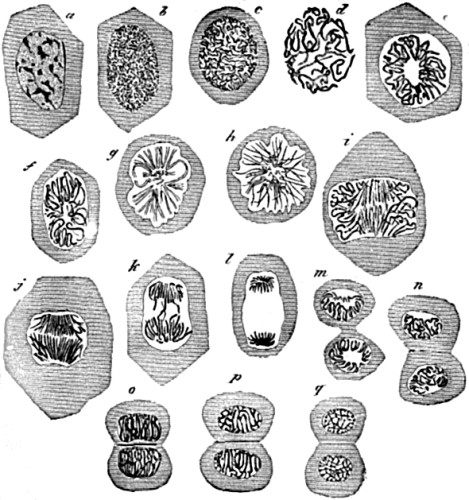
Fig. 37.—Study of successive changes taking place in the
nucleus of an epithelium cell, preparatory to division of the cell.
(From Quain’s Anatomy, after Flemming.) a, resting cell,
showing the nuclear network; b, first stage of division, the
chromatoplasm transformed into a skein of closely contorted filaments;
c to f, further stages in the growth and looping arrangement of the
filaments; g, stellate phase, or aster; h, completion of the
splitting of the filaments, already begun in f and g; i, j, k,
successive stages in separation of the filaments into two groups; l,
the final result of this (diaster); m to q, stages in the division
of the whole cell into two, showing increasing contortion of the
filaments, until they reach the resting stage at q.
[132]
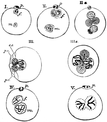
Fig. 38.—Formation and conjugation of the pronuclei in
Ascaris megalocephala. (From Quain’s Anatomy, after E. von
Beneden.) f, female pronucleus; m, male pronucleus; p, one of the
polar bodies.
I. The second polar body has just been extruded; both male and female
pronuclei contain two chromatin particles; those of the male pronucleus
are becoming transformed into a skein. II. The chromatin in both
pronuclei now forms into a skein.
II a. The skeins are more distinct. Two attraction (or protoplasmic)
spheres, each with a central particle united with a small spindle of
achromatic fibres, have made their appearance in the general substance
of the egg close to the mutually approaching pronuclei. The male
pronucleus has the remains of the body of the spermatozoön adhering to
it.
III. Only the female pronucleus is shown in this figure. The skein is
contracted and thickened. The attraction-spheres are near one side of
the ovum, and are connected with its periphery by a cone of fibres
forming a polar circle, p.c.; e.c., equatorial circle.
III a. The pronuclei have come into contact, and the spindle-system is
now arranged across their common axis.
IV. Contraction of the skein, and formation of two U-or V-shaped
chromatin fibres in each pronucleus.
V. The V-shaped chromatin filaments are now quite distinct: the male and
female pronuclei are in close contact.
[133]
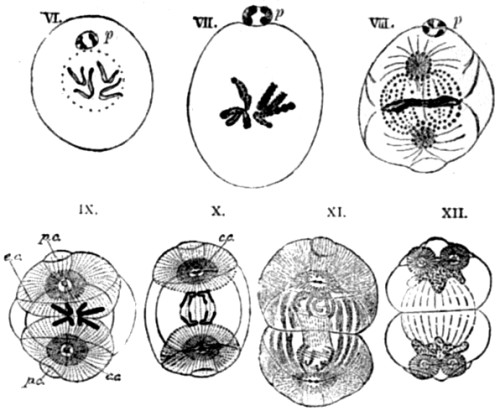
VI., VII. The V-shaped filaments are splitting longitudinally; their
structure of fine granules of chromatin is apparent in VII., which is
more highly magnified. The conjugation of the pronuclei is apparently
complete in VII. The attraction-spheres and achromatic spindle, although
present, are not depicted in IV., V., VI., and VII.
VIII. Equatorial arrangement of the four chromatin loops in the middle
of the now segmenting ovum: the achromatic substance forming a
spindle-shaped system of granules with fibres radiating from the poles
of the spindle (attraction-spheres); the chromatin forms an equatorial
plate. (Compare Fig. 36 G.)
IX. Shows diagrammatically the commencing separation of the chromatin
fibres of the conjugated nuclei, and the system of fibres radiating from
the attraction-spheres. (Compare again Fig. 36 G.) p.c., polar circle;
e.c., equatorial circle; c.c., central particle.
X. Further separation of the chromatin filaments. Each of the central
particles of the attraction-spheres has divided into two.
XI. The chromatin fibres are becoming developed into the skeins of the
two daughter-nuclei. These are still united by fibres of achromatin. The
general protoplasm of the ovum is becoming divided.
XII. The two daughter-nuclei exhibit a chromatin network. Each of the
attraction-spheres has divided into two, which are joined by fibres of
achromatin, and connected with the periphery of the cell in the same way
as in the original or parent sphere, III.
As I shall have more to say about these processes in the next volume,
when we shall see the important part which they bear in Weismann’s
theory of heredity, it is with a double purpose that I here introduce
these yet further illustrations of them upon a somewhat larger scale.
The present purpose is merely that of showing, more clearly than
hitherto, the great complexity of these processes on the one hand, and,
on the other, the general similarity which they display in egg-cells and
in tissue-cells. But as in relation to this purpose the illustrations
speak for themselves, I may now pass on at once to the history of
embryonic development, which follows fertilization of the ovum.
We have seen that when the new nucleus of the fertilized ovum (which is
formed by a coalescence of the male pronucleus with the female) has
completed its karyokinetic processes, it is divided into two equal
parts; that these are disposed at opposite poles of the ovum; and that
the whole contents of the ovum are thereupon likewise divided into two
equal parts, with the result that there are now two nucleated cells
within the spherical wall of the ovum where before there had only been
one. Moreover, we have also seen that a
precisely similar series of events repeat themselves in each of these
two cells, thus giving rise to four cells (see Fig. 29). It must now be
added that such duplication is continued time after time, as shown in
the accompanying illustrations (Figs. 39, 40).
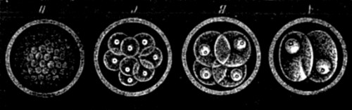
Fig. 39.—Segmentation of ovum. (After Häckel.)
Successive stages are marked by the letters A, B, C. D represents
several stages in advance of C.
All
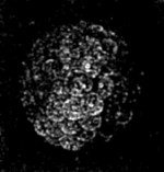 Fig. 40.—The contents of an ovum in an advanced stage of
segmentation, drawn in perspective. (After Häckel.)
this, it will be noticed, is a case of cell-multiplication, which
differs from that which takes place in the unicellular organisms only in
its being invariably preceded (as far as we know) by karyokinesis, and
in the resulting cells being all confined within a common envelope, and
so in not being free to separate. Nevertheless, from what has already
been said, it will also be noticed that this feature makes all the
difference between a Metazoön and a Protozoön; so that already the ovum
presents the distinguishing character of a Metazoön.
Fig. 40.—The contents of an ovum in an advanced stage of
segmentation, drawn in perspective. (After Häckel.)
this, it will be noticed, is a case of cell-multiplication, which
differs from that which takes place in the unicellular organisms only in
its being invariably preceded (as far as we know) by karyokinesis, and
in the resulting cells being all confined within a common envelope, and
so in not being free to separate. Nevertheless, from what has already
been said, it will also be noticed that this feature makes all the
difference between a Metazoön and a Protozoön; so that already the ovum
presents the distinguishing character of a Metazoön.
I have dealt thus at considerable length upon the processes whereby the
originally unicellular ovum and spermatozoön become converted into the
multicellular germ, because I do not know of any other exposition of the
argument from Embryology where this, the first stage of the argument,
has been adequately treated. Yet it is evident that the fact of all the
processes above described being so similar in the case of sexual (or
metazoal) reproduction among the innumerable organisms where it occurs,
constitutes in itself a strong argument in favour of evolution. For the
mechanism of fertilization, and all the processes which even thus far we
have seen to follow therefrom, are hereby shown to be not only highly
complex, but likewise highly specialized. Therefore, the remarkable
similarity which they present throughout the whole animal kingdom—not
to speak of the vegetable—is expressive of organic continuity, rather
than of absolute discontinuity in every case, as the theory of special
creation must necessarily suppose. And it is evident that this argument
is strong in proportion to the uniformity, the specialization, and the
complexity of the processes in question.
Having occupied so much space with supplying what appear to me the
deficiencies in previous expositions of the argument from Embryology, I
can now afford to take only a very general view of the more important
features of this argument as they are successively furnished by all the
later stages of individual development. But this is of little
consequence, seeing that from the point at which we have now arrived
previous expositions of the argument are both good and numerous. The
following then is to be regarded as a mere sketch
Of the evidences of phyletic (or ancestral) evolution, which are so
abundantly furnished by all the subsequent phases of ontogenetic (or
individual) evolution.
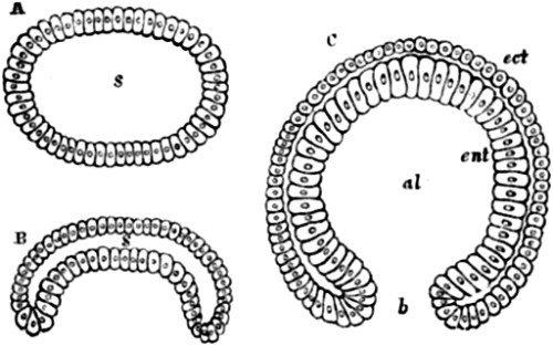
Fig. 41.—Formation of the gastrula of Amphioxus. (After
Kowalevsky.) A, wall of the ovum, composed of a single layer of cells;
B, a stage in the process of gastrulation; C, completion of the process;
S, original or segmentation cavity of ovum; al, alimentary cavity of
gastrula; ect, outer layer of cells; ent, inner layer of cells; b,
orifice, constituting the mouth in permanent forms.
The multicellular body which is formed by the series of segmentations
above described is at first a sphere of cells (Fig. 40). Soon, however,
a watery fluid gathers in the centre, and progressively pushes the cells
towards the circumference, until they there constitute a single layer.
The ovum, therefore, is now in the form of a hollow sphere containing
fluid, confined within a continuous wall of cells (Fig. 41 A). The next
thing that happens is a pitting in of one portion of the sphere (B). The
pit becomes deeper and deeper, until there is a complete invagination of
this part of the sphere—the cells which constitute it being
progressively
pushed inwards until they come into contact with those at the opposite
pole of the ovum. Consequently, instead of a hollow sphere of cells, the
ovum now becomes an open sac, the walls of which are composed of a
double layer of cells (C). The ovum is now what has been called a
gastrula; and it is of importance to observe that probably all the
Metazoa pass through
this stage. At any rate it has been found to occur in all the main
divisions of the animal kingdom, as a glance at the accompanying figures
will serve to show (Fig. 42)[14].
Moreover many of the lower kinds of Metazoa never pass beyond it; but
are all their lives nothing else than gastrulæ, wherein the
orifice becomes the mouth of the animal, the internal or invaginated
layer of cells the stomach, and the outer layer the skin. So that if we
take a child’s india-rubber ball, of the hollow
kind with a hole in it, and push in one side with our fingers till
internal contact is established all round, by then holding the indented
side downwards we should get a very fair anatomical model of a
gastræa form, such as is presented by the adult condition of many
of the most primitive Metazoa—especially the lower Cœlenterata.
The preceding figures represent two
other such forms in nature, the first locomotive and transitory, the
second fixed and permanent (Figs. 43, 44).
[138i.]
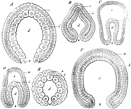
Fig. 42.—Gastrulation. A, Gastrula of a Zoophyte (Gastrophysema). (After Häckel.) B, Gastrula of a Worm (Sagitta). (After Kowalevsky.) C, Gastrula of an Echinoderm
(Uraster). (After A. Agassiz.) D, Gastrula of an Arthropod (Nauplius).
(After Häckel.) E, Gastrula of a Mollusk (Limnæus). (After Rabl.) F, Gastrula of a Vertebrate (Amphioxus). (After Kowalevsky.) In all, d,
indicates the intestinal cavity; o, the primitive mouth; s, the
cleavage-cavity; i, the endoderm, or intestinal layer; e, the
ectoderm or skin-layer.
[139i.]
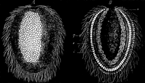
Fig. 43.—Gastrula of a Chalk Sponge. (After Häckel.) A,
External view. B, Longitudinal section. g, digestive cavities; o,
mouth; i, endoderm; e, ectoderm.
[140i.]
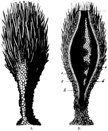
Fig. 44.—Prophysema primordiale, an extant gastræa-form.
(After Häckel.) (A). External view of the whole animal, attached by its
foot to seaweed. (B). Longitudinal section of the same. The digestive
cavity (d) opens at its upper end in the mouth (m). Among the cells
of the endoderm (g) lie amœboid egg-cells of large size (e). The
ectoderm (h) is encrusted with grains of sand, above the sponge
spicules.
Here, then, we leave the lower forms of Metazoa in their condition of
permanent gastrulæ. They differ from the transitory stage of other
Metazoa only in being enormously larger (owing to greatly further
growth, without any further development as to matters of fundamental
importance), and in having sundry tentacles and other organs added later
on to meet their special requirements. The point to remember is, that in
all cases a gastrula is an open sac composed of two layers of cells—the
outer layer being called the ectoderm, and the inner the endoderm. They
have also been called the animal layer and the vegetative layer, because
it is the outer layer (ectoderm) that gives rise to all the organs of
sensation and movement—viz. the skin, the nervous system, and the
muscular system; while it is the inner layer (endoderm) that gives rise
to all the organs of nutrition and reproduction. It is desirable only
further to explain that gastrulation does not take place in all the
Metazoa after exactly the same plan. In different lines of descent
various and often considerable modifications of the original and most
simple plan have been introduced; but I will not burden the present
exposition by describing these modifications[15].
It is enough for us that they always end in the formation of the two
primary layers of ectoderm and endoderm.
The next stage of differentiation is common to all the Metazoa, except
those lowest forms which, as we
Have just seen, remain permanently as large gastrulæ, with sundry
specialized additions in the way of tentacles, &c. This stage of
differentiation consists in the formation of either a pouch or an
additional layer between the ectoderm and the endoderm, which is called
the mesoderm. It is probably in most cases derived from the endoderm,
but the exact mode of its derivation is still somewhat obscure.
sometimes it has the appearance of itself constituting two layers; but
it is needless to go into these details; for in any case the ultimate
result is the same—viz. that of converting the metazoön into
the form of a tube, the walls of which are composed of concentric layers
of cells. The outermost layer afterwards gives rise to the epidermis
with its various appendages, and also to the central nervous system with
its organs of special sense. The median layer gives rise to the
voluntary muscles, bones, cartilages, &c., the nutritive systems of
the blood, the chyle, the lymph, and the muscular tube of the intestine.
lastly, the innermost layer developes into the epithelium lining of the
intestine, with its various appendages of liver, lungs, intestinal
glands, &c.
I have just said that this three or four layered stage is shared by all
the Metazoa, except those very lowest forms—such as sponges and
jelly-fish—which do not pass on to it. But from this point the
developmental histories of all the main branches of the Metazoa
diverge—the Vermes, the Echinodermata, the Mollusca, the Articulata,
and the Vertebrata, each taking a different road in their subsequent
evolution. I will therefore confine attention to only one of these
several roads or methods, namely, that which is
followed by the Vertebrata—observing merely that, if space
permitted, the same principles of progressive though diverging histories
of evolution would equally well admit of being traced in all the other
sub-kingdoms which have just been named.
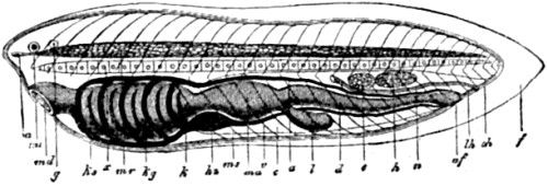
Fig. 45.—Ideal primitive vertebrate, seen from the left
side. (After Häckel.) na, nose; au, eye; g, ear; md, mouth; ks,
gill-openings; x, notochord; mr, spinal tube; kg, gill-vessels;
k, gill-intestine; hz, heart; ms, muscles; ma, stomach; v,
intestinal vein; c, body-cavity; a, aorta; l, liver; d, small
intestine; e, ovary; h, testes; n, kidney canal; af, anus; lh,
true or leather-skin; oh, outer-skin (epidermis); f, skin-fold,
acting as a fin.
In order to trace these principles in the case of the Vertebrata, it is
desirable first of all to obtain an idea of the anatomical features
which most essentially distinguish the sub-kingdom as a whole.
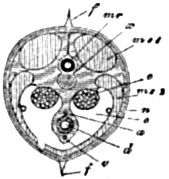 Fig. 46.—The same in transverse section through the
ovaries; lettering as in the preceding Fig.
The following, then, is what may be termed the ideal plan of vertebrate
organization, as given by Prof. Häckel. First, occupying the major
axis of body we perceive the primitive vertebral column. The parts lying
above this axis are those which have been developed from the ectoderm
and mesoderm—viz. voluntary muscles, central nervous system, and
organs of special sense. The parts lying below this axis are for the
most part those which have been developed from the
endoderm—namely,
the digestive tract with its glandular appendages, the circulating
system and the respiratory system. In transverse section, therefore, the
ideal vertebrate consists of a solid axis, with a small tube occupied by
the nervous system above, and a large tube, or body-cavity, below. This
body-cavity contains the viscera, breathing organs, and heart, with its
prolongations into the main blood-vessels of the organism. Lastly, on
either side of the central axis are to be found large masses of
muscle—two on the dorsal and two on the ventral. As yet, however,
there are no limbs, nor even any bony skeleton, for the primitive
vertebral column is hitherto unossified cartilage. This ideal animal,
therefore, is to all appearance as much like a worm as a fish, and swims
by means of a lateral undulation of its whole body, assisted, perhaps,
by a dorsal fin formed out of skin.
Fig. 46.—The same in transverse section through the
ovaries; lettering as in the preceding Fig.
The following, then, is what may be termed the ideal plan of vertebrate
organization, as given by Prof. Häckel. First, occupying the major
axis of body we perceive the primitive vertebral column. The parts lying
above this axis are those which have been developed from the ectoderm
and mesoderm—viz. voluntary muscles, central nervous system, and
organs of special sense. The parts lying below this axis are for the
most part those which have been developed from the
endoderm—namely,
the digestive tract with its glandular appendages, the circulating
system and the respiratory system. In transverse section, therefore, the
ideal vertebrate consists of a solid axis, with a small tube occupied by
the nervous system above, and a large tube, or body-cavity, below. This
body-cavity contains the viscera, breathing organs, and heart, with its
prolongations into the main blood-vessels of the organism. Lastly, on
either side of the central axis are to be found large masses of
muscle—two on the dorsal and two on the ventral. As yet, however,
there are no limbs, nor even any bony skeleton, for the primitive
vertebral column is hitherto unossified cartilage. This ideal animal,
therefore, is to all appearance as much like a worm as a fish, and swims
by means of a lateral undulation of its whole body, assisted, perhaps,
by a dorsal fin formed out of skin.
[145]
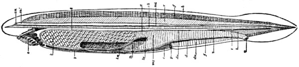
Fig. 47.—Amphioxus lanceolatus. (After Häckel.) a,
anus; au, eye; b, ventral muscles; c, body-cavity; ch,
notochord; d, intestine; do and du, dorsal and ventral walls of
intestine; f, fin-seam; h, skin; k, gills; ka, gill-artery;
lb, liver; lv, liver-vein; m 1, brain-bladder; m 2, spinal
marrow; mg, stomach; o, mouth; p, ventral pore; r, dorsal
muscle; s, tail-fin; t, aorta; v, intestinal vein; x, boundary
between gill-intestine and stomach-intestine; y, hypobranchial
groove.
Now I should not have presented this ideal representation of a primitive
vertebrate—for I have very little faith in the “scientific use of
the imagination” where it aspires to discharge the functions of a
Creator in the manufacture of archetypal forms—I say I should not have
presented this ideal representative of a primitive vertebrate, were it
not that the ideal is actually realized in a still existing animal. For
there still survives what must be an immensely archaic form of
vertebrate, whose anatomy is almost identical with that of the imaginary
type which has just been
described. I allude, of course, to Amphioxus, which is by far the most
primitive or generalized type of vertebrated animal hitherto discovered.
Indeed, we may say that this remarkable creature is almost as nearly
allied to a worm as it is to a fish. For it has no specialized head, and
therefore no skull, brain, or jaws: it is destitute alike of limbs, of a
centralized heart, of developed liver, kidneys, and, in short, of most
of the organs which belong to the other Vertebrata. It presents,
however, a rudimentary backbone, in the form of what is called a
notochord. Now a primitive dorsal axis of this kind occurs at a very
early period of embryonic life in all vertebrated animals; but, with the
exception of Amphioxus, in all other existing Vertebrata this structure
is not itself destined to become the permanent or bony vertebral column.
On the contrary, it gives way to, or is replaced by, this permanent bony
structure at a later stage of development. Consequently, it is very
suggestive that so distinctively embryonic a structure as this temporary
cartilaginous axis of all the other known Vertebrata should be found
actually persisting to the present day as the permanent axis of Amphioxus. In many other respects, likewise, the early
embryonic history of other Vertebrata refers us to the permanent
condition of Amphioxus. In particular, we must notice that the wall of
the neck is always perforated by what in Amphioxus are the
gill-openings, and that the blood-vessels as they proceed from the heart
are always distributed in the form of what are called gill-arches,
adapted to convey the blood round or through the gills for the purpose
of aeration. In all existing fish and other gill-breathing Vertebrata,
this
arrangement is permanent. It is likewise met with in a peculiar kind of
worm, called Balanoglossus—a creature so peculiar, indeed, that it
has been constituted by Gegenbaur a class all by itself. We can see by
the wood-cuts that it presents a series of gill-slits, like the
homologous parts of the fishes with which it is compared—i. e.
fishes of a comparatively low type of organization, which dates from a
time before the development of external gills. (Figs. 48, 49, 50.) Now,
as I have already said, these gill-slits are supported internally by
the gill-arches, or the blood-vessels which convey the blood to be
oxygenized in the branchial apparatus (see below, Figs. 51, 52, 53); and
the whole arrangement is developed from the anterior part of the
intestine—as is likewise the respiratory mechanism of all the
gill-breathing Vertebrata. That so close a parallel to this peculiar
mechanism should be met with in a worm, is a strong additional piece of
evidence pointing to the derivation of the Vertebrata from the Vermes.
[148]
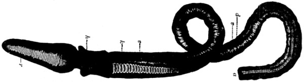 Fig. 48.—Balanoglossus. (After A. Agassiz.) r,
proboscis; h, collar; k, gill-slits; d, digestive posterior
intestine; v, intestinal vessel; a, anus.
Fig. 48.—Balanoglossus. (After A. Agassiz.) r,
proboscis; h, collar; k, gill-slits; d, digestive posterior
intestine; v, intestinal vessel; a, anus.
 Fig. 49.—A large Sea-lamprey (Petromyzon marinus), much
reduced in size. (After Cuvier and Häckel.) A series of seven gill-slits
are visible.
Fig. 49.—A large Sea-lamprey (Petromyzon marinus), much
reduced in size. (After Cuvier and Häckel.) A series of seven gill-slits
are visible.
[149]
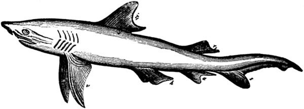 Fig. 50—Adult Shark (Carcharias melanopterus). (After
Cuvier and Häckel.)
Fig. 50—Adult Shark (Carcharias melanopterus). (After
Cuvier and Häckel.)
[150]
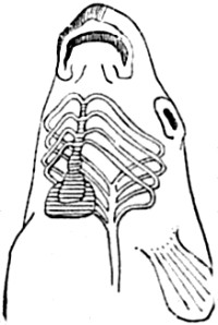
Fig. 51.—Diagram of heart and gill-arches of a fish.
(After Owen.) |
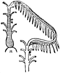
Fig. 52.—One gill-arch, with branchial fringe attached.
(After Owen.) H, Heart. |
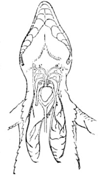
Fig. 53.—Diagram of heart and gill-arches in a lizard.
(After Owen.) The gill-arches, a a' a'', and b b' b'', are called
aortic arches in air-breathing vertebrata. |
Well, I have just said that in all the gill-breathing Vertebrata, this
mechanism of gill-slits and vascular gill-arches in the front part of
the intestinal tract is permanent. But in the air-breathing Vertebrata
such an arrangement would obviously be of no use. Consequently, the
gill-slits in the sides of the neck (see Figs. 16 and 57, 58), and the
gill-arches of the large blood-vessels (Figs. 54, 55, 56), are here
exhibited only as transitory phases of development. But as such they
occur in all air-breathing Vertebrata. And, as if to make the homologies
as striking as possible, at the time when the gill-slits and the
gill-arches are developed in the embryonic young of air-breathing
Vertebrata, the heart is constructed upon the fish-like type. That is to
say, it is placed far forwards, and, from having been a simple tube as
in Worms, is now divided into two chambers, as in Fish. Later on it
becomes progressively pushed further back between the developing lungs,
while it progressively acquires the three cavities distinctive of
Amphibia, and finally the four cavities belonging only to the complete
double circulation of Birds and Mammals. Moreover, it has now been
satisfactorily shown that the lungs of air-breathing Vertebrata, which
are thus destined to supersede the function of gills, are themselves the
modified swim-bladder or float, which belongs to Fish. Consequently, all
these progressive modifications in the important organs of circulation
and respiration in the air-breathing Vertebrata, together make up as
complete a history of their aquatic pedigree as it would be possible for
the most exacting critic to require.
[151]
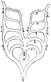
Fig. 54.—Ideal diagram, of primitive gill- or
aortic-arches. (After Rathke.) H, outline of heart. The arrows show the
course of the blood. |
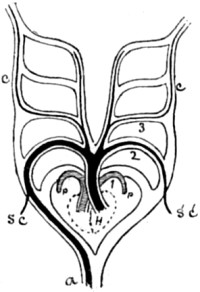
Fig. 55.—The same, modified for a bird. (After Le
Conte.) The dark lines show the aortic arches which persist. A, aorta;
p, pulmonary arches; SC, S'C', sub-clavian; C, C', carotids. |
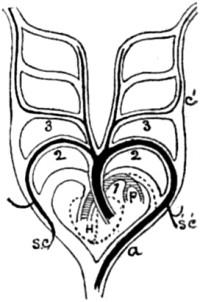
Fig. 56.—The same, modified for a mammal. (After Le
Conte.) |
[152]
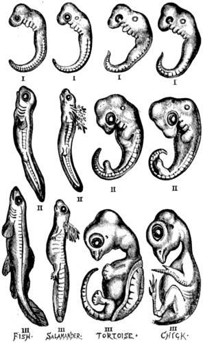
Fig. 57.—A series of embryos at three comparable and
progressive stages of development (marked I, II, III), representing each
of the classes of vertebrated animals below the Mammalia (After
Häckel.)
[153]
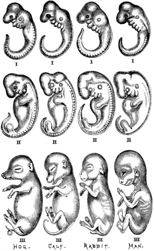
Fig. 58.—Another series of embryos, also at three
comparable and progressive stages of development (marked I, II, III),
representing four different divisions of the class Mammalia. (After
Häckel.)
If space permitted, it would be easy to present abundance of additional
evidence to the same effect from the development of the skeleton, the
skull, the brain, the sense-organs, and, in short, of every constituent
part of the vertebrate organization. Even without any anatomical
dissection, the similarity of all vertebrated embryos at comparable
stages of development admits of being strikingly shown, if we merely
place the embryos one beside the other. Here, for instance, are the
embryos of a fish, a salamander, a tortoise, a bird, and four different
mammals. In each case three comparable stages of development are
represented. Now, if we read the series horizontally, we can see that
there is very little difference between
the eight animals at the earliest of the three stages
represented—all having fish-like tails, gill-slits, and so on. In
the next stage further differentiation has taken place, but it will be
observed that the limbs are still so rudimentary that even in the case
of Man they are considerably shorter than the tail. But in the third
stage the distinctive characters are well marked.
So much then for an outline sketch of the main features in the embryonic
history of the Vertebrata. But it must be remembered that the science of
comparative embryology extends to each of the other three great branches
of the tree of life, where these take their origin, through the worms,
from the still lower, or gastræa, forms. And in each of these three
great branches—namely, the Echinodermata, the Mollusca, and the
Arthropoda—we have a repetition of just the same kind of evidence in
favour of continuous descent, with adaptive modification in sundry
lines, as that which I have thus briefly sketched in the case of the
Vertebrata. The roads are different, but the method of travelling is the
same. Moreover, when the embryology of the Worms is closely studied, the
origin of these different roads admits of being clearly traced. So that
when all this mass of evidence is taken together, we cannot wonder that
evolutionists should now regard the science of comparative embryology as
the principal witness to their theory.
[156]