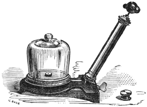
COMMON OBJECTS OF THE MICROSCOPE — PREFACE TO THE SECOND EDITION
Transcriber’s notes:
Minor punctuation errors have been corrected silently (e.g. missing
full stops after abbreviated words such as Fig), as have the following
misspellings: Bretahing → Breathing, Pedicillaria → Pedicellaria,
Pedicelaria → Pedicellaria, Chœtonotus → Chætonotus, Spurganium
→ Sparganium, veiw → view. Unorthodox spelling and inconsistent
hyphenation has not been altered. Several wrongly numbered
cross-references to Plates and Figures have been corrected.
Plate VIII (and its accompanying key) was originally displayed
at the beginning of the book, before the Title Page, but has been
repositioned in the body of the text in correct numerical sequence.
A black underline indicates a hyperlink to a page or illustration
(hyperlinks are also highlighted when the mouse pointer hovers over
them). Page numbers are shown in the right
margin.
The cover image of the book was created by the
transcriber and is placed in the public domain.
BY THE LATE
Rev. J. G. WOOD, M.A., F.L.S., Etc.
AUTHOR OF
“COMMON OBJECTS OF THE COUNTRY” “COMMON OBJECTS OF THE SEA-SHORE”
“MY FEATHERED FRIENDS” ETC. ETC.
WITH ILLUSTRATIONS BY TUFFEN WEST
SECOND EDITION, REVISED AND RE-WRITTEN, BY
E. C. BOUSFIELD, L.R.C.P.(Lond.)
AUTHOR OF
“A GUIDE TO THE SCIENCE OF PHOTO-MICROGRAPHY”
WITH ADDITIONAL ILLUSTRATIONS BY THE REVISER
LONDON
GEORGE ROUTLEDGE AND SONS, Limited
BROADWAY HOUSE, LUDGATE HILL
1900
The task of revising and bringing up to date a work
which has been the guide, philosopher, and friend
of thousands of commencing microscopists has been,
in the present case, one of no small difficulty.
On the one hand, there was the natural desire to
interfere as little as possible with the original work;
and on the other, the necessity of rendering available,
to some extent at least, the enormous advance
in every department which has taken place in the
thirty-six years which have elapsed since the work
was first offered to the public. The reviser has
done his best not only to fulfil these two objects,
but to keep in view the original purpose of the
book.
In the popular department of pond-life especially,
about fifty new illustrations have been added, mostly
from the reviser’s own notebook sketches. The
whole of the botanical part has been revised by one
of our first English authorities, and, in short, no
effort has been spared to make the work as accurate
as its necessarily condensed form permits of. It is
hoped, therefore, that it may be found not less
useful than its predecessor by those for whom it is
alone intended.
PREFACE TO THE FIRST EDITION
In my two previous handbooks, the “Common
Objects” of the Sea-shore and Country, I could
but slightly glance at the minute beings which
swarm in every locality, or at the wonderful
structures which are discovered by the Microscope
within or upon the creatures therein described.
Since that time a general demand has arisen for
an elementary handbook upon the Microscope and
its practical appliance to the study of nature, and
in order to supply that want this little volume has
been produced.
I must warn the reader that he is not to expect
a work that will figure and describe every object
which may be found on the sea-shore or in the
fields, but merely one by which he will be enabled
to guide himself in microscopical research, and
avoid the loss of time and patience which is almost
invariably the lot of the novice in these interesting
studies. Upwards of four hundred objects have
been figured, including many representatives of
the animal, vegetable, and, mineral kingdoms, and
among them the reader will find types sufficient for
his early guidance.
Neither must he expect that any drawings can
fully render the lovely structures which are revealed
by the microscope. Their form can be given faithfully
enough, and their colour can be indicated;
but no pen, pencil, or brush, however skilfully
wielded, can reproduce the soft, glowing radiance,
the delicate pearly translucency, or the flashing
effulgence of living and ever-changing light with
which God wills to imbue even the smallest of His
creatures, whose very existence has been hidden for
countless ages from the inquisitive research of man,
and whose wondrous beauty astonishes and delights
the eye, and fills the heart with awe and adoration.
Owing to the many claims on my time, I left the
selection of the objects to Mr. Tuffen West, who
employed the greater part of a year in collecting
specimens for the express purpose, and whose well-known
fidelity and wide experience are the best
guarantees that can be offered to the public. To
him I also owe many thanks for his kind revision
of the proof-sheets. My thanks are also due to
Messrs. G. and H. Brady, who lent many beautiful
objects, and to Messrs. Baker, the well-known
opticians of Holborn, who liberally placed their
whole stock of slides and instruments at my disposal.
CONTENTS
CHAPTER I
PAGE
Pleasures and Uses of Microscopy—Development of the
Microscope—Extemporised Apparatus
Examination of Objects—Principles of Illumination—Mirror
and its Action—Substage Condenser—Use of Bull’s-eye—Opaque
Objects—Photography of Microscopic Objects
Vegetable Cells and their Structure—Stellate Tissues—Secondary
Deposit—Ducts and Vessels—Wood-Cells—Stomata,
or Mouths of Plants—The Camera Lucida, and
Mode of Using—Spiral and Ringed Vessels—Hairs of
Plants—Resins, Scents, and Oils—Bark Cells
Algæ and their Growth—Desmidiaceæ, where found—Diatoms,
their Flinty Deposit—Volvox—Mould, Blight,
and Mildew—Mosses and Ferns—Mare’s-Tail and the
Spores—Common Sea-weeds and their Growth
Antennæ, their Structure and Use—Eyes, Compound and
Simple—Breathing Organs—Jaws and their Appendages—Legs,
Feet, and Suckers—Digestive Organs—Wings,
Scales, and Hairs—Eggs of Insects—Hair, Wool, Linen,
Silk, and Cotton—Scales of Fish—Feathers—Skin and
its Structure—Epithelium—Nails, Bone, and Teeth—Blood
Corpuscles and Circulation—Elastic Tissues—Muscle
and Nerve
Marine Life—Sponges—Infusoria—Foraminifera—Radiolaria—Hydroid
Zoophytes—Polyzoa—Worms—Lingual Ribbons and Gills of Mollusca—
Star-Fishes and Sea-Urchins—Cuttle-Fish—Corallines—Miscellaneous
Objects
Pleasures and Uses of Microscopy—Development of the
Microscope—Extemporised Apparatus.
Within the last half-century the use of the
microscope, not only as an instrument of scientific
research, a tool in the hands of the investigator
of the finer organisation of the world of nature,
nor even as an adjunct to the apparatus of the
chemist or the manufacturer, but as a means of
innocent and instructive recreation, has become
so firmly rooted amongst us that it seems hardly
necessary to advocate its claims to attention on
any of these grounds.
So wonderful is the information which it affords,
so indispensable is it in many, if not in all,
branches of scientific research, that not only would
the lover of nature be deprived of one of his most
valued sources of information and enjoyment, but
some sciences would be brought absolutely to a
standstill if by any conceivable means the
microscope were to be withdrawn from their
followers.
On the other hand, from every improvement in
the construction of the latter, a corresponding enlargement
and enlightenment of the fields reviewed
by these sciences has taken place, and the beauty
and interest of the revelations made by its means
has attracted an ever-increasing host of earnest
and intelligent volunteers, who have rendered
yeoman service to the cause of knowledge.
Moreover, so vast is the scope of the instrument,
that whilst discoveries in other fields of research
are few and far between, comparatively speaking,
in microscopic science they are of everyday
occurrence, and the number of problems calling
for solution by means of the instrument in question
is so vast that even the humblest worker may be
of the greatest assistance.
In the following pages we propose to carry out,
as far as possible with reference to the microscope,
the system followed in the “Common Objects of
the Seashore and of the Country,” and to treat in
as simple a manner as may be of the marvellous
structures which are found so profusely in our
fields, woods, streams, shores, and gardens. Moreover,
our observations will be restricted to an
instrument of such a class as to be inexpensively
purchased and easily handled, and to those pieces
of supplementary apparatus which can be extemporised
at small cost of money and ingenuity by
the observer himself, or obtained of the opticians
for a few shillings.
With the same view, the descriptions will be
given in language as simple and as free from
technicalities as possible, though it must be
remembered that for many of the organisms and
structures which we shall have to describe there
are none but scientific names; and since, moreover,
this little work is intended to furnish a stepping-stone
between the very elements of microscopic
science, and those higher developments of it which
should be the aim of every worker, it would be
unwise to attempt to invent commonplace appellations
for the purpose of this book, and leave him
to discover, when he came to consult works of
reference in any particular subject, that his
“simplified” knowledge had all to be unlearnt,
and a new vocabulary acquired. Rather will it be
our purpose to use correct terms, and explain them,
as far as necessary, as we introduce them.
The commencing microscopist is strongly recommended,
whilst not confining his interest entirely
to one branch of research or observation, to adopt
some one as his particular province.
The opportunities for discovery and original
work, which are afforded by all alike, will be more
readily appreciated and utilised by adopting such
a plan than by a general and purposeless distribution
of effort. To mention one or two. The
student of the fascinating field of pond-life will find
new organisms awaiting description by the hundred,
and of the old ones, life-histories to make out; if
he be attracted rather to the vegetable inhabitants
of the same realm, the diatoms will furnish him
with the opportunity of studying, and perhaps
solving, the enigma of their spontaneous movement,
and of watching their development. The smaller
fungi, and indeed the larger ones too, will amply
repay the closest and most laborious study of their
habits of life and processes of development. Since
the first edition of this work was published, the
whole subject has been practically revolutionised,
and more remains to be done than has yet been
accomplished.
In short, there is scarcely an organism, even of
those best known and most studied, which is so
completely exhausted that persevering investigation
would reveal nothing new concerning it.
There can be little doubt but that if any worker,
with moderate instrumental means, but with an
observant mind, were to set determinately to work
at the study of the commonest weed or the most
familiar insect, he, or she, would by patient labour
accomplish work which would not only be of value
to science, but would redound to the credit of the
worker.
Something like finality appears to have been
reached, at least for the present, in the development
of the microscope; and whilst it is beyond
the scope of this work to treat of the refined and
costly apparatus which is essential to useful work
in certain departments of research, the result has,
on the whole, been highly favourable to the worker
of moderate means and ambitions, since lenses are
now accessible, at the cost of a few shillings, comparatively
speaking, which could not have been
purchased at all when this work first appeared.
It is with such appliances that we have here to
deal. The worker whose finances are restricted
must be contented to extemporise for himself many
pieces of apparatus, and will find pleasure and
occupation in doing so. And let him remember,
for his encouragement, that many such home-made
appliances will fulfil their purpose quite as well as
the imposing paraphernalia of glittering brass and
glass which decorates the table of the wealthy
amateur. It is not the man who possesses the
best or most costly apparatus, but the one who
best understands the use of that which he possesses,
that will make the most successful microscopist.
A good observer will discover, with only the aid of
a pocket-magnifier, secrets of Nature which have
escaped the notice of a whole army of dilettante
microscopists, in spite of the advantages which, as
regards instruments, the latter may enjoy.
It is for those who desire to be of the former
class that this book is written, and in the course
of the following pages instances will be given in
which the exercise of a small amount of ingenuity
and the expenditure of a few pence will be found
equivalent to the purchase of costly and complicated
apparatus.
An enormous amount of valuable work was done
in the earliest days of microscopy, when the best
apparatus available was a single lens, composed of
the bead formed by fusing the drawn-out end of a
rod of glass. Inserted into a plate of metal, or a
piece of card, such a primitive instrument was
capable of affording a large amount of information.
Similar instruments are to be purchased for a few
pence at the present day, and are not without their
use for purposes of immediate examination of
material. A very common form is a glass marble,
ground flat on one side, and mounted in a tube.
The material to be examined is placed upon the
flat side, and is seen magnified to an extent
inversely proportional to the diameter of the sphere
of glass.
CHAPTER II
Elementary Principles of Optics—Simple Microscopes—Compound Microscope—Accessory Apparatus—Cover-glasses—Troughs—Condensers—Dissection—Dipping-tubes—Drawing—Measurement.
Before proceeding to deal with the microscope
itself, it may be useful to give a short summary of
the optical laws upon which its working depends.
To go into the minutiæ of the matter here would
be out of place, but it will be found very helpful,
especially in the matter of illumination, to acquire
some knowledge of, and facility in applying, these
elementary principles. We shall confine our
remarks to convex lenses, as being the form to
which all the combinations in the microscope may
be ultimately reduced.
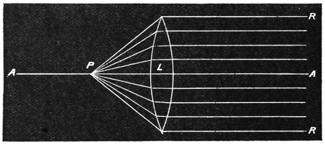
Every convex lens has one “principal” focus,
and an infinite number of “conjugate” foci. The
principal focus is the distance at which it brings
together in one point the rays which fall upon it
parallel to its axis, as shown in Fig. 1, in which A
is the axis of the lens L, and the rays RR are
brought together in the principal focus P. Thus
a ready means of finding the focal length of any
lens is to see at what distance it forms an image
of the sun, or of any other distant object, upon a
screen, such as a piece of smooth white cardboard.
In the figure this distance will be PL. Conversely,
if the source of light be at P, a parallel beam of
light will be emitted from the lens.
Fig. 1.
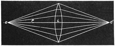
The focal length may, however, be found in
another way. When an object is placed at a
distance from a lens equal to twice the principal
focal length of the latter, an image of the object is
formed at the same distance upon the other side of
the lens, inverted in position, but of the same
dimensions as the original object. The object and
image then occupy the equal conjugate foci of the
lens, so that by causing them to assume these
relative positions, and halving the distance at
which either of them is from the lens, the focal
length of the latter is known.
These points will be seen on reference to Fig. 2,
in which L being the lens, and P the principal
focus, as before, rays from the point C are brought
together at the conjugate focus C', at the same
distance on the other side of L. In this case it
manifestly does not matter whether the object be
at one or the other of these points.
Fig. 2.
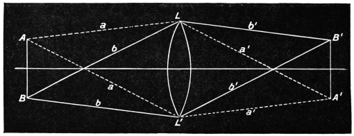
So far we have been dealing with points on the
line of the axis of the lens. The facts mentioned
apply equally, however, to rays entering the lens
at an angle to the axis, only that in this case they
diverge or converge, correspondingly, upon the
other side. It is evident, from Fig. 1, that no
image is formed of a point situated at the distance
of the principal focus; but Fig. 3, which is really
an extension of Fig. 2, shows how the rays passing
along secondary axes form an inverted image of the
same size as the object, when the latter is situated
at twice the focal length of the lens from this last.
To avoid confusion, the bounding lines only are
shown, but similar lines might be drawn from each
and every point of the object; and if the lines
ALA', BL'B' be supposed to be balanced at L and
L' respectively, they will indicate the points at
which the corresponding parts of the object and
image will be situated along the lines AB, B'A'
respectively. Moreover, rays pass from every part
of the object to every part of the lens, so that we
must imagine the cones LAL', LA'L' to be filled
with rays diverging on one side of the lens and
converging on the other.
The image so formed is a “real” image,—that is
to say, it can be thrown upon a screen.
Fig. 3.
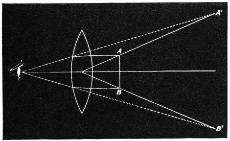
The microscopic image, on the other hand, is
a virtual image, which can be viewed by the eye
but cannot be thus projected, for it is formed by an
object placed nearer to the lens than the principal
focal length of the latter, so that the rays diverge,
instead of converging, as they leave the lens, and the
eye looks, as it were, back along the path in which
the rays appear to travel, and so sees an enlarged
image situated in the air, farther away than the
object, as shown in Fig. 4. In this case, as the
diagram shows, the image is upright, not inverted.
Images of the latter class are those formed by
simple microscopes, of the kind described in the
previous chapter. In the compound microscope
the initial image, formed by the object-glass, is
further magnified by another set of lenses, forming
the eye-piece, by which the diverging rays of the
virtual image are brought together to a focus at
the eye-point; and when viewed directly, the eye
sees an imaginary image, as in a simple microscope,
whilst, when the rays are allowed to fall upon a
screen, they form a real image of the object, larger
or smaller, as the screen is farther from or nearer
to the eye-point.
Fig. 4.
These remarks must suffice for our present
purpose. Those who are sufficiently interested in
the subject to desire to know more of the delicate
corrections to which these broad principles are
subjected in practice, that objectives may give
images which are clear and free from colour, to say
nothing of other faults, will do well to consult some
such work as Lommel’s Optics, in the International
Science Series.
In following a work such as the present one, the
simple microscope, in some form or other, will be
found almost indispensable. It will be required
for examining raw material, such as leaves or other
parts of plants, for gatherings of life in fresh or
salt water, for dissections. With such powers as
those with which we shall have to deal, it will
rarely happen that, for example, a bottle of water
in which no life is visible will be worth the
carrying-home; whilst, on the other hand, a few
months’ practice will render it not only possible,
but easy, not only to recognise the presence, but to
identify the genus, and often even the species, of
the forms of life present. Moreover, these low
powers, affording a general view of the object,
allow the relation to each other of the details
revealed by the power of the compound microscope
to be much more easily grasped. A rough example
may suffice to illustrate this. A penny is a sufficiently
evident object to the naked eye, but it will
require a sharp one to follow the details in
Britannia’s shield, whilst the minute scratches
or the bloom upon the surface would be invisible in
detail without optical aid. Conversely, however,
it would be rash to conclude from an examination
of a portion of the surface with the microscope
alone that the portion in view was a sample of the
whole surface. The more the surface is magnified,
the less are the details grasped as a whole, and for
this reason the observer is strongly recommended
to make out all that he can of an object with a
simple magnifier before resorting to the microscope.
For general purposes, the intending observer
cannot do better than supply himself with a
common pocket-magnifier, with one, two, or three
lenses, preferably the last, as although the absolute
performance is not so accurate, the very considerable
range of power available by using the lenses
singly, or in various combinations, is of great
advantage. Such a magnifier may be obtained
from Baker for about three-and-sixpence,
or, with the addition
of a powerful Coddington lens
(Fig. 5) in the same mount, for
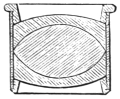
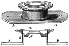
nine shillings more. Aplanatic
lenses, such as the one shown in
section in Fig. 6, with a much
flatter field of vision, but of one
power only each, cost about
fifteen shillings, and a simple
stand, which adapts them for
dissecting purposes, may be obtained
of the same maker for half a crown, or may
easily be extemporised from a cork sliding stiffly
on an iron rod set in a heavy foot, the cork carrying
a loop of wire terminating in a ring which
carries the lens.
Fig. 5.
Fig. 6.
So much may suffice for the simple microscope.
We pass on now to the consideration of the instrument
which forms the subject of the present work,
an instrument which, whilst moderate in price, is
yet capable of doing a large amount of useful and
valuable work in the hands of a careful owner, and
of affording him a vast amount of pleasure and recreation,
even if these be his only objects in the
purchase, though it is difficult to understand that,
an insight being once attained into the revelations
effected by the instrument, without a desire being
excited in any intelligent
mind to pursue
the subject as a study
as well as a delightful
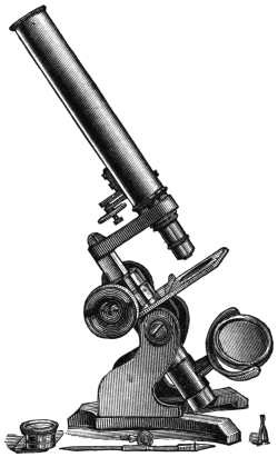
relaxation. The microscope
described, and
adopted as his text
by the author of this
work, is still made,
and has shared to a
very considerable extent
in the general
improvement of optical
apparatus which has
taken place during the
last thirty years. It
is to be obtained from
Baker, 244 High Holborn,
and is provided
with most of the apparatus which will be found
indispensable by the beginner, costing, with a case
in which to keep it, the modest sum of three
guineas.
Fig. 7.
If this instrument represent the limit of the
purchaser’s power of purse, he may very well make
it answer his purpose for a considerable time. The
same instrument, however, with separate objectives
of good quality, together with a bull’s-eye condenser
(an almost indispensable adjunct), a plane mirror in
addition to a concave one, and a simple but efficient
form of substage condenser, may be obtained for
£5, 12s. 6d.,
and the extra
outlay will be
well repaid by
the advantage
in working
which is
gained by the
possession of
the additional
apparatus.
Fig. 8.
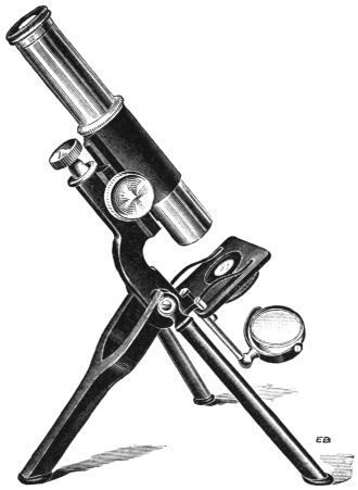
A still
better stand,
and one
which is good
enough for
almost any
class of work,
is that shown
in Fig. 8,
which is
known as the “Portable” microscope. In this
instrument the body is made up of two tubes, so
that the length may be varied at will, and this
gives a very considerable range of magnification
without changing the object-glass, a great convenience
in practice; whilst the legs fold up
for convenience of carriage, so that the whole
instrument, with all necessary appliances, may
be readily packed in a corner of a portmanteau for
transport to the country or seaside.
The objectives supplied with the simplest form of
microscope above referred to are combinations of
three powers in one, and the power is varied by
using one, two, or three of these in combination.
This form of objective is very good, as far as it
goes, though it is impossible to correct such a combination
with the accuracy which is possible in
manufacturing one of a fixed focal length.
Perhaps the best thing for the beginner to do
would be to purchase the combination first, and,
later on, to dispose of it and buy separate objectives
of, say, one-inch, half-inch, and quarter-inch focal
lengths. It may be explained here, that when a
lens is spoken of as having a certain focal length,
it is meant that the magnification obtained by its
use is the same, at a distance of ten inches from the
eye, as would be obtained by using a simple sphere
of glass of the same focal length at the same
distance. This, of course, is simply a matter of
theory, for such lenses are never used actually.
Fig. 9.
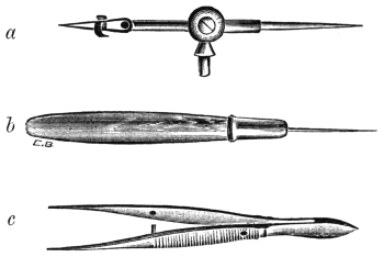
Of accessory apparatus, we may mention first the
stage forceps (Fig. 9, a). These are made to fit into
a hole upon the stage, so as to be capable of being
turned about in any direction, with an object in
their grasp, and for some purposes, especially such
as the examination of a thin object, say the edge of
a leaf, they are extremely
useful.
Fig. 10.
The live box,
in which drops of
water or portions
of water-plants, or
the like, may be
examined, will be
found of great
service. By adjustment
of the
cap upon the cylinder, with proper attention to the
thickness of the cover-glass in the cap, any required
amount of pressure, from that merely sufficient to
fix a restless object to an amount sufficient to crush
a resistent tissue, may easily be applied, whilst the
result of the operation is watched through the
microscope. This proceeding is greatly facilitated
if the cap of the live-box be slotted spirally, with a
stud on the cylinder, so that a half-turn of the cap
brings the glasses into contact. By this means the
pressure may be adjusted with the greatest nicety.
In examining delicate objects, such as large infusoria,
which invariably commit suicide when
pressure is applied, a good plan is to restrict their
movements by placing a few threads of cotton-wool,
well pulled out, in the live-box with the drop of
water.
A variety of instruments has been invented for
the same purpose, of which Beck’s parallel compressorium
may be mentioned as the most efficient,
though it is somewhat complicated, and consequently
expensive, costing about twenty-five shillings.
A few glass slips and cover-glasses will also be
required. The latter had better be those known as
“No. 2,” since the beginner will find it almost
impossible to clean the thinner ones satisfactorily
without a large percentage of fractures. The safest
way is to boil the thin glass circles in dilute nitric
acid (half acid, half water) for a few minutes, wash
well in several waters, first tap-water and then distilled,
and finally to place the covers in methylated
spirit. When required for use, the spirit may be
burnt off by applying a light, the cover-glass, held
in a pair of forceps, being in no way injured by the
process.
In addition to the glass slides, the observer will
find it advisable to be provided with a few glass
troughs, of various thicknesses, in which portions of
water-plants, having organisms attached to them,
may be examined. Confined in the live-box, many
of the organisms ordinarily found under such circumstances
can rarely be induced to unfold their beauties,
whilst in the comparative freedom of the trough
they do so readily. The troughs may be purchased,
or may be made of any desired shape or size by
cutting strips of glass of a thickness corresponding
to the depth desired, cementing these to a glass
slide somewhat larger than the ordinary one, and
cementing over the frame so formed a piece of thin
glass, No. 3; the best material to use as cement
being marine glue of the best quality, or, failing
this, Prout’s elastic glue, which is much cheaper,
but also less satisfactory. The glass surface must
be made, in either case, sufficiently hot to ensure
thorough adhesion of the cement, as the use of any
solvent entails long waiting, and considerable risk
of poisoning the organisms. A useful practical
hint in the use of these troughs is that the corners,
at the top, should be greased slightly, otherwise the
water finds its way out by capillary attraction,
to the detriment of the stage of the microscope.
Of optical accessories, the bull’s-eye is almost the
most valuable. So much may be effected by its
means alone, in practised hands, that it is well
worth the while of the beginner to master its use
thoroughly, and the methods of doing so will be
explained in the succeeding chapter.
The substage condenser, too, even in its most
simple form, is an invaluable adjunct, even though
it be only a hemisphere of glass, half an inch or so
in diameter, mounted in a rough sliding jacket to fit
underneath the stage. Such an instrument, properly
fitted, will cost about fifteen shillings, but the ingenious
worker will easily extemporise one for
himself.
Fig. 11.
Many plants and animals require to be dissected
to a certain extent before the details of their structure
can be made out, and for this purpose the
naked eye alone will rarely serve. The ordinary
pocket magnifier, however, if mounted as described
in a preceding chapter, will greatly facilitate matters,
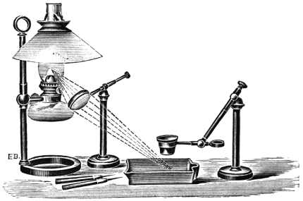
and the light may be focused upon the object by
means of the bull’s-eye condenser, as shown in
Fig. 11. In the figure the latter is represented
as used in conjunction with the lamp, but daylight
is preferable if it be available, the strain upon the
eyes being very much less than with artificial light.
Two blocks of wood, about four inches high, will
form convenient rests for the hands, a plate of glass
being placed upon the blocks to support the dish,
and a mirror being put in the interspace at an angle
of 45° or so if required. A piece of black paper
may be laid upon the mirror if reflected light alone
is to be used.
As all delicate structures are dissected under fluid,
a shallow dish is required. For this purpose nothing
is better than one of the dishes used for developing
photographic negatives. The bottom of the dish
is occupied by a flat cork, to which a piece of flat
lead is attached below, and the object having been
pinned on to the cork in the required position, the
fluid is carefully run in. This fluid will naturally
vary according to the results desired to be obtained,
but it must not be plain water, which so alters all
cellular structures as to practically make them unrecognisable
under the microscope. Nothing could
be better than a 5 per cent. solution of formalin,
were it not for the somewhat irritating odour of
this fluid, since it at once fixes the cells and preserves
the figure. For many purposes a solution of
salt, in the proportion of a saltspoonful of the
latter to a pint of water, will answer well for short
dissections. For more prolonged ones, a mixture of
spirit-and-water, one part of the former to two of
the latter, answers well, especially for vegetable
structures. When the dilution is first made, the
fluid becomes milky, unless pure spirit be used, but
with a little trouble the Revenue authorities may
be induced to give permission for the use of pure
methylated spirit, which answers every purpose. The
trouble then is that not less than five gallons can be
purchased, an embarras de richesses for the average
microscopist, but, after all, the spirit is extremely
cheap, and does not deteriorate by keeping.
When the dissection in either of these media is
completed, spirit should be gradually added to
bring the strength up to 50 per cent., in which
the preparation may remain for a day or two, after
which it is gradually brought into pure spirit, or
into water again, according to the medium in which
it is to be mounted.
Fig. 12.
As to the tools required, they are neither
numerous nor expensive. Fine-pointed but strong
forceps (Fig. 9, c), curved and straight; a couple of
pairs of scissors, one strong and straight, the other
more delicate, and having curved blades, and a few
needles of various thicknesses and curves, are the
chief ones. The latter may be made by inserting
ordinary needles, for three-fourths of their length,
into sticks of straight-grained deal (ordinary firewood
answers best), and thereafter bending them
as required. A better plan, however, is to be
provided with a few of the needle-holders shown
in Fig. 9, b. These are very simple and inexpensive,
and in them broken needles are readily
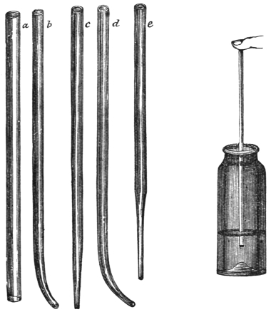
replaced by others. Dipping-tubes, such as are
shown in Fig. 12, will also be extremely useful
for many purposes. These are very easily made
by heating the centre of a piece of soft glass
tubing of the required size, and, when it is quite
red-hot, drawing the ends apart. The fine tube
in the centre should now be divided by scratching
it with a fine triangular file, and the scratch may
of course be made at such a point as to afford a
tube of the required fineness. The edges should
be smoothed by holding them in the flame until
they just run (not melt, or the tube will close).
These tubes can often be made to supply the place
of a glass syringe. They may be used either for
sucking up fluid or for transferring it, placing the
finger over the wide end, allowing the tube to fill
by displacement of air, and then re-closing it with
the finger. This last method is especially useful
for transferring small objects from one receptacle
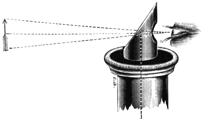
to another. In speaking of the dissection of
objects, it should have been mentioned that the
microscope itself may, under careful handling, be
made to serve very well, only, as the image is
reversed, it is almost impossible to work without
using a prism to re-erect the image. Such a
prism is shown in Fig. 13. The microscope is
placed vertically, and the observer, looking straight
into the prism, sees all the parts of the image
in their natural positions. This appliance is
extremely useful for the purpose of selecting
small objects, and arranging them on slides in
any desired manner. A few words may be added
as to the reproduction of the images of objects.
Fig. 13.
The beginner is strongly recommended to
practise himself in this from the outset. Even
a rough sketch is worth pages of description,
especially if the magnification used be appended;
and even though the worker may be devoid of
artistic talent, he will find that with practice he
will acquire a very considerable amount of facility
in giving truthful outlines at least of the objects
which he views. Various aids have been devised
for the purpose of assisting in the process. The
simplest and cheapest of these consists of a cork
cut so as to fit round the eye-piece. Into the
cork are stuck two pins, at an angle of 45° to
the plane of the cork, and, the microscope being
placed horizontally, a thin cover-glass is placed
upon the two pins, the light being arranged and
the object focused after the microscope is inclined.
On looking vertically down upon the cover-glass,
a bright spot of light will be seen, and as the eye
is brought down into close proximity with it the
spot will expand and allow the observer to see
the whole of the image without looking into the
microscope. If a sheet of paper be now placed
upon the table at the place occupied by the image
so projected, the whole of the details will be
clearly seen, as will also the point of a pencil
placed upon the paper in the centre of the field
of view; and, after a little practice, it will be
found easy to trace round the chief details of the
object. Two points require attention. The first
is that if the light upon the paper be stronger
than that in the apparent field of the microscope,
the image will not be well seen, or if the paper be
too feebly lighted, it will be difficult to keep the
point of the pencil in view. The light from the
microscope is thrown into the eye, and the view
of the image upon the paper is the effect of a
mental act, the eye looking out in the direction
from which the rays appear to come. The paper
has therefore to be illuminated independently, and
half the battle lies in the adjustment of the
relative brightness of image and paper. The
second point is, that it is essential to fix one
particular point in the image as the starting-point
of the drawing, and this being first depicted, the
image and drawing of this point must be kept
always coincident, or the drawing will be distorted,
since the smallest movement of the eye alters the
relations of the whole. The reflector must be
placed at an angle of 45°, or the field will be
oval instead of circular. The simple form of
apparatus just described has one drawback, inasmuch
as the reflection is double, the front and
back of the cover-glass both acting as reflectors.
The image from the latter being much the more
feeble of the two, care in illumination will do
much to eliminate this difficulty; but there are
various other forms in which the defect in
question is got rid of. The present writer has
worked with all of them, from the simple neutral
tint reflector of Beale to the elaborate and costly
apparatus of Zeiss, and, upon the whole, thinks
that he prefers the cover-glass to them all.
A very simple plan, not so mechanical as the
last-named, consists in the use of “drawing-squares,”
which are delicate lines ruled upon a
piece of thin glass, and dropped into the eye-piece
so that the lines rest upon the diaphragm
of the eye-piece, and therefore are in focus at the
same time as the object. By the use of these,
in combination with paper similarly ruled, a
diagram of any required size can be drawn with
very great facility. The squares, if compared with
a micrometer, will furnish an exact standard of
magnitude for each object-glass employed. The
micrometer is a piece of thin glass upon which
are ruled minute divisions of an inch or a millimeter.
Suppose the micrometer to be placed
under the microscope when the squares are in
the eye-piece, and it be found that each division
corresponds with one square of the latter, then,
if the micrometric division be one one-hundredth
of an inch, and the squares upon the paper
measure one inch, it is clear that the drawing
will represent the object magnified a hundred
“diameters”; if two divisions of the micrometer
correspond to three squares, the amplification will
be a hundred and fifty diameters; if three divisions
correspond to two squares, sixty-six diameters, and
so on. If a draw-tube be used, it will be necessary
to know the value of the squares at each inch of
the length, if they are to be used for measuring
magnification.
CHAPTER III
Examination of Objects—Principles of Illumination—Mirror
and its Action—Substage Condenser—Use of Bull’s-eye—Opaque
Objects—Photography of Microscopic Objects.
So much depends upon a right method of employing
the microscope, as regards both comfort and
accuracy, that we propose to devote a little space
to the consideration of the subject.
Let us first warn the intending observer against
the use of powers higher than are required to
bring out the details of the object. Mere magnification
is of very little use: it increases the
difficulties both of illumination and of manipulation,
and, as already said, interferes with that
grasp of the object which it is most desirable
to obtain. Rather let the beginner lay himself
out to get the very most he can out of his
lowest powers, and he will find that, by so doing,
he will be able far better to avail himself of the
higher ones when their use is indispensable.
The essential means to this end is a mastery of
the principles of illumination, which we now proceed
to describe.
We suppose the microscope to be inclined at
an angle of about 70° to the horizontal, with a
low-power objective attached to it, a one-inch by
preference. Opposite to the microscope, and about
a foot away from it, is a lamp with the edge of
the flame presented to the microscope, the concave
mirror of which is so arranged as to receive the
rays from the flame and direct them up the tube
of the microscope. Upon the stage is placed a
piece of ground-glass, and the mirror-arm is now
to be moved up or down upon its support until the
ground-glass receives the maximum of illumination,
which it will do when the lamp-flame is at one
conjugate focus of the mirror and the ground-glass
at the other. The focus will not be an image of
the flame, but a bar of light.
If an object be now placed upon the stage,
instead of the ground-glass, and the objective
focused upon it, it will, if the mirror be properly
adjusted, be brilliantly illuminated.
It will be understood that every concave mirror
has a focus, and converges the rays which fall upon
it to this focus, behaving exactly like a convex
lens. The principal focus of a concave mirror is
its radius of curvature, and this is not difficult to
determine. Place side by side a deep cardboard
box and the lamp, so that the concave mirror may
send the rays back, along a path only slightly
inclined to that by which they reached it, to the
bottom of the box. The lamp and box being
equidistant from the mirror, it is evident that
when the mirror forms an image of the former
upon the latter equal to the flame in size, we
have the equivalent of the equal conjugate foci
shown in Fig. 2. Now move the box to the
distance from the mirror which corresponds to
the distance of the stage of the microscope from
the mirror when the latter is in position upon
the microscope, and then move the lamp to or
fro until the mirror casts a sharp image of the
flame upon the bottom of the box, which is not
to be moved. The lamp distance so found will be
the correct one for working with the concave
mirror. The writer is led to lay special stress
upon this matter, from the fact that he almost
invariably finds that the mirror is arranged to be
used for parallel rays, i.e. for daylight, and is
therefore fixed far too close to the stage to be
available for correct or advantageous working with
the lamp, unless, indeed, the bull’s-eye condenser
be used, as hereinafter described, to parallelise the
rays from the lamp.
Work done with the concave mirror can, however,
under the most favourable conditions, only
be looked upon as a pis aller. The advantages
gained by the use of some substage condenser,
even the most simple, in conjunction with the plane
mirror, or even without any mirror at all, are so
manifold that the beginner is strongly urged to
provide himself with some form or other of it,
and we now proceed to describe the way in which
this should be used to produce the best effect.
To reduce the problem to its most simple
elements, turn the mirror altogether out of the
way, and place the microscope upon a block at
such a height as shall be convenient for observation,
and shall allow the rays from the lamp,
placed in a line with it on the table, to shine
directly into the tube of the microscope. Ascertain
that this is so by removing both objective and
eye-piece and looking down the tube, when the
flame should be seen in the centre, edgewise.
Now replace the eye-piece, and screw on to the
tube the one-inch combination or objective. Place
upon the stage an object, preferably a round diatom
or an echinus-spine, and focus it as sharply as
possible. Now place the substage condenser in its
jacket, and slide it up and down until the image of
the object is bisected by the image of the flame.
The centre of the object will now be brilliantly
illuminated by rays travelling in the proper direction
for yielding the best results. The object is
situated at the common focus of the microscope and
the condenser, and, whatever means of illumination
be adopted, this is the result which should always
be aimed at.
Satisfactory as this critical arrangement is, however,
from a scientific point of view, it has its
drawbacks from an artistic and æsthetic one. It
is not pleasant, for most purposes, to have merely
the centre of an object lighted up, and we have
now to consider how the image of the edge of
the flame may be so expanded as to fill the field
without sacrificing more than a very small fraction
of the accuracy of the arrangement just attained.
Referring to Fig. 1, we see that if we place
the lamp at the principal focus of a lens, it will
emit a bundle of parallel rays equal in diameter
to the diameter of the lens. This is the key of
the position. We cannot place the lamp at an
infinite distance from the substage condenser, but
we can supply the latter with rays approximately
parallel, so that it shall bring them to a focus upon
the object at very nearly its own principal focus.
This we do by means of the bull’s-eye condenser.
Place the latter, with its flat side toward the edge
of the flame, and at its principal focal distance
(the method of determining which has already
been described) from the latter, so that the bundle
of parallel rays which issue from it may pass up
to the substage condenser. On examining the
object again, it will be found that, after slight
adjustments of the position of the bull’s-eye have
been made, the object lies in the centre of an
evenly and brilliantly lighted field.
It may be necessary to place the bull’s-eye a
little farther from or nearer to the lamp, or to
move it a little to one side or the other, but when
it is at the correct distance, and on the central
line between the lamp and the substage condenser,
at right angles to this line, the effects will be as
described. It may help in securing this result if
we mention that when the bull’s-eye is too far
from the lamp, the image of the flame is a spindle-shaped
one; whilst, when the distance between the
two is too short, i.e. less than the principal focal
length of the lens, the field is crossed by a bar
or light, the ends of which are joined by a ring,
whilst on either side of the bar there is a semi-circular
dark space.
We have hitherto supposed the objects viewed
to be transparent, but there are many, of great
interest, which are opaque, and call for other
means of illumination. Of these there are several.
The simplest and, in many ways, the best is to
use the bull’s-eye condenser to bring to a focus
upon the object the rays of light from some source
placed above the stage of the microscope. If light
can be obtained from the sun itself, no lens will
be needed to concentrate it; and indeed, if this
were done, there would be considerable risk of
burning the object. The light from a white
cloud, however, with the help of the bull’s-eye,
answers admirably. At night-time an artificial
source of light, the more intense and the more
distant the better, is required. For most cases,
and with powers not higher than one inch, a good
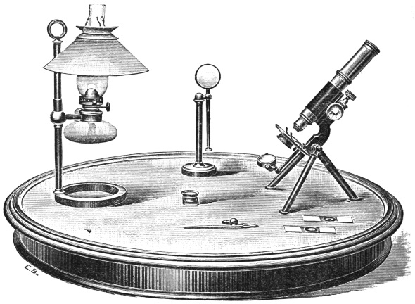
paraffin lamp, placed about two feet away from the
stage, and on one side of it, so as to be about a
foot above the level of the object, will give all
that is needed. Such a lamp is shown in Fig. 14.
Low magnifications are, as a rule, all that is called
for in this method.
Lieberkuhn’s condensers are useful aids, but are
somewhat expensive. They are concave mirrors,
which are so adjusted to the objective that the
latter and the reflector come into focus together,
the light being sent in from below, or from one side.
One other method of illumination must be mentioned
before leaving the topic, and this is the
illumination of objects upon a “dark field.” With
suitable subjects, and when carefully managed,
there is no method which gives more beautiful
effects, and it has the great advantage of allowing
the object to be brilliantly lighted, without the
strain to the eyes which is involved in such lighting
by the usual method of direct illumination.
Fig. 14.
It consists essentially in allowing the light to
fall upon the object from below, at such an angle
that none of it can enter the objective directly.
Thus the concave mirror, turned as far as possible
to one side, and reflecting on to the object the rays
from the lamp placed upon the opposite side, will
give very fair results with low powers; this plan,
however, is capable of but very limited application.
Again, a disc of black paper may be stuck on to
the middle of the bull’s-eye, and the latter be
placed below the stage between it and the mirror.
In this case everything depends upon the size of
the disc, which, if too small, will not give a black
ground, and if too large will cut off all light from
the object.
The best and only really satisfactory plan is to
arrange the illumination with the substage condenser,
as previously described, and then to place
below the lens of the latter a central stop of a
suitable size, which can only be determined by
trial. When this has been done the object will
be seen brilliantly illuminated upon a field of
velvety blackness. Such stops are supplied with
the condenser.
We have devoted a considerable portion of space
to this question, since it is, of all others, the most
important to a successful, satisfactory, and reliable
manipulation of the microscope; but even now, only
the main points of the subject have been touched
upon, and the worker will find it necessary to
supplement the information given by actual experiment.
A few failures, rightly considered, will
afford a great amount of information, but those
who desire to go thoroughly into the matter are
recommended to consult the present writer’s Guide
to the Science of Photomicrography, where it is
treated at much greater length, as an essential
part of the subject-matter of the book.
It may be added here, that no method of reproducing
the images of objects is on the whole so
satisfactory as the photographic one; and whilst a
lengthened reference to the topic would be out of
place in a work of the character of the present one,
the one just mentioned will be found to contain all
that is necessary to enable the beginner to produce
results which, for faithfulness and beauty, far excel
any drawing, whilst they have the additional advantage
that they can, if required, be exhibited to
hundreds simultaneously.
CHAPTER IV
Vegetable Cells and their Structure—Stellate Tissues—Secondary
Deposit—Ducts and Vessels—Wood-Cells—Stomata,
or Mouths of Plants—The Camera Lucida, and
Mode of Using—Spiral and Ringed Vessels—Hairs of
Plants—Resins, Scents, and Oils—Bark Cells.
We will now suppose the young observer to have
obtained a microscope and learned the use of its
various parts, and will proceed to work with it.
As with one or two exceptions, which are only
given for the purpose of further illustrating some
curious structure, the whole of the objects figured
in this work can be obtained without any difficulty,
the best plan will be for the reader to procure
the plants, insects, etc., from which the objects are
taken, and follow the book with the microscope at
hand. It is by far the best mode of obtaining a
systematic knowledge of the matter, as the quantity
of objects which can be placed under a microscope is
so vast that, without some guide, the tyro flounders
hopelessly in the sea of unknown mysteries, and
often becomes so bewildered that he gives up the
study in despair of ever gaining any true knowledge
of it. I would therefore recommend the reader to
work out the subjects which are here mentioned,
and then to launch out for himself on the voyage
of discovery. I speak from experience, having
myself known the difficulties under which a young
and inexperienced observer has to labour in so wide
a field, without any guide to help him to set about
his work in a systematic manner.
The objects that can be most easily obtained are
those of a vegetable nature, as even in London
there is not a square, an old wall, a greenhouse, a
florist’s window, or even a greengrocer’s shop, that
will not afford an exhaustless supply of microscopic
employment. Even the humble vegetables that
make their daily appearance on the dinner-table
are highly interesting; and in a crumb of potato, a
morsel of greens, or a fragment of carrot, the enthusiastic
observer will find occupation for many hours.
Following the best examples, we will commence at
the beginning, and see how the vegetable structure is
built up of tiny particles, technically called “cells.”
That the various portions of every vegetable
should be referred to the simple cell is a matter of
some surprise to one who has had no opportunity
of examining the vegetable structure, and indeed it
does seem more than remarkable that the tough,
coarse bark, the hard wood, the soft pith, the green
leaves, the delicate flowers, the almost invisible
hairs, and the pulpy fruit, should all start from
the same point, and owe their origin to the simple
vegetable cell. This, however, is the case; and by
means of a few objects chosen from different
portions of the vegetable kingdom, we shall obtain
some definite idea of this curious phenomenon.
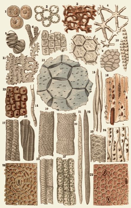
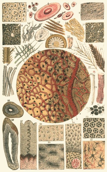
I.
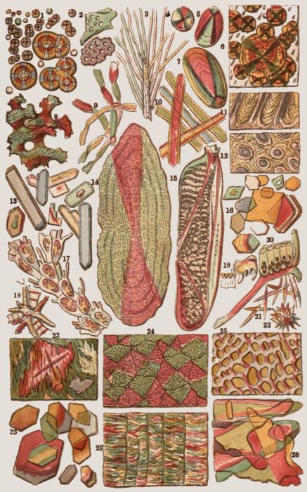
FIG.
1.
Strawberry, cellular tissue
15.
Wood-cells, Elder
2.
Buttercup leaf, internal layer
16.
Glandular markings and resin, “Cedar” pencil
3.
Privet, Seed Coat, showing star-shaped cells
17.
Do. Yew
4.
Rush, Star-shaped cells
18.
Scalariform tissue, Stalk of Fern
5.
Mistletoe, cells with ringed fibre
19.
Dotted Duct, Willow
6.
Cells from interior of Lilac bud
20.
Do. Stalk of Wheat
7.
Bur-reed (Sparganium), square cells from leaf
21.
Wood-cell, Chrysanthemum
8.
Six-sided cells, from stem of Lily
22.
Do. Lime-tree
9.
Angular dotted cells, rind of Gourd
23.
Dotted Duct, Carrot
10.
Elongated ringed cells, anther of Narcissus
24.
Cone-bearing wood, Deal
11.
Irregular star-like tissue, pith of Bulrush
25.
Cells, outer coat, Gourd
12.
Six-sided cells, pith of elder
26.
Ducts, Elm
13.
Young cells from Wheat
27.
Cellular tissue, Stalk of Chickweed
14.
Do. rootlets of Wheat
28.
Holly-berry, outer coat
I.
On Plate I. Fig. 1, may be seen three cells of a
somewhat globular form, taken from the common
strawberry. Any one wishing to examine these
cells for himself may readily do so by cutting a
very thin slice from the fruit, putting it on a slide,
covering it with a piece of thin glass (which may
be cheaply bought at the optician’s, together with
the glass slides on which the objects are laid), and
placing it under a power of two hundred diameters.
Should the slice be rather too thick, it may be
placed in the live-box and well squeezed, when the
cells will exhibit their forms very distinctly. In
their primary form the cells seem to be spherical;
but as in many cases they are pressed together, and
in others are formed simply by the process of subdivision,
the spherical form is not very often seen.
The strawberry, being a soft and pulpy fruit,
permits the cells to assume a tolerably regular
form, and they consequently are more or less
globular.
Where the cells are of nearly equal size, and are
subjected to equal pressure in every direction, they
force each other into twelve-sided figures, having
the appearance under the microscope of flat six-sided
forms. Fig. 8, in the same Plate, taken from
the stem of a lily, is a good example of this form
of cell, and many others may be found in various
familiar objects.
We must here pause for a moment to define a
cell before we proceed further.
The cell is a close sac or bag formed of a
substance called from its function “cellulose,” and
containing certain semi-fluid contents as long as it
retains its life. In the interior of the cell may
generally be found a little dark spot, termed the
“núcleus,” and which may be seen in Fig. 1, to
which we have already referred. The object of the
nucleus is rather a bone of contention among the
learned, but the best authorities on this subject
consider it to be the vital centre of the cells, to and
from which tends the circulation of the protoplasm,
and which is intimately connected with the growth
and reproduction of the cell. On looking a little
more closely at the nucleus, we shall find it marked
with several small light spots, which are termed
“nucléoli.”
On the same Plate (Fig. 2) is a pretty group of
cells taken from the internal layer of the buttercup
leaf, and chosen because they exhibit the series of
tiny and brilliant green dots to which the colour
of the leaf is due. The technical name for this
substance is “chlorophyll,” or “leaf-green,” and it
may always be found thus dotted in the leaves of
different plants, the dots being very variable in size,
number, and arrangement. A very fine object for
the exhibition of this point is the leaf of Anácharis,
the “Canadian timber-weed,” to be found in almost
every brook and river. It also shows admirably
the circulation of the protoplasm in the cell.
In the centre of the same Plate (Fig. 12) is a
group of cells from the pith of the elder-tree.
This specimen is notable for the number of little
“pits” which may be seen scattered across the
walls of the cells, and which resemble holes when
placed under the microscope. In order to test the
truth of this appearance, the specimen was coloured
blue by the action of iodine and dilute sulphuric
acid, when it was found that the blue tint spread
over the pits as well as the cell-walls, showing
that the membrane is continuous over the pits.
Fig. 7 exhibits another form of cell, taken from
the Spargánium, or bur-reed. These cells are
tolerably equal in size, and have assumed a square
shape. They are obtained from the lower part of
the leaf. The reader who has any knowledge of
entomology will not fail to observe the similarity
in form between the six-sided and square cells of
plants and the hexagonal and square facets of the
compound eyes of insects and crustaceans. In a
future page these will be separately described.
Sometimes the cells take most singular and unexpected
shapes, several examples of which will be
briefly noticed.
In certain loosely made tissues, such as are
found in the rushes and similar plants, the walls of
the cells grow very irregularly, so that they push
out a number of arms which meet each other in
every direction, and assume the peculiar form which
is termed “stellate,” or star-shaped tissue. Fig. 3
shows a specimen of stellate tissue taken from the
seed-coat of the privet, and rather deeply coloured,
exhibiting clearly the beautiful manner in which
the arms of the various stars meet each other. A
smaller group of stellate cells taken from the stem
of a large rush, and exemplifying the peculiarities
of the structure, are seen in Fig. 4.
The reader will at once see that this mode of
formation leaves a vast number of interstices, and
gives great strength with little expenditure of
material. In water-plants, such as the reeds, this
property is extremely valuable, as they must be
greatly lighter than the water in which they live,
and at the same time must be endowed with
considerable strength in order to resist its
pressure.
A less marked example of stellate tissue is
given in Fig. 11, where the cells are extremely
irregular, in their form, and do not coalesce
throughout. This specimen is taken from the
pithy part of a bulrush. There are very many
other plants from which the stellate cells may
be obtained, among which the orange affords very
good examples, in the so-called “white” that lies
under the yellow rind, a section of which may
be made with a very sharp razor, and placed in
the field of the microscope.
Looking toward the bottom of the Plate, and
referring to Fig. 27, the reader will observe a
series of nine elongated cells, placed end to end,
and dotted profusely with chlorophyll. These
are obtained from the stalk of the common
chickweed. Another example of the elongated
cell is seen in Fig. 14, which is a magnified
representation of the rootlets of wheat. Here
the cells will be seen set end to end, and each
containing its nucleus. On the left hand of the
rootlet (Fig. 13) is a group of cells taken from
the lowest part of the stem of a wheat plant
which had been watered with a solution of
carmine, and had taken up a considerable amount
of the colouring substance. Many experiments
on this subject were made by the Rev. Lord
S. G. Osborne, and may be seen at full length
in the pages of the Microscopical Journal, the
subject being too large to receive proper treatment
in the very limited space which can here be
given to it. It must be added that later
researches have caused the results here described
to be gravely disputed.
Fig. 9 on the same Plate exhibits two notable
peculiarities—the irregularity of the cells and
the copiously pitted deposit with which they are
covered. The irregularity of the cells is mostly
produced by the way in which the multiplication
takes place, namely, by division of the original
cell into two or more new ones, so that each of
these takes the shape which it assumed when a
component part of the parent cell. In this case
the cells are necessarily very irregular, and when
they are compressed from all sides they form
solid figures of many sides, which, when cut
through, present a flat surface marked with a
variety of irregular outlines. This specimen is
taken from the rind of a gourd.
The “pitted” structure which is so well shown
in this figure is caused by a layer of matter
which is deposited in the cell and thickens its
walls, and which is perforated with a number of
very minute holes called “pits.” This substance
is called “secondary deposit.” That these pits do
not extend through the real cell-wall has already
been shown in Fig. 12.
This secondary deposit assumes various forms.
In some cases it is deposited in rings round the
cell, and is clearly placed there for the purpose of
strengthening the general structure. Such an
example may be found in the mistletoe (Fig. 5),
where the secondary deposit has formed itself
into clear and bold rings that evidently give
considerable strength to the delicate walls which
they support. Fig. 10 shows another good instance
of similar structure; differing from the preceding
specimen in being much longer and containing a
greater number of rings. This object is taken
from an anther of the narcissus. Among the
many plants from which similar objects may be
obtained, the yew is perhaps one of the most
prolific, as ringed wood-cells are abundant in its
formation, and probably aid greatly in giving to
the wood the strength and elasticity which have
long made it so valuable in the manufacture of
bows.
Before taking leave of the cells and their
remarkable forms, we will just notice one example
which has been drawn in Fig. 6. This is a
congeries of cells, containing their nuclei, starting
originally end to end, but swelling and dividing
at the top. This is a very young group of cells
(a young hair, in fact) from the inner part of a
lilac bud, and is here introduced for the purpose
of showing the great similarity of all vegetable
cells in their earliest stages of existence.
Having now examined the principal forms of
cells, we arrive at the “vessels,” a term which is
applied to those long and delicate tubes which are
formed of a number of cells set end to end, their
walls of separation being absorbed.
In Fig. 19 the reader will find a curious
example of the “pitted vessel,” so called from the
multitude of little markings which cover its walls,
and are arranged in a spiral order. Like the pits
and rings already mentioned, the dots are composed
of secondary deposit in the interior of the tube,
and vary very greatly in number, function, and
dimensions. This example is taken from the wood
of the willow, and is remarkable for the extreme
closeness with which the dots are packed together.
Immediately on the right hand of the preceding
figure may be seen another example of a dotted
vessel (Fig. 20), taken from a wheat stem. In
this instance the cells are not nearly so long, but
are wider than in the preceding example, and are
marked in much the same way with a spiral series
of dots. About the middle of the topmost cell is
shown the short branch by which it communicates
with the neighbouring vessel.
Fig. 23 exhibits a vessel taken from the common
carrot, in which the secondary deposit is placed in
such a manner as to resemble a net of irregular
meshes wrapped tightly round the vessel. For
this reason it is termed a “netted vessel.” A very
curious instance of these structures is given in
Fig. 26, at the bottom of the Plate, where are
represented two small vessels from the wood of the
elm. One of them—that on the left hand—is
wholly marked with spiral deposit, the turns being
complete; while, in the other instance, the spiral
is comparatively imperfect, and the cell-walls are
marked with pits. If the reader would like to
examine these structures more attentively, he will
find plenty of them in many familiar garden
vegetables, such as the common radish, which is
very prolific in these interesting portions of vegetable
nature.
There is another remarkable form in which this
secondary deposit is sometimes arranged that is
well worthy of our notice. An example of this
structure is given in Fig. 18, taken from the stalk
of the common fern or brake. It is also found
in very great perfection in the vine. On inspecting
the illustration, the reader will observe
that the deposit is arranged in successive bars
or steps, like those of a winding staircase. In
allusion to the ladder-like appearance of this
formation, it is called “scalariform” (Latin, scala,
a ladder).
In the wood of the yew, to which allusion has
already been made, there is a very peculiar structure,
a series of pits found only in those trees that
bear cones, and therefore termed the coniferous
pitted structure. Fig. 16 is a section of a common
cedar pencil, the wood, however, not being that of
the true cedar, but of a species of fragrant Juniper.
This specimen shows the peculiar formation which
has just been mentioned.
Any piece of deal or pine will exhibit the same
peculiarities in a very marked manner, as is seen in
Fig. 24. A specimen may be readily obtained by
making a very thin shaving with a sharp plane.
In this example the deposit has taken a partially
spiral form, and the numerous circular pits with
which it is marked are only in single rows. In
several other specimens of coniferous woods, such
as the Araucaria, or Norfolk Island pine, there are
two or three rows of pits.
A peculiarly elegant example of this spiral deposit
may be seen in the wood of the common yew (Fig. 17).
If an exceedingly thin section of this wood be made,
the very remarkable appearance will be shown which
is exhibited in the illustration. The deposit has not
only assumed the perfectly spiral form, but there
are two complete spirals, arranged at some little
distance from each other, and producing a very
pretty effect when seen through a good lens.
The pointed, elongated shape of the wood-cells
is very well shown in the common elder-tree (see
Fig. 15). In this instance the cells are without
markings, but in general they are dotted like Fig. 21,
an example cut from the woody part of the chrysanthemum
stalk. This affords a very good instance of
the wood-cell, as its length is considerable, and both
ends are perfect in shape. On the right hand of
the figure is a drawing of the wood-cell found in the
lime-tree (Fig. 22), remarkable for the extremely
delicate spiral markings with which it is adorned.
In these wood-cells the secondary deposit is so
plentiful that the original membranous character of
the cell-walls is entirely lost, and they become elongated
and nearly solid cases, having but a very small
cavity in their centre. It is to this deposit that the
hardness of wood is owing, and the reader will easily
see the reason why the old wood is so much harder
than the young and new shoots. In order to permit
the passage of the fluids which maintain the life of
the part, it is needful that the cell-wall be left thin
and permeable in certain places, and this object is
attained either by the “pits” described on page 43,
or by the intervals between the spiral deposit.
At the right-hand bottom corner of Plate I. (Fig. 28)
may be seen a prettily marked object, which is
of some interest. It is a slice stripped from the
outer coat of the holly-berry, and is given for the
purpose of illustrating the method by which plants
are enabled to breathe the atmospheric air on which
they depend as much as ourselves, though their
respiration is slower. Among the mass of net-like
cells may be seen three curious objects, bearing a
rather close resemblance to split kidneys. These
are the mouths, or “stómata,” as they are scientifically
called.
In the centre of the mouths may be seen a dark
spot, which is the aperture through which the air
communicates with the passages between the cells
in the interior of the structure. In the flowering
plants their shape is generally rounded, though they
sometimes take a squared form, and they regularly
occur at the meeting of several surface cells. The
two kidney-shaped cells which form the “mouth”
are the “guard-cells,” so called from their function,
since, by their change of form, they cause the mouth
to open or shut, according to the needs of the plant.
In young plants these guard-cells are very little below
the surface of the leaf or skin, but in others they
are sunk quite beneath the layer of cells forming
the outer coat of the tissue. There are other
cases where they are slightly elevated above the
surface.
Stomata are found chiefly in the green portions
of plants, and are most plentiful on the under side of
leaves. It is, however, worthy of notice, that when
an aquatic leaf floats on the water, the mouths are
only to be found on the upper surface. These
curious and interesting objects are to be seen in
many structures where we should hardly think of
looking for them; for instance, they may be found
existing on the delicate skin which envelops the
kernel of the common walnut. As might be expected,
their dimensions vary with the character of
the leaf on which they exist, being large upon the
soft and pulpy leaves, and smaller upon those of a
hard and leathery consistence. The reader will find
ample amusement, and will gain great practical
knowledge of the subject, by taking a plant, say a
tuft of groundsel, and stripping off portions of the
external skin or “epidermis” from the leaf or stem,
etc., so as to note the different sizes and shapes of
the stomata.
On the opposite bottom corner of Plate I. Fig. 25,
is an example of a stoma taken from the outer skin
of a gourd, and here given for the purpose of showing
the curious manner in which the cells are
arranged about the mouth, no less than seven cells
being placed round the single mouth, and the
others arranged in a partially circular form around
them.
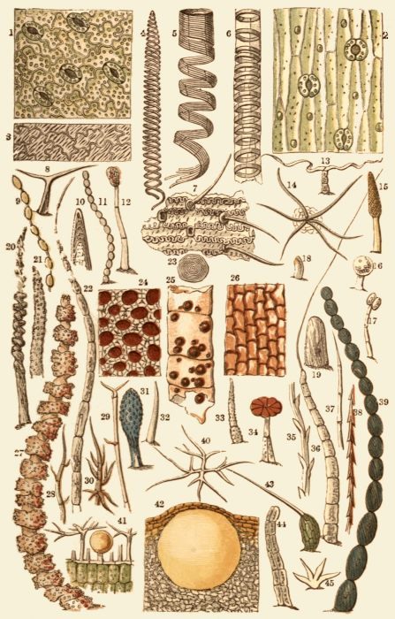
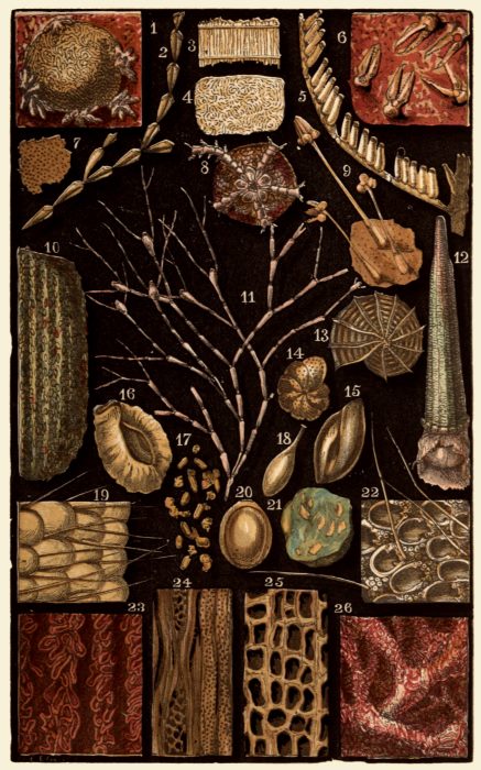
Turning to Plate II., we find several other examples
of stomata, the first of which (Fig. 1) is
obtained from the under surface of the buttercup
leaf, by stripping off the external skin, or “epidermis,”
as it is scientifically termed. The reader will
here notice the slightly waved outlines of the cell-walls,
together with the abundant spots of chlorophyll
with which the leaf is coloured. In this
example the stomata appear open. Their closure
or expansion depends chiefly on the state of the
weather; and, as a general rule, they are open by
day and closed at night.
A remarkably pretty example of stomata and
elongated cells is to be obtained from the leaf of the
common iris, and may be prepared for the microscope
by simply tearing off a strip of the epidermis
from the under side of the leaf, laying it on a slide,
putting a little water on it, and covering it with a
piece of thin glass. (See Plate II. Fig. 2.) There
are a number of longitudinal bands running along
the leaf where these cells and stomata appear.
The latter are not placed at regular intervals, for it
often happens that the whole field of the microscope
will be filled with cells without a single stoma, whilst
elsewhere a group of three or four may be seen
clustered closely together.
Fig. 3 on the same Plate exhibits a specimen of
the beautifully waved cells, without mouths, which
are found on the upper surface of the ivy leaf.
These are difficult to arrange from the fresh leaf,
but are easily shown by steeping the leaf in water
for some time, and then tearing away the cuticle.
The same process may be adopted with many leaves
and cuticles, and in some cases the immersion must
be continued for many days, and the process of
decomposition aided by a very little nitric acid in
the water, or by boiling.
On the same Plate are three examples of spiral
and ringed vessels, types of an endless variety of
these beautiful and interesting structures. Fig. 4
is a specimen of a spiral vessel taken from the lily,
and is a beautiful example of a double spire. The
deposit which forms this spiral is very strong, and
it is to the vast number of these vessels that the
stalk owes its well-known elasticity. In many cases
the spiral vessels are sufficiently strong to be visible
to the naked eye, and to bear uncoiling. For
example, if a leaf-stalk of geranium be broken
across, and the two fragments gently drawn asunder,
a great number of threads, drawn from the spiral
vessels, will be seen connecting the broken ends.
In this case the delicate membranous walls of the
vessel are torn apart, and the stronger fibre which
is coiled spirally within it unrolls itself in proportion
to the force employed. In many cases these
fibres are so strong that they will sustain the weight
of an inch or so of the stalk.
In Fig. 5 is seen a still more bold and complex
form of this curious structure; being a coil of five
threads, laid closely against each other, and forming,
while remaining in their natural position, an almost
continuous tube. This specimen is taken from the
root of the water lily, and requires some little care
to exhibit its structure properly.
Every student of nature must be greatly struck
with the analogies between different portions of the
visible creation. These spiral structures which we
have just examined are almost identical in appearance,
and to some extent in their function, with
the threads that are coiled within the breathing
tubes of insects. This is in both cases twofold,
namely, to give support and elasticity to a delicate
membrane, and to preserve the tube in its proper
form, despite the bending to which it may be
subjected. When we come to the anatomy of the
insect in a future page we shall see this structure
further exemplified.
In some cases the deposit, instead of forming a
spiral coil, is arranged in a series of rings, and the
vessel is then termed “annulated.” A very good
example of this formation is given in Fig. 6, which
is a sketch of such a vessel, taken from a stalk of
the common rhubarb. To see these ringed vessels
properly, the simplest plan is to boil the rhubarb
until it is quite soft, then to break down the pulpy
mass until it is flattened, to take some of the most
promising portions with the forceps, lay them on
the slide and press them down with a thin glass
cover. They will not be found scattered at random
through the fibres, which elsewhere present only
a congeries of elongated cells, but are seen grouped
together in bundles, and with a little trouble may
be well isolated, and the pulpy mass worked away
so as to show them in their full beauty. As may
be seen in the illustration, the number of the rings
and their arrangement is extremely variable. A
better, but somewhat more troublesome, plan is to
cut longitudinal sections of the stem, as described
in our concluding chapter, when not only the
various forms of cells and vessels, but their
relations to each other, will be well shown. The
numerous crystals of oxalate of lime, which make
rhubarb so injurious a food for certain persons,
will also be well seen. These crystals are called
“raphides,” and are to be found in very many
plants in different forms.
II.
FIG.
1.
Cuticle, Buttercup leaf
24.
Do. Pine cone
2.
Do. Iris
25.
Vitta, Caraway Seed
3.
Do. Ivy leaf
26.
Cork
4.
Spiral vessel, Lily
27.
Hair, Flower of Garden Verbena
5.
Do. root, (rhizome) Water Lily
28.
Do. fruit of Plane
6.
Ringed vessel, Rhubarb
29.
Do. do.
7.
Chaff, after burning
30.
Do. do.
8.
Bifid hair, Arabis
31.
Do. Lobelia
9.
Hair, Marvel of Peru
32.
Do. Cabbage
10.
End of hair, leaf of Hollyhock
33.
Do. Dead-nettle flower
11.
Hair, Sowthistle leaf
34.
Do. Garden Verbena flower
12.
Do. Tobacco
35.
Fruit-hair, Dandelion
13.
Do. Southernwood
36.
Hair, Thistle leaf
14.
Group of hairs, Hollyhock leaf
37.
Do. Cactus
15.
Hair, Yellow Snapdragon
38.
Do. do.
16.
Do. Moneywort
39.
Do. Virginian Spider-wort
17.
Hair, Geum
40.
Do. Lavender
18.
Do. Flower of Heartsease
41.
Section, Lavender leaf, Hairs
19.
Do. Dockleaf
and perfume-gland
20.
Do. Throat of Pansy
42.
Section, Orange Peel
21.
Do. Dead-nettle Flower
43.
Sting of Nettle
22.
Do. Groundsel
44.
Hair, Marigold flower
23.
Cell, Beech-nut
45.
Do. Ivy
II.
The hairs of plants form very interesting
objects, and are instructive to the student, as they
afford valuable indications of the mode in which
plants grow. They are all appendages of and arise
from the skin or epidermis; and although their
simplest form is that of a projecting and elongated
cell, the variety of shapes which are assumed by
these organs is inexhaustible. On Plate II. are
examples of some of the more striking forms,
which will be briefly described.
The simple hair is well shown in Figs. 18, 19,
and 32, the first being from the flower of the
heartsease, the second from a dock-leaf, and the
third from a cabbage. In Fig. 18 the hair is seen
to be but a single projecting cell, consisting only of
a wall and the contents. In Fig. 19 the hair has
become more decided in shape, having assumed a
somewhat dome-like form; and in Fig. 32 it has
become considerably elongated, and may at once be
recognised as a true hair.
In Fig. 8 is a curious example of a hair taken
from the white Arabis, one of the cruciferous
flowers, which is remarkable for the manner in
which it divides into two branches, each spreading
in opposite directions. Another example of a
forked hair is seen in Fig. 13, but in this instance
the hair is composed of a chain of cells, the three
lower forming the stem of the hair, and the two
upper being lengthened into the lateral branches.
This hair is taken from the common southernwood.
In most cases of long hairs, the peculiar elongation
is formed by a chain of cells, varying greatly
in length and development. Several examples of
these hairs will be seen on the same Plate.
Fig. 9 is a beaded hair from the Marvel of Peru,
which is composed of a number of separate cells
placed end to end, and connected by slender threads
in a manner that strongly reminds the observer of
a chain of beads strung loosely together, so as to
show the thread by which they are connected with
each other. Another good example is seen at
Fig. 11, in a hair taken from the leaf of the sowthistle.
In this case the beads are strung closely
together, and when placed under a rather high
power of the microscope have a beautifully white
and pearly aspect. The leaf must be dry and quite
fresh, and the hairs seen against the green of the
leaf. Fig. 39 represents another beaded hair taken
from the Virginian Spiderwort, or Tradescantia.
This hair is found upon the stamens, and is
remarkable for the beautifully beaded outline, the
fine colouring, and the spiral markings with which
each cell is adorned.
A still further modification of these many-celled
hairs is found in several plants, where the hairs
are formed by a row of ordinarily shaped cells,
with the exception of the topmost cell, which is
suddenly elongated into a whip-like form. Fig. 22
represents a hair of this kind, taken from the
common groundsel; and Fig. 36 is a still more
curious instance, found upon the leaf of the thistle.
The reader may have noticed the peculiar white
“fluffy” appearance of the thistle leaf when it is
wet after a shower of rain. This appearance is
produced by the long lash-like ends of the hairs,
which are bent down by the weight of the moisture,
and lie almost at right angles with the thicker
portions of the hair.
An interesting form of hair is seen in the
“sting” of the common nettle. This may readily
be examined by holding a leaf edgewise in the
stage forceps, and laying it under the field of the
microscope. In order to get the proper focus
throughout the hair, the finger should be kept
upon the screw movement, and the hair brought
gradually into focus from its top to its base. The
general structure of this hair is not unlike that
which characterises the fang of a venomous serpent.
The acrid fluid which causes the pain is situated in
the enlarged base of the hair, and is forced through
the long straight tubular extremity by means of
the pressure exerted when the sting enters the
skin. At the very extremity of the perfect sting
is a slight bulb-like swelling, which serves to
confine the acrid juice, and which is broken
off on the least pressure. The sting is seen in
Fig. 43.
The extremities of many hairs present very
curious forms, some being long and slender, as in
the examples already mentioned, while others are
tipped with knobs, bulbs, clubs, or rosettes in
endless variety.
Fig. 12 is a hair of the tobacco leaf, exhibiting
the two-celled gland at the tip, containing the
peculiar principle of the plant, known by the name
of “nicotine.” The reader will see how easy it is
to detect adulteration of tobacco by means of the
microscope. The leaves most generally used for
this purpose are the dock and the cabbage, so that
if a very little portion of leaf be examined the
character of the hairs will at once inform the
observer whether he is looking at the real article
or its substitute.
Fig. 15 is a hair from the flower of the common
yellow snapdragon, which is remarkable for the
peculiar shape of the enlarged extremity, and for
the spiral markings with which it is decorated.
Fig. 16 is a curious little knobbed hair found
upon the moneywort, and Fig. 17 is an example
of a double-knobbed hair taken from the Geum.
Fig. 34 affords a very curious instance of a
glandular hair, the stem being built up of cells
disposed in a very peculiar fashion, and the
extremity being developed into a beautiful rosette-shaped
head. This hair came from the Garden
Verbena.
Curiously branched hairs are not at all uncommon,
and some very good and easily obtained
examples are given on Plate II.
Fig. 28 is one of the multitude of branched hairs
that surround the well-known fruit of the plane-tree,
the branches being formed by some of the
cells pointing outward. These hairs do not assume
precisely the same shape; for Fig. 29 exhibits
another hair from the same locality, on which the
spikes are differently arranged, and Fig. 30 is a
sketch of another such hair, where the branches
have become so numerous and so well developed
that they are quite as conspicuous as the parent
stem.
One of the most curious and interesting forms
of hair is that which is found upon the lavender
leaf, and which gives it the peculiar bloom-like
appearance on the surface.
This hair is represented in Figs. 40 and 41.
On Fig. 40 the hair is shown as it appears when
looking directly upon the leaf, and in Fig. 41
a section of the leaf is given, showing the mode in
which the hairs grow into an upright stem, and
then throw out horizontal branches in every
direction. Between the two upright hairs, and
sheltered under their branches, may be seen a
glandular appendage not unlike that which is
shown in Fig. 16. This is the reservoir containing
the perfume, and it is evidently placed under the
spreading branches for the benefit of their shelter.
On looking upon the leaf by reflected light the
hairs are beautifully shown, extending their arms
on all sides; and the globular perfume cells may
be seen scattered plentifully about, gleaming like
pearls through the hair-branches under which they
repose. They will be found more numerous on the
under side of the leaf.
This object will serve to answer a question which
the reader has probably put to himself ere this,
namely, Where are the fragrant resins, scents, and
oils stored? On Plate I. Fig. 16, will be seen the
reply to the first question; Fig. 41 of the present
Plate has answered the second question, and Fig. 42
will answer the third. This figure represents a
section of the rind of an orange, the flattened cells
above constituting the delicate yellow skin, and the
great spherical object in the centre being the reservoir
in which the fragrant essential oil is stored.
The covering is so delicate that it is easily broken,
so that even by handling an orange some of the
scent is sure to come off on the hands, and when
the peel is stripped off and bent double, the reservoirs
burst in myriads, and fling their contents
to a wonderful distance. This may be easily seen
by squeezing a piece of orange peel opposite a lighted
candle, and noting the distance over which the oil
will pass before reaching the flame, and bursting
into little flashes of light. Other examples are
given on the same plate.
Returning to the barbed hairs, we may see in
Fig. 35 a highly magnified view of the “pappus”
hair of a dandelion, i.e. the hairs which fringe the
arms of the parachute-like appendage which is
attached to the seed. The whole apparatus will
be seen more fully on Plate III. Figs. 44, 45, 46.
This hair is composed of a double layer of elongated
cells lying closely against each other, and having
the ends of each cell jutting out from the original
line. A simpler form of a double-celled, or more
properly a “duplex” hair, will be seen in Fig. 44.
This is one of the hairs from the flower of the
marigold and has none of the projecting ends to
the cells.
In some instances the cell-walls of the hairs
become greatly hardened by secondary deposit, and
the hairs are then known as spines. Two examples
of these are seen in Figs. 37 and 38, the former
being picked from the Indian fig-cactus, and well
known to those persons who have been foolish
enough to handle the fig roughly before feeling it.
The wounds which these spines will inflict are said
to be very painful, and have been compared to
those produced by the sting of the wasp. The
latter hair is taken from the Opuntia. These spines
must not be confounded with thorns; which latter
are modified branches.
Fig. 10 represents the extreme tip of a hair from
the hollyhock leaf, subjected to a lens of very high
power.
Many hairs assume a star-like appearance, an
aspect which may be produced in different ways.
Sometimes a number of simple hairs start from the
same base, and by radiating in different directions
produce the stellate effect. An example of this
kind of hair may be seen in Fig. 14, which is a
group of hairs from the hollyhock leaf. There is
another mode of producing the star-shape which
may be seen in Fig. 45, a hair taken from the leaf
of the ivy. Very fine examples may also be found
upon the leaf of Deutzia scabra.
Hairs are often covered with curious little
branches or protuberances, and present many other
peculiarities of form which throw a considerable
light upon certain problems in scientific microscopy.
Fig. 33 represents a hair of two cells taken
from the flower of the well-known dead-nettle,
which is remarkable for the number of knobs
scattered over its surface. A similar mode of
marking is seen in Fig. 31, a club-shaped hair
covered with external projections, found in the
flower of the Lobelia. In order to exhibit these
markings well, a power of two hundred diameters
is needed. Fig. 21 shows this dotting in another
hair from the dead-nettle, where the cell is drawn
out to a great length, but is still covered with these
markings.
Fig. 20 is an example of a very curious hair
taken from the throat of the pansy. This hair
may readily be obtained by pulling out one of the
petals, when the hairs will be seen at its base.
Under the microscope it has a particularly beautiful
appearance, looking just like a glass walking-stick
covered with knobs, not unlike those huge, knobby
club-like sticks in which some farmers delight,
where the projections have been formed by the
pressure of a honeysuckle or other climbing plant.
A hair of a similar character, but even more
curious, is found in the same part of the flower of
the Garden Verbena (see Fig. 27), and is not only
beautifully translucent, but is coloured according to
the tint of the flower from which it is taken. Its
whole length is covered with large projections, the
joints much resembling the antennæ of certain
insects; and each projection is profusely spotted
with little dots, formed by elevation of the outer
skin or cuticle. These are of some value in determining
the structure of certain appearances upon
petals and other portions of the flowers, and may be
compared with Figs. 33 to 35 on Plate III.
Fig. 26 offers an example of the square cells
which usually form the bark of trees. This is a
transverse section of cork, and perfectly exhibits
the form of bark cells. The reader is very strongly
advised to cut a delicate section of the bark of
various trees, a matter very easily accomplished
with the aid of a sharp razor and a steady hand.
Fig. 24 is a transverse section through one of the
scales of a pine-cone, and is here given for the
purpose of showing the numerous resin-filled cells
which it displays. This may be compared with
Fig. 16 of Plate I. Fig. 25 is a part of one of
the “vittæ,” or oil reservoirs, from the fruit of the
caraway, showing the cells containing the globules
of caraway oil. This is rather a curious object,
because the specimen from which it was taken was
boiled in nitric acid, and yet retained some of the
oil globules. Immediately above it may be seen
(Fig. 23) a transverse section of the beechnut,
showing a cell with its layers of secondary
deposit.
In the cuticle of the grasses and the mare’s-tails
is deposited a large amount of pure flint. So
plentiful is this substance, and so equally is it
distributed, that it can be separated by heat or
acids from the vegetable parts of the plant, and
will still preserve the form of the original cuticle,
with its cell-walls, stomata, and hairs perfectly well
defined.
Fig. 7, Plate II., represents a piece of wheat
chaff, or “bran,” that has been kept at a white heat
for some time, and then mounted in Canada balsam.
I prepared the specimen from which the drawing
was made by laying the chaff on a piece of
platinum, and holding it over the spirit-lamp. A
good example of the silex or flint in wheat is often
given by the remains of a straw fire, where the
stems may be seen still retaining their tubular form
but fused together into a hard glassy mass. It is
this substance that cuts the fingers of those who
handle the wild grasses too roughly, the edges of
the blades being serrated with flinty teeth, just like
the obsidian swords of the ancient Mexicans, or the
shark’s-tooth falchion of the New Zealander.
These are but short and meagre accounts of a
very few objects, but space will not permit of
further elucidation, and the purpose of this little
work is not to exhaust the subjects of which it
treats, but to incite the reader to undertake investigation
on his own account, and to make his
task easier than if he had done it unaided.
CHAPTER V
Starch, its Growth and Properties—Surface Cells of Petals—Pollen
and its Functions—Seeds.
The white substance so dear to the laundries under
the name of starch is found in a vast variety of
plants, being distributed more widely than most of
the products which are found in the interior of
vegetable cells.
The starch grains are of very variable size even
in the same plant, and their form is as variable as
their size, though there is a general resemblance in
those of the same plant which allows of their being
fairly easily identified after a moderate amount of
practice. Sometimes the grains are found loosely
packed in the interior of the cells, and are then
easily recognised as starch grains by their peculiar
form and the delicate lines with which they are
marked; but in many places they are pressed so
closely together that they assume an hexagonal
shape under the microscope, and bear a close resemblance
to ordinary twelve-sided cells. In other
plants, again, the grains never advance beyond the
very minute form in which they seem to commence
their existence; and in some, such as the common
oat, a great number of very little granules are
compacted together so as to resemble one large
grain.
There are several methods of detecting starch in
those cases where its presence is doubtful; and the
two modes that are usually employed are polarised
light and the iodide of potassium. When polarised
light is employed—a subject on which we shall
have something to say presently—the starch grains
assume the characteristic “black-cross,” and when
a plate of selenite is placed immediately beneath
the slide containing the starch grains, they glow
with all the colours of the rainbow. The second
plan is to treat them with a very weak solution of
iodine and iodide of potassium, and in this case the
iodine has the effect on the starch granules of
staining them blue. They are so susceptible of
this reaction that when the liquid is too strong the
grains actually become black from the amount of
iodine which they imbibe.
Nothing is easier than to procure starch granules
in the highest perfection. Take a raw potato, and
with a razor cut a very thin slice from its interior,
the direction of the cut not being of the slightest
importance. Put this delicate slice upon a slide,
drop a little water upon it, cover it with a piece of
thin glass, give it a good squeeze, and place it
under a power of a hundred or a hundred and fifty
diameters. Any part of the slice, provided that it
be very thin, will then present the appearance
shown in Plate III. Fig. 9, where an ordinary cell
of potato is seen filled loosely with starch grains
of different sizes. Around the edges of the slice a
vast number of starch granules will be seen, which
have been squeezed out of their cells by pressure,
and are now floating freely in the water. As cold
water has no perceptible effect upon starch, the
grains are not altered in form by the moisture, and
can be examined at leisure.
III.
FIG.
1.
Laurel leaf, transverse section
27.
Do. Heath, another species
2.
Starch, Wheat
28.
Pollen, Furze
3.
Do. from Pudding
29.
Do. Tulip
4.
Do. Potato
30.
Petal, Pelargonium
5.
Outer Skin, Capsicum pod
31.
Do. Periwinkle
6.
Starch, Parsnip
32.
Do. Golden Balsam
7.
Do. Arrow Root, West Indian
33.
Do. Snapdragon
8.
Do. “Tousles Mois”
34.
Do. Primrose
9.
Do. in cell of Potato
35.
Do. Scarlet Geranium
10.
Do. Indian Corn
36.
Pollen, Crocus
11.
Do. Sago
37.
Do. Hollyhock
12.
Do. Tapioca
38.
Fruit, Galium, Goosegrass
13.
Root, Yellow Water-Lily
39.
A hook of ditto more magnified
14.
Starch, Rice
40.
Seed, Red Valerian
15.
Do. Horsebean
41.
Portion of Parachute of same, more magnified
16.
Do. Oat
42.
Seed, Foxglove
17.
Pollen, Snowdrop
43.
Do. Sunspurge
18.
Do. Wallflower
44.
Parachute, Dandelion seed
19.
Do. Willow Herb, a pollen tube
45.
Seed, Dandelion
20.
Do. Violet
46.
Do. Hair of Parachute
21.
Do. Musk Plant
47.
Do. Yellow Snapdragon
22.
Do. Apple
48.
Do. Mullein
23.
Do. Dandelion
49.
Do. Robin Hood
24.
Do. Sowthistle
50.
Do. Bur-reed
25.
Do. Lily
51.
Do. Willow Herb
26.
Do. Heath
52.
Do. Musk Mallow
III.
On focusing with great care, the surface of each
granule will be seen to be covered with very minute
dark lines, arranged in a manner which can be
readily comprehended from Fig. 4, which represents
two granules of potato starch as they appear when
removed from the cell in which they took their
origin. All the lines evidently refer to the little
dark spots at the end of the granule, called
technically the “hilum,” and represent the limits of
successive layers of material deposited one after
another. The lines in question are very much
better seen if the substage condenser be used with
a small central stop, so as to obtain partial dark-field
illumination. Otherwise they are often very
difficult of detection.
In the earliest stages of their growth the starch
granules appear to be destitute of these markings,
or at all events they are so few and so delicate as
not to be visible even with the most perfect instruments,
and it is not until the granules assume
a comparatively large size that the external markings
become distinctly perceptible.
We will now glance at the examples of starch
which are given in the Plate, and which are a very
few out of the many that might be figured. Fig. 2
represents the starch of wheat, the upper grain
being seen in front, the one immediately below it
in profile, and the two others being examples of
smaller grains. Fig. 6 is a specimen of a very
minute form of starch, where the granules do not
seem to advance beyond their earliest stage. This
specimen is obtained from the parsnip; and although
the magnifying power is very great, the
dimensions of the granules are exceedingly small,
and except by a very practised eye they would not
be recognisable as starch grains.
Fig. 3 is a good example of a starch grain of
wheat, exemplifying the change that takes place
by the combined effects of heat and moisture. It
has already been observed that cold water exercises
little, if any, perceptible influence upon starch; but
it will be seen from the illustration that hot water
has a very powerful effect. When subjected to the
action of water at a temperature over 140° Fahr.,
the granule swells rapidly, and at last bursts, the
contents escaping in a gelatinous mass, and the
external membrane collapsing into the form which
is shown in Fig. 3, which was taken out of a piece
of hot pudding. A similar form of wheat starch
may also be detected in bread, accompanied, unfortunately,
by several other substances not generally
presumed to be component parts of the “staff of life.”
In Fig. 7 are represented some grains of starch
from West Indian arrowroot, and Fig. 8 exhibits
the largest kind of starch grain known, obtained
from the tuber of a species of canna, supposed to
be C. edúlis, a plant similar in characteristics to
the arrowroot. The popular name of this starch is
“Tous les Mois,” and under that title it may be
obtained from the opticians, or chemists.
Fig. 10 shows the starch granules from Indian
corn, as they appear before they are compressed
into the honeycomb-like structure which has
already been mentioned. Even in that state,
however, if they are treated with iodine, they
exhibit the characteristics of starch in a very
perfect manner. Fig. 11 is starch from sago, and
Fig. 12 from tapioca, and in both these instances
the several grains have been injured by the heat
employed in preparing the respective substances
for the market.
Fig. 13 exhibits the granules obtained from the
root of the water-lily, and Fig. 14 is a good
example of the manner in which the starch granules
of rice are pressed together so as to alter the
shape and puzzle a novice. Fig. 16 is the compound
granule of the oat, which has already been
mentioned, together with some of the simple
granules separated from the mass; and Fig. 15
is an example of the starch grains obtained from
the underground stem of the horse-bean. It is
worthy of mention that the close adhesion of the
rice starch into those masses is the cause of the
peculiar grittiness which distinguishes rice flour to
the touch.
Whilst very easily acted on by heat, starch-granules
are very resistent to certain other
reagents. Weak alkalies, in watery solution,
readily attack them, but by treating portions of
plants with caustic potash dissolved in strong
spirit, the woody and other parts may be dissolved
away; and after repeated washing with spirit the
starch may be mounted. This, however, must
never be in any glycerine medium, except that
given on p. 172.
In Plate III. Fig. 1, may be seen a curious little
drawing, which is a sketch of the laurel-leaf cut
transversely, and showing the entire thickness of
the leaf. Along the top may be seen the delicate
layer of “varnish” with which the surface of the
leaf is covered, and which serves to give to the
foliage its peculiar polish. This varnish is nothing
more than the translucent matter which binds all
the cells together, and which is poured out very
liberally upon the surface of the leaf. The lower
part of this section exhibits the cells of which the
leaf is built, and towards the left hand may be seen
a cut end of one of the veins of the leaf, more
rightly called a wood-cell.
We will now examine a few examples of surface
cells.
Fig. 5 is a portion of epidermis stripped from a
Capsicum pod, exhibiting the remains of the nuclei
in the centre of each cell, together with the great
thickening of the wall-cells and the numerous pores
for the transmission of fluid. This is a very pretty
specimen for the microscope, as it retains its bright
red colour, and even in old and dried pods exhibits
the characteristic markings.
In the centre of the Plate may be seen a wheel-like
arrangement of the peculiar cells found on the
petals of six different flowers, all easily obtainable,
and mounted without difficulty.
Fig. 30 is the petal of a geranium (Pelargonium),
a very common object on purchased slides. It is
a most lovely subject for the microscope, whether
it be examined with a low or a high power,—in
the former instance exhibiting a most beautiful
“stippling” of pink, white, and black, and in the
latter showing the six-sided cells with their curious
markings.
In the centre of each cell is seen a radiating
arrangement of dark lines with a light spot in the
middle, looking very like the mountains on a map.
These lines were long thought to be hairs; but
Mr. Tuffen West, in an interesting and elaborate
paper on the subject, has shown their true nature.
From his observations it seems that the beautiful
velvety aspect of flower petals is owing to these
arrangements of the surface cells, and that their
rich brilliancy of colour is due to the same cause.
The centre of each cell-wall is elevated as if pushed
up by a pointed instrument from the under side of
the wall, and in different flowers this elevation
assumes different forms. Sometimes it is merely
a slight wart on the surface, sometimes it becomes
a dome, while in other instances it is so developed
as to resemble a hair. Indeed, Mr. West has concluded
that these elevations are nothing more than
rudimentary hairs.
The dark radiating lines are shown by the same
authority to be formed by wrinkling of the
membrane forming the walls of the elevated
centre, and not to be composed of “secondary
deposit,” as has generally been supposed.
Fig. 31 represents the petal of the common
periwinkle, differing from that of the geranium by
the straight sides of the cell-walls, which do not
present the toothed appearance so conspicuous in
the former flower. A number of little tooth-like
projections may be seen on the interior of the cells,
their bases affixed to the walls and their points
tending toward the centre, and these teeth are,
according to Mr. West, formed of secondary
deposit.
In Fig. 32 is shown the petal of the common
garden balsam, where the cells are elegantly
waved on their outlines, and have plain walls.
The petal of the primrose is seen in Fig. 34, and
that of the yellow snapdragon in Fig. 33; in the
latter instance the surface cells assume a most
remarkable shape, running out into a variety of
zigzag outlines that quite bewilders the eye when
the object is first placed under the microscope.
Fig. 35 is the petal of the common scarlet
geranium.
In several instances these petals are too thick
to be examined without some preparation, and
glycerine will be found well adapted for that
purpose. The young microscopist must, however,
beware of forming his ideas from preparations of
dried leaves, petals, or hairs, and should always
procure them in their fresh state whenever he
desires to make out their structure. Even a fading
petal should not be used, and if the flowers are
gathered for the occasion, their stalks should be
placed in water, so as to give a series of leaves and
petals as fresh as possible.
We now pass from the petal of the flower to the
pollen, that coloured dust, generally yellow or
white, which is found upon the stamens, and which
is very plentiful in many flowers, such as the lily
and the hollyhock.
This substance is found only upon the stamens
or anthers of full-blown flowers (the anthers being
the male organs), and is intended for the purpose
of enabling the female portion of the flower to
produce fertile seeds. In form the pollen grains
are wonderfully diverse, affording an endless variety
of beautiful shapes. In some cases the exterior is
smooth and marked only with minute dots, but in
many instances the outer wall of the pollen grain
is covered with spikes, or decorated with stripes or
belts. A few examples of the commonest forms of
pollen will be found on Plate III.
Fig. 17 is the pollen of the snowdrop, which, as
will be seen, is covered with dots and marked with
a definite slit along its length. The dots are
simply tubercles in the outer coat of the grain, and
are presumed to be formed for the purpose of
strengthening the membrane, otherwise too delicate,
upon the same principle which gives to “corrugated”
iron such strength in proportion to the amount of
material. Fig. 18 is the pollen of the wall-flower,
shown in two views, and having many of the same
characteristics as that of the snowdrop. Fig. 19
is the pollen of the willow-herb, and is here given
as an illustration of the manner in which the
pollen aids in the germination of plants.
In order to understand its action, we must first
examine its structure.
All pollen-grains are furnished with some means
by which their contents when thoroughly ripened
can be expelled. In some cases this end is accomplished
by sundry little holes called pores; in
others, certain tiny lids are pushed up by the
contained matter; and in some, as in the present
instance, the walls are thinned in certain places so
as to yield to the internal pressure.
When a ripe pollen-grain falls upon the stigma
of a flower, it immediately begins to swell, and
seems to “sprout” like a potato in a damp cellar,
sending out a slender “pollen-tube” from one or
other of the apertures already mentioned. In
Fig. 19 a pollen-tube is seen issuing from one
of the projections, and illustrates the process better
than can be achieved by mere verbal description.
The pollen-tubes insinuate themselves between the
cells of the stigmas, and, continually elongating,
worm their way down the “style” until they come
in contact with the “ovules.” By very careful
dissection of a fertilised stigma, the beautiful sight
of the pollen-tubes winding along the tissues of the
style may be observed under a high power of the
microscope.
The pollen-tube is nothing more than the interior
coat of the grain, very much developed, and filled
with a substance technically named “fovilla,” composed
of “protoplasm” (the semi-liquid substance
which is found in the interior of cells), very minute
starch grains, and some apparently oily globules.
In order to examine the structure of the pollen-grains
properly, they should be examined under
various circumstances—some dry, others placed in
water to which a little sugar has been added, others
in oil, and it will often be found useful to try the
effect of different acids upon them.
Fig. 20 is the pollen of the common violet, and
is easily recognisable by its peculiar shape and
markings. Fig. 21 is the pollen of the musk-plant,
and is notable for the curious mode in which its
surface is belted with wide and deep bands, running
spirally round the circumference. Fig. 22
exhibits the pollen of the apple, and Fig. 23
affords a very curious example of the raised
markings upon the surface of the dandelion pollen.
In Fig. 24 there are also some very wonderful
markings, but they are disposed after a different
fashion, forming a sort of network upon the surface,
and leaving several large free spaces between
the meshes. The pollen of the lily is shown in
Fig. 25, and is a good example of a pollen-grain
covered with the minute dottings which have
already been described.
Figs. 26 and 27 show two varieties of compound
pollen, found in two species of heath. These
compound pollen-grains are not of unfrequent occurrence,
and are accounted for in the following manner.
The pollen is formed in certain cavities within the
anthers, by means of the continual subdivision of
the “parent-cells” from which it is developed. In
many cases the form of the grain is clearly owing
to the direction in which these cells have divided,
but there is no great certainty on this subject.
It will be seen, therefore, that if the process of
subdivision be suddenly arrested, the grains will
be found adhering to each other in groups of
greater or smaller size, according to the character
of the species and the amount of subdivision that
has taken place. The reader must, however, bear
in mind that the whole subject is as yet rather
obscure, and that further discovery may throw
doubt on many theories which at present are
accepted as established.
Fig. 28 shows the pollen of the furze, in which
are seen the longitudinal slits and the numerous dots
on the surface; and Fig. 29 is the curiously shaped
pollen of the tulip. The two large yellow globular
figures at each side of the Plate represent the pollen
of two common flowers; Fig. 36 being that of the
crocus, and Fig. 37 a pollen-grain of the hollyhock.
As may be seen from the illustration, the latter
is of considerable size, and is covered with very
numerous projections. These serve to raise the
grain from a level surface, over which it rolls
with a surprising ease of motion, so much so indeed
that if a little of this substance be placed on
a slide and a piece of thin glass laid over it, the
glass slips off as soon as it is in the least inclined,
and forces the observer to fix it with paper or
cement before he can place it on the inclined
stage of the microscope. The little projections
have a very curious effect under a high power,
and require careful focusing to observe them
properly; for the diameter of the grain is so large
that the focus must be altered to suit each individual
projection. Their office is, probably, to aid
in fertilisation.
The seeds of plants are even easier of examination
than the pollen, and in most cases require
nothing but a pocket lens and a needle for making
out their general structure. The smaller seeds,
however, must be placed under the microscope,
many of them exhibiting very curious forms. The
external coat of seeds is often of great interest,
and needs to be dissected off before it can be
rightly examined. The simplest plan in such a
case is to boil the seed well, press it while still
warm into a plate of wax, and then dissect with a
pair of needles, forceps, and scissors under water.
Many seeds may also be mounted in cells as dry
objects, after being thoroughly dried themselves.
A few examples of the seeds of common plants
are given at the bottom of Plate III.
Fig. 38 exhibits the fruit, popularly called the
seed, of the common goosegrass, or Galium, which
is remarkable for the array of hooklets with which
it is covered. Immediately above the figure may
be seen a drawing of one of the hooks much
magnified, showing its sharp curve (Fig. 39). It
is worthy of remark that the hook is not a simple
curved hair, but a structure composed of a number
of cells terminating in a hook.
Fig. 40 shows the seed, or rather the fruit, of
the common red valerian, and is introduced for the
purpose of showing its plumed extremity, which
acts as a parachute, and causes it to be carried
about by the wind until it meets with a proper
resting-place. It is also notable for the series of
strong longitudinal ribs which support its external
structure. On Fig. 41 is shown a portion of one
of the parachute hairs much more magnified.
The seed of the common dandelion, so dear to
children in their play-hours, when they amuse
themselves by puffing at the white plumy globes
which tip the ripe dandelion flower-stalks, is a very
interesting object even to their parents, on account
of its beautiful structure, and the wonderful way
in which it is adapted to the place which it fills.
Fig. 45 represents the seed portion of one of these
objects, together with a part of the parachute
stem, the remainder of that appendage being shown
lying across the broken stem.
The shape of the seed is not unlike that of the
valerian, but it is easily distinguished from that
object by the series of sharp spikes which fringe
its upper end, and which serve to anchor the seed
firmly as soon as it touches the ground. From
this end of the seed proceeds a long slender shaft,
crowned at its summit by a radiating plume of
delicate hairs, each of which is plentifully jagged
on its surface, as may be seen in Fig. 46, which
shows a small portion of one of these hairs greatly
magnified. These jagged points are evidently intended
to serve the same purpose as the spikes
below, and to arrest the progress of the seed as
soon as it has found a convenient spot.
Fig. 42 is the seed of the foxglove, and Fig. 43
the seed of the sunspurge, or milkwort. Fig. 47
shows the seed of the yellow snapdragon; remarkable
for the membranous wing with which the seed
is surrounded, and which is composed of cells with
partially spiral markings. When viewed edgewise,
it looks something like Saturn with his ring, or, to
use a more homely but perhaps a more intelligible
simile, like a marble set in the middle of a penny.
Fig. 48 is a seed of mullein, covered with net-like
markings on its external surface. These are probably
to increase the strength of the external coat,
and are generally found in the more minute seeds.
On Fig. 50 is shown a seed of the burr-reed; a
structure which is remarkable for the extraordinary
projection of the four outer ribs, and their powerful
armature of reverted barbs. Fig. 51 shows another
form of parachute seed, found in the willow-herb,
where the parachute is not expanded nearly so
widely as that of the valerian; neither is it set
upon a long slender stem like that of the dandelion,
but proceeds at once from the top of the seed,
widening towards the extremity, and having a very
comet-like appearance. Two more seeds only remain,
Fig. 49 being the seed of Robin Hood, and
the other, Fig. 52, that of the muskmallow, being
given in consequence of the thick coat of hairs with
which it is covered.
Many seeds can be well examined when mounted
in Canada balsam.
CHAPTER VI
Algæ and their Growth—Desmidiaceæ, where found—Diatoms,
their Flinty Deposit—Volvox—Mould, Blight,
and Mildew—Mosses and Ferns—Mare’s-Tail and the
Spores—Common Sea-weeds and their Growth.
On Plate IV. will be seen many examples of the
curious vegetables called respectively algæ and
fungi, which exhibit some of the lowest forms of
vegetable life, and are remarkable for their almost
universal presence in all parts of this globe, and
also almost all conditions of cold, heat, or climate.
Many of them are well known under the popular
name of sea-weeds, others are equally familiar
under the titles of “mould,” “blight,” or “mildew,”
while many of the minuter kinds exhibit such
capability of motion, and such apparent symptoms
of volition, that they have long been described as
microscopic animalcules, and thought to belong to
the animal rather than to the vegetable kingdoms.
Fig. 1 represents one of the very lowest forms
of vegetable life, being known to the man of science
as the Palmella, and to the general public as
“gory dew.” It may be seen on almost any damp
wall, extending in red patches of various sizes,
looking just as if some blood had been dashed on
the wall, and allowed to dry there. With a
tolerably powerful lens this substance can be
resolved into the exceedingly minute cells depicted
in the figure. Generally, these cells are single, but
in many instances they are double, owing to the
process of subdivision by which the plant grows, if
such a term may be used.
Fig. 2 affords an example of another very low
form of vegetable, the Palmoglæa, that green slimy
substance which is so common on damp stones.
When placed under the microscope, this plant is
resolvable into a multitude of green cells, each
being surrounded with a kind of gelatinous substance.
The mode of growth of this plant is very
simple. A line appears across one of the cells, and
after a while it assumes a kind of hour-glass aspect,
as if a string had been tied tightly round its middle.
By degrees the cell fairly divides into two parts,
and then each part becomes surrounded with its
own layer of gelatine, so as to form two separate
cells, placed end to end.
One of the figures, that on the right hand,
represents the various processes of “conjugation,”
i.e. the union and fusion together of two cells.
Each cell throws out a little projection; these meet
together, and then uniting, form a sort of isthmus
connecting the two main bodies. This rapidly
widens, until the two cells become fused into one
large body. The whole subject of conjugation is
very interesting, and is treated at great length in the
Micrographic Dictionary of Messrs. Griffith and
Henfrey, a work to which the reader is referred
for further information on many of the subjects
that, in this small work, can receive but a very
hasty treatment.
Few persons would suppose that the slug-like
object on Fig. 3, the little rounded globules with a
pair of hair-like appendages, and the round disc
with a dark centre, are only different forms of the
same organism. Such, however, is the case, and
these are three of the modifications which the
Protococcus undergoes. This vegetable may be
seen floating like green froth on the surface of
rain-water.
On collecting some of this froth and putting it
under the microscope, it is seen to consist of a vast
number of little green bodies, moving briskly about
in all directions, and guiding their course with such
apparent exercise of volition that they might very
readily be taken for animals. It may be noticed
that the colour of the plant is sometimes red, and
in that state it has been called the Hæmatococcus.
The “still” state of this plant is shown in the
round disc. After a while the interior substance
splits into two portions; these again subdivide, and
the process is repeated until sixteen or thirty-two
cells become developed out of the single parent-cell.
These little ones then escape, and, being furnished
with two long “cilia” or thread-like appendages,
whirl themselves merrily through the water.
When they have spent some time in this state,
growing all the while, they lose their cilia, become
clothed with a strong envelope, and pass into the
still stage from which they had previously emerged.
This curious process is repeated in endless succession,
and causes a very rapid growth of the plant. The
moving bodies are technically called zoospores, or
living spores, and are found in many other plants
besides those of the lowest order.
IV.
FIG.
1.
Gory Dew, Palmella cruenta
Do. single green body, above
2.
Palmoglæa macrococca
23.
Synedra
3.
Protococcus pluvialis,
24.
Gomphonema acuminatum
a, in its motile,
Do. larger frustules, below
b, in its fixed state,
25.
Yeast
c, zoospores
26.
Sarcina ventriculi
4.
Closterium
27.
Eunotia diadema
5.
Ditto, end more magnified
28.
Melosira varians
6.
Pediastrum
Do. two bleached frustules
7.
Scenedesmus
29.
Cocconeis pediculus
8.
Oscillatoria
30.
Achnanthes exilis
9.
Spirogyra
31.
Navicula amphisbœna
10.
Tyndaridea
32.
Uredo, “Red-rust” of corn
11.
Do. spore
33.
Puccinea, Mildew of corn
12.
Sphærozosma
34.
Botrytis, mould on grapes
13.
Chlorococcus
Do. Sporules, beside it
14.
Scenedesmus
35.
Do. parasitica, Potato blight
15.
Pediastrum, to show cells
36.
Ectocarpus siliculosus
16.
Ankistrodesmus
37.
Ulva latissima
17.
Cosmarium
38.
Polypodium
18.
Desmidium
Do. single spore, below
19.
Cosmarium, formation of Resting Spore
39.
Moss capsule, Hypnum
20.
Cocconema lanceolatum
40.
Mare’s tail, Equisetum, a
21.
Diatoma vulgare
Do. do. b and c
Do. larger frustules, at the side
41.
Porphyra laciniata
22.
Volvox globator
IV.
On Fig. 13 is delineated a very minute plant,
called from its colour Chlorococcus. It may be
found upon tree-trunks, walls, etc., in the form of
green dust, and has recently been found to take
part in forming the first stage of lichens.
A large and interesting family of the “confervoid
algæ,” as these low forms of vegetable life are
termed, is the Desmidiaceæ, called in more common
parlance desmids. A few examples of this family
are given in Plate IV.
They may be found in water, always preferring
the cleanest and the brightest pools, mostly congregating
in masses of green film at the bottom of
the water, or investing the stems of plants. Their
removal is not very easy, but is best accomplished
by very carefully taking up this green slippery
substance in a spoon, and straining the water away
through fine muslin. They may also be separated
by allowing a ring, covered with muslin, to float
upon the surface of the water collected in a jar,
for, being great lovers of light, they assemble where
it is most abundant. An opaque jar should be
used. For preservation, glycerine-gelatine seems
to be the best fluid. A very full and accurate
description of these plants may be found in Ralfs’
British Desmidieæ.
Fig. 4 represents one of the species of Closterium,
more than twenty of which are known. These
beautiful objects can be obtained from the bottom
of almost every clear pool, and are of some interest
on account of the circulating currents that may be
seen within the living plants. A high power is
required to see this phenomenon clearly. The
Closteria are reproduced in various ways. Mostly
they divide across the centre, being joined for a
while by two half-cells. Sometimes they reproduce
by means of conjugation, the process being
almost entirely conducted on the convex sides.
Fig. 5 represents the end of a Closterium, much
magnified in order to show the actively moving
bodies contained within it.
Fig. 16 is a supposed desmid, called Ankistrodesmus,
and presumed to be an earlier stage of
Closterium.
Fig. 6 is a very pretty desmid called the
Pediastrum, and valuable to the microscopist as
exhibiting a curious mode of reproduction. The
figure shows a perfect plant composed of a number
of cells arranged systematically in a star-like shape;
Fig. 15 is the same species without the colouring
matter, in order to show the shape of the cells.
The Pediastrum reproduces by continual subdivision
of the contents of each cell into a number of
smaller cells, termed “gonidia” on account of their
function on the perpetuation of the species. When
a sufficient number has been formed, they burst
through the envelope of the original cell, taking
with them a portion of its internal layer, so as to
form a vesicle, in which they move actively. In a
few minutes they arrange themselves in a circle,
and after a while they gradually assume the perfect
form, the whole process occupying about two days.
Fig. 18 exhibits an example of the genus Desmidium.
In this genus the cells are either square
or triangular in their form, having two teeth at
their angles, and twisted regularly throughout their
length, causing the wavy or oblique lines which
distinguish them. The plants of this genus are
common, and may be found almost in any water.
I may as well mention that I have obtained nearly
all the preceding species, together with many others,
from a little pond on Blackheath.
Fig. 7 is another desmid called Scenedesmus, in
which the cells are arranged in rows of from two
to ten in number, the cell at each extremity being
often furnished with a pair of bristle-like appendages.
Fig. 14 is another species of the same
plant, and both may be found in the water supplied
for drinking in London, as well as in any pond.
A common species of desmid is seen at Fig. 12,
called Sphærozosma, looking much like a row of
stomata set chainwise together. It multiplies by
self-division.
Fig. 17 is a specimen of desmid named Cosmarium,
plentifully found in ponds on heaths and
commons, and having a very pretty appearance
in the microscope, with its glittering green centre
and beautifully transparent envelope. The manner
in which the Cosmarium conjugates is very remarkable,
and is shown at Fig. 19.
The two conjugating cells become very deeply
cleft, and by degrees separate, suffering the contents
to pour out freely, and, as at present appears, without
any envelope to protect them. The mass,
however, soon acquires an envelope of its own, and
by degrees assumes a dark reddish-brown tint. It
is now termed a sporangium, and is covered with a
vast number of projections, which in this genus
are forked at their tip, but in others, which also
form sporangia, are simply pointed. The Closteria
conjugate after a somewhat similar manner, and it
is not unfrequent to find a pair in this condition,
but in their case the sporangium is quite smooth
on its surface.
Another very remarkable family of confervoid
algæ is that which is known under the name of
Oscillatoriæ, from the oscillating movement of the
plant. They are always long and filamentous in
character, and may be seen moving up and down
with a curious irregularity of motion. Their
growth is extremely rapid, and may be watched
under a tolerably powerful lens, thus giving many
valuable hints as to the mode by which these
plants are reproduced. One of the commonest
species is represented at Fig. 8.
Figs. 9, 10, and 11 are examples of another
family, called technically the Zygnemaceæ, because
they are so constantly yoked together by conjugation.
They all consist of a series of cylindrical
cells, set end to end, and having their green
contents arranged in similar patterns. Two of the
most common and typical species are here given.
Fig. 9 is the Spirogyra, so called from the
spiral arrangement of the chlorophyll; and Fig. 10
is the Tyndaridea, or Zygnema, as it is called by
some writers. A casual inspection will show how
easy it is to distinguish the one from the other.
Fig. 11 represents a portion of the Tyndaridea
during the process of conjugation, showing the tube
of connection between the cells and one of the
spores.
We now arrive at the diatoms, so called because
of their method of reproduction, in which it appears
as if a cut were made right along the original cell.
The commonest of these plants is the Diatóma
vulgáre, seen in Fig. 21 as it appears while growing.
The reproduction of this plant is effected by splitting
down the centre, each half increasing to the
full size of the original cell; and in almost every
specimen of water taken from a pond, examples of
this diatom undergoing the process of division will
be distinguished. It also grows by conjugation.
The diatoms are remarkable for the delicate shell
or flinty matter which forms the cell skeleton, and
which will retain its shape even after intense heat
and the action of nitric acid. While the diatoms
are alive, swimming through the water, their
beautiful markings are clearly distinct, glittering
as if the form were spun from crystalline glass.
Just above the figure, and to the right hand, are
two outlines of single cells of this diatom, the
one showing the front view and the other the
profile.
Fig. 20 is an example of a diatom—Cocconéma
lanceolátum—furnished with a stalk. The left-hand
branch sustains a “frustule” exhibiting the
front view, while the other is seen sideways.
Another common diatom is shown in Fig. 23,
and is known by the name of Synedra. This
constitutes a very large genus, containing about
seventy known species. In this genus the frustules
are at first arranged upon a sort of cushion, but in
course of time they mostly break away from their
attachment. In some species they radiate in every
direction from the cushion, like the spikes of the
ancient cavalier’s mace.
Fig. 24 is another stalked diatom called Gomphonéma
acuminátum, found commonly in ponds and
ditches. There are nearly forty species belonging
to this genus. A pair of frustules are also shown
which exhibit the beautiful flinty outline without
the coloured contents (technically called endochrome).
Fig. 27 is a side view of a beautiful diatom,
called Eunótia diadéma from its diadem-like form.
There are many species of this genus. When seen
upon the upper surface, it looks at first sight like a
mere row of cells with a band running along them;
but by careful arrangement of the light its true
form may easily be made out.
Fig. 28 represents a very common fresh-water
diatom, named Melosíra várians. The plants of
this genus look like a cylindrical rod composed of
a variable number of segments, mostly cylindrical,
but sometimes disc-shaped or rounded. An end
view of one of the frustules is seen at the left hand,
still coloured with its dots of “endochrome,” and
showing the cylindrical shape. Immediately above
is a figure of another frustule seen under both
aspects with the endochrome removed.
A rather curious species of diatom, called Cocconeïs
pedículus, is seen at Fig. 29 as it appears
on the surface of common water-cress. Sometimes
the frustules, which in all cases are single, are
crowded very closely upon each other and almost
wholly hide the substance on which they repose.
Fig. 30 is another diatom of a flag-like shape,
named Achnanthes, having a long slender filament
attached to one end of the lower frustule, representing
the flag-staff. There are many wonderful
species of such diatoms, some running almost end
to end like a bundle of sticks, and therefore called
Bacillária; others spreading out like a number of
fans, such as the genus Licmophora; while some
assume a beautiful wheel-like aspect, of which the
genus Meridion affords an excellent example.
A very remarkable, and not uncommon, fresh-water
diatom is the Bacillária paradóxa. It
looks, when at rest, like a broad brown ribbon of
varying length. The diatoms lie across the ribbon,
on edge, and slide upon each other exactly like the
ladders of a fire-escape, so that the broad ribbon is
converted into a fine long thread, which speedily
closes up again into the original ribbon, and so da
capo. The reason for this movement, and how it
is effected, is absolutely unknown; indeed, nothing
certain is known as to the way in which diatoms
move, nor has ever a probable guess yet been made.
The last of the diatoms which we shall be able
to mention in this work is that represented on
Fig. 31. The members of this genus have the
name of Navícula, on account of their boat-like
shape and their habit of gliding through the water
in a canoe-like fashion. There are many species of
this genus, all of which are notable for the graceful
and varied courses formed by their outlines, and
the extreme delicacy of their markings. In many
species the markings are so extremely minute that
they can only be made out with the highest powers
of the microscope and the most careful illumination,
so that they serve as test objects whereby the performance
of a microscope can be judged by a
practical man.
The large spherical figure in the centre of Plate IV.
represents an example of a family belonging to
the confervoid algæ, and known by the name of
Volvox globator. There seems to be but one
species known.
This singular plant has been greatly bandied
about between the vegetable and animal kingdoms,
but seems now to be satisfactorily settled
among the vegetables. In the summer it may be
found in pools of water, sufficiently large to be
visible to the naked eye, like a little green speck
proceeding slowly through the water. When a
moderate power is used, it appears as shown in
the figure, and always contains within its body a
number of smaller individuals, which after a while
burst through the envelope of the parent and start
upon an independent existence. On a closer examination,
a further generation may be discovered
even within the bodies of the children. The whole
surface is profusely covered with little green bodies,
each being furnished with a pair of movable cilia,
by means of which the whole organism is moved
through the water. These bodies are analogous to
the zoospores already mentioned, and are connected
with each other by a network of filaments. Reproduction
also takes place by conjugation as in
other algæ. A more magnified representation of
one of the green bodies is shown immediately above
the larger figure. The volvox is apt to die soon
when confined in a bottle.
Fig. 25 is the common yeast-plant, consisting
simply of a chain of cells, which increase by
budding, and only form spores when they have
exhausted the nutriment in the fluid in which they
live. Fig. 26 is a curious object, whose scientific
name is Sárcina ventrículi. It is found in the
human stomach. Similar forms are often to be
found in the air; for instance, a piece of cocoa-nut
will exhibit this, and many other kinds of Bacteria
and moulds, after a few days’ exposure to the air,
preferably in a dark cupboard.
We now come upon a few of the blights and
mildews. A very interesting series of forms is
first to be alluded to. Upon the bramble-leaf may
often be found spots, at first red, then orange,
then reddish black. These are known as Œcidium
berberidis. Fig. 32 shows the “red-rust” of
wheat, the Urédo; and Fig. 33 is the mildew of
corn, known as Puccinia. The interest lies in the
fact that these three forms are successive stages in
the life-history of the same plant. Another species
of Urédo, together with a Phragmídium, once
thought to be another kind of fungus, is seen on
a rose-leaf on Plate V. Fig. 1. On Fig. 10,
however, of the same Plate, the Phragmídium may
be seen proceeding from Urédo, thus proving them
to be but two states of the same plant. There is
room for any amount of observation and work in
connection with the life-histories of many of these
fungi.
Another species of Puccinia, found on the thistle,
is shown on Plate V. Fig. 7. Fig. 34 is the mould
found upon decaying grapes, and called therefrom,
or from the clustered spores, Botrýtis. Some of
the detached spores are seen by its side. Fig. 35
is another species of the same genus, termed
Botrýtis parasítica, and is the cause of the well-known
“potato-disease.”
The mosses and ferns afford an endless variety
of interesting objects to the microscopist; but as
their numbers are so vast, and the details of their
structure so elaborate, they can only be casually
noticed in the present work. Fig. 38 represents
a spore-case of the Polypodium, one of the ferns,
as it appears while in the act of bursting and
scattering the contents around. One of the spores
is seen more magnified below. The spore-cases of
many ferns may be seen bursting under the microscope,
and have a very curious appearance, writhing
and twisting like worms, and then suddenly filling
the field with a cloud of spores. Fig. 9, Plate V.,
is a piece of the brown, chaff-like, scaly structure
found at the base of the stalk of male fern cells,
showing the manner in which a flat membrane is
formed. Fig. 39 is a capsule of the Hypnum, one
of the mosses, showing the beautiful double fringe
with which its edge is crowned. Fig. 2, Plate V.,
is the capsule of another moss, Polytríchum, to
show the toothed rim; on the right hand is one
of the teeth much more magnified.
Fig. 3, Plate V., is the capsule of the Jungermannia,
one of the liverworts, showing the
“elaters” bursting out on every side, and scattering
the spores. Fig. 4 is a single elater much
magnified, showing it to be a spirally coiled
filament, that, by sudden expansion, shoots out
the spores just as a child’s toy-gun discharges the
arrow. Fig. 5 is a part of the leaf of the Sphagnum
moss, common in fresh water, showing the
curious spiral arrangement of secondary fibre
which is found in the cells, as well as the circular
pores which are found in each cell at a certain
stage of growth. Just below, and to the left hand,
is a single cell greatly magnified, in order to show
these peculiarities more strongly. Fig. 8 is part
of a leaf of Jungermannia, showing the dotted
cells.
Fig. 6, Plate V., is a part of a rootlet of moss,
showing how it is formed of cells elongated and
joined end to end.
On the common mare’s-tail, or Equisétum, may
be seen a very remarkable arrangement for scattering
the spores. On the last joint of the stem is
a process called a fruit-spike, being a pointed head
around which are set a number of little bodies just
like garden-tables, with their tops outward. One
of these bodies is seen in Fig. 40. From the top
of the table depend a number of tiny pouches,
which are called sporangia; these lie closely against
each other, and contain the spores. At the proper
moment these pouches burst from the inside, and
fling out the spores, which then look like round
balls with irregular surfaces, as shown in Fig. 40, c.
This irregularity is caused by four elastic filaments,
knobbed at the end, which are originally coiled
tightly round the body of the spore, but by rapidly
untwisting themselves cause the spore to leap
about, and so aid in the distribution. A spore
with uncoiled filaments is seen at Fig. 40, b. By
breathing on them they may be made to repeat
this process at will.
Fig. 36 is a common little sea-weed, called
Ectocarpus siliculósus, that is found parasitically
adhering to large plants, and is figured in order
to show the manner in which the extremities of
the branches are developed into sporangia. Fig. 37
is a piece of the common green laver, Ulva latíssima,
showing the green masses that are ultimately
converted into zoospores, and by their extraordinary
fertility cause the plant to grow with such rapid
luxuriance wherever the conditions are favourable.
Every possessor of a marine aquarium knows how
rapidly the glass sides become covered with growing
masses of this plant. The smaller figure above is a
section of the same plant, showing that it is composed
of a double plate of cellular tissue.
Fig. 41 is a piece of purple laver or “sloke,”
Porphýra laciniáta, to show the manner in which
the cells are arranged in groups of four, technically
named “tetraspores.” This plant has only one
layer of cells.
On Plate V. may be seen a number of curious
details of the higher algæ.
Fig. 11 is the Sphacelária, so called from the
curious capsule cells found at the end of the
branches, and termed sphacelæ. This portion of
the plant is shown more magnified in Fig. 12.
Another sea-weed is represented in Fig. 13, in
order to show the manner in which the fruit is
arranged; and a portion of the same plant is given
on a larger scale at Fig. 14.
A very pretty little sea-weed called Cerámium
is shown at Fig. 15; and a portion showing the
fruit much more magnified is drawn at Fig. 22.
Fig. 23 is a little alga called Myrionéma, growing
parasitically on the preceding plant.
Fig. 16 is a section of a capsule belonging to the
Hálydris siliquósa, showing the manner in which
the fruit is arranged; and Fig. 17 shows one of
the spores more magnified.
Fig. 18 shows the Polysiphónia parasítica, a
rather common species of a very extensive genus of
sea-weeds, containing nearly three hundred species.
Fig. 19 is a portion of the stem of the same plant,
cut across in order to show the curious mode in
which it is built up of a number of longitudinal
cells, surrounding a central cell of large dimensions,
so that a section of this plant has the aspect of a
rosette when placed under the microscope. A
capsule or “ceramídium” of the same plant is
shown at Fig. 20, for the purpose of exhibiting
the pear-shaped spores, and the mode of their
escape from the parent-cell previous to their own
development into fresh plants. The same plant
has another form of reproduction, shown in Fig. 21,
where the “tetraspores” are seen imbedded in the
substance of the branches. There is yet a third
mode of reproduction by means of “antheridia,” or
elongated white tufts at the extremities of the
branches. The cells produced by these tufts
fertilise the rudimentary capsules, and so fulfil
the function of the pollen in flowering plants.
Fig. 25 is the Cladóphora, a green alga, figured
to illustrate its mode of growth; and Fig. 26
represents one of the red sea-weeds, Ptilóta élegans,
beautifully feathered, and with a small portion shown
also on a larger scale, in order to show its structure
more fully. A good contrast to this species is seen
on Fig. 27, and the mode in which the long, slender,
filamentary fronds are built up of many-sided cells
is seen just to the left hand of the upper frond.
Fig. 24 is a portion of the lovely Delesséria sanguínea,
given in order to show the formation of
the cells, as also the arrangement by which the
indistinct nervures are formed.
V.
FIG.
1.
Rose Leaf, with fungus
15.
Ceramium
2.
Moss capsule, Polytrichum
16.
Capsule, Halidrys
3.
Jungermannia, capsule
17.
Spore of do.
4.
Do. an elater more magnified
18.
Polysiphonia parasitica
5.
Leaf of Moss, Sphagnum
19.
Do. stem, more magnified
6.
Rootlet, Moss
20.
Do. Capsule, tetraspores escaping
7.
Puccinia, from Thistle
21.
Do. fruit, another form
8.
Jungermannia, leaf
22.
Ceramium, fruit
9.
Scale from stalk of male fern
23.
Myrionema, parasitic Seaweed
10.
Uredo
24.
Delesseria sanguinea, Frond
11.
Sphacelaria filicina
25.
Cladophora
12.
Do. top, more magnified
26.
Ptilota elegans
13.
Seaweed, showing fruit
27.
Enteromorpha clathrata
14.
Do. fruit, more magnified
28.
Nitophyllum laceratum
V.
The figure on the bottom left-hand corner of
Plate V. is a portion of the pretty Nitophyllum
lacerátum, a plant belonging to the same family
as the preceding one. The specimen here represented
has a gathering of spores upon the frond,
in which state the frond is said to be “in fruit.”
Fig. 27 represents a portion of the common sea-grass
(Enteromorpha), so common on rocks and
stones between the range of high and low water.
On the left hand of the figure, and near the top,
is a small piece of the same plant much more
magnified, in order to show the form of its cells.
CHAPTER VII
Antennæ, their Structure and Use—Eyes, Compound and
Simple—Breathing Organs—Jaws and their Appendages—Legs,
Feet, and Suckers—Digestive Organs—Wings,
Scales, and Hairs—Eggs of Insects—Hair, Wool, Linen,
Silk, and Cotton—Scales of Fish—Feathers—Skin and
its Structure—Epithelium—Nails, Bone, and Teeth—Blood
Corpuscles and Circulation—Elastic Tissues—Muscle
and Nerve.
We now take leave of the vegetables for a time,
and turn our attention to the animal kingdom.
On Plate VI. may be seen many beautiful
examples of animal structures, most of them
being taken from the insect tribes. We will
begin with the antennæ, or horns, as they are
popularly termed, of the insect.
The forms of these organs are as varied as those
of the insects to which they belong, and they are
so well defined that a single antenna will, in
almost every instance, enable a good entomologist
to designate the genus to which the insect belonged.
The functions of the antennæ are not satisfactorily
ascertained. They are certainly often used as
organs of speech, as may be seen when two ants
meet each other, cross their antennæ, and then
start off simultaneously to some task which is too
much for a single ant. This pretty scene may be
witnessed on any fine day in a wood, and a very
animated series of conversations may readily be
elicited by laying a stick across their paths, or
putting a dead mouse or large insect in their
way.
I once saw a very curious scene of this kind
take place at an ant’s nest near Hastings. A
great daddy long-legs had, unfortunately for itself,
settled on the nest, and was immediately “pinned”
by an ant or two at each leg, so effectually that
all its struggles availed nothing. Help was, however,
needed, and away ran four or five ants in
different directions, intercepting every comrade
they met, and by a touch of the antennæ sending
them off in the proper direction. A large number
of the wise insects soon crowded round the poor
victim, whose fate was rapidly sealed. Every ant
took its proper place, just like a gang of labourers
under the orders of their foreman; and by dint of
pushing and pulling, the long-legged insect was
dragged to one of the entrances of the nest, and
speedily disappeared.
Many of the ichneumon-flies may also be seen
quivering their antennæ with eager zeal, and evidently
using them as feelers, to ascertain the
presence of the insect in which they intend to lay
their eggs; and many other similar instances will
be familiar to anyone who has been in the habit of
watching insects and their ways.
It is, however, most likely that the antennæ
serve other purposes than that which has just
been mentioned, and many entomologists are of
opinion that they serve as organs of hearing.
Fig. 15, Plate VI., represents a part of one of
the joints belonging to the antennæ of the common
house-fly; it is seen to be covered with a multitude
of little depressions, some being small, and
others very much larger. A section of the same
antenna, but on a larger scale, is shown by Fig. 16,
in order to exhibit the real form of these depressions.
Nerves have been traced to these curious
cavities, which evidently serve some very useful
purpose, some authors thinking them to belong to
the sense of smell, and others to that of hearing.
Perhaps they may be the avenues of some sensation
not possessed by the human race, and of which we
are therefore ignorant. Fig. 17 represents a section
of the antennæ of an ichneumon-fly, to show
the structure of these organs of sense.
We will now glance cursorily at the forms of
antennæ which are depicted in the Plate.
Fig. 1 is the antenna of the common cricket,
which consists of a vast number of little joints,
each a trifle smaller than the preceding one, the
whole forming a long, thread-like organ. Fig. 2 is
taken from the grasshopper, and shows that the
joints are larger in the middle than at either end.
Figs. 3 and 5 are from two minute species of
cocktailed beetles (Staphylínidæ), which swarm
throughout the summer months, and even in the
winter may be found in profusion under stones
and moss. The insect from which Fig. 5 was
taken is so small that it is almost invisible to the
naked eye, and was captured on the wing by
waving a sheet of gummed paper under the shade
of a tree. These are the tiresome little insects
that so often get into the eye in the summer, and
cause such pain and inconvenience until they are
removed.
Fig. 4 shows the antenna of the tortoise beetle
(Cássida), so common on many leaves, and remarkable
for its likeness to the reptile from which it
derives its popular name. Fig. 3 is from one of
the weevils, and shows the extremely long basal
joint of the antennæ of these beetles, as well as
the clubbed extremity. Fig. 7 is the beautifully
notched antenna of the cardinal beetle (Pyrochróa),
and Fig. 11 is the fan-like one of the common
cockchafer. This specimen is taken from a male
insect, and the reader will find his trouble repaid
on mounting one of these antennæ as a permanent
object.
Fig. 12 is an antenna from one of the common
ground beetles (Cárabus) looking like a string of
elongated pears, from the form of the joints. The
reader will see that in beetles he is sure to find
eleven joints in the antennæ.
Fig. 10 is the entire antenna of a fly (Syrphus),
one of those pretty flies which may be seen hovering
over one spot for a minute, and then darting off
like lightning to hang over another. The large
joint is the one on which are found those curious
depressions that have already been mentioned.
Fig. 8 is one of the antennæ of a tortoise-shell
butterfly (Vanessa), showing the slender, knobbed
form which butterfly antennæ assume; and Figs. 13
and 14 are specimens of moths’ antennæ,
showing how they always terminate in a point.
Fig. 13 is the beautiful feathery antenna of the
ermine moth (Spilosóma); and Fig. 14 is the
toothed one of the tiger moth (Arctia caja). In
all these feathered and toothed antennæ of moths,
the male insects have them much more developed
than the female, probably for the purpose of
enabling them to detect the presence of their
mates, a property which some possess in wonderful
perfection. The male oak-egger moth, for example,
can be obtained in any number by putting a female
into a box with a perforated lid, placing the box
in a room, and opening the window. In the course
of the evening seven or eight males are seen to
make their appearance, and they are so anxious to
get at their intended mate that they will suffer
themselves to be taken by hand.
Fig. 9 is an antenna of the male gnat, a most
beautiful object, remarkable for the delicate transparency
of the joints, and the exquisitely fine
feathering with which they are adorned.
We now arrive at the eyes of the insects, all of
which are very beautiful, and many singularly full
of interest.
In the centre of Plate VI. may be seen the front
view of the head of a bee, showing both kinds of
eyes, three simple eyes arranged triangularly in the
centre, and two large masses, compound eyes, at the
sides.
The simple eyes, termed “ocelli,” are from one
to three in number, and usually arranged in a
triangular form between the two compound eyes.
Externally they look merely like shining rounded
projections, and can be seen to great advantage in
the dragon-flies. The compound eyes may be
considered as aggregations of simple eyes, set
closely together, and each assuming a more or
less perfect six-sided form. Their number varies
very greatly; in some insects, such as the common
fly, there are about four thousand of these simple
eyes in one compound one, in the ant only fifty,
in the dragon-fly about twelve thousand, and in
one of the beetles more than twenty-five thousand.
Fig. 18 shows a portion of the compound eye of
the Atalanta butterfly, and Fig. 20 the same organ
of the death’s-head moth. A number of the protecting
hairs may be seen still adhering to the eye
of the butterfly. Fig. 22 is a remarkably good
specimen of the eye of a fly (Helióphilus), showing
the facets, nearly square, the tubes to which they
are attached, and portions of the optic nerves.
Fig. 23 is part of the compound eye of a lobster,
showing the facets quite square. All these drawings
were taken by the camera lucida from my
own preparations, so that I can answer for their
authenticity.
On Plate VIII. Figs. 6 and 12, the reader will
find two more examples of eyes, these being taken
from the spiders. Fig. 6 is an example of the
eight eyes of the well-known zebra spider, so
common on our garden walls and similar situations,
hunting incessantly after flies and other prey,
and capturing them by a sudden pounce. The
eyes are like the ocelli of insects, and are simple
in their construction. The number, arrangement,
and situation of the eyes is extremely varied in
spiders, and serves as one of the readiest modes of
distinguishing the species. Fig. 12, Plate VIII.,
represents one of the curious eyes of the common
harvest spider, perched on a prominence or
“watch-tower” (as it has been aptly named), for
the purpose of enabling the creature to take a more
comprehensive view of surrounding objects.
Returning to Plate VI., in Fig. 21 we see a
curiously branched appearance, something like the
hollow root of a tree, and covered with delicate
spiral markings. This is part of the breathing
apparatus of the silkworm, extracted and prepared
by myself for the purpose of showing the manner
in which the tubes branch off from the “spiracle”
or external breathing-hole, a row of which may be
seen along the sides of insects, together with the
beautiful spiral filament which is wound round
each tube for the purpose of strengthening it. One
of these spiracles may be seen in the neck of the
gnat (Fig. 27). Another spiracle, more enlarged,
may be seen on Plate VII. Fig. 34, taken from
the wireworm, i.e. the larva of the skipjack beetle
(Eláter), to show the apparatus for excluding dust
and admitting air. The object of the spiral coil is
very evident, for as these breathing-tubes extend
throughout the whole body and limbs, they would
fail to perform their office when the limbs were
bent, unless for some especial provision. This is
achieved by the winding of a very strong but
slender filament between the membranes of which
the tube is composed, so that it always remains
open for the passage of air throughout all the
bends to which it may be subjected. Flexible
tubes for gas and similar purposes are made after
the same fashion, spiral metal wire being coiled
within the india-rubber pipe. A little piece of
this thread is seen unwound at the end of a small
branch towards the top, and this thread is so strong
that it retains its elasticity when pulled away from
the tube, and springs back into its spiral form.
I have succeeded in unwinding a considerable
length of this filament from the breathing-tube of a
humble bee.
Fig. 28 represents the two curious tubercles upon
the hinder quarters of the common green-blight,
or Aphis, so very common on our garden plants, as
well as on many trees and other vegetables. From
the tips of these tubercles exudes a sweet colourless
fluid, which, after it has fallen upon the leaves, is
popularly known by the name of honey-dew. Ants
are very fond of this substance, and are in the
habit of haunting the trees upon which the aphides
live, for the purpose of sucking the honey-dew as
it exudes from their bodies. A drop of this liquid
may be seen on the extremity of the lower tubercle.
The head of the same insect may be seen in Fig. 24,
where the reader may observe the bright scarlet
eye, and the long beak with which the aphis
punctures the leaves and sucks the sap. Fig. 29
is the head of the sheep-tick, exhibiting the organ
by which it pierces the skin of the creature on
which it lives. Fig. 25 is the head of another
curious parasite found upon the tortoise, and
remarkable for the powerful hooked apparatus
which projects in front of the head.
Turning to Plate VII. Fig. 4, we find the head
of a ground beetle (Cárabus), valuable as exhibiting
the whole of the organs of the head and mouth.
Immediately above the compound eyes are seen
the roots of the antennæ, those organs themselves
being cut away. Above there are two pairs of
similarly constructed organs termed the “maxillary
palpi,” because they belong to the lesser jaws or
maxillæ, seen just within the pair of great curved
jaws called the mandibles, which are extended in so
threatening a manner. The “labial palpi,” so called
because they belong to the “labium,” or under lip,
are seen just within the others; the tongue is seen
between the maxillæ, and the chin or “mentum”
forms a defence for the base of the maxillæ and
the palpi. A careful examination of a beetle’s
mouth with the aid of a pocket lens is very instructive
as well as interesting.
Fig. 1 on the same Plate shows the jaws of the
hive bee, where the same organs are seen modified
into many curious shapes. In the centre may be
seen the tongue, elongated into a flexible and hair-covered
instrument, used for licking the honey
from the interior of flowers. At each side of the
tongue are the labial palpi, having their outermost
joints very small, and the others extremely large,
the latter acting as a kind of sheath for the tongue.
Outside the labial palpi are the maxillæ, separated
in the specimen, but capable of being laid closely
upon each other, and outside all are the mandibles.
VI.
FIG.
1.
Antenna, Cricket
16.
Do. do. section
2.
Do. Grasshopper
17.
Do. Ichneumon
3.
Do. Staphylinus
18.
Eye of Butterfly, Atalanta
4.
Do. Cassida
19.
Eyes, Bee
5.
Do. Staphylinus
20.
Eye, Death’s Head Moth
6.
Do. Weevil
21.
Breathing-tube, Silkworm
7.
Do. Pyrochroa
22.
Eye, Heliophilus
8.
Do. Butterfly, Tortoiseshell
23.
Do. Lobster
9.
Do. Gnat, male
24.
Do. Aphis of Geranium
10.
Do. Syrphus
25.
Head, Parasite of Tortoise
11.
Do. Cockchafer, male
26.
Hind leg, Aphis of Geranium
12.
Do. Ground Beetle
27.
Head, Gnat
13.
Do. Ermine Moth
28.
“Paps” of Aphis
14.
Do. Tiger Moth
29.
Head, Sheep-tick
15.
Do. Blowfly
30.
Foot, Tipula
VI.
The curiously elongated head of the scorpion-fly
(Panorpa), seen at Fig. 7, affords another example
of the remarkable manner in which these organs
are developed in different insects. Another
elongated head, belonging to the daddy long-legs,
is seen in Plate VI. Fig. 27, and well shows
the compound eyes, the antennæ, and the palpi.
Fig. 2 represents the coiled tongue of the Atalanta
butterfly; it is composed of the maxillæ, very
greatly developed, and appearing as if each had
originally been flat, and then rolled up so as to
make about three-fourths of a tube. A number of
projections are seen towards the tip, and one of
these little bodies is shown on a larger scale at
Fig. 3. These curious organs have probably some
connection with the sense of taste. Along the
edges of the semi-tubes are arranged a number of
very tiny hooks, by means of which the insect can
unite the edges at will.
Fig. 11, in the centre of the Plate, shows one of
the most curious examples of insect structure, the
proboscis or trunk of the common bluebottle-fly.
The maxillary palpi covered with bristles are seen
projecting at each side, and upon the centre are
three lancet-like appendages, two small and one
large, which are used for perforating various
substances on which the insect feeds. The great
double disc at the end is composed of the lower lip
greatly developed, and is filled with a most complex
arrangement of sucking-tubes, in order to enable
it to fulfil its proper functions. The numerous
tubes which radiate towards the circumference are
strengthened by a vast number of partial rings of
strong filamentary substance, like that which we
have already seen in the breathing-tube of the
silkworm. Some of these partial rings are seen on
Fig. 12, a little above. The mode in which the
horny matter composing the rings is arranged
upon the tubes is most wonderful, and requires a
tolerably high power to show it. The fine hairs
upon the proboscis itself afford most admirable
practice for the young microscopist. They should,
when properly lighted and focused, be quite black
and sharp. Any errors of manipulation will cause
them to be “fuzzy.”
Fig. 5 shows the tongue of the common cricket,
a most elegantly formed organ, having a number of
radiating bands covered with zigzag lines, due to
the triangular plates of strengthening substance
with which they are furnished, instead of the rings.
A portion more highly magnified is shown at
Fig. 6, exhibiting the manner in which the
branches are arranged.
The legs of insects now claim our attention.
Fig. 9, Plate VII., shows the “pro-leg” of a
caterpillar. The pro-legs are situated on the
hinder parts of the caterpillar, and, being set in
pairs, take a wonderfully firm hold of a branch or
twig by pressure toward each other. Around the
pro-legs are arranged a series of sharp hooks, set
with their points inwards, for greater power in
holding. Fig. 10 represents one of the hooks more
magnified.
Fig. 15 is the lower portion of the many-jointed
legs of the long-legged spider (Phalángium), the
whole structure looking very like the antenna of
the cricket. Fig. 17 is the leg of the glow-worm,
showing the single claw with which it is armed.
Fig. 26 shows the foot of the flea, furnished with
two simple claws. Fig. 16 is the foot of the
Trombídium, a genus of parasitic creatures, to
which the well-known harvest-bug belongs. Fig. 26,
Plate VI., shows the leg of the green Aphis of
the geranium, exhibiting the double claw, and the
pad or cushion, which probably serves the same
purpose as the pads found upon the feet of many
other insects. Fig. 8 is the lower portion of the
leg of the ant, showing the two claws and the
curious pad in the centre, by means of which the
insect is able to walk upon slippery surfaces. The
Típula has a foot also furnished with a single pad
(see Plate VI. Fig. 30). This organ is seen under
a very high power to be covered with long hair-like
appendages, each having a little disc at the
end, and probably secreting some glutinous fluid
which will enable the creature to hold on to
perpendicular and smooth surfaces. Many of my
readers will doubtless have noticed the common
fly, towards the end of autumn, walking stiffly
upon the walls, and evidently detaching each foot
with great difficulty, age and infirmity having made
the insect unable to lift its feet with the requisite
force.
Fig. 21 is the foot of one of the ichneumon-flies
(Ophíon), the hairy fringe being apparently for the
purpose of enabling it to hold firmly to the caterpillar
in which it is depositing its eggs, and which
wriggles so violently under the infliction that it
would soon throw its tormentor had not some
special means been provided for the purpose of
enabling the latter to keep its hold. Fig. 20 is
a beautiful example of a padded foot, taken from
the little red parasitic creature so plentifully found
upon the dor or dung beetle (Geotrúpes), and of
which the afflicted insect is said to rid itself by
lying on its back near an ant’s nest, and waiting
until the ants carry off its tormentors.
Fig. 18 is the foot of the common yellow dung-fly
(plentiful in pasture lands), having two claws
and two pads; and Fig. 19 shows the three pads
and two claws found in the foot of the hornet-fly
(Ásilus).
Few microscopic objects call forth such general
and deserved admiration as the fore-foot of the
male water-beetle (Dytiscus), when properly prepared
and mounted, for which see Fig. 13.
On examining this preparation under the microscope,
it is seen that three of the joints are greatly
expanded, and that the whole of their under
surface is covered profusely with certain wonderful
projections, which are known to act as suckers.
One of them is exceedingly large, and occupies
a very considerable space, its hairs radiating like
the rays of the heraldic sun. Another is also
large, but scarcely half the diameter of the former,
and the remainder are small, and mounted on the
extremities of delicate foot-stalks, looking something
like wide-mouthed trumpets. In the specimen
from which the drawing was taken the smaller
suckers are well shown, as they protrude from the
margin of the foot.
One of the larger suckers is seen more magnified
on Fig. 14.
Plate VIII. Fig. 1, exemplifies the manner
in which the muscles of insects do their work,
being well attached in the limbs to the central
tendon, and pulling “with a will” in one direction,
thus giving very great strength. This leg is taken
from the water boatman (Notonecta), and has been
mounted in Canada balsam.
On Plate VII. Fig. 29, may be seen a curiously
formed creature. This is the larva of the tortoise
beetle (Cássida), the skin having been flattened
and mounted in Canada balsam. The spiracles are
visible along the sides, and at the end is seen a
dark fork-like structure. This is one of the
peculiarities of this creature, and is employed for
the purpose of carrying the refuse of its food,
which is always piled upon its back, and retained
in its place by the forked spines, aided probably
by the numerous smaller spines that project from
the side.
Fig. 33 shows part of the stomach and gastric
teeth of the grasshopper. This structure may be
seen to perfection in the “gizzard,” as it is called,
of the great green locust of England (Ácrida
viridíssima). The organ looks like a sudden swelling
of the œsophagus, and when slit longitudinally
under water, the teeth may be seen in rows set
side by side, and evidently having a great grinding
power. The common house cricket has a similar
organ of remarkable beauty. Just above (Fig. 27)
is the corresponding structure in the hive bee,
three of the teeth being shown separately at
Fig. 28.
We now cast a rapid glance at the wings of
insects.
They have no analogy, except in their use, with
the wings of birds, as they are not modifications of
existing limbs, but entirely separate organs. They
consist of two membranes united at their edges,
and traversed and supported by sundry hollow
branches or “nervures,” which admit air, and serve
as useful guides to entomologists for separating the
insects into their genera. Indeed, the general
character of the wings has long been employed as
the means of dividing the insect race into their
different orders, as may be seen in any work on
entomology. The typical number of wings is four,
but it often happens that two are almost wholly
absent, or that the uppermost pair are thickened
into a shelly kind of substance which renders them
useless for flight; while in many insects, such as
the ground beetles and others, the upper wings
become hardened into firm coverings for the body,
and the lower pair are shrivelled and useless.
Fig. 22 shows two of the wings of a humble
bee, together with their nervures, and the peculiar
system by which the upper and lower pair are
united together at the will of the insect. At the
upper edge of the lower wing, and nearly at its
extremity, may be seen a row of very tiny hooks,
shown on a larger scale at Fig. 25. These hooklets
hitch into the strengthened membrane of the upper
wing, which is seen immediately above them, and
so conjoin the two together. The curious wing-hooks
of the Aphis may be seen on Fig. 24, very
highly magnified.
Fig. 31 is the wing of the midge (Psychóda),
that odd little insect which is seen hopping and
popping about on the windows of outhouses and
similar localities, and is so hard to catch. The
whole wing is plentifully covered with elongated
scales, and is a most lovely object under any power
of the microscope. These scales run along the
nervures and edges of the wings, and part of a
nervure is shown more highly magnified at
Fig. 32.
At Fig. 23 is shown the wing of one of the
hemipterous insects, common along the banks of
ditches and in shady lanes, and known by the
name of Cíxius. It is remarkable for the numerous
spots which stud the nervures, one being always
found at each forking, and the others being very
irregularly disposed.
Fig. 30 is one of the balancers or “haltéres” of
the house-fly. These organs are found in all the
two-winged insects, and are evidently modifications
of the second pair of wings. They are covered
with little vesicles, and protected at their base by
scales. Some writers suppose that the sense of
smell resides in these organs. Whatever other
purpose they may serve, they clearly aid in the
flight, as, if the insect be deprived of one or both
of the balancers, it has the greatest difficulty in
steering itself through the air.
The wings of insects are mostly covered with
hairs or scales, several examples of which are given
in Plate VIII. Fig. 4 shows one of the scales of
the Adippe or fritillary butterfly, exhibiting the
double membrane—part of which has been torn
away—and the beautiful lines of dots with which
it is marked. The structure of the scales is further
shown by a torn specimen of tiger moth scale
seen on Fig. 16. On many scales these dots
assume a “watered” aspect when the focus or
illumination changes, an example of which may be
seen in Fig. 15, a scale of the peacock butterfly.
Fig. 11 is one of the ordinary scales of the azure
blue butterfly, and Fig. 10 shows one of the curious
“battledore” scales of the same insect, with its
rows of distinct dottings. Fig. 14 is one of the
prettily tufted scales of the orange-tip butterfly,
and Fig. 8 is the splendid branched scale of the
death’s-head moth. Fig. 19 shows a scale of the
sugar-runner (Lepisma saccharína), a little silvery
creature with glistening skin, and long bristles at
the head and tail, that is found running about
cupboards, window-sills, and similar places. It is
not easy to catch with the fingers, as it slips
through them like oil; but by holding a cover-glass
in a pair of forceps, and pressing it upon one of
the little creatures, a number of the scales may be
caused to adhere to it, and these should be mounted
dry for examination. The gnats also possess very
pretty scales, with the ribs projecting beyond the
membrane.
VII.
FIG.
1.
Tongue, Hive Bee
18.
Do. Dung fly
2.
Do. Tortoiseshell Butterfly
19.
Do. Asilus
3.
Do. do. one of the barrel-shaped bodies
20.
Do. Acarus of Dor-beetle
4.
Head, Violet Ground Beetle (Carabus)
21.
Claws and Pad, Ophion
5.
Tongue, Cricket
22.
Wings, Humble Bee
6.
Do. do.
23.
Do.
7.
Head, Scorpion Fly (Panorpa)
24.
Wing hooks, hind wing of Aphis
8.
Leg, Ant
25.
Wing hooks, Humble Bee
9.
Proleg, Caterpillar
26.
Foot, Flea
10.
Do. do. single hook
27.
Stomach and gastric teeth, Bee
11.
Proboscis, Fly
28.
Three teeth of do.
12.
Do. do. “modified trachea”
29.
Cast skin, Larva of Tortoise Beetle (Cassida)
13.
Part of Foreleg of Water Beetle (Acilius)
30.
Balancer, Blow fly
14.
Do. large sucker
31.
Wing, Midge (Psychoda)
15.
Leg, long-legged Spider (Phalangium)
32.
Do. do. part of a nervure with scales
16.
Do. Harvest-bug (Trombidium)
33.
Stomach and gastric teeth, Grasshopper
17.
Do. Glow-worm
34.
Spiracle, Wire-worm
VII.
Fig. 21 is a scale from the common spring-tail
(Podúra plúmbea), a little creature which is found
plentifully in cellars and other damp places, skipping
about with great activity. Some flour scattered
on a piece of paper is a sure trap for these little
beings. Fig. 3 is one of the scales taken from the
back of the celebrated diamond beetle, showing
the cause of the magnificent gem-like aspect of
that insect. We have in England many beetles of
the same family—the weevils—which, although
much smaller, are quite as splendid when exhibited
under a microscope by reflected light. The wing-case
or “elytron” of a little green weevil, very
common in the hedges, may be seen on Plate XII.
Fig. 10.
The reader will observe that all these scales are
furnished with little root-like appendages, by means
of which they are affixed to the insect. Fig. 13
shows a portion of the wing of the azure blue
butterfly, from which nearly all the scales have
been removed, for the purpose of exhibiting the
pits or depressions in which they had formerly
been fastened, and one or two of the scales are
left still adherent to their places. The scales are
arranged in equal rows like the slates of a housetop,
as may be seen on Fig. 18, which represents part
of the same wing, to show the scales overlapping
each other, and the elegant form which they take
near the edges of the wing, so as to form a delicate
fringe. The long hair-like down which covers the
legs and bodies of the moths and butterflies (which
are called Lepidóptera, or scale-winged insects, in
consequence of this peculiarity), is seen under the
microscope to be composed of scales very much
elongated, as is shown in Fig. 17, a portion taken
from the leg of a tiger moth.
The eggs of insects are all very beautiful, and
three of the most curious forms are given on
Plate VIII.
Fig. 2 is the empty egg of the gad-fly, as it
appears when fastened to a hair of the horse.
Fig. 5 represents the pretty ribbed egg of the
common tortoise-shell butterfly; and Fig. 7 is
the very beautiful egg of the very horrid bed-bug,
worthy of notice on account of the curious lid
with which its extremity is closed, by means of
which the young larva creeps out as soon as it is
hatched.
The feathers of birds, and the fur of animals,
will furnish many examples of the eggs of parasites,
some of which are of extreme beauty. The feather
or hair may be mounted in a cell without disturbing
the eggs, which should, however, be heated
sufficiently to kill the embryo if present.
Fig. 9 shows the penetrating portions of the sting
of the wasp. The two barbed stings, which seem
to be the minute prototypes of the many-barbed
spears of the South Sea islanders, are seen lying
one at each side of their sheath, and a single barb
is drawn a little to the left on a very much larger
scale. It is by reason of these barbs that the
sting is always left adhering to the wound, and is
generally drawn wholly out of the insect, causing
its death in a short while.
The sting is only found in female insects, and is
supposed to be analogous to the “ovipositor” of
other insects, i.e. the instrument by which the
eggs are deposited in their places. Fig. 20 shows
the curious egg-placing apparatus of one of the
saw-flies. The backs of these “saws” work in
grooves, and they work alternately, so that the
fly takes but a very short time in cutting a slit
in the young bark of a tender shoot, and laying
her eggs in the slit. When she has completed
one of these channels, she sets to work upon
another, and in the early spring the young branches
of the gooseberry bushes may be seen plentifully
covered with these grooves and the eggs. When
hatched, black caterpillar-like grubs from the eggs
issue, and devastate the bushes sadly, turning in
process of time into blackish flies, which are seen
hovering in numbers over the gooseberries, and may
be killed by thousands.
The scales and hairs of other animals deserve
great attention. Fig. 23 is a single hair of the
human beard, as it often appears when tied in a
knot—by Queen Mab and her fairies, according
to Mercutio. Fig. 22 is a portion of the same
hair as it appears when splitting at its extremity.
The structure of the hair is not, however, so well
seen in this object as in that represented on Fig. 24,
which is a beautiful example of white human
hair that once adorned the head of the victor of
Waterloo. It formed one of a tiny lock given to
me by a friend, and is so admirable an example of
human hair, that I forthwith mounted it for the
microscope. In this hair the cells may be seen
extending down its centre, and the peculiar
roughened surface produced by the flattened cells
which are arranged around its circumference are
also seen. By steeping in caustic potash, these
scales can be separated, but generally they lie
along the hair in such a manner that if the hair
be drawn through the fingers from base to point,
their projecting ends permit it to pass freely;
whilst if it be drawn in the reverse direction, they
cause it to feel very harsh to the touch.
In the sheep’s wool (Fig. 30) this structure is
much more developed, and gives to the fibres the
“felting” power that causes them to interlace so
firmly with each other, and enables cloth—when
really made of wool—to be cut without unravelling.
Fig. 37 is the smooth hair of the badger; and
Fig. 34 is the curious hair of the red deer, which
looks as if it had been covered with a delicate
net.
Fig. 28 is the soft, grey, wool-like hair of the
rat; and Fig. 29 is one of the larger hairs that
protrude so plentifully, and form the glistening
brown coat of that animal. Fig. 38 is the curiously
knobbed hair of the long-eared bat, the knobs
being formed of protuberant scales that can easily
be scraped off. Fig. 31 shows a hair of the
common mole; and Fig. 32 is one of the long
hairs of the rabbit. Fig. 27 is a flat hair of the
dormouse, slightly twisted, the difference in the
breadth showing where the twist has taken place.
The hair of the mouse is beautifully ribbed, so as
to look like a ladder. Fig. 26 is one of the very
long hairs that so thickly clothe the tiger moth
caterpillar; and Fig. 25 is a beautifully branched
hair taken from the common humble bee.
All hairs should be examined by polarised light,
with a plate of selenite, when most gorgeous colour
effects may be obtained.
The four fibres mostly used in the manufacture
of apparel are: wool, Fig. 30, which has already
been described; linen, Fig. 39; cotton, Fig. 40;
and silk, Fig. 41. The structure of each is very
well marked and easily made out with the microscope;
so that an adulterated article can readily
be detected by a practised eye. Cotton is the
most common adulteration of silk and linen fabrics,
and may at once be detected by its flat twisted
fibre. Silk is always composed of two parallel
threads, each proceeding from one of the spinnerets
of the caterpillar, and it may be here remarked
that if these threads are not quite parallel the silk
is of bad quality. Silken fibre is always covered,
when new, with a kind of varnish, usually of a
bright orange colour, which gives the undressed
“floss” silk its peculiar hue, but which is soluble
and easily washed away in the course of manufacture.
Figs. 35 and 36 are the small and large hairs of
that magnificent creature, the sea mouse (Aphrodíte
aculeáta), whose covering, although it lies in the
mud, glows with every hue of the rainbow, and in
a brilliant light is almost painfully dazzling to the
eye.
VIII.
FIG.
1.
Boat-fly, leg
23.
Do. Human Beard
2.
Gadfly, empty egg
24.
Do. do. aged
3.
Diamond Beetle, scale
25.
Do. Humble Bee
4.
Scale, Fritillary, Adippe
26.
Do. Tiger Moth, Larva
5.
Egg, Tortoiseshell Butterfly
27.
Do. Dormouse
6.
Head and Eyes, Zebra Spider
28.
Do. Rat
7.
Eyes, Bed-Bug
29.
Do. do. long hair
8.
Scale, Death’s-Head Moth
30.
Do. Sheep
9.
Sting, Wasp
31.
Do. Mole
10.
Scale, battledore, Azure blue
32.
Do. Rabbit
11.
Do. ordinary scale
33.
Scale, Greenbone Pike
12.
Eye, Harvest Spider
34.
Hair, Red Deer
13.
Wing Membrane, Azure Blue
35.
Do. fine, Sea Mouse
14.
Scale, Anthocera cardaminis
36.
Do. do. large
15.
Do. Peacock Butterfly
37.
Do. do. Badger
16.
Do. Tiger Moth
38.
Do. do. long-eared Bat
17.
Do. Thigh of Tiger Moth
39.
Fibre, Linen
18.
Wing and Scales, Azure Blue
40.
Do. Cotton
19.
Scale, Lepisma
41.
Do. Silk
20.
Saws, Sawfly
42.
Scale, Perch
21.
Scale, Podura
43.
Do. do.
22.
Hair, Black Human
VIII.
The scales of some of the fishes are shown on
Plate VIII., in order to exhibit their mode of
growth by successive layers. The scales are always
enveloped in membranous sacs, and in some cases,
as in the eel, they do not project beyond the
surface, and require some little observation to
detect them. A scale of an eel is shown on Plate XI.
Fig. 14, and is a magnificent object under
polarised light. Fig. 33 is a scale of the greenbone
pike; and Figs. 42 and 43 are scales of the perch,
showing the roots by which they are held in their
places. The roach, dace, bleak, and many other
similar fish have a beautiful silvery substance on
the under surface of the scales, which was greatly
used in the manufacture of artificial pearls, glass
beads being thinly coated in the interior with the
glittering substance, and then filled in with wax.
A piece of sole-skin, when preserved in Canada
balsam and placed under the microscope, is a very
beautiful object.
More examples of hairs, and other processes
from the skin, together with the structure of the
skin itself, of bone, of blood, and the mode in
which it circulates, are given on Plate X.
In all important points of their structure the
feathers of birds are similar to the hairs of animals,
and are developed in a similar manner. They are
all composed of a quill portion, in which the pith
is contained, and of a shaft, which carries the vane,
together with its barbs. The form of each of these
portions varies much, even in different parts of the
same bird, and the same feather has almost always
two kinds of barbs; one close and firm, and the
other loose, floating, and downy. If a small feather
be plucked from the breast or back of a sparrow or
any other small bird, the upper part of the feather
is seen to be close and firm, while the lower is
loose and downy, the upper part being evidently
intended to lie closely on the body and keep out
the wet, while the lower portion affords a soft and
warm protection to the skin.
Fig. 12, Plate X., shows the feather of a peacock,
wherein the barbs are very slightly fringed and lie
quite loosely side by side. Fig. 18 is part of
the same structure, in a duck’s feather, wherein
are seen the curious hooks which enable each
vane to take a firm hold of its neighbour, the
whole feather being thus rendered firm, compact,
and capable of repelling water. The reader will
not fail to notice the remarkable analogy between
these hooks and those which connect the wings of
the bee.
Fig. 17 is a part of the shaft of a young feather
taken from the canary, given for the purpose of
showing the form of the cells of which the pith
is composed. Fig. 20 is part of the down from a
sparrow’s feather, showing its peculiar structure;
and Fig. 21 is a portion of one of the long drooping
feathers of the cock’s tail.
Fig. 13 exhibits a transverse section of one of
the large hairs or spines from the hedgehog, and
shows the disposition of the firm, horn-like exterior,
and the arrangement of the cells. Sections of
various kinds of hair are interesting objects, and
are easily made by tying a bundle of them together,
soaking them in gum, hardening in spirit, and then
cutting thin slices with a razor. A little glycerine
will dissolve the gum, and the sections of hair will
be well shown. Unless some such precaution be
taken, the elasticity of the hair will cause the tiny
sections to fly in all directions, and there will be
no hope of recovering them.
Several examples of the skin are also given.
Fig. 27 is a section through the skin of the human
finger, including the whole of one of the little
ridges which are seen upon the extremity of every
finger, and half of two others. The cuticle, epidermis,
or scarf-skin, as it is indifferently termed,
is formed of cells or scales, much flattened and
horny in the upper layers, rounder and plumper
below. The true skin, or “cutis,” is fibrous in
structure, and lies immediately beneath, the two
together constituting the skin, properly so called.
Beneath lies a layer of tissue filled with fatty
globules, and containing the glands by which the
perspiration is secreted.
One of the tubes or channels by which these
glands are enabled to pour their contents to the
outside of the body, and, if they be kept perfectly
clean, to disperse them into the air, is seen running
up the centre of the figure, and terminating in a
cup-shaped orifice on the surface of the cuticle.
On the palm of the hand very nearly three thousand
of these ducts lie within the compass of a
square inch, and more than a thousand in every
square inch of the arm and other portions of the
body, so that the multitude of these valuable
organs may be well estimated, together with the
absolute necessity for keeping the skin perfectly
clean in order to enjoy full health.
Fig. 1 shows a specimen of epidermis taken from
the skin of a frog, exhibiting the flattened cells
which constitute that structure, and the oval or
slightly elongated nuclei, of which each cell has
one. In Fig. 32, a portion of a bat’s wing, the
arrangement of the pigment is remarkably pretty.
Immediately above, at Fig. 31, is some of the
pigment taken from the back of the human eye-ball.
The shape of the pigment cells is well
shown. Similar specimens may easily be obtained
from the back of a sheep’s eye which has been hardened
in spirit, or from that of a boiled fish. Fig. 33
shows the pigment in the shell of the prawn.
On various parts of animal structures, such as
the lining of internal cavities, the interior of the
mouth, and other similar portions of the body, the
cells are developed into a special form, which is
called “Epithélium,” and which corresponds to the
epidermis of the exterior surface of the body. The
cells which form this substance are of different
shapes, according to their locality. On the tongue,
for example (for which see Fig. 11), they are
flattened, and exhibit their nucleus, in which the
nucléolus may be discovered with a little care.
Cells of this kind are rounded, as in the case just
mentioned, or angular, and in either case they are
termed squamous (i.e., scaly) epithelium. Sometimes
they are like a number of cylinders, cones, or
pyramids, ranged closely together, and are then
called cylindrical epithelium. Sometimes the free
ends of cylindrical epithelium are furnished with a
number of vibrating filaments or cilia, and in this
case the structure is called “ciliated” epithelium.
Cylindrical epithelium may be found in the ducts
of the glands which open into the intestines, as
well as in the glands that secrete tears; and
ciliated epithelium is seen largely in the windpipe,
the interior of the nose, etc. A specimen taken
from the nose is seen at Fig. 15. A beautiful
example of ciliated epithelium is to be found in
the gills of the mussel. A portion of one of the
yellowish bands which lie along the edge of the
shell on the opening side is carefully removed
with sharp scissors, and examined in the shell-liquor,
being protected from pressure by placing
a piece of paper beneath each end of the cover-glass.
Such a preparation is shown in Plate IX.
Fig. 39, but no drawing can give an idea of its
wonderful beauty and interest. The cilia will
continue to move for a long time after removal
from the shell.
Bone in its various stages is figured on Plate X.
Fig. 9 is a good example of human bone, and is
a thin transverse section taken from the thigh.
When cut across, bone exhibits a whitish structure
filled with little dottings that become more numerous
towards the centre, and are almost invisible
towards the circumference. In the centre of the
bone there is a cavity, which contains marrow in
the mammalia and air in the birds. When placed
under a microscope, bone presents the appearance
shown in the illustration.
The large aperture in the centre is one of innumerable
tubes that run along the bone, and serve
to allow a passage to the vessels which convey blood
from one part of the bone to another. They are
technically called Haversian canals, and if a longitudinal
section be made they will be found running
tolerably parallel, and communicating freely with
each other. Around each Haversian canal may be
seen a number of little black spots with lines
radiating in all directions, and looking something
like flattened insects. These are termed bone-cells
or “lacúnæ,” and the little black lines are called
“canalículi.” In the living state they contain cells
which are concerned in the growth of the bone, and
these may be made evident by softening fresh bone
with acid, cutting sections of it, and staining.
When viewed by transmitted light the lacunæ and
canaliculi are black; but when seen by dark-field
illumination the Haversian canals become black,
and the lacunæ are white.
As these canaliculi exist equally in every direction,
it is impossible to make a section of bone
without cutting myriads of them across; and when
a high power is employed they look like little dots
scattered over the surface. A very pretty object
can be made of the bone taken from a young animal
which has been fed with madder, as the colour gets
into the bone and settles chiefly round the Haversian
canal. A young pig is a very good subject, so
is a rabbit.
Fig. 16 is a similar section cut from the leg-bone
of an ostrich.
The development of bone is beautifully shown in
Fig. 30, a delicate slice taken from a pig’s rib.
Above may be seen the gristle or cartilage, with the
numerous rows of cells; below is the formed bone,
with one of the Haversian canals and its contents;
while between the two may be seen the cartilage-cells
gathering together and arranging themselves
into form. The cartilage-cells are well shown in
Fig. 28, which is a portion of the cup which had
contained the eye of a haddock.
The horn-like substances at the end of our fingers,
which we call the nails, are composed of innumerable
flattened cells. These cells are generally so fused
together as to be quite indistinguishable even with
a microscope, but can be rendered visible by soaking
a section of nail in liquor potassæ, which causes the
cells to swell up and resume to a degree their
original rounded form.
It is worthy of remark that the animal form is
built up of cells, as is the case with the vegetables,
although the cells are not so variable in shape.
They generally may be found to contain well-marked
nuclei, two or more of the latter being often found
within a single cell, and in many cases the tiny
nucleoli are also visible. Good examples of these
cells may be obtained from the yolk of an egg, and
by careful management they may be traced throughout
every part of the animal form.
The teeth have many of the constituents of bone,
and in some of their parts are made after precisely
the same fashion. When cut, the teeth are seen to
consist of a hard substance, called enamel, which
coats their upper surfaces, of dentine, or ivory,
within the enamel, and of “cement,” which surrounds
the fangs. In Fig. 26, Plate X., which is
a longitudinal section of the human “eye” tooth, is
seen the ivory occupying the greater part of the
tooth, coated by the enamel at the top and the
cement at the bottom. In the centre of each tooth
there is a cavity, which is plentifully filled with a
pulpy substance by which the tooth is nourished,
and which conveys the nerves which endow it with
sensation. A traverse section of the same tooth is
seen in Fig. 25.
The enamel is made of little elongated prisms, all
pointing to the centre of the tooth. When viewed
transversely, their ends are of a somewhat hexagonal
shape, something like an irregular honeycomb. The
dentine is composed of a substance pierced with
myriads of minute tubes. They require a rather
high power—say 300 diameters—to show them
properly. The cement is found at the root of the
fangs, and is best shown in the tooth of an aged
individual, when it assumes very clearly the character
of bone.
Sections may be made by sawing a slice in the
required direction, polishing one side, and cementing
it with old Canada balsam to a slide. It may then
be filed down to nearly the required thinness,
finished by carefully rubbing with a hone, and
polished with buff leather. Canada balsam may
then be dropped upon it, and a glass cover pressed
firmly down.
Sections of young bone form magnificent objects
for the polariser.
Fig. 29 is a section cut from one of the palate
teeth of the ray (Myliobátes).
A rather important element in the structure of
animals is the “elastic ligament,” which is found
in the back of the neck and other parts of the body,
especially about the spine. It is made of a vast
number of fibres of variable shape and length,
branching and communicating, arranged generally
in bundles, and remarkable for containing very few
vessels, and no nerves at all. At Fig. 14 may be
seen an example of elastic ligament, popularly called
“paxwax,” taken from the neck of a sheep.
The white fibrous tissue by which all the parts
of the body are bound together is seen at Fig. 10;
and at Fig. 11 is a beautiful example of the
“ultimate fibres” of the crystalline lens of a
sturgeon’s eye.
The muscles of animals are of two kinds, the one
termed the striped, and the other the unstriped.
Of these, the latter belongs to organs which work
independently of will, such as the stomach, etc.,
while the former belongs to those portions of the
body which are subject to voluntary motion, such
as the arm and the leg. The unstriped muscle is
very simple, consisting merely of long spindle-shaped
cells, but the striped or voluntary muscle is of more
complex construction. Every voluntary muscle
consists of myriads of tiny fibres, bound together in
little bundles, enveloped in a kind of sheath.
Fig. 24 is an example of this muscular fibre, taken
from beef. When soaked in spirit, it often splits
into a number of discs, the edges of which are
marked by the transverse lines.
A fibre of nerve is drawn at Fig. 23, and is given
for the purpose of showing the manner in which the
nerve is contained in and protected by its sheath,
just like a telegraph-wire in its coverings. Just
above is a transverse section of the same fibre,
showing the same arrangement from another point
of view, and also illustrating the curious phenomenon,
that when nerve-fibres are treated with
carmine the centre takes up the colouring matter,
while the sheath remains white as before. The
best way of studying nerves is to decapitate a frog,
and cut off a piece of one of the nerves, which, like
fine silk threads, come out between the joints of the
spine inside the abdomen. By careful teasing out
it is easy to obtain preparations showing all the
above points, and, in addition, the folding-in of the
internal sheath which correspond to the insulators
of a telegraph-line.
The blood of animals is analogous in its office to
the sap of plants, but differs greatly from it under
the microscope. In sap there seem to be no
microscopic characters, except that when a branch
is cut, as in the vine, the flowing sap may contain
certain substances formed in the wounded cells, such
as chlorophyll, starch, and raphides; but the blood
is known to be an exceedingly complex substance
both in a microscopic and a chemical point of view.
When a little fresh blood is placed under the
microscope, it is seen to consist of a colourless fluid
filled with numerous little bodies, commonly called
“blood-globules,” varying very greatly in size and
shape, according to the animal from which they
were taken. Those of the reptiles are very large,
as may be seen at Fig. 4, Plate X., which represents
a blood corpuscle of the Proteus. In this curious
reptile the globules are so large that they may be
distinguished during its life by means of a common
pocket lens.
In the vertebrated animals these corpuscles are
red, and give to the blood its peculiar tint. They
are accompanied by certain colourless corpuscles,
spherical in form, which are sometimes, as in man,
larger than the red globules, and in others, as in the
siren and the newt, considerably smaller. The
general view of the red corpuscles has sufficient
character to enable the practised observer to name
the class of animal from which it was taken, and in
some cases they are so distinctive that even the
genus can be ascertained with tolerable certainty.
In point of size, the reptiles have the largest and
the mammalia the smallest, those of the Proteus
and the musk-deer being perhaps the most decidedly
opposed to each other in this respect.
IX.
FIG.
1.
Amœba diffluens
21.
Head of Snake-headed Zoophyte
2.
Arcella
22.
Bugula avicularia
3.
Sun animalcule
23.
Polyzoön, Eretea
4.
Miliolina
24.
Do. Notamia
5.
Paramœcium
25.
Zoea, Young of Crab
6.
Chilodon subdividing
26.
Hydra tuba
7.
Melicerta ringens
27.
Medusa, cast off from above
8.
Spicula of Sponge, Grantia
28.
Naked-eyed Medusa, Thaumantias
9.
Noctiluca miliaris
29.
Compound Eye, Medusa
10.
Rotifer vulgaris
30.
Larva, Snake Star
11.
Do. jaws
31.
Water Flea
12.
Sponge animalcule
32.
Serpula, Pushing Pole
13.
Sertularia operculata
33.
Comatula, early stage of Starfish
14.
Sponge, Grantia
34.
Carbonate of Lime, artificial
15.
Sertularia operculata, with ovicells
35.
Sea Urchin, transverse section of spine
16.
Actinia, showing weapons
36.
Serpula, bundle of spears
17.
Do. base of weapon more magnified
37.
Sun-star, part of skin
18.
Sponge granule, ciliated
38.
Oyster shell in different stages
19.
Anguinaria anguina
39.
Cilia on mussel
20.
Spicules of sponge from Oyster Shell
IX.
In shape, those of the mammalia are circular
discs, mostly with a concave centre, though the
camel has oval ones; those of the birds are more
or less oval and convex; those of the reptiles are
decidedly oval, very thin, and have the nucleus
projecting; and those of the fishes are oval and
mostly convex. During the process of coagulation
the blood corpuscles run together into a series of
rows, just as if a heap of pence had been piled on
each other and then pushed down, so that each
penny overlaps its next neighbour.
These objects are illustrated by six examples on
Plate X. Fig. 2 is human blood, showing one of
the white corpuscles; Fig. 3 is the blood of the
pigeon; Fig. 4, of the Proteus anguínus; Fig. 5, of
the tortoise; Fig. 6, of the frog, showing the projecting
nucleus; and Fig. 7, of the roach. The
blood possesses many curious properties, which cannot
be described in these few and simple pages.
In the centre of Plate X. is a large circular figure
representing the membrane of a frog’s foot as seen
through the microscope, and exhibiting the circulation
of the blood. The mode of arranging the foot
so as to exhibit the object without hurting the frog
is simple enough.
Take an oblong slip of wood,—my own was made
in five minutes out of the top of a cigar-box,—bore a
hole about an inch in diameter near one end, and
cut a number of little slits all round the edge of the
wooden slip. Then get a small linen bag, put the
frog into it, and dip him into water to keep him
comfortable. When he is wanted, pull one of his
hind feet out of the bag, draw the neck tight
enough to prevent him from pulling his foot back
again, but not sufficiently tight to stop the circulation.
Have a tape fastened to the end of the bag,
and tie it down to the wooden slide. Then fasten
a thread to each of his toes, bring the foot well over
the centre of the hole, stretch the toes well apart,
and keep them in their places by hitching the
threads into the notches on the edge of the wooden
strip. Perhaps an easier plan is to secure the
threads by drops of sealing-wax when in the desired
position. Push a glass slide carefully between the
foot and the wood, so as to let the membrane rest
upon the glass, and be careful to keep it well wetted.
If the frog kick, as he will most likely do, pass a
thin tape over the middle of the leg, and tie him
gently down to the slide.
Bring the glass into focus, and the foot will
present the appearance so well depicted in the
engraving. The veins and arteries are seen spreading
over the whole of the membrane, the larger
arteries being often accompanied by a nerve, as seen
in the illustration. Through all these channels the
blood continually pours with a rather irregular
motion, caused most probably by the peculiar
position of the reptile. It is a most wonderful
sight, of which the observer is never tired, and
which seems almost more interesting every time
that it is beheld.
The corpuscles go pushing and jostling one
another in the oddest fashion, just like a British
crowd entering an exhibition, each one seeming to
be elbowing its way to the best place. To see them
turning the corners is very amusing, for they always
seem as if they never could get round the smaller
vessels, and yet invariably accomplish the task with
perfect ease, turning about and steering themselves
as if possessed of volition, and insinuating their
ends when they could not pass crosswise.
By putting various substances, such as spirit or
salt, upon the foot, the rapidity of the circulation
at the spot can be greatly increased or reduced at
will, or even stopped altogether for a while, and the
phenomenon of inflammation and its gradual natural
cure be beautifully illustrated. The numerous black
spots upon the surface are pigment-cells.
The tails of young fish also afford excellent objects
under the microscope, as the circulation can be seen
nearly as well as in the frog’s foot. The gills of
tadpoles can also be arranged upon the stage with
a little care, and the same organs in the young of the
common newt will also exhibit the circulation in a
favourable manner. The frog, however, is perhaps
the best, as it can be arranged on the “frog-plate”
without difficulty, and the creature may be kept for
months by placing it in a cool, damp spot, and
feeding it with flies, little slugs, and similar
creatures.
CHAPTER VIII
Pond-Life—Apparatus and Instructions for Collecting Objects—Methods
of Examination—Sponge—Infusoria.
Of all departments of microscopic research the
most fascinating and the most popular is that
which deals with what is known by the generic
name of “pond-life.” The minute forms of the
animal creation included in this term are of such
exquisite beauty, and allow the processes of their
life-history to be followed with such facility, from
the cradle (when they have one) to the grave
(which is very often the body of another, larger,
organism), that there is none which has attracted
more observers. Indeed, the first application of
the microscope, by Leeuwenhoek, early in the
seventeenth century, was to the observation of
these forms of life.
X.
FIG.
1.
Skin, Frog
18.
Do. Wild Duck
2.
Blood, Human
19.
Circulation of blood, Frog’s foot
3.
Do. Pigeon
20.
Feather, Sparrow
4.
Do. Proteus
21.
Do. Cock’s tail
5.
Do. Tortoise
22.
Fibre, crystalline lens of fish
6.
Do. Frog
23.
Nerve
7.
Do. Fish
24.
Muscle, Meat
8.
Human nail
25.
Tooth, transverse section
9.
Bone, Human
26.
Do. Longitudinal section
10.
White fibrous tissue
27.
Sweat duct
11.
Epithelial cells from tongue
28.
Eye of Haddock
12.
Feather, Peacock
29.
Myliobates, palate
13.
Spine, Hedgehog, transverse section
30.
Gristle, Pig
14.
Pax-wax
31.
Pigment, Human eye
15.
Epithelial cells from nose
32.
Do. Wing of Bat
16.
Bone, Ostrich
33.
Do. Shell of Prawn
17.
Feather, Shaft of Canary’s
X.
A few words may be said, in the first place, as
to the outfit. A very useful part of it is a walking-stick,
to which can be attached either a net for
capturing the larger forms of life, or a hook for
collecting the weeds, to which many forms of great
interest and beauty are attached (Fig. 15). The
stick is telescopic, and can also have attached to it
a bottle, which, put into the water at any desired
spot,—say, amongst a clump of weeds, or near the
bottom, upside down, and then suddenly reversed,—will
bring away samples of the inhabitants of
the neighbourhood. When these are sparsely distributed
through the water, the latter may be concentrated
by the use of a bottle round the neck of
which is firmly tied a coarse calico bag, funnel-shaped,
and supported by a wire ring, somewhat as
shown in the illustration. Muslin is, however, too
coarse for many organisms. This net is immersed
in the water so that the ring is just above the
surface, and one bottleful after another poured
through. The water strains off, the organisms are
left behind. The immersion is necessary to reduce
the pressure to which delicate organisms would
otherwise be subjected. When the bottle is full,
or sufficiently concentrated as to its contents, the
latter are poured into one of the ordinary collecting-bottles,
of which half a dozen at least should
always be taken.
Fig. 15.
On reaching home, and as often as possible on
the way, the corks should be removed, as these
organisms soon use up the air in the water.
For examination a glass trough of considerable
size, say three inches in length, half an inch in
depth, and two inches in height, should be half
filled with the water, and examined with the pocket
magnifier. With a little practice it will be found
easy to take up not only the larger organisms, but
even very minute ones, with one of the dipping-tubes
with a long tapering point already referred
to. The organism, when “spotted,” is followed by
eye and tube, the finger being held over the mouth
of the latter, and at the critical moment the finger
is removed, and the organism swept into the tube
by the in-rushing water. Now wipe off the excess
with a clean handkerchief, “spot” the organism in
the tube again, and carefully absorb the superfluous
water with a piece of blotting paper; and
finally, gently but sharply blow the remainder on
to the plate of the live-box, put on the cover, and
examine with a one-inch power. If, as often
happens, the organism sticks to the side of the
tube, a little more water must be drawn in, and
the process repeated. The use of the cotton-wool
trap spoken of previously will often be very helpful
in the examination of actively moving organisms.
In the case of weeds, a small portion should be
placed in the trough and carefully examined from
end to end, first with the pocket lens and then
with the one-inch power. Let us now consider
the objects most likely to be met with.
A piece of stick may be coated with a white
layer, feeling rough to the touch, and full of small
holes. The chances are that this will be a piece
of fresh-water sponge, Spongilla fluviatílis, and by
dark-field illumination particles may be seen to
enter at some orifices and be ejected at others.
With a very high power and a very thin section,
properly prepared, these holes will be seen to be
the mouths of channels which are lined by the most
delicate organisms possible, each having a minute
body crowned with a tiny crystal cup, in the
middle of which is a long cilium, or flagellum, as it
is here called (Plate XIII. Fig. 1). The currents
are produced by the combined action of these
flagella. In point of fact, the sponge is a colony
of minute animals working harmoniously for the
common good. If the specimen be found in winter
the sponge will be full of tiny balls, the “gemmules”
of the next season’s growth. The roughness
is due to the flinty spicules, which are at once the
scaffolding and the protection of the sponge, and
by boiling the sponge in a mixture of nitric acid
and water (half and half) these spicules will be set
free, and may be washed, allowed to settle, washed
again, dried, and mounted in balsam. The gemmules
are coated by very beautiful spicules, consisting
of two wheels connected by a rod. These
may be treated in the same way. The life-history
of the common sponge is as yet but imperfectly
known.
Perhaps the lowest form of life is the Amœba,
shown in Plate IX. Fig. 1, a mere lump of jelly,
which flows along, and when it comes into contact
with any likely subject for digestion flows round
it, encloses it, absorbs what it can from it, and
leaves it behind. A near relative of the Amœba
is the Arcella (Fig. 2), which is simply an Amœba
with a shell. Being unable to swim, these organisms
are naturally to be most often found at the bottom
of the collecting bottle, and it is always advisable
to take up a portion of the débris with a dipping
tube, which is then held upright on a slide with
the finger upon it until the dirt settles on to the
slide, when it is removed, a cover-glass put upon
the dirt, and a quarter-inch power used for
examination. Many forms will be discovered
in this way which would otherwise escape
observation.
Another curious organism, of great size (comparatively)
and extreme beauty, is the sun animalcule
(Actínophrys), which has a round body and
long tentacles (Fig. 3), to which free-swimming
organisms adhere, and by the combined action of
the neighbouring ones are drawn to the body and
received into it; one cannot say swallowed.
Fig. 6, Plate IX., shows the curious arithmetical
process whereby the Infusoria multiply by
division, a groove appearing at one point, rapidly
deepening, and finally separating the animal completely
into two. The species is the Chílodon, a
flattened creature, ciliated all over, having a set of
teeth arranged in the form of a tube, and at its
fore-part a kind of membranous lip. A similar
phenomenon, in an earlier stage, is shown in
Fig. 26, Plate XIII., the organism in this case being
Euplótes.
It has been said that sponges are colonies of
extremely minute organisms, each furnished with a
membranous collar or funnel, the whole looking
like an exquisite wine-glass without a foot. These
organisms are not always grouped in colonies, however.
Many are free-growing, and may be found
attached to the stems of water-plants, but they are
extremely minute, and will hardly be noticed until
the microscopist has acquired considerable experience,
nor even then—with such an instrument
as we have postulated—will he see more than a
tiny pear, with a straight line, the margin of the
cup, on each side of its summit. The flagellum
will be quite invisible.
Some similar organisms may, nevertheless, be
found which, though still minute, are within the
range of a properly managed quarter-inch objective.
Such an one, of extreme beauty, is the Dinobrýon
shown in Plate XIII. Fig. 3. Each “zoöid,” as
the separate animals are called, among the Infusoria,
or each generation of zoöids, stands upon its parent
and has two flagella. When alarmed, the zoöid
sinks to the bottom of its cell, and withdraws its
flagella. In Fig. 2 (Eugléna) we have a similar
zoöid, but of far greater size, and free-swimming.
It is a very common object, and possesses a red
eye-speck close to the “contractile vesicle.” All
Infusoria have the latter, some a great number, as
in Fig. 9. The vesicle contracts at regular intervals,
and is then simply blotted out, but reforms
in the same place, so that it is probably the heart
or the urinary bladder of these minute animals.
The lovely rosette shown in Fig. 4 is the Synura,
a spherical colony of zoöids, each of which has
two flagella, and is in addition clothed with rows
of cilia. A beautiful sight it is to watch these
colonies rolling through the field of view. Not
uncommon, especially in brackish water, is the
Peridinium (Fig. 5), with its plate armour, long
flagellum, and girdle of cilia. A gigantic species
of the same family is common in sea-water, and
will be easily recognised by its body, not much
larger than that of Peridinium, being furnished
with three long arms, curiously bent. It is called
Ceratium, and is sometimes present in such abundance
as to thicken the water, near the surface of
which it swims.
We now come to a class of Infusoria which is
characterised by the possession of a complete
covering of cilia, arranged in rows all over the
body. The number of these is enormous; we can
only glance at a few types, by mastering which the
observer will, at all events, know whereabouts he
is. The first we will take is the Coleps (Fig. 6), a
very common kind, whose body is marked by a
series of geometrical lines, so that the organism
looks very much like an elongated geographical
globe. These markings are on the tunic, which is
of a brownish colour. Very different is the Trachelocerca
(Fig. 7), with its long flexible neck, which
is in constant movement from side to side as the
creature swims along. As seen in the figure, the
neck is clear and the head has a fringe of longer
cilia.
The Trachelius (Fig. 8) is perhaps the largest of
all the Infusoria, being readily visible to even an
inexperienced eye. Its body is richly furnished
with contractile vesicles, and the protoplasm is
curiously reticulated. We may here remark that
the Trachelius is especially prompt in doing what
most of these organisms do when put under pressure
in a live-box, namely, in performing a kind of harakiri.
The outline first becomes irregular, then the
body rapidly swells and finally comes to pieces, the
fragments dancing mockingly away under the influence
of their still-moving cilia. The remedy is to
use the cotton-wool trap and the lightest possible
pressure.
A very elegant organism is shown in the bottom
right-hand corner of the Plate (Fig. 25). It is the
Loxophyllum, and has a strongly marked contractile
vesicle.
Another large form is Amphileptus (Fig. 9),
already referred to as having a large number of
contractile vesicles arranged in a regular row;
and more massive still is Bursaria (Fig. 10), a very
curious organism, very much like a purse indeed,
and possessing a wonderful arrangement of cilia
inside the funnel. These are arranged like a ladder,
a series of rows of short stiff cilia, which move at
short intervals in unison, and tend to sweep down
into the cavity any small particles of food. This
arrangement is here described for the first time,
and appears to be quite unlike anything else among
the Infusoria. Not unlike Bursaria, but having no
ladder, and being furnished with a delicate membranous
pouch in front of the slit of the purse, is
Condylostoma, which we shrewdly suspect to be the
young form of Bursaria. This is a point which
requires elucidation.
One of the most beautiful of all these forms is
shown in Fig. 11, Folliculina, a type of a large
group characterised by the possession of a transparent
case, of extremely elegant form, within which
the animal retreats on the slightest alarm.
Fearless and independent, as becomes its size, is
the trumpet-shaped Stentor (Fig. 12), which may
easily be seen when present, as it is in almost
every good gathering of water-weed. The particular
form drawn (S. Mülleri) does not make a case,
but many members of the genus do, and it is very
common to see a stem almost covered with them.
Such a sight, once seen under dark-field illumination,
will never be forgotten. The method of multiplication
of the Stentors (by division) is extremely
easy to watch, and very instructive.
A curious organism is Trichodina (Fig. 13), which,
though a free-swimmer, is always parasitic upon
the body of some higher animal. We have found
it sometimes upon Hydra, and always in hundreds
upon the stickleback. The next group of Infusoria
is distinguished by the body’s being only ciliated at
particular points, usually round the mouth, or what
acts as such. The first form is Vorticella (Fig. 14),
a beautiful vase-like creature upon a stem. Down
the stem runs a muscular fibre, and on the least
shock the fibre contracts and draws the stem into
a beautiful spiral, whilst the cilia are drawn in,
and the zoöid assumes the appearance of a ball at
the end of a watch-spring. An exquisite sight is
a colony of Vorticellæ, for these actions are always
going on, as, for example, when one member of the
family touches another, which is quite sufficient to
provoke the contraction.
XI.
POLARIZED LIGHT.
FIG.
1.
Carbonate of Lime
16.
Chlorate of Potash, Crystals
2.
Starfish
17.
Cellularia reptans
3.
Thistle down
18.
Star-shaped hair, Stalk of Yellow Water-Lily
4.
Starch, Wheat
19.
Teeth, Palate of Whelk
5.
Do. Potato
20.
Zoophyte, Bowerbankia
6.
Prawn-shell
21.
Raphides, i.e. crystalline formations in
7.
Starch, “Tous les mois”
vegetable cells, Bulb of Hyacinth
8.
Bone, cancellous
22.
Do. Rhubarb
9.
Gun-cotton
23.
Sulphate of Magnesia, Crystals
10.
Cow’s hair
24.
Bone, Skate
11.
Hoof, donkey, longitudinal
25.
Cherrystone, transverse section
12.
Do. transverse
26.
Sugar, Crystals in honey
13.
Nitre, Crystals
27.
Tendon, Ox
14.
Scale, Eel
28.
Calcareous plates. Tooth of Echinus
15.
Wing, Water-Boatman
XI.
Many compound tree-like forms of Vorticella are
known, one of which, Carchesium (Fig. 15), may
serve as a type of all. In the case of this organism,
the colony contracts in sections on a moderate
shock; in the second, Zoothamnium, as a whole;
whilst in Epistylis the stalks are rigid, and the
individuals contract singly. When the shock is
violent, the appearance presented by the two former
is that shown in Fig. 16. In all three cases the
colonies are usually so large that they are visible
as trees to the naked eye, and some members of
the group are extremely common. Moreover, they
are often parasitic, as, for example, upon Cyclops,
which is frequently loaded with them.
Another compound form is Ophrydium, a colony
of which (not unusually large) is shown of the
natural size in Fig. 18, with a single zoöid, magnified,
by the side of it, in Fig. 19.
Lastly, we have an exquisite group of organisms
related to Vorticella, but possessing a transparent
envelope, the forms of which are most varied, but
always graceful. Vaginicola (Fig. 17) is a good
example of this, and Cothurnia (Fig. 20) still
more so. Many of these organisms, too, are furnished
with a plate, attached either to the head or
to the body, which plate, when they withdraw into
their cases, closes the latter perfectly, as in the case
of the exquisite Pyxicola (Fig. 21).
A very interesting but singularly obtrusive
organism is the Stylonychia (Figs. 22, 23). How
often has it happened to us to have an interesting
object nicely in the field of view, and then to have
it knocked out of sight by the blundering incursion
of this burly fellow, who runs so rapidly by means
of his “styles” that he gives nothing time to get
out of the way. He is of interest to us, however,
as the representative of a class in which the body
is not ciliated, or very partially and slightly so,
usually round the mouth. We have frequently
found Stylonychia, in company with Vorticella and
Paramœcium (Plate IX. Fig. 6), in the water in
which flowers have been standing for a few days;
sometimes the numbers are so great as to make the
water quite milky.
One more form must conclude this short sketch
of the great Infusorial family. It is the Acineta
(Fig. 24), which, attached by its foot-stalk, and
devoid of cilia, patiently waits, with outspread arms,
to receive and embrace smaller members of the
family as they dance merrily about. Alas! its
embrace is as fatal as that of the image of the
Virgin which bore beneath its robe spikes and
daggers, for the victim struggles vainly to escape,
and the nourishment from its body is rapidly
absorbed.
And here we take our leave of a group which,
simple as is the construction of the animals which
it includes (for every one, great and small alike, is
composed of a single cell), is yet full of beauty and
interest. He who wishes to pursue the matter
further will find in Saville Kent’s Manual of the
Infusoria a perfect mine of information, to which
we gladly acknowledge our indebtedness, both now
and in time past.
The fresh-water worms form a large and well-defined
group, and a few words regarding them
may be useful.
They are very common, and very difficult to
find information about, most of the work relating
to them having been done in Germany. At the
same time, they are so highly organised and so
transparent that the process of their life-history
may be easily followed.
One large group has the peculiarity of multiplying
by division, the last joints or segments
being devoted to the formation of the new individual.
At one time of the year the ordinary
sexual process of reproduction takes the place of
this method, and each worm is then surrounded by
a belt such as may be seen in the common earthworm
under similar conditions. Further information
on this subject is greatly needed.
The type is the common Naïs, which has a body
of thirty segments or more, two eye-specks on the
head, and a double row of bristles along the back;
whilst below, each segment carries strong hooked
bristles, nearly buried in the body, by means of
which the worm crawls. Inside the mouth is a
large proboscis, which can be protruded, and this
leads into a stomach which is merely an enlargement
of the intestine which succeeds it. The
circulation of the blood (which is colourless) can
be easily watched. It begins at the tail with a
contraction of the dorsal vessel, passes up to the
head, and then down below the intestine to the
tail again. The intestine is ciliated inside, and it
is by a current of water carried into the intestine
by these cilia that the blood is aërated.
In the next genus, Dero, this is clearly seen, for
the tail (Plate XIV. Fig. 1) is opened out into a
wide shield, from which rise four, six, or even eight
finger-like processes. These parts are all ciliated,
and contain a network of blood-vessels. The worm
lives in a case which it builds in the mud, and the
way to find it is to put some of the mud into a
glass beaker with water, and allow it to stand.
If there be members of this family in it, their tails
will be seen protruding above the water. Pour out
the mud sharply, fill up with water, and allow the
dirt to subside, and the worms may then be made
to leave their cases by pressure by a camel hair
pencil on the lower end of the tube, and may be
caught with the dipping tube and placed in the
live-box. They have no eyes, otherwise the general
outline of the body closely resembles that of Naïs.
Slavína (Fig. 2) has a row of touch-organs, like
pimples, round each segment, and is a dirty looking
creature, with an inordinately long first pair of
bristles, but this reaches its acme in Pristina (Fig. 3)
(sometimes, though wrongly, called Stylaria)
parasita, which has three long sets of bristles upon
the back, and keeps these in constant wing-like
motion. The true Stylaria has a long trunk, set
right in the head, and tubular (Fig. 6); it grows
to a considerable length, and when in the stage of
fission it is very funny to see the two proboscides
waving about, one on the middle, as well as the
original one at the head. There is also a form
with a shorter proboscis of the same kind.
Bohemilla has a tremendous array of saw-like
bristles upon the back, whilst Chætogaster has none
at all in this position, and few below. Æolosoma
has merely tufts of hair instead of bristles, and
swims freely. It is easily recognised by the red,
yellow, or green pigment spots in its skin, and by
the ciliated head. Rarest of all the family is the
one which connects it with the ordinary Tubifex,
the red worm which lives in masses in the mud of
brooks and ponds, the waving tails protruding above
the water, and being instantly withdrawn when a
foot is stamped upon the bank. Their Naid cousin
is Naidium, and has red blood, but multiplies by
fission, which Tubifex does not.
Another group of worms is the Planarians, small
leech-like worms, black, white, or brown, which are
rarely absent from a gathering. The would-be
investigator will find in them an abundant field for
work, as they are but very imperfectly known or
studied.
The great enemy of all these worms is the
Hydra, a good idea of which may be formed from
Plate IX. Fig. 13. There are three species, all
of which are fairly common. They capture their
prey in exactly the same way as sea-anemones and
the marine hydroid forms, so numerous and varied.
Nor must we omit to notice the exquisitely beautiful
Polyzoa, such as Lophopus (Plate XIV. Fig. 4),
with its ciliated tentacles and transparent social
home; Fredericella (Fig. 5), with its graceful stems,
and their still more graceful inhabitants; and the
wonderful Cristatella, whose colonies form bodies
which crawl over the stems of water plants.
But for grace, beauty, and variety, the Rotifers
assuredly outshine all their fellow inhabitants of
our ponds and streams.
We can only take a few types, and of all these
the most common is the common Rotifer (Plate IX.
Fig. 10). It is there shown in the act of swimming,
but it can withdraw its “wheels” and creep
like a leech, protruding its foot as it does so. It is
distinguished by the two eye-spots on the proboscis
from Philodina, in which they are on the breast,
and Callidina, which has none. When at ease in
its mind, the animal protrudes its wheels, and by
their action draws in particles of food, these passing
down to the incessantly moving jaws, which act
like a mill and crush the food before it passes on to
be digested. The movement of the jaws may even
be seen in the young Rotifer whilst still in the egg
within the body of the parent, and as the egg
reaches its full development other eggs again
are visible within it, so that we may have three
generations in one individual. The males of most
of the Rotifera are unknown. Those that are
known are very lowly organised, having only the
ciliary wreath and the reproductive organs, and
are only found at certain seasons of the year. For
the remainder of the time parthenogenesis is the
rule, just as among the Aphides. We select a few
individuals for illustration as types. Those who
wish to pursue this study further must be referred
to the monumental work of Hudson and Gosse.
The common Rotifer, already referred to, may be
taken as the type of the Bdelloida, or leech-like
class, so called from their mode of “looping” themselves
along. The group is a comparatively small
one in comparison with the next, the Ploïma, or
free-swimmers. We can only select from the vast
variety a few species, first of those classed as
illoricated, from their being without a loríca, or
case, and then of the loricated, which possess it.
A very large and common form is Hydátina (Plate XIV.
Fig. 7), which lives by choice in the reddish
pools of water found often by the roadside. It
shows the whole organisation of the class magnificently;
the ciliary wreath on the head, with the
striped muscles which draw the latter back, the
powerful jaws, the digestive canal with its crop and
intestine, the ovary with the developing eggs, the
water-vascular system with the curious vibratile
tags, and finally, the cloaca, which receives the
waste of the body and expels it at intervals.
Another form, also common, especially in clear
water, is Synchæta (Fig. 8), very much like a kite
or peg-top in shape, which has the power of attaching
itself by a glutinous thread, and spinning round
at a tremendous rate. Then there is the gigantic
Asplanchna (Fig. 9), which has no opening below,
so that the waste must be discharged by the mouth;
and curious Sacculus, which gorges itself with
chlorophyll until it looks like a green bag with a
string round it, but clear and sparkling. Of the
Notommatæ there is a whole host, but we can only
mention the beautiful N. Aurita (Fig. 10), with an
eye of a beautiful violet colour, composed of several
spherules massed together, and two curious ear-like
processes on the head, from which it takes its
name. Some of the Ploïma have powers of leaping
which must be noticed. The Triarthra (Fig. 11)
has three arms, or what we may call such, which
it can stretch out suddenly and leap to a considerable
distance, whilst in Polyarthra the arms become
a whole cluster of broad saw-like bristles.
We pass on to note a few species of the mail-clad
or loricated Rotifers, chief among which the
great Euchlanis (Fig. 12), a noble-looking fellow,
calls for our attention, his great size rendering him
easily visible to the naked eye. It is difficult to
avoid using the masculine gender, but, of course, all
those figured and described are of the gentler sex.
Salpina, too (Fig. 14), with its box-like lorica, armed
with spines at each of the upper angles, and having
three below, is quite easily recognised, and very
common. Brachionus (Fig. 13) has a shield-shaped
case, well furnished with spines, symmetrically
arranged at the top, and an opening below for the
flexible wrinkled tail, like the trunk of an elephant.
Pterodina (Fig. 15) has a similar tail, but a round
case, and the head is much more like that of the
common Rotifer when extended. Anuræa (Fig. 16),
on the other hand, has no tail, and its case is shaped
like a butcher’s tray, with a handle at each corner.
Dinocharis (Fig. 17) has a roof-like case, with long
spines on the root of the tail, and a forked stiff
foot. Noteus (Fig. 18) is much like Pterodina,
except in its foot, which more nearly resembles
that of Dinocharis.
The list might be indefinitely extended, but
sufficient has probably been said to enable the tyro
to find his bearings in this large, beautiful, and
interesting class.
We pass on to notice in conclusion two or three
of the fixed forms, of which a beautiful example is
the Melicerta ringens (Plate IX. Fig. 7), whose building
operations have a never-ending charm. Particles
of débris are accumulated in a curious little
cavity in the chin, in which they are whirled round,
and mixed with a secretion which binds them
together, and when a brick is made the head is
bent down and the brick applied to the desired
spot with mathematical regularity. By supplying
fine particles of innocuous colouring matters, the
Melicerta may be made to build a variegated case.
The most remarkable specimen known is the one
figured in Hudson and Gosse’s work, which was
found by the present writer in a specimen of water
from which he had already obtained five-and-twenty
species of various kinds of Rotifer; the water was
collected by an inexperienced person, and there was
only a pint of it. It had, moreover, been kept for
three weeks, and the moral of that is, to preserve
one’s gatherings, and keep an aquarium into which
they may be poured when done with for the
moment. New forms will often develop with
startling rapidity, their eggs having been present in
the original gathering. The young form of Melicerta,
shown in Plate XIV. Fig. 20, is strangely
unlike its mother, and much more nearly resembles
its father.
Another group of extreme beauty is the Flosculariæ
(Fig. 19), several species of which are very
common. They will be easily known by their
appearance, which resembles a shaving brush when
closed; whilst, when opening, the shaving brush
resembles a cloud of delicate shimmering threads,
which at last stand out straight, radiating all round
the head of the creature, and forming the trap by
means of which it catches its prey. Finally, there
is the lovely Stephanoceros (not, unfortunately, very
common), with its five symmetrically placed and
gracefully curved arms, perhaps the most lovely of
all Rotifers, with its exquisitely transparent body,
sparkling with masses of green and golden brown.
He who finds this has a treasure indeed, and will
be encouraged to prosecute his studies in this
“Fairyland of Microscopy.”
Two irregular forms call for a word of remark.
The first is Chætonotus (Plate XIII. Fig. 27), which
stands on the borderland of the Infusoria and the
Rotifers, neglected as a rule by the students of both;
and the second the Tardigrada (Plate XIV.
Fig. 21), or water-bears, which have feet like those of
the red wriggling larva of Chironomus, whose silky
tubes are common enough on submerged walls and
on the stems of plants, these feet consisting of a
mass of radially arranged hooklets, which can be
protruded or withdrawn at will; whilst the head of
the water-bear is far more like that of a louse,
pointed and hard, and suited for burrowing about,
as the animal does, among the rubbish at the
bottom of the bottle. Both the genera just referred
to will repay careful study, as little is
known of their life-history or development.
A few words must be devoted, in conclusion, to
the Entomostraca, those shrimp-like animals which,
like their marine relatives, act as scavengers to
the community. Fig. 22 is a portrait of Cypris, a
not very handsome form, but one very commonly
found. Its shell is opaque, so that the internal
organs are difficult to observe. Far different in
this respect is the beautiful Daphnia, the water-flea
par excellence, whose carapace is of crystalline
clearness, so that every movement of every one
of the internal organs may be followed with the
greatest facility. There are many species of the
genus, and some of them are very common, so that
the opportunity of examining these lovely objects
is easily obtained. Plate XIV. Fig. 23, shows the
most common of all the class under notice, the
Cyclops, so named from the fact that, like the fabled
giants of classical literature, it has a single eye in
the middle of its forehead. It is often loaded with
Infusoria, especially Vorticella and Epistylis, already
described, to such an extent that its movements
are greatly hampered.
XII.
FIG.
1.
Tubercle, Sun-star
14.
Do. Truncatulina
2.
Zoophyte, Gemellaria
15.
Do. Polymorphina
3.
Cuttle bone
16.
Do. Miliolina
4.
Plate of ditto from above
17.
Gold dust, with quartz
5.
Zoophyte, Antennularia
18.
Foraminifer, Lagena vulgaris
6.
Pedicellaria, skin of Starfish
19.
Pouches, Skin of Rat’s tail
7.
Shell, Foraminifer
20.
Foraminifer, Biloculina ringens
8.
Snake-star, disc from below
21.
Ore, Copper
9.
Pedicellaria, Echinus
22.
Zoophyte, Membranipora pilosa
10.
Wing-case, Weevil
23.
Human skin, injected
11.
Coralline
24.
Coal, Longitudinal section
12.
Spine, Echinus
25.
Do. Transverse section
13.
Foraminifer, Polystomella
26.
Lung, Frog
XII.
We have not space to figure more of these
creatures, but other forms will be found not inferior
in interest to those mentioned. The most
curious of all are those which earn a dishonest and
lazy living by attaching themselves to the bodies of
other and larger animals, chiefly fish. One of the
largest is the Argulus, the bane of aquarium
keepers, which is of considerable size, and attacks
gold-fish, and in fact almost any fish to which it
can obtain access.
The gills of the stickleback will furnish examples
of the curious Ergasilus, which consists chiefly of
an enormous pair of hooks and two long egg-bags,
the latter, in varying form, being carried by many
of the Entomostraca.
Upon the fins of the same fish will be found the
remarkable Gyrodactylus, a worm-like animal which
attaches itself by a large umbrella-like foot, in the
centre of which are two huge claws. The head is
split down the middle for some distance. We may
mention, in concluding our notice of the external
and involuntary guests of the unlucky stickleback,
that its skin is usually frequented by hosts of the
Trichodina described in the last chapter. Of the
internal parasites, want of space forbids us to
speak.
CHAPTER X
Marine Life—Sponges—Infusoria—Foraminifera—Radiolaria—Hydroid
Zoophytes—Polyzoa—Worms—Lingual Ribbons
and Gills of Mollusca—Star-Fishes and Sea-Urchins—Cuttle-Fish—
Corallines—Miscellaneous Objects.
Great as is the range of objects presented to the
student of fresh-water life, the latter field is limited
indeed as compared with that afforded by the sea.
The Infusoria and Rotifers furnished by the latter
are, indeed, much fewer in number and variety,
but the vast host of sponges, polyzoa, hydroids,
crustacea, molluscs, ascidians, and worms, to say
nothing of the wealth of vegetable life, renders
the sea the happy hunting-ground of the microscopist.
Whether it be along the edge of the water, as
the tide retreats, especially after a gale; or in the
rock-pools; or, perhaps best of all, upon those
portions of the shore left uncovered only by the
lowest spring-tides, the harvest is simply inexhaustible.
Stones turned up will exhibit a world
in miniature. Encrusted with green or pink
sponges, or with gelatinous masses of ascidians,
fringed at its edges with hydroids, coated above
with polyzoa, a single one will often supply more
work than could be got through in a week of
steady application.
A description of the fresh-water sponge already
given may serve very well to indicate the general
outlines of the organisation of the marine ones too.
The spicules of the latter are, however, not always
flinty; very often, as in the case of Grantia (Plate IX.
Figs. 8 and 14), they are calcareous, a point
which can be settled by the application of a little
nitric acid and water. If lime be present there
will be strong effervescence, and the separation of
the spicules can only be effected by gently warming
a portion of the sponge in caustic potash
solution, pouring the resulting mass into water,
and allowing the spicules to settle. The washing
and settling must be repeated several times, and a
portion of the deposit may then be taken up with
a dipping-tube, spread upon a slide and dried, and
then covered in balsam solution. The forms are
endless, and the same sponge will often supply
three or four, or even more. Among them may
be seen accurate likenesses of pins, needles, marlin-spikes,
cucumbers, grappling-hooks, fish-hooks,
porters’-hooks, calthrops, knife-rests, fish-spears,
barbed arrows, spiked globes, war-clubs, boomerangs,
life-preservers, and many other indescribable forms.
The flinty forms must be prepared by boiling, as
described in speaking of the mounting of diatoms
in Chapter XI., except that, of course, only one
settlement is required after thorough washing.
Every one who has been by or on the sea on a
fine summer night must have noticed the bright
flashes of light that appear whenever its surface is
disturbed; the wake of a boat, for example, leaving
a luminous track as far as the eye can reach. This
phosphorescence is caused by many animals resident
in the sea, but chiefly by the little creature represented
at Fig. 9, the Noctilúca, myriads of which
may be found in a pail of water dipped at random
from the glowing waves. A tooth of this creature
more magnified is shown immediately above.
A large group of microscopic organisms is known
to zoologists under the name of Foraminifera, on
account of the numerous holes in their beautiful
shells, most of which are composed of carbonate
of lime, though some are horny and others are
composed of aggregations of minute grains of sand,
the forms in one class often closely imitating those
in another. It is of the shells of these minute
animals that the “white cliffs of old England” are
very largely composed, and those who desire to
understand the part which these tiny creatures
have played, and are playing, in geology, will do
well to study Huxley’s fascinating essay on “A
Piece of Chalk.”
XIII.
FIG.
1.
Grantia compressa
14.
Vorticella nebulifera
2.
Euglena viridis
15.
Zoothamnium arbuscula
3.
Dinobryon sertularia
16.
Do. do. contracted
4.
Synura uvella
17.
Vaginicola crystallina
5.
Peridinium tabulatum
18.
Ophrydium versatile (colony)
6.
Coleps hirtus
19.
Do. do. (single zoöid)
7.
Trachelocerca viridis
20.
Cothurnia imberbis
8.
Trachelius ovum
21.
Pyxicola affinis
9.
Amphileptus gigas
22–23.
Stylonychia mytilus
10.
Bursaria Mülleri
24.
Acineta grandis
11.
Folliculina elegans
25.
Loxophyllum meleagris
12.
Stentor polymorphus
26.
Euplotes charon (dividing)
13.
Trichodina pediculus
27.
Chætonotus larus
XIII.
The inhabitants of these shells are Amœbæ,
mere masses of jelly, and some forms may be
found sliding over the weeds in almost every rock-pool.
The anchor-mud, already spoken of, always
contains some, and many forms may be found in
the sand from sponges, which should be passed
through a series of wire sieves, of increasing
fineness, and the residuum in each case be examined
dry under a one-inch power. The shells
may be picked up with a needle which has been
slightly greased by being passed over the hair, and
they may be mounted by sticking them to the
slide with thin starch paste, putting on a cover-glass
properly supported, and then running
turpentine under the cover-glass, heating to expel
the air, and finally filling up with balsam. Or, as
opaque objects, they may be mounted in a cell
dry. The forms are endless, but all are beautiful,
and a few examples are given in Plate IX. Fig. 4
(Miliolína), and Plate XII. Fig. 7, which is a
portion of the shell to show the holes, Fig. 13
(Polystomella), Fig. 14 (Truncatulína), Fig. 15
(Polymorphína), Fig. 16 (Miliolína, partly fossilised),
Fig. 18 (Lagéna). and Fig. 20 (Biloculína).
Allied to these are the lovely Radiolaria, whose
shells, constructed on a similar plan, are composed
of flint. They are found in remarkable profusion
in the deposit from Cambridge, Barbados, but also
in a living state at even enormous depths in the
ocean. The present writer has obtained many
forms from Challenger soundings, and the great
authority on this subject is Haeckel’s report in the
official accounts of the expedition of the above-named
vessel.
The Hydroid Zoophytes are represented by
several examples. These creatures are soft and
almost gelatinous, and are furnished with tentacles
or lobes by which they can catch and retain their
prey. In order to support their tender structure
they are endowed with a horny skeleton, sometimes
outside and sometimes inside them, which is called
the polypidom. They are very common on our
coasts, where they may be found thrown on the
shore, or may be dredged up from the deeper
portions of the sea.
Fig. 13 is a portion of one of the commonest
genera, Sertularia, showing one of the inhabitants
projecting its tentacles from its domicile. Fig. 15
is the same species, given to show the egg-cells.
This, as well as other zoophytes, is generally classed
among the sea-weeds in the shops that throng all
watering-places.
The form just referred to is a near relative of
the Hydra, already described, and belongs to the
same great family as the sea-anemones. One form,
shown in Fig. 26, is the Hydra Tuba, long thought
to be a distinct animal, but now known to be the
young form of a jelly-fish, or Medusa. The Hydra
Tuba throws off joints at intervals, each of which
becomes a perfect jelly-fish. One of them is shown
in Fig. 27. Fig. 28 represents a very small and
pretty Medusa, the Thaumantias. When this
animal is touched or startled, each of the purple
globules round the edge flashes into light, producing
a most beautiful and singular appearance. Fig. 29
exhibits the so-called compound eye of another
species of Medusa, though it would appear that
these are really connected with the nervous system
of the animal, and have to do with the pulsating
contractions of the bell by which it is propelled
through the water.
In my Common Objects of the Sea-Shore the
Actíniæ, or Sea-Anemones, are treated of at some
length. At Fig. 16 is shown part of a tentacle
flinging out the poison-darts by which it secures
its prey; and Fig. 17 is a more magnified view of
one of these darts and its case.
Much more might be said under this head, but
we must pass on to another group, which, whilst
possessing a certain general resemblance to the
hydroid zoophytes, differs utterly from them in
internal organisation. We have already referred
to the fresh-water polyzoa. The marine forms are
vastly more numerous, and more easily found, since
not only pieces of weed upon which they grow
are to be found upon every beach, but whole
masses of leaf-like colonies, forming what is known
as horn-wrack, may be plentifully found. Instead
of the tentacles armed with sting-cells, like the
anemone’s, possessed by the Hydrozoa, the Polyzoa
have arms clothed with active cilia, by which the
food is swept into the mouth, passing on into the
stomach, and then through the intestine to another
opening.
Fig. 19 is a very curious zoophyte called
Anguinaria, or snake-head, on account of its
shape, the end of the polypidom resembling the
head of the snake, and the tentacles looking like
its tongue as they are thrust forward and rapidly
withdrawn. Fig. 21 is the same creature on an
enlarged scale, and just below is one of its
tentacles still more magnified. Fig. 23 is the
ladies’-slipper zoophyte (Eretea); and Fig. 24 is
called the tobacco-pipe or shepherd’s-purse zoophyte
(Notamia).
Fig. 22 is a portion of the Bugula, with one of
the curious “birds’-head” processes. These appendages
have the most absurd likeness to a bird’s
head, the beak opening and shutting with a smart
snap (so smart, indeed, that the ear instinctively
tries to catch the sound), and the head nodding
backward and forward just as if the bird were
pecking up its food. On Plate XII. Fig. 2, is a
pretty zoophyte called Gemellaria, on account of
the double or twin-like form of the cells; and
Fig. 5 represents the Antennularia, so called on
account of its resemblance to the antennæ of an
insect. Fig. 22 is an example of a pretty zoophyte
found parasitic on many sea-weeds, and known by
the name of Membranipora. Two more specimens
of zoophytes may be seen on Plate XI. as they
appear under polarised light. Fig. 17 is the
Cellularia reptans; and Fig. 20 is the Bowerbankia,
one form of which occurs in fresh water.
Among the worms we may refer to the beautiful
little Spirorbis, whose tiny coiled spiral tube may
be found attached to almost every sea-weed, and
which, when placed in a trough of sea-water,
protrudes its beautiful crown of plumes. In chalk
or other soft rocks, again, the tubes of Spio, with
its two long waving tentacles, may be found by
hundreds. Then there are the centipede-like
worms, which may be found under nearly every
stone, and which belong to the great family of
Nereids, provided with formidable jaws and stiff
bristles of various forms. The Serpulæ are allied
to the Spirorbis already mentioned. Parts of the
so-called feet of one of these worms are shown in
Fig. 36, where the spears or “pushing-poles” are
seen gathered into bundles, as during life. One
of them, on a larger scale, is shown in Fig. 32.
The gorgeous hairs of Aphrodite have already been
alluded to.
In the sea the few species of Crustacea which
fresh water offers to the observer in the shape of
Cyclops and its allies become thousands, and their
changes during development are numerous and
puzzling. Who, for example, would suppose that
the young stage of the Cyclops was indistinguishable
in habits, and almost in form, from that of
the barnacle which adheres to the rocks? Yet
such is the case, and there are other metamorphoses
even more startling. Fig. 25 is the larva
of the common crab, once thought to be a separate
species, and described as such under the name of
Zoæa.
The Mollusca proper will not afford us many
objects, except in the form of their lingual ribbon,
which may be extracted from the mouth, gently
heated in liquor potassæ, and mounted in balsam
after well washing, when the rows of teeth form
splendid objects by polarised light. The palate of
a whelk is shown in Plate XI. Fig. 19.
Again, the gills of the mussel will afford a
beautiful illustration of ciliary action. If a portion
of the thin plates which lie along the edge
of the shell be examined in a little of the liquor,
the action may be splendidly seen, and watched for
a long time (Fig. 39).
The structure of shell, e.g. oyster-shell, is well
shown in three examples: Fig. 34 is a group of
artificial crystals of carbonate of lime; and on
Figs. 38 and 39 may be seen part of an oyster-shell,
showing how it is composed of similar
crystals aggregated together. Their appearance
under polarised light may be seen on Plate XI.
Figs. 1 and 6.
We now pass on to the Echinoderms, including
the star-fishes and sea-urchins.
The old story of the goose-bearing tree is an
example of how truth may be stranger than fiction.
For if the fable had said that the mother goose
laid eggs which grew into trees, budded and
flowered, and then produced new geese, it would
not have been one whit a stranger tale than the
truth. Plate IX. Fig. 33, shows the young state
of one of the common star-fishes (Comátula),
which in its early days is like a plant with a
stalk, but afterwards breaks loose and becomes
the wandering sea-star which we all know so well.
In this process there is just the reverse of that
which characterises the barnacles and sponges,
where the young are at first free and then become
fixed for the remainder of their lives. Fig. 30 is
the young of another kind of star-fish, the long-armed
Ophiúris, or snake-star.
Fig. 37 is a portion of the skin of the common
sun-star (Solaster), showing the single large spine
surrounded by a circle of smaller spines, supposed
to be organs of touch, together with two or three
of the curious appendages called pedicellariæ.
These are found on star-fishes and Echini, and
bear a close resemblance in many respects to the
bird-head appendages of the zoophytes. They are
fixed on foot-stalks, some very long and others
very short, and have jaws which open and shut
regularly. Their use is doubtful, unless it be to
act as police, and by their continual movements to
prevent the spores of algæ, or the young of various
marine animals, from effecting a lodgment on the
skin. A group, of pedicellariæ from a star-fish is
shown on a large scale on Plate XII. Fig. 6, and
Fig. 9 of the same Plate shows the pedicellariæ of
the Echinus.
Upon the exterior of the Echini, or sea-urchins,
are a vast number of spines having a very beautiful
structure, as may be seen by Fig. 35, Plate IX.,
which is part of a transverse section of one of
these spines. An entire spine is shown on Plate XII.
Fig. 12, and shows the ball-and-socket joint
on which it moves, and the membranous muscle
that moves it. Fig. 8 is the disc of the snake-star
as seen from below. Fig. 1 is a portion of skin
of the sun-star, to show one of the curious
madrepore-like tubercles which are found upon
this common star-fish. Fig. 3 is a portion of
cuttle “bone,” very slightly magnified, in order
to show the beautiful pillar-like form of its
structure; and Fig. 4 is the same object seen
from above. When ground very thin this is a
magnificent object for the polariscope.
One or two miscellaneous objects now come
before our notice. Fig. 11 is one of those curious
marine plants, the Corallines, which are remarkable
for depositing a large amount of chalky matter
among their tissues, so as to leave a complete
cast in white chalk when the coloured living
portion of the plant dies. The species of this
example is Jania rubens.
Fig. 19 is part of the pouch-like inflation of the
skin, and the hairs found upon the rat’s tail,
which is a curious object as bearing so close a
similitude to Fig. 22, the sea-mat zoophyte.
Fig. 23 is a portion of the skin taken from the
finger, which has been injected with a coloured
preparation in order to show the manner in which
the minute blood-vessels or “capillaries” are distributed;
and Fig. 26 is a portion of a frog’s lung,
also injected.
The process of injection is a rather difficult one,
and requires considerable anatomical knowledge.
The principle is simple enough, being merely to fill
the blood-vessels with a coloured substance, so as
to exhibit their form as they appear while distended
with blood during the life of the animal. It sometimes
happens that when an animal is killed
suddenly without effusion of blood, as is often seen
in the case of a mouse caught in a spring trap,
the minute vessels of the lungs and other organs
become so filled with coagulated blood as to form
what is called a natural injection, ready for the
microscope.
Before leaving the subject I must ask the reader
to refer again for a moment to the frog’s foot on
Plate X., and to notice the arrangement of the dark
pigment spots. It is well known that when frogs
live in a clear sandy pond, well exposed to the rays
of the sun, their skins are bright yellow, and that
when their residence is in a shady locality, especially
if sheltered by heavy overhanging banks, they are
of a deep blackish-brown colour. Moreover, under
the influence of fear they will often change colour
instantaneously. The cause of this curious fact is
explained by the microscope.
Under the effects of sunlight the pigment granules
are gathered together into small rounded spots, as
seen on the left hand of the figure, leaving the skin
of its own bright yellow hue. In the shade the
pigment granules spread themselves so as to cover
almost the entire skin and to produce the dark
brown colour. In the intermediate state they
assume the bold stellate form in which they are
shown on the right hand of the round spots. Very
remarkable forms of these cells may be found in
the skin of the cuttle-fish.
Figs. 24 and 25 are two examples of coal, the
former being a longitudinal and the latter a transverse
section, given in order to show its woody
character. Fig. 17 is a specimen of gold-dust
intermixed with crystals of quartz sand, brought
from Australia; and Fig. 21 is a small piece of
copper-ore.
Every possessor of a microscope should, as soon
as he can afford it, add to his instrument the
beautiful apparatus for polarising light. The
optical explanation of this phenomenon is far too
abstruse for these pages, but the practical application
of the apparatus is very simple. It consists of two
prisms, one of which, called the polariser, is fastened
by a catch just below the stage; and the other,
called an analyser, is placed above the eye-piece.
In order to aid those bodies whose polarising
powers are but weak, a thin plate of selenite is
generally placed on the stage immediately below
the object. The colours exhibited by this instrument
are gorgeous in the extreme, as may be seen
by Plate XI., which affords a most feeble representation
of the glowing tints exhibited by the
objects there depicted. The value of the polariser
is very great, as it often enables observers to distinguish,
by means of their different polarising
properties, one class of objects from another.
If the expense of a polarising apparatus be too
great for the means of the microscopist, he may
manufacture a substitute for it by taking several
thin plates of glass, arranging them in a paper tube
so that the light may meet the surface of the
lowest one at an angle of about 52°, and placing
the bundle above the eye-piece to act as an analyser;
whilst, by using a plate of glass, and so arranging
the lamp that the light falls upon it at the above
angle, and is reflected up the tube of the microscope,
he will find on rotating the extemporised
analyser that the phenomena of polarisation are to
a great extent reproduced; whilst by splitting an
extremely thin film from the surface of a sheet of
mica, such as is employed for making smoke-screens
above glass globes, he will have a substitute for the
selenite by means of which alone can the gorgeous
effects be produced. The extemporised
apparatus will not, of course, give such perfect
effects, but this is sometimes an advantage, and
the present writer has used the same means with
considerable success in photographing starch-granules.
XIV.
FIG.
1.
Dero latissima
13.
Brachionus amphiceros
2.
Slavina serpentina
14.
Salpina mucronata
3.
Pristina longiseta
15.
Pterodina patina
4.
Lophopus crystallinus
16.
Anurœa brevispina
5.
Fredericella sultana
17.
Dinocharis tetractis
6.
Stylaria proboscidea (head)
18.
Noteus quadricornis
7.
Hydatina senta
19.
Floscularia ornata
8.
Synchœta mordax
20.
Young Melicerta
9.
Asplanchna Brightwellii
21.
Macrobiotus (sp.?)
10.
Notommata aurita
22.
Cypris fusca
11.
Triarthra longiseta
23.
Cyclops quadricornis
12.
Euchlanis triquetra
XIV.
CHAPTER XI
Hints on the Preparation of Objects—Preservative Fluids—Mounting
Media—Treatment of Special Objects.
The microscopist who relies altogether on the dealer
for his permanent preparations may expend a good
deal of money, but the satisfaction which he derives
from his hobby will be very inferior to that experienced
by the worker who endeavours to secure,
for exhibition or for reference, specimens of the
objects which he finds most interesting and instructive
to himself.
It will be our endeavour in the following pages
to give a summary of the elementary principles
upon which reliance is to be placed, though it
must be clearly understood that the technique of
the subject, already occupying a vast amount of
literature, is extending day by day, so that it is
impossible to deal exhaustively even with one
single section of it. Reference must be made, for
further information, to such publications as the
Journal of the Royal Microscopical Society, or that of
the Quekett Club, or to the monographs on the
various departments. Davies’ work on the general
subject will also be found useful by the beginner.
Taking first the question of reagents, we may
mention five which leave the cells of a tissue as
nearly as possible in the natural condition, but fit
for permanent preservation. The first of these, in
order of importance and of general applicability, is
alcohol, represented for most purposes by methylated
spirit, which contains about 84 per cent. of
absolute alcohol, though, unfortunately for our purpose,
there is a certain quantity of mineral naphtha
in it in addition. This last has the effect of making
it go milky upon dilution with water, which is a
considerable disadvantage, though the milkiness
disappears to some extent on standing, and it is
rarely worth the while of the ordinary microscopist
to go through the formalities necessary to obtain
permission to purchase unmineralised spirit, which
cannot be had in quantities of less than five gallons
(as it is only to be had from the distillers under an
Excise permit), and distillers may not supply less.
Four parts of methylated spirit with one of
water forms the classical “70 per cent.” alcohol,
the most generally useful of all fluids for hardening
and preserving purposes. A considerable quantity
of this fluid should always be available.
Whatever other fluid may be used to begin with,
spirit must almost always be used to finish the
process, and fit the tissue for section-cutting and
staining.
Of purely preservative, or fixative, fluids, we
may mention “formalin,” a 40 per cent. solution
of formic aldehyde, which is rapidly coming to the
front, as indeed it deserves to do. It is but slightly
poisonous, if at all, and leaves in the tissue nothing
which requires subsequent removal before proceeding
to harden for section-work, whilst it is an
admirable preservative of cell-form.
Another admirable but highly poisonous reagent
is corrosive sublimate, in saturated solution, with
2 per cent. of acetic acid.
A fourth is osmic acid, used in 1 per cent.
solution. This is a highly valuable reagent, but
extremely expensive, very poisonous, and giving off
fumes which are most irritating to the eyes.
The fifth, a very gentle, but in many respects
very satisfactory one, is picric acid in saturated
solution. Tissues preserved in this medium must
not be washed out with water, as it enters into
very feeble combination with protoplasm, and the
cells swell and disintegrate as the reagent is dissolved
out.
Of mounting media we may mention glycerine,
glycerine jelly (made by dissolving starch in glycerine
with the aid of heat), and Canada balsam,
dissolved in xylol or benzole. The Canada balsam
must be dried hard by evaporation over a water-bath,
and dissolved as wanted. Under no circumstances
should raw balsam be used, as it takes
years to set hard, and turns of a deep yellow colour
in the process.
Chloroform is frequently used as a solvent, but
it has the disadvantage of attacking and extracting
a large number of the aniline dyes used for staining
structures, an objection from which the mineral
solvents are free.
We will now proceed to go through the objects
already referred to, and indicate the method of
preservation.
For the study of the cell-structures of plants the
portion to be examined is to be placed in spirit of
about 30 per cent. strength, which is changed after
twenty-four hours for 40 per cent., after a further
twenty-four hours for 55 per cent., and finally, as
regards our present purpose, in 70 per cent. spirit,
in which it may remain until required for section-cutting.
The effect of this treatment is to extract
the bulk of the water from the tissue, with the
minimum of shrinkage of the cells, the latter being
preserved in their natural relations to surrounding
parts.
In some cases, however, it is desirable to examine
and preserve delicate structures, or parts, or dissections,
in a medium which allows of the retention
of the greater part of the natural moisture, and in
such a case the tissue is immersed in glycerine
diluted very much in the same way as the alcohol
in the last process, but with very much longer
intervals between the alterations of strength, until
it reaches pure glycerine, in which it remains for a
considerable time, as the exchange between the
tissue and the dense fluid surrounding it goes on
very slowly.
A combination of the two methods is also possible,
the spirit-hardening being carried out for a portion
of the time, and the tissue being thereafter transferred
to glycerine, diluted or pure.
The object of using glycerine at all is merely
that it has a much lower refractive index than
balsam, so that delicate structures may sometimes
be better seen in the former medium, but balsam
is to be preferred wherever it is possible to use it,
i.e. almost always. The writer has not mounted a
preparation in glycerine or a medium containing it
for many years, nor, with proper staining, does he
think it can ever be necessary to do so, except in
the case of dissections in which the glycerine can
be slowly run in without disturbing the arrangement,
as spirit would be pretty sure to do. The
harder portions of plants, woody stems, shells of
fruit, or the like, require different treatment, and
must, as a rule, be allowed to dry thoroughly before
being cut.
Starch granules are somewhat troublesome to
mount satisfactorily. The writer has tried many
methods, and, on the whole, prefers a glycerin-gelatin
medium, which keeps for an almost indefinite
time, and may be made as follows: Thirty
grains of gelatine (Nelson’s “brilliant” or other
transparent gelatine is to be preferred) are allowed
to soak in water, and the swollen gelatine is
drained, and dissolved in the water which it has
absorbed, by the aid of a gentle heat. An equal
bulk of pure glycerine is then added. In using, a
small portion is transferred to a slide with the
point of a knife and melted, a small quantity of
starch granules added, and stirred into it with a
needle. The cover-glass is then laid up on the still-fluid
drop, pressed gently down so that the drop is
extended to the margin of the cover, and the whole
allowed to cool. It is then to be painted round with
several layers of Brunswick black, or Hollis’s glue, or
zinc-white cement, to prevent evaporation,—Hollis’s
glue being perhaps the best medium for the purpose.
Petals, or other parts of which it is desired to
obtain a surface view, must be mounted in cells,
which may be made by the use of button-moulds
of suitable size, cemented to the glass slide with
marine glue. The slide must be free from grease,
as the tissue must be fixed in position by the use
of gum, and allowed to dry thoroughly before closing
the cell, or the cover-glass will be bedewed with
moisture when the cell is closed. The best plan is,
after air-drying for a couple of days, to place the
preparation on a metal plate over a beaker of boiling
water for an hour or more, and then to close
the cell immediately with Brunswick black, maintaining
the heat at first to ensure rapid drying, and
then slowly withdrawing it. When cool, another
coat should be given, and rather thick covers should
be used, as these preparations are never required to
be examined with high powers.
To mount pollen-grains, they should be sprinkled
upon the surface of a slide which has been previously
moistened with thin gum, and allowed to
dry until it has become just “tacky”; the drying
is then completed by gentle heat and a drop of
balsam placed upon the grains, with a cover-glass
over all. Bubbles will probably form, but with
Canada balsam this is not of the slightest importance,
as they always come out of their own accord,
and balsam mounts should never be closed with
cement of any kind until thoroughly dry.
Air-bubbles in other media may be eliminated
by the use of the air-pump shown in Fig. 16,
which may be obtained from Baker at a very
reasonable rate, and which is useful not only for
that purpose, but for accelerating the drying of
moist tissues. To do this, there is placed upon the
plate of the pump a porcelain dish containing
strong sulphuric acid, and upon this is placed a
little triangle of platinum wire, which serves to
support the preparation. The air is now exhausted;
the tissue
parts with moisture
to supply its
place, and this
moisture is in turn
greedily absorbed
by the sulphuric
acid, so that drying
is rapid and
continuous, as well
as very thorough,
whilst the process has the great advantage of dispensing
entirely with the use of heat.
Fig. 16.
Portions of many of the delicate algæ may be
mounted in glycerine, having previously been soaked
in it as already described; whilst the unicellular
forms, such as desmids and diatoms, may be preserved
in almost exactly the natural condition by
simply mounting them in a saturated solution of
picric acid.
Probably formalin, in a solution of 10 per cent.
strength, would answer the purpose equally well,
but the writer has not tried it. It is hardly
necessary to say that, with such extremely fluid
media, great care is required in closing the cell.
A thin layer of Hollis’s glue should be first painted
on, to secure the cover in position, and when this
is thoroughly dry, several successive layers must
be added in the same way.
It may be said here, that it is advisable in all
cases to use circular cover-glasses, as far as possible,
as they lend themselves with great facility to a
mechanically accurate closure. This slide is placed
upon a turn-table, carefully adjusted until the cover
is seen to be central when rotated, and a brush,
preferably a small camel-hair pencil, charged with
the desired fluid, but not in large excess, is held
against the junction of the slide and cover, whilst
the table is rapidly spun. A little experience will
teach better than any description what amount of
fluid there should be in the brush, and how thick
the cement should be. If too thick, it will drag off
the cover; if too thin, it will flow over the latter
and over the slide.
The preparation of diatom-skeletons as permanent
objects is easy. Consisting, as they do,
of pure silex, or flint,—i.e., practically glass,—they
resist long boiling in acids, so that there is little
difficulty in isolating them from any organic matter
with which they are mingled. It is generally
recommended to treat them with strong nitric
acid. This is a mistake. The acid acts much
more powerfully and less violently when diluted
with an equal bulk of water, and it is in an acid so
diluted that portions of water-plants, or other
diatomaceous material, should be boiled in a glass
beaker until all the organic matter is dissolved.
The beaker should be covered with a glass plate,
to prevent dissipation of the acid fumes. When
the process is complete, usually in about half an
hour, the contents of the beaker are thoroughly
stirred with a glass rod, poured rapidly off into a
larger bulk of cold water, and allowed to settle for
another half-hour. The process is then repeated
with a smaller bulk of water, several times, to
allow the removal of the last traces of acid, and
finally with distilled water. The separation of the
diatoms into grades is effected by settlement. The
final result is poured into a tall glass vessel, and
allowed to settle for, at first, a minute, the supernatant
fluid again poured off, and allowed to settle
for two minutes, and so on, the period being gradually
increased, and each sediment preserved apart.
The first will probably only be sand, but the proportion
of diatoms will increase with each separation,
though there will always be a certain proportion of
sand of such a size as to settle at the same rate
as the diatoms. Marine plants especially will
furnish a rich harvest by treatment as described.
Solid diatomaceous deposits, such as kiesel-guhr,
mountain-meal, and especially the famous Oamaru
deposit from New Zealand, demand different treatment,
and perhaps the best way is to disintegrate
the mass, either by boiling with Sunlight soap
(though the alkali attacks the flint to some extent)
or to mix the mass with a super-saturated solution of
acetate of soda (made by saturating water with the
crystals whilst boiling), and by successive coolings,
heatings, and stirrings to cause the process of
crystallisation to break up the mass, which it
will do very thoroughly. The diatoms are then
separated by sedimentation, as above described.
A small portion of the deposit may now be
spread thinly on a glass slide, allowed to dry
thoroughly, be treated with balsam, and covered.
If it be desired to select individual diatoms,
this must be done under the microscope, by means
of a bristle fixed in a handle either with glue or
sealing-wax. The diatom selected will adhere to
the bristle if the latter be slightly greasy, and
should then be transferred to a slightly adhesive
slide, coated either with thin solution of white
shellac, or with thin gum nearly dry. When the
forms desired are mounted, the preparation should
be covered in balsam. The process is by no means
as easily effected as described, however.
The preparation of insects, or parts of insects, as
microscopic objects is a tedious and difficult task.
The main point is the trouble of softening the
integument and eliminating the colour.
The latter can, in any case, be only partially
effected. The beginner would do well to begin
with a fairly easy form, such as the worker-ant.
A good supply of these insects may be placed in a
bottle of liquor potassæ, and left there for at least
some days until they begin to become clear and
limp. From time to time a specimen may be
taken, well washed with several waters, then with
acetic acid and water of a strength of about 10
per cent., then with weak spirit, about 50 per cent.
An attempt may then be made to arrange the
insect upon a slide, spreading out the legs so as
to exhibit them to the best advantage, and when
this has been done a cover-glass may be put on,
supported in such a way as to prevent absolute
pressure. The spirit is then withdrawn by means
of a piece of filtering-paper cut to a point, and
strong spirit added. This is again succeeded by
absolute alcohol, then by a mixture of turpentine
and crystal carbolic acid in equal proportions, and
finally the cover-glass is carefully lifted, and some
thick balsam solution dropped on, the limbs finally
arranged by means of warm needles, and the cover-glass
carefully replaced and pressed gently down
by means of a clip, which may be obtained for a
few pence. The whole is then set aside to harden,
the deficiency caused by evaporation made good,
the balsam allowed to dry, and the preparation
finally painted round.
The contents of the body, in large insects, must
be removed, and this is effected during the washing
in water by gentle pressure with a camel-hair
brush, the process being aided, if necessary, by a
small incision made through the integument at the
root of the tail. Sections of insects require very
special methods, which will hardly fall within the
scope of this work.
CHAPTER XII
Section-Cutting—Staining
No method of examination can equal, for general
applicability and usefulness, that of section-work.
The relations of the parts to each other being
preserved, it is possible to draw conclusions as to
their actual relations which no other mode allows
of, and we shall devote this concluding chapter to
some account of the methods to be employed to
this end.
The apparatus required is not necessarily complicated.
Reduced to its elements, it consists
essentially only of a razor to cut the sections and
a dish to receive them. It but seldom happens,
however, that the relations of the parts in sufficiently
thin sections can be preserved by such a
rough-and-ready method, and frequently the object
to be cut is of such small dimensions as to render
it impossible to deal with it in this way. It is
therefore necessary to “imbed” it, so as to obtain
a handle by which to hold it, in such a way that
it shall be equally supported in all directions.
Moreover, since the human hand can only in exceptional
cases be brought to such a pitch of skill
as to cut a series of sections, or even single ones,
of the needful delicacy, some mechanical means
of raising the object through a definite distance
is highly desirable. The writer has cut many
thousands of sections with the “free hand,” but
the personal equation is a large one, and is not
always the same in the same person. For single
sections the method will, with practice, succeed
very well, but some means of securing a number
of sections of more or less the same thickness is
usually required.
Let us deal with the imbedding first.
If it be desired to imbed a tissue which has
merely been fixed with formalin, the block should
be immersed in strong gum (made by saturating
water with picked gum arabic, white and clean)
for several days. It is then taken out and, without
draining, transferred to the plate of a freezing
microtome, and the sections cut from the frozen
block, and mounted in glycerine at once.
This plan is of limited usefulness, since it allows
of very little differentiation of the tissue elements,
and that only optical.
To get the best results, some plan of staining
must be adopted. Perhaps the simplest, and certainly
a very excellent one, is as follows. After
the tissue has been passed from the hardening, or
fixing, fluid into the successive alcohols, as described,
it is placed in the following solution. Take about
forty grains of carmine and eighty grains of borax,
dissolve in about an ounce of water, add to the
mixture an ounce of methylated spirit, and let it
stand for some time with frequent shaking; about
a week will be sufficient, and the process of solution
may be hastened by gentle warming at intervals.
The clear upper portion is then poured off, and
into this the block of tissue is dropped, and allowed
to remain until thoroughly penetrated. Perhaps
the best plan is to substitute the carmine solution
for the 50 per cent. alcohol, and thus to make the
staining a stage in the hardening process. From
the carmine solution the tissue is transferred to
70 per cent. alcohol, to each ounce of which two
drops of hydrochloric acid have been added, and
after remaining in it for a day (with a piece of
the usual size) is placed in 70 per cent. alcohol,
in two successive quantities. Sections from this
material now only require treatment with the carbolic
acid and turpentine above mentioned to be
fit for mounting and covering in balsam. We now
proceed to indicate how the sections may be cut.
A mixture of wax and almond oil, in proportions
varying with the heat of the weather, usually about
equal ones, is prepared. The piece of tissue is freed
from superfluous spirit by being placed on a bit of
blotting-paper for a minute or two, and is then
immersed in a quantity of the wax-and-oil mixture
contained in a little box of paper or lead-foil. The
tissue is held on the point of a needle, and lifted
up and down until it is coated with the mixture,
and, before solidification of the mass sets in, is
lowered into the box and left to cool. The block
now furnishes a handle, and this should be wrapped
round with paper, the sections cut with the keenest
possible razor, and as thin as possible, and placed
in spirit as cut. From the spirit, which must be
the strongest obtainable, they are placed in the
clearing liquid, carbolic and turpentine, and then
slid on to the slide, a drop of balsam placed on the
section, and the cover over all. Of late years all
sections of ordinary soft tissues, animal or vegetable,
have been cut by one of the infiltration methods,
in which the interstices of the tissue are filled up
by some material which prevents the relations of
the cells from being altered during the process of
cutting. To enter fully into this matter would
occupy too much space, and would serve no useful
purpose, for the worker who requires to make use
of such means will find it indispensable to obtain
Bolles Lee’s Microtomist’s Vade Mecum, in which
the whole matter is exhaustively treated.
The simple method above detailed will answer
most ordinary purposes, provided that a few precautions
be attended to. The chief are as follows.
The outside of the block of tissue must be sufficiently
dry for the wax-and-oil to adhere to it.
The razor must be extremely sharp, and must be
kept so by application to a Turkey stone during the
section-cutting. The blade must be drawn across
the tissue from heel to point, and kept wetted with
spirit the whole time, so as to prevent any dragging
of the section. The transference of the section to
the slide must be effected by means of a section-lifter,
which may be made by beating out a piece
of stout copper wire to a thin flat blade; or a small
palette-knife, or German-silver lifter, may be purchased
for a few pence. The carbolic turpentine is
best used by placing a little in a watch-glass, and
floating the sections on to it by lifting them singly
with the lifter, freeing them from superfluous spirit
by draining on to blotting-paper, and allowing them
to float on to the surface of the liquid in the watch-glass,
so that the spirit may evaporate from above,
and be replaced by the clearing agent from below
The balsam solution should be thin, and the cover-glass
must be allowed to settle down into place
without pressure.
The question of staining sections is a very large
one, and is becoming of daily increasing complexity.
We cannot go into it here, further than to say
that most sections cut from unstained tissue will
yield excellent results if stained first with Delafield’s
logwood solution (to be obtained at Baker’s) to a very
slight extent, and then with a solution of safranin.
The sections should be washed with tap-water after
the logwood stain, and should be of a pale violet
colour. If over-stained, the colour may to a great
extent be removed by washing with a very weak
solution of hydrochloric acid, about two drops of
acid to each ounce of water, and repeated washing
in tap-water to remove the acid, and restore the
violet. The safranin stain should be weak, and
should be allowed to act for some time. From
this last the sections are transferred to strong
spirit, the latter being renewed until the sections
cease to give up the red dye; and they may then
be mounted as described. The results with most
tissues are superb, every detail of the structure
being splendidly brought out. Safranin alone is
also an admirable stain for many purposes.
Further information must be sought in the book
already mentioned. Let us, in closing, warn the
beginner of two things which are of general application
in practical microscopy. The first is, not to
be discouraged by failures. The manipulations are
in many cases very delicate, and premiums must be
paid to experience for insurance against failure in
every one of the processes.
The second is, that the most scrupulous cleanliness
will hardly suffice to prevent contamination
of preparation by the all-pervasive dust which,
invisible to the eye, assumes colossal proportions
under the microscope, and the particles of which
have an unpleasant habit of collecting on the most
interesting or most beautiful portion of the preparation.
This can only be guarded against by
careful filtration of all fluids, and constant watchfulness.
A preparation properly made is a thing of beauty,
and a joy for ever,—or if not for ever, at any rate
for many years; and one such will repay an infinitude
of pains taken in its production.