Biology (Greek, bios, life; logos, discourse) means the
science of life. It treats of animals and plants. That
branch of biology which treats of animals is called zoology
(Gr. zoon, animal; logos, discourse). The biological
science of botany (Gr. botane, plant or herb) treats of
plants.
Living things are distinguished from the not living by a
series of processes, or changes (feeding, growth, development,
multiplication, etc.), which together constitute what
is called life. These processes are called functions. Both
plants and animals have certain parts called organs which
have each a definite work, or function; hence animals and
plants are said to be organized. For example, men and
most other animals have a certain organ (the mouth) for
taking in nourishment; another (the food tube), for its
digestion.
Because of its organization, each animal or plant is said
to be an organism. Living things constitute the organic
kingdom. Things without life and not formed by life
constitute the inorganic, or mineral, kingdom. Mark I for
inorganic and O for organic after the proper words in this
list: granite, sugar, lumber, gold, shellac, sand, coal, paper,
glass, starch, copper, gelatine, cloth, air, potatoes, alcohol,
oil, clay. Which of these things are used for food by
animals? Conclusion?
2Energy in the Organic World.—We see animals exerting
energy; that is, we see them moving about and doing
work. Plants are never seen acting that way; yet they
need energy in order to form their tissues, grow, and raise
themselves in the air.
Source of Plant Energy.—We notice that green plants
thrive only in the light, while animal growth is largely independent
of light. In fact, in the salt mines of Poland
there are churches and villages below the ground, and
children are born, become adults, and live all their lives
below ground, without seeing the sun. (That these people
are not very strong is doubtless due more to want of fresh
air and other causes than to want of sunlight.)
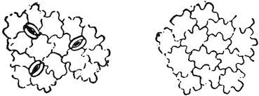
Fig. 1.—Surfaces of a Leaf, magnified.
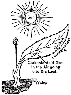
Fig. 2.—A Leaf storing Energy in Sunlight.
The need of plants for
sunlight shows that they
must obtain something
from the sun. This has
been found to be energy.
This enables them to lift
their stems in growth, and form the various structures
called tissues which make up their stems and leaves.
It is noticed that they take in
food and water from the soil
through their roots. Experiments
also show that green plants take
in through pores (Fig. 1), on the
surface of their leaves, a gas
composed of carbon and oxygen,
and called carbon dioxide. The
energy in the sunlight enables the
plant to separate out the carbon,
of the carbon dioxide and to build
mineral and water and carbon
3into organic substances. The oxygen of the carbon dioxide
is set free and returns to the air (Fig. 2). Starch, sugar,
oil, and woody fibre are examples of substances thus
formed. Can you think of any fuel not due to plants?
How Animals obtain Energy.—You have noticed that
starch, oil, etc., will burn, or oxidize, that is, unite with the
oxygen of the air; thus the sun’s energy, stored in these
substances, is changed back to heat and motion. The
oxidation of oil or sugar may occur in a furnace; it may
also occur in the living substance of the active animal.
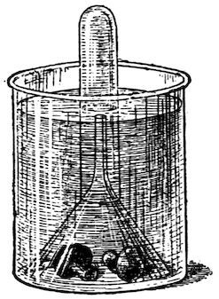
Fig. 3.—Colourless plants, as Mushrooms, give off no oxygen.
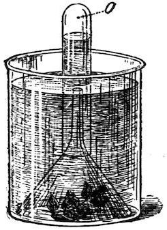
A green leaf, even after it is cut, gives off oxygen (O) if kept in the sun.
Fortunately for the animals, the plants oxidize very little
of the substances built up by them, since they do not move
about nor need to keep themselves warm. We notice that
animals are constantly using plant substances for food, and
constantly drawing the air into their bodies. If the sunlight
had not enabled the green plant to store up these
substances and to set free the oxygen (Fig. 3), animals
would have no food to eat nor air to breathe; hence we
may say that the sunlight is indirectly the source of the
life and energy of animals. Mushrooms and other plants
without green matter cannot set oxygen free (Fig. 3).
4Experiment to show the Cause of Burning, or Oxidation.—Obtain
a large glass bottle (a pickle jar), a short candle,
and some matches. Light the candle and put it on a table
near the edge, and cover it with the glass jar. The flame
slowly smothers and goes out. Why is this? Is the air
now in the jar different from that which was in it before
the candle was lighted? Some change must have taken
place or the candle would continue to burn. To try
whether the candle will burn again under the jar without
changing the air, slide the jar to the edge of the table and
let the candle drop out. Light the candle and slip it up
into the jar again, the jar being held with its mouth a little
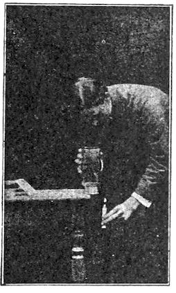
over the edge of the table to receive the candle (Fig. 5).
The flame goes out at once. Evidently the air in the jar
is not the same as the air outside. Take up the jar and
wave it to and fro a few times, so as to remove the old air
and admit fresh air. The candle now burns in it with as
bright a flame as at first. So we conclude that the candle
will not continue to burn unless there is a constant supply
of fresh air. The gas formed by the burning is carbon
dioxide. It is the gas from which plants extract carbon.
(Beginners’ Botany, Chap. XIII.) One test for the presence
of this gas is that it forms a white, chalky cloud in lime
water; another is that it smothers a fire.
Experiment to show that Animals give off Carbon Dioxide.—Place
a cardboard over the mouth of a bottle containing
pure air. Take a long straw, the hollow stem of a weed,
a glass tube, or a sheet of stiff paper rolled into a tube,
and pass the tube into the bottle through a hole in the
cardboard. Without drawing in a deep breath, send one
long breath into the bottle through the tube, emptying the
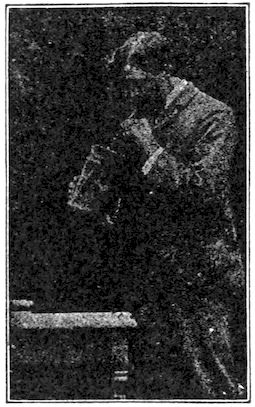
lungs by the breath as nearly as possible (Fig. 4). Next,
invert the bottle on the table as in the former experiment,
5afterward withdrawing the cardboard. Move the bottle
to the edge of the table and pass the lighted candle up
into it (Fig. 5). Does the flame go out as quickly as
in the former experiment?
If you breathe through a tube into clear lime water,
the water turns milky. The effect of the breath on the
candle and on the lime water shows that carbon dioxide is
continually leaving our bodies in the breath.
Fig. 4.—Breathing into a bottle.
Fig. 5.—Testing the air in the bottle.
Oxidation and Deoxidation.—The union of oxygen with
carbon and other substances, which occurs in fires and
in the bodies of animals, is called oxidation. The separation
of the oxygen from carbon such as occurs in the
leaves of plants is called deoxidation. The first process
sets energy free, the other process stores it up. Animals
give off carbon dioxide from their lungs or gills, and plants
give off oxygen from their leaves. But plants need some
energy in growing, so oxidation also occurs in plants, but
to a far less extent than in animals. At night, because
of the absence of sunlight, no deoxidation is taking place
6in the plant, but oxidation and growth continue; so at
night the plant actually breathes out some carbon dioxide.
The deepest part of the lungs contains the most carbon
dioxide. Why was it necessary to empty the lungs as
nearly as possible in the experiment with the candle? Why
would first drawing a deep breath interfere with the experiment?
Why does closing the draught of a stove, thus
shutting off part of the air, lessen the burning? Why does
a “firefly” shine brighter at each breath? Why is the pulse
and breathing faster in a fever? Very slow in a trance?
The key for understanding any animal is to find how
it gets food and oxygen, and how it uses the energy
thus obtained to grow, move, avoid its enemies, and get
more food. Because it moves, it needs senses to guide it.
The key for understanding a plant is to find how it gets
food and sunlight for its growth. It makes little provision
against enemies; its food is in reach, so it needs no senses
to guide it. The plant is built on the plan of having the
nutritive activities near the surface (e.g. absorption by roots;
gas exchange in leaves). The animal is built on the plan
of having its nutritive activities on the inside (e.g. digestion;
breathing).
Cell and Protoplasm.—Both plants and animals are
composed of small parts called cells. Cells are usually
microscopic in size. They have various shapes, as spherical,
flat, cylindrical, fibre-like, star-shaped. The living
substance of cells is called protoplasm. It is a stiff, gluey
fluid, albuminous in its nature. Every cell has a denser
spot or kernel called a nucleus, and in the nucleus is a still
smaller speck called a nucleolus. Most cells are denser and
tougher on the outside, and are said to have a cell wall,
but many cells are naked, or without a wall. Hence the
indispensable part of a cell is not the wall but the nucleus,
7and a cell may be defined as a bit of protoplasm containing
a nucleus. This definition includes naked cells as well as
cells with walls.
One-celled Animals.—There are countless millions of
animals and plants the existence of which was not suspected
until the invention of the microscope
several centuries ago. They are
one-celled, and hence microscopic in size.
It is believed that the large animals and
plants are descended from one-celled animals
and plants. In fact, each individual
plant or animal begins life as a single
cell, called an egg cell, and forms its
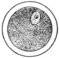
organs by the subdivision of the egg cell into many cells.
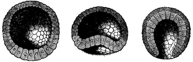
An egg cell is shown in Fig. 6, and the first stages in the
development of an egg cell are shown in Fig. 7.
Fig. 6.—Egg cell of mammal with yolk.
Fig. 7.—Egg cell subdivides into many cells forming a sphere (morula) containing a liquid. A dimple forms and deepens to form the next stage (gastrula).
The animals to be studied in the first chapter are one-celled
animals. To understand them we must learn how
they eat, breathe, feel, and move. They are called Protozoans
(Greek protos, first, and zoon). All other animals
are composed of many cells and are called Metazoans
(Greek meta, beyond or after). The cells composing the
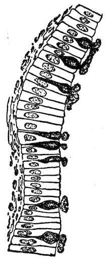
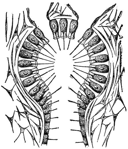
mucous membrane in man are shown in Fig. 8. The cellular
structure of the leaf of a many-celled plant is illustrated
in Fig. 1.
8Method of Classifying Animals.—The various animals
display differences more or less marked. The question
arises, are not some of them more closely related than
others? We conclude that they are, since the difference
between some animals is very slight, while the
difference between others is quite marked.
Fig. 8.—Mucous Membrane formed of one layer of cells. A few cells secrete mucus.
To show the different steps in classifying
an animal, we will take an example,—the
cow. Even little children
learn to recognize a cow, although individual
cows differ somewhat in form,
size, colour, etc. The varieties of cows,
such as short-horn, Jersey, etc., all
form one species of animals, having the
scientific name taurus. Let us include
in a larger group the animals closest
akin to a cow. We see a cat, a bison,
and a dog; rejecting the cat and the
dog, we see that the bison has horns,
hoofs, and other similarities. We include
it with the cow in a genus called
Bos, calling the cow Bos taurus, and
the bison, Bos bison. The sacred cow
of India (Bos indicus) is so like the
cow and the buffalo as also to belong to the genus Bos.
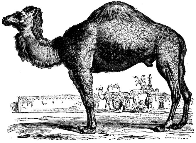
Why is not the camel, which, like Bos bison, has a hump,
placed in the genus Bos?
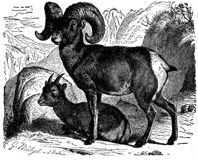
The Old World buffaloes,—most abundant in Africa
and India,—the antelopes, sheep, goats, and several other
genera are placed with the genus Bos in a family called
the hollow-horned animals.
This family, because of its even number of toes and
the habit of chewing the cud, resembles the camel family,
9the deer family, and several other families. These are all
placed together in the next higher systematic unit called
an order, in this case, the order of ruminants.
The ruminants, because they are covered with hair
and nourish the young with milk, are in every essential
respect related to the one-toed horses, the beasts of
prey, the apes, etc. Hence they are all placed in a
more inclusive division of animals, the class called
mammals.
All mammals have the skeleton, or support of the
body, on the inside, the axis of which is called the vertebral
column. This feature also belongs to the classes
of reptiles, amphibians, and fishes. It is therefore
consistent to unite these classes by a general idea or
conception into a great branch of animals called the
vertebrates.
Returning from the general to the particular by successive
steps, state the branch, class, order, family, genus,
and species to which the cow belongs.
The Eight Branches or Sub-kingdoms.—The simplest
classification divides the whole animal kingdom into
eight branches, named and characterized as follows, beginning
with the lowest: I. Protozoans. One-celled.
II. Sponges. Many openings. III. Polyps. Circular;
cuplike; having only one opening which is both mouth and
vent. IV. Echinoderms. Circular; rough-skinned; two
openings. V. Molluscs. No skeleton; usually with external
shell. VI. Vermes. Elongate body, no jointed legs.
VII. Arthropods. External jointed skeleton; jointed
legs. VIII. Vertebrates. Internal jointed skeleton with
axis or backbone.[1]
1. This is the briefest classification. Animals have also been divided into twelve
branches. The naming of animals is somewhat chaotic at present, but an attempt
to come to an agreement is now being made by zoologists of all nations.
CHAPTER II PROTOZOA (One-celled Animals) — The Amœba — The Slipper Animalcule or Paramecium
10
Suggestions.—Amœbas live in the slime found on submerged
stems and leaves in standing water, or in the ooze at the bottom.
Water plants may be crowded into a glass dish and allowed to
decay, and after about two weeks the amœba may be found in
the brown slime scraped from the plants. An amœba culture
sometimes lasts only three days. The most abundant supply
ever used by the writer was from a bottle of water where some
oats were germinating. Use ⅕ or ⅙ inch objective, and cover
with a thin cover glass. Teachers who object to the use of
the compound microscope in a first course should require a
most careful study of the figures.
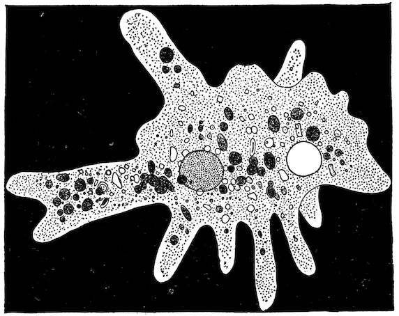
Fig. 9.—Amœba Proteus, much enlarged.
11
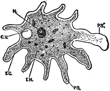
Fig. 10.—Amœba.
cv, contractile vacuole; ec, ectoplasm; en, endoplasm; n, nucleus; ps, pseudopod; ps, pseudopod forming; ectoplasm protrudes and endoplasm flows into it.
Form and Structure.—The amœba looks so much like
a clear drop of jelly that a
beginner cannot be certain
that he has found one until
it moves. It is a speck of
protoplasm (Fig. 9), with a
clear outer layer, the ectoplasm;
and a granular, internal
part, the endoplasm.
Is there a distinct line between
them? (Fig. 10.)
Note the central portion
and the slender prolongations
or pseudopods (Greek,
false feet). Does the endoplasm extend into the pseudopods?
(Fig. 10.) Are the pseudopods arranged with any
regularity?
Sometimes it is possible to see a denser appearing portion,
called the nucleus; also a clear space, the contractile
vacuole (Fig. 10).
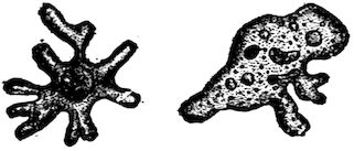
Fig. 11.—The same amœba seen at different times.
Movements.—Sometimes while the pseudopods are being
extended and contracted, the central portion remains
in the same place (this is motion).
Usually only one pseudopod
is extended, and the body
flows into it; this is locomotion
(Fig. 11). There is a new foot
made for each step.
Feeding.—If the amœba crawls near a food particle, the
pseudopod is pressed against it, or a depression occurs (Fig.
12), and the particle is soon embedded in the endoplasm.
Often a clear space called a food vacuole is noticed around
the food particle. This is the water that is taken in with
12the particle (Fig. 12). The water and the
particle are soon absorbed and assimilated
by the endoplasm.
Fig. 12.—The Amœba taking food.
Excretion.—If a particle of sand or other
indigestible matter is taken in, it is left behind
as the amœba moves on. There is a clear
space called the contractile vacuole, which
slowly contracts and disappears, then reappears
and expands (Figs. 9 and 10). This
possibly aids in excreting oxidized or useless
material.
Circulation in the amœba consists of the
movement of its protoplasmic particles. It
lacks special organs of circulation.
Feeling.—Jarring the glass slide seems to
be felt, for it causes the activity of the amœba
to vary. It does not take in for food every
particle that it touches. This may be the
beginning of taste, based upon mere chemical
affinity. The pseudopods aid in feeling.
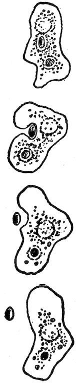
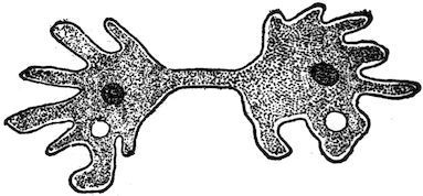
Reproduction.—Sometimes an amœba is
seen dividing into two parts. A narrowing
takes place in the middle; the nucleus also
divides, a part going to each portion (Fig. 13).
The mother amœba finally divides into two daughter
amœbas. Sex is wanting.
Fig. 13.—Amœba, Dividing.
Source of the Amœba’s Energy.—We thus see that the
amœba moves without feet, eats without a mouth, digests
without a stomach, feels
without nerves, and, it
should also be stated,
breathes without lungs,
for oxygen is absorbed
from the water by its whole
13surface. Its movements require energy; this, as in all animals,
is furnished by the uniting of oxygen with the food.
Carbon dioxide and other waste products are formed
by the union; these pass off at the surface of the amœba and
taint the water with impurities.
Questions.—Why will the amœba die in a very small quantity of
water, even though the water contains enough food? Why will it die
still quicker if air is excluded from contact with the drop of water?
The amœba never dies of old age. Can it be said to be immortal?
According to the definition of a cell (Chapter I), is the amœba a
unicellular or multicellular animal?
Cysts.—If the water inhabited by a protozoan dries up,
it encysts, that is, it forms a tough skin called a cyst.
Upon return of better conditions it breaks the cyst and
comes out. Encysted protozoans may be blown through
the air: this explains their appearance in vessels of water
containing suitable food but previously free from protozoans.
Suggestions.—Stagnant water often contains the paramecium as
well as the amœba; or they may be found in a dish of water containing
hay or finely cut clover, after the dish has been allowed to
stand in the sun for several days. A white film forming on the
surface is a sign of their presence. They may even be seen with
the unaided eye as tiny white particles by looking through the side
of the dish or jar. Use at first a ⅓ or ¼ in. objective. Restrict
their movements by placing cotton fibres beneath the cover glass;
then examine with ⅕ or ⅙ objective. Otherwise, study figures.
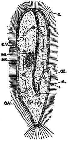
Shape and Structure.—The paramecium’s whole body,
like the amœba’s, is only one cell. It resembles a slipper
in shape, but the pointed end is the hind end, the front end
being rounded (Fig. 14). The paramecium is propelled
by the rapid beating of numerous fine, threadlike appendages
14on its surface, called cilia (Latin, eyelashes) (Figs.).
The cilia, like the pseudopods of the amœba, are merely
prolongations of the cell protoplasm,
but they are permanent. The separation
between the outer ectoplasm
and the interior granular endoplasm
is more marked than in the amœba
(Fig. 14).
Fig. 14.—Paramecium, showing cilia, c.
Two contractile vacuoles, cv; the macronucleus, mg; two micronuclei, mi; the gullet (Œ), a food ball forming and ten food balls in their course from gullet to vent, a.
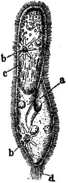
Fig. 15.
Nucleus and Vacuoles.—There is
a large nucleus called the macronucleus,
and beside it a
smaller one called the
micronucleus. They are
hard to see. About one
third of the way from
each end is a clear, pulsating
space (bb. Fig.
15) called the pulsating
vacuole. These
spaces contract until
they disappear, and then
reappear, gradually expanding.
Tubes lead from the vacuoles which probably
serve to keep the contents of the cell in circulation.
Fig. 16.—Two Paramecia exchanging parts of their nuclei.
Feeding.—A depression, or groove, is seen on one side;
this serves as a mouth (Figs.). A tube which serves as a
gullet leads from the
mouth-groove to the interior
of the cell. The
mouth-groove is lined
with cilia which sweep
food particles inward.
The particles accumulate
15in a mass at the inner end of the gullet, become separated
from it as a food ball (Fig. 14), and sink into the soft protoplasm
of the body. The food balls
follow a circular course through the
endoplasm, keeping near the ectoplasm.
Fig. 17.—Vorticella (or bell animalcule), two extended, one withdrawn.
Reproduction.—This, as in the amœba,
is by division, the constriction being in
the middle, and part of the nucleus going
to each half. Sometimes two individuals
come together with their
mouth-grooves touching and
exchange parts of their nuclei
(Fig. 16). They then separate
and each divides to form two
new individuals.
Fig. 18.—Euglena.
We thus see that the paramecium,
though of only one
cell, is a much more complex and advanced
animal than the amœba. The tiny paddles,
or cilia, the mouth-groove, etc., have their
special duties similar to the specialized organs
of the many-celled animals to be studied later.
Fig. 19.—Shell of a Radiolarian.
If time and circumstances
allow a prolonged study, several
additional facts may be
observed by the pupil, e.g.
Does the paramecium swim
with the same end always
foremost, and same side
uppermost? Can it move
backwards? Avoid obstacles?
Change shape in a
narrow passage? Does refuse
16matter leave the body at any particular place? Trace
movement of the food particles.
Draw the paramecium.
Which has more permanent parts, the amœba or paramecium?
Name two anatomical similarities and three differences; four
functional similarities and three differences.
The amœba belongs in the class of protozoans called
Rhizopoda “root footed.”
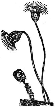
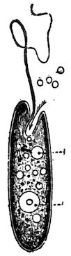
Other classes of Protozoans are the Infusorians (in the
broad sense of the term), which have many waving cilia
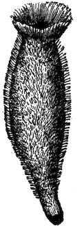
(Fig. 17) or one whiplike flagellum (Fig. 18), and the
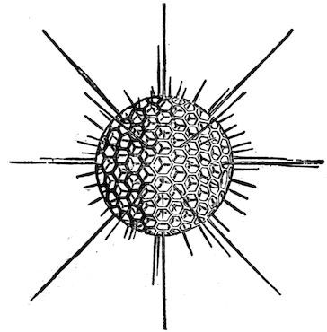
Foraminifers, which possess a calcareous shell pierced with
holes (Fig. 19). Much chalky limestone has been formed
of their shells. To which class does the paramecium
belong?
Protozoans furnish a large amount of food to the higher
animals.
CHAPTER III SPONGES
17
Suggestions.—In many parts of North America, fresh-water
sponges may, by careful searching, be found growing on rocks and logs
in clear water. They are brown, creamy, or greenish in colour, and resemble
more a cushion-like plant than an animal. They have a characteristic
gritty feel. They soon die after removal to an aquarium.
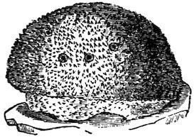
Fig. 21.—Fresh-water Sponge.
A number of common small bath sponges may be bought and kept
for use in studying the skeleton of an ocean sponge. These sponges
should not have large
holes in the bottom; if
so, too much of the
sponge has been cut
away. A piece of marine
sponge preserved in alcohol
or formalin may be
used for showing the
sponge with its flesh in
place. Microscopic slides
may be used for showing
the spicules.
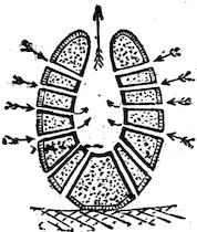
Fig. 22.—Section of fresh-water sponge (enlarged).
The small fresh-water
sponge (Fig. 21) lacks
the more or less vaselike
form typical of sponges. It is a rounded mass growing
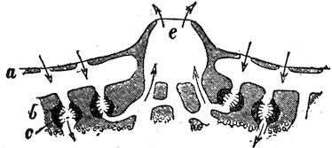
upon a rock or a log. As indicated by the Arrows, where does
water enter the sponge? This
may be tested by putting colouring
matter in the water near
the living sponge. Where does
the water come out? (Fig. 22.)
Does it pass through ciliated
chambers in its course? Is the
18surface of the sponge rough or smooth? Do any of the skeletal
spicules show on the surface? (Fig. 21.) Does the sponge thin
out near its edge?
Fig. 23.—Eggs and SPICULES of fresh-water sponge (enlarged).
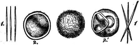
The egg of this sponge is shown in Fig. 23. It escapes from
the parent sponge through the osculum, or large outlet. As in
most sponges, the first
stage after the egg is
ciliated and free-swimming.
Marine Sponges.—The
grantia (Fig. 24) is
one of the simplest of
marine sponges. What is the shape of grantia? What is its length
and diameter? How does the free end differ from the fixed end?
Are the spicules projecting from its body few or many?
Fig. 24.—Grantia.
Where is the osculum, or large outlet? With what
is this surrounded? The osculum opens from a central
cavity called the cloaca. The canals from the pores
lead to the cloaca.
Buds are sometimes seen growing out from the
sponge near its base. These are young sponges formed
asexually. Later they become detached from the
parent sponge.
Fig. 25.—Plan of a sponge.
Commercial “Sponge.”—What part of the complete
animal remains in the bath sponge? Slow growing
sponges grow more at the top and form tall, simple,
tubular or vaselike animals. Fast growing sponges
grow on all sides at once and form a complicated system of canals,
pores, and oscula. Which of these habits of growth do you think
belonged to the bath sponge? Is there a large
hole in the base of your specimen? If so, this
is because the cloaca was reached in trimming
the lower part where it was attached to a rock.
Test the elasticity of the sponge when dry and
when wet by squeezing it. Is it softer when wet
or dry? Is it more elastic when wet or dry?
How many oscula does your specimen have?
How many inhalent pores to a square inch?
19Using a probe (a wire with knob at end, or small hat pin), try
to trace the canals from the pores to the cavities inside.
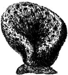
Fig. 26.—Bath Sponge.
Do the fibres of the sponge appear to
interlace, or join, according to any system?
Do you see any fringe-like growths on the
surface which show that new tubes are beginning
to form? Was the sponge growing
faster at the top, on the sides, or near the
bottom?
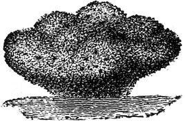
Fig. 27.—Bath Sponge.
Fig. 28.—Bath Sponge.
Burn a bit of the sponge; from the odor,
what would you judge of its composition?
Is the inner cavity more conspicuous in a simple sponge or in a
compound sponge like the bath sponge? Is the bath sponge
branched or lobed? Compare a number of specimens (Figs. 26,
27, 28) and decide whether the common sponge has a typical
shape. What features do their forms
possess in common?
Fig 29.—Skeleton of a glass sponge.
Sponges are divided into three classes,
according as their skeletons are flinty
(silicious), limy (calcareous), or horny.
Some of the silicious sponges have
skeletons that resemble spun glass in
their delicacy. Flint is chemically nearly
the same as glass. The skeleton shown
in Fig. 29 is that of a glass sponge which
lives near the Philippine Islands.
The horny sponges do not have spicules
in their skeletons, as the flinty and
limy sponges have, but the skeleton
is composed of interweaving fibres of
20spongin, a durable substance of the same chemical nature as silk
(Figs. 30 and 31).
The limy sponges have skeletons made of numerous spicules of
lime. The three-rayed spicule is the commonest form.
Fig. 30.—A horny sponge.
Fig. 31.—Section of horny sponge.
The commercial sponge, seen as it grows in the ocean, appears
as a roundish mass with a smooth, dark exterior, and having about
the consistency of beef liver. Several large openings (oscula),
from which the water flows, are visible on the upper surface.
Smaller holes (inhalent pores—many of them so small as to be
indistinguishable) are on the sides. If the sponge is disturbed,
the smaller holes, and
perhaps the larger
ones, will close.
The outer layer of
cells serves as a sort
of skin. Since so
much of the sponge
is in contact with
water, most of the
cells do their own
breathing, or absorption
of oxygen and giving off of carbon dioxide. Nutriment is
passed on from the surface cells to nourish the rest of the body.
Reproduction.—Egg cells and sperm-cells are produced by
certain cells along the canals. The egg cell, after it is fertilized
by the sperm cell, begins to divide and form new cells, some of
which possess cilia. The embryo sponge passes out at an osculum.
By the vibration of the cilia, it swims about for a while.
It afterwards settles down with the one end attached to the ocean
floor and remains fixed for the rest of its life. The other end develops
oscula. Some of the cilia continue to vibrate and create
currents which bring food and oxygen.
The cilia in many species are found only in cavities called
ciliated chambers. (Figs. 22, 32.) There are no distinct organs
in the sponge and there is very little specialization of cells. The
ciliated cells and the reproductive cells are the only specialized
cells. The sponges were for a long time considered as colonies
of separate one-celled animals classed as protozoans. They are,
21without doubt, many-celled animals. If a living sponge is cut
into pieces, each piece will grow and form a complete sponge.
Fig. 32.—Microscopic plan of ciliated chamber. Each cell lining the chamber has a nucleus, a whip-lash, and a collar around base of whip-lash. Question: State two uses of whip-lash.
That the sponge is not a colony of one-celled animals, each like
an amœba, but is a many-celled animal, will be realized by examining
Fig. 32, which shows a bit of sponge highly magnified. A
sponge may be conceived as having developed from a one-celled
animal as follows: Several
one-celled animals
happened to live side by
side; each possessed a
threadlike flagellum (E,
Fig. 32) or whip-lash for
striking the water. By
lashing the water, they
caused a stronger current
(Fig. 25) than protozoans
living singly
could cause. Thus they
obtained more food and
multiplied more rapidly
than those living alone.
The habit of working
together left its impress
on the cells and was transmitted
by inheritance.
Cell joined to cell
formed a ring; ring
joined to ring formed a tube which was still more effective than
a ring in lashing the water into a current and bringing fresh food
(particles of dead plants and animals) and oxygen.
Few animals eat sponges; possibly because spicules, or fibres,
are found throughout the flesh, or because the taste and the odour are
unpleasant enough to protect them. Small animals sometimes
crawl into sponges to hide. One sponge grows upon shells inhabited
by hermit crabs. Moving of the shell from place to place
is an advantage to the sponge, while the sponge conceals and thus
protects the crab.
Special Report: Sponge “Fisheries.” (Localities; how sponges
are taken, cleaned, dried, shipped, and sold.)
CHAPTER IV POLYPS (CUPLIKE ANIMALS) — The Hydra, or Fresh-Water Polyp
22
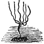
Fig. 33.—A Hydra.
Suggestions.—Except in the drier regions of North America
the hydra can usually be found by careful search in fresh-water ponds
not too stagnant. It is found attached to stones, sticks, or leaves,
and has a slender, cylindrical body from a quarter to half an
inch long, varying in thickness from that of a fine
needle to that of a common pin. The green hydra
and the brown hydra, both very small, are common
species, though hydras are often white or colourless.
They should be kept in a large glass dish filled with
water. They may be distinguished by the naked
eye but are not studied satisfactorily without a
magnifying glass or microscope. Place a living specimen attached
to a bit of wood in a watch crystal filled with water, or on a hollowed
slip, or on a slip with a bit of weed to support the cover
glass, and examine with hand lens or lowest power of microscope.
Prepared microscopical sections, both transverse and longitudinal,
may be bought
of dealers in microscopic
supplies. One is
shown in Fig. 39.
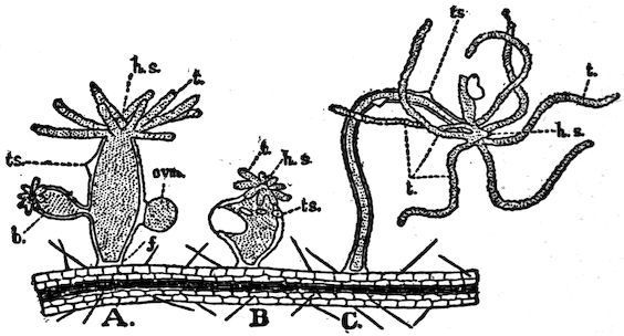
Fig. 34.—Forms assumed by Hydra.
Is the hydra’s
body
round or two-sided?
(Fig.
35.) What is
its general shape? Does one individual keep the same
shape? (Fig. 34.) How does the length of the threadlike
23tentacles compare with the length of the hydra’s body?
About how many tentacles are on a hydra’s body? Do all
have the same number of tentacles? Are the tentacles
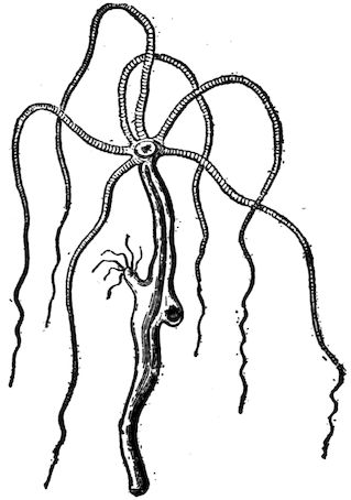
knotty or smooth? (Fig. 35.) The hydra is usually extended
and slender; sometimes it is contracted and rounded.
In which of these conditions is the base (the foot) larger
around than the rest of the body? (Fig. 34.) Smaller?
How many openings into the
body are visible? Is there a
depression or an eminence at
the base of the tentacles? For
what is the opening on top of
the body probably used? Why
are the tentacles placed at the
top of the hydra’s body? Does
the mouth have the most convenient
location possible?
Fig. 35.—Hydra (much enlarged).
The conical projection bearing
the mouth is called hypostome
(Fig. 34). The mouth
opens into the digestive cavity.
Is this the same as the general
body cavity, or does the stomach have a wall distinct from
the body cavity? How far down does the body cavity
extend? Does it extend up into the tentacles? (Fig. 39.)
If a tentacle is touched, what happens? Is the body ever bent?
Which is more sensitive, the columnar body or the tentacles? In
searching for hydras would you be more likely to find the tentacles
extended or drawn in? Is the hypostome ever extended
or drawn in? (Fig. 34.)
Locomotion.—The round surface, or disk, by which the
hydra is attached, is called its foot. Can you move on
one foot without hopping? The hydra moves by alternately
24elongating and rounding the foot. Can you discover
other ways by which it moves? Does the hydra
always stand upon its foot?
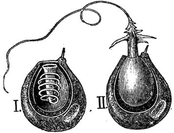
Fig. 36.—Nettling Cell. II. discharged, and I. not discharged.
Lasso Cells.—Upon the tentacles (Fig. 35) are numerous
cells provided each with a threadlike process (Fig. 36)
which lies coiled within the
cell, but which may be
thrown out upon a water
flea, or other minute animal
that comes in reach. The
touch of the lasso paralyzes
the prey (Fig. 37). These
cells are variously called
lasso cells, nettling cells, or
thread cells. The thread is
hollow and is pushed out by the pressure of liquid within.
When the pressure is withdrawn the thread goes back as
the finger of a glove may be turned back into the glove by
turning the finger outside in.
When a minute animal, or
other particle of food comes in
contact with a tentacle, how
does the tentacle get the food
to the mouth? By bending
and bringing the end to the
mouth, or by shortening and
changing its form, or in both
ways? (Fig. 34, C.) Do the
neighbouring tentacles seem to
bend over to assist a tentacle in
securing prey? (Fig. 34, C.)
Fig. 37.—Hydra capturing a water flea.
Digestion.—The food particles
break up before remaining
25long in the stomach, and the nutritive part is absorbed
by the lining cells, or endoderm (Fig. 39). The indigestible
remnants go out through the mouth. The hydra is
not provided with a special vent. Why could the vent not
be situated at the end opposite the mouth?
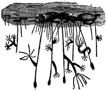
Fig. 38.—Hydras on the under surface of pondweed.
Circulation and Respiration.—Does water have free
access to the body cavity? Does the hydra have few or
nearly all of its cells exposed to the water in which it
lives? From its structure, decide whether it can breathe
like a sponge or whether
special respiratory cells are
necessary to supply it with
oxygen and give off carbon
dioxide. Blood vessels are
unnecessary for transferring
oxygen and food from
cell to cell.
Reproduction.—Do you
see any swellings upon the
side of the hydra? (Fig. 34, A.) If the swelling is near
the tentacles, it is a spermary; if near the base, it is an
ovary. A sperm coalesces with or fertilizes the ovum after
the ovum is exposed by the breaking of the ovary wall.
Sometimes the sperm from one hydra unites with the ovum
of another hydra. This is called cross-fertilization. The
same term is applied to the process in plants when the
male element, developed in the pollen of the flower, unites
with the female element of the ovule of the flower on
another plant. The hydra, like most plants and some other
animals, is hermaphrodite, that is to say, both sperms and
ova are produced by one individual. In the autumn, eggs
are produced with hard shells to withstand the cold until
spring. Sexual reproduction takes place when food is
26scarce. Asexual generation (by budding) is common with
the hydra when food supply is abundant. After the bud
grows to a certain
size, the
outer layer of
cells at the base
of the bud constricts
and the
young hydra is
detached.
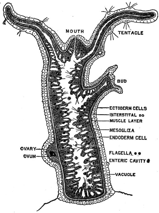
Fig. 39.—Longitudinal section of hydra (microscopic and diagrammatic).
Compare the
sponge and the
hydra in the following
respects:—many
celled,
or one-celled;
obtaining food;
breathing; tubes
and cavities;
openings; reproduction;
locomotion.
Which
ranks higher
among the metazoa? The metazoa, or many-celled animals,
include all animals except which branch?
Figure 39 is a microscopic view of a vertical section of a hydra to
show the structure of the body wall. There is an outer layer called the
ectoderm, and an inner layer called the endoderm. There is also a thin
supporting layer (black in the figure) called the mesoglea. The mesoglea
is the thinnest layer. Are the cells larger in the endoderm or the ectoderm?
Do both layers of cells assist in forming the reproductive bud? The ectoderm
cells end on the inside in contractile tails which form a thin line and
have the effect of muscle fibres. They serve the hydra for its remarkable
changes of shape. When the hydra is cut in pieces, each piece makes a
complete hydra, provided it contains both endoderm and ectoderm.
27In what ways does the hydra show “division of labour”? Answer
this by explaining the classes of cells specialized to serve a different
purpose. Which cells of the hydra are least specialized? In what particulars
is the plan of the hydra different from that of a simple sponge?
An ingenious naturalist living more than a century ago, asserted that it
made no difference to the hydra whether the ectoderm or the endoderm
layer were outside or inside,—that it could digest equally well with
either layer. He allowed a hydra to swallow a worm attached to a
thread, and then by gently pulling in the thread, turned the hydra inside
out. More recently a Japanese naturalist showed that the hydra could
easily be turned inside out, but he also found that when left to itself
it soon reversed matters and returned to its natural condition, that
the cells are really specialized and each layer can do its own work and
no other.
Habits.—The hydra’s whole body is a hollow bag, the
cavity extending even into the tentacles. The tentacles
may increase in number as the hydra grows but seldom
exceed eight. The hydra has more active motion than
locomotion. It seldom moves from its place, but its tentacles
are constantly bending, straightening, contracting,
and expanding. The body is also usually in motion, bending
from one side to another. When the tentacles approach
the mouth with captured prey, the mouth (invisible
without a hand lens) opens widely, showing five lobes or
lips, and the booty is soon tucked within. A hydra can
swallow an animal larger in diameter than itself.
The endoderm cells have amœboid motion, that is, they
extend pseudopods. They also resemble amœbas in the
power of intra-cellular digestion; that is, they absorb the
harder particles of food and digest them afterwards, rejecting
the indigestible portions. Some of these cells have
flagella (see Fig. 39) which keep the fluid of the cavity
in constant motion.
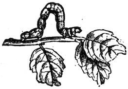
Sometimes the hydra moves after the manner of a small
caterpillar called a “measuring worm,” that is, it takes
hold first by the foot, then by the tentacles, looping its
28body at each step. Sometimes
the body goes end over end in
slow somersaults.
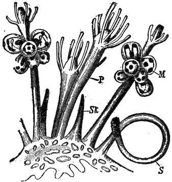
Fig. 40.—Hydroid Colony, with nutritive (P) reproductive (M) and defensive (S) hydranths.
The length of the extended
hydra may reach one half
inch. When touched, both
tentacles and body contract
until it looks to the unaided
eye like a round speck of
jelly. This shows sensibility,
and a few small star-shaped
cells are believed to be nerve
cells, but the hydra has not a nervous system. Hydras
show their liking for light by moving to the side of
the vessel or aquarium whence the light comes.
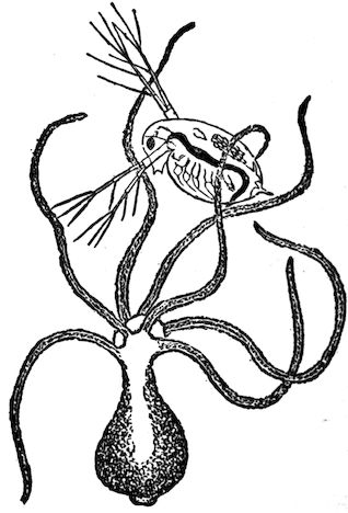
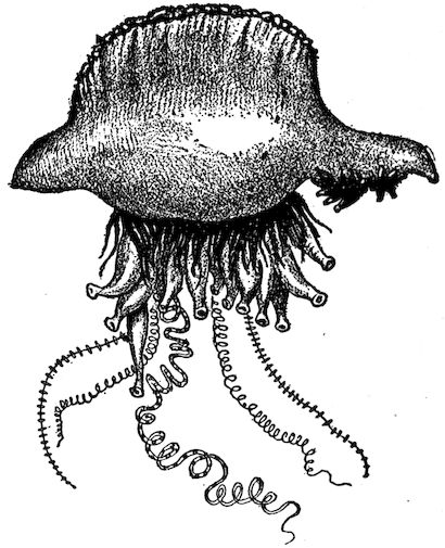
Fig. 41.—“Portuguese Man-o’-war” (compare with Fig. 40). A floating hydroid colony with long, stinging (and sensory) streamers. Troublesome to bathers in Gulf of Mexico. Notice balloon-like float.
The Branch Polyps
(sometimes called Cœlenterata).—The
hydra is
the chief fresh-water representative
of this great
branch of the animal
kingdom. This branch
is characterized by its
members having only
one opening to the body.
The polyps also include
the salt water animals
called hydroids, jellyfishes,
and coral polyps.
Hydroids.—Figure 40
shows a hydroid, or
group of hydra-like
growths, one of which
29eats and digests for the group, another defends by nettling
cells, another produces eggs. Each hydra-like part of a
hydroid is called a hydranth. Sometimes the buds on the
hydra remain attached so long that a bud forms upon the
first bud. Thus three generations are represented in one
organism. Such growths show us that it is not always
easy to tell
what constitutes
an individual
animal.
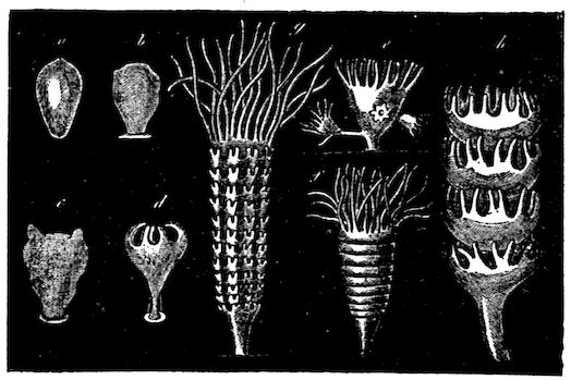
Fig. 42.—The formation of many free-swimming jellyfishes from one fixed hydra-like form. The saucer-like parts (h) turn over after they separate and become like Fig. 43 or 44. Letters show sequence of diagrams.
Hydroids
may be conceived
to have
been developed
by the failure
of budding hydras
to separate
from the
parent, and by
the gradual formation of the habit of living together and
assisting one another. When each hydranth of the hydroid
devoted itself to a special function of digestion, defence, or
reproduction, this group lived longer and prospered; more
eggs were formed, and the habits of the group were transmitted
to a more numerous progeny than were the habits
of a group where members worked more independently of
one another.
As the sponge is a simple example of the devotion
of special cells to special purposes, the hydroid is a
primitive and simple example of the occurrence of organs,
that is of special parts of the body set aside for a special
work.
30
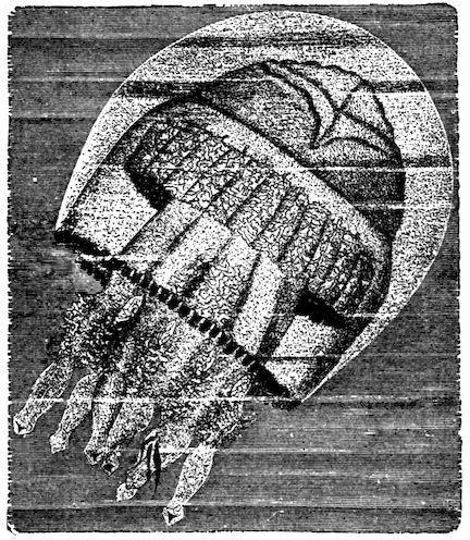
Fig. 43.—A Jellyfish.
How many mature hydranths are seen in the hydroid
shown in Fig. 40? Why are the defensive hydranths
on the outside of the
colony? Which hydranths
have no tentacles?
Why not?
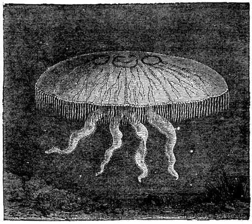
Fig. 44.—A Jellyfish (medusa).
Jellyfish.—Alternation
of Generations.—Medusa.—With
some
species of hydroids, a
very curious thing happens.—The
hydranth
that is to produce the
eggs falls off and becomes
independent of
the colony. More surprising
still, its appearance
changes entirely and instead of being hydra-like, it
becomes the large and complex creature called jellyfish
(Fig. 43). But
the egg of the
jellyfish produces
a small
hydra-like animal
which gives
rise by budding
to a hydroid,
and the cycle is
complete.
The bud (or
reproductive
hydranth) of
the hydroid
31does not produce a hydroid, but a jellyfish; the egg of the
jellyfish does not produce a jellyfish, but a hydroid. This is
called by zoologists, alternation of generations. A complete
individual is the life from the germination of one egg to
the production of another. So that an “individual” consists
of a hydroid colony fixed in one place together with
all the jellyfish produced from its buds, which may
now be floating miles away from it in the ocean. Bathers
in the surf are sometimes touched and stung by the long,
streamer-like tentacles of the jellyfish. These, like the
tentacles of the hydra, have
nettling cells (Fig. 41).
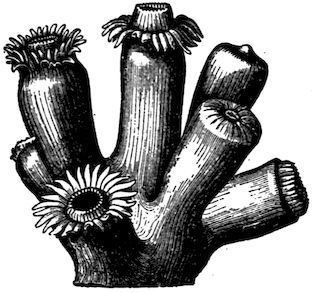
Fig. 45.—Coral Polyps (tentacles, a multiple of six). Notice hypostome.
The umbrella-shaped free-swimming
jellyfish is called a
medusa (Fig. 44).
Coral Polyps.—Some of the
salt water relatives of the hydra
produce buds which remain
attached to the parent without,
however, becoming different
from the parent in any way.
The coral polyps and corallines are examples of colonies of
this kind, possessing a common stalk which is formed as
the process of multiplication goes on. In the case of coral
polyps, the separate animals and the flesh connecting them
secrete within themselves a hard, limy, supporting structure
known as coral. In some species, the coral, or stony part,
is so developed that the polyp seems to be inserted in the
coral, into which it withdraws itself for partial protection
(Fig. 45).
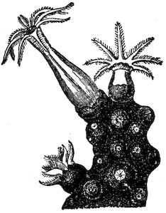
The corallines secrete a smooth stalk which affords
no protection, but they also secrete a coating or sheath
which incloses both themselves and the stalk. The
32coating has apertures through which the polyps protrude
in order to feed when no danger is near (Fig. 46).
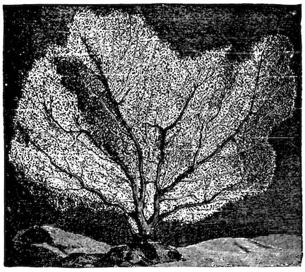
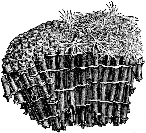
The red “corals” used for jewelry are bits of stalks of corallines.
The corallines (Figs. 47, 48) are not so abundant
nor so important
as the coral polyps
(Figs. 45, 49).
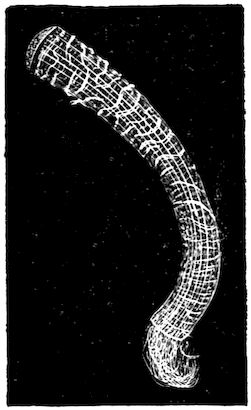
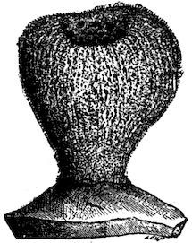
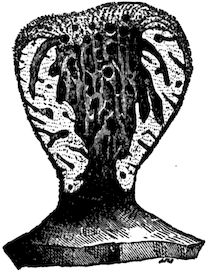
Fig. 46.—Red Coralline with crust and polyps (eight tentacles).
Fig. 47.—Sea Fan (a coralline).
Fig. 48.—Organ Pipe “Coral” (a coralline).
Colonies of coral
polyps grow in
countless numbers
in the tropical seas.
The coral formed
by successive colonies
of polyps accumulates
and builds
up many islands
and important additions
to continents. The Florida “keys,” or islands, and
the southern part of the mainland of Florida were so
formed.
33
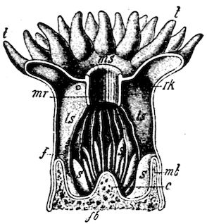
Fig. 49.—Upright cut through coral polyp × 4.
ms, mouth; mr, gullet; ls, ls, fleshy partitions (mesenteries) extending from outer body wall to gullet (to increase absorbing surface); s, s, shorter partitions; mb, fb, stony support (of lime, called coral); t, tentacles.
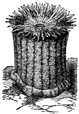
Fig. 50.—Sea Anemone.
The Sea Anemone, like the coral
polyp, lives in the sea, but like
the fresh-water hydra, it deposits
no limy support for its body. The
anemone is much larger than the
hydra and
most coral
polyps,
many species
attaining
a
height of
several
inches. It
does not
form colonies.
When its arms are drawn in,
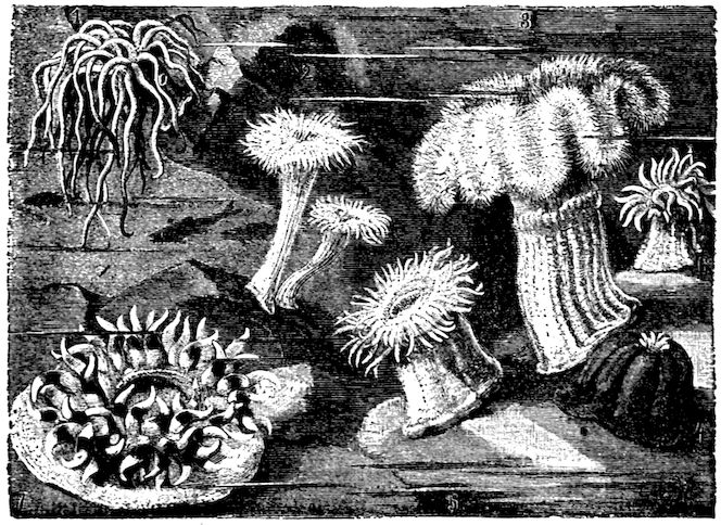
it looks like a large knob of shiny but opaque jelly. Polyps
used to be called zoophytes (plant-animals), because of
their flower-like appearance (Figs. 50, 51).
Fig. 51.—Sea Anemones.
CHAPTER V ECHINODERMS (SPINY ANIMALS) — The Starfish — The Sea Urchin — Other Echinoderms — Comparative Review
34
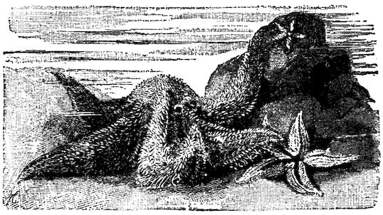
Fig. 52.—Starfish on a rocky shore.
Suggestions. Since the echinoderms are aberrant though interesting
forms not in the regular line of development of animals, this
chapter may be
omitted if it
is desired to
shorten the
course.—The
common starfish
occurs
along the Atlantic
coast. It
is captured by
wading along
the shore when
the tide is out.
It is killed by immersion in warm, fresh water. Specimens are usually
preserved in 4 per cent formalin. Dried starfish and sea urchins are also
useful. A living starfish kept
in a pail of salt water will be
instructive.
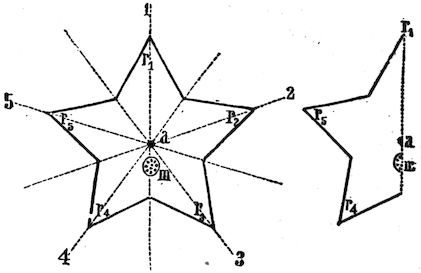
Fig. 53.—Plan of starfish; III, madreporite.
External Features.—Starfish
are usually brown
or yellow. Why? (See
Fig. 52.) Has it a head or
a tail? Right and left sides?
What is the shape of the
disk, or part which bears
the five arms or rays? (Fig. 53.) Does the body as a whole
have symmetry on two sides of a line (bilateral symmetry), or
around a point (radial symmetry)? Do the separate rays have
35bilateral symmetry? The skeleton consists of limy plates embedded
in the tough skin (Fig. 54). Is the skin rough or smooth? Hard
or soft? Are the projections (or spines)
in the skin long or short? The skin is
hardened by the
limy plates, except
around the
mouth, which is
at the centre of
the lower side and surrounded by a membrane.
Which is rougher, the mouth side,
(oral side) or the opposite (aboral side)?
Which side is more nearly flat? The
vent is at or near the centre of the
disk on the aboral surface. It is usually
very small and sometimes absent. Why a vent is not of much
use will be understood after learning how the starfish takes food.
Fig. 54.—Limy Plates in portion of a ray.
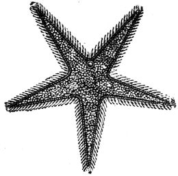
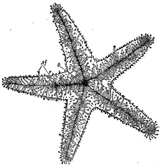
Fig. 55.—Starfish (showing Madreporite).
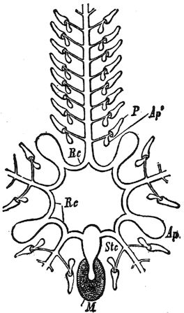
Fig. 56.—Water tube System of starfish.
m, madreporite; stc, stone canal; ap, ampulla.
An organ peculiar to animals of this
branch, and called the madreporic plate,
or madreporite, is found on the aboral
surface between the bases of two rays
(Fig. 55). It is wartlike, and usually
white or red. This plate is a sieve; the
small openings keep out sand but allow
water to filter through.
Movements: the Water-tube System.—The
water, which is filtered through
the perforated madreporite, is needed
to supply a system of canals (Fig. 56).
The madreporite opens into a canal
called the stone canal, the wall of which
is hardened by the same kind of material
as that found in the skin. The stone
canal leads to the ring canal which surrounds
the mouth (Fig. 56). The ring
canal sends radial canals into each ray to supply the double row
of tube feet found in the groove at the lower side of each ray
(Fig. 57). Because of their arrangement in rows, the feet are
36also called ambulacral feet (Latin ambulacra, “forest walks”).
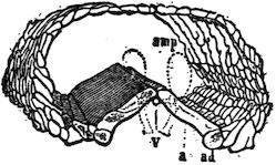
There is a water holder (ampulla), or muscular water bulb at the
base of each
tube foot (Fig.
58). These contract
and force
the water into
the tube feet and
extend them.
The cuplike
ends of the
tubes cling to
the ground by
suction. The
feet contain
delicate muscles
by which they
contract and
shorten. Thus
the animal pulls
itself slowly
along, hundreds of feet acting together. The tube feet, for their
own protection, may contract and retire into the groove, the
water which extended them being sent back into the ampulla.
This system of water
vessels (or water-vascular
system) of
the echinodermata
is characteristic of
them; i.e. is not
found elsewhere in
the animal kingdom.
The grooves and the
plates on each side
of them occupy the
ambulacral areas. The rows of spines on each side of the grooves
are freely movable. (What advantage?) The spines on the aboral
surface are not freely movable.
Fig. 57.—Starfish, from below; tube feet extended.
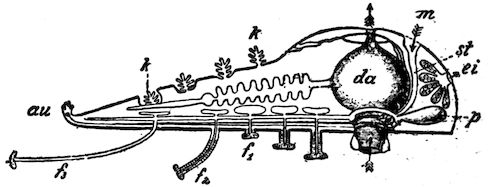
Fig. 58.—Section of one ray and central portion of starfish.
f1, f2, f3, tube feet more or less extended; au, eye spot; k, gills; da, stomach; m, madreporite; st, stone canal; p, ampulla; ei, ovary.
37Respiration.—The system of water vessels serves the additional
purpose of bringing water containing oxygen into contact with
various parts of the body, and the starfish was formerly thought
to have no special respiratory organs. However, there are holes in
the aboral wall through which the folds of the delicate lining membrane
protrude. These are now supposed to be gills (k, Fig. 58).
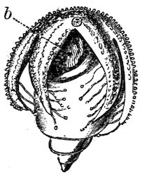
Fig. 59.—Starfish eating a sea snail.
b, stomach everted.
The nervous system is so close to the aboral surface that much
of it is visible without dissection. Its chief parts are a nerve ring
around the mouth, which sends off a branch along each ray.
These branches may be seen by separating the
rows of tube feet. They end in a pigmented
cell at the end of each ray called the eye-spot.
The food of starfish consists of such animals
as crabs, snails, and oysters. When the prey
is too large to be taken into the mouth, the
starfish turns its stomach inside out over
the prey (Fig. 59). After the shells separate,
the stomach is applied to the soft digestible
parts. After the animal is eaten, the stomach
is retracted. This odd way of eating is very
economical to its digestive powers, for only
that part of the food which can be digested and absorbed is taken
into the body. Only the lower part of the stomach is wide and
extensible. The upper portion (next to the aboral surface) is
not so wide. This portion receives the secretion from five
pairs of digestive glands, a pair of which is situated in each ray.
Jaws and teeth are absent. (Why?) The vent is sometimes
wanting. Why?
Reproduction.—There is a pair of ovaries at the base of each
ray of the female starfish (Fig. 58). The spermaries of the male
have the same position and form as the ovaries, but they are
of a lighter colour, usually white.[2]
2. The sperm cells and egg cells are poured out into the water by the adults,
and the sperm cell, which, like nearly all sperm cells, has a vibratory, taillike
flagellum to propel it, reaches and fertilizes the egg cell.
Regeneration after Mutilation.—If a starfish loses one or more
rays, they are replaced by growth. Only a very ignorant oysterman,
angry at the depredations of starfish upon his oyster beds,
38would chop starfish to pieces, as this only serves to multiply them.
This power simulates multiplication by division in the simplest
animals.
Fig. 60.—Young starfish crawling upon their mother. (Challenger Reports.)
Steps in Advance of Lower Branches.—The starfish and other
echinodermata have a more developed nervous system, sensory
organs, and digestion, than forms previously studied; most distinctive
of all, they have a body
cavity distinct from the food
cavity. The digestive glands,
reproductive glands, and the
fluid which serves imperfectly
for blood, are in the body
cavity. There is no heart or
blood vessels. The motions
of the stomach and the bending
of the rays give motion to
this fluid in the body cavity.
It cannot be called blood,
but it contains white blood
corpuscles.
The starfish when first
hatched is an actively swimming
bilateral animal, but it soon becomes starlike (Fig. 60). The
limy plates of the starfish belong neither to the outer nor to the inner
layer (endoderm and ectoderm) of the body wall, but to a third
or middle layer (mesoderm); for echinoderms, like the polyps,
belong to the three-layered animals. In this its skeleton differs
from the shell of a crawfish, which is formed by the hardening
of the skin itself.
Protective Coloration.—Many starfish are brown or yellow. This
makes them inconspicuous on the brown rocks or yellow sand.
Brightly coloured species are usually chosen for aquaria.
External Features.—What is the shape of the body? What
kind of symmetry has it? Do you find the oral (or mouth) surface?
The aboral surface? Where is the body flattened? What
is the shape of the spines? What is their use? How are the tube
39feet arranged? Where do the rows begin and end? Would you
think that a sea urchin placed upside down in water, could right itself
less or more readily than a
starfish? What advantage in
turning would each have that the
other would not have? The
name sea urchin has no reference
to a mischievous boy, but
means sea hedgehog (French
oursin, hedgehog), the name
being suggested by its spines.
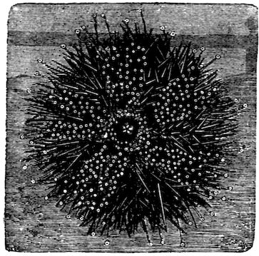
Fig. 61.—A Sea Urchin crawling up the glass front wall of an aquarium (showing mouth spines and tube feet).
Comparison of Starfish and
Sea Urchin.—The water system
of the sea urchin, consisting
of madreporite, tubes, and
water bulbs, or ampullæ, is
similar to that of the starfish.
The tube feet and locomotion are alike. There is no need for
well-developed respiratory organs in either animal, as the whole
body, inside and out, is bathed in water. The method of reproduction
is the same.
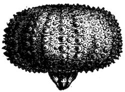
Fig. 62.—A Sea Urchin with spines removed, the limy plates showing the knobs on which the spines grew.
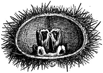
Fig. 63.—Section of Sea Urchin with soft parts removed, showing the jaws which bear the teeth protruding in Fig. 62.
The starfish eats soft animal food. The food of the sea
urchin is mainly vegetable, and it needs teeth (Fig. 62, 63);
its food tube is longer than that of a starfish, just as the food tube
of a sheep, whose food digests slowly, is much longer than that of
a dog.
40
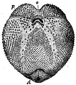
Fig. 64.—The Sea Otter, an urchin with mouth (o) and vent (A) on same side of body.
The largest species of sea urchins are almost as big as a
child’s head, but such size is unusual. The spines are mounted
on knobs, and the joint resembles a
ball-and-socket joint, and allows a wide
range of movement. Some sea urchins
live on sandy shores, other species live
upon the rocks. The sand dollars are
of a lighter colour. (Why)? They are usually
flatter and have lighter, thinner
walls than the other species. The five-holed
sand cake or sand dollar has its
weight still further diminished by the
holes, which also allow it to rise more
easily through the water.
Both starfish and sea urchin rest on
the flattened lower surface of the body,
while the tube feet are stretching forward
for another step.
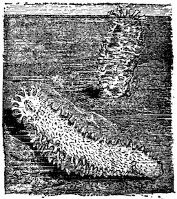
Fig. 65.—Sea Cucumbers.
The sea cucumbers, or holothurians, resemble
the sea urchin in many respects,
but their bodies are elongated,
and the limy plates
are absent or very minute.
The mouth is surrounded
by tentacles (Fig.
65).
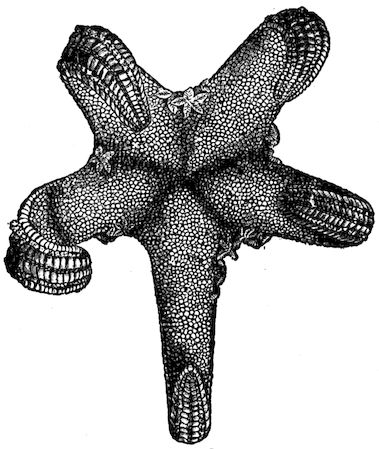
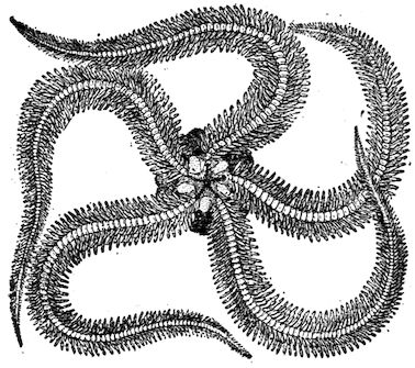
Fig. 66.—A Brittle Star.
The brittle stars resemble
the starfish in form,
but their rays are very
slender, more distinct
from the disk, and the tube feet are on the edges of the rays, not
under them (Fig. 66).
41
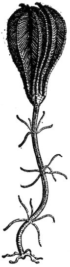
Fig. 67.—Crinoid, arms closed.
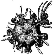
Fig. 68.—Disk of Crinoid from above, showing mouth in centre and vent near it, at right (arms removed).
The crinoids are the most ancient of the echinoderms.
(Figs. 67, 68.) Their fossils are very
abundant in the rocks. They
inhabited the geological seas,
and it is believed that some of
the other echinoderms descended
from them. A few
now inhabit the deep seas.
Some species are fixed by
stems when young, and later
break away and become free-swimming,
others remain
fixed throughout life.
The four classes of the branch echinoderms are
Starfish (asteroids), Sea urchins (echinoids), Sea
cucumbers (holothurians), and Sea lilies (crinoids).
Make a table like this as large as the page of the
notebook will allow, and fill in without guessing.
Amœba
Sponge
Hydra
Coral Polyp
Starfish
Is body round, two-sided, or irregular
What organs of sense
Openings into body
Hard or supporting
parts of body
How food is taken
How move
How breathe
CHAPTER VI WORMS
42
Suggestions:—Earthworms may be found in the daytime after
a heavy rain, or by digging or turning over planks, logs, etc., in
damp places. They may be found on the surface at night by
searching with a lantern. Live specimens may be kept in the
laboratory in a box packed with damp (not wet) loam and dead
leaves. They may be fed on bits of fat meat, cabbage, onion,
etc., dropped on the surface. When studying live worms, they
should be allowed to crawl on damp paper or wood. An earthworm
placed in a glass tube with rich, damp soil, may be watched
from day to day.
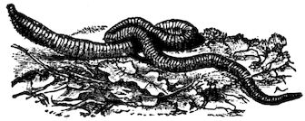
Fig. 69.—An Earthworm.
External Features.—Is the body bilateral? Is there a
dorsal and a ventral surface? Can you show this by a test
with live worm? Do you know of an animal with dorsal
and ventral surface, but not
bilateral?
Can you make out a head?
A head end? A neck? Touch
the head and test whether it
can be made to crawl backwards. Which end is more
tapering? Is the mouth at the tip of the head end or on
the upper or lower surface? How is the vent situated?
Its shape? As the worm lies on a horizontal surface, is
the body anywhere flattened? Are there any very distinct
divisions in the body? Do you see any eyes?
Experiment to find whether the worm is sensitive (1) to touch,
(2) to light, (3) to strong odours, (4) to irritating liquids. Does it
show a sense of taste? The experiments should show whether
43it avoids or seeks a bright light, as of a window; also whether any
parts of the body are especially sensitive to touch, or all equally
sensitive. What effect when a bright light is brought suddenly near
it at night?
Is red blood visible through the skin? Can you notice
any pulsations in a vessel along the back? Do all earthworms
have the same number of divisions or rings? Compare
the size of the rings or segments. Can it crawl faster
on glass or on paper?
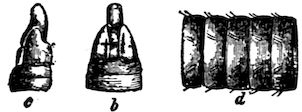
Fig. 70.—Mouth and Setæ.
A magnifying glass will show on most species tiny bristle-like
projections called setæ. How are the setæ arranged?
(d, Fig. 70.) How many on
one ring of the worm? How
do they point? Does the worm
feel smoother when it is pulled
forward or backward between
the fingers? Why? Are setæ on the lower surface?
Upper surface? The sides? What is the
use of the setæ? Are they useful below ground?
Does the worm move at a uniform rate? What
change in form occurs as the front part of the
body is pushed forward? As the hinder part is
pulled onward? How far does it go at each
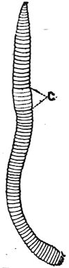
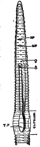
movement? At certain seasons a broad band,
or ring, appears, covering several segments and
making them seem enlarged (Fig. 71). This is
the clitellum, or reproductive girdle. Is this girdle
nearer the mouth or the tail?
Fig. 71.—Earthworm, mouth end above.
Draw the exterior of an earthworm.
Dorsal and Ventral Surfaces.—The earthworm
always crawls with the same surface to the ground; this
is called the ventral surface, the opposite surface is the
dorsal surface. This is the first animal studied to which
44these terms are applicable. What are the
ventral and dorsal surfaces of a fish, a frog,
a bird, a horse, a man?
Fig. 72.—Food Tube of earthworm. (Top view.)
The name “worm” is often carelessly applied
to various crawling things in general. It is properly
applied, however, only to segmented animals
without jointed appendages.
Although a caterpillar crawls,
it is not a worm for several
reasons. It has six jointed
legs, and it is not a developed
animal, but only an early stage
in the life of a moth or a butterfly.
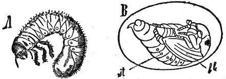
A “grubworm” also
has jointed legs (Fig. 167).
It does not remain a grub, but
in the adult stage is a beetle.
A worm never develops into
another animal in the latter
part of its life; its setæ are
not jointed.
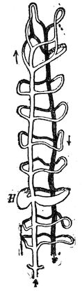
Fig. 73.—Food Tube and Blood Vessels of earthworm showing the ringlike hearts. (Side view.)
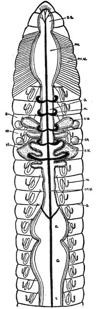
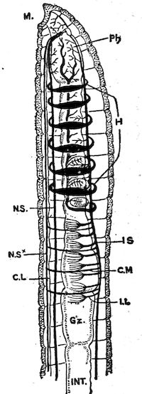
The Food Tube.—The earthworm has
no teeth, and the food tube, as might be
inferred from the form of the
body, is simple and straight. Its
parts, recognizable because of
slight differences in size and
structure, are named the pharynx
(muscular), gullet, crop, gizzard
(muscular), and stomach-intestine. The last extends
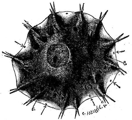
through three fourths of the length of the
body (Fig. 72). The functions of the parts of
the food tube are indicated by their names.
Fig. 74.
Circulation.—There is a large dorsal blood
vessel above the food tube (Fig. 73). From the
45front portion of this tube arise several large tubular rings
or “hearts” which are contractile and serve to keep the
blood circulating. They lead to a ventral vessel below the
food tube (Fig. 74). The blood is red, but the colouring
matter is in the liquid, not in the blood cells.
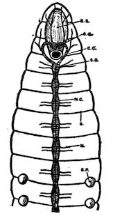
Fig. 75.—Ganglia near Mouth and part of nerve chain of earthworm.
Nervous System.—Between the ventral blood vessels
is a nerve cord composed of two strands (see Fig. 75).
There is a slight swelling, or ganglion, on each strand, in
each segment (Fig. 75). The strands separate
near the front end of the worm, and a
branch goes up each side of the gullet and
enters the two pear-shaped cerebral ganglia,
or “brain” (Fig. 75).
Food.—The earthworm eats earth containing
organic matter, the inorganic part passing
through the vent in the form of circular casts;
these are found in the morning at the top of
the earthworm’s burrow.
The earthworm has no teeth. It excretes
through the mouth an alkaline fluid which
softens and partly digests the food before it
is eaten. When this fluid is poured out upon a green leaf,
the leaf at once turns brown. The starch in the leaf is
also acted upon. The snout aids in pushing the food into
the mouth.
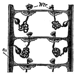
Kidneys.—Since oxidation is occurring in its tissues,
and impurities are forming, there must be some way of
removing impurities from the tissues. The earthworm
does not possess one-pair organs like the kidneys of
higher animals to serve this purpose, but it has numerous
pairs of small tubular organs called nephridia which serve
the purpose. Each one is simply a tube with several coils.
There is a pair on the floor of each segment. Each nephridium
46has an inner open end within the body cavity, and
its outer end opens by a pore on the surface between the
setæ. The nephridia absorb waste
from the liquid in the celom, or body
cavity surrounding the food tube,
and convey it to the outside.
Fig. 76.—Two Pairs of Nephridia in a worm (diagram).
Respiration.—The skin of the
earthworm is moist, and the blood
capillaries approach so near to the
surface of the body that the oxygen
is constantly passing in from the air,
and carbon dioxide passing out; hence
it is constantly breathing through all parts of its skin.
It needs no lungs nor special respiratory organs of any
kind.
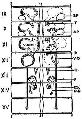
Fig. 77.—Sperm (sp) and egg glands (es) of worm.
Reproduction.—When one individual animal produces both
sperm cells and egg cells, it is said to be hermaphrodite. This
is true of the earthworm. The egg cell
is always fertilized, however, not by the
sperm cells of the same worm, but by
sperm cells formed by another worm.
The openings of these ovaries consist of
two pairs of small pores found in most
species on the ventral surface of the
fourteenth segment (see Fig. 77). There
are also two pairs of small receptacles
for temporarily holding the foreign sperm
cells. One pair of the openings from
these receptacles is found (with difficulty)
in the wrinkle behind the ninth
segment (Fig. 77), and the other pair
behind the tenth segment. The spermaries
are in front of the ovaries (Fig. 77), but the sperm ducts
are longer than the oviducts, and open behind them on the fifteenth
segment (Figs. 77, 78). The worms exchange sperm cells, but not
47egg cells. The reproductive girdle, or clitellum, already spoken of,
forms the case which is to hold the eggs (see Fig. 71). When the
sperm cells have been exchanged, and the ova are ready for fertilization,
the worm draws itself backward from the collar-like case or
clitellum so that this slips over the head. As it passes the fourteenth
segment, it collects the ova, and as it passes the ninth and
tenth segments, it collects the sperm cells previously
received from another worm. The elastic, collar-like
clitellum closes at the ends after it has slipped over the
worm’s head, forming a capsule. The ova are fertilized
in this capsule, and some of them hatch into worms in
a few days. These devour the eggs which do not
hatch. The eggs develop into complete but very
small worms before escaping from the capsule.
Fig. 78.—Side view, showing setæ, nephridia pores, and reproductive openings.
Habits.—The earthworm is omnivorous. It
will eat bits of meat as well as leaves and
other vegetation. It has also the advantage,
when digging its hole, of eating the earth which
must be excavated. Every one has noticed the
fresh “casts” piled up at the holes in the morning.
As the holes are partly filled by rains, the
casts are most abundant after rains. The chief
enemies of the earthworm are moles and birds.
The worms work at night and retire so early in
the morning that the very early bird has the
advantage in catching worms. Perhaps the nearest to
an intelligent act the earthworm accomplishes is to conceal
the mouth of its hole by plugging it with a pebble or
a bit of leaf. Worms hibernate, going below danger of
frost in winter. In dry weather they burrow several feet
deep.
The muscular coat of the body wall is much thicker than
the skin. It consists of two layers: an outer layer of fibres
which run around the body just beneath the skin, and an
48inner, thicker layer of fibres which run lengthwise. The
worm crawls by shortening the longitudinal muscles. As
the bristles (setæ) point backward, they prevent the front
part of the body from slipping back, so the hinder part is
drawn forward. Next, the circular muscles contract, and
the bristles preventing the hind part from slipping back,
the fore portion is pushed forward. Is the worm thicker
when the hinder part is being pulled up or when the fore
part is being thrust forward? Does the earthworm pull or
push itself along, or does it do both? Occasionally it travels
backward, e.g. it sometimes goes backward into its hole.
Then the bristles are directed forward.
The right and left halves of the body are counterparts of
each other, hence the earthworm is bilaterally symmetrical.
The lungs and the gills of animals must always be kept
moist. The worm cannot live long in dry air, for respiration
in the skin ceases when it cannot be kept moist, and the
worm smothers. Long immersion in water is injurious to it,
perhaps because there is far less oxygen in water than in
the air.
Darwin wrote a book called “Vegetable Mould and Earthworms.”
He estimated that there were fifty thousand earthworms
to the acre on farm land in England, and that they
bring up eighteen tons of soil in an acre each year. As
the acids of the food tube act upon the mineral grains that
pass through it, the earthworm renders great aid in forming
soil. By burrowing it makes the soil more porous and
brings up the subsoil.
Although without eyes, the worm is sensitive to light
falling upon its anterior segments. When the light of a
lantern suddenly strikes it at night, it crawls quickly to its
burrow. Its sense of touch is so keen that it can detect a
light puff of breath. Which of the foods kept in a box of
49damp earth disappeared first? What is indicated as to a
sense of taste?
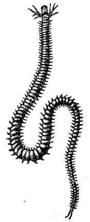
Fig. 79.—Sand Worm × ⅔ (Nereis).
Why is the bilateral type of structure better adapted for
development and higher organization than the radiate type
of the starfish? The earthworm’s body is a
double tube; the hydra’s body is a single
tube; which plan is more advantageous, and
why? Would any other colour do just as well
for an earthworm? Why, or why not?
The sandworm (Nereis) lives in the sand of the
seashore, and swims in the sea at night (Fig. 79).
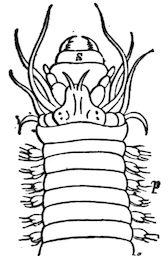
It is more advanced in structure than the earthworm,
as it has a distinct head (Fig. 80), eyes, two
teeth, two lips, and several pairs of antennæ, and
two rows of muscular projections which serve as
feet. It is much used by fishermen for bait. If
more easily obtained, it may be studied instead of
the earthworm.
Fig. 80.—Head of Sandworm (enlarged).
There are four classes in the branch Vermes:
1) the worms, including sandworms and leeches; 2) the
roundworms, including trichina, hairworms,
and vinegar eels; 3) flatworms, including
tapeworm and liver fluke; 4) rotifers, which
are microscopic aquatic forms.
The tapeworm is a flatworm which has lost
most of its organs on account of its parasitic
life. Its egg is picked up by an herbivorous
animal when grazing. The embryo undergoes
only partial development in the body
of the herbivorous animal, e.g. an ox. The
next stage will not develop until the beef is eaten by a
carnivorous animal, to whose food canal it attaches itself
and soon develops a long chain of segments called a
“tape.” Each segment absorbs fluid food through its
50body wall. As the segments at the older end mature,
each becomes full of eggs, and the segments become
detached and pass out of the canal, to be dropped and
perhaps picked up by an herbivorous animal and the
life cycle is repeated.
The trichina is more dangerous to human life than is the
tapeworm. It gets into the food canal in uncooked pork
(bologna sausage, for example), multiplies there, migrates
into the muscles, causing great pain, and encysts there,
remaining until the death of the host. It is believed to
get into the bodies of hogs again when they eat rats, which
in turn have obtained the cysts from carcasses.
Summary of the Biological Process.—An earthworm is
a living machine which does work (digging and crawling;
seizing, swallowing, and digesting food; pumping blood;
growing and reproducing). To do the work it must have
a continual supply of energy. The energy for its work is
set free by the protoplasm (in its microscopic cells) undergoing
a destructive chemical change (oxidation). The
waste products from the breaking down of the protoplasm
must be continually removed (excretion). The broken-down
protoplasm must be continually replaced if life is to
continue (the income must exceed the outgo if the animal
is still growing). The microscopic cells construct more
protoplasm out of food and oxygen (assimilation) supplied
them by the processes of nutrition (eating, digesting,
breathing, circulating). This protoplasm in turn oxidizes
and releases more energy to do work, and thus the cycle
of life proceeds.
CHAPTER VII CRUSTACEANS — Crawfish
51
Suggestions.—In regions where crayfish are not found, a live
crab may be used. Locomotion and behaviour may be studied by
providing a tub of water, or better, a large glass jar such as a
broad candy jar. For suggestions on study of internal structure,
see p. 58.
Habitat.—Do you often see crawfish, or crayfish, moving
about, even in water where they are known to be abundant?
What does your answer suggest as to the time
when they are probably most active?
Why do you never see one building its chimney, even
where crayfish holes are abundant? Is the chimney
always of the same colour as the surface soil? Are the
crayfish holes only of use for protection? In what kind
of spots are crayfish always dug; Why? What
becomes of crayfish when the pond or the creek dries up?
How deep are the holes? How large are the lumps of
mud of which the chimney is built? How does it get
them out of the hole? Why is the mud built into a chimney
instead of thrown away? (What would happen to a
well with its mouth no higher than the ground?) Why
are crayfish scarce in rocky regions?
How does the colour of the crayfish compare with its
surroundings? Is its colour suited to live in clear or muddy
water? Define protective colouration.
52Habits.—Does the crayfish walk better in water or out
of it? Why? Does it use the legs with the large claws
to assist in walking? Do the swimmerets (under the abdomen)
move fast or slow? (Observe it from below in a
large jar of clear water.) What propels it backward?
Forward? Does the crayfish move at a more uniform
rate when swimming backward or forward? Why? In
which way can it swim more rapidly? Do the big legs
with claws offer more resistance to the water while it is
swimming backward or forward? How does it hold the
tail after the stroke, while it is darting backward through
the water? Hold a crayfish with its tail submerged and
its head up. Can the tail strike the water with much
force? Allow it to grasp a pencil: can it sustain its own
weight by its grip?
Feeding.—Offer several kinds of food to a crayfish that
has not been alarmed or teased. Does it prefer bread,
meat, or vegetables? How does it get the food to its mouth?
Does it eat rapidly or slowly? Does it tear the food with
the big pincers? Can it gnaw with the small appendages
near the mouth?
Breathing.—Does the crayfish breathe with gills or
lungs? Place a few drops of ink near the base of the hind
legs of a crayfish resting quietly in shallow water. Where
is the ink drawn in? Where does it come out? To explain
the cause and the purpose of this motion, place a crayfish
in a large glass jar containing water, and see the
vibratory motion of the parts under the front portion of
the body. There is under the shell on each side of the body
a gill paddle, or gill bailer, that moves at the same rate.
Senses.—Crayfish are best caught with a piece of meat
or beef’s liver tied to a string. Do they always lose hold
as soon as they are lifted above the water? What do you
53conclude as to the alertness of their senses? Does the covering
of their bodies suggest the possession of a delicate or
a dull sense of touch?
Of what motions are the eyes capable? Touch one of
the eyes. The result? Can a crayfish see in all directions?
To test this, place a crayfish on a table and try
whether you can move to a place where you can see the
crayfish without seeing its eyes. What are the advantages
and disadvantages of having the eyes on stalks?
Fig. 81.—Crawfish (dorsal surface).
Fig. 82.
Touch the body and the several appendages of the
crayfish. Where does it seem most sensitive to touch?
Which can reach farther, the antennæ or the big claws?
Why are short feelers needed as well as long ones?
Make a loud and sudden noise without jarring the crayfish.
Is it affected by sound?
External Anatomy (Figs. 81, 82, 83, 84).—Is the body of
the crayfish rounded out (convex) everywhere, or is any
part of its surface either flat or rounded in (concave)?
54What colour has the crayfish? Is this colour of any use to
the crawfish?
Fig. 83.—Lateral view of Crawfish.
Make out the two distinct regions or divisions of the body
(Fig. 81). The anterior (front) region is called the head chest
or cephalothorax, and the posterior (rear) region is
called the tail.
Which region is
larger? Why?
Which is flexible?
Why?
Is the covering
of the body hard
or soft? What
is the advantage of such a covering? What are its disadvantages?
How is the covering modified at the joints
to permit motion?
Fig. 84.—Fourth Abdominal Segment of Crawfish with swimmeret.
Tail.—How many joints, or segments, of the tail? (Figs.
81, 83.) Does the hard covering of each segment slip
under or over the segment behind it when
the tail is straight? Does this lessen
friction while swimming forward?
Is there a pair of swimmerets to each
segment of the tail? (Figs. 82, 86.)
Notice that each swimmeret has a main
stalk (protopod), an outer branch (exopod),
and an inner branch (endopod) (Fig. 84).
Are the stalk and the branches each in
one piece or jointed? The middle part of the tail fin is
called the telson. By finding the position of the vent,
decide whether the food tube goes into the telson
(Fig. 82). Should it be called an abdominal segment.
Are the side pieces of the tail fin attached to the telson
or to the sixth segment? Do these side pieces correspond
55to swimmerets? Do they likewise have the Y-shaped
structure? (Fig. 86.)
If the swimmerets on the first abdominal segment are
large, the specimen is a male. If they are small, it is a
female. Which sex is shown in Fig. 82?
Fig. 86?
Fig. 86.—Crayfish (ventral surface).
Carapace.—The covering of the head
chest (cephalothorax) is called the carapace.
Has it free edges? The gills are
on the sides of the body and are covered
by the carapace (Fig. 87). The projection
in front is called the rostrum, meaning beak.
Does the rostrum project beyond the eyes?
There is a transverse groove across the carapace
which may
be said to divide
the head from the
abdomen. Where
does this groove end at the sides?
Legs.—How many legs has the
crayfish? How many are provided
with large claws? Small claws?
Is the outer claw hinged in each
of the large grasping pincers?
The inner claw?
Appendages for Taking Food.—If
possible to watch a living crayfish
eating, notice whether it places
the food directly into the mouth with the large claws. Bend
the large claws under and see if they will reach the mouth.
Attached just in front of the legs the crayfish has three
pairs of finger-like appendages, called foot jaws (maxillipeds),
with which it passes the food from the large pincers
56to its mouth (Figs. 85, 86). They are in form and in use
more like fingers than feet. In front of the foot jaws are two
pairs of thin jaws
(maxillæ) and in
front of the thin
jaws are a pair of
stout jaws (mandibles)
(Fig. 85). Do
the jaws move
sidewise or up
and down? Which
of the jaws has a
jointed finger (palp) attached to it? Do all the appendages
for taking food have both exopod and endopod
branches on a basal stalk or protopod? Which of the
appendages have a scalloped edge? How would you know
from looking at the crayfish that it is not merely a
scavenger? Why are there no pincers on the hind feet?
Fig. 87.—Gill cover removed and gills exposed.
Mp, gill bailer.
Sense Organs.—Find the antennæ, or long feelers (Figs.
82, 90). Are the antennæ attached above or below the
eyes? (Fig. 87.)
Fig. 88.—Lengthwise Section of Male Crawfish.
c, heart; Ac, artery to head; Aa, artery to abdomen; Km, stomach; D, intestine; L, liver; T, spermary; Go, opening of sperm duct; G, brain; N, nerve chain.
Find the pair of antennules, or small feelers. Are their
divisions like or unlike each other? Compare the length
of the antennules and the antennæ. Compare the flexibility
of the antennæ with that of the other appendages.
57Observe the position of the eyes (Figs. 81, 88). How long
are the eyestalks? Is the stalk flexible or stiff? Touch the
eye. Where is the joint which enables the stalk to move?
Is the outer covering of the eye hard or soft? A mounted
preparation of the transparent covering (cornea) of the
eye, seen with lower power of microscope, reveals that the
cornea is made up of many divisions, called facets. Each
facet is the front of a very small eye, hundreds of which
make up the whole eye, which is therefore called a compound
eye. The elongated openings to the ear sacs are
located each on the upper side of the base of a small feeler
just below the eye.
Respiratory System.—The respiratory organs are gills
located on each side of the thorax in a space between the
carapace and the body (Fig. 87). The gills are white, curved,
and feathery. Is the front gill the largest or the smallest?
The gills overlap each other; which is the outermost gill?
On the second maxilla is a thin, doubly curved plate called
a gill bailer (Fig. 85). The second maxilla is so placed
that the gill bailer comes at the front end of the gill
chamber. The bailer paddles continually, bringing the
water forward out of the gill. The gills are attached
below at the base of the legs. Are the gills thick or thin?
How far upward do they go? Does the backward motion
in swimming aid or hinder the passage of the water through
the gills? Does a crayfish, when at rest on the bottom
of a stream, have its head up or down stream? Why?
Openings.—The slitlike vent is on the under side of
the telson (Figs. 82, 88). The mouth is on the under side
of the thorax behind the mandibles. At the base of the
long antennæ are the openings from the green glands, two
glands in the head which serve as kidneys (Fig. 89).
The openings of the reproductive organs are on the third
58pair of legs in the female, and the fifth
pair of legs in the male (Fig. 88). The
eggs are carried on the swimmerets.
Fig. 89.—Level lengthwise section showing h, heart. d, green gland. le, liver. kie, gills. kh, gill cavity. ma, stomach.
(After Huxley.)
Internal Structure.—Suggestions. If
studied by dissection, it will be necessary
to have several crayfish for each pupil, one
for gaining general knowledge, and others for
studying the systems in detail. Specimens
should have lain in alcohol for several days.
Fig. 90.—Section of Crayfish showing—stomach s, liver li, and vent a.
The Food Tube.—Is the stomach in the
head portion of the cephalothorax or in the
thoracic portion? (Figs. 88,89). Is the stomach
large or small? What is its general shape?
Does the gullet lead upward or backward?
Is it long or short? (Fig. 88.) The mid tube,
which is the next portion of the food tube, is
smaller than the stomach. On each side of
it are openings from the bile ducts which
bring the secretion from the digestive gland,
sometimes called the liver. Does this gland
extend the whole length of the thorax? Is
it near the floor or the top of the cavity?
The third and last portion of the food tube
is the intestine. It extends from the thorax
to the vent. Is it large
or small? Straight or
curved? The powerful
flexor muscles of the tail
lie in the abdomen below
the intestines. Compare
the size of these muscles
with the extensor muscle
above the intestine (Fig.
90). Why this difference?
Does the food tube extend
into the telson? Locate
the vent (Fig. 90).
59
Fig. 91.—Showing heart and main blood vessels.
The Circulation.—The blood is a liquid containing white corpuscles.
It lacks red corpuscles and is colourless. The heart is in
the upper part of the thorax. It is surrounded
by a large, thin bag, and thus it is
in a chamber (called the pericardial sinus).
The blood from the pulmonary veins enters
this sinus before it enters the heart. The
origin of this pericardial sinus by the fusing of
veins is shown in Fig. 130. Does one artery,
or do several arteries, leave the heart? There
is a larger dorsal artery lying on the intestine
and passing back to the telson; there are
three arteries passing forward close to the
dorsal surface (Figs. 89, 91). One large artery
(the sternal) passes directly downward (Figs.
88, 91), and sends a branch forward and
another backward near the ventral surface.
The openings into the heart from the sinus
have valvular lips which prevent a backward
flow of blood into the sinus. Hence, when
the heart contracts, the blood is sent out into the several
arteries. The arteries take a supply of fresh blood
to the eyes, stomach, muscles, liver, and the various
organs. After it has given oxygen to the several organs
and taken up carbon dioxide, it returns by veins to pass
through the gills on each side, where it gives out the useless
gas and takes up oxygen from the water. It is then
led upward by veins into the pericardial sinus again.
Fig. 92.
The central nervous system consists of a double
chain of ganglia (Fig. 92). This main nerve chain
lies along the ventral surface below the food tube
(Fig. 90), except one pair of ganglia which lie
above the œsophagus or gullet (Fig. 88), and are
called the supra-œsophageal ganglia, or brain.
Crustacea.—The crayfish and its kindred are placed in
the class called Crustacea.
60
Fig. 93.—Crab from below.
Fig. 94.—Hermit Crab, using shell of sea snail for a house.
Decapods.—All crustacea which have ten feet belong
in the order called decap´oda (ten-footed). This order
includes the crabs, lobsters, shrimp,
etc. The crabs and the lobsters are of
considerable importance because of
use as food. Small boys sometimes
catch crayfish, and in some instances
are known to cook and eat them for
amusement,
the only part cooked being the
muscular tail. The crab’s tail is
small and flat and held under the
body (Fig. 93).
Fig. 95.—Development of a Crab.
a, nauplius just after hatching; b, c, d, zoëa; e, megalops; f, adult.
Question: Which stage is most like a crayfish? Compare with metamorphoses of insects.
Since the limy covering to serve
the purpose of protection is not
soft enough to be alive and growing, it is evident that the
crustacea are hampered in their growth by their crusty
covering. During
the first
year the crayfish
sheds its
covering, or
moults three
times, and
once each year
thereafter. It
grows very fast
for a few days
just after moulting,
while the
covering is soft
and extensible.
Since it is at
61the mercy of birds, fish, and other enemies while in this
soft and defenceless condition, it stays hidden until the
covering hardens. Hence it cannot eat much, but probably
by the absorption of water the tissues grow; that is, enlarge.
In the intervening periods, when growth is impossible, it
develops; that is, the tissues and organs change in structure
and become stronger. “Soft-shelled crab” is a popular dish,
but there is no species by that name, this being only a crab
just after moulting which has been found by fishermen in
spite of its hiding.
General Questions.—How do crayfish choose their food?
How long can they live out of water? Why do their gills remain
moist out of water longer than a fish? How do they breathe
out of water? Are they courageous or cowardly animals? When
they lose appendages in fighting or moulting these are readily
reproduced, but an organ moults several times in regaining its
size. Have you seen crayfish with one claw smaller than the other?
Compare the crayfish and crab (Figs. 81, 93, and 95) in the
following particulars: shape, body, eyes, legs, abdomen, habitat,
movement.
KEY TO THE FOUR CLASSES IN BRANCH ARTHROPODS
1. Insects
3 body divisions, 6 legs
2. Arachnids
2 body divisions, 8 legs
3. Myriapods
many body divisions, many legs
4. Crustaceans
gill breathers, skeleton (external) limy
By the aid of the key and of figures 96–105, classify the following
Arthropods: tick, thousand-leg centipede, king crab, pill bug, spider,
scorpion, beetle.
62
Fig. 96.—Pill Bug.
Fig. 97.—Lady Beetle.
Fig. 98.—Scorpion.
Fig. 99.—Tick before and after feeding.
Fig. 100.—Centipede.
Fig. 101.—One Segment of Centipede with one pair of legs.
Fig. 102.—One Segment of Thousand Legs with two pairs of legs.
Fig. 103.—Thousand Legs.
Fig. 104.—A Spider.
Fig. 105.—King Crab.
Illustrated Study.Classification of Arthropods. Key on p. 61.
CHAPTER VIII INSECTS — The Grasshopper — Anatomy and General Characteristics of the Class Insecta — RECOGNITION-CHARACTERS FOR THE PRINCIPAL ORDERS OF ADULT WINGED INSECTS
63
Suggestions.—Collect grasshoppers, both young and full-grown,
and keep alive in broad bottles or tumblers and feed on
fresh grass or lettuce. When handling a live grasshopper, never
hold it by its legs, as the joints are weak. To keep them for
some time and observe their moults, place sod in the bottom of a
box and cover the box with mosquito netting or wire gauze.
Fig. 106.—A Grasshopper.
What is the general shape of its body? (Fig. 106.)
Where is the body thickest? Is it bilaterally symmetrical,
that is, are the two sides of the
body alike? Is the skeleton, or hard
part of the body, internal or external?
Is the skeleton as stiff and thick
as that of a crayfish? What is the
length of your specimen? Its colour?
Why does it have this coloration? In what ways does the
grasshopper resemble the crayfish? Differ from it?
The Three Regions of the Body.—The body of the grasshopper
is divided into three regions—the head, the thorax,
and the abdomen. Which of these three divisions has no distinct
subdivisions? The body of the grasshopper, like that of
the earth worm, is made of ringlike segments. Are the segments
most distinct in the head, the thorax, or the abdomen?
Which region is longest? Shortest? Strongest? Why?
Which region bears the chief sense organs? The appendages
for taking food? The locomotory appendages?
Which division of the body is most active in breathing?
64The Abdomen.—About how many segments or rings in
the abdomen? Do all grasshoppers have the same number
of rings? (Answer for different species and different
individuals of the same species.) The first segment and
the last two are incomplete rings. Does the flexibility of
the abdomen reside in the rings or in the joints between the
rings? Is there merely a thin, soft line between the rings,
or is there a fold of the covering? Does one ring slip into
the ring before it or behind it when the abdomen is bent?
Fig. 107.—A Grasshopper Dissected.
As the grasshopper breathes, does each ring enlarge
and diminish in size? Each ring is divided into two parts
by folds. Does the upper half-ring
overlap the lower half-ring, or the
reverse? With magnifying glass, find
a small slit, called a spiracle, or breathing
hole, on each side of each ring just
above the side groove (Fig. 106). A
tube leads from each spiracle. While
the air is being taken in, do the two
portions of the rings move farther
apart? When they are brought
together again, what must be the
effect? In pumping the air, the abdomen may be said to
work like a bellows. Bellows usually have folds to allow
motion. Is the comparison correct?
How many times in a minute does the grasshopper take
in air? If it is made to hop vigorously around the room
and the breathing is again timed, is there any change?
Find the ears on the front wall of the first abdominal
ring (Fig. 107). They may be seen by slightly pressing
the abdomen so as to widen the chink between it and
the thorax. The ears are merely glistening, transparent
membranes, oval in form. A nerve leads from the inner
65surface of each membrane. State any advantage or disadvantage
in having the ears located where they are.
Fig. 108.—Grasshopper Laying Eggs. (Riley.)
Ovipositor.—If the specimen is a female, it has an egg-placer
or ovipositor, consisting of four blunt projections at
the end of the abdomen (Fig. 107). If it is a male,
there are two appendages above the end of the abdomen,
and smaller than the parts of the ovipositor. Females
are larger and more abundant than males. In laying
the eggs, the four blunt points are brought tightly together
and then forced into the
ground and opened (Fig. 108). By
repeating this, the grasshopper
makes a pit almost as deep as the
abdomen is long. The eggs are
laid in the bottom of the pit.
Draw a side view of the grasshopper.
Thorax.—This, the middle portion
of the body, consists of three
segments or rings (Fig. 107). Is the division between the
rings most apparent above or below? Which two of the
three rings are more closely united?
The front ring of the thorax is called prothorax. Is it
larger above or below? Does it look more like a collar
or a cape? (Fig. 106.) A spiracle is found on the second
ring (mesothorax, or middle thorax) just above the second
pair of legs. There is another in the soft skin between
the prothorax and the mesothorax just under the large
cape or collar. The last ring of the thorax is called the
metathorax (rear thorax).
How many legs are attached to each ring of the thorax?
Can a grasshopper walk? Run? Climb? Jump?
Fly? Do any of the legs set forward? (See Fig. 106.)
66Outward? Backward? Can you give reasons for the position
of each pair? (Suggestion: What is the use of each
pair?) If an organ is modified so that it is suited to serve
some particular purpose or function, it is said to be specialized.
Are any of the legs specialized so that they serve
for a purpose different from that of the other legs?
Fig. 109.—How a Grasshopper Walks.
Fig. 110.—How a Spider Walks.
The leg of a grasshopper (as of all insects) is said to
have five parts, all the small parts after the first four parts
being counted as one part and called the foot. Are all the
legs similar, that is, do the short and the long joints in all
come in the same order? Numbered in order from the
body, which joint of the leg is the largest,—the first, second,
third, or fourth? Which joint is the shortest? The
slenderest? Which joint has a number of sharp points or
spines on it? Find by experiment whether these spines
are of use in walking (Fig. 106). Jumping? Climbing?
In what order are the legs used in walking? How many
legs support the body at each step?
All animals that have ears have ways of communicating
by sounds. Why would it be impossible for the grasshopper
to have a voice, even if it had vocal cords in its
throat? The male grasshoppers of many species make a
chirping, or stridulation, by rubbing the wing against the
leg. Look on the inner side (why not outer side?) of the
67largest joint of the hind leg for a row of small spines visible
with the aid of a hand lens (Fig. 111). The sound is
produced by the outer wings rubbing against the spines.
Have you noticed whether the sound is
produced while the insect is still or in
motion? Why? The male grasshoppers
of some species, instead of having
spines, rub the under side of the front
wing on the upper side of the hind wing.
Fig. 111.—A, Row Of Spines, z, used in chirping.
B, the same more enlarged.
Wings.—To what is the first pair
of wings attached? The second pair?
Why are the wings not attached to the
prothorax? Why are the wings attached
so near the dorsal line of the body? Why are the second
and third rings of the thorax more solidly joined than the
first and second rings?
Fig. 112.—Grasshopper in Flight.
Compare the first and second pairs of wings in shape,
size, colour, thickness and use (Fig. 112). How are the
second wings folded so as to go
under the first wings? About
how many folds in each?
Draw a hind wing opened out.
Head. What is the shape of
the head viewed from the front, the
side, and above? Make sketches.
What can you say of the neck? Is
the head movable in all directions?
What is the position of the large
eyes? Like the eyes of the crayfish,
they are compound, with many facets. But the grasshopper
has also three simple eyes, situated one in the middle
of the forehead and one just above each antenna. They
are too small to be seen without a hand lens. How does
68the grasshopper’s range of vision compare with that of the
crayfish?
Fig. 113.
Are the antennæ flexible? What is their shape? Position?
Are they segmented? Touch an antenna, a wing,
a leg, and the abdomen in succession. Which seems to be
the most sensitive to touch? The antennæ
are for feeling. In some species of insects
they also are organs of hearing and smelling.
The mouth parts of a grasshopper should
be compared with the mouth parts of a beetle
shown in Fig. 113, since they correspond
closely. If the grasshopper is fed with a
blade of fresh grass, the function of each organ may be
plainly seen. It is almost impossible to understand these
functions by studying a dead specimen, but a fresh specimen
is much better than a dry one.
Fig. 114.—a, Food Tube of Beetle. b, gizzard; d, intestine; c, biliary vessels. See Fig. 127.
The upper lip, or labrum, is seen in front. Is it tapering
or expanded? In what direction is it movable? The dark
pointed biting jaws (mandibles) are next. Are they curved
or straight? Sharp or blunt pointed? Notched or smooth?
Do they work up and down, or sideways? The holding jaws
(maxillæ), each with two jaw fingers (maxillary palpi), are
behind the chewing jaws. Why? The lower lip (labium)
has a pair of lip fingers (labial palpi) upon it. The brown
69tongue, usually bathed in saliva, is seen in the lower part of
the mouth. Since the grasshopper has no lips, or any way
of producing suction, it must lap the dew in drinking. Does
it merely break off bits of a grass blade, or does it chew?
The heart, circulation, nervous system, digestive and respiratory
organs of the grasshopper agree mainly with the
general description of the organs of insects given in the
next section.
Fig. 115.—Egg and Moults of a Grasshopper.
Microscopic Objects.—These may be bought ready
mounted, or may be examined fresh. A portion of the
covering of the large eye may be cut off and the dark layer
on the inside of the covering scraped off to make it transparent.
What is the shape of the facets? Can you make
any estimate of
their number? A
portion of the
transparent hind
wing may be used,
and the “veins”
in it studied. A
thin bit of an abdominal segment containing a spiracle
will show the structure of these important organs.
Growth of the Grasshopper.—Some species hibernate in
sheltered places and lay eggs in the spring, but adult species
are scarce at that season. Most species lay the eggs in the
fall; these withstand the cold and hatch out in the spring.
Those hatched from one set of eggs sometimes stay together
for a few days. They eat voraciously, and as they grow, the
soft skin becomes hardened by the deposit of horny substance
called chitin. This retards further growth until the insect
moults, the skin first splitting above the prothorax. After
hatching, there are five successive periods of growth. At
which moult do the very short wings first appear? (Fig. 115.)
70After the last moult the animal is complete, and changes
no more in size for the rest of its life. There has been an
attempt among writers to restrict the term
grasshopper to the long-winged, slender
family, and to call the shorter winged,
stouter family locusts, according to old
English usage.
Fig. 116.—Cockroach.
Fig. 117.—Praying Mantis, or devil’s horse.
Fig. 118.—Cricket.
Fig. 119.—Mole Cricket.
Fig. 120.—Front Leg of Mole Cricket. × 3.
Economic Importance of Grasshoppers.—Great
injury is often done to vegetation by
grasshoppers; however, the millions of tiny
but ravenous eaters hatched in early spring
are usually soon thinned out by the birds. The migratory
locusts constitute a plague when they appear, and
they have done so since ancient times. The Rocky Mountain
locusts flying eastward have darkened the sky, and
where they settled to the earth
ate almost every green thing.
In 1874–5 they produced almost
a famine in Kansas, Nebraska,
and other Western states. The young hatched away
from the mountains were not healthy,
and died prematurely, and their devastations
came to an end. Of course the
migrations may occur again. Packard
calculates that the farmers of the
West lost $200,000,000 because of grasshopper
ravages in 1874–5.
71The cockroaches (Fig. 116), kindred of the grasshoppers,
are household pests that have migrated almost everywhere
that ships go. The praying mantis (Fig. 117), or devil’s
horse, also belongs to this order. It is beneficial, since it
destroys noxious insects. Which of its legs are specialized?
The walking stick (Fig. 121) and the cricket (Fig. 118), like
most members of the order, are vegetarian.
Fig. 121.—Four Walking Stick Insects.
Are grasshoppers more common in fields and meadows,
or in wooded places? How many different colours have you
seen on grasshoppers? Which
colours are most common?
Grasshoppers are very scarce
in Europe as they love dry,
warm countries. Why do locusts
migrate? Give an instance
in ancient times.
How long do most grasshoppers
live? Does a grasshopper
spread its wings before
it flies? Does it jump and fly
together? Can it select the
place for alighting?
Note to Teacher.—Field work in
Zoology should be systematic. Every trip
has a definite region and definite line of
study in view, but every animal seen should be noted. The habitat, adaptation
by structure and habits to the environment, relations to other animals,
classification of animals seen, should be some of the ideas guiding the study.
The excursions may be divided somewhat as follows, according as opportunities
offer: Upland woods, lowland woods, upland pastures, fields, swamps, a fresh-water
lake, a pond, lower sea beach, higher sea beach, sand hills along shore,
roadside, garden, haunts of birds, insect visits to flowers, ground insects,
insects in logs.
72Collecting Insects.—In cities and towns insects, varying
with the season, are attracted by electric lights. Beetles
and bugs will be found under the lights, moths on posts
near the lights, grasshoppers and crickets and other insects
in the grass near by. A lamp placed by a window
brings many specimens. In the woods and in rocky
places insects are found under logs and stones, and under
the bark of dead trees. In open places, prairies, meadows,
and old fields with grass and flowers, it will be easy to find
grasshoppers, butterflies, and some beetles. Ponds and
streams are usually rich in animal forms, such as bugs and
beetles, which swim on or under the surface, and larvæ
of dragon flies crawling on the bottom. Dragon flies and
other insects that lay eggs on the water are found flying
in the air above. (In the spring, newly hatched crayfish,
tadpoles, and the eggs of frogs and toads should also be
collected, if found.) Moths may be caught at night by
daubing molasses or syrup made from brown sugar upon
the trunks of several trees, and visiting the trees at intervals
with a lantern.
An insect net for catching butterflies and for dredging
ponds may be made by bending a stout wire into a circle
one foot in diameter, leaving enough straight wire to
fasten with staples on an old broomstick. To the frame
is fastened a flour sack, or cone made of a piece of mosquito
netting.
Butterflies and moths should be promptly killed, or
they will beat their wings to pieces. The quickest method
is by dropping several drops of gasoline upon the ventral
(under) side of the thorax and abdomen. (Caution:
Gasoline should never be used near an open fire, or lamp,
as explosions and deaths result from the flame being led
through the gasoline-saturated air to the vessel containing
it.)
73A cigar box and a bottle with a notched cork may be
used for holding specimens. Cigar boxes may be used for
holding collections of dried insects. Cork or ribbed
packing paper may be fixed in the bottom for supporting
the insect pins. Moth balls or tobacco may be placed
in each box to keep out the insect pests which infest collections.
Captured insects which, in either the larval or the perfect
stage, are injurious to vegetation, should always be
killed after studying their actions and external features,
even if the internal structure is not to be studied. Beneficial
insects, such as ladybugs, ichneumon flies, bees,
mantis (devil’s horse), dragon flies, etc., should be set
free uninjured.
The body of an insect is divided by means of two marked
narrowings into three parts: the head, the chest, and the
abdomen.
The head is a freely movable capsule
bearing four pairs of appendages.
Hence it is regarded as
having been formed by the union
of four rings, since the ancestor of
the insects is believed to have consisted
of similar rings, each ring
bearing a pair of unspecialized legs.
The typical mouth parts of an
insect (Fig. 123) named in order
from above, are (1) an upper lip (labrum, ol), (2) a pair
74of biting jaws (mandibles, ok), (3) a pair of grasping
jaws (maxillæ, A, B), and (4) a lower lip (labium, m, a, b).
The grasping jaws bear two pairs of
jointed jaw fingers (maxillary palpi,
D, C), and the lower lip bears a pair
of similar lip fingers (labial palpi, d).
The biting jaws move sideways; they
usually have several pointed notches
which serve as teeth. Why should the
grasping jaws be beneath the chewing
jaws? Why is it better for the lower
lip to have fingers than the upper lip?
Why are the fingers (or palpi)
jointed? (Watch a grasshopper
or beetle eating.) Why does an
insect need grasping jaws?
Fig. 123.—Mouth Parts of Beetle.
Fig. 124.—External Parts of a Beetle.
The chest, or thorax, consists
of three rings (Fig. 124) called
the front thorax (prothorax),
middle thorax (mesothorax) and
hind thorax (metathorax), or
first, second, and third rings.
The first ring
bears the first
pair of legs, the second ring bears the
second pair of legs and the upper or front
wings, and the third ring bears the third
pair of legs and the under or hind wings.
Fig. 125.—Leg of Insect.
The six feet of insects are characteristic of
them, since no other adult animals have that
number, the spider having eight, the crayfish
and crabs having ten, the centipedes still more, while
birds and beasts have less than six. Hence the insects
75are sometimes called the Six-Footed class (Hexapoda).
The insects are the only animals that have the body in
three divisions. Man, beasts, and birds have only two
divisions (head and trunk). Worms are not divided.
Define the class insecta by the two facts characteristic of
them (i.e. possessed by them alone), viz.: Insects are animals
with ———— and ————. Why would it be ambiguous
to include “hard outer skeleton” in this definition? To
include “bilateral symmetry”? “Segmented body”? The
definition of a class must include all the individuals of the class,
and exclude all the animals that do not belong to the class.
Fig. 126.—Foot of Fly, with climbing pads.
The leg of an insect (Fig. 125) has five joints (two short
joints, two long, and the foot). Named in order from above, they
are (1) the hip (coxa), (2) thigh ring (trochanter), (3) thigh
(femur), (4) the shin (tibia), (5) the foot, which
has five parts. Which of the five joints of a
wasp’s leg (Fig. 161) is thickest? Slenderest?
Shortest? One joint (which?) of the foot
(Fig. 161) is about as long as the other four
joints of the foot combined. Is the relative
length of the joints of the leg the same in
grasshoppers, beetles, etc., as in the wasp (Figs.)? Figure 125 is
a diagram of an insect’s leg cut lengthwise. The leg consists of
thick-walled tubes (o, n) with their ends held together by thin,
easy-wrinkling membranes which serve as joints. Thus motion is
provided for at the expense of strength. When handling live
insects they should never be held by the legs, as the legs come
off very easily. Does the joint motion of insects most resemble
the motion of hinge joints or ball-and-socket joints? Answer by
tests of living insects. There are no muscles in the foot of an
insect. The claw is moved by a muscle (m) in the thigh with which
it is connected by the long tendon (z, s, t, v). In which part are
the breathing muscles? As the wings are developed from folds
of the dorsal skin, the wing has two layers, an upper and a lower
layer. These inclose the so-called “nerves” or ribs of the wing,
each of which consists of a blood tube inclosed in an air tube.
76The abdomen in various species consists of from five
to eleven overlapping rings with their fold-like joints between
them. Does each ring overlap the ring in front
or the one behind it?
Fig. 127.—Viscera of Grasshopper. Key in text. Compare with Fig. 114.
Fig. 128.—Air Tubes of Insect.
The food tube (Fig. 127) begins at the mouth, which
usually bears salivary glands (4, Fig. 127, which represents
internal organs of the grasshopper). The food tube
expands first into a croplike enlargement; next to this
is an organ (6, Fig. 127), which resembles the gizzard
in birds, as its inner wall is furnished with chitinous teeth
(b, Fig. 114). These reduce the food fragments that were
imperfectly broken up by the biting jaws before swallowing.
Glands comparable to the liver of higher animals
open into the food tube where the stomach joins the small
intestine. At the junction of the small and the large intestine
(9) are a number of fine tubes (8) which correspond to
kidneys and empty their secretion into the large intestine.
The breathing organs of the insects are peculiar to
them (see Fig. 128). They consist of tubes which are
77kept open by having in their walls continuous spirals of
horny material called chitin. Most noticeable are the
two large membranous tubes filled with air and
situated on each side of the body. Do these
tubes extend through the thorax? (Fig. 128.) The
air reaches these two main tubes by a number
of pairs of short windpipes, or tracheas, which
begin at openings (spiracles). In which division
are the spiracles most numerous? (Fig. 128.)
Which division is
without spiracles?
Could an insect
be drowned, i.e.
smothered, by holding its
body under water? Could
it be drowned by immersing
all of it but its head? The
motion of the air through
the breathing tubes is caused by a bellowslike motion of the
abdomen. This is readily observed in grasshoppers, beetles,
and wasps. As each ring slips into the ring in front of it,
the abdomen is shortened, and the impure air, laden with
carbon dioxide, is forced out. As the rings slip out, the
abdomen is extended
and the fresh air comes
in, bringing oxygen.
Fig. 129.—Insect’s Heart (plan).
Fig. 130.—Diagrams of Evolution of Pericardial Sac around insect’s heart from a number of veins (Lankester).
Fig. 131.—Position of Insect’s Heart, food tube, and nerve chain.
The Circulation.—Near
the dorsal surface
of the abdomen (Fig.
131) extends the long, slender heart (Fig. 129). The heart
has divisions separated by valvelike partitions. The blood
comes into each of the heart compartments through a pair
of openings. The heart contracts from the rear toward
78the front, driving the blood forward. The blood contains
bodies corresponding to the white corpuscles of human
blood, but lacks the red corpuscles and the red colour. The
blood is sent even to the wings. The veins in the wings
consist of horny tubes inclosing air tubes surrounded
by blood spaces, and the purification of the blood
takes place throughout the course
of the circulation. Hence the imperfect
circulation is no disadvantage.
The perfect provision for
supplying oxygen explains the
remarkable activity of which insects
are capable and their great
strength, which, considering their
size, is unequalled by any other
animals.
Fig. 132.—Nervous System of Bee.
Fig. 133.—Feeler of a beetle.
The Nervous System.—The
heart in backboned animals, e.g.
man, is ventral and the chief nerve trunk is dorsal. As
already stated, the heart of an insect is dorsal; its chief
nerve chain, consisting of a double row of ganglia, is near
the ventral surface (Fig. 131). All the ganglia are below
the food tube except the first pair in the head, which are
above the gullet. This pair may be said to
correspond somewhat to the brain of backboned
animals; the nerves from the eyes and the feelers
lead to it. With social insects, as bees and
ants, it is large and complex (Fig. 132). In a
typical insect they are the largest ganglia.
The Senses.—The sense of smell of most insects
is believed to be located in the feelers.
The organ of hearing is variously located in different insects.
Where is it in the grasshopper? The organs of
79sight are highly developed, and consist of two compound
eyes on the side of the head and three simple eyes on the
top or front of the head between the compound
eyes. The simple eye has nerve
cells, pigments, and a lens resembling
the lens in the eyes of vertebrates (Fig.
134). The compound eye (Fig. 135) has
thousands of facets, usually hexagonal,
on its surface, the facets being the outer
ends of cones which have their inner
ends directed toward the centre of the
eye. It is probable that the large, or
compound, eyes of insects only serve to distinguish bright
objects from dark objects. The simple eyes afford distinct
images of objects within a
few inches of the eye. In general,
the sight of insects, contrary
to what its complex sight organs
would lead us to expect, is not at
all keen. Yet an insect can fly
through a forest without striking
a twig or branch. Is it better for
the eyes that are immovable in
the head to be large or small?
Which has comparatively larger
eyes, an insect or a beast?
Fig. 134.—Diagram of simple eye of insect.
L, lens; N, optic nerve.
Fig. 135.—Compound Eye of Insect.
1, hexagonal facets of crystalline cones. 6, blood vessel in optic nerve.
Inherited Habit, or Instinct.—Insects and other animals
inherit from their parents their particular form of
body and of organs which perform the different functions.
For example, they inherit a nervous system with a structure
similar to that of their parents, and hence with a tendency
to repeat similar impulses and acts. Repeated acts
constitute a habit, and an inherited habit is called an instinct.
80Moths, for example, are used to finding nectar in
the night-blooming flowers, most of which are white. The
habit of going to white flowers is transmitted in the structure
of the nervous system; so we say that moths have
an instinct to go to white objects; it is sometimes more
obscurely expressed by saying they are attracted or drawn
thereby.
Instincts are not Infallible.—They are trustworthy in
only one narrow set of conditions. Now that man makes
many fires and lights at night, the instinct just mentioned
often causes the death of the moth. The instinct to
provide for offspring is necessary to the perpetuation of
all but the simplest animals. The dirt dauber, or mud
wasp, because of inherited habit, or instinct, makes the
cell of the right size, lays the egg, and provides food for
offspring that the mother will never see. It seals stung
and semiparalyzed spiders in the cell with the egg. If
you try the experiment of removing the food before the
cell is closed, the insect will bring more spiders; if they
are removed again, a third supply will be brought; but if
taken out the third time, the mud wasp will usually close
the cell without food, and when the egg hatches the grub
will starve.
The Development of Insects.—The growth and the moulting
of the grasshopper from egg to adult has been studied.
All insects do not develop exactly by this plan. Some
hatch from the egg in a condition markedly different from
the adult. The butterfly’s egg produces a wormlike caterpillar
which has no resemblance to the butterfly. After
it grows it forms an inclosing case in which it spends a
quiet period of development and comes out a butterfly.
This change from caterpillar to butterfly is called the
metamorphosis. The life of an insect is divided into four
81stages: (1) egg, (2) larva, (3) pupa, and (4) imago, or perfect
insect (Figs. 136, 137, 138).
Fig. 136.—Measuring worm, the larva of a moth.
Fig. 137.—Pupa of a mosquito.
The egg stage is one of development, no nourishment
being absorbed. The larval stage is one of voracious feeding
and rapid growth. In the pupa
stage no food is taken and there is
no growth in size, but rapid development
takes place. In the perfect
stage food is eaten, but no
growth in size takes place. In this
stage the eggs are produced. When
there is very little resemblance between
the larva and the imago, and no pupal stage,
the metamorphosis, or change, is said to be
complete. When, as with the grasshopper, no
very marked change takes place between the
larva and the imago, there being no pupal
stage, the metamorphosis is said to be incomplete.
By studying the illustrations and
specimens, and by thinking of your past observations of
insects, determine which of the insects in the following list
have a complete metamorphosis: beetle, house fly, grasshopper,
butterfly, cricket, wasp.
Fig. 138.—The Four Stages of a Botfly, all enlarged.
a, egg on hair of horse (bitten off and swallowed); b, larva; c, larva with hooks for holding to lining of stomach; d, pupal stage, passed in the earth; e, adult horse fly.
82
(All are wingless when young, and wingless adult forms occur
in all the orders: order Aptera lacks wing-bearing thoracic
structures.)
A single pair of wings is characteristic of the order Diptera.
A jointed beak, that is sheath-like, inclosing the other mouth
parts, is characteristic of the order Hemiptera.
A coiled sucking proboscis and a wing covering of dust-like
microscopic scales are characteristic of the order Lepidoptera.
Horny sheath-like fore wings, covering the hind wings and
meeting in a straight line down the middle of the back, will distinguish
the order Coleoptera.
Hind wings folded like a fan beneath the thickened and overlapping
fore wings, will distinguish most members of the order
Orthoptera.
The possession of a sting (in females) and of two pairs of thin
membranous wings—the small hind wing hooked to the rear margin
of the fore wing—will distinguish the common Hymenoptera.
Besides these, there remain a number of groups most of which
have in the past been included under the order Neuroptera,
among which the Mayflies will be readily recognized by the lack
of mouth parts and by the possession of two or three long tails;
the dragon flies by the two pairs of large wings, enormous eyes, and
minute bristle-like antennæ; the scorpion flies, by the possession
of a rigid beak, with the mouth parts at its tip; the caddis flies,
by their hairy wings and lack of jaws; the lace wings, by the
exquisite regularity of the series of cross veins about the margin
of their wings, etc.
83
Fig. 139.—May Fly. What order (see table)?
Fig. 140.—Silver Scale. (Order?)
Exercise in the Use of the Table or Key.—Write
the name of the order after each of the following
names of insects:—
Moths and Butterflies.—Order ____? Why ____ (p. 82)?
The presence of scales on the wings is a never-failing
test of a moth or a butterfly. The wings do not fold at all.
They are so large and the legs so weak and delicate
that the butterfly keeps its balance with difficulty when
walking in the wind.
The maxillæ are developed to form the long sucking
proboscis. How do they fit together to form a tube?
(See Fig. 147.) The proboscis varies from a fraction of an
inch in the “miller” to five inches in some tropical moths,
which use it to extract nectar from long tubular flowers.
When not in use, it is held coiled like a watch spring under
the head (Fig. 148). The upper lip (labrum), under lip
(labium), and lip fingers (labial palpi) are very small, and
the mandibles small or wanting (Fig. 146).
The metamorphosis is complete, the contrast between
the caterpillar or larva of the moth and the butterfly and
the adult form being very great. The caterpillar has the
three pairs of jointed legs typical of insects; these are
84found near the head (Fig. 141). It has also from three
to five pairs of fleshy unjointed prolegs, one pair of
which is always on the last segment. How many pairs
of prolegs has the silkworm caterpillar? (Fig. 143.)
The measuring worm, or looper? (Fig. 136.) The pupa
has a thin shell. Can you see external signs of the
antennæ, wings, and legs in this stage? (Fig. 143.) The
pupa is concealed by protective coloration and is sometimes
inclosed in a silken cocoon which was spun by the
caterpillar before the last moult. Hairy caterpillars are
uncomfortable for birds to eat. The naked and brightly
marked ones (examples of warning coloration) often contain
an acrid and distasteful fluid. The injuries from
lepidoptera are done in the caterpillar stage. The codling
moth (Fig. 141) destroys apples to the estimated value of
$6,000,000 annually. The clothes moth (Fig. 171) is a
household pest. The tent caterpillar denudes trees of their
leaves. The only useful caterpillar is the silkworm (Fig.
143). In Italy and Japan many of the country dwellings
have silk rooms where thousands of these caterpillars are
fed and tended by women and children. Why is the cabbage
butterfly so called? Why can it not eat cabbage?
Why does sealing clothes in a paper bag prevent the
ravages of the clothes moth?
Flight of Lepidoptera.—Which appears to use more exertion
to keep afloat, a bird or a butterfly? Explain why.
Of all flying insects which would more probably be found
highest up mountains? How does the butterfly suddenly
change direction of flight? Does it usually fly in a straight
or a zigzag course? Advantage of this? Bright colours are
protective, as lepidoptera are in greatest danger when at
rest on flowers. Are the brightest colours on upper or
under side of wings of butterfly? Why? (Think of the
85colours in a flower.) Why is it better for moths to hold
their wings flat out when at rest? Where are moths during
the day? How can you test whether the colour of the
wings is given by the scales?
State how moths and butterflies differ in respect to:
body, wings, feelers, habits.
Insects and Flowers.—Perhaps we are indebted to insects
for the bright colours and sweet honey of flowers.
Flowers need insects to carry their pollen to other flowers,
as cross-fertilization produces the best seeds. The insects
need the nectar of the flowers for food, and the bright
colours and sweet odours are the advertisements of the
flowers to attract insects. Flowers of brightest hues are
the ones that receive the visits of insects. Moths, butterflies,
and bees carry most pollen (see Beginners’ Botany,
Chap. VI).
Comparative Study.—Make a table like this, occupying entire page
of notebook, leaving no margins, and fill in accurately:—
Fig. 141.—Codling Moth, from egg to adult. (See Farmers’ Bulletin, p. 95.)
Fig. 142.—Cabbage Butterfly, male and female, larva and pupa.
Fig. 143.—Life History of Silkworm.
Fig. 144.—Scales from Butterflies’ Wings, as seen under microscope.
87
Fig. 145.—Scales on Moth’s Wing.
To the Teacher: These illustrated studies require
slower and more careful study than the text. One, or at
most two, studies will suffice for a lesson. The questions can
be answered by studying the figures.
Figs. 141–148.Illustrated Study of Lepidoptera.—Study
the stages in the development of codling moth, silkworm
moth, and cabbage butterfly.
Where does each lay its eggs? What does the larva of
each feed upon? Describe the pupa of each. Describe
the adult forms. Find the spiracles and prolegs on the
silkworm. Compare antennæ of moth and butterfly.
Which has larger body compared to size of wings?
Fig. 146.—Head of Butterfly.
Describe the scales from a butterfly’s wings as seen under
microscope (144). How are the scales arranged on moth’s
wing (145)? By what part is scale attached to wing? Do
the scales overlap?
Study butterfly’s head and proboscis (Figs. 146–148).
What shape is compound eye? Are the antennæ jointed?
Is the proboscis jointed? Why not call it a tongue?
(See text.)
Which mouth parts have almost disappeared? What is
the shape of cut ends of halves of proboscis? How are
the halves joined to form a tube?
If you saw a butterfly on a flower, for what purpose
would you think it was there? What, if you saw it on a
leaf? How many spots on fore wing of female cabbage
butterfly? (Fig. 124, above.)
Does the silkworm chrysalis fill its cocoon?
Fig. 148.—Head of Butterfly (side view).
Fig. 147.—Section of Proboscis of butterfly showing lapping joint and dovetail joint.
88
Fig. 149.
Fig. 150.
Fig. 151.
Fig. 152.
Fig. 153.
Fig. 154.
Fig. 155.
Fig. 156.
Fig. 157.
Fig. 158.—Anatomy of bee.
Figs. 149–161.Illustrated
Study of Bees and their Kindred.—Head
of worker (Fig.
149): o, upper lip; ok, chewing
jaws; uk, grasping jaws;
kt, jaw finger; lt, lip finger;
z, tongue.
How do heads of drone
(150) and queen (151) differ
as to mouth, size of the two
compound eyes, size and position of the three simple eyes? Is the head of a
worker more like head of drone or head of queen? Judging by the head, which
is the queen, drone, and worker in Figs. 154–156? Which of the three is largest?
Smallest? Broadest?
Figure 152 shows hind leg of worker. What surrounds the hollow, us, which
serves as pollen basket? The point, fh, is a tool for removing wax which is
secreted (c, Fig. 157) between rings on abdomen. In Fig. 158, find relative
positions of heart, v, food tube, and nerve chain. Is crop, J, in thorax or abdomen?
In this nectar is changed to honey, that it may not spoil. Compare
nerve chain in Fig. 132.
89
Fig. 159.—Ichneumon fly.
Compare the cells of
bumble bee (Fig. 153) with
those of hive bee. They
differ not only in shape but
in material, being made of
web instead of wax, and
they usually contain larvæ
instead of honey. Only a
few of the queens among
bumble bees and wasps
survive the winter. How
do ants and honey bees
provide for the workers
also to survive the winter?
Name all the social
insects that you can think of. Do
they all belong to the same order?
Fig. 160.
The ichneumon fly shown enlarged in
Fig. 159 lays its eggs under a caterpillar’s
skin. What becomes of the eggs? The
true size of the insect is shown by the
cross lines at a. The eggs are almost
microscopic in size. The pupæ shown
(true size) on caterpillar are sometimes
mistaken for eggs. The same mistake is
made about the pupa cases of ants.
Ichneumon flies also use tree-borers as
“hosts” for their eggs and larva. Is
this insect a friend of man?
Fig. 161.—Wasp using pebble.
From Peckham’s “Solitary Wasps,” Houghton, Mifflin & Co.
The digging wasp (Figs. 160 and 161)
supplies its larva with caterpillars and
closes the hole, sometimes using a stone
as pounding tool. Among the few
other uses of tools among lower
animals are the elephant’s use
of a branch for a fly brush, and
the ape’s use of a walking stick.
This wasp digs with fore feet
like a dog and kicks the dirt
out of the way with its hind
feet.
Are the wings of bees and
wasps more closely or less
closely veined than the wings
of dragon flies? (Fig. 177.)
90Illustrated Study of Beetles.
Fig. 162.—Diving beetle (Dytiscus), with larva, a.
Fig. 163.—Weevil.
Fig. 164.
Fig. 165.
Fig. 166.—Click beetle.
Fig. 167.—May Beetle.
Fig. 168.
Fig. 169.—Colorado beetle (potato bug).
91Illustrated Study of Beetles (Figs. 162–169).—Write the life history of the
Colorado beetle, or potato bug (Fig. 169), stating where the eggs are laid and describing
the form and activities of each stage (the pupal stage, b, is passed in the ground).
Do the same for the May beetle (Figs. 167–168). (It is a larva—the white
grub—for three years; hogs root them up.) Beetles, like moths, may be trapped
with a lantern set above a tub of water.
Where does a Scarab (or sacred beetle of the Egyptians), also called tumble
bug (Fig. 164), lay its eggs (Fig. 165)? Why?
How does the click beetle, or jack snapper (Fig. 166), throw itself into the air?
For what purpose?
The large proboscis of the weevil (Fig. 163) is used for piercing a hole in which
an egg is laid in grain of corn, boll of cotton, acorn, chestnut, plum, etc.
How are the legs and body of the diving beetle suited for swimming (Fig. 162)?
Describe its larva.
What is the shape of the lady bug (Fig. 97)? It feeds upon plant lice (Fig. 185).
Is any beetle of benefit to man?
Fig. 170.—Life history of ant lion.
Illustrated Study of Ant Lion, or Doodle Bug (Fig. 170).—Find the pitfall
(what shape?); the larva (describe it); the pupa case (ball covered with web and
sand); the imago. Compare imago with dragon fly (Fig. 177).
How does ant lion prevent ant from climbing out of pitfall (see Fig. 170)?
What is on edge of nearest pitfall? Explain.
Ant lions may be kept in a box half filled with sand and fed on ants. How is
the pitfall dug? What part of ant is eaten? How is unused food removed?
How long is it in the larval state? Pupal state? Keep net over box to prevent
adult from flying away when it emerges.
92
Fig. 171.
Fig. 172.—Metamorphosis of house fly (enlarged).
Fig. 173.—Metamorphosis of flea.
Fig. 174.—Louse and its eggs attached to a hair. Natural size and magnified.
Fig. 175.—Bed bug. × 5.
Fig. 176.—Life history of mosquito.
93Illustrated Study of Insect Pests (Figs. 171–176).—Why does the clothes
moth (171) lay its eggs upon woollen clothing? How does the larva conceal itself?
The larva can cut through paper and cotton, yet sealing clothes in bags of paper
or cotton protects them. Explain.
The house fly eats liquid sweets. It lays its eggs in horse dung. Describe its
larval and pupal forms. Banishing horses from city would have what beneficial
effect?
Describe the louse and its eggs, which are shown attached to a hair, natural size
and enlarged.
Describe the bed bug. Benzine poured in cracks kills bed bugs. Do bed
bugs bite or suck? Why are they wingless?
Describe the larva, f, pupa, g, and the adult flea, all shown enlarged. Its
mandibles, b, b, are used for piercing. To kill fleas lather dog or cat completely
and let lather remain on five minutes before washing. Eggs are laid and first
stages passed in the ground.
How does the mosquito lay its eggs in the water without drowning (176)? Why
are the eggs always laid in still water? Which part of the larva (wiggletail) is held
to the surface in breathing? What part of the pupa (called tumbler, or bull head)
is held to the surface in breathing? Give differences in larva and pupa. Where
does pupa change to perfect insect? Describe mouth parts of male mosquito (at
left) and female (at right). Only female mosquitoes suck blood. Males suck
juice of plants. Malarial mosquito alights with hind end of body raised at an
angle. Why does killing fish and frogs increase mosquitoes? 1 oz. of kerosene for 15 ft.
of surface of water, renewed monthly, prevents mosquitoes.
What is the use to the squash bug (Fig. 184) of having so bad an odour?
Fig. 177.Illustrated Study of Dragon Fly.—3 shows dragon fly laying its eggs in water while poised on wing. Describe the larval form (water tiger). The extensible tongs are the maxillæ enlarged. The pupa (1) is active and lives in water. Where does transformation to adult take place (5)? Why are eyes of adult large? its legs small? Compare front and hind wings.
Do the eyes touch each other? Why is a long abdomen useful in flight? Why would long feelers be useless? What is the time of greatest danger in the development of the dragon fly? What other appropriate name has this insect? Why should we never kill a dragon fly?
94
Fig. 178.—The tarantula.
Fig. 179.—Trap-door spider.
Fig. 180.
Fig. 181.—Anatomy of spider.
Fig. 182.—Laying egg.
Fig. 183.—Foot of spider.
Illustrated Study of Spiders (Figs. 178–183).—The tarantula, like most spiders,
has eight simple eyes (none compound). Find them (Fig. 178). How do
spiders and insects differ in body? Number of legs? Which have more joints to
legs? Does trap-door spider hold the door closed (Fig. 179)? How many pairs
of spinnerets for spinning web has a spider (Spw, 180)? Foot of spider has how
many claws? How many combs on claws for holding web? Spiders spin a
cocoon for holding eggs. From what part of abdomen are eggs laid (E, 182;
2, 181)? Find spider’s air sacs, lu, Fig. 181; spinning organs, sp; fang, kf; poison
gland, g; palpi, kt; eyes, au; nerve ganglia, og, ug; sucking tube, sr; stomach, d;
intestine, ma; liver, le; heart, h, (black); vent, a. Give two reasons why a spider
is not an insect. How does it place its feet at each step (Fig. 110)? Does the
size of its nerve ganglia indicate great or little intelligence? Why do you think
first part of body corresponds to both head and thorax of insects?
95The following Farmer’s Bulletins, (revised to 1921)
are available for distribution to those interested, by
the United States Department of Agriculture, Washington,
D.C.—
Farmer’s Bulletin No. 47, Insects Affecting the Cotton
Plant; No. 447, Bee Keeping; No. 440, The Peach Twig
Borer; No. 120, The Principal Insects Affecting the
Tobacco Plant; No. 856, Important Insecticides; No. 835,
The Principal Insect Enemies of Growing Wheat;
No. 799, Carbon Bisulphide as an Insecticide; No. 243,
Insecticides and Fungicides; No. 152 (revised) Mange
in Cattle; No. 155, How Insects Affect Health in Rural
Districts; No. 492, The Control of the Codling Moth;
No. 172, Scale Insects and Mites on Citrus Trees; No. 196,
Usefulness of the Toad; No. 209, Controlling the Boll
Weevil in Cotton Seed and at Ginneries; No. 211, The
Use of Paris Green in Controlling the Cotton Boll
Weevil; No. 872, The Cotton Bollworm; No. 848, The
Control of the Boll Weevil; No. 223, Miscellaneous
Cotton Insects in Texas; No. 908, The Control of the
Codling Moth and Apple Scab.
Bulletins of the Bureau of Entomology may be
obtained from the same source, while the supply lasts,
as follows:
Destructive Locusts; The Honey Bee; The San José
Scale; The Principal Household Insects of the United
States; The Gypsy Moth in America; The Periodical
Cicada; The Chinch Bug; The Hessian Fly; Insects
Injurious to Vegetables; Notes on Mosquitoes; Some
Insects Attacking the Stems of Growing Wheat, Rye,
Barley, and Oats.
96Bulletins on Similar Topics, Published by the Department
of Agriculture, Ontario—
(Write to the Publications Branch)
Bulletin No. 187—The Codling Moth
Bulletin No. 195—The Insecticides and Fungicides
Bulletin No. 198—Lime Sulphur Wash
Bulletin No. 219—The San José and Oyster-shell Scales
Bulletin No. 241—Peach Growing in Ontario
Bulletin No. 250—Insects Attacking Fruit Trees
Bulletin No. 251—Insects Affecting Vegetables
Bulletin No. 256—The Wintering of Bees
Bulletin No. 257—The More Important Fruit Tree
Diseases in Ontario
Bulletin No. 258—The More Important Fungus and
Bacterial Diseases of Vegetables in Ontario
Bulletin No. 271—The Apple Maggot
Bulletin No. 276—Bee Diseases in Ontario
Bulletins Published by the Department of Agriculture,
Ottawa—
(Write to the Publications Branch)
Bulletin No. 9—The Army Worm
Bulletin No. 10—Cutworms and their Control
Bulletin No. 26—Bees and How to Keep Them
Circular No. 9—1921—Common Garden Insects and
their Control
97
Pearl divers.
CHAPTER IX MOLLUSCS — The Fresh-water Mussel
Suggestions.—The mussel is usually easy to procure from
streams and lakes by raking or dredging. In cities the hard-shelled
clam, or quahog, is for sale at the markets, and the following
descriptions apply to the anodon, unio, or quahog, with
slight changes in regard to the siphons. Mussels can be kept
alive for a long time in a tub with sand in the bottom. Pairs of
shells should be at hand for study.
External Features.—The shell is an elongated oval,
broader and blunter at one end (Fig. 188). Why does
the animal close its shell? Does it open the shell?
Why? Does it thrust the foot forward and pull up to it,
or thrust the foot back and push? (Mussels and clams
have no bones.) Does it go with the blunt end or the more
tapering end of the shell forward? (Fig. 188.) Can a
mussel swim? Why, or why not?
98
Fig. 188.—Anodon, or fresh-water mussel.
Lay the shells, fitted together, in your hand with the hinge
side away from you and the blunt end to the left (Fig. 188).
Is the right or the left shell
uppermost? Which is the
top, or dorsal, side? Which
is the front, or anterior,
end? Is the straight edge
at the top or at the bottom?
Our word “valve” is derived
from a word meaning shell,
because the Romans used
shells for valves in pumps. Is the mussel a univalve or a
bivalve? Which kind is the oyster? The snail?
Does the mussel have bilateral symmetry? Can you
find a horny covering, or epidermis, over the limy shell
of a fresh specimen? Why is it necessary? Does water
dissolve lime? Horn? Find a bare spot. Does any of
the shell appear to be missing there?
Fig. 189.—Diagram of Shell open and closed, showing muscle, m, and ligament, b.
The bare projection on each shell is called the umbo.
Is the umbo near the ventral or the dorsal line? The
posterior or anterior end? Is
the surface of the umbones
worn? Do the umbones rub
against the sand as the mussel
ploughs its way along? How are
the shells held together? Where
is the ligament attached? (Fig.
189.) Is it opposite the umbones
or more to the front or
to the rear? (Fig. 189.) Is the ligament
of the same material as the shell? Is the ligament
in a compressed condition when the shell is open or when
it is closed? (Fig. 189.) When is the muscle relaxed?
99
Fig. 190.—Mussel crawling in sand.
Notice the lines on the outside of the shell (Figs. 188
and 190). What point do they surround? They are lines
of growth. Was each line once the
margin of the shell? If the shell
should increase in size, what would
the present margin become? (Fig.
191.) Does growth take place on
the margin only? Did the shell
grow thicker as it grew larger?
Where is it thinnest?
Draw the outside of the shell from
the side. Draw a dorsal view. Near the drawings write the
names of the margins of the shell (p. 98) and of other parts
learned, using lines to indicate the location of the parts.
Study the surface of the shell inside and out. The
inside is called mother-of-pearl. Is it of lime? Is the
deeper layer of the shell of lime? (When weak hydrochloric
acid or strong vinegar is dropped on limy substances,
a gas, carbon dioxide, bubbles up.) Compare the thickness
of the epidermal layer, the middle chalky layer, and the
inner, pearly layer.
Fig. 191.—Diagram.
Change of points of attachment of muscles as mussel enlarges. (Morgan.)
Anatomy of the Mussel.—What parts protrude at any
time beyond the edge of the shell? (Fig. 190.) The shell
is secreted by two folds of the outer
layer of the soft body of the mussel.
These large, flaplike folds hang
down on each side, and are called
the mantle. The two great flaps
of the mantle hang down lower than
the rest of the body and line the
shell which it secretes (Fig. 192).
The epidermis of the mantle secretes the shell just as the
epidermis of the crayfish secretes its crust. Can you find
100the pallial line, or the line to which the mantle extended
on each shell when the animal was alive? A free portion
of the mantle extended like a fringe below the pallial line.
Fig. 192.—Cross Section of Mussel. (Diagram, after Parker.)
The shells were held together by
two large adductor muscles. The
anterior adductor (Fig. 193) is near
the front end, above the foot. The
posterior adductor is toward the rear
end, but not so near the end as the
anterior. Can you find both muscle
scars in the shells? Are they nearer
the ventral or the dorsal surface?
The points of attachment travelled
downward and farther apart as the
animal grew (see Fig. 191). Higher
than the larger scars are small scars, or impressions, where
the protractor and retractor muscles that extend and draw
in the foot were attached.
Fig. 193.—Anatomy of Mussel. (Beddard.)
The muscular foot extends downward in the middle, halfway
between the shells (Fig. 193). On each side of the
foot and behind
it hang down
the two pairs of
gills, the outer
pair and the inner
pair (Fig.
192). They may
be compared to
four V-shaped
troughs with
their sides full of holes. The water enters the troughs
through the holes and overflows above. Is there a marked
difference in the size of the two pairs of gills? A kind of
101chamber for the gills is made by the joining of the mantle
flaps below, along the ventral line. The mantle edges are
separated at two places, leaving openings called exhalent
and inhalent siphons.
Fig. 194.—Mussel.
A, left shell and mantle flap removed. B, section through body.
Question: Guided by other figures, identify the parts to which lines are drawn.
Fresh water with its oxygen, propelled by cilia at the
opening and on the gills, enters through the lower or
inhalent siphon, passes between the gills, and goes to an
upper passage, leaving the gill chamber by a slit which
separates the gills from the foot.
For this passage, see arrow
(Fig. 194). The movement of
the water is opposite to the way
the arrow points. After going
upward and backward, the water
emerges by the exhalent siphon.
The gills originally consisted of
a great number of filaments.
These are now united, but not
completely so, and the gills still
have a perforated or lattice
structure. Thus they present a
large surface for absorbing oxygen
from the water.
The mouth is in front of the foot, between it and the
anterior adductor muscle (Fig. 194). On each side of the
mouth are the labial palps, which are lateral lips (Fig. 195).
They have cilia which convey the food to the mouth after
the inhalent siphon has sent food beyond the gill chamber
and near to the mouth. Thus both food and oxygen enter
at the inhalent siphon. The foot is in the position of a
lower lip, and if regarded as a greatly extended lower lip,
the animal may be said to have what is to us the absurd
habit of using its lower lip as a foot. The foot is sometimes
102said to be hatchet-shaped (Fig. 195). Do you see
any resemblance? Does the foot penetrate deep or shallow
into the sand? (Fig. 190.) Why,
or why not?
Fig. 195.—Mussel. From below. Level cut across both shells.
Se, palp; P, foot; O, mouth; G, liver; Gg, Vg, Pg, ganglia.
The food tube of the mussel is comparatively
simple. Behind the mouth it
enlarges into a swelling called the stomach
(Fig. 193). The bile ducts of the
neighbouring liver empty into the stomach.
The intestine makes several turns in the
substance of the upper part of the foot
and then passing upward, it runs approximately
straight to the vent (or anus),
which is in the wall of the exhalent
siphon. The intestine not only runs
through the pericardial cavity (celome)
surrounding the heart, but through the
ventricle of the heart itself (Fig. 196).
Fig. 196.—Heart of Mussel, with intestine passing through it.
The kidneys consist of tubes which
open into the pericardial chamber above
and into the gill chamber below (Neph.,
Fig. 193). The tubes are surrounded by
numerous blood vessels (Fig. 198) and
carry off the waste matter from the blood.
Fig. 197.
The nervous system consists of three
pairs of ganglia and nerves (Fig. 197).
The ganglia are distinguishable because of
their orange colour. The pedal
ganglia on the front of the foot
are easily seen also; the visceral
ganglia on the posterior
adductor muscle may be seen
without removing the mussel
from the shell (Fig. 193). The reproductive organs
open into the rear portion of the gill cavity (Fig. 193).
The sperms, having been set free in the water, are drawn into
the ova by the same current that brings the food. The eggs
103are hatched in the gills. After a while the young mussels go out
through the siphon.
Summary.—In the gills (Fig. 198) the blood gains what?
Loses what? From the digestive tube the blood absorbs nourishment.
In the kidneys the blood is partly purified by the loss of
nitrogenous waste.
The cilia of the fringes on the inhalent, or lower, siphon,
vibrate continually and drive water and food particles into
the mouth cavity. Food particles that are brought near the
labial palps are conveyed by them
to the mouth. As the water passes
along the perforated gills, its oxygen
is absorbed; the mantle also absorbs
oxygen from the water as it passes.
The water, as stated before, goes
next through a passage between the
foot and the palp into the cavity above
the gills and on out through the
exhalent siphon. By stirring the
water, or placing a drop of ink near
the siphons of a mussel kept in a tub,
the direction of its flow may be seen.
The pulsations of the heart are
plainly visible in a living mollusc.
Habits of the Mussel.—Is it abundant in clear or in muddy
water; swift, still, or slightly moving water? Describe
its track or furrow. What is its rate of travel? Can you
distinguish the spots where the foot was attached to the
ground? How long is one “step” compared to the length
of the shell? The animal usually has the valves opened
that it may breathe and eat. The hinge ligament acts like
the case spring of a watch, and holds the valves open unless
the adductor muscles draw them together (Fig. 189).
When the mussel first hatches from the egg, it has a triangular
shell. It soon attaches itself to some fish and thus
travels about. After two months it
drops to the bottom again.
Fig. 200.—Trochus.
Other Mollusca.—The oyster’s shells
are not an exact pair, the shell which
lies upon the bottom being hollowed
out to contain the body, and the upper
shell being flat. Can you tell by examining
an oyster shell which was the
lower valve? Does it show signs of
having been attached to the bottom?
The young oyster, like the young mussel,
is free-swimming. Like the arthropoda, most molluscs
undergo a metamorphosis to reach
the adult stage (Fig. 199).
Fig. 201.—Cypræa. (Univalve, with a long opening to shell.)
Examine the shells of clams,
snails, scallops, and cockles. Make
drawings of their shells. The slug
is very similar to the snail except
that it has no shell. If the shell of the snail shown in
Fig. 202 were removed, there would be left a very good
representation of a slug.
Economic Importance of
Mollusca.—Several species
of clams are eaten. One of
them is the hardshell clam
(quahog) found on the Atlantic
coast from Cape Cod
to Texas. Its shell is white. It often burrows slightly
beneath the surface. The softshell clam is better liked as
food. It lives along the shores of all northern seas. It
burrows a foot beneath the surface and extends its siphons
105through the burrow to the surface when the tide is in,
and draws into its shell the water containing animalcules
and oxygen.
Oysters to the value of many millions of dollars are gathered
and sold every year. The most valuable oyster fisheries
of North America are in Chesapeake Bay. The
young oysters, or “spat,” after they attach themselves to
the bottom in shallow water, are transplanted. New oyster
beds are formed in this way. The beds are sometimes
strewn with pieces of rock, broken pottery, etc., to encourage
the oysters to attach themselves. The dark spot in the
fleshy body of the oyster is the digestive gland, or liver.
The cut ends of the tough adductor muscles are noticeable
in raw oysters. The starfish is very destructive in oyster
beds.
Fig. 202.—A Snail.
l, mouth; vf, hf, feelers; e, opening of egg duct; f, foot; ma, mantle; lu, opening to lung; a, vent.
Pearls are deposited by bivalves around some irritating
particle that gets between the shell and the mantle. The
pearl oyster furnishes most of the pearls; sometimes
pearls of great value are obtained from fresh-water
mussels. Name
articles that are
made partly or
wholly of mother-of-pearl.
Study of a Live
Snail or Slug.—Is
its body dry or
moist? Do land
snails and slugs have lungs or gills? Why? How many pairs
of tentacles have they? What is their relative length and position?
The eyes are dark spots at bases of tentacles of snail and at the
tips of the rear tentacles of slug. Touch the tentacles. What
happens? Do the tentacles simply stretch, or do they turn inside
106out as they are extended? Is the respiratory opening on the
right or the left side of the body? On the mantle fold or on the body?
(Figs. 202–3–4.) How
often does the aperture
open and close?
Fig. 203.—A Slug.
Place the snail in a
moist tumbler. Does
the whole under surface seem to be used in creeping? Does the
creeping surface change shape as the snail creeps? Do any folds
or wrinkles seem to
move either toward the
front or the rear of its
body? Is enough mucus
left to mark the
path travelled? The
fold moves to the front,
adheres, and smooths
out as the slug or snail
is pulled forward.
Fig. 204.—Circulation and Respiration in Snail.
a, mouth; b, b, foot; c, vent; d, d, lung; h, heart. Blood vessels are black. (Perrier.)
Cephalopods.—The
highest and best developed
molluscs are
the cephalopods, or “head-footed” molluscs. Surrounding the
mouth are eight or ten appendages which serve both as feet and
as arms. These appendages have two rows of sucking disks by
which the animal attaches itself to the sea bottom, or seizes fish
or other prey with a firm grip. The commonest examples are the
squid, with a long body and ten
arms, and the octopus, or devilfish,
with a short body and
eight arms. Cephalopods have
strong biting mouth parts and
complex eyes somewhat resembling
the eyes of backboned,
or vertebrate, animals. The
large and staring eyes add to the uncanny, terrifying appearance.
Fig. 205.—A Squid.
The sepia or “ink” discharged through the siphon of the squid
makes a dark cloud in the water and favours its escape from
107enemies almost as
much as does its swiftness
(Fig. 205). The
squid sometimes approaches
a fish with
motion so slow as to be
imperceptible, and then
suddenly seizes it, and
quickly kills it by
biting it on the back behind
the head.
Fig. 206.—Pearly Nautilus. (Shell sawed through to show chambers used when it was smaller, and siphuncle, S, connecting them. Tentacles, T.)
The octopus is more
sluggish than the squid.
Large species called
devilfish sometimes have a spread of arms of twenty-five feet.
The pearly nautilus (Fig. 206) and the female of the paper argonaut
(Fig. 207) are examples of cephalopods that have shells.
The cuttlefish is closely related to the squid.
Fig. 207.—Paper Argonaut (female). × ⅓ (i.e. the animal is three times as long and broad as figure).
Fig. 208.—Paper Argonaut (male). × ½.
General Questions.—The living parts of the mussel are
very soft, the name mollusca being derived from the
Latin word mollis, soft. Why is it that the softest animals,
the molluscs, have the hardest coverings?
To which class of molluscs is the name acephala (headless)
appropriate? Lamellibranchiata (platelike gills)?
108Why is a smooth shell suited to a clam and a rough
shell suited to an oyster? Why are the turns of a snail’s
shell so small near the centre?
Why does the mussel have no use for head, eyes, or projecting
feelers? In what position of the valves of a mussel
is the hinge ligament in a stretched condition? How does
the shape of the mussel’s gills insure that the water current
and the blood current are brought in close contact?
The three main classes of molluscs are: the pelecypoda
(hatchet-footed); gastropoda (stomach-footed); and cephalopoda
(head-footed). Give an example of each class.
Comparison of Mollusks
Mussel
Snail
Squid
Shell
Head
Body
Foot
Gills
Eyes
Comparative Review.—(To occupy an entire page in notebook.)
Grasshopper
Spider
Crayfish
Centipede
Mussel
Bilateral or radiate
Appendages for locomotion
Names of divisions of body
Organs and method of breathing
Locomotion
CHAPTER X FISHES — RECOGNITION GROUP CHARACTERS
109
Suggestions.—The
behaviour of a
live fish in clear
water, preferably in
a glass vessel or an
aquarium, should be
studied. A skeleton may be
prepared by placing a fish in
the reach of ants. Skeletons
of animals placed on ant beds
are cleaned very thoroughly.
The study of the perch, that follows, will apply to almost any other
common fish.
Movements and External Features.—What is the general
shape of the body of a fish? How does the dorsal, or
upper, region differ in form from the ventral? Is there a
narrow part or neck where the head joins the trunk?
Where is the body thickest? What is the ratio between
the length and the height? (Fig. 209.) Are the right and
the left sides alike? Is the symmetry of the fish bilateral
or radial?
The body of the fish may be divided into three regions—the
head, the trunk, and the tail. The trunk begins with the
foremost scales; the tail is said to begin at the vent, or
anus. Which regions bear appendages? Is the head
movable independently of the trunk, or do they move
together? State the advantage or the disadvantage in this.
Is the body depressed (flattened vertically) or compressed
110(flattened laterally)? Do both forms occur among fishes?
(See figures on pages 123, 124.)
How is the shape of the body advantageous for movement?
Can a fish turn more readily from side to side, or
up and down? Why? Is the head wedge-shaped or conical?
Are the jaws flattened laterally or vertically? The
fish swims in the water, the bird swims in the air. Account
for the differences in the shape of their bodies.
Fig. 209.—White Perch (Morone Americana).
Is the covering of the body like the covering of any animal
yet studied? The scales are attached in little pockets,
or folds, in the skin. Observe the shape and size of scales
on different parts of the body. What parts of the fish are
without scales? Examine a single scale; what is its
shape? Do you see concentric lines of growth on a scale?
Sketch a few of the scales to show their arrangement.
What is the use of scales? Why are no scales needed on
the head? How much of each scale is hidden? Is there
a film over the scale? Are the colours in the scale or
on it?
The Fins.—Are the movements of the fish active or
sluggish? Can it remain stationary without using its fins?
111Can it move backward? How are the fins set in motion?
What is the colour of the flesh, or muscles, of a fish? Count
the fins. How many are in pairs? (Fig. 209.) How many
are vertical? How many are on the side? How many
are on the middle line? Are the paired or the unpaired fins
more effective in balancing the fish? In turning it from
side to side? In raising and lowering the fish? In propelling
it forward? How are some of the fins useful to
the fish besides for balancing and swimming?
The hard spines supporting the fins are called the fin
rays. The fin on the dorsal line of the fish is called the
dorsal fin. Are its rays larger or smaller than the rays of
the other fins? The perch is sometimes said to have two
dorsal fins, since it is divided into two parts. The fin
forming the tail is called the tail fin, or caudal fin. Are
its upper and its lower corners alike in all fishes? (Fig. 228.)
On the ventral side, just behind the vent, is the ventral
fin, also called the anal fin. The three fins mentioned are
unpaired fins. Of the four-paired fins, the pair higher on
the sides (and usually nearer the front) are the pectoral
fins. The pair nearer the ventral line are the pelvic fins.
They are close together, and in many fish are joined
across the ventral line. The ventral fins are compared to
the legs, and the pectoral fins to the arms, of higher vertebrates.
(Fig. 244.) Compare fins of fish, pages 123, 124.
Make a drawing of the fish seen from the side, omitting
the scales unless your drawing is very large.
Are the eyes on the top or on the sides of the head, or on
both? Can a fish shut its eyes? Why, or why not? Is the eyeball
bare, or covered by a membrane? Is the covering of
the eyeball continuous with the skin of the head? Is
there a fold or wrinkle in this membrane or the surrounding
skin? Has the eye a pupil? An iris? Is the eye of
112the fish immovable, slightly movable, or freely movable?
Can it look with both eyes at the same object? Is the
range of vision more upward or downward? To the front
or the side? In
what direction is
vision impossible?
Can a fish close its
eyes in sleep? Does
the eyeball appear
spherical or flattened
in front?
The ball is really
spherical, the lens is very convex, and fish are nearsighted.
Far sight would be useless in a dense medium like water.
In what direction from the eyes are the nostrils (Fig.
211.) There are two pair of nostrils, but there is only one
pair of nasal cavities, with two nostrils opening into each.
There are no nasal passages to
the mouth, as the test with a
probe shows that the cavities do
not open into the mouth. What
two functions has the nose in man?
What function has it in the fish?
Fig. 210.—Blackboard Outline of Fish.
There are no externalears.
The ear sacs are embedded in the
bones of the skull. Is hearing acute or dull? When you are
fishing, is it more necessary not to talk or to step lightly,
so as not to jar the boat or bank?
Fig. 211.—Head of Carp.
What is the use of the large openings found at the back
of the head on each side? (Fig. 211.) Under the skin at
the sides of the head are thin membrane bones formed from
the skin; they aid the skin in protection. Just under these
membrane bones are the gill covers, of true bone. Which
113consists of more parts, the membranous layer, or the true
bony layer in the gill cover? (Figs. 211 and 212.)
Is the mouth large or small? Are the teeth blunt or
pointed? Near the outer edge, or far in the mouth?
(Fig. 212.) Does the fish have lips? Are the teeth in
one continuous row in either jaw? In the upper jaw
there are also teeth on the premaxillary bones. These
bones are in front of the maxillary bones, which are without
teeth. Teeth are also found in the roof of the mouth,
and the tongue bears horny appendages similar to teeth.
Are the teeth of the fish better suited for chewing or for
grasping? Why are teeth on the tongue useful? Watch
a fish eating: does it chew its food? Can a fish taste?
Test by placing bits of brown paper and food in a vessel
or jar containing a live fish. Is the throat, or gullet, of the
fish large or small?
Fig. 212.—Skeleton of Perch.
The skeleton of a fish is simpler than the skeleton of
other backboned animals. Study Fig. 212 or a prepared
skeleton. At first glance, the skeleton appears to have
two vertebral columns. Why? What bones does the fish
have that correspond to bones in the human skeleton?
Are the projections (processes) from the vertebræ long or
short? The ribs are attached to the vertebræ of the trunk,
the last rib being above the vent. The tail begins at the
114vent. Are there more tail vertebræ or trunk vertebræ?
Are there any neck (cervical) vertebræ (i.e. in front of
those that bear ribs)? The first few ribs (how many?) are
attached to the central body of the vertebræ. The remaining
ribs are loosely attached to processes on the
vertebræ. The ribs of bony fishes are not homologous
with the ribs of the higher vertebrates. In most fishes
there are bones called intermuscular bones attached to the
first ribs (how many in the perch?) which are possibly homologous
to true ribs; that is, true ribs in the higher vertebrates
may have been developed from such beginnings.
Fig. 213.
Fig. 214.—Soft-rayed and Spiny-rayed Fins.
Which, if any, of the fin skeletons (Fig. 214) are not
attached to the general skeleton? Which fin is composed
chiefly of tapering, pointed rays? Which fins consist of
rays which subdivide
and widen
toward the end?
Which kind are
stiff, and which are
flexible? Which of
the fin rays are segmented, or in two portions? The outer
segment is called the radial, the inner the basal segment.
Which segments are longer? There is one basal segment
that lacks a radial segment. Find it (Fig. 212).
115
Fig. 215.—Carp, with right gill cover removed to show gills.
What is the advantage of the backbone plan of structure
over the armour-plate plan? You have seen the spool-like
body of the vertebra in canned salmon. Is it concave,
flat, or convex at the ends?
Fig. 216.—Skeleton around Throat of Fish.
The gills are at the sides of the head (Fig. 215) under
the opercula, or gill covers. What is the colour of the gills?
Do the blood vessels appear to be
very near the surface of the gills, or
away from the surface? What advantage
in this? Are the gills smooth
or wrinkled? (Fig. 215.) What advantage?
The bony supports of the
gills, called the gill arches, are shown
in Fig. 216 (k1 to k4). How many
arches on each side? The gill arches have projections
on their front sides, called gill rakers, to prevent food
from being washed
through the clefts
between the arches.
The fringes on the
rear of the gill
arches are called
the gill filaments (a,
Fig. 216). These
filaments support
the thin and much-wrinkled
borders of
the gills, for the
gills are constructed
on the plan of exposing the greatest possible surface to
the water. Compare the plan of the gills and that of the
human lungs. The gill opening on each side is guarded by
seven rays (kh, Fig. 216) along the hinder border of the
116gill cover. These rays grow from the tongue bone. (Zu,
Fig. 216. This is a rear view.)
Fig. 217.—Circulation in Gills.
Fig. 218.—Nostrils, Mouth, and Gill Openings of Sting-ray.
Watch a live fish and determine how the water is forced
between the gills. Is the mouth opened and closed in the
act of breathing? Are the openings behind
the gill covers opened and closed? How
many times per minute does fresh water reach
the gills? Do the mouth and the gill covers
open at the same time? Why must the water
in contact with the gills be changed constantly? Why
does a fish usually rest with its
head up stream? How may a
fish be kept alive for a time
after it is removed from the
water? Why does drying of
the gills prevent breathing? If
the mouth of a fish were propped open, and the fish returned
to the water, would it suffocate? Why, or why not?
Fig. 219.—Gill Openings of Eel.
Food Tube.—The gullet is short and wide. The stomach is
elongated (Fig. 220). There is a slight constriction, or narrowing,
where it joins the intestine. Is the intestine straight, or does
it lie in few or in many loops? (Fig. 220.) The liver has a gall
bladder and empties into the intestine through a bile duct. Is the
117liver large or small? Simple or lobed? The spleen (mi, Fig. 220)
lies in a loop of the intestine. The last part of the intestine is
straight and is called the rectum. Is it of the same size as the
other portions of the intestine? The fish does not possess a pancreas,
the most important digestive gland of higher vertebrates.
Fig. 220.—Anatomy of Carp. (See also coloured figure 4.)
bf, barbels on head (for feeling); h, ventricle of heart; as, aortic bulb for regulating flow to gills; vk, venous sinus; ao, dorsal aorta; ma, stomach; l, liver; gb, gall cyst; mi, spleen; d, small intestine; md, large intestine; a, vent; s, s, swim bladder; ni, ni, kidney; hl, ureter; hb, bladder; ro, eggs (roe); mhe, opening of ducts from kidney and ovary.
Questions: Are the kidneys dorsal or ventral? The swim bladder? Why? Why is the swim bladder double? Does blood enter gills above or below?
The ovary lies between the intestine and the air bladder. In Fig.
220 it is shown enlarged and filled with egg masses called roe. It
opens by a pore behind the vent. The silver lining of the body cavity
is called the peritoneum.
Is the air bladder in the perch simple or partly divided? In the
carp? (Fig. 220.) Is it above or below the centre of the body? Why?
The air bladder makes the body of the fish about as light as water
that it may rise and sink with little effort. When a fish dies, the gases
of decomposition distend the bladder and the abdomen, and the fish
turns over. Why?
Where are the kidneys? (Fig. 220.) Their ends unite close under
the spinal column. The ureters, or tubes, leading from them, unite,
and after passing a small urinary bladder, lead to a tiny urinary pore
just behind the opening from the ovary. (Coloured figure 4.)
The Circulation.—The fish, unlike other vertebrates, has its
breathing organs and its heart in its head. The gills have already
been described. The heart of an air-breathing vertebrate is near
118its lungs. Why? The heart of a fish is near its gills for the same
reason. The heart has one auricle and one ventricle. (Coloured
figure 1.)
Fig. 221.—Plan of Circulation.
Ab, arteries to gills; Ba, aortic bulb; V, ventricle.
Blood returning to the heart comes through several veins into a
sinus, or antechamber, whence it passes down through a valve
into the auricle; from the auricle it goes forward into the ventricle.
The ventricle sends it into an artery, not directly, but through a
bulb (as, Fig. 220), which serves to maintain
a steady flow, without pulse beats, into the
large artery (aorta) leading to the gills. The
arteries leading from the gills join to form a
dorsal aorta (Ao, Fig. 221), which passes
backward, inclosed by the lower processes of
the spinal column. After going through the
capillaries of the various organs, the blood
returns to the heart through veins.
Fig. 222.—Brain of Perch, from above.
n, end of nerve of smell; au, eye; v, z, m, fore, mid, and hind brain; h, spinal bulb; r, spinal cord.
The colour of the blood is given by red
corpuscles. These are nucleated, oval, and
larger than the blood corpuscles of other vertebrates.
The blood of the fish is slightly
above the temperature of the water it inhabits.
Notice the general shape of the brain
(Fig. 222). Are its subdivisions distinct or
indistinct? Are the lobes in pairs? The
middle portion of the brain is the widest,
and consists of the two optic lobes. From these lobes the optic
nerves pass beneath the brain to the eyes (Sn, Fig. 223). In
119front of the optic lobes lie the two cerebral lobes, or the cerebrum.
The small olfactory lobes are seen (Fig. 224) in front of the cerebrum.
The olfactory nerves may be traced to the nostrils. Behind
the optic lobes (mid brain) is the cerebellum (hind brain) and
behind it is the medulla oblongata
or beginning of the spinal cord.
Fig. 223.—Brain of Perch, side view.
Fig. 224.—Brain of Perch, from above.
If you take the eyeball for comparison, is the whole brain as large
as one eyeball? (Fig. 222.) If you judge from the size of the parts of
the brain, which is more important with the fish, thinking or perception?
Which is the most important sense?
The scales along a certain line on each side of the fish, called
the lateral line, are perforated over a series of lateral line sense
organs, supposed to be the chief organs of touch (see Fig. 209).
Fig. 225.—The Stickleback. Instead of depositing the eggs on the bottom, it makes a nest of water plants—the only fish that does so—and bravely defends it.
Questions.—Which of the fins of the fish have a use
which corresponds to the keel of a boat? The rudder? A
120paddle for sculling?
An oar? State several
reasons why the head
of the fish must be
very large, although
the brain is very small.
Does all the blood go
to the gills just after
leaving the heart?
Fig. 226.—Artificial Fecundation. The egg cells and sperm-cells are pressed out into a pan of water.
Make a list of the
different species of
fish found in the
waters of your neighbourhood;
in the markets of your town.
Fig. 227.—Newly Hatched Trout, with yolk-sac adhering, eyes large, and fins mere folds of the skin. (Enlarged.)
Reproduction.—The female fish deposits the unfertilized
eggs, or ova, in a secluded spot on the bottom. Afterward
the male fish deposits the sperms in the same place (see
Fig. 225). The eggs, thus unprotected, and newly hatched
fish as well, are used for food by fish of the same and other
species. To compensate for this great destruction, most
fish lay (spawn) many thousands of eggs, very few of
which reach maturity. Higher vertebrates (e.g. birds) have,
by their superior intelligence,
risen above
this wasteful method
of reproduction. Some
kinds of marine fish,
notably cod, herring,
and salmon, go many
miles up fresh rivers
to spawn. It is possible that this is because they were
originally fresh-water species; yet they die if placed in
fresh water except during the spawning season. They go
121because of instinct, which is simply an inherited habit.
Rivers may be safer than the ocean for their young. They
are worn and exhausted by the journey, and never survive
to lay eggs the second time.
Fig. 228.—A Shark (Acanthias vulgaris).
The air bladder is developed from the food tube in the
embryo fish, and is homologous with lungs in the higher
vertebrates. Are their functions the same?
Fish that feed on flesh have a short intestine. Those
that eat plants have a long intestine. Which kind of food
is more quickly digested?
There are mucous glands in the skin of a fish which
supply a secretion to facilitate movement through the
water; hence a freshly caught fish, before the secretion
has dried, feels very slippery.
The air bladder, although homologous to lungs, is not a
breathing organ in common fishes. It is filled by the
formation of gases from the blood, and can be made
smaller by the contraction of muscles along the sides of
the body; this causes the fish to sink. In the gar and
other ganoids, the air bladder contains blood vessels, is connected
with the gullet, and is used in breathing. Organs
serving the same purpose in different animals are said to be
analogous. To what in man are the gills of the fish analogous?
Organs having a like position and origin are
said to be homologous. The air bladders of a fish are
homologous with the lungs of man; but since they have
not the same use they are not analogous.
122How does the tail of a shark or a gar differ from the
tail of common fishes? (Fig. 228.) Do you know of fish
destitute of scales? Do you know of fish with whiplike
feelers on the head? (Figs.) Why are most fishes white
on the under side?
Comparative Review.—(Copy table on one page or two facing pages of notebook.)
Is there a Head? A Neck?
Method of Feeding
Digestive Organs and Digestion
Reproduction
Senses
Amœba
Sponge
Hydra
Starfish
Earthworm
Wasp
Mussel
Fish
Fig. 229.—Drawing the Seine.
123
Fig. 230.—Sunfish.
Fig. 231.—Tunny.
Fig. 232.—Swordfish.
Fig. 233.—Swellfish.
Fig. 234.—Turbot.
Fig. 235.—Carp.
Fig. 236.—Herring.
Fig. 237.—Speckled Trout.
Fig. 238.—Perch.
Fig. 239.—Salmon.
Seven Food Fish. Three Curious Fish.
Special Reports. (Encyclopedia, texts, dictionary.)
124
Fig. 240.—Sea Horse (Hippocampus), with incubating pouch, Brt.
Fig. 241.—Band Fish.
Fig. 242.—Torpedo. Electrical organs at right and left of brain.
Fig. 243.—Lantern Fish (Linophryne lucifer). (After Collett.)
Fig. 244.—Lung Fish of Australia (Ceratodus miolepis).
The commoner members of the several branches may be recognized
by the following characters:—
1. The Protozoans are the only one-celled animals.
2. The Sponges are the only animals having pores all over the
body for the inflow of water.
3. The Polyps are the only many-celled animals having a single
opening into the body, serving for both mouth and vent. They
are radiate in structure, and usually possess tentacles.
4. The Echinoderms are marine animals of more or less radiate
appearance, having a food tube in the body separate from the
body wall.
The following groups are plainly bilateral: that is, dorsal and
ventral surfaces, front and hind ends are different.
5. The Vermes have usually a segmented body but lack jointed
legs.
6. The Arthropods[3] have an external skeleton and jointed legs.
7. The Molluscs have soft bodies, no legs, no skeleton, but
usually a limy shell.
8. The Vertebrates have an internal skeleton of bones, and
usually two pairs of legs.
Fig. 247.—A Snail. (Which branch? Why?)
CHAPTER XI BATRACHIA — Tadpoles — Frogs
126
The theory of evolution teaches that animal life began in a very
simple form in the sea, and that afterward the higher sea animals
lost their gills and developed lungs and legs and came out to live
upon the land; truly a marvellous procedure, and incredible to
many, although the process is repeated every spring in countless
instances in pond and brook.
In popular language, every cold-blooded vertebrate breathing
with lungs is called a reptile. The name reptile is properly
applied only to lizards, snakes, turtles, and alligators. The common
mistake of speaking of frogs and salamanders as reptiles
arises from considering them only in their adult condition. Reptiles
hatch from the egg as tiny reptiles resembling the adult
forms; frogs and salamanders, as every one knows, leave the egg
in the form of tadpoles (Fig. 248). The fact that frogs and
salamanders begin active life as fishes, breathing by gills, serves to
distinguish them from other cold-blooded animals, and causes
naturalists to place them in a separate class, called batrachia
(twice breather) or amphibia (double life).
Suggestions.—Tadpoles may be studied by placing a number
of frog’s eggs in a jar of water, care being taken not to place
a large number of eggs in a small amount of water. When they
hatch, water plants (e.g. green algæ) should be added for food.
The behaviour of frogs may be best studied in a tub of water. A
toad in captivity should be given a cool, moist place, and fed well.
A piece of meat placed near a toad may attract flies, and the toad
may be observed while catching them, but the motion is so swift
as to be almost imperceptible. Live flies may be put into a glass
jar with a toad. Toads do not move about until twilight, except
127in cloudy, wet weather. They return to ponds and brooks in
spring at the time for laying eggs. This time for both frogs and
toads is shown by trilling. All frogs, except tree frogs, remain in
or near the water all the year.
Fig. 248.—Metamorphoses of the Frog, numbered in order.
Do eggs hatch and tadpoles grow more rapidly in a
jar of water kept in a warm place or in a cold place?
In pond water or in drinking water? Can the tadpoles be
seen to move in the eggs before hatching? When do
the external gills show? (Fig. 248.)
Fig. 249.—Tadpole, from below, showing intestine and internal gills. (Enlarged.)
What parts may be described in a tadpole? What is
the shape of the tail? Compare the tadpole with the fish
as to (1) general
shape, (2) covering,
(3) fins, (4)
tail, (5) gills.
Do the external
gills disappear
before or after any rudiments of limbs appear?
(6, 7, Fig. 248.) Can you locate the gills after they become
internal? (Fig. 249.)
128In what state of growth are the legs when the tadpole
first goes to the surface to breathe? Which legs appear
first? Of what advantage is this? What becomes of the
tail? Is the tail entirely gone before the frog first leaves
the water? Are tadpoles habitually in motion or at
rest?
Is the intestine visible through the skin? (Fig. 249.)
Is it straight or coiled? Remembering why some fish
have larger intestines than others, and that a cow has a
long intestine and a cat a short one, state why a tadpole
has a relatively longer intestine than a frog.
Compare the mouth, jaws, eyes, skin, body, and habits
of tadpole and frog.
Prove that frogs and toads are beneficial to man. Did
you ever know of a frog or a toad destroying anything
useful, or harming any one, or causing warts? How
many pupils in class ever had warts? Had they handled
frogs before the warts came? Frogs are interesting,
gentle, timid animals. Why are they repulsive to some
people?
Environment.—Where are frogs found in greatest
numbers? What occurs when danger threatens them?
What enemies do they have? What colour, or tint, is most
prominent on a frog? Does the colour “mimic” or imitate
its surroundings? What is the colour of the under
side of the body? (Fig. 250.) Why is there greater
safety in that colour? What enemies would see water frogs
from below? Do tree frogs mimic the bark? The
leaves?
Can a frog stay under water for an indefinite time?
Why, or why not? What part of a frog is above the
129surface when it floats or swims in a tub of water? Why?
Do frogs croak in the water or on the bank? Why do
they croak after a rain? Do toads croak?
Are the eggs laid in still or in flowing water? In a clear
place or among sticks and stems? Singly, or in strings or
in masses? (Fig. 248.) Describe an egg. Why do frogs
dig into the mud in autumn in cold climates? Why do
they not dig in mud at the bottom of a pond?
Fig. 250.—Painted Frog (Chorophilus ornatus), of Mexico.
Describe the position of the frog when still (Fig. 250).
Of what advantage in this position? Does the frog use
its fore legs in swimming or jumping? Its hind legs?
How is the frog fitted for jumping? Compare it in this
respect with a jumping insect; a jumping mammal. How
is it fitted for swimming? Is the general build of its body
better fitted for swimming or for jumping? How far can a
frog jump?
External Features.—The frog may be said to have two
regions in its body, the head and the trunk. A neck hardly
130exists, as there is only one vertebra in front of the shoulders
(Fig. 252), while mammals have seven neck (cervical)
vertebræ. There are no tail (caudal) vertebræ, even in the
tadpole state of frogs and toads.
The head appears triangular in shape when viewed from
what direction? The head of a frog is more pointed than
the head of a toad. Is the skull a closed case of broad
bones or an open structure of narrow bones? (Fig. 252.)
Fig. 251.—Head of Frog.
Describe the mouth. Observe the extent of the mouth
opening (Fig. 251). Are teeth present in the upper jaw?
The lower jaw? Are the teeth sharp or dull? Does the
frog chew its food? Is the tongue
slender or thick? (Fig. 251.) Is
it attached to the front or the back
of the mouth? In what direction
does the free end extend when the
tongue lies flat? Is the end pointed
or lobed? How far out will the
tongue stretch? For what is it
used? Why is it better for the teeth to be in the upper
jaw rather than in the lower jaw? That the teeth are of
little service is shown by the fact that the toad with similar
habits of eating has no teeth. Will a toad catch and
swallow a bullet or a pebble rolled before it? The toad is
accustomed to living food, hence it prefers a moving
insect to a still one.
The Senses.—Compare the eyes with the eyes of a
fish in respect to position and parts. Are the eyes protruding
or deep-set? Touch the eye of a live frog. Can
it be retracted? What is the shape of the pupil? The
colour of the iris? Is the eye bright or dull? What probably
gave rise to the superstition that a toad had a
jewel in its head? Is there a third eyelid? Are the
131upper and lower eyelids of the same thickness? With
which lid does it wink? Close its eye?
Observe the large oval ear drum or tympanum. What
is its direction from the eye? (Fig. 251.) The mouth?
Is there a projecting ear? Does the frog hear well?
What reason for your answer? As in the human ear, a
tube (the Eustachian tube) leads from the mouth to the
inner side of the tympanum.
How many nostrils? (Fig. 251.) Are they near together
or separated? Large or small? A bristle passed
into the nostril comes into the mouth not far back in the
roof. Why must it differ from a fish in this?
Fig. 252.—Skeleton of Frog.
How do the fore and hind legs differ? How many toes
on the fore foot or hand? On the hind foot? On which
foot is one of the toes rudimentary? Why is the fore limb
of no assistance in propelling the body in jumping? Do
the toes turn in or out? (Fig. 250.) How does the frog
give direction to the
jump? What would
be the disadvantage
of always jumping
straight forward
when fleeing? Which
legs are more useful
in alighting?
Divisions of the
Limbs.—Distinguish
the upper arm, forearm,
and hand in the
fore limb (Figs. 252 and 253). Compare with skeleton of
man (Fig. 399). Do the arms of a man and a frog both
have one bone in the upper arm and two in the forearm?
Both have several closely joined bones in the wrist and
132five separate bones in the palm. Do any of the frog’s
fingers have three joints? Compare also the leg of man
and the hind leg
of the frog (Figs.
253 and 399). Does
the thigh have one
bone in each? The
shank of man has
two bones, shin and
splint bone. Do
you see a groove
near the end in the
shank bone of a
frog (Fig. 252), indicating
that it
was formed by the
union of a shin and
a splint bone? The
first two of the five bones of the ankle are elongated, giving
the hind leg the appearance of
having an extra joint (Fig. 253). The
foot consists of six digits, one of which,
like the thumb on the fore limb, is
rudimentary. The five developed toes
give the five digits of the typical vertebrate
foot. Besides the five bones corresponding
to the instep, the toes have
two, three, or four bones each. How
is the hind foot specialized for swimming?
Which joint of the leg contains
most muscle? (Fig. 254.) Find other bones of the
frog analogous in position and similar in form to bones in
the human skeleton.
Fig. 253.—Skeleton of Frog.
Fig. 254.—Leg Muscles of Frog.
133Is the skin of a frog tight or loose? Does it have any
appendages corresponding to scales, feathers, or hair of
other vertebrates? Is the skin rough or smooth? The
toad is furnished with glands in the skin which are sometimes
swollen; they form a bitter secretion, and may be,
to some extent, a protection. Yet birds and snakes do not
hesitate to swallow toads whole. Show how both upper
and under surfaces of frog illustrate protective colouration.
Fig. 255.—Digestive Canal of Frog.
Mh, mouth; Z, tongue pulled outward; S, opening to larynx; Oe, gullet; M, stomach; D, intestine; P, pancreas; L, liver; G, gall bladder; R, rectum; Hb, bladder; Cl, cloaca; A, vent.
All batrachians have large and numerous blood vessels
in the skin by which gases are exchanged with the air,
the skin being almost equal to a third lung. That the
skin may function in this way, it
must not become dry. Using this
fact, account for certain habits of
toads as well as frogs.
If a frog is kept in the dark or
on a dark surface, its skin will become
darker than if kept in the light
or on a white dish. Try this experiment,
comparing two frogs. This
power of changing colour is believed
to be due to the diminution in size
of certain pigment cells by contraction,
and enlargement from relaxation.
This power is possessed to a certain
degree not only by batrachians but
also by many fishes and reptiles.
The chameleon, or green lizard,
surpasses all other animals in this
respect (Fig. 280). What advantage
from this power?
Digestive System.—The large mouth cavity is connected
by a short throat with the gullet, or œsophagus (Fig. 255).
134A slit called the glottis opens from the throat into the
lungs (Fig. 255). Is the gullet long or short? Broad
or narrow? Is the stomach short or elongated? Is the
division distinct between stomach and gullet, and
stomach and intestine? Is the liver large or small? Is
it simple or lobed? The pancreas lies between the
stomach and the first bend of the intestines (Fig. 255).
What is its shape? A bile duct connects the liver with
the small intestine (Dc, Fig.
255). It passes through the
pancreas, from which it receives
several pancreatic
ducts. After many turns, the
small intestine joins the large
intestine. The last part of
the large intestine is called
the rectum (Latin, straight).
The last part of the rectum is
called the cloaca (Latin, a
drain), and into it the ducts
from the kidneys and the reproductive
glands also open.
The kidneys are large, elongated,
and flat. They lie under
the dorsal wall. The urinary bladder is also large. Does
the salamander have a similar digestive system? (Fig. 256)
Why are the liver and the lungs (Fig. 256) longer in a
salamander than in a frog?
Respiration.—How many lungs? Are they simple
or lobed? (Fig. 256.) A lung cut open is seen to be
baglike, with numerous ridges on its inner surface.
This increases the surface with which the air may come
in contact. In the walls of the lungs are numerous
135capillaries. Does the frog breathe with mouth open or
closed? Does the frog have any ribs for expanding the
chest? What part of the head expands and contracts?
Is this motion repeated at a slow or a rapid rate? Regularly
or irregularly? There are valves in the nostrils for
opening and closing them. Is there any indication of
opening and closing as the throat expands and contracts?
The mouth and throat (pharynx) are filled with air each
time the throat swells, and the exchange of gases (which
gases?) takes place continually through their walls and
the walls of the lungs. At intervals the air is forced
through the glottis into the lungs. After a short time
it is expelled from the lungs by the muscular abdominal
walls, which press upon the abdominal organs, and so
upon the lungs. Immediately the air is forced back
into the lungs, so that they are kept filled. In some
species the lungs regularly expand at every second contraction
of the throat. This is shown by a slight outward
motion at the sides. Does the motion of the throat
cease when the frog is under water? Why would the
frog be unable to breathe (except through the skin) if its
mouth were propped open? Why does the fact that the
breathing is so slow as almost to cease during hibernation,
aid the frog in going through the winter without
starving? (Chap. I.) Why must frogs and toads keep their
skins moist? Which looks more like a clod? Why?
The Heart and Circulation.—What is the shape of the heart?
(Fig. 257.) Observe the two auricles in front and the conical
ventricle behind them. The great arterial trunk from the ventricle
passes forward beyond the auricles; it divides into two branches
which turn to the right and the left (Fig. 257). Each branch immediately
subdivides into three arteries (Fig. 257), one going to
the head, one to the lungs and skin, and a third, the largest,
136passes backward in the trunk, where it is united again to its
fellow.
Fig. 257.—Plan of Frog’s Circulation.
Venous system is black; the arterial, white. AU, auricles; V, ventricle; L, lung; LIV, liver. Aorta has one branch to right, another to left, which reunite below. Right branch only persists in birds, left branch in beasts and man.
Both the pulmonary veins, returning to the heart with pure
blood from the lungs, empty into the left auricle. Veins with the
impure blood from the body empty into the right auricle. Both
the auricles empty into the ventricles,
but the pure and the impure
blood are prevented from thoroughly
mixing by ridges on the inside of the
ventricle. Only in an animal with
a four-chambered heart does pure
blood from the lungs pass unmixed
and pure to all parts of the body,
and only such animals are warm-blooded. The purer (i.e. the more
oxygenated) the blood, the greater the oxidation and warmth.
Fig. 258.—Frog’s Blood (magnified 2500 areas). Red cells oval, nucleated, and larger than human blood cells. Nuclei of two white cells visible near centre. (Peabody.)
The red corpuscles in a frog’s blood are oval and larger than those
of man. Are all of them nucleated? (Fig. 258.) The flow of blood in
the web of a frog’s foot is a striking and interesting sight. It may
be easily shown by wrapping a small frog in a wet cloth and laying
it with one foot extended upon a glass slip on the stage of a
microscope.
137
Fig. 259.—Brain of Frog.
Fig. 260.—Nervous System of Frog.
The brain of the frog (Fig. 259) is much like that of a fish
(Fig. 224). The olfactory, cerebral, and optic lobes, cerebellum
and medulla are in the same relative position, although their relative
sizes are not the same. Compared with the
other parts, are the
olfactory lobes more
or less developed
than in a fish? The
cerebral hemispheres?
The optic lobes? The
cerebellum? There is
a cavity in the brain.
It is readily exposed
on the under surface
of the medulla by cutting
the membrane,
which is there its only
covering (Fig. 259).
Fig. 261.—Position of legs in tailless (A) and tailed (B) amphibian.
Frogs and toads are beneficial (why?) and do not the slightest
injury to any interest of man. If toads are encouraged
to take up their abode in a garden, they will aid in ridding
it of insects. A house may be made in a shady corner with
four bricks, or better still, a hole a foot deep may be dug to
furnish them protection from
the heat of the day. A toad’s
muzzle is not so tapering as a
frog’s (why?), its feet are not
so fully webbed (why?), and its
skin is not so smooth (why?).
In case of doubt, open the
mouth and rub the finger along
the upper jaw; a frog has sharp
teeth, a toad none at all. The tadpoles of frogs, toads, and
salamanders are much alike. In toad’s spawn the eggs lie in
strings inclosed in jelly; frogs spawn is in masses (Fig. 248).
138Any batrachian may easily be passed around the class after placing
it in a tumbler with gauze or net tied over top. It should be kept in a
box with two inches of moist earth on the bottom. If no live insects
are obtainable for feeding a toad, bits of moist meat may be dangled
from the end of a string. If tadpoles are placed in a pool or a tub in a
garden, the toads hatched will soon make destructive garden insects
become a rarity.
Does a frog or a salamander have the more primitive
form of body? Why do you think so? Salamanders are
sometimes called mud puppies. The absurd belief that
salamanders are poisonous is to be classed with the belief
that toads cause warts. The belief among the ancients
that salamanders ate fire arose perhaps from seeing them
coming away from fires that had been built over their
holes on river banks by travellers. Their moist skin protected
them until the fire became very hot.
Describe the “mud puppy” shown in Fig. 262. The
pouched gopher, or rat (Fig. 371), is sometimes absurdly
called a salamander.
Fig. 262.—Blind Salamander (Proteus anguinus). × ½. Found in caves and underground streams in Balkans. Gills external, tail finlike, legs small.
CHAPTER XII REPTILIA (REPTILES) — Study of a Turtle or a Tortoise
139
This class is divided into four orders which have such
marked differences of external form that there is no difficulty
in distinguishing them. These orders are represented
by Lizards, Snakes, Turtles, and Alligators. Of these, only
the forms of lizards and alligators have similar proportions,
but there is a marked difference in their size,
lizards being, in general, the smallest, and alligators the
largest of the reptiles.
Fig. 263.—A Salamander.
Fig. 264.—A Lizard.
Comparison of Lizards and Salamanders.—To make clear
the difference between reptiles and batrachians, it will be
well to compare the orders in the two classes which resemble
each other in size and shape; namely, lizards and
salamanders (Figs. 263 and 264). State in a tabular form
their differences in skin, toe, manner of breathing, development
from egg, shape of tail, habitat, habits. Each has
an elongated body, two pairs of limbs, and a long tail, yet
they are easily distinguished. Are the differences suggested
above valid for the other batrachians (frogs) and
other reptiles (e.g. turtles)? Trace the same differences
140between the toad or frog (Fig. 250) and the “horned
toad,” which is a lizard (Fig. 265).
Fig. 265.—“Horned Toad” Lizard, of the Southwest (Phrynosoma cornita). × ⅔.
Suggestions.—Because of the ease with which a tortoise or
a turtle may be caught and their movements and habits studied, it is
suggested that one of these be studied as an example of reptiles.
Besides a live specimen, a skeleton of one species and the shells of
several species should be available.
The body (of a turtle or a tortoise) is divided distinctly
into regions (Fig. 266). Is there a head? Neck? Trunk?
Tail? The trunk is inclosed by the so-called shell, which
141consists of an upper portion, the carapace, and a lower
portion, the plastron. How are the other regions covered?
What is the shape of the head? Is the mouth at the
front, or on the under side? Where are the nostrils?
Are the motions of breathing visible? Is there a beak or
snout? Do the jaws contain teeth?
Do the eyes project? Which is thinner and more
movable, the upper or the lower lid? Identify the third eyelid
(nictitating membrane). It is translucent and comes
from, and is drawn into, the inner corner of the eye. It
cleanses the eyeball. Frogs and birds have a similar
membrane. The circular ear drum is in a depression back
of the angle of the mouth. What other animal studied
has an external ear drum?
The tortoise has a longer, more flexible neck than any
other reptile. Why does it have the greatest need for
such a neck? Is the skin over the neck tight or loose?
Why?
Do the legs have the three joints or parts found on the
limbs of most vertebrates? How is the skin of the legs
covered? Do the toes have claws? Compare the front
and the hind feet. Does the tortoise slide its body or lift it
when walking on hard ground? Lay the animal on its
back on a chair or a table at one side of the room in view
of the class. Watch its attempts to right itself. Are
the motions suited to accomplish the object? Does the
tortoise succeed?
What are the prevailing colours of turtles? How does
their colouration correspond to their surroundings?
What parts of the tortoise extend at times beyond the
shell? Are any of these parts visible when the shell is
closed? What movements of the shell take place as it is
closed? Is the carapace rigid throughout? Is the plastron?
142
Fig. 267.—Skeleton of European Tortoise.
C, rib plates; M, marginal plates; B, plastron; H, humerus bone; R, radius; U, ulna; Fe, femur.
The Skeleton (Fig. 267).—The carapace is covered with
thin epidermal plates which belong to the skin. The bony
nature of the carapace is
seen when the plates are
removed, or if its inner
surface is viewed (Fig.
267). It is seen to consist
largely of wide ribs
(how many?) much flattened
and grown together
at their edges. The ribs
are seen to be rigidly attached
to the vertebræ.
The rear projections of
the vertebræ are flattened
into a series of bony plates
which take the place of
the sharp ridge found
along the backs of most
vertebrates. Show that the shell
of a turtle is not homologous with
the shells of mollusks. Does the
turtle have shoulder blades and
collarbones? Hipbones? Thigh
bones? Shin bone (fibia) and splint
bone (fibula)? (Fig. 267.)
Fig. 268.—Three-chambered Heart of a Reptile (tortoise).
a, veins; b, f, right and left auricles; cg, ventricle; d, arteries to lungs; e, veins from lungs; i, n, two branches of aorta. Compare with Fig. 269 and coloured Fig. 2.
Do the plates formed by the ribs
extend to the edge of the carapace?
See Fig. 267. About how
many bony plates form the carapace?
The plastron? Do the
horny plates outside correspond
to the bony plates of the shell?
143How many axial plates? How many costal (rib) plates?
How many border plates? Which plates are largest?
Smallest? Do the horny plates
overlap like shingles, or meet edge
to edge? Is there any mark where
they meet on the bony shell?
Basing it upon foregoing facts,
give a connected and complete description
of the structure of the
carapace. Compare the skeleton
of the turtle with that of the snake,
and correlate the differences in
structure with differences in habits.
Fig. 269.—Plan of Reptilian Circulation. See arrows.
Draw the tortoise seen from the
side or above, with its shell closed, showing the arrangement
of the plates.
Place soft or tender vegetable
food, lettuce, mushroom, roots, berries,
and water, also meat, in reach
of the turtle. What does it prefer?
How does it eat? It has no
lips; how does it drink?
Study the movements of its eyeballs
and eyelids, and the respiratory
and other movements already
mentioned. State a reason for
thinking that no species of land
animals exists that lacks the simple
power of righting itself when
turned on its back.
Tortoise, Turtle, Terrapin.—The
turtles belong to the order of reptiles
called chelonians. No one
144can have any difficulty in knowing a member of this order.
The subdivision of the order into families is not so easy,
however, and the popular attempts to classify chelonians
as turtles, tortoises, and terrapins have not been entirely
successful. Species with a vaulted shell and imperfectly
webbed toes and strictly terrestrial habits are called tortoises.
Species with flattened shells and strictly aquatic
habits should be called terrapins (e.g. mud terrapin). They
have three instead of two joints in the middle toe of each
foot. The term turtle may be applied to species which are
partly terrestrial and partly aquatic (e.g. snapping turtle
(Fig. 271)). Usage, however, is by no means uniform.
Fig. 271.—Snapping Turtle (Chelydra serpentina).
Most reptiles eat animal food; green terrapins and some
land tortoises eat vegetable food. Would you judge that
carnivorous chelonians catch very active prey?
The fierce snapping turtle, found in ponds and streams,
sometimes has a body three feet long. Its head and tail
are very large and cannot be withdrawn into the shell.
It is carnivorous and has great strength of jaw. It has
been known to snap a large stick in two. The box tortoise
is yellowish brown with blotches of yellow, and like its
close kinsman, the pond turtle of Europe (Fig. 266), withdraws
itself and closes its shell completely. Both lids of the
plastron are movable, a peculiarity belonging to these two
species. The giant tortoise of the Galapagos Islands, according
145to Lyddeker, can trot cheerfully along with three
full-grown men on its back. “Tortoise shell” used for
combs and other articles is obtained from the overlapping
scales of the hawkbill turtle, common in the West Indies.
The diamond-back terrapin, found along the Atlantic Coast
from Massachusetts to Texas, is prized for making soup.
Fig. 272.—A Rattlesnake.
Fig. 273a.—Head of Viper, showing typical triangular shape of head of venomous snake.
Fig. 273b.—Side View, showing poison fangs; also tongue (forked, harmless).
Fig. 274.—Viper’s Head, showing poison sac at base of fangs.
Fig. 275.—Skull, showing teeth, fangs, and quadrate bone to which lower jaw is joined. See Fig. 284.
Venomous snakes named in
order of virulence: 1. Coral snakes,
Elaps, about seventeen red bands
bordered with yellow and black
(coloured figure 6) (fatal). 2.
Rattlesnakes (very deadly). 3.
Copperhead (may kill a small
animal of the size of a dog).
4. Water moccasin (never fatal).
5. Ground rattler.—Effects: Pulse
fast, breathing slow, blood tubes
dilated, blood becomes stored in abdominal
blood tubes, stupefaction
146and death from blood being withdrawn from brain. Always
two punctures, the closer together the smaller the
snake. Remedies: Ligature between wound and heart,
lance wound and suck; inject into wound three drops of 1
per cent solution of chromic acid or potassium permanganate.
Give strychnine, hypodermically, until strychnine
symptoms (twitchings) appear. No one but a physician
should give strychnine. Digitalin or caffein acts like
strychnine; alcohol has opposite effect.
Protective Coloration and Mimicry.—When an animal
imitates the colour or form of its inanimate surroundings it
is said to be protectively
coloured or formed. Give
an instance of protective
Coloration or form among
lizards; butterflies;
grasshoppers; amphibians;
echinoderms.
When an animal imitates the colour or the form of
another animal it is said to mimic the animal. Mimicry
usually enables an animal to deceive
enemies into mistaking it for an animal
which for some reason they avoid.
The milkweed butterfly has a taste
that is repulsive to birds. The viceroy
butterfly is palatable to birds, but
it is left untouched because of its
close resemblance to the repulsive
milkweed butterfly. The harlequin
snake (Elaps) of the Gulf states is the
most deadly snake of North America
(Figs. 277, 278). It is very strikingly coloured with rings of
scarlet, yellow, and black. This is an example of warning
coloration. The scarlet snake (Lampropeltis) has bands of
scarlet, yellow, and black (coloured Fig. 6) of the same tints,
and it is hardly distinguishable from the harlequin. The
scarlet snake is said to mimic the harlequin snake. It also
imitates the quiet inoffensive habits
of the harlequin snake, which
fortunately does not strike except
under the greatest provocation.
The rattles of the less poisonous
but deadly rattlesnake (Fig.
272) may be classed as an
example of warning sound which
most animals are quick to heed
and thus avoid encounters which
might be destructive to either the
snake or its enemy.
Coloured Figures 1, 2, 3.—Circulation in Fish, Reptile, Mammal.
In which is blood from heart all impure? Mixed? Both pure and impure?
Fig. 4.—Anatomy of Carp. For description see Fig. 220, page 117.
The Deadly Harlequin Snake is mimicked by the Harmless Scarlet Snake.
Fig. 5.—Harlequin Snake (Elaps).
Fig. 6.—Scarlet Snake (Lampropeltis).
147
Fig. 279.—Gila Monster (Heloderma suspectum), of Arizona. If poisonous, it is the only instance among lizards. It is heavy-built, orange and black mottled, and about 16 inches long. Compare it with the green lizard (Fig. 280).
Fig. 280.—Chameleon (Anolis), or green lizard of southern U.S. Far excels European chameleon (Fig. 281) and all known animals in power of changing colour (green, gray, yellow, bronze, and black).
Survival of the Fittest.—The two
facts of most far-reaching importance
in the history of animals and plants
are: (1) Heredity; animals inherit
the characteristics of their parents.
(2) Variation; animals are not exactly
like their parents. The first
fact gives stability, the second makes
148evolution possible. The climate of the world is slowly changing,
and animals must change to adapt themselves to it. A more
sudden change of environment (surroundings) of animals occurs
because of migration or isolation; these in turn are caused
by the crowding of
other animals or by
the formation or disappearance
of geographical
barriers,
such as deserts, water,
mountain chains.
Fig. 281.—Chameleon of Southern Europe.
The young vary in
many ways from their
parents. Some have
a more protective
colour or form, sharper
claws, swifter movements, etc. The individuals possessing such beneficial
variations live longer and leave more offspring, and because
of heredity transmit the desirable qualities to
some of their young. Variations which are disadvantageous
for getting food, defence, etc., cause
shorter life and fewer offspring. Thus the fittest
survive, the unfit perish; an automatic natural
selection occurs.
Fig. 282.—Embryo of a Turtle, showing four gill slits. (Challenger Report.)
Darwin taught that variations are infinitesimal
and gradual. Recent experiments and observations
seem to show that many variations are by
sudden jumps, somewhat resembling so-called
“freaks of nature.” As to whether these “sports,”
or individuals with new peculiarities, survive,
depends upon their fitness for their environment.
“Survival of the fittest” results from this
natural selection, but the selection occurs between
animals of marked, not infinitesimal, differences,
as Darwin taught. Darwin’s theory is
probably true for species in the usual state of nature; the new
theory (of De Vries) is probably true for animals and plants under
domestication and during rapid geographical changes.
This and Fig. 282 suggest descent of turtles from a lizardlike form. Figure 282 shows earlier ancestors to have been gill breathers.
CHAPTER XIII BIRDS
150
Suggestions.—The domestic pigeon, the fowl, and the English
sparrow are most commonly within the reach of students. The
last bird has become a pest and is almost the only bird whose
destruction is desirable. The female is somewhat uniformly mottled
with gray and brown in fine markings. The male has a black
throat with the other markings of black, brown, and white, in
stronger contrast than the marking of the female. As the different
species of birds are essentially alike in structural features, the directions
and questions may be used with any bird at hand. When
studying feathers, one or more should be provided for each pupil
in the class. The feet and the bills of birds should be kept for study.
Does the body of the bird like the toad and the turtle,
have a head, a trunk, a tail, and two
pairs of limbs? Do the fore and
hind limbs differ from each other more or
less than the limbs of other backboned
animals? Does any other vertebrate use
them for purposes as widely different?
Eye.—Does the eyeball have parts corresponding
to the eyeball of a fish or a frog; viz., cornea, iris, pupil?
Which is more movable, the upper or the lower eyelid? Are
there any lashes? The bird (like what other animal?) has
a third eyelid, or nictitating membrane. Compare its
thickness with that of the other lids. Is it drawn over
the eyeball from the inner or the outer corner of the eye?
Can you see in the human eye any wrinkle or growth
which might be regarded as remains, or vestige, of such a
membrane?
151How many nostrils? In which mandible are they
located? Are they nearer the tip or the base of the
mandible? (Fig. 284.) What is their shape? Do the nasal
passages go directly down through the mandible or do they
go backward? Is the inner nasal opening into the mouth
or into the throat?
Fig. 284.—Skull of Domestic Fowl.
q, quadrate (“four-sided”) bone by which lower jaw is attached to skull (wanting in beasts, present in reptiles; see Fig. 277).
The beak or bill consists of the upper and lower mandibles.
The outside of the beak seems to be of what kind
of material? Examine the decapitated head of a fowl or of
a dissected bird, and find
if there is a covering on
the bill which can be cut
or scraped off. Is the
mass of the bill of bony
or horny material? With
what part of the human
head are the mandibles
homologous? (Fig. 284.)
Ears.—Do birds have
external ears? Is there an external opening leading to the
ear? In searching for it, blow or push forward the feathers.
If found, notice its location, size, shape, and what
surrounds the opening. There is an owl spoken of as the
long-eared owl. Are its ears long?
The leg has three divisions: the uppermost is the thigh
(called the “second joint” in a fowl); the middle division
is the shank (or “drumstick”); and the lowest, which is
the slender bone covered with scales, is formed by the
union of the ankle and the instep. (The bones of the three
divisions are named femur, tibiotarsus, and tarsometatarsus.)
The foot consists entirely of toes, the bones of
which are called phalanges. Is there a bone in each claw?
(See Fig. 285.) Supply the numerals in this sentence:
152The pigeon has ____ toes, the
hind toe having ____ joints;
of the three front toes, the
inner has ____ joints (count
the claw as one joint), the
middle has ____
joints, and the
outer toe has ____
joints (Fig. 285).
Is the thigh of a bird bare or
feathered? The shin? The
ankle? Where is the ankle
joint of a bird? Do
you see the remains
of another bone (the
splint bone, or fibula)
on the shin bone of
the shank? (Fig. 285
or 286.) Why would
several joints in the
ankle be a disadvantage
to a bird?
Questions: Which is the stiffest portion of the vertebral column? How are the ribs braced against each other? Which is longer, thigh bone or shin? Compare shoulder blade with man’s (Fig. 399). Which is the extra shoulder bone? Compare tail vertebræ with those of extinct bird, Fig. 290.
153The thigh hardly projects beyond the skin of the trunk,
as may be noticed in a plucked fowl. The thigh extends
forward from the hip joint (Figs. 286, 299) in order to bring
the point of support forward under the centre of weight.
Why are long front toes more necessary than long hind toes?
As the bird must often bring its head to the ground, the
hip joints are near the dorsal surface and the body swings
between the two points of support somewhat like a silver
ice pitcher on its two pivots. Hence stooping, which makes
a man so unsteady, does not cause a bird to lose steadiness.
Fig. 287.—Hand and Wrist of Fowl (after Parker).
DG. 1–3, digits; MC. 1–3, metacarpals; CC. 3, wrist.
The wing has three divisions which correspond to the
upper arm, the forearm, and the hand of man (Fig. 286).
When the wing is folded, the three divisions lie close
alongside one another. Fold your arm in the same manner.
The similarity of the bones of the first and second divisions
to the bones of our upper arm and forearm is very obvious
(Fig. 286). Explain.
The hand of
a bird is furnished
with only three digits
(Fig. 287). The
three palm bones
(metacarpals) are
firmly united (Fig.
287). This gives
firmness to the
stroke in flying.
Fig. 288.—Hand, Wrist (c), Forearm, and Elbow of Young Chick (after Parker).
That the bird is
descended from animals
which had the
fingers and palm bones less firmly united is shown by
comparing the hands of a chick and of an adult fowl
(Figs. 287, 288). The wrist also solidifies with age, the
154five carpals of the chick being reduced to two in the fowl
(Figs. 287, 288). The thumb or first digit has a covering
of skin separate from that of the other
digits, as may be seen in a plucked bird.
The degenerate hand of the fowl is of
course useless as a hand (what serves
in its place?) but it is well fitted for
firm support of the feathers in flying. The
two bones of the forearm are also firmly
joined. There are eighteen movable
joints in our arm and hand. The bird
has only the three joints which enable
it to fold its wing. The wrist joint is
the joint in the forward angle of the wing.
Fig. 289.—Breastbone And Shoulder Bones of Cassowary.
Fig. 290.—A Fossil Bird (archæopteryx) found in the rocks of a former geological epoch.
Question: Find two resemblances to reptiles in this extinct bird absent from skeletons of extant birds.
Since the fore limbs are taken up with locomotion,
the grasping function has been assumed
by the jaws. How does their
shape adapt them to this use? For
the same reason the neck of a bird
surpasses the necks of all other animals
in what respect? Is the trunk
of a bird
flexible or
inflexible?
There is
thus a correlation
between structure
of neck and trunk.
Explain. The same
correlation is found in
which of the reptiles?
(Why does rigidity of
trunk require flexibility
of neck?) Why does
155the length of neck in birds correlate with the length of
legs? Examples? (See Figs. 314, 315, 332.) Exceptions?
(Fig. 324.) Why does a swan or a goose have a long
neck, though its legs are short?
Fig. 291.—Quill Feather.
D, downy portion.
To make a firm support for the wings the vertebræ of
the back are immovably joined, also there are three bones
in each shoulder, the collar bone,
the shoulder blade, and the
coracoid bone (Fig. 286). The
collar bones are united (why?)
and form the “wishbone” or
“pulling bone.” To furnish surface
for the attachment of the
large flying muscles there is a
prominent ridge or keel on the
breastbone (Fig. 286). It is
lacking in most birds which do
not fly (Fig. 289).
The feathers are perhaps the
most characteristic feature of
birds. The large feathers of the
wings and tail are called quill
feathers. A quill feather (Fig.
291) is seen to consist of two
parts, the shaft, or supporting
axis, and the broad vane or web.
What part of the shaft is round? Hollow? Solid? Is
the shaft straight? Are the sides of the vane usually
equal in width? Can you tell by looking at a quill whether
it belongs to the wing or the tail, and which wing or which
side of the tail it comes from? Do the quills overlap
with the wide side of the vane above or beneath the next
feather? Can you cause two parts of the vane to unite again
156by pressing together the two sides of a split in the vane?
Does the web separate at the same place when pulled until
it splits again?
Fig. 292.—I, Contour Feather. II, III, Parts of Quill Feather, enlarged.
The hollow part of the
shaft of a quill feather is
called the quill. The part
of the shaft bearing the
vane is called the rachis
(rā-kis). The vane consists
of slender barbs which are
branches of the shaft (II,
Fig. 292). As the name
indicates (see dictionary), a
barb resembles a hair. The
barbs in turn bear secondary
branches called barbules,
and these again have
shorter branches called barbicels
(III, Fig. 292). These are sometimes bent in the
form of hooklets (Fig. 292, III), and the hooklets of
neighbouring barbules interlock, giving
firmness to the vane. When two
barbules are split apart, and then reunited
by stroking the vane between
the thumb and the finger, the union
may be so strong that a pull upon
the vane will cause it to split in a
new place next time.
Fig. 293.—A Down Feather, enlarged.
There are four kinds of feathers,
(1) the quill feathers, just studied;
(2) the contour feathers (I, Fig. 292),
which form the general surface of the body and give it its
outlines; (3) the downy feathers (Fig. 293), abundant on
157nestlings and found among the contour feathers of the
adult but not showing on the surface; (4) the pin feathers,
which are hair-like, and which are removed from a plucked
bird by singeing. The contour feathers are similar in
structure to the quill feathers. They protect the body
from blows, overlap so as to shed the rain, and, with the
aid of the downy feathers, retain the heat, thus accounting
for the high temperature of the bird. The downy feathers
are soft and fluffy, as they possess few or no barbicels;
sometimes they lack the rachis (Fig. 293). The pin feathers
are delicate horny shafts, greatly resembling hairs, but
they may have a tuft of barbs at the ends.
Fig. 294.—Dorsal and Ventral View of Plucked Bird, showing regions where feathers grow.
A feather grows from a small projection (or papilla) found
at the bottom of a depression of the skin. The quill is
formed by being moulded around the papilla. Do you see
any opening at the tip of the quill for blood vessels to enter
and nourish the feather? What is in the quill? (Fig. 291.)
The rachis? A young contour
or quill feather is inclosed
in a delicate sheath
which is cast off when the
feather has been formed.
Have you seen the sheath
incasing a young feather in
a moulting bird?
There are considerable
areas or tracts on a bird’s
skin without contour feathers.
Such bare tracts are
found along the ridge of the breast and on the sides of
the neck. However, the contour feathers lie so as to overlap
and cover the whole body perfectly (Fig. 294).
158
Fig. 295.—Wing of Bird.
1, false quills (on thumb); 2, primaries; 3, secondaries; tertiaries (dark) are one above another at right; a, b, coverts.
The shedding of the feathers is called moulting. Feathers,
like the leaves of trees, are delicate structures and lose
perfect condition with age. Hence the annual renewal
of the feathers is
an advantage. Most
birds shed twice a
year, and with many
the summer plumage
is brighter coloured
than the winter
plumage. When
a feather is shed on
one side, the corresponding
feather on
the other side is
always shed with it. (What need for this?) A large
oil gland is easily found on the
dorsal side of the tail. How does
the bird apply the oil to the
feathers?
Fig. 296.
A, point dividing primaries from secondaries; B, coverts.
Fig. 297.—Cedar Waxwing, with regions of body marked.
In describing and classifying
birds, it is necessary to know the
names of the various external
regions of the body and plumage.
These may be learned by
studying Figs. 295, 296, 297, 298. The quills on the hand
159are called primaries, those on the forearm are the secondaries,
those on the upper arm are the tertiaries. Those
on the tail are called the tail quills. The feathers at the
base of the quills are called the coverts. The thumb bears
one or more quills called the spurious quills. Is the wing
concave on the lower or the upper
side? Of what advantage is this
when the bird is at rest? When
it is flying?
Fig. 298.—Plan of Bird.
s, centre of gravity.
Fig. 299.—Position of Limbs of Pigeon.
Control of Flight.—Did you ever
see a bird sitting on a swinging
limb? What was its chief means
of balancing itself? When flying,
what does a bird do to direct its
course upward? Downward? Is
the body level when it turns to
either side? Birds with long,
pointed wings excel in what respect? Examples? Birds
with great wing surface excel in what kind of flight? Examples.
Name a common bird with short wings which
has a laboured, whirring flight. Is its tail large or small?
Does it avoid obstacles and direct its
flight well? Why or why not? When
a boat is to be turned to the right,
must the rudder be pulled to the right
or to the left? (The rudder drags in
the water and thus pulls the boat
around.) When the bird wishes to
go upward, must its tail be turned up
or down? How when it wishes to go
down? When a buzzard soars for an hour without flapping
its wings, does it move at a uniform rate? For what does
it use the momentum gained when going with the wind?
160
Fig. 300.
a, clambering foot of chimney sweep; b, climbing foot of woodpecker; c, perching foot of thrush; d, seizing foot of hawk; e, scratching foot of pheasant; f, stalking foot of kingfisher; g, running foot of ostrich; h, wading foot of heron; i, paddling foot of gull; k, swimming foot of duck; l, steering foot of cormorant; m, diving foot of grebe; n, skimming foot of coot. Question: Does any bird use its foot as a hand? (Fig. 320.)
Flying.—When studying the quill feathers of the wing,
you saw that the wider side of the vane is beneath the
feather next behind it. During the downward stroke of
the wing this side of the vane is pressed by the air against
the feather above it and the air cannot pass through the
wing. As the wing is raised the vanes separate and
the air passes through. The convex upper surface of
the wing also prevents the wing from catching air as
it is raised. Spread a wing and blow strongly against
161its lower surface; its upper surface. What effects are
noticed?
Study the scales on the leg of a bird (Fig. 300). Why is
the leg scaly rather than feathered from the ankle downward?
Which scales are largest? (Fig. 300.) How do
the scales on the front and the back differ? What can you
say of the scales at the bottom of the foot; at the joints
of the toes? Explain. How does the covering of the
nails and the bill compare in colour, texture, hardness, and
firmness of attachment with the scales of the leg?
Fig. 301.—An Altrical Bird, i.e. poorly developed at hatching. Young pigeon, naked, beak too weak for eating.
Draw an outline of the bird seen from the side. Make
drawings of the head and the feet
more detailed and on a larger
scale.
Fig. 302.—A Precocial Bird (well developed at hatching). Feathered, able to run and to pick up food. Precocity is a sign of instinctive life and low intelligence. A baby is not precocious.
Question: Is pigeon or fowl exposed to more dangers in infancy?
Why does a goose have more
feathers suitable for making pillows
than has a fowl? In what
country did the domestic fowl
originate? (Encyclopædia.) Why
does a cock crow for dawn?
(Consider animal life in jungle.)
Activities of a Bird.—Observe
a bird eating. Does it seem to
chew or break its food before
swallowing? Does it have to
lift its head in order to swallow
food? To swallow drink? Why
is there a difference? After feeding
the bird, can you feel the
food in the crop, or enlargement
of the gullet at the base of the
neck? (Fig. 304.)
Feel and look for any movements
162in breathing. Can you find how often it breathes
per minute? Place hand under the bird’s wing. What
do you think of its temperature; or better, what temperature
is shown by a thermometer held under its wing? Do
you see any connection between the breathing rate and the
temperature? Test (as with the crayfish) whether a bird
can see behind its head. Notice the movements of the
nictitating membrane. Does it appear to be transparent?
Watch a bird fly around a closed room and review the
questions on Control of Flight.
Bend a bird’s leg and see if it has any effect upon its
toes. Notice a bird (especially a large fowl) walk, to see
if it bends its toes as the foot is lifted. Pull the rear
tendon in a foot cut from a fowl for the kitchen. Does
the bird have to use muscular exertion to grasp a stick
upon which it sits? Why, or why not? When is this
bending of the toes by bending the legs of special advantage
to a hawk? To a duck? To a wading bird? Why
is a fowl safe from a hawk if it stands close to a tree?
Fig 303.—Head of Woodpecker.
c, tongue; a, b, d, hyoid bone; e, q, windpipe; f, salivary gland.
Do you see any signs of teeth in the bird’s jaws? Why
are duck’s “teeth” (so called by children) not teeth?
Can the tongue of a bird be
pulled forward? (Fig. 303.)
What is its shape? If there
is opportunity, dissect and
study the slender, bony
(hyoid) apparatus to which
the base of the tongue is
attached (Fig. 303), the opening
of the windpipe, or
trachea, the slitlike opening
of windpipe, which is so narrow as to prevent food falling
into the windpipe.
163
Fig. 304.—Anatomy of Dove × ½.
bk, keel of breastbone; G, g, brain; lr, windpipe; lu, lung; h, heart; sr, gullet; k, crop; dr, glandular stomach; mm, gizzard; d, intestine; n, kidney; hl, ureter; eil, openings of ureter and egg duct into cloaca, kl.
Fig. 305.—Food Tube of Bird.
P, pancreas; C, cæca.
Question: Identify each part by means of Fig. 304.
The Internal Organs, or Viscera (Figs. 304 and 305).—The
viscera (vis’se-ra), as in most vertebrates, include
the food tube and its glands; the lungs, the heart, and the
larger blood vessels; the kidneys and bladder and the
reproductive organs. The lower part, or gullet, is enlarged
into a crop. It is largest in grain-eating birds. It
is found in the V-shaped depression at the angle of the
wishbone, just before the food tube enters the thorax.
The food is stored and softened in the crop. From the
crop the food passes at intervals into the glandular stomach.
Close to this is the muscular stomach, or gizzard. Are the
places of entrance and exit on opposite sides of the gizzard,
or near together? (Fig. 304.) Is the lining of the gizzard
164rough or smooth? Why? Is the gizzard tough or weak?
Why are small stones in the gizzard? Why do not hawks
and other birds of prey need a muscular gizzard? The
liver and pancreas empty their secretions into the intestines
by several ducts a little way beyond the gizzard. Beyond
the mouths of two cæca (Fig. 305) the many-coiled
intestine empties into the straight
rectum, which terminates in a
widened part called the cloaca.
Not only the intestine, but the
two ureters of the urinary system
and the two genital ducts of the
reproductive system all empty into
the cloaca (Figs. 304, 305).
Fig. 306.—Position of Lungs and Air Sacs (Pigeon).
Tr, windpipe; P, lungs; Lm, sac under clavicle with prolongation (Lh) into humerus; La, sacs in abdomen.
The lungs have their rear surfaces
attached to the spinal
column and ribs (lu, Fig. 304).
They are connected with thin-walled,
transparent air sacs which
aid in purifying the blood. When
inflated with warm air, they probably
make the body of the bird
more buoyant. For the names,
location, and shape of several
pairs of air sacs, see Fig. 306.
The connection of the air sacs with
hollows in the humerus bones is also shown in the figure.
Many of the bones are hollow; this adds to the buoyancy of
the bird. The pulmonary artery, as in man, takes dark
blood to the lungs to exchange its carbon dioxide for
oxygen. Of two animals of the same weight, which expends
more energy, the one that flies, or the one that runs
the same distance? Does a bird require more oxygen
165or less, in proportion to its weight, than an animal that
lives on the ground? Are the vocal chords of a bird
higher or lower in the windpipe
than those of a man?
(Fig. 307.)
Fig. 307.—Position of Vocal Cords (str) of Mammal and Bird.
Question: Does a fowl ever croak after its head and part of its neck are cut off? Explain.
The heart of a bird, like a
man’s heart, has four chambers;
hence it keeps the
purified blood separate from
the impure blood. Since
pure blood reaches the organs
of a bird, oxidation is
more perfect than in the
body of any animals yet
studied. Birds have higher
temperature than any other class of animals whatsoever.
Tell how the jaws, the tail, and the wings of the fossil
bird Archæopteryx differed from living birds (Fig. 290).
Suggestions.—In the field work, besides seeking the answers to
definite questions, pupils may be required to hand in a record of the
places and the times of seeing a certain number of birds (20–40) with
the actions and features which made each distinguishable. Also, and
more important, each pupil should hand in a record of a careful and
thorough outdoor study of one common species (see below) as regards
habits, nesting, relation to environment, etc.
Field Study of a Common Species.—(For written report.)
Name of species. Haunts. Method of locomotion when not
flying. Flying (rate, sailing, accompanying sound if any, soaring).
What is the food? How obtained? Association with birds of
its own species. Relation to birds of other species.
Where does it build its nest? Why is such a situation selected?
Of what is the nest built? How is the material carried, and
how built into the nest? Does the bird’s body fill the nest?
Describe the eggs. Does the male bird ever sit or otherwise
assist female before hatching? Does it assist after hatching?
166How long is taken to lay
a sitting of eggs? How
long before the birds are
hatched? When hatched
are they helpless? Blind?
Feathered? (Figs. 301,
302.) Do the nestlings
require much food?
How many times in an
hour is food brought?
How distributed? Even
if the old birds sometimes
eat fruit do they
take fruit to the young?
What do they feed to the
young? How long before
they leave the nest?
Do the parents try to teach
them to fly? Do the parents
care for them after the
nest is left? What songs or
calls has the bird?
Fig. 308.—European Tomtit’s Nest.
What are the advantages of its shape?
Fig. 309.—Tailor Bird’s Nest (India).
Instinct for nest building highly perfected.
General Field Study.—(For
written report.)
Name the best and poorest
flyers you know;
birds that fly most of the
time; birds that seldom
fly. Observe birds that
pair; live in flocks. Does
their sociability vary
with the season? Do you ever see birds quarrelling?
167Fighting? What birds do you observe whipping or driving
birds larger than themselves? Which parent do young
birds most resemble? Name the purposes for which birds
sing. Which senses are very acute? Why? Dull? Why?
Can you test your statements by experiment? A partridge
usually sits with 18 to 24 eggs in nest. About how
long after laying first egg before sitting begins? Do
several partridge hens lay in the same nest?
Fig. 310.—House Wren.
Haunts.—Name some
birds that are found most
often in the following localities:
about our homes,
in gardens and orchards,
fields and meadows, in
bushes, in the woods, in
secluded woods, around
streams of water, in
thickets, in pine woods.
Size.—Name birds as
large as a robin or larger,
nearly as large, half as
large, much smaller.
Colours.—Which sex is more brilliant? Of what advantage
are bright colours to one sex? Of what advantage are
dull colours to the other sex? Which have yellow breasts,
red patch on heads, red or chestnut breasts, blue backs,
black all over?
Habits.—Name the birds that walk, jump, swim, live in
flocks, sing while flying, fly in undulations, in circles,
have laboured flight.
Economic Importance of Birds.—Farmers find their
most valuable allies in the class aves, as birds are the deadliest
enemies of insects and gnawing animals. To the innumerable
robbers which devastate our fields and gardens,
nature opposes the army of birds. They are less numerous
168than insects and other
robbers, it is true, but
they are skilful and
zealous in pursuit, keen
of eye, quick, active,
and remarkably voracious.
The purely insectivorous
birds are
the most useful, but the
omnivorous and graminivorous
birds do not
disdain insects. The
perchers and the woodpeckers
should be protected
most carefully.
The night birds of prey
(and those of the day to a less degree) are very destructive
to field mice, rabbits, and other
gnawing animals. Some ignorant
farmers complain continually
about the harm done by
birds. To destroy them is as
unwise as it would be to destroy
the skin which protects the human
body because it has a spot
upon it! It cannot be repeated
too plainly that to hunt useful
birds is a wrong and mischievous
act, and it is stupid and barbarous
to destroy their nests.
Fig. 311.—Screech Owl (Megascops asio).
Question: Compare posture of body, position of eyes, and size of eyes, with other birds.
Fig. 312.—Goshawk, or chicken hawk.
Injurious birds are few. Of
course birds which are the enemies
of other birds are enemies
169of mankind, but examples are scarce (some owls and
hawks). Many birds of prey are classed thus by mistake.
Sparrow-hawks, for instance, do not eat birds except in
rare instances; they feed chiefly upon insects. A sparrow
hawk often keeps watch over a field where grasshoppers
are plentiful and destroys great numbers of them. When
a bird is killed because it is supposed to be injurious, the
crop should always be examined, and its contents will often
surprise those who are sure it is a harmful bird. The
writer once found two frogs, three grasshoppers, and five
beetles that had been swallowed by a “chicken hawk”
killed by an irate farmer, but no sign of birds having been
used for food. Fowls should not be raised in open places,
but among trees and bushes, where hawks cannot swoop.
Birds which live exclusively upon fish are, of course,
opposed to human interests. Pigeons are destructive to
grain; eagles feed chiefly upon other birds.
Fig. 313.—Road Runner, or chaparral bird (Tex. to Cal.). What order? (Key, p. 177.)
If the birds eat the grapes, do not kill the birds, but plant
more grapes. People with two or three fruit trees or a small
170garden are the only ones that lose a noticeable amount of
food. We cut down the forests from which the birds obtain
part of their food. We destroy insect pests at great
cost of spraying, etc. The commission the birds charge
for such work is very small indeed. (See pages 177–183.)
Fig. 314.—Wood Duck, male (Aix sponsa). Nests in hollow trees throughout North America. Also called summer duck in South. Why?
The English sparrow is one bird of which no good word
may be said. Among birds, it holds the place held by rats
among beasts. It is crafty, quarrelsome, thieving, and a
nuisance. It was imported in 1852 to eat moths. The
results show how ignorant we are of animal life, and how
slow we should be to tamper with the arrangements of
nature. In Southern cities it produces five or six broods
each year with four to six young in each brood. (Notice
what it feeds its young.) It fights, competes with and
drives away our native useful birds. It also eats grain and
preys upon gardens. They have multiplied more in Australia
171and in North America than in Europe, because they
left behind them their native enemies and their new enemies
(crows, jays, shrikes, etc.) have not yet developed, to
a sufficient extent, the habit of preying upon them. Nature
will, perhaps, after a long time, restore the equilibrium
destroyed by presumptuous man.
Protection of Birds.—1. Leave as many trees and bushes
standing as possible. Plant trees, encourage bushes.
2. Do not keep a cat. A mouse trap is more useful than
a cat. A tax should be imposed upon owners of cats.
3. Make a bird house and place it on a pole; remove
bark from pole that cats may not climb it; or put a broad
band of tin around the pole.
4. Scatter food in winter. In dry regions and in hot
weather keep a shallow tin vessel containing water on the
roof of an outhouse, or in an out-of-the-way place, for
shy birds.
5. Do not wear feathers obtained by the killing of birds.
What feathers are not so obtained?
6. Report all violators of laws for protection of birds.
7. Destroy English sparrows.
Fig. 315.—Great Blue Heron. In flight, balancing with legs.
Migration.—Many birds, in fact most birds, migrate to
warmer climates to spend the winter. Naturalists were
once content to speak of the migration
of birds as a wonderful instinct,
and made no attempt
to explain it. As
birds have the warmest covering
of all animals, the winter migration
is not for the purpose
of escaping the cold; it
is probably to escape starvation,
because in cold countries food is largely hidden by
snow in winter. On the other hand, if the birds remained
172in the warm countries in summer, the food found in northern
countries in summer would be unused, while they
would have to compete with the numerous tropical birds
for food, and they and their eggs would be in danger from
snakes, wild cats, and other beasts of prey so numerous in
warm climates. These are the best reasons so far given
for migration.
Fig. 316.—European Swallows (Hirundo urbica), assembling for autumn flight to South.
The manner and methods of migration have been studied
more carefully in Europe than in America. Migration is
not a blind, infallible instinct, but the route is learned and
taught by the old birds to the young ones; they go in
flocks to keep from losing the way (Fig. 316); the oldest
and strongest birds guide the flocks (Fig. 317). The birds
which lose their way are young ones of the last brood, or
mothers that turn aside to look for their strayed young.
The adult males seldom lose their way unless scattered
by a storm. Birds are sometimes caught in storms or
join flocks of another species and arrive in countries
unsuited for them, and perish. For example, a sea or
173marsh bird would die of hunger on arriving in a very dry
country.
The landmarks of the route are mountains, rivers, valleys,
and coast lines. This knowledge is handed down from one
generation to another. It includes the location of certain
places on the route where food is plentiful and the birds
can rest in security. Siebohm and others have studied
the routes of migration in the Old
World. The route from
the nesting places in
northern Europe
to
Africa follows
the Rhine,
the Lake of Geneva,
the Rhone, whence some species
follow the Italian and others the Spanish
coast line to Africa. Birds choose the
lowest mountain passes. The Old World
martin travels every year from the North
Cape to the Cape of Good Hope and back again! Another
route has been traced from Egypt along the coast
of Asia Minor, the Black Sea and Ural Mts. to Siberia.
Fig. 317.—Cranes Migrating, with leader at point of V-shaped line.
Field Study of Migration.—Three columns may be filled
on the blackboard in an unused corner, several months in
spring or fall being taken for the work. First column, birds
that stay all the year. Second column, birds that come
from the south and are seen in summer only. Third
column, birds that come from the north and are seen in
winter only. Exact dates of arrival and departure and
flight overhead should be recorded in notebooks. Many
such records will enable American zoologists to trace the
migration routes of our birds.
174
Fig. 318.—Apteryx, of New Zealand. Size of a hen, wings and tail rudimentary, feathers hair-like.
Moulting.—How do birds arrange their feathers after
they have been ruffled? Do they ever bathe in water?
In dust? Dust helps to remove old oil. At what season
have birds the brightest feathers? Why? Have you ever
seen evidence of the moulting of birds? Describe the
moulting process (page 120).
Fig. 319.—Golden, Silver, and Noble Pheasants, males. Order? (Key, p. 177.) Ornaments of males, brightest in season of courtship, are due to sexual selection (Figs. 321–7–9, 333).
175
Fig. 320. Cockatoo.
Fig. 321.—Bird Of Paradise (Asia).
Adaptations for
Flying.—Flight
is the most difficult
and energy-consuming
method
of moving
found among animals,
and careful
adjustment is
necessary. For
balancing, the
heaviest muscles
are placed at the
lower and central
portion of the body.
These are the flying
muscles, and in some
birds (humming birds)
they make half of the
entire weight. Teeth
are the densest of animal
structures; teeth
and the strong chewing
muscles required
would make the head
heavy and balancing
difficult; hence the chewing apparatus is
transferred to the heavy gizzard near the
centre of gravity of the body. The bird’s
neck is long and excels all other necks in
flexibility, but it is very slender (although
apparently heavy), being inclosed in a
loose, feathered skin. A cone is the best
176shape to enable the body to penetrate the air, and a small
neck would destroy the conical form. The internal organs
are compactly arranged and rest in the cavity of the breast
bone. The bellowslike air sacs filled with warm air
lighten the bird’s weight. The bones are hollow and very
thin. The large tail quills are used by the bird only in
guiding its flight up and down, or balancing on a limb.
The feet also aid a
flying bird in balancing.
The wing
is so constructed as
to present to the
air a remarkably
large surface compared
with the
small bony support
in the wing skeleton.
Are tubes
ever resorted to by
human architects when lightness combined with strength
is desired? Which quills in the wing serve to lengthen
it? (Fig. 296.) To broaden it? Is flight more difficult
for a bird or for a butterfly? Which of them do the flying
machines more closely resemble? Can any bird fly for a
long time without flapping its wings?
Fig. 322.—Herring Gull. (Order?)
Exercise in the Use of the Key.—Copy this list and write the name
of the order to which each of the birds belongs. (Key, page 177.)
D1 Bill hooked, both bill and feet used for climbing
Bill-climbers
The Food of Birds.—Extracts from Bulletin No. 54
(United States Dept. of Agriculture), by F. E. L. Beal.
Fig. 323.—Head of Duck.
The practical value of birds in controlling insect pests should
be more generally recognized. It may be an easy matter to
exterminate the birds in an orchard or grain field, but it is an
extremely difficult one to control the insect pests. It is certain,
too, that the value of our native sparrows as weed destroyers is
not appreciated. Weed seed forms an important item of the
winter food of many of these birds, and it is impossible to estimate
the immense numbers of noxious weeds which are thus annually
178destroyed. If crows or blackbirds are seen in numbers about
cornfields, or if woodpeckers are noticed at work in an orchard,
it is perhaps not surprising that they
are accused of doing harm. Careful investigation,
however, often shows that
they are actually destroying noxious insects;
and also that even those which
do harm at one season may compensate
for it by eating insect pests at another.
Insects are eaten at all times by the
majority of land birds. During the
breeding season most kinds subsist largely on this food, and rear
their young exclusively upon it.
Fig. 324.—Jacana. (Mexico, Southwest Texas, and Florida.)
Questions: What appears to be the use of such long toes? What peculiarity of wing? head?
Partridges.—Speaking of 13 birds which he shot, Dr. Judd says:
These 13 had taken weed seed to the extent of 63 per cent of
their food. Thirty-eight per cent was ragweed, 2 per cent tick
trefoil, partridge pea, and locust seeds, and 23 per cent seeds of
miscellaneous weeds. About 14 per cent of the quail’s food for
179the year consists of animal matter (insects and their allies).
Prominent among these are the Colorado potato beetle, the
striped squash beetle, the cottonboll-weevil, grasshoppers. As a
weed destroyer the quail has few, if any, superiors. Moreover,
its habits are such that it is almost constantly on the ground,
where it is brought in close contact with both weed seeds and
ground-living insects. It is a good ranger, and, if undisturbed, will
patrol every day all the fields in its vicinity as it searches for food.
Fig. 325.—Nightingale, × ⅓.
Fig. 326.—Skylark, × ⅓.
Two celebrated European songsters.
Doves.—The food of the dove consists of seeds of weeds,
together with some grain. The examination of the contents of
237 stomachs shows that over 99 per cent of the food consists
wholly of vegetable matter.
Cuckoos.—An examination of the stomachs of 46 black-billed
cuckoos, taken during the summer months, showed the remains
of 906 caterpillars, 44 beetles, 96 grasshoppers, 100 sawflies, 30
stink bugs, and 15 spiders. Of the yellow-billed cuckoos, or
“rain-crow,” 109 stomachs collected from May to October, inclusive,
were examined. The contents consisted of 1,865 caterpillars,
93 beetles, 242 grasshoppers, 37 sawflies, 69 bugs, 6 flies,
and 86 spiders.
180
Fig. 327.—Lyre Bird, male.
Woodpeckers.—Careful observers have noticed that, excepting
a single species, these birds rarely leave any conspicuous mark on
a healthy tree, except when it is affected by wood-boring larvæ,
which are accurately located, dislodged,
and devoured by the woodpecker.
Of the flickers’ or yellow-hammers’
stomachs examined, three
were completely filled with ants.
Two of the birds each
contained more than
3,000 ants, while the
third bird contained fully
5,000. These ants belong
to species which
live in the ground. It is
these insects for which
the flicker is reaching
when it runs about in the
grass. The yellow-bellied
woodpecker or sapsucker
(Sphyrapicus varius) was shown to be guilty of pecking holes in
the bark of various forest trees, and sometimes in that of apple
trees, and of drinking the
sap when the pits became
filled. It has been proved,
however, that besides taking
the sap the bird captures
large numbers of
insects which are attracted
by the sweet fluid, and
that these form a very
considerable portion of
its diet. The woodpeckers
seem the only agents
which can successfully
cope with certain insect enemies of the forests, and, to some
extent, with those of fruit trees also. For this reason, if for no
other, they should be protected in every possible way.
Fig. 328.—Sacred Ibis. (Order?)
181The night hawk, or “bull bat,” may be seen most often soaring
high in air in the afternoon or early evening. It nests upon rocks or
bare knolls and flat city roofs. Its food consists of insects taken
on the wing; and so greedy is the bird that when food is plentiful,
it fills its stomach almost to bursting. Ants (except workers) have
wings and fly as they are preparing to propagate. In destroying
ants night hawks rank next to, or even with, the woodpeckers, the
acknowledged ant-eaters among birds.
Fig. 329.—Top-knot Quail, or California Partridge. (West Texas to California.)
The kingbird, or martin, is largely insectivorous. In an examination
of 62 stomachs of this bird, great care was taken to
identify every insect or fragment that had any resemblance to a
honeybee; as a result, 30 honeybees were identified, of which 29
were males or drones and 1 was a worker.
Blue Jay.—In an investigation of the food of the blue jay 300
stomachs were examined, which showed that animal matter comprised
24 per cent and vegetable matter 76 per cent of the bird’s
diet. The jay’s favourite food is mast (i.e. acorns, chestnuts,
chinquapins, etc.), which was found in 200 of the 300 stomachs,
and amounted to more than 42 per cent of the whole food.
182
FIG. 330.—Penguin of Patagonia. Wings used as flippers for swimming.
Crow.—That he does pull up sprouting corn, destroy chickens,
and rob the nests of small birds has been repeatedly proved. Nor
are these all of his sins. He is known to eat frogs, toads, salamanders,
and some small snakes, all
harmless creatures that do some good
by eating insects. Experience has
shown that they may be prevented
from pulling up young corn by tarring
the seed, which not only saves the
corn but forces them to turn their attention
to insects. May beetles, “dorbugs,”
or June bugs, and others of
the same family constitute the principal
food during spring and early summer,
and are fed to the young in
immense quantities.
Ricebird.—The annual loss to rice
growers on account of bobolinks has
been estimated at $2,000,000.
Fig. 331.—Umbrella holding the nests of social weaver bird of Africa; polygamous.
Meadow Lark.—Next to grasshoppers,
beetles make up the most important
item of the meadow lark’s food,
amounting to nearly 21 per cent.
May is the month when the dreaded
cut-worm begins its deadly career, and
then the lark does some of its best
work. Most of these caterpillars are
ground feeders, and are overlooked
by birds which habitually frequent
trees, but the meadow lark finds and
devours them by thousands.
Sparrows.—Examination of many stomachs shows that in
winter the tree sparrow feeds entirely upon seeds of weeds.
Probably each bird consumes about one fourth of an ounce a
day. Farther south the tree sparrow is replaced in winter by the
white-throated sparrow, the white-crowned sparrow, the fox sparrow,
the song sparrow, the field sparrow, and several others; so
that all over the land a vast number of these seed eaters are at
183work during the colder months reducing next year’s crop of worse
than useless plants.
Fig. 332.—African Ostrich, × ¹⁄₂₀. (order?)
Robin.—An examination of 500 stomachs shows that over
42 per cent of its food is animal matter, principally insects,
while the remainder is made up largely of small fruits or
berries. Vegetable food forms nearly 58 per cent of the stomach
contents, over 47 per cent being wild fruits, and only a
little more than 4 per cent being possibly cultivated varieties.
Cultivated fruit amounting to about 25 per cent was found
in the stomachs in June and July, but only a trifle in August.
Wild fruit, on the contrary, is eaten in every
month, and constitutes during half the year a
staple food.
Questions.—Which of these birds are common
in your neighbourhood? Which of them
according to the foregoing report are plainly injurious?
Clearly beneficial? Doubtful? Which
are great destroyers
of weed seeds?
Wood-borers? Ants?
Grain? Why is the
destruction of an ant
by a night hawk of
greater benefit than
the destruction of an
ant by a woodpecker?
Name the only woodpecker
that injures
trees. If a bird eats
two ounces of grain
and one ounce of insects,
has it probably
done more good or
more evil?
CHAPTER XIV MAMMALS
184
Suggestions.—A tame rabbit, a house cat, or a pet squirrel may
be taken to the school and observed by the class. Domestic animals
may be observed at home and on the street. A study of the
teeth will give a key to the life of the animal, and the teacher
should collect a few mammalian skulls as opportunities offer. The
pupils should be required to identify them by means of the chart
of skulls (p. 194). If some enthusiastic students fond of anatomy
should dissect small mammals, the specimens should be killed with
chloroform, and the directions for dissection usual in laboratory
works on this subject may be followed. There is a brief guide on
page 223. The following outline for the study of a live mammal
will apply almost as well to the rabbit or the squirrel as to the cat.
The Cat.—The house cat (Felis domestica) is probably
descended from the Nubian cat (Felis maniculata, Fig. 333)
found in Africa. The wild species is about half as large
again as the domestic cat, grayish brown with darker
stripes; the tail has dark rings. The lynx, or wild cat
of America (Lynx rufus), is quite different. Compare the
figures (333, 335) and state three obvious differences.
To which American species is the house cat closer akin,
the lynx (Fig. 335) or the ocelot (Fig. 334)? The domestic
cat is found among all nations of the world. What is
concluded, as to its nearest relatives, from the fact that the
Indians had no cats when America was discovered? It
was considered sacred by the ancient Egyptians, and after
death its body was embalmed.
The body of the cat is very flexible. It may be divided
into five regions, head, neck, trunk, tail, and limbs. Its
185eyes have the same parts as the eyes of other mammals.
Which part of its eye is most peculiar? (Fig. 333.) What
part is lacking that is present in birds? How are the eyes
especially adapted for seeing at night? Does the pupil in
the light extend up or down or across the iris? Does it
ever become round?
Fig. 333.—Wild Cat of Africa (Felis maniculata), × ⅛.
What is the shape and the position of the ears? Are they
large or small compared with those of most mammals?
They are fitted best for catching sound from what direction?
What is thus indicated in regard to the cat’s habits?
(Compare with ears of rabbit.) Touch the whiskers of the
cat. What result? Was it voluntary or involuntary motion?
Are the nostrils relatively large or small compared
with those of a cow? Of man?
Is the neck long or short? Animals that have long fore
legs usually have what kind of neck? Those with short
legs? Why? How many toes on a fore foot? Hind foot?
Why is this arrangement better than the reverse? Some
mammals are sole walkers (plantigrade), some are toe
walkers (digitigrade). To which kind does the cat
186belong? Does it walk on the ends of the toes? Does it
walk with all the joints of the toes on the ground? Where
is the heel of the cat? (Fig. 334.) The wrist? To make
sure of the location of the wrist, begin above: find the shoulder
blade, the upper arm (one or two bones?), the lower
arm (one or two bones?), the wrist, the palm, and the
fingers (Fig. 337). Is the heel bone prominent or small?
Fig. 334.—Ocelot (Felis pardalis), of Texas and Mexico. × ⅑.
In what direction does the knee of the cat point? The
heel? The elbow? The wrist? Compare the front and
the hind leg in length; straightness; heaviness; number and
position of toes; sharpness of the claws. What makes the
dog’s claws duller than a cat’s? What differences in habit
go with this? Judging from the toe that has become useless
on the fore foot of the cat, which toe is lacking in the
hind foot? Is it the cat’s thumb or little finger that does
not touch the ground? (Fig. 337.) Locate on your own
hand the parts corresponding to the pads on the forefoot
of a cat. Of what use are soft pads on a cat’s foot?
Some animals have short, soft fur and long, coarse over
hair. Does the cat have both? Is the cat’s fur soft or
coarse? Does the fur have a colour near the skin different
187from that at the tip? Why is hair better suited as a covering
for the cat than feathers would be? Scales? Where
are long, stiff bristles found on the cat? Their length
suggests that they would be of what use to a cat in going
through narrow places? Why is it necessary for a cat to
be noiseless in its movements?
Fig. 335.—Lynx (Lynx rufus). The “Bob-tailed cat” (North America).
Observe the movements of the cat.—Why cannot a cat
come down a tall tree head foremost? Did you ever see a
cat catch a bird? How does a cat approach its prey?
Name a jumping insect that has long hind legs; an amphibian;
several mammals (Figs. 362, 374). Does a cat
ever trot? Gallop? Does a cat chase its prey? When
does the cat move with its heel on the ground? The
claws of a cat are withdrawn by means of a tendon (see
Fig. 338). Does a cat seize its prey with its mouth or with
its feet.?
How does a cat make the purring sound? (Do the lips
move? The sides?) How does a cat drink? Do a cat
188and dog drink exactly the same way? Is the cat’s tongue
rough or smooth? How is the tongue used in getting the
flesh off close to the bone? Can a cat clean a bone
entirely of meat?
Fig. 336.—Jaguar, of tropical America.
In what state of development is a newly born kitten?
With what does the cat nourish its young? Name ten
animals of various kinds
whose young are similarly
nourished. What
is this class of animals
called?
Why does a
cat bend its back
when it is frightened or
angry? Does a cat or a dog eat a greater variety of food?
Which refuses to eat an animal found dead? Will either
bury food for future use? Which is sometimes troublesome
on account of digging holes in the garden? Explain
this instinct. Which lived a solitary life when wild? Which
had a definite haunt, or home? Why are dogs more sociable
than cats? A dog is more devoted to his master. Why?
A cat is more devoted
to its home,
and will return if
carried away. Why?
Why does a dog
turn around before
lying down? (Consider
its original
environment.)
Fig. 337.—Skeleton of Cat.
The Skeleton (Fig. 337).—Compare the spinal column
of a cat in form and flexibility with the spinal column of
a fish, a snake, and a bird.
189The skull is joined to the spinal column by two knobs
(or condyls), which fit into sockets in the first vertebra.
Compare the jaws with those of a bird and a reptile.
There is a prominent ridge in the temple to which the
powerful chewing muscles are attached. There is also a
ridge at the back of the head where the muscles which
support the head are attached (Fig. 348).
Fig. 338.—Claw of Cat (1) retracted by ligament, and (2) drawn down by muscle attached to lower tendon.
Count the ribs. Are there more or fewer than in man?
The breastbone is in a number of parts, joined, like the
vertebræ, by cartilages. Compare it with a bird’s sternum;
why the difference? The shoulder girdle, by which
the front legs are attached to the
trunk, is hardly to be called a girdle,
as the collar bones (clavicles)
are rudimentary. (They often escape
notice during dissection, being
hidden by muscles.) The shoulder
blades, the other bones of this girdle,
are large, but relatively not so
broad toward the dorsal edge as
human shoulder blades. The clavicles
are tiny because they are useless. Why does the cat
not need as movable a shoulder as a man? The pelvic, or
hip girdle, to which the hind legs are attached, is a rigid
girdle, completed above by the spinal column, to which it
is immovably joined. Thus the powerful hind legs are
joined to the most rigid portion of the trunk.
Mammals.—The cat belongs to the class Mammalia or
mammals. The characteristics of the class are that the
young are not hatched from eggs, but are born alive, and
nourished with milk (hence have lips), and the skin is
covered with hair. The milk glands are situated ventrally.
The position of the class in the animal kingdom was
190shown when the cow was classified (p. 9). Their care for
the young, their intelligence, and their ability to survive
when in competition with other animals, causes the mammals
to be considered the highest class in the animal
kingdom.
According to these tests, what class of vertebrates should
rank next to mammals? Compare the heart, the lungs,
the blood, and the parental devotion of these two highest
classes of animals.
Fig. 339.—Skeleton of Lion (cat family).
The first mammals, which were somewhat like small
opossums, appeared millions of years ago, when the world
was inhabited by giant reptiles. These reptiles occupied
the water, the land, and the air, and their great strength
and ferocity would have prevented the mammals from
multiplying (for at first they were small and weak), but
the mammals carried their young in a pouch until able to
care for themselves, while the reptiles laid eggs and left
them uncared for. The first mammals used reptilian eggs
for food, though they could not contend with the great
reptiles. Because birds and mammals are better parents
than reptiles, they have conquered the earth, and the reptiles
191have been forced into subordination, and have become
smaller and timid.
Fig. 340.—Walrus (Trichechus rosmarus).
Classification of Mammals.—Which two have the closest
resemblances in the following lists: Horse, cow, deer. Why?
Cat, cow, bear. Why? Monkey, man, sheep. Why? Rat,
monkey, squirrel. Why? Giraffe, leopard, camel. Why?
Walrus, cat, cow. Why?
Check the five mammals
in the following lists that
form a group resembling
one another most closely:
Lion, bear, pig, dog, squirrel,
cat, camel, tiger, man.
State your reasons. Giraffe,
leopard, deer, cow,
rat, camel, hyena, horse,
monkey. State reasons.
Fig. 341.—Weasel, in summer; in Canada in winter it is all white but tip of tail.
Fig. 342.—Foot of Bear (Plantigrade).
Teeth and toes are
the basis for subdividing
the class mammalia into
orders. Although the
breathing, the circulation,
and the internal organs
and processes are
similar in all mammals,
the external organs vary
greatly because of
the varying environments
of different species. The internal structure
enables us to place animals together which are essentially
alike; e.g. the whale and man are both mammals, since
they resemble in breathing, circulation, and multiplication
of young. The external organs guide us in separating the
class into orders. The teeth vary according to the food
192eaten. The feet vary according to use in obtaining food
or escaping from enemies. This will explain the difference
in the length of legs of lion
and horse, and of the forms of
the teeth in cat and cow. Make
a careful study of the teeth and
the limbs as shown in the figures
and in all specimens accessible.
Write out the dental formulas as
indicated at the top of page 194. The numerals above the
line show the number of upper teeth, those below the line
show the number of lower teeth, in one half of the jaw.
They are designated as follows: I, incisors; C, canine;
M, molars. Multiplying by two gives the total number.
Which skulls in the chart have the largest canines?
Why? The smallest, or none at all? Why? Compare
the molars of the cow, the hog, and the dog. Explain
their differences. In which skulls are some of the molars
lacking? Rudimentary? Why are the teeth that do not
touch usually much smaller than those that do?
Fig. 343.—Polar Bear (Ursus maritimus).
193
KEY, OR TABLE, FOR CLASSIFYING MAMMALS (class Mammalia) INTO ORDERS
Orders
A1 Imperfect Mammals, young hatched or prematurely born
B1 Jaws a birdlike beak, egg-laying
Mon’otremes
B2 Jaws not beaklike, young carried in pouch
Marsu’pials
A2 Perfect Mammals, young not hatched, nor prematurely born
B1Digits with claws
C1
Front part of both jaws lack teeth
Eden’tates
C2
Teeth with sharp points for piercing shells of insects
Insect’ivors
C3
Canines very long, molars suited for tearing
Car’nivors
C4
Canines lacking, incisors very large
Rodents
B2Digits not distinct
C1
Head large; carnivorous
Ceta’ceans
C2
Head small; herbivorous
Sire’neans
B3Digits with nails or hoofs
C1
Five toes, nose prolonged into a snout
Proboscid’eans
C2
Toes odd number, less than five
E’quines
Ungulates
C3
Toes even number, upper front teeth lacking, chew the cud
Ru’minants
C4
Toes even number, upper front teeth present, not cud-chewers
Swine
C5
All limbs having hands
Quad’rumans
C6
Two limbs having hands
Bi’mans
Exercise in Classification.—Copy the following list, and by reference
to figures write the name of its order after each mammal:—
The lowest order of mammals contains only two species,
the duckbill and the porcupine ant-eater, both living in
the Australian region.
Do you judge
that the duckbill
of Tasmania (Fig.
359) lives chiefly in
water or on land?
Why? Is it probably
active or slow in movement? It dabbles in mud and
slime for worms and mussels, etc. How is it fitted for
doing this? Which
feet are markedly
webbed? How far
does the web extend?
The web can be
folded back when not
in use. It lays two
eggs in a nest of
grass at the end of a
burrow. Trace resemblances
and differences
between this
animal and birds.
Fig. 360.—Spiny Ant-eater (Echidna aculeata). View of under surface to show pouch. (After Haacke.)
The porcupine ant-eater
has numerous
quill-like spines (Fig.
360) interspersed with
its hairs. (Use?) Describe
its claws. It
has a long prehensile
tongue. It rolls into a ball when attacked. Compare its
jaws with a bird’s bill. It lays one egg, which is carried
197in a fold of the skin until hatched. Since it is pouched
it could be classed with the pouched mammals (next order),
but it is egg-laying. Suppose the two animals in this
order did not nourish their young with milk after hatching,
would they most resemble mammals, birds, or reptiles?
Write the name of this order. —— (See Table,
p. 193.) Why do you place them in this order (——)?
(See p. 193.) The name of the order comes from two Greek
words meaning “one opening,” because the ducts from
the bladder and egg glands unite with the large intestine
and form a cloaca. What other classes of vertebrates
are similar in this?
Fig. 361.—Opossum (Didelphys Virginianus).
Pouched Mammals.—These animals, like the last, are
numerous in the Australian region, but are also found in
South America, thus indicating that a bridge of land once
connected the two regions. The opossum is the only
species which has penetrated to North America (Fig. 361).
Are its jaws slender or short? What kinship is thus suggested?
As shown by its grinning, its lips are not well developed.
198Does this mean a low or a well-developed mammal?
Where does it have a thumb? (Fig. 361.) Does
the thumb have a nail? Is the tail hairy or bare? Why?
Do you think it prefers the ground or the trees? State
two reasons for your answer. It hides in a cave or bank
or hollow tree all day, and seeks food at night. Can it run
fast on the ground? It feigns death when captured,
and watches for a
chance for stealthy
escape.
Fig. 362.—Giant Kangaroo.
The kangaroo
(Fig. 362), like the
opossum, gives
birth to imperfectly
developed young.
(Kinship with what
classes is thus indicated?)
After
birth, the young
(about three fourths
of an inch long)
are carried in a ventral pouch and suckled for seven or
eight months. They begin to reach down and nibble grass
before leaving the pouch. Compare fore legs with hind
legs, front half of body with hind half. Describe tail.
What is it used for when kangaroo is at rest? In jumping,
would it be useful for propelling and also for balancing
the body? Describe hind and fore feet. Order ——.
Why? ________. See key, page 193.
Imperfectly Toothed Mammals.—These animals live
chiefly in South America (sloth, armadillo, giant ant-eater)
and Africa (pangolin). The sloth (Fig. 363) eats leaves.
Its movements are remarkably slow, and a vegetable growth
199resembling moss often gives its hair a green colour. (What
advantage?) How many toes has it? How are its nails
suited to its manner
of living? Does
it save exertion by
hanging from the
branches of trees
instead of walking
upon them?
Fig. 363.—Sloth of South America.
Fig. 364.—Giant Ant-eater of South America. (See Fig. 354.) Find evidences that the edentates are a degenerate order. Describe another ant-eater (Fig. 360).
Fig. 365.—Nine-banded Armadillo of Texas and Mexico. (Dasypus novemcinctus.) It is increasing in numbers; it is very useful, as it digs up and destroys insects. (See Fig. 347.)
Judging from the
figures (363, 364,
365), are the members
of this order
better suited for attack,
active resistance, passive resistance, or concealment
when contending
with other
animals? The
ant-eater’s claws
(Fig. 364) on the
fore feet seem
to be a hindrance
in walking; for what
are they useful?
Why are its jaws
so slender?
What is probably
the use of
the enormous
bushy tail? The
nine-banded armadillo (Fig. 365) lives in Mexico and Texas.
It is omnivorous. To escape its enemies, it burrows into
200the ground with surprising rapidity. If unable to escape
when pursued, its hard, stout tail and head are turned
under to protect
the lower side of
the body where
there are no scales.
The three-banded
species (Fig. 366)
lives in Argentina.
Compare the ears
and tail of the two
species; give reasons
for differences. Why are the eyes so small? The
claws so large? Order______. Why? ______.
Insect Eaters.—The soft interior and crusty covering of
insects makes it unnecessary for animals that prey upon
them to have flat-topped teeth for grinding them to
201powder, or long cusps for tearing them to pieces. The
teeth of insect eaters, even the molars (Fig. 368), have
many sharp tubercles, or points, for holding insects and
piercing the crusty outer skeleton and reducing it to bits.
As most insects dig in the ground or fly in the air, we
are not surprised to learn that some insect-eating mammals
(the bats) fly and others (the moles) burrow. Are
the members of this order friends or competitors of man?
Fig. 367.—The Mole.
Fig. 368.—Skeleton of Mole. (Shoulder blade is turned upward.)
Why does the mole have very small eyes? Small ears?
Compare the shape of the body of a mole and a rat.
What difference? Why? Compare the front and the hind
legs of a mole. Why are the hind legs so small and
weak? Bearing in mind that the body must be arranged
for digging and using narrow tunnels, study the skeleton
202(Fig. 368) in respect to the following: Bones of arm
(length and shape), fingers, claws, shoulder bones, breastbone
(why with ridge like a bird?), vertebræ (why are the
first two so large?), skull (shape). There are no eye
sockets, but there is a snout gristle; for the long, sensitive
snout must serve in place of the small and almost useless
eyes hidden deep in the fur. Is the fur sleek or rough?
Why? Close or thin? It serves to keep the mole clean.
The muscles of neck, breast, and shoulders are very
strong. Why? The mole eats earthworms as well as
insects. It injures plants by breaking and drying out
their roots. Experiments show that the Western mole will
eat moist grain, though it prefers insects. If a mole is
caught, repeat the experiment, making a careful record of
the food placed within its reach.
Fig. 369.—Skeleton of Bat.
As with the mole, the skeletal adaptations of the bat
are most remarkable in the hand. How many fingers?
(Fig. 369.) How many nails on the hand? Use of
nail when at rest? When creeping? (Fig. 369.) Instead
of feathers, the flying organs are made of a pair
of extended folds of the skin supported by elongated
bones, which form a framework like the ribs of an umbrella
or a fan. How many digits are prolonged? Does
203the fold of the skin extend to the hind legs? The tail?
Are the finger bones or the palm bones more prolonged
to form the wing skeleton?
Fig. 370.—Vampire (Phyllostoma spectrum) of South America. × ⅙.
The skin of the wing is rich in blood vessels and nerves,
and serves, by its sensitiveness to the slightest current of
air, to guide the bat in the thickest darkness. Would you
judge that the bat has sharp sight? Acute hearing?
The moles do not hibernate; the bats do. Give the
reason for the difference. If bats are aroused out of a
trance-like condition in winter, they may die of starvation.
Why? The mother bat carries the young about with her,
since, unlike birds, she has no nest. How are the young
nourished? Order ________. Why? ________. (Key, p. 193.)
204
Fig. 371.—Pouched Gopher (Geomys bursarius) × ¼, a large, burrowing field rat, with cheek pouches for carrying grain.
Fig. 372.—Hind foot a, fore foot b, tail c, of Beaver.
Fig. 373.—Beaver.
The Gnawing Mammals.—These animals form the most
numerous order of mammals. They lack canine teeth. Inference?
The incisors are four in number in all species
except the rabbits, which have six (see Fig. 345). They
are readily recognized by their large incisors. These teeth
grow throughout life, and if they are not constantly worn
away by gnawing upon hard food, they become inconveniently
long, and may prevent closing of the mouth and
cause starvation. The hard enamel is all on the front surface,
the dentine in the rear being softer; hence the incisors
sharpen themselves by use to a chisel-like edge.
The molars are set close together and have their upper
surfaces level with each other. The ridges on them run
crosswise so as to form a continuous filelike surface for
205reducing the food still finer after it has been gnawed off
(Fig. 345). The lower jaw fits into grooves in place of
sockets. This allows the jaw to work back and forth instead
of sidewise. The rabbits and some squirrels have a
hare lip; i.e. the upper lip is split. What advantage is
this in eating? In England the species that burrow are
called rabbits; those that do not are called hares.
Fig. 374.—Position of Limbs in Rabbit.
Name six enemies of rabbits. Why does a rabbit usually
sit motionless unless approached very close? Do you
usually see one before it dashes off? A rabbit has from
three to five litters of from three to six young each year.
Squirrels have fewer and smaller
litters. Why must the rabbit
multiply more rapidly than the
squirrel in order to survive?
English rabbits have increased
in Australia until they are a
plague. Sheep raising is interfered
with by the loss of grass.
The Australians now ship them to England in cold storage
for food. Rabbits and most rodents lead a watchful,
timid, and alert life. An exception is the porcupine,
which, because of the defence of its barbed quills, is dull
and sluggish.
The common rodents are:—
squirrels
rabbits
rats
mice
beavers
muskrats
porcupines
guinea pig
pouched gopher
prairie dog
prairie squirrel
chipmunk
ground hog
field mouse
Which of the above rodents are commercially important?
Which are injurious to an important degree? Which have
long tails? Why? Short tails? Why? Long ears? Why?
206Short ears? Why? Which are aquatic? Which dig or burrow?
Which are largely nocturnal in habits? Which are
arboreal? Which are protected by coloration? Which
escape by running? By seeking holes?
Economic Importance.—Rabbits and squirrels destroy the
eggs and young of birds. Are rabbits useful? Do they
destroy useful food? The use of beaver and muskrat skins
as furs will probably soon lead to their extinction. Millions
of rabbits’ skins are used annually, the hair being made into
felt hats. There are also millions of squirrel skins used
in the fur trade. The hairs of the tail are made into fine
paint brushes. The skins of common rats are used for the
thumbs of kid gloves. Order ________. Why? ________.
Elephants.—Elephants, strange to say, have several
noteworthy resemblances to rodents. Like them, elephants
have no canine teeth; their molar teeth are few, and marked
by transverse ridges and the incisors present are prominently
developed (Figs. 376, 377). Instead of four incisors,
however, they have only two, the enormous tusks, for there
are no incisors in the lower jaw. Elephants and rodents
207both subsist upon plant food. Both have peaceful dispositions,
but one order has found safety and ability to survive
by attaining enormous size and strength; the other (e.g.
rats, squirrels) has found safety in small size. Explain.
Fig. 376.—Head of African Elephant.
Suppose you were
to observe an elephant
for the first time, without
knowing any of its
habits. How would
you know that it does
not eat meat? That it
does eat plant food?
That it can defend itself?
Why would you make the mistake of thinking that
it is very clumsy and stupid? Why is its skin naked?
Thick? Why must its legs be so straight? Why must it
have either a very long neck or a substitute for one?
(Fig. 376.) Are the eyes large or small? The ears? The
brain cavity? What anatomical feature correlates with
the long proboscis? Is the proboscis a new organ not
found in other animals, or is it a specialization of one or
more old ones? Reasons? What senses are especially
active in the proboscis? How is it used in drinking? In
grasping? What evidence that
it is a development of the
nose? The upper lip?
Fig. 377.—Molar Tooth of African Elephant.
The tusks are of use in uprooting
trees for their foliage
and in digging soft roots for
food. Can the elephant graze? Why, or why not? There
is a finger-like projection on the end of the snout which is
useful in delicate manipulations. The feet have pads to
prevent jarring; the nails are short and hardly touch the
ground. Order ________. Why? ________. Key, page 193.
208Whales, Porpoises, Dolphins.—As the absurd mistake
is sometimes made of confusing whales with fish, the pupil
may compare them in the following respects: eggs, nourishment
of young, fins, skin, eyes, size, breathing, temperature,
skeleton (Figs. 209, 379, and 397).
Fig. 378.—Harpooning Greenland Whale (see Fig. 351).
Porpoises and dolphins, which are smaller species of
whales, live near the shore and eat fish. Explain the expression
“blow like a porpoise.” They do not exceed five
or eight feet in length, while the deep-sea whales are from
thirty to seventy-five feet in length, being by far the largest
animals in the world. The size of the elephant is limited
by the weight that the bones and muscles support and
move. The whale’s size is not so limited.
The whale bears one young (rarely twins) at a time.
The mother carefully attends the young for a long time.
The blubber, or thick layer of fat beneath the skin, serves
to retain heat and to keep the body up to the usual temperature
of mammals in spite of the cold water. It also serves,
along with the immense lungs, to give lightness to the body.
209Why does a whale need large lungs? The tail of a whale
is horizontal instead
of vertical, that it may
steer upward rapidly
from the depths when
needing to breathe.
The teeth of some
whales do not cut the
gum, but are reabsorbed and are replaced by horny plates
of “whalebone,” which act as strainers. Give evidence
from the flippers, lungs, and other organs, that the whale
is descended from a land mammal (Fig. 397). Compare
the whale with a typical land mammal, as the dog, and
enumerate the specializations of the whale for living in
water. What change took place in the general form of the
body? It is believed that on account of scarcity of food
the land ancestors of the whale, hundreds of thousands of
years ago, took to living upon fish, etc., and, gradually becoming
swimmers and divers, lost the power of locomotion
on land. Order____. Why?____.
Fig. 379.—Dolphin.
Elephants are rapidly becoming extinct because of the
value of their
ivory tusks.
Whales also
furnish valuable
products,
but they will
probably exist
much longer.
Why?
Fig. 380.—Manatee, or sea cow; it lives near the shore and eats seaweed. (Florida to Brazil.)
The manatees and dugongs (sea cows) are a closely related
order living upon water plants, and hence living close
to shore and in the mouths of rivers. Order____. Why? ____.
210Hoofed Mammals.—All the animals in this order walk
on the tips of their toes, which have been adapted to this
use by the claws having developed into hoofs. The order
is subdivided into the odd-toed (such as the horse with one
toe and the rhinoceros with three) and the even-toed (as
the ox with two toes and the pig with four). All the even-toed
forms except the pig and hippopotamus chew the cud
and are given the name of ruminants.
Fig. 381.—Left leg of man, left hind leg of dog and horse; homologous parts lettered alike.
Horse and Man Compared.—To which finger and toe
on man’s hand and foot does a horse’s foot correspond?
(Figs. 381, 383, 399.)
Has the horse kneecaps?
Is its heel bone large or
small? Is the fetlock on
toe, instep, or ankle?
Does the part of a horse’s
hind leg that is most elongated
correspond to the
thigh, calf, or foot in
man? On the fore leg,
is the elongated part the
upper arm, forearm, or
hand? (Figs. 395, 399.)
Does the most elongated
part of the fore foot correspond to the finger, the palm, or
the wrist? (Fig. 382.) On the hind foot is it toe, instep, or
ankle? Is the fore fetlock on the finger, the palm, or the
wrist? (Figs. 382, 385, 399.) Is the hock at the toe,
the instep, the heel, or the knee?
Specializations of the Mammals.—The early mammals,
of which the present marsupials are believed to be typical,
had five toes provided with claws. They were not very
rapid in motion nor dangerous in fight, and probably ate
both animal and vegetable food.
211
Fig. 382.—Skeletons of Feet of Mammals.
P, horse; D, dolphin; E, elephant; A, monkey; T, tiger; O, aurochs; F, sloth; M, mole.
Question: Explain how each is adapted to its specialized function.
Fig. 383.—Feet of the ancestors of the horse.
According to the usual rule, they tended to
increase faster than the food supply, and there
were continual contests for food. Those whose
claws and teeth were sharper drove the others
from the food, or preyed upon them. Thus the
specialization into the bold flesh eating beasts
of prey and the timid vegetable feeders began.
Which of the flesh eaters has already been studied
at length? The insectivora escaped their
enemies and found food by learning to burrow
or fly. The rodents accomplished the same result either by
acquiring great agility in climbing, or by living in holes, or
by running. The proboscidians acquired enormous size
and strength. The hoofed animals found safety in flight.
212
Fig. 384.—Tapir of South America (Tapirus americanus). × ¹⁄₂₅.
Questions: How does it resemble an elephant? (Fig. 376.) A horse? (p. 210.)
Fig. 385.—Horse, descended from a small wild species still found in Western Asia.
Ungulates, as the horse, need no other protection than
their great speed, which is due to lengthening the bones of
the legs and rising
upon the very tip of
the largest toe, which,
to support the weight,
developed an enormous
toe-nail called a
hoof. The cattle, not
having developed such
speed as the horse,
usually have horns
for defence. If a calf
or cow bellows with distress, all the cattle in the neighbourhood
rush to the rescue. This unselfish instinct to
help others was an aid to the survival of wild cattle living
in regions infested with beasts of prey. Which of Æsop’s
fables is based upon this instinct? The habit of rapid
grazing and the correlated habit of chewing the cud were
also of great value, as it enabled cattle to obtain grass hurriedly
213and to retire to a safe place to chew it. Rudiments of
the upper incisors are present in the jaw of the calf, showing
the descent from animals which had a complete set of
teeth. The rudiments are absorbed and the upper jaw of
the cow lacks incisors entirely, as they would be useless
because of the cow’s habit of seizing the grass with her
rough tongue
and cutting it
with the lower
incisors as the
head is jerked
forward. This
is a more rapid
way of eating
than by biting.
Which leaves
the grass shorter
after grazing, a cow or a horse? Why? Grass is very
slow of digestion, and the ungulates have an alimentary
canal twenty to thirty times the length of the body.
Thorough chewing is necessary for such coarse food, and
the ungulates which chew the cud (ruminants) are able,
by leisurely and thorough chewing, to make the best use
of the woody fibre (cellulose) which is the chief substance
in their food.
Fig. 386.—Skeleton of Cow. Compare with horse (Fig. 395) as to legs, toes, tail, mane, dewlap, ears, body.
Ruminants have four divisions to the stomach. Their
food is first swallowed into the roomy paunch in which,
as in the crop of a bird, the bulky food is temporarily
stored. It is not digested at all in the paunch, but after
being moistened, portions of it pass successively into the
honeycomb, which forms it into balls to be belched up and
ground by the large molars as the animal lies with eyes
half closed under the shade of a tree. It is then swallowed
214a second time and is acted upon in the third division
(or manyplies) and the fourth division (or reed). Next
it passes into the intestine. Why is the paunch the largest
compartment? In the figure do you recognize the paunch
by its size? The honeycomb by its lining? Why is it
round? The last two
of the four divisions
may be known by their
direct connection with
the intestine.
Fig. 387.—Food traced through stomachs of cow. (Follow arrows.)
Fig. 388.—Section of cow’s stomachs. Identify each. (See text.)
Fig. 389.—Okapi. This will probably prove to be the last large mammal to be discovered by civilized man. It was found in the forests of the Kongo in 1900.
Questions: It shows affinities (find them) with giraffe, deer, and zebra. It is a ruminant ungulate (explain meaning—see text).
The true gastric juice
is secreted only in the
fourth stomach. Since
the cud or unchewed
food is belched up in
balls from the round
“honeycomb,” and since
a ball of hair is sometimes
found in the stomach
of ruminants, some
ignorant people make the absurd mistake of calling the
ball of hair the cud. This ball accumulates in the paunch
215because of the friendly custom cows have of combing each
other’s hair with their rough tongues, the hair sometimes
being swallowed. Explain the saying that if a cow stops
chewing the cud she will die.
Does a cow’s lower jaw move sidewise or
back and forth? Do the ridges on the molars
run sidewise or lengthwise? Is a
cow’s horn hollow? Does it
have a bony core? (Fig. 344.)
The permanent hollow
horns of the cow
and the solid deciduous
horns of the deer
are typical of the two
kinds of horns possessed
by ruminants.
The prong-horned antelope
(Fig. 391) of
216the United States, however, is an intermediate form, as its
horns are hollow, but are shed each year. The hollow
horns are a modification of hair. Do solid or hollow
horns branch? Which are possessed by both sexes?
Which are pointed? Which are better suited for fighting?
Why would the deer have less need to fight than
the cattle? Deer are polygamous, and the males use their
horns mostly for fighting one another. The sharp hoofs of
deer are also dangerous weapons. The white-tail deer
(probably the same species as the Virginian red deer) is
the most widely distributed of the American deer. It
keeps to the lowlands, while the black-tailed deer prefers
a hilly country. The moose, like the deer, browses on
twigs and leaves. The elk, like cattle, eats grass.
The native sheep of America is the big horn, or Rocky
Mountain sheep (Fig. 392). The belief is false that they
217alight upon their horns when jumping down precipices.
They post sentinels and are very wary. There is also a
native goat, a white species, living high on the Rocky
Mountains near the snow. They are rather stupid animals.
The bison once roamed in herds of countless thousands,
but, with the exception of a few protected in parks,
it is now extinct. Its shaggy hide was useful to man in
winter, so it has been well-nigh destroyed. For gain man
is led to exterminate elephants, seals, rodents, armadillos,
whales, birds, deer, mussels, lobsters, forests, etc.
Fig. 393.—Peccary (Dicotyles torquatus) of Texas and Mexico. × ¹⁄₁₂.
Our only native hog is the peccary, found in Texas (Fig.
393). In contrast with the heavy domestic hog, it is
slender and active. It is fearless, and its great tusks are
dangerous weapons. The swine are the only ungulates
that are not strictly vegetable feeders. The habit of fattening
in summer was useful to wild hogs, since snow hid
most of their food in winter. The habit has been preserved
under domestication. Are the small toes of the
hog useless? Are the “dew claws” of cattle useless?
Will they probably become larger or smaller? Order?
Illustrated Study of Vertebrate Skeletons:
Taking man’s skeleton as complete, which of these
seven skeletons is most incomplete?
Regarding the fish skeleton as the original vertebrate
skeleton, how has it been modified for
(1) walking, (2) walking on two legs, (3) flying?
Which skeleton is probably a degenerate reversion
to original type? (p. 209.)
How is the horse specialized for speed?
Do all have tail vertebræ, or vertebræ beyond
the hip bones? Does each have shoulder blades?
Compare (1) fore limbs, (2) hind limbs, (3) jaws
of the seven skeletons. Which has relatively the
shortest jaws? Why? What seems to be the
typical number of ribs? limbs? digits?
Does flipper of a dolphin have same bones as arm of a man?
How many thumbs has a chimpanzee? Which is more specialized, the foot of a
man or that of a chimpanzee? Is the foot of a man or that of a chimpanzee better
suited for supporting weight? How does its construction fit it for this?
Which has a better hand, a man or a chimpanzee? What is the difference in
their arms? Does difference in structure correspond to difference in use?
Which of the seven skeletons bears the most complex breastbone?
Which skeleton bears no neck (or cervical) vertebræ? Which bears only one?
Are all the classes of vertebrates represented in this chart? (p. 125.)
220
Fig. 401.—Sacred Monkey of India (Semnopithecus entellus). × ¹⁄₁₂.
Fig. 402.—Lemur (Lemur Mongoz). × ⅒. Which digit bears a claw?
Monkeys, Apes, and Man.—Study
the figures (399, 400);
compare apes and man and explain
each of the differences in
the following list: (1) feet, three
differences; (2) arms; (3) brain
case; (4) jaws; (5) canine
teeth; (6) backbone; (7) distance
between the eyes.
A hand, unlike a foot, has
one of the digits, called a
thumb, placed opposite the
other four digits that it may be
used in grasping. Two-handed
man and four-handed apes and
monkeys are usually placed in
one order, the Primates, or
221in two orders (see table, page 193). The lowest members
of this order are the lemurs of the old world. Because of
their hands and feet being true grasping organs, they are
placed among the primates, notwithstanding the long
muzzle and expressionless,
foxlike face.
(Fig. 402.) Next in
order are the tailed
monkeys, while the
tailless apes are the
highest next to man.
Fig. 403.—Broad-nosed Monkey. × ⅒. America.
Fig. 404.—Narrow-nosed Monkey. × ¹⁄₁₂. Old World.
Fig. 405.—Gorilla. (Size of a man.)
The primates of the
New World are all
monkeys with long
tails and broad noses.
They are found from
Paraguay to Mexico.
The monkeys and
apes of the Old World
have a thin partition
between the nostrils,
and are thus distinguished
from the
222monkeys of the New World, which have a thicker partition
and have a broader nose. (Figs. 403, 404.) The
monkeys of America all have six molar teeth in each half
jaw (Fig. 352); the monkeys and apes of the Old World
have thirty-two teeth, which agree both in number and
arrangement with those of man.
Fig. 406.—Chimpanzee.
Which of the primates figured in this book appear to
have the arm longer than the leg? Which have the
eyes directed forward instead of
sideways, as with cats or dogs?
Nearly all the primates are
forest dwellers, and inhabit warm
countries, where the boughs of
trees are never covered with ice
or snow. Their ability in climbing
serves greatly to protect
them from beasts of prey.
Many apes and monkeys are
able to assume the upright position
in walking, but they touch
the ground with their knuckles
every few steps to aid in preserving the balance.
The Simians are the highest family of primates below
man, and include the gorilla, chimpanzee, orang, and gibbon.
Some of the simians weave together branches in the
treetops to form a rude nest, and all are very affectionate
and devoted to their young. How are apes most readily
distinguished from monkeys? (Figs. 401, 406.)
223
Fig. 407.—Anatomy of Rabbit.
a, incisor teeth;
b, b′, b″, salivary glands;
k, larynx;
l, windpipe;
c, gullet;
d, diaphragm (possessed only by mammals);
e, stomach;
g, small intestine;
h, h′, large intestine;
f, junction of small and large intestine;
g, g′, cæcum, or blind sac from f (corresponds to the shrunken rudimentary vermiform appendix in man);
m, carotid arteries;
n, heart;
o, aorta;
p, lungs;
q, end of sternum;
r, spleen;
s, kidney;
t, ureters (from kidney to bladder v).
2. brain of rabbit:
a, olfactory nerves;
b, cerebrum;
c, midbrain;
d, cerebellum.
Table for Review
Fish
Frog
Turtle
Bird
Cat
Horse
Man
Names of limbs
Acutest sense
Digits on fore and hind limb
Locomotion
Kind of food
Care of young
224
St. Bernard
German mastiff
Pointer
Newfoundland
Eskimo
English bloodhound
Bulldog
Shepherd
Poodle
Greyhound
Spitz
Dachshund
Fig. 408.—Artificial Selection. Its effects in causing varieties in one species. Which of the dogs is specialized for speed? Driving cattle? Stopping cattle? Trailing by scent? Finding game? Drawing vehicles? Going into holes? House pet? Cold weather? In Mexico there is a hairless dog specialized for hot climates. The widely differing environments under various forms of domestication cause “sports” which breeders are quick to take advantage of when wishing to develop new varieties. Professor De Vries by cultivating American evening primroses in Europe has shown that a sudden change of environment may cause not only varieties but new species to arise.