No attempt will be made to cover the entire subject of
the physiology of the circulation. Only in so far as it relates
to arteriosclerosis and blood pressure and has a bearing
on the probable explanation of blood pressure phenomena
will it be discussed.
"The heart and the blood vessels form a closed vascular
system, containing a certain amount of blood. This blood
is kept in endless circulation mainly by the force of the
muscular contractions of the heart; but the bed through
which it flows varies greatly in width at different parts of
the circuit, and the resistance offered to the moving blood
is very much greater in the capillaries than in the large
vessels. It follows, from the irregularities in size of the
channels through which it flows, that the blood stream is
not uniform in character throughout the entire circuit—indeed,
just the opposite is true. From point to point in
the branching system of vessels the blood varies in regard
to its velocity, its head of pressure, etc. These variations
are connected in part with the fixed structure of the system
and in part are dependent upon the changing properties of
the living matter of which the system is composed." (W.
H. Howell.)
If the vascular system were composed of a central pump,
projecting at every stroke a given amount of liquid into
a series of rigid tubes, the aggregate cross sections of which
were equal to the cross section of the main pipe, then the
velocity at the openings would be the same as at the source
(making allowances for friction). The problem would then
be a simple one. In the circulation of the blood no such
simple condition obtains. The capillary beds is an enormous
area through which the blood flows slowly. From the
time the blood is thrown into the aorta the velocity begins
to diminish until it reaches its minimum in the capillaries.
In no two persons is the initial velocity at the heart the
same, nor in the same person is it the same at all times of
day. The size of the heart, the actual strength of the
muscle, the amount of blood ejected at every beat, and the
size and elasticity of the aorta are some of the factors which
determine the velocity of blood at the aortic orifice. When
to these factors are added the differences in arterial tissue,
the activity or resting stage of the various organs, etc., the
question becomes exceedingly complicated. In spite of
these many disturbing elements, attempts more or less successful
have been made to estimate the velocity of the blood
in animals. Thus, in the carotid of the horse the velocity
was found to be 300 mm. per second (Volkman) and 297
mm. (Chauveau); in the carotid of the dog, 260 mm.
(Vierordt). In the jugular vein of the dog Vierordt found
the velocity to be 225 mm. per second. These figures do
not represent the actual velocity of the blood in all horses
or all dogs, but they do give us some general idea of the
rate of flow of the blood. For man it has been calculated
that the velocity in the aorta is about 320 mm. per second.
The velocity is not uniform in the large arteries, where at
every heart beat there is a sudden increase followed by a
decrease as the heart goes into diastole. The farther away
from the heart the measurements are made the more even
is the flow.
Observations by W. H. Luedde with the Zeiss binocular
corneal microscope on the rate of flow in the conjunctival
capillaries must modify somewhat our former conceptions.
He finds that "The rate varies in the different arteries,
capillaries, and veins from a barely perceptible motion to
a little more than 1 mm. per second. Further, some parts
of the capillary network are ordinarily supplied with blood
elements only occasionally. This is shown by the passage
of a column of corpuscles along a certain line, followed
after an interval of seconds, during which no corpuscles
pass, by another column in the same line as before."
The vessels of the conjunctiva probably are quite like
superficial vessels in the skin and mucous membranes.
Therefore, we must be free to admit that the circulation
in them is not absolutely steady. Luedde found further that
in syphilitics there were tortuosities, irregularities, minute
aneurysmal dilatations and even obliterations of capillaries.
Some of the changes occurred as early as one month after
infection.
The rate in the capillaries of man is estimated to be between
0.5 mm. and 0.9 mm. per second. As the blood is
collected into the veins and the bed becomes smaller, the
velocity increases until at the heart it is almost the same
as in the aorta. That the velocity could not be exactly the
same is evident from the fact that the cross section of the
veins, which return the blood to the right auricle, is greater
than is the cross section of the aorta.
The volume of the bed is subject to rapid and wide
fluctuations, which are dependent on many causes, both
physiologic and pathologic. The call of an actively functionating
organ or group of organs causes a widening
of a more or less extensive area, and the velocity necessarily
varies. In states of great relaxation of the vessels there
may be a capillary pulse. In order to force blood at the
same rate through dilated vessels as through normal vessels,
there must be more blood or there must be a more
rapid contraction of the central pump. What actually
happens, as a rule, is an increase in the rate of the heart
beat. There are conditions—such, for example, as aortic
insufficiency—where actually more blood is thrown into
the circulation at every beat, so that the rate is not
changed.
It has been calculated that the average amount of blood
thrown into the aorta at every systole of the heart is from
50 to 100 c.c. This is forcibly ejected into a vessel already
filled (apparently) with blood. In order to accommodate
this sudden accession of fluid, the aorta must expand. The
aortic valves close, and during diastole the blood is forced
through the vascular system by the forcible, steady contraction
of the highly elastic aorta. Other large vessels which
branch from the aorta also have a part in this steady propulsion
of blood. From seventy to eighty times a minute
the aorta is normally forcibly expanded to accommodate
the charge of the ventricle. It is not difficult to understand
the great frequency of patches of sclerosis in the arch
when these facts are borne in mind.
What relationship the viscosity of the blood has to the
rate and volume of flow is not fully understood. As yet
there is not much known about the subject, and no one has
devised a satisfactory means of measuring the viscosity.
It is thought by some that an increased viscosity assists in
producing an increased amount of work for the heart.
Blood pressure is the expression used for a series of
phenomena resulting from the action of the heart. As every
heart beat is actual work done by the heart in overcoming
resistance to the outflow of blood, this force is approximately
measurable in a large artery such as the brachial.
It has been determined that the pressure in the brachial
artery is almost equal to the intraventricular pressure in
the left ventricle. In animals it is easy to attach manometers
to the carotid artery and to measure the blood pressure
accurately. Formerly the method consisted in attaching
a tube and allowing the blood to rise in the tube. The
height to which the blood rose measured the maximum pressure.
This is a crude method and has been replaced by
the U-tube of mercury with connection made to the artery
by saline or Ringer's solution. This apparatus is familiar
to all physiologists.
In man the measurement is most conveniently made from
the brachial artery. There is some difference in the pressure
in the femoral and the brachial and some use both
arteries. However, the difficulty of adjusting instruments
to the upper leg, the great force which must be used to compress
the femoral artery and the relative inaccessibility of
the leg as compared to the arm, make the leg an inconvenient
part for use in blood pressure determinations. It is
not to be recommended.
Blood pressure is a valuable aid in diagnosis and of material
help in many cases in prognosis, but it is not infallible
neither can it be used alone to diagnose a case. Blood
pressure is only one of many links in a chain of evidence
leading to diagnosis. It has been badly used and much
abused. It has been condemned unjustly when it did not
furnish all the evidence. It has been made a fetish and
worshipped by both doctors and patients. A sane conception
of blood pressure must be widely disseminated lest
we find it being discarded altogether.
Blood pressure consists of more than the estimation of
the systolic pressure. The blood pressure picture consists
of (1) the systolic pressure, (2) the diastolic pressure, (3)
the pulse pressure which is the difference between the
systolic and diastolic pressure, (4) the pulse rate. Expressed
in the literature it should read thus: 120-80-40; 72.
That tells the whole story in a brief, accurate form. This
is recommended in history reporting. It must be ever kept
in mind that a blood pressure reading represents the work
of the heart at the moment when it was taken. Within a
few minutes the pressure may vary up or down. There is
no normal pressure as such, but an average pressure for
any group of people of the same age living under similar
conditions. The habit of speaking of any systolic figure as
normal should be broken. A pressure picture may be normal
but a systolic reading, whatever it may be, is not accurately
designated as normal. This distinction is worth
insisting upon.
There are several instruments which are in common use
for the purpose of recording blood pressure in man.
Historically, the determination of blood pressure for man
began with the attempt of K. Vierordt in 1855 to measure
the blood pressure by placing weights on the radial pulse
until this was obliterated. The first useful instrument,
however, was devised by Marcy in 1876. He placed the
hand in a closed vessel containing water connected by tubing
with a bottle for raising the pressure and by another
tube with a tambour and lever for recording the size of the
pulse waves. He maintained that when pressure on the
hand was made, the point where oscillations of the lever
ceased was the maximal pressure, the point where the oscillations
of the recording lever was largest, was the minimal
pressure.
This pioneer work was practically forgotten for twenty-five
years. It was not until 1887 that V. Basch devised an
instrument which was used to some extent. This instrument
recorded only maximum pressure. It consisted of a
small rubber bulb filled with water communicating with a
mercury manometer. The bulb was pressed on the radial
artery until the pulse below it was obliterated and the
pressure then read off on the column of mercury. V. Basch
later substituted a spring manometer for the mercury
column. Potain modified the apparatus by using air in the
bulb with an aneroid barometer for recording the pressure.
These instruments are necessarily grossly inaccurate.
Moreover, they do not record the diastolic pressure.
In 1896 and 1897 further attempts were made to record
blood pressure by the introduction of a flat rubber bag
encased in some nonyielding material, which was placed
around the upper arm. Riva-Rocci used silk, while Hill
and Barnard used leather. The latter used a bulb or
Davidson syringe to force air into the cuff around the arm
and palpated the radial artery at the wrist, noting the point
of return of the pulse after compression of the upper arm,
and reading the pressure on a column of mercury in a tube.
Except that the width of the cuff has been increased from
5 cm. to 12 cm., this is the general principle upon which all
the blood pressure instruments now in use are based.
Most of the apparatuses make use of a column of mercury
in a U-tube to record the millimeters of pressure. As the
mercury is depressed in one arm to the same extent as it
is raised in the other arm the scale where readings are made
is .5 cm. and the divisions represent 2 mm. of mercury but
are actually 1 mm. apart.
The cuff was made 12 cm. in diameter because it was
shown (v. Recklinghausen) that with narrow cuffs much
pressure was dissipated in squeezing the tissues. Janeway
has shown that with the use of the 12 cm. cuff
accurate values are obtained independently of the amount
of muscle and fat around the brachial artery. In other
words if an actual systolic blood pressure of 140 mm. is
present in two individuals, the one with a thin arm, the
other with a thick arm, the instrument will record these
pressures the same where a 12 cm. arm band is used. We
need have no fear of obtaining too high a reading when
we are taking pressure in a stout or very muscular individual.
Janeway also was the first to call attention to the fact
that the diastolic or minimal pressure was at the point
where the greatest oscillation of the mercury took place.
This is difficult to estimate in many cases as the eye can
not follow slight changes in the oscillation when the pressure
in the cuff is gradually reduced. Practically this is
the case in small pulses.
The Riva-Rocci instrument was modified by Cook. (See
Fig. 13.) He used a glass bulb containing mercury into
which a glass tube projected. The bulb was connected by
outlet and tubing to the cuff and syringe. The glass tube
was marked off in centimeters and millimeters and for convenience
was jointed half way in its length. The instrument
could be carried in a box of convenient size. This
instrument is fragile and more cumbersome, although
lighter in weight, than others and is very little used at
present.
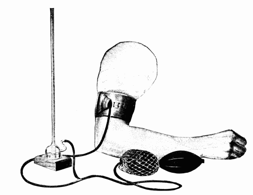 Fig. 13.—Cook's modification of Riva-Rocci's blood pressure instrument.
Stanton's instrument (Fig. 14) is practically Cook's
made more rigid in every way but without the jointed tube.
The cuff has a leather casing, the pressure bulb is of heavy
rubber, the glass tube in which the mercury rises is fixed
against a piece of flat metal and there are stopcocks in a
metal chamber introduced between the bulb and mercury
with which to regulate the in- and out-flow of air. The
pressure can be gradually lowered conveniently without removing
the pressure bulb.
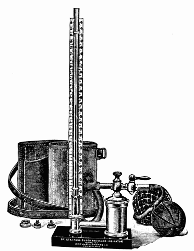 Fig. 14.—Stanton's sphygmomanometer.
The most accurate mercury manometer is that of Erlanger.
(Fig. 15.) The instrument is bulky and is not
practicable for the physician in practice. The principle is
that used by Riva-Rocci. There is an extra T-tube introduced
between the manometer and air bulb connecting with
a rubber bulb in a glass chamber. The oscillations of this
are communicated to a Marey tambour and recorded on
smoked paper revolving on a drum. There is a complicated
valve which enables the operator to reduce the pressure
with varying degrees of slowness. The mercury is
placed in a U-tube with a scale alongside it. The instrument
is expensive and not as easy to manipulate as its
advocates would have us believe. Hirschfelder has added
to the usefulness (as well as to the complexity) of the Erlanger
instrument, by placing two recording tambours for
the simultaneous registering of the carotid and venous
pulses. In spite of its complexity and necessary bulkiness,
very valuable data are obtained concerning the auricular
contractions.
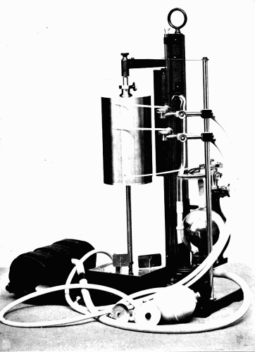 Fig. 15.—The Erlanger sphygmomanometer with the Hirschfelder attachments by
means of which simultaneous tracings can be obtained from the brachial, carotid, and
venous pulses.
Fig. 15.—The Erlanger sphygmomanometer with the Hirschfelder attachments by
means of which simultaneous tracings can be obtained from the brachial, carotid, and
venous pulses.
One of the best of the mercury instruments is the Brown
sphygmomanometer. In this (Fig. 16) the mercury is in
a closed, all-glass tube so that it can not spill under any
sort of manipulation. It is in this sense "fool-proof." The
cuff, however, is poorly constructed. It is too short and
there are strings to tie it around the arm. I have found
that this causes undue pressure in a narrow circle and renders
the reading inaccurate. In the clinic we use this mercury
instrument with a long cuff like that provided by the
Tycos instrument.
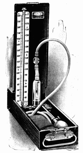 Fig. 16.—Desk model Baumanometer.
The Faught instrument (Fig. 17) is larger than the
Brown, but is less easily broken and is not too cumbersome
to carry around. The substitution of a metal air pump for
the rubber makes the apparatus more durable.
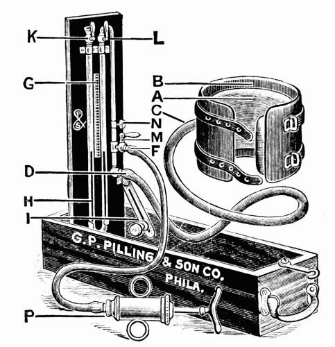 Fig. 17.—The Faught blood pressure instrument. An excellent instrument which is
quite easily carried about and is not easily broken.
Fig. 17.—The Faught blood pressure instrument. An excellent instrument which is
quite easily carried about and is not easily broken.
The v. Recklinghausen instrument is not employed to
any extent in this country. It is both expensive and cumbersome,
and has no advantages over the other instruments.
Several other instruments have been devised and new ones
are constantly being added to the already large list. With
those employing mercury the principle is the same. The
aim is to make an instrument which is easily carried, durable,
and accurate.
In all the mercury instruments the diameter of the tube
is 2 mm. One would suppose that there would be noticeable
differences in the readings of the different mercury
instruments depending upon the amount of mercury used
in the tube. By actual weight there is from 35 to 45 gms.
of mercury in the several instruments. After many trials,
no noticeable differences in blood pressure readings can be
made out between a column weighing 35 gm. and one weighing
45 gm.
There is, however, the inertia of the mercury to be overcome,
friction between the tube and the mercury, and vapor
tension. The mercury is therefore not as sensitive to rapid
changes of pressure in the cuff as a lighter fluid would be.
The mercury must be clean and the tube dry so that there
is no more friction than what is inherent between the mercury
and glass. In making readings on a rapid pulse the
oscillations of the mercury column are apt to be irregular
or to cease now and then, due to the fact that the downward
oscillation coincides with a pulse wave, or an upward oscillation
receives the impact of two pulse waves transmitted
through the cuff. Instruments have been devised to obviate
this difficulty, but they have not come into favor. They are
usually too complicated and at present can not be recommended.
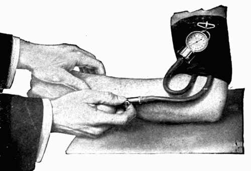 Fig. 18.—Rogers' "Tycos" dial sphygmomanometer.
An instrument devised by Dr. Rogers (the "Tycos")
has met with considerable popularity. (Fig. 18.) This is
not an instrument which operates with a spring and lever.
The instrument is composed essentially of two metal discs
carefully ground and attached at their circumferences to
the metal casing below the dial. There is an air chamber
between these discs through the center of which air is
forced by the syringe bulb. When air is forced into the
space between these two discs, they are forced apart to a
very slight extent, with the highest pressures only 2-3 mm.
of bulging occurs. From data gathered after extensive use
for five years these discs were not found to have sprung.
A lever attached to a cog which in turn is attached to the
dial needle magnifies to an enormous extent the slightest
expansion of the discs. Every dial is handmade and every
division is actually determined by using a U. S. government
mercury manometer of standard type. No two dials
therefore are alike in the spacing of the divisions of the
scale but every one is calibrated as an individual instrument.
There is no doubt in the author's mind that for the
general practitioner the instrument has some advantages
over the mercury instruments. It reveals the slightest
irregularity in force of the heart beat. The oscillation of
the dial needle is more accurately followed by the eye than
is that of the column of mercury. The needle passes directly
over the divisions of the scale, while with usual mercury
instruments the scale is an appreciable distance (sometimes
.5 cm.) from the column of mercury at the side. (Fig.
19.) The diastolic pressure is more easily read on the
"Tycos." It is where the maximum oscillation of the needle
occurs as the pressure is slowly released from the cuff.
Although it does not appear that this instrument, if properly
made and standardized, could become inaccurate,
nevertheless it is advisable to check it every few months
against a known accurate mercury manometer instrument.
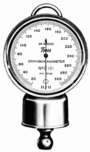 Fig. 19.—Detail of the dial in the "Tycos" instrument.
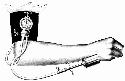 Fig. 20.—Faught dial instrument.
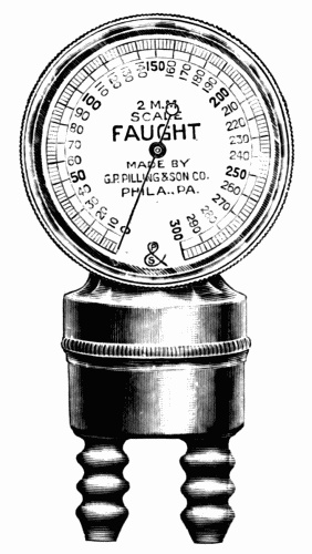 Fig. 21.—Detail of the dial of the Faught instrument.
Another perfectly satisfactory dial instrument is the
Faught (Figs. 20 and 21). The general plan of this differs
in some minor points from the "Tycos." I have compared
the two and have found no difference in the readings. Both
can be recommended.
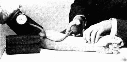 Fig. 22.—The Sanborn instrument.
One or two other cheaper dial instruments are on the
market. The Sanborn seems to be quite satisfactory. (Fig.
22.) It is cheaper than the other dial instruments. There
is this much to be said, no instrument using a spring as resistance
to measure pressure can be recommended.
The same technic applies to all the mercury instruments.
The patient sits or lies down comfortably. The right or left
arm is bared to the shoulder, the cuff is then slipped over
the hand to the upper arm. (See Fig. 23.) At least an inch
of bare arm should show between the lower end of the cuff
and the bend of the elbow. The rubber is adjusted so that
the actual pressure from the bag is against the inner side
of the arm. The straps are tightened, care being taken not
to compress the veins. The upper part of the cuff should
fit more snugly than the lower part. The part of the instrument
carrying the mercury column is now placed on a
level surface; the two arms of the mercury in the tube must
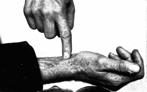
be even, and at 0 on the scale. With the fingers of one hand
on the radial pulse, the bag is compressed until the pulse
is no longer felt. (See Fig. 24.) One should raise the pressure
from 10-12 mm. above this, and close the stopcock
between the bulb and the mercury tube. In a good instrument
the column should not fall. If it does there is a leak
of air in the system of tubing and arm bag. Now with the
finger on the pulse, or where the pulse was last felt, gradually
allow air to escape by turning the stopcock so that
the column of mercury falls about 2 mm. (one division on
the scale) for every heart beat or two. One must not allow
the column of mercury to descend too slowly as it is uncomfortable
for the patient and introduces a psychic element
of annoyance which affects the blood pressure. On the
other hand, the pressure must not be released too rapidly,
else one runs over the points of systolic and diastolic pressure
and the readings are grossly inaccurate. It is impossible
to say how rapidly the mercury must fall. Every operator
must find that out for himself by practice. The first perceptible
pulse wave felt beneath the palpating finger at the
wrist, represents on the scale the systolic pressure. This
can be seen to correspond to a sudden increase in the magnitude
of the oscillation of the mercury column. The systolic
pressure, thus obtained, is from 5-10 mm. lower than the
real systolic pressure. The more sensitive the palpating
finger, the more nearly does the systolic pressure reading
approach that found by using such an instrument as Erlanger's,
where the first pulse wave is magnified by the
lever of the tambour.
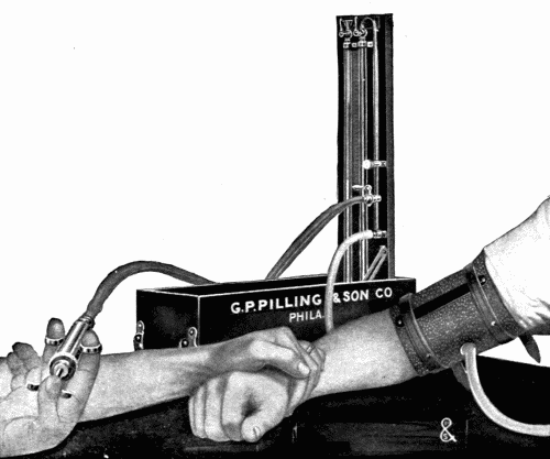 Fig. 23.—Method of taking blood pressure with a patient in sitting position.
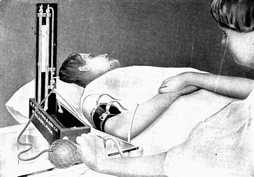 Fig. 24.—Method of taking blood pressure with patient lying down.
The pressure is now allowed to fall, until the palpating
finger feels the largest possible pulse wave, which is coincident
with the greatest oscillation of the mercury. This is
the diastolic pressure. Beyond this point there is no oscillation
of the mercury column. The difference between the
two is the pulse pressure. Thus the pulse is felt after compression
at 120 on the scale, and the maximum oscillation
occurs at 80. The systolic pressure is 120 mm., the diastolic
is 80 mm., and the pulse pressure is 40 mm.
With the "Tycos" or Faught the arm band is snugly
wound around the arm, the bag next to the skin and the end
tucked in, so that the whole band will not loosen when air
is forced into the bag. The cuff is blown up until the pulse
is no longer felt. One should raise the pressure not more
than 10 mm. above the point of obliteration of the pulse.
The valve is then carefully opened so that the needle gradually
turns toward zero. At the first return of the pulse
wave felt at the wrist, the needle is sure to give a sudden
jump. This is the systolic pressure and is read off on the
scale. The needle is now carefully watched until it shows
the maximum oscillation. This is the diastolic pressure.
The difference between the two is, as above, the pulse pressure.
In taking pressure one should take the average of several,
three or four. Moreover, one must not take consecutive
readings too quickly and one must be sure that between
every two readings all the air is out of the cuff and that the
mercury or dial is at zero. It has been repeatedly shown
that in a cyanosed arm the systolic pressure is raised so
that even slight cyanosis between readings must be carefully
avoided.
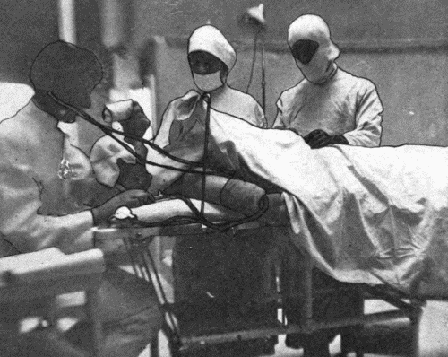
The only accurate method of determining both the systolic
and diastolic pressure, but especially the diastolic, is
by the so-called auscultatory method. (See Fig. 25.) The
cuff is adjusted in the usual way and one places the bell
of a binaural stethoscope over the brachial artery from
one to two centimeters below the lower edge of the
cuff.[3] Care must be taken that the bell is not pressed too
firmly against the arm and that the edge of the bell nearest
the cuff is not pressed more firmly than the opposite end.
For this purpose, one can not use the ordinary Bowles
stethoscope or any of the other much lauded stethoscopes,
because the surface of the bell is too large. The
diameter of the bell must not be more than twenty-five millimeters,
twenty is still better. It is advisable before beginning
the observation to locate with the finger the pulse
in the brachial artery just above the elbow, so that the
stethoscope may be placed over the course of the artery.
(Fig. 26.) The first wave which comes through is heard as
a click, and occurs at a point on the manometer or dial scale
from 5-10 mm. higher than can usually be palpated at the
radial artery. This is the true systolic pressure. By keeping
the bell of the stethoscope over the brachial artery
while the pressure is falling, one comes to a point when all
sound suddenly ceases. This is said to be the diastolic pressure.
This is incorrect as will be shown later.
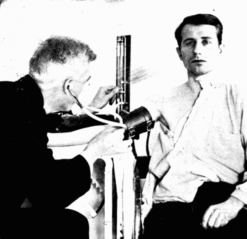 Fig. 25.—Observation by the auscultatory method and a mercury instrument. One hand
regulates the stop cock which releases air gradually.
Fig. 25.—Observation by the auscultatory method and a mercury instrument. One hand
regulates the stop cock which releases air gradually.
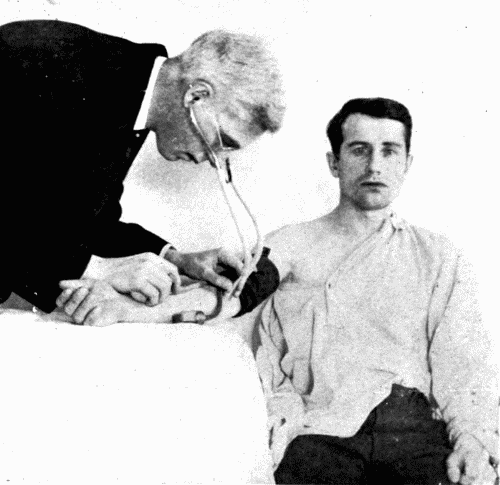 Fig. 26.—Observation by the auscultatory method and a dial instrument. The right
hand holds the bulb and regulates the air valve.
Fig. 26.—Observation by the auscultatory method and a dial instrument. The right
hand holds the bulb and regulates the air valve.
The arterial pressure in the large arteries undergoes extensive
fluctuations with every heart beat. The maximum
pressure produced by the systole of the left ventricle of the
heart is known as the maximum or systolic pressure. It
practically equals the intraventricular pressure. The minimum
pressure in the artery, the pressure at the end of diastole,
is called the diastolic pressure. The difference between
the systolic and diastolic pressures is known as the
pulse pressure. There is yet another term known as the
mean pressure. For convenience, this may be said to be the
arithmetical mean of the systolic and diastolic pressures.
Actually, however, this can not be the case, owing to the
form of the pulse wave, which is not a uniform rise and fall—the
upstroke being a straight line, but the downstroke being
broken usually by two notches. We do not make use of
the mean pressure in recording results. It is of experimental
interest and needs only to be mentioned here.
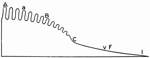 Fig. 27.—Schema to illustrate the gradual decrease in pressure from the heart to the
vena cava: (a), arteries; (c), capillaries; (v), veins; (A), aorta, pressure 150 mm.;
(B), brachial artery, pressure 130 mm.; (F), femoral vein, 20 mm.; (IVC), inferior
vena cava, 3 mm. (Modified from Howell.)
Fig. 27.—Schema to illustrate the gradual decrease in pressure from the heart to the
vena cava: (a), arteries; (c), capillaries; (v), veins; (A), aorta, pressure 150 mm.;
(B), brachial artery, pressure 130 mm.; (F), femoral vein, 20 mm.; (IVC), inferior
vena cava, 3 mm. (Modified from Howell.)
It has been shown that the mean pressure is quite constant
throughout the whole arterial system. The maximum
pressure necessarily falls as the periphery of the vascular
system is approached. In general it may be said that
the minimal pressure is quite constant. Too little attention
is paid to minimal and pulse pressure. The minimal pressure
is important, for it gives us valuable data as to the
actual propulsive force driving the blood forward to the
periphery at the end of diastole.
It is readily understood how the maximum pressure falls
as the periphery is approached, until in the arterioles the
maximum and minimum pressures are about equal. The
pressure then in these arterioles is practically the same as
the diastolic pressure. Actually it is a few millimeters
less. The diastolic blood pressure would, therefore, measure
the peripheral resistance and, as the maximum for
systolic pressure represents approximately the intraventricular
pressure, the difference between the two, the pulse
pressure, actually represents the force which is driving the
blood onward from the heart to the periphery. It is hence
very evident that the mere estimation of the systolic pressure
gives us but a portion of the information we are
seeking.
The pulse pressure is subject to wide fluctuations but as
a rule for any one normal heart it remains fairly constant
as the rate varies. In a rapidly beating heart the diastole
is short and the diastolic pressure rises. If the systolic
pressure does not also rise, as in a normal heart following
exercise, we will say, the pulse pressure falls. We know
that when the pulse rate is constant, vasodilatation causes a
fall in diastolic pressure and a rise in pulse pressure. On
the contrary, vasoconstriction causes a rise in diastolic
pressure and a fall in pulse pressure.
It is very probably the case that with two individuals of
equal age and equal pulse rate, and equal systolic pressure
of 160 mm., the one with a diastolic pressure of 110 mm.
and, therefore, a pulse pressure of 50 mm. is much worse
off than the other with a diastolic pressure of 90 mm. and a
pulse pressure of 70 mm. The latter may be normal for the
age of the person especially when certain forms of fibrous
arteriosclerosis accompanied by enlarged heart are present.
The former is not normal for any age. Low pulse pressure
usually means a weak vasomotor control and is only
found in failing circulation or in markedly run down states,
such as after serious illness or in tuberculosis. Therefore,
it is most important to estimate accurately the diastolic
pressure as well as the systolic pressure, for only in this
way can we obtain any data of value regarding the driving
power of the heart and the condition of the vasomotor system.
A high systolic pressure does not necessarily mean
that a great deal of blood is forced into the capillaries. Actually
it may mean that very little blood enters the periphery.
The heart wastes its strength in dilating constricted
vessels without actually carrying on the circulation adequately.
The systolic pressure varies considerably under conditions
which are by no means abnormal. Thus, the average
for men at all ages is about 127 mm. Hg. (All measurements
are taken from the brachial artery, with the individuals
in the sitting posture.) For women the average is
somewhat lower, 120 mm. Hg. The pressure is lowest in
children. In children from 6-12 years the average systolic
pressure is 112 mm. Normally, there is a gradual increase
as age comes on, due, as will be shown in the succeeding
chapter, to physiologic changes which take place in the
arteries from birth to old age. In the chart here appended
is graphically shown the normal variations in the blood
pressure at different ages compiled from observations made
on one thousand presumably normal persons. (Fig. 28.)
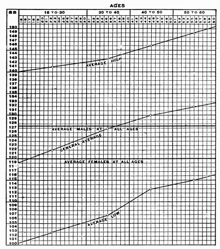 Fig. 28.—Chart showing the normal limits of variation in systolic blood pressure. (After
Woley.)
Fig. 28.—Chart showing the normal limits of variation in systolic blood pressure. (After
Woley.)
The diastolic pressure has been estimated to be about 35
to 45 mm. Hg lower than the systolic pressure, and consequently
these figures represent the pulse pressure in the
brachial artery of man. This is equivalent to saying that
every systole of the left ventricle distends this artery by a
sudden increase in pressure equal to the weight of a column
of mercury 2 mm. in diameter and 35 to 45 mm. high. Naturally,
at the heart the pressure is highest. As the blood
goes toward the capillary area the pressure gradually decreases
until, at the openings of the great veins into the
heart, the pressure is least. At the aorta (A) the pressure
(systolic) is approximately 150 mm. Hg, at the brachial
artery (B) it is 130 mm., in the capillary system (C) it is
30 mm., in the femoral vein (F) it is 20 mm., at the opening
of the inferior vena cava (I) it is 3 mm.
Attention has been called to the normal systolic pressure
at different ages. This is not the only cause for variations
in the blood pressure. Normally, it is greater when in the
erect position than when seated, and greater when seated
than when lying down. During the day there are well-recognized
changes. The pressure is lowest during the
early morning hours, when the person is asleep. In women
there are variations due to menstruation. Muscular exercise
raises the blood pressure markedly. The effect of a
full meal is to raise the blood pressure. The explanation
is that during and following a meal there is dilatation of
the abdominal vessels. This takes blood from other parts
of the body, provided that the other factors in the circulation
remain constant. A fall of pressure would necessarily
occur in the aorta. To compensate for this, there is increased
work on the part of the heart, which reveals itself
as increased pressure and pulse pressure. It is well known
that the interest in the process taken by an individual upon
whom the blood pressure is estimated for the first time tends
to increase the rate of the heart and to raise the blood pressure.
For this reason the first few readings on the instrument
must be discarded, and not until the patient looks
upon the procedure calmly can the true blood pressure be
obtained. As a corollary to this statement, mental excitement,
of whatever kind, has a marked influence on the
pressure. The patient must remain absolutely quiet. Raising the head or
the free arm causes the pressure to rise.
Another important physiologic variation is produced by
concentrated mental activity. This tends to hurry the heart
and increase the force of the beat. In short, it may be
stated as a general rule that any active functioning of a
part of the body which naturally requires a great excess of
blood tends to elevate the blood pressure. At rest the pressure
is constant. Variations caused by the factors mentioned
act only transitorily, and the pressure shortly returns
to normal.
Since the first description of the auscultatory blood pressure
sounds by Korotkov in 1905, this method has been more
and more employed until today it is the standard, recognized
method of determining the points in the blood pressure
reading. When one applies the 12 cm. arm band over
the brachial artery and listens with the bell of the stethoscope
about one cm. below the cuff directly over the brachial
artery near the bend of the elbow, one hears an interesting
series of sounds when the air in the cuff is gradually
reduced. The cuff is blown up above the maximum pressure.
As the air pressure around the arm gradually is
lowered, the series of sounds begins with a rather low-pitched,
clear, clicking sound. This is the first phase. This
only lasts through a few millimeters fall when a murmur
is added and the tone becomes louder. This click and murmur
phase is the second phase. A few millimeters more of
drop in pressure and a clear, sharp, loud tone is audible.
Usually this tone lasts through a greater drop than any of
the other tones. This is the third phase. Rather suddenly
the loud, clear tone gives place to a dull muffled tone. In
general the transition is quite sharp and distinct. This is
the fourth phase. The tone gradually or quickly ceases
until no tone is heard. This is the fifth phase (Ettinger.)
The first phase is due to the sudden expansion of the
collapsed portion of the artery below the cuff and to the
rapidity of the blood flow. This causes the first sharp
clicking sound which measures the systolic pressure.
The second, or murmur and sound phase, is due to the
whorls in the blood stream as the pressure is further released
and the part of the artery below the cuff begins to
fill with blood.
The third tone phase is due to the greater expansion of
the artery and to the lowered velocity in the artery. A
loud tone may be produced by a stiff artery and a slow
stream or by an elastic artery and a rapid stream. This
tone is clear cut and in general is louder than the first
phase.
The fourth phase is a transition from the third and becomes
duller in sound as the artery approaches the normal
size.
The fifth phase, no sound phase, occurs when the pressure
in the cuff exerts no compression on the artery and the
vessel is full throughout its length.
It is generally conceded that the sounds heard are produced
in the artery itself and not at the heart.
The tones vary greatly in different hearts. A very
strong third tone phase or prolongation of this phase usually
means that the heart which produces the tone is a
strongly acting one, although allowances must be made for
a sclerosed artery in which there is a tendency to the production
of a sharp third phase.
Weakness of the third phase, as a rule, indicates weakness
of the heart and this dulling of the third phase may be
so excessive that no sound is produced. Goodman and
Howell have carried this method further by measuring the
individual phases and calculating the percentage of each
phase to the pulse pressure. Thus, if in a normal individual
the systolic pressure is 130 mm., the diastolic 85 mm.,
and the pulse pressure 45 mm., the first phase lasts from
130 to 116 or 14 mm., the second from 116 to 96, or 20 mm.,
the third from 96 to 91 or 5 mm., the fourth from 91 to 85, or
6 mm. The first phase would then be 31.1 per cent of the
total pulse pressure, the second phase 44.4 per cent, the
third phase 11.1 per cent, and the fourth phase 13.3 per
cent. They consider that the second and third phases represent
cardiac strength (C. S.) and the first and fourth represent
cardiac weakness (C. W.). They believe that C. S.
should normally be greater than C. W. In the example
above C. S.:C. W. = 55.5:44.4. In weak hearts, especially in
uncompensated hearts, the conditions are reversed and C.
W. > C. S. This is often the case. As a heart improves
C. S. again tends to become greater than C. W. They think
that the phases should be studied in respect to the sounds
and also to the encroachment of one sound upon another.
These observations are interesting but we have not found
the division into phases as helpful as it was thought to be.
We spent a great deal of time on this question. All that
can be said, in my opinion, is that a loud, long third phase
is usually evidence of cardiac strength.
A further interesting feature which can be heard in all
irregular hearts is a great difference in intensity of the individual
sounds. Goodman and Howell call this phenomenon
tonal arrhythmia. Irregularities can be made out by
the auscultatory method which can not be heard at the
heart.
In anemia the sounds are very loud and clear and do not
seem to represent the actual strength of the heart.
The general lack of vasomotor tone in the blood vessels
together with some atrophy and flabbiness of the coats
probably explains the loud sounds.
In polycythemia the sounds have a curious, dull, sticky
character and can not be differentiated accurately into
phases, a condition which was predicted from the knowledge
of the sharp sounds in anemia.
In not all cases can all phases be made out. It is usually
the fourth phase which fails to be heard.
In such cases the loud third tone almost immediately
passes to the fifth phase or no sound phase. The importance
of this will later be taken up.
"In arteriosclerosis, with hardening and loss of elasticity
of the vessel walls, the auscultatory phenomena, according
to Krylow, are apt to be more pronounced, since the back
pressure at the cuff probably causes some dilatation of the
vessel above it, while the lumen of the vessel is smaller
than normal. Both of these factors cause an increased
rapidity in the transmission of the blood wave when pressure
in the cuff is released, which in time favors the vibration
of the vessel walls.
"In high grade thickening of the arterial walls, however,
especially where calcification had occurred, Fischer
found that the sounds were distinctly less loud than normal,
the more so in the arm, which showed the greater
degree of hardening. According to Ettinger's experience,
the rapidity of the flow distinctly increases the auscultatory
phenomenon." (Gittings.)
The sounds depend upon the resonating character of the
cuff, upon the size and accessibility of the vessel, upon the
force of the heart beat, and upon the velocity of the blood.
The maximum (systolic) pressure is read at the point
where the first audible click is heard after the cuff is blown
up and the pressure gradually reduced by means of the
needle valve in the hand bulb or on the upright of the
glass containing the mercury. All are agreed upon this
point. There has been some dispute as to the place where
the diastolic pressure should be read. Korotkov considered
that the diastolic pressure should be read at the fourth
phase when the loud tone suddenly becomes dulled. Others
held that the diastolic pressure should be read at the fifth
phase, the absence of all sound. Experiments carried out
to determine this point were made by me with the assistance
of Prof. Eyster and Dr. Meek at the Physiological
Laboratory of the University of Wisconsin. We arranged
apparatus making it possible to hold the pressure in the
carotid artery of dogs at maximum or minimum. A femoral
artery was then dissected and an instrument devised to
compress the artery with a water jacket. The whole was
connected up with a kymograph. A time marker was put
in so as to record the place where changes in sound were
heard while listening below the cuff around the femoral
artery. Two sets of records were taken. One with pressure
greater than minimum pressure and a falling pressure
over the femoral artery (Fig. 29), the other with
pressure at zero and gradually raised to minimum pressure
(Fig. 30). Both sets of records showed the same result;
viz., that at a point corresponding to the sudden
change of tone the pressure on the artery corresponded to
the minimum pressure. It was therefore concluded that
experimentally in dogs the point where diastolic pressure
should be read is at the tone change from clear to dull,
not at the point where all sound disappears.
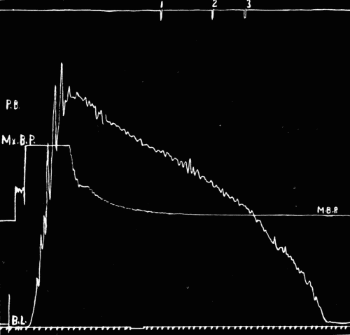 Fig. 29.—Tracing of auscultatory phenomena. (See explanation in legend of Fig. 30.)
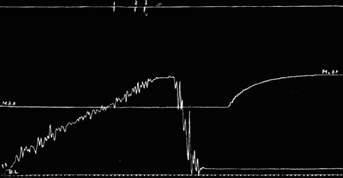 Fig. 30.—Figures are to be read from left to right. The top line records the points
where sounds were heard, the figures above the short vertical lines refer to tones (see
text). Mx. B. P., maximum blood-pressure. M. B. P., minimum blood-pressure. P. B.,
pressure bulb recorder. It was impossible to lower and raise this bulb by hand without
obtaining the great irregular oscillations of the attached lever above the mercury manometer.
B. L., base line.
Fig. 30.—Figures are to be read from left to right. The top line records the points
where sounds were heard, the figures above the short vertical lines refer to tones (see
text). Mx. B. P., maximum blood-pressure. M. B. P., minimum blood-pressure. P. B.,
pressure bulb recorder. It was impossible to lower and raise this bulb by hand without
obtaining the great irregular oscillations of the attached lever above the mercury manometer.
B. L., base line.
Erlanger showed some years ago, that with his instrument,
the point at which diastolic pressure should be read
was at the instant when the maximum oscillation of the
lever suddenly became smaller. While checking up the
graphic with the auscultatory method using Erlanger's instrument,
it was noticed that the disappearance of all sound
did not correspond with the sudden diminution of the oscillation
of the lever connected with the brachial artery. A
series of records were carefully made on patients. It was
seen that during the period of the third tone phase the oscillations
of the lever on the drum reached a maximum
(Fig. 31) and remained at approximately the same height
for some millimeters while the pressure was gradually falling.
At a point at which the third tone, clear and distinct,
became dull, there was an appreciable decrease in the height
of the pulse wave. From this point to the disappearance
of all sound there was a gradual diminution of the size of
the pulse waves.
 Fig. 31.—Fast drum. Sudden decrease in size of pulse wave at 4, marking the change
from clear sharp tone to dull tone.
Fig. 31.—Fast drum. Sudden decrease in size of pulse wave at 4, marking the change
from clear sharp tone to dull tone.
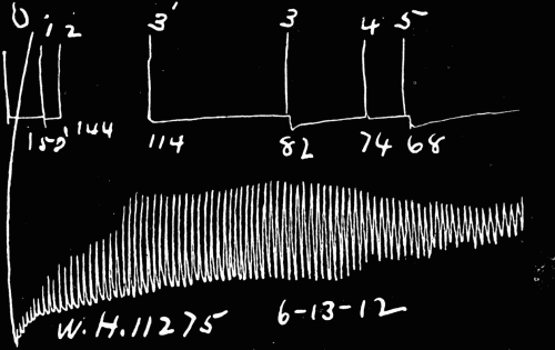 Fig. 32.—Slow drum. Sudden decrease in amplitude at 4.
For normal pressures the difference between the fourth
(dull) tone and the fifth (disappearance of all tone) phase,
amounted to 4 to 10 mm. Occasionally the difference was
so little, the change from sharp third tone through fourth
dull tone to disappearance of all sound was so abrupt, that
one could take the disappearance of all sound as the diastolic
pressure, with an error of not more than 2 to 4 mm.
This is within the limits of normal error and practically
may be used by those who have difficulty in noting the
change from third to fourth phase. For high pressures,
however, the difference between fourth and fifth phases was
never less than 8 mm., and was found as much as 16 mm.
The diastolic, therefore, should always be taken at the
fourth phase if possible.
It was found that with the dial instrument the greatest
fling of the lever corresponded to the third phase and the
sudden lessened amplitude of the oscillation was at the
fourth phase and was coincident with the change of tone
from sharp to dull. Thus the diastolic pressure may be
read off on the dial scale by watching the fling of the hand
and with some practice one might acquire considerable accuracy.
It is better, simpler, and, for most observers, more
accurate to use the stethoscope and hear the change of
sound.
The systolic pressure represents the maximum force of
the heart. It is measured by noting the first sound audible
over the brachial artery using the auscultatory method.
It is the summation of two factors largely; the force expended
in opening the aortic valves (potential) and the
force expended from that point to the end of systole, the
force which is actually driving the blood to the periphery
(kinetic). To start the blood in motion, the heart must
overcome a dead weight equal to the sum of all the forces
holding the aortic valves closed. This sum of factors,
called the peripheral resistance, must be reached and passed
by the force of the ventricular beat before one drop of
blood is set in motion along the aorta. This factor of resistance
assumes a great importance.
The systolic pressure is always fluctuating as it depends
upon so many conditions, and the calls of the body except
during sleep are many and various. In a study of diurnal
variations in arterial blood pressure it has been found that—(1)
A rise of maximum pressure averaging 8 mm. of Hg.
occurs immediately on the ingestion of food. A gradual
fall then takes place until the beginning of the next meal.
There is also a slight general rise of the maximum pressure
during the day. (2) The range of maximum pressure
varies considerably in different individuals, but the highest
and lowest maximum pressures are practically equidistant
from the average pressure of any one individual.[4]
The pressure is lowest during sleep and gradually rises
near the end of sleep, so that on awakening the pressure
was the same as before sleep.
Physiologically there are many conditions which modify
the systolic pressure. Sleep, position, meals, exercise, emotional
states cause often wide fluctuations which may be
very sudden. It should be constantly borne in mind, that
the systolic pressure reading which is made, is the maximum
effort of the heart at that moment only.
The diastolic pressure measures the peripheral resistance.
It measures the work of the heart, the potential energy, up
to the moment of the opening of the aortic valves. It is
the actual pressure in the aorta. The diastolic pressure is
not very variable; it is not subject to the same influences
which disturb the systolic pressure. It fluctuates as a rule,
within a small range. It is not affected by diet, by mental
excitement, by subconscious psychic influences, to anything
like the extent to which the systolic pressure is affected by
the action of these factors. The diastolic pressure is determined
by the tone in the arterioles and is under the control
of the vasomotor sympathetic system. Any agent
which causes chronic irritation of the whole vasomotor system
produces increase in the peripheral resistance with consequent
rise in the diastolic pressure. Any agent which
acts to produce thickening of the walls of the arterioles,
narrowing their lumina, produces the same effect.
Such states naturally result in increased work on the
part of the heart, which as a result, hypertrophies in the
left ventricle. The increase in size and strength is a compensatory
process in order to keep the tissues supplied
with their requisite quota of blood. Conversely, paralysis
of the vasomotor system produces fall of diastolic pressure
which, if long continued, results in death.
The diastolic pressure then is of importance for the following
reasons:
1. It measures peripheral resistance.
2. It is the measure of the tonus of the vasomotor system.
3. It is one of the points to determine pulse pressure.
4. Pulse pressure measures the actual driving force, the
kinetic energy of the heart.
5. It enables us to judge of the volume output, for pulse
pressure which is only determined by measuring both systolic
and diastolic pressure, is such an index.
6. It is more stable than the systolic pressure, subject to
fewer more or less unknown influences.
7. It is increased by exercise.
8. It is increased by conditions which increase peripheral
resistance.
9. The gradual increase of diastolic pressure means
harder work for the heart to supply the parts of the body
with blood.
10. Increased diastolic pressure is always accompanied
by increased pulse pressure, and increased size of the left
ventricle, temporarily (exercise) or permanently.
11. Decreased diastolic pressure goes hand in hand with
vasomotor relaxation, as in fevers, etc.
12. Low diastolic pressure is frequently pathognomonic
of aortic insufficiency.
13. When the systolic and diastolic pressures approach,
heart failure is imminent either when pressure picture is
high or low.
When all these factors are taken into consideration, it
becomes apparent that the diastolic pressure is most important,
if not the most important part of the pressure
picture.
Up to within a very brief time all the statistical evidence
of blood pressure was based on systolic readings alone.
This data is most valuable and much has been learned as
to diagnosis and prognosis, but it is a mass of data based
on a one-sided picture and can not be as valuable as the
statistics which will undoubtedly be published later when
all the pressure picture figures can be analyzed.
The pulse pressure is the actual head of pressure which
is forcing the blood to the periphery. At every systole a
certain amount of blood 75-90 c.c. (Howell) is thrown violently
into an already comfortably filled aorta. The sudden
ejection of this blood instigates a wave which rapidly
passes down the arteries as the pulse wave. The elastic
recoil of the aorta and large arteries near the heart contract
upon the blood and keep it moving during diastole.
Normally the blood-vessels are highly elastic tubes with
an almost perfect coefficient of elasticity. The pulse pressure
varies under normal conditions from 30 to 50 mm. Hg.
There is a very definite relationship between the velocity
of blood and the pulse pressure which is expressed thus;
velocity = pulse rate × pulse pressure.[5]
Further it has been demonstrated that under normal conditions
and during various procedures—the pulse pressure
is a reliable index of the systolic output.[6]
Increased pulse pressure therefore goes hand in hand
with greater systolic output. Physiologically this is most
ideally seen during exercise. Following exercise the pulse
rate increases, the systolic pressure rises greatly, the diastolic
slightly or not at all. The pulse pressure therefore is
increased. The velocity also is much increased. The call
comes for more blood and the heart responds. In the chronic
high pulse pressures there are four correlated conditions
which, so far as I have studied them, are always present.
These are: (1) An increase in size of the cavity of the left
ventricle. The ventricle actually by measurement contains
more blood than normal, and therefore throws out more
blood at every systole. The volume output is greater per
unit of time. (2) There is actual permanent increase in
diameter of the arch of the aorta. This is a compensating
process to accommodate the increased charge from the left
ventricle. (3) There are on careful auscultation over the
manubrium, particularly the lower half, breath sounds
which vary from bronchial to intensely tubular, depending
upon the anatomic placing of the aorta, the shape of the
chest, and the degree of dilatation. Often there is very
slight impairment of the percussion note as well. (4)
There is increase in size of all the large distributing arteries,
carotids, brachials, femorals, renals, celiac axis, etc.,
with fibrous changes in the media, loss of some elasticity,
and increase in size of the pulse wave. Increased pulse
pressure means increased volume output, but does not always
mean increased velocity. The proper distribution of
blood to the various organs of the body is regulated by the
vasomotor system acting upon the small arteries which contain
considerable unstriated muscle. When fibrous arteriosclerosis
is present there is loss of elasticity in the distributing
arteries and a greater volume of blood must be
thrown out by the ventricle at every systole in order that
every organ shall have its full quota of blood. A force
which is sufficient to send blood through elastic normal distributing
tubes becomes totally insufficient to send the same
amount of blood through tortuous and more or less inelastic
tubes.
It is evident then that pulse pressure is exceedingly important.
It can only be determined by measuring both the
systolic and diastolic pressure. The pulse rate must also
be known in order to compute the velocity. It is essential
to have the whole pressure picture for all cases if correct
conclusions are to be drawn.
In an irregular heart, especially in the cases due to myocardial
disease, it is quite impossible to determine the true
diastolic pressure. One can only approximate it and say
that the pulse pressure is low or high. As a matter of fact
the real systolic pressure can not be determined. For this
figure the place on the scale where most of the beats are
heard may be taken for the average systolic pressure. No
one can seriously maintain that he can measure the diastolic
pressure under all circumstances.
By means of the auscultatory method of measuring blood
pressure we are able to determine irregularities of force in
the heart beats more easily than by listening to the heart
sounds. A pulsus alternans is readily made out. The irregular
tones heard over the brachial artery in cases of irregular
heart action have been called "tonal arrhythmias."
A recent study of diurnal variations in blood pressure has
shown that while the maximum pressure rises after the ingestion
of food and steadily rises slightly throughout the
day, the minimum blood pressure is very uniform throughout
the day, and is little affected by the ingestion and digestion
of meals. When it is affected, a rise or a fall may
take place. Throughout the day, it tends to become slightly
lower. The pulse pressure then is greater towards evening.
Weysse and Lutz in a study of this question draw the following
conclusions:
1. A rise of maximum pressure averaging 8 mm. of Hg
occurs immediately on the ingestion of food. A gradual
fall then takes place until the beginning of the next meal.
There is also a slight general rise of the maximum pressure
during the day.
2. The average maximum blood pressure for healthy
young men in the neighborhood of 20 years of age is 120
mm. of Hg. This pressure obtains commonly one hour
after meals. The higher maximum pressures occur immediately
after meals, and the lower, as a rule, immediately
before meals.
3. The range of maximum pressure varies considerably in
different individuals, but the highest and lowest maximum
pressures are practically equidistant from the average pressure
of any one individual.
4. The minimum blood pressure is very uniform throughout
the day, and is little affected by the ingestion and digestion
of meals. When it is affected a rise or fall may take
place. There is a tendency for a slight general lowering
of the minimum pressure throughout the day.
5. The average minimum blood pressure for healthy
young men in the neighborhood of 20 years of age is 85 mm.
of Hg. Thus we get an average pulse pressure of 35 mm.
of Hg.
6. Pulse pressure, pulse rate, and the relative velocity
of the blood flow are increased immediately upon the ingestion
of meals. They attain the maximum, as a rule, in
half an hour, and then decline slowly until the next meal.
There is a general increase in each throughout the day.
These measurements were made upon persons at rest.
Almost any form of exercise would have made the variations
much greater. No account is taken of the psychic
variations which for the physician are the most important
to bear in mind. Neglect to take this variation into account
will inevitably lead to false conclusions.
The Average Diurnal Blood Pressure Record of the Ten Subjects
| TIME |
MAXIMUM |
MINIMUM |
MEAN |
PULSE |
PULSE |
PP x PR |
NOTES |
| |
|
|
|
PRESSURE |
RATE |
|
|
| | mm.Hg | mm.Hg | mm.Hg | mm.Hg |
| 4:30 p.m. | 119.5 | 84.1 | 101.8 | 35.4 | 72.0 | 2549 |
| 5:00 p.m. | 117.7 | 83.5 | 100.6 | 34.2 | 71.1 | 2432 |
| 6:00 p.m. | 118.0 | 84.0 | 101.0 | 34.0 | 74.9 | 2547 | Before dinner |
| 6:45 p.m. | 127.2 | 88.2 | 107.7 | 39.0 | 78.1 | 3046 | After dinner |
| 7:00 p.m. | 124.7 | 87.7 | 106.2 | 37.0 | 76.0 | 2812 |
| 7:30 p.m. | 122.0 | 83.4 | 102.7 | 38.6 | 76.0 | 2934 |
| 8:00 p.m. | 122.4 | 85.5 | 103.4 | 36.9 | 71.2 | 2527 |
| 8:30 p.m. | 120.0 | 85.0 | 102.5 | 35.0 | 69.7 | 2439 |
| 9:00 p.m. | 120.5 | 84.7 | 102.5 | 35.8 | 65.2 | 2334 |
| 9:30 p.m. | 118.2 | 84.4 | 101.6 | 33.8 | 64.4 | 2177 |
| 7:30 a.m. | 118.4 | 87.6 | 103.0 | 30.8 | 70.3 | 2165 |
| 8:00 a.m. | 116.4 | 86.4 | 101.4 | 30.0 | 69.8 | 2094 | Before breakfast |
| 8:30 a.m. | 124.2 | 85.4 | 104.8 | 38.8 | 79.4 | 3081 | After breakfast |
| 9:00 a.m. | 123.8 | 84.4 | 104.1 | 39.4 | 84.1 | 3313 |
| 10:00 a.m. | 118.2 | 83.6 | 100.9 | 34.6 | 70.7 | 2446 |
| 11:00 a.m. | 116.2 | 84.8 | 100.5 | 31.4 | 67.7 | 2126 |
| 12:00 m | 114.4 | 83.2 | 98.8 | 31.2 | 66.2 | 2065 | Before luncheon |
| 12:30 p.m. | 122.8 | 83.2 | 103.0 | 39.6 | 70.9 | 2808 | After luncheon |
| 1:00 p.m. | 122.3 | 82.0 | 102.1 | 40.3 | 79.7 | 3212 |
| 2:00 p.m. | 118.4 | 81.4 | 99.9 | 37.0 | 77.6 | 2871 |
| 3:00 p.m. | 118.8 | 82.6 | 100.7 | 36.2 | 75.1 | 2719 |
| 4:00 p.m. | 115.8 | 82.0 | 98.9 | 33.8 | 71.9 | 2420 |
| 5:00 p.m. | 117.2 | 83.4 | 100.3 | 33.8 | 69.6 | 2352 |
| 6:00 p.m. | 117.4 | 84.4 | 100.9 | 33.0 | 72.8 | 2402 | Before dinner |
| 6:45 p.m. | 124.6 | 83.1 | 103.8 | 41.5 | 80.4 | 3337 | After dinner |
| 7:00 p.m. | 125.2 | 84.2 | 104.7 | 41.0 | 76.1 | 3120 |
| 7:30 p.m. | 122.0 | 84.0 | 103.0 | 38.0 | 73.7 | 2801 |
| 8:00 p.m. | 119.6 | 85.0 | 102.3 | 34.6 | 72.3 | 2502 |
| 8:30 p.m. | 119.7 | 84.0 | 101.3 | 34.7 | 69.0 | 2394 |
| 9:00 p.m. | 120.0 | 86.2 | 103.1 | 33.8 | 68.0 | 2298 |
| Average | 120.0 | 85.0 | 102.5 | 35.0 | 72.0 | 2550 |
(Taken from Weysse and Lutz.)
In some experiments to determine the changes upon the
blood pressure induced by hot and cold applications on and
within the abdomen, Hammett, Tice and Larson found that
heat applied to the outside of the abdomen raises the blood
pressure. The application of cold produces no change. Either
hot or cold saline introduced within the abdomen
causes a fall in blood pressure.
Experimentally, certain drugs such as adrenalin, barium
chloride, nicotine, digitalis, strophanthus and the infundibular
portion of the pituitary body known as pituitrin raise
the maximum pressure. In the clinic it is difficult to conclude
always whether the drug alone is responsible for rise
in maximum pressure. Adrenalin given intravenously will
raise the pressure. So will digitalis and strophanthus. I
have watched the maximum pressure rise within three minutes
following an intravenous injection of gr. 1⁄100 (0.0006
gm.) strophanthin 20 mm. of Hg: I have seen the subcutaneous
injection of 10 minims of adrenalin repeated several
times daily for six months fail to have the least effect
on the blood pressure picture.
Elevation of the foot of the bed about nine inches proved
so efficacious in steadying failing hearts in acute infectious
diseases, particularly typhoid, that a study was made of
the effect upon blood pressure. Many observations were
made, but no instrumental proof of rise in blood pressure
could be adduced.
Exercise always raises blood pressure, the maximum
much more than the minimum. In athletes the minimum
pressure may actually fall, the maximum rise so that a
greater volume output results from the greater pulse pressure.
Shock and hemorrhage lower it. Hemorrhage lowers
also the pulse pressure, and it may be possible to prognosticate
internal hemorrhage by frequent estimations of the
systolic and diastolic pressures (Wiggers). Compression
of the superior mesenteric artery or the celiac axis in dogs
raises the blood pressure measured in the carotid artery
for a period of at least an hour. This seems to be dependent
on purely mechanical causes, and is not a reflex vasomotor
phenomenon. (Longcope and McClintock.)
Experimentally blood pressure can be increased by direct
compression of the brain as Cushing has shown. It was
thought at one time that in man the same effect would result
from tumor of the brain or especially from subdural
or extradural hemorrhage following head injuries. This,
however, is not the case. No information of great value
can be obtained by the measurement of blood pressure in
these states. We do know that too high and too prolonged
compression of the medulla brings about exhaustion of the
cardiac center accompanied with rapid pulse, low pressure
and eventual death.
All the conflict during the past few years over the subject
of blood pressure has revolved around this much overworked
word. Hypertension means high pressure, and yet
it carries with it a suggestion of high pressure which is
harmful to the individual. As a matter of fact hypertension
is a compensatory process, it is often a saving process
in spite of the fact that it carries possibilities of harm in
its possessor. It has been made a fetish, a god to fall down
before and worship and it has been the means of holding a
torch of fear over a patient which has not been lost on the
charlatans. Popularization of blood pressure has brought
its crop of evils, no one of which has been as fruitful in
dollars to unprincipled quacks as hypertension.
Hypertension is the expression on the part of the circulation
to meet new conditions in the tissues so that all tissues
will be nourished and all will be enabled to function.
Looked at from that point of view it is a conservative process
and in many cases it is. It is not an average normal
state, but it is normal state for the man who has it in
chronic form. Hypertension should be viewed rationally
and its proper place in the whole make-up of the patient determined.
Hypertension is a relative term. What might
be high pressure in a man of sedentary habits who reaches
the age of fifty, might not be high pressure in a full blooded
formerly athletic man of the same age. Temporary hypertension
due to excitement, exercise, etc., must be kept in
mind. It is not intended to convey the impression that
hypertension is of no moment. It is a matter for investigation,
but not a matter to worship as the all-in-all.
Hypertension is, after all, a physiologic response on the
part of the organism in order to maintain the circulation in
equilibrium in the face of conditions which tend to produce
vasoconstriction in large areas and, therefore tend to deprive
these areas of blood. That there must be some substance
in the blood stream which causes this constriction
seems certain. What it is, is not at present known. Recently,
Voegtlin and Macht[7] have isolated a crystalline substance
from the blood of man and other mammals which
they regard as a lipoid and closely related to cholesterin.
This substance was recovered by them from the cortex of
the adrenal gland. This becomes of added interest in the
light of observations made by Gubar (quoted by Voegtlin
and Macht). He noted "that the vasoconstricting properties
of blood serum vary in different pathologic conditions,
being increased in nephritis, for instance, and diminished
in others." In some experiments made in the summer of
1913, we found there was no marked difference in the anaphylactic
shock produced in half-grown rabbits by the injection
of normal and uremic blood serum. As lipoids do
not cause anaphylaxis, there should be no difference in the
reaction of normal and uremic sera unless in one there was
some form of protein not in the other. This does not seem
to be the case. The presence of something in the circulation,
therefore, produces constriction of vessels. This calls
for more force in contraction on the part of the heart. This
substance may be of lipoid nature. The continued presence
of this hypothetical substance naturally would lead to hypertrophy
of the heart.
What makes hypertension of significance is not the hypertension
itself, but the fact that it is the expression of
processes going on in the body which demand exhaustive
investigation. To attach a blood pressure cuff to the arm,
find the pressure, and diagnose hypertension is like putting
a thermometer under the tongue, noting a rise in the mercury,
and diagnosing fever. What causes the hypertension?
Can the causes be removed? Those are the really
vital questions after the symptom hypertension has been
discovered.
All states of hypertension are accompanied by more or
less increase of pulse pressure. In other words the systolic
pressure is always increased to greater degree than the diastolic
pressure. In studies carried out in the wards and
Pathological Laboratory of the Milwaukee County Hospital,
Milwaukee, we found that in all of the cases of chronic
high blood pressure with resulting high pulse pressure four
correlated factors were found. If any one of these factors
is present, the other three are found.
1. In all high pulse pressure cases there is increase in the
size of the cavity of the left ventricle. The ventricle actually
contains more blood when it is full, and throws out,
therefore, more blood at each systole. The actual volume
output is greater per unit of time. Such hearts always
show increase in thickness of the ventricular wall. I quite
agree with Stone,[8] who says, "It is merely to be emphasized
that when the pulse pressure persistently equals the
diastolic pressure (high pressure pulse, in other words)
with a resulting 50 per cent, overload, which means the expenditure
of double the normal amount of kinetic energy on
the part of the heart muscle, cardiac hypertrophy has occurred."
They are found in aortic insufficiency, in chronic
nephritis, in the diffuse fibrous type of arteriosclerosis, and
in some cases of exophthalmic goiter. Such a condition
occurs temporarily after exercise.
2. In all high pulse pressure cases there is actual permanent
increase in diameter of the arch of the aorta. This
is a compensating process to accommodate the increased
charge from the left ventricle. Smith and Kilgore[9] have
shown this to be true in cases of chronic nephritis with hypertension.
Their research confirms my own observations.
They found dilatation of the arch in (1) syphilis (that is,
aortitis); (2) age over 50 (that is, probable factor of arteriosclerosis);
(3) other serious cardiac enlargement, and
(4) hypertension (with more or less hypertrophy, as in
chronic nephritis).
In ten cases showing arches at the upper limit of normal
(that is, 6 cm. in diameter) and hypertrophy of the heart,
three were chronic mitral endocarditis; one was chronic
aortic endocarditis; three were chronic mitral and aortic
endocarditis, and there was one each of hyperthyroidism,
pericarditis and adherent pericardium.
In fourteen cases of hypertension (highest systolic 270
mm., average systolic, 215 mm.), all showed cardiac hypertrophy.
"All but three of these cases had great vessels
whose transverse diameters measured over the normal
limit of 6 cm., and in one of those measuring 6 cm. the
Roentgen-ray diagnosis was 'slight dilatation' of the
arch." Smith and Kilgore are at a loss to explain the three
exceptions. They did not give diastolic pressures, so pulse
pressures are not known. Possibly the three exceptions
were cases of high diastolic pressure in which the pulse
pressure possible was not over 60 mm. Such cases might
show "slight dilatation of the arch," but not marked dilatation,
such as was found in the other, evidently high pulse
pressure cases.
We have found that only the high pulse pressure cases
show dilatation of the arch. Certain high tension cases
which have had a very high diastolic pressure do not reveal
any accurately measurable dilatation of the aortic
arch. An empty aorta after death is quite different from a
functionating aorta during life. Hence the dilatation
which is found postmortem must have been considerable
during life. And conversely, a dilatation which was present
during life might not be looked on as such after death.
3. In all high pulse pressure cases one will find on careful
auscultation over the manubrium, particularly its lower
half, breath sounds which vary from bronchial to intensely
tubular. At times the percussion note will be slightly impaired,
as McCrae[10] has shown in dilatation of the arch of
the aorta. This auscultatory sign is evidence of some more
or less solid body in the anterior mediastinum which is
lying on the trachea and permits the normal tubular breathing
in the trachea to be audible over the upper part of the
sternum. It is found in cases of dilated aortic arch. Fluoroscopic
examination has confirmed the findings on auscultation.
4. In all high pulse pressure cases, in which the pulse
pressure is over 70 mm. of mercury, there is increase in
the size of all large distributing arteries, carotids, brachials,
femorals, renals, celiac axis, etc., with fibrous changes in
the media, loss of some of the elasticity, and in the palpable
superficial arteries, increase in size of the pulse wave.
Increased pulse pressure means increased volume output,
but does not always mean increased velocity. The proper
distribution of blood to the various organs of the body is
regulated by the vasomotor system acting on the small arteries
which contain considerable unstriated muscle. In
order that there may be enough blood at all times and under
varying conditions of rest and function, there must be a
proper supply coming through the distributing vessels, the
large arteries, those containing much elastic tissue, and
only a very small amount of unstriated muscle tissue or
none whatever. Fibrous sclerosis of these vessels causes
them to become enlarged and tortuous and to lose much
of their elasticity, which is essential for the even distribution
of blood. A greater blood volume is therefore necessary
in order that the organs may receive their quota of
blood. A force which is sufficient to send blood through
elastic normal distributing tubes becomes totally insufficient
to send the same amount of blood through tortuous and
more or less inelastic tubes. As a compensatory process
the pulse pressure increases. For this to increase, the left
ventricular cavity dilates, the arch dilates, and as a greater
force must be exerted to keep the increased mass in motion,
the heart responds by hypertrophy of its left ventricle and
becomes itself the subject of fibrous changes in the myocardium.
The mass movement of blood is therefore greater
in high pulse pressure cases than in cases of normal pulse
pressure.
In cases of chronic interstitial nephritis—contracted
granular kidney—it may well be that the sclerosis of the
arteries is a secondary process caused, as Adami thinks,
by the hypertension itself. In aortic insufficiency the situation
is somewhat different. The high pulse pressure is due
to a very low diastolic pressure, for in my experience with
uncomplicated aortic insufficiency the systolic pressure is,
as a rule, not much increased above the normal for the individual's
age. Here peripheral resistance is so low that
a capillary pulse is common. The volume output per unit
of time is greatly increased, the arch of the aorta is dilated,
and the pulse is large. The fact that a large part of the
blood regurgitates during diastole back into the ventricle,
and the fact that the diastolic pressure is low means that
there is no increased resistance to overcome, and the systolic
pressure is not raised.
Stone[11] has divided the cases of hypertension into the cerebral
and cardiac types. He finds that there is a difference
in prognosis and in the mode of death in the two groups.
He has further attempted to judge of the work placed upon
the heart by calculating what he calls the heart load or pressure-ratio.
For example, he takes a normal pressure at
120-80-40. The relation between 80 and 40 is ½ or 50 per
cent. That he considers normal. When the heart load increases
so that the pulse pressure equals or exceeds the
diastolic pressure, the heart load is 100 per cent or more,
he considers the danger of myocardial exhaustion graver
than when the heart load is normal or less than 50 per cent.
It is his opinion, in which I heartily concur, "that an
individual with a systolic pressure of 200 and a diastolic
pressure of 140, is in greater danger of cerebral death than
an individual with a systolic pressure of 200 and a diastolic
pressure of 100." He is "likewise certain that the individual
with a systolic pressure of 200 and a diastolic of
90 to 100 is in greater danger of a cardiac death. It is
apparently the constant high diastolic pressure rather than
the intermittently high systolic pressure which predisposes
to cerebral accident."
I have not been able to confirm all of Stone's conclusions.
His contention holds good for some cases, but not, in my
experience, for the great majority of the hypertension cases.
I feel that in the classification of the chronic high pressure
case we can go one step farther and split his first group
into two usually differentiable groups. Syphilis is not an
etiological factor in any of these groups. It is not considered
that these groups are absolutely distinct and can always
be rigidly separated. There are variations and combinations
which render an exact separation impossible.
But bearing this in mind the following classification is proposed
as a working classification.
Group A. Chronic nephritis.
Group B. Essential hypertension.
Group C. Arteriosclerotic hypertension.
Group A. Chronic Nephritis. These are the cases with
a high-pressure picture, that is to say, high systolic (200+)
and high diastolic (120-140+). The pulse pressure is much
increased. The palpable arteries are hard and fibrous.
There is puffiness of the under eyelids, which is more pronounced
in the morning on arising. Polyuria with low
specific gravity and nycturia are present. There are almost
constant traces of albumin in the urine, with hyaline and
finely granular casts.
Functionally these kidneys are much under normal. The
functional capacity determined by Mosenthal's modification
of the Schlayer-Hedinger method shows a marked inability
to concentrate salts and nitrogen. The phthalein output
is below normal. As the case advances the phthalein output
becomes less and less, until a period is reached when there
are only traces or complete suppression at the end of a two-hour
period. Such patients may live for ten weeks (one of
our cases) or longer, all the time showing mild uremic
symptoms, and suddenly pass into coma and die.
The natural end of patients in this group is either uremia
or cardiac decompensation (so-called cardiorenal disease).
Cerebral accidents may happen to a small number. It is
only to this group, in my opinion, that the term cardiorenal
disease should be applied. Formerly I believed that all high
systolic pressure cases were cases of chronic nephritis of
some definite degree. From the purely pathologic standpoint
that is true, but from the important, functional standpoint
it is far from being the true state of the cases.
In this group there is marked hypertrophy and moderate
dilatation of the left ventricle with dilatation and nodular
sclerosis of the aorta. The kidneys are firm, red, small,
coarsely granular, the cortex much reduced, the capsule
adherent. Cysts are common. It is the familiar primary
contracted kidney. Mallory calls this capsular-glomerulonephritis.
The etiology is obscure. Often no cause can be
found. Again, there is a history of some kidney involvement
following one of the acute infectious diseases, or it
may follow the nephritis of pregnancy. Usually, however,
these cases fall into the group of secondary contracted kidneys,
chronic parenchymatous nephritis.
Illustrative Case.—R. Z., a woman, aged thirty-six years, was seen July 26,
1916, in coma. There was a history of typhoid fever at nineteen years, but no
other disease. She had had nine full-term pregnancies, the last one thirteen
months previously. For a week before the onset of the present illness she had
complained of severe headaches and dizziness. There were no heart symptoms.
For the past year she has had nycturia. Physical examination revealed tubular
breathing beneath the manubrium, a few rales in the chest, an enlarged heart
(left side), with a systolic murmur over the aortic area. Blood pressure was
178-125-53, the pulse rate 96, leucocytes 27,250. Venesection of 500 c.c. of
blood and intravenous injections of 500 c.c. of 5 per cent NaHCO3 in normal
saline were employed. Lumbar puncture withdrew 60 c.c. of clear fluid under
pressure with 6 cells per cubic millimeter. The eye grounds showed distinct
haziness of the disks and dilatation of the veins. Blood pressure after venesection
was 164-122-42, pulse 76, but in a few days rose to 222-142-80, pulse 70.
A second venesection of 400 c.c. and proctoclysis of 1000 c.c. saline solution
was tried. The blood-pressure now was 198-140-58. The pH of the blood was
7.6, the alkaline reserve was 35 volume per cent (van Slyke), and the CO2
tension of the alveolar air (Marriott) was 25 mm. The phthalein on the day
following the second venesection was 45 per cent in two hours. The urine
at first showed 500 c.c. in twenty-four hours, specific gravity 1016, albumin
and casts. Later she passed 1300 to 1600 c.c. with specific gravity around
1010. The blood-pressure fluctuated considerably, reaching as low as 138-98-40,
pulse 88. She was discharged improved September 10, 1916. She had
constant headache but managed to keep up. In June, 1917, she suddenly
died in an uremic coma.
Group B. This one might designate as the hereditary
type, although there is not always a history in the antecedent.
This group includes the robust, florid, exuberantly
healthy people. They often are heard to boast that they
have never had a doctor in their lives. They are usually
thick-set or very large, fleshy people. The pressure picture
is exceedingly high. The pulse pressure is moderately
increased. The arteries are rather large, fibrous, and often
quite tortuous, although this is not always the case. Some
persons have hard, small, fibrous arteries. There is no
puffiness beneath the eyes, no polyuria, and no nycturia as
a rule. The urine is of normal amount, color, and specific
gravity. Albumin is only rarely found and then in traces,
but careful search of a centrifuged specimen invariably reveals
a few hyaline casts. The phthalein excretion is normal
or only slightly reduced. The kidneys excrete salt and
nitrogen normally. It is in this group that apoplexy is
found most frequently. The rupture of the vessel occurs
when the victim is in perfect health, often without any
warning. Occasionally when such a case recovers sufficiently
to be around, cardiac decompensation sets in later
and he dies then of the cardiac complications.
Pathologically the hearts of such persons are found to
have the most enormous hypertrophy of the wall of the left
ventricle. The cavity is somewhat enlarged, as is always
the case when the pulse-pressure is increased, but the size
of the cavity is not the striking feature. The aorta is
fibrous, thick walled, and the arch is slightly dilated. There
are patches of arteriosclerosis. One such case seen only
at autopsy had a rupture of the aorta just above the sinus
of Valsalva and died of hemopericardium. The kidneys
are of normal size, dark red, firm, the capsule strips readily,
the surface is smooth or finely granular, the cortex is not
decreased. The pyramids are congested and red streaks extend
into the cortex. Microscopically the capsules of the
glomeruli are a trifle thickened; a few show hyaline
changes. There is rather diffuse, mild, round-cell infiltration
between the tubules. The tubular epithelium shows little
or no demonstrable changes. The arterioles are generally
the seat of a moderate thickening of the intima and media,
but it is not usual to find obliterating endarteritis.
There is evidently a diffuse fibrous change which has not
affected either the tubules or glomeruli to any great extent.
Illustrative Case.—L. C., a man, aged fifty-six years, stonemason by trade,
is a stocky, thick-necked individual. He had never been ill in his life until
a year ago, when he fell from his chair unconscious. He had a right-sided
hemiplegia which has cleared up so completely that except for a very slight
drag to his foot he walks perfectly well. He came in complaining of shortness
of breath and cough. There was no swelling of the feet. Here evidently
was left-heart decompensation. Examination showed the blood pressure
to be 240-130-110, pulse irregular, 104 to the minute. There were cyanosis
and rales throughout both chests. The urine was normal in color, specific
gravity 1025, small amount of albumin, few casts, hyaline and granular.
The phthalein elimination was 65 per cent in two hours. Under rest, purgatives,
and digitalis he was much improved. He has since had two other
apoplectic strokes, the last of which was fatal.
When these patients are seen with acute cardiac decompensation,
there are, of course, much albumin and many
casts in the urine, and the phthalein output is, for the time
being, decreased.
Group C. This might be called the arteriosclerotic high-tension
group (Stone's cardiac group). The cases are usually
over fifty years old. They are men and women who
have lived high and thought hard. Often they have had
periods of great mental strain. Many men in this group
were athletes in their young manhood. Many have been
fairly heavy drinkers, although never drinking to excess.
They are usually well nourished and inclined to stoutness.
The pressure picture is high systolic with normal or only
slightly increased diastolic and large pulse pressure. The
arteries are large, full, fibrous, usually tortuous. The heart
is very large, the apex far down and out. There is no polyuria;
nycturia is uncommon, quite the exception. The urine
is normal in color, amount, and specific gravity. Albumin
is only rarely found and hyaline casts are not invariably
present. The phthalein excretion is quite normal and the
excretions of salt and nitrogen are also normal. The terminal
condition in most of the patients in this group is cardiac
decompensation. They may have several attacks from
which they recover, but after every attack the succeeding
one is produced by less exertion than the preceding one, and
it becomes more and more difficult to control attacks.
Eventually the patients become bed- or chair-ridden, and
finally die of acute dilatation of the heart.
Occasionally patients in this group may have a cerebral
attack, but in my experience this is uncommon. Pathologically
the heart is large, at times true cor bovinum, dilated
and hypertrophied. The cavity of the left ventricle is much
dilated. The aorta is dilated and sclerosed.
The kidneys are increased in size, are firm, dark red
in color, with fatty streaks in the cortex. The capsule strips
readily and the cortex is normal in thickness or only
slightly increased. The organ offers some resistance to the
knife. The microscope shows small areas scattered
throughout where the glomeruli are hyalinized, the stroma
full of small round cells, the tubules dilated, and the cells
are almost bare of protoplasm. Naturally the tubules are
full of granular cast material. Also the arterioles show
extensive intimal thickening, fibrous in character, with occasional
obliterating endarteritis. One gets the impression
that the small sclerotic lesions are the result of anemia and
gradual replacement of scattered glomeruli by fibrous tissue.
For the most part the kidney, except for the chronic
passive congestion, appears quite normal. One can readily
understand that in such a kidney function could not have
been much interfered with.
Illustrative Case.—C. K., an active, stout, business man, aged fifty-six
years, consulted me on account of shortness of breath and swelling of the feet
in May, 1915. He had just returned from a hospital in another city, where
he had gone with what was apparently cardiac decompensation. In his early
manhood he had been a gymnast and a prize winner. He has worked hard,
often given way to violent paroxysms of temper, has eaten heavily but drunk
very moderately. The heart was greatly enlarged, the arch of the aorta
dilated, a mitral murmur was audible at the apex. The radials and temporals
were large, tortuous, and fibrous. The blood pressure picture ranged around
180-90-90. He was easily made dyspneic and had a tendency to swelling of
the lower legs. The urine was acid, of normal specific gravity, normal in
amount, normal phthalein, normal concentration of salt and nitrogen, contained
albumin only when he was suffering from decompensation of the heart.
Casts were always found. He finally died, after sixteen months, with all
the symptoms of chronic myocardial insufficiency. The heart was enormous,
a true cor bovinum. The kidneys were typical of this condition, possibly
somewhat larger than usual.
When the pressure is constantly below the normal, it is
called hypotension. This may be transient—as in fainting—it
may be a normal state of the individual, it occurs in
most fevers and in a great variety of diseases, including
anemias.
In arteriosclerosis, especially the diffuse (senile) type,
the blood pressure is invariably low, and may be spoken of
as hypotension. The heart in such a case is small, the
muscle is flabby, there is brown atrophy of the fibers, and
some replacement of the muscle cells by connective tissue.
The same causes which have produced general arteriosclerosis
have also produced sclerosis of the coronary arteries,
and probably the lessened blood supply accounts for much
of the atrophy of the heart muscle.
In typhoid fever the maximum blood pressure during
beginning convalescence may be as low as 65 mm. Hg. I
have frequently seen hypotension of 80 mm. This is common.
Meningitis is the only acute infectious disease in which
the blood pressure is more often high than low. This is
accounted for by the increased intracranial tension.
Following large hemorrhages the blood pressure is reduced.
In venesection the withdrawal of blood may not
affect the blood pressure. The procedure is done to relieve
overdistension of the heart.
In pleurisy with effusion and in pericarditis with effusion
there is hypotension.
Collapse, whether from poisoning by drugs or as the result
of dysentery, cholera, or profuse vomiting from whatever
cause, reduces the blood pressure.
In cachectic states, such as cancer, the blood pressure is
low. General wasting of the whole musculature includes
that of the heart and the heart muscle shows the condition
known as "brown atrophy."
A most interesting and important condition in which
hypotension occurs is pulmonary tuberculosis. Haven
Emerson has recently gone over the whole subject in a careful
piece of work and his summary is as follows:
"Hypotension or subnormal blood pressure is universally
found in advanced pulmonary tuberculosis, in which
condition emaciation may play a part in its causation.
Hypotension is found in almost all cases of moderately advanced
tuberculosis, or in early cases in which the toxemia
is marked except when arteriosclerosis, the so-called arthritic
or gouty diathesis, chronic nephritis, or diabetes
complicate the tuberculosis and bring about a normal pressure
or a hypertension. Occasionally the period just preceding
a hemoptysis or during a hemoptysis may show hypertension
in a patient whose usual condition is that of
hypotension.
"Hypotension has been found by so many observers in
early, doubtful or suspected cases with or before physical
signs of the disease in the lungs, and is considered by competent
clinicians so useful a differential sign between various
conditions and tuberculosis, that it should be sought for
as carefully as it is the custom at present to search for
pulmonary signs.
"Hypotension when found persistently in individuals or
families or classes living under certain unhygienic conditions
should put us on our guard against at least a predisposition
to tuberculosis. Most unhygienic conditions,
overwork, undernourishment and insufficient air, are of
themselves causes of a diminished resistance, and it seems
likely that a failure of normal cardiovascular response to
exercise or change of position may be found to indicate this
stage of susceptibility, especially to tuberculous infection.
"... Hypotension, when it is present in tuberculosis, increases
with an extension of the process. Recovery from
hypotension accompanies arrest or improvement. Return
to normal pressure is commonly found in those who are
cured. Continuation of hypotension seems never to accompany
improvement. Prognosis can as safely be based on
the alteration in the blood pressure as on changes in the
pulse or temperature...."
There are a few drugs which lower the blood pressure,
but, as a rule, their effects are more or less transitory. We
know of no drug, unless it be iodide of potassium, which has
the property of causing changes in the blood (decrease in
viscosity?), which tends to reduce the blood pressure when
it is excessive. This drug fails us many times.
SOME DRUGS WHICH INFLUENCE THE BLOOD PRESSURE
Pressure Raisers
Adrenalin, when injected directly
into a vein or deep into the muscles.
The action is transitory.
Caffeine, preferably in the form
of caffeine-sodium-benzoate. A good
drug.
Strychnine, which does not act directly
but seemingly through the
higher centers.
Ergot, somewhat uncertain.
Nicotine, not used therapeutically.
Camphor, used in sterile olive oil
and injected deeply into the muscles.
Digitalis, when the cardiac tone is
low and decompensation is present.
Its action is prolonged but slow. Injections
of the infundibular portion
of the pituitary body. Not in use
clinically.
Pressure Depressors
Nitroglycerine and amyl nitrite,
action transitory but rapid.
Sodium nitrite and erythrol tetranitrate.
Action somewhat more prolonged.
Aconite, veratrum viride, chloral,
etc. These depress the heart.
Purgatives, drastic and hydragogue.
Potassium and sodium iodide may
lower blood pressure. When they do,
the action is prolonged.
Diuretin and theocin-sodium-acetate.
Comparatively little work has been done upon the determination
of the pressure in the veins in man. It is conceivable
that this procedure may, at times, be of great
value. A number of attempts have been made to measure
the venous pressure by compressing the arm veins and noting
on a manometer the force necessary to obliterate the
vein. As the pressure is so slight, water is used instead
of mercury, and readings have been given in centimeters
of water.
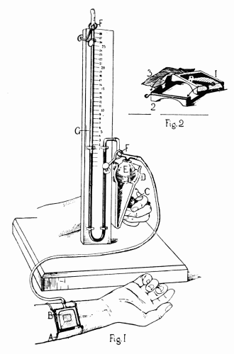 Fig. 33.—Apparatus for estimating the venous blood pressure in man, devised by
Drs. Hooker and Eyster. The small figure is the detail of the box B. See explanation
in text.
Fig. 33.—Apparatus for estimating the venous blood pressure in man, devised by
Drs. Hooker and Eyster. The small figure is the detail of the box B. See explanation
in text.
In the apparatus shown in the figure (Fig. 33), Drs.
Hooker and Eyster succeeded in making estimations of the
venous pressure. The box B is held in position by the tapes
A, so that the vein is visible through the rectangular opening
in the thin rubber covering the bottom. The box is connected
with the water manometer G, by a rubber tube,
from which a T-tube enters the rubber bulb E. When the
bulb E is compressed between the plates D, by the coarse
thumbscrew C, air is forced into the box B, exerting a pressure
on the vein lying exposed beneath. This pressure is
transmitted directly to the manometer G, and may be read
off in centimeters of water on the accompanying scale. The
veins of the back of the hand are used and there must be no
obstruction between them and the heart. The rubber-covered
box is accurately and lightly fitted over a vein and
pressure made until it is obliterated. By measuring the
distance above or below the heart level that the hand was
when the observation was made, and subtracting or adding
these figures to the manometer reading, we obtain the
venous pressure at the heart level.
Eyster has modified this instrument so that it is now
much simpler to operate. He uses a small glass cup with a
flaring edge and a diameter of about 2 cm. This is sealed
to the skin directly over a vein on the back of the hand by
means of collodion. The stem of the cup has a rubber tube
leading to a small hand bulb and to the manometer tube
which contains colored water. Slight compression of the
hand bulb obliterates the vein which can be seen through
the glass cup. The pressure in centimeters of water is then
read off. (Fig. 34.) The principle is the same as in the
earlier instrument, but the application is easier.
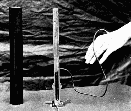 Fig. 34.—New venous pressure instrument. (After Eyster.)
Practically Hooker and Eyster found that the normal
variation in healthy subjects was from 3 to 10 cm. of water.
The pressure rose in cases of decompensated hearts with
dyspnea and venous stasis, and returned to normal with
improvement in the condition of the patient. It might be
possible with this instrument to foretell an oncoming decompensation
by the rise in venous pressure.
The venous pressure may also be estimated roughly by
slowly elevating the arm and noting the instant at which
a particular vein collapses. By measuring the height of
the vein above the heart some idea may be obtained of the
pressure within the right auricle.
There is nothing characteristic about the pulse of a
person suffering from arteriosclerosis, except it be the difference
in the pulse of high tension and of low tension.
The pulse of high tension has a gradual rise, a more or less
rounded apex, and the dicrotic wave is slightly marked and
occurs about half-way down on the descending limb. In
arteriosclerosis with low tension the radial artery is usually
so rigid that very little pulse wave can be obtained. The
general form of a low tension pulse is a sharp upstroke, a
pointed summit, and a secondary wave on the base line,
which corresponds to the dicrotic wave. Such a pulse can
be easily palpated, and is known as a dicrotic pulse. However,
such a pulse can occur only when the artery still retains
all or a large part of its elasticity; hence in arteriosclerotic
low tension we would never see such a pulse as
the typical dicrotic.
It would carry us too far to discuss fully the character
of the venous pulse, but a brief summary of the essential
features of the normal venous pulse is presented. The
venous pulse is a term used to express the tracing obtained
from the internal or external jugular vein at the root of
the neck. Normally a very characteristic curve is produced,
which can be readily analyzed into a series of waves corresponding
to the fluctuations in the cardiac cycle. To understand
these waves and their values, the accompanying
figure is helpful. (Fig. 35.)
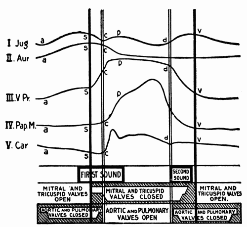 Fig. 35.—Semidiagrammatic representation of the events in the cardiac cycle: Jug.,
pulse in the jugular vein; Aur., contraction of auricle; V. Pr., intraventricular pressure;
Pap. M., contraction of the papillary muscles; Car., carotid pulse. Below are
given the times of occurrence of the heart sounds and of the opening and closing of
the heart valves. (After Hirschfelder.)
Fig. 35.—Semidiagrammatic representation of the events in the cardiac cycle: Jug.,
pulse in the jugular vein; Aur., contraction of auricle; V. Pr., intraventricular pressure;
Pap. M., contraction of the papillary muscles; Car., carotid pulse. Below are
given the times of occurrence of the heart sounds and of the opening and closing of
the heart valves. (After Hirschfelder.)
Bachmann summarizes the normal waves in the venous
pulse tracing as follows:
"The physiological or so-called venous pulse consists of
three positive and three negative waves, bearing a more or
less definite relation to the events of the cardiac cycle, and
having their origin in the various movements of the chambers
and structures of the right heart. The first positive
wave (a) is presystolic in time, and is due to the contraction
of the auricle, causing a slowing of the venous current
and producing a centrifugal wave through a sudden arrest
of the inflowing blood. The second positive wave (S) is
presystolic in time, and originates in the sudden projection
of the tricuspid valve into the cavity of the auricle during
the quick, incipient rise in the intraventricular pressure
occurring in the protosystolic period. The third positive
wave (v) occurs toward the end of ventricular systole. It
consists of two lesser waves separated by a shallow notch.
The factors entering into its formation are the relaxation
of the papillary muscle at a time when the intraventricular
is still higher than the intraauricular pressure, resulting
in an upward movement of the tricuspid leaflets and a return
of the auriculoventricular septum to its position of
rest.
"The first negative wave (between positive wave a and
S) is due to the relaxing auricle. The second negative
wave (Af) occurs during the diastole of the auricle. It
is due to the dilatation of its walls, to the displacement of
the auriculoventricular septum toward the apex occurring
at the time of ventricular systole, and to the pull of the
papillary muscles on the tricuspid valve leaflets. The third
negative wave (Vf) appears during ventricular diastole and
in the common pause of the heart chambers. Its cause is
found in the passage of the blood from the auricle into the
ventricle. It is somewhat modified possibly by the continual
ascent of the auriculoventricular septum and by a wave
of stasis due to the accumulation of blood coming from the
periphery." (Fig. 36.)
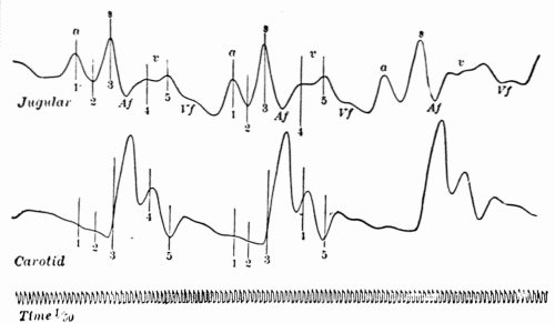 Fig. 36.—Simultaneous tracings of the jugular and carotid pulses showing normal waves
in the venous pulse and relation to carotid pulse. (After Bachmann.)
Fig. 36.—Simultaneous tracings of the jugular and carotid pulses showing normal waves
in the venous pulse and relation to carotid pulse. (After Bachmann.)
Hirschfelder has described another wave which he calls
the "h" wave, which is due to the floating up of the tricuspid
valve by the blood in the ventricle before the complete
filling of the ventricle following the auricular systole. (Fig.
37.)
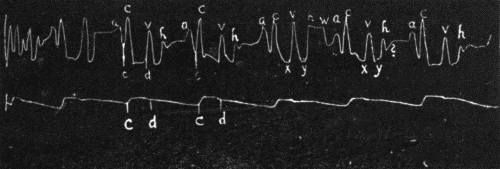 Fig. 37.—Jugular and carotid tracing from a normal individual with a well-marked
third heart sound showing a large "h" and a smaller pre-auricular wave "w." ? indicates
a small wave in mid-diastole following the "h" wave, occasionally found though
perhaps an artefact. (After Hirschfelder.)
Fig. 37.—Jugular and carotid tracing from a normal individual with a well-marked
third heart sound showing a large "h" and a smaller pre-auricular wave "w." ? indicates
a small wave in mid-diastole following the "h" wave, occasionally found though
perhaps an artefact. (After Hirschfelder.)
In the past few years an immense amount of work has
been done by numerous observers on the changes in the
electrical potential of the various portions of the heart
during contraction. The very elaborate and delicate electrocardiograph
with the string galvanometer devised by
Einthoven is used. It has been definitely determined that
the impulse to cardiac contraction originates in the sinus
node, a collection of differentiated nerve cells situated at
the junction of the superior vena cava with the right auricle.
From there the impulse travels in certain fibers in the interauricular
wall, passes through another node, the auriculoventricular
or Tawara node, situated in the auricular
wall just above the auriculoventricular ring, thence via
the Y-bundle, or bundle of His to the ventricles. This sequence
is orderly, regular, and normally invariable. (Fig.
38.)
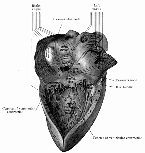 Fig. 38.—Right side of the heart showing diagrammatically the distribution of the
two vagus nerves to different parts of the viscus. The impulse to contraction originates
at the sino-auricular node and passes over the wall of the auricle to Tawara's node, and
thence over His' bundle across the auriculoventricular septum to be distributed throughout
the ventricular wall. If the upper, sino-auricular, node is damaged, or if its impulses
fail to get across the wall of the auricle, Tawara's node acts in its place to start
off the ventricle. If a lesion at the base of the mesial segment of the tricuspid valve
damages His' bundle, so that Tawara's node is cut off from the ventricle, then the ventricle
may originate its own impulses to contraction. (Hare's Practice of Medicine.)
Fig. 38.—Right side of the heart showing diagrammatically the distribution of the
two vagus nerves to different parts of the viscus. The impulse to contraction originates
at the sino-auricular node and passes over the wall of the auricle to Tawara's node, and
thence over His' bundle across the auriculoventricular septum to be distributed throughout
the ventricular wall. If the upper, sino-auricular, node is damaged, or if its impulses
fail to get across the wall of the auricle, Tawara's node acts in its place to start
off the ventricle. If a lesion at the base of the mesial segment of the tricuspid valve
damages His' bundle, so that Tawara's node is cut off from the ventricle, then the ventricle
may originate its own impulses to contraction. (Hare's Practice of Medicine.)
The sino-auricular (s-a) node is the most irritable portion
of the heart, it is endowed with the greatest amount
of rhythmicity as well. It is under the control of the vagus
nerve. Its inherent rate of rhythmicity is probably more
rapid than the usual numbers of impulses per minute, but
it is inhibited by the vagus. Paralysis of the vagus endings
increases the rate of impulse formation and therefore the
rate of the heart.
The electrocardiogram is a graphic representation on a
photographic film or sensitive bromide paper of the changes
of electrical potential during muscular activity. The lines
are made by the highly magnified string of the galvanometer
as it moves across the slit in the photographic apparatus
in response to the induction currents set up in the
heart magnified by the special galvanometer.
The record is made in three so-called Leads.
Lead I
The electrodes are attached to right arm and left arm.
Lead II
The electrodes are attached to right arm and left leg.
Lead III
The electrodes are attached to left arm and left leg.
A series of regular figures is normally obtained in which
are depressions and elevations and regular spacing of these
elevations and depressions. The waves so-called have been
arbitrarily designated P, Q, R, S, T. There is some difference
in the three leads. "The wave P is positive in all
leads. P to R interval varies slightly in the three leads.
All the waves of Lead II are greater than those of Leads I
and III. The wave R is positive in all leads. T is usually
positive in all leads, but is occasionally negative in Lead
III. Even in normal individuals there is a considerable
range of variation in the electrocardiogram which is within
the limits of the normal." (Hart.) (Fig. 39.)
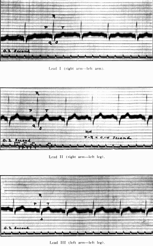 Fig. 39.—Normal electrocardiogram. (After Hart.)
The P wave is admitted to be the wave of auricular contraction.
Q, R, S, is the ventricular complex caused, it is
thought, by the current passing over the ventricles. T
wave is not yet definitely settled. It has been thought by
some that it represented actual ventricular contraction and
its height and shape had some meaning in heart force.
This is denied by others. Hart defines it as "The final activity
of the ventricle." The T wave is usually increased
in size during exercise.
The P-R interval is almost the most important feature
of the tracing. It is the actual conduction time in fractions
of a second of the impulse from s-a node to the ventricles.
Normally this is about 0.2 second or slightly less. Much
that was hoped for from the electrocardiograph in the clinic
has not been forthcoming. Its greatest value is in states
of abnormal conductivity, such as various grades of heart
block, extrasystoles, whether originating in auricles or in
either ventricle, abnormalities of rhythm, as flutter and
fibrillation. It has, however, aided materially in the intelligent
interpretation of many phenomena heretofore not
well understood, and has enormously increased our knowledge
of the physiology and pathologic physiology of the
heart.
It is not possible to enter farther into the subject here.
This brief discussion must suffice. The reader is referred
to works on this subject in connection with diseases of the
heart.