—Histology is that part of descriptive anatomy
which treats of the intimate structure of the tissues
as seen under the microscope.
Histology as taught in most professional schools constitutes
a one year's course, but for the embalmer this
is not entirely necessary and with the short term of schooling
now existing it is quite impossible, but certain of
the fundamental principles of histology are important.
For this reason a few of the more important tissues have
been discussed, not, however, in great detail, but only
superficially, merely to have the embalmer acquainted
with them.
—A cell is defined as a nucleated mass of protoplasm
endowed with the attributes of life.
Protoplasm is the name applied to the semi-fluid,
granular substance contained within the cell.
The simplest forms of animal life are organisms consisting
of only one cell which are called protozoa.
Cells having similar shape and similar functions are
grouped to form tissues.
Tissues are grouped together to form organs.
Every cell consists of a cell body and a nucleus. The
cell body consists of a substance known as protoplasm.
The nucleus is the essential part of a typical cell and
is the controlling center of its activity.
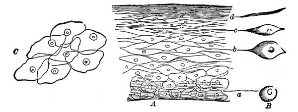
Fig. 1—A, A vertical section of the cuticle; B, the lateral view of the
cells; C, the flat side of scales like (d) magnified
250 diameters.
Cells divide or reproduce themselves by means of
direct or indirect division. In direct division the nucleus
and the cell wall simply divide into two equal divisions
and results in the formation of two new cells. In indirect
division the process is much more complicated,
and several stages must be passed through before there
is a complete division.
The process of fertilization consists in the conjugation
of two sexual cells. The male sexual cell is called the
spermatazoon, and the female sexual cell is called the
ovum.
The nucleus of the ovum in its earlier development
stages is known as the germinal vessicle.
In the living organism many cells are destroyed during
the various physiologic processes and are replaced by new
ones. When a cell dies, changes take place in the nucleus
which result in its gradual disappearance. This
process is known as chromatolysis.
—A tissue is an aggregate of cells all having
a common function.
Those important tissues with which the embalmer
should be more or less acquainted are the following:
Skin, nails, hair, superficial fascia, deep fascia, lymphatics,
glands, cartilage, bone, teeth, nerves, muscles,
tendons, aponeuroses, ligaments, fat, mucous membranes,
serous membranes, synovial membranes, arteries, veins
and blood.
—The skin or integument (intego, to cover)
is the outside covering of the human body. It is the
first tissue that is cut when
operating upon the body.
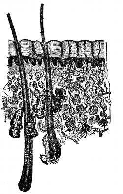
Fig. 2—A cross section of
the skin. (Gray)
The skin is the seat of the
organs of touch. The multitudes
of sensory nerve endings
convey the sensations of temperature,
pressure and pain to
the brain, thus informing the
brain at all times, to keep the
body from harm, and in a
strong and healthful condition.
The skin is also the regulator
of the body temperature, for
connected with the skin are
sweat glands, and sebaceous
glands, each having important
excretory functions.
The skin is also a protective coat, very elastic, and
varies greatly in thickness. It is thinnest in the eyelids
and thickest over the back of the neck, back of the
shoulders, palms of the hands and the soles of the feet.
The color of the skin depends upon two things, first,
on the pigment, which is found, one of the discriminating
points between the races, named by the color of the skin
as white, black, yellow, etc.; second, the color depends
upon the amount of blood in circulation, the deepest hue
being in the parts exposed to the air, light and the varied
temperatures. Besides these the color of the skin varies
with age, pinkest in the infant and becoming yellow with
old age. It varies with exposure and with climate, the
people living in the north having a much different complexion
than those living in the south under the tropical
sun. The color of the skin also varies with certain diseases,
being extremely pale in anaemia, brown in Addison's
disease, and yellow in jaundice.
The skin can be said to be moveable, although in
places it is attached firmly to the underlying structures,
especially on the scalp, the soles of the feet, and the palms
of the hands.
Upon close examination the skin discloses a multitude
of openings, creases, furrows, depressions, folds and
hairs.
A dimple is a permanent pit or depression due to the
adhesion of the surface to parts beneath.
Structure.—The skin consists of two intimately connected
structures, the one is the true skin, corium, or
dermis and is the deepest layer of the skin; and the other
is the false skin, cuticle, or epidermis, and is the outermost
layer of the skin.
The true skin, is composed mostly of connective tissues
and elastic fibers. It is the real seat of the sense
of touch, for it is here that the sensory nerves have their
termination. In this layer we also have the termination
of the minute capillaries of the skin.
The false skin, contains no blood vessels or nerves,
and being without these it is practically dead tissue, and
to illustrate this fact one can take a needle and run it
through this outside layer without the least pain or the
drawing of blood.
The false skin is the part which slips off in case of
skin slip. In as much as the minute capillaries end at
the termination of the true skin, when putrefaction and
fermentation begin there is an oozing of water from the
capillaries and the surrounding tissues, between the two
layers of skin, causing a blister to form, and known as
skin slip.
At the lowest part of the false skin is a layer of
germinal cells, from which all the other cells are derived,
and becoming more flattened and horny as they are pushed
farther away from the blood supply; and also a layer
of pigment cells, which give the discriminating color to
the skin.
In the skin are seen numerous sebaceous and sweat
glands.
The sweat glands are the organs by which a large
portion of the aqueous and gaseous materials are excreted
by the skin. Sweat glands are found in almost every
portion of the skin, and are situated in small pits below
the surface of the skin, surrounded by a quantity of
adipose tissue or fat. They are small, round, reddish
bodies, consisting of a single tubule, convoluted in form,
which extends up through the skin and opens on the surface.
The size of these glands, of course, vary, being
especially large in those regions where the flow of perspiration
is copious as in the axilla.
The sebaceous glands are small, sacculated, glandular
organs, lodged in the substance of the skin. They are
found in most parts of the skin and are usually connected
with the hair follicles. Each gland consists of a
single duct, more or less capacious, which terminates
in a cluster of small secreting pouches or saccules. These
glands secrete an oily fluid, which keeps the skin soft
and also oils the shaft of the hair.
—The nails are a peculiar modification of
the epidermis and have the same cellular structure as that
of the epidermis. The nails are found on the dorsal surface
of the fingers and toes and act as a protection, and
enable one to pick up small objects, or to grasp more
firmly any object. Were it not for the nails it would
be impossible for one to pick up a needle from off the
floor.
Each nail is convex on its outer surface, and its chief
mass which is called the body lies upon the nail bed, or
true skin; the free end projects out over the surface of
the finger, and is that part which is not attached below,
and since it is the continuation of the epidermis, it likewise
will have no nerve or blood supply and therefore
can be trimmed without pain to the individual.
The root is implanted in a groove in the skin and
is composed of cells which have not become horny. The
root is white in color and is the little half moon which
you can see next to the skin.
The matrix is that part of the true skin beneath the
body and the root of the nail, and is so called, because,
it is that part from which the nail is produced and so
long as the matrix at the root of the nail is uninjured,
the nail will be reproduced after an accident.
After death the nail turns black, due to the infiltration
of blood into the matrix.
Treatment by the Embalmer.—The blackened condition
of the nail due to the infiltration of blood into the
matrix can in many cases be overcome by carefully rubbing
the nail at the time the body is being injected. After
the discoloration is removed the fingers should be kept
elevated so that the blood will not settle there again.
—The hair, like the nails, is a peculiar modification
of the epidermis and consists of practically the
same cellular structure as the epidermis. Hair is found
on nearly every part of the body excepting the palms
of the hands and the soles of the feet, the borders of
the lips, etc. It varies much in length, thickness and in
the different races of mankind. In the eyelids it is short,
on the scalp it is of considerable length. In other parts
as the eye-lashes, the hair of the pubis region, the whiskers
and beard the thickness is remarkable.
A hair consists of the root and the shaft. The root
of the hair or that part implanted in the skin presents
at its extremity a bulbous enlargement, called the hair
bulb. Into this bulb we find the small arterial capillary
circulating and at its termination the beginning of the
venous capillary. In this way the hair is nourished in
life. We also find a small nerve going to the hair bulb.
The shaft is the remaining part or that part coming out
from the skin.
The hair grows from its roots and as it grows it
pushes itself out from the skin and owes its growth to the
small capillary circulation, carrying pure arterial blood to
each and every hair, and for this reason you can understand
for yourself the erroneous idea of what is termed
the “post-mortem growth of hair.” Only a few weeks
ago one of the students declared that he had actually seen
a subject shaved and the body at the time of the funeral
was placed in a vault to await the arrival of a close relative
who had to come from Europe.
Three weeks later the student, together with the undertaker
and relatives, went to the vault to view the remains.
The body was in a perfect state of preservation,
only for a large growth of beard as the student supposed.
This student had observed rightly, but he did not go
far enough. He did not think of how the hair actually
got its nourishment. The hair owes its life to the circulation
of the blood, just as much as the heart or any
other organ does, and will die and cease to grow just
as soon as the body dies and the circulation is cut off.
What this student saw was only an apparent growth, for
after the body dies the tissues begin to shrink, squeezing
the blood and fluid substances out of them, thus giving
the hair cylinder a more projected appearance.
The student was very much surprised at his mistake,
but after the explanation he saw that the hair owed its
life to the circulation and that when this circulation was
cut off, the hair must cease to grow.
The chief function of hair is that of protection from
heat or cold and to help shield the brain from the effect
of a blow upon the head.
The hair, next to the teeth and bones, is the least destructible
part of the body.
—The fascia (fascia, a bandage) is areolar
or aponeurotic tissue of variable thickness and
strength found in all regions of the body and invests or
surrounds the softer and more delicate organs. From
its situation in the body the fascia is divided into two
groups, superficial and deep.
Superficial fascia is found immediately beneath the
skin over almost the entire surface of the body. It connects
the skin with the deep fascia and consists of areolar
tissue.
The superficial fascia varies in thickness in different
parts of the body and some places, especially in the groin
is capable of being subdivided into several different
layers. The first layer of the superficial fascia, which
is just beneath the skin, usually contains a great amount
of fat or adipose tissue. This, in most text books, has
been termed the subcutaneous tissue. The second layer
is comparatively devoid of adipose or fatty tissue and
in this we find the trunks of the subcutaneous vessels and
nerves, as for example, the radial and ulnar veins in the
arms and the saphenous vein in the leg.
The superficial fascia facilitates the movement of the
skin, serves as a soft medium for the passage of the vessels
and nerves to the skin and retains the warmth of
the body, since the fat contained in its meshes is a had
conductor of heat.
Deep fascia or aponeurotic fascia is a dense inelastic,
unyielding fibrous membrane, forming a sheath for the
muscles and affording them broad surfaces for attachment.
On removal of the superficial fascia, the deep
fascia is usually exposed and can be seen as a dense,
tough membrane, which not only binds down the muscles
to each region, but gives to each a separate sheath as
well as to the blood vessels and nerves.
Thus, on going down into the arm between the biceps
and triceps muscles to raise the brachial artery, you
would first cut through the skin, then the subcutaneous
tissue, the superficial fascia and then you would come
to a membrane investing the artery, vein and nerve.
This membrane is the part of the deep fascia which
covers the vessels, making a distinct sheath for them and
you must go through this sheath before you can hope
to raise the artery.
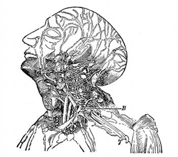
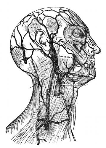
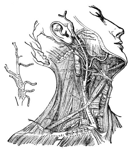
Fig. 3—Lymphatics of the head and neck.
B, the thoracic duct.
—The lymphatics occur in all parts
of the body, and in many respects resemble the veins,
one of the most striking similarities being that the lymphatics
contain valves just the same as the venous system.
The lymphatic capillaries are arranged in the form of
a net work and resemble closely in structure the blood
capillaries. These capillaries then unite to form the
lymph vessels and these then convey the lymph to the
subclavian veins. The lymph is a colorless fluid and
contains numerous blood corpuscles known as lymphocytes.
But in those lymphatic vessels, which have their
origin in the walls of the small intestines, the lymph,
especially during digestion, contains a great amount of
fat, so that it has a milky appearance, and for this reason
the lymphatics of this region, have been termed lacteals.
There are two main lymphatic trunks, the one on the left
side is called the thoracic duct. This duct extends from
the lower border of the second lumbar vertebra, through
the entire length of the thorax, and opens into the left
subclavian vein, close to the point where it is joined to
the left internal jugular. It receives the lymph from the
lower limbs, the pelvic walls and
viscera, the abdominal walls and
viscera; the lower part of the
right half and the whole of the
left half of the thoracic viscera,
the left side of the neck and head
and the left arm.
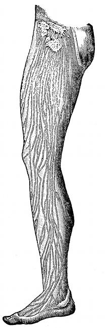
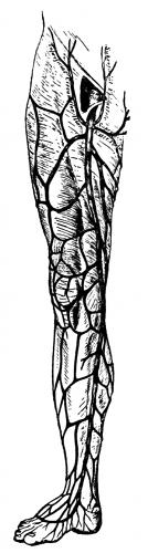
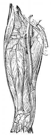
Fig. 4—Lymphatics of
the leg.
The other duct is called the
right lymphatic duct and receives
lymph from the upper part of
the right side of the thoracic wall,
part of the right side of the diaphragm
and the right lobe of the
liver, the whole of the right arm
and neck and right side of the
head. This trunk is very short
and empties its supply of lymph
into the right subclavian vein.
Receptaculum chyli is the expanded
portion of the thoracic
duct just at its beginning. Its
function is to receive the lacteals
which come from the villi of the
intestines.
Lymph glands are the enlargements
of the lymph vessels. They
occur frequently in the lymphatic
system, being most numerous in
the axillary space, the cervical region (in the neck)
and in Scarpa's triangle.
The lymphatic system aids greatly in warding off such
diseases as blood poisoning, anthrax, etc.
The lacteals are the lymphatics which carry the
chyme from the villi of the intestines and deposit it in
the receptaculum chyli.
—The glands of the human body are divided
into three classes called tubular, alveolar and tubulo-alveolar
glands.
Tubular Glands.—In these, the secreting portion consists
of a long or short tubule, which may be relatively
straight or variously twisted, one end of which ends
blindly, while the other end opens on the free surface
or into a duct.
Tubular glands may be simple, or having only a single
tubule; they may be simple branched, having more than
one tubule; or they may be compound branched, thus
resembling the branching of a tree.
Some tubular glands would be the liver, kidneys,
testes, lachrymal glands, serous glands of the mucous
membranes, fundus glands of the stomach, uterine glands,
the majority of the pyloric glands and the majority of
the sweat glands.
Alveolar Glands.—In these, the secreting compartments
have the form of variously shaped vesicles or saccules,
known as alveoli which open on the free surface
or into a duct.
Alveolar glands may be either simple, simple branched,
or compound branched.
Some alveolar glands would be the sebaceous glands,
pancreas, mammary gland, ovary and thyroid.
Tubulo-alveolar Glands.—In these, there is a combination
of the tubular and the alveolar type. They may
also be simple, simple branched or compound branched.
Some of this type would be certain of the pyloric
glands, certain of the sweat glands, some mucous glands,
the prostate and the lungs.
The most important glands will be discussed under
the tissue or the organ in which they are situated.
—Cartilage is a transition stage between
connective tissue and bone; when it is boiled it yields
condrin. It is found in various parts of the body, in
the adults being found chiefly in the joints, in the sides
of the thorax, and in various tubes which are not kept
permanently open, such as the air passages, nostrils, ears,
etc. In the foetus, the greater part of the framework is
cartilaginous and as the foetus matures this cartilage
is finally replaced by bone. Cartilage is divided into
hyaline cartilage, elastic cartilage, and fibro cartilage.
Hyaline cartilage is found in the nose, larynx, trachea,
and bronchi.
Elastic cartilage is found in the epiglottis and the
cartilages of the larynx.
Fibro cartilage is found at the point of insertion
of the ligaments, into the body of the bone, such as the
cartilage which helps to hold the femur or long bone
of the thigh into the hip.
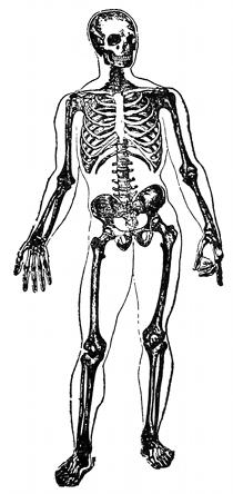
—Bone results from the calcification of cartilage
or fibrous tissue. It is a highly specialized form of
connective tissue. There are two varieties of bone;
dense or compact bone and cancellous, loose, or spongy
bone. Compact bone is dense, like ivory, and is always
found on the exterior of bones.
Cancellous bone is found in the interior of bones, and
has a lattice-work appearance.
Bone consists of one-third animal or organic matter
and two-thirds earthy or inorganic matter. These proportions,
however, vary with age. In youth it is nearly
half and half, while in the adult the earthy is greatly
in excess. It also varies with disease. With some defect
of nutrition, the bone is deprived of its normal proportion
of earthy matter, while the animal matter is of
unhealthy quality, and we have as a result, a disease
called rickets, so common in the children of the poor.
The earthy or inorganic matter consists of phosphate,
carbonate, fluoride of calcium, sodium chloride, and
phosphate of magnesium. The animal matter consists of
fat collagen, which when boiled with water is resolved
into gelatin.
To illustrate the two substances, take a bone and place
it in dilute hydrochloric acid. The acid will eat out all
the mineral matter and we have left only the animal
matter. After this operation one can take the bone and
can bend it into any position whatever, which experiment
shows that the animal matter gives elasticity to the bone.
The second experiment would be to put the bone on
a bed of hot coals and burn it. Only the animal matter
will burn and we will have the mineral matter remaining.
After this operation one will find that the bone is very
brittle and will easily break, which experiment shows
that the mineral matter gives stability and support to
the bone.
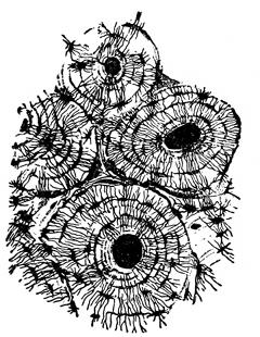
Fig. 5—Cross section of
bone. (Sharpey)
If a cross section is made of any
long bone, such as the humerus,
and this section placed under the
low power of the microscope, the
Haversian canal system can be
discerned. The Haversian canal
system consists of the numerous
small openings or canals through
which the blood vessels ramify in
distributing the nourishment to
the bone. Around each individual
canal are seen smaller spaces arranged
in a circle. These are known
as the lacunae (small lakes). Going from the lacunae
are smaller canals which take on the name canaliculae,
and joining all the lacunae together, making the appearance
of concentric circles, we have the lamellae. The
outside covering of the bone is called the periosteum and
the inside covering is called the endosteum. Most of
the long bones and many of the smaller bones are supplied
by a nutrient artery, which enters the bone near
its center, enters the bone marrow, and divides into
two branches, one going up and the other down in the
marrow. The blood is then distributed through the Haversian
canal system. Veins emerge from the long bones
in three places: 1. One or two large veins accompany
the nutrient artery. 2. Numerous veins emerge from
the articular extremities. 3. Many small veins arise
in and emerge from the compact substance.
Bones are divided, according to shape, into four
classes: long, short, flat and irregular.
Long Bones.—These bones are usually used as a system
of levers to confer the power of locomotion. A long
bone consists of a shaft and two extremities. The shaft
is a hollow cylinder within which is the medullary canal.
The extremities are somewhat expanded for the purpose
of articulation, and to afford a broad surface for the
attachment of muscles. The long bones are as a rule
curved in two directions to give greater strength to the
bone. Some examples of this class of bone are the
clavicle, radius, ulna, humerus, femur, tibia, fibula, metacarpal,
metatarsal, and the phalanges.
Short Bones.—These bones are placed in that part
of the skeleton where there is need for strength and compactness,
and where the motion of the part is slight and
limited. Some examples of this class of bone are the
bones of the carpus and tarsus (in the hand and the
foot).
Flat Bones.—Flat bones are found where the principle
requirement is either extensive protection, or the
need of a broad surface for the attachment of muscles.
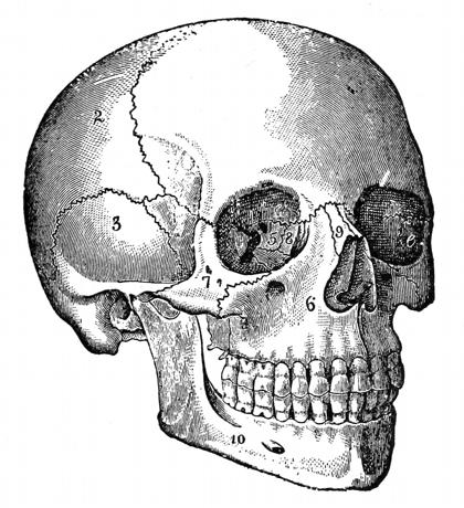
Some of the bones of this class are the occipital, parietal,
frontal, nasal, lachrymal, vomer, scapula, sternum, and
the ribs.
Irregular Bones.—These bones are such as from their
peculiar shape and form can not be grouped under any
of the preceding heads. Some of the bones of this class
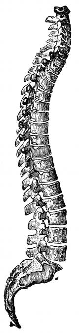
are the vertebrae, sacrum, coccyx, temporal, sphenoid,
ethmoid, etc.
If the surface of a bone is examined, certain articular
and non-articular eminences and depressions will be seen.
Articular Eminences.—Examples of this class are
found in the heads of the humerus and the femur.
Articular Depressions.—Examples of this class are
found in the glenoid cavity of the scapula and the
acetabulum.
Non-articular Eminences.—These are designated according
to their form.
A tuberosity is a broad, rough, and uneven elevation.
A tubercle is a small, rough prominence.
A spine is a sharp, slender, pointed eminence.
A ridge, line, or crest is a narrow, rough elevation,
running some way along the surface.
Non-articular Depressions.—These are of variable
form, and are described as notches, sulci, fossae, grooves,
furrows, fissures, etc. These non-articular eminences and
depressions may serve to increase the extent of surface
for the attachment of ligaments and muscles or may
receive blood vessels, nerves, tendons, ligaments, or portions
of organs.
Canals or foramina are channels or openings in bone
through which pass the nerves and blood vessels.
—In the human body we find two sets of teeth.
One appearing in childhood, and are known as milk teeth,
twenty in number, the permanent teeth replacing these
about the sixth year.
There are thirty-two permanent teeth, divided into
four incisors, two canines, four bicuspids and six molars.
Teeth are made up of three different substances, which
are known as enamel, dentine and cement.
The enamel is a very hard substance, the hardest in
the body, and may be compared to quartz. The enamel
covers the entire tooth down as far as the gums.
The cement is a continuation of the enamel below the
gums, and is closely adherent to the dentine. The cement
consists of bone tissue, but the lamellae as a rule
do not contain Haversian canals.
The dentine is, next to the enamel, the hardest tissue
of the tooth, and composes the main body of the tooth.
The pulp cavity is found within the center of the tooth,
with the opening toward the jaw bone. The tooth is
nourished by a nutrient artery and vein and nerve which
pass into the pulp of the tooth.
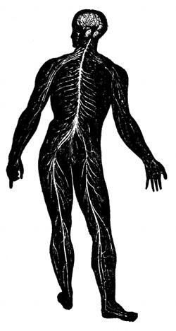
—Nerves are divided into two general classes,
called medullary and non-medullary nerves. The non-medullated
type arise mostly from the sympathetic system,
while the medullated type arise from the brain and
cord. As a rule, the nerves of
the body follow the course of
the arteries, and are generally
found in the same sheath with
the artery and vein.
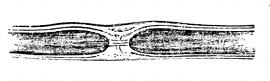
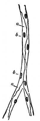
Fig. 6—Section of a nerve fibre.
(Klein and Noble Smith)
They are easily distinguished from the arteries and
veins by touch and by their color, being very inelastic
and fibrous, hard to the touch, and unlike the artery
or vein, since they have no central opening.
—Myology is that branch of anatomy which
treats of the muscles. The muscles are formed of bundles
of reddish fibres, endowed with the property of contractility.
In the body we find two kinds of muscular
tissue, called voluntary and involuntary muscle. The
voluntary type is characterized by the
striped appearance which it displays when
seen under the microscope, and for this
reason it is called striped or striated
muscle. It is so named “voluntary” because
it is capable of being put into action
and controlled by the will. The involuntary
muscles do not present any
striped appearance, and consequently are
called unstriped or non-striated, and are
not under the control of the will. An
example of voluntary muscle would be
any muscle of the bony framework as
for example, the biceps or triceps.
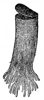
Fig. 7—View of
muscle fibers.
An example of involuntary muscle would be those of
the intestines and stomach, the muscles of the bladder
and uterus and the walls of the arteries and veins, etc.
When viewed under the microscope, the muscle is
seen to be composed of many fibrils. The sheath covering
each fibril is called the sarcolemma, and contains
within its boundaries the muscle plasma, or protoplasm,
and a nucleus. Many of the fibrils when grouped together
constitute the entire muscle.
The muscles get their blood supply from the nutrient
artery, which ramifies the tissues, the smallest capillaries
coming in contact with each muscle cell.
—Tendons are white, glistening, fibrous
cords, varying in length and thickness, sometimes round,
sometimes flattened, of considerable strength, and devoid
of elasticity. It consists principally of a substance
which yields gelatin.
Tendons do not have a direct blood supply.
—Aponeuroses are flattened or ribbon-like
tendons, of a pearly-white color, irridescent, glistening,
and similar in structure to the tendons.
—Ligaments consist of bands of various
forms, serving to connect the articular extremities of
bones. They are strong bands of smooth, silverwhite
fibrous tissue.
A ligament is pliable and flexible, so as to allow the
most perfect freedom of movement, but at the same
time it is tough and strong, so as not to yield readily under
the severe applied force, and for this reason they
serve as good connecting links for the binding of bones
together.
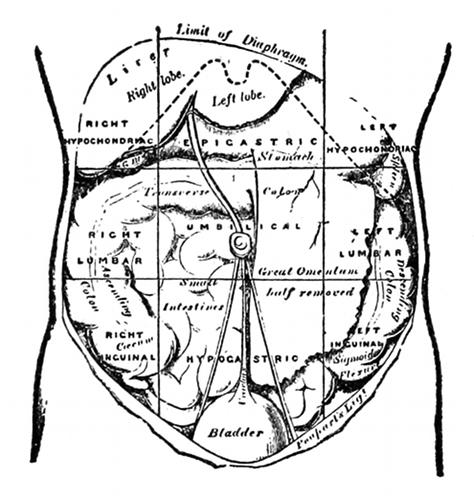
Poupart's Ligament.—Poupart's ligament extends
from the crest of the ilium to the top of the pubic bone.
This ligament is of utmost importance to the embalmer,
as it serves as a guide to locate the femoral artery. By
placing the thumb on the crest of the ilium and the second
finger on the top of the public bone, then letting
the first finger drop midway between the two, which
would be the center of Poupart's ligament, we have a
point which marks the exit of the artery from the body
and the beginning of the femoral artery.
Poupart's ligament also forms the base of Scarpa's
triangle. The structure of this triangle will be taken
up later.
—Fat is a deposit of an oil in the cells of the tissues,
just beneath the skin, giving roundness and plumpness
to the body, and acting as an excellent non-conductor
for the retention of heat.
So tiny are these cells, that there are over sixty-five
million in a cubic inch of fat. As they are kept moist,
the liquid does not ooze out; but, on drying, it comes to
the surface, and thus a piece of fat feels oily when exposed
to the air. The quantity of fat varies with the
state of nutrition. In corpulent persons, the masses of
fat beneath the skin, in the mesentery, on the surface of
the heart and the great vessels, between the muscles, and
in the neighborhood of the nerves, are considerably increased.
Conversely, in the emaciated we sometimes find
beneath the skin cells which contain only one oil drop.
Many masses of fat which have an important relation
to muscular actions—such as the fat of the orbit or the
cheek, do not disappear in the most emaciated persons.
Even in starvation, the fatty substance of the brain and
spinal cord are retained.
Fat collects as pads in the hollows of the bones,
around the joints and between the muscles, causing them
to glide more easily upon each other. As marrow, it
nourishes the skeleton, and also distributes the shock of
any jar the limb may sustain.
Fat does not gather within the cranium, the lungs
or the eyelids, where its accumulation would clog the
organs.
—Mucous membranes line all the
open cavities of the body, or all those cavities which communicate
with the outside.
At the edges of the openings into the body, the skin
seems to stop and give place to a tissue which is redder,
more sensitive, more liable to bleed, and is moistened by
a fluid or mucous, as it is called. Really, however, the
skin does not cease, but passes into a more delicate covering
of the same general structure, and it is to this
that the name mucous membrane is applied.
The entire alimentary canal, the entire respiratory
tract, and the genito-urinary tract, are lined with a mucous
membrane. Mucous membrane secretes a mucous
fluid.
—Serous membranes line the closed
cavities of the body. The pleurae, the pericardium
and the peritoneum are examples of serous membranes.
Serous membranes secrete a serous fluid.
—Synovial membranes are serous
in character, and consist of loose connective tissue,
containing fat, vessels and nerves, its inner surface being
usually lined with secreting cells. The fluid secreted is
yellowish-white or slightly reddish, resembling very much
the white of an egg. It contains fats, salts, albumen,
extractives from the lymph, and a fluid known as synovia.
The chief function of this fluid is to act as an oil
to lubricate the joints and surfaces in which there is any
friction.
Synovial membranes are divided into three classes,
known as articular, bursal and vaginal.
Articular synovial membranes are found in every
free movable joint.
Bursal synovial membranes are sacs interposed between
the surfaces which move upon each other, producing
friction, as in the gliding of a tendon or of the
integument over projecting bony surfaces.
Vaginal synovial membranes serve to facilitate the
gliding of a tendon in the bony canal through which it
passes.
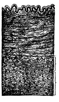
Fig. 8—Section of
artery. (Grunstein)
—The arteries are cylindrical vessels which
serve to convey the blood from both ventricles of the
heart to every part of the body. They are called arteries
from the Greek words which mean
“to contain air,” and they were supposed,
by our ancients, to have this
function until the time of Galen, when
he refuted this opinion and showed that
these vessels, though for the most part
empty after death, actually contained
blood. The distribution of the arteries
may be compared to a tree, the common
trunk of which corresponds to the aorta,
and the smallest twigs corresponding
to the minute capillaries. When
one artery communicates with another
it is said to anastomose, and
this communication is very free between
the larger as between the smaller branches.
Anastomosis between trunks of equal size is found where
great activity of the circulation is requisite, as at the
base of the brain, where the two vertebrals unite to
form the basilar artery.
In the limbs and arms the anastomoses are more numerous
and of larger size around the joints. The branches
of the artery above, unite with branches, from the vessels
below. These anastomoses are called collateral circulations.
The principal ones of interest to the embalmer
are those of the deep brachial uniting with the
recurrent radial and ulnar arteries, forming the collateral
circulation in the arm; the deep femoral uniting
with the recurrent posterior and anterior tibials, forming
the collateral circulation in the leg; the superficial
and deep mammary arteries, branches of the subclavian
artery uniting with the superficial and deep epigastric
arteries, branches of the external iliac, forming the collateral
circulation over the abdomen and chest, and may
be considered the longest collateral circulation in the
body.
A terminal artery is one which forms no anastomoses;
such vessels are found in the heart, brain, spleen, kidneys,
lungs and mesentery.
Structure.—An artery consists of an internal, a middle
and an external coat.
The inner coat consists of endothelial cells and elastic
fibrous tissue, sometimes arranged longitudinally, but
usually they form a distinct fenestrated membrane (similar
to a doorscreen).
The middle coat consists mostly of elastic tissue and
white fibrous tissue.
The external coat is called the fibrous coat. It contains
fibrous connective tissue and elastic tissues.
Vasa-Vasorum.—Running in the outer wall of the
artery, we find small capillary vessels, and their function
is that of nourishing the outer wall, for the blood
which passes through the artery does not nourish the
artery from within, but depends on these small capillaries,
called vasa-vasorum, for their nutrition.
The individual sheath, or arterial sheath, the covering
for the artery, is composed of connective tissue, and
at places may adhere very tightly to the artery.
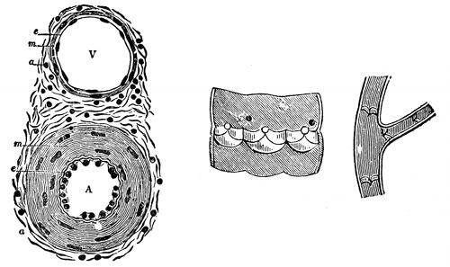
Fig. 9—Valves of the veins.
Fig. 10—Cross section through a small artery
and vein. (Klein and Noble Smith)
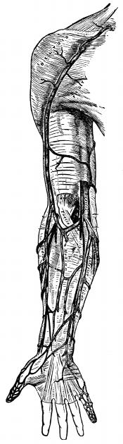
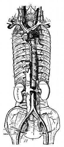
—The veins are the vessels which carry the
blood from the capillaries back to the right auricle of
the heart, and are found in nearly every tissue of the
body. They commence as venous capillaries, uniting
together into larger and larger veins, until we have the
great ascending and descending venae cavae. In form the
veins are perfectly cylindrical, like the arteries, but with
this difference, that their walls collapse when empty and
that they contain valves.
Structure.—The vein has about the same structure
as the artery, only that the middle coat is much thinner
and less elastic than the artery, and for this reason it
easily collapses.
Veins are divided into superficial, deep and sinuses.
Superficial veins are found between the layers of the
superficial fascia, just underneath the skin.
Deep veins accompany the arteries, and are usually
enclosed in the same common sheath with the artery.
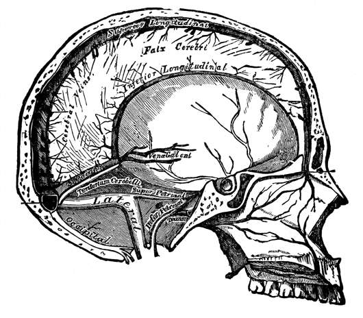
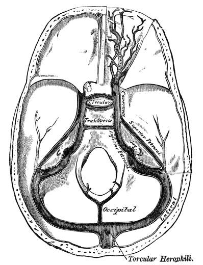
Sinuses are venous channels, which in their structure
and mode of distribution differ altogether from the veins.
They are found only in the interior of the skull, and consist
of channels formed by a separation of the two layers
of the dura mater.
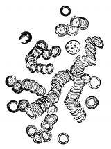
Fig. 11—Human blood.
—The blood of the body is
contained in a practically closed system
of tubes, the blood vessels, within
which it is kept circulating by force
of the heart beat. It is usually spoken
of as the nutritive liquid of the body,
but the functions may be stated explicitly,
although still in quite general
terms, by saying that it carries to the
tissues food stuffs after they have been
properly prepared by the digestive organs; that it transports
to the tissues oxygen, absorbed from the air by the
lungs; that it carries from the tissues various waste products
formed in the processes of dissimilation; that it is
the medium for the transmission of the internal secretion
of certain glands; that it aids in equalizing the temperature
and water contents of the body.
The total quantity of blood in the body has been determined
approximately for man as one-thirteenth of the
body weight. The specific gravity of human blood in
the adult may vary from 1.041 to 1.067, the average being
about 1.055.
The blood is composed of a liquid part, the plasma,
in which float a vast number of microscopical bodies, the
blood corpuscles, known respectively as the red corpuscles,
the white corpuscles or leucocytes, of which in turn
there are a great many different kinds, and the blood
plates.
Blood plasma, when obtained free from corpuscles, is
perfectly colorless, in thin layers, for example, in microscopical
preparation; when seen in large quantities it
shows a slightly yellowish tint. The red color of the
blood is not due, therefore, to coloration of the blood
plasma, but is caused by the mass of red corpuscles held
in suspension in the liquid. The proportion by bulk of
plasma to corpuscles is usually given roughly as two to
one. The blood plasma is composed of two substances,
blood serum and blood fibrin. You have noticed that
blood, after it has escaped from the vessels, usually clots
or coagulates. The clot, as it forms, gradually shrinks
and squeezes out a clear liquid, to which the name blood
serum has been given. Serum resembles the plasma of
normal blood in general appearance, but differs from it
in composition. Here it is sufficient to say that blood
serum is the liquid part of the blood after coagulation
has taken place. You can prepare this experiment for
yourself: If shed blood is whipped vigorously with a rod
or some similar object while it is clotting, the essential
part of the clot, namely the fibrin, forms differently from
what it does when the blood is allowed to coagulate
quietly. It is deposited in shreds on the whipper. Blood
that has been treated in this way is known as defibrinated
blood. It consists of blood serum plus the red and
white corpuscles, and as far as appearances go it resembles
exactly the normal blood; it has lost, however, its
power of clotting.
Red blood corpuscles are bi-concave, circular disks,
without nuclei; their average diameter is 7.7 microns (1
micron equals 1-25,000 of an inch); their number, which
is usually reckoned as so many to a cu. millimeter, varies
greatly under different conditions of health and disease.
The average number is given as 5,600,000 per cubic millimeter
for males and 4,500,000 per cubic millimeter for
females.
The number of red corpuscles also varies in individuals
with the constitution, nutrition and manner of life.
It varies with age, being greatest in the fetus and in
the new-born child. It varies with the time of the day,
showing a distinct diminution after meals. In the female
it varies somewhat with menstruation and pregnancy,
being slightly increased in the former and diminished in
the latter condition.
The red color of the corpuscles is due to the presence
in them of a pigment, known as hemoglobin. Owing to
the minute size of the corpuscles, their color when seen
singly under the microscope is a faint yellowish red, but
when seen in mass they exhibit the well-known blood-red
color, which varies from a scarlet in arterial blood to a
purplish red in venous blood, this variation in color being
dependent upon the amount of oxygen contained in
the blood in combination with the hemoglobin. The
function of the red blood corpuscles is to carry oxygen
from the lungs to the tissues. This function is entirely
dependent upon the presence of hemoglobins, which have
the power of combining easily with the oxygen gas.
White blood corpuscles or leucocytes contain no hemoglobin
or coloring matter. They have a nucleus or center
spot. Their size varies from 5 to 12 microns, and are
less numerous than the red corpuscles, being in this proportion:
one white corpuscle to 500 red corpuscles. The
chief functions of the white corpuscles are: (1) That they
protect the body from pathogenic or disease-producing
bacteria. In explanation of this action it has been suggested
that they may either ingest the bacteria and thus
destroy them directly, or they may form certain substances,
defensive proteids, that destroy the bacteria.
White corpuscles that act by ingesting the bacteria are
spoken of as phagocytes (meaning to eat the cell). (2)
They aid in the absorption of fats from the intestines.
(3) They aid in the absorption of peptones from the intestines.
(4) They take part in the process of blood
coagulation. (5) They help in maintaining the normal
composition of the blood plasma in proteids.
Blood plates are small circular or elliptical bodies,
nearly homogeneous in structure, variable in size, always
much smaller than the red blood corpuscles. Less is
known of their origin, fate and functions than in the
case of the other blood corpuscles, but there is some considerable
evidence to show that they take part in the
process of coagulation or clotting.
Coagulation of the Blood.—One of the most striking
properties of the blood is its power of clotting, or coagulating,
shortly after it leaves the blood-vessels, or if
any foreign elements come in contact with it. The general
changes in the blood during this process are easily
followed. At first perfectly fluid, in a few minutes it
becomes viscous, and then sets into a soft jelly, which
quickly becomes firmer, so that the vessel containing it
can be inverted without spilling the blood. The clot continues
to grow more impact, and gradually shrinks in
volume, pressing out a greater or smaller amount of
clear, faintly yellow liquid, to which the name blood serum
is given. The essential part of the clot is the fibrin.
Fibrin is an insoluble proteid not found in normal
blood. In shed blood, and under certain conditions while
still in the blood-vessels, this fibrin is formed. In forming,
it shows an exceedingly fine network of delicate
threads that permeate the whole mass of the blood and
gives the clot its jelly-like character. The shrinking of
the threads causes the subsequent contraction of the
clot. If the blood has not been disturbed during the
act of clotting, the red corpuscles are caught in the fine
fibrin mesh-work, and as the clot shrinks these corpuscles
are held more firmly, only the clear liquid of the blood
being squeezed out, so it is possible to get specimens of
serum containing few or no red blood corpuscles. The
white corpuscles or leucocytes, on the contrary, although
they are also caught at first in the forming meshes of
fibrin, in latter stages of the clotting they readily pass
out into the serum, on account of their power of having
movement. If the blood has been agitated during the
process of clotting, the delicate net work will be broken
in places, and the serum will be more or less bloody—that
is, it will contain numerous red blood corpuscles. If
during the time of clotting the blood is vigorously whipped
with a bundle of fine rods, all the fibrin is deposited
as a stringy mass on the whipper, and the remaining liquid
part consists of serum plus red corpuscles. Blood
that has been whipped in this way is known as defibrinated
blood. It resembles normal blood in appearance,
but is different in composition; it can not clot again. The
way in which fibrin is normally deposited can be easily
demonstrated by taking a drop of blood on a slide and
covering it with a cover slip, allow it to stand several
minutes until coagulation is complete, and view under a
microscope. If the drop is examined, it is possible by
careful focusing, to discover in the spaces between the
masses of corpuscles many examples of delicate fibrin
net work. The physiological value of the clotting of blood
in life is that it stops hemorrhages by closing the openings
of the wounded blood vessels, but the clotting of the
blood after death, is to the embalmer one of the bugbears,
and a real method of preventing it, or of dissolving
the clot after it has once formed in the blood vessels is
one of those difficult problems which remains as yet unsolved.
Since we have no real method of preventing coagulation
in the blood vessels, let us search out the things
which will hasten or retard this coagulation. Blood
coagulates normally within a few minutes after it is
liberated from the blood vessel, but this process may
be hastened by increasing the amount of foreign substance
with which it comes in contact. Thus the agitation
of the liquid in quantity or the application of a
sponge or handkerchief or the application of heat hastens
the onset of clotting.
Coagulation in drawn blood may be retarded or prevented
altogether by a variety of means, of which the
following are the most important:
(1) By cooling.
(2) By the action of neutral salts.
(3) By the action of oxalate solutions.
(4) By the action of sodium fluoride.
Summary.—To summarize then, the following statements
may be made:
(1) The immediate factor necessary to the clotting
of the blood is the fibrin.
(2) That blood does not clot normally in the blood
vessels before death.
(3) That after death blood remains for a long time
without clotting, provided some outside agent is not
introduced to cause it.
Such an agent may be the blood coming in contact
with the air, or the blood drainage tube. The one point
then to be emphasized is that when a vein is cut, and the
blood begins to flow, you know that the blood is not in a
coagulated condition. Then work rapidly, put the blood
drainage tube quickly into the vein and draw off as much
blood as you can before it begins to clot at the end of
the tube. The great trouble has been, that the embalmer
does not work with precision. He first raises the vein,
and exposes it on the surface of the incision. He then
raises the artery. He places the drainage tube into the
vein, but shuts it off till he is ready with the artery.
Now, by the time he has placed the arterial tube in the
artery, injected a few bulbs full to see that all is in
working order, and has perhaps attended to a few other
duties, he is amazed to find that the blood will not flow,
that it has clotted. What is the reason? He gave it
time to clot after the drainage tube was inserted.
A better procedure would be not to touch the vein
until every other procedure has been attended to. Then
raise the vein, insert the drainage tube and withdraw
the blood quickly, and at the same time keep injecting
slowly into the arterial system to keep up the needed
pressure to keep the blood flowing.
(4) That when a clot is once formed in a blood vessel,
it is not dissolved by the addition of fluid or any other
solution.
(5) That sometimes when the blood has become
clotted at the end of the drainage tube, it can be loosened
up or be slightly pushed away by attaching the pump
to the drainage tube and injecting a few bulbs of fluid,
which, when it runs out, will again start the flow of
blood.