THE plants belonging to the Class Algæ grow in water, either in that of
the sea or in fresh water; a few of them, however, being found on damp
earth, damp walls, &c. The marine Algæ are commonly known as sea-weeds;
but the fresh-water Algæ generally receive but little popular notice,
forming, as they do, slimy masses or strata, of a green or brownish,
sometimes red, colour.
Algæ are of simple structure, consisting entirely of cells; in some
these are single, in others, united end to end, to form threads or
filaments, or grouped into a leaf-like expansion, or collected few
together into a little spherical group or a flat plate. They possess
none of the fibres, vessels, or ducts of the higher plants, although
some long and slender cells, existing in the stalks of the fronds of the
larger kinds, bear considerable resemblance to woody fibre. They exhibit
no distinction of stem and leaf, but consist of fronds representing the
stem and leaf combined and undistinguishable. And the term frond must be
understood to signify the separate parts arising from the point of
attachment when they are fixed; and in the case of those which are
unattached or free, the entire plant is called a frond.
The Algæ are divided into three Orders, viz. the Fucoid´eæ or
olive-coloured Algæ, the Florid´eæ or red, and the Confervoid´eæ or
green Algæ.
Fucoid´eæ, Fucoid Algæ, or Melanospor´eæ (μἑλας, black or dark). The
plants composing this order form our largest sea-weeds, and are found
everywhere in the sea and on the sea-shore. They are of an olive-green
or olive-brown colour, and usually become darker on drying.
| PLATE IV. [PAGE 64.] |
| Marine Algæ. |
| Fig. | |
| 1. | Dasya coccinea, piece of. |
| 2. | Dasya coccinea, portion with capsule (ceramidium). |
| 3. | Dasya coccinea, portion of main filament. |
| 4. | Dasya coccinea, section of filament. |
| 5. | Melobesia polymorpha. |
| 6. | Melobesia polymorpha, portion with capsules (ceramidia). |
| 7. | Jania rubens. |
| 8. | Jania rubens. |
| 9. | Lithocystis Allmanni. |
| 10. | Ceramium nodosum. |
| 11. | Ceramium nodosum, filament. |
| 12. | Ceramium rubrum, filament. |
| 13. | Ceramium rubrum, tetraspore. |
| 14. | Ceramium rubrum, end of filament. |
| 15. | Ceramium rubrum, capsule (favella). |
| 16. | Fucus vesiculosus, receptacles of. |
| 17. | Fucus vesiculosus, capsules (conceptacles). |
| 18. | Fucus serratus, antheridial conceptacles. |
| 19. | Spore of Fucus vesiculosus. |
| 20. | Antheridia of Fucus serratus. |
| 21. | Plocamium coccineum, sporophyll. |
| 22. | Plocamium coccineum, with capsule (coccidium). |
| 23. | Plocamium coccineum, portion of frond. |
| 24. | Plocamium coccineum, tetraspore from sporophyll. |
| 25. | Polysiphonia fastigiata, portion of. |
| 26. | Polysiphonia fastigiata, filament with capsules (ceramidia). |
| 27. | Polysiphonia fastigiata, portion of filament. |
| 28. | Corallina officinalis. |
| 29. | Corallina officinalis, portion of filament. |
| 30. | Corallina officinalis, capsule (ceramidium). |
| 31. | Enteromorpha compressa. |
| 32. | Enteromorpha compressa, cells of frond. |
| 33. | Hypnea purpurascens, capsule (coccidium). |
| 34. | Hypnea purpurascens, spores. |
| 35. | Hypnea purpurascens, filament. |

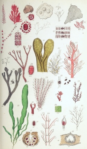 Plate IV.
W Bagg sculp
London: John Van Voorst.
Fúcus vesiculósus, with its parallel-sided or linear olive-brown
fronds, is known to every one as the seaweed which is hung up to act as
a weather-glass. The fronds have a central stout vein, or midrib, and
scattered air-bladders, mostly in pairs.
The fructification consists of yellowish oval enlargements of the ends
of the fronds, called the receptacles (fig. 16); but these are
somewhat variable in form, being often angular or truncate. On holding
one of the receptacles to the light, it will appear to contain a number
of little grains imbedded in its substance, slightly projecting above
the surface, and in the centre of each is a minute dot or pore. These
grains are the capsules, or conceptacles, and contain the spores. The
substance of the receptacles is composed of a beautiful network of
colourless, jointed, cellular fibres (figs. 17 a and 18 a), the
meshes of which are filled with a transparent gelatinous substance; but
immediately around the conceptacles the cells are shorter and more
closely packed. The spores (fig. 19) are arranged in the conceptacles in
a radiate manner; they are brown, and surrounded by a colourless sac,
called the perispore (περἰ, around, σπορἀ, seed); and between them are
numerous slender, colourless, jointed filaments, the paraph´yses. The
spores are not, however, truly single spores, for they ultimately divide
into eight segments or sporules, each of which is capable of producing a
new plant.
In the conceptacles of some fronds of Fucus no spores will be found,
the conceptacles (fig. 18) being filled with elegantly branched
colourless filaments (fig. 20), the ends of many of them being distended
into little yellowish sacs; these are the antherid´ia. The antheridia
contain large numbers of exceedingly minute spermatozoa, furnished with
two cilia, and very similar to those existing in the antheridia of the
mosses; these, escaping through the pore of the conceptacle, fertilize
the spores.
The figure (20) in the plate was drawn from a conceptacle of Fucus
serrátus, another common species, differing from F. vesiculosus in
having the margins of the frond serrate; the antheridia of the two
species do not, however, differ in any important respect. To examine the
conceptacles of Fucus and their contents, the receptacles should be
soaked in water, if not fresh, and thin sections made with a sharp
knife. They form very beautiful objects, and may be preserved in
chloride of calcium or glycerine.
Florid´eæ, or Rhodosper´meæ (ῥὁδον, rose, σπἑρμα, seed).—The second
Order of Algæ, forming the Florideæ (flos, a flower), comprises the
red sea-weeds; a few of them are purple, or greenish-red; so that by the
colour alone they may be readily distinguished from the Fucoids, and
from nearly all those of the next Order, the Confer´voids. A few of them
are leaflike, or possess flat fronds; but most of them consist of finely
divided or feathery fronds. They are often found upon the sea-shore of a
dirty white colour, the colouring matter having been decomposed or
washed out by rain.
We shall consider a few of the genera and species under the heads of the
families to which they belong.
Corallina´ceæ, the Corallines, or calcareous Algæ.—In this family we
have the beautiful Corallína officinális (Pl. IV. fig. 28), the common
Coralline, which is very abundant on the sea-shore, attached to larger
sea-weeds, shells, and rocks. It is hard and chalky, from the presence
of a large proportion of carbonate of lime in its minute cells. The
fronds are composed of jointed and branched filaments. The
fructification (figs. 29 and 30) consists of ovate cellular capsules, or
ceramid´ia (κερἁμιον, earthen vessel), placed mostly at the ends of
pinnate stalks, and containing a tuft of somewhat club-shaped jointed
spores, springing from the base of the capsules (fig. 30). When ripe,
the spores escape from a pore or hole in the end of the capsules. The
spores are 4-jointed, and hence are called tet´raspores (τἑτρα,
four).
To observe these spores, the capsules must be soaked in strong vinegar
for some hours, and then washed with water, to dissolve the calcareous
matter.
Jánia rúbens (Pl. IV. figs. 7 and 8) is another common and very
elegant little coralline, and is of a pale red colour. It differs from
the last in the branches being dichot´omous (δἱχα, in two, τομὀς,
cutting) or forked, instead of pinnate. The capsules, or ceramidia, have
also two short horn-like branchlets, placed one on each side, near the
end.
The genus Melobésia has the frond crustaceous, i. e. forming a hard
crust or layer. M. polymor´pha (Pl. IV. figs. 5 and 6) is common on
shells, stones, &c. The capsules (ceramidia) here form little blunt
cones, scattered over the crusts, and containing the tufted tetraspores,
as in Corallina.
Lithocys´tis Allman´ni (fig. 9) is very minute, and not uncommon upon
sea-weeds, stones, &c. It consists of a single fan-shaped crustaceous
layer of cells, closely investing the body to which it is attached; its
fructification is unknown.
Leaving the family of crustaceous Florideæ, we shall now pass to those
of softer consistence, although all the marine Algæ contain a
considerable quantity of calcareous matter.
Rhodomela´ceæ.—In this family we have the large genus Polysiphónia,
in which the frond (Pl. IV. figs. 25 and 26) is filamentous, the
filaments being apparently jointed and longitudinally striated. The
filaments are composed of rings of cells (fig. 27), arranged end to end,
and containing dark endochrome. The ends of the colourless cell-walls
separating the endochromes of the cells of adjacent rings produce the
jointed appearance; while the striated appearance is caused by the dark
cells being elongate and the cell-walls thick, so as to form white
interspaces.
The fructification consists of capsules (ceramidia), attached to the
sides of the branches, containing pear-shaped spores, with tetraspores
imbedded in swollen branches of separate plants.
Polysiphónia fastigiáta (fig. 25, a small piece) is common, attached
to the fronds of Fucus. Its filaments are rigid, bristle-like, of the
same breadth throughout, forked, and forming globular brown or yellowish
tufts, from 2 to 4 inches long. The joints are broader than long, each
with 16-18 of the dark cells. In the centre of the branches of this
sea-weed is a row of curious objects (fig. 26 a), consisting of a
dark-coloured body surrounded with irregular spiny marginal processes,
and with a colourless short process above and below. These require
further investigation.
P. nigres´cens is also common among masses of seaweeds. Its filaments
are brown, pinnate, the branches awl-shaped, and the joints about as
long as broad.
Dásya coccin´ea (Pl. IV. figs. 1 and 2, representing small portions of
a filament) is a very common filamentous red sea-weed of the same
family. The filaments are 6-8 inches long, and bipinnate,—the larger
ones somewhat resembling those of Polysiphonia, in being composed of
parallel longitudinal cells, arranged round the centre, but containing
also smaller intermediate cells; while the smallest branches (fig. 2),
which arise in tufts, consist of a single row of cells, little longer
than broad. The fruit consists of ovate capsules (ceramidia), placed at
the base of the branches, and containing a round mass of spores. There
is also another kind of fructification, occurring on distinct plants;
this is formed of one or two rows of tetraspores, immersed in pod-like
capsules, called stichid´ia (στἱχος, row).
Delesseria´ceæ.—In this family, the typical or most highly developed
genus of which, Delesséria, has beautiful leaf-like rose-red fronds,
we shall examine the common Plocámium coccin´eum (Pl. IV. figs. 23 and
22). This is of a fine red colour; the fronds are from 2 to 12 inches
long, and consist of numerous branched and bushy filaments. These are
compressed, with the branchlets arranged in alternate rows on the two
margins of the stem. The end branchlets are acute and pectinate
(pecten, a comb), or arranged like the teeth of a comb. The cells of
which the filaments consist are small and angular, giving the surface
the appearance of being elegantly netted under a high power. The fruit
(fig. 22) consists of globular capsules, called coccid´ia (κὁκκος, a
berry), placed in the axils or forks at which two branches separate, and
containing a mass of angular spores. There are also tetraspore-pods
(stichidia), as in Dasya; and tetraspores (fig. 24) in little
leaf-like altered branches (fig. 21), called spor´ophylles (σπορἁ,
seed, φὑλλον, leaf), and antheridia are present.
Rhodymenia´ceæ.—In this family we have Hyp´nea purpuras´cens (Pl. IV.
fig. 35). The filamentous pale purple frond of this sea-weed is from 6
inches to a foot or more in length, the branches being alternate and
spreading. The fructification consists of capsules or coccidia (fig.
32), immersed in the branches, and containing the spores (fig. 34).
Tetraspores also occur in the cells of the surface of the filaments.
Ceramia´ceæ.—This is the last family to be noticed. Cerámium nodósum
(Pl. IV. figs. 10 and 11), which belongs to it, is a most delicate and
elegant filamentous sea-weed, commonly found attached to other
sea-weeds. The filaments are hair-like or capillary, irregularly
dichot´omous; they consist of colourless cells, 3 or 4 times as long as
broad, and with thick walls. The junctions of the cells are swollen
(fig. 11), and covered with very minute dark red cells, giving them a
knotty and jointed appearance to the naked eye or under a low power. The
globular capsules, or favel’læ (favus, a honeycomb), containing the
numerous spores, are situated at the ends of the branchlets, and the
tetraspores (fig. 11) in twos or threes on the outer margins of them.
In Cerámium rúbrum, which is also very common, being found attached to
stones, rocks, and the larger Algæ, the filaments (Pl. IV. fig. 12) are
stouter than in C. nodosum, branched so as to form tufts from 2 to 10
inches long, and their ends forked, with the tips hooked inwards (fig.
14). The central cells of the filaments are large and rounded, and their
walls are entirely covered with a layer of very small angular red cells.
The globular capsules (fig. 15), or favellæ, are situated on the suter
surface of the branches, stalked, and supported by 3 or 4 short
branchlets. The tetraspores (fig. 13) are imbedded in the branches,
towards the ends. The capsules called favellæ differ from the coccidia
in the walls being simply membranous, while the walls of the coccidia,
like those of the ceramidia, are composed of cells.
The tetraspores are usually imbedded, among the cells of the superficial
layer of the filaments, and are not very easily recognized by an
unpractised eye; it will be observed in the figures that they are
sometimes cleft horizontally, at others obliquely.
Confervoid´eæ.—This Order consists principally of the green freshwater
Algæ, although some of them are yellowish brown, purple, or red, and
some are marine. Their general structure may be best illustrated by
selecting certain common examples from the families composing the order.
The families are 13 in number. The species which are figured in the
plates are found in fresh water, except when otherwise stated.
| PLATE V. [PAGE 70.] |
| Freshwater Algæ. |
| Fig. | |
| 1. | Conferva floccosa, single filament. |
| 2. | Lyngbya muralis, single filament. |
| 3. | Ulothrix mucosa (?), filament. |
| 4. | Synedra radians, prepared frustules. |
| 5. | Synedra radians, tuft of natural frustules. |
| 6. | Cladophora crispata, with zoospores (a). |
| 7. | Batrachospermum moniliforme, portion of filament. |
| 8. | Batrachospermum moniliforme, filament. |
| 9. | Closterium acerosum. |
| 10. | Draparnaldia glomerata. |
| 11. | Spirogyra quinina. |
| 12. | Spirogyra nitida, filaments conjugating. |
| 13. | Zygnema cruciata. |
| 14. | Coleochæte scutata. |
| 15. | Xanthidia in flint. |
| 16. | Micrasterias rotata. |
| 17. | Gomphonema acuminatum. |
| 18. | Gomphonema acuminatum, prepared frustules. |
| 19. | Ankistrodesmus falcatus. |
| 20. | Pediastrum Boryanum. |
| 21. | Hyalotheca dissiliens. |
| 22. | Pinnularia viridis. |
| 23. | Fragilaria capucina. |
| 24. | Fragilaria capucina, prepared frustules; s* side view of F. virescens. |
| 25. | Scenedesmus quadricauda. |
| 26. | Schizogonium, probably a form of Lyngbya. |
| 27. | Campylodiscus costatus. |
| 28. | Nitzschia minutissima, front view. |
| 29. | Nitzschia minutissima, valves. |
| 30. | Epithemia turgida. |
| 31. | Diatoma elongatum, natural frustules. |
| 32. | Diatoma elongatum, prepared frustules. |
| 33. | Cocconeis placentula. |
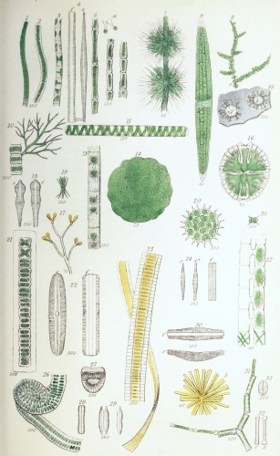 Plate V.
W Bagg sculp
London: John Van Voorst.
Conferva´ceæ.—On removing some of the soft green matter found adhering
to the stems of water-plants in any pool or pond, one of the species of
Conferva, C. flocculosa, is almost sure to be met with. On close
inspection with the naked eye, the green filaments of which it consists
are just visible, as extremely fine, soft, silky threads; and, under a
high power of the microscope, the filaments are seen to be unbranched,
and composed of a single row of cells (Pl. V. fig. 1), or joints, as
they are called in technical works; these are 2 or 3 times as long as
broad. In some specimens the joints are swollen, so as to present a
rounded outline. In another common species, C. bombyc´ina, the
filaments are somewhat more slender, and the joints are from 3 to 5
times as long as broad.
Cladoph´ora crispáta.—This Confervoid forms large, entangled,
dull-green masses, composed of branched, tufted, somewhat rigid and
coarse filaments. It is often a troublesome overrunner of the
fresh-water vivarium. The filaments are composed of thick-walled cells
(Pl. V. fig. 6), from 4 to 6 times as long as broad, and often
containing minute starch-granules.
The Confervaceæ have two modes of reproduction. The first of these
consists in the division of the endochrome of the joints into a number
of distinct segments, each of which becomes furnished at one end with
two very slender cilia (Pl. V. fig. 6 a). After a time, these ciliated
bodies, which are called zo´ospores (ξῶον, animal, σπορἀ, seed) or
gonid´ia (λονἠ, seed, εἶδος, resemblance), escape from the cells
either by their rupture or through a papillary orifice, and swim about
in the water, ultimately losing their cilia and growing into cells
resembling those of the parent plant. In the second method, which
occurs, for instance, in Conferva bombycina, certain of the joints
enlarge so as to become rounded or inflated; their endochrome then
becomes coated with a new cell-wall, and so forms a spore, which
subsequently escapes from the cell and germinates.
Chætophora´ceæ.—Draparnal´dia glomeráta forms small green jelly-like
masses, adhering to sticks and stones in water. These consist of
branched filaments (Pl. V. fig. 10), prolonged at the ends into
colourless hair-like points, and composed of single rows of cells, the
green endochrome forming a band across the middle of each cell, the ends
being colourless.
In Coleochæ´te scutáta (Pl. V. fig. 14) the cells are closely united,
so as to form a minute flat green disk. In the natural state, this
beautiful little object adheres to the submerged leaves and stems of
water-plants, and is therefore difficult to be found. But if a few
healthy water-plants be kept for some time in a glass jar, the little
Coleochæte, which is about as large as a pin’s head, will often be
found adhering to the side of the glass.
Bat´rachosper´meæ.—The members of this family resemble to the naked eye
the little masses of Draparnaldia, and they are found in the same
localities. They are of various colours, being green, brown, purple, or
red. They consist, as in Bat´rachosper´mum monilifor´me (Pl. V. fig.
8), of branched filaments, which have a knotty appearance under a low
power. The larger filaments are composed of cells arranged end to end,
the knots consisting of numerous smaller whorled filaments, i. e.
filaments arising from around them at the same level (fig. 7). The cells
composing the whorled filaments are beaded or moniliform, and are
prolonged into colourless hair-like points. The globules seen among the
branches (fig. 7) consist of groups of spores.
Zygnema´ceæ.—The members of this family resemble the Confervaceæ in
consisting of simple cellular filaments (Pl. V. figs. 11, 13), but
differ from them in the elegant arrangement of the endochrome: this
forms beautiful spiral bands, as in Spirog´yra quini´na (fig. 11), or
star-shaped masses, as in Zygne´ma crucia´ta (fig. 13). A remarkably
curious phenomenon met with in them is the manner in which the spores
are formed, and which is known as conjugation. In this process the
opposite cells of two distinct filaments, lying near together, push out
protrusions of the cell-walls, which meet and open into each other,
forming cross tubes, as in Spirog´yra nit´ida (Pl. V. fig. 12). The
contents of the opposite cells of the filaments then unite, forming
large spores, which remain either in the cells of one of the filaments
or in the cross tubes.
The three species figured are common in clear pools.
Desmidia´ceæ.—The Desmidiaceæ are truly microscopic, few of them being
even perceptible to the naked eye without the very closest examination.
They are very beautiful, on account of their bright green colour and
often elegant forms. Many of them are very common, existing in every
pond or ditch; but they abound most in clear open boggy pools on heaths.
On placing some water containing them in a glass jar and exposing it to
the light, they will often be found adhering to the glass, or forming a
layer on the surface of the muddy sediment.
The Desmidiaceæ consist mostly of single cells (Pl. V. figs. 9, 16); and
these consist of two equal halves or segments, as indicated either by a
paleness of the endochrome or a deep constriction at the line of
junction, which is called the suture. The cells are often elegantly
lobed and cut, or spiny; and in many the surface exhibits minute
markings, consisting of little protrusions of the cell-wall outwards, or
inflations, as they are called.
Their reproduction is effected by division and conjugation. In the
process of division the cells gradually separate at the suture, and a
new half-cell is formed upon each old half, which grows until it attains
the size and form of the original half of the parent-cell. The
conjugation is effected by two cells approximating so that their sutures
are near together, the cells then open at the sutures, and the effused
contents become united to form a spore or sporange, from which one or
more individuals are formed. These spores are often elegantly spinous on
the surface.
Among the species selected for illustration is Clostérium acerósum
(Pl. V. fig. 9), in which the cells are single, elongate, very slightly
curved or lunate; the endochrome forms long bands, often containing
numerous globules or transparent vesicles. At each end of the cells is a
round transparent vesicle, containing exceedingly minute granules, which
exhibit a trembling kind of motion. Between the cell-wall and the
cell-contents very fine currents may also be detected, forming a
circulation resembling that in the hairs of Tradescantia.
In Micrastérias rotáta (Pl. V. fig. 16) the cells, which are single,
are deeply cleft into two segments at the suture, the segments being
again regularly cut into five lobes, which are toothed or dentate.
In Hyalothéca dissil´iens (Pl. V. fig. 21) the cells are united into a
cylindrical filament, and are surrounded by a very delicate gelatinous
sheath. In Ankistrodes´mus falcátus (Pl. V. fig. 19) the cells
resemble those of Closterium in shape, but are aggregated into
faggot-like bundles, and are very much smaller. In the beautiful little
Pedias´trum boryánum (Pl. V. fig. 20) the cells are aggregated into a
disk, the marginal cells being bidentate or having each two points, so
that the whole resembles a star. The species of Pediastrum are
reproduced by the contents of each cell subdividing into numerous
ciliated segments or zoospores, which subsequently escape in a mass from
the cell, ultimately losing their cilia, and reuniting to form a new
individual.
In Scenedes´mus quadricau´da (Pl. V. fig. 25) the oblong cells are
united, side by side, the outermost cells being furnished with a bristle
at each end. The division of these cells takes place obliquely, so that
in the divided groups the cells are situated in two alternate rows.
The spores of many of the Desmidiaceæ are spinous, and they are often
found fossil in flint (Pl. V. fig. 15). To detect them in this
substance, thin slips of flint may be examined under a half-inch power;
or the chips of flint may be cemented to a slide with balsam, and ground
down on a hone.
The Desmidiaceæ must be mounted in the moist state: the smaller ones
will keep well in chloride of calcium; but the larger ones are injured
both by that liquid and by glycerine. The remarks made upon mounting, at
page 15, are especially applicable to these delicate organisms.
Diatoma´ceæ, or Siliceous Algæ.—The members of this family are singly
very minute; but when existing in large numbers, as they are often found
at the bottom of ditches and ponds, on the submerged stems of
water-plants, or upon damp ground, they form yellowish-brown evident
masses or strata. They occur both in sea-and in fresh water. They
usually consist, like the Desmidiaceæ, of single cells, which are called
frustules. But they are especially characterized by the cell-walls being
imbued with silica or flint, so that if the frustules be heated to
redness upon the point of a knife or a slip of platinum-foil, which
destroys the organic part of the cells, the coat of silica remains,
exhibiting the perfect form of the original cells or frustules. The form
of the frustules is very different in the various genera and species, as
represented in Pl. V. figs. 22, 23, 27, 30, 31, and Pl. VI. figs. 16,
17, 23; and it will be noticed that, in the figures, two views are given
of each frustule, f indicating the front view, and s the side view.
In all the front views, as in Pl. V. fig. 22, one or more lines will be
observed running longitudinally down the middle of the frustules, and
corresponding to the indications of division existing in the cells of
the Desmidiaceæ. Each half of a frustule is called a valve, and the
line at which these valves meet is called the suture. That side or
aspect of the frustule in which the suture lies (fig. 22 f) is the
front view; and the other aspect of the frustule (fig. 22 s) is the
side view. The frustules are mostly four-sided—the main breadths of
the two opposite valves forming two sides, and the bent margins of the
valves, with the back and front of the hoop, forming the two other
sides; so that the view presented by the side of a frustule is the same
as that of a single valve. The suture is the line at which the division
of the frustules takes place in the formation of new individuals. In
this process the cell-contents divide into two parts, as in ordinary
endogenous cell-formation,—the two new surfaces thus produced becoming
coated with a new portion of cell-wall or valve, so that two frustules
now occupy the place of the original one. At the same time a siliceous
band, encircling the frustules at the line of suture, is formed to fill
up the interval between the edges of the parent valves; this is the
hoop (Pl. V. fig. 22 f; Pl. VI. fig. 10 f), and beneath it lie the
two newly formed valves. In many cases I believe that each half-frustule
becomes coated with a new entire cell-wall, with its siliceous valves.
The frustules of the Diatomaceæ are constantly undergoing division when
in vigorous growth. After the frustules have divided, the new ones
either separate entirely, as is perhaps most commonly the case; or they
remain united, sometimes completely, so as to constitute a filament (Pl.
V. fig. 23), while at others the frustules cohere only at the angles (Pl
VI. fig. 23), so as to form a zigzag chain.
In some species, the frustules are attached to foreign bodies by means
of a gelatinous cushion (Pl. V. fig. 5; Pl. VI. fig. 7); while in others
they are situated upon a simple or branched gelatinous stalk (Pl. V.
fig. 17) or stipes (stipes, a stem).
When the frustules are examined in the living state, the cell-contents
resemble those of ordinary vegetable cells, excepting in regard to the
colour, and exhibit granules and globules, and sometimes a nucleus is
visible. It will also be noticed that many of the free frustules move
slowly across the field of the microscope; but the cause of the motion
is unknown.
When the frustules have been properly prepared, the surface of the
valves exhibits a number of coarser or finer markings, consisting of
dots, lines (striæ), flutings, or networks, &c., arranged with great
regularity and symmetry, often of extreme minuteness, and rendering them
exquisite objects under the microscope. The exhibition of these markings
requires not only that the valves shall be properly prepared and
mounted, but that the object-glasses be of good quality, and that the
management of the light be thoroughly understood; so that to a beginner,
their examination is often a matter of great difficulty; for only the
very coarsest or largest of these markings can be perceived in the
natural frustules.
The appearance of these markings, and even their apparent absence or
presence, frequently depends upon the kind of illumination used: thus,
under one kind of illumination the valves may appear simply white or
coloured, while under another they appear covered with lines, and under
a third with dots. It will often be observed, also, that the colour of
the valves varies according to the illumination and the power used—the
same valve appearing white, yellow, brown, blue, &c.; and the wet or dry
state of the frustules often cause a decided difference in their
appearance as regards colour.
To illustrate the forms and markings of the frustules and valves, we may
select the following species taking first those which occur in fresh
water.
In Epithémia tur´gida (Pl. V. fig. 30), the side-view or valve (s)
exhibits transverse or slightly radiating lines, with intermediate rows
of dots—these markings being continued over the margins of the valves
so as to appear also in the front view (fig. 30 f), ceasing at the
hoop. The frustules are curved or arcuate (ar´cus, a bow) in the side
view, oblong and narrowed at the ends in the front view.
In Fragilária capucína (Pl. V. fig. 23), which is extremely common in
fresh-water pools, &c., the frustules are united side by side into long
filaments, which are often twisted. In the separate and prepared
frustule, the front view (Pl. V. fig. 24 f) is rectangular, the valves
(s) being narrowly lance-shaped or lanceolate. The valves under
ordinary illumination appear colourless and without markings, but, by
proper management of the light, very fine transverse striæ are seen upon
them, consisting of rows of very minute dots. Fig. 24 s* represents
the valve of Fragilaria vires´cens, a nearly allied species.
Diat´oma elongátum (Pl. V. fig. 31) is often found with the above. Its
frustules are coherent at the angles. The front view (fig. 32 f) is
rectangular, often slightly narrowed in the middle; and the valves are
narrowly linear, and capitate at the ends; they are also transversely
striated.
In Synédra splen´dens (Pl. V. fig. 5) the frustules radiate from a
soft gelatinous cushion. They are linear in the front view (fig. 4 f),
the valves (fig. 4 s) being gradually narrowed or attenuated from the
middle to the ends, and exhibit transverse striæ interrupted opposite a
middle longitudinal line.
In Campylodis´cus costátus (Pl. V. fig. 27) the frustules are
disk-shaped and curved, so as somewhat to resemble a saddle. The
markings consist of central dots, with radiating coarse flutings.
Nitzsch´ia minutis´sima (Pl. V. fig. 28) has oblique valves, i. e.
the front half of the suture is not opposite the back portion; the
valves (fig. 29) are constricted in the middle, and the ends narrowed
and prolonged. The markings consist of a row of oblong dots or puncta
(punctum, a point). This species often forms yellowish layers upon
damp paths, &c.
In the next group, the valves have a longitudinal line running down the
middle of the valves, with a little knob or nodule in its centre (Pl. V.
fig. 22 s), both consisting of internal thickened portions of the
valves.
In Cocconéis placen´tula (Pl. V. fig. 33) the valves are oval, and the
markings consist of longitudinal rows of minute dots, with a marginal
row of puncta; these markings are invisible under ordinary illumination.
In Gomphonéma acuminátum (Pl. V. fig. 17) the frustules are attached
to a branched stalk (stipes); they are wedge-shaped or cúneate
(cúneus, a wedge) and transversely striate (fig. 18), the striæ
consisting of dots.
In Pinnulária vir´idis (Pl. V. fig. 22), which is very often seen
slowly traversing the field of the microscope when a drop of pond-water
is examined, the frustules in the front view are linear, the valves
being elliptic oblong, and transversely striated, the striæ consisting
of furrows.
In Gyrosig´ma (Pleurosig´ma) attenuátum (Pl. VI. fig. 16) the valves
are sigmoid, or somewhat resemble a Greek ς (sigma) in outline, and the
markings consist of rectangularly crossed rows of very fine dots; in the
front view, the frustules are linear-oblong with truncate ends.
Tabellária flocculósa (Pl. VI. fig. 23) has the frustules adherent
only at the angles, as in Diátoma. They are rectangular, and in the
front view exhibit a row of longitudinal dark lines interrupted in the
middle; these have been compared to the vittæ of the fruit of the
Umbelliferæ, and have received the same name.
Among the marine species may be mentioned Melosíra nummuloídes (Pl.
VI. fig. 9), in which the frustules are united into a chain or
cylindrical filament. This is very common among sea-weeds, &c.; and it
illustrates well the process of division of the frustules (fig. 10 f).
The valves are covered with fine dots, and near each end of the
frustules is a projecting rim encircling it, and appearing as a curved
line extending beyond the margin of the frustule in the front view. In
Actinocy´clus undulátus (Pl. VI. fig. 5) the frustules are separate,
disk-shaped, and the valves are divided into six equal parts by six
rays, each alternate portion of the surface of the valves being situated
on a lower level than those adjacent, so that an alteration in the focus
is required to bring into view the dots on any two adjacent divisions of
the valve. The surface of the valves is covered with easily recognized
dots. The form of the surface is best seen in the front view (fig. 5
f) when the frustule is placed on its edge.
Rhabdonéma arcuátum (Pl. VI. fig. 7), which is very commonly found
attached to sea-weeds, resembles Tabellária in the frustules having
the vittæ (Pl. VI. fig. 8 f). The frustules form short filaments,
attached by a little gelatinous cushion. The valves have transverse
striæ, interrupted in the middle (fig. 8 s).
Gyrosig´ma (Pleurosig´ma) angulátum (Pl. VI. fig. 17) resembles G.
attenuátum in the sigmoid form; but the markings consist of lines
crossing each other obliquely; and these are resolvable into rows of
dots (fig. 17 a) under suitable illumination.
In Coscinodis´cus radiátus (Pl. VI. fig. 3) the frustules are
disk-shaped, the valves being elegantly sculptured with easily
recognized cell-like markings or dots, so as to resemble a piece of
vegetable cellular tissue. But in some other species the dots are very
minute, and difficult to be shown satisfactorily. These markings consist
of depressions or pits in the surface of the valves. That this is the
case may easily be seen by examining a fragment of the valve, when the
shadings of the broken ends of the netted thicker portions, which
project like teeth, strongly contrast with the difficultly
distinguishable portions of the thin interspaces. The fossil forms from
the Bermuda deposit are best for the investigation of this structure;
many of these are extremely beautiful microscopic objects, their
markings resembling those on the engine-turned back of a watch.
The detection of the finer markings of the Diatomaceæ, which, according
to my view, consist of depressions like those upon the valves of
Coscinodiscus, is a matter of great difficulty to those who are
unaccustomed to the use of the microscope, and who have not a complete
set of apparatus. The main point to be attended to in bringing them into
view, is to use one-sided oblique light, i. e. to turn the mirror by
its stem as much as possible to one side, and then to incline it so as
to throw the light upon the object. In this way the valves of the
species of Gyrosigma, for instance, appear covered with lines (Pl. VI.
figs. 16 and 17); but the lines are spurious, i. e. they are the
optical expression of rows of minute dots (figs. 16 a, 17 a); and
when oblique light is thrown upon the valves from all sides, by means of
a special achromatic condenser, in which the central rays are excluded,
the dots become distinct, and the markings resemble those on the valves
of Coscinodiscus. To show the finer dots clearly, a valve should be
crushed, so as to obtain a fragment as flat as possible; for the surface
of the valves is curved more or less in all the species. The valves of
G. angulatum are generally used to test the quality of the
object-glasses of the microscope, and also for practice in “making out”
the lines and dots; there are, however, many Diatomaceæ more difficult.
As the nature of these markings is a disputed point, the discussion of
which is not adapted for an elementary work, I must refer for further
details to the ‘Micrographic Dictionary;’ it may be remarked, however,
that some observers have regarded them as cells, and others as
elevations or tubercles on the surface of the valves.
The preparation of the valves for showing the markings should be
effected by burning the frustules, or the mass containing them, on a
strip of platinum-foil over a spirit-lamp. The incinerated mass should
then be transferred to a slide, and the valves separated with the
greatest care by a bristle mounted in a hair-pencil stick under a low
power of the microscope.
This is, however, a substitute for the proper method, which is dangerous
in the hands of one unpractised in chemical manipulation. It is
this:—The mass of Diatomaceæ (the water containing it having been
carefully poured off as far as possible) is put into a Florence
oil-flask, and strong nitric acid (aquafortis) gently added, more than
sufficient to cover it. The mixture is then carefully boiled over a
spirit-lamp for some time. When it is cold, distilled water is added,
the whole shaken, and allowed to settle. The watery part is then gently
poured off, more water added, and this poured off after settling, and
the process repeated until a drop of the water evaporated to dryness on
a slide leaves no residue. The Diatomaceæ then form a white sediment at
the bottom of the water, and can be transferred to a slide with a
dipping-tube. The drop is then dried with a gentle heat, and the valves
mounted as dry transparent objects (p. 12).
If the valves have coarse markings, they may be mounted in balsam; but
if the markings are fine, balsam makes them much more difficult of
detection.
Many of the most beautiful Diatomaceæ are found in the fossil state; and
specimens of these are sold already mounted. I would advise those
unacquainted with them to purchase a slide of the “Bermuda” or
“Richmond” earth, which abounds in the species of Coscinodiscus; and
of the “San Fiore deposit,” which contains many species of Epithemia,
Navicula, Pinnularia, &c. These may be procured from Mr. Norman, 178
City Road, or from the microscope-makers.
Volvocin´eæ.—The Volvocineæ are inhabitants of clear fresh-water pools,
on heaths and bogs. They are very minute, of a rounded or plate-like
(tabular) form, of a green colour, and are pretty readily distinguished
from most of the other Algæ by their free motion; for they swim about in
the water like animals, as which they were formerly considered. They
consist usually of groups of thick-walled soft cells, each being
furnished with one or two cilia, by means of which the movement of the
compound bodies is produced.
In the beautiful Volvox globátor (which is not uncommon) the cells
form a hollow sphere (Pl. VI. fig. 18), studded with exceedingly minute
green spots or zoospore-like bodies, representing the green endochrome
of the component cells, and from each of which very fine radiating lines
extend, so as to give the surface a netted appearance; the lines
consisting of delicate processes of the endochrome, which may be
compared with those existing in the cells of the hairs of
Tradescantia. In the interior of the parent globes are often seen
several young organisms, usually eight, of a deep green colour; these
escape by the rupture of the parent, so as to form independent beings.
Sometimes they are found of a yellow colour, and furnished with a thick
transparent coat; these are called “resting spores,” as they remain for
some time before undergoing their full development.
The cilia of Volvox, of which there are two to each of the component
cells, are difficult to detect; they are best seen when the organism is
dried without a cover, or after moistening them with a little solution
of iodine, which dyes them brown.
Synúra volvox (Pl. VI. fig. 13) is a still more minute member of this
family, and is often found rolling along among Confervæ. The greenish
zoospore-like bodies of this Alga have one cilium only, and arise from a
common centre by a narrowing of the base (fig. 14).
In Gónium pectorále (fig. 11 a) the green bodies, which are sixteen
in number, and furnished each with two cilia, are grouped into a flat
square plate; and in the very minute Gonium tranquil´lum (fig. 11 c)
these bodies are also sixteen in number, and arranged in a tabular form,
but are without cilia.
Siphona´ceæ.—The structure of this family may be illustrated by the
genus Vauchéria, of which two or three species are common on damp
ground or in freshwater pools, forming a green layer. At first sight,
the filaments of which the little plants consist appear like those of a
stout Conferva; but on close examination they are found to be
branched, and not jointed, consisting of a single cell from end to end
(Pl. VI. fig. 26). The reproduction is effected by the agency of two
kinds of organs, antheridia and capsules (sporangia), situated near each
other (fig. 26 a) on the walls of the filaments, of which they are
protrusions or outgrowths—their cavities being separated from that of
the filament by a partition or septum. The antheridia produce
spermatozoa, and the sporangia each a spore, the one fertilizing the
other in the ordinary manner. In addition to this method of
fructification, zoospores are also produced—the ends of the filaments
becoming swollen, the contents cut off by a septum, and forming single
large zoospores covered with cilia, the further development of which
resembles that occurring in the Confervaceæ.
Oscillatoria´ceæ.—The members of this family are commonly found in
stagnant water or on shaded damp ground, especially in the cold seasons
of the year, forming green strata or masses.
| TABLE VI. [PAGE 84.] |
| Freshwater Algæ. |
| Fig. | |
| 1. | Oscillatoria autumnalis. |
| 2. | Oscillatoria nigra. |
| 3. | Coscinodiscus radiatus. |
| 4. | Nostoc minutissimum. |
| 5. | Actinocyclus undulatus. |
| 6. | Bacterium. |
| 7. | Rhabdonema arcuatum. |
| 8. | Rhabdonema arcuatum, prepared frustules. |
| 9. | Melosira nummuloides. |
| 10. | Melosira nummuloides, prepared frustules. |
| 11. | a, b, Gonium pectorale: 11 c. Gonium tranquillum. |
| 12. | Spirulina oscillarioides. |
| 13. | Synura volvox. |
| 14. | Synura volvox. |
| 15. | Gyrosigma attenuatum, front view. |
| 16. | Gyrosigma attenuatum, side view; 16 a, portion of a valve. |
| 17. | Gyrosigma angulatum; 17 a, portion of a valve. |
| 18. | Volvox globator. |
| 19. | Glœocapsa. |
| 20. | Chara vulgaris, globule. |
| 21. | Chara vulgaris, portion of filament. |
| 22. | Chara vulgaris, branch with nucule and globule. |
| 23. | Tabellaria flocculosa. |
| 24. | Tabellaria flocculosa, prepared frustules. |
| 25. | Palmella cruenta. |
| 26. | Vaucheria Ungeri (sessilis). |
| 27. | Vaucheria Ungeri, capsule. |
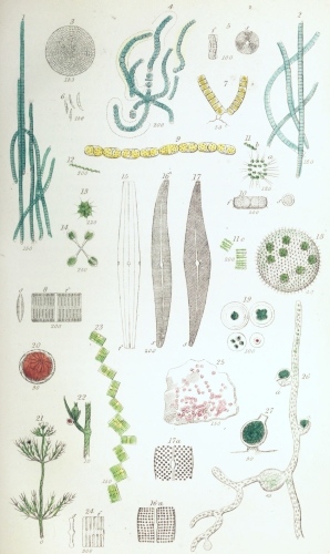 Plate VI.
W Bagg sculp
London: John Van Voorst.
Oscillatoria autumnális (Pl. VI. fig. 1) occurs everywhere upon damp
shaded banks of ditches, especially when newly made, forming a
greenish-black closely adherent stratum. Under the microscope it is seen
to consist of innumerable palish-green filaments; these are jointed or
transversely striated, some being straight, others curved, the ends
often exhibiting a writhing or worm-like movement. The appearance of
these fibres is peculiar, seeming as if they were solid throughout, and
so differing from that of the Confervaceæ, in which the cell-walls are
readily distinguishable from the cell-contents. The fibres easily break
across at the joints; and the last few segments are often narrowed and
rounded, so as to form a blunt point. When they have been left in water,
they exhibit colourless tubular sheaths surrounding and extending beyond
them. These sheaths consist of the consolidated outer portions of the
cell-walls; for when the cells undergo transverse division, and expand
by growth in the direction of the length of the filament, the original
septa or inner walls are broken through, and their remains may often be
seen on the inner surface of the sheath, appearing as little teeth.
Oscillatoria nigra (Pl. VI. fig. 2) is another very similar species,
forming blackish-green masses, and is common in ditches. It has longer
filaments than the last, with narrowed and slightly curved ends; and the
endochrome is distinctly granular.
In two other genera of this family, Vib´rio and Spirulína, the
filaments are spiral. Vib´rio spiril´lum is excessively minute,
colourless, and found in decomposing vegetable mixtures. The short
filaments move rapidly through the water, with a corkscrew-like motion.
In Spirulína oscillarioídes (Pl. VI. fig. 12), which is more rarely
found in clear pond-waters among Confervæ, the filaments are greenish,
and form a beautiful simple spiral, resembling that of a very slender
spiral vessel.
Lyng´bya murális (Pl. V. fig. 2) is very common on damp walls, gravel
walks, &c. It forms a bright grass-green layer, consisting of somewhat
rigid curled filaments. The endochrome is usually broader than long; and
the cells of the filaments are often found empty, the endochrome having
escaped in the form of gonidia.
Pl. VI. fig. 6 represents a species of Bactérium which is not uncommon
in decomposing vegetable liquids; the filaments are short, curved,
pointed at the ends, and have four joints.
Fig. 26 represents a Schizogónium, found upon damp paths. The
filaments resemble those of Lyngbya, but are united in pairs.
Fig. 3 represents a filament of a U´lothrix, which is common in
freshwater pools, showing the curious manner in which the endochrome is
arranged in the cells, forming bands partially lining the cell-walls.
Nostocha´ceæ.—Two species of the typical genus Nos´toc will serve to
represent this family. Nos´toc commúne is found on damp ground or in
ponds, and forms to the naked eye firmish, olive-green, skin-like,
plaited masses, an inch or more in diameter. Under the microscope it is
seen to consist of numerous beaded fibres, imbedded in worm-like
gelatinous sheaths; these are curved and interwoven to form the compound
mass. In the middle of many of the filaments is an enlarged colourless
cell, called the vesicular cell, which is related to the reproduction,
but in a manner not yet determined.
Nostoc minutis´simum (Pl. VI. fig. 4) forms solid gelatinous
bluish-green masses, varying in size from a pin’s head to a pea; it is
found upon unhealthy water-plants kept in glass vessels. The component
filaments are very slender, wavy, and the sheaths often have a brownish
tinge.
Ulvaceæ.—These Algæ are mostly marine—some, however, being found in
brackish or fresh water, or on damp ground, thatch, moss, &c. They are
generally of considerable size, forming flat or tubular fronds, often
several inches long, a few being filamentous. They consist of one or
more sheets or layers of cells, containing mostly green endochrome. This
at first fills the cells, but subsequently becomes converted into single
spores, or subdivided into numerous ciliated zoospores.
Ul´va latis´sima is very common on the sea-coast, being found attached
to stones, shells, &c. It forms a broad, flat, green, rounded or oblong,
thin frond, wavy and crumpled at the margins, and from 6 to 18 inches in
length. The minute cells form two layers, adherent to each other. The
zoospores formed are numerous in each cell.
Enteromor´pha compres´sa (Pl. IV. fig. 31) is also common in the sea
and in brackish ditches; it is often found floating. The frond is green,
tubular, flattened or compressed, and branched, the branches being
usually simple and narrowed at the base. The frond consists of two
layers of minute cells, separated by a space rendering it hollow. The
zoospores are numerous in the cells (fig. 32).
Palmella´ceæ.—These Algæ are found in fresh or salt water, or on damp
earth, wood, &c. They are green or red, forming round or irregular
masses or strata. They consist of loosely connected cells, imbedded in a
gelatinous mass or matrix, thus forming a frond.
Chlorococ´cum vulgáre (Pl. II. fig. 1) is very common upon the bark of
elm-trees, palings, &c., forming a green granular crust. It consists of
minute rounded or oval cells, mostly undergoing division into twos,
fours, or eights. These cells are attached to the sides or ends of very
fine colourless filaments. It is most probable that this organism, which
is usually placed among the Algæ, consists of the gonidia of a Lichen.
Chlorococcum muror´um forms a somewhat similar but soft and thin green
layer, upon damp walls or other porous bodies. It consists of very
minute oval green cells, with thick walls, and imbedded in the ends of
prolongations of a gelatinous matrix.
Palmel´la cruen´ta (Pl. VI. fig. 25) forms a portwine-red layer at the
bottom of damp walls or on the ground. It is composed of pale red cells,
imbedded in no definite order in a colourless gelatinous matrix. The
cells are filled with red granules, and are often found undergoing
division.
Pl. VI. fig. 19 represents a species of Glœocap´sa, in which the
cell-envelopes do not soften and unite to form a gelatinous matrix, as
in Palmella and other members of the family, but are persistent. This
species occurs in fresh water containing Confervæ.
Chara´ceæ.—This family consists of the single genus Chára, the
systematic position of which is not agreed upon by authors; as however
its structure will be better understood after what has been gone over,
it may be conveniently considered here.
There are several species of Chara, the one illustrated, Chara
vulgáris (Pl. VI. fig. 21), being commonly found in ditches and pools.
It consists of long main stems, often a foot in length, which are
branched, and surrounded at tolerably regular intervals by whorls of
branchlets. In some species, the stems and branches consist simply of
elongated cells, arranged end to end; while in others, of which Chara
vulgaris is one, the central cells are surrounded by a number of
narrower spirally arranged cells, forming an outer coating.
The Charæ have long formed interesting microscopic objects, on account
of the circulation of the protoplasm being visible in the cells, as in
the hairs of Tradescantia. This is best seen in those species in which
the outer layer of cells is absent from the stems, and which were
formerly arranged in a separate genus (Nitella). But it may also be
seen in the stems and especially the young branchlets of any of the
other species; and as the granules of the protoplasm are large, the
phenomenon is more easily witnessed than in Tradescantia.
The fructification consists of two kinds of organs, viz. red globules
(Pl. VI. fig. 22) representing the anther-organ, and green capsules
(fig. 22), or nucules, corresponding to the ovaries. The structure of
the globules is very curious. Their transparent walls (fig. 20) consist
of eight somewhat triangular plates, each of which is composed of cells
radiating from a centre; and from the inside of each of these centres
arises a tubular cell extending to the middle of the globule, the
unattached ends giving origin to numerous colourless coiled filaments,
consisting of minute cells arranged end to end, each containing a very
minute coiled spiral fibre, to which are attached two exceedingly
slender cilia. These ciliated fibres are the spermatozoa. The capsules
or nucules (fig. 22), which are situated near the globules, are
urn-shaped, coated with spiral cells, and crowned with five shorter
cells. When the globules are ripe, they become ruptured by the
separation of the valves; and the spermatozoa, escaping from the cells
of the coiled filaments, swim about and enter a canal in the capsules to
fertilize the ovule contained within.
The Charæ grow readily in a glass jar of fresh water, with a few
pebbles at the bottom; and if the plants be not overgrown with
Confervoids, the fructification will continue to be produced almost
throughout the year.
The circulation is best seen in the whorled branchlets, a portion of the
growing ends being placed in a live-box, or simply laid upon a slide in
water and covered with thin glass.
Preservation.—The Algæ are best preserved in two ways,—the entire
fronds being dried upon paper under pressure, as directed for the Ferns;
and small portions, showing the minuter structures and fructification,
being mounted in chloride of calcium or glycerine. If it is required to
preserve the marine Algæ according to the first method, they should
first be immersed for a time in fresh water, to dissolve out the saline
matters derived from the sea-water, which would keep them damp and
ultimately spoil them. After these matters have been removed, the fresh
water should be changed, and pieces of paper placed beneath them while
suspended in the water; on withdrawing the paper carefully, keeping the
Algæ at the same time spread out, they may be made to retain the
required position; and when the water has drained away, and the
remaining moisture has mostly evaporated, they may be submitted to
pressure in a press.
The Confervoid Algæ may be conveniently spread out upon paper and
preserved in the same manner, as some of the distinguishing characters
are founded upon their appearance in the dry state, their adhesion to
the paper, &c. Moreover they can then at any time be minutely examined,
by the immersion of a small portion in water.