A TREATISE — MEMOIR I. — ON THE FRACTURE OF THE CONDYLES OF THE LOWER JAW. — MEMOIR II. — ON THE FRACTURE OF THE CLAVICLE. — EXPLANATION OF THE FIRST PLATE. — MEMOIR III. — ON THE LUXATION OF THE CLAVICLE. — LUXATION OF THE STERNAL EXTREMITY. — LUXATION OF THE HUMERAL EXTREMITY. — MEMOIR IV. — ON FRACTURES OF THE ACROMION, AND OF THE LOWER ANGLE OF THE SCAPULA. — FRACTURE OF THE LOWER ANGLE OF THE SCAPULA. — MEMOIR V. — ON THE FRACTURES OF THE UPPER END OR NECK OF THE HUMERUS. 10 — MEMOIR VI. — ON THE FRACTURE OF THE LOWER EXTREMITY OF THE HUMERUS, WITH A SEPARATION OF THE CONDYLS. — MEMOIR VII. — ON THE LUXATION OF THE HUMERUS. — MEMOIR VIII. — ON THE FRACTURE OF THE BONES OF THE FORE-ARM. — FRACTURE OF THE FORE-ARM. — FRACTURE OF THE RADIUS. — FRACTURE OF THE ULNA. — FRACTURE OF THE OLECRANON. — MEMOIR IX. — ON THE LUXATION OF THE FORE-ARM. — MEMOIR X. — ON THE LUXATIONS OF THE RADIUS OVER THE ULNA. — LUXATION OF THE LOWER EXTREMITY OF THE RADIUS. — MEMOIR XI. — ON THE FRACTURES OF THE THIGH. — FRACTURES OF THE BODY OF THE OS FEMORIS. — FRACTURES OF THE UPPER END OF THE OS FEMORIS. — FRACTURES OF THE GREAT TROCHANTER. — FRACTURES OF THE NECK OF THE OS FEMORIS. — FRACTURES OF THE LOWER EXTREMITY OF THE OS FEMORIS. — EXPLANATION OF THE SECOND PLATE. 30 — THOUGHTS ON LUXATIONS OF THE OS FEMORIS, UPWARD AND FORWARD. — MEMOIR XII. — ON SPONTANEOUS LUXATIONS OF THE OS FEMORIS. — MEMOIR XIII. — ON THE FRACTURE OF THE ROTULA. — MEMOIR XIV. — ON THE FORMATION OF FOREIGN BODIES IN THE JOINT OF THE KNEE. — OBSERVATIONS AND REFLECTIONS ON FORMS OF APPARATUS FOR FRACTURES OF THE LEG. — MEMOIR XV. — ON THE DIVISION OF THE TENDO ACHILLIS. — MEMOIR XVI. — ON THE FRACTURE OF THE OS CALCIS. — MEMOIR XVII. — ON COMPLICATED LUXATIONS OF THE FOOT.
ON
FRACTURES, DISLOCATIONS, &c.
§ I.
1. The lower jaw, a kind of moveable hammer,
destined, to use the words of a certain physiologist,
to triturate the aliments against the almost
immoveable anvil of the upper jaw, is more exposed
to the action of external bodies, and consequently
to fractures, than most of the other bones of the face.
But all parts of it are not alike subject to such accidents.
Common in its body, but less frequent in
its branches or sides, fractures sometimes occur in
the two processes in which its branches terminate.
One of these processes, concealed by the zygoma,
embosomed in the temporal muscle, and covered by
the masseter, is less liable to fractures than the other,
which serves as the centre of the motions performed
by the bone, and is protected externally only by the
parotid gland.
§ II.
2. A fracture of the condyle may sometimes
arise from a counter-stroke, as when, in consequence
of some external force being applied from before
backwards, and from below upwards against the
chin, this process is driven against the projecting
rim of the glenoid cavity; at other times it may be
the effect of an immediate or direct stroke, as when
a body in motion strikes with force against the region
of the joint, and does violence to that portion of
the bone.
3. But in whatever manner the fracture may be
produced, it generally occurs in the slender part of
the bone which supports the condyle, below the insertion
of the pterygoideus externus. It is characterized
by a pain more or less acute, necessarily
accompanying the motions of the jaw; by a difficulty
more or less considerable, in the performance of
these motions; by a crepitation, oftentimes distinct,
when, in consequence of the angle of the jaw being
pushed forward, or the jaw itself alternately depressed
and elevated, the separated surfaces rub against each
other; by an inequality of surface sometimes perceptible
directly over the fractured condyle; by the
ease with which, on being pushed forward, it may
be forced into the depression beneath the zygoma;
and by its remaining stationary, during the movements
of the lower jaw, from which it is separated.
These signs, though generally characteristic, are
subject to an uncertainty proportioned to the swelling
that occurs in the part.
4. In this accident a displacement is almost always
produced by muscular action. The pterygoideus
externus, being attached to the condyle, draws it
forward and upward, towards the external wing of
the pterygoid apophysis, its fixed point of insertion.
On the other hand, the body of the bone is left behind,
being held by the masseter and external pterygoid
muscles, the course of which is opposed to a
displacement in the same direction; so that there
always exists a separation, more or less perceptible,
between the two fragments of bone.
5. Hence, if proper means be not used to restore
the contact between the broken ends of the bone,
the following consequences will be likely to occur:
1st. Their reunion will be tedious, because in every
bone this process is, in point of rapidity, inversely
proportioned to the separation of the divided surfaces:
2dly, This reunion may even entirely fail to
take place, if the bone be subject to the slightest
movements, as I have witnessed in a particular case,
where the condyle, not being reunited to the other
part, exfoliated, and was in part discharged through
the external integuments: 3dly, Under such circumstances,
the callus produced in the process of healing,
being situated near to the joint, and rendered irregular
and deformed by the separation of the parts, is apt
to impede muscular action, and do a permanent injury
to the functions of the jaw.
§ III.
6. As the whole apparatus in this case consists
in a passive resistance to the active powers employed
in producing a displacement, it follows from what
has been said (4), that the bandage intended to prevent
this displacement, and by that means to guard
against the accidents specified above (5), ought,
either effectually to bring back to its natural situation,
the condyle which is drawn forward, or pull in this
last direction (that is, forward) the body of the bone
which is still retained in its usual position, in order
that it may thus be brought into contact with the
condyle.
The first of these measures is impracticable, in
consequence of the situation of the condyles, which
are too deeply enveloped by the surrounding parts,
and offer a hold too small to be acted on. The
second, therefore, remains to be adopted, and is the
more easily executed, in as much as the angle of the
jaw, from its projecting and being but slightly
covered by the integuments, may without difficulty
be directed from behind forward by a proper force.
7. The fingers of the surgeon temporarily supply
this force, at the time of reduction; but it is necessary
that it should be permanently kept up by means
of the apparatus. This end is attained, in the following
manner:
Place behind the angle of the jaw, which must
be first pushed forward, thick compresses, to fill up
the hollow under the ear, and form an eminence
higher than the surface of the surrounding parts;
pass over these compresses, in an oblique manner,
the bandage commonly used in lateral fractures of
the bone, the application of which must in this case
commence on the sound side.
These compresses, being more projecting than
the surrounding surface, will necessarily sustain a
greater pressure, because the compression made by
a bandage is in proportion to the projection of the
part on which it is applied. Hence, being firmly
supported, they will retain the body of the bone in a
line with the displaced condyle (4).
8. In addition to this mode of applying the bandage,
it is necessary that the fractured bone should
be kept in a state of perfect rest. The internal pterygoid
and masseter muscles, tending by their contractile
efforts to draw the angle of the jaw backwards,
sometimes overcome the resistance of the apparatus,
and, by producing a second displacement, give rise
to the accidents formerly mentioned (5).
Let the lower jaw be now brought into perfect
contact with the upper one, and not separated from
it during the first few days after the injury, except
so far as may be necessary for the admission of nourishing
broths. Should a tooth have been lost, the
space which it occupied will furnish, without disturbing
the bone, an opening for the conveyance of
nourishment to the patient. Let talking, laughing,
and every thing that might produce a separation between
the body of the bone and the condyle, be carefully
avoided. The further treatment of the accident
should be such as is generally applicable to all fractures
of bones, and need not be at present particularly
detailed.
The following cases, reported by citizen Giraud,
second surgeon to the Hotel-Dieu, will confirm the
advantages of this mode of treatment.
Case I. Margaret Bessonet, aged thirty-four,
was admitted into the hospital on the 10th of May,
1791. On the preceding day she had received a violent
fall on her chin: a severe pain, and a preternatural
mobility in the left side of the jaw, had been the
immediate consequences of the accident: from these
symptoms, taken in conjunction with those formerly
mentioned (3), Desault discovered that a fracture of
the condyle existed, which he reduced and supported
in the usual manner (7).
After being somewhat uneasy during the first few
days, the patient became reconciled to the action of
the bandage, which, by inattention, had been two or
three times disturbed and put out of order, but
which, by being carefully reapplied, and aided by
the necessary precautions (8), restored to the bone
its natural form and solidity, by the thirtieth day, and
on the thirty-sixth the patient was discharged perfectly
cured. The only inconvenience she experienced,
was a slight difficulty in the motions of the
jaw, an effect naturally resulting from the long continued
inactivity of the muscles, but which was soon
removed by means of exercise.
Case II. Claudius Laurat, aged twenty-seven,
fell as he was carrying a heavy burden. In his fall his
chin struck with violence against a beam that lay in
his way. In an instant he experienced a sharp pain
in his right temple, and found it almost impossible to
move his jaw. Two hours afterwards a considerable
swelling appeared in the part, extending from the angle
of the jaw above the ear. The patient was admitted
into the Hotel-Dieu, where the circumstances of the
fall and the symptoms that followed, gave satisfactory
evidence of a fracture of the condyle. It was reduced
and supported as in the preceding case. On the day
following, the swelling was removed, doubtless by
means of the compression which had been made on
it; the other symptoms (3), hitherto scarcely perceptible,
became more obvious; the bandage was reapplied,
and the disease terminated, in about twenty-nine
days, in the same manner with that of case 1.
§ I.
1. Man enjoys an advantage which nature has
bestowed on but few of the quadrupeds, namely, a
power of moving his upper extremities in every
direction. The clavicle being a kind of arch placed
between the breast and shoulder, forms a centre,
moveable indeed but solid, for these motions, a part
of which can no longer be performed, when this arch,
in consequence of being broken, ceases to afford
them a point of support. Hence it follows, that the
fracture of this bone may be said to reduce the individual
who sustains it, when considered in relation
to its functions, to a level with that numerous division
of animals that are destitute of clavicles.
2. Few diseases of the kind are more frequently
met with than this. The natural curve of the clavicle,
its situation immediately under the skin, the
want of a support to its middle part, the great proportion
of spongy substance which enters into its composition,
the projection of the shoulder exposing it
to the action of external bodies; all these circumstances
concur in rendering the accident frequent,
particularly among that class of men subject, from
their occupations, to violent exertions of the upper
extremities.
Here then, more than in the generality of fractures,
we should feel an interest in the advancement
of the art of surgery; and yet, having hitherto employed
in it but feeble means, our efforts have been
attended with imperfect success. Hippocrates has
observed, that some degree of deformity almost always
accompanies the reunion of a fractured clavicle;
all writers since his time have made the same
remark; experience has confirmed the truth of it,
and as much time has been spent in hypothetical
speculations to explain the accident, as in serious inquiries
how to prevent it. At length Desault proved
that a feeble and unskilful mode of treatment was the
sole cause of a want of success, and that, by being
more correct and judicious, art might be as successful
here, as in other fractures. In order to give a
correct view of his practice in this disease, I will
examine the causes, varieties, and signs of a fracture
of the clavicle; the accidents of which it is susceptible;
the mode and the causes of the displacement
of the broken ends of the bone; the indications that
arise out of those causes, and the manner of answering
these indications as well during, as after, the reduction.
§ II.
OF THE CAUSES AND VARIETIES.
3. The action of external bodies is almost the
only known cause of this fracture, whether these bodies
strike the shoulder with violence, or the shoulder
be forcibly driven against them. But this action is
not in every case the same; its application is most
frequently mediate or indirect, but is sometimes
immediate or direct.
In the first case there is a true counter-stroke, the
ordinary effect, either of a severe blow on the point
of the shoulder, which is the most common occurrence;
or, as happens less frequently, of a fall on the
arm when it is extended for the purpose of guarding
the body from the force of the accident. Under
these circumstances, being pressed between the
sternum, which makes resistance, and the body
which acts on its extremity, the clavicle is bent in
that direction which is most natural to it; but, not
being sufficiently flexible, it gives way generally in
the place where its curvature is the greatest. Thus
the ribs are broken, when the sternum, by being
violently driven backward, forces them to bend in
the centre beyond their natural flexibility.
In the second case, the fracture occurs at the
spot where the stroke is given. Here the momentum
or quantity of force applied on the bone, surpassing
the solidity which the bone possesses, its
continuity is necessarily destroyed.
4. But in whatever way the fracture is produced,
it is either oblique or transverse, single or double, in
the middle or towards the extremities of the bone,
simple or compound.
An oblique fracture is most frequently the effect
of a counter-stroke; a transverse fracture is the more
common result of the immediate action of external
bodies; a counter-stroke seldom produces any thing
but a simple fracture; while compound fractures are
generally owing to a direct stroke. The one produces
a solution of continuity in the middle of the bone,
or thereabout; because in that part the curvature
is most considerable. The other is almost always
the cause of this solution, when it occurs at the extremities.
To the latter alone, is a double division
to be attributed. The reason of these differences
is already so plain, that it would be a waste
of time to dwell on an explanation of them.
§ III.
OF THE SIGNS.
5. The several phenomena that attend a fracture
of the clavicle, taken together, leave in general but
little doubt as to its existence, particularly when the
fracture is oblique. As is the case in most other instances
of the kind, so here, an acute pain is felt at the
instant of the stroke; sometimes a cracking of the
bone is distinctly heard by the person injured; on
every occasion, it becomes suddenly impracticable
to perform circular or rotatory motions with the arm;
motions from before backwards can still be executed,
but are difficult and painful, and, as I have already
observed (1), the individual injured is reduced
to the class of animals destitute of clavicles.
Oftentimes the shoulder of the injured side, being
more or less depressed, loses its level with the
other. It is also evidently drawn forward and inward.
The distance between the acromion and sternum, on
the affected side, is found on comparison, to be evidently
less than on the opposite side. In almost
every case, that portion of the fractured bone, which
adheres to the sternum, forms a visible protuberance
above and on the inside of the shoulder.
6. In the mean time the pain continues. The painful
drawing or dragging occasioned by the weight of
the arm forces the patient, for the purpose of relieving
it, to bend his body towards the side affected, and incline
his head in the same direction. This forms a peculiar
attitude, which of itself was frequently sufficient
to disclose to Desault the nature of the disease. We
have oftentimes witnessed him establishing the truth
of this diagnostic, by merely looking at patients entering
the amphitheatre, who had been brought thither
for the reduction of such fractures.
By this position, the pains are generally relieved,
because the arm finds some degree of support; but
should the patient wish to change his position, or
perform any particular motions, the pains return
almost as acutely as at first.
7. If to these signs, which are almost all of them
founded in reason, we add those that are still more
palpable to the senses, such as the mobility of the
two broken ends of the bone; the crepitation produced
by their friction against each other; the depression
felt at the point of fracture, by passing the
fingers over the upper surface of the bone; and the
facility of restoring to it its natural form and direction,
by moving the shoulder upwards, outwards,
and backwards; it will be difficult to be mistaken
respecting the nature of this fracture. This is perhaps
more particularly the case, when the fracture is
oblique, as this kind offers the most striking diagnosis,
and cannot be involved in uncertainty, unless when
a considerable swelling occurs in the parts around the
fracture. But, even then, as the circumspection of
the practitioner will necessarily direct his attention to
this circumstance, the obscurity of the signs will have
no unfavourable influence on the cure.
8. When the fracture is transverse, there is sometimes
more difficulty attending the diagnosis. The
corresponding inequalities of the divided surfaces
may mutually penetrate each other and interlock,
and thus prevent a displacement. Does any uncertainty
on this score exist? Placing your fingers on the
two extremities of the bone, order an assistant to
move the arm in every direction, and the motions
will be communicated to the clavicle; but, if a fracture
exist, they will be most perceptible in the fragment
adjoining the shoulder, and will separate it from
that attached to the sternum. This method will seldom
deceive us, is easily employed, and subjects the
patient to but a momentary pain.
§ IV.
OF ACCIDENTS.
9. We do not generally find fractures of the clavicle
accompanied by such accidents as the anatomical
relations of the parts might lead us to apprehend.
The external force being all expended in fracturing
the bone, extends but feebly to the brachial plexus,
which would be much injured by the shock, were
the bone to yield, without breaking, to the action of
external bodies striking against it. Hence, without
doubt, would arise serious affections, as may be fairly
inferred from the analogy of blows on the head and
vertebral column, and as is indeed confirmed by
certain cases reported by Desault.
Case I. Two bricklayers were brought to the
Hotel-Dieu, who had met with similar accidents. A
piece of timber, thrown from a building, in which
they were engaged, had struck them, the one on the
external part of the left clavicle, the other about the
middle of the right. A considerable wound pointed
out in each the place on which the blow had been received.
But the former, having escaped a fracture,
experienced nothing but an acute pain, while the
second had the bone broken in two places.
The customary apparatus was applied to the latter,
and the treatment which we shall presently describe,
being pursued, the result was that complete success
which never failed to crown the attentions of Desault.
In the other patient a considerable swelling made its
appearance the day after the accident. On the third
day a numbness and partial loss of the power of motion
occurred in the arm of the affected side. Soon
afterwards an insensibility came on, and by the seventh
day, the paralysis of the arm was complete. It
was not till after a tedious treatment, an account of
which would be foreign from my present subject,
that the limb recovered in part its original strength.
From whatever cause the fracture of the clavicle
in this latter patient was prevented, it is evident, that
the whole of the force employed to produce the fracture
in the other, acted here on the brachial plexus,
and gave rise, by means of concussion, to the accidents
which followed.
10. The axillary artery, though running near to
the clavicle, in common with the brachial nerves,
experiences, notwithstanding, less frequently than
they do, injurious effects from the fracture of this
bone. I know not of any instance where a puncture
from the broken ends of the clavicle has produced in
this artery a false aneurism. To conclude, like all
other fractures, that of which we are now treating,
may be connected with wounds, splinters, &c. But
in general, as Hippocrates remarks, the fracture of
the clavicle assumes in common cases a mild aspect.
§ V.
OF DISPLACEMENT.
11. Most of the symptoms formerly mentioned
(5 and 6) as accompanying a fracture of the clavicle,
are evidently the result of a displacement of its broken
ends. Yet this phenomenon, taken notice of by
all authors, and considered by them as a necessary
effect of the disease, does not occur in every case (8).
There are instances, in cases of transverse fractures,
where the extremity attached to the shoulder, has
retained its natural position. Three examples of
this kind occurred in the Hotel-Dieu in the course
of the year 1787.
12. Instances have also been known, in which the
sternal fragment, when fractured obliquely upwards,
has supported the end of the humeral in such a manner
as to prevent any derangement. Desault was
accustomed to relate several cases, where similar
occurrences took place; but, in general, this state
of things is rare, in comparison with that in which
the fragments lose their natural level.
Almost always, then, there is more or less of a
perceptible overlapping (chevauchement) produced,
either, by the elevation (a circumstance which is
very rare) of the external fragment over the internal;
or, (as commonly occurs) by the depression of the
former beneath the latter.
13. Of the first of these modes of displacement
(a mode but rarely mentioned by authors) a few examples
are to be found among the observations of
Desault, one of which he has recorded in his journal.
Hippocrates speaks of the phenomenon as a thing
that was familiar to him.
14. The second kind of displacement, that which
we constantly find in practice, and which the laws
of muscular action render almost inevitable, takes
place in such a manner that the shoulder appears to
obey the impulse of two powers, one of which draws
it downwards, and along with it the external fragment
of the clavicle, which is displaced by this power
in the direction of its transverse diameter, or
thickness. The other power approximates the shoulder
to the breast, and draws it forward, carrying
along with it the same fragment, which is by this
means displaced in a longitudinal direction.
That we may the better understand them and
their effects, let us, in our minds, separate these two
powers, although they are perfectly simultaneous in
their action. A knowledge of them will lead us to a
knowledge of the resistances which ought to be
opposed to them. But let us first remark, that the
humeral fragment, being drawn downward and inward,
takes sometimes such a direction, that its
internal extremity passes backward under the sternal
fragment, its external end continuing to point forward:
this disposition can be understood from its
natural direction.
15. The first of these powers, namely, that which
depresses the point of the shoulder, appears to have
escaped the notice of the ancient physicians of
Greece, who attributed the apparent depression of
this part, to the elevation of the sternal fragment,
and, accordingly, endeavoured by making compression
on the latter, to restore it to a level with the
other. Hippocrates, more judicious than those who
had preceded him, demonstrated that their doctrine,
false in its principles, was still more dangerous in its
consequences, and that the sternal fragment being
immoveable, lost its relative position with respect to
the humeral, only because the latter was depressed
by the weight of the arm. This doctrine of the father
of physic is satisfactorily proved, by a comparison of
the sound shoulder with the diseased one, and has
since been admitted by all practitioners. Indeed, the
mere recollection that one of the uses of the clavicle
is to support the shoulder at that level necessary for
the performance of its functions, is alone sufficient
to convince us, that, in case of its ceasing to fulfil
that office, the shoulder must obey the laws of its
own gravity, increased by that of the hand and arm.
16. The illustrious Petit, and with him Duverney,
in acknowledging this cause of displacement, have added
to it as another the action of the deltoid muscle on
the external end of the bone; in this action, the end
of the clavicle is the moveable point, while the humerus
affords the fixed point. But how can we
admit this cause, when the humeral fragment, in
passing under the sternal, moves in a backward
direction? So far is the deltoid muscle from drawing
the bone downward, that here the bone rather draws
the muscle in part backward, and yet, in such a case,
the displacement is as perceptible as in any other.
Besides, when the sternal fragment, broken obliquely
upwards, supports the humeral and prevents a displacement,
why does not the deltoid produce this
displacement?
It is then in the weight of the arm and shoulder
alone, that we must look for the passive power,
which depresses them, and which produces a displacement
in the direction of the transverse diameter
or thickness of the clavicle.
17. A second power, highly active, co-operates
with this. I allude to the permanent contraction of
the muscles, that extend from the breast to the clavicle
and shoulder: from this cause arises the displacement
in the longitudinal direction of the bone.
The pectoralis major, the pectoralis minor, the
subclavius, the serratus major, and the trapezius,
unite their efforts in producing this displacement.
These muscles are, in certain respects, antagonists
to each other, but they all unite in drawing the
shoulder forward and inward. None of them appears
to act with more effect than the pectoralis major.
To this, in particular, is to be attributed the displacement
in a forward direction.
Except in the instances stated above, the action
of the muscles is not immediate. They act only
secondarily on the external fragment, which, being
stedfastly attached to the scapula and humerus, is
obedient to the motions impressed by the muscles
on these two bones; motions which, in a sound state,
the clavicle has a power of controlling.
18. To the weight of the lower extremity (15
and 16), and the spontaneous action of the muscles
(17) must be added, as another cause of displacement,
the motions which are communicated to the
arm by external bodies, and which, being imparted
ultimately to the clavicle, derange the fragments, by
separating them, approximating them, or making
them overlap each other, according to the direction
in which they act.
19. When a fracture occurs at the extremity next
the shoulder, no displacement of the fragments in
general takes place. This circumstance is attributed
to the action of the trapezius, which draws each fragment
upwards with equal force. However this may
be, it is doubtless to such cases that we must refer
the complete cures, obtained without any retentive
apparatus, by Gasparetti, Brown, and other writers.
Hence also, without doubt, arise the difficulties experienced
by certain practitioners, such as Duverney,
with respect to the diagnosis of this disease. These
fractures may be mistaken for fractures of the acromion,
being situated so immediately in its vicinity.
§ VI.
OF THE REDUCTION.
20. On looking into the causes of that displacement
(15...18), so common in fractures of the clavicle,
it appears that in almost every case, the external
extremity of the humeral fragment is drawn, by
a double power, downward, inward, and forward.
Hence it follows, 1st. That the resistance opposed to
this power, by the means used for the purpose of
reduction, and the retentive apparatus subsequently
employed, ought to be directed upward, backward,
and outward, these directions being the reverse of
those in which the powers of displacement act: 2dly.
That, in as much as these powers, viz. the weight
of the parts and the action of the muscles, are in
constant operation, and, besides, as the motions of
the arm are continually disturbing the fragments of
the bone, the apparatus ought to be equally constant in
its action, and should keep up, without any remission,
the effect produced, at first, by the means of
reduction. This principle is applicable to every
case, and ought to be the standard of comparison, for
determining the advantages or disadvantages of different
bandages, and processes for the reduction of
fractures of the clavicle.
21. But we are not to suppose, that these processes
have heretofore manifested an exact application
of this rule. Hippocrates directed to press the
arm close to the ribs, and at the same time to push
it upwards, in such a manner, as to make the shoulder
appear as sharp and pointed as possible. Hence
his precept, to lay the patient down on his back, the
back being supported by some projecting body, and
then to press the shoulders backward; hence again,
when the humeral fragment is drawn inward, his
advice to press the elbow close to the breast. This
twofold expedient was attended with great difficulties,
even under the direction of the father of
medicine. Celsus only copied Hippocrates, adding
nothing whatever to his mode of practice. Paul of
Egina, more judicious in this case, conceived, that
for the purpose of forcing the shoulder outward, and
rendering it, agreeably to the idea of Hippocrates,
very projecting and sharp, it would be advisable to
place the fulcrum or point of support, not in the
middle of the back, but under the arm-pit. A woollen
ball was employed by him for this purpose, a
practice which would, at once, have carried the art
near to perfection, if, after being employed to reduce
the fragments, this process had been continued for
the purpose of retaining them in apposition.
22. No new method distinguished the surgery of
the Arabians. It is necessary to come down to the
time of Guy of Chauliac, before we meet with the
method which is almost universally adopted at present,
and which consists in placing between the
shoulders, the knee of an assistant, whose hands are
to be employed in drawing them forcibly backwards.
But it is evident that this is only doing, while the
patient is in an erect position, what Hippocrates did,
after having laid him with his back on a projecting
body. Here, then, the art seems to have degenerated,
after the time of Paul of Egina: and, indeed, on
comparing this process with the general principles
already established (20), it will be immediately
perceived, that the powers of replacement do not
here act in an opposite direction to those of displacement.
Hence the difficulties of reduction, the time spent
in the operation, and the sufferings by which it was
sure to be accompanied. The fragments were
brought together, it is true; but it was only by varying
the movements, and changing their direction,
that the point of contact was ultimately found.
23. Desault conceived, in the year 1768, that to
reduce, in the most effectual manner, a fracture of
the clavicle, it was necessary not only to push the
shoulder backward and upward, as was commonly
done, but, above all, to force it outward, and that the
power destined to draw it in this latter direction,
ought to act horizontally, according to the course of
the clavicle, in the same way, as, in an oblique fracture
of the thigh or leg, the extension for replacing
the fragments is made in the direction of the bone.
24. As the union of the humerus to the clavicle,
by means of the scapula, communicates to the one
the movements of the other, it is easy, by placing
the ball used by Paul of Egina, under the arm-pit,
to convert the arm into a lever of the first kind.2
The lower extremity of the arm being then pressed
towards the body, the upper end is separated from
it, and becomes, with regard to the clavicle, what
the efforts of an assistant who makes the extension,
in a fracture of the leg, are to the foot of the patient.
The mode of reduction being established, it was
necessary, in the next place, to invent a bandage, calculated
to retain the broken ends of the bone in contact.
Desault thought it practicable to unite these two
points of treatment, in the same process, that is to say,
to reduce, and at the same time to retain the fracture.
Here the art is indebted to him for an improvement,
which, I will venture to say, carries it near to
perfection. To judge of this, it will be necessary
only to take a hasty survey of the different kinds of
apparatus proposed by different writers.
§ VII.
OF THE MEANS OF RETAINING A REDUCTION.
25. Here all authors seem to have been directed
by the same principle. This is to keep the shoulder
of the affected side, 1st, drawn forcibly backwards,
2dly, approximated towards the shoulder of the sound
side. Such was the practice of the Greek physicians,
whom we have seen in common with Hippocrates,
Celsus, and Paul of Egina, employing a kind of
bandage, varied in its form, according to the displacement
it was intended to remedy.
Above all others, we find an application of this
principle, in the figure of 8 bandage, a particular
form, which was employed in practice by Albulasis,
an Arabian, and afterwards by his countrymen, as
well as by Lanfranc, Guy of Chauliac, and their contemporaries.
The use of this bandage was continued
by Pare and his successors, and has been lately
modified by several authors, such as Heister, Petit,
Brunninghausen, &c.
26. But under whatever form it shows itself, its
action is always the same, and always insufficient.
On comparing its effect with the general principle,
on which every apparatus for the clavicle should be
constructed (20), we perceive, that it by no means
answers the threefold indication, of retaining the
shoulder backward, outward, and upward.
27. In relation to carrying the shoulder backward
it loses half of its effect, because, its force being decomposed
(so to speak) by the obliquity of its direction,
is divided into two channels. One of these
runs parallel to the shoulder and acts to no purpose,
while the other, being perpendicular to it, is alone
effective; hence it must act with a force equal to 10,
in order to produce an effect equal to 5.
28. The indication, to draw the humeral fragment
outward, far from being fulfilled, is here diametrically
counteracted. The scapula, being approximated
to that of the opposite side, draws the humeral
fragment towards the trunk, making it underlap
the internal one, and, in this respect, the figure of 8
bandage acts posteriorly in precisely the same manner,
during the treatment, that the contractions of
the muscles did anteriorly before the reduction.
29. Should the shoulder be supported, at such an
elevation, as might have a constant tendency to destroy
the influence of its own gravity? this is evidently
prevented by the very oblique direction of
the turns of the bandage. Suspending the arm in a
sling, is the only way, in which that end can be attained.
But does this mode always possess sufficient
firmness and stability? The arm, not being here sufficiently
fixed, may be constantly in motion, which, by
deranging its situation, must communicate very troublesome
and injurious movements to the fragments
of the clavicle. One of the principal faults of all bandages
consists, in not preventing these movements,
by restraining the movements of the arm.
To the other disadvantages of this mode, need I
add that of its making, by the turns of the bandage,
an undue compression on the projecting edges of
the arm-pit, and producing thereon troublesome and
painful excoriations?
30. From the want of a mutual correspondence
and fitness between the indications already enumerated
(20), and the manner in which the figure of 8
bandage acts, it is evident that the former can never
be satisfactorily fulfilled by the latter. Hence we
may judge, what improvement the art has received
from the iron cross of Heister, the compress of Petit,
drawn transversely over the oblique turns of the
bandage, the waistcoat which Brasdor fastened round
the thorax of his patients, and the leathern apparatus,
lately proposed by a German practitioner. These
means, though diversified in their form, are similar
in their effect, and, being nothing but modifications
of the figure of 8 bandage, possess, like it, the radical
fault of not offering a resistance directly opposed
to the two-fold power, arising from the muscular
action and the gravitation of the shoulder.
As to what remains, it will be sufficient to show
the insufficiency of the process of reduction (22), by
means of the knee placed between the shoulders, in
order to demonstrate the existence of a like insufficiency
in all those forms of apparatus, which, as
Brasdor remarks, have for their object a continuance,
during the treatment, of the effect produced by that
process.
31. On the other hand, those indications will be
fulfilled with exactness, by such a form of apparatus
as will render permanent the action of the means of
reduction which were employed by Paul of Egina,
by certain Arabian physicians, and by Pare; which
have been renewed by Desault, and tend to draw
the shoulder upward, backward, and outward (23
and 24).
Pecceti appears, in the last century, to have had
a faint view of the proper indication on this subject,
when, under the article of fractures, he advises the
ball to be suffered to remain under the arm during
the treatment of the injury. But the figure of 8 bandage,
united to this expedient, counterbalances its
effect, rendering it of no avail, and Pecceti was
therefore no more successful than others, in obtaining
a cure of the fracture, unaccompanied by deformity.
32. An overlapping more or less perceptible
never failed to accompany the consolidation or knitting
of the bone, and here, as in many other cases,
practitioners laboured to explain what they knew not
how to prevent. The impracticability of surrounding
the part, as in other fractures, with a circular
bandage, appeared to Heister, Petit, and Duverney,
to be the cause of this deformity. They supposed
that to be a superabundant callus, which was
nothing but a displacement of the fragments. These
visionary hypotheses ceased to exist, as soon as this
displacement was prevented by a proper apparatus.
Desault sought for this form of apparatus, as well
as for his other bandages, in the multiplied application
of means already known, without inventing
new ones. Bandages and compresses, easy to be
procured, and already rendered familiar to surgeons
by daily use, served him for the construction of his
apparatus, for which several machines had been already
proposed.
33. The pieces of which this apparatus is composed,
are,
1st. Three rollers, three inches broad; the two
first, six, and the other eight, ells long, each one
rolled up separately.
2dly. A bolster or pad (a, b, Fig. 1), made in the
form of a wedge, out of pieces of old linen. Its
length should be equal to that of the humerus, its
breadth four or five inches, and its thickness at the
base (a), about three inches.
3dly. Two or three long compresses.
4thly. A small sling for the arm, (Fig. 5).
5thly. A piece of linen large enough to cover the
whole bandage.
Every thing being properly arranged, the following
is the mode of applying the apparatus, which of
itself reduces the fracture.
34. The patient being placed in a standing position,
or, if his case render that impracticable, on a
seat without a back, an assistant elevates the arm of
the affected side, and supports it at nearly a right
angle with the body (Fig. 2), while the surgeon
places under the arm-pit the head of the bolster,
which descends along the side of the thorax, and
which another assistant, situated at the patient’s
sound side, holds by the two upper corners.
35. The surgeon now takes one of the first rollers,
applies the end of it on the middle of the bolster,
fixes it there by two circular turns round the body,
and passes a turn obliquely (a a) along the fore part
of the thorax, ascending to the sound shoulder: the
roller then descends behind, passes under the arm,
and returning in front of the thorax, makes a circular
turn and a half, horizontally. Having reached
the hind part of the thorax, it reascends obliquely by
the cast (b), as it had done before, and passes over,
before, and under, the sound shoulder; having thus
crossed the turn (a a), the roller again passes across
the hind part of the thorax, and finishes by circular
turns, which completely cover the bolster. A pin is
now to be fixed in the place of crossing of the roller
on the sound shoulder, to prevent the turn (a) from
slipping downward.
The application of this first roller is intended for
no other purpose, than firmly to fix the bolster which
is held up by the two oblique turns before and behind,
and secured against the body, by the subsequent
circular turns.
36. The bolster being fixed, the surgeon applying
one hand to its external surface, pushes it upwards,
and, with the other, taking hold of the elbow,
after having half-bent the fore arm, lowers the arm,
till it is laid along the bolster. He then presses its
lower extremity forcibly against the side of the thorax,
pushing it upwards at the same time, and directing
its upper extremity a little backwards.
The application of the bandage constitutes a part
of the process of reduction. The humerus, now converted
into a lever of the first kind, is drawn at its
upper end from the shoulder, in proportion as its
lower end, is approximated to the thorax. The
scapulary fragment being drawn along with it, and
directed at the same time upward and backward,
comes into contact with the sternal fragment, and in
an instant the deformity of the part disappears.
37. The arm being thus situated, is given in
charge to an assistant, who retains it in the same position
in which he received it from the surgeon, by
pressing on it with one hand, and with the other
supporting the fore arm half bent, and placed horizontally
across the breast.
The second roller is next to be applied. The
end of this is carried under the arm-pit of the sound
side. It is then brought across the breast, over the
superior part of the diseased arm, and extends across
the thorax behind till it passes under the arm-pit.
Two circular turns cover the first. The roller must
then ascend to the lower part of the shoulder, by
oblique turns (c. c. Fig. 3), each of which must be
overlapped by the succeeding one, to the extent of
about the third part of its breadth. It is necessary
that these turns be applied in such a way, as to bind
but very gently above, and to increase in tightness,
as they descend nearer to the lower extremity of the
humerus.
The use of this second roller is, to supply the
place of the hand of the assistant, in pressing the
arm against the side of the thorax; its effect evidently
is to draw the upper extremity of the arm outwards,
and, as it is already directed backwards, to retain it
in that position. The compression of the circular
turns on the arm, being thus gradually augmented,
becomes, on the one hand, more efficacious, because
it acts on a greater surface, and on the other, less troublesome,
because, being more divided, it is less felt
at the lower extremity of the arm, where it bears
with most force.
38. A third indication remains still to be fulfilled,
namely, to retain the shoulder in its elevated position,
and, by that means, to assist in the extension of the
fragments, which already has some effect in preventing
a depression.
To fulfil this indication, an assistant sustains the
elbow in its elevated position, with one hand, and,
with the other, supports the patient’s hand before his
breast, while the surgeon fills with lint the hollow
spaces around the clavicle. He then applies on the clavicle,
at the place where it is fractured, the two long
compresses, wet with vegeto-mineral water, or some
other cooling liquid. Taking now the last roller, he
fixes the end of it under the sound shoulder; from
thence he brings it obliquely across the breast, over
the long compresses, and carries it down behind the
shoulder along the posterior part of the arm, till it
passes under the elbow. From this point, he again
carries it obliquely upwards across the breast to the
arm-pit, then across the back, over the compresses,
and brings it down again before the shoulder, along
the front of the humerus till it again reaches the elbow.
From thence the roller again ascends obliquely
behind the thorax, passing under the arm-pit,
where the first cast of the roller is covered, and from
whence it again starts, to run the same course we
have just described. This constitutes a second
round, which covers in part the first, and forms a
kind of double triangle (e, f, d), situated before the
breast, and over the circular turns of the other rollers
(c. c. Fig. 4). The remaining part of the roller,
brought from behind forward, is employed in circular
turns over the arm, and round the thorax, for the
purpose of preventing the displacement of the first
part. To make it the more secure, it is fastened with
pins at its different places of crossing.
The sling (Fig. 4) is next passed under the hand,
and fastened above to the ascending turns (d), and
not to the circular (c c), which the weight of the
hand would be likely to draw downward.
39. It is only necessary to examine the course of
this third roller, to see, that, united to the sling, it is
well calculated to support the external fragment,
which the weight of the shoulder has a tendency to
depress, on a level with the internal one. It supplies
the place of the assistant, who raises the elbow and
supports the hand of the patient, in like manner as
the second roller performs the office of the assistant,
who presses the lower part of the humerus against
the side of the thorax.
On the other hand, the circular turns, by which
the application of the third roller is finished, being
directed from before backward, push in the same
direction the arm and shoulder, which have been
already carried that way, by the process of reduction,
and thus retain them in their proper places.
Hence may be inferred the truth of the proposition,
which we have been endeavouring to demonstrate;
namely, that the bandage of Desault, constructed
according to the general principle formerly
established (20), for fractures of the body of the clavicle,
is calculated to retain the external extremity
of the humeral fragment upward, outward, and backward.
40. The casts of the rollers, thus surrounding the
thorax, however well they may be secured, are yet
liable to be displaced, particularly when the patient
is in bed. This inconvenience may be avoided, by
surrounding the whole with a piece of linen, leaving
nothing uncovered, but the sound arm, which is at
liberty to perform its usual motions.
The arm of the diseased side, being thus fixed in
such a manner, as to constitute a whole or entire
body with the thorax, follows its movements, without
producing any displacement. It is thus, that by
the apparatus for a continued extension of the thigh,
the fragments of the os femoris, forming an immoveable
whole with the pelvis, cannot change their situation,
even in following the motions of the trunk.
Hence arises, in fractures of the clavicle, this advantage,
that the patient is not obliged to keep his
bed, but is able even to attend to his business, during
the progress of the cure.
41. I will not dwell on the numerous objections
urged by different authors against the bandage which
has just been described. What answer, indeed, can
be given to those writers, who fancy that they behold
the patient in the greatest danger of immediate suffocation;
who dread an approaching mortification of
the arm of the diseased side; who allege, contrary
to the rules of the art, that there is no impression
made immediately on the clavicle, but on a neighbouring
bone; who, &c. &c.? Twenty times in a
year, has experience answered those objections, in
the Hotel-Dieu; and there is not a pupil of Desault,
who has not, as well in this, as in many other cases,
seen that objections, plausible, indeed, when considered
in the closet, or at a distance from a sick room,
dwindle to nothing at the bed-side of the patient.
42. In those cases (which, as Hippocrates remarks,
very rarely occur) where the external fragment projects
over the internal one (12), the bandage must
be somewhat varied, although the two principal indications,
of drawing the shoulder backward and outward,
must still, as in other cases, be fulfilled. The
only additional circumstance, therefore, necessary to
be attended to here, is, not to elevate the shoulder, by
pushing it upwards. This may be easily avoided,
1st, by omitting to raise the elbow, when applying
the bandage; 2dly, by drawing the third roller a little
tighter than usual.3
The fragments, being reduced to the same level,
and brought into apposition, by this two-fold attention,
will unite as in ordinary cases.
If the fracture exist at the end of the clavicle next
to the humerus, the difficulty of their being displaced
renders the application of the bandage less necessary.
Prudence, however, demands that it be not altogether
neglected.
§ VIII.
OF THE TREATMENT DURING THE FORMATION
OF THE CALLUS.
The regimen to be pursued during the reunion of
the clavicle, varies according to circumstances. It is
impracticable to lay down general rules, applicable to
all affections of this kind. Here, however, much
more than in other cases, if the division of the bone be
simple, and no unfortunate accident occur, it is always
unnecessary to restrain the patient from his
usual course of life, beyond the second or third day.
But, though internal means are for the most part
omitted in the treatment, the apparatus is a subject on
which too much attention cannot be bestowed. With
whatever degree of exactness it may be at first applied,
it will soon become loose, and oppose a diminished
resistance to the weight of the shoulder, and
the action of the muscles. Hence, unless it be frequently
examined, the fragments will be displaced.
The following case furnishes a detail of the treatment
subsequent to the reduction, to which, in ordinary
cases, Desault had recourse.
Case III. Mary Adel, aged thirty, as she was
crossing a path covered with ice, in the severe winter
of 1788, fell on the point of her left shoulder, and
fractured the clavicle about the middle. Being
brought to the Hotel-Dieu a few hours after the accident,
she was dressed in the manner just described,
and, as the fracture was simple, it was judged sufficient
to make a slight diminution in the quantity of
her aliment, during the two or three first days. The
dressing was moistened every morning, with vegeto-mineral
water, at the place corresponding to the fracture.
On the fourth day the piece of linen that surrounded
the bandage was removed, for the purpose of
examining the state of the parts. Every thing was
found in its proper situation, and the covering was
replaced till the seventh day, when the rollers appeared
to be somewhat relaxed. The apparatus was
taken off, and reapplied as at first, the compresses
being carefully moistened with vegeto-mineral water,
at the part lying over the fracture. After the third
day the patient was permitted to return to her usual
regimen. The third roller being a little deranged
on the tenth day, it was taken off, and reapplied as
at first, together with the sling. The fragments were
examined and found in perfect contact. The patient
was up during the whole day, walked about the
house, and experienced no other inconvenience than
that of not being able to use the left arm.
On the thirteenth day, the bandage was again reapplied,
and allowed to remain till the sixteenth, when
the patient having disturbed it, it was once more
changed. At this period, the fragments, already firmly
united, exhibited scarcely a vestige of the division
they had sustained.
The reunion was complete by the twentieth day,
when all the pieces of apparatus were dispensed with,
except the bolster and the second roller, which were
also removed two days afterwards, as they were found
to be no longer necessary.
The continued inactivity of the limb, during the
treatment, had occasioned a stiffness in the shoulder.
This was gradually done away by making the patient
move her arm in all directions, twice a day, each
time, for the space of an hour.
On the twenty-ninth day she left the hospital,
carrying with her nothing to remind her of the injury
she had sustained. She was free from that uneasiness
which is the consequence of a tedious and ill-managed
treatment, during which the exercise of the limb
has been neglected.
§ IX.
OF COMPLICATIONS.
44. We are in possession of but few observations
particularly relative to the different complications,
that may accompany fractures of the clavicle. The
treatment, in such cases, varied according to circumstances,
must be accommodated to the indications
common to all fractures of this kind.
When splinters, displaced in different directions,
whether adhering to the bone or not, irritate the soft
parts, and, having passed through the integuments,
appear without, most practitioners advise to remove
them and cut off such parts as project beyond the
fractured end of the bone, previously to reduction.
This direction is founded on the severe pains which,
in such cases, accompany the common treatment of
the injury, and which the figure of 8 bandage always
augments, by drawing the shoulder inward, and consequently
pressing the soft parts against the projecting
parts of the fragment, or the points of the splinters.
But if the splinters, adhering as yet to each other and to
the bone, by means of the periosteum, have not assumed
the nature of foreign bodies, (that is, if they be not
actually dead) it is always proper to replace them. It
is here only that we meet with an occasion for that
part of the process, of reduction denominated conformation,4
which is never requisite in other cases.
A fragment which has penetrated the soft parts,
but has not been long exposed to the air, disappears,
and is replaced by extension, provided it be properly
directed. Being retained afterwards in a state of constant
extension, it can neither be displaced, nor cause
pain by irritating the parts, which is the inevitable
result of the figure of 8 bandage.
In cases of this kind, it is useful to protect the
shoulder with a small splint, which may support the
turns of the bandage, and prevent their pressure on
the splinters, or the broken ends, which they might
otherwise disturb. These precautions are alike indispensable
when the fracture is double.
Case IV. Francis Ricord, twenty-five years of
age, was received in the month of July, 1790, into the
Hotel-Dieu of Paris. On the preceding day, a piece
of timber having fallen from a considerable height on
his right shoulder, had broken the clavicle of that
side into several pieces. Severe pains, which occurred
at the moment of the accident, had continued
throughout the night, and were still sensibly felt. The
slightest motion of the part augmented them to such
a degree, as to extort from the patient piercing cries.
The point of the shoulder being very much depressed,
was also drawn perceptibly forward and inward;
and a large echymosis, without any external
wound, occupied its whole extent.
Desault being satisfied that the several fragments
were all connected together, and that none of them
was separated from the periosteum, placed, as in ordinary
cases, the bolster under the arm, completed
the reduction, and applied a splint along the course
of the clavicle, after having, with his hands, brought
the fractured pieces into contact. Confident, then,
that the form of the part was perfectly restored, he
applied the bandage, which was moistened with
vegeto-mineral water, twice or thrice a day.
At the moment of reduction the pains ceased, and
were felt no more till the fifth day, when the bandage
being a little relaxed, admitted of a slight displacement
of the fragments. This displacement was removed,
and the pains along with it, by the reapplication
of the apparatus.
During the six first days a very strict diet was
enjoined. This, however, was dispensed with by
degrees, till, on the thirteenth day, the patient returned
to his usual regimen. On the seventeenth
day, there remained nothing of the echymosis, but a
yellow tinge, the customary consequence, of such an
accident. The precautions inculcated in the preceding
case, were employed also in this, and the patient
was discharged perfectly cured, on the forty-second
day from the time of his admission. Nor had he
experienced, during his treatment, those severe and
long continued pains, which, under a different management,
so frequently accompany this kind of fracture.
Fig. 1. A bolster made in the form of a wedge, intended
to be placed between the arm and the side
of the thorax.
a. Its base, which should fit the hollow of the arm-pit.
b. Its summit reversed, against which the elbow is to
be applied.
Fig. 2. The first roller applied for the purpose of
fixing the bolster against the side of the thorax.
a a. Oblique casts before, passing over the opposite
shoulder, in order to hold it up.
b. Oblique casts from behind, crossing the first ones
on the shoulder.
d d. Circular casts round the trunk, covering the bolster,
which they fix laterally.
Fig. 3. The second roller, applied to fix the arm
against the bolster.
a & b. Portions of the oblique casts of the first roller,
left uncovered by this one.
c c. Turns of the second roller, covering those of the
first, loose above, and tighter below, for the purpose
of drawing the superior extremity of the
humerus outwards.
d. Their passage over the side opposite to the bolster.
Fig. 4. The third roller intended to keep the point of
the shoulder raised.
a a & b. Oblique casts of the first roller, remaining
uncovered.
c c. Turns of the second, seen through the opening
of those of the third.
d. Oblique casts of the third, ascending from the arm-pit
over the shoulder of the diseased side, to descend
again behind, along the arm, and pass under
the elbow.
f k. A continuation of the preceding casts, reascending
under the sound arm-pit, and from thence
behind the thorax, over the diseased shoulder.
e. A continuation of the same casts, descending on
the fore side of the arm, passing under the elbow,
and ascending again under the arm-pit of the
sound side.
g. The remainder of the roller, intended to be employed
in circular turns, in order to secure the
casts e, and prevent them from slipping outward.
Fig. 5. A sling which should be fastened to the oblique
cast d (Fig. 4), to support the hand.
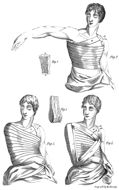
Pl. 1
Engrav'd by W. Kneafs.
§ I.
1. The clavicle, which forms a moveable abutment
for the shoulder, and receives and sets bounds to
most of the movements of that part, and of the arm,
exhibits at its extremities, two articulations, essentially
different from each other in their form, dispositions,
and uses. These differences give rise to differences
equally essential with regard to the dislocations
to which they are subject.
2. On the sternal extremity, a small surface, convex
from above downwards, and concave from before
backwards, is fitted, by means of an intervening cartilage,
to a much smaller surface of the sternum,
concave and convex in opposite directions.
One capsule, two ligaments, viz. the interclavicular
and costo-clavicular ligaments,5 and the anterior
portion of the sterno-cleido-mastoideus muscle,
strengthen the connexion of these two surfaces, and
tend, on one hand, to prevent their luxation, while,
on the other, this luxation is favoured and facilitated
by the following circumstances: 1st, the disproportion
between the dimensions of the two articulating
surfaces; 2dly, the mobility of the joint; and 3dly,
by this joint’s constituting a kind of centre for the
motions of the arm.
3. On the humeral side, an elliptical surface,
slightly convex, and inclined downwards, is immediately
joined to a corresponding surface of the acromion,
elliptical also, a little concave and directed
upwards. Hence two kinds of inclined plains, which
would be very liable to dislocations, by sliding easily
over each other, were they not firmly secured by a
capsule, by accessory fibres, by the intersection of
those of the deltoid and trapezius muscles, and,
above all, by two ligaments, the rhomboid and the
conoid.6
Having laid down these preliminary considerations,
let us proceed to examine, in particular, each
kind of luxation to which the clavicle is subject.
§ II.
OF THE CAUSES AND DIFFERENT KINDS OF DISPLACEMENT.
4. The sternal articulation of the clavicle experiences
different changes, according to the different
movements of this bone. If these movements be in
a backward direction, the articulating surface is
turned forward, straining the anterior part of the capsule,
the corresponding ligament, and the extremity
of the sterno-cleido-mastoideus muscle. If, on the
other hand, they be in a forward direction, the posterior
ligament, and the adjacent portion of the capsule
are overstretched. In motions directed upwards, the
costo-clavicular ligament, and the external and inferior
part of the capsule, and in those directed downwards,
the inter-clavicular ligament, and the internal
portion of the capsule, experience a similar degree of
tension.
5. Hence it follows, 1st. That the natural movements
of the shoulder may be regarded as predisposing
causes of luxation, because at the part where
tension is excessive, the ligaments are disposed to
give way, and suffer the sternal extremity to escape:
2dly. That the efficient causes will be, all external
forces acting on the clavicle in such a way as to increase
its motions beyond their natural degree, and
beyond the resistance which the ligaments are capable
of making. Thus a fall on the point of the shoulder,
forcing it suddenly backward and inward, produces
a luxation forward. But, in general, as the
strength of the articular ligaments is superior to the
resistance of the clavicle itself, a fracture takes place
more frequently than a luxation, in the proportion of
nearly six to one.
Though falls on the point of the shoulder are oftentimes
productive of luxation of the clavicle, they
are not the exclusive causes of that accident. Desault
has seen the sternal extremity forced from
its cavity by the knee being pushed violently against
the middle of the back, while the shoulders were
drawn at the same time backwards.
Case I. A porter dislocated his clavicle in the
following manner. He was carrying a very heavy burden,
suspended from his shoulders by cords that
passed under each arm-pit. Being desirous of resting
himself by the way, he placed on a block the burden
he carried, which slipping backward, drew his shoulders
in the same direction, and at the instant of his
attempting to retain it and prevent it from falling,
produced a luxation of the clavicle.
7. It follows from what has been said respecting
the different states of the articulation, during its various
motions (4), that the clavicle is not equally liable
to be luxated in every direction. Inclining naturally
backward, but a very slight degree of motion in that
direction is necessary, to effect a luxation forward.
To produce a luxation backward or inward, it is necessary,
on the other hand, that the humeral extremity
of the bone should make a sweep at least three
times the extent of that required in the preceding
case. Besides, motions in this direction are accompanied
with pain, particularly if they be made by force.
Luxation downward is prevented, on the one hand,
by the cartilage of the first rib, which presents to the
bone an insurmountable barrier. On the other hand,
to produce this kind of luxation, it would be necessary
for the external extremity of the clavicle to be
forced upwards, an occurrence very seldom occasioned
by falls. Luxation upward, or over the superior
edge of the sternum, must be the effect of a
stroke, which, by depressing the point of the shoulder,
and forcing it at the same time forward, presses
the sternal extremity against the internal and superior
part of the capsule, which, being thus lacerated, suffers
a luxation to take place. But such a derangement
of the articulating surfaces is very rarely produced
by falls. Whence it follows, that of the different kinds
of luxation of the clavicle, that in a downward direction
is altogether impracticable. Those backward
and upward, though possible, occur but rarely in
practice; while that in a forward direction, on the
contrary, is not an unfrequent accident. This tends
to confirm the observations of practitioners, and particularly
of Desault, whose immense collection on
the subject furnishes examples of the last kind of
luxation only.
8. In these luxations, there is for the most part, a
rupture of the capsular ligament, and an escape of the
bone through the opening. But sometimes the ligament
is only preternaturally distended, and then the
luxation is incomplete.
§ III.
OF THE SIGNS.
9. But whatever may be the causes or kind of the
luxation, its diagnosis is always easy. If it be forward,
the direction of the stroke which the shoulder has
received, furnishes, at first, some ground of suspicion.
The accident is certainly known by the appearance of
a hard and unnatural protuberance in front of the sternum,
and behind the extremity of the sterno-cleido-mastoideus
muscle, by the existence of a sensible depression
or hollow at the joint, and by the situation of
the shoulder, which is pushed further backward, and
is less projecting and more approximated to the trunk,
than in its natural state. Add to these, a difficulty in
performing motions in a forward direction, which,
when somewhat forced, reduce, in proportion as they
are accomplished, the size of the protuberance formed
by the displaced end. The head is always inclined
towards the side where the luxation exists; an attitude
which relieves the painful drawing or tension
produced in the sterno-mastoideus muscle, by the
humeral extremity pushing it forward.
10. A protuberance over the superior edge of the
sternum, a difficulty in raising the shoulder, the pain
which results from attempting such a motion, the diminution
of the protuberance which it occasions, the
absence of the sternal extremity from its natural cavity,
the approximation of the shoulder to the thorax, and
its depression and diminished projection, compared
to its usual state, afford evidence of a luxation upwards.
11. A luxation inward or backward, would be
characterized by a projection of the shoulder exteriorly,
by a difficulty in performing motions in a backward
direction, by the alarming effects, which, as Petit
remarks, the compression of the trachea would doubtless
produce, and by a depression or hollow at the
joint, more perceptible here than in the two preceding
cases.
12. These appearances will be more or less striking,
accordingly as the membranes, lacerated or only
distended, offer a greater or less resistance.
§ IV.
OF THE REDUCTION.
13. To reduce a luxation, is, in general, to make
the bone re-enter its cavity, by retracing, or returning
along, the same route which it followed in escaping
from it. Now, in a forward luxation, the displacement
is from behind forward, in an upward one from
below upward, in an inward or backward one from
before backward, but, in each of the three, it is more
particularly from without inward. In the first case,
therefore it is backward, in the second, forward, in
the third downward, but, in each of the three, more
particularly outward, that the powers for producing
extension must be directed.
14. Hence the method generally employed by
most practitioners, recommended by almost every
author who has written on the subject, adopted by
Petit, Duverney, Heister, &c. and which consists in
placing the knee between the shoulders of the patient,
as a point of resistance, by the aid of which the shoulders
may be drawn backward, fulfils only half of the
indication of cure; because at the time that the humeral
extremity is drawn backward, it is not directed
sufficiently outward.
Hence a difficulty of replacing the bone sometimes
occurs, a difficulty always removed, when, pursuant
to the method employed by Desault in fractures of
the clavicle (see Desault’s method), the arm is
made to serve as a lever of the first kind, to carry
backward and outward, the head of the bone, which
is displaced in the opposite directions, when the luxation
is forward. This method possesses the advantage,
not only of giving the powers of extension a
proper direction, but also of increasing them to a
degree even beyond what is necessary for effecting a
reduction, by removing them further from the resisting
force. Hence it is unnecessary to adopt any particular
measures for restoring and preserving the form
of the part, as the extension is alone sufficient for that
purpose.
These principles, evidently applicable in effecting
a reduction, are still more strikingly so in the means
destined for retaining it. Let us apply what I have
just said, to a case of dislocation in a forward direction.
It will be easy to transfer it afterwards to the
other kinds of luxation.
§ V.
OF THE MEANS OF RETAINING A REDUCTION.
15. Few luxations are so speedily reduced, but
few are more easily displaced again, than that of the
clavicle. This disposition is the reverse of that of
most other luxations, which are reduced indeed with
difficulty, but seldom afterwards suffer a displacement.
The cause of this we find, 1st, in the extreme
mobility of the clavicle, to which all the motions of
the arm are communicated; 2dly, in this further consideration,
that most of the muscles, which have their
insertion towards the shoulder, tend to draw this bone
inward, when the ligaments, in consequence of being
either broken or distended, as happens in this accident,
do not offer a sufficient resistance.
16. From this two-fold cause of displacement,
arises a two-fold indication in the arrangement and
application of the apparatus. These are, 1st, to render
the clavicle immoveable, by restraining every
kind of motion in the shoulder and arm; 2dly, to
retain the extremity of the clavicle outward, a direction
opposed to that in which it has a tendency to be
displaced. But if to those indications we compare
the forms of apparatus hitherto used, we will readily
perceive that they are insufficient to fulfil them.
17. The figure of 8 bandage, so generally in use,
and all the various modifications, under which it has
been revived, without being improved, fix the clavicle
in the very direction most favourable to a displacement,
and even do it in the very manner in which
that accident is sometimes brought about; as maybe
seen in the history of the case of the porter (6). This
bandage does not, under any of its modifications,
prevent the motions of the shoulder, because it does
not restrain those of the arm, which remains free and
unencumbered. Far from constituting an antagonist
power to, it even co-operates with, that which has
produced the displacement. (For further light on this
subject, see what has been already said on the fracture
of the clavicle, pages 22 and 25.)
Bell, in condemning the figure of 8 bandage, not
so much because of its action being insufficient, as
because of its obstructing respiration, proposes, as a
substitute for it, a kind of machine analogous to the
iron cross of Heister, which, being fixed by straps
passing under the arm-pit, and round the neck and
body, is intended to retain the parts firm and immoveable.
But the motions of the arm not being restrained,
nor the action of the muscles of the shoulder opposed
by an antagonizing power, places this piece of
apparatus in the same class with those, which, from
not being devised and constructed on a proper view
and conception of the causes of displacement, have
no affinity to rational practice.
18. The apparatus for a continued extension, invented
by Desault, for fractures of the clavicle, fulfil
here all those indications in which the others fail.
By this, 1st. The aim, being firmly fixed against
the side, by means of the roller (c c Fig. 3), can communicate
no motion either to the shoulder, or the
clavicle. 2dly. The shoulder itself, being forcibly
drawn outward, with the upper extremity of the
humerus, by the action of the kind of lever into which
this bone is converted, and to which the bolster (a b
Fig. 1) serves as a fulcrum, cannot, by its movements,
derange the luxated bone. 3dly. The sternal
extremity, being drawn both by the muscles which
tend to displace it inwardly, and by the bandage
which acts on it in an opposite direction, remains
fixed between those two antagonizing forces, which
thus destroy each other. Hence the apparatus of
Desault, when accurately applied, offers to both of
these powers of displacement, a resistance perfectly
calculated to combat them.
20. We must, however, admit that this apparatus
partakes of one inconvenience, common indeed to all
bandages, but which is perhaps more particularly applicable
to this in consequence of the numerous casts
of the rollers that form it, namely, the great facility
with which it becomes relaxed. Hence one cause of
displacement, which the most exact and scrupulous
attention cannot at all times prevent.
Case. Desault had, for a long time, the care of
a patient, whose luxation, having been neglected for
four days, was reduced on the fifth, by a surgeon,
who, for the purpose of retaining it, employed a bandage
of a particular kind. An hour afterwards, a
motion of the shoulder backward, displaced the
luxated extremity: a new reduction was the consequence;
on the day following, another displacement,
and so on in succession, for ten days, at the expiration
of which, Desault being consulted, applied to
the part the bandage formerly described.
On being examined the next day, the apparatus
was found in a favourable state. On the day following,
a slight displacement rendered necessary a new
application of the bandage, which, this time, continued
longer than before. But, about the expiration of
the third day, the projection of the bone was again
considerable. Finally, the patient recovered, with a
very perceptible protuberance in front of the sternum,
and a difficulty of motion, great at first, but less afterwards,
and which exercise succeeded ultimately in
removing.
21. The application of the apparatus differs from
that intended for a fracture of the clavicle, only in
this, that it is of service to place on the luxated extremity,
graduated compresses,7 calculated to make
pressure backward and outward, and which are to be
secured by the turns of the roller (b Fig. 4).
A second precaution, not less essential, is, to
push the humeral extremity of the clavicle, a little
forward, and fix it in that direction, in order that the
sternal being directed backward, may be removed
from the place8 through which it has a tendency to
escape.
22. Desault almost always obtained complete
success by this process, and by the most accurate
attention to prevent the relaxation of the bandage.
In the mean time, a stiffness, more or less considerable,
always remains in the joint for a long time after
the reduction, and it is not unfrequently a month
or two before the part recovers its usual facility of
motion.
The following cases, collected by Brochier, confirm
the doctrine for which I have been contending.
Case II. A man luxated the clavicle by falling
on the point of his shoulder, and forcing it backward.
He was immediately brought to the Hotel-Dieu,
where Desault demonstrated to his pupils,
that the head of the bone, carried in front of the sternum,
was removed nearly an inch from its natural
cavity, the ligaments of which were no doubt lacerated.
Here, as in the fracture of the clavicle, the application
of the bandage answered the purpose of reduction,
and removed the protuberance formed by
the extremity of the bone.
The patient, being strong and vigorous, and having
received besides a violent contusion, was bled
twice, and confined to a low diet. On the following
day, no derangement; on the fourth day, a slight
displacement of the bone, the rollers a little relaxed,
bandage applied anew. Eighth day, no sensible displacement.
Eleventh day, some swelling around the
joint; compresses, wet with vegeto-mineral water,
ordered to be frequently renewed. Twentieth day,
the swelling almost gone, and no disposition to a
displacement; the apparatus was removed; motions
at first difficult, and contracted. Twenty-ninth day,
more free and easy. Thirty-fourth day, returned to
their natural state.
Case III. Mary Rivert luxated her clavicle, on
the seventh day of January 1789. Being brought
some time afterwards, to the Hotel-Dieu, she was
treated in the same manner as the foregoing patient,
and with the same result, except that a very slight
protuberance remained at the extremity of the bone,
and the confined state of the motions continued a
little longer. Desault related, in his lectures, other
instances of cures being performed without the least
remaining deformity.
After all, even supposing the method just proposed,
to possess no other advantage, than that of diminishing
the protuberance of the bone, which, under
other modes of treatment, is almost inevitable,
and by that means preventing the motions of the part
from being confined, it would still, without doubt,
be a great step towards the perfection of the art.
23. Should a luxation backwards occur, the same
process of reduction should be adopted, with this difference,
that the extension ought to be made forward
and outward; and the same apparatus should be applied
for retaining the parts, except that the humeral
extremity ought to be directed a little backward, in
order that the sternal extremity, being carried forward,
may be removed from the place of laceration
in the capsule.
In like manner, should the bone be luxated upwards,
it would be necessary to draw the arm outwards,
and elevate slightly the point of the shoulder,
for the purpose of depressing the sternal extremity.
§ V.
OF THE KINDS OF LUXATION.
24. Luxations of the humeral extremity of the
clavicle, take place, according to Petit, in two ways,
1st, under, and 2dly, over the acromion. If we attend
to the disposition of the articulating surfaces, the
superior of which rests obliquely on the inferior; if
we examine, in particular, the relative position of the
corocoid apophysis with respect to the clavicle, it
will be difficult to conceive how the first kind of
luxation can occur, without being accompanied by a
fracture. Yet some facts added by Desault to the
doctrine of Petit, on this point, seem to demonstrate
the possibility of the clavicle sliding under the acromion.
As to luxations forward and backward, the
mobility of the shoulder, the facility with which it
yields to motions impressed on it in these two directions,
and the want of a resisting power, make the
two bones that compose it, move together, still preserving
their relative position.
The luxation upward, then, is that which ought
chiefly to occupy the attention of the practitioner.
Yet even this is less frequent than the luxation of the
sternal extremity, on account of the very great
strength of the retaining ligaments, which, when this
luxation occurs, must be, if not lacerated, at least
very much distended.
§ VI.
OF THE MECHANISM AND THE APPEARANCES.
25. A fall on the point of the shoulder is the most
frequent cause of this luxation. The two articulating
surfaces, representing an inclined plain, slide along
each other, in such a manner, that that which belongs
to the acromion is pushed inward, while that of the
clavicle is directed outward. The capsule being
stretched, gives way, and then the displacement is
manifested by a preternatural protuberance over the
acromion; by a stiffness in the motion of the shoulder;
by the direction of this part, which is evidently
drawn inward and downward; by the inclination of
the head of the patient to the side affected; by a
bending of the body; and by severe pains in the
luxated part. These characters are essentially distinct,
and ought to have prevented the error of
Galen, who mistook a case of this kind for a luxation
of the os humeri downward. Hippocrates and
Ambrose Pare have foreseen the possibility of this
mistake and even warned young practitioners to be on
their guard against it. But, as citizen Sabattier judiciously
observes, the position of the head of the humerus,
under the arm, in a luxation of that bone,
will remove all uncertainty respecting the nature of
the injury.
§ VII.
OF THE REDUCTION AND THE MEANS OF RETAINING IT.
26. The reduction, in this case, is generally attended
with but little difficulty. The acromion being
drawn outward, by the upper end of the os humeri,
which, by means of a fulcrum placed under the armpit,
is made to act as a lever of the first kind, is restored,
without much trouble, to its natural contact
with the corresponding surface of the clavicle. But,
it soon becomes deranged again, unless it be retained
in its place by a proper apparatus. Now, on what
principle ought this apparatus to be constructed?
To prevent the displacement, which generally occurs
from without inwards, it ought evidently to act
from within outwards. Whence it follows, that the
rollers in the figure of 8 bandage, recommended in
this case by all writers, instead of preventing, actually
favour the displacement (17), because they act in
the same direction with, and therefore assist, the
powers that produce it.
27. Here, in like manner as in the preceding
case, the bandage of Desault fulfils with precision
the indications of cure, because, by it, the point of
the shoulder is, particularly, drawn outward; and if,
in certain cases, a slight projection of the humeral
extremity still remains, it is to be attributed to the
inefficacy of the means of execution, and not to the
principles on which they are founded.
§ I.
FRACTURE OF THE ACROMION.
1. There is no part of the scapula more liable
to fractures than the acromion. Being but slightly
covered by the soft parts, this insulated kind of appendix
has not, in all positions of the humerus, a solid
point of support. A strong muscular force oftentimes
acts on it with great energy. Being large in front, it
presents in that direction a considerable surface to
receive the action of external bodies. Whence it follows,
that if it is not oftentimes broken, this is to be
attributed, not so much to its natural disposition, as
to the position which it generally assumes in falls.
2. The fracture, which is almost always transverse,
is sometimes at the summit, and sometimes
at the base of this apophysis, and is usually produced
by a violent shock from a body falling on the shoulder,
by a blow received on that part, &c.
3. But in whatever place it occurs, it greatly resembles
a fracture of the humeral extremity of the
clavicle, of which the acromion appears like a continuation.
There is accordingly a strong analogy between
the phenomena, the consequences, and the
modes of treating these two kinds of fractures.
4. This accident is characterized, 1st, By a severe
pain experienced by the patient, at the place of
the fracture. This pain is increased by the elevation
of the arm, which, generally hangs motionless down
along the side. 2dly, If the humerus be removed
from the trunk, the hand being at the same time placed
on the acromion, the extremity of this apophysis
is felt sinking downward, creating thereby an evident
depression in the part. 3dly, Generally, the two fragments
lose, of their own accord, their relative position;
and unless the precaution about to be mentioned
be used, their displacement becomes manifest,
being produced by the weight of the arm, and the
contractions of the deltoid muscle. 4thly, The head
is inclined to the affected side.
§ II.
OF THE REDUCTION.
5. Two different processes have been long in
use for the reduction of fractures of the acromion.
One consists in elevating the arm almost to a right
angle with the body, in order, as Heister remarks,
to throw the deltoid muscle into a state of relaxation,
and then to be able, with the fingers, to place the
fragments in their natural situation. In the other
mode, the humerus is suffered to retain the position
it has assumed, that is, to hang down the side; the
surgeon then taking hold of the elbow, pushes it vertically
from below upwards, in such a manner, that
the head of the bone, pressing against the acromion,
elevates and replaces it.
6. Petit seems to have adopted indiscriminately
these two methods, one of which, however, is greatly
superior to the other. Indeed, it is evident, that if the
humerus be removed from the trunk, its head will
necessarily sink down in the glenoid cavity. Being
thus separated from the fractured apophysis, it leaves
beneath it a hollow or vacancy, into which the fragment
will be pushed, should it be in any measure,
compressed by the casts of the roller, during the application
of the bandage.
7. On the other hand, if the arm still fixed against
the side, be pushed upwards, it will afford to the acromion
a solid point of support, which, during the application
of the apparatus, will prevent its displacement
downwards. This consideration is unquestionably of
moment, during the process of reduction; but ought
more particularly, to command the attention of the
practitioner, in the choice of means destined to maintain
the reduction.
§ III.
OF THE MEANS FOR MAINTAINING THE
REDUCTION.
8. As the displacement is most likely to occur
downwards, particularly in motions of the arm, it is
necessary that a continued resistance be opposed to
this tendency. But this resistance should be made
by the head of the humerus, which, if properly directed,
will represent, during the treatment, a kind of
splint, which art ought to render fixt and immoveable
lest, being contiguous to the divided surfaces, it might
derange, by its motions, the work of nature in effecting
their reunion. Hence it follows, that the precise
and immediate intention of the whole apparatus destined
to support fractures of the acromion apophysis,
is, 1st, to keep the head of the humerus constantly
elevated or pushed upwards: 2dly, carefully to prevent
all motions of the arm and shoulder.
9. If the means, heretofore employed in this fracture,
be examined, it will be perceived that they by
no means fulfil this twofold indication.
A compress placed immediately over the fracture;
a roller passed round this to secure it; a ball or bolster9
put into the hand; the fore-arm supported in a
sling; such is the apparatus recommended by Petit.
In addition to this, Duverney judiciously advises to
keep the sling elevated, for the purpose of keeping
the head of the humerus applied under the fractured
pieces. He employed also the spica-bandage which
has been equally recommended by Heister.
10. But, in the employment of these means, the
arm, not being confined against the trunk, can move
with ease, and derange the fragments, and, therefore,
the second indication is not fulfilled. Will even the
first be fulfilled? By no means. The sling, being
soon deranged, by the movements of the arm, which
are oftentimes involuntary, suffers it to sink down
again, and then the fragments, being no longer supported,
are displaced. Hence the difficulties attending
the treatment, difficulties which have not escaped
the notice of authors, and which Heister thus expresses:
“Nemo ita curari solet, ut brachium postea,
liberi sursum attollere queat,” an observation, which
the celebrated Cheselden made before him, in describing
the scapula.
11. These inconveniences will be avoided, by
fixing the arm, as recommended by Desault, firmly
against the trunk, by converting, so to speak, the arm
and the trunk into one single and solid piece, in such
a manner, that the humerus having no other motions
but those in common to it and the thorax, may not
be able to communicate any others to the fragments,
which are supported by its head. This advantage
can be easily obtained, by means of the bandage,
already described, for fractures of the clavicle, modified
agreeably to the circumstances of the case, in
which it is to act.
12. A bolster or pad of an equal thickness in all
its parts, is placed under the arm. The arm is then
to be pressed down on this, in like manner as in the
bandage for the clavicle. The fractured apophysis is
now to be covered by two compresses; one extending
from the clavicle to the spinous processes of the
vertebræ, while the other, lying over this, and running
in a contrary direction, crosses it at the place of
the fracture. The whole is then to be secured by a
roller, which, starting from the arm-pit of the sound
side, pursues nearly the same course with that destined,
in fractures of the clavicle, to retain the shoulder
upwards. (See what has been said on this subject,
when treating of the structure of the bandage.)
13. By this mean, the two-fold indication of keeping
the humerus immoveable, and directed upwards
(8), is evidently fulfilled, as I have already proven at
full length, when treating of fractures of the clavicle;
and a reunion, without deformity, may be looked for,
of which we have an instance in the following case,
recorded by Derrecagaix.
Case I. Nicholas Gay, aged twenty-nine, was
struck, in passing under a decayed building, by a
stone, which, having become loosened, fell on the
point of his shoulder. In an instant he experienced
severe pain, and a difficulty of moving his arm, particularly
upwards. Soon afterwards there occurred a
swelling of the shoulder, and a large echymosis at the
place where the blow was received. The pain, not
so acute during a state of rest, was increased by the
motions of the arm, and even by leaning the head towards
the opposite side, which latter circumstance
was, no doubt, owing to the contraction of the trapezius
muscle.
A surgeon being called, judged it sufficient to
make use of discutient applications, to which the
swelling and the echymosis appeared to yield in the
course of a few days. On a more accurate examination,
a fracture was discovered, supposed to be in the
external part of the clavicle, and for which the patient
was sent to the Hotel-Dieu.
The fracture was discovered to be in the middle
of the acromion, which it divided transversely. The
bandage already mentioned (12) was applied, and, from
the first day, the patient was left to his usual regimen.
Fifth day, a renewal of the apparatus which had become
relaxed: seventh day, a fresh displacement, in
consequence of an unguarded motion: a fresh application
of the bandage, which continued in its place
till the sixteenth day, when it was replaced anew:
thirty-second day, reunion complete; a stiffness in
the motions of the part, which exercise removed by
degrees, and of which the patient felt no remains after
the forty-eighth day.
§ IV.
OF THE SIGNS OR APPEARANCES AND DISPLACEMENT.
14. Next to the acromion, the lower angle of the
scapula is that portion of the bone most liable to be
fractured. The usual causes of it are, falls on the
side, or blows received on the part. It is characterized
by a displacement which is always perceptible.
On the one hand, the fleshy portion of the serratus-major,
which is attached to the inferior fragment,
draws it directly forward, while it is drawn upward
by the teres-major, and some of the fibres of the
latissimus dorsi. On the other hand, the body of the
bone itself remains behind, being held by the rhomboid
muscles. Hence arises a separation, which
renders it difficult to mistake the fracture. Should
any doubts still remain, let the shoulder be drawn
backwards and the scapula along with it: let the fingers
be, at the same time, placed on the lower angle,
to ascertain whether or not it follows the motions of
the bone; if it does, there is no division: but if, on
the contrary, it remains stationary, the existence of
the fracture is evident.
§ V.
OF THE REDUCTION, AND THE MEANS OF
RETAINING IT.
15. Here, as in all other fractures, the means of
reduction must be founded on the causes of displacement.
To effect the replacement, therefore, it is necessary,
either to push backward and downward, the angle
which is displaced in a contrary direction, in order
that it may be brought into contact with the body of
the bone, or else to draw the body of the bone forward
and upward, that it may meet the displaced angle.
It is thus, that in a fracture of the condyle, the
body of the jaw, is drawn into contact with the fragment.
16. But the first mode of reduction is difficult, and
the means of maintaining it impracticable. It is necessary,
therefore to have recourse to the second,
which is the more easily executed, as the scapula follows
the movements of the arm, and as, by drawing
this limb forward, and the elbow of it outward, that
bone is removed from the spinous processes of the
vertebræ, and directed in such a manner, as to be
brought into contact with its inferior fragment. This
situation possesses another advantage, as it throws into
a state of relaxation the muscles which tend to
displace the lower fragment.
Hence it follows, 1st, That here, in like manner
as in fractures of the clavicle, it is not on the fractured
bone, that the force must act, but on the humerus.
2dly, That the humerus ought to be, during the
whole treatment, immoveably fixed, because its motions,
being communicated to the scapula, must soon
derange the contact necessary to a reunion.
18. On these principles were founded the apparatus
of Desault, and his process of reduction, which
consisted, 1st, in drawing the arm forward, and separating
the elbow a little from the thorax; 2dly, in
fixing the fore-arm at an angle sufficiently acute, to
direct the hand to the point of the opposite shoulder;
and 3dly, to bring afterwards into proper apposition
and form, the fragments already approximated to each
other by the first movements.
19. To retain this reduction, the arm and fore-arm
must be permanently fixed in the above position. This
object is attained in the following manner, 1st, A bolster
in form of a wedge, being placed between the arm
and the side, its apex situated in the arm-pit, affords
the double advantage of keeping the elbow at a distance
from the thorax, and of rendering the position of
the arm less fatiguing by serving as a point of support
to it. 2dly, Compresses wet with some discutient
liquid, are applied on the part corresponding to the
fracture. 3dly, The whole is now to be secured by a
roller, seven or eight yards long. The first turns of
this roller, must secure the hand of the affected side
on the sound shoulder to which it had been applied,
during the reduction, and running afterwards from
before backwards, pass over the place of the fracture,
for the purpose of retaining the compresses on it. The
wedge-formed bolster is to be secured on the thorax,
by circular turns around it. Then, passing under the
sound arm-pit, after having made these circular turns,
the roller must be brought behind again, conducted
obliquely over the shoulder of the diseased side, along
the anterior part of the arm, under the elbow, and behind
the thorax, where it is carried obliquely, in order
to pass again under the sound arm-pit. From
this place it ascends again anteriorly over the affected
shoulder, redescends along the posterior part of the
arm, repasses under the elbow, returns under the arm-pit,
and terminates finally in circular turns round the
trunk, and arm together. Hence it may be observed,
that, in the first turns of the roller, this bandage greatly
resembles the third roller applied in the fracture of
the clavicle, to retain the point of the shoulder upward
and backward (Fig. 4. plate I.)
20. By this, 1st, The movements of the arm being
entirely impeded, they cannot have any influence
on those of the shoulder; hence, in this respect, the
fragments are suffered to remain in contact. 2dly,
The serratus-major and teres-major muscles, being
kept in a state of habitual relaxation, can no longer
draw the inferior fragment forward, which cannot,
therefore, be separated from the body of the bone.
3dly, Nor can the body of the bone, being permanently
held in this situation, be removed from the
fragment; whence, in both respects, a displacement
will be effectually prevented. This threefold advantage
is not possessed by any of the different kinds of
apparatus hitherto proposed, such as the sling and
cross-bandage, employed by Petit, which have the
fault of suffering the arm to move backward, of allowing
the scapula to be easily separated from its
insulated angle, of retarding by that means the cure,
and even of preventing it entirely, while, by the process
just described, it is usually completed by the
thirtieth day.
1. The language of the surgeon differs, in this
case, from that of the anatomist, and by the expression,
“fracture of the neck of the humerus,” is here
meant not that of the slight circular depression,
which separates the head from the tuberosities of the
bone, but rather that of the contracted or diminished
portion of the bone, which commences at the tuberosities
above, and being continued down the body
of the bone, receives at its lower end the insertion of
the tendons of the pectoralis major, the latissimus
dorsi, and the teres major. Many practitioners consider
this neck or contracted portion as extending
even to the insertion of the deltoid muscle.
2. Several facts, the truth of which it is difficult
to call in question, attest the possibility of a fracture
of the neck of the bone, so called in anatomical language.
I have myself seen, in the humerus of a young
man, aged seventeen years, the head of the bone exactly
separated from its body, by a division which
had but slightly affected the upper extremity of the
tuberosities. But the examples of this nature which
occur in the annals of surgery are too few, to enable
us to lay down any general principles for the treatment
of such fractures.
§ II.
OF THE VARIETIES AND THE CAUSES.
3. The operation of external bodies, active, when
they are thrown against the shoulder, passive, when
the shoulder, or the arm, is forcibly driven against
them, is always the cause of a fracture of the neck
of the humerus. From the mechanism of the part,
the division is sometimes direct, and sometimes the
effect of a counter-stroke.
The first of these arises very generally from a
fall on the point of the shoulder, and as in such a case,
the commotion or shock must be very great, to extend
with sufficient force through the thick mass
which forms the deltoid muscle, that muscle sometimes
suffers both contusion and an echymosis.
Blood may even escape from a rupture of some of
the arteries or veins of the joint, and form, as Desault
has observed, a collection or tumour which it
would be imprudent to open.
The other is the effect of a fall on the elbow,
separated, at the time, some distance from the trunk,
or on the hand, which, by a natural instinct, is thrown
out, together with the arm and fore-arm, in order to
break the violence of the fall.
4. The varieties of this kind of fracture originate,
1st, from the spot which it occupies, being either the
middle or the lower part, rarely the upper part, of the
neck of the humerus: 2dly, from the state of the
surrounding soft parts, which sometimes remain quite
natural, and at other times become distended and
tumefied. This circumstance always involves the
diagnosis in more or less uncertainty; 3dly, from the
direction of the fracture, which is sometimes transverse,
but usually oblique, particularly when produced
in the second mode, that is, by a counter-stroke
(3); 4thly, from the relative situation of the
fragments, which may remain in contact, an occurrence
however but very rare, or may separate from
one another in a direction inwardly or upward; and,
5thly, from different complications, with which it may
be attended.
§ III.
OF THE SIGNS AND THE DISPLACEMENT.
The whole of the signs of a fracture of the neck
of the humerus, taken together, characterize its existence
in a manner sufficiently evident. But it is not
always an easy matter to take a view of them all at
once, and in such a case, there are more difficulties
attending the diagnosis here, than in any other fracture
of the humerus.
An acute pain is felt at the instant of the fall; and
sometimes a crack or report is plainly heard. There
is always a sudden inability to move the limb, which,
being left to itself, hangs motionless. But if any
external force act on it, it yields to it without resistance,
and may be moved by it with great ease in
every direction.
These motions are accompanied with severe pain,
and, if carried too far, may give rise to very troublesome
affections, as has been observed in patients,
where the fracture was mistaken for a luxation.
Beneath the acromion, is discovered a depression,
always situated lower down, than that which accompanies
the fracture of that apophysis. If one hand be
placed on the head of the bone, while the other is employed
in moving the lower fragment in different directions,
or, while an assistant, engaged in making
the necessary extension, communicates to this fragment
a rotatory motion, 1st, the head will be perceived
to remain motionless; 2dly, the friction of the
two divided ends will produce a crepitation more or
less perceptible. This twofold sign is always decisive
as to the existence of a fracture; but the swelling
of the joint may occasionally prevent the practitioner
from availing himself of it.
The fragments remain sometimes in contact, without
experiencing any displacement, in which case,
most of the signs not manifesting themselves, the
diagnosis is rendered more difficult. But most frequently
a displacement occurs, and then it is the inferior
fragment that is deranged, and not the superior
one which is so short that it can be but little effected
by the action of the muscles.
7. The displacement is in general but slightly
perceptible in the longitudinal direction of the bone,
unless when, in a very oblique fracture, the fragments
present points which irritate the muscles, excite
them to contraction, and augment their force;
or, when a blow of great violence, continuing to act
after the bone is broken, causes the fragments to overlap
each other. Thus has the body of the bone been
forcibly drawn upwards, or driven in the same direction,
till having passed through the deltoid muscle,
and the external integuments, it has even risen considerably
above the level of its head.
But in general, as Petit observes, the weight of
the limb hanging down the side, opposes to the action
of the muscles a sufficient degree of resistance; and
it is in the direction of the cross-diameter or thickness
of the bone, that the displacement most frequently
occurs. It is to be observed, that the lower fragment
is driven either inward or outward, rarely
in any other direction. In the first case, which is by
far the most common, the elbow is somewhat removed
from the body, and cannot be brought near to
it without pain; in the second, which is more rarely
met with, it is moved in an opposite direction.
In the one, the contractions of the deltoid muscle
and the natural curve of the humerus, in the other,
the united action of the pectoralis major, the latissimus
dorsi, and the teres-major, appear to have an
essential influence on the displacement.
In each case, the displacement is facilitated by
the mobility of the lower fragment, and of the shoulder,
when an apparatus from being improperly constructed,
fails to prevent the movements of the whole
extremity.
8. The signs which have just been detailed, do
not always furnish such luminous evidence, particularly
to an inexperienced practitioner, as to prevent
the occurrence of very serious mistakes. Of this Desault
related many examples in his lectures.
Case. J. M*** Est*** falling on his elbow,
fractured the neck of the humerus. A surgeon was
immediately called, who, finding a depression beneath
the acromion, a protuberance in the hollow of
the arm-pit, and the humerus directed outwards,
pronounced, without further examination, that there
existed a luxation in a downward direction. Wishing
to reduce it immediately, he employed, to no
purpose, the common processes. Acute pains were
the consequence. The opening in the capsule being
too narrow was irritated, and the member subjected
to great violence of motion. At length the pains became
insupportable; the operators gave over their
fruitless efforts, and Desault was called.
He discovered the mistake from the immobility
of the head; from the depression beneath the acromion
being lower down than in a luxation; and from
the existence of a crepitation. A reduction was
effected without loss of time; the apparatus was applied,
but in the evening a considerable swelling occurred
around the arm-pit; soon afterwards inflammation
was superadded; a vast collection of matter
succeeded, and, notwithstanding the utmost attention,
it was five months before the patient was restored
to health.
9. To this example, I could add others, where
the most serious accidents have resulted from a similar
mistake. It must be acknowledged, however,
that, if, in a fracture, the displacement be inward
and a little forward, the greater part of the signs herein
detailed (5) apply equally to a fracture and a luxation:
but then, as we have just seen in the preceding
case, the immobility of the head, the place of
the depression beneath the acromion, and the crepitation,
will remove any doubts that may be excited
in the mind of the surgeon, by the protuberance in
the arm-pit, the direction of the arm, &c. &c.
§ IV.
OF THE PROGNOSIS.
10. A fracture of the neck of the humerus assumes,
in general, a character not very troublesome;
and if, as Heister says, “a fracture near the head is
worse, and more difficult to be cured,” this is less
owing to the nature and seat of the disease, than to
the difficulty of keeping the fragments in contact.
Seldom have the reunion of the bone, and the
removal of all the disagreeable effects accompanying
the accident, required a longer time than is necessary
for the cure of other fractures. The numerous examples,
which occurred in the Hotel-Dieu, during
Desault’s direction of the surgical department, confirm
the truth of this assertion, notwithstanding some
doubts that may have been raised respecting it, by
prejudices formerly entertained, on the subject of
fractures in the vicinity of joints.
From twenty-six to thirty days are sufficient for
the reunion: this was the term commonly required
in the Hotel-Dieu.
11. If judiciously managed, art readily removes
all the accidents attendant on this fracture; but, if
otherwise, the consequences are apt to prove troublesome.
It is here, much more particularly than in
other places, that all deformity of the part ought to
be prevented; because, the neck of the humerus being
near to the centre of the motions of the arm, will
very essentially impede those motions if it be not
properly reunited. A deformed callus has been known
to produce, in the hollow of the arm-pit, a protuberance,
which has, in part, prevented abduction, and
appeared to keep up an habitual swelling in the limb.
It is, then, from the perfection of the apparatus,
and not from the vicinity of the injury to a joint,
that the prognosis is to be formed, both as to the
consequences, and as to the duration of the fracture.
Keep the fragments in exact and regular contact, and
there will be no obstacle to that success which seldom
forsook Desault.
§ V.
OF THE REDUCTION.
12. The reduction in this case is usually attended
with but little difficulty, and the great multiplicity
of means hitherto used for that purpose, demonstrate
only the barrenness of the art.
Most of the machines destined to reduce the
luxation of the humerus, have been applied to this
fracture. Thus the ladder,11* the door,† and the
club,‡ placed under the arm-pit, served at once the
purposes of counter-extension, and conformation,
while the powers for producing extension were applied
to the elbow, and more rarely to the wrist.
Thus Hippocrates recommended a wooden cross,
the effect and mode of action of which are nearly the
same. These means, in general, besides being insufficient,
are liable to a further objection, in consequence
of their acting on the edges of the pectoralis
major, latissimus dorsi, and teres major, which being
thus forced upwards, draw the fragment to which
they adhere in the same direction, and thereby constitute
an obstacle to the reduction. (See what will
be advanced on the subject of luxations of the humerus.)
13. To machines succeeded the use of straps,
weights suspended to the limb, &c. These processes
were entirely useless, in as much as they were intended
only to increase the natural powers of the operator,
which are already more than sufficient of themselves.
They will, therefore, in a short time, exist only in the
history of surgery.
Petit proposed to reduce this fracture, by first
raising the arm to a right angle with the body, and
then directing one assistant to make the requisite extension,
by taking hold of the elbow with his hands,
while another grasped the point of the shoulder, for
the purpose of counter-extension. This method was
attended with the threefold inconvenience, of subjecting
the patient to great fatigue and pain, of weakening
the extending powers, by bringing them too near
to the point required to be moved, and of irritating
the muscles that draw the lower fragment upwards,
and thus exciting them to contract. Hence the difficulties
sometimes attendant on reduction, which is
always simple in itself, when, after the trunk is properly
fixed, gentle extensions are made by taking
hold of the fore-arm in a half-bent state. The following
is the mode of reduction practised by Desault.
14. The patient is seated either on a chair or on
the side of a bed. The arm is slightly separated from
the body, and carried a little forward.
One assistant is directed to fix and secure the
trunk in a proper manner. This he does by pulling
at the arm of the sound side, taking hold of it near to
the hand, and extending it in a direction perpendicular
to the axis of the body. This mode of counter-extension
is preferable to that commonly employed,
which consists in applying the hands to the upper part
of the patient’s shoulder. Indeed, on the one hand,
the power being farther removed from the resisting
force, need not be so great. And, on the other, the
body being entirely unencumbered, renders it easy
for the surgeon to apply the roller without discontinuing,
or in any way disturbing, the extension.
Another assistant makes extension on the fore-arm,
which serves him as a lever, where, one hand
being placed behind or on the back of the wrist,
forms the point of support, (or fulcrum), while the
other applied to the anterior and middle part of the
fore arm, on which it makes pressure from above
downward, represents the power; the fragments to
be brought into contact constitute the resistance.
The relaxation of the muscles produced by this
semi-flexion of the fore-arm, and the slight separation
of the arm from the trunk, greatly favour this mode
of extension; a mode recommended by the ancients,
adhered to by the English, and which possesses the
advantage of leaving uncovered all that portion of the
limb on which the apparatus is to be applied, and by
that means of allowing the hands of the assistant to
keep the same position during the whole time of the
application.
A small degree of force, judiciously directed according
as the displacement is inward or outward, is
sufficient to effect the reduction, which even takes
place of its own accord, under this process. If the
surgeon lays his hands on the place of fracture, it is
rather to examine the state of the fragments, than to
assist in bringing them into apposition.
§ VI.
OF THE MEANS OF MAINTAINING THE
REDUCTION.
16. All kinds of apparatus for fractures, being
nothing but resistances opposed by art, to the powers
which produce displacement, it follows, that they
should all act in directions precisely opposed to the
directions of those powers. But, we have seen (7),
that, in the present case, these powers are, 1st, the
action of external bodies, favoured by the extreme
mobility of the arm and shoulder; 2dly, the action
of the latissimus dorsi, the pectoralis major, and the
teres major, which carry the inferior fragment inward,
or, what is more common, of the deltoid muscle,
which draws it outward; 3dly, the contractions of
the muscles of the arm, which have a slight tendency
to draw the same fragment upwards.
17. Therefore, 1st, to render the arm and shoulder
immoveable; 2dly, to carry the upper end of the
lower fragment outward or inward, according to the
direction in which it is displaced; and, 3dly, to draw
this fragment downward, are the three indications
that ought to be fulfilled by every bandage intended
for a fracture of the neck of the humerus. The last
merits less attention than the other two, because, as
already observed, the weight of the limb alone is
nearly sufficient to answer it.
18. Let us inquire, whether or not the kinds of
apparatus, hitherto employed, have been adequate to
the fulfilment of these indications.
The ancients, in obedience to the precept of Hippocrates,
fixed the arm against the breast, and confined
it there by a bandage recommended by Celsus,
and constantly employed by Paul of Egina. “Præstat
antem, says he, brachium, ad thoracem moderate
deligare, ut ne, si id commoveatur, figuram avertat.”
Pare still preserved this process, which the moderns
have now entirely abandoned, and which, taken alone,
could properly fulfil only the first indication. The
second indication was less happily fulfilled, by a kind
of bandage added to the first, by Celsus, Paul of
Egina, and the Arabians, the necessary effect of which
was, to force the lower fragment outwards. It is
surprising that Heister and Lamotte should have confined
themselves to the use of this for the retention
of the fragments.
What shall we say of the eighteen-tailed bandage
exclusively adopted by Petit and Duverney? The
arm, not being fixed by it, was liable to be moved,
and the fragments to be displaced by the least shock.
There was nothing to prevent the lower fragment
from obeying the powers tending to carry it either
inward or outward. Indeed the bandage was of no
avail whatever in giving support to a fracture, as was
observed by Louis, in his “Dissertation on Petit’s
Diseases of the Bones.”
Suppose the arm, as some have advised, to be
supported only by a sling. Not one of the indications
just established (17) could by such means be fulfilled.
Perhaps the bolster of tow proposed by Moscatti,
would have surpassed all these means, in the advantages
it offered, had it not, by leaving the arm moveable
below, and the shoulder above, still favoured a
displacement.
Le Dran has also advised the use of a bolster
composed of that of Moscatti, and bole Armenian.
It fixed the arm more firmly against the trunk and
in this respect, certainly approached nearer to the
attainment of the object in view.
19. It is obvious, from this comparison between
the indications of cure (17), and the means hitherto
employed for the fulfilment of them (18), that nothing
satisfactory had yet been done, and that a proper apparatus
was still a desideratum. The success experienced
by Desault, in the use of that which we are
about to describe, has perhaps proven, that this desideratum
exists no longer.
20. The pieces which compose it, are, 1st, Two
rollers, the one from five to six, and the other from
eight to ten yards long, each one about three inches
wide: 2dly, Three strong splints, of different lengths,
each about two inches broad: 3dly, A small bolster
made of linen, from three to four inches thick, at one
end, tapering like a wedge to the other, and of a sufficient
length to reach from the arm-pit to the elbow;
4thly, A sling for the purpose of supporting the fore-arm;
5thly, A piece of linen to surround the whole
apparatus.
Every thing being properly arranged, the reduction,
effected in the manner already stated (14), and
the assistants still continuing the extension:
1st, The surgeon takes the first roller, wet with
vegeto-mineral water, fixes one end of it by two circular
turns on the upper part of the fore-arm, and
carries it up along the arm by oblique turns, moderately
tight, and overlapping each other about two-thirds
of their breadth. Having reached the upper part
of the limb, he makes some reversed turns to prevent
the wrinkles that would be caused by the unevenness
that occurs in this place. He then passes two
casts of the roller under the opposite arm-pit, and
bringing the ball to the top of the shoulder again,
gives it into the hand of an assistant.
2dly, The first splint is then placed before, and
reaches from the fold of the arm, to a level with the
acromion. The second on the outside, reaching from
the external condyle to the same level. The third behind,
reaching from the olecranon to the fold of the
arm-pit. The bolster placed between the arm and
the thorax is a substitute for a fourth splint, which
is by that rendered unnecessary. An assistant now
secures them, by grasping them with his hand towards
the curvature of the elbow, so as not to hinder
the application of the remaining part of the bandage.
3dly, The surgeon takes hold of the roller again,
descends by oblique and reversed turns along the
splints, which he fixes by binding them moderately
tight, and terminates the bandage at the upper part
of the fore-arm, where he had commenced.
4. The assistants still continuing the extension,
the surgeon places the bolster between the arm and
the trunk, taking care that the thick end be uppermost,
if the displacement be in an inward direction,
but lowermost, if it be in an outward one, as is most
commonly the case (7). The bolster is to be fastened
at top by two pins to a cast of the roller.
5. The arm is now pressed towards the trunk,
and fixed against the bolster, by means of the second
roller. This roller is applied like that which, in fractures
of the clavicle, fastens down the arm to the bolster,
by the oblique turns c. c. (Fig. 3. plate I.), with
this difference, that in the present case, the turns
ought to be very tight below, and looser above, if
the displacement be in an inward direction. But, on
the other hand, if it be outwardly, they must be loose
below, and tight above.
6. The fore-arm is now to be suspended in a sling,
and the whole apparatus afterwards surrounded by a
piece of linen, which, by protecting the casts of the
roller from friction, prevents them from being disturbed.
21. If we now compare the action of this apparatus
with the indications of cure formerly laid down
(17), it will be easy to perceive, that, by it, they are
extremely well fulfilled. Indeed, the arm, being
firmly fixed against the trunk, cannot move, otherwise
than by motions common to it and the trunk,
and nothing can derange the lower fragment, which
is equally immoveable. Nor can the shoulder communicate
any motion to the superior fragment. The
bolster being differently disposed, according to the
direction in which the lower fragment is displaced,
will serve to move it in an opposite direction.
Should this fragment be forced inwards, the thick
head of the bolster will separate it to a distance from
the thorax. It will be maintained in this state of separation,
by the casts of the roller, which, being very
tight below, will act on it as on a lever of the first
kind, of which the bolster, forms the fulcrum, while
the resistance to be overcome is the action of the
latissimus dorsi, the pectoralis major and the teres
major. The casts of the roller, by pressing the elbow
to the body, will draw the fractured end of the bone
in a contrary direction; and, in this respect, the bandage
may be considered as an artificial muscle, forming
a perfect antagonist to the natural ones.
22. If the displacement be in an external direction,
as most commonly occurs (7), a contrary effect
must be produced, as well by the pressure made by
the bandage, on the upper extremity of the displaced
fragment, as by the situation of the elbow which is
directed outwards by means of the thick end of the
bolster being placed lowermost. The external splint
will also prevent the displacement outwards, as well
by opposing to the bone a mechanical resistance, as
in compressing the deltoid muscle, which is the
principal cause of the displacement. The derangement
of the lower fragment forward and backward,
will be prevented by the two splints before and behind.
The displacement longitudinally, already checked
by the weight of the limb, will be still further prevented,
by the compression made on the muscles of
the arm, which are the instruments of displacement,
by the splints and the bandage.
23. To the advantage of keeping the fragments
exactly in place, this apparatus unites that of not confining
the patient, who is not obliged to keep his bed,
and to whom a lying position is even, in general, more
troublesome and injurious than an erect one. This
observation applies to the treatment of fractures of the
clavicle, of the scapula, and even of the fore-arm,
when no accident has rendered them complicated.
Desault has cured several patients, but more particularly
two, who, being obliged to travel daily, did
not, except on the day of the accident, deviate in any
measure from their usual mode of life.
An inexperienced surgeon sometimes applies the
rollers too tight, in which case, a swelling of the fore-arm
is the consequence. This is remedied by relaxing
the bandage; but if, notwithstanding this, the
swelling still continues, it will be necessary to extend
the bandage from the hand to the shoulder.
§ VII.
OF THE SUBSEQUENT TREATMENT.
24. The pain ceases as soon as the apparatus is
applied, because the fragments, now brought into
perfect contact, cease to irritate the surrounding parts.
Nor does it return during the treatment, as they are
firmly retained, and not suffered again to separate.
It is rare that any serious accident follows this
fracture, and, among the numerous examples met
with by Desault, he has scarcely ever had one such
to encounter: yet he generally paid but little attention
to those internal means which are usually combined
with external ones. In most cases, the patients
pursued the regimen to which they had been accustomed.
In cases, where a considerable swelling attacked
the upper part of the joint, a circumstance which
occasionally occurs in practice, one or two bleedings,
a diet more or less strict, and the use of diluent
drinks, constituted the internal treatment. Of this
the following case, related by Brochier, furnishes a
detail.
Case II. Maria Catharine Bardelle, aged forty-five,
of a high complexion, fell, as she was carrying
a heavy load, on the elbow of the right side, the arm
being extended a little from the body. The neck of
the humerus was fractured, and all the usual signs
combined in pointing out the nature of the accident.
A surgeon was immediately called, who mistaking
it for a luxation, made useless attempts to reduce
it, tormented the patient for half an hour, and then
left her to be sent to the Hotel-Dieu.
Desault discovered it at first sight to be a fracture,
and foreseeing the consequences of the improper
steps that had been taken, ordered blood-letting, and
a low diet, after having effected the reduction, and
applied the apparatus already described (20).
In the evening, a considerable swelling appeared
around the articulation; the pains continued; a diluting
drink composed of dog-grass and oxymel was
prescribed. Second day, the swelling is gaining
ground; pains increased; blood drawn again; diet
and drink continued; apparatus is frequently wet
with vegeto-mineral water, particularly at the upper
part. Third day, a little better; pains diminished,
swelling checked; weak soup is allowed. Fourth
day, a diminution of the swelling; pains almost gone.
Sixth day, the swelling has almost disappeared; light
food; bandage, having become loosened, is reapplied.
Tenth day, tongue foul; nausea; want of appetite;
symptoms of a bilious diathesis. Bitter drink
is prescribed; the day following, a grain of tartar
emetic is given in solution.
Thirteenth day, the patient is in her ordinary
state; the apparatus is renewed. Twenty-fourth day,
the reunion is evidently advancing. Thirty-second
day, the consolidation is complete.
The patient now began to perform gentle motions
with the limb, which she gradually increased,
till about the fortieth day, when she was perfectly
well, and free in all her motions.
25. I will here repeat an observation, already
made, on the subject of the bandage for the clavicle,
of which this is nothing else than a modification. In
a short time the bolster sinking downward, the bandage
becoming relaxed, and the splints less tight, do
not effectually oppose a displacement, unless the bandage
be daily examined, and reapplied, when it seems
to act too feebly. There are many surgeons, who
have not been fortunate in retaining the fragments
with exactness, because, trusting too far to the action
of the bandage, they have for a long time neglected
to examine it; this remark is applicable to every
apparatus composed of rollers.
26. Here, much more than in other cases, it is
all-important that the limb be accustomed to motion,
after the process of consolidation. Situated near to
the joint, the fracture always leaves some stiffness in
it, which time no doubt wears away, but which will
sooner disappear under the above treatment.
§ VIII.
REMARKS ON COMPLICATED FRACTURES.
27. Complicated fractures of the neck of the humerus
are to be classed with all other accidents of a
similar nature, and it would be difficult here to lay
down such general rules as would be applicable to
every case. The practitioner must be always governed
by circumstances. The necessity of the case
sometimes urges him to the adoption of daring measures,
which, in developing the resources of the art,
do honour to the talents of him who exercises it. Of
this the following case is a proof.
Case III. Pierre Lena, aged fifteen, as he was
at work on a scaffold, forty feet from the ground, fell
from that height on the corner of a stone. He experienced
instantly such severe pains, that he was unable
to rise. He was carried to the house of a surgeon,
who, believing that he had suffered a luxation, made
fruitless attempts to reduce it, produced in the part
an enormous swelling, augmented his pains, and all
to no purpose.
The patient was carried to the Hospital of Charity,
where Desault at the time was surgeon in chief.
A fracture of the neck of the humerus was discovered,
through the tumefaction and echymosis, which
had overspread the whole shoulder. A suitable bandage
was applied.
A few days afterwards, a manifest fluctuation, an
evidence of an effusion of blood, disclosed the necessity
of making an opening. This was accordingly
done, and the fingers being introduced into the part,
several large splinters were discovered, and a sharp-pointed
bone, the end of the lower fragment, pricking
the deltoid muscle, and occasioning, no doubt, the
pains which had hitherto continued without intermission.
The indication was evident. To give vent to all
the splinters, and cut off the point of the bone, or to
amputate the limb, was the only alternative that was
left. Most of the practitioners that were consulted
were in favour of the last measure. But Desault ventured
to repose a hope in the first, the successful issue
of which would be the certain preservation of
the limb. He performed the operation as follows.
A large incision made in the posterior, and one
still larger in the anterior part of the arm, enabled
him to remove with ease all the splinters. Then taking
hold of the pointed extremity of the inferior
fragment, he drew it through the anterior opening,
and cut it off with a saw and a pair of cutting forceps.
He then replaced it with his fingers, and fixing the
head of the bone in its proper position, applied an
apparatus somewhat similar to that intended for the
retention of fractures.
A suppuration taking place, the patient was dressed
every day. Several abscesses were formed during
the course of the treatment; each time the pus was
discharged by means of an incision.
At the end of four months, the bone was perceived
to be in a state of necrosis. The dressings were
regularly continued; but the patient, becoming tired
of his residence in the hospital, left it, being able to
move without difficulty, and having, in the upper
part of his arm, a deep fistula, from whence there was
a constant escape of ichorous matter, and through
which several splinters were discharged, in the space
of six months which he passed at his own house.
About the expiration of this period he returned
to the Hospital of Charity. Desault had, in the mean
time, left this institution to take charge of the Hotel-Dieu.
Amputation was proposed to the patient as his
only resource. He refused to comply, and went to
Desault, who, examining the state of the parts, found
an irregular callus formed, which he removed, together
with a portion of the soft parts corresponding to
the fracture. At the end of two months and a half,
the patient was discharged perfectly cured, except a
weakness in the limb, which disqualified him for hard
labour.
28. This case may throw considerable light on
the difficult question relative to amputations at the
joint. But this is not the place to state the ideas of
Desault on that point of practice.
I will only observe, that in many cases of gun-shot
wounds, a similar treatment would probably
save life, without exposing the wounded to the dangers
of an operation, in which so considerable a portion
of the system cannot be removed with impunity,
and would secure to them a limb, for the preservation
of which they ought not to shrink from the pains and
hazards of a tedious treatment. To sacrifice a part
for the preservation of the whole, is the last resource
of the art. It is necessary, before resolving on this,
to exhaust those previous ones that might restore the
whole of our organs to life and their proper functions.
1. Fractures of the humerus, accompanied
with a separation of the condyls, appear to have
escaped the notice of most authors who have written
on diseases of the bones. The ancients have transmitted
nothing to us on this point. Petit, Duverney,
and Bell, among the moderns, have made no mention
of it. Heister adverts to this fracture of the bone,
only to express an unfavourable prognostic respecting
it, without determining the mode in which it is to be
remedied. Yet it is by no means rare to meet with
examples of it in practice. Desault, in particular,
has had frequent occasions to observe it.
§ I.
OF ITS VARIETIES AND SIGNS.
2. These fractures, like those of the condyls of
the os femoris, are rarely the effect of a counter-stroke.
They are almost always produced by the immediate
action of external bodies; such, for instance, as a fall
on the joint of the arm; the wheel of a carriage passing
over this part, &c. &c.
3. Whatever may be their cause, they generally
occur in such a manner, that a longitudinal division
separates the condyls from each other, and, extending
upwards to a greater or less distance, is terminated by
another transverse or oblique division, which passes
through the whole thickness of the body of the bone,
so that there are three fragments, and two fractures.
4. Sometimes the division is simple; in which
case, being directed outwards or inwards, it crosses
the lower end of the humerus, obliquely from above
downwards, and terminating at the joint, separates
but one of the condyls from the body of the bone,
leaving the other adhering to it.
5. In the first case (3), there is more deformity
at the broken extremity of the humerus; and the
mobility is also greater. If the fingers, placed before
or behind, press on the limb in the direction of the
longitudinal fracture, the two condyls will be separated
from each other, the one yielding in an outward,
and the other in an inward direction, leaving a fissure
or opening between them. The part at the same time
expands in breadth. Thus, the two condyls of the
os femoris, are seen to separate from each other when
in a similar fracture, pressure is made on the rotula.
The fore-arm is almost constantly in a state of pronation.
When we take hold of one of the condyls
in each hand, and endeavour to make them move in
opposite directions, they can be brought alternately
forward or backward: and, if their surfaces touch,
a manifest crepitation is heard.
6. In the second case (4), the condyls cannot be
so easily separated from each other; but it is always
practicable by taking hold of that one which is divided
from the body of the bone, and moving it from before
backward, to produce a crepitation, which is a sufficient
proof that a fracture exists. In a case where the
external condyl was alone separated, Desault found
the limb in a constant state of supination, a position
to be attributed, without doubt, to the muscles attached
to this condyl.
7. An acute pain, the almost inevitable effect of
the flexion or extension of the fore-arm, the habitual
semi-flexion of the limb, a tumefaction of it sometimes
supervening, and a swelling more or less considerable,
around the joint, are symptoms which accompany
both modes of division (3 and 4). The
fracture may further be rendered complicated by
means of wounds, splinters, &c. when the blow has
been very severe, or when a pointed fragment has
made its way through the surrounding soft parts, &c.
§ II.
OF THE PROGNOSIS.
8. The preceding assemblage of signs, leaves in
general but little doubt on the subject of the diagnosis;
but is the establishment of a solid prognosis
attended with the same facility?
If authors be consulted, they will be all found to
agree, in considering the communication of fractures
with a joint as a complication of a very serious nature.
A swelling of the adjacent parts, their inflammation,
a continuance of the pains after the reduction,
extensive abscesses, a gangrene even of the soft parts,
and a caries of the bones; such, according to these
authors, are the almost inevitable consequences of
these kinds of fractures, of which an anchylosis is
the most favourable termination that can be expected.
Petit, Heister, and Duverney, do not describe the
evils that accompany these accidents. Pare, in speaking
of them, says, “In such cases, the violence done
to the tendons generally gives rise to great inflammation.”
9. From whence can arise these exaggerated
fears? Can it be, as is pretended, the mere communication
of the fracture with a joint, that is productive
of such serious affections? What relation, then, can
reason discover between the cause and the effect?
Does not experience answer in other cases, in a
manner that admits of no reply, when it furnishes us
with the analogy of fractures of the rotula, the olecranon,
&c.?
Modern researches have entirely abolished the
ancient theory of an effusion of callus into the joint,
and with it one of the principal causes assigned by
writers for the accidents and evils which they so
much dreaded.
The admission of air into an articular cavity,
would be, without doubt, in such cases, a consideration
of more weight; but this does not occur except
in fractures accompanied with wounds, and yet all
those in the vicinity of joints, whether compound of
not, are regarded as extremely dangerous. Besides,
observation has oftentimes proven to Desault, that
even the contact of air is not so dangerous as it has
been commonly supposed, and many instances occurred
to him in his practice, where, notwithstanding
such a complication, a perfect cure was obtained.
10. It is, then, to the want of a skilful mode of
treatment, and to the irritation experienced in the
parts, in consequence of an ill-constructed apparatus
being applied to them, that the accidents formerly
mentioned (8) ought to be attributed. Such accidents
never occurred to Desault, in any of the numerous
cases that fell under his care. It is thus that in
the fracture of the neck of the os femoris, the insufficiency
of the means, is oftentimes productive of
unfavourable results.
§ III.
OF THE REDUCTION, AND THE MEANS OF MAINTAINING IT.
11. The displacement here is, in general, inconsiderable,
because, being drawn in contrary directions,
by the muscles of the arm and fore-arm, the
condyls separated from the bone remain stationary
between these two forces. A fall, a percussion, or
some other external cause, can alone produce a displacement,
by communicating some degree of motion
to the fragments. Now, from what was formerly
said (5 & 6), the condyls may, under such an impulse,
move either forward or backward, or else may separate
from each other, leaving between them an intermediate
void. Hence the apparatus ought to oppose
to them a resistance in these four directions, namely,
inward, outward, forward, and backward; a resistance
which it will be easy to make, by means of four
splints, placed in these directions, and properly retained
by a roller. The two lateral splints, are particularly
necessary, when the condyls are both separated
from the body of the bone, and divided from each
other (3). If one of them be still attached to the humerus
(4), a splint on that side becomes less useful.
12. It is needless for the apparatus to extend as
high up as in a fracture of the arm. What effect, in
sustaining the fragments, would casts of a roller have,
when applied to the body of the bone, above the place
of division? Their only advantage would consist in
compressing the brachial and triceps muscles, and in
that way preventing their action.
On the other hand, the roller must be continued
along the fore-arm, in order that the elbow joint may
correspond, according to the judicious precept of
Paul of Egina, to the middle of the bandage, which
is in general more compact and solid, in the middle
than in any other part, and also, that a slight compression
may be thus made on the muscles attached
to the condyls.
13. The pieces which compose the apparatus, are
1st, A roller five or six yards long, and three inches
broad, made of soft linen or muslin, and rolled up in
a ball. 2dly, Four splints (11), two of them flexible
in the middle, in order that they may bend to fit the
fold of the arm, and the elbow, and the other two very
strong and inflexible, intended to be applied at the two
sides.
14. Every thing being arranged, the reduction is
effected in the following manner. One assistant is
directed to make extension at the upper part of the
arm, which he grasps with both his hands; another
makes extension on the fore-arm half-bent, which he
uses as a lever of the second kind,12 where one of his
hands, being placed under the wrist, forms a fulcrum,
while the other applied towards the fold of the arm,
represents the power. The surgeon, in the mean time
brings the condyls together, adjusts their level and
apposition, both between themselves, and with the
body of the bone, and then proceeds to the application
of the apparatus, the limb being still kept in a half-bent
position, as was long since recommended in
such cases by Paul of Egina. “Si in vicinia cubiti
brachium fractum est, etiam ipse cubitus deligandus,
angulari figura servata.”
15. The roller (13), wet with vegeto-mineral
water, which facilitates its application, and prevents
the swelling of the part, is fastened by one of its
ends, about two-thirds down the fore-arm, and continued
upwards by oblique and reverse casts, to the
joint. The surgeon then passes a cast of the roller
from the anterior and superior part of the fore-arm,
to the posterior and inferior part of the arm (humerus),
redescends by an oblique cast from the other side,
over the fore-arm, and returning along the first track,
makes a number of casts in the form of the figure of
8, round the joint, which he next covers by circular
casts, applied so close to each other, as to leave no
opening between them; he then proceeds upwards by
oblique casts, as at the first, to the middle part of
the arm (humerus), when the roller is given into the
hand of an assistant.
The first of the splints (13) is now placed anteriorly
on the fore-arm and arm, and, if it does not bend,
in such a manner, as to accommodate itself to the
fold of the arm, the vacant space beneath it is filled
up with compresses, laid on top of each other, so as
to make its compression uniform throughout. The
second, being applied on the posterior side part of
the arm, moulds itself to the projection of the elbow,
while the other two occupy the sides. These are secured
below by an assistant, while the surgeon resuming
the roller, which he had just given out of his
hand, fixes them firmly by circular casts descending
along the arm and fore-arm.
The limb is then laid on a pillow, so disposed
that the hand may be raised higher than the elbow,
in order to prevent the subsequent swelling, which is
oftentimes produced by the fracture, as has been already
mentioned (7).
16. The effect of this bandage is simple, and has
a particular relation to those directions and causes, in
and by which, displacements might occur. The two
lateral splints prevent the separation of the condyls
from each other; the anterior and posterior ones prevent
them from moving backward or forward; the
muscles are compressed; the motion of the joint is
prevented; and, in common, no great length of time
is necessary for the reunion of the bone.
As soon as this is accomplished, it is of importance
to move the limb in every direction, to prevent
that stiffness, which is so commonly the consequence
of fractures situated in the neighbourhood
of joints. The following case, drawn up by Le-geulle,
will furnish the reader with a detailed account
of the treatment pursued, in such accidents, by Desault.
Case I. Joseph Kisler, an ostler, aged forty-one,
fell from a height of thirty feet, on the left side,
his arm being undermost, and fractured the lower
end of the humerus. A surgeon gave him immediate
assistance, and, at the expiration of two days, he
was carried to the Hotel-Dieu.
From the signs formerly mentioned (5 and 7), Desault
discovered a fracture, consisting in a separation
of the condyls from each other, by a longitudinal division,
and from the body of the bone, by a transverse
one. The usual apparatus (16) was applied, and,
from a state of extreme anguish, which he had till
now suffered, the patient experienced immediate
relief.
In the mean time, a considerable swelling around
the joint, pointed out the necessity of blood-letting,
a low diet, and other antiphlogistic remedies. These
were immediately had recourse to, and the limb was
placed in the proper position (16).
Notwithstanding these precautions, the tumefaction
and redness were increased on the following day.
The bandage was applied anew, and wet from time
to time with vegeto-mineral water.
On the following days, pain less severe, the bandage
kept constantly wet with the same fluid.
Eighth day, the swelling almost gone; the apparatus,
being loosened, was again reapplied.
Fifteenth day, the fragments, being examined,
were found in regular contact, and already united by
a substance of considerable firmness; the strictness
of regimen gradually relaxed; solid food taken in
small quantity.
Nothing new till the twenty-second day, when
the splints were laid aside, having become useless,
in consequence of the rapid progress of reunion:
from this time till the completion of the cure, nothing
was used but the simple roller.
On the twenty-fourth day, gentle flexion and extension
of the arm and fore-arm were for a short time
performed; these movements were attended with
acute pain, notwithstanding which, the fortitude of
the patient enabled him to persevere in them.
Thirtieth day, no pain accompanies the movements
of the arm: the range of these movements
visibly increased; the roller laid aside; from this
time the range of motion increases rapidly.
On the thirty-seventh day, the patient was discharged
perfectly cured, and free from every vestige
of his disease, except a trifling stiffness, which was
doubtless soon removed by the motions of the joint.
17. When wounds, splinters, or severe contusions,
render these kinds of fractures more complicated,
an inflammation occurring on the articular surfaces,
may cause them to unite together, and by that
means give rise to an anchylosis. But this accident,
inevitable in such a case, according to writers, does
not always occur, provided nature be assisted, by a
judicious mode of treatment, in her attempt to reunite
the broken bone. Desault has established this
truth, in many instances. Here, as in other joints,
he has oftentimes obtained a complete cure, without
the loss of motion, although the part had sustained
the greatest violence. Incisions, easily made, the extraction
of splinters, a frequent renewal of dressings,
a most vigilant care to prevent all jarring of the limb,
and consequently all derangement of the contact of
the fragments, an assemblage or combination of those
minute attentions, which art cannot teach, which genius
suggests, and which characterize the true surgeon;
a precaution (not to be dispensed with) to
make the limb perform motions, gentle at first, but
gradually increased afterwards, when the adhesion of
the parts has acquired sufficient solidity to admit of
it; such are, in general, the steps and circumstances
constituting the bases of that treatment, requisite in
these complicated fractures, which, like all others,
appear, in each case, to assume a new aspect, and
to present different indications.
Case II. A person, carrying a heavy burden, fell
with his elbow on a sharp corner of a bar of iron.
The external condyl was broken, being separated
from the body of the bone, by an oblique division
running into the joint. Anteriorly, a large contusion;
posteriorly, a transverse wound; on the outside, the
end of the condyl projecting through the soft parts,
which it had lacerated: such were the complications
of a fracture, for which the patient was admitted into
the Hotel-Dieu, on the seventh day of January, 1794.
On examining the state of the parts, Desault discovered,
in the transverse wound, two splinters which
when extracted, gave vent to an effusion of blood.
He reduced, instead of cutting it off, as authors have
advised, the end of the separated condyl, applied a
bandage of strips,13 and, to prevent accidents, ordered
a strict diet, copious blood-letting, and diluting
drinks.
Compresses wet with vegeto-mineral water, kept
the apparatus constantly moist.
On the day following, the dressing was renewed
superficially; severe pains in the part; abated towards
evening; almost gone next day; low diet continued.
Fourth day, pains returned; an incipient swelling
around the joint; more blood drawn.
Sixth day, considerably better; all the apparatus
renewed; suppuration beginning to appear.
Tenth day, a small abscess on the external condyl
opened, and a splinter extracted posteriorly.
Fifteenth day, the parts assume a flattering aspect;
suppuration favourable; fragments in contact; from
this time the dressings are less frequent.
Twentieth day, a bilious diathesis; edges of the
wounds livid; loss of appetite; nausea; vomiting;
an emetic is administered.
Thirtieth day, unfavourable appearances gone;
reunion commencing; wounds visibly healing. Fortieth
day, all external injuries healed, except the one
situated anteriorly; callus already very firm; gentle
motions performed with the limb, which is still surrounded
by the apparatus.
Forty-seventh day, the apparatus become useless;
motions gradually increased; articulation already
tolerably free. Fifty-seventh day, bilious diathesis
returned; low diet and evacuants. Sixty-fifth day,
the patient discharged from the hospital; consolidation
perfect; wounds entirely healed; motions of
extension still difficult to be performed in their full
extent, but are recovered in a great measure, and
will doubtless, in a short time, be completely re-established,
provided the same mode of treatment be
continued.
§ I.
GENERAL REMARKS ON THE JOINTS, AND ON
THAT OF THE HUMERUS IN PARTICULAR.
1. Nature, who, according to the wants of different
species of animals, has varied the number of
their articulations, knows also how to vary their
structure, according to the uses of the different parts
of their bodies. With great mobility, she has sometimes
connected great solidity and strength, as is the
case in the vertebral column; in other instances, parts
very solid and compact, are capable of performing
but feeble motions, as the carpus, the tarsus, &c.
And, lastly, other parts, again, capable of great motion,
possess so little solidity and firmness as to be
easily deranged by the action of external bodies.
Such, in man, is the articulation of the humerus with
the scapula, of the sternum with the clavicle, &c.
2. Hence there exist three classes of articulations,
very different from each other. To the last, as enumerated
above, belongs, in a particular manner, the
history of luxations, and, in this, as the solidity varies,
the frequency of dislocations is equally various; no luxation
occurs more frequently than that of the humerus;
indeed, in a comparative catalogue of accidents
of this kind, it alone has, during certain years, occurred
oftener, in the Hotel-Dieu, than that of all the
other bones, taken collectively.
3. Every thing seems to favour the escape of this
bone from its natural cavity. 1st, On the part of the
articulating surfaces, a cavity somewhat oval and very
shallow, aided by a slight cartilaginous ring, receives
a half-spherical head, twice its own diameter from
above downwards, and three times as large from before
backwards. 2dly, On the part of the ligaments,
this articulation is strengthened by only a simple
capsule. This capsule is thin and weak on its lower
side, a direction in which there is nothing to prevent
a luxation, while it is thicker on its upper side, where
the acromion and coracoid apophyses, and a strong
ligament, present an obstacle almost insurmountable.
3dly, As far as respects the muscles and the motions
of the joint, strong and numerous bundles of fibres
surrounding the articulating surfaces, communicate
to them motions easily performed in every direction,
and which, by pushing the head of the humerus
against the different parts of the capsule, distend it,
predispose it to laceration, and indeed even rupture
it, when the quantum of their force is superior to its
resistance. 4thly, As far as relates to external bodies,
what bone is more exposed to their action than this,
particularly among that class of persons, engaged, for
a livelihood, in hard labour?
4. Subject to the influence of these different predisposing
causes, the humerus would be constantly
liable to luxations, did not the scapula, moveable like
itself, furnish it, by accompanying its motions, with
a point of support, differently disposed, according to
the different position of its superior extremity; so
that, to this two-fold mobility of the articulating surfaces,
is to be attributed, in a great measure, the
stability of their connexion.
§ II.
OF THE KINDS OF THIS LUXATION.
5. The upper articulation of the humerus, though
predisposed, in general, to luxations, is not equally
so in every direction. There is a point at which luxation
cannot take place. There are others, where,
though possible, this accident has never been observed.
It is necessary, therefore, before examining
the mechanism of this luxation, to mention with precision,
the directions in which it may occur. On this
point, writers have differed in a very singular manner.
Sometimes, to express the same thing, they have
used a different language; and, at other times, have,
by the same words, expressed things widely different.
Always agreeing as to certain modes of dislocation,
they have been divided as to others; while, in the
midst of those contrarieties, the surgeon being embarrassed,
is at a loss on what ground to found his
practice.
6. The ancients, knowing but little of the natural
relation of the parts surrounding the joint, were ignorant
of the accidental ones, which these several
parts assume, in the case now under our consideration.
Hence, without doubt, arises the confusion,
of their opinions on the subject.
Many admitted of four kinds of luxations; a
great number acknowledged only three; some subscribed
to but two; while others believed in the
possibility of none but one.
7. The first divided differently the directions in
which the bone might be luxated. Some contended
for luxations upward, downward, forward, and backward;
and such was the opinion of the first Greek
physicians, predecessors to the father of medicine,
who has transmitted the opinion to us accompanied
with a demonstration of its fallacy. Others have divided
them into those that take place downward, upward,
outward, and forward. This division is adopted by
Galen, who yet produces only an example of a forward
luxation, and does not give us to understand
what he means by a luxation upward and inward.
8. The second distinguished the modes of this
luxation, sometimes into downward, forward, and
backward; as was the case with Oribazes; at other
times, into downward, outward, and inward, according
to the opinion of Paul of Egina, who, no doubt,
adopted exactly the preceding division, expressing it
only in different words; sometimes into downward,
forward, and upward; such was the sentiment of Albucasis,
who, notwithstanding, considered a luxation
upward, as a very difficult, and very rare occurrence.
9. The third were of opinion, that, in undergoing
a displacement, the head of the humerus could
be carried only downward, under the arm-pit, which
is the most common direction, or forward, a course
which it takes more rarely. Celsus is almost the only
writer who has contended for this division. “Humerus,
says he, modo in alam excidit, modo in partem
priorem.”
10. Lastly, The fourth believe, with Hippocrates,
in none but a displacement downwards, the only
one which that physician has met with in his practice.
“At vero humerus, inferiorem in partem excidit;
aliam in partem excidere non audivi.”
11. The moderns, in borrowing from the ancients
their divisions of luxations, did not, like them, determine
a priori and in a vague manner, the precise
spot and direction of displacement; but ascertained
these points by subsequent observation, with more
precision, in proportion as a knowledge of anatomy
shed light on them. They also paid particular attention
to the essential difference between primitive and
consecutive luxations.
12. Petit admitted of four kinds of luxations, 1st,
downward, on the edge of the scapula: this is a very
rare occurrence: 2dly, outward, under the spine of
that bone, a kind very difficult to be primitively produced.
3dly, inwards, under the hollow of the armpit.
4thly, forward, between the corocoid apophysis
and the clavicle. With this illustrious practitioner,
Heister acknowledged four kinds of displacements;
but, here again, was a new variety, both in expression,
and in meaning. The one says, downward,
under the arm-pit, the other forward, under the pectoralis
major; the one, backward, under the scapula,
the other outward, under its spine. According
to Duverney, luxations are never primitively in
any other direction than downward; the others being
only the subsequent effect of muscular action.
13. In the midst of these very complicated modes
of treating a very simple subject, it is necessary, first,
in order to acquire definite ideas, to divide luxations
of the humerus into primitive, which are the immediate
effect of external violence, and consecutive,
which succeed the primitive, through the influence
of causes which I am about to consider.
Let us suppose four lines to inscribe, in form of
a parallelogram, the oval surface of the glenoid cavity,
one representing the superior edge, another the inferior,
a third the internal, and the fourth the external.
14. It is evident that the head of the humerus
cannot be displaced towards the superior edge. In that
direction, must be encountered, the acromion and
corocoid apophyses, the strong ligament passing between
them, the tendons of the triceps and supra-spinatus
muscles, and the fleshy mass of the deltoid;
all which, taken together, constitute an insurmountable
obstacle to the escape of the head of the bone
upwards. Besides, what power could carry it upwards?
In order that this kind of luxation might take
place, it would be necessary, that the head of the
bone should be carried outwards at the same time,
as well as upwards, a circumstance which is impossible,
because the trunk prevents the lower extremity
(the elbow) from being directed sufficiently
inward to produce such an effect.
15. On the contrary, in other directions, but very
little resistance is to be met with. Towards the inferior
edge of the cavity, the long portion of the triceps;
the tendon of the subscapularis, towards the internal
edge; and towards the external edge, the tendons of
the infra-spinatus, and teres minor, yield with ease
to a force directed against them, and permit the occurrence
of primitive luxations, downward, inward, and
outward. Downward, between the tendon of the long
portion of the triceps, and that of the subscapularis; inward,
between the subscapulary muscle and fossa;14
and outward, between the fossa infra-spinatus and the
muscle of the same name. These modes of displacement
are not alike frequent, as will be mentioned presently.
16. Having escaped from its cavity, and being
primitively placed in one of these three situations, the
head of the humerus oftentimes changes its position.
Then, to a primitive luxation downward or inward,
succeeds a consecutive or secondary one; but never
to a luxation in an outward direction, if such ever
occur, because the spine of the scapula forms an obstacle
to it.
A secondary luxation inward, may succeed a primitive
one downward; there is nothing to oppose the
head of the humerus in the course it takes, to enter
between the subscapulary muscle and fossa. If, on
the other hand, it be disposed to pass to the external
side, the tendon of the triceps opposes it; and, notwithstanding
what Petit has advanced, there is no
secondary luxation in that direction.
17. It sometimes happens, that, having escaped
either from the internal part, or from the inferior part
of the capsule, the head passes behind the clavicle,
and forms there a secondary luxation upwards, as
has been observed by Ambrose Pare, and by
Gallien, and of which an example or specimen was
preserved in the cabinet of Desault. But, here, the
secondary displacement must take place in a slow
manner, and when it has taken place, art can seldom
remove it, on account of the strong adhesions formed
by the bony surfaces. Thus, in the example mentioned,
there was a new cavity formed behind the
clavicle, and the humerus adhered to the surrounding
parts by a kind of new ligaments.
18. It appears from what has been said, that the
humerus is subject to four different kinds of displacement.
1st, Downward: 2dly, outward, in both of
which directions the luxation is always primitive;
3dly, inwards, where it is sometimes primitive, and
sometimes consecutive; 4thly, upwards, where it
can never be otherwise than consecutive.
The second and fourth are very rare occurrences,
and bear so small a proportion to the others, that the
latter alone ought to command the attention of the
practitioner.
§ III.
OF THE CAUSES AND THE MECHANISM.
19. The causes and the mechanism of luxations
of the humerus, vary according as the displacement
is primitive or consecutive.
The action of external bodies, directed against
the arm, but more particularly falls, where this part
strikes forcibly against a resisting body, give rise in
general to primitive luxations, and, according as it is
differently situated at the time of the fall, the humerus
determines, by its position, the different kinds.
20. If it be separated from the trunk, without being
carried either before or behind it, if the elbow be
elevated, and the fall be on the side, the weight of
the body, being almost entirely supported by this
bone, pushes its upper extremity downward, distends
the inferior part of the capsule, lacerates it, and
produces a luxation downward, in which it may even
be favoured by the action of the pectoralis major, the
latissimus dorsi, and the teres major, as has been judiciously
observed by Fabre. In such a case, these
muscles, involuntarily contracting to support the
trunk, act like the power or force in a lever of the
second kind, the resistance to which is formed by
the head of the bone, which they draw downward,
while the lower extremity of the humerus, resting
on the ground, constitutes the fulcrum. Some authors
even regard, as an immediate cause of luxation,
the powerful contraction of the deltoid muscle, which
depresses the head of the bone, and forces it through
the lower side of the capsule, a mode of displacement,
the existence of which observation incontestibly establishes.
The case of a scrivener, so often cited, is
well known, who in lifting a book of records luxated
his humerus in a downward direction.
21. The mechanism of a primitive luxation inwards,
differs a little from the preceding. The elbow,
being at once separated from the trunk, and carried
backward, the person falls: the weight of the body
rests on the humerus; the capsule is lacerated in its
fore-part; and a displacement in the same direction
supervenes.
22. In a luxation outwards, the elbow is carried
forward, towards the opposite shoulder; the capsule
being stretched outwardly, gives way in that part,
provided the humerus be acted on by a sufficient
power. But what can this power be? In a fall, the
arm being pushed against the trunk, and stopped by
it, cannot carry its motion to a sufficient extent to
produce a laceration of the capsule. Hence a luxation
outwards must be extremely rare. Indeed no
instance of it is to be found in books of surgery.
Desault, in particular, never witnessed it. Besides,
when in a fall, the arm, separated from the body, is
carried backward or forward, the weight of the body
acts obliquely on it, and it is but partially subjected
to the action of the latissimus dorsi, the pectoralis
major, and the teres major. So that no kind of luxation
ought from these considerations, to be very frequent,
except that in a downward direction, where
the influence of both causes is direct. Yet luxation
inwards is common enough, and in many instances
Desault has observed this primitive mode of displacement,
though many modern authors doubt the fact,
believing, with Hippocrates, that, primitively, all
luxations are downwards.
23. It may so happen that in a primitive luxation,
the capsule is only greatly stretched, in which case,
the articulating surfaces are but partially displaced;
but this membrane more frequently suffers a rupture,
through which the head of the bone escapes. To this
phenomenon writers, in general, have paid too little
attention, notwithstanding the opening of dead bodies
has oftentimes demonstrated its existence to practitioners,
particularly to Desault, who has given two
examples of it modelled in wax; one, of a luxation
inward, and the other downward, both found in subjects
that died in the Hotel-Dieu. Bell relates some
analogous facts, and another English surgeon has
also had occasion to meet with them.
24. Oftentimes, in compound fractures, one of the
fragments passes through the integuments. In the
dislocation of the humerus something similar to this
occurs. The capsule is sufficiently lacerated to allow
the head to escape; but the opening, being then too
narrow, forms around the neck of the bone a noose or
kind of collar, which prevents it from re-entering the
place which it originally occupied. Thus, in the fractures
of which I have just spoken, the aperture in the
skin does not, at times, admit of the reduction of the
fragment, without a previous dilatation.
In this case, an attempt is made to reduce the luxation:
the capsule is pressed in folds against the glenoid
cavity, and, interposing itself between it and the
head of the humerus, renders fruitless the efforts of
the surgeon who would replace the bone. Desault
was the first who observed this practical fact, two instances
of which are recorded in his journal, and
which has frequently since occurred in the Hotel-Dieu.
In such a case, the head is in general extremely
moveable, because, being entirely without the
capsule, there is nothing to impede its motion.
25. When, to a primitive luxation a consecutive
one succeeds, several causes may concur in its production.
If a second fall happen, the arm, being
separated from the body, the head of the humerus
having nothing to retain and secure it, obeys, with
great facility, the power tending to displace it in that
direction, and suffers a fresh removal from the bed
which it accidentally occupies.
Case I. A man fell in descending a ladder, and
luxated his humerus in a downward direction. Desault
being immediately called, discovered the nature
of the disease, but deferred the reduction till evening.
In the interval, the patient went to get into a chaise:
his foot slipped, and he fell a second time. The pains
became more severe than at the time of the first accident;
and Desault, on his return, instead of finding,
as in the morning, the head of the humerus under the
hollow of the arm-pit, discovered it to be behind the
pectoralis major.
26. Muscular action is one permanent cause of a
new displacement. Suppose the humerus luxated
downward, the pectoralis major, and the deltoid muscles
draw its superior extremity upward and inward,
which, offering to their action but a feeble resistance,
changes its position and that in a two-fold direction.
27. The different motions of the arm may also,
according to their direction, produce the same effect.
Thus we have often witnessed a luxation inwards
succeeding to a luxation downwards, in consequence
of unskilful attempts to reduce it.
§ IV.
OF THE SIGNS.
28. The diagnosis of luxations of the humerus,
presents in general, but few difficulties.
Whatever may be the mode or seat of the displacement,
there always exists, as Hippocrates has
observed, a manifest depression under the acromion,
which exhibits a prominence more perceptible than
in its natural state. The motions of the part are almost
all accompanied with pain; the greater part of
them are impracticable; all of them very much confined.
The arm cannot move, without a synchronous
motion of the shoulder, because, the articulation being
no longer able to exercise its functions, these two
parts constitute, so to speak, but one body.
29. Besides these signs, which characterize generally
every species of luxation of the humerus, each
species is marked by certain others peculiar to itself.
If the displacement be downward, the arm is a little
longer than in its natural state; it can be moved
gently outwards; but an acute pain is the inevitable
consequence of moving it forward or backward.
The elbow is more or less removed from the axis of
the body, by the action of the deltoid, the long portion
of the biceps, and the supra-spinatus muscles,
which, being unnaturally stretched, contract themselves
and tend to carry the bone outward. The pains
which result from this position, force the patient, in
order to relieve them, to lean towards the affected
side, to keep the fore-arm half-bent, the elbow resting
on the hip, so that the arm, finding a place of support,
may be freed from the painful movements, and from
the disagreeable sensation produced by its own
weight. From this attitude alone, was Desault in the
habit of discovering luxations in a downward direction,
and was rarely mistaken in his diagnosis. It is
thus, that, in a fracture of the clavicle, the inclined
position of the patient is oftentimes, at first sight,
characteristic of the nature of his complaint. Beneath
the hollow of the arm-pit there always exists a protuberance
more or less perceptible, formed by the head
of the humerus.
30. To the general signs of luxations of the humerus
(28), that in an inward direction adds the following:
the elbow, being separated from the trunk
of the body, is carried a little backward; the humerus
seems to direct itself towards the middle of the
clavicle; motions backward are not very painful,
while those in a forward direction are extremely so;
under the pectoralis major a manifest protuberance
exists; the arm is but little longer than in a natural
state; the attitude is the same as in the preceding case.
31. Should a luxation in an outward direction occur,
it would be particularly characterized by a hard
tumour under the spine of the scapula, by the direction
of the elbow forward, by its separation from the
trunk, and by a little increase in the length of the arm.
A protuberance behind the clavicle, an obvious
shortening of the arm, together with its direction,
would plainly disclose a luxation upwards.
32. The signs discriminative of the nature of
luxations of the humerus, are not always accompanied
by the same degree of certainty as those that
announce merely its existence. Thus, nothing is
more difficult than to determine when a luxation inward
is primitive, and when it is consecutive, the
same phenomena being common to both. Nothing
but an exact history of the disease, stating the order
in which the phenomena have succeeded each other,
can throw light on this point, which is the more interesting
and important, as, according to the one or
the other state of things, the processes of reduction
ought to vary. In the first case, the head re-enters
its natural cavity by a short route; whereas, in the
second, it arrives there by a much longer one.
If, as Petit pretended, there exist luxations backward,
sometimes primitive, and sometimes consecutive,
the same remark may be applied to them with
equal propriety.
33. Certain signs, common to luxations of the
humerus, fractures of its neck, and dislocations of
the scapulary extremity of the clavicle, might here
create some uncertainty, if in the one, the absence of
a tumour under the arm-pit, and of a depression under
the acromion, did not prevent a mistake, which Hippocrates
declared to be easily committed, into which,
according to Galen, the masters of the art of wrestling
fell, and which Pare cautions us to avoid; and if, in
the other, the appearances proper to a fracture, did
not prevent a mistake which would be serious in
its consequences, and which sometimes results from
the direction of the humerus, and the kind of protuberance
formed in the arm-pit, by the end of the inferior
fragment. (See Fracture of the neck of the humerus).
§ V.
OF ACCIDENTS PRODUCED BY THE LUXATION.
34. Luxations of the humerus are but rarely followed
by any serious accidents. Sometimes a swelling
more or less considerable appears, immediately
after the fall, in and around the arm-pit. This is the
effect of an increased irritability of the part, and is
seldom of long duration. Desault’s remedy for it
was the application of compresses wet with vegeto-mineral
water, or of cataplasms moistened with the
same liquid.
35. Several authors, particularly Bell, speak (as
if it were a familiar accident) of an œdematous swelling
of the whole upper extremity, caused, in inward
luxations, by a compression of the axillary glands.
This phenomenon has not often occurred in the Hotel-Dieu,
except in luxations of long standing: and
when it has been met with in certain cases, very
happy effects have been produced, by the action,
continued for several days, of a roller applied with
considerable tightness after the reduction, and reaching
from the fingers to the arm-pit.
Case II. Maria ***, falling from some height,
her elbow being separated from her body and directed
backwards, luxated her shoulder inwardly. Several
days elapsed before she received any surgical aid.
She was afterwards admitted into the Hotel-Dieu,
where the displacement was discovered through a
very considerable swelling, which occupied the parts
around the articulation of the humerus. The reduction
was accomplished, and the swelling left to itself,
which, far however from disappearing, with the cause
that produced it, seemed to gain ground. A roller
was then applied, and on the day following the tumefaction
was reduced to half its former size. The
same means are continued. The compression is gradually
increased, and by the ninth day, the limb restored
to its natural form, performs, as before, all its
functions.
36. There is another accident, on which authors
have dwelt a little, which was known to Avicenna,
and which oftentimes fell under the notice of Desault.
I allude to a paralysis of the upper extremity, the
effect of compression made by the head of the bone,
in inward luxations, on the nerves of the brachial plexus.
This accident sometimes resists every expedient
of art, as appears from the following case, collected
by myself, in the Hotel-Dieu.
Case III. Maria Dougour, fell on her right side,
and experienced immediately all the signs of a downward
luxation. A surgeon was called, who moved the
bone violently in every direction; he made no extension;
he kept the patient in torture for an hour; and at
the expiration of that time pronounced the luxation irreducible,
because the head of the bone, instead of returning
into its natural cavity, had moved inwardly.
Indeed, in the midst of his unskilful efforts, a consecutive
luxation inwards had succeeded to a primitive
one downwards.
On the same evening, an evident insensibility
occurred in the part. A swelling, joined to a sense
of coldness, accompanied it. On the fourth day, the
paralysis was complete.
On the tenth day the patient was brought to the
Hotel-Dieu, where the processes of art which we
shall presently describe, replaced the bone, without
removing the effects of its luxation.
To remedy this, irritating means were employed,
simple at first, but multiplied and combined afterwards,
and pushed so far as to occasion redness accompanied
by small blisters. These were continued
for three weeks; blistering plasters were applied; all
in vain; the paralysis continued, and as long as a year
afterwards the patient was still affected with it.
37. This accident is, in general, extremely obstinate,
when, as in the preceding case, the nerves have
experienced a long continued pressure. Under such
circumstances, the most powerful means are often
ineffectual. Moxa has been oftentimes used by Desault,
which he applied over the clavicle, at the very
origin of the brachial plexus. The success, with
which he at first applied this remedy, did not always
accompany his use of it, so that notwithstanding several
cures performed by it, yet, to the majority of
patients to whom it was applied, it was wholly
useless.
39. But, if the head of the humerus make on the
nerves but a momentary pressure, and the reduction
be accomplished shortly after the paralytic symptoms
occur, oftentimes then the insensibility disappears of
itself, and the cure may be always greatly assisted by
the application of powerful stimulants; such, for instance,
as volatile liniment, composed of oil of almonds
and ammonia, which Desault frequently employed,
and of which he increased the strength, so
as to render it rubefacient.
Case IV. Joanna Saq luxated her arm, by falling
on her right side, in the month of July, 1788. In the
evening of the same day, all the precursory symptoms
of paralysis made their appearance. The affection
was complete on the day following.
The patient was brought to the Hotel-Dieu,
where the reduction, being accomplished, afforded
no relief. On the third day, the paralysis still continuing,
the volatile liniment was directed, in the proportion
at first of two drachms of ammonia to an
ounce of the oil of almonds. This produced no effect.
Being increased in strength on the fifth day, it still
appeared to be useless. On the eighth it was made
of such a degree of strength, as to occasion slight
pustules over the whole of the diseased extremity.
On this, motion began to return, feeble at first, but
increasing by degrees, till by the sixteenth day, it
was as free and perfect as in a natural state. During
all the time, the liniment was applied twice a day over
the arm and fore-arm, which were at the same time
subjected to strong friction during the space of half
an hour.
39. The pains which accompany luxations of the
humerus, claim in general some attention from the
practitioner. These have sometimes arisen to such a
height, as to produce real disorder in the animal economy,
particularly in luxations inwardly, where, resulting,
no doubt, from the compression of the axillary
nerves, they render immediate reduction more
necessary, and are generally removed by it.
§ VII.
OF THE REDUCTION.
40. We may throw into two classes the numerous
means, under all their variety of modifications, proposed
for the reduction of luxations of the humerus.
The one consists in forcing, by some mechanical
power, the head of the bone into the cavity from
which it had escaped, whether extension has been
previously employed or not. The other is confined
to disengaging it from the situation, into which it
has been accidentally driven, leaving to the action of
the muscles the care of its replacement.
In the first of these, art does every thing; in the
second, it only gives the proper direction to the powers
of nature. These latter give but one course or
direction to the action of powers externally applied:
in the former, the head of the bone always moves in
the diagonal of two powers opposed to each other at
an angle more or less acute.
41. A history of the means destined to act in the
first mode, would be too tedious to be introduced in
this place, would throw no light on the processes
about to be proposed, and are detailed at full length
in many authors, to which the reader is referred. It
will be sufficient to observe, that they all act somewhat
in the following manner. Some body, placed
under the arm-pit, serves as a fulcrum, on which the
arm is made to move like a lever of the first kind, the
resistance to which is constituted by the displaced
head of the humerus, while the power is applied
either at the lower part of this bone, or at the wrist.
The extremity of the humerus being directed upwards
and inwards, moves its head in the opposite
directions, towards the glenoid cavity, where it replaces
it with more or less facility.
In this manner acted that machine so celebrated
among the ancients and moderns, under the name of
“Ambi Hippocratis,” whether it was employed in the
precise form described by that prince of physicians,
or with the additions and corrections, infinitely varied,
which it has received from Paul of Egina, Ambrose
Pare, Duverney, Freke, &c. By this, a double motion
is communicated to the head of the humerus,
which is, at the same time, directed, 1st, in the course
above mentioned; and, 2dly, in such a manner, as to
disengage it from the unnatural situation which it
occupies.
42. Extension by the arm produces, in common,
the second effect, and this is made in different
modes. Sometimes the weight of the body on one
side, and pulling by the luxated limb on the other,
serve to produce it; and such was the mode of action
of the ladder, the door, and the club, described
in the treatise on fractures by Hippocrates, and revived
in many modern works. At other times, the body
was immoveably fixed, and a powerful extension
made by the arm. This was the mode in which the
machines of Oribasus operated, and also one of the
methods formerly consecrated in the public places
where the athletæ held their combats.
43. On some occasions, no extension is perceptibly
made, but at the same time in which the extremity
of the humerus is forced outwards by a body
placed under the arm-pit, the surgeon pushes it upwards
into the glenoid cavity: and such were the
other modes of reduction practised by the masters in
the athletic art. Hence it appears, that the first class
of the numerous means, employed for the reduction
of luxations, may be divided into those which consist
in impulsion, those where mere extension is practised,
and those where recourse is not had to either. My
object here is not to examine into the inconveniences
attending each of these means: a sufficient number
of authors have already done this; particularly Petit
and Bell. I shall only point out the disadvantages,
which, being common to all, ought to induce them
all to be excluded from a rational mode of practice.
44. The escape of the head of the bone through
the ruptured and lacerated capsule, constitutes essentially
the displacement under consideration. But, it
is never possible to ascertain with precision the place
of this rupture: how, then, can the head of the humerus
be directed towards it by an artificial force?
45. However well prepared for the purpose the
body may be, which is placed under the arm-pit, to
serve as a fulcrum, always a chafing more or less
troublesome, and oftentimes distensions and serious
lacerations are the consequences of its application,
when the body is suspended on it, as in the mode by
the door, the club, &c. where Petit has seen a fracture
of the neck of the humerus occur, and even a
laceration, followed by an aneurism of the axillary
artery.
46. Every one has not at his command the different
means above mentioned (41 and 42), whence the
difficulty of procuring them, or of constructing them,
and the loss of time, precious in relation to the reduction,
which is almost always easily accomplished in
proportion to the expedition used; these charges apply,
among many other means, to that machine so
complicated, and so well known, under the name of
the mitten15 of Petit.
47. Supposing the luxation to be consecutive,
how can mechanical means make the head of the bone
retrace the route it has pursued in becoming displaced?
For example, if to a displacement downwards
has succeeded a displacement inwards, it will be necessary
for the head to return downwards before it
can re-enter its cavity. But, can the direction of the
movements be thus varied? This whole apparatus of
artificial means, for ever contradictory, oftentimes
acts in an inverse direction to that of the muscular
action, which is the essential and chief agent in the
process of reduction.
Should the luxation take place upwards, the insufficiency
of these means must be evident.
48. Perhaps, however, they may be employed
with some advantage, when a primitive luxation
downwards is quite recent, and the head of the bone
is near to its cavity. In such a case the lower edge
of the scapula presents to it an inclined plane, along
which it can easily glide, when pushed by some external
force. It is doubtless to this disposition of the
bone to replace itself, that we must attribute the successes,
greatly exaggerated, but in part real, on which
the inventor of every machine endeavours to found
the superior merit of his mode of operating.
But, in this case, it is useless to accumulate artificial
forces, where natural ones are sufficient, and
where the operator may, with his hands, effect the reduction
the more easily, as he can with more accuracy
vary the direction of his movements.
49. Thus Desault oftentimes employed a process
which was attended with great success, and which,
like some of the preceding, ought to be referred to
impulsion. The patient being seated on a chair of a
moderate height, he took hold of the hand of the
affected side, placed it between his knees, and carried
it downward and backward, for the purpose of making
extension, and disengaging the head of the bone;
while an assistant held the trunk with a view to counter-extension,
which was sometimes effected to a
sufficient degree, by the weight of the body and the
efforts of the patient. At the same time the hands of
the surgeon, applied to the arm, in such a manner
that the fingers of each were in contact with the hollow
of the arm-pit, and the thumbs with the external
part of the arm, drew upwards and a little outwards
the head of the humerus, which in common returned
with ease into its natural cavity.
50. Petit mentions this process, not such as it is
here described, but complicated with the use of a
napkin passed under the arm-pit of the patient, and
round the neck of the surgeon, who, by raising his
head, drew the displaced extremity upwards. This
additional mean, always unnecessary, and not judiciously
constructed, is generally ineffectual, because,
with it, the operator cannot at pleasure vary his
movements. The hands alone are always sufficient,
and a vast number of examples attest the efficacy of
this method, when employed after the manner of Desault.
Case VI. Nicholas Juan fell on his side, his
arm being separated from his body, as he was crossing,
in January 1790, the place Notre-Dame, opposite
to the Hotel-Dieu. An acute pain was immediately
experienced; a protuberance appeared suddenly
under the arm-pit; and under the acromion a
depression equally sudden. He was lifted up, and
carried straight to the Hospital, where Desault was
just beginning a clinical lecture. The luxation being
manifested by these appearances, was immediately
reduced by the foregoing means. A few days rest
were enjoined on the patient, but, on the same evening,
he proceeded on his way, blaming himself, for
having lost half of his day’s journey.
51. In analogous cases of very recent luxations
downwards, Desault twice or three times effected the
reduction by means still more simple, as the following
case, reported by Heraut, testifies.
Case VII. Maria Louisa Favert fell, as she was
descending a ladder, and having luxated her arm,
was carried at her request, immediately after the
accident, to the Hotel-Dieu. Desault perceiving the
nature of the disease, placed, under the hollow of the
arm-pit, his left hand, to serve as a fulcrum while
with his right, applied on the inferior and external
part of the arm, he approximated the humerus to the
trunk, pushing it at the same time upwards. By this
double movement, directed upwards and outwards,
the head of the humerus re-entered its cavity without
the least resistance. The arm was suspended in
a sling for two days, and on the fourth the patient returned
to her usual labour.
52. There exists some analogy between this method
and one of those mentioned, by Hippocrates, to
have been practised in ancient times, in the public
games, where the exertions of body exposed those
engaged to frequent luxations.
It is not only in luxations downwards, that the
first of the simple processes which I have mentioned
(49), may be applied. Primitive luxations inwardly,
yield sometimes to its use, and the Journal of Surgery
furnishes two instances of success in similar
cases; one in a female sixty-three years of age, and
the other in one of fifty-one, of a strong constitution,
and in whom the reduction was effected without
resistance.
53. But, in general, these means are ineffectual
and it becomes necessary to have recourse to extension,
which, when employed alone, forms the second
class of means intended for reducing luxations of
the humerus. Many writers have adopted this exclusively,
though some practitioners, indulging their
imagination in the vast field of invention, deserted
the common track, and had recourse to various kinds
of machinery. Celsus depended on extension alone,
in common cases of luxation downward and forward.
Albucasis employed no other means, Douey, Douglass,
and Heister, among the moderns, reject unconditionally
the use of machines, as always useless, and
often dangerous. Finally, Dupoui and Fabre, examined
and analysed with great exactness the process
of extension, and pointed out, in every case, the
means of rendering it advantageous, by managing in
the best manner the extending forces, and in the luxation
of the humerus, in particular, to prevent the
inconvenience of straps placed under the arm-pit of
the patient, demonstrated the inutility of the movement
commonly called conformation. In these respects,
surgery stands indebted to them for real
advancement, and their doctrine, at this day, very
generally known and received, was principally reduced
to practice by Desault, who made it the basis of
his method of reduction in all fractures and luxations.
54. To proceed to the reduction of a luxation of
the humerus, it is necessary to have such a number
of assistants as to be able, according to the resistance
of the parts, to increase the force intended to overcome
it. But two are commonly sufficient. They
should furnish themselves with a linen ball, thick
enough to project beyond the level of the pectoralis
major and the latissimus dorsi, when placed in the
axilla, and two straps, one formed of flannel doubled
several times, four inches broad, and eight or nine
feet long, the other of a napkin regularly folded. This
latter is not often absolutely necessary.
Every thing being properly arranged, the patient
is seated on a chair of a moderate height, or else laid
on a table firmly fixed and covered with a simple
mattress, in order that the trunk, by being in a horizontal
position, may not prevent the motions communicated
to the arm from being directed downwards.
55. Desault continued, for a long time, to place
the patient in the first of these positions, which,
though employed by practitioners generally, is by
no means the most favourable. By adopting it indeed
the arm may be very well drawn in a transverse direction;
but if, as oftentimes happens, it becomes
necessary to direct extension upwards or downwards,
the assistant, then, being obliged to elevate
himself, or to stoop, cannot, in either of these attitudes,
exert his strength to advantage, but is confined
and embarrassed, and cannot with ease vary, at
the pleasure of the surgeon, the direction in which
the arm is drawn.
As far as relates to the patient, that situation in
which the body is only in part supported, is much
more fatiguing than one where the whole of it reposes
equally on a horizontal plain. Perhaps, in relation to
the surgeon, it would be more advantageous, in enabling
him to accomplish the process of conformation16;
but, as will be presently observed, this process is always
useless. These considerations induced Desault,
in the last years of his practice, to renounce the first
position, and have recourse only to the second.
56. The patient being properly situated, under
the arm-pit of the affected side is placed the linen
ball, on which the middle of the first strap is then
applied. The two ends of this strap being now
brought obliquely upwards, before and behind the
thorax, so as to meet on the top of the sound shoulder,
and being held by an assistant, serve to fix the
body, and to make counter-extension, nor does the
action of the strap bear on the edges of the pectoralis
major and latissimus dorsi, in consequence of the ball
which projects beyond their edges. Were it not for
this, these muscles, being pulled upwards, would
draw in the same direction the humerus, to which
they are attached, and would thus destroy the effect
of the extension, which is made in the following
manner.
57. Two assistants take hold of the fore-arm
above the wrist, or else a folded napkin is fixed on
that part, having its two ends twisted around each
other. These ends, thus folded together, are given
to one or two assistants, who begin to pull in the direction
of the humerus. To this first movement, intended
to disengage the head of the bone from the
bed which it occupies, another succeeds, which must
vary according to the kind of luxation. If the luxation
be downwards, the surgeon gradually approximates
the arm to the trunk, at the same time that he
pushes it gently upwards. By this process, the head
of the bone, being drawn from the trunk, and brought
towards the glenoid cavity, usually re-enters it with
but little resistance.
If the luxation be inwards, the extremity of the
humerus, after extension according to the direction
of the bone, should be carried upward and forward,
in order that its head may be directed backwards.
Steps the very reverse of these must be pursued, if
a luxation in an outward direction is to be reduced.
58. In general, when, by the first extension, the
head of the bone is disengaged, the motion communicated
to it by the subsequent ones, ought to be in
a direction precisely opposite to that which it pursued
in escaping from its cavity. But what are the variations
of this direction? Extensive experience alone
can clearly determine this point. Without experience
the practitioner works in darkness. The minutiæ
or particulars of the process of reduction, being different
in different cases and according to different
circumstances, can be neither foreseen, nor taught
by precept.
59. If the head of the bone experience any difficulty
in re-entering its cavity, it is necessary, when
the extensions have been made, to communicate to
the bone different movements, varied according to
the different directions of displacement, and regulated
by the principle just established. Oftentimes this method
effects what extensions alone cannot; and the
head of the bone, carried by these movements towards
its cavity, enters it while they are performing.
60. If the luxation be consecutive, then the first
extension made in the direction of the displaced bone,
is intended to bring its head into the place where it
was primitively lodged, in order that it may be
afterwards acted on as if it were a case of primitive
luxation. It is oftentimes only at the moment of
reduction, that it is practicable to ascertain to which
kind of luxation the accident belongs. Indeed, as in
most cases, the reduction takes place of itself when
the extensions are well executed, if the head be consecutively
drawn inward, it is seen to descend along
the internal part of the scapula, till it arrives near to
the inferior part of that bone, and then to reascend towards
the rupture in the capsule through which it
passes into its natural situation.
61. I have said that when the extension is properly
made, the reduction takes place almost spontaneously.
Indeed whatever may be the kind of primitive
luxation, it is evident, that the muscles surrounding
the articulation must be stretched on one side, while
they are relaxed on the other; whence there will
necessarily arise a change both in their contractions,
and in the direction of these contractions, and such
is the nature of this change, that, in case the muscles
act, instead of bringing the head towards the rupture
in the capsule, they will draw it in another direction,
and by that means produce a consecutive luxation.
62. But, if the extensions render the muscles
straight, and restore to them their primitive direction,
then, obeying their natural irritability, which is still
further increased, by means of the extension, they
will draw the head to the rupture in the capsule, and
force it to re-enter it, with much more certainty than
this can be done by the efforts of the surgeon, who
is always ignorant of the precise situation of this rupture.
On the other hand, if the extension be not
judiciously made how can it restore to the muscles
their natural direction? In such a case, the head of
the bone will be drawn towards some other part of
the capsule than that where the rupture exists, and
hence, the difficulties that so frequently occur, in reducing
luxations of the humerus.
63. From these circumstances it follows; 1st,
that the whole art in the treatment of luxations, consists
in giving to the extending powers a proper direction;
2dly, that, in general, the process of conformation
is unnecessary and useless; 3dly, that to
reduce a luxation is not to replace the head of the
bone in its cavity, by force, but to restore the muscles
to such a state, as to enable them to replace it. Here,
therefore, as in every other case, art is only the
minister and handmaid of nature.
There are instances, however, where the muscles
cannot act properly in consequence of the long standing
or age of the luxation, and in consequence of
adhesions, more or less strong, having taken place
between the surrounding parts. In such cases it is
necessary to employ proper measures to force the
head of the bone into its cavity, as it cannot be carried
thither by the muscles.
64. Reason concurs with experience, which is on
all subjects the best authority, in establishing the
truth of this doctrine, respecting the reduction of
luxations of the humerus. In this operation, Desault
employed, in general, nothing but extensions, varied
according to circumstances, until the muscles thrown
into a favourable state, were themselves enabled to
accomplish the reduction. The most immediate success
constantly crowned his practice on this point.
This success was, no doubt, owing to his judiciously
remaining inactive himself, and suffering the muscles
to do the work, after the necessary extensions had
been made.
65. When the reduction is accomplished, if the
arm, in consequence of being very moveable, appears
likely to be displaced again, it is necessary, for
a few days, to fix it in such a manner as to prevent
all motion; an object which may be effectually attained
by Desault’s bandage for fractures of the
clavicle.
All writers recommend, for this purpose, the use
of the Spica bandage.17 But what service can this
render? It does not restrain the motions of the humerus,
which, hanging down the side, may move forward,
backward, &c. and produce a new displacement;
an accident that may be always prevented by
the bandage proposed.
§ VIII.
OF CIRCUMSTANCES RENDERING REDUCTION
DIFFICULT.
66. I will close this memoir by an examination
into some circumstances, which may either prevent
reduction or render it difficult, and into certain accidents
that sometimes accompany it.
I have said that, on some occasions, the rupture
of the capsule, being too narrow to admit the head of
the humerus to repass it, and return to its cavity, constitutes
one of the principal obstacles to a reduction
(24). To enlarge this passage, by further lacerating
its edges, is evidently the indication that here presents
itself. This is fulfilled by communicating to the bone
great motion, either by circumduction, or rotation on
its own axis, forcing it suddenly in different directions,
particularly in that direction in which the luxation
has been produced. Its superior extremity
must be, at the same time, pushed forcibly against
the ruptured capsule, which, by being thus pressed
between two resisting bodies, will suffer a more extensive
rupture. Reduction, oftentimes impracticable
previously to these violent and varied motions, takes
place of itself as soon as they have been properly
made. Of this the practice of Desault furnishes many
examples.
Case VIII. In the Journal of Surgery are recorded
two cases, one by Anthaume, the other by
Faucheron, which establish this doctrine.
John Seligni, a robust man, forty-four years of
age, fell on the point of his shoulder, on the 19th of
July 1791; the pain, which was increased by moving
his arm, and the swelling which supervened almost
immediately, induced him to enter the Hotel-Dieu.
The efforts of assistants were at first insufficient, and
it was not till after a uniform extension continued
for several minutes, that the head of the humerus
was drawn by the muscles against the glenoid cavity.
The bone appeared to enter the cavity, although the
persons present did not hear the collision or clashing
of the articulating surfaces, which is almost always
perceived in cases of recent luxation: but immediately
the humerus was again displaced, without its
being practicable to retain it. On the occurrence of
this phenomenon, Desault conceived that the head
of the bone had pushed before it the capsular ligament,
through which it could not pass, in consequence
of the narrowness of the opening which had been
made at the time of the luxation. He proceeded to
move the arm forcibly in every direction, in order to
enlarge the opening, and immediately felt a kind of
laceration, which satisfied him that his views were
accomplished. He then re-commenced extension,
which it was again necessary to continue, as at first,
for some time, in order to overcome the resistance of
the muscles. The reduction was attended with no
further difficulty. The humerus continued still to
have a great tendency to be displaced, and it was necessary
to employ, for several days, a bandage similar
to that for a fractured clavicle.
Case IX. Maria Laurencier, aged sixty, fell on
her right elbow, and luxated the humerus of the same
side. Eight hours afterwards she came to the Hotel-Dieu,
on the eighth of March, 1789. The reduction
was attempted in the usual manner; but, although
the extensions were properly directed, and the head
of the humerus brought against the glenoid cavity, it
was still displaced again as soon as the limb was let
go, a circumstance which created a suspicion, that
the opening of the capsule was too narrow to allow
the head of the bone to pass. The assistants ceased
making extension, and Desault, taking hold of the
lower extremity of the arm, impressed on it great
motion, particularly in the direction of the luxation,
for the purpose of enlarging the laceration of the capsule.
The extensions were now renewed, and the
reduction succeeded with great ease.
67. A second obstacle, more difficult to be surmounted
in the process of reduction, is that arising
from the long continuance of the luxation. The
head of the bone, having continued for a long time in
the bed into which it has been accidentally thrown,
forms adhesions to it; the surrounding cellular membrane
becomes thickened, and makes, so to speak, a
new capsule for the head, which opposes its replacement,
and, when the reduction cannot be accomplished,
supplies in some measure the office of the
old joint, by the movements which it allows to take
place.
Most writers, and Bell in particular, advise, in
such a case, never to attempt a reduction, which, being
of no avail as to the luxation, might prove dangerous
to the patient, in consequence of the violence it would
do to the parts. This doctrine was for a time, the
doctrine of Desault: but experience, in his latter
years, led him to a bolder practice.
68. The complete success which he experienced
in luxations of fifteen or twenty day’s standing, encouraged
him to make the attempt, at the end of thirty
and thirty-five days, and we have, three or four times,
during the two last years of his life, seen him successful
in replacing, after the expiration of two and a
half, and even three months, the head of the bone
which had escaped, both through the inferior, and the
internal side of the capsule.
However powerful, and however long continued
the extensions were, none of those terrible accidents
occurred, with which we are threatened by authors.
Twice only did a phenomenon occur, which it was
difficult to foresee, and of which I will presently
speak.
69. In cases of this kind, it is necessary, previously
to making extension, to move the bone very forcibly
in every direction, in order first to break the
adhesions, to tear the condensed cellular membrane,
which serves as an accidental capsule, and to produce,
so to speak, a second luxation, with a view to make
way for a perfect reduction of the first. The straps
being then applied, as in ordinary cases, serve the
purpose of extension, for the accomplishment of
which the number of assistants must be increased.
70. Oftentimes the first efforts are fruitless, and
the luxated head remains stationary, amidst the most
violent efforts. Let the extensions then be discontinued:
renew the forcible motions of the limb: carry
the humerus upwards, downwards, forward, and
backward: force the resistances to give way; make
the arm describe a large arch of a circle round the
place which it occupies; let the rotatory motions on
its own axis be impressed on it anew; and then recommence
the extensions, and let them be made in
every direction. By these, the head, already disengaged
by means of preceding violent motions, will
be brought to a level with the glenoid cavity, and
ultimately replaced.
It may not be improper to confirm by experience
the truth of these precepts, which might to some appear
rash, in consequence of the changes that seem
likely to occur in the glenoid cavity, during the absence
of the head of the humerus. I will relate,
among others, a case reported by Giraud, second
surgeon to the Hotel-Dieu.
Case X. Maria Gauthier, thirty-four years of
age, entered the Hotel-Dieu, on the twentieth of June,
1790, to be cured of a luxation of three months
standing, produced originally by a fall on the arm,
which was separated, at the time, from the body and
carried backward. Different surgeons had, at different
times, attempted the reduction, but always without
effect, and when every hope of a cure appeared
to have expired, Desault, imboldened by reiterated
success, tried whether or not he would be equally
fortunate in this case.
The patient being placed in a horizontal position,
as already directed (55), great motions were first impressed
on the limb: extensions were then commenced,
first in the direction of the luxation, and
afterwards forward and backward. Vain efforts; the
head remains immoveable; new motions are again
made in every direction, and afterwards, the extensions
are renewed: the same want of success. The
patient being fatigued, the reduction was deferred till
the day following, when the same trials were, after a
short time, attended with complete success. The
head being at first disengaged, and afterwards drawn
slowly along the track through which it had passed in
becoming displaced, at length re-entered the capsule.
The arm, being supported by the usual apparatus,
soon became œdematous, and, notwithstanding the
remedies common in such cases, the swelling continued
for two months. The motions performed by
the limb in the mean time, restored to it, by degrees,
its usual suppleness; and, on the sixty-eighth day
after her entrance, the patient was discharged cured.
To this case, I might subjoin many others, and,
in particular, that of John Putot, also reported by
Giraud, and in which the means just described (69
and 70), succeeded in the reduction of a luxation at
the expiration of the fourth month. But too great
an accumulation of facts distract the attention, and
add nothing to conviction, especially when they are
already so plain as those just related.
71. But further, in cases of this kind, where a
very old luxation presents great obstacles to reduction,
although the attempts made to reduce it, do not
actually force the head of the bone into the capsule,
still they may not prove entirely useless. By bringing
the head nearer to the glenoid cavity, or placing
it even on that cavity, and making it form new adhesions
there, after having destroyed its old ones, the
motions of the limb will be facilitated. For, in cases,
where the luxation is not fairly reduced, these motions
will be always less impeded, in proportion as the
head of the bone occupies a place less remote from
its natural situation.
72. A third obstacle, common to the reduction,
of every kind of luxation, is the muscular force, increased
by the irritation of the displaced bone, beyond
its natural degree. So great, at times, is this force,
that the head of the bone cannot be moved, even by
the strongest efforts. What means should then be
employed? 1st, Those which diminish general irritability,
such as blood-letting, bathing, a relaxing regimen,
&c. 2dly, Those which act locally, in diminishing
the irritability of the muscles of the shoulder.
For example: the application of emollient cataplasms
or fomentations to the part; or, what is still better,
a powerful extension, continued for some time. In
consequence of such extension, the muscles of the
part become fatigued, whence their contractions are
succeeded by a state of atony, of which advantage
may be taken to replace the bone. Frequently this
extension ought to be continued for a very long time;
and we have known Desault not to complete the reduction
in less than half a day, or even a whole day,
the limb being suffered to remain all that time in the
apparatus for fractures of the clavicle, which draws
the shoulder and muscles outwards. Thus, in fractures
of the thigh, where muscular contraction prevents
the contact of the fragments, permanent extension
previously employed, assists in producing this
contact.
§ IX.
OF ACCIDENTS CONSEQUENT TO REDUCTION.
73. It is rare that any serious accident follows
the reduction of a luxation of the humerus. A swelling,
more or less extensive, sometimes shows itself
around the joint, particularly when extension has
been forcible and long continued; but this, being,
in general, easily removed, by means of discutients,
does not demand particular attention.
74. Another accident which rarely occurs in
practice, respecting which but little is to be found in
surgical writings, and with which Desault occasionally
met, is, a considerable emphysema, suddenly
appearing at the time of reduction. In the midst of
those powerful extensions, rendered necessary by the
ancient state of the luxation, a tumour suddenly appears
under the pectoralis major. By a rapid increase
it extends itself towards the hollow of the arm-pit,
the whole of which it soon occupies. It then propagates
itself in a backward direction, and, in the space
of a few minutes, its bulk is sometimes equal to that
of the head of an infant. A practitioner, if unacquainted
with the nature of this accident, might take
it for an aneurism, produced by a sudden rupture
of the axillary artery, in consequence of the violence
done to that vessel by the extensions of the limb.
But, if attention be paid to the resistance of the tumour,
to its want of pulsation, to the place of its first
appearance, (which is usually under the pectoralis
major, and not under the hollow of the arm-pit, to
which it only propagates itself afterwards, as Desault
has observed in similar cases that fell under his notice),
to the action of the pulse still continuing, unless the
patient should faint from debility, as happened to the
subject of the following case, which we had occasion
to witness at the amphitheatre, sometime previous to
the death of Desault, and to the colour of the skin
which suffers no change; if these circumstances be
attended to, it will be difficult not to distinguish one
of these accidents from the other. In that now under
consideration, discutients applied to the tumour,
such, for example, as vegeto-mineral water, and a
gentle and regular compression made by the bandage
intended to support the arm after reduction,
are the most efficacious means that art can employ.
Case XI. Simon Cerisiat, sixty years of age,
presented himself on the nineteenth of December,
1794, as the subject of a public consultation, which,
every day preceded the clinical lecture of Desault,
to receive advice for a luxation inwards, which he had
suffered a month and a half before, and for the reduction
of which no attempt had been yet made.
Convinced, by the example of luxations more
ancient, of the practicability of reducing this, Desault
undertook it immediately in the presence of his
pupils.
The patient being laid on a table, firmly fixed and
covered with a mattress, great motions were impressed
on the luxated limb upward, forward, and outward,
with a view to destroy the adhesions contracted with
the surrounding parts. Extensions were then made in
the manner already mentioned (66 and 67).
Nothing was gained by the first attempt, the head
remaining immoveable, in the midst of the efforts to
displace it. Further motions were made in every
direction, to break if possible, the attachments which
held it; and these were followed by further extensions.
While these were making in a forcible manner,
the head was perceived to approach by degrees towards
the glenoid cavity, near to the edge of which
it reached in two minutes, and was at length replaced,
by a sudden movement of the limb from behind
forward.
Scarcely was the reduction accomplished, when
a tumour rose suddenly under the pectoralis major,
propagated itself towards the arm-pit, and occupied
immediately its whole extent.
All the assistants, astonished at the phenomenon,
knew not to what circumstance to attribute it. Desault
himself, a little embarrassed, thought first of an
aneurism suddenly produced by the violence of the
extension. The pulse of the patient, being scarcely
perceptible in the side affected, and a syncope which
supervened, appeared at first to favour this suspicion:
but immediately the absence of a fluctuation, of a
pulsation, and of a change in the colour of the skin,
the return of the pulse, the circumscription of the tumour,
its resistance, and the sound caused by striking
on it, produced a belief that it was owing, not to
an effusion of blood, but to a disengagement of air
that had been confined in the now lacerated cells of
the cellular membrane.
Over the whole of the swelling were applied compresses
wet with vegeto-mineral water, while a regular
compression was made on it by means of a bandage,
which, at the same time, kept the arm fixed
against the trunk.
In the night there occurred severe pains around
the articulation and the tumour, accompanied with
high fever, both which symptoms disappeared on the
following day. Third day, a diminution of the emphysematous
swelling; and an entire cessation of
fever and pain. Eighth day, tumour reduced to half
its original size; the arm made to perform gentle
motions, and disengaged from the apparatus; discutients
continued. Thirteenth day, tumour entirely
gone. In the place which it had occupied a large
echymosis appeared, produced no doubt, by a rupture
of the small vessels at the time of reduction, but
which, till now, had not been perceptible externally,
in consequence of the emphysematous swelling of
the parts, and which was treated by the same means
as the emphysema. Seventeenth day, a yellow tinge,
mixed with the colour of the echymosis, an evidence
of its resolution, which was complete by the twenty-seventh
day.
During all this time, the patient had accustomed
his limb to constant motion; a facility in the movement
of it had thereby returned; and he was perfectly
well when he left the Hotel-Dieu, on the thirtieth day
from the time of his admission, and the sixty-fifth
from the occurrence of the accident.
§ I.
1. The fore-arm, composed of two bones, neither
of them very strong, and covered below by a
small quantity of soft parts, is exposed still more than
the humerus, to the action of external bodies, and is
articulated at the upper end in such a manner, as not
to yield, like it, in every direction to the impulses
which it receives. From these considerations, it is
one of those parts where fractures most frequently
occur, and, in a comparative view of affections of
this kind in the Hotel-Dieu, it has oftentimes held
the first place.
2. It would be useless to mention here the disposition
of the bones which compose the fore-arm,
their irregularly prismatical form, their thickness
unequally distributed, their direction obviously
different, and their motions differently combined.
It is sufficient to observe, that, for the perfection of
one part of these motions, a space, wide in the middle,
and narrow at the ends, must separate the two
bones, that, without this space the radius, impeded
in its movements on the cubitus, would compress the
muscles, restrain their action, and would be unable
to perform the motions of pronation and supination;
whence the fore-arm, being confined, as it were, to
mere flexion and extension, would not, in its uses,
correspond to our wants.
These things being premised, we will observe,
that fractures of the fore-arm may have their seat, 1st,
in both bones at the same time; 2dly, they may occupy
but one of them: hence three kinds of fractures
more or less different in their phenomena, their consequences,
and their treatment.
§ II.
OF THE VARIETIES AND THE CAUSES.
3. Fractures of both bones of the fore-arm,
may occur either at the ends, or in the middle of the
limb. Frequent in the middle, and somewhat common
below, they seldom occur in its upper part,
where the fleshy portions of numerous muscles, combined
with a considerable thickness of the ulna, resist
the motions which tend to produce them. The
two bones, though most commonly broken on the
same line, are, however, sometimes broken on different
ones. The fracture is almost always single: at
times, however, it is double, and Desault, in particular,
was once called to a patient, over whose fore-arm
the wheels of a carriage had passed, and had broken
it both in the middle and at the lower end, so that it
evidently exhibited six fragments distinct from each
other. The two middle ones, though completely insulated,
united again to the others with but very little
deformity. Like all other similar affections, these
may be rendered compound by wounds, splinters,
&c. circumstances which, as they fall within the general
class of such injuries, will not be treated of at
present.
14. They occur, in general, in two ways, being
the result, sometimes of the action of external bodies,
immediately applied, and at other times of the same
action, operating by way of a counter-stroke. The
occasional percussion of a body on the fore-arm, furnishes
an example of the first mode of fracture. This
is much more frequent, in general, than the other,
which usually arises from a fall on the wrist; but, in
such a case, as it is the large lower end of the radius
that forms the principal point of articulation with
the hand, that bone alone sustains almost all the force
of the stroke, and is very generally the exclusive seat
of the fracture.
§ III.
OF THE SIGNS OR APPEARANCES.
It is in general difficult to be mistaken with respect
to the signs which characterize fractures of the
fore-arm. A mobility of the limb where it was before
inflexible; a crepitation almost always easily perceived;
a depression, sometimes evident, at the place
of division; a protuberance sometimes formed under
the skin by the fragments; pain produced by the
motion of the part; a crack sometimes heard by the
patient, at the moment of the accident; an inability
to perform the motions of pronation and supination;
the almost constant semi-flexion of the fore-arm;
such, together with the phenomena common to all
fractures, are those which essentially characterize
this, and which must generally remove all doubts
which the swelling of the limb may temporarily create
respecting its existence.
6. There is a circumstance, however, where a fracture
near to the joint of the wrist, may give rise to
appearances similar to a luxation of that part. In both
cases, indeed, a convexity behind, and a depression
before, or the reverse, are perceived, and are the
effect of a displacement of the fragments. But the
styloid apophysis being carefully examined, will
always determine, according as it is found above or
below the deformity, from which of the two causes
the deformity arises. Besides, a greater mobility in
one than in the other affection, and a crepitation, will
guard the practitioner from an error, into which I
saw a surgeon fall in the presence of Desault, whom
he called on to consult, in the case of a child six years
old, which laboured under a supposed luxation.
§ IV.
OF DISPLACEMENT.
7. Most of the phenomena which accompany
fractures of the fore-arm (5), are evidently the result
of a displacement of the fragments; a displacement,
not, in general, very perceptible in the longitudinal
direction of the bones, because the muscular action,
tending to produce it in that direction, is not very
powerful. When it does occur in this way, it is most
frequently the immediate effect of the stroke that
produced the fracture.
8. But it is different with respect to a displacement
in the cross direction of the bone. Here the
cause of the separation of the broken ends, may be
the same with the cause of their fracture, as happens
in the passage of a carriage wheel over the limb, or
in the falling of some body against it; and then, 1st,
the fragments are separated from before backward,
or contrarywise, and hence, a protuberance on the
one side of the limb, and a depression on the other;
2dly, or else they are pressed against each other laterally
from without inwards. From this latter cause
arises that inequality which the limb exhibits at the
place of the fracture; the slight depression which it
manifests on its sides; and the protrusion or bulging
out of its anterior and posterior surfaces, by means of
the mass of muscles which are pushed in these directions,
by the approximation of the fragments to each
other.
9. A proper reduction removes the first kind of displacement,
namely, that which occurs in the cross direction
of the bone backward or forward (8); and unless
an external force be applied anew, it does not again
return. On the contrary, how exact soever the reduction
may be, in the second kind, namely, that which
takes place laterally from without inwards, the fragments
are soon found to have approached each other
again. Above, the pronator teres presses the superior
fragment of the radius against that of the ulna; below,
the two fractured extremities are pressed against each
other, by the contractions of the pronator quadratus.
From this double cause arises, unless something prevent
it, the contact of the four fractured ends, which
have been sometimes found united together by a
common callus, as is proven by several cases sent to
Desault, and by the cases of different patients admitted
into the Hotel-Dieu, after having undergone an
improper treatment. In such cases, the movements
of pronation and supination being entirely destroyed,
are but imperfectly supplied, as Duverney remarks,
by those of a rotation of the arm.
But if the four broken ends should not even be
joined together by a common callus, still the space between
the bones being evidently diminished, impedes
muscular action and the motions of the limb depending
thereon.
§ V.
OF THE REDUCTION.
10. It follows from what has been said on the displacement
of the fragments (8), and on the causes
which have a constant tendency to re-produce this
displacement (9), that the extending forces, intended
to remove it, should be, in general, less powerful
than in most other fractures, because their principal
object is, to restore to the limb its natural length,
which is here but very little affected.
11. Previously to the application of these, it is
necessary, according to the precept of Hippocrates, to
place the fore-arm in a middle state between pronation
and supination, flexion and extension. This position
is highly favourable to the relaxation of the muscles,
and is that, above all others, as the father of medicine
observes, which those who have sustained a fracture
naturally assume, and which alone they can, for a
long time, retain, without experiencing any inconvenience.
12. The fore-arm being thus placed, an assistant
makes extension, by taking hold of the four fingers;
a mode to be adopted in preference to that of Petit,
who directs us to make extension at the wrist; for the
real momentum or force of a power is in the inverse
ratio of its distance from the place of resistance.
At the same time, another assistant makes counter-extension
on the humerus, which he grasps with both
his hands, in such a manner, that his thumbs correspond
to the back part of it, while his fingers cross
each other anteriorly.
13. It further follows, from what was said (8 and
9), that the process of conformation, so often useless
and even injurious, in other fractures of bones, is necessary
here, to restore to the fragments that exact
contact which they have lost, in a transverse direction.
If the displacement be forward, the surgeon
pushes both fragments backward, while the assistants
maintain the extension. If the bones project backwards,
they must be pushed in the contrary direction.
As to a lateral displacement (8) in which the
broken ends approach each other, it is not altogether
useless, as Petit observes, to endeavour to remove
it, by forcing the muscles into the space between the
bones. This is done by making a moderate pressure
on the anterior and posterior surfaces of the fore-arm,
in order that the bones, being thus removed from
each other, may come in contact with their broken
ends. If this be done, when the apparatus is first applied,
the reduction is attended with but little difficulty,
and the ends of the fragments are easily retained
in apposition.
§ VI.
OF THE MEANS OF MAINTAINING THE
REDUCTION.
14. Our forms of apparatus are nothing but
means of continuing, for a long time, that state of
things, which extension and the process of conformation
temporarily produce at the time of reduction.
This principle, though generally acknowledged, was
particularly neglected in such fractures, as that now
under consideration, till the time of Petit, who made
it the basis of his practice. Before him, there was a
common inconvenience attached to every kind of
bandage. The two bones being pressed against each
other, by circular rollers applied externally, were
thus drawn in that very direction, in which the action
of the pronator muscles already tended to displace
them; because, the form of the fore-arm being irregular,
made these rollers act more powerfully on
its lateral parts, where it is very convex, than either
behind or before, where it is very flat. It is well
known that bandages will make the greatest pressure
on the most projecting parts of the limbs round
which they are applied; so that, if the fore-arm be
bound or pressed on behind and before with a force
equal to one, it will sustain laterally, that is, on its
sides or edges, a pressure equal to two. Hence results,
not only a tendency in the radius to approach the ulna,
but also, a want of resistance in the muscles situated
on the posterior and anterior sides of the fore-arm.
For these muscles being, from their flatness,
less compressed than the bones, give way, and do
not, by forcing themselves between them, oppose
the approach of the bones to each other.
15. Such was the disadvantage of the apparatus
of Hippocrates, consisting of one roller applied immediately
on the skin, of a many-tailed bandage intended
to retain this, and of four splints, applied on
the seventh day, and secured by another roller. Such
was also the inconvenience of several bandages, proposed
after the time of Hippocrates, by different authors,
who modified his without improving it, and
who, in attempting to alter it, even added to its imperfections.
Thus, the compresses with which the
limb was first covered, previously to the application
of the rollers, served only, by becoming wrinkled, to
render the compression unequal, fatiguing, and even
painful to the patient. In like manner the pasteboard,
which many authors, particularly Duverney, have
substituted for splints, and which a majority of practitioners
employ, even at the present day, soon becoming
softened by moisture, bends without resistance,
becomes incapable of preventing a displacement,
and has at least the inconvenience of being
useless.18
16. Is it to be wondered at then that a perfect
cure of fractures of the fore-arm has been regarded
as a thing of so much difficulty, and that most authors
should have advised practitioners, as a thing of prudence,
to warn the patient of its being impracticable
to cure him, without the loss of the motions of pronation
and supination? Thus, in like manner, it was
formerly declared, that a constant deformity was the
necessary consequence of fractures of the clavicle.
17. Petit first conceived, that he discovered, in
the very means destined to prevent the displacement,
the cause of its being continued, and that, in order
to be effective, the bandage ought to do constantly
what the hands of the surgeon do at the time of reduction
(13); that is, it ought to oppose to the unremitting
action of the pronators, a resistance equally
unremitting, by pressing the muscles into the interstice
between the bones. But, after having discovered
the end to be attained, he accomplished it only in
an imperfect manner. For by first applying a roller
immediately round the fore-arm, he reproduced, in
part, the very inconveniences and disadvantages he
wished to prevent, by placing anteriorly and posteriorly
two long and thick graduated compresses, intended
to keep the bones asunder, by forcing the
muscles between them.
18. Duverney, more judicious, proposed to place
the graduated compresses of Petit on two circular
compresses, previously applied round the fore-arm.
But what availed these circular compresses? If they
be drawn tight, will they not produce that approximation
of the bones which the surgeon wishes to
avoid? And if they be not tight, they will, in consequence
of their loose and pliable state, form troublesome
and inconvenient wrinkles, without being productive
of any good to counterbalance this inconvenience.
19. It was from these different considerations,
that Desault modified, as I am about to mention, the
apparatus for fractures of the fore-arm. To a great
degree of simplicity, this apparatus unites, when thus
improved, great ease to the patient, and its advantages
are proven by the freedom in the different movements
of the arm always experienced by the numerous
patients, whom he was called to attend. The
pieces of the apparatus are, 1st, Two graduated compresses,
one of them of such a length as to extend
from the elbow to the wrist, and the other from the
wrist to the fold of the arm on the inside. They are
to be formed each of a single piece of linen, folded
seven or eight times on itself, in such a manner, that
the lower fold may be an inch wide, while the others,
laid on top of each other, gradually diminish in width
to the last. The thickness of these compresses ought
to be less in very fat persons, where the anterior and
posterior surfaces of the arm are more convex. 2dly,
A roller about four yards and a half long, and four
inches wide. 3dly, Four thin but stiff wooden splints,
long enough to reach, one, from the fold of the arm
to the wrist, the second, from the interval or hollow
space between the olecranon and the condyl to the
same part, the third from the internal condyl of the
humerus to the styloid process of the ulna, and the
fourth from the eternal condyl to the styloid apophysis
of the radius. The breadth of the two first
ought to be double that of the other two, as the latter
occupy a space of but half the width of that occupied
by the former.
20. Every thing being arranged, the reduction
is to be executed as already directed (11–13); and
while the extensions are still continued, the surgeon
wets with vegeto-mineral water, or some other discutient
liquid, the graduated compresses, and places
them on the anterior and posterior part of the fore-arm,
(which must be firmly supported in a state between
that of pronation and supination, 11), in such
a manner, that their broadest part or base may be
in immediate contact with the limb. He then secures
them with a roller wet with the same liquid, the casts
of which, being first fixed at the place of the fracture,
descend obliquely to the wrist, and are secured at
the hand by being passed between the thumb and
the fore-finger. Running across the back of the
hand, the roller then reascends, either by oblique or
reverse turns, according to the inequalities of the
fore-arm, till it reaches the elbow. Here the surgeon
relinquishes the roller, giving it into the hand of an
assistant, and places the four splints on the parts already
mentioned (19), while the hands of a second
assistant secures them, by grasping them all at their
lower end, next to the wrist. The surgeon then resumes
the roller, and, in order to fix the splints immoveably,
descends with it along the fore-arm by
circular casts, till he reaches the hand, where he
finishes.
There is, in the application of this bandage, an
essential precaution to be observed; which is, that as
each turn of the roller passes over the graduated compresses,
the surgeon ought to press on these compresses
with the thumb and fore-finger of his left
hand, in order that the muscles, by being forced into
the interstice between the radius and ulna may prevent
their approximation, which would produce an
inequality in the compression made by the apparatus.
21. After the application of the apparatus, if the
patient be obliged to keep his bed, the fore-arm is to be
extended on a pillow, taking care to keep it always half-bent,
and guarded by hoops from the weight of the
bed-clothes. But if the fracture be not a compound
one, and if the fall has done no injury to the system
in general, it is unnecessary to confine the patient to
a position wearisome, and oftentimes insupportable
to many persons. Then the limb is to be suspended
in a sling, which is always sufficient to support it,
without having recourse to the kind of hollow case
recommended by Bell, which is seldom at hand, and
the use of which must be extremely inconvenient.
22. The subsequent treatment to be adopted in
such fractures is simple and easy: to wet the apparatus
daily, for a few days, with vegeto-mineral water,
to obviate, by proper means, the accidents that may
occur; to renew the application of the roller at the
end of eight days, or perhaps later, according to the
degree of its relaxation; to repeat this application two
or three times during the course of the treatment; to
allow, at first, but light diet, which may be afterwards
more solid, and given in larger quantity, and
to admit finally of a return to the patient’s usual mode
of living: such was, in cases of fracture, the practice
of Desault, which was always attended with
happy effects.
23. Sometimes a considerable swelling occurs,
after the application of the bandage, on the back and
face of the hand; small blisters appear between the
fingers; the patient experiences sharp pains along
the fore-arm; and other small blisters rise on its surface.
It is then necessary to remove the apparatus,
to open the blisters by pricking the cuticle, and dress
the part with cerate spread on linen rags; replace the
apparatus, making it less tight than before, taking care
to renew it every day, till the excoriation be entirely
gone. This accident, of no great consequence in
itself, has frequently occurred to Desault, although
the rollers were applied at first with but a moderate
degree of tightness.
24. The consolidation being completed generally
in twenty-four or twenty-five days, leaves, at this
time, a little stiffness in the joints, in consequence of
their having remained so long without motion: the
movements of pronation and supination are performed
but imperfectly. Their return is facilitated and
hastened by frequent exercise of the limb, as well at
its junction with the os humeri, as in its own proper
joints; and, in general, by the fifteenth or twentieth
day from the removal of the apparatus, things are in
the same state in which they stood before the fracture.
§ VII.
OF THE CAUSES AND THE DISPLACEMENT.
25. The radius, which is the moveable and almost
the only support or abutment of the hand, receives, in
falls on that part, a much greater share of the shock
than the ulna, which is joined to the hand by only a
small surface. Hence, without doubt, arises the
greater frequency of the fractures of the radius;
fractures which, when produced by falls on the hand,
are evidently the result of a counter-stroke. Oftentimes
also this bone is broken by the immediate action
of external bodies, because it is defended below
with but a thin covering of muscles.
In whatever way the fracture may be produced,
it occurs in the middle or at the extremities of the
bone; very rare near its articulation with the os humeri,
it is more common in its middle; but more
frequent still at its lower end. The difference arises
probably from this circumstance, that, in falls on the
wrist or hand, the shock is weakened and lost in proportion
as it is propagated upwards.
26. In such fractures, displacement is almost constantly
observable in the thickness or cross direction
of the bone and fore-arm, and is produced by the
action of the pronator muscles, which, by forcing the
fragments of the radius towards the ulna, tend to diminish
the interval between the bones. The ulna remaining
unbroken, always prevents any displacement
in a longitudinal direction. The first kind of displacement
is the more perceptible, in proportion as
the fracture is nearer to the middle part of the radius,
where the bones are at the greatest distance from each
other. This displacement is seldom outwards, because
the interosseous ligament prevents that: yet
experience furnishes some exceptions to this rule.
Case I. Desault was called, in the month of
July, 1781, to visit a mason, who, sleeping at the
foot of a wall, with his fore-arm stretched out, received
on the anterior part of it, a round stone, of the
size of a bowl, which, falling from a scaffold, fractured
the radius in its middle, and produced a large contusion,
accompanied by an enormous swelling, to which
the usual discutient remedies were applied. On the
fifth day the swelling had in part disappeared; but
then there was discovered a very evident protuberance
of the inferior fragment, which, by pointing
outwards, separated itself from the superior one,
which remained nearly in its place. The interval between
the bones was evidently increased below.
The fracture was reduced by pressing the lower
fragment inwards, and, instead of employing graduated
compresses the whole length of the limb, they
reached only to its middle, along the part corresponding
to the superior fragment. The roller was drawn
a little tighter below than above, in order to keep the
inferior part of the radius near to the ulna.
By being treated afterwards in the usual mode,
the fracture was cured. But, in consequence of being
over-stretched by the separation of the bones, the
ligaments of the wrist became the seat of a tedious
lymphatic swelling, which left behind it some degree
of stiffness.
27. Examples of this kind occur too rarely to
affect the general law relative to the direction of the
displacement of a fractured radius, a displacement
which, if not properly treated, makes the fragments
unite in such a manner as to form an angle pointing
inwardly towards the ulna, as is evinced by a perceptible
depression under the cuticle. In such a case,
from this contraction or narrowing of the interval between
the bones, arise the inconveniencies already
mentioned (9).
§ VIII.
OF THE SIGNS.
28. The diagnosis of fractures of the radius is in
general easy, when they occur at the lower end, or in
the middle (25). In these two cases, a depression
more or less perceptible, on the external side of the
fore-arm; an inability to perform pronation or supination,
by the action of the muscles alone; and a severe
pain, necessarily resulting from moving the bone
in this two-fold direction. Such are the particular
signs or appearances which first disclose the existence
and the place of the fracture. The reality of the accident
is afterwards more fully confirmed by the signs
common to all fractures, namely, the flexibility of the
bone, the crepitation perceived by moving it in different
directions, &c.
29. Desault cautioned his pupils not to confound
this last sign or symptom with a kind of noise, sometimes
heard in the sheaths of the tendons of the extensor
longus, extensor brevis, and abductor longus;
a noise resulting from a filtration of synovial fluid into
the sheaths, or produced by some other cause. But,
besides this crepitus in the sheaths being a very rare
occurrence, it is always easy to distinguish it from a
crepitation of the bone, by this circumstance, that
the first is heard on merely pressing the parts, but
the latter only by making the bony surfaces rub
against each other. Besides, by an experienced ear
there is no danger of any mistake being committed.
30. If the fracture exist at the upper end, the thick
muscular covering which there surrounds the radius,
renders the diagnosis more difficult. Petit has, however,
thrown some light on the subject, by judiciously
advising to place one hand on the upper extremity of
the radius, and with the other to make the fore-arm
rotate on this bone. The solution of continuity or
fracture will be rendered evident, if, in the midst of
these motions, the head remain stationary. But if, on
the other hand, it rotate, it has sustained no injury.
These two circumstances can be easily explained;
but, it is not so easy for the practitioner to avail himself
of them in every case. This precept may also be
applied in cases where a considerable swelling, occupying
the whole fore-arm, conceals from the touch
of the surgeon the fragments of the bone, even when
broken in the middle.
§ IX.
OF THE REDUCTION, AND THE MEANS OF MAINTAINING IT.
31. The reduction of a fracture of the radius is effected
in nearly the manner already described for that
of the two bones of the fore-arm (10–13), except
that, here, the extension must be less, because
there exists no displacement in a longitudinal direction
(26).
To remove that which exists in a cross direction,
an assistant whose business it is to make extension,
places the hand in a state of adduction, for the purpose
of removing the inferior fragment outwards.
This precept cannot be applied to much advantage,
if the division exist towards the upper end, on account
of the interosseous ligament.
At the same time the surgeon endeavours to bring
the ends of the bone into perfect contact, by pushing
them in a direction opposite to that of their displacement;
and when he has attained this end, he begins
the application of a bandage or apparatus the same as
that already described (19 and 20), with this difference,
that as the ulna is here sound, and performs, in
relation to the fractured radius, the office of a natural
splint, it is unnecessary to place an artificial one between
the internal condyl of the humerus, and the
styloid apophysis of the ulna.
32. The consolidation or cure is here always more
speedy than in the preceding cases, where nature,
with the same amount of means and resources, has
twice the quantum of labour to perform, and where
she supplies her deficiency of power, by the greater
length of the time which she employs. In general the
bone is united by the twentieth or twenty-fourth day.
33. When the fracture exists at the superior part
of the radius, it is essential, after the removal of the
apparatus, to make the limb very frequently perform
all its natural motions. In such a case, indeed, the
parts sometimes swell, become stiff, and an anchylosis
of the fore-arm may be the consequence, as
Ambrose Pare observes, in his book on fractures,
where he says he has seen many accidents of this
kind. Galen has remarked the same thing before
him. The following case reported by Jeo. Dol**,
confirms the truth of it.
Case II. Jane Rene was received into the Hotel-Dieu,
in consequence of a fracture of the upper extremity
of the radius, produced by a fall on that part,
for which she was subjected to the treatment already
described (31). The apparatus being removed at
the expiration of twenty-five days, the consolidation
was perceived to be complete. The motions of pronation
and supination were impracticable; those of
flexion and extension very much impeded. The patient
was now ordered to have the fore-arm moved
daily, in these several directions, for the space of an
hour, and this space was even increased morning and
evening, notwithstanding the pains which, at first,
accompanied the exercise. On the eighth day pronation
and supination could already be performed in a
small degree; they became more and more free, in
proportion as the exercise of the limb was longer
continued; finally, on the twenty-second day from
the removal of the apparatus, the patient was conducted,
according to custom, to the amphitheatre,
where all the pupils witnessed the perfect freedom of
the motions.
Case III. A few days after this, a man, who had
left the Hotel-Dieu about eight months before, while
under treatment for a similar fracture, returned, to be
the subject of a public consultation in consequence
of a different disease.
Desault, on interrogating him, learnt from him
that the treatment for the fracture had been continued
at his own house (31), but that, when the apparatus
was removed, no motion had been impressed on the
limb, and that the surgeon had even kept it in a sling.
The fore-arm was then examined; it was half bent,
constantly in a state of pronation, and could not, by
any force, be brought into a state of supination. The
motions of flexion and extension, were so limited as
to be scarcely sufficient for the common wants of the
patient, who, under proper treatment, might have
been cured like the preceding one, as Desault remarked
to his pupils at the time.
34. The patient who was the subject of this second
case, was sent to the mineral springs, but derived
no benefit from the use of the waters. If this
were a proper occasion, I could mention many instances
where this remedy, so highly spoken of by
many physicians of the present time, has had no effect,
except to deprive the patient of more efficacious
means, by making him lose that time, during which
exercise frequently repeated, would have effected
a cure, but which, coming too late, could be of no
avail.
§ X.
OF THE CAUSES AND THE SIGNS.
35. The ulna, less frequently broken, in general,
than the radius, scarcely ever suffers alone from falls
on the wrist or hand. Most commonly its fracture is
direct, and occurs, in particular, in cases where a person
in falling, extends the fore-arm for the purpose
of supporting himself, and strikes its internal part
against some resisting body.
The division, though it does take place occasionally
in all parts of the bone, occurs most frequently near
to the lower end, where its slender size, compared to
that of its upper end, its more projecting situation,
and its thinner covering of soft parts, act as predisposing
causes.
36. In whatever part it may exist, the touch must
readily detect it, when the fingers are drawn along the
internal surface of the ulna, which lies almost immediately
under the skin. If moved in contrary directions,
the fragments will also, by their mobility and
crepitation, disclose the nature of the injury. A depression
more or less perceptible is observed on the
internal part of the fore-arm, produced by a displacement
of the fragments, which are carried towards the
radius, more particularly of the inferior fragment, as
Petit has well observed, the superior one remaining
almost immoveable.
§ XI.
OF THE REDUCTION, AND THE MEANS OF MAINTAINING IT.
37. The reduction does not differ from that of
the radius (31), except in this, that the assistant who
makes the extension, must place the hand in the opposite
state, namely, that of abduction, in order that
the fragments may be brought into contact, while
the surgeon assists in this process, by pushing the
broken ends of the bone in a direction opposite to
that of their displacement.
As in the foregoing case, three splints are sufficient
for the apparatus, where the radius, being unbroken,
performs the office of a fourth.
The exercise of the limb, after the consolidation
of the bone, is in general less necessary here, than in
fractures of the radius (34), because the ulna, being
an immoveable point of support for the motions of
rotation, concurs in them only in a passive manner.
§ XII.
REMARKS ON THE OLECRANON.
38. The ulna is surmounted, at its upper end, by
a considerable appendix, curved before, where it corresponds
to the articulation of the fore-arm, and is
covered with cartilage; convex behind, where there
is nothing to separate it from the external integuments,
and is attached at its upper end to the strong
tendon of the triceps muscle, which appears to be incorporated
with it. This appendix resembles greatly,
in its structure, form, and uses, the rotula, from
which it would differ in nothing, if the inferior ligament
of the latter were ossified, so as to form a bony
continuity between it and the tibia. It is exposed to
fractures, perfectly similar to those of the rotula, but
which differs so essentially from the other fractures of
the ulna, as to call for a separate examination.
39. The ancients appear to have had but little
knowledge of fractures of the olecranon, respecting
which they have transmitted nothing to us, unless
with Dalechamps, we find cause to recognize a reference
to this affection in the following passage of Paul
of Egina: Cubitus frangitur . . . circa partem ad cubiti
gilbum.
Most of the moderns have spoken of it only in a
vague manner; no one has described with accuracy
the signs which characterize it; and few have given
satisfactory ideas on its treatment. Petit has not
spoken of it separately, and Duverney, who concludes
with it his article respecting fractures of the fore-arm,
has but imperfectly described for it a bandage which
is in itself equally imperfect. Bell does not give us,
on this point, an exposition of either his opinions or
his practice.
Yet this fracture is by no means so rare as to justify
the silence of authors, and its treatment merits a
degree of attention beyond that which is requisite in
most other fractures.
§ XIII.
OF THE VARIETIES AND CAUSES.
40. The olecranon suffers fractures at its base and
at its summit, but more frequently in the first, than in
the second situation. The division, though very
generally transverse, is sometimes oblique. Desault
met with an instance of an oblique fracture of the olecranon
in a man, who had sustained a violent blow
on his fore-arm from a club.
41. The causes which produce it are, either muscular
action, a circumstance that very rarely occurs,
or the direct action of external bodies, which is by
far the most common case. The reverse of this is
true with regard to fractures of the rotula, which are
almost always produced by the contraction of the
muscles attached to that bone.
42. The olecranon has been at times separated
from the ulna, by the act of throwing a stone with
great force. In such cases, the fracture has been produced
by the immediate action of the triceps muscle.
This is the first mode of division.
The second occurs when a violent blow is received
on the elbow, or, more particularly, from falls on
that part: for example, if, when descending a flight
of stairs, our heel slip and we fall backwards, the arm
is suddenly thrown behind to save the body. In such
a case, the olecranon striking forcibly against one of
the steps, and being pressed between it and the weight
of the body, is broken. In this way was the disease
produced in a majority of the patients attended by
Desault for fractures of the olecranon.
§ XIV.
OF THE SIGNS.
43. We meet here with the same appearances and
state of things, which constantly occur in fractures of
the rotula. The triceps extensor, finding no longer in
the continuity or sound state of the ulna, a resistance
to its contractions, draws upwards the short fragment
to which it adheres, produces between it and the
lower one an interval more or less perceptible, and
gives rise to the greater part of the other characteristic
signs of the affection: these are, 1st, An interval
or space between the fragments, corresponding to
the posterior part of the articulation. This interval
may be increased at pleasure, by increasing the flexion
of the fore-arm, or by making the patient contract the
triceps muscle, and may be again diminished, by
bringing the arm into a state of extension: 2dly, An
inability in the patient to extend the fore-arm spontaneously,
which is the necessary result of the separation
of the triceps from the ulna: 3dly, A constant
semiflexion or half-bent state of the fore-arm, produced
by the contractions of the biceps and brachialis
internus muscles, to which no antagonists are now
opposed: 4thly, An elevation, more or less perceptible,
of the olecranon above the condyls, which, on the
contrary, rise above it, when, in a natural state of the
parts, the fore-arm is half-bent: 5thly, A facility of
moving the upper fragment in every direction, without
communicating any motion to the ulna; 6thly, A
peculiar sensation experienced by the patient, to
whom it seems, when he makes an effort to extend the
fore-arm, as if some body or substance were detached
or broken off from his elbow, and carried upwards.
The patient may realize the justness of this sign, by
comparing it with what he feels on attempting to extend
the opposite fore-arm, placed in the same position.
44. If to these signs be added the circumstances
which accompany the accident, the severe pain that
is always felt, the crack which is sometimes heard
by the patient, and the possibility of producing a perceptible
crepitation, by rubbing the fragments in
contrary directions, after having first brought them
together, it will be difficult to be mistaken respecting
the existence of the fracture, which indeed the swelling
of the part alone can conceal from the practitioner,
if, as sometimes happens, it be considerable.
But then, being soon dispersed, either spontaneously,
or by the action of discutients, it leaves the
accident unmasked, accompanied by the signs just
enumerated.
45. To the swelling is oftentimes added, an echymosis
more or less considerable, when the accident
has been produced by a fall on the elbow. But by
this, no change is effected in the essential characters,
which are always sufficient to distinguish a fracture
from a luxation backwards, with which it has been
sometimes confounded, as appears from many examples
recorded in different works.
§ XV.
OF THE PROGNOSIS.
46. I will not dwell on the question, so much
agitated of late, namely, whether or not the olecranon
be susceptible of consolidation or reunion. Already
has it been hundreds of times answered by experience.
What could theory add to the conviction already
impressed on us from that quarter? It was by
exhibiting to the crowd of pupils who attended his
clinical lectures, fractures of this kind perfectly reunited,
that Desault refuted the weak arguments, of
the periosteum not being able, in consequence of not
covering the anterior surface of the olecranon, to produce
a union between its fragments, of the synovia
mixing with the matter of callus, diluting it, weakening
it, preventing it from becoming sufficiently
hard for the purpose of reunion, &c. We will only
observe, that these ideas are borrowed from a theory
which modern experiments have proven to be unfounded,
and which, were it true, would be applied
in the present case quite unphilosophically, since it
would deny to certain parts of man the power or
property of restoration or being healed, a property
common to all the component parts of beings endowed
with life, and which even constitutes one of their essential
and discriminative characters.
47. Is the consolidation of the olecranon effected
in the same mode as in other bones? The observations
of many practitioners, Camper in particular, seem to
prove that a ligamento-cartilaginous substance is always
the medium of the union of fragments. Desault
once found this substance in a corpse, but it
was in a case where the fracture had been improperly
treated, and where, of course, no inference could be
drawn with regard to ordinary cases.
48. But of what import to us are the means
which nature employs? The indication is still the
same. The fragments must be always kept in contact,
that the reunion may be immediate, and that,
as David observes, in his memoir on motion and rest
in surgical diseases, the apophysis may not, by becoming
too long in consequence of the space occupied
by the callus, impede the extension of the forearm
on the os humeri.
§ XVI.
OF THE MEANS OF CONTACT BETWEEN THE FRAGMENTS.
49. There are no fractures, the treatment of which
demands more attention, or is surrounded with more
difficulties, than that of the olecranon. Here art cannot,
as in the thigh, and the clavicle, oppose to the
ever active power of the natural muscles, a constant
resistance produced by the action of a kind of artificial
muscle, consisting in permanent extension. The
superior fragment, being too small to give any purchase
to extending forces, can be only pushed downwards,
and kept in that position with a greater or less
degree of stability and firmness, while the ulna, so to
speak, is drawn to meet it. Whence it follows, that
extension here is of little use, and that it is chiefly by
position or attitude, aided by a judicious conformation,
that the reduction is effected.
50. The position has varied in the hands of different
practitioners. Some have proposed that, in
which the fore-arm is half-bent, so as to form a right
angle with the os humeri. The example mentioned
by David, is not the only one where recourse has
been had to this. But, by rejecting the general
principle respecting the reunion of parts, which requires
them to be kept in perfect contact, this mode
is exposed to a double inconvenience. The reunion
is extremely slow in being accomplished, and, when
ultimately obtained, is accompanied by the loss of one
part of the movements of the limb, in consequence
of the length of the callus. This callus must necessarily
fill up the whole space that intervened between
the fragments during the treatment, and being thus
added to the natural extent of the olecranon, lengthens
this appendix to such a degree, that, in extending
the fore-arm, its summit or upper end comes too
soon into contact with the cavity in the os humeri
destined to receive it.
51. This practice appears to have been chiefly
owing to an opinion then in existence, that an anchylosis
being the necessary consequence of the fracture,
it was proper to place the arm in that position in
which it would be most likely to be still of some
service.
52. We must not, however, by throwing the
fore-arm into the greatest possible degree of extension,
allow it to be drawn into the opposite extreme.
From this error the same inconveniencies would result.
In such a case, should the fragments touch
each other, and press too hard at their posterior
edges, they must inevitably leave an intervening vacuity
or space between their anterior edges. Hence
a greater thickness of callus on the one side than on
the other, and consequently an impediment more or
less troublesome in the motions of the joint. If the
inferior fragment do not touch the superior one, it
sinks into the olecranon cavity, leaves the other behind
it, and hence another source of irregularity in
the consolidation.
53. Between these two extremes (50 and 51), it
remains to choose a middle course, and that position
will be best, in which the fore-arm shall be, so to
speak, in a state between semi-flexion and extension.
By this the fragments, being brought into perfect
contact, will experience no obstacle to a reunion,
which will be therefore both speedy and uniform.
54. But it would be useless to place the limb in
a proper position, if no means were made use of to
retain it there. Being immediately submitted to the
action and influence of a multitude of causes, it will
lose its position, and the work of nature being interrupted,
the consolidation will be retarded.
Hence appears, both the necessity of placing a
solid body, as Desault did, before the whole of the
limb, to prevent its flexion, and the insufficiency of
the apparatus proposed by Duverney and others, who
directed to lay a thick compress on the fracture, to
surround the elbow then by a circular one, to secure
the whole by a kind of figure of 8 bandage, similar
to that used in blood-letting, and, finally, to place the
limb on a pillow, without further precaution.
55. Position alone evidently acts only on the
lower fragment, which it directs towards the upper
one. But it is also necessary to draw the upper fragment
towards the lower one, and fix it there, and this
is certainly the most difficult point; because, the
triceps muscle having a constant tendency to contract,
opposes its action to the approximation of the
fragments, and indeed prevents it, if, as in the means
usually proposed and adopted, the pieces of the bandage
glide easily over each other.
56. These considerations determined Desault to
search for some means which, being more efficacious
than those already in use, might better fulfil the indications
of the fracture. He accordingly invented the
apparatus which we are about to describe; some
ideas of this apparatus are indeed borrowed from
other bandages. The success which attended the
use of it at the Hotel-Dieu, will, without doubt, introduce
it generally into rational practice, where the
insufficiency of the old forms of apparatus is acknowledged.
1st, The fore-arm being placed in the position already
directed (53), two assistants retain it in that situation,
while the surgeon applies on its lower part the
end of a roller five or six yards long, and about four
inches wide, wet with some discutient liquid, making
with it, at first, one or two circular turns to fasten it.
Then ascending from below upwards, he covers the
whole of the fore-arm with oblique and reverse turns
moderately tight.
2dly, Having arrived at the joint, he stops, and
makes an assistant draw the skin of the elbow upwards,
lest, being loosened and wrinkled by means
of the extension, it might get between the fragments,
and create an impediment to their reunion. Then,
taking hold of the olecranon, he draws it down towards
the ulna, and passes behind it, as a substitute
for his fingers which have hitherto kept it firmly fixed,
a cast of the roller, which he brings from the anterior
part of the fore-arm above the elbow. Descending
again with the roller along the external side of
the arm, and returning across the anterior part, he
pursues again the same course, so as to make the
casts of the roller lie on each other, and surround the
elbow like a kind of figure of 8.
3dly, The surgeon proceeds now by oblique
turns, to the upper part of the arm, where he fixes
the roller, by a circular turn, and gives it into the
hand of an assistant. He next applies along the arm
and fore-arm, a splint very strong, but a little bent at
the place which corresponds to the joint, in order to
prevent too great an extension of the limb: then,
resuming the roller, he employs it, in a descending
direction, to secure the splint.
4thly, The apparatus being applied, the limb is
placed on a pillow, so as to be equally supported
throughout its length, and is protected by hoops from
the weight of the bed-clothes.
57. To the bandage which we have just described,
Desault added formerly a strip of linen, to be
placed all along the posterior part of the arm, secured
first at its upper end by circular casts, which
began above; this strip was secured afterwards by
oblique casts, as far as to the place where it met the
olecranon, separated from the ulna. Here, the surgeon
quitting the roller, took hold of the bit of linen,
and drew it downwards, and along with it the circular
casts of the roller, together with the muscles on which
these casts were applied, and also the fragment which
the muscles drew upwards. An assistant then secured
it here, while the surgeon, after having made some
casts in form of the figure of 8, descended to the inferior
part of the fore-arm, where the end of the strip
was made fast by tight circular turns. (See fracture
of the rotula.)
58. The intention of this additional piece of apparatus,
was to draw down the superior fragment, to
prevent the circular casts of the roller from separating
by their relaxation, and, by that means, to retain
the fragments in apposition. But, on the one hand,
may not the superior fragment be drawn by the hand,
as well as by a roller employed for the purpose?
And, on the other, if the circular casts of the roller
be liable to become relaxed, why not the strip of linen
also? These considerations induced Desault to lay
it aside, and use the bandage in the form just described.
59. The advantages it offers are far from being
equivocal. 1st, The limb is kept in a state of invariable
extension by the anterior splint, and, on this account,
there can be no displacement on the part of
the inferior fragment. 2dly, The bandage, which accurately
envelopes the whole limb, restrains the action
of the muscles by compressing them, and prevents
in part the contractions of the triceps; while the casts
in the form of the figure of 8, applied with skill and
precision, hold down the superior fragment, and render
it difficult for it to be displaced. 3dly, Without
the application of a roller over the whole limb, a swelling,
more or less considerable, would probably be
the effect of the constriction at the elbow, which must
necessarily be somewhat tight, because, as the turns
of the roller, in form of the figure of 8, act on the
olecranon obliquely, if they be too loose, they will
slip and not perform the office of retention.
60. Like all kinds of apparatus composed of rollers,
this ought to be frequently examined, lest, by
becoming relaxed, it should not make sufficient resistance
to the triceps, which is always disposed to
draw itself upwards. There can be no period fixed
on for the reapplication of the apparatus; the moment
it begins to become slack, it ought to be renewed:
three or four times during the course of the treatment
are generally sufficient. Should a considerable swelling
give reason to suspect that the constriction is too
great, it will be necessary to remove the bandage in
order to apply it anew.
61. The period necessary for the reunion of fractures
of the olecranon varies, according as the bandage
is more or less exactly kept in its place. Among ten
cases of this kind, collected in the Hotel-Dieu, four
united in twenty-four days, three in twenty-eight,
and three in thirty-two. Hence, taking the mean term,
all other circumstances being alike, the process of
cure requires about twenty-six days.
62. When this is completed, it is necessary to
impress on the limb motions of flexion and extension,
gradually increased every day. This is, as David
properly observes, the most certain method of
avoiding a stiffness, and even an anchylosis, too often
the consequence of this fracture.
63. But that illustrious practitioner, in recommending
this salutary remedy, has erred with regard
to the mode in which it operates. To consume, by
degrees, a superabundant callus in the interior of the
articulation, and thus reduce it to a level with the articulating
surfaces, is not, as he conceives, the effect
which these motions produce. This opinion, founded
on the ancient doctrine of an osseous juice, is refuted
by the dissection of many bodies of patients that died
during the treatment, and in which Desault discovered
no trace, either of an effusion of osseous juice,
during the reunion, or of its superabundance after
this reunion had been completed.
The exercise communicated to the limb, appears
to act principally by removing the congestion of the
tendons and membranes surrounding the joint, which,
being at first irritated by the fracture, are thrown into
a state of engorgement; and further by dissipating
a kind of numbness which affects the muscles after
they have remained too long in a state of rest.
64. But whatever may be its mode of action, it
ought to be gradually increased, according to the
state of the parts, and continued for at least twenty
days, a period sufficiently long to restore to the limb,
in general, its natural motions.
65. It is seldom that after this methodical treatment,
the patient is exposed to an anchylosis, a thing
inevitable in such cases, according to most authors,
A celebrated surgeon, believing the long continued
extension of the fore-arm to be the cause of this accident,
has advised here to abandon every kind of bandage,
and to commit the cure entirely to nature.
But this doctrine, contrary to the general principles
of the reunion of divided parts, has not in its favour
the result of experience, which proves that, under
such neglect, the stiffness in the parts near to the joint
is always as great as in other cases, that the reunion
is more tedious and more deformed, and that sometimes
it cannot be accomplished at all. The analogy
of the inconveniences and disadvantages attributed to
the method of Foubert, in fractures of the neck of the
os femoris, constitutes another argument against this
method, which is now almost entirely abandoned.
66. To the cases already published, proving the
success of that which we have proposed, let us add
one more, reported by Maublanc.
Case IV. Silvan de la Noue, aged thirty, fell on
his elbow, having his fore-arm bent, while the shoulder
of the same side supported a heavy load. Acute
pains at the instant of the fall; a sudden inability to
extend the fore-arm; a considerable swelling appears
almost immediately, around the joint; and a superficial
echymosis at the hind part.
During the night the pains were augmented, the
swelling increased, and, on the day following, February
9th, 1791, the patient was received into the Hotel-Dieu.
From the presence of the signs formerly mentioned
(43), Desault recognized the fracture, and applied
the apparatus (56), notwithstanding the swelling
and echymosis, persuaded that the compression made
by this apparatus on the tumefied parts, was the most
effectual mode to remove the enlargement.
Next day, pains almost gone; swelling diminished;
on the fifth day, the bandage become loose, by
the almost entire disappearance of the swelling; a
new application of it; the joint wet frequently with
vegeto-mineral water.
Seventh day, usual regimen allowed; ninth day,
a slightly bilious disposition; evacuants somewhat
active administered, to remove it.
Thirteenth day, a third application of the apparatus;
echymosis entirely gone.
Thirtieth day, the consolidation complete; the
apparatus laid aside; from this time motions gradually
impressed on the limb.
Fifty-eighth day, the patient discharged, free in
all his motions, except a little stiffness, which exercise
will soon remove. Since that time, it has been
understood that the limb had completely recovered its
natural functions.
§ I.
1. The solidity and security of joints are increased
in proportion as the extent of their motions is diminished.
This inverse proportion of these two properties
to each other, is in a particular manner remarkable
in the upper extremities, where the connexion
of the humerus with the scapula, of the fore-arm with
the humerus, and of the bones of the wrist among
themselves, appear, in regular gradation, to acquire
the one as they lose the other: hence, their predisposition
to luxations is extremely different. We will
examine those to which the fore-arm is subject.
2. An angular ginglymus unites to the humerus
the bones of the fore-arm, which are again connected
with each other by a double lateral ginglymus. Eminences
and depressions, reciprocally receiving and
received, constitute the first kind of articulation,
where, proceeding from without inwardly, we find,
1st, the small head of the humerus, entering or rather
joining the upper articular cavity of the radius, which
moves on it: 2dly, the external groove of the humerus,
receiving the rim of the same cavity of the radius:
3dly, a projection which, rising from the external
edge of the coronoide19 cavity, extends to the corresponding
edge of that of the olecranon, and is received
into the external depression of the sigmoid cavity:20
4thly, the large groove of the humerus, receiving the
middle eminence of this same cavity: 5thly, a considerable
projection, obliquely applied to the internal
depression which receives it.
3. These numerous connexions secure the solidity
of the joint, which is still farther strengthened before
by the coronoid apophysis, together with the
fleshy and tendinous extremities of the biceps, and
brachialis muscles, and by the olecranon behind; at
the inferior part, by the anconeus; on the sides by
two ligaments which descend from the two tuberosities,
and strong muscular fasciæ running from the
same parts. The whole articulation is also surrounded
by a thick capsule, strengthened by numerous
accessory fibres. With such powers of resistance,
how can this joint suffer a luxation? Yet next to that
of the os humeri, it is perhaps most frequently subject
to this accident.
§ II.
OF THE KINDS AND CAUSES OF DISPLACEMENT.
4. Writers have admitted in general four kinds of
displacement; backward, forward, outward, and inward.
But all these are not alike frequent, as is proved
by experience, and demonstrated by the relative
situation of the parts.
5. In a backward direction the olecranon and
the radius may pass up behind the humerus, as the
coronoid apophysis offers but little resistance in consequence
of its slight curvature. On the other hand,
the kind of hook formed by the olecranon, prevents
it and the radius from passing before the lower articulating
extremity of the humerus, and therefore,
without a fracture of the olecranon, a luxation in
that direction is impracticable: at the sides, the two
lateral ligaments, but more particularly the reciprocal
joining or interlocking of the uneven articulating
surfaces, present almost insurmountable obstacles
to luxations laterally. Whence it follows, that luxation
backward is much more frequent than the others;
compared to lateral luxations, it is, at least, in the
proportion of ten to one: with luxations forward,
no comparison can be made; neither Petit nor Desault
having ever met with such.
6. An external force produces the whole of these
luxations, but according to each, this force must
vary. In a fall sideways, suppose the hand be applied
to the ground, with the arm extended, to save the
body. It is evident that the resistance of the ground
will tend to make the bones of the fore-arm pass upwards
over the humerus, while the weight of the
body pushing that bone downward and forward, will
make it glide over the coronoid apophysis. Thus,
the capsule, being distended before by the humerus,
and behind by the bones of the fore-arm, will give
way in one or the other place, or in both, as Desault
observed in a man, who fell on his side, as he was
carrying a heavy load: the weight of the body increased
by the load, had such an effect, that the
bones overlapped each other nearly two inches.
7. It appears from this, that a state of extension
is the position most favourable to a displacement
backwards; a doctrine by no means conformable to
that of most practitioners, who consider a state of
flexion as necessary to the accident. But, then, in
what direction should the fall take place, in order
that the olecranon may pass upwards? Applied as it
is against the side of the cavity that receives it when
the arm is extended, would not this apophysis prevent
such passage? Whatever may be the mode of
displacement, the olecranon, in passing upward and
backward, may incline a little to the one or the other
side.
8. I have already said, that without a fracture of
the olecranon, no luxation forward can occur (5).
But what cause can act with sufficient power on the
parts to produce both accidents at the same time?
It would be necessary that a fall which had produced
a fracture should be succeeded by another fall; but
in such a case, the fore-arm would be half-bent, and
it is in a state of extension alone (7) that the luxation
can take place.
9. Lateral luxations, that is, luxations at the sides
have been divided into complete, when the two articular
ranges of the arm and fore-arm, have lost
their connexion entirely, and incomplete, when only
one bone or one part of a bone has been separated
from its natural connexion with the humerus. But
what cause can act with sufficient force to produce
the first kind of luxation, namely, that which
is complete? In such an accident so great would be
the extent of the wreck and ruin of the part, that
without doubt amputation would be the only resource.
10. The second kind of lateral displacement is the
result of a stroke which forces violently the extremity
of the fore-arm outward or inward. A footman,
says Petit, in falling from a carriage, had his arm
entangled between the spokes of the wheel, and suffered
in consequence a luxation outwards. Another
produced one inwardly, by being thrown from his
horse, and falling with his arm under him, on rough
ground. Strokes of this kind may, as that author remarks,
vary in a singular manner. But, in general,
in all of them, the fore-arm must represent a lever of
the first kind, where the power acts on the end next
the hand; the resistance being in the joint, and the
fulcrum in the middle.
§ III.
OF THE SIGNS.
11. To form an idea of the signs or appearances
of a luxation backwards, let us examine, for a moment,
the natural situation of the olecranon, and
the condyls of the humerus. As these eminences
are easily felt under the skin, a knowledge and recollection
of their situation will serve as a standard of
comparison, to judge of the changes they experience
in a luxation. When the fore-arm is extended, the
olecranon is on a level with the internal condyl,
and a little above the external one. In a state of
flexion, it descends below this level, and is then farther
below the internal than the external condyl. In
either situation, it is nearer to the first than to the
second, the radius separating it from the latter.
12. But, when a luxation has taken place, this
apophysis, still remaining on a level with the two
condyls, even although the fore-arm be half-bent, is
oftentimes separated from the internal one, and driven
towards the other: a preternatural protuberance announces
this change of position of the olecranon.
The coronoid apophysis, whose posterior surface
glides in the large groove of the humerus, corresponds
to this groove now only with its anterior surface:
sometimes the olecranon cavity21 receives its
extremity. The radius passes backward over the
small head of the humerus. At the fold of the arm,
a transverse protuberance, more perceptible on the
internal side, announces the presence of the displaced
articular extremity of the os humeri. Over this extremity
are reflected the biceps and the brachialis
muscles in a state of violent distension. These muscles,
greatly irritated by such distension, continue in
a state of habitual contraction, in consequence of
which, they keep the fore-arm half-bent. Nor can
the anconeus muscle, which is necessarily relaxed,
act so as to prevent this semiflection. Severe pains
would be the consequence of attempts to extend the
fore-arm; the limb is in a state of pronation; yet I
find among the cases collected by Desault, several
examples where supination existed; this state is explained
by the relaxed condition of the pronator muscles.
At the level of or opposite to the coronoid cavity
is a depression or hollow manifesting the absence of
the apophysis of that name.
13. Should chance give rise to a luxation forward,
an anterior projection of the two bones of the forearm,
and above all, of the coronoid eminence, a depression
corresponding to the olecranon cavity, the
extremity of the humerus carried backward and
downward, the rigid extension of the fore-arm, a protuberance
behind formed by the fractured olecranon
(5), and severe pains, necessarily resulting from attempts
to bend the limb, &c. would constitute the
principal characteristic signs of the displacement.
14. In lateral luxations, a protuberance at the internal
or external side of the articulation, always shows
of what kind it is. If the displacement be to the internal
side, the olecranon is then situated behind the
small tuberosity: the middle protuberance of the os
humeri bears on the radius, which is sometimes
placed even behind the internal articular eminence of
that bone, which then rests on the external depression
of the great sigmoid cavity. Hence, as Petit judiciously
observes, arises the direction of the fore-arm
outward, the above eminence presenting a manifest
obliquity in that direction. In this luxation, the ulna
has been known to lose entirely its connexion with the
humerus, and the radius to be brought into contact
with the internal condyl of that bone. This is what
some authors call a complete luxation. Others reserve
that name for cases where, the two articular
ranges have lost their correspondence or apposition
entirely.
15. In a luxation outwards, the olecranon corresponds
to the external condyl; the middle projection
of the humerus, to the internal depression of the
great sigmoid cavity; the small head of this bone,
to the external depression; the radius projects outwards;
and the humerus makes a protuberance inwardly.
16. After all, these changes of situation vary remarkably,
and it belongs to theory rather than practice,
to trace their history, with precision. In general,
luxations outwardly happen more frequently than
those inwardly, a circumstance which is fully explained
by the structure of the joint. In both, the
lateral ligaments are almost always lacerated.
A swelling more or less considerable accompanies
all the different kinds of luxation, and is sometimes
carried so far as to involve the diagnosis in great uncertainty,
particularly when the displacement is not
very great. This phenomenon (the swelling), seems,
in general, to correspond, in a direct ratio, to the
force with which the articulation resists. Indeed the
violence, and consequently the irritation, are always
in proportion to the resistance of the parts.
§ IV.
OF THE REDUCTION.
17. The means of reduction vary according
to the different kinds of displacement. They are all,
however, founded on nearly the same principles, and
it will be easy to form proper ideas of them, when
we shall have given an account of the means necessary
to be employed in luxations backward, of which
the others are only modifications.
Here genius seems to have been as prodigal of
resources, as nature has been of obstacles. Indeed,
to accomplish the reduction, we sometimes see the
surgeon placing his elbow in the fold of the affected
arm, interlocking his fingers with those of the same
limb, and, then, bending with his whole force, both
his own fore-arm, and that of the patient, to effect at
the same time extension, counter-extension, and
reduction or conformation: at another time we see
him fixing the fold of the injured arm against some
resisting body, such as a bed-post; and while an assistant,
then, pushes the displaced olecranon against
this body, he himself, pressing on the shoulder with
one hand, and grasping the fore-arm with the other,
bends it forcibly, in order, by that means, to produce
a replacement: again, a body of some size, being
placed in the fold of the arm, serves as a fulcrum, on
which the fore-arm, being suddenly flexed, moves
and acts like a lever of the first kind, of which the
power, being applied at the extremity next the hand,
draws it backward and upward, and by that means
pushes in a contrary direction its luxated end, where
the resistance is made. On some occasions, the fore-arm
of the diseased side, bent at a right angle, is
placed on a horizontal table, and, while the lower extremity
of the humerus is thus resting on the table,
the surgeon pushes it backward with one hand, and
with the other, taking hold of the extremity of the
fore-arm, draws it in a contrary direction.
18. The ancients employed the three first modes.
Pare has had engravings of them made: Scultel has
also given figures of them as practised by Hippocrates.
The Arabians knew of no other modes, nor
did their descendants, who were only compilers from
them. The practitioners of our own day still continue
their use. But, in general, they are chargeable
with the numerous inconveniences and faults of producing
intense pain, of not being completely under
the direction of the surgeon, of bringing the point of
luxation too near to the place on which counter-extension
is made, and of bruising and doing violence to
the parts: nor do they disengage, by means of previous
extension, the luxated ends of the bones, to
facilitate their replacement in their natural situations.
This last charge is not applicable to the last of
the processes proposed by Petit. But, here, the extending
forces are most commonly insufficient; the
surgeon, having both his hands engaged, is not able
to act on the joint to assist in the replacement: and
the counter-extension made is too near to the point
of luxation.
19. In common cases, Desault employed a method
as simple and more efficacious, which few writers
have recommended, and none have described
with accuracy.
The patient is, indifferently, either seated or standing.
The fore-arm being half-bent, an assistant takes
hold of the extremity next the hand, to make extension;
another, to make counter-extension, takes hold
of the humerus a little below its middle, with both
hands, the fingers crossing before, and the thumbs
behind. The extension is made gradually, and when
it begins to move the olecranon, and draw it from the
place it accidentally occupies, the surgeon, to aid in
the reduction, grasps the lower end of the humerus
with both hands, crosses his fingers in the fold of the
arm, applies his thumbs to the olecranon, and drawing
the first backward, pushes at the same time the
latter forward; thus, he favours, on the one hand extension,
and on the other counter-extension, and in
that way finishes the reduction.
20. This method is most commonly practised
with success, in recent luxations, where we have oftentimes
seen the reduction effected at the Hotel-Dieu,
by the simple process of pushing, as just mentioned,
the olecranon forward, the humerus being
held backward, without any previous extension, while
the fore-arm was merely supported by the assistants.
21. But the luxation being oftentimes of long
standing, presents very great difficulties. What means
must then be employed? It is an established principle,
that the force with which a power acts, is in direct
proportion to its distance from the point of resistance.
Augment this distance, and the extending
forces, being doubled and even trebled, will more
easily dislodge the luxated extremity. But this indication
is fulfilled, by two long straps, formed each of
a towel folded several times, one of which is fixed
above the wrist, and the other round the humerus a
little below its middle. Extension is then made at
their extremities, and is almost always sufficient,
when aided by skilful efforts of the surgeon (19), to
accomplish the reduction. The application of a strap
round the humerus is never necessary, unless when
the resistance is very great; because, in counter-extension,
it is requisite only to withstand or bear
against the efforts of extension, but not to act in a
contrary direction.
22. But in cases of this kind, the strap, placed, as
we have directed, round the lower part of the humerus,
has sometimes the disadvantage of compressing
too much the brachialis and the biceps muscles, and
thus preventing them from acting; this inconvenience
is particularly felt in old luxations, where great
force is employed; for, the more active then the contraction
of these muscles is, the more it will aid the
surgeon in his efforts to draw the bones into their natural
situation, when once disengaged by extension,
from that which they had accidentally occupied. If,
in such a case, we impede the contraction of these
muscles, how can they fulfil this office?
23. It was this which, in certain cases, induced
Desault to place his counter-extension under the armpit,
by means of a strap passing, as in the luxation of
the humerus, over a ball previously fixed in this hollow,
and crossing, not on the top of the opposite
shoulder, but behind that of the diseased side. By
this contrivance the humerus was drawn or rather
held back, by a force acting perfectly in the line of
its direction. But is not this force situated too near
to the centre of motion? The strap for making extension,
fastened at the wrist, answers very well, as has
been already mentioned (21).
24. Should the luxation be forward, the extension
must be directed according to the state and position
in which the fore-arm is found, which is always extended.
The hands of assistants alone (19), or
straps (21), may then serve to make the extension,
which the surgeon must aid, by grasping, in a direction
the reverse of that in the preceding case, the
lower extremity of the humerus, that is, by crossing
his fingers behind, and placing his thumbs on the
coronoid apophysis, to push it downward and backward.
25. The strap for counter-extension, would in
such a case, always afford the greatest advantage, by
being placed exactly as in luxations of the humerus,
that is, by running to, and crossing on, the opposite
shoulder; the direction or course of the fore-arm,
which is necessarily in a state of extension, sufficiently
explains this; finally, the reduction of the luxation
must be succeeded by the reduction of the olecranon
(5), and by the application of a proper apparatus to
retain the whole.
26. The reduction of lateral luxations, differs but
little from that of luxations backwards. The displaced
extremities must be first dislodged by previous
extension (19). The surgeon, then, taking hold
of the lower part of the arm, places his fingers before,
and with his thumbs, crossed on the olecranon,
pushes that apophysis forward and inward, if the displacement
be outwardly, but forward and outward
if it be inwardly. Does the case prove very difficult,
recourse must be had to the other means (21 and 23).
The hands of the surgeon must still, according to
the direction of the displacement, assist the extension
made by the straps.
§ V.
OF THE MEANS OF MAINTAINING THE REDUCTION.
27. Luxations of the fore-arm have, oftentimes,
a great disposition to occur anew, after having been
reduced, whether they be recent, or of long standing.
Extension readily dislodges the olecranon and the
radius, and replaces them perfectly in their natural
situation; but if any thing interrupt them, the displacement
is sometimes immediately renewed: suppose
the parts even remaining in contact, the slightest
motion may derange this contact, and give rise to
a necessity for a new reduction, more difficult, oftentimes,
than the first. Hence it is always prudent to
employ a retentive apparatus for some time.
28. But, on what principle and for what purpose
ought it to be applied? The motions communicated
to the fore-arm by external bodies, but, more particularly,
the action of the muscles inserted in the bones
that have been reduced, are here the causes of their
displacement. Hence, 1st, to render the limb immoveable;
2dly, to push the articular ends of the
bones in a direction opposite to that in which they
are drawn by the muscles, and have a tendency to be
displaced: such is the twofold indication of the bandage;
an indication not fulfilled by the kind of bandage
and the sling which Petit proposed, and which
leave the arm free to move, and the muscles free
to act.
29. Desault employed the following apparatus:
1st, The arm and fore-arm are first covered by oblique
turns of a roller, intended both to protect them from
the impression of splints, and to diminish the power
and action of the muscles, by the pressure made on
them: 2dly, Behind the olecranon is to be placed a
thick compress, designed to retain it downwards, and
which must be secured by a strong splint, situated
behind, and curved at the elbow, to accommodate it
to the flexion of the fore-arm: 3dly, On the sides are
placed two other splints, chiefly necessary in lateral
luxations: 4thly, The whole is to be secured by the
remaining part of the roller, by which the arm and
fore-arm are already covered.
In this bandage, the immobility of the arm is
secured by the splints, while the olecranon is pushed
by the compress, in a direction the reverse of that of
its displacement. But these circumstances constitute
the double indication that was to be fulfilled (28).
30. The period at which these means may be dispensed
with, is undetermined. It belongs to the
surgeon to examine and ascertain, when the natural
connexions of the joint are sufficiently confirmed.
Then motions, at first gentle, are to be impressed on
the limb; being afterwards gradually increased, they
remove by degrees that stiffness, which usually follows
a dislocation, particularly an old one. But if it
has existed too long, to give the limb motion, is then
the only resource: the new attachments or adhesions,
contracted by the articulating surfaces in their displaced
state, render reduction impracticable. We
must then confine ourselves merely to increasing the
extent of the motions, which the displaced fore-arm
is yet capable of performing.
31. There is, in general, all other things being
favourable, a hope of accomplishing the reduction,
till the end of the second month after the accident.
Desault succeeded in it, at even a later period. What
trouble or hardship is it, at last, to try extension?
Should no other end be gained, but merely to bring
the bones nearer to their natural cavities or situations,
even without actually replacing them, this will aid
their movements, the extent of which is inversely
proportioned to their distance from these cavities.
1st, Most authors who have written on the luxations
of the fore-arm, have omitted considering separately
those confined to the radius alone. Some
detached observations may be found here and there,
on the luxations of the upper extremity of this bone,
which Duverney alone has treated at some length.
Those of its lower extremity, though more frequent,
and more easily produced, appear to have almost
entirely escaped the attention of the French practitioners,
who have transmitted nothing to us on that
point, owing, no doubt, to their having had no
knowledge of it from experience. But since, at the
present day, a sufficient number of facts are collected
on the subject, some account of these displacements
cannot be a matter of indifference to the art, and it
may be traced with as much precision as the accounts
of other similar accidents.
§ I.
OF THE DIFFERENCES IN POINT OF STRUCTURE
BETWEEN THE TWO ARTICULATIONS OF THE
RADIUS WITH THE ULNA.
2. The radius, the moveable agent in pronation
and supination, rolls on the ulna its fixed basis or abutment,
by means of two small articulating surfaces, the
one at its upper end, slightly convex, broad within,
and narrow without, corresponding to the small sigmoid
cavity, in which it is lodged; and the other
at its lower end, concave, semicircular, and fitted to
the convex edge of the ulna, which it receives. Hence
two kinds of articulation different from each other,
with respect to their motions, the connexion of their
surfaces, and the ligaments which strengthen them.
Let us specify these differences; they will serve to
shed light on those that exist between the displacements
of the two extremities of the radius.
3. At its upper end, the radius, in performing
pronation and supination, moves only on its own axis;
at its lower end, it rolls round the axis of the ulna:
therefore, being farther removed from their centre,
its motions must have both a greater range and greater
force, in the latter case than in the former. The
head of the radius, turning on itself within the annular
ligament, cannot distend it in any direction or
part. The cellular membrane attached to this ligament
is alone slightly stretched, but being loose and
elastic, it yields without resistance. At its lower
end, on the contrary, the radius, turning from without
inwards during pronation, keeps the capsule
posteriorly in a state of tension, and draws it against
the immoveable head of the ulna, which tends to pass
through it, if the motion be forcible. The same
phenomenon occurs in a contrary direction, during
supination; the radius is directed backward, and the
ulna inward. Being in this case distended before,
and relaxed behind, the capsule is disposed to laceration
anteriorly.
4. In addition to this disposition, the ligaments
of the two articulations are disproportioned in their
strength. Thin and weak at the lower articulation,
thick and firm at the upper one, they are in this respect
strikingly different. The head of the radius,
resting against the small but firmly fixed head of the
humerus, finds there, in most of its movements, an
obstacle to displacement. On the contrary, its lower
end, drawing along with it in its movements, the
bones of the carpus which are connected with it, derives
from them no solid support.
§ II.
DIFFERENCES IN THE LUXATIONS OF THE RADIUS;
DIFFICULTIES OF THAT AT ITS UPPER END.
5. It follows from what has been said (3 and 4),
1st, that the lower articulation of the radius is not only
exposed to the action of more causes of displacement,
but possesses fewer means of resisting those causes,
and that, from the threefold consideration of its
motions, the ligaments which connect its articulating
surfaces, and their relation to each other, it must be
frequently subject to luxations: 2dly, that for reasons
the very reverse of these, its upper articulation must
be very seldom subject to luxation.
6. Indeed, what cause is there to produce luxation
in this latter joint. Is it from a forcible exertion
of pronation or supination that this accident can occur?
Surely not: for, on the one hand, as the lower
articulation offers less resistance than the upper one,
it is evident that, in either state of motion, it being
the weakest, will be displaced first, and the motion
being thus checked, can no longer operate to the displacement
of the other. On the other hand, however
forcible the motion may be, there will be in the upper
articulation, nothing but a rotation of the bone on its
own axis (3). How, then, without being carried forward,
backward, &c. can the head be displaced?
Indeed, it would be necessary that all the fastenings
or bonds of attachment muscular and ligamentous,
should be first broken. But these are too strong, and
the motion is too weak. Can the displacement be
produced by a blow impressed on the radius from
below upwards? By no means: because the head
of the humerus making, in this case, a solid resistance
will not permit it to escape from the capsule (4).
Can it arise from a violent extension or flexion of
the fore-arm? No. This effort being altogether confined
to the ulna, affects the radius in but a very faint
degree.
7. It appears from hence, that the accidental luxation
of the upper end of the radius, suddenly produced
by external causes, must, if it ever occur, be
extremely rare. But it is not so with respect to luxations
which take place slowly in this joint, particularly
in children, where, in consequence of repeated efforts,
the ligaments become relaxed. But this kind of displacement,
being almost always complicated with a
swelling of the joint, and sometimes not to be reduced
by the expedients of art, cannot be comprised in
my present plan.
9. But experience would seem to have at times
exposed the fallacy of these considerations and reasonings,
founded merely on the structure of the parts.
Duverney relates some instances of luxations of the
head of the radius, produced suddenly by external
causes. Two other practitioners are also of opinion
that they have witnessed similar displacements. But
did they examine the subject with all that attention
which it required? A similar case was reported to the
Academy of Surgery, by one of its associates; but
doubts were entertained with regard to its reality:
and, ultimately, there were so few facts in its favour,
and such strong presumptions against it, that Desault
was induced to deny the luxation altogether, till its
reality should, by new proofs, be more certainly
established.
After all, if it should occur, the same signs which
announce the luxation, when the ligaments, in consequence
of being gradually relaxed or in some way
distended, permit the head of the radius to be insensibly
displaced, would then appear as the sudden effect
of external violence.
§ III.
OF THE KINDS OF DISPLACEMENT.
9. The causes which produce the displacement
of the lower end of the radius, are the same with those
that give rise to other similar affections. 1st, The
convulsive action of the pronator and supinator muscles,
is doubtless a rare cause of the accident, since
Desault never met with an instance of it. 2dly, The
action of external bodies, which, by forcibly and suddenly
producing the motions of pronation, rupture
the posterior portion of the capsule, or, by those of
supination, lacerate its anterior portion.
10. Hence two kinds of displacement, the one
forward, the other backward. The first is somewhat
frequent; the second is much less so. The latter was
never seen by Desault but once, and that was in the
corpse of a man who had had both his arms luxated,
but respecting the circumstances of which he could
receive no information. The other kind occurred frequently
in his practice, of which five examples have
been already published. The difference no doubt
arises from this circumstance, that the greater part
of our powerful motions are performed only in the
direction of pronation. This appears to be proven by
the following circumstances.
11. If, in several dead bodies, we lay bare the
bones of the fore-arm, still united by their ligaments,
and push the extremity of the radius forcibly backwards,
that is, in the direction of supination, a laceration
will as readily occur in the anterior part of the
capsule, as it will in its posterior part, when, by forcibly
pushing the same extremity forward, the motion
of pronation is performed. Hence the difference does
not arise from the structure, but from the direction
of the motions impressed on the limb.
§ IV.
OF THE SIGNS.
12. The signs which characterize a luxation of
the radius forward, are, 1st, The constant pronation
of the limb: 2dly, An inability in it to assume the
state of supination, and even severe pains arising
from attempting it: 3dly, A protuberance larger than
common, formed behind, by the small head of the
ulna passing through the capsule: 4thly, The end of
the radius being situated more anteriorly than natural:
5thly, The constant adduction, and almost constant
extension of the wrist: 6thly, The semiflexion of
the fore-arm, and very often of the fingers: this position
is generally assumed by the fore-arm, in affections
of the bones that form it, and, in the present
case, cannot be changed without considerable pain:
7thly, A swelling more or less extensive, which sometimes
appears around the articulation, at the moment
of the accident, and which never fails to occur afterwards,
unless the reduction be immediately effected.
This occurrence may conceal the state of the articulation,
and make the accident be considered, at first
sight, as a sprain, as Desault witnessed in certain
cases, where the disease had been mistaken by the
surgeons who were first called to the persons injured.
It is easy to conceive of the sad consequences of this
mistake, which, by preventing any effort at reduction,
gives the articular surfaces time to form adhesions,
and thus oftentimes renders the mischief irreparable.
13. If to these signs be added, the severe pains
experienced by the patient, the circumstances of the
fall, in which the fore-arm is violently drawn into a
state of pronation, we will have a view of every thing
that can here aid the practitioner in his diagnosis.
14. Most of the foregoing signs, taken in the opposite
sense, would characterize a luxation of the radius
backward, should it occur: such, for example,
as a forced supination of the limb, an inability as to
pronation, the pains that would result from this movement
if performed by force, the tumour formed anteriorly
by the extremity of the ulna, the posterior
situation of the large head of the radius, and the abduction
of the wrist.
15. The dead body, in which Desault observed
this kind of displacement (9), being dissected with
care, exhibited in the articular parts, the following
diseased state. The tendons of the flexor muscles,
pushed outwards, adhered to one another and to the
skin; a substance of a cellular texture filled up the
sigmoid cavity of the radius, and occupied the place
of the cartilage which naturally invests it: the inter-articular
ligament, which passes between the ulna
and the os pyramidalis, scarcely touched the head of
the ulna, having followed the radius backwards; and
the head of the ulna, situated before the sigmoid
cavity of the radius, rested on one of the ossa sesamoidea,
to which it was attached by a capsular ligament.
§ V.
OF THE REDUCTION.
16. Extension so important in the reduction of
other luxations, renders scarcely any service in this:
impulsion alone answers the purpose. If the displacement
be forward, it is reduced in the following
manner: The patient sits or stands indifferently; the
latter position, however, has sometimes this advantage
over the former, that by placing the part to be
operated on more on a level with the hands of the
surgeon, it gives him both more readiness and more
force in his motions: one assistant supporting the
elbow, separates the arm a little from the body; while
another taking hold of the hand and fingers, gives
them also an equable support.
17. The surgeon grasps the extremity of the fore-arm,
with both hands, one placed on its internal, and
the other on its external side, so that his two thumbs
may meet before, between the ulna and the radius,
and the fingers behind. He then exerts himself to
separate the two bones from each other, by pushing
the radius backward and outward, and retaining the
ulna in its place; in the mean time the assistant who
supports the hand, endeavours to move it in the
direction of supination, and consequently to draw the
radius, with which it is connected, into the same
state. Being thus pushed in a direction opposite
to that of its displacement, by two forces, the one
exerted directly on it, and the other acting indirectly,
the radius is forced outwards, and the ulna, returning
through the opening in the capsule, is replaced
in the sigmoid cavity.
18. Should a luxation of the radius backwards
ever occur, the same process executed in an inverse
direction, would serve the purposes of reduction.
The surgeon with his fingers would have to press
the extremity of the radius forward and inward, while
a forcible pronatory movement impressed by the assistant
on the hand intrusted to him, would favour
the effort and finish the reduction.
19. The disappearance of the signs (12...14) of
the luxation bespeak its reduction. In general the
pain is entirely removed; sometimes a perceptible
sound, or report, caused by the passage of the bone
through the opening in the capsule, announces the
replacement.
20. When the luxation is of long standing, it is
always attended with more or less difficulty, occasioned
by the adhesions of the surrounding soft parts
to the articulating surfaces, by the thickening of the
capsule, which diminishes the size of its opening,
by the rigidity contracted by the whole part, &c. It
is, in such cases, useful to employ emollient applications
for some time previously to attempting the reduction,
in order to produce such a relaxation, and
diminution of the congestion, as may favour the
efforts of the surgeon.
21. The first patient whom Desault visited at the
Hotel-Dieu in quality of surgeon in chief, had a
luxation forward, of more than two months standing,
in which the use of these means facilitated the reduction:
but they are sometimes insufficient, and
then the radius remains immoveable, and the forearm
performs its motions but partially.
22. It would seem as if nature, always industrious
to provide, amid the disorders of our organs, some
resources for the exercise of their functions, has been
desirous of preventing here, the inconvenience attendant
on a failure of reduction, by rendering luxations
backward much more difficult than those forward.
Indeed if the fore-arm be kept constantly in
a state of supination, it will be much less useful,
than if it were always in a state of pronation, the
situation in which most of the motions necessary to
our existence are performed.
§ VI.
OF THE SUBSEQUENT TREATMENT.
23. When the reduction is finished, the articulating
surfaces have sometimes a great tendency to be
displaced, by the different movements of the fore-arm,
a tendency of which we may easily form an idea, if
we observe, that in a state of pronation, the head of
the ulna presses against the back part of the strained
capsule, and consequently against its opening, when
the luxation has been forward: a contrary state of
things occurs in a luxation backward. Whence it is
always prudent to avoid, for some time, the motions
of pronation and supination, according to the direction
of the displacement.
24. Should the tendency to displacement be very
great, it will be necessary to adopt the simple method
pointed out in a case already published by Desault.
Case I. The case was a luxation forward, which
was easily reduced. But the easier the reduction, the
more difficult was it to retain the replaced parts. This
was at length accomplished, by fixing the fore-arm in
a state of supination, and applying one thick compress
behind the ulna, while the radius was pushed backward
by another compress, placed on its anterior part,
both secured by a common roller. This apparatus
was continued for the space of a month, after which
the reduced bones remained in their natural situation.
The patient began, at first, to perform gentle motions
of the wrist, avoiding those of pronation, on which
he afterwards ventured by degrees, and with great
caution.
25. These gentle motions frequently repeated,
when a displacement is no longer to be apprehended,
remove that unavoidable rigidity which, for some
time, occupies the parts around the joint. It is advisable,
for some time, to apply on the hand and
extremity of the fore-arm, compresses wet with some
discutient liquor, to prevent the swelling resulting
perhaps from the inactivity and sprain of the parts.
This was the practice of Desault.
I will close this memoir by two cases, extracted
from the Journal of Surgery, in order to confirm, by
experience, what has been already settled in theory.
Case I. Desault was called to visit a child five
years old, supposed to be labouring under a fracture
of the arm. He learnt from the parents of the child,
that, as it was lying in a very low bed, a young man
who was playing with it, had taken hold of its fore-arm,
and drawn it towards him, twisting it forcibly
at the same time in the direction of pronation; that the
effort had been accompanied by a report, and the
child had immediately experienced an acute pain
throughout the whole limb, but more particularly
along the posterior part of the fore-arm.
When Desault saw the patient, no swelling had as
yet supervened; the arm was removed from the body,
and carried a little forward, while the fore-arm, half-bent,
was kept in a state between pronation and supination.
There existed, at its lower and back part, a
preternatural tumour, formed by the head of the ulna
carried behind the sigmoid cavity of the radius. The
hand was a little extended, and in a state of adduction.
The patient carefully preserved that position, and, as
soon as it was changed, or the part affected touched,
manifested signs of the most acute pain.
From these appearances, Desault discovered immediately
a luxation of the radius forward, which was
reduced in the manner already mentioned (16 and 17).
By this process, the bones, being a little separated
from each other, were replaced with facility. The
suffering of the patient was immediately at an end;
the limb resumed its natural state, and performed its
functions as freely as before; lest some congestion
might be the consequence, the injured parts were
covered by compresses wet with camphorated spirits;
these were secured by a bandage moderately tight,
and no accident whatever supervened.
Case II. On the 29th of January, 1789, Madeleine
Fuser, a washer-woman, thirty-four years of
age, had the lower extremity of the radius luxated
forward.
Just as she had finished wringing a sheet, another
washer-woman, who was assisting her to wring it,
giving it a forcible jerk, did violence to her left arm,
which was at the time in a state of strong pronation.
The woman experienced immediately a severe
pain, accompanied by a sensation as if something had
been torn. The sheet dropt from her hand, and she
fell on the ground. Believing that she had received
only a sprain, she neglected to apply for aid, and
did not enter the Hotel-Dieu till the sixth day after
the accident.
There was then a little swelling at the lower part
of the fore-arm and at the wrist: the latter was extended
and in a state of adduction; the fingers were
bent. This woman suffered but little, when her hand
was supported and kept still; but the pains became
severe, when she attempted to move it. It was plainly
perceived that the radius was placed before the
ulna, and that the bones overlapped each other.
Process of reduction the same as in the preceding
case. It was accompanied by a kind of report, and
its completion was clearly announced by the restoration
of the natural shape of the limb, and by the freedom
of its motions. Compresses wet with vegeto-mineral
water were applied to the wrist.
This patient remained fifteen days in the hospital,
at the end of which, she performed with ease the motions
of the wrist and hand.
§ I.
1. The os femoris, being in man, a moveable
support for the weight of the whole body, appears to
be better secured than the other bones, from accidents
that might affect its continuity. The numerous masses
of muscle that immediately surround it; the thick
and compact layers or fasciæ that form its more exterior
covering; and an articulation loose, and ready to
yield, in every direction, to the motions impressed
on it, all seem, on the one hand, calculated for its
preservation.
2. But, on the other hand, being visibly curved
in its middle, bent at its upper end almost at a right
angle, longer in proportion in man than in quadrupeds,
placed, in most falls, between the ground which
resists, and the weight of the body which presses on
it; it would seem, from these latter circumstances,
to be less calculated to resist external force. And, if
to these considerations be added those of the causes
which have an immediate action on it, it will be easy
to perceive, that, in a comparative scale of the bones
most exposed to fractures, it holds, next to the bones
of the leg, one of the highest grades. The proportion
which its fractures bear to those of the leg, is,
according to the observations of Desault, as one to
three; but to that of most other bones it is equal if
not superior.
3. The os femoris, being in its body irregularly
cylindrical and curved behind, becomes larger towards
its lower end, where it terminates in two articulating
masses, which rest immediately on two corresponding
surfaces of the os tibiæ; changing its
direction above, it inclines towards the acetabulum,
and inserts into that cavity a round head, supported
by a neck which is entirely enclosed in the capsule
of the joint.
4. From this different conformation of its different
parts, arises such a variety in the fractures which occur
in it, that they cannot be treated of under the
same head. Hence the division into fractures of the
body and of the extremities, which is borrowed from
anatomists, and will be followed in the present memoir,
where we will consider in order,
1st, The fractures of its body,
2dly, Those of its upper extremity,
3dly, Those of its lower extremity.
§ II.
OF THE VARIETIES AND CAUSES.
5. The os femoris may be fractured indifferently
at any point between its condyls and its neck. But
the part where this accident most frequently occurs,
is about the centre of the curve of the bone, where
most of the motions and shocks impressed on it by
external violence expend their force.
6. Whatever may be the seat of the fracture, its
direction is sometimes transverse, but most frequently
oblique, a variety which does not affect the real
nature of the disease, but which possesses, as to its
consequences, a very important influence. As in other
affections of the kind, so here, the bone is sometimes
affected alone, and, at other times, to a fracture simple
or complicated by means of splinters, is added
an injury done to the surrounding soft parts. Hence
result compound fractures, differently varied, according
to the nature of the parts affected, and to the extent
and other circumstances of these affections. But,
as Petit observes, this bone is less frequently shattered
or crushed into several pieces, than those that are
more superficially situated.
7. Extraneous causes are known to render falls
more frequent in man than in other animals, and to
multiply in him the fractures of the lower extremities,
by multiplying the action of external bodies on these
extremities. This action may be exerted on the os
femoris in two modes. Sometimes only passive, it
merely offers a resistance to the power which puts the
bone in motion; thus, in a fall, the os femoris, being
pressed between the ground which resists, and the
weight of the body that bears on it, bends beyond
the extent of its flexibility or pliancy, and finally gives
way. At other times the influence of external bodies
is actively and directly exerted in this accident: thus
a stone, or a piece of timber, falling on the thigh,
fractures the bone, in consequence of communicating
to it a degree of motion greater than its power
of resistance.
8. In common, the first mode of division is by a
true counter-stroke, similar to that which fractures
the clavicle, the ribs, &c. In the second mode, the
fracture is always direct. The middle part of the
bone is generally broken in a counter-stroke: wherever
the direct stroke is received, that is the place
of the fracture which it produces; the division, most
frequently oblique in the first case, is sometimes
perpendicular or transverse in the second. From a
counter-stroke result most commonly simple fractures,
while compound ones are usually owing to a
direct stroke.
§ III.
OF THE SIGNS AND THE DISPLACEMENT.
9. In whatever manner a fracture of the os femoris
may have occurred (7), its existence is characterized
by the following signs: severe local pain at the
instant of the accident; a sudden inability to move
the limb; a preternatural mobility occurring in some
particular part; a crepitation sometimes distinct, when
the two fragments are rubbed against each other;
and a deformity, which may be considered under the
threefold relation, of length, thickness, and direction.22
These signs, being common to most fractures,
exhibit but few circumstances peculiar to those of
the os femoris, except that of the deformity. Respecting
this circumstance, in particular, it is essentially
necessary to possess accurate ideas, because, having
an incessant tendency to recur, especially in oblique
fractures, it must constitute a primary object of attention
during the treatment.
10. It may be laid down as a general principle,
that all fractures of the os femoris are accompanied
with some deformity; the exceptions to this rule are
too few to be worthy of notice. If this deformity be
considered in relation to length, it will be found that,
in oblique fractures, the limb is always shorter than
that of the opposite side, a circumstance which plainly
points out an overlapping of the fragments. But, on
examining the place of fracture, it is easy to discover,
that this overlapping arises from the inferior
fragment mounting upwards on the superior one,
which itself remains immoveable. Now, what power,
but the contraction of the surrounding muscles, can
communicate to the inferior fragment a motion from
below upwards? Attached, on the one hand, to the
pelvis, and on the other to this fragment, to the rotula,
the tibiæ, and the fibula, these muscles have on
the former their fixed, and on the latter their moveable
points, and, drawing the leg, the knee, and the
inferior portion of the thigh upward, they produce
the displacement and shortening either mediately or
immediately. In this displacement, the adductores,
the semi-tendinosus, the semi-membranosus, the
rectus anterior, the rectus internus, &c. are the principal
agents.
11. The following case communicated to Desault
by a surgeon, who had been formerly his pupil,
proves how great the influence of this cause is;
a cause which is indeed generally acknowledged, but
not sufficiently attended to by practitioners, with a
reference to permanent extension. It is this that induces
me to relate the case.
Case I. A carpenter falling under the ruins of
his scaffold, was immediately taken up and carried
home, where a surgeon discovered an oblique fracture
of the os femoris, but without any displacement.
The thigh, which appeared even a little longer
than the other, was fixed in an apparatus too slack
to prevent muscular action. Next day, the length
of the thigh was the same, but the whole extremity
was in a paralytic state, accompanied by an entire
inability to discharge urine.
The moxa was proposed. The patient being
placed in the position directed by Pott, for fractures
of the os femoris, the fire was applied; some movements
were the consequence; the application being
repeated on the sixth day, the muscles instantly recovered
their power of action, and then the shortening
of the limb became evident, and still returning
immediately after being removed by extension,
rendered it necessary to have recourse to an apparatus
calculated for permanent extension.
12. In this case, the muscular influence is evident.
Indeed the shock having produced a temporary
suspension of the excitability of the part, the
fragments remained in place and in proper contact
with each other: but the moxa having awakened the
excitability again in the muscles of the thigh, they
resumed their action and caused, as usual, the inferior
fragment to mount on the superior.
13. Hence it follows, 1st, that it is principally to
the action of the muscles that we must attribute the
displacement, in the longitudinal direction of the
bone; 2dly, that, as that action, being the effect of an
inherent power, is constantly exerted, the limb must
have a constant tendency to this displacement, particularly
in oblique fractures, where the two extremities
of the bone represent two inclined plains,
which readily glide along each other.
14. To this must be still added another cause,
which operates injuriously in the course of the treatment.
However solid the bed may be on which the
patient lies, the nates or buttocks, being the most
projecting part of the body, soon form a depression
in it; hence arises an inclination or descent of the
plain or surface on which the body lies. The body
therefore sliding downwards, pushes before it the
superior fragment, and makes it overlap the inferior
one. In consequence of this, the muscles, being
irritated by the points of the bones, increase the force
of their contractions, and, as we have already observed
(10), draw the inferior fragment upwards. This
double movement of the two ends of the bone in
contrary directions, produces only a single effect,
namely, the overlapping of these ends, but carries
this overlapping to a higher degree.
15. Transverse fractures are less exposed to
displacement, in the longitudinal direction of the
bone, because the fragments when in contact, support
each other. In such a case, the inferior fragment,
drawn by the muscles, finds a point of resistance
against the superior one, while the latter, when pressed
downward by the weight of the body, pushes the
former before it, and thus both preserve their relative
position.
16. A deformity of the fractured os femoris, in
the direction of its cross-diameter or thickness (9),
always accompanies that in its longitudinal direction,
and sometimes exists alone. This takes place when,
in a transverse fracture the two ends of the bone,
losing their contact, are carried, the one outward, and
the other inward, or when the one remains in its place,
while the other is separated from it. In such a case,
the superior fragment is not, as in the preceding one,
immoveable by means of muscular action; because
the action of the pectineus, the psoas, the illiacus,
and the first adductores, derange its natural direction,
and contribute to its displacement.
17. The deformity of the limb, in relation to its
direction, in other words, the crookedness of the
limb (9) is either the result of the stroke which
fractured it, or, what is more common, of the ill
directed efforts of those who lift and carry the patient,
and, by an improper position, bend the two
fragments, so as to make them form an angle with
each other. Desault was once called to a patient,
whom he found seated on a bed, in such a manner,
that the upper part of the thigh was in a horizontal
position, and the lower, hanging with the leg in almost
a perpendicular one. Doubtless the triceps femoralis,
equally attached to both fragments, bends
them by its contraction, and produces a change in
the direction of the limb.
18. Whatever may be the kind of the deformity,
whether in a longitudinal or lateral direction, the inferior
fragment may either preserve the natural position
in which it is placed, or experience a rotatory
motion on its axis outwards, which is a common
occurrence, or inwards, a circumstance which is more
rare. This rotation always renders the displacement
more serious, and ought to have an influence, as I
shall presently observe, on the means of reduction.
§ IV.
OF THE PROGNOSIS.
19. Fractures of the os femoris, though seldom
very distressing, in common cases, from any accidents
that immediately accompany them, are sometimes
rendered so, by inconveniences which are the
consequences of them, when they are oblique. Celsus
declared, that a shortening of the limb, more or less
considerable, was always the result of such fractures.
Most authors have copied and repeated this assertion,
and, even at the present day, the opinion is advocated
by a great number of practitioners. It must be acknowledged,
that, if we compare the natural powers
that are engaged in producing displacement (10...15),
with the artificial resistance made by most of our
forms of apparatus, we will perceive that there is between
the two forces so great a disproportion in point
of strength, that the former can never yield to the
latter. But, is it in the nature of all forms of apparatus
to be unable to overcome the force of the displacing
powers? Cannot an equilibrium be established,
so as to retain the fragments in contact? The remainder
of the present memoir will throw some light on
this problem, which will become less difficult of solution,
if we call to mind, that the action of muscles,
though very powerful at first, diminishes afterwards
by degrees, in consequence of their being kept in a
state of permanent extension; that even a weaker
power may, by acting constantly, accomplish ultimately,
what could not have been effected at once, by
another power much stronger, if only momentarily
applied; and that compression made by circular bandages,
tends also to diminish the force and prevent
the action of muscles.
Desault has cured, at the Hotel-Dieu, a vast number
of fractures of the os femoris, without the least
remaining deformity, and there are but few of his
pupils who have not witnessed his success, some instances
of which will be cited in this memoir.
20. It is, above all, from the well combined use
of these two means, the extension and compression of
the muscles, that that success was derived. The advantage
of extension, in diminishing muscular force,
is evident, particularly in the reduction of certain
luxations, those of the humerus, for example, where
we are frequently unable to succeed, till after having
extended the muscles for a longer or shorter time.
Fractures of the rotula and of the olecranon, prove
equally the utility of compression in effecting the same
purpose; for, when the muscles are not compressed
by a bandage, they draw the fragment upwards with
a double and even treble force.
21. Accidents relating to complications of the
fracture, such as splinters, wounds, &c. are to be
classed with compound fractures in general, and cannot
be treated of in this place.
§ V.
OF THE REDUCTION.
22. Two great indications enter into the treatment
of fractures in general, and of that of the os femoris
in particular; namely, to bring the fragments into
proper contact, and to maintain them so. Let us examine
each of these in all their details.
Hippocrates, and every practitioner since his time,
have replaced the fragments by what they call extension,
counter-extension, and coaptation.23 This threefold
method, though sanctioned by long usage, and
rendered almost venerable by age, is by no means
necessary at all times in practice, as will presently be
observed. Previously to having recourse to it, it is
necessary to place the patient in a suitable position.
But this position varies: most of the moderns adopt,
after the example of the ancients, a horizontal position:
so that the thigh may be extended on the body,
and the leg on the thigh. This is the common practice
at present in France.
23. Pott imagined, on the contrary, that if the
lower extremity were kept in a half-bent position, the
muscles, being more relaxed, would offer less resistance
to the efforts of the extension: he, therefore,
proposed to bend the leg on the thigh, and the thigh
on the pelvis, and to lay the patient on his side, a
position, which, when first employed in reduction,
was to be continued throughout the treatment, during
which it would render the causes of displacement less
active (10...15). Bell adopted this method, which
indeed appears to be generally in use in England.
24. But the difficulty of making extension and
counter-extension, with the limb thus situated, the
necessity of making them on the fractured bone itself,
and not on a part distant from the fracture, such as
the lower part of the leg; the impossibility of comparing
the diseased thigh with the sound one, to judge
of the regularity of the conformation; the uneasiness
occasioned by this position, if long continued, though
it may at first appear the most natural; the troublesome
and painful pressure of the body on the great
trochanter of the affected side; the derangements to
which the fragments are exposed when the patient
goes to stool; the difficulty of fixing the leg with
sufficient steadiness, to prevent it from affecting the
os femoris by its motions; the evident impracticability
of this method, when both thighs are broken;
and, finally, experience, which, in France, has been
by no means favourable to the position recommended
by Pott: such were the considerations, which determined
Desault to have recourse to it no more, after
having tried it on two patients, in one of whom the
limb was considerably shortened, notwithstanding the
most scrupulous attentions.
25. Besides, all that is gained by the relaxation of
some muscles, is lost, by the tension of several
others. The knee cannot be bent without the triceps
flexor being brought into action; an inconvenience
the more serious, as this muscle acts immediately on
both fragments. The rectus anterior, though relaxed
by the flexion of the thigh, will be thrown into a
state of tension by the flexion of the leg. The muscles
attached posteriorly to the upper fragment, and
even to the superior part of the lower one, will also
in certain positions of the limb be rendered tense.
26. Hence it follows, that there can be no just
comparison instituted as to the position proper for
the limb, between fractures of the upper and those
of the lower extremities; that, in the latter, the
method pursued by the English surgeons presents an
aggregate or general amount of inconveniences so
great as to overbalance that of its advantages; and,
that the position directed by Hippocrates and the
other Greek physicians (22) ought to be adopted.
27. Having determined on the position, the operator
proceeds to extension and counter-extension,
which are to be made first in the direction of the limb
as deformed or altered by the fracture, but must be
changed afterwards according to the natural direction
of the thigh. I need not here repeat the directions
for this double operation: common to all fractures,
they contain nothing particular, in relation to that of
the thigh. But, on what part should extension be
made? Petit, Heister, Duverney, and all their predecessors,
recommend to apply the means or powers
for making extension above the knee; a precept
which is still to be found in the surgical department of
the Encyclopedia. A strap surrounding the lower
part of the thigh, aided by another placed at the ancle,
serves, in this respect, to draw the inferior fragment
downward.
28. Dupouy was the first to remark, that this
practice rendered it necessary to employ great force,
and that it would be better to make extension only
on the foot. To this consideration Fabre added
that of the inconvenience of the pressure made on
the muscles, a pressure which, by irritating them
and making them contract, multiplies the obstacles
to the reduction.
Desault adopted their doctrine, from nearly the
same views of the subject, introduced it into the
Hotel-Dieu, and the success which attended it, in
his practice, contributed not a little to bring it into
general use.
29. For the purpose of making extension, he
used the foot as a lever of the first kind. The two
hands of an assistant, grasping it in such a manner,
as to make the fingers cross on the back of it, while
the thumbs, also crossing each other, corresponded
to the sole, represented the power; the articulation
represented the centre of motion, or fulcrum, and
the leg together with the lower fragment, the resistance.
The requisite motion was then communicated
to the foot, and in that way was the extension effected.
This mode is more advantageous than that usually
employed, where the hands are applied to the lower
extremity of the leg; for the force of the extending
power is generally in the inverse ratio of its distance
from the resistance intended to be overcome.
30. What I have said of extension (28), applies
also to counter-extension. The strap, which was formerly
placed for this purpose in the groin of the affected
side, by compressing the adductores, and the
rectus internus, produced in them a contraction,
which, by drawing the lower fragment towards the
pelvis, opposed obstacles to the reduction, which are
seldom experienced, when, like Desault, the surgeon
contents himself with having the trunk held by assistants,
either exclusively at the hips, or both at the
hips and under the arm-pits. The resistance being
always easily overcome in this affection, renders
it unnecessary to have recourse to more powerful
means.
31. Hippocrates advises, in fractures of the os
femoris, to aid extension by coaptation, performed
with the hand. All practitioners, since his time, have
added this third manœuvre or process to the two first,
namely, extension and counter-extension. But, what
effect can the hands produce, in most cases, on the
bone through the thick covering of the soft parts?
Are we able to communicate to it through such a
mass whatever lateral movement we please? Being
quite lost in the intervening soft parts, will our efforts
reach the bone, in the direction which we give them?
The muscles bring the fragments into contact, much
better than we can, as soon as extension has removed
their overlapping. Indeed, if well executed extension
makes the lower fragment return along the same track
which it pursued in becoming displaced, it will then
be inevitably brought into contact with the upper
one by the contraction of the muscles. Besides, in
most oblique fractures, is it not evident, that the
lower fragment must be made to slide from above
downward, on the inclined plain presented by the
upper one, and on which it has slided from below
upward, in becoming displaced? Is it while extension
is making that coaptation ought to be performed?
Certainly not: because, if the extension be well
directed, an attempt at coaptation will derange it;
and if it be not well directed, its course ought to be
changed. Is it after extension is finished that recourse
ought to be had to coaptation? By no means: because
if there be then any remains of deformity, it must be
owing to extension having been improperly directed.
The remedy, therefore, is, to renew the extension,
and direct it properly.
32. Hence, it follows, in general, that coaptation
is here a feeble assistant towards reduction; that if it
renders any service, it is only in cases of displacement
laterally, or in the direction of the cross-diameter of
the bone; and that it is by giving the proper direction
to extension, by managing it according to the
disposition of the muscles, and by knowing when to
augment and when to slacken it, that the fragments
are brought into regular contact.
33. If the inferior fragment has experienced a
rotatory motion on its own axis outwards or inwards
(18), the assistant ought, in making extension, to
turn the limb very gradually in the opposite direction.
34. Extension was formerly attended with difficulties,
which are rarely met with at the present day.
Oftentimes, if we give credit to authors, it was altogether
useless to endeavour, in the first instance, to
reduce the fracture, and restore the limb to its natural
length, as the contraction of the muscles rendered
the operation absolutely impracticable. Hence, applications
of a soothing and sedative nature were employed,
previously to an attempt at reduction. The
following circumstances have been assigned as the
cause of these difficulties. The upper strap, irritating
and drawing upwards the adductores and the
rectus internus, drew the lower fragment in the same
direction, while the semi-tendinosus, semi-membranosus,
biceps, &c. being drawn downwards by the
lower strap applied over them, drew the pelvis also
downwards, and, consequently, the superior fragment
connected with it. From this double action arose a
double motion directly opposed to that which ought
to be produced by extension.
35. There are still cases, where the muscles, in
consequence of being irritated by projections or
points of bone, by the efforts of the assistants, and
by a morbid state of the nerves, increase their contraction
to such a pitch that no practicable force can
bring the fragments into apposition. What means are
then to be employed? All those, in general, that diminish
irritability, varied according to circumstances,
such as diet, venesection, &c. In such cases, Desault
obtained the most happy effects, by placing the limb
in a state of continued extension; fatigued by the
permanent tension in which they are thus kept, the
muscles relax by degrees; their force diminishes; at
length they yield, and the reduction is accomplished.
§ VI.
OF THE MEANS OF MAINTAINING THE REDUCTION.
36. The mere reduction of a fracture of the os
femoris, is but one step towards the cure. In this,
more than in the fracture of any other bone, causes
which act incessantly (13) tend to subvert the momentary
work of art; it is here, then, in particular,
that means ought to be devised for maintaining this
work. But, the first of these means is a suitable position
of the limb. I have already mentioned the
inconveniences that result, both as to the reduction
and subsequent treatment, from that proposed by Pott
(24...26). The patient must, then, be laid horizontally
on a plain exactly even, and not capable of
being affected or rendered uneven by the weight of
the body. Instead of feather beds generally used in
other cases at the Hotel-Dieu, Desault, in cases
of fractures, substituted firm and hard mattresses,
which, not sinking in the least, by the pressure of the
body, give no cause to apprehend those continual
displacements, to which a soft bed exposes the patient.
These mattresses supersede the advice of
certain authors who direct a plank to be placed under
the patient.
37. The second means, not less efficacious than
the first, consist in the forms of apparatus, in which the
limb is placed, and which, being differently modified
according to the fancies of their different authors, present
us with an assemblage of various splints, compresses,
&c. To appreciate, with accuracy and correctness,
the advantage and disadvantage of these, let us
first unfold the curative indications which they ought
to fulfil; we will then compare their mode of action
with these indications, from whence will result, as
necessary inferences, the object of our research.
38. The intention of every form of apparatus
being, to prevent the displacement of the fragments,
the causes of this displacement ought to be the basis
or foundation of its mechanism and construction.
But these causes in the present case are, 1st, the
muscular action drawing the inferior fragment upwards
(10); 2dly, the weight of the body pushing
the superior fragment downwards (14); whence
every form of apparatus intended to keep the os
femoris in place when fractured obliquely, ought,
1st, to draw the lower fragment downward and retain
it there; 2dly, to draw and retain upwards the superior
fragment, and the trunk which bears on its upper
end. This principle is applicable generally, and
subject only to a few exceptions which I shall notice
when treating of transverse fractures, where the displacement
is lateral, or in the direction of the cross-diameter
of the bone, or where no displacement at
all exists. 3dly, The apparatus must also be so constructed
as to prevent the rotatory motions of the
lower fragment (18), and secure the immobility of
the limb, lest by means of some motion being communicated
to it, the fragments might be deranged.
§ VII.
OF THE MANNER IN WHICH THE DIFFERENT
PIECES OF THE APPARATUS ACT.
39. If to these indications we compare the
mode of action of the different pieces that unite in
composing our common forms of apparatus which
do not make permanent extension, such as common
bandages, splints, compresses, bolsters, &c.
we will perceive that they are but ill calculated to
fulfil them: and first of bandages. Whether the
common roller, or the eighteen-tailed bandage be
employed, their mode of operation is the same: their
only action is, to add a second exterior and artificial
covering to the natural cutaneous and aponeurotic
covering of the thigh; to press against the fragments
the muscles which form for them a kind of natural
case intended to keep them in apposition; and to
augment, by this pressure, the lateral resistance of
the soft parts. By this contrivance, lateral displacements
will, in part, be well guarded against, and, in
this respect, these bandages are useful in transverse
fractures. But what is there in them to prevent the
two inclined plains of an oblique fracture from sliding
on each other? What provision is there in them to
secure the limb from the effects of motions and
shocks which may be accidentally impressed on it
from without? Will the pelvis be kept steady by
them? or will muscular action be sufficiently checked
and kept under by them? The force of the muscles
will indeed be slightly diminished by means of
compression; and to make this compression is the
principal use of these bandages in oblique fractures.
But will mere compression be sufficient to prevent
a displacement in the longitudinal direction of the
bone, particularly if the rollers be slack, as certain
practitioners recommend, on the ground of the fallacious
theory of Duhamel, who conceived, that a
constriction too tight, would injure the action of the
periosteum, which, according to him, is the sole
agent in the formation of callus? So much, then, for
bandages, whose only use is to prevent, by compression,
the swelling of the limb, and to diminish, in
some degree, the contraction of the muscles, which
they press against the fractured bone.
40. These remarks apply equally well to the use
of compresses; which make but a very feeble resistance
against a powerful cause, and cannot be considered
as any obstacle whatever to displacement.
What can be said of those surgeons who, from
servile attachment to a particular form of apparatus,
do not consider a fracture as reduced, unless a certain
quantity of compress secured by a given quantity
of roller, be applied on the limb. Servile imitators
in an art which calls for genius in its votaries,
they are only capable of following, without reflection
or judgment, the steps of their predecessors.
41. Bandages will do nearly as much harm as
good in fractures of the os femoris, if, as was practised
by the ancients, they be formed by a single roller
surrounding the limb: in such a case, the limb being
necessarily raised up at each time of their reapplication,
will be exposed to continual displacements.
Hence the ingenious idea of applying to simple fractures
of the lower extremities the eighteen-tailed bandage,
invented for compound fractures, and by means
of which the thigh may be suffered to remain at rest.
But to this bandage belongs also an inconvenience.
The pieces which compose it, being stitched together,
cannot be separated, and if one of them be soiled they
must all be changed. Hence the superiority of the
bandage of slips, known in former times, and engraved
by Scultel, but long since forgotten, till Desault
revived the use of it, and adopted it exclusively, both
in simple and in compound fractures.
42. Splints of different kinds, which form the second
division of the pieces of apparatus, used for
fractures of the os femoris, have the advantage of
fixing the limb in a solid and firm manner, and securing
it from any displacement that might result from
jolts, or muscular contraction, arising from the inattention
of patients: these prevent, more effectually
than bandages, any displacement laterally, and, on this
account, their use is sufficient, even without extension,
in transverse fractures: they also prevent, particularly
if they be made of wood, the rotatory motion
of the thigh either outwards or inwards (18). But if
the division be oblique, will they prevent the gliding
of the fragments over one another, and the consequent
shortening of the limb? They can evidently produce
this effect in no other way, than by the forcible pressure
made by the pieces of apparatus, particularly by
the straps or bandages that secure the splints, and
then, to make effectual resistance, it would be necessary
to apply them with such a degree of tightness as
would endanger the life of the limb. Will splints
prevent the trunk from sinking downwards, and pushing
the superior fragment before it? Will they prevent
the muscles from acting on the lower fragment?
Can they, in a word, fulfil all the indications formerly
pointed out (28)? Certainly they cannot. Splints,
then, are calculated only to prevent displacement in
the lateral or cross direction of the bone, and to secure,
better than bandages, the immobility of the limb.
Whence it follows, that they ought not, in this case,
to be confined to the thigh alone, but should extend
to the leg, whose movements, if communicated to the
os femoris, may derange the contact of the ends of the
bone. The neglect of this precaution, contributed
formerly not a little to displacement and deformity.
43. In former times a kind of splints was in use,
which were made by securing bundles of straw round
sticks proportioned in length to the length of the limb
to which they were to be applied. But as these, from
being of a round or cylindrical form, touched the limb
with but a narrow surface, they did not retain the
fracture with sufficient firmness. They were, therefore,
very properly exchanged for flat and strong
wooden splints, (such as Desault used) which retain
the fracture much better, in consequence of presenting
to the limb a broad surface, and thereby rendering
it in some measure immoveable.
44. The bolsters for filling up interstices, being
less intended to prevent displacement, (in which respect
they are, notwithstanding useful) than to guard
the limb from the immediate pressure of the splints,
usually consist of several folds of old linen; this is the
form adopted at the Hotel-Dieu; but to these Desault
preferred small pallets or bags filled with chaff, which,
fashioned according to the form and disposition of the
limb, may, at the pleasure of the surgeon, be made
thicker or thinner, to suit the inequalities of the surface,
in consequence of the ease with which the chaff
may be moved from one part of the bag to another.
45. From the foregoing examination of the action
of bandages and splints, it appears, that the common
apparatus, formed by their union, but not calculated
to effect a permanent extension, may perhaps answer
in cases of transverse fractures, which indeed but
rarely occur, but are always insufficient when the
division is oblique, because they cannot fulfil the
double indication of drawing the inferior fragment
down, and retaining the superior one up (38).
§ VIII.
OF PERMANENT EXTENSION, AND THE MEANS
OF EFFECTING IT.
46. By what means then can this indication be
effectually fulfilled? By that which will perpetuate,
throughout the whole treatment, the action of those
means by which reduction was effected; by that
which, to the ever active power of the muscles, will
oppose a resistance constant in its action; by that
which, forming on the exterior of the thigh a kind of
artificial muscle, may become an antagonist to the
natural muscles of the part, and neutralize their efforts,
by acting in a contrary direction, and which,
by pushing up the pelvis and retaining it there, may
prevent it from being pushed downward by the
weight of the trunk (14). But what other mean than
that of permanent extension, unites these advantages?
Whatever form of apparatus may be employed to obtain
it, it is permanent extension alone that can prevent
the displacement, because it alone is founded
on principles calculated to meet and obviate the causes
by which displacement is produced.
47. The ancients, though less informed than we
are respecting the nature of these causes, knew better
how to appreciate their effects. All their forms of
apparatus made a permanent extension, a measure
which has been abandoned by most of the moderns,
and regarded, even at present, in France, by a great
number of practitioners, as always dangerous, and
seldom useful. Let us set in opposition to the vain
fears which it inspires, a few thoughts, and much
experience.
48. It is from the very object which is proposed
to be attained, that the first difficulties arise. What
must we think of a limb in a state of preternatural
tension, where all the parts being overstretched must
experience an uneasiness not to be supported? I answer,
that it is not a preternatural elongation that extension
produces; on the contrary, being intended
to prevent a preternatural shortening, it has for its object
to restore the parts to their ordinary state, and to
give to the muscles their habitual degree of tension,
by opposing such contractions in them as are not habitual:
under this point of view, it performs, in relation
to the muscles, those very functions which are
discharged by the bone itself, when sound and unbroken,
as is fully experienced by all patients, when
the apparatus is applied. Its use not only does not
increase pain, but is alone calculated to diminish it,
because, by bringing the fragments into apposition,
it prevents the soft parts from being irritated by the
points of the bones.
49. Is the swelling of the limb to be dreaded, as
some pretend? But whence can this swelling arise?
From the over-stretching of the parts? I have already
proved that they are not over-stretched. From the
pressure of the straps? Perhaps this cause might produce
a swelling, if, as formerly, it were applied above
the knee; but, by placing it, as Desault did, near to
the ancle, by surrounding, with a thick compress,
the lower part of the leg, where the straps pass, and
by securing the foot with a tight bandage under the
straps, if their action be dreaded, this fear must be
done away; and, besides, experience, which is the
only true test of the dangers of a process, by no
means justifies the apprehension. The venæ saphenæ
and the absorbents that accompany them, are free
from the compression, which bears principally on
the tendo Achillis, and the malleoli.
50. What shall we say in reply to the censure
thrown on the process of permanent extension, in the
memoirs of the academy, where it is charged with
having been oftentimes productive of ulceration and
even gangrene, in consequence of the pressure of the
upper strap? What method can escape censure, if it
be unskilfully pursued? What process will not be
injurious, if mutilated and ill managed by ignorance
or a want of discretion? To represent the thing properly,
let us suppose the worst: a circular bandage,
in consequence of being drawn too tight, produces
mortification in the subjacent parts; must circular
bandages be on this account entirely rejected? An
unskilful hand, in an operation for cancer, opens the
axillary artery; must we therefore cease to search
under the arm-pit for schirrous glands?
51. Permanent extension is, say some, insufficient
to overcome muscular action; it is opposing to
a power equal to 1000, a resistance equal to only 100.
But this force of the muscles, which is oftentimes so
great at the time of reduction, diminishes gradually,
by the pressure of the bandages, by the immobility
of the limb, and, above all, by long continued extension;
for a continued effort equal to 10, will soon
perform what could not be effected by a temporary
exertion of a force equal to 100. Let us pass over
the more minute objections made against permanent
extension, and search among the different modes of
performing it, that to which practitioners ought to
give a preference.
52. We may throw into two classes the general
modes proposed by different authors for effecting
permanent extension. Under the one are included
those modes requiring simple means, such as straps,
splints, &c. while the other embraces such as, from
being complex, necessarily call for the use of different
machines.
53. In the first class are comprehended:
1st, The method employed in the first instance
by the Arabians, adopted afterwards by their successors
in medicine, and proposed, at a still later
period, by Petit, Heister, and Duverney, and which
consists in fixing, at the head and foot of the bed,
during the whole treatment, straps intended for the
purpose of extension.
2dly, The mode of extension, adopted by many
practitioners, which consisted in suspending to a strap
fixed at the knee, and reflected over some suitable
body, a weight proportioned to the power of the
cause which it was intended to combat.
3dly, The ingenious idea of Bruninghausen, who,
confining by a kind of stirrup, the diseased leg
against the sound one, made the latter serve as a
splint to retain the fractured limb on its proper line,
and thus preserve its natural length.
4thly, Under this class also must we arrange the
means employed by Desault, and which we will
presently describe.
54. The second class of means invented for the
purpose of making permanent extension, in fractures
of the thigh, comprehends:
1st, The Glossocome, the bed of Hippocrates,24
and other machines, used by the ancients, to effect
a reduction, in fractures of the os femoris, and, at the
same time, to maintain the reduction, by being left
on the limb.
2dly, Numerous machines, invented for the
purpose of suspending a weight intended to make
extension. These have been differently varied and
modified, more by the imagination than the judgment.
Engravings of some of them are to be found in
Scultet, Fabricius of Hilden, Pare, &c.
3dly, The machine of Bellocq, proposed to the
Academy of Surgery, a description of which is contained
in their memoirs, and which possesses an
advantage not found in the others, namely, that of
taking its point of extension at the lower part of the
leg.
4thly, The machine of Nook, surgeon at Norwich,
improved by Aitkin, an engraving of which is
given by Bell.
5thly, A new Glossocome, published in the
works of Manne, and a great number of other machines,
the ephemeral offspring of the genius of their
authors, the utility of which experience has seldom
confirmed, and which were even dead-born in the
opinion of practitioners. I barely mention these,
because a circumstantial description of them would
lead me from my subject.
55. We may discover at a single glance the comparative
merits of these two classes of apparatus.
Extension produced by simple means, such as straps,
splints, &c. (50), may at all times, and under all circumstances,
be had recourse to; because the means
necessary for making it may always and every where
be found. Are we desirous, on the other hand, of
having recourse to machines (54)? These are seldom
at hand, and oftentimes not to be obtained: the expense
attending the purchase of them, prevents most
surgeons from procuring them. They no doubt possess
the advantage of multiplying forces, and rendering
them more powerful: but, I have already said
(51), that a gentle resistance, if long continued, is
sufficient to overcome at length the contraction of the
muscles, though at first extremely active and energetic.
56. In the first point of view, the first class of
means is doubtless preferable to the second. But
they both partake generally of the inconvenience of
placing the point of extension above the knee. I have
already mentioned the effects which this produces with
respect to muscular action, at the time of the reduction
(28). To the injurious effects there stated may be
added the swelling of the limb, arising from the compression
made by the straps, the disadvantage of the
mobility of the leg, which is not fixed, and the motions
of which, being communicated to the thigh,
may separate the fragments after they have been
brought into apposition. Further, the straps may
readily slip down over the knee, and thus leave the
fragments subject to the mischievous influence of
muscular contraction.
57. To these general disadvantages, add those
peculiar to each form of apparatus, which are too tedious
to be detailed at present, and you will perceive,
that the little success hitherto obtained from continued
extension, is owing, not to the nature of the measure
itself, but to the manner of employing it, and that, in
the present case, as in cases of fractured clavicles,
another step towards perfection remained to be made.
58. Desault, in the first instance, attempted only
to improve the ancient process, which consisted in
fixing the straps for extension to the foot and head of
the bed. He remedied the inconvenience of fixing
the straps at the knee, by doing, throughout the whole
treatment, what Fabre and Dupouy did only at the
time of reduction (29); that is, he placed the seat of
extension at the foot. The hold for counter-extension
was also changed. This he made by a bandage for
the body, fastened round the breast, and drawn only
moderately tight, lest it might impede the patient’s
respiration. The rest of the apparatus was nearly as
I shall presently describe.
59. This was, for a long time, the only apparatus
which Desault used. He introduced it into the Hotel-Dieu,
after having employed it at the hospital of Charity,
with great success. In the mean time, the utmost
care and attention were here indispensable:
every day it was necessary to examine the rollers several
times, as they readily became relaxed. The
pelvis, not being well secured, could communicate
motion to the fracture: it was difficult to raise the
patients to the close-stool. Besides, the slightest
disease of the chest, rendered the pressure of the body-bandage
insupportable. It was this very inconvenience
which, having, in a certain case, rendered the
preceding apparatus inadmissible, suggested to Desault
the following one.
60. This consists, to speak in general terms, in
taking the points of extension, above, on the tuberosity
of the os ischium of the diseased side, and
below, on the malleoli; in securing the straps or
rollers, destined for making extension, on the two
ends of a strong splint, placed along the outside of
the limb; and in converting, so to speak, the pelvis,
the thigh, the leg, and the foot into one entire and
solid piece.
The pieces which compose it are, 1st, A common
junk-cloth25 (FFF plate II.), accommodated to
the size of the limb and the splints: 2dly, a bandage
for the body (BB) and one passing under the
thigh (H) to secure the first on the side opposite to
the fracture: 3dly, three stiff splints, an inch and a
half wide, the external one of which (AA) being
very strong, must be long enough to extend from
the spine of the ileum, to the distance of four
inches below the sole of the foot. This splint is
hollowed out or notched at its lower end, and
has a mortise in it a little higher up. The upper
splint (CC) occupies the space included between
the fold of the groin and the upper part of the knee:
and the internal one, which reaches from the upper
and internal fold of the thigh, to the sole of the foot:
4thly, three bolsters, an external, an internal, and an
upper one (d d d d) consisting of small bags of
chaff: 5thly, a bandage of strips (E) accommodated
as to number to the circumstances of the case,
separate from one another, each three inches broad,
and long enough to go twice round the limb, arranged
from below upwards, and overlapping each
other, about one third of their breadth: 6thly, one
long and two circular compresses, intended to be
applied immediately on the limb next to the skin:
7thly, two strong rollers (g g and L) intended for
extension and counter-extension, at least an ell and
a half long: 8thly, one long and thick compress, and
a sufficient number of bits of tape.
61. Every thing being ready, previously to
putting the patient to bed, the pieces of apparatus
are to be arranged on that part of the bed corresponding
to the fractured thigh, in the order in
which they are to be successively applied. If the
patient has been already laid in the place where he is
to remain, the limb must be raised with great caution,
and, during the extension, each piece gently slipped
under it, or the whole must be passed under at once,
being first rolled round the several splints, in such
a manner, that the apparatus requires only to be
opened.
62. Extension is now made in the mode already
pointed out (29 and 30), and then the application of
the apparatus is begun, for which the surgeon must
be situated on the external side of the fractured thigh,
while an aid, placed on the other side, gives him
assistance.
1st, On the thigh, next to the skin, are first
applied the long and circular compresses, accurately
spread out so as to have no wrinkles in them, and
previously wet with vegeto-mineral water. Around
it are then applied, in succession and from below
upwards, each strip of the bandage (EE) moderately
tight.
2dly, The lower end of the leg is now covered
with a thick compress, intended to prevent the impression
of the roller (L), which is fixed in such a
manner, that its middle is first laid on the tendo Achillis,
a little above the heel, while its two ends, crossing
each other on the upper part of the foot, are carried
on each side to its sole, where crossing again, they
are then laid down till the close of the application of
the apparatus.
3dly, Along the thigh are placed laterally two bolsters,
which, from their thickness being easily increased
or diminished in consequence of the moveable
nature of the chaff, mould themselves to the inequalities
of the limb.
4thly, Around the two lateral splints, the surgeon
and his assistant roll, each on his respective side, the
two edges of the junk-cloth, so that both splints, by
being accurately applied on the bolsters, may make
a uniform compression on the whole part.
5thly, The third bolster (d d d d) is then applied
on the anterior part of the limb, and over it the
splint (CC).
6. The bits of tape passed under the apparatus to
the number of four for the thigh, and three for the leg,
are tied in succession on the external splint, lest the
knots, should they correspond to the thigh, might, by
their contact, prove troublesome. That one next to
the fracture is tied first, and they are all drawn as tight
as the patient can bear them without uneasiness.
7. The body-bandage is now fixed on the pelvis,
in such a manner, as to secure laterally the external
splint, and is itself retained by the sub-femoral bandage
(H), that is, the bandage passing under the thigh.
8. A thick compress placed beneath the tuberosity
of the ischium, serves as a cushion or bolster to
protect the part from the pressure of the roller (g g)
which, being passed first under the apparatus, and
drawn afterwards obliquely from within outwards,
and from above downwards, takes its points of bearing
or action, in one part, on the tuberosity of the
ischium, and in the other, on the upper end of the external
splint, and is tied in the fold or hollow of the
groin.
9. The two ends of the bandage (L) previously
made to cross each other on the sole of the foot, are
passed the one through the mortise, and the other
through the hollow or notch in the lower end of the
same splint, and then, being drawn forcibly, are tied
in a firm knot, so as to act as a substitute for the hands
of the assistant, who now lets go the patient’s foot.
10. If the roller (g g) become relaxed, it is tightened
again, and the patient being laid in a suitable
position, the limb is protected from the pressure of
the bed clothes, by a kind of basket placed over it.
11. A roller (K k) laid first on the sole of the
foot, and then brought across over its upper side, and
fastened laterally to each splint, serves to secure that
part from turning outward or inward, and thus prevents
the rotation of the limb.
63. If the mode of operation of this bandage be
compared with the general indications formerly established
(38) for all oblique fractures of the os femoris,
it will be easy to perceive that, conformably to
those indications, it tends, 1st, to draw the inferior
fragment downward; 2dly, to retain the superior one
up; 3dly, and to prevent the rotation of the lower
fragment, and secure the immobility of the limb.
64. It is evident that the bandage or roller (g g) so
unites the pelvis to the external splint (AA), that the
latter cannot be pushed upwards, without drawing the
former in the same direction, as well as the superior
fragment which adheres to it. But if, after this roller is
fixed, the lower one (L) be tightened, the first effect
produced is, to push the external splint forcibly upwards;
the second, to draw the leg, and with it the
inferior fragment downwards; so that, by fixing the
roller (L) in the notch and mortise of the splint with
the necessary degree of tightness, extension and counter-extension
are made permanent. By this means
the muscles, being kept on a stretch, lose by degrees
their power of contraction, which is still further diminished,
by the immoveable state in which they are
kept, and by the compression made on them by the
bandage of strips. So that, on the one hand, the inferior
fragment will have no tendency to rise upwards,
and even if it had, it will meet with a sufficient resistance
to prevent it; while, on the other hand, the
superior fragment will not be pushed downwards by
the pelvis.
65. To this advantage is added that of a state of
perfect immobility. The pelvis, the leg, the thigh,
and the foot being firmly fixed on the external splint,
constitute one entire whole, all the parts of which
must retain, with respect to each other, the same relative
position. Should even a stroke be accidentally
given to this assemblage of parts now converted into
a solid whole, each portion of it will move at the same
time, there will be no partial motion, and the relative
position of the parts will not be changed. Hence the
advantage of being able to raise the patient without
apprehension; a most desirable circumstance indeed,
in a position so painful and so long continued (26).
The external splint, being extended beyond the sole
of the foot, prevents the lower fragment from obeying
a tendency, which it sometimes has, to displace
itself by a rotation on its axis. Should this tendency
be towards the internal side, an occurrence much
more rare, the lengthening of the internal splint will
effectually prevent it.
66. These considerations induced Desault to renounce
his ancient mode of making permanent extension,
and employ this exclusively, in the latter
years of his practice. Like all other kinds of apparatus,
formed principally of rollers, this is very subject
to become relaxed; and requires, therefore, great
attention on the part of the surgeon. It ought to be
examined attentively every day, particularly the two
extending bandages (L and g g). As soon as they become
relaxed, they must be immediately tightened
again: without this precaution, the effect of the apparatus
will be lost. Be vigilant also, with respect to
the compress placed between the roller (g g) and the
tuberosity of the ischium. Should this slip, the roller
being frequently tightened, and pressing immediately
on the skin, may produce excoriations and ulcers
difficult to be healed, particularly in females. The
roller itself may slip, and then, having no longer a
solid point of support and action on the tuberosity of
the ischium, it makes extension in but an imperfect
manner.
67. One of the charges brought against this apparatus
is, the facility with which the upper roller becomes
displaced, a facility that imposes a degree of
care and attention, of which few surgeons are capable,
and which, when bestowed even by Desault himself,
did not always prevent the shortening of the limb.
68. Further, the extension made on the fold of
the thigh, partakes, a little, of the inconvenience that
accompanied the ancient mode of reduction, namely,
that of compressing and irritating the muscles of the
upper and internal part of the thigh (30). This inconvenience
would be still more sensible, if, for want of
extending to a sufficient distance up the pelvis, the
upper splint should allow the roller to cross the muscles
at an angle somewhat acute, as it would then
enclose and press on the greater part of them.
69. If some unfavourable cases, resulting without
doubt from these inconveniences, did occur in the
practice of Desault, a multitude of successful ones
still attest the advantages of this method; and there
is not a pupil who attended any time at the Hotel-Dieu,
without witnessing them. I will relate but one
case, collected by Chorin, to furnish a detail of the
treatment subsequent to reduction, referring the reader
for further information to the Journal of Surgery.
Case II. Theresa Little-John, aged 45 years,
fell, drawn by the weight of her own body, through
a window in a balcony, from which she was leaning.
She was instantly taken up, carried to her own house,
and from thence to the Hotel-Dieu, which she entered
on the 28th of October, 1790. From the signs
mentioned (9), a fracture was discovered towards the
lower part of the thigh; its oblique disposition required
an apparatus to make permanent extension.
This was applied in the usual manner, and, in an instant,
the patient, who had experienced, since her
fall, severe pain, became calm, and was completely
relieved.
In the night, pains returned; agitation; some
spasmodic motions; an anodyne draught administered
in the morning. In the course of the day, these
troublesome symptoms disappear; a slight swelling
at the ancle. Third day, no pain; swelling gone;
aliment increased. Sixth day, patient permitted to
return to her usual regimen; extending rollers relaxed;
lower one tightened: eleventh day, apparatus
renewed; fragments in perfect contact: thirteenth
day, limb moved incautiously; a slight shortening;
apparatus reapplied; extending bandages drawn
tight: sixteenth day, a disposition slightly bilious;
evacuants administered with success: twenty-fourth
day, a third application of the bandage: thirtieth day,
progress in consolidation already very evident; the
limb straight: fortieth day, extending rollers laid
aside: fifty-second day, consolidation complete without
the least deformity.
70. The muscular force, in children, being weak,
and the weight of the body inconsiderable, have, in
general, much less influence in producing a displacement
in them than they do in adults. When, therefore,
fractures occur in subjects under six or seven
years old, the resistance, on the part of the apparatus,
need not be so great. In general the lateral pressure
which it makes, and the bearing of its different pieces
against the limb, are sufficient to prevent the return
of deformity, when this has been perfectly removed
by reduction.
71. In such cases Desault covered the thigh with
a circular bandage, made of a roller seven ells long,
and three inches broad. Beginning with this below,
near to the condyls, he carried it upwards, by oblique
and reversed turns, to the pelvis, round which he
threw a cast; then, giving the ball into the hand of
an assistant, he applied four splints, one before, another
behind, and one on each side: directing these to
be held at their lower part near to the knee, he resumed
the roller, and secured them firmly by a second
series of circular and reversed turns, descending to
the lower extremity of the thigh. The limb was then
placed in a proper position, and, in general, of whatever
kind the fracture was, whether oblique or transverse,
this simple bandage, without the aid of permanent
extension, was sufficient to retain it.
72. It would be difficult to determine the period
necessary for the consolidation of fractures of the os
femoris. Numerous circumstances concur to influence
this work of nature, which is, in general, extended
beyond the term of forty days, vulgarly
assigned to it by the people at large. Besides, a
stiffness of the limb, the inevitable effect of its long
state of rest, still adds to the length of the patient’s
confinement, by retarding the necessary motions, the
return of which, as in other similar cases, can be accelerated
only by exercise.
73. Complicated fractures of the os femoris,
being included in the general class of solutions of
continuity of that description, cannot be at present
particularly considered. We will only remark, that
here, in like manner, as in fractures of the clavicle,
permanent extension constitutes the most effectual
method of preventing the pains, oftentimes insupportable,
occasioned by splinters or points of bone
irritating the soft parts, from being pressed against
them by muscular action in its tendency to shorten
the limb.
§ IX.
OF PERMANENT EXTENSION IN OLD FRACTURES.
74. I will close this article by a few remarks on
the advantages of permanent extension in old fractures.
Nature reunites fractures differently, according
to the relation of the divided surfaces to each
other. Are those surfaces in perfect contact? If so,
they are chiefly instrumental in the formation of
callus, which then probably acts in a manner similar
to the reunion of wounds. On the other hand, does
an overlapping of the fragments separate the divided
surfaces from each other; the reunion takes
place then principally on the sides, by a kind of
enlargement of the bones, produced no doubt by the
periosteum. Such is the mode of consolidation,
which, on opening dead bodies, is found in most
oblique fractures of the os femoris, succeeded by a
shortening of the limb.
75. Hence it follows, that this shortening, which
would readily yield to extension, at the time of the
fracture, becomes obstinate in its resistance, in proportion
to the age of the accident. In such a case,
indeed, the substances destined to reunite the overlapping
fragments, acquiring daily more and more
solidity, oppose to the reduction obstacles constantly
increasing. Hence, most practitioners regard this
reduction as beyond the resources of art, after the
expiration of the twelfth or fifteenth day. Nor is this
opinion entertained without some foundation, for at a
later period, almost all efforts at reduction, however
powerful, have proved unavailing. But that which
cannot be performed by a very powerful effort, acting
momentarily, is, notwithstanding, oftentimes easily
attainable by a much weaker one, provided it be long
continued. The following cases are in proof of this.
Case III. Ann Gallot, of Melun, aged sixty-nine,
having fractured her right thigh, by falling
down the steps of a cellar, remained twenty-two days
without assistance, and without even knowing the
nature of the accident, when, on consulting a surgeon,
she was sent to the hospital at Versailles.
From the long standing of the disease, a reduction
being despaired of, and no one being willing even to
undertake it, the patient was sent to the Hotel-Dieu,
on the 27th of February, 1791.
A shortening of four inches distinguished the
diseased thigh from the sound one. The overlapping
was sensible to the touch: in the mean time,
a slight mobility at the place of fracture, inspired
a hope of being able, if not to restore to the limb its
natural form, at least to diminish the contraction.
Several efforts were made at first, but without success,
as Desault foresaw. The apparatus for permanent
extension was applied: on the day following,
the extending rollers being a little relaxed, were
again tightened. Fourth day, a sensible increase in
the length of the limb; apparatus renewed. Ninth
day, the left thigh but an inch longer than the other:
eleventh day, equality in length almost re-established.
After this, the apparatus was kept constantly applied
and renewed from time to time.
Fortieth day, consolidation already perceptible:
forty-sixth day, symptoms of a putrid fever have
made their appearance: fiftieth day, symptoms worse;
fifty-second, something better: fifty-fifth, worse
again: fifty-seventh, dead. On opening the body,
an oblique fracture was found, its surfaces very
nearly in apposition, and already united by a very
solid callus.
Case IV. Joseph Maugrin, a saddler, broke his
thigh in the month of July, 1793. A surgeon being
called to him, placed his limb in an old form of apparatus,
which did not prevent a shortening, to
the extent of an inch and a half, from showing itself
on the following day: hence a new reduction, and
a new application of the apparatus: but soon afterwards,
another shortening; the same means to
remove it; the same failure of means. Weary of
such trials, the surgeon abandoned the limb to its
fate, contenting himself with merely keeping it in
the apparatus.
On the twenty-ninth day, Desault being called
in consultation, and finding the thigh shorter by three
inches than that on the opposite side, proposed permanent
extension, persuaded that this expedient
alone would soon be sufficient to re-establish the
contact of the fragments. The proposal was acceded
to. On the day following, the effects were already
perceptible; the thigh was lengthened by almost an
inch. By the sixth day, it was equal in length to the
other: during this period, the extending rollers were
tightened twice a day.
At the end of two months the consolidation was
complete, and the patient walked perfectly well, except
that there was a little shortening of the limb,
trifling though indeed, compared to what would have
been the consequence, had the original treatment
been continued.
76. The lengthening of the limb, in these cases,
was evidently owing to the continued action of the
apparatus, which effected, in a length of time, what
the momentary efforts of the surgeon could not accomplish.
This it did, by destroying or gradually
lengthening the medium of union, which already
connected the overlapping fragments, by that means
bringing their separated surfaces or ends into contact,
and almost restoring to the bone its primitive form.
Art cannot always, with certainty, command such
success, and perhaps, even at a less advanced period,
a more rapid progress of reunion might leave but little
ground for hope. But, could only an inch in length
be gained by permanent extension, would it not be
proper to have recourse to it, particularly as no inconvenience
can result from the trial? To prevent
deformity altogether, is the first object of art;
but when that cannot be attained, to lessen it is the
second.
The history of fractures of the upper end of the
os femoris, includes, 1st, Those of the great trochanter:
2dly, Those of the neck. These fractures,
sometimes existing together, and at other times
separately, are very different with regard to the frequency
of their occurrence: the one taking place
very rarely, has but slightly engaged the attention of
practitioners, who have multiplied their researches
with regard to the other, particularly in late years.
§ X.
OF THE VARIETIES AND CAUSES.
77. Fractures of the great trochanter are the effect
either of falls on that protuberance, or of the action
of bodies striking against it. Oblique or transverse,
situated sometimes at its summit, and sometimes at
its base, these fractures may be either simple or
complicated. They are rendered complicated sometimes
by splinters and a swelling, as happens when a
ball produces the division, and at other times by a
fracture of the neck of the bone, an example of which
we find in the Journal of Surgery, in the case of a
man seventy years of age, who had been long subject
to the itch.
78. Whatever the varieties may be, the fracture
will be characterized, 1st, By a facility of moving
the great trochanter in every direction, while the pelvis
and the thigh remain without motion: 2dly, By
a crepitation, arising from the friction of the divided
surfaces against each other: 3dly, By there being no
shortening of the limb, when the fracture exists alone:
4thly, By the fragments being brought together in
abduction, and separated in adduction: 5thly, By
the position of the great trochanter being higher and
more anterior than natural. The presence of these
signs is the more readily perceived, because, being
superficially situated, this protuberance can be easily
felt, and yields to the motions impressed on it.
§ XI.
OF THE REDUCTION, AND THE MEANS OF RETAINING IT.
79. The reduction is effected, by pushing the
separated fragment in the direction opposite to that
of its displacement, by bringing it to its natural level,
and, in certain cases, by moving the thigh a little
outwards; it is retained by means of some compresses
placed by its sides, and secured by a roller
directed obliquely from the sound hip towards that
part of the thigh corresponding to the fracture, and
representing a true spica bandage.
80. A fracture produced by a gun-shot wound,
always renders large incisions necessary, for the purpose
of extracting foreign bodies, and relaxing the
aponeurosis of the fascia lata, which suffers too great
a degree of tension in this place, and might, if not
dilated, produce a very troublesome stricture. A
fracture complicated by splinters, but without an external
wound, and produced by a body striking against
the part, seldom requires any particular apparatus,
because, adhering as yet to the periosteum, the separated
portions of the os femoris may unite again,
either among themselves, or with the fragments.
§ XII.
OF THE CAUSES.
81. The neck of the os femoris, being surrounded
by a large mass of soft parts, and protected by the
great trochanter, which forms its external boundary,
is almost completely secured from the immediate action
of external bodies, and consequently from direct
fractures. Whenever it sustains a fracture, it is always
by a true counter-stroke, resulting from a fall,
sometimes on the great trochanter, and at other times
on the sole of the foot or the knee. But fractures
produced in the first mode, are much more frequently
met with in practice, than those produced in the second,
doubtless because, in the latter, the motion is
weakened by the extent of parts through which it is
distributed, previously to its arrival at the neck of
the os femoris. Out of thirty observations made by
Desault, on fractures of this description, twenty-four
of them were produced by falls on the side. All those
recorded by Sabatier, in his interesting memoir, appear
to have been produced by similar falls.
§ XIII.
OF THE VARIETIES.
82. Fractures of the neck of the os femoris may
occur, 1st, in the middle part of it, where it is smallest,
and where nature has not thrown together, as she
does in the middle of the long bones so often exposed
to fractures, a great quantity of compact substance:
2dly, at its upper end, where it is united to the head
of the bone: 3dly, at its junction with the great trochanter,
where the solution of continuity may be outside
of the joint, a circumstance which doubtless
happens much more frequently than has been hitherto
suspected.
83. The division, rarely oblique, is almost always
transverse: sometimes, in the latter case, the neck
remains enclosed or imbedded, as it were, in the body
of the bone, being fractured in such a way, as to present
a hollow or notch of greater or less depth. Several
cases of this kind occurred to Desault; one of
them, modelled in wax, is deposited in the collection
of the School of Health, and the original preparation
is in my possession. The fracture, though frequently
simple, is sometimes complicated with that of the
great trochanter.
Case V. A man having received a kick from a
horse, on the external and upper part of the left thigh,
fell down, and, not being able to move, was carried
home. Desault being called to him, discovered, 1st,
that the great trochanter, separated from the bone,
yielded readily to every impression it received: 2dly,
that the limb was perceptibly shortened; that the
least effort was sufficient to restore to it its natural
length; and, that the foot was turned outwards, all
which are characteristic signs of a fracture of the neck.
§ XIV.
OF THE SIGNS.
84. Whatever may be the mode and place of the
fracture, its diagnosis presents difficulties which experience
and habit may doubtless overcome, but
which too frequently puzzle and embarrass the most
enlightened practitioner. Let us endeavour to diminish
them somewhat, by tracing, in their order of
succession, the symptoms which characterize the
accident.
85. At the time of the fall, a sharp pain is felt;
sometimes a report is plainly heard; a sudden inability
to move the limb occurs; the patient cannot rise,
a circumstance, however, which does not always take
place. A case is recorded in the fourth volume of the
Memoirs of the Academy of Surgery, where the patient
walked home after the fall, and even rose up on
the following day. Some examples of a similar nature
fell under the notice of Desault, one of which he
has recorded. The interlocking of the two fragments
formerly mentioned (83), may serve to explain this
fact, which is, however, in general, very rare.
86. A shortening almost always occurs in the
broken limb, but this is more or less perceptible,
according as the extremity of the fragments is retained
by the capsule, or as, the division being without
the cavity, no resistance is made to their displacement.
The muscular action, drawing the lower fragment
upwards, and the weight of the trunk, pushing the
pelvis and the superior fragment downwards, furnish
here, as in fractures of the body of the bone, the two-fold
cause of this shortening. I will not repeat what
has been already said on this subject (10...14); I will
only observe, that, in the present case, the influence
of the muscles is even more considerable, because,
the lower fragment being much longer, is of course
attached to a greater mass of muscular fibres. A
slight effort is sufficient, in general, to remove this
shortening, which, however, soon returns, when the
effort ceases. This circumstance Goursault and Sabatier
have observed, not to occur in certain cases,
till some time after the accident. A tumefaction appears
in the anterior and upper part of the thigh,
almost always proportioned to its shortening, of which
it appears to be the effect.
87. The projection of the great trochanter is almost
entirely removed. That protuberance, being
directed upward and backward, is approximated to
the spine of the ilium. But if it be pushed in the opposite
direction, it readily yields, and then, returning
to its proper level, allows the patient to move the
thigh.
88. The knee is a little bent. A severe pain
always accompanies the motions of abduction, when
they are communicated to the limb. If, while the
hand is applied to the great trochanter, the limb be
made to rotate on its axis, this bony protuberance
is perceived to turn on itself as on a pivot, instead
of describing, as it does in its natural state, the arch
of a circle, of which the neck of the os femoris is the
radius. This sign, which was first observed by
Desault, is very perceptible, when the fracture is at
the root of the neck, less, when it is in the middle,
and very little, when it exists towards the head of the
bone; these are circumstances, the cause of which it
is unnecessary to unfold. In rotatory motions, the
lower fragment, rubbing against the upper one, produces
a distinct crepitation, a phenomenon which
does not however always occur.
89. The point of the foot is usually turned outwards;
a position which Sabatier, Bruninghausen,
and most other practitioners regard as a necessary
effect of the fracture, although Ambrose Pare and
Petit have borne witness that it does not always
exist. Two cases, reported on this subject by celebrated
surgeons, have been thought unfounded by
Louis, who has attributed them either to an error in
language, or a mistake of the transcriber. But the
practice of Desault has fully confirmed their possibility.
The first patient whom he had under his care,
at the hospital of Charity, after he was appointed
surgeon in chief, laboured under a fracture which
presented this phenomenon. Many other examples
occurred to him afterwards, and he believed it might
be laid down as an established principle, that, in
fractures of the neck of the os femoris, the direction
of the foot outwards is to that inwards as 8 to 2.
90. The common opinion is, that this direction
outwards is to be attributed to the muscles that
perform rotation. But, were that the case, 1st, it is
evident that it would always exist: 2dly, all the
muscles running from the pelvis towards the trochanter,
except the quadratus, are in a state of relaxation,
in consequence of the approximation of the os femoris
to their points of insertion: 3dly, muscles in a
state of contraction would not allow the point of the
foot to be drawn so easily inwards. Is it not more
probable, that the weight of the part draws it in the
direction in which it is usually found.
91. From the foregoing considerations, it follows,
that none of the signs of a fracture of the neck of
the os femoris, is exclusively characteristic, that the
whole of them, taken separately, would be insufficient,
and that it is their assemblage alone which can
throw on the diagnosis that light which is oftentimes
wanting to it, even in the view of able practitioners.
But after all, in the present case, as in every other
one, should any doubt exist, it is right to take the
safe side, and apply the apparatus, which is indeed
useless but not dangerous if the disease does not
exist, but indispensably necessary if it does.
§ XV.
OF THE PROGNOSIS.
92. The existence of a fracture being ascertained,
what prognosis is to be formed respecting it? In answer
to this general question, it will be sufficient, I
think, to resolve the following particular ones. What
accidents accompany the fracture in the first instance?
What phenomena make their appearance during its
reunion? In what manner does it affect the patient,
as to his power of walking, after reunion has taken
place.
93. If we attend to the opinion of authors, on
this fracture, we will find that they represent it in
very dismal colours, as if it were necessarily productive
of the most serious effects. Inflammation of
the parts adjacent to the neck of the os femoris, numerous
and repeated abscesses arising from this
inflammation, propagating themselves externally and
communicating with the interior of the joint, gangrene
itself, as Morgagni remarks in a particular
case, convulsions of the limb, an œdema occurring
in it, and a slow fever destroying the patient by degrees;
such is the dismal catalogue of misfortunes,
generally considered as necessarily attendant on the
kind of fracture under consideration. Bruninghausen
remonstrated against this fatal prognosis of authors,
and Siebold, one of the most celebrated German
practitioners, among a great number of cases that
fell under his care, had no such accidents to encounter.
Desault never experienced them. Doubtless
they are prevented by our more exact and more
skilful modes of treatment. It is thus that under a
more judicious treatment, fractures of the olecranon
and of the rotula, are no longer marked with those
terrible consequences formerly attributed to them.
94. In as much as the organization of the os femoris,
is nearly the same in its neck and in its body,
it is difficult to conceive how the progress of nature
can be different in fractures of these two parts; why
the first, in being denied the power of healing or
reunion should be, in this respect, distinguished from
all other living parts of animals, which are particularly
characterized by that power, when they have sustained
a solution of continuity. Many practitioners, even
at the present day, advocate this doctrine, which is
built, one while, on the circumstance of the periosteum
not being continued along the neck of the os
femoris; another while, on a belief that the head of
this bone cannot receive a sufficiency of nourishment
for the work of consolidation, in consequence of being
attached to the rest of the system, only by the round
ligament, and again, on an opinion, that the synovial
fluid, by wetting the divided surfaces, prevents their
reunion.
95. But is the periosteum the only agent in the
formation of callus? Modern experience has refuted
this opinion, which, like many others, will therefore
in a short time exist only in the history of our errors.
Were it even true that the periosteum is here indispensably
necessary, is not its place supplied by the
fold of the capsule, which surrounds both the head
and neck of the os femoris? Besides, why cannot
callus be formed by that part which has had sufficient
power to accomplish ossification, since it is universally
acknowledged, that, in these two processes, the
labour of nature is nearly the same.
96. The head of the bone, separated from the
soft parts, and attached to the acetabulum by the
round ligament, always receives through that ligament
a sufficiency of nutriment to enable it to live in
that cavity; for, there is no instance of its having suffered
mortification in consequence of a fracture.
Why, then, should it not partake of the properties of
life, and particularly of the faculty of reunion when
placed in regular apposition with the body of the
bone?
97. What shall we say respecting the idea of the
synovia wetting the divided surfaces, and by that
means preventing their reunion? The history of fractures
communicating with joints, better known at the
present day, answers this objection, which is indeed
nothing but the offspring of mere hypothesis. To
these considerations, which are dictated by reason,
and to which many more might be added, let us unite
the proofs derived from experience, and we will find
numerous examples of cures actually performed, particularly
in latter times; the truth of this is attested
by many cases collected by Desault, both at the hospital
of Charity and the Hotel-Dieu. Bruninghausen
and Siebold, have had equal success. Many analogous
facts have been presented to the Academy of
Surgery. In the cabinet of the School of Health, are
deposited some preparations obtained from the cabinet
of Desault, calculated to remove all difficulties and
doubts from this subject.
98. We must acknowledge, however, that in persons
advanced in years, the cure is always difficult,
often very tedious, and sometimes impracticable,
however carefully the treatment may be conducted.
But this is only a necessary consequence of the laws
of ossification, which, constantly accumulating in the
bones too great a quantity of calcareous matter,
seems to deprive them by degrees both of life and all
its properties. Yet Lesne laid before the academy a
case of reunion obtained in a subject at the advanced
age of eighty-four.
99. The observations of some modern practitioners
seem to prove, that the reunion here is not
produced by a substance similar to common callus,
but by a kind of ligamento-cartilaginous tissue, in
like manner as in the rotula, and the olecranon. But
why need we inquire after the means employed by
nature? those of art must be the same. It will be
always necessary to favour the reunion, by bringing
the fragments into contact, and maintaining them so.
Without this contact, either a cure will never be obtained,
or the substance destined to effect a reunion,
becoming deformed and too bulky, will impede motion.
100. Lameness has been long considered as the
inevitable consequence of fractures of the neck of the
os femoris. Ludwig, professor of surgery at Leipsick,
has particularly advocated this opinion, which is
supported by Sabatier, and Louis, who considered
the total destruction of the neck of the bone, as the
cause of the lameness. But few such examples are to
be found on record. Ruisk has given an engraving of
one. Lameness when it does take place, depends, as
it does in oblique fractures of the body of the bone,
on the overlapping of the fragments, to which no
opposition has been made; so that the insufficiency
of our means, and not the nature of the disease, gives
rise to this accident, which Desault seldom experienced
in his practice.
101. From what has been said, it appears, that,
in all respects, authors have given a much more unfavourable
prognosis in fractures of the neck of the
os femoris than facts and the nature of the affection
will justify, that the progress of these fractures is
the same with that of all others, and that, when treated
with equal skill, there is no reason why their
termination should not be equally favourable.
§ XVI.
OF THE REDUCTION AND THE MEANS OF MAINTAINING IT.
102. Reduction, in this case, is attended in
general with but little difficulty. The patient, lying
on his back, is held under the arm-pits, and by the
upper part of the pelvis, by assistants who make
counter-extension in this way, without being obliged
to pass, as recommended by the Academy of Surgery,
a strap under the affected thigh (30). Another
assistant makes extension, according to the method
formerly described (29), drawing the point of the
fragment very gradually in the direction opposite to
that which it has taken in becoming displaced, and
making the thigh at the same time rotate a little on
its own axis. This gentle rotation renders success
more certain.
103. If things be properly arranged, a slight effort
is sufficient to bring the separated fragments into
contact and to restore to the limb its natural form;
for, as I have already observed (86), a facility of
reduction is even one of the characters of this fracture.
But it is very difficult for art to maintain permanently
what she easily effects at the time of
reduction, and on this account, our curative processes
are oftentimes insufficient.
104. These processes may be considered under
three classes, according as they relate 1st, to position;
2dly, to bandages; 3dly, to the forms of apparatus
for making permanent extension.
In the first class must be included the method
of Foubert, employed in ancient times, according
to Louis, and which consists in placing the patient
on a horizontal plain, while the limb is secured by
simple splints, and the foot by a kind of shoe. But
in a short time the muscular action, to which no
resistance whatever is made, draws the lower fragment
upwards, while the weight of the body pushes
the pelvis downwards, and along with it the superior
fragment. Hence a new reduction, the effect of
which is again immediately destroyed as at first.
Thus are new displacements succeeded by new replacements
throughout the whole course of the
treatment.
105. This method, almost universally adopted
in latter times, and approved of by Louis, was in
vogue at the hospital of Charity, when Desault entered
it. Ought we then to be surprised, that the
fracture was considered as incurable? Here indeed
the plainest and most important indication is evidently
disregarded. Nothing to retain the fragments in
apposition, nothing to prevent them from being constantly
moved. Does not the method of Foubert
very closely resemble those experiments, in which,
the bone of an animal is broken intentionally, and
then to prevent a reunion and form an artificial
joint, the fragments are kept in constant motion?
106. Will any better success attend the method
of securing the leg, as Dalechamp recommends, to
the foot of the bed? In such a case the trunk and
the pelvis glide down along the inclined plain made
by the pressure of the nates (14), and hence a constant
cause of the shortening of the limb.
107. The second class of curative means, includes
different forms of apparatus simply retentive. Pare,
Petit, and Heister, recommend, as most useful, the
Spica of the groin.26 But what effect can this produce?
What force applied in that part can keep the
lower fragment down, and the pelvis up, secure the
immobility of the limb, and prevent its rotation outwards?
If the bandage be tight, it will compress the
muscles unequally, make them contract, and thus
become the cause of a contraction or shortening of
the limb. In some respects, there is more advantage
to be derived from the tin case lined with cloth on
its inside, which Fabricius of Hilden applied to the
external part of the thigh; a method which has been
renewed since his time, by certain celebrated practitioners
of Germany; from the pasteboard case proposed
by Duverney as a substitute for that of tin;
from the retentive plaster27 of Buffle employed likewise
by Arnaud; and from the splints adopted by most
practitioners. But can these means, (so differently
varied in form, yet still the same in their action), while
they prevent displacement laterally or in the cross-direction
of the bone, prevent it also in the longitudinal
direction, which latter ought to be the principal
object in view? Will they make any resistance to
the muscular action? See what has been already said
on splints, bandages, &c. (89...95).
108. The insufficiency of these forms of apparatus,
arises from their not being constructed with a
proper view or reference to the general principle that
ought to be observed in the treatment of every fracture;
namely, that the means intended to prevent
displacement, ought to be founded on the causes that
produce it. But, these causes here, are, 1st, The action
of the muscles which draw the lower fragment
upwards: 2dly, The weight of the body which pushes
the pelvis downwards: 3dly, The weight and direction
of the foot and leg, which tend to carry the toes
outwards by a rotatory motion. Hence, the threefold
indication or end of every apparatus, is, 1st, To keep
the body of the bone down: 2dly, To retain the pelvis
up: and 3dly, To secure the foot nearly in a
right line with the leg.
109. The first consideration leads us naturally to
the second. The means destined to fulfil this three-fold
indication, must be constant in their action,
since the causes which they have to combat act without
remission. Hence the necessity of an apparatus
for permanent extension. See what has been said on
the nature of this expedient (46...51), on the different
modes of effecting it (52...57), and particularly
on the mode pursued by Desault (58...70).
110. The mechanism of his apparatus for permanent
extension is the same here as in fractures of the
body of the bone. The limb is secured against a
strong splint, to the two ends of which two rollers,
running one from the pelvis, and the other from the
foot, are firmly tied. The first of these rollers holds
the pelvis up, and the second draws the foot down:
hence the two first indications are fulfilled. The third
is also fulfilled by the extension of the limb, which
prevents its rotation outwards, by means of the outside
splint, which passing beyond the sole of the foot
keeps it immoveable.
111. The bandage of strips and compresses,
which in fractures of the body of the bone, are previously
applied round the limb, and oppose in some
measure its motions laterally, are here entirely useless.
Being all indeed applied on the lower fragment,
what purpose could they answer towards fixing it
against the upper one? They could do nothing but
compress the muscles, and by that means diminish
their power of contraction: but extension alone produces
this effect. Desault rejected the bandage altogether,
and contented himself with the use of splints
and bolsters, as appears from the following case reported
by Couteau.
Case VI. Maria Nof, as she was running on
the ice, in the severe winter of 1788, slipped, and
falling on the great trochanter, fractured the neck of
the os femoris. She was immediately carried to the
Hotel-Dieu, where the signs formerly mentioned
(60...66) disclosed at once the nature of her disease.
The shortening of the limb was less than in ordinary
cases.
The apparatus was applied in the following manner.
The junk-cloth, the body-bandage, and the bits
of tape, were laid on the bed, in the order already
mentioned (60): the patient was then placed in such
a manner that the affected thigh corresponded exactly
to the middle of them. The reduction being
effected, the two splints were applied, one on the external
and the other on the internal side of the limb;
on each side, and along the anterior part of the thigh,
the bolsters were laid: three bits of tape for the leg,
four for the thigh, and the body-bandage for the
pelvis, served to secure the splints. One end of a
roller, which had been previously fixed on the upper
side of the foot, passing through the mortise on the
external splint, and being tied to the other end which
passed through the hollow or notch, produced extension,
while counter-extension was made by means
of another roller directed obliquely from the tuberosity
of the ischium over the superior part of the same
splint, which it drew downwards. This was the same
apparatus formerly described (60...66), except as
to the bandage of strips, the compresses, and the anterior
splint, which running only from the fold of the
groin, had no effect in retaining the fragments.
The treatment was simple. No general disease
of the system existing, the patient returned, in a few
days, to her usual regimen. Being visited every day,
the apparatus was frequently tightened; and was renewed
six times at different intervals.
A bilious disposition shewed itself on the seventeenth
day. This was removed by an emetic given in
solution, and after this nothing remarkable occurred.
On the fifty-second day the state of the parts was examined.
The consolidation was almost accomplished;
by the sixtieth day it was complete, and the patient
was discharged a few days afterwards, experiencing
only a slight degree of lameness.
§ XVII.
OF THE SUBSEQUENT TREATMENT.
112. It is more essential here than in fractures of
the body of the os femoris, to keep up extension with
the utmost exactness, because, in the present case, a
much greater number of muscles being attached to
the lower fragment, very greatly augment the powers
tending to displace it. Hence the necessity of examining
the apparatus every day, to see whether or not
any shortening of the limb has occurred, to tighten,
if they be relaxed, the rollers that make extension,
and to renew the application of the whole, if it be in
any measure deranged.
113. The proper treatment here, as well as in
most other fractures, consists more in these attentions,
taken collectively, than in the use of internal
means. It is to the want or neglect of such attentions,
that we ought to attribute the little success obtained
by many surgeons from the bandage of Desault.
Case VII. A man, having fractured his thigh by
a fall, called in a surgeon, who, reducing the fracture,
and retaining it by this bandage, examined the state
of the parts every day, and finding no derangement of
the splints, neglected attending to the rollers destined
for making extension. Seventh day, a shortening of
two inches; a new reduction, and a new application
of the bandage; the same want of attention as before;
the same shortening at the expiration of a few days;
the means were then rejected, and declared, in a publication,
to be insufficient. How often do processes
and modes of practice of great utility, by being transmitted
from person to person, or from book to book,
lose at length, that credit they are entitled to, and that
approbation which they ought to command!
114. Serious accidents so seldom accompany
fractures of the neck of the os femoris, that there is
no necessity of employing numerous means to remove
them. A diet somewhat strict for a few days, diluting
drinks, and then a return to the patient’s usual
mode of living, unless something besides the fracture
should forbid it, constituted the simple treatment pursued
by Desault in common cases. Any varieties
resulting from accidental circumstances, must fall
under the general treatment of fractures.
115. The period necessary for the healing of fractures
of the neck of the os femoris, is represented by
most authors as being longer than the term required
in other similar affections. We read, in the Memoirs
of the Academy of Surgery, that oftentimes the cure
is not complete in less than three or four months.
The reason of this will be evident, if we consider, on
the one hand, that the reunion is always more tedious,
in proportion as the contact of the fragments is more
frequently interrupted: and, on the other, that, in the
means formerly employed, there was nothing opposed
to the powers of displacement. Hence it follows, that,
if skilfully treated, this fracture ought to follow nearly
the same course with others. It is this that confirmed
the superior excellence of the practice of Desault,
who almost always obtained a cure, all other things
being equal, such as age, strength of constitution, &c.
in the space of forty-five or fifty-five days.
116. We discover, in general, that the cure is
complete, from a disappearance of the signs of the
fracture, more particularly from the motions of the
great trochanter, in which circumduction28 succeeds
to rotation on its own axis, when the limb is made to
move on itself, that is, to rotate outwards or inwards.
The power of standing and walking is an infallible
evidence of this reunion; nor are these exertions
practicable, till the expiration of some time after it is
completed; this circumstance is owing to a stiffness
remaining in the parts around the joint, occasioned
by long extension and a want of motion, and which
exercise alone can effectually remove. (See what has
been already said on this subject, in several parts of
this work.)
117. Numerous cases may be adduced in favour
of the doctrine laid down in this memoir. But a sufficient
number have been already published in the
Journal of Surgery. I shall subjoin only two, drawn
up by Manoury and Seveille.
Case VIII. Maria ***, aged forty, falling on
the great trochanter, experienced a sudden pain, and
heard a considerable report: she rose, however, and
with difficulty made her way home. On the day following,
a shortening of an inch was perceived in her
thigh: the great trochanter was drawn backward and
upward: walking was now impracticable, the foot
remained turned inwards. Notwithstanding this latter
circumstance, Desault, being called to the patient,
declared that a fracture existed, which was evidenced
in particular by a rotatory motion of the great trochanter
on its own axis. The necessary apparatus
being applied, was carefully examined every day by
Manoury, to whom the patient was intrusted. No
shortening of the limb occurred, nor did any unfavourable
accident supervene, and, by the thirty-ninth
day, the fracture was exactly and firmly united; on
the forty-third, the splints were removed; and on the
fiftieth, the patient could walk without assistance.
Case IX. John Rignal fractured the neck of the
os femoris by falling, not as in the preceding case,
on the great trochanter, but on the knee, which was
bent at the time of the fall, while the shoulder of the
same side supported a heavy load. He was brought
to the Hotel-Dieu, where the same signs, as in the
preceding case, (except that here the foot was turned
inwards) furnishing ground for the same diagnosis,
gave rise to the same treatment, which, in fifty days,
was followed by a result equally favourable.
118. The lower extremity of the os femoris, being
thicker than the rest of the bone, and protected
from the action of external bodies by a thinner covering
of soft parts, is yet better secured from fractures
than the other parts, for the following reasons: 1st,
because counter-strokes, so frequently the cause of
fractures of the body and neck of the bone, can affect
this part but rarely: 2dly, because the os femoris,
being more moveable at a distance from the centre
of its motions, yields more easily to whatever strokes
and impressions it there receives: 3dly, because
motion, when distributed through a greater bulk of
matter, has less power to destroy its continuity.
§ XVIII.
OF THE VARIETIES AND THE CAUSES.
119. The fractures which occur in the lower
extremity of the os femoris, are of two kinds very
different from each other. Sometimes situated above
the condyls, they only separate these from the body
of the bone: at other times, affecting the condyls
themselves, they extend into the very joint. My
attention shall at present be confined exclusively to
the latter kind, as the other may, in almost every
respect, be classed with the fractures which have
been already considered. Most authors have neglected
to treat of fractures of this kind, under a distinct
head, from a persuasion, that, owing to their communication
with the joint, they ought to be ranked
among complicated fractures, which are known to
require a mode of treatment very different from that
employed in such as are simple. But I shall presently
show what regard ought to be paid to this
ancient opinion.
120. The division presents itself, in general,
under two different forms: 1st, running obliquely
from above downwards, and from within outwards
or from without inwards, it may separate a greater
or smaller portion of one of the condyls from the
rest of the bone: 2dly, these two bony protuberances
may be divided from each other by a longitudinal
fracture, meeting another transverse or oblique fracture,
which by either passing through the whole
thickness of the bone, separates both condyls from
it, or extending only half way through it, separates
but one of them. The fracture is single in the
first case, but double in the second. The latter
occurs in practice more frequently than the former.
Both are usually produced directly, that is, by the
immediate action of external bodies. Yet the following
fact seems to evince that the accident may,
possibly at least, arise from a counter stroke.
Case X. The corpse of a man of forty, was
brought into the amphitheatre of Desault, soon
after he became a public teacher. One of the pupils,
on preparing to dissect the body, discovered a preternatural
mobility in one of the condyls. The knee
was examined. A double fracture was found, accompanied
by a separation of the two condyls. On
inquiry it was ascertained that the corpse came from
the Hotel-Dieu. It was further discovered, with
certainty, that the injured subject, in jumping
through a window, had alighted on his feet, and
that he experienced instantly a severe pain in his
knee, and fell on the ground, unable to support
himself.
Here, no doubt but the condyls, by being violently
pressed between the weight of the body and
the articulating surfaces of the tibia, had been fractured
by a counter-stroke.
§ XIX.
OF THE SIGNS.
121. But whatever may be the precise form and
figure of the fracture, its signs are easily comprehended:
a very perceptible separation oftentimes
exists between the two condyls, increasing the transverse
diameter of the knee. The rotula, sinking
into this chasm between the condyls, renders the
part more flat from before backwards, than it is in
its natural state. If the rotula be pressed in a backward
direction, the condyls are separated still further
from each other. If, on the other hand, pressure be
made on each side of the lower part of the os femoris,
the condyls are brought together, and the knee
resumes its usual shape. If we take hold of a condyl
in each hand, it will be easy, by moving them
alternately backward and forward, to make them
rub against each other, and produce a crepitation
which characterizes the fracture beyond a doubt.
122. If the upper fracture be oblique, a shortening
of the limb more or less perceptible is always
the effect of it: this appears to be principally owing
to the weight of the body which pushes the upper
fragment down, and to the action of the muscles
which draws the lower ones up (10...14). In this
case, the superior fragment, being forcibly pushed
against the integuments, has sometimes lacerated,
and even passed through them, giving rise to consequences
of a serious nature. Desault has published
a case of this kind. A similar effect has been
produced, though more rarely, by the inferior fragment,
in which case much mischief has arisen from
the admission of air into the joint.
123. Sometimes when the upper fracture extends
through the whole thickness of the os femoris, the
extremity of the bone is turned round, so that the
external condyl lies behind, the internal before, and
the rotula on the outside, while the foot points in
the same direction. A case of this kind is recorded
in the Journal of Surgery. The body of the bone,
being pressed into the chasm or interval between
the two condyls, may prevent their reunion, by
pushing them asunder, and thus give rise to various
accidents.
124. Most of these phenomena will fail to occur,
if the upper division, passing only half way through
the bone, break off but one of the condyls, or if,
passing through even the whole of the bone, it be
perfectly transverse; but cases of this description
are seldom met with.
§ XX.
OF THE PROGNOSIS.
125. I have little to add to the observations
already made on the prognosis in fractures of the
condyls of the os humeri. All that I have there
said is applicable to the os femoris. As is the case
with regard to the former fractures, so also here,
the apprehensions of authors have been greatly exaggerated
by their visionary doctrine respecting injuries
of the joints: both reason and experience unite
in showing such apprehensions to be unfounded.
I shall only observe, that in the present case,
even more particularly than in fractures of the condyls
of the os humeri, most of the unfortunate events
that take place, are owing to the insufficiency of the
means employed for effecting a cure. Indeed, as I
have already observed (45), all those means can
have no effect in opposing the continual tendency of
the fragments to become displaced, if the upper fracture29
of the os femoris be oblique. And in most
cases, this fracture is oblique: hence it follows, 1st,
that the bony points of the fragments being constantly
pushed, during their displacement, against
the ligaments that surround the joint, will perpetuate
in them the irritation first produced by the fracture,
and thus give rise to swelling, inflammation, and all
the other morbid affections of the part, so much
dreaded by authors, and attributed by them to the
mere communication of the fracture with the joint:
2dly, that the best expedient to prevent such affections,
is an apparatus that shall retain the divided
surfaces in perfect contact with each other by means
of permanent extension.
126. It is obvious that this extension will be less
necessary, if the upper division of the os femoris be
transverse, because, then, the condyls and the body
of the bone will find a mutual point of support against
each other.
§ XXI.
OF THE MEANS OF CONTACT BETWEEN THE FRAGMENTS.
127. Since the same causes, as in the preceding
cases, tend here to destroy this contact, when the superior
fracture is oblique (122), the apparatus ought,
therefore, to be so constructed as to counteract these
causes, that is, it ought, 1st, to draw the two condyls
down; 2dly, to retain the pelvis up, and with it the
superior fragment. This twofold indication relates
only to the upper division of the bone, without any
reference to that which separates the condyls; 3dly,
it is necessary to counteract the tendency which the
condyls may have to separate from each other.
128. Permanent extension, made in the manner
already mentioned (60...63), fulfils the two first indications;
while two lateral splints, and the bandage
of strips fulfils the third. Desault, therefore, applied
to this particular case his apparatus for permanent
extension, modified only in such a way, that, instead
of terminating at the knee, the bandage of strips was
continued to a distance down the leg, in order that its
action might be the more efficacious. For, it is well
known, that it is at its middle part that the firmness
and retentive power of a bandage are greatest, because
the casts of the roller at the upper and lower ends,
serve to secure those in the middle. The upper splint
being altogether useless, was not employed.
129. If the superior fracture be transverse, the
condyls, as I have already said, meet with resistance
against the body of the bone, while they, on the other
hand, support it in such a manner, as to prevent it
from descending, though pushed by the weight of
the body along the inclined plain made by the pressure
of the nates. Here, then, permanent extension is
generally useless, and all that is necessary is, to retain
the condyls and prevent their separation by means
of lateral pressure. The same apparatus may still be
employed, provided the two rollers for extension be
laid aside.
130. If a wound in the soft parts accompany the
fracture, whether it be produced by the same cause,
or by the subsequent passage of the fragments
through the integuments, and whether it communicate
with the articulation or not, it is necessary, as
soon as suppuration has taken place, to renew the
dressings every day or every other day, taking care,
in the mean time, to supply, by the hands of an assistant,
the want of extension by means of the apparatus.
The following case, extracted from the Journal,
exhibits a specimen of the treatment that ought
to be adopted in similar cases.
Case XI. Claudius Legrange, aged thirty-one,
and of a sound constitution, was wounded by the
kick of a horse, on the internal condyl of the left os femoris.
The violence of the pain obliged him to throw
himself on a heap of straw, that lay at a little distance,
and which he reached by hopping on his right foot.
The pains were augmented by this, for at each step,
the thigh being alternately bent or extended at the
injured part, was moved sometimes backward and
sometimes forward. The patient was brought to the
Hotel-Dieu, a few hours after the accident.
The signs already specified (121 and 122) announced
to Desault, a longitudinal fracture separating
the two condyls, and terminated above by another
fracture of the body of the bone, which descended
obliquely from about five inches above the external
condyl, to within two inches of the internal one.
The muscles of the thigh, by means of violent
contraction, had drawn that portion of the os femoris
attached to the external condyl upwards, and the superior
fragment downwards. The sharp point of the
latter had passed through the skin, and produced a
wound of an inch and a half in extent, on the inside
of the thigh, and a little above the condyl.
The patient being undressed, was placed on a
bed nearly horizontal, on which had been previously
spread the necessary pieces of apparatus, disposed in
proper order. Desault then examined the wound,
extracted a splinter of the bone, covered the wound
with lint, and then proceeded to the application of the
apparatus which he usually employed in such cases
(128).
The extension was accompanied by no pain: on
the other hand, it gave immediate relief: diluting
drinks were prescribed. Next day, no pain; pulse a
little raised; no dryness, nor any alteration of the
skin; diet the same as on the preceding day; the apparatus
wet with vegeto-mineral water. Fourth day,
a new application of the apparatus, which had become
relaxed; appearances of suppuration.
From this time the dressing was renewed every
other day, till the sixteenth, when the wound was cicatrized.
After this the apparatus was not touched except
when deranged; it was only wet from time to time with
vegeto-mineral water, and great pains were taken to
keep up the extension. The apparatus was not laid
aside till the sixty-fourth day, although the callus appeared
to have acquired a state of solidity somewhat
sooner.
The patient was soon in a situation to take exercise.
The stiffness then disappeared rapidly, and, in
about three weeks, he left the hospital, able to bend
the leg to a right angle with the thigh, and under a
full confidence that he would in a short time regain
all the motions of the limb.
§ XXII.
OF THE SUBSEQUENT TREATMENT.
131. As soon as the consolidation is complete, the
motions of the limb must commence. These, at first
gentle and confined, must be afterwards, increased in
extent, and more frequently repeated, till, at length,
the limb should be exercised every day for two or
three hours without intermission. The position and
direction of the leg ought to be constantly changed.
One while, the thigh should be elevated by a bolster,
so as to flex the leg; at another time, the bolster
should be fixed under the leg to keep it extended.
The rotula must be moved in every direction, and,
as soon as the patient can leave his bed, he should
take exercise himself. These precautions are more
necessary here than in any other fracture, because a
stiffness of the parts adjacent to the joint, is always
the inevitable consequence of a long state of rest.
Certainly writers would not have considered anchylosis
as the most favourable termination of such fractures,
had they been acquainted with the effect of exercise
and rest in that now under consideration.
132. Provided the mode of treatment here laid
down be faithfully pursued, the affection is seldom
accompanied by those numerous accidents, of which
so much has been said. The callus is formed in the
usual manner: and, on some occasions, where the
patients have died at the Hotel-Dieu, in consequence
of some affection not connected with the fracture, the
two condyls have been found perfectly united together
and to the body of the bone. An instance of this kind
is recorded in the Journal of Surgery.
133. Let us, in the mean time, not speak too
favourably of that, respecting which the ancients were
accustomed to speak too unfavourably. Even the
practice of Desault would expose our error. Sometimes
the most assiduous attention, and the most
careful application of the apparatus, have not been
sufficient to prevent abscesses around the knee, and
an anchylosis of the joint. Desault related a case
where even a caries of the articulating surfaces occurred.
But some extraneous circumstances appeared
to have an influence in these instances: and it may be
laid down as a general rule, that fractures of the lower
extremity of the os femoris, require the same treatment
with fractures of its other parts.
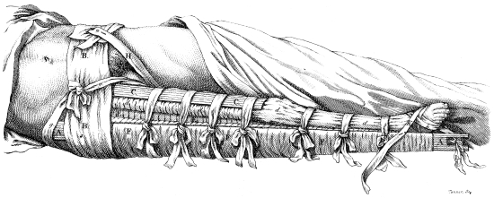
This figure represents the apparatus for permanent
extension, employed by Desault in oblique fractures
of the os femoris.
AA. The external splint, with a notch and a mortise
in it at the lower end to fix the inferior extending
roller.
BB. A bandage passing round the body, intended to
secure this splint against the pelvis.
CC. The anterior splint, reaching only to the knee.
d d d d. The anterior bolster, extending along the
whole limb, and secured by pieces of strong tape.
EE. A portion of the bandage of strips, seen between
the anterior and the external lateral bolsters.
FF. The junk-cloth intended to be folded round the
two lateral splints.
g g. The superior extending roller, passing round the
end of the external splint, and fixed underneath
on the tuberosity of the ischium.
H. The sub-femoral roller or strap, intended to prevent
the bandage BB, which passes round the
body, from slipping upwards.
K k. A roller usually passed round the foot, to prevent
it from turning.
L. The inferior extending roller, fixed in the mortise
and the notch of the external splint.
1. Few kinds of luxation of the os femoris
occur in practice more rarely than this. Practitioners
who have seen it, and those who, on the authority of
others, have described it, without having seen it, have
all given an unfavourable prognosis respecting it, for
the following reasons: 1st, on account of the inevitable
rupture of the round ligament: 2dly, on account
of the distension, and even laceration of the capsule,
and of the compression and overstretching of the
nerves and blood-vessels: 3dly, on account of the
great difficulty of reduction. The following case will
prove, that in all these respects, the apprehensions of
authors have been exaggerated, that the obstacles to
reduction arise less from the nature of the displacement,
than from the nature of the means employed to
remedy it; and that, if properly directed, art would
here be as successful as in other cases.
Case. (Collected by C***). About the close
of the winter which preceded the death of Desault,
a porter was brought to the Hotel-Dieu, in consequence
of a fall which he had received about two
hours before, in the following manner. As he was
carrying on his shoulders a heavy burden, his foot
slipped, while his leg and thigh were directed backwards:
he fell on his knee, his thigh maintaining
still the same direction; so that the conjoined weight
of his own body and of the burden which he carried,
aided by the velocity of the fall, forcing the head of
the os femoris, which pointed at the time forward
and upward, against the distended capsule, lacerated
it and drove the articulating end through the opening.
Continuing still to act, it ruptured the ligament,
which connects the extremity of the bone to the articulating
cavity, and forced the head in front of the os
pubis, where it could be easily felt.
At the moment of the fall, an acute pain was
felt in the part; and the power of moving the limb
was suddenly lost; the patient was carried home,
where a surgeon who visited him, considered the
accident as a fracture of the neck of the os femoris,
and sent him to the Hotel-Dieu, to undergo the
necessary treatment.
Desault having examined the parts, discovered,
from the following appearances, not a fracture, but
a luxation upward and forward. The limb was
nearly an inch shorter31 than natural; the point of
the foot was turned outwards; the thigh being in
a state of painful extension, could not be flexed on
the body; adduction and abduction were alike painful;
the great trochanter, being more approximated
than usual to the anterior and superior spine of the
os ilium, was also too far forward; finally, the projecting
head of the bone could be felt, as I have
already said, in the groin.
The reduction was effected in the following manner.
The patient being laid on a firm table, spread
with a mattress, a strap was fastened above the ancle,
for the purpose of extension; another, intended for
counter-extension was placed between the scrotum
and the thigh of the sound side, and brought up the
back and front of the pelvis, along the body, till it
passed over the shoulder, where it was twisted together
and secured.
Extension was then begun, precisely in the
direction in which the thigh pointed; and, during
the execution of it, a rotatory motion inwards was
given to the limb. At the expiration of a few minutes,
the head of the bone remaining almost immoveable,
notwithstanding the efforts to dislodge
it, Desault directed extension to be discontinued,
and, taking hold of the thigh, moved it in every
direction, with a view to enlarge the opening in the
capsule, the narrowness of which he suspected to be
the cause that prevented the reduction.
Extension was then resumed, and varied in every
direction, while the surgeon endeavoured to give
assistance by pushing the head of the bone forcibly
downwards, with his thumbs, and the palms of his
hands. Useless efforts; the displaced bone remained
stationary.
Desault ordering extension to be again discontinued,
recommenced the motions of the os femoris,
and even increased their force, changing them in
every direction, for the purpose of lacerating the capsule.
After this, extension was again renewed, with
better success than before. Indeed, on the very first
effort, the head resumed, of itself, its natural situation,
without any further assistance on the part of
the surgeon.
The sufferings of the patient ceased almost instantaneously;
towards evening a slight swelling appeared
around the joint, over which an emollient
cataplasm was applied. On the day following, all the
unfavourable symptoms were gone, and in about a
fortnight the patient was able to return to his usual
exercises, which, however, he was directed to pursue,
for some time, with moderation.
2. There are, in this case, two circumstances, on
which the practitioner should fix his attention, and
which may throw great light on the reduction of all
luxations of the os femoris, as they will be found applicable
to most accidents of the kind. These are,
1st, The narrowness of the opening in the capsule.
2dly, The inutility of the motion or process of conformation,
when that opening has been enlarged.
3. We formerly observed, when treating of luxations
of the humerus, that one of the obstacles to
reduction was, the narrowness of the opening in the
capsule; the same circumstance occurs here. That
membrane, lacerated at the time when the head of
the os femoris is driven against it, is dilated sufficiently
to let the head escape: but, the edges of the
lacerated membrane, coming together again, and being
thus drawn tight around the neck of the bone,
retain it in that position, and prevent the head from
re-entering the acetabulum. Thus, in a fracture,
where one of the extremities of the bone is protruded
through the integuments, the opening in the skin, by
closing tightly round that extremity, sometimes prevents
its reduction.
4. In such a case, what is the first and most obvious
indication? It is necessary to increase the extent
of the opening in the capsule, by moving the limb in
every direction. Some persons have deemed it impossible
to tear this membrane anew. But, if we
recollect, that the neck of the os femoris, being placed
between the edges of the opening, must necessarily
draw them asunder by the motions impressed
on it, it is easy to conceive, that the angles, where
these edges unite, will be torn, if the motions be
carried to an inordinate degree: besides, experience
proves here, as well as with regard to the humerus,
the truth of the doctrine contended for. Are we to
apprehend, as these same persons will have it, that
serious accidents may be produced by such violent
motions? Experience again answers in the negative.
Nothing, then, can be more certain, than that this
observation, respecting the opening in the capsule, is
a great stride towards perfection in the treatment of
luxations in general, and particularly of that now under
consideration.
5. When this obstacle to reduction has been
removed, it is then very readily effected, and that
without the process of conformation. Indeed that
process is almost always unnecessary. For what purpose
should it be employed? Is it to increase the
effect of extension, and thus disengage the head of
the bone from the place which it accidentally occupies?
In this point of view, it is nothing but a very
feeble force, added to a very powerful one, which
receives from it, therefore, but little assistance: it is
much better, if necessary, to augment the extending
forces themselves. Is it to push the head of the bone
into its cavity, after the extensions have dislodged it?
It is to the muscles, and not to the efforts of the surgeon,
that the performance of this office belongs. Indeed,
the surgeon must act altogether in the dark in
this respect, as he cannot possibly ascertain the precise
point where the opening in the capsule exists:
he may, therefore, even push the head of the bone
against a sound part of the capsule, and thus himself
create an obstacle to the reduction, which he is
attempting to favour.
6. The muscles, on the other hand, by their contraction,
naturally draw the head of the bone into its
place, because the direction of their fibres is such as
obliges them to do it. The great art of managing
luxations, then, consists, in ascertaining clearly the
obstacles that prevent reduction, in removing them,
and, then, committing the rest to extension, and the
powers of nature properly directed.
1. Our modern treatises on diseases are little else
than fabrics artfully constructed of materials confusedly
scattered through the writings of the ancients.
Many of those materials oftentimes escape our notice,
and we find them only, after practice has disclosed
them to us, in the chambers of the sick.
Thus, Hippocrates had an accurate knowledge of
spontaneous luxations of the os femoris, and has
even left an aphorism expressly on that subject.
Yet this disease appeared to be unknown to the
physicians who came after him, till John Louis Petit,
having met with it in his practice, drew the attention
of practitioners to it, by a memoir respecting it,
published among those of the Academy of Sciences,
in the year 1722.
2. The history of this affection, which was considered
afterwards, ex professo, in his course on
diseases of the bones, has been sanctioned by the
assent of all practitioners, to whom it has since very
frequently occurred, and who have generally admitted
as Louis observes, the doctrine of Petit, respecting
the cause on which it seems to depend.
3. Experience bears witness, that usually a fall
on the great trochanter, more rarely on the knee,
or the sole of the foot, precedes it, and has doubtless
some share in producing it. But what is the nature
of the primary affection which, rising immediately
from this occasional cause, becomes the immediate
cause of the luxation? Petit, and with him the practitioners
of the present day, have conceived, that the
different parts of the joint, being irritated and contused,
pour out, in consequence of the injury received,
a superabundant quantity of synovial fluid, which,
not being absorbed with the same rapidity, accumulates
in the articular cavity, distends the capsule,
and, by degrees, forces the head of the os femoris
from its natural cavity. Hence astringent and tonic
remedies, with alum, spirit of wine, &c. are directed
to be applied externally to the upper part of
the diseased thigh.
4. But this doctrine, and the practice which
results from it, seem by no means to accord with
our knowledge of anatomy. The truth of this was
deeply impressed on the mind of Desault, who had
frequent opportunities of witnessing the disease.
Case I. A young woman walking hastily along
the street, slipped and made a false step, in which
the left thigh, being violently twisted, supported for
a moment the whole weight of the body.
A severe pain experienced at the moment, obliged
her to stop at first, but becoming easier afterwards,
permitted her to proceed on her way, and
soon ceased entirely. A sensation of weight occurring
in the part about fifteen days afterwards, was
at first troublesome to the patient in walking. This
sensation was afterwards succeeded by a dull, deep-seated
pain, accompanied by a swelling in the parts
around the joint.
During six or seven months the limb was observed
to increase in length gradually, but very slowly.
At the expiration of that time, a contraction took
place suddenly, and, in one night, the diseased thigh
became shorter than the other by nearly two inches.
The patient was then admitted into the hospital,
where, after some time, she sunk under her disease.
On opening the body the following appearances were
presented to Desault, who was then consulting surgeon
to the institution.
The cartilage of the acetabulum swollen to such
a degree as to fill up the whole extent of that cavity,
was yellowish and inorganic, somewhat resembling
bacon, both in colour and consistence. A soft,
spongy, whitish substance projected in the middle of
it, the remains no doubt of the round ligament. The
head of the os femoris, situated where it is usually
found in luxations outward and upward, was surrounded
by a cartilage equally tumefied.
5. Here the cause of the displacement of the os
femoris was evident. The cartilages becoming tumefied,
in consequence of the contusion and violence
done to them, had by degrees, filled up the acetabulum,
forcing out in the same gradual manner the
head of the bone. Hence arose the original lengthening
of the limb. But as soon as the head had escaped
from the lacerated capsule, the limb was drawn upwards
and consequently shortened, by the action of
the muscles, and the weight of the body pushing the
pelvis downward.
Case II. Some years afterwards, Desault had
occasion to witness again the same disease, in the
person of a man aged thirty-seven, who put himself
under his care, but, being obliged to leave Paris, a
short time afterwards, retired into the country, where
he died in about six months, enfeebled and consumed
by a hectic fever.
On opening the body, the surgeon of the place
discovering the same phenomenon as in the preceding
case, made a preparation of the part, and sent it
to Desault, whose pupil he had been.
6. In this case the shortening was not so sudden
as in the preceding one. It appeared at first to be
coming on, during five days, in an imperceptible
manner, when, fatigued with lying in bed, and having
on that day drank a little, the patient attempted
to walk, supported only by a cane. By evening, a
shortening of two inches and a half had taken place,
an effect evidently produced by the weight of the
body on the diseased thigh. Hence the necessity of
confining the patient to a state of perfect rest, of preventing,
in particular, standing and walking, and all
positions in which the diseased thigh would have to
sustain the weight of the body.
Case III. Maria Genette was received into the
Hotel-Dieu, in consequence of a fall on the great trochanter.
She had been attacked by a pain in the joint
of the thigh of the same side. Walking, which was
performed with difficulty, augmented the pain, and
standing, though more tolerable, could not be long
continued. The thigh was evidently longer than the
other. To leave the disease to nature, and confine
the patient to a state of rest, constituted the practice
of Desault. What effect could the external use of
astringents, recommended by Petit, produce in such
a disease? Some time after her admission, the patient
was attacked by dysentery, in consequence of which
she was removed to the medical ward, where she
died.
On opening the body, the parts in the neighbourhood
of the joint were evidently tumefied, and the
capsule was stretched from above downwards. The
head of the os femoris was situated on the external
edge of the acetabulum. The capsule, though greatly
elongated was still in a state of tension; and the articular
cartilage was swollen to such a degree, as
nearly to fill up the cavity. The quantity of synovial
fluid was less than natural.
7. This case, taken at a period of the disease not
far advanced, fully confirms the inference deducible
from the two preceding ones, respecting the cause of
spontaneous luxations. Here, indeed, the capsule
not having given way, the swelling having only just
commenced, and the synovia existing in but small
quantity, the progress of nature was evident. Here,
also, occurred a sign not noticed by Petit; namely,
the elongation of the limb, which always, in such
cases, precedes its contraction.
From what we have said, it appears, 1st, that the
efficient cause of spontaneous luxations of the os femoris,
is a swelling of the articular cartilages, which
alters and destroys their organization: 2dly, that the
presence of this swelling must necessarily render
fruitless all attempts that might be made to replace
the head of the bone in its cavity: 3dly, that the
change in the organization of the cartilages, renders
astringents, discutients, and other external means applied
for the purpose of removing the tumefaction,
entirely useless: 4thly, that here, as in many other
cases, art ought to confine itself to the palliation of
effects, and not attempt the removal of causes.
§ I.
1. The rotula, a sort of bony production of the
sesamoid kind, attached to the tendon common to
the extensors of the leg, represents a moveable pulley,
intended to slide on that formed by the separation
of the condyls of the os femoris. It protects the joint
which it covers, and, in point of structure, use, and
situation, greatly resembles the olecranon, from which
it differs only in this, that, instead of being a continuation
or process of the tibia as the olecranon is of the
ulna, it is only attached to that bone by a strong and
thick ligament which is inserted into its tuberosity.
Hence it follows, that between the injuries of the one
and the other, there must be a great analogy: and indeed
most of the signs characteristic of fractures of the
olecranon, are characteristic also of those of the rotula,
and the treatment which, in such cases, is suitable
for the former, differs but little from that required
by the latter.
§ II.
OF THE VARIETIES AND CAUSES.
2. Fractures of the rotula may, in general, assume
any direction, transverse, longitudinal, or oblique:
but the first kind occurs in practice much
more frequently than the others; and so great indeed
is the disproportion, that it has almost exclusively
attracted the attention of authors, in the numerous
forms of apparatus invented to retain the fragments.
3. A shattering of the bone, the effect of a violent
blow; a contusion; an echymosis; an effusion
of blood into the adjacent soft parts; one or more
wounds of the soft parts, with or without an opening
into the joint; a swelling, the degree of which varies
greatly, according to the state of the fracture, and the
disposition of the subject, but which is constantly
present; a double division of the bone, one of which,
being longitudinal, forms an angle with the other,
which is transverse; and a concomitant fracture of
the condyls of the os femoris, or of the tibia: such
are the varieties and complications, of which the fracture
under consideration is susceptible.
4. But this fracture may be produced in two
modes. 1st, by the action of external bodies: 2dly,
by that of the extensor muscles. The first mode of
division takes place in falls on the knee, or when a
body in motion strikes against it, and, in this case,
there is no counter-stroke, the rotula being too small
for such an occurrence, and always sustaining the
fracture where it receives the blow. In the second,
the fall is only subsequent to the fracture, and, as
Camper has well observed, is most frequently the
effect of it. For instance, the line of gravity of the
body is, by some cause, removed behind it; the anterior
muscles contract themselves to bring it forward
again; the extensors act on the rotula; it is broken,
and a fall ensues. Again, the leg is suddenly
thrown into a state of violent extension; the extensors
act with great force; a fracture is the consequence,
and the patient falls. A soldier once fractured
his rotula in kicking at his serjeant; thus the olecranon,
in like manner, has been broken by throwing
a stone. A man, in the Hotel-Dieu, fractured the
rotula of each knee, in the operating room, by means
of convulsive motions, produced by the operation of
lithotomy.
5. The action of external bodies, can alone produce
a longitudinal fracture, as when a person falls on
a sharp projecting piece of timber: but this may also
produce a transverse fracture. On the other hand,
muscular action can never give rise to any but the
latter kind, since the direction of this fracture is at a
right angle with that of the extensors. A fracture
resulting from the action of external bodies, is oftentimes
accompanied by a wound, a contusion, or a
shattering of the part (2); a fracture, arising from
muscular action, is always simple, except as to a
swelling around the joint. The latter cause may,
instead of fracturing the rotula, rupture the common
tendon of the muscles, or, what is more common, the
inferior ligament. Desault has seen many examples
of this: Petit has also observed several, and Sabatier
has sometimes met with them. External violence
seldom produces this double accident.
§ III.
OF THE SIGNS AND THE DISPLACEMENT.
6. In longitudinal fractures the diagnosis is always
accompanied with more or less difficulty, because the
extensor muscles, drawing by their contractions the
two fragments equally upwards, and the inferior ligament
holding them equally down, tend to keep them
in apposition, and to prevent them from separating.
Sometimes also the ligamentous production which
covers the rotula, remains entire and serves to keep
the fragments together. It will be necessary, therefore,
should the existence of such a fracture be suspected,
to move the two sides of the rotula in opposite
directions, by pressing them to the right and to
the left, in order to arrive at certainty on the subject.
Should a wound exist, as is oftentimes the case (5)
the diagnosis is less difficult.
7. If the division be transverse, the diagnosis becomes
then as plain and easy, as it is difficult and
obscure in cases where it is longitudinal. In such a
case, a considerable separation or space exists between
the two fragments, sensible to the touch, when
the hand is placed on the knee. In this separation,
the fragments are not displaced by the same means.
The superior fragment being attached to the extensors,
is drawn upwards with great force by these
muscles, the action of which the rotula no longer
resists. The lower fragment, on the other hand,
being attached only to the inferior ligament, is not
moved by any muscle, and cannot be displaced in
any other way than by the motions of the leg with
which it is still connected.
8. Hence it follows, 1st, that, in a state of extension,
the separation is the least possible, because it
is then produced on the part of the superior fragment
only; 2dly, that in a state of flexion it is greatest,
because then both fragments concur alike in producing
it; 3dly, that it may be increased or diminished
by varying the degrees of flexion.
9. This fracture is further characterized by the
following circumstances, namely, a practicability of
moving the fragments transversely in opposite directions,
and of producing, by that means, some
degree of crepitation, provided they be first brought
close together; by the pain which accompanies these
motions; by the swelling common to every kind of
fracture of the rotula, and which, if very great, may
involve the other signs in more or less uncertainty;
by a difficulty of standing; and an almost entire loss
of the power of walking, in consequence of the extensors
being no longer able to communicate motion
to the leg, unless when the fracture exists very low
down, near to the inferior ligament.
10. The touch will always discover in what part
of the bone the fracture is situated, which, if it be
oblique, will partake more or less of the characters of
the longitudinal or the transverse, accordingly as it
approaches to the one or the other.
§ IV.
OF THE PROGNOSIS.
11. Many authors have pretended that fractures
of the rotula cannot be cured, and it even appears
that the Academy of Surgery adopted this opinion,
on receiving a memoir from a Flemish physician,
which contained several facts tending to establish
that principle. But what do these facts prove? That
in some particular cases, reunion did not take place,
but they do not show that this was owing to the nature
of the fracture.
12. But, what, in such cases, could prevent a
cure from taking place? The structure of the rotula
differs, say they, from that of the other bones.
Now, admitting this difference of structure to be
real, it certainly approaches to the structure of tendons
to which indeed it bears a strong affinity. But, who
does not know, that, when tendons are divided, they
unite as readily as bones? Besides, is not the power
of reunion common to every part endowed with life?
I have already shown, when treating of other fractures
that communicate with joints, what credit is
due to those hypotheses so often revived but never
confirmed, nay even clearly proved to be unfounded,
such as, an effusion of callus into the joint, a failure
of reunion from a want of periosteum on the posterior
part of the bone, the synovia diluting the matter
of callus, and thus preventing it from being duly
prepared, &c.
13. The inflammation of the articulating surfaces
and of the ligaments around the joint, ought to
have more influence in constituting an unfavourable
prognosis, than any circumstance that authors have
mentioned. But experience proves, that, when judiciously
treated, these fractures are not accompanied
by that accident, and even that the swelling, which
for the most part attends them, always yields more
or less speedily, when a bandage, uniformly applied,
presses equally on all parts around the joint, and thus
forms a kind of discutient, while at the same time
it retains the fragments.
14. Pare, Fabricius of Hilden, and a number of
other writers, have pretended, that some degree of
lameness must always be the consequence of this
fracture. But, from what causes must this lameness
so certainly arise? Is it from a want of reunion in
the part? I have already shown (11 and 12) that this
apprehension is wholly unfounded. Is it from an anchylosis?
This accident cannot take place, except
either in consequence of inflammation occurring in
the articulating surfaces, (and I have already shown
how that may be avoided, 13) or of a stiffness in the
ligaments, and I shall hereafter make it appear that
that may be readily prevented by motion. Is it from
the fragments being drawn asunder, and in that state
united by an intermediate substance of too great an
extent? I shall prove, that a bandage properly constructed,
is always sufficient to keep these fragments
in contact.
From these considerations it appears, that writers
have, in general, without sufficient cause, given an
unfavourable prognosis, in relation to fractures of the
rotula, which have, indeed, a great affinity to other
affections of the same kind.
§ V.
OF THE REDUCTION AND THE MEANS OF MAINTAINING IT.
15. I have already observed (7), that the causes
of the separation of the fragments are, as far as respects
the upper one, the contraction of the extensor muscles;
and, in relation to the lower one, the flexion of
the leg; whence it follows, that the means of preserving
contact between these fragments are 1st, all
those that are calculated for the prevention of muscular
action; 2dly, such as may keep the limb in a
state of permanent extension. Hence two leading
curative indications must be fulfilled by the bandage
constructed for fractures of the rotula: the last of
these indications presents in general but little difficulty;
but, with regard to the other, the case is
different. To fulfil the latter, it is necessary first,
to weaken the contractile force of the muscles, and
by that means diminish the effort which they make
to draw the superior fragment upwards; and then,
to oppose to them a proper mechanical resistance,
which, by acting in a direction the very reverse of
that in which they act, may countervail their efforts.
16. But the force of contraction is diminished, 1st,
by throwing the muscular fibres into a state of relaxation;
this end is best attained by bending the thigh on
the pelvis: 2dly, by making compression over the
whole limb, by means of a circular bandage, which,
by confining the muscles, tends to restrain and weaken
their action. Thus it is known that the advantage
of the bandage employed to unite transverse wounds,
consists chiefly in that compression which, by diminishing
muscular action, prevents the retraction of
their edges. Another advantage resulting from the
bandage in this case is, that it prevents the swelling
of the limb.
17. As to the mechanical resistance, which must
act in a direction opposite to that of the contraction
of the muscles, and, by that means, prevent the displacement
of the superior fragment, it cannot, in the
present case, be of the same nature as in fractures of
the thigh, the clavicle, &c. where permanent extension
is practised. The superior fragment offers too
small a purchase for any extending forces to act on.
This resistance must be made, then, by placing some
body above this fragment, and retaining it in that
situation with a force sufficient to hinder the fragment
from rising upwards: such as a few turns of a roller
drawn tight, a bit of leather, some hollow compresses,
&c.
18. It is evident from the foregoing principles,
that every bandage intended to retain a transverse
fracture of the rotula, ought to be calculated to maintain
the following state of things: 1st, the extension
of the leg on the thigh; 2dly, the flexion of the thigh
on the pelvis; 3dly, a uniform compression over the
whole limb; and, 4thly, some mechanical resistance
properly secured above the superior fragment: the
three last expedients relate to the displacement of that
fragment alone; while the first has a relation to that
of the lower one. Let us examine whether or not the
bandages, hitherto employed by different authors, be
calculated for these purposes.
19. M. Valentin, believing that position alone
was sufficient to retain the fragments in contact, neglected
the application of apparatus entirely, which
he even considered as hurtful, in consequence of the
swelling it produced; but experience soon proved
the insufficiency of this method. The slightest movement,
or the least effort on the part of the patient,
made the extensor muscles contract, which, drawing
the superior fragment upwards, separated it from the
lower one; and, as the time of reunion is in direct
proportion to the distance of the fragments from each
other, it must, under such treatment, have been
necessarily tedious, and sometimes must have even
failed altogether.
20. As to a swelling being produced by the bandage,
this never occurs, unless when some openings
are left, through which the integuments protruding
become tumefied: but, when the pressure is uniform
throughout, when the fluids find throughout an equal
resistance, this accident is not to be apprehended, as
is proved by the practice of Desault, who never met
with it; on the contrary, a bandage properly constructed
and applied, is calculated to prevent swelling
(16).
Mere position, then, though always of service in
this affection, is not alone sufficient, because it fulfils
only the first of the indications or principles laid down
with respect to every form of apparatus for transverse
fractures (18), namely, that which relates only to the
lower fragment; while those that relate to the upper
one, remain still to be fulfilled.
21. Most authors have employed, with a view to
these, a kind of figure of 8 bandage, known in art by
the name of Kiastre,32 and approved of by Petit,
Heister, &c. This is made of a roller formed into
two balls, which are brought across each other alternately
under the ham, passing over two hollow or
forked compresses, that enclose the two fragments of
the rotula.
But the unequal pressure which this makes on
the unequally projecting parts of the knee, renders
its application extremely painful, particularly below,
where the pasteboard covering applied by Louis, immediately
on the skin, afforded but a feeble protection
to the tendons of the flexors. Besides, it did not
prevent the swelling, which is indeed a necessary
consequence both of this unequal pressure, and of
the openings left between the casts of the bandage.
This swelling is taken notice of by all writers, and
is, according to them, one of the troublesome circumstances
attending the fracture. The third indication
is not all fulfilled (18).
22. The extensor muscles, not being at all compressed,
will act with their whole force on the upper
fragment, and, on the slightest effort of the patient,
overcome the resistance of the bandage, the action of
which, being oblique with respect to the fragment,
is inconsiderable, unless it be drawn very tight, and
thus a displacement will again occur. This obliquity
of the turns of the roller obliges the surgeon, either
to draw it very tight, in which case a swelling is inevitable,
or to make it but moderately tight, and then
the apparatus will be insufficient to resist the action
of the muscles.
23. Most of the objections to the ancient apparatus
for fractures of the rotula, apply also both to that
proposed by Ravaton in his surgery, and to that
which Bell employs in his practice. Both of these,
while they fail in making sufficient resistance to muscular
action, as well as in fulfilling the third condition
laid down as necessary to every bandage (18), contribute
to the swelling, and can rarely produce a perfect
contact between the fragments. Thus Bell has well
observed, that the reunion is rarely perfect, and that
there is always a separation more or less perceptible.
24. The complication, the intricacy, the expense,
and other more weighty inconveniences of the machine
described by Garengeot in his treatise on instruments,
and employed, for the first time, by
Arnaud, and also of that which was proposed and
used by Solingen, have, long since, entirely banished
them from among the means of reduction.
25. Some practitioners have advised the uniting
bandage used in cases of transverse wounds, which is
formed, as is well known, of two small rollers or
strips placed in the longitudinal direction of the limb,
one of them having holes in it, to which the divisions
of the other are fastened. Both of these are first
secured by circular turns; being then drawn in opposite
directions so as to meet, they draw the parts
on which they are applied in the same directions.
But the action of this bandage is confined to the integuments,
and would have of course but a feeble
influence on the fragments beneath. It is also attended
with this further inconvenience, that by wrinkling
the integuments, and throwing them into folds, it
might press them down between the fragments, and
thus prevent their contact. Besides, it is liable to most
of the objections urged against the preceding one.
26. This view of the means employed by different
practitioners, to counteract the causes of displacement
in this fracture, are sufficient to convince us, that the
difficulties hitherto experienced in the treatment of it,
have arisen from the feebleness of the former, and the
strength of the latter. So great indeed have been
these difficulties, that some authors, conceiving a reunion
impossible, have, in conformity to such an
opinion, though contrary to all the rules and principles
of the profession, advised us to abandon the patient to
himself. But I have already exposed the fallacy of
that opinion, respecting the want of a healing power
in the rotula (12), an opinion which, if generally
adopted, would give rise to consequences of the most
serious nature. In the present case, as in all other
fractures, the contact of the fragments ought to be the
chief object of the surgeon’s efforts.
27. But ought this contact to be perfect and exact?
Several authors, particularly Bell, have conceived,
that the motions of the limb can be performed as well
with a slight separation of the fragments. Pott even
declares that such a separation will enable the patient,
after his recovery, to walk with more ease. Flajani
advances the same opinion in a dissertation on the
subject.
From this doctrine arose a new mode of treatment,
which consisted in not suffering the fragments to be
at rest. They were accordingly, during the cure, put
frequently in motion, the more effectually to prevent
an anchylosis, which is sometimes the consequence
of this fracture.
28. But, on the one hand, it is difficult to conceive,
on what this opinion of these authors can be
founded; while, on the other, reason declares, in the
plainest and most forcible terms, that the more the
state of a bone, after it has been broken, differs from
its natural state, the less free will be the exercise of
its functions, and, that the perfection of the treatment
of fractures consists, in leaving behind it no vestige
of the accident.
29. This truth was frequently confirmed in the
experience of Desault, who had an opportunity of
seeing numerous fractures of the rotula, both in the
Hotel-Dieu, and in his private practice. He always
observed, that, when the separation of the fragments
was considerable, and the ligamento-cartilaginous
substance uniting them was of some extent, standing
and walking were performed with much difficulty;
that the patient was exposed to frequent falls, from
the want of a proper correspondence, in point of
strength and motion, between the two limbs; and
that, on the contrary, the less extensive the separation
and the substance that filled it up were, the
more free and easy were the motions of the part,
which still remained, however, somewhat defective and
imperfect, unless every vestige of the division was
obliterated.
Paul of Egina long since observed, that, when
no means of reduction were employed, though the
patient might walk tolerably well on a level surface,
he could not, without difficulty go up an ascent.
30. From what has been said, it follows, 1st,
that in the treatment of this fracture, the perfect
contact of the fragments ought to be the principal
object of the practitioner; 2dly, that the kinds of
apparatus employed by different authors, are but ill
calculated for the attainment of this end, because they
fulfil but imperfectly the indications formerly laid
down (18). Let us see whether or not the apparatus
of Desault be any better suited to this purpose.
31. The bandage, which he employed in this
case, analogous to that for fractures of the olecranon,
is composed, 1st, of one splint, two inches broad,
and long enough to reach from the tuberosity of the
ischium, to a little above the heel; 2dly, of two
rollers, five or six yards long, and nearly three inches
wide; 3dly, of another single roller, with two holes
about the middle of it, a little longer than the injured
limb of the patient, along the fore part of which it
must be extended.
32. Every thing being arranged for the application
of the apparatus,
1st, One assistant secures the pelvis, in the
same manner as in fractures of the lower extremities;
while another keeps the leg in a state of perfect extension
on the thigh, and the thigh on the pelvis.
2dly, The surgeon, then, standing by the side
of the fractured limb, extends along the anterior part
of the leg and thigh the roller with holes in it, having
previously wet it with vegeto-mineral water, taking
care to make the two openings correspond to the
lateral parts of the rotula, that, by being thus better
adapted to its shape, it may not be thrown into
wrinkles.
3dly, He then secures it on the top of the foot,
by three circular casts of a roller placed one over
the other, three or four inches above its lower end
which must next be turned up over the three first
casts, and made fast by two other ones. Then,
while the compress roller33 is secured above by an
assistant, he passes up along the leg by oblique and
reverse turns, according to the inequalities of the
limb.
4thly, Having arrived at the lower part of the
knee, he pushes the lower fragment upwards, and
makes below it two or three circular turns to secure
it. He then gives the roller into the hands of an
assistant, and directing him who holds the long compress
roller, to draw it forcibly upwards, pushes the
integuments of the knee in the same direction, lest,
by becoming interposed between the fragments, they
might prove an obstacle to their reunion. Passing
then the fingers of his left hand through the holes in
the compress-roller he places them behind the superior
fragment and pushes it forcibly downwards.
5thly, When the reunion of the fragments is
exact, without any space intervening, he resumes
the roller, and passing it obliquely under the ham,
and bringing it up again behind the superior fragment,
withdraws his fingers which held this fragment
down. In place of his fingers, he then applies two or
three tight circular casts, covers the knee with several
oblique casts in form of the figure of 8, so as to
leave no opening between them, and, then, continues
the bandage up along the thigh, securing by it the
compress-roller extended along the fore part of the
limb.
6thly, When he has arrived at the upper part
of the limb, the assistant who holds the compress-roller,
drawing it forcibly upwards, doubles down its
end over the circular casts. The surgeon next fixing
this end by several additional casts, descends
again along the thigh, covers the knee by a few
more oblique turns, and finishes with the roller on
the leg.
33. This first part of the bandage evidently fulfils
the third and fourth indications (18). The compression
of the roller on the muscles weakening their
action and impeding their motions prevents their
tendency to draw the superior fragment upwards:
while the circular casts passed behind this fragment,
acting in opposition to the muscular contractions,
prevents it from moving upwards in obedience to
them. The long compress-roller, stretched on the
fore part of the limb, being first secured below, and
then drawn forcibly upwards, presses the casts of
the roller against each other, and prevents those that
correspond to the thigh from slipping upwards, and
thus abandoning the superior fragment, and prevents
also those on the leg from slipping down and withdrawing
their support from the inferior fragment. As
there remains no vacant space between the circular
turns, their pressure is uniform throughout: no
swelling can consequently supervene (20).
34. But the first and second indications remain
still to be fulfilled (18): it is necessary to prevent the
separation of the lower fragment, by the extension of
the leg on the thigh, and to throw the muscles into
a state of relaxation by extending the thigh on the
pelvis, and to maintain permanently, by the apparatus,
that double position, which the assistant maintains
only during the operation.
35. To obtain the first effect different means have
been employed; but none answers so well, to extend
the limb and retain it immoveably in that state, as a
long and strong splint, placed, as Desault did it, subsequently
to the application of the first part of the
bandage, along the posterior part of the limb. An
assistant must hold the end of this splint, while the
surgeon secures it in its place by the second roller
(31): in this way the extension of the leg is effected.
36. To obtain the extension of the thigh, it is
necessary to place on the top of each other, two or
three bolsters or little bags filled with chaff, so disposed
as to form an inclined plain, considerably elevated
towards the heel above the level of the bed,
but which, gradually descending to the same level
towards the tuberosity of the ischium, forms a supporting
basis on which the whole limb may rest in a
uniform manner. By this twofold extension of the leg
and of the thigh, the lower fragment is kept up immoveably,
and the muscles are kept in a state of
relaxation.
Hence it follows, that this bandage fulfils extremely
well the conditions laid down (18), and that
it ought to be preferred to all the others (19...25),
which answered the indications only in part.
37. Whatever may be the advantages of this bandage
over the others, it must still be acknowledged
to have its inconveniences. The rollers become relaxed
in a short time; their compression is less active;
the muscles, being less confined, contract more readily;
hence the necessity of frequently repeating the
application of the apparatus, a circumstance which is
very troublesome, on account of the roller which
composes it, and covers the whole limb. The resistance
of it even when it is recently applied, is not
always equal to the power of the muscles, whence
the most assiduous attention is necessary, to obtain
such a consolidation as to leave no trace of the fracture
behind. Few persons ever possessed, like Desault,
the art of overlooking nothing that might in
any way contribute to the success of his treatment:
from this, no less than from the excellence of his processes,
arose the number of his cures. Let us confirm,
by a few examples selected from among a great
many, the doctrine here laid down. The following
cases were collected by Julian and Bezard.
Case I. Francis Leclert, of a sanguine temperament,
fell on the 7th of October, 1790, on his right
knee, and produced a transverse fracture of the
rotula. He was not able to rise; he was carried
home, where a surgeon, on discovering the nature of
his disease, advised him to be taken to the Hotel-Dieu.
He was conveyed thither on the day following,
and, in the interval, a considerable swelling had occurred
around the joint. The usual bandage was
employed; the pains ceased immediately after its application;
a copious blood-letting was directed, and
a low diet was prescribed.
The whole apparatus was wet with vegeto-mineral
water, two or three times a day. On the next day
some light food was allowed, and the quantity increased
by degrees, till in a short time the patient
returned to his usual regimen. Eighth day, the
swelling being almost gone, the bandage had become
relaxed it was therefore reapplied. Every day the
inclined plain formed by the bolsters was carefully
examined, and put in order again as often as it became
deranged.
Fifteenth day, a new application of the apparatus:
twentieth day, an evacuation in consequence of a
bilious disposition. Nothing particular occurred from
this time till the completion of the cure, which took
place on the sixty-seventh day after the accident: no
depression existed at the place of the fracture: the
motions were perfectly free; these were aided, by
daily exercising the knee joint for some time.
Case II. Vincent Grenier, aged thirty-eight,
making a false step, fell on the rotula, and fractured
it, on the 6th of June, 1791: he was brought to the
Hotel-Dieu, where Desault demonstrated to his pupils,
by the usual signs the existence of the disease:
a considerable swelling had already taken place. The
bandage formerly described was applied: the same
precaution as in the preceding case; apparatus examined
every day; renewed as often as relaxed;
extension maintained with great exactness. On the
forty-fifth day, the consolidation was nearly effected;
on the fifty-second it was complete, the joint was
exercised for some time, and on the seventy-seventh
day the cure being in all respects complete, the patient
was discharged.
1. The history of foreign bodies divides itself
naturally into two great sections; the one includes
those that are introduced from without; the other
such as are formed within our own systems. This
latter section may be again divided into two classes; to
the first class belong bodies altogether inorganic, such
as the different kinds of stones; to the second, those
which are truly organic, and become foreign only by
being situated in places where they impede the functions,
such as cartilaginous or bony productions,
existing accidentally within the joints.
On the subject of the latter class, art is much more
deficient than she is with regard to the former. Let
us endeavour to assist her a little, by giving a sketch
of the opinions and practice of Desault with respect
to these productions.
2. Before his time, the surgery of France appears
to have contained scarcely a record of this affection.
Described only in some ancient works, such as the
writings of Pare, it had been forgotten by the moderns,
when numerous instances of it were suddenly met
with by English and German surgeons, and soon afterwards
by Desault, who illustrated and confirmed
the practice of his predecessors in it, and even added
something of his own.
3. All the joints may become the seat of these
concretions; Haller found many of them in that of the
lower jaw; Bell mentions, as a very rare occurrence,
their existence at the junction of the foot with the leg.
Some authors have met with them in the wrist; but
none are more common, or merit more particular attention,
than those that exist in the joint of the knee.
To these alone shall the following observations be
confined, because these alone have fallen under the
notice of Desault.
§ II.
OF THE VARIETIES.
4. Concretions of the joints do not always assume
the same aspect. They vary greatly as to number,
size, figure, structure, &c. In general, these bodies
exist singly; sometimes, however, two of them are
found in the same joint, and then they may be extracted
either at the same time, or in succession, as
was once done by Desault. Some English surgeons
have also met with two concretions, and Morgagni
has found even twenty-five, in the same joint.
5. They vary also in size. The largest ever met
with by Desault, was fourteen lines in its longest,
and ten in its shortest diameter. Six lines diameter
in every direction, was the measure of the smallest
one that occurred in his practice.
6. Their figure is sometimes lenticular and
smooth on both sides, sometimes unequal, rough in
one part, even in another, concave on one side, convex
on the opposite, sometimes marked around the
circumference and sometimes not with reddish
points, and having occasionally a stem of a cellular
texture and of some length, as may be seen in a paper
by Theden. They usually consist of a single mass,
but are in some cases divided into several lobules
united by a kind of ligaments, as in the fourth case
related in the Journal of Surgery. Though most frequently
detached and floating in the interior of the
joint, they have yet been found adhering by means of
small portions of cellular substance, loose and capable
of being stretched, or tight, hard, and even of a ligamentous
nature.
7. If, from the external figure, we pass to the
structure of these bodies, we will find them existing
in three different states. Sometimes purely cartilaginous,
sometimes completely bony, they at other
times partake of both these states, in which case a
bony nucleus is covered with a cartilaginous crust.
Out of five cases, recorded by Desault, three are of
the first, and two of the third kind. Many authors
have met with the second kind, particularly Morgagni,
who has even found in the same joint, some bodies
of a bony and others of a cartilaginous nature. Hence
it appears, that this variety of structure is to be attributed
to the longer or shorter standing of the disease,
that every concretion must pass successively through
these three states, and that there is a great analogy
between the formation of such bodies and natural
ossification.
8. If we examine a body of the third kind cut in
two through the middle, we will find it red and vascular
in the centre, like an epiphysis, even when it is
floating in the joint perfectly loose and free from adhesion.
9. Bell, in his treatise on surgery, speaks of a
kind of tumour, at first soft, membranous, and adhering
to the internal surface of the capsule, but which,
according to him, may become afterwards hard and
solid, and be detached so as to float loose in the joint.
But are not these tumours different in their nature
from those destined to be converted into bone? Do
they, in fact, ever undergo the changes mentioned by
Bell? Desault having never met with any of them,
was unable to offer an opinion on the subject. In the
mean time, an observation made by Monro, may
serve to throw some light on the question: he once
saw, in one of these productions, a cellular nucleus
surrounded by a covering of bone.
10. Though usually simple and free from complication,
this affection may, according to some
authors, give rise occasionally to a dropsy in the
joint. Pare is the first who has made mention of this:
he found one of these bodies in a patient’s knee, into
which he had made an incision for the purpose of
drawing off a collection of water. Simson, on extracting
a similar body, gave vent to four ounces of water.
But, as on the one hand, a dropsy of a joint oftentimes
exists without these foreign bodies; so, on the
other, these bodies are almost always found disconnected
from dropsy. Nor is there any affinity between
the acknowledged causes of an accumulation of
synovia, and the presence of these bodies; so that
when the two diseases do exist together, it is altogether
probable, that they are independent of each
other.
§ III.
OF THE CAUSES.
11. The formation of articular concretions succeeds
frequently to blows or falls received on the
joint, in which case, a swelling more or less considerable
in the surrounding soft parts, showing
itself from the first, and remaining for some time, at
length allows the foreign body to be perceived, and
does not, in general, disappear during the continuance
of the body in the part.
12. Sometimes no external injury contributes to
the formation of the body, and then, a spontaneous
swelling precedes its detection, as Desault observed
in two patients, where nothing was known to have
concurred in the production of the disease. Constant
rest increases this swelling, while exercise and a temperate
mode of life diminish it.
13. But what can be the immediate cause of
these tumours? Are they, as some allege, an aggregation
or crystallization of particles of matter conveyed
into the interior of the joint by the synovia, in
the same manner as the rudiments of a stone are conveyed
into the bladder by the urine? Their organic
appearance and the vessels that pervade them, are
unfavourable to such an opinion. Can they be, agreeably
to the conjecture of Theden, articular glands
bruised by means of strokes or falls? Or are they,
as some authors will have it, portions of the cartilage
of the joint, detached by the same causes?
How then will their spontaneous formation be explained?
But why trouble ourselves about the cause, provided
we can remedy the effects? Nature conceals
from us the means, and discloses to us nothing but
the results. Theories are fluctuating; but experience
is still the same: let us search, then, by an attention
to facts, for that which we cannot learn from first
principles.
§ IV.
OF THE SIGNS.
14. The phenomena which announce the presence
of foreign bodies in the joint of the knee, are sometimes
clothed in a character of such evidence, that
they cannot be mistaken; at other times, the nature
of the disease eludes the most accurate researches:
the cause of this variety may be easily perceived.
As the joint presents different depressions and
eminences, and as the bodies, being usually loose
and detached, may travel through its whole extent,
they produce different effects, according to the particular
situations which they occupy. If lodged in a
depression, they are not compressed, and cannot, of
course, give rise to any troublesome affection. If
they bear on an eminence, such as the condyls, or
the posterior part of the rotula, they are forcibly
compressed, and must derange, in some measure,
the functions of the joint. Hence the precise nature
of the affection cannot be at all times derived from the
state of the symptoms.
15. Sometimes the patient can stand and walk
with perfect freedom and ease, while, at other times,
a sudden pain seizing him, obliges him to sit down,
or even causes him to fall, if there be nothing at hand
to support him. This pain subsists for a longer or
shorter time. One motion produces it, and sometimes
another, made in an opposite direction, removes
it. But in common it is of some continuance, and
then the patient is obliged to keep his bed.
16. If the state of the joint be examined, it will
be found more or less swollen, when the pain is very
acute. When the pain ceases, the swelling in part
disappears. It is never sufficient to prevent the fingers,
when drawn along the external surface of the
joint, from discovering the presence of the foreign
body, when it forms a protuberance under the integuments.
It is then found sometimes above the
rotula, by the side of the tendon of the extensor
muscles, and that is the place where it usually produces
least pain; at other times, it is lower down, in
front of the condyls, and by the side of the rotula.
It is occasionally found immediately behind the tendon
of the extensor muscles; in this case so acute is
the pain, that the patient is generally unable to stand.
But it is when it is situated behind the rotula, near to
the projecting ridge which runs across its posterior
surface, that it gives rise to the most serious affections.
17. The body passes from one place to another,
on the least motion, and sometimes, as Bell observes,
the patient, on changing his position during sleep, is
awakened by severe pain, in consequence of the foreign
body being moved by this change. It happens,
in certain cases, that it disappears, and lies concealed
for some time, in the back part of the joint. During
this period the joint performs all its functions with
freedom and ease. Desault made this remark, in the
case of a captain of dragoons, from whom, for the first
time in his practice, he extracted one of these bodies,
and who, for six months previously, had been able to
perform all the motions of the joint freely, without
pain. This person, experiencing no uneasiness, considered
himself perfectly cured, when the body suddenly
reappeared, in consequence of a hasty extension
of the leg.
18. If the body, when projecting under the integuments,
be gently compressed, it yields to the pressure,
changes its situation, and, according to the impression
it has received, moves either to the internal
or the external side of the joint, or reciprocally from
one side to the other, passing also behind the rotula,
behind the inferior ligament, or sometimes behind the
tendon of the extensor muscles. In these alternate
displacements, it may in some cases be turned round,
in such a manner that its anterior surface will take the
place of its posterior one, and then resume its primitive
situation. Desault met with an instance, in which
the patient himself was in the habit of turning the
body round in this manner.
19. Bell, in conformity to the distinction of articular
concretions into cellular and solid, attributes to
each division its peculiar signs. In the first case, the
pains, being rather obtuse than sharp, are constant;
in the second, they are extremely acute, but disappear
and return at intervals. Supposing the division
to be a real one, cases of the last description certainly
occur much more frequently than those of the first.
§ V.
OF THE TREATMENT.
20. From what has been said it follows, 1st, that
these cartilages floating through the joints, do mischief
mechanically (14), by coming into contact with the
articular surfaces: 2dly, that to obviate this mischief,
it is necessary either to prevent their contact, by fixing
the bodies in a spacious part of the joint, and thus
doing constantly what nature does on certain occasions,
or to extract them through an opening made into
the articular cavity.
21. Hence, art can have recourse to but two methods
of cure, all hope of discussing these tumours
by external applications being, as Bell observes,
entirely extinguished.
22. The first method was proposed by Middleton
and Gooch, who having brought the foreign body
into a situation where it produced no pain, endeavoured
to confine it there a length of time sufficient to
make it form adhesions with the corresponding part
of the capsule. As we are not informed of the result
of the experiments of these two physicians, we are
left to our own conjectures on the subject.
23. Are these foreign bodies capable of forming
adhesions? Supposing they are, will the internal surface
of the capsule attach itself to them at the pleasure
of the surgeon? Even admitting the existence of
both these conditions, by what means can the bodies
be kept stationary for a length of time sufficient for
the formation of these adhesions? Will they not be
displaced by the slightest motion? Besides, experience
seems to be unfavourable to the expedient. I
have already said (17) that, in a certain case, the
foreign body disappeared for six months, remaining,
no doubt, during that whole time, in the same place:
but, if it could not, on that occasion, form adhesions,
if a motion was sufficient to produce its reappearance,
can we expect that art will be more fortunate in her
attempts?
24. But, even admitting that the foreign body
does form these adhesions with the capsule, if it
should increase in size in the part of the joint which
it occupies, becoming in a short time disproportioned
to its extent, it will impede motion as before, and
produce, by degrees, nearly the same affections.
25. From these considerations it follows, that the
only expedient which can promise a radical cure is,
the extraction of the foreign body. In the performance
of this extraction, an incision must first be made
through the integuments and the capsule.
26. This operation, simple and easy in itself, has
given rise to apprehensions as to its consequences,
which have long prevented practitioners from undertaking
it.
It was in former times a maxim in surgery, that
wounds of the joints are, if not mortal, at least extremely
dangerous, in consequence of their admitting
air into contact with the articulating surfaces.
But observation has demonstrated the fallacy of this
doctrine, and Desault in particular, has thrown great
light on the subject, as I have frequently had occasion
to mention in the course of this work: so that, at
the present day, it is clearly ascertained, that, if judiciously
treated, these wounds are seldom productive
of serious consequences.
27. Hence it follows, that the operation we are
considering, when skilfully performed, never gives
rise to any dangerous or disagreeable affections. Experience
has proved the truth of this assertion in the
practice of Theden, Simson, Gooch, Broomfield,
Bell, and Desault, the latter of whom performed the
operation five times with complete success. The
only case in which he was less fortunate, was that of
a man, in whom the wound of the integuments closed
up at first without any accident, but which was succeeded
by two abscesses, one in the thigh, and the
other in the leg, but without any affection of the interior
of the joint. This patient was subject to a wandering
rheumatism, which oftentimes attacked the
lower extremities, and was perhaps in the present
case the chief cause of the unfavourable occurrences.
28. It is to the English that we are indebted for
the first operation performed for the extraction of
these bodies. An account of this is given in the
Transactions of a society in Edinburgh. Since that,
the operation has been frequently repeated, and more
than ten instances of it were already on record, when
Desault first performed it in France. His method,
somewhat different from that of others, was as follows.
1st, The patient must be laid on a bed, or seated
on a high chair. The first position, however, is to
be preferred, because when it is adopted, the patient
need not be moved after the operation.
2dly, The leg is extended on the thigh, in order
to relax the anterior part of the capsule of the joint.
3dly, The surgeon then searches for the foreign
body, moves it to the internal side of the joint,
against the attachment of the capsule, and secures it
between his thumb and the fore-finger of his left
hand, while an assistant draws the skin over the fore
part of the rotula.
4thly, Taking then a common bistoury, he makes,
on the protuberance formed by the body, a longitudinal
incision of an extent proportioned to its size,
through both the integuments and the capsule, so as
to lay the body bare at the first stroke.
5thly, Sometimes the body escapes immediately
of its own accord, in consequence of the compression
made on it by the fingers. If its passage out be not
spontaneous, a small scoop or a taper-pointed spatula
passed under it, answers the purpose of extracting it.
But, in the introduction of these instruments, it is
necessary to avoid touching the articulating surfaces
with their ends, lest, by being irritated, they might
swell, and give rise to troublesome accidents.
6. If any resistance be met with, enlarge the opening
and the extraction will become easy. Without
this precaution, the edges of the wound, being bruised
and irritated by the passage of the body, will swell,
inflame, and unite again with difficulty.
7. When the extraction is finished, the assistant
who draws the skin towards the inside of the joint,
suddenly lets it go, when it returns to its natural situation.
This causes the two incisions, which corresponded,
at the time of the operation, to change their
relative situation, the one remaining internal and
the other becoming external.
8. Hence arises a twofold advantage; the entrance
of air into the interior of the joint is prevented, and
the external and loose portion of the capsule, being
drawn inwards with the skin, unites with the condyl,
if it be not brought into exact apposition with the
other portion of the capsule, divided near its attachment.
9. The extraction being finished, it is then necessary
to examine carefully, in order to ascertain
whether or not the joint contain any more of these
foreign bodies. On some occasions, when this is
even the case, they cannot at the time be discovered.
Desault himself was once deceived on this score, in
consequence of which his patient was obliged to
submit to a second operation.
10. The incision in the integuments is now united
by means of adhesive plaster. Over this are laid
compresses and a little lint, and the whole secured
by a few turns of a roller drawn moderately tight.
11. The leg being then placed on a pillow, is kept
in a state of extension, by means of a splint applied,
for a few days, behind the joint.
29. If we examine but for a moment the process
in this operation, we must perceive, that an incision
made through the skin and capsule at a single stroke,
is, in no respect, less advantageous than one made
at two strokes, as recommended by all practitioners,
and that, it is in the following respects greatly preferable
to it: 1st, it shortens the operation very
considerably: 2dly, it diminishes the pain: 3dly, it
exposes the joint a much shorter time to the contact
of the air.
30. The object of the operator is better answered
by drawing the skin outward and towards the rotula,
than by either depressing it, as Broomfield did, or
raising it, as Bell does. Being more loose and more
easily stretched in this direction, the opening in it is
removed farther from that in the capsule, which prevents
more certainly the access of air to the joint,
and also favours the examination of the capsule.
31. In the mean time, the operation may succeed,
even although the openings in the integuments and
the capsule correspond to each other. Many English
and German practitioners, without previously stretching
and changing the natural situation of the skin,
make a common incision, which they dress afterwards
like a simple wound, and are yet no less successful
than others in the result of their operations.
This is a further proof of the fallacy of the ancient
surgical doctrine, respecting the admission of air into
the cavities of joints. Perhaps Desault might have
omitted this precaution, had any operations of the
kind occurred in his practice during the last years of
his life.
32. The operation is seldom attended with much
pain. Only one patient manifested signs of this in
the practice of Desault. Nor have those operations
of the kind performed in England been more painful,
so that it may be laid down as a principle, that in
most cases no primitive accident is to be dreaded.
33. With regard to hemorrhagy, as there is no
large artery near the place of the incision, there is
nothing to be apprehended on that score. Oftentimes
there is scarcely any loss of blood at all, as may be
seen in the first case published in the Journal of Surgery.
But, even admitting that a small articular
branch be divided, the reunion of the edges of the
wound will be sufficient to check the hemorrhagy,
in the same manner as in the operation for the hare-lip,
the contact of the divided integuments of the lip,
puts an end to the hemorrhagy from the small arteries
of the part.
§ XIV.
OF THE SUBSEQUENT TREATMENT.
34. I have already said that but little is to be apprehended
on the score of accidents subsequent to
the operation (26). Out of the numerous operations
of the kind performed lately in Europe, but few cases
have proved troublesome in their consequences, and
even these were influenced by some foreign circumstances.
Thus, for example, one of the patients of
Simson rode out on horseback a few hours after the
operation, on a cold and stormy day, and thus produced
a troublesome affection of the part. A similar
remark may be made respecting the case formerly
mentioned (27).
35. The apparatus or dressing remains untouched
for the two or three first days, during which time it
is necessary to wet it frequently with vegeto-mineral
water. It is a certain truth, that the use of this liquid
retards the suppuration of wounds, and that, when
continued a due length of time, it keeps inflammation
at that degree most proper for the process of healing.
36. On the removal of the dressing, the wound is
sometimes so perfectly healed up, as not to exhibit
the least discharge. At other times a slight suppuration
takes place; but, at the end of a few days, the
cure is complete. Under the care of Desault, it was
always effected in eight or ten days.
37. Let us bring the doctrine just laid down to
the test of experience. Five cases have been published
on this point. I have selected two of them,
both which occurred in the same subject, where we
find the same operation twice performed with equal
success.
Case I. M. Vielle, aged nineteen, was attacked
about the beginning of the year 1790, by a spontaneous
swelling in the joint of the knee. Inconsiderable
at first, but increased afterwards by a laborious journey,
it disappeared at the end of two months, discovering
to the touch, near the internal edge of the
rotula, a foreign body, which was hard and moveable,
and which somewhat impeded the motions of the
joint.
About a month afterwards, the swelling returned,
and, having continued for three months, disappeared
again, when the body was found at the external side
of the rotula, increased in size. Sundry external applications
were tried for six months without success.
Weary of this unavailing practice, the patient
came, in March, 1791, to consult Desault, who discovered
a cartilaginous substance of a flat and circular
figure. Its usual situation was at the external side of
the joint, but it could be easily moved to the internal
side, and could be turned on its own axis within the
joint, nor did it occasion any pain, when suffered to
remain at rest by the side of the rotula. But, when
it passed behind the tendon of the extensor muscles,
the patient was unable to stand, and he experienced
severe pains when it made its way under the condyls
or behind the rotula.
The indication was evident. Before the operation
Desault prepared the patient by a proper regimen,
and then, in the method already described (28), extracted
a foreign body, whitish, and oval, fourteen
lines in length, ten in breadth, and two and a half in
thickness at its middle. It consisted of three pieces,
united by a ligamentous substance, and was smooth
on the side next the joint, but rough with irregular
tubercles on the opposite side, and on its circumference.
There was no loss of blood during the operation:
the usual dressing was applied, after the most
attentive examination, as to the existence of a second
body.
Neither pain nor swelling supervened, and by the
fourth day, the reunion was complete. In a short
time motion was performed with as much ease as
before the occurrence of the complaint. In the mean
while, a degree of uneasiness remained in the joint;
but barely perceptible at first, this uneasiness continued
to increase; in about four months, symptoms
of the existence of another foreign body made their
appearance.
M. Vielle being now a distance from Desault,
put himself under the care of his brother, who extracted
a second body, in the manner already described,
except that the incision was made at two strokes.
Dressing the same as in the preceding case, with
this additional precaution, that the thigh and leg were
covered by a roller, for the purpose of moderating
the action of the muscles.
No fever, no pain; the reunion completed on the
eighth day, except a small point in the centre of the
wound, which suppurated slightly till the fourteenth.
There was now neither difficulty in walking, nor the
least sensation of pain. Since that time, M. Vielle
has enjoyed the entire use of his limb.
§ I.
1. Case I. (Reported by Levacher). Catharine
Belet, aged fifty-five, of a strong and vigorous constitution,
fractured her leg in the middle, by a false
step in alighting from a carriage. Being carried home,
in a careless manner, she was visited by a surgeon,
who merely reducing the fracture, but applying nothing
to retain the reduction, sent the patient to the
Hotel-Dieu. She was conveyed on the same day to
the amphitheatre, where Desault discovered the existence
of the affection by the following signs.
Pain in the middle of the leg, less severe when
the limb was at rest, more so when it was suddenly
moved; the patient absolutely unable to support herself
on it so as either to stand or walk since the accident;
inequalities sensible to the touch on the anterior
surface of the tibia; a shortening or contraction
of about half an inch; a preternatural mobility at the
place of the fracture; evident crepitation, produced
by the rubbing of the fragments against each other,
when moved in contrary directions: a change in the
direction of the lower fragment, which was bent
somewhat outwards. These signs, added to the circumstance
of the fall, evidently announced a simple
fracture of both bones. The reduction was effected
in the following manner.
One assistant made counter-extension by grasping
the lower part of the thigh with both his hands, the
fingers being placed behind it, and his thumbs corresponding
to its anterior surface. Another made
extension, not as writers recommend, at the lower
part of the leg, but on the foot itself, which was taken
hold of in such a manner that the fingers met on its
upper side, while the thumbs crossed each other on
its sole. In this way a lever of the first kind was
formed, the resistance to which was the fragment to
be replaced, while its centre of motion was in the
joint.
Extension being directed at first in the course or
line of the displacement, till the limb had attained its
usual length, was then directed in such a way as
to restore to the leg its natural form. By this, the
fragments being brought into apposition, united exactly
without the process of coaptation being employed.34
The apparatus, usually employed by Desault
in such cases, was applied to maintain the
reduction: the different pieces of it had been previously
arranged on a pillow in the following order:
1st, four strong pieces of tape placed at equal distances
from each other; 2dly, a junk-cloth,35 long
enough to reach from the knee beyond the sole of
the foot; 3dly, a bandage of strips, similar to that
described for the thigh (page 246), arranged in the
usual mode; 4thly, two long compresses, the lower
one of which being the longest was turned back on
the other; three bolsters had also been prepared;
these, being formed of several pieces of linen joined
together, were about one inch and a half thick: the
broadest of these was designed to be placed on the
anterior part of the leg; the two other lateral ones,
though narrower, were a little longer, in order that,
by folding back on themselves, they might be accommodated
to the inequalities of the limb; 6thly,
lastly, there were also prepared two splints, an inch
broad, three lines thick, and of the same length with
the junk-cloth.
Every thing being ready, the assistants still keeping
up extension raised the leg a little, while a pillow
was slipped under it, to support it equally and
uniformly throughout its whole length. The leg was
placed on this pillow in such a way as to correspond
exactly to the middle of the apparatus which was
arranged in order on it, and was previously wet with
vegeto-mineral water.
On the anterior part of the leg was then applied
a long compress, extending from the knee to the
upper part of the foot. Over this were lapped the
other two compresses, which had been previously
placed in order as part of the apparatus. These were
then secured by the bandage of strips, the application
of which was begun at the lower strip next to the
foot, and continued successively upwards with the
rest, making them cross each other at the anterior
part of the leg.
On the sides were placed the bolsters which
were doubled at the ancle to protect that part from
the pressure it might otherwise sustain. The splints
were then applied along the external surfaces of the
bolsters, the edges of the junk-cloth having been
previously folded round them, in order to render
their pressure the more close and steady. Along the
fore part of the leg was laid the largest of the bolsters,
and the whole was then secured by the four pieces
of tape tied on the external splint, with a degree
of tightness sufficient to keep the fragments immoveable.
A compress wet with vegeto-mineral water
covered the foot, and was secured by a roller, applied
in such a manner, that its two ends, crossing on
the back of the foot, were fastened laterally to the
two splints.
The leg, being firmly fixed by this apparatus,
and gently flexed by means of a pillow placed under
it, was protected by hoops from the pressure of the
bedcloaths. The fragments being now in complete
apposition irritated the parts no longer, in consequence
of which the pain ceased.
The patient being properly disposed in bed, remained
tranquil and easy throughout the remainder
of the day. Diluting drinks and light nourishment
were prescribed.
Next day, no pain; patient composed; a slight
swelling on the back of the foot; the apparatus wet
anew with vegeto-mineral water. Fourth day, the
bandages a little relaxed; the point of the foot turned
somewhat outwards; a new application of the apparatus.
Seventh day, bilious symptoms appear, loathing
of food, nausea, and bitterness of the mouth.
Eighth day, tongue furred, inclination to vomit; loss
of appetite; a grain of tartar emetic given in solution;
copious dejections; evidently better: next day, appetite
returned, tongue clean. Tenth day, a third
application of the apparatus, which had become too
loose. Fifteenth day, fresh bilious symptoms; further
evacuations; success the same. Twentieth day,
consolidation evidently advancing; no deformity of
the limb; fourth application of the apparatus. Thirty-second
day, consolidation almost complete; the
apparatus still kept on till the forty-second day, when
the patient was discharged perfectly cured.
2. This case, which is in no respect different
from those that most frequently occur in practice,
presents us with a view of the mode of reduction, the
means of retention, and the subsequent treatment,
employed by Desault, in cases of the kind. The advantage
of the bandage of strips, which allows the
limb to be uncovered without being disturbed, is
now generally acknowledged in fractures of the lower
extremities. In the treatment of these, practitioners
reject entirely, at present, the roller bandage, which
was recommended by Petit, Heister, and all the authors
who preceded them, and which, by producing
a new displacement, at each time of reapplication,
may entirely prevent the fragments from uniting.
The bandage of Scultet, brought into use again by
Desault, the form of which has been just described,
is also preferable to the eighteen-tailed bandage,
which some practitioners still employ.
3. The strips which compose the former bandage,
being narrower than the tails of the latter, can be
more neatly applied to the leg, as they more readily
mould themselves to its inequalities; the compression
made by them is, therefore, more exact, more
uniform, and consequently less inconvenient. If one
of the strips become soiled, it can be changed without
deranging the bandage. (Respecting this point,
see what was said on fractures of the thigh.) This
bandage being less bulky than the eighteen-tailed
one, is therefore less troublesome to the patient.
4. The broad and strong splints which form a part
of this apparatus, have the following advantages over
those previously used both by the ancients and the
moderns; 1st, they come in contact with and bear
on a larger extent of the surface of the limb; 2dly,
they consequently maintain the fragments in apposition
with greater firmness and effect; 3dly, they prevent
the rotation of the foot outwards, an accident
which very frequently occurs when the fracture is
complete, that is, when both bones are broken; 4thly,
they remain constantly in their place, without slipping
either forward or backward, an inconvenience
necessarily attendant on the other splints, which,
from their roundish form, touch the limb in only one
point or line.
5. To sustain the foot, Petit recommends a piece
of a board to be applied immediately to its sole, and
supported by two bits of tape fastened to the splints.
This practice is adopted to some extent even at present:
but a simple roller, applied in the manner already
mentioned, is sufficient for the purpose; the tendency
of the foot to turn outwards is never so strong
as not to be effectually resisted by this expedient:
besides, should the piece of board be placed ever so
little too vertically, it retains the foot in a state of inconvenient
and painful flexion.
6. The situation of the leg, gently flexed by
means of a thick cushion or pillow placed between it
and the mattress, is, in all respects to be preferred to
the method of Pott, which is exclusively adopted by
Bell. What, indeed, can be the object of this latter
method? To relax, say they, the muscles, that tend
to make the lower fragment overlap the upper one.
But is it not evident, that most of these muscles, not
being attached to the os femoris at all, cannot be influenced
by this position? To obtain the relaxation of
the posterior muscles, it is necessary to flex the foot;
but, in such a case, the anterior muscles are necessarily
in a state of tension: this completely counter-balances
the relaxation of the others, and, therefore,
there is nothing whatever gained. It is certainly much
best to allow the leg to be in a state of moderate flexion,
such as we assume when asleep, and which appears
to be the most natural.
7. The apparatus just described, produces on the
fragments a twofold action: 1st, by a kind of side
walls formed by the splints, it prevents their displacement
laterally, and from this circumstance alone, is
fully sufficient for the retention of transverse fractures:
2dly, the pressure of the rollers, splints, and bolsters,
if these be applied with sufficient tightness, prevents
the lower fragment from mounting on the upper one,
and thus preserves the natural length of the limb.
Hence its advantages in oblique fractures; and, as
the powers of displacement are weaker here than in
the thigh, this apparatus, is in general, sufficient to
counteract them.
8. It is true that cases do sometimes though
rarely occur, where, in consequence, of being irritated
by splinters, or the points of the fractured bone,
or acted on by some other causes which make them
contract, the muscles overcome the resistance of the
apparatus, and make the fragments overlap. Under
such circumstances, permanent extension affords here
the same advantages as in fractures of the thigh.
9. Most authors, to obtain the desired end in such
cases, recommend means calculated to act on the
thigh. Thus, Manne proposes the use of his glaussocome.
Desault, under such circumstances, effected
his purpose by the apparatus described in the following
case.
§ II.
10. Case II. Pierre Bejol, aged thirty-seven, of
a strong and vigorous constitution, fell, as he was
carrying a heavy load, over a beam which lay in his
way. His leg was fractured towards its lower part;
he was lifted up and carried home, where a surgeon,
by making unskilful efforts at reduction, gave him
extreme pain.
A roller and a kind of round splint applied to each
side of the limb, forming the whole of the apparatus,
and not being sufficient to retain the fragments, soon
allowed them to overlap each other nearly two inches.
The pains continue; a considerable swelling appears
around the fracture; the patient is greatly agitated;
he is brought to the Hotel-Dieu, where, from the
deformity of the limb, Desault was satisfied, at first
sight, of the existence of a fracture; on a more attentive
examination, it was discovered to be complete
and very oblique.
The muscles being tense and in a state of violent
contraction, drew the inferior fragments very forcibly
upwards; these were finally, however, by means of
well directed efforts, brought into perfect contact,
with the superior fragments: the difficulty now lay
in maintaining this contact. The age of the patient,
his strength, and the almost convulsive state of the
muscles, gave reason to apprehend that a displacement
was about to occur. An attempt was made to
prevent this in the following manner.
The patient being laid on a bed properly prepared,
1st, The foot and the leg above the ancle, were
covered by a bolster or compress, round which was
passed a strong roller intended for the purpose of
making extension. The ends of this roller, being
left free, were carried, one on the outside, and the
other on the inside of the limb.
2dly, Below the tubercle of the tibia was placed
another bolster, surrounding the leg, and on this,
was secured another roller for the purpose of counter-extension.
The ends of this roller, after crossing
under the knee, were left hanging loose one on each
side of the limb.
3dly, The two rollers being thus arranged, while
the assistants, still continued to make extension, the
surgeon applied successively, and in the order already
mentioned, the compresses, the bandage of
strips, and the bolsters.
4thly, He then took two splints with notches in
their lower ends, of the same breadth with the splints
already described, but long enough to reach, each of
them, from four inches above the knee to the distance
of four inches beyond the sole of the foot. One of
these was applied on the outside and the other on
the inside of the leg.
5thly, The surgeon then taking hold of the two
ends of the upper roller, drew them over the upper
ends of the corresponding splints, while an assistant
crossing the two ends of the lower roller under the
sole of the foot, drew the external end over the lower
extremity of the internal splint, and the internal end
over the lower extremity of the external splint. Carrying
them, then, up along each side, he brought
them, at the middle of each splint, to meet the ends
of the upper roller, to which they were firmly secured
by knots, so as to make extension at the foot,
and counter-extension at the knee. The two fragments,
being drawn by this apparatus, the one down
and the other up, could not again overlap.36
On the same day the patient was bled copiously;
a low diet was prescribed; some diluting drinks were
administered; and the whole apparatus was frequently
wet with vegeto-mineral water.
Next day, fever; restlessness; blood-letting repeated;
the extending rollers, having become relaxed,
were tightened. Third day, evidently better. Fifth
day, a new application of the apparatus; some swelling
of the foot; a few small blisters on the leg; these
were opened and dressed with cerate spread on linen.
Eighth day, the patient easy and tranquil; a little
shortening of the limb; a third application of the
bandage. Twelfth day, bilious symptoms appear.
Thirteenth day, an emetic given in solution; symptoms
decline. Twentieth day, the fractured limb in
a favourable state; the roller for extension laid aside;
that formerly described employed in its place. Thirtieth
day, an appearance of consolidation. Thirty-fourth
day, bilious symptoms recur; further evacuations.
Forty-third day, consolidation perfect; scarcely
a vestige of the fracture remains. Exercise is repeated
for several days. Fiftieth day the natural strength
and motion of the part completely restored.
11. The general end to be answered by every
bandage intended to retain a very oblique fracture of
the leg, is evidently, 1st, to hold the knee up, and
with it the superior fragments; 2dly, to draw the
lower fragment down: from this twofold effect arises
a twofold resistance diametrically opposed to the
powers of displacement, which are; 1st, the slipping
down of the trunk, which pushes the thigh before it,
and with it the upper fragments of the leg; 2dly, the
action of the muscles of the leg, drawing the foot upwards,
and the lower fragment along with it.
12. But, if to these indications we compare the
bandage described in the foregoing case, we will
perceive that they are perfectly fulfilled by it. Indeed
the splints forming a kind of pullies which
change the direction of the rollers, we must count
on the action of these rollers only from the part of
the limb which they surround, to the ends of the
splints over which they are reflected: whence it
follows, that the two ends of the upper roller, reflected
over the superior extremities of the splints, cannot
be drawn down along each of these splints, without
that part of the rollers, which reaches from the
leg to these extremities, being drawn up, and with
it the knee and the upper fragment. In like manner,
the ends of the lower roller cannot be drawn up
towards the ends of the upper one, without those
portions of them which run from the sole of the foot,
being drawn down and pulling the foot and the inferior
fragments along with them.
13. Hence it follows, that by tying on each side,
one end of the upper roller to the corresponding end
of the lower one with sufficient tightness, the two
indications above laid down (12) are accurately fulfilled.
14. But, in general, the common bandage is sufficient,
as I have already mentioned, even in cases of
oblique fractures, to prevent the ascent of the lower
fragments on the upper ones. Desault never employed
any others in the last years of his practice, and it
was only in cases of extraordinary disposition to muscular
contraction, that he ever had recourse to the
second kind. By means of the common apparatus,
he was able to prevent the overlapping of the fragments
from forming any protuberance on the anterior
and internal part of the leg.
15. We must acknowledge, however, that this
apparatus is liable to the same objection with most
others intended for permanent extension. The roller
placed below the knee, for the purpose of counter-extension,
surrounds almost all the muscles, which
tend to make the inferior fragments overlap the superior
ones, by drawing the foot upwards. By pressing
on and irritating these, it favours, and even excites
their contractions, and, by that means, gives rise to
a shortening of the limb, the very accident which
the apparatus is intended to prevent. This inconvenience
induced Desault, in a particular case, to substitute
to the preceding apparatus, that used for
permanent extension in fractures of the thigh.
1. It might be supposed that a work on diseases
of the soft parts, would be a more proper place for
this article, than the present one, where my express
object is to treat of affections of the hard parts. What
induces me to insert it here is, the analogy which
exists between a division of the tendo Achillis and a
fracture of the os calcis, the light which the treatment
of the one throws on that of the other, and the example
of the celebrated Petit, who, in his work on
diseases of the bones, speaks also of this division.
§ I.
OF THE CAUSES AND VARIETIES.
2. The division of the tendo Achillis is the result,
either, 1st, of the action of a cutting instrument; or,
2dly, of muscular action: hence two very different
modes of its production, the one by a wound, the
other by a rupture. The first is not a very rare accident,
because the projection of the tendon exposes it
oftentimes to the stroke of external bodies: the second,
though but little noticed by the ancients, has
been frequently observed by the moderns, since their
attention was called to it by Petit.
3. The manner in which the division is produced
by a wound, has nothing particular in it; that by a
rupture, takes place in the following manner. A man
leaps over a ditch, but his spring or exertion is too
weak; he reaches the opposite bank only with the
ends of his feet: the line of gravity not falling on the
ground, the weight of the body throws the feet into
a state of violent flexion, the muscles contract with
great force, to prevent a fall backwards, and, at that
instant, the tendon is ruptured, in consequence of
being drawn downwards by the violent flexion of the
foot, and upwards by the effort of the muscles: hence
it appears that Petit was deceived with regard to the
mechanical cause of the rupture, which he considered
as taking place at the moment of the patient’s alighting
on his feet, when, as he said, the tendons were
surprised, so to speak, into a state of too great tension.
It is easy to apply the principles of this particular
case to others that may happen, and where the
position may not be the same; such as, when we
leap on a table, &c. Sometimes slighter efforts have
produced the effect; and, as Louis observes, dancers
have sometimes ruptured the tendo Achillis by making
a powerful exertion on the point of the foot, as
well as by other motions.
4. Divisions produced in the first mode, may be
situated in any part of the tendon. Those produced
in the second, occur more particularly about its middle:
to that part the effort or strain is most forcibly
determined, and there the resistance is the weakest.
The rupture of the tendon may, according to Petit,
be either complete or incomplete; but, if we consider
the simultaneous contraction of the gastrocnemii
and soleus muscles, and the intimate manner in which
their two tendons are united at a considerable distance
above the heel, it will be difficult to conceive
how these tendons can be ruptured separately. With
regard to divisions produced by cutting instruments,
the case is different: there, the weapon may pass
half way through the tendon either from behind or
laterally; and perhaps divisions of this kind are
much more frequently incomplete than otherwise,
in consequence of the great resistance of the tendinous
fibres.
§ II.
OF THE SIGNS.
5. The superficial situation of the tendo Achillis,
always renders the diagnosis of its division easy. It
can be rendered difficult only by the occurrence of
a considerable swelling, an accident that rarely happens.
If there be an external wound, the depth to
which the instrument has penetrated, and the possibility
of sometimes feeling the ends of the tendon
between the edges of the wound, are the first evidences
of its division. If, on the other hand, the tendon
be only ruptured, then at the moment when the rupture
happens, a report is heard by the patient, not
sharp, and like the crack of a whip, as is said to take
place when the plantaris muscle is ruptured, but
more dull and flat, according to the account given to
Desault by a patient, whom he interrogated on the
subject.
6. In either case, there occurs suddenly, if not an
entire inability, at least, an extreme difficulty in either
standing or walking: hence the patient falls, and is
unable to rise again; but, in divisions that are only
partial or incomplete (4), this sign does not occur.
Between the divided ends of the tendon there exists
a depression sensible to the touch. This depression
is increased by the flexion of the foot, but diminished
and even entirely removed by its extension.
7. The patient can spontaneously flex the foot,
none of the flexor muscles being affected, and this
flexion may be carried even beyond what is natural,
because the divided tendon forms no obstacle to it
behind. Spontaneous extension is also practicable,
in as much as the peroneus longus, tibialis posticus,
&c. which remain uninjured, are capable of producing
that motion. Some have alleged that the calf of the leg
must be increased in size by the swelling of the gastrocnemii
and soleus muscles, in consequence of their
state of contraction; but modern experience has
shown, that there is but little reliance to be placed on
that appearance.
§ III.
OF THE PROGNOSIS.
8. Divisions of the tendons are not in general dangerous.
These organs, being insensible in their nature,
are not painful when ruptured, as is proved both
by experiments on living animals, and by the observations
of surgeons who have had such affections
under their care, more particularly of Monro, who
experienced the accident in his own person. No
inflammation supervenes, and if a swelling be sometimes
the consequence, it is in general soon dispersed,
leaving behind it nothing serious.
9. Whence arose then the exaggerated fears of
the ancients respecting injuries of this kind? Doubtless
from an opinion which was then entertained, that
tendons and nerves were of the same nature. Hence
the severe pains, the convulsions, and even death
itself, which, according to them, frequently happened,
and was always to be apprehended, as the consequence
of injuries done to these organs. Lamotte, among the
moderns, still entertained these prejudices, when, in
speaking of affections of the tendo Achillis, he said,
“So dangerous are they in their consequences, that
they can seldom be brought to a favourable termination.”
10. Doubtless the unskilful treatment, employed
by the ancients, in cases of this kind, the use of the
bloody suture without proper means to retain the
parts in a suitable situation, the abuse of irritating
remedies applied externally, the imprudent administration
of oily substances, and, still more, the motions
of the patient, contributed not a little to the production
of those accidents, which no longer occur in the
practice of the moderns, since the nature and treatment
of the disease is better understood. It has been
proved, by late observations, that the division of the
tendo Achillis is apt to produce some diminution in
the size of the affected leg. But this soon disappears,
nor does it, indeed, even occur, if, by a proper application
of the bandage, a speedy union of the divided
part be obtained. The patients of Desault never experienced
it.
§ IV.
OF THE INDICATIONS OF CURE.
11. That I may present, in order, what I have to
offer on the treatment of the division of the tendo
Achillis, 1st, I will lay down, with precision, the indications
of cure that arise out of this division: 2dly,
with these indications I will compare the means used
by different authors, by which the insufficiency of
almost all of them will be demonstrated: 3dly, by
showing the relation or correspondence that subsists
between these indications, and the apparatus employed
by Desault, I will prove that it fulfils them sufficiently,
and is, therefore, to be preferred to every other.
12. To bring the edges of the division into contact,
and to retain them so, are here, as in other simple
wounds, the two general principles of treatment.
The first of the principles presents an easy indication;
it is only to extend the foot forcibly on the leg. The
indications that arise out of the other, are more difficult
to be fulfilled.
13. To form a proper idea of these, let us call to
mind what it is that prevents the contact of the divided
ends. As far as relates to the lower end, it is the
flexion of the foot on the leg, and with respect to the
upper one, the contractions of the gastrocnemii and
soleus muscles, which are not now opposed by the
continuity of the tendon. Therefore, 1st, to keep
the foot permanently extended; and 2dly, to oppose
the action of these muscles, are the two general indications
or objects of every apparatus destined to
retain the two ends of the tendon in contact.
14. But, the action of the muscles may be opposed
in different ways; 1st, by keeping the muscles
themselves in a state of relaxation. This relaxation
may be easily effected, as far as relates to the gastrocnemii,
in consequence of their insertion into the
posterior part of the condyls of the os femoris: it is
sufficient, for this purpose, to keep the leg half-bent
on the thigh: 2dly, by a judicious and well directed
compression made on the muscles. I say judicious
and well directed, because it ought to bear chiefly on
the fleshy portion, and not on the tendon, otherwise
it will depress its divided ends, destroy their contact,
and make them unite, not with each other, but with
the adjacent parts, and thus produce considerable
lameness. At the same time that care is taken not
to depress the divided ends, these ends must not be
permitted to move from side to side, a kind of displacement
which may readily occur, in consequence
of the hollow or depression situated on each side of
the tendon. But, the only expedient to attain this
twofold purpose, is, to place in these hollows, some
soft substance, lint, for example, which may project
sufficiently to protect the tendon behind, and to retain
it laterally.
15. This compression, that ought to be made by
the bandage, appears to have escaped all writers, as
none of them have given it a place among their means
of cure. Yet, do we not plainly perceive, that, by
confining the muscles, impeding their contractions,
and reducing their irritability by its long continued
use, it must tend to prevent the superior end from
being drawn upwards and thus separated from the
inferior one? Will not compression, in this case, be
similar to the effect of the uniting bandage, in transverse
wounds, where the great number of circular
casts which cover the limb, are particularly intended
to weaken muscular action, analogous to what takes
place in hare-lip, where the compresses do as much
good by compressing the muscles, as by bringing
together the edges of the divided lip? But further,
besides reducing the force of the muscles, does not
this compression serve to prevent the swelling of the
limb, an effect almost inevitably resulting from its
state of rest and deficiency of action? So far, then,
from being, as Louis says, one of the inconveniencies
of the first bandage of Petit, it constitutes one of its
principal titles to a preference among practitioners.
16. It appears from what has been just advanced
(13...15), that the following are the three ends to
be attained by every bandage, intended to retain the
divided ends of the tendo Achillis in contact; 1st,
the immobility of the foot in a state of permanent
extension on the leg; 2dly, the immobility of the
leg, in a state of semiflexion, on the thigh; 3dly, a
judicious and well directed compression made on the
whole leg and foot, but bearing on the tendon with
only sufficient force, to keep it from moving backward
or laterally. Let us compare the methods of
authors with these indications.
§ V.
OF THE DIFFERENT METHODS OF CURE.
17. The treatment recommended by authors
may be reduced to three general methods. The first
consists in rejecting all artificial aid, and leaving the
cure to nature and the position of the limb. To the
second belongs the use of sutures, intended to retain
the edges of the division together. The third includes
the different kinds of apparatus employed for
the same purpose.
18. First method. Chronological order places
this method after the others. But this order must be
disregarded by him, whose object is things rather
than time. The history of the sciences calls sometimes
for the approximation of distant periods, and,
at other times, for the separation of those already
approximated.
19. Several practitioners, in France and England,
have lately proscribed the use of all external means.
Pibrac and Dupouy were of opinion, that the mere
precaution of the patient not to flex the foot, assisted
by constant rest, was sufficient. Hoin and Gauthier
mention many cases in confirmation of this doctrine.
M. J. Rodbard, surgeon at Ipswich, having ruptured
his own tendon about three inches above the heel
in leaping over a little rivulet, instead of confining
himself to bed, continued in the exercise of his profession.
He walked every day, without any other
precaution than that of not flexing the foot, and five
years afterwards, he was able, as he mentions, “to
walk, run, mount or alight from his horse, without
pain, in a word, the affected leg performed its functions
as well as the other one.” We have an account
of a patient who was cured without a bandage by A.
Petit.
20. Was there indeed a true rupture of the tendon,
in all these cases, particularly in those where the
patients continued to walk as before the accident?
Most of the cases which we have seen prove the impossibility
of either standing or walking (6). But,
admitting that they were ruptures, are we authorized
to pursue the mode of treatment there adopted? Certainly
we are not. None of the indications formerly
mentioned (16) is there fulfilled. What is there,
under such circumstances, to prevent an involuntary
motion from destroying the contact of the divided
ends, by forcibly flexing the foot and extending the
leg? The limb is not subject to any compression.
Should such an accident happen, the cure must necessarily
be tedious. Besides, if the ends be separated,
a reunion cannot take place, except by an intermediate
substance, which, by filling up the vacant
interval between them, must lengthen the tendon.
In consequence of this, the muscles will be impeded
in their contractions, and the foot in its motions, as
Desault has oftentimes observed in animals, which
he left to themselves, after having divided the tendo
Achillis. Thus, in a fracture of the rotula, the motion
of the limb is very much impaired, when the
ligamento-cartilaginous substance which unites the
fragments is too long.
21. Hence it follows, that here, in like manner as
in other ruptures of the tendons, art must assist nature,
because without the former the powers of the
latter will be insufficient.
22. Second method. The ancients pursued a
course not less uncertain, and much more dangerous.
Sutures, sanctioned by general custom, were extended
to wounds in the tendons, and were even more
especially employed in such cases, because the tendinous
end being drawn forcibly and greatly displaced
by the contraction of the fleshy portion in which it
terminates, it was deemed necessary to oppose to this
force a greater resistance.
23. What useful end was attained by this practice?
Muscular action was left perfectly free; and the only
thing done was an attempt made to resist its effect.
But, in a short time the tendinous ends, in consequence
of being forcibly stretched by the contractions
of the muscles, either gave way at the points where
the stitches were introduced, or, in case they did not
give way, became swollen, painful, and inflamed, in
consequence of the violent distension which they suffered:
hence the serious affections produced by such
treatment (9 and 10).
24. The ancients, then, were mistaken, with
respect to the indications in this disease, which are,
not to resist muscular contraction left free and unimpeded,
but to check and prevent this contraction, by
the means formerly pointed out (16). It is a principle
generally acknowledged at the present day, that
sutures ought not to be used as a mean of approximating
divided parts, but only to keep the edges of
parts already approximated in perfect contact. But,
in the present case, the means of approximation being
sufficient for the purpose of exact contact, sutures are
altogether unnecessary. This, however, does not
hold true in every case, though certain practitioners,
who have too generally rejected the use of sutures, contend
that it does. Finally, however, these means have
been excluded from the treatment of the division of
the tendo Achillis, and the doctrine of the Academy
of Surgery, though erroneous in many other cases,
has established, with regard to the present one, the
true practice.
25. Third method. It is to the celebrated Petit
that we are indebted for that method of treating the
division of the tendo Achillis, which consists merely
in position maintained by apparatus. Having ascertained
that the extension of the foot brought the
fragments into contact, he conceived the idea of continuing
this extension throughout the whole treatment,
for the purpose of continuing the contact also.
This was a happy idea, the simplicity of which recommended
it to practitioners, and which, being once
discovered, has formed the common basis of all the
numerous processes devised since by different authors.
26. When we consider the action of these several
processes, and compare it with the indications formerly
laid down (16), we may divide the processes
themselves into three general classes. Thus, some
of them fulfil only the first and third of these indications,
namely, the permanent extension of the foot,
and a regular compression made on the leg; others
fulfil only the first and second, the latter of which
consists in keeping the leg constantly flexed on the
thigh; while those of the third and last class, fulfil
the first indication only. This manner of classing the
processes, will shorten the consideration of each of
them individually, since it is evident that each class
is chargeable with one general inconvenience, namely,
that of being deficient with respect to one or two of
the leading indications. I shall examine nothing,
therefore, but the disadvantages peculiar to each.
27. To the first class belongs, almost exclusively,
the first bandage invented by Petit. It is formed by
a long compress, placed longitudinally behind the
leg and foot, and secured by a roller applied regularly
on these parts. The two ends of the compress, being
reflected back, are then knotted together behind the
leg so as to extend the foot. This expedient is simple
and ingenious, and would be preferable to all
others, were it not that, besides the charge of not
fulfilling the second general indication (16), it is further
liable to the following objections: 1st, the
compression which it makes is injudicious and ill
directed, because it bears not only on the fleshy portion
of the leg, but also on the divided tendon, which
being more projecting and therefore more exposed,
has its two ends pressed down and separated: 2dly,
in some cases, it does not maintain the extension of
the foot with sufficient certainty: 3dly, it does not
prevent displacement in a lateral direction.
28. To the second class belong, 1st, the celebrated
slipper of Petit, substituted by that author for his
first bandage; this machine was composed of a slipper
fixed to the foot, of a knee-piece37 secured on the
lower part of the thigh, and of a strap running from
the one and fastened to the other, to extend at pleasure
the foot on the leg, and to flex the leg on the
thigh: 2dly, the bandage of Duchanoy, made in
imitation of the preceding apparatus, and consisting
of a simple sock surmounted by a roller, which running
along the back part of the leg, was fastened to
another roller applied round the lower part of the
thigh. Besides the general objection of not at all fulfilling
the third indication (16), these processes are
liable also to the following ones; 1st, they fatigue
the toes by the constant pressure of the slipper and
the sock, as Monro experienced in his own person,
to such an extent that he was unable to support their
use; 2dly, the slipper is quite too complicated, and
is therefore seldom at hand when wanted. The apparatus
of Duchanoy, does not possess sufficient
solidity and steadiness.
29. In the third class are included, 1st, the first
machine of Monro, formed of a slipper similar to
that of Petit, surmounted by a strap of leather, which
was to be fastened by a buckle to a kind of guetre or
spatterdash, fixed on the upper part of the leg; 2dly,
the second apparatus of the same author, subject,
like the other, to several inconveniences; 3dly, the
simple apparatus of Schneider, who rested satisfied
with maintaining the extension of the foot, by a splint
placed anteriorly. Besides various other objections
to them, these are all chargeable, alike, with the
radical fault, of not fulfilling the second and third
indications (16).
30. From this comparison of the indications (16)
with the means destined to fulfil them, it appears
that there were material defects on the part of the
latter. Let us examine whether or not that of Desault
was better calculated for the purpose. It is, so
to speak, nothing but a modification of the apparatus
of Petit (27), but such a modification as amounts to
an improvement in principle, and entitles it to be
called the apparatus of Desault.
31. The pieces which compose it are; a compress
two inches broad, and long enough to reach
from the lower part of the thigh to the distance of
four inches beyond the foot; a roller five or six yards
long and two inches wide; a sufficient quantity of
lint; and two long graduated compresses.
32. Every thing being ready;
1st, An assistant supports the foot and leg, the
former in a state of great extension, and the latter
half-flexed: another assistant supports the thigh,
grasping it about its middle.
2dly, If there be a wound of the integuments, a
little lint wet with vegeto-mineral water is laid directly
over the division of the tendon; if it be a simple
rupture, this precaution is unnecessary. Under the
foot, up behind the leg, and the lower part of the
thigh, is then extended the long compress, which is
to be secured in that situation by the hands of the
assistants.
3dly, The hollows situated at the sides of the
tendo Achillis, are then filled up with pledgets of dry
lint, surmounted by the two long graduated compresses,
which retain the pledgets, and must project
a little beyond the tendon, because they are liable
to be rendered flat by pressure.
4thly, The surgeon now taking the roller, makes
at first several circular turns round the toes, fixing
the long compress there, the end of which, being
reflected over these first casts, is secured by a few
additional ones which cover the whole foot, and are
afterwards directed obliquely above and below the
division, round which is formed a kind of figure of
8, that brings the edges of the wound into perfect
contact. If there be no wound of the integuments,
it is necessary to take care, lest the skin interposing
between the divided ends of the tendon, should separate
them, and thus prevent their reunion. Ascending,
then, by circular casts, along the whole leg, and
even to the lower part of the thigh, the surgeon
there turns down the upper end of the long compress,
and securing it by a few more circular casts, finishes
the application of the roller.
5thly, The apparatus being thus applied, and the
extension of the foot and the flexion of the leg firmly
secured by it, the leg is then placed on a pillow or
bolster, one side of which corresponding to the angle
which the leg forms with the thigh, assists in keeping
it half-flexed.
6thly, Should the long compress prove insufficient
to keep the foot extended, or should it, by becoming
relaxed too soon, render frequent reapplications
of the apparatus necessary, (circumstances
which rarely happen when the bandage is well applied),
a splint placed anteriorly, as was the case in
Schneider’s apparatus (29), completely remedies the
defect.
33. On comparing this apparatus with the indications
formerly laid down, in the present disease
(16), we find it evidently calculated to fulfil them
with great exactness. 1st, The extension of the
foot is permanently secured, both by the long compress,
and by the splint when it is employed: 2dly,
the same compress, aided by the bolster or pillow
placed under the leg, maintains the flexion of the
leg on the thigh: 3dly, The muscles are effectually
compressed; their action is impeded in part by the
compression of the circular bandage, which does not
bear on the tendon, in consequence of the bolsters
of lint placed on each side of it: these bolsters prevent
the tendon both from moving laterally, and from
being depressed: hence it follows, that the action of
the circular bandage is precisely conformable to the
principles already established (14 and 15); and that
the whole of the apparatus, taken together, fulfils
perfectly all the indications (16); this is an advantage
not to be derived from any of the forms of apparatus
used by preceding authors.
34. This apparatus is in no degree complex or
troublesome. Simple and easy, it requires nothing
for its construction but what the surgeon can easily
obtain, and what he can even himself prepare. There
exists a great analogy between it and the bandages
which Desault used for the reunion of transverse
wounds, and for fractures of the rotula and the olecranon.
A truly great man does not estimate his merit,
by the number of processes which he invents; he well
knows that the perfection of art consists in producing
numerous effects by few and simple means.
35. We will confirm, by two cases, the principles
laid down in this memoir. One of these relates to a
division of the tendon, connected with a wound, and
was reported by Bezard; the other by Manouri, and
relates to a simple rupture of the tendon.
Case I. J. B. Lavigne, aged thirty, as he was
going down into a cellar without light, struck his leg
against the edge of a sharp saw, which completely
divided the tendo Achillis. The patient was immediately
carried to the Hotel-Dieu. The wound of the
integuments was transverse, two inches long, and
had its edges but slightly separated from each other.
The ends of the tendon, in contact during the extension
of the foot, were separated two inches when it
was flexed.
The usual apparatus was applied (32), and the
limb placed on a bolster, in a position favourable for
the relaxation of the posterior muscles.
In the evening, wound painful; pulse raised; a
copious blood-letting; low diet. Next day, more
blood taken away; antiphlogistic regimen, which
was continued for several days, till the symptoms
were gone: no troublesome accident occurred. Tenth
day, the apparatus taken off; the wound partly healed;
the apparatus reapplied, and continued till the twentieth
day, when the perfect reunion of the parts rendered
its further use unnecessary. From this time
the patient began to walk on crutches. Thirty-sixth
day, could walk well without his crutches. At this
period, a small abscess occurring in his heel, induced
him to remain in the Hotel-Dieu two weeks longer,
when he was discharged perfectly cured.
Case II. M. Delp, leaping with some of his
young companions, ruptured the tendo Achillis,
about two inches above the os calcis. Both standing
and walking became instantly impracticable: the patient
falling down, was taken up, carried home, and
from thence to Paris, where he arrived in the evening.
Desault being immediately called to him, found
him affected with all the signs of a division of the
tendon; such as, a hollow between its divided ends,
which was increased by the flexion of the foot, diminished
by its extension, &c. The usual apparatus
was immediately applied, and as the patient felt but
little pain, only a moderate blood-letting was prescribed.
Next day, no alteration in the treatment;
antiphlogistic regimen; low diet continued for some
days, when the patient was permitted to return to his
usual mode of living. Ninth day, apparatus removed
for the first time: a slight separation of the ends of
the tendon; and a wrinkle in the skin interposed between
them: a new application of the bandage,
taking care to free the integuments from wrinkles.
Twelfth day, a relaxation of the rollers; a third application
of the bandage: every thing found in a good
state; but, the patient being, from sprightliness, too
much inclined to exert himself, a splint was applied
anteriorly to prevent the extension of the foot. Seventeenth
day, a fourth application of the apparatus,
which was not moved again till the thirtieth. At this
period, the reunion was somewhat advanced: fortieth
day, almost complete. Fiftieth day, the patient was
permitted to leave his bed, and take very gentle exercise,
which he continued to increase gradually, till
the sixtieth day, when he was discharged cured.
Doubtless the tediousness of this case was owing to
the slight separation which existed for some time
between the fragments.
§ I.
1. The os calcis, being a short and thick bone,
has such a power of resistance, that it is but seldom
fractured. Such an accident does, however, sometimes
occur, and may arise from two causes, 1st, the
action of external bodies, which is rare: 2dly, the
contraction of the gastrocnemii and soleus muscles,
from which it almost always proceeds. Thus, the
rotula is more frequently broken by the action of the
extensor muscles, than by blows received on the bone
from without: there is, however, this difference between
the effects of muscular contraction in these two
cases, namely, that in the former, the rupture of the
tendo Achillis is common, and the fracture of the os
calcis very rare; whereas in the latter, on the contrary,
the rotula is oftentimes broken, while the tendon of
the extensors remains almost always sound. This phenomenon
is explained by the difference between the
thickness of the two bones, between the length of the
two tendons, and between the power of the causes.
2. Be the cause of the fracture what it may, it
generally occurs in that portion of the os calcis, called
its great tuberosity, which projects behind the astragulus,
which corresponds above and below to a large
quantity of cellular membrane, inwardly to the great
groove of the bone, externally to some ligamentous
attachments, and behind to the insertion of the tendo
Achillis.
3. It is known, 1st, by an evident inequality under
the heel: 2dly, by an elevation, sensible to the
touch, of the posterior fragment above its usual level:
3dly, by an almost entire inability either to stand or
walk: 4thly, by severe pain being the inevitable
consequence of moving the foot: 5thly, by its being
practicable to increase the displacement by flexing,
and to diminish it by extending the foot: 6thly, by
the facility with which the posterior fragment may be
moved in every direction by taking hold of it with
one hand, and steadying the foot with the other:
7thly, by a swelling more or less considerable, which
frequently appears around the divided surfaces.
4. The ancients gave in general an unfavourable
prognosis respecting this kind of fracture. Hippocrates
was apprehensive of some injury being done
to the surrounding parts. Pare considered the case
mortal, on account of the laceration of numerous
vessels which are connected with the bone. Most of
the moderns adopt these principles, not for the foregoing
reasons, but on account of the vicinity of the
accident to the joint of the foot. The practice of
Desault cannot throw much light on this subject, as
he never had more than one or two such cases of fracture
under his care; but the analogy of other fractures,
situated in the neighbourhood of joints and
even extending into them, induces us to believe, that,
if properly treated, fractures of the os calcis will terminate
as favourably as those of other bones.
§ II.
5. If we attend to the signs just mentioned (3),
we will perceive that they almost all result from the
displacement of the fragments. But, whence arises
this displacement? As far as the anterior fragment is
concerned in it, it arises from, and is increased by,
the flexion of the foot; and, as far as relates to the
posterior one, it is to be attributed to the contraction
of the muscles attached to the tendo Achillis, which
is itself inserted in that fragment. Hence it follows,
that the apparatus intended to prevent this displacement,
ought, 1st, to keep the foot permanently extended
on the leg: 2dly, to prevent the action of the
muscles, by keeping them in a state of habitual relaxation
by means of the constant flexion of the leg
on the thigh, by making on those whose contractions
are dreaded, such a regular and well directed compression
as may disqualify them for contracting; and,
lastly, by placing behind the posterior fragment some
resisting substance, to prevent it from rising upwards.
6. If to these indications we compare Desault’s
apparatus for a rupture of the tendo Achillis, as described
in the preceding memoir, we will readily
perceive 1st, that it perfectly fulfils that indication
which relates to the anterior fragment; 2dly, that
that one which relates to the posterior fragment will
be equally well fulfilled by the half flexed state of the
leg, by the compression made on the muscles, and
by a thick compress, not very broad, laid transversely
above the fragment, secured by the long roller, and
afterwards by a circular bandage, which must form
here, as in the case of a fractured rotula, a kind of
figure of 8 around the fracture. This compress is the
only modification of the apparatus requisite to accommodate
it to the particular case now under
consideration.
7. In applying the figure of 8 bandage here, as
well as in the case of a fractured rotula, to prevent
the ascent of the fragment, it is necessary to use the
utmost care to free the integuments from wrinkles
both above and below the fracture, lest by getting
between the fragments, they might keep them asunder,
and thus impede their reunion.
8. Desault used to relate in his lectures, the case
of a woman whom he had formerly seen receiving
a fracture of the os calcis, by falling from a great
height. I cannot state the case fully, because I am
not in possession of all the facts. The following one
however drawn up during the first years of Desault’s
practice in the Hotel-Dieu, will furnish an example
of his mode of treatment.
Case. A man, likely to be arrested by some
one who pursued him, leapt from a window nearly
twelve feet high. In lighting, his feet struck on a
beam which lay in his way, in such a manner, that
the fore part of them only was supported. He made
an effort to recover his equilibrium, but as the line
of gravity of his body had nothing to rest on, he fell
backwards, rose in order to make his escape, but fell
a second time unable to rise again.
When assistance came to him, he complained of
a severe pain in his heel, and said, that on falling, he
had heard a considerable report. He was taken up,
and assisted in walking to the Hotel-Dieu, where he
did not arrive without pain, being able to bear only
on the point of the diseased foot, and suffering greatly
if he attempted to put his heel to the ground.
From the signs mentioned (3), Desault perceiving
that there existed a fracture of the os calcis, made
arrangements for reducing it. This he did by extending
the foot on the leg, and drawing down the
exterior fragment from the elevation to which muscular
contraction had raised it, so as to bring it into
perfect contact with the body of the bone. He then
applied the common apparatus for a rupture of the
tendo Achillis, with the modification already pointed
out (6).
In the evening venesection was prescribed: the
patient experienced sharp pains at the place of the
fracture; a slight swelling occurred at the ends of
the toes; an anodyne was prescribed. Next day,
evidently better: venesection again. Fourth day, the
patient is allowed to return to his usual regimen.
Eighth day, apparatus removed for the first time;
fragments in contact. Fifteenth day, a second application
of the bandage. Nineteenth day, bilious symptoms.
Twenty-first, an emetic given in solution.
Thirty-second day, further evacuations: apparatus
renewed. Forty-seventh day, consolidation complete.
A stiffness remained in the part for some time, but
this was gradually removed by exercise.
§ I.
1. Complicated luxations of the foot, like complicated
fractures, show themselves under such a variety
of forms, are accompanied by so many peculiar
affections, and so many different circumstances are
connected with them, that it would be difficult to lay
down rules applicable to their treatment in all cases.
On this subject, indeed, art is in possession of certain
general principles, liable however to numerous exceptions
and modifications. In the treatment of such
cases, who can fix the limits between reduction, and
amputation or extirpation? Who can point out, with
precision, where the one ceases to be useful and becomes
hazardous; while the others constitute the
only resources of art? Experience and talents alone
are capable of deciding on these points, and that only
in the chambers of the sick. It is, therefore, less by
precept than example that practitioners ought to be
instructed here.
2. To furnish suitable examples on this head constitutes
my only object in the present memoir, which
will consist of the histories of a few cases, with such
inferences and remarks as the occasion may seem most
naturally to suggest. Here the practice of a great
master, varying his means with the varying forms of
disease, will serve as models to those who may meet
with similar cases. Our experience is composed of
the facts which we receive from reading, as well as of
those derived from observation. Who would have a
right to call himself a surgeon, if he had no other title
to that name, but such as resulted exclusively from
his own personal observation?
3. However difficult it may be, as already observed,
to speak in general terms, on the present subject,
we may yet assert with safety, that authors have greatly
exaggerated the danger of complicated luxations
of the foot. Terrified at the extent and unpromising
appearance of the accidents, these writers have lost
that confidence in the powers of nature which we
never ought to abandon. They have taken up an
opinion, that luxations of the foot, differing in their
symptoms from other luxations, require also a different
mode of treatment; that reduction, by perpetuating
the accidents of the case, must prove fatal, and
that amputation ought to be adopted as the only resource.
Cases do certainly at times occur, where a
doctrine different from this would be fatal in its effects:
such are those terrible lacerations, where the foot is
entirely separated from the leg, except some shreds
of flesh with a few tendons among them that still
retain it.
4. But, provided the blood-vessels have escaped,
and any hope of circulation and life in the part still
remain, the success of reduction should always be
first tried; and the following examples will show,
what ought to be expected from this practice, when
accompanied by skilful treatment.
§ II.
LUXATION OF THE FOOT, COMPLICATED BY A
FRACTURE OF THE FIBULA, TIBIA, &C.
Case I. (The following case was collected by
Leveille.) Abraham Genty, aged forty-three, a dealer
in wine, as he was running along the street, slipped,
and made a false step on his left foot, which turned
with its external edge under him, and its internal
edge upwards. He fell, luxated his foot, and fractured
the fibula.
The patient was carried home, where a surgeon
who was ignorant of his profession being called, did
nothing but apply a cataplasm to the foot. In the
evening the parts began to swell, and were extremely
painful; fever supervened, accompanied with great
restlessness. Third day, to a rapid increase of all the
symptoms was added a delirium; blood-letting from
the jugular vein was prescribed to no purpose; things
continued to grow worse till the sixth day, when the
patient was brought to the Hotel-Dieu, in extreme
danger.
The following was then the state of the parts. A
considerable swelling around the joint; a projection
of the malleolus internus, with a depression underneath
it; a preternatural direction of the tibia before,
and of the os calcis, behind; a depression near the
lower end of the fibula; a crepitation readily perceived,
on moving the fragments; a large tumour
on the outside of the foot: with a depression and
mobility of the malleolus externus.
The luxation was immediately reduced. Extension
made on the foot and leg brought the parts
into their proper situations, where they were retained
by means of the bandage for fractures of the leg, and
four compresses well secured by the bandage. One
of these compresses, being placed on the malleolus
internus, another on the outside of the foot, a third
on the anterior and lower part of the tibia, and the
fourth under the os calcis, prevented these different
parts from being again displaced.
As soon as the apparatus was applied, the pupils
remarked with astonishment, that the restlessness of
the patient ceased, that his pains were relieved, and
his delirium disappeared; he expressed his surprise
at the state from which he had just recovered, and
was now able to give an accurate account of his fall.
He was scarcely put to bed, when he fell into a tranquil
sleep, which lasted three hours. For six days
previously he had not slept a moment.
In the evening, the patient was free from pain,
and perfectly tranquil. Diluting drinks were prescribed,
with twenty-five drops of Hoffman’s anodyne
liquor: the apparatus was wet from time to time
with vegeto-mineral water: the patient slept well
throughout the night.
Next day, the apparatus renewed: the parts perfectly
in place: swelling diminished; same means
continued. Sixth day, the anodyne discontinued.
Tenth day, swelling still less; echymosis gone; a
yellow colour in the skin, an evidence that resolution
is going forward. Twentieth day, swelling gone.
Thirtieth day, the fracture of the fibula healed;
that bone slightly separated from the tibia: the circular
roller drawn tighter; and a thick compress placed
on the external malleolus, to bring it to its proper
place. Forty-fifth day, the apparatus for a fractured
leg removed, and a simple roller substituted in its
place: the motions of the foot painful and contracted;
a small gangrenous spot appeared on the heel. Fifty-fifth
day, the ulcer which proceeded from this spot
healed: motions of the part more free and extensive.
Sixty-first day, the patient able to walk without
assistance, though not without pain.
5. It is difficult to find an instance where the
advantages of reduction have been more remarkable
than in the preceding one. The patient had passed
six days in pain, extreme agitation, and uninterrupted
delirium: the foot was reduced, and these unfavourable
appearances instantly vanished, and were
succeeded by a state of tranquillity. Alarmed by
such a state of things, the ancients would doubtless
have proceeded to amputation. Let us examine into
the motives which led Desault to an opposite line of
practice, and then inquire into the cause of the success
with which that practice was crowned.
6. Had amputation been performed here, it must
have been for one of the two following reasons; 1st, for
fear of gangrene; or 2dly, to remove the unfavourable
symptoms that existed. The first apprehension
would have been quite visionary, in as much as all
the blood-vessels were sound. Would the second
consideration have been any better founded? It was
perfectly obvious, that the unfavourable symptoms
which existed arose from the tension and overstretching
of the parts, in consequence of the preternatural
position of the bones of the foot. The indication was
evident. Replace these bones in their natural situations,
the strained parts will then necessarily become
relaxed, and all the troublesome and alarming symptoms
cease with the cause that produced them.
Experience confirmed the justness of this reasoning.
7. But the mere replacement of the parts would
be of very little avail, if it were not permanently
maintained, and followed up by a judicious mode of
treatment. Without such treatment there would
doubtless be reason to apprehend all that train of
troublesome consequences, of which authors speak
in such frightful terms, and which arise, not from the
nature of the luxation, but from the manner in which
the patient is treated. A loose apparatus, incapable
of preventing displacement, would allow the bones
to be deranged anew, and to produce again an overstraining
of the parts, accompanied with pain, swelling,
&c. Irritating local applications, such as camphorated
spirits of wine, &c. would increase these
pains; emollients which are employed in other cases
would keep up the swelling.
8. It follows from what has just been said, that an
opposite mode of treatment ought to succeed the reduction,
and this we see was the case in the preceding
instance. There, when the bones were once reduced,
all new displacement was prevented, because the apparatus
was so constructed, as to counteract the
tendency of the bones to be displaced. The external
edge of the foot, which had been turned outwards,
was now pushed inwards by a thick compress; the
same means served to push outwards the internal
malleolus, which had received by the accident an
inclination inwards, and to push the anterior part of
the tibia backward, and the os calcis forward, both
of which were displaced, as has been remarked, in
contrary directions. Two strong splints fixed the
lateral compresses, while the anterior and posterior
ones were firmly secured by rollers. In the midst of
all these resistances, the foot being necessarily immoveable,
no new displacement could occur.
9. A suitable and judicious position, in which
the foot, raised a little higher than the leg, was placed
on a pillow forming an inclined plain, prevented
swelling, while gentle compression, made by a bandage,
contributed to the same end. This end was
also further attained by the external topical applications.
To relieve pain and remove congestion, were
here, as in sprains, the two indications to be fulfilled.
To these indications the spirituous and the relaxing
applications formerly in use, are alike opposed. Vegeto-mineral
water, on the contrary, fulfils them
extremely well. Hence the necessity of keeping the
apparatus constantly wet with that liquid.
10. Regimen influences not a little the success of
the treatment. A strict diet is necessary during the
time of the inflammatory and unfavourable symptoms.
Any excess might then prove fatal. Desault gave, in
his lectures, an account of a woman, who had her
foot luxated outward, and the astragulus forward.
Her fever was considerable, her pains excruciating,
and the swelling wore an alarming aspect. The luxations
were reduced: all the threatening appearances
vanished, and every thing seemed to promise a favourable
termination of the disease. But, on the
fiftieth day, the patient, having procured strong food,
ate largely of it: in the evening all the unfavourable
symptoms returned; the swelling became great, and
a few days afterwards she died.
But if strong food be prejudicial during the time
in which bad symptoms are to be dreaded, a diet too
strict would be equally injurious when that time is
passed. The weakened powers of the system would
not be adequate to the purposes of a cure, particularly
to the consolidation of the bone, should the case be
a fracture. Desault, therefore, permitted the patient
to increase his diet by degrees, and at length to return
to his usual regimen.
11. The following case, reported by Giraud,
proves still further the advantages of this simple
mode of treatment, which, should it even fail, always
allows the surgeon to avail himself of amputation,
which is indeed the last resource of art, and should
never be employed till rendered indispensable by the
failure of all other means.
Case II. Maria Constant, aged forty-six, descending
a flight of stairs in haste, fell, and luxated
her right foot outwards, the tibia inwards, and fractured
the fibula near to the lower end.
Her cries brought assistance to her, and she was
carried to the Hotel-Dieu. Giraud, who then officiated
as surgeon in chief, visited her, and discovered that
there existed both a luxation and a fracture, though
most of the signs were rendered obscure by a considerable
swelling: a slight echymosis occupied the
back of the foot, and severe pains were experienced.
Extension on the foot, and counter-extension on
the leg, dislodged the parts by degrees from their accidental
situations, while, by the process of conformation,
the surgeon endeavoured to bring them into
their natural ones. This was soon effected without
much violence: the unfavourable appearances soon
vanished; the pain ceased; an apparatus similar to
the former one was applied, and kept wet with a
strong solution of common salt, instead of the vegeto-mineral
water.
In the evening, the pulse being full, and somewhat
raised, a moderate blood-letting was deemed
necessary. Next day, the apparatus was kept constantly
moist, and some part of it which had become
relaxed was tightened. The pulse continuing full, a
low diet was prescribed.
Fifth day, the apparatus taken off; contact between
the bones perfect: a yellowish tinge bespoke
an incipient resolution of the echymosis: a slight
swelling of the leg: vesications formed on the part:
these are opened and a quantity of acrid water discharged
from them. Sixth day, light nourishment
allowed; a small excoriation of the heel, which is
dressed with cerate spread on a linen rag. Seventh
day, regimen less strict; no bad symptoms supervene.
Eighth day, the excoriation enlarged; same
dressing. Tenth day, the excoriation become fungous:
caustic is applied to remove it.
Twenty-eighth day, the discharge from the leg
decreased; from this time the dressings are renewed
only every other day. Thirty-second day, the ulcer
is cicatrized: no pains in the leg. Thirty-ninth day,
fracture of the fibula firmly united: no deformity
remaining; the apparatus is laid aside: the joint remains
stiff: motions performed by the limb difficult
at first, but become gradually more free. Forty-sixth
day, symptoms of bile; gentle evacuants. Fifty-fourth
day, the patient is discharged cured, except a
slight impediment in walking, which exercise will
soon remove.
12. To this example, I might add many others,
where similar displacements, properly treated, terminated
with equal success: no pain; no swelling; no
inflammation; and therefore, of course, no mortification.
Yet these are occurrences of which authors
speak, as if they were the usual consequences of such
luxations, where, to a violent injury done to the soft
parts, is added a fracture near to, or even communicating
with, a joint. The erroneous opinions of the
ancients and most of the moderns, respecting the
dangers arising from such vicinity or communication,
have contributed not a little to their unfavourable
prognosis in the cases under consideration. Should
the patient survive the disease, his inevitable lot, according
to them, must be, a complete anchylosis of
the leg with the foot. But, the preceding cases fully
prove, that this apprehension is unfounded. A considerable
time is doubtless necessary, for the recovery
of motion, on account of the distension and rupture
of the ligaments, the long continued inactivity of the
parts, and the swelling which they have undergone.
But this recovery can always be effected by means of
exercise, gentle at first, increased afterwards, and
regulated according to the principles so frequently
laid down in the course of this work. Much more to
be relied on is such exercise, than the long catalogue
of discutient means, such as pumping of water on the
parts, alkaline baths, mineral waters, and all other
external applications, so often extolled as efficacious,
and so often found entirely useless.
§ III.
LUXATION OF THE FOOT, COMPLICATED BY A
SEPARATION OF THE BONES OF THE LEG, AT
THEIR LOWER END.
Case III. (The following case was reported by
Thevenot). I. Joseph Schneider, an ebonist, aged
thirty-six, as he was walking in haste, on the 23d of
March, 1792, fell forward, his foot being forced backward
and outward. He experienced at the instant
severe pains in the joint: he was unable to rise, and
was therefore carried home, where a surgeon, after
making a slight extension, applied a roller on the
limb, and did nothing further. The patient experienced
no relief. The pains increased; a swelling
supervened; convulsive motions began to occur; and
the patient was brought to the Hotel-Dieu, six days
after the accident.
From the deformity of the foot, Desault immediately
discovered that it was luxated. Its point was
directed outwards, while its sole was turned in the
same direction: beneath the malleolus internus, which
was too prominent, was a tumour formed by the
astragulus. The crepitation of the bones which was
easily heard, the preternatural distance between the
tibia and the fibula, the mobility of this latter bone,
and the absence of the signs of a fracture, plainly
showed that a separation of the two bones of the leg
had taken place.
A reduction was immediately effected by means
of extension and counter-extension, and was announced
when it took place by a report distinctly
heard. It was then retained by a bandage, calculated
to answer a twofold purpose; 1st, to approximate,
and keep together, the two bones of the leg: 2dly,
to secure the contact and immobility of the bones of
the foot.
Blood-letting was prescribed: an anodyne mixture
administered; low diet; in the night severe
pains were felt; next day, they were increased; on
dressing the limb, nothing amiss discovered; all the
parts in perfect contact: no vestige of separation between
the tibia and the fibula: a new apparatus applied;
and kept constantly moist. Third day, the
patient better: fifth day, the fibula a little separated
from the tibia: the circular bandage drawn tighter to
reduce it to its place again. Tenth day, every thing
in its natural state: no pains: swelling gone. Fifteenth
day, a simple roller substituted in place of the
bandage for a fracture of the leg. Nineteenth day,
the patient began to walk, with the assistance of a
stick: twenty-third day, walks easily: twenty-eighth
day, is dismissed perfectly cured, and nearly free
in all his motions.
13. The separation of the bones of the leg, at
their lower end, does not constitute a very serious
complication of luxations of the foot, although the
contrary is asserted by several authors. Desault
met with this accident several times in the course of
his practice, but never saw it terminate otherwise
than favourably. But here, as in all other cases, the
most minute attention is necessary to ensure success,
the want of which is more frequently owing to the
negligence of the surgeon, than to the deficiencies
of the art. The bandage employed after reduction
ought to act principally from without inwards, and in
a direction perpendicular to the axis of the lower part
of the leg, in order to approximate the two bones.
It will be of some service, in this respect, to place
on each bone a compress, which, projecting more
than the rest of the circumference of the limb, will
be more compressed, and on that account, contribute
to the end in view.
§ IV.
LUXATION OF THE FOOT, ACCOMPANIED BY A
DISPLACEMENT OF THE ASTRAGULUS AT ITS
ARTICULATION WITH THE OS SCAPHOIDE.
14. Petit never met with more than two instances
of that displacement of the astragulus, now under
consideration. His general prognosis on the subject
is more favourable than that respecting other luxations
of the foot, with which he never saw the present
luxation of the astragulus complicated, as occurred
in the following cases. Had such complex cases
fallen under his notice, there can be little doubt, but
he would have declared amputation to be the only
recourse of art on the occasion.
But the experience of Desault demonstrates to
us here, as well as in the preceding cases, the great
extent to which we ought to carry our confidence in
the powers of nature, when skilfully guided by the
hand of art. The following case was communicated
to me by Leveille.
Case IV. John Baptist Landrin, a postilion, aged
thirty-six, was brought to the Hotel-Dieu, on the
19th of February, 1791.
On the morning of the same day, a horse on
which he was mounted having fallen, his foot was
caught under the belly of the animal. As soon as he
was disencumbered of the vast weight, he endeavoured
to rise, but in vain. The pains which he experienced
in his foot were extreme. He was carried
home, where some surgeons, having ascertained that
his foot was luxated, but being unable to reduce it,
sent him to the Hotel-Dieu.
Desault on examining him, found the bones of his
foot to be situated as follows. The internal part of
the os calcis corresponded to the lower extremity of
the tibia: the back of the foot was directed outwards,
and its external edge downwards: under the skin
and in front of the tibia was the astragulus, resting
on the scaphoide and first of the cuneiform bones,
where it formed a considerable projection: on the
back of the leg, the fibula corresponded to the tendo
Achillis. The pains had been inconceivably great
from the moment of the accident.
Imboldened by numerous instances of success
in similar cases, Desault, notwithstanding the extent
of the disease, attempted the reduction. One assistant
took hold of the superior part of the leg to make
counter-extension, and another, for the purpose of
extension, grasped the metatarsus with one hand, and
the heel with the other. While these were pulling in
different directions, the surgeon applying his thumb
on the astragulus, endeavoured to force it into its
place. His efforts were ineffectual: the opening
through the capsule of the astragulus being too narrow,
would not suffer it to pass. Desault perceiving
this, cut through the integuments which covered the
bone, and having laid bare the capsule and the ligaments
which strengthen it, made an incision into
them of a sufficient extent, taking care to avoid the
tendon of the tibialis anticus, which was brought into
view. The openings being thus enlarged, admitted of
an easy reduction, and all the parts resumed, without
difficulty, their natural situation.
The reduction being effected, the wound was
closed, and covered with some lint. A square compress
was then placed on the back of the foot, while
a long one was applied to its sole, and the whole secured
by an apparatus similar to that described in the
preceding case. The patient was confined to a very
strict diet, and ordered to use diluting drinks.
Next day, a slight bilious diathesis; an emetic
in solution given; apparatus renewed. Fourth day,
an abscess on the malleolus externus opened; a copious
discharge of pus. Eighth day, the parts in
perfect contact; a favourable discharge from the
wounds; dressings applied twice a day. Fifteenth
day, a general œdema; aperient ptisans ordered.
Twentieth day, the œdema gone: a bilious diathesis
returned: in consequence of this, the wounds became
pale: another emetic given. Twenty-seventh day,
a very painful excoriation occurred on the heel: care
taken not to let the foot rest on that part, as the sore
appeared to be the effect of compression. Thirtieth
day, the wounds in a favourable way: all the bones
in exact contact. Fortieth day, apparatus laid aside,
and a simple roller substituted in its place: wounds
already cicatrizing. Fiftieth day, the limb put in
gentle motion, which is gradually increased every
day. Same dressing continued till the eightieth day:
wounds not yet cicatrized. Hundred and twenty-seventh
day, a considerable swelling around the joint:
a splinter made its way out, and was followed in a
few days by several more. In the fifth month an abscess
formed on the heel, from which, when opened,
another splinter escaped. In the mean time, the patient
left the Hotel-Dieu. During his absence more
splinters were discharged. Returning about a year
afterwards with a small ulcer, he was dismissed again
in a short time, perfectly cured, except a slight stiffness
of the joint.
15. The reduction of this luxation of the foot presents
a difficulty worthy of the attention of practitioners,
as well on account of its own nature, as in consideration
of the process which was employed on the
occasion: I allude to the narrowness of the opening
in the capsule. I mentioned, on a former occasion,
the obstacles sometimes created by this circumstance
to the reduction of luxations of the os humeri and
the os femoris. It was impracticable in the present
to enlarge the opening in the capsule, as could be done
in those cases, by moving the head of the bone in all
directions; because the bone was too small to afford
any purchase to the surgeon. The only resource was,
the use of the knife; and the operation was the more
easily performed, on account of the capsule being
situated immediately beneath the integuments, from
which circumstance it could be the more speedily
brought into view.
16. The apprehension of mischief resulting from
the admission of air to the articulating surfaces, would
no doubt, in this case, have restrained most practitioners:
but, even supposing this apprehension to be
well founded, ought it to deter the surgeon from
adopting the only possible mean of effecting a reduction,
and of thus putting an end to the alarming state
of things arising from the displacement of the bones?
Desault proved in numerous instances, that the apprehensions
of authors respecting such cases have been
greatly exaggerated, and that it is practicable to cure
wounds that penetrate into the cavities of joints,
in the same manner as if they were simple wounds,
and with but very little more danger to the patient.
Yet it would seem, that the tediousness of the cure
and the exfoliation of the bones, arose, in the present
case, from the opening made into the joint, and perhaps
also, in part, from the disposition and habit of
the patient. Finally, in those alarming injuries of the
joints, unconnected with external wounds, where
practitioners have looked to no resource but that of
amputation, Desault has, in a short time, and without
any dangerous occurrence, restored to the subjects
the free use of their limbs. The following case,
drawn up by Plaignault, is a proof of this.
Case V. Pierre Phipe, aged twenty-four, fell,
on the 20th of February, 1788, from an elevation of
more than twenty feet: lighting on his foot, he turned
it outwards, sunk instantly to the ground, and was
unable to rise again. He was carried home, and from
thence to the Hotel-Dieu.
Desault examining him on his arrival, discovered
a luxation of the foot outwards, and of the astragulus
forward and upward: the patient’s sufferings were
great. Convinced that the most effectual method to
relieve these was to reduce the luxation, the surgeon
undertook it immediately, pushed the astragulus into
its place without difficulty, and with a report which
was heard by every one present, while the bones of
the foot were brought into their proper situation by
means of extension. In an instant the pains vanished
and the motions of the foot became easy. The
necessary apparatus was applied. The activity of the
pulse called for blood-letting, which was immediately
performed. A low diet was prescribed.
On the two following days blood-letting was repeated,
both on account of the active state of the
pulse, and of a considerable swelling which took
place in the joint. The apparatus was kept constantly
wet with vegeto-mineral water. Eighth day,
somewhat better: echymosis gone. Fifteenth day,
the apparatus laid aside. From this time the foot was
gently moved every day. Eighteenth day, the patient
able to stand on the affected foot without pain.
Twenty-sixth day, walks with the assistance of a
staff. Thirty-ninth, walks without limping, and enjoys
all the motions of the foot. Discharged.
§ V.
LUXATION OF THE FOOT, COMPLICATED WITH AN
ESCAPE OF THE ASTRAGULUS, THROUGH THE
CAPSULE AND THE LACERATED INTEGUMENTS.
17. When, in a luxation of the foot, the integuments,
capsule, and ligaments are so lacerated, as to
suffer the astragulus to escape, it would oftentimes be
imprudent to attempt its reduction, as was done in
the preceding cases. The violence already done to
the parts around the joint is excessive; but this
would be increased by the extension, and other
efforts necessary in reduction. Whatever care might
be taken, it would be difficult to prevent a vast
swelling, long continued pains, and perhaps even a
caries of the bone exposed to the air, with all those
sufferings and dangers, to which such an accident
gives origin. In such a case all the bones of the
foot have been known to become carious, a state of
things, which calls for the ultimate resources of art,
and draws after it a train of evils, which it is always
of the utmost importance to prevent.
18. What means are then to be employed? Two
expedients only remain. 1st, the amputation of the
foot; 2dly, the extirpation of the astragulus. The
first is a cruel resource, which should never be
adopted but in the last extremity, because it deprives
the patient of a portion of himself, necessary to the
performance of his functions. But the measure is
forbidden by a reason still more powerful. Amidst
the general disorder of the system, the severe pains
experienced by the patient, the convulsions, and the
delirium which oftentimes exists, what ground has
the surgeon to hope for success? Will not the operation
add to the number of these alarming appearances?
Will it not aggravate them? May it not
render them fatal? Both reason and experience reply
in the affirmative.
19. In such a case, then, the extirpation of the
astragulus is the expedient to be preferred. What,
indeed, are its inconveniences? 1st, An inevitable
anchylosis of the foot and leg: 2dly, a shortening of
the affected limb. But a leg even in this state, is still
better than a wooden leg, which is the necessary consequence
of amputation: besides, a leg of the former
description occasions no great inconvenience in either
walking or standing, whereas one of the latter, produces
extreme lameness: in the first case, a heel on
the affected side somewhat higher than that on the
other, is an easy method of removing the deformity.
What, then, are such trifling disadvantages, when
compared with the evils which they ward off? The
extirpation of the astragulus is accompanied with but
little pain. The want of this intermediate body between
the leg and the foot, by producing a relaxation
of the surrounding soft parts, prevents pain and swelling
in such cases: should abscesses supervene, they
will not, if properly treated, greatly retard the cure.
In a word, experience coincides with this doctrine.
Desault has seen it twice verified in the practice of
other surgeons, and three times in his own. I have
known of but one case of the kind, in which the termination
was fatal, and there, a malignant fever, induced
by the contaminated air of the hospital, certainly
contributed to the death of the patient, which
did not occur till two months after the reduction.
Case VI. Desault usually gave in his lectures,
the history of a case where the success of this practice
was remarkable. A man was brought to the
Hotel-Dieu, with a luxation of the foot, complicated
with a fracture of the lower part of the leg, and a
laceration of the ligaments and capsule, through
which the astragulus had escaped by a luxation forward
and upward, so as to have half of its anterior
surface exposed. The extent of the injury seemed
to call for amputation. But the youth, the vigour,
and sound constitution of the patient, gave ground to
hope that a process less desperate might succeed.
The astragulus, already separated anteriorly, was
extracted by dividing the attachments which held it
to the os calcis, and the bones of the leg: the reduction
was then effected without difficulty. The parts being
replaced more readily in consequence of the removal
of this bone, were retained so by means of a
bandage similar to that for fractures of the leg, but
modified so as to suit the particular case. The patient
lost blood once or twice: the apparatus was
kept constantly wet; a very strict regimen was prescribed
for some days; but few troublesome symptoms
occurred; a slight swelling took place, but was
soon removed; a favourable suppuration came on;
the dressing was renewed twice a day; some splinters
escaped occasionally; several abscesses formed successively
were opened, and healed up again: the
wounds themselves closed, and the patient finally
recovered, with an anchylosis, indeed, between the
foot and leg, and a limb a little shorter than natural,
but which still served the purposes of walking and
standing.
20. To this example, I might add those of other
patients treated at the Hotel-Dieu, in the same manner
and with equal success. But what purpose would
such an accumulation of facts answer? It would only
fatigue the reader without adding to his conviction.
21. If the injury accompanying the luxation be so
extensive, as to destroy the principal blood-vessels,
and leave no hope of saving the limb, amputation
becomes then the only resource, and the case assumes
a resemblance to those where the limbs are
shattered by cannon balls: the success then depends
very much on the strength or weakness of the patient.
Case VII. A man fell from a carriage: his foot
becoming entangled between the spokes of the wheel,
was almost separated from the leg. It adhered only
by a small portion of skin behind, and by the tendons
of the muscles which run to the toes both above and
below.
Desault was called to the patient, whom he found
in a most deplorable state: the pains which he suffered
were excruciating: the parts around the wound
were greatly swollen; a general spasm affected the
system: amputation was judged necessary, but was
deferred till the symptoms should be mitigated. The
limb was dressed: next day, a gangrene began to
affect the foot, which was then separated from the
leg by cutting the tendons: the ends of the tibia and
fibula exfoliated; the wound healed, and the patient
now enjoys, in part, the functions and uses of the leg,
by means of an artificial foot, framed and fitted on by
an ingenious mechanic. Had the limb been too
hastily amputated while the patient was affected with
general spasms, fatal consequences would probably
have ensued.


