The supporting structure of the horse’s body is the bony
framework or skeleton (Fig. 1, page 18).
We distinguish in the skeleton the bones of the head, trunk, and limbs.
The bones of the head are numerous and, excepting the lower
jaw, are solidly united with one another. In general, we distinguish in
the head only the upper and lower jaws (1 and 1′). Both form various
cavities; for example, the cranial cavity, in which the brain lies,
the orbital cavities (eye-sockets), the nasal passages, and the mouth.
Besides, the teeth are set in the jaws.
The trunk comprises the bones of the spinal column, thorax,
and pelvis.
The spinal or vertebral column
(2 to 6), which bears the head at its anterior end, is the chief support,
of the entire skeleton. It consists of from fifty-two to fifty-four
single and irregular bones called vertebræ, placed in the upper part
of the median vertical plane of the body. Each vertebra, with the
exception of those of the tail (coccygeal or caudal vertebræ), is
traversed by a large opening called the vertebral foramen. The vertebræ
are placed end to end in a row, and through them runs a continuous
large canal called the vertebral or spinal canal, in
which lies the spinal cord. The horse has seven cervical, eighteen
dorsal, six lumbar, five sacral, and sixteen to eighteen caudal vertebræ. The sacral
vertebræ are grown together to form one piece called the sacrum.
Fig. 1.
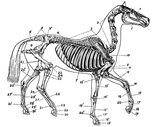
Skeleton of the
Horse.—1, bones of the head; 1′, lower jaw; 2,
cervical vertebræ; 3, dorsal vertebræ; 4, lumbar vertebræ;
5, sacral vertebræ (sacrum); 6, coccygeal vertebræ; 7,
ribs; 8, sternum (breast-bone); 9, pelvis; 9′, ilium;
9″, ischium; 10, scapula (shoulder-blade); 11, humerus;
12, radius; 13, ulna; 14, carpus (knee); 15, large
metacarpal bone (cannon); 16, rudimentary metacarpal bones
(splint-bones); 17, os suffraginis (long pastern); 18,
os coronæ (short pastern); 19, os pedis (hoof-bone); 20,
sesamoid bones; 21, femur; 22, patella (knee-pan, stifle);
23, tibia; 24, fibula; 25, tarsus, or hock; 26, large
metatarsal bone (cannon); 27, rudimentary metatarsals
(splint-bones); 28, os suffraginis (long pastern); 29, os
coronæ (short pastern); 30, os pedis (hoof-bone, “coffin
bone”); 31, sesamoid bones.
The thorax is formed by the ribs and the breast-bone or sternum.
The horse has eighteen ribs on each side (7), and all articulate with
the dorsal vertebræ. The first eight pairs unite by their lower ends
directly to the sternum or breast-bone, and are therefore called
true ribs, while the last ten pairs are only indirectly attached
to the sternum, and are consequently called false ribs. The
sternum (8) lies between the forelegs, and helps to form the floor of
the chest cavity. The space enclosed by the bones of the thorax is
called the thoracic, pulmonary, or chest cavity, and contains the heart
and lungs. The bones of the pelvis form a complete circle or
girdle. The upper part, called the ilium (9′), articulates on its
inner side with the sacrum (5), while its outer side is prolonged to
form a prominent angle, which is the support of the hip, and is called
the “point of the hip.” The posterior part of the pelvis is called the
ischium (9″), and that part lying between the ilium and the
ischium and forming part of the floor of the pelvis is called the pubis.
The space between the thorax and the pelvis, bounded above by
the lumbar vertebræ and shut in below and on the sides by the skin and
muscular walls of the belly (abdomen), is called the abdominal
cavity. This cavity opens directly into the pelvic cavity, and
contains the stomach, intestines, liver, spleen, pancreas, kidneys, and
a part of the generative organs. The thoracic and abdominal cavities
are separated by a muscular partition, the diaphragm.
The bones of the limbs may be likened to columns, upon which the
body rests; they articulate with one another at various angles, are
tubular in structure, and strong.
The bones of the fore-limbs do not articulate directly with
the bones of the trunk, but are attached to the body by means of the
skin and muscles. From above to below we distinguish the following bones:
1. The scapula, or shoulder-blade (10), a flat, triangular bone,
prolonged at its upper border by a flat, very elastic cartilage, called
the scapular cartilage. At its lower end the scapula articulates with—
2. The upper end of the humerus (11), forming the
shoulder-joint (scapulo-humeral articulation). The humerus
articulates at its lower end with—
3. The radius (12) and the ulna (13), to form
the elbow joint. These two bones are the basis of the
forearm. The ulna, smaller and weaker than the radius, lies
behind and projects above it to form the point of the elbow. The lower
end of the radius articulates with—
4. The carpus, or knee (14), which comprises seven small,
cubical bones disposed in two horizontal rows, one above the other. The
upper row comprises four bones and the lower row three. The lower row
rests upon—
5. The large metacarpal or cannon bone, and the two rudimentary
metacarpal or splint-bones. The lower end of the radius,
the upper ends of the metacarpal bones, and the small carpal bones
together form the carpal or knee-joint (wrist of man). Of
the metacarpals, the middle one is the largest, longest, strongest,
and most important, and is called the large metacarpal,
cannon, or shin-bone (15). It articulates at its lower
end with the os suffraginis, or long pastern (17), and with the
two small sesamoid bones (20). On each side of the upper part of
its posterior surface lie the two long, slender splint-bones (16).
The inner splint-bone is sometimes affected with bony thickenings
(exostoses) called “splints.”
6. The bones of the phalanges (all bones below the cannon) will
be fully described in another place.
The bones of the hind limbs articulate directly with
the pelvis at the hip-joint. They are stronger than the bones of the
anterior limbs. We distinguish the following bones in the hind legs:
1. The highest bone in the hind limb is the femur (21). It is
the strongest bone in the entire body. It lies in an oblique direction
downward and forward, and at its lower end articulates with—
2. The patella (22), the tibia (23), and the
fibula (24), to form the stifle-joint (knee of man). The
patella plays over the anterior surface of the lower end of the femur.
The fibula is small, and lies against the upper and outer side of the
tibia. The latter at its lower end articulates with—
3. The bones of the tarsus, or hock (25), which are six small,
irregular bones disposed in three rows, one above another. The os
calcis, or heel-bone, and the astragalus are in the
uppermost row, and are the most important. The former projects above
the true hock-joint from behind, to form a long lever, the upper end
of which is called the “point of the hock,” and the latter articulates
with the tibia. The tarsal (hock) bones articulate below with—
4. The metatarsal bones (26 and 27), which are longer, and the
cannon narrower from side to side, than the corresponding metacarpal
bones, but are otherwise similar.
5. The phalanges of a hind limb (28 to 31) are also narrower
than those of a fore-limb, but are nearly alike in other respects.
All the horse’s bones present small, but more or less distinct
openings (nutrient foramina) for the passage of blood-vessels and
nerves. Many bones possess roughened elevations and depressions, to
which ligaments, tendons, or muscles are attached. With the exception
of the os pedis, all bones are enveloped in a sort of “bone-skin”
called periosteum. The bones unite among themselves to form
either movable or immovable unions. A movable union
between two or more bones is termed a “joint,” or articulation.
The articulating ends of the bones, presenting on one side a convex
surface (head or condyle) and on the other a corresponding concave
surface (glenoid or cotyloid cavity), are covered with elastic
articular cartilage. The bones are bound together by means
of ligaments, which are tough, fibrous, cord-like, or
sheet-like structures. Ligaments are either (1) capsular or (2)
funicular (cord-like). Every articulation in the limbs possesses
a capsular ligament, and all, except the shoulder-joint, have several
funicular (cord-like) ligaments. The capsular ligaments are lined upon
their inner face with a delicate membrane (synovial membrane) which
secretes the synovia, or “joint-water,” whose function is to
lubricate the joint and prevent friction; they enclose the joint in a
sort of air-tight cuff or sack. The funicular ligaments are very strong
and often large, and are the chief means of union of the bones. The
immovable articulations are termed sutures; they are found
principally in the head. The mixed joints are found between the
bodies of the vertebræ, each two of which are united by an elastic
fibro-cartilage which, in the form of a pad, lies between them, and by
its elasticity allows of very slight movement, though the spinal column
as a whole can execute manifold and wide movements, as shown by the
neck and tail.
Joints which permit motion in all directions are known as free
joints; such are the shoulder- and hip-joints (ball-and-socket
joints). Those which admit of motion in but two directions (flexion
and extension), and often to a very limited extent, are called
hinge-joints,—e.g., the elbow, hock, and fetlock. The
joints between the long and short pasterns and between the latter and
the pedal bone are imperfect hinge-joints, because they allow of some
other movements besides flexion and extension. The articulation between
the first and second cervical vertebræ (atlas and axis) is called a
pivot-joint.
The skeleton represents a framework which closely approaches the
external form of the body, and by reason of its hardness and stiffness
furnishes a firm foundation for all other parts of the body. By reason
of the great variety of position and direction of the bones, and of the
fact that changes of position of each single part of this complicated
system of levers may result in the greatest variety of bodily
movements, we can readily understand how the horse is enabled to move
from place to place. Of course, the bones have no power of themselves
to move, but this power is possessed by other organs that are attached
to the bones. These organs are the muscles, and, owing to their
ability to contract and shorten themselves, and afterwards to relax and
allow themselves to be stretched out, they furnish the motive power
that is communicated to and moves the bones.
Fig. 2.
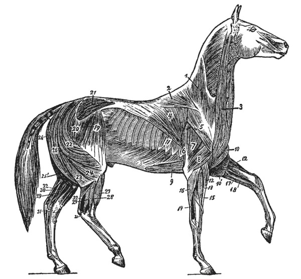
Outer Muscles
of the Horse.—1, cervical trapezius; 2, dorsal
trapezius; 3, mastoido-humeralis; 4, great dorsal
muscle; 5, long abductor of the arm; 6, long extensor
of the forearm; 7, large extensor of the forearm; 8,
short extensor of the forearm; 9, sterno-trochinus (deep
pectoral); 10, sterno-aponeuroticus; 11, great serratus;
12, common extensor of the metacarpus; 13, common extensor
of the toe (anterior extensor); 14, common extensor of the
long pastern (lateral extensor); 15, oblique extensor of
the metacarpus; 16, external flexor of the metacarpus; 17,
internal flexor of the metacarpus; 18, oblique flexor of
the metacarpus; 19, fascia lata; 20, superficial gluteus
(anterior portion); 21, middle gluteus; 22, superficial
gluteus (posterior portion); 23 and 24, femoral biceps; 25,
semitendinosus; 26, semimembranosus; 27, anterior extensor
of the toe; 28, lateral extensor of the toe; 29, perforans
muscle (deep flexor of toe); 30, oblique flexor of the
phalanges; 31, perforatus tendon (superficial flexor of
phalanges); 32, Achilles tendon (ham-string).
The muscles of the body massed together are the red flesh which
we observe in every slaughtered animal. They are not, however, so
shapeless as they appear while in this condition; on the contrary, they
present well-arranged muscular layers of variable size, thickness,
length, and position. (See Fig. 2.) The muscles clothe the skeleton
externally, give the body its peculiar form, and, by their special
power of contraction, change the relative positions of the bones and
thus make it possible for the animal to move. For this reason, the
muscles are called the active, and the bones the passive,
organs of motion. By carefully examining a muscle it will be found to
consist of actual, minute, reddish, muscular fibres. As a rule,
muscles terminate in more or less strong, glistening, fibrous cords
called tendons, or fibrous sheets termed aponeuroses, by which
they are attached to the bones. In the limbs are muscles terminating
in very long tendons, which act as draw-lines upon the distant bones
of the foot (long and short pasterns and pedal bone) and set them in
motion. Such long tendons are enclosed in sheaths of thin, membranous
tissue, known as tendon sheaths. The inner surface of such
a sheath is in direct contact with the surface of the tendon, and
secretes a thin slippery fluid (synovia) which lubricates the tendon
and facilitates its gliding within the sheath.
As long as the bones, articulations, muscles, and tendons of the limbs
remain healthy, just so long will the legs maintain their natural
direction and position. Frequently, however, this normal condition of
the limbs is gradually altered by disease of the bones, joints, and
tendons, and defects in the form and action of the lower parts of the
limbs arise that often require attention in shoeing.
THE FOOT.
Since the horse is useful to man only by reason of his movements, his
foot deserves the most careful attention. The horseshoer should be
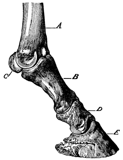
familiar with all its parts. Fig. 3 shows the osseous framework
of the foot, consisting of the lower end of the cannon bone (A), the
long pastern (B), the two sesamoid bones (C), the short
pastern (D), and the pedal bone (E). The lower end of
the cannon, or large metacarpal bone (A) exhibits two convex
articular surfaces (condyles) separated by a median ridge running from
before to behind, and all covered by articular cartilage. On both the
external and the internal aspects of the lower end of the cannon are
small uneven depressions in which ligaments take their attachment.
Fig. 3.
The condyles of the cannon articulate with the os suffraginis (long
pastern) and the two sesamoids (Figs. 3, C, and
4, B) in such a manner that in the forefeet
the cannon makes an angle with the long pastern of from one hundred and
thirty-five to one hundred and forty degrees, and in the hind feet of
from one hundred and forty to one hundred and forty-five degrees.
The long pastern (first phalanx) (Fig. 4, A)
is about one-third the length of the cannon; its upper and thicker end presents
two condyloid cavities (a) (glenoid cavities), separated by a
median groove, which exactly fit the condyles and ridge at the lower
end of the cannon. The lower end of the long pastern is smaller than
the upper, and is provided with two condyles, between which is a
shallow groove (e). The anterior face of the bone is smooth,
rounded from side to side, and blends into the lateral borders. The
posterior face is flatter, and shows a clearly marked triangle to which
ligaments attach.
The two sesamoid bones (Fig. 4, B) are small,
and somewhat pyramidal in shape, and, lying against the posterior part of
the condyles of the cannon bone, increase the articular surfaces at the
upper end of the long pastern.
Fig. 4.
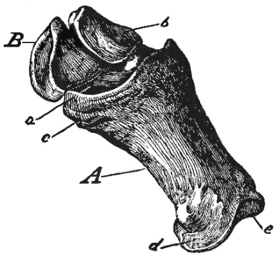
Os suffraginis with both sesamoid
bones in position, as in Fig. 3. A, os suffraginis;
B, sesamoid bones; a, upper joint-surface
of long pastern; b, joint-surface of sesamoid
bones; c, roughened surface at upper end; d,
roughened surface at lower end, both for attachment of
ligaments; e, lower joint-surface.
The short pastern (second phalanx) (Figs. 5
and 6) lies under the first phalanx and above the
os pedis; it is somewhat cubical in shape. Its upper articular surface
(Fig. 5, a) presents two glenoid cavities to correspond
with the condyles of the first phalanx. The lower articular surface
(Fig. 5, d) resembles the lower end of the first phalanx.
The upper posterior border of this bone is prominent and prolonged transversely
(Fig. 6, a), to serve as a supporting ledge
for the first phalanx, as a point of attachment for the perforatus tendon,
and as a gliding surface for the perforans tendon.
Fig. 5.
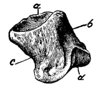
Short pastern (os coronæ)
viewed in front and in profile:
a, upper joint-surface;
b, anterior surface;
c, lateral surface;
d, lower joint-surface.
Fig. 6.
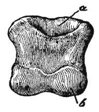
Short pastern seen from behind:
a, smooth surface over
which the perforans tendon glides;
b, lower joint-surface.
The lowest bone of the limb is the third phalanx or os
pedis (Fig. 7). In form it is similar to the hoof. The anterior
or wall-surface (a) is rough, like pumice stone. Above and
in front is the pyramidal eminence to which the tendon of the
anterior extensor of the phalanges attaches. Behind, the bone extends
backward to form the inner and outer branches (c,
c) or wings of the os pedis. The upper, articular surface
(b) slopes backward and downward. The lower, solar or
plantar surface (Fig. 8, a) is slightly concave, and presents
posteriorly a half-moon-shaped excavation, with a roughened border
called the semilunar crest (c), to which the perforans tendon
attaches; just above this crest are two small holes (e) known as
the plantar foramina, through which the plantar arteries pass
into the bone. The surfaces of wall and sole come together in a sharp
edge, which is circular in its course. It is easy to tell whether a
pedal bone is from a fore or a hind limb; the os pedis of a hind leg
has a steeper and more pointed toe, and a more strongly concaved solar
surface than the same bone of a fore-leg. Not only is the outline of the
sharp inferior border of the os pedis of a front foot more rounded
at the toe, but when placed on a flat surface the toe does not
touch by reason of being turned slightly upward, much as a shoe
designed to give a “rolling motion.” The os pedis of a hind foot is
narrower from side to side (pointed), and does not turn up at
the toe.
Fig. 7.
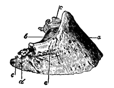
Os pedis seen in profile and in front;
a, anterior face with pyramidal eminence above;
b, joint-surface; c, wings or branches of
hoof-bone; d, notch which, by the attachment of the
lateral cartilage, is converted into a foramen and leads to
e, the preplantar fissure.
Fig. 8.
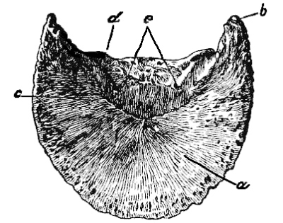
Lower surface of hoof-bone; a,
anterior portion covered by the velvety tissue of the sole;
b, wing of the os pedis; c, semilunar crest,
to which the perforans tendon attaches; d, plantar
fissure leading to e, plantar foramen.
The right and left hoof-bones are also, as a rule, easily distinguished
by variations in the surfaces of wall and sole. The shape of the os
pedis corresponds to the form of the horny box or hoof, and therefore a
knowledge of this bone is absolutely necessary.
The navicular bone (os naviculare, nut-bone,
Figs. 9 and 10)
is an accessory or sesamoid bone to the os pedis. It is a small bone,
transversely elongated and situated behind and below the os pedis and
between the wings of the latter. It adds to the articular surface of
the pedal joint. Its under surface is smooth, and acts as a gliding
surface for the perforans tendon, which is quite wide at this point.
Fig. 9.
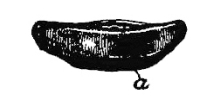
Fig. 10.
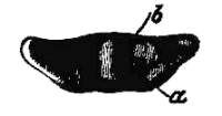
Fig. 9 represents the upper surface of
the navicular bone; Fig. 10 the lower surface of the same:
a, anterior border; b, slight elevation in
middle of under surface.
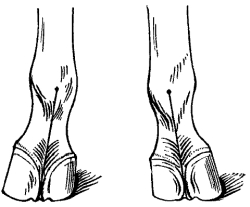
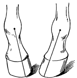
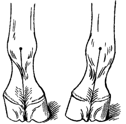
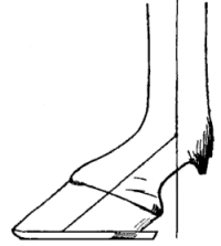
The long axes of the three phalanges (os suffraginis, os coronæ, and os
pedis) should unite to form a straight line, when viewed either from
in front or from one side; that is, the direction of each of these
three bones should be the same as the common direction of the three
considered as a whole.
In young colts both the long and short pasterns are in three parts
and the pedal bone in two parts, all of which unite later in life to
form their respective single bones.
In mules and asses the os pedis is comparatively small and narrow.
In cattle all three phalanges are double, and split hoofs cover the
divided os pedis.
There are three articulations in the foot—namely, the fetlock,
coronary, and pedal joints. All are hinge-joints, the fetlock being a
perfect hinge-joint, and the other two imperfect hinge-joints. Each
has a capsular ligament, and also several funicular or
cord-like ligaments which are placed at the sides of (lateral
ligaments), or behind (on the side of flexion) the joints.
I. The fetlock or metacarpo-phalangeal articulation
is formed by the condyles at the lower end of the cannon bone and the
glenoid cavities formed by the union of the articular surfaces of
the sesamoids and the upper end of the first phalanx. The following
ligaments are about this joint:
Fig. 11.
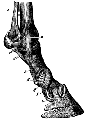
1. Two lateral ligaments, an external and an internal
(Fig. 11, a).
2. Two lateral sesamoid ligaments (f).
3. An intersesamoid ligament (Fig. 12, b),
a thick, fibrous mass, binding the sesamoid bones almost immovably together,
extending above them and presenting on its posterior face a smooth
groove, in which glide the flexor tendons of the phalanges (perforans
and perforatus).
4. The suspensory ligament of the fetlock (Figs. 11,
c, 12, c, and 13, c,
pages 29 and 30). This may also be called the superior sesamoid
ligament. It is a long and very powerful brace, originating on the
lower row of carpal bones (bones of the hock in the hind leg) and on
the upper end of the cannon between the heads of the two splint-bones,
and dividing at the lower third of the cannon into two branches
(c), which are attached one to each sesamoid bone. Below these
bones these two branches are prolonged obliquely downward and forward
on opposite sides of the long pastern to pass into the borders of the
anterior extensor tendon of the toe at about the middle of the long
pastern (Fig. 14, b′, page 32).
Fig. 12.
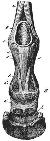
Fig. 13.
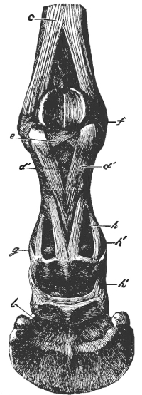
Fig. 11 shows a side view,
and Figs. 12 and 13 a posterior view of the phalangeal bones, with
their articular ligaments. The lettering is the same in all
three figures: a, lateral ligament of fetlock-joint;
b, intersesamoid ligament; c, suspensory
ligament of the fetlock; d, median branch of
inferior sesamoid ligament; d′, lateral branches
of inferior sesamoid ligament; e, deep inferior
sesamoid ligament; f, lateral sesamoid ligaments;
g, inferior coronary ligaments; h, superior
coronary ligaments; h′, median coronary ligaments;
i, lateral pedal ligament; k, lateral
coronary ligament and suspensory ligament of the navicular
bone; l, interosseous ligament.
5. The inferior sesamoid ligament (Figs. 11, d′,
12, d, d′, and 13, d′, E).
This originates at the lowest part of the sesamoid bones and intersesamoid ligament, and
consists of three parts or branches. The median branch
(d) is the longest and strongest, and takes its lower attachment
in the middle of the fibro-cartilaginous lip found on the upper border
of the posterior face of the second phalanx. The two lateral
branches (d′) approach each other as they descend, and
terminate on the sides of the roughened triangle on the posterior face
of the first phalanx.
6. The deep inferior sesamoid ligament (Fig. 13, e)
is quite short, and consists of a number of distinct, thin fibrous bands
lying directly against the bone and entirely covered by the median and
lateral inferior sesamoid ligaments. These fibrous bands cross one
another in passing from the sesamoids to the first phalanx.
II. The coronary joint is the simplest of the three
articulations of the foot. The long pastern furnishes two condyles and
the short pastern two glenoid cavities. Besides a capsular ligament
there are—
1. Two lateral coronary ligaments (k) and,
2. Six posterior coronary ligaments,—namely, two
superior coronary ligaments (h), two median coronary
ligaments (h′), and two inferior coronary ligaments
(g).
III. The pedal articulation (“coffin” joint)
is an imperfect hinge-joint, and is formed by the condyles at the
lower end of the short pastern and the two glenoid cavities in the
united upper surfaces of the pedal and navicular bones. Besides the
capsular ligament (Figs. 12 and 13, l),
which binds all three bones together, there are the following accessory ligaments:
1. Two strong lateral ligaments, an external and an internal
(Fig. 11, i), whose posterior borders are
lost in the lateral cartilages which cover them.
2. Two lateral suspensory ligaments of the navicular bone
(k). They begin on the posterior border and ends of the
navicular bone, and terminate on the lower part of the anterior surface
of the os suffraginis, where they are lost in the lateral ligaments of
the coronary articulation.
3. The lateral ligaments of the lateral cartilages,
navicular bone, and os pedis. They are short, and unite
the navicular bone with the os pedis and lateral cartilages.
Of the three phalangeal articulations, the pedal is the only one that
permits of any lateral movement; hence it is an imperfect hinge-joint.
Though the muscles are the organs which produce motion, the
horseshoer need concern himself only with the tendons of those
muscles which extend and flex the phalanges. These tendons are
either extensors or flexors. The extensors lie on the
anterior face and the flexors on the posterior face of
the phalanges.
Fig. 14.
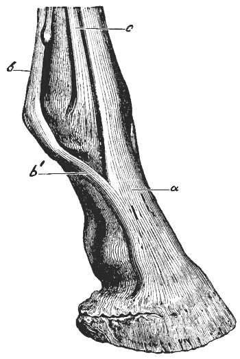
Right forefoot viewed from in front
and from the external side: a, anterior extensor
tendon of the toe; b, suspensory ligament of the
fetlock; b′, branch of the same passing forward and
uniting with the extensor tendon of the toe; c,
extensor tendon of the os suffraginis (absent in the hind
leg), called the lateral extensor.
The anterior extensor of the phalanges (Fig. 14, a)
extends the long and short pasterns and the hoof-bone; it is broad,
and made somewhat broader by receiving the branches of the suspensory
ligament (b′) that come from the sesamoid bones. It takes a firm
attachment on the pyramidal eminence of the os pedis. In the forefeet
the long pastern has a special extensor tendon (c), which is
known as the lateral extensor. When the muscles to which these
tendons are attached act,—that is, when they draw themselves together,
or contract, as we term this action,—the foot is carried forward
(extended).
There are two flexor tendons of the phalanges,—namely, the
superficial (perforatus tendon) and the deep (perforans
tendon).
Fig. 15.
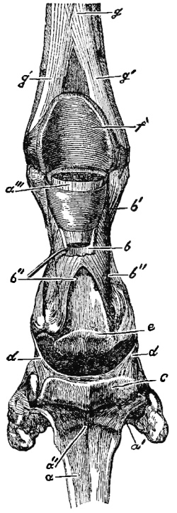
Right forefoot seen from behind:
a, lower end of the perforans tendon, cut through
and hanging down, so that its anterior surface is visible;
a′, lower expanded end (plantar aponeurosis) of
this tendon, which attaches itself to the semilunar crest
of the os pedis; a″, shallow groove which receives
the slight elevation on the under surface of the navicular
bone; a‴, piece of the perforans tendon enclosed
by the ring formed by the perforatus tendon; b,
perforatus tendon bent over backward so that its anterior
surface is visible; b′, ring of the perforatus
tendon; b″, terminal branches of the same; the
perforans tendon passes through the space between these two
branches; c, navicular bone; d, suspensory
ligament of the same; e, smooth surface on the os
coronæ over which the perforans tendon glides; f,
the smooth groove (sesamoid groove) on the posterior
surface of the intersesamoid ligament for the gliding of
the perforans tendon; g, body of the suspensory
ligament of the fetlock; g′, terminal branches of
the same, attaching to the sesamoid bones.
1. The superficial flexor or perforatus tendon (Figs. 15, b,
and 16, a, b) lies behind, immediately under the skin,
and covers the deep flexor or perforans tendon. At the gliding surface
between the sesamoid bones (Fig. 15, f) it broadens,
and forms a ring or tube (Fig. 15, b′) through which
the perforans tendon (a‴) passes, while a short distance farther down it bifurcates,
or divides into two branches (Figs. 15, b″, and
16, b), which terminate, one on either
side, partly on the inferior lateral borders of the first phalanx
and partly on the fibro-cartilage of the second phalanx. It acts
simultaneously on the long and short pasterns.
2. The deep flexor or perforans tendon (Figs. 15,
a, and 16, c) is cylindrical and stronger
than the perforatus tendon; above the fetlock-joint it lies between the
perforatus and the suspensory ligament of the fetlock. At the sesamoid
bones it passes through the ring formed by the perforatus tendon
(Fig. 15, b′), then becomes broad and double-edged,
passes between the two terminal branches of the perforatus, glides over the
fibro-cartilage of the second phalanx and over the inferior surface
of the navicular bone, and finally ends on the semilunar crest of the
third phalanx. In common with the perforatus tendon it flexes the foot.
Fig. 16.
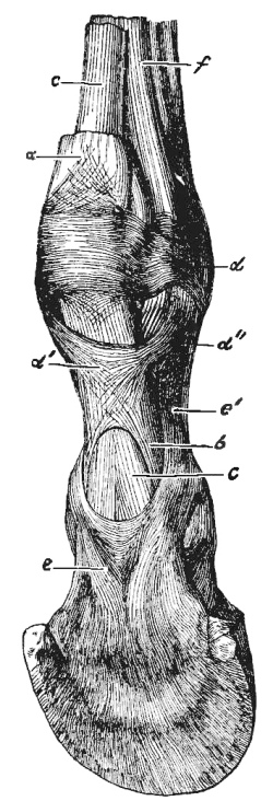
Right forefoot seen from behind and
a little from the external side: a, perforatus
tendon; b, terminal branches of the same; c,
perforans tendon; d, annular ligament which attaches
to the sesamoid bones: d′, the “x” ligament, which
attaches by four branches to the os suffraginis; d″,
an upper branch of the same (the lower branches are not
shown in the figure); e, reinforcing sheath of the
perforans tendon, covering the under surface of the latter
and attached by its branches at e′ to the lower end
of the os suffraginis; f, suspensory ligament of the
fetlock.
If at a point a few inches above the fetlock a limb be cut through
from behind, the knife will pass successively through the following
structures: skin, perforatus tendon, perforans tendon, suspensory
ligament, cannon bone, lateral extensor tendon, anterior extensor
tendon, and, lastly, the skin on the anterior surface of the limb. The
flexor tendons are frequently thickened and shortened by inflammation
due to injury, and as a result the foot is pulled backward and the
hoof gradually becomes more nearly upright,—i.e., stubby,
steep-toed. A knowledge of the normal condition of the tendons is,
therefore, absolutely necessary to the horseshoer. Both flexor tendons
are embraced and held in place by ligaments and fascia passing out from
the phalanges (Figs. 16, d′,
and 24, e, f). The
extensor and flexor tendons essentially contribute to the strong union
of the phalangeal bones, and especially to the support and stability
of the fetlock-joint. The gliding of the tendons is made easy by the
secretion of a lubricating fluid, called synovia, from the inner
surface of the sheaths which surround them. In thin-skinned well-bred
horses with sound limbs one can not only distinctly feel the tendons
through the skin, but can see their outline. When the tendons and
bones are free from all inflammatory thickenings, and the tendon
sheaths are not visibly distended, we say that the leg is “clean.”
Mucous Bursæ and Tendon Sheaths.
Accessory to the tendons, there are in the foot roundish, membranous
sacs (mucous bursæ) and membranous tubes (tendon sheaths). Both contain
a liquid resembling synovia (“joint-water”), which facilitates the
gliding of the tendons. These bursæ and sheaths are often distended to
form soft tumors, known as hygromata (“wind-puffs,” “wind-galls”).
(a) Mucous Bursæ.—They lie beneath tendons at those
places where the tendons pass over bony prominences.
1. The mucous bursa of the anterior extensor tendon of the toe is about
the size of a walnut, and lies between the tendon and the capsular
ligament of the fetlock-joint (Figs. 17, g,
and 18, e).
2. The mucous bursa of the extensor tendon of the long pastern (lateral
extensor) is somewhat smaller, and lies, likewise, beneath the tendon,
between it and the capsular ligament of the fetlock-joint (Fig. 17,
h).
3. The mucous bursa of the navicular region lies between the under
surface (gliding surface) of the navicular bone and the flexor pedis
perforans tendon (deep flexor). Its width equals the length of the
navicular bone, and it extends upward and downward beyond the bone.
Above, it is separated from the sheath of the perforans tendon (“great
sesamoid sheath”) by a membranous partition; below, it passes to the
attachment of the perforans tendon to the semilunar crest of the os pedis.
(b) There is but one tendon sheath in the foot—the sheath
common to the two flexor tendons (great sesamoid sheath). It encloses
the flexor tendons from the middle third of the cannon down to the
middle of the short pastern, and is intimately united with the flexor
pedis perforans tendon (Fig. 17, f, f′, f″,
f‴. Fig. 18, d, d′, d″, d‴).
Fig. 17.
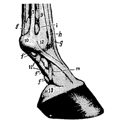
Right forefoot seen from the external
side; f, f′, f″, f‴, great
sesamoid sheath (tendon sheath); g, mucous bursa
beneath anterior extensor tendon of the toe; h,
mucous bursa beneath extensor tendon of long pastern;
i, synovial distension of the fetlock-joint; 7,
suspensory ligament; 9, cannon bone; 10, outer sesamoid
bone; 12, fetlock-joint; 13, lateral cartilage; 14,
suspensory ligament of the lateral cartilage. (Ellenberger
in Leisering’s Atlas and Veterinary Anatomy, Sisson,
Saunders.)
Fig. 18.
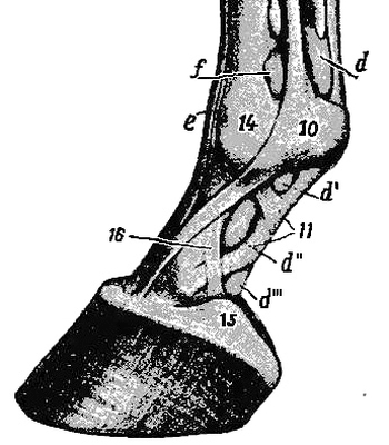
Right forefoot seen from the inner
side; d, d′, d″, d‴, great
sesamoid sheath; e, mucous bursa beneath anterior
extensor tendon of the toe; f, synovial distension
of fetlock-joint; 10, inner sesamoid bone; 11, “x”
ligament; 14, fetlock-joint; 15, lateral cartilage; 16,
suspensory ligament of lateral cartilage (Ellenberger
in Leisering’s Atlas and Veterinary Anatomy, Sisson,
Saunders.)
Altering the Relative
Tension of the Flexor Tendons
and Suspensory Ligament of the
Fetlock-Joint.
The body-weight imposed at the fetlock-joint is supported, in large
part, by the suspensory ligament; somewhat less weight is borne by the
perforans tendon, and a still smaller amount by the perforatus. The
coronary joint is supported chiefly by the perforatus, assisted by
the perforans. The pedal joint is pressed forward and upward by the
perforans tendon passing in a curve beneath the navicular bone. Each of
these three structures bears its normal proportion of the body-weight
when the three phalanges, as viewed from the side, form a continuous
straight line from the fetlock-joint to the ground. In such a case the
obliquity of the long pastern will be the same as that of the toe
(see Foot-Axis, p. 70).
Fig. 19.
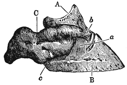
Right forefoot viewed from the
external side: A, os coronæ; B, os pedis;
C, external lateral cartilage; a, lateral
pedal ligament; b, ligament uniting the lateral
cartilage with the os coronæ; c, aponeurosis joining
lateral cartilage and os pedis.
Raising the toe by means of a tip, a full shoe with thinned
branches or a toe-calk, or paring away the quarters will tilt
the os pedis backward, break the foot-axis backward in the pedal joint
and to a less extent in the coronary joint, and increase the tension of
the perforans tendon considerably and of the perforatus slightly. These
tendons tightening behind the fetlock-joint force it forward, causing
the long pastern to stand steeper, and taking some strain from the
suspensory ligament. Hence, the perforans tendon is under greatest
tension, and the suspensory ligament under least tension, when the
foot-axis is broken strongly backward.
Shortening the toe, or raising the
quarters by heel-calks or thickened branches, will tilt the os
pedis forward, break the foot-axis forward in the pedal joint, and
will greatly lessen the tension of the perforans tendon.
The aggregate tension of perforans and perforatus tendons being diminished, the
fetlock sinks downward and backward, the long pastern assumes a more
nearly horizontal direction, and the tension of the suspensory ligament
is increased. Thus, the perforans tendon is under least tension, and
the suspensory ligament under greatest strain, when the foot-axis is
broken strongly forward.
Fig. 20.
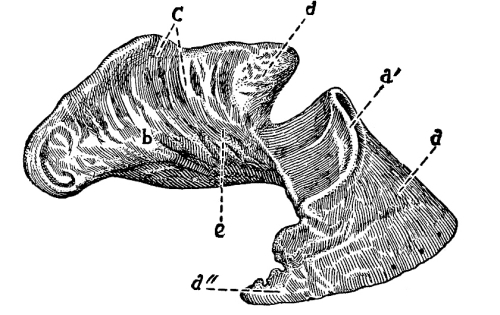
Os pedis and inner face of one lateral
cartilage; a, toe of os pedis; a′, pyramidal
eminence to which the extensor tendon attaches; a″,
wing of pedal bone; b, lateral cartilage; C,
points of attachment of suspensory ligament of lateral
cartilage; d, point of insertion of ligament to the
short pastern; e, point of insertion of ligaments
from navicular bone.
All bodies which under pressure or traction change their form, but
return again to their original shape as soon as the pressure or
traction ceases, are called elastic or springy. Nearly
all parts of the horse’s foot, except the bones, possess more or
less elasticity. The lateral cartilages and the plantar
cushion are elastic to a high degree, but the coronary band,
the laminæ, the articular cartilage, and the horny box
or hoof are less elastic. This property or characteristic is
possessed by the respective parts of the foot in accordance with their
function, location, and structure.
Fig. 21.
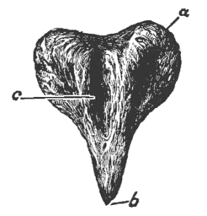
Plantar cushion seen from below:
a, base or bulb of the plantar cushion; b,
summit; c, median lacuna or cleft in which lies the
“frog-stay” of the horny frog.
Fig. 22.
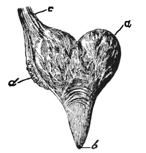
Plantar cushion seen from above:
a, base (bulbs) of same; b, summit; c,
suspensory ligament of plantar cushion; d, place at
which the elastic ligament connecting the os suffraginis
and the lateral cartilage unites with the plantar cushion.
The two lateral cartilages (Figs. 19, C
and 20, b) are irregular, quadrangular
plates, attached to the wings of the os pedis, and extending so far
upward and backward that one can feel them yield to pressure on the
skin above the coronet, and can thus test their elasticity. The
perforans tendon and the plantar cushion lie between the lateral
cartilages, and on the sides and behind are partially enclosed by them.
The internal concave surface of the lateral cartilage (Fig. 20)
is attached to the plantar cushion, the os pedis, and the navicular bone,
and, like the external, slightly convex surface, is covered with many
blood-vessels (veins) Fig. 25, B.
Fig. 23.
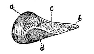
Section lengthwise through middle
of the plantar cushion: a, glome (bulb) of heels;
b, apex or point of fleshy frog; c, fibro
fatty tissue of plantar cushion; d, median cleft
which receives the frog-stay of the horny frog.
The plantar cushion (Figs. 21,
22, 23) is composed almost
entirely of yellow elastic and white fibrous tissues, with adipose
(fat) cells distributed throughout their substance. It is similar in
form to the horny frog, and lies between it and the perforans tendon
(Fig. 24, a). The bulbs are formed by the posterior
thicker portion which lies between the lateral cartilages and is divided into
two parts by the cleft or median lacuna (Figs. 21, a,
and 23, d). The summit is attached to the
plantar face of the os pedis in front of the semilunar crest, and the
bulbs are attached to the lateral cartilages. It is covered inferiorly
by the velvety tissue of the frog (pododerm).
Fig. 24.
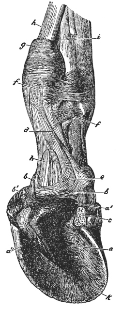
Right forefoot viewed from below,
behind, and the external side. This figure shows clearly
the position of the plantar cushion. The external lateral
cartilage and the tissues covering the plantar cushion and
under surface of the os pedis (velvety tissue of the sole
and fleshy frog) have been removed: a, fleshy frog
or plantar cushion; a′, bulbs of plantar cushion;
the remaining visible parts belong to the so-called “fleshy
frog;” a″, groove (median lacuna) in the lower
surface of the fleshy frog, in which lies the frog-stay
of the horny frog; b, suspensory ligament of the
plantar cushion passing out of the bulbs; b′, small
elastic cords passing to the lateral cartilage; c,
elastic ligament coming from the lateral cartilage and
uniting with the suspensory ligament of the plantar
cushion; d, small tendinous cord beginning in
the skin behind the fetlock-joint and ending on the os
suffraginis in common with b and c; e,
tendinous reinforcing sheath of the perforans tendon;
f, reinforcing stay of the perforatus tendon;
g, perforatus tendon; h, perforans tendon;
i, suspensory ligament of the fetlock; k,
plantar surface of the os pedis, to which the plantar
cushion is joined by fibrous bands.
Vessels which carry blood from the heart to the tissues are called
arteries, while those which return the blood to the heart from
the tissues are called veins. Arteries and veins are connected
by very small, thread-like vessels called capillaries, which
originate in the smallest arteries and are so minute that they can not
be seen without the aid of a microscope. The capillaries penetrate the
soft tissues in every direction, and finally unite to form small veins.
For our purpose we need consider only the arteries and veins.
The arteries carrying blood from the heart ramify and
subdivide in all parts of the body, and thus reach the foot. They are
thick-walled, very elastic tubes, without valves, and carry
bright-red blood, which flows in spurts, as can be seen when
an artery is cut. If a finger be pressed lightly over an artery lying
near the surface, the blood-wave can be felt as a light stroke (pulse).
The character of the pulse is important, because in inflammations of
the pododerm or horn-producing membrane of the foot we can ascertain
by feeling that the pulse is stronger than usual in the large arteries
carrying blood to the inflamed foot.
On either side of the phalanges below the fetlock-joint there lies an
artery called the digital artery (Fig. 25, a).
The pulse can be felt in it as it passes over the fetlock at A, Fig. 25.
It gives off the following collateral (side) branches: 1. The artery
of the first phalanx (perpendicular artery), with anterior and
posterior branches. 2. The artery of the plantar cushion, which
supplies with blood the plantar cushion, the velvety tissue of the
sole and frog, the bar portion of the coronary band, and the sensitive
laminæ of the bars. 3. The coronary artery, which carries blood
to the coronary band, os coronæ, ligaments of the coronary and pedal
joints, flexor tendons, and skin.
The terminal branches of the digital arteries are the preplantar
and plantar ungual arteries. The preplantar artery passes
through the notch in the wing of the os pedis, then along the
preplantar fissure, splitting up into many branches, which spread
over and penetrate the porous surface of the os pedis. The plantar
artery courses along the plantar fissure, enters the plantar foramen,
and passes into the semilunar sinus of the os pedis, where it unites
with the terminal branch of the opposite digital artery, forming the
semilunar arch.
Fig. 25.

Side view of forefoot, showing
blood-vessels and nerves: a, digital artery;
b, anterior artery of the os suffraginis; d,
anterior coronary artery, or circumflex artery of the
coronet; e′, preplantar ungual artery; f′,
inferior communicating arteries passing out from the
semilunar artery of the os pedis, through minute holes just
above the lower border of the bone; they unite to form
(f″) the circumflex artery of the toe; A,
digital vein; B, superficial venous plexus of
coronary band and lateral cartilage; C, podophyllous
venous plexus; G, circumflex vein of the toe; 1,
plantar nerve; 2, anterior digital branch of same; 3,
posterior digital branch of same; 4, small cutaneous
branches of same.
After the arterial or pure blood passes through the capillaries it is
collected by the veins, to be returned to the heart; then it
is driven to the lungs for purification, and is again returned to the
heart, from whence it is pumped through the arteries to all parts of
the body.
Fig. 26.
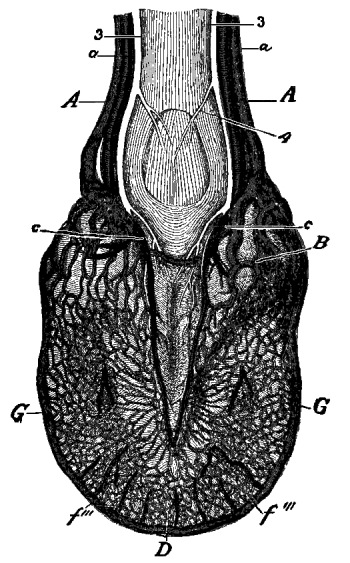
Foot viewed from below and behind:
a, digital arteries; c, arteries of the
plantar cushion; f‴, small branches of the semilunar
artery of the os pedis, which ramify in the velvety tissue
of the sole; A, digital vein; B, venous
plexus of the heels or bulbs; D, solar venous
plexus; G, circumflex vein of the toe; 3, posterior
digital branch of the plantar nerve; 4, cutaneous branches
of the same.
The veins are more numerous than the arteries; they have thinner walls,
and the larger ones are provided with valves that prevent
the impure blood from flowing backward. The veins carry impure or
dark-red blood towards the heart, and if one is opened the dark
blood flows in a steady stream; it does not spurt. The great number
of veinlets in the lower parts of the foot form a complex net-work
(plexus) of vessels which are in such manifold and close union with one
another that checking the flow of blood in one part does not seriously
interfere with the flowing of the blood towards the larger veins. The
following are the most important of these net-works of veins or venous
plexuses: (1) the solar venous plexus (Fig. 26, D);
(2) the podophyllous venous plexus (Fig. 25, C);
(3) superficial coronary venous plexus (Fig. 25, B);
(4) bulbar venous plexus (Fig. 26, B).
All these plexuses of small veins contribute to form the digital veins
(Figs. 25 and 26, A).
Nerves are roundish white cords which
come from the brain and spinal cord; they generally accompany arteries.
They divide and subdivide into smaller and smaller branches till
they become invisible to the naked eye and are lost in the tissues.
The nerves that are found in the foot come from the spinal cord, and
because the largest nerves of the foot accompany the digital arteries
they are called digital nerves (Fig. 25, 1). The
branches ramify throughout all parts of the foot except the horny box and the
hair. Nerves, according to their use or function, are classed as
motor and sensory. The motor nerves end in muscles which
they stimulate to action and control. The sensory nerves terminate
in the skin and in the soft tissues just under the horny box or hoof
(pododerm), and render these parts sensitive; that is, they convey
certain feelings, as, for example, the pain caused by bruising,
pricking, or close-nailing, to the brain and consciousness.
The protective organs are the skin and the horny box or hoof.
The external skin, or hide, covers the entire body; in
the feet it covers the bones, tendons, and ligaments, even passing
in under the hoof and directly covering the os pedis. This portion
of the skin, enclosed by the hoof and therefore invisible, is called
the pododerm or foot-skin. In Germany it is called the
hoof-skin (huflederhaut), because it is a continuation of the
outer visible skin, and because it secretes the hoof,—that is, the
hoof is produced by it. That part of the skin which is covered with
hair is known as the external or hair-skin.
(a) The hair-skin (Fig. 27, a)
consists of three superposed layers,—(1) the external
superficial layer, or epidermis; (2) the middle layer,
derm or leather-skin (so-called because leather is made from
it); (3) the internal layer, or subcutaneous connective tissue.
1. The external layer, or epidermis, is composed
merely of single flattened, horn-like cells (scales) lying side by side and
over one another, and uniting to form one entire structure,—a thin,
horn-like layer, without blood-vessels or nerves. It extends over the
entire surface of the body, and protects the underlying, very sensitive
middle layer from external influences. The oldest cell-layers lie on
the outer surface, and are being continuously brushed off in patches or
scales, while new ones are constantly being formed on the outer surface
of the middle layer.
2. The middle layer, leather-skin or dermis,
is composed of solid, fibrous, and elastic tissues, and contains many
blood-vessels, small nerves, sweat-and oil-glands, and hair follicles
from which the hair grows. The hair upon the posterior surface of
the fetlock-joint is usually long and coarse, forming a tuft known
as the “foot-lock,” which encloses a horny spur, called the ergot.
Common-bred horses have, as a rule, larger and coarser footlocks than
thoroughbreds. The derm or leather-skin, which produces the hair and
epiderm, is the thickest and most important layer of the skin.
3. The inner layer, or subcutaneous tissue, unites the
middle layer with the muscles, tendons, ligaments, bones, or other structures.
It is that loose fibrous mesh or net-work through which the butcher
cuts in removing the hide from the carcass.
Fig. 27.
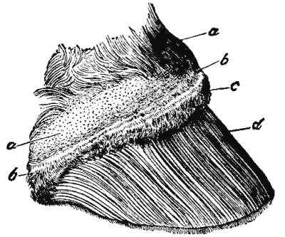
Foot from which the horny capsule or
hoof, has been removed by prolonged soaking: a,
skin; on the left the hair has been rubbed away; b,
perioplic band; c, coronary cushion; d,
podophyllous tissue (fleshy leaves); at the lower border
of the figure can be seen the minute thread-like processes
or villi which grow down from the lower end of each fleshy leaf.
(b) The hoof-skin (Figs. 27
and 28, b, c, d), or
pododerm, is completely enclosed by the hoof. Although it is only an
extension of the derm or middle layer of the hair-skin, it differs from
the latter in structure and relations.
Fig. 28.
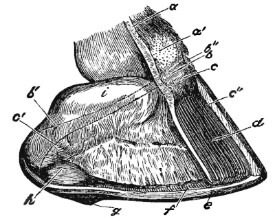
Foot from which the near half of the
horny wall and a greater part of the so-called fleshy
wall have been removed, in order to show the relation of
the lateral cartilage to adjacent structures: a,
vertical section of the skin prolonged downward through
the pododerm (foot-skin) to show clearly that the latter
is but a continuation of the former; a′, hairless
place on the skin; b, perioplic band; b′,
line indicating the upper border of the same; b″,
surface of section of the periople, or perioplic horn-band;
c, coronary cushion; c′, (left) line which
marks the upper border of the coronary cushion; c″,
section of wall at the toe; d, podophyllous tissue
(sensitive laminæ); e, horny sole; f,
white line; g, horny frog; h, fleshy frog;
i, lateral cartilage.
In order to study the pododerm we should not wrench the hoof off with
violence, but should allow the foot to partially decompose by leaving
it for six to eight days at ordinary room temperature; it can then
be removed without injuring the pododerm. After the hoof has been
removed the entire pododerm presents a more or less dark-red color
(flesh-color), which is due to the great number of blood-vessels that
it contains. For this reason different parts of the pododerm have
received the prefix “fleshy,” as for example, fleshy wall, fleshy sole,
fleshy frog, etc. The pododerm is what the uninformed horseshoer calls
the “quick.” I will here remark that the three layers of the external
or hair-skin are represented in the foot; however, the epidermis is
in an entirely different form,—namely, the horny box or hoof. The
internal layer or subcutaneous tissue of the hair-skin is absent in
those parts of the foot where the pododerm covers the os pedis. There
remains, therefore, only the middle layer, derm, or pododerm, which
secretes the hoof, and which is the prolongation and representative
of the middle layer of the hair-skin. The pododerm is distinguished
from the derm of the hair-skin chiefly by the absence of hairs, oil-
and sweat-glands, and the presence on its outer surface of fleshy,
sensitive laminæ and small thread-like projections called villi.
The pododerm consists of five different parts: the perioplic
band, the coronary band, the sensitive laminæ
(podophyllous tissue), the velvety tissue of the sole, and the
velvety tissue of the fleshy frog.
1. The perioplic band (Fig. 28, b)
is a narrow ridge, about one-fifth to one-fourth of an inch wide, lying between the
hair-skin and the coronary band. Somewhat broader at the toe than on
the sides, it broadens out near the bulbs of the heels, over which it
passes to end in the velvety tissue of the fleshy frog. It is separated
from the coronary band by a narrow depression called the coronary
furrow (Moeller). The surface of the perioplic band glistens
faintly, and is thickly studded with numerous thread-like projections
called villi, which are from one-twenty-fourth to one-twelfth of an
inch in length. The perioplic band secretes the soft horn of the
perioplic ring and the perioplic or varnish-like outer layer of the wall.
2. The coronary band (Fig. 27, c)
lies between the perioplic band and the sensitive laminæ or fleshy leaves. It presents
a prominent convex band or cushion about three-fourths of an inch wide,
which extends entirely around the foot from one bulb of the heel to the
other. In front it directly covers the anterior extensor tendon of the
toe, and at the sides the lateral surfaces of the os coronæ and the
upper part of the lateral cartilages, while farther back towards the
heels the lateral cartilages project considerably above both coronary
and perioplic bands. The coronary band is more convex (rounded) in
front than on the sides of the foot, and is flattened in the region
of the bulbs of the heels. Its surface is thickly covered with villi,
which are longer and stronger than those of the perioplic band. At the
bulbs of the heels the coronary band turns forward and inward along the
fleshy frog nearly to its summit. This portion of the coronary band is
from one-third to one-half an inch wide, and is called the bar
portion of the coronary band. It is also covered with villi, which
are directly continuous with those of the fleshy frog. The coronary
band secretes the principal part (middle layer) of the horny wall of
the hoof, including the bar portion (bars) of the wall.
Fig. 29.
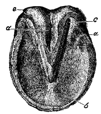
Plantar surface of a foot deprived
of its horny capsule by prolonged maceration: a,
laminæ of the bars; b, velvety tissue of the sole;
c, velvety tissue of the frog; d, median
cleft of the fleshy frog, into which the velvety tissue
dips; e, bulbar portion of the perioplic band, which
passes insensibly into the velvety tissue of the fleshy frog.
3. The fleshy wall, or podophyllous tissue
(Figs. 27, 28, d,
and 29, a), is all that portion of the pododerm
on which there are fleshy leaves. This leafy tissue covers the anterior
surface of the os pedis and the lower portion of the external surface
of the lateral cartilages. At the bulbs of the heels it turns inward at
a sharp angle and extends forward and inward, between the bar portion
of the coronary band and the posterior part of the velvety tissue of
the sole, nearly to the middle of the solar surface of the foot, to
form the laminæ of the bars (Fig. 29, a).
The fleshy wall and fleshy bars are not covered with villi, but with numerous
prominent, parallel, fleshy leaves placed close together, each
of which runs in a straight line downward and forward from the coronary
band to the lower border of the os pedis. Between the fleshy leaves are
deep furrows in which, in a foot which has not been deprived of its
horny capsule, lie the horny or insensitive leaves of the wall. The
fleshy leaves (podophyllous laminæ) are related to one another somewhat
as the leaves of a book; their posterior borders are attached to the
body or basement membrane of the fleshy wall, while their anterior
borders and sides are free. At their upper ends immediately below the
coronary band the leaves are quite narrow, but they gradually increase
in width down to the middle, and thereafter maintain that breadth to
the lower border of the os pedis, where they terminate in free, fleshy
villi, which differ in no respect from those of the fleshy sole. The
number and length of the fleshy leaves vary; in a medium-sized foot
there are about five hundred, while in a large foot there may be as
many as six hundred. On the anterior surface of the os pedis the leaves
are thickest and longest; on the sides and quarters they gradually
decrease in length, while in the bar region they are the shortest and
gradually disappear near the anterior ends of the bars. The width of
the leaves decreases as they become shorter. Viewed with the naked eye
the leaves appear flat and smooth, but under the microscope one can see
on both sides of a fleshy leaf numerous small, fleshy leaflets parallel
to one another and extending lengthwise with the larger leaf. The large
ones are called principal leaves, and the small ones are known
as collateral leaves, or simply as leaflets.
The fleshy leaves (podophyllous tissue)
secrete the horny leaves (keraphyllous tissue) and serve to bind the
horny wall to the pododerm. The strength of this union is due largely
to the dovetailing of the horny leaves and their leaflets with the
fleshy leaves and their leaflets.
4. The fleshy sole or velvety tissue of the sole
(Fig. 29, b) is that part of the pododerm which covers
all the under surface of the foot except the plantar cushion, the bar laminæ, and
the bar portion of the coronary band. It is sometimes slate-colored or
studded with black spots, but is usually dark-red. It is thickly set
with villi, which are especially long and strong[1]
near its periphery. The fleshy sole covers the solar plexus, or
net-work of veins, and secretes the horny sole.
5. The velvety tissue of the frog (Fig. 29, c)
covers the lower surface of the plantar cushion, and in the region of the bulbs
(e) passes insensibly into the perioplic band. In comparison
with the fleshy sole, it has much finer and shorter villi and contains
fewer blood-vessels. It secretes the soft, horny frog.
Fig. 30.
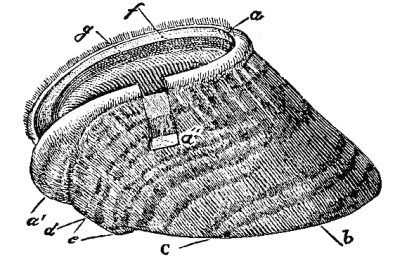
Side view of hoof recently removed:
a, the perioplic horn-band; it is swollen from
prolonged maceration in water; the upper border shows
adhering hairs; the inner surface (perioplic groove)
presents many minute openings; a′, the perioplic
horn-band broadens in passing over the bulb or glome of the
heel, and is finally lost in the horny frog; a″,
section of wall removed. That part of hoof on the right of
b is called the toe; between b and c
is the side wall or “mamma,” and between c and
d the “quarter;” e, projecting horny frog;
f, coronary groove with numerous minute openings;
g, keraphyllous layer of the wall (horny leaves).
(c) The horn capsule or hoof (Fig. 30)
is the entire mass made up of the horn-cells secreted from the whole surface
of the pododerm, and next to the shoe is the organ with which the
horseshoer has most to do. The horn capsule or hoof is nothing more
than a very thick epidermis that protects the horse’s foot, just as
a well-fitting shoe protects the human foot. The hoof of a sound
foot is so firmly united with the underlying pododerm that only an
extraordinary force can separate them. In its normal condition the
hoof exactly fits the soft structures within it; hence it is evident
that local or general contraction of the hoof must produce pressure
on the blood-vessels and nerve-endings of the pododerm, disturb the
circulation of the blood and the nutrition of the foot, and cause pain.
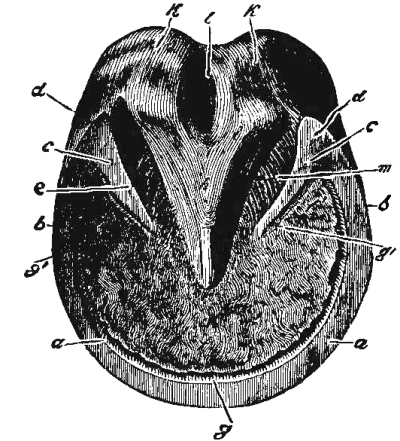
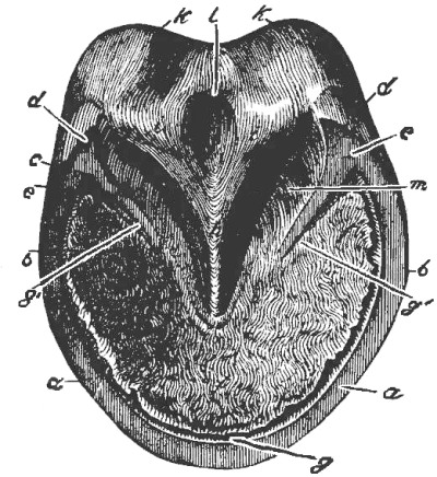
Fig. 31.
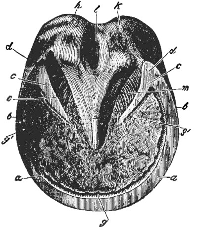
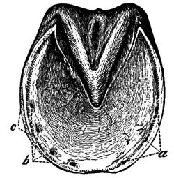
Plantar surface of right fore-hoof:
a, a, bearing-surface of the toe; a,
b, bearing-surface of the side walls or mammæ;
b, c, bearing-surface of the quarters; d,
buttress, or angle formed by wall and bar; e, bar;
f, sole; f′, branches of the sole; g,
white line; it passes between the sole and bars and ends
at g′; h, horny frog; i, branches of
the frog; k, heels, bulbs, or glomes of the hoof;
l, median lacuna of horny frog. Between the bars and
the horny frog lie the lateral lacunæ of the frog.
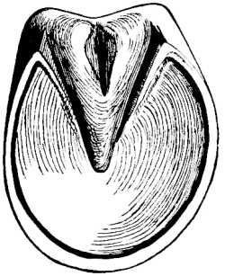
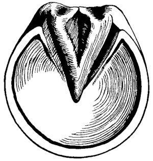
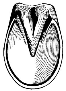
The hoof is divided into three principal parts, which are solidly
united in the healthy foot,—namely, the wall, the sole,
and the frog. That part of the hoof which is almost wholly
visible when the foot is on the ground (Fig. 30, b, c),
and which protects the foot in front and upon the sides, is known as
the wall. In position, course, direction, and arrangement of
its parts it simulates the different parts of the pododerm from which
it is developed. It extends from the edge of the hair just above the
coronary band to the ground; backward it gradually decreases in height
(length), passes around the bulbs of the heels, and turns forward and
inward (Fig. 32, d, e, and
34, a, b) to
form the bars, which are finally lost in the edge of the sole
near the summit of the frog. It thus forms at each heel an angle
(Fig. 31, d, and 32, d)
known as a buttress, which encloses a branch of the horny sole.
Externally the wall is smooth, covered with the varnish-like periople,
and presents indistinct ring-like markings (Fig. 30).
Its inner surface, on the contrary, presents a great number of horn-leaves
which are spoken of collectively as the keraphyllous tissue
(Figs. 32, g, and 35, f).
The upper or coronary border of the wall is thin and flexible, and on its
inner aspect is the coronary groove, into which fits the coronary
band (Fig. 30, f). The lower border of the wall,
called the “bearing-edge” or plantar border (Fig. 31, a),
is the one to which the horseshoe is fastened. By dividing a
hoof from before to behind along its median line, outer and
inner halves or walls are produced, and by dividing
the entire lower circumference of the wall into five equal parts
or sections, a toe, two side walls or mammæ, and two
quarters will be exhibited (Figs. 32
and 33). In order to designate these regions of
the hoof still more accurately, they are spoken of as outer and inner
toes, quarters, and heels.
Fig. 32.
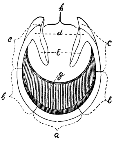
Wall and bars seen from below:
a, toe; b, side wall, or mamma; c,
quarter; d, buttress; e, bar; g,
horn-leaves; h, space occupied by the frog.
The direction (slant) and length of the wall vary in one
and the same hoof, as well as between fore and hind hoofs. The portion
of the wall of fore-hoofs is the most slanting,—that is, forms the
most acute angle with the surface of the ground,—and is also the
longest. Towards the quarters the wall gradually becomes very nearly
vertical; in almost all hoofs the posterior part of the quarters slants
downward and inward towards the median vertical antero-posterior plane
of the foot. At the same time the wall, in passing back from the toe to
the heel, becomes gradually shorter in such a manner that the heights
of the toe, side walls, and quarters are related to one another about
as 3: 2: 1 in front hoofs and as 4: 3: 2 in hind hoofs. The outer
wall is, as a rule, somewhat more slanting than the inner. Viewing a
foot in profile, the toe and heel should be parallel; that is, the line
from the hair to the ground at the toe should be parallel to the line
from the hair to the ground at the buttress. All deviations of the
wall from a straight line (outward or inward bendings) are to be
regarded as faults or defects.
Fig. 33.
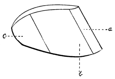
A hoof in profile; a, toe (one half);
b, side wall; c, quarter.
Fig. 34.
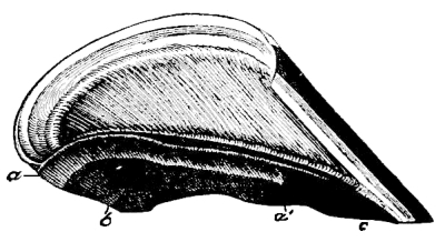
Vertical section through the middle
of a hoof, with horny frog removed, to show the position
of the bar: a, b, marks the line at which
the wall bends forward and inward towards the median line
of the foot to become the bar. Bar runs forward and passes
imperceptibly into the sole c; a, a′,
the light shading shows the part of the bar that was in
contact with the horny frog.
The thickness of the wall is also variable. In front hoofs the
wall is thickest at the toe, and becomes gradually thinner towards
the quarters, while in hind hoofs, there is very little difference
in the thickness of the wall of the toe, sides, and quarters. The
more slanting half of the hoof is always the thicker; thus, for
example, the outer wall of a base-wide foot is always longer and more
oblique than the inner wall, and is also thicker. According to Mayer,
the thickness of the wall at the toe varies from three- to five-eighths
of an inch, and at the quarters from two to three-eighths of an inch.
These measurements are dependent upon the size and breeding of the horse.
Fig. 35.
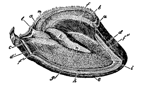
The outer wall of the hoof has
been removed by cutting vertically through the middle
of the toe, down to the upper surface of the sole, then
horizontally backward into the quarter, and, finally,
upward through the quarter: a, perioplic horn-band;
b, coronary groove; it turns inward and forward at
c to form the upper border of the bar; d,
surface of section of the wall at the toe; d′, at
the quarter; e, surface of horizontal section of the
wall near its lower border; f, keraphyllous layer of
the wall; at f′ it turns forward and inward to cover
the bar; f″, horny leaves standing free and passing
insensibly into the white horn of the middle layer or true
wall; g, horny sole; h, white line; i,
small horn-spur in middle of toe; k, part of horny
frog which is in intimate union with the upper edge of the
bar; l, frog-stay of horny frog; it divides the
trough-like depression of the upper surface of the frog
into m, the two upper channels of the frog.
The horn wall is composed of three superposed layers. These
from without to within are: (1) the periople, secreted by the
perioplic band. It is very thin, glistening, and varnish-like in
appearance, and covers the entire outer surface of the wall, except
where it has been removed by the rasp, and prevents rapid evaporation
of moisture from the horn. (2) The middle or protective layer
(Fig. 35, d) is the thickest, strongest, and most
important of the three layers; it forms the principal mass of the wall, and
is developed or secreted by the coronary band, which fits into the
coronary groove. There are in the coronary groove a great number of
small, funnel-shaped openings into which project the horn-producing
villi or papillæ of the coronary band. (3) The inner layer or
keraphyllous layer (Fig. 35, f) consists of
prominent, parallel horn-leaves lying side by side over the entire inner surface
of the middle layer of the wall, and continuing beyond the buttresses
to the ends of the bars (Fig. 35, f′). This layer of
horn-leaves (keraphyllous layer) has in a general way about the same shape and
arrangement as the layer of fleshy leaves (podophyllous layer) which
secretes it; for the horn-leaves fit in with the fleshy leaves in such
a way that every fleshy leaf is embraced by two horn-leaves, and every
horn-leaf by two fleshy leaves (Fig. 36). The keraphyllous
layer and the horn of the inmost part of the middle or protective layer are
always white, even in pigmented (colored) hoofs.
Fig. 36.
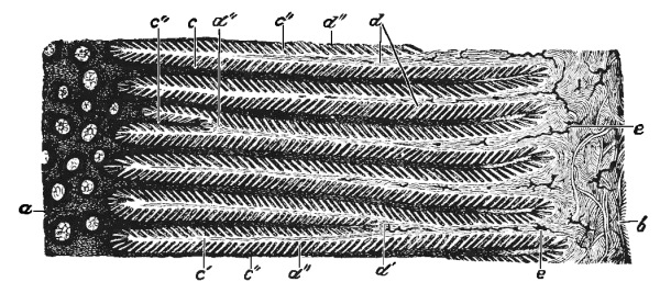
Cross-section of keraphyllous and
podophyllous laminæ (horny and fleshy leaves): a,
inmost part of the solid wall; the horn-tubes approach
very close to the horny leaves; b, body of the
podophyllous membrane; c, horny portion of a
horn-leaf directly continuous with the middle or principal
layer of the wall; c′, a rudimentary horn-leaf that
does not reach the body of the podophyllous membrane;
c″, cross-section of horny leaves from the sides of
which branch many secondary leaves (leaflets) composed of
soft (young) horn-cells. These soft cellular horn-leaflets
dovetail with the podophyllous or fleshy leaflets;
d, podophyllous laminæ extending from the body of
the podophyllous membrane; d′, podophyllous laminæ
which have branched in their course to the wall, and
thus given rise to c′, rudimentary horn-leaves;
d″, cross-section of podophyllous leaflets extending
from the sides of the podophyllous leaves; each two such
leaflets secrete a keraphyllous leaflet between them;
e, injected arterial vessels.
Fig. 37.
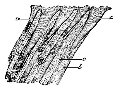
Vertical section of the horny
sole magnified: a, funnel-shaped openings which contain
the horn-producing villi of the fleshy sole; they are of various
sizes; b, horn-tubes; c, intertubular horn.
Fig. 38.
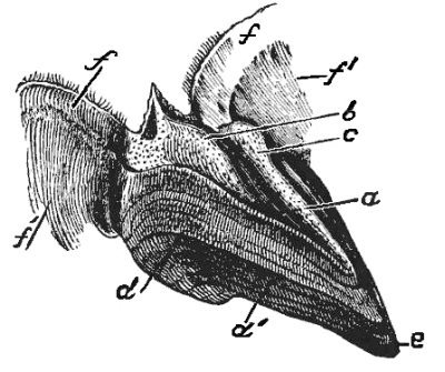
Horny frog, with the posterior
portion of the perioplic horn-band and the periople which
covers the quarters removed from the hoof as one piece by
maceration: a, trough-shaped depression of upper
surface, which is divided posteriorly into the two upper
channels of the frog by b, the frog-stay; c,
part of the frog that is joined to the bar and forms
the lateral wall of the depression (channels) on upper
surface of frog; d, lateral surface of horny frog
which, in its upper part, adheres to the bar, but below,
at d′, lies free; e, point or summit of the
frog; f, perioplic horn-band; f′, periople of
the quarters.
The horn sole (Fig. 31, f,
and Fig. 35, g) is secreted by the velvety
tissue of the sole. A sole from which the loose flakes of old horn
have been removed is about as thick as the wall. It covers the under
surface of the foot, and presents upon its upper surface a convexity
which exactly fits into the concavity on the under surface of the os
pedis. This upper surface is thickly covered by a multitude of minute
funnel-shaped openings for the reception of the villi of the velvety
tissue of the sole (Fig. 37). The lower surface
of the sole is more or less concave, rough, uneven, and often covered
by loose scales of dead horn. Behind, the sole presents a triangular
opening whose borders lie partly in contact with the horny frog and
partly with the bars. This opening or re-entering angle divides
the sole into a body (Fig. 31, f)
and two wings or branches (Fig. 31, f′).
The outer border of the sole unites through the medium of the white
line with the lower part of the inner surface of the wall,—that
is, with the keraphyllous layer of the wall. This white line
(Figs. 31, g, and 35, h),
of so much importance to the horseshoer, is formed by the horn-leaves,
and by those short plugs of tubular horn which are secreted by the
villi that are always found at the lower ends of the fleshy leaves.
The white line may be said to exist wherever the horn-leaves can be
discerned upon the plantar surface of the hoof. It not only passes
around the circumference of the sole from heel to heel, but may be
followed forward from the buttresses along the bars almost to the
summit of the frog. The horn of the white line is soft, unpigmented
(white), and possesses so very little resistance (strength) that it is often
found crumbling or even absent in places. The visible part of the white line
is usually of a grayish-black color, owing to the working in from below
of dirt and liquid manure, and to staining by rust from the nails.
The white line is very important, since it serves as the point from
which we judge of the thickness of the wall, and because the horseshoe
nail should penetrate it.
Fig. 39.
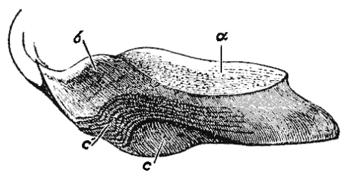
A horny frog cut vertically and
lengthwise through its middle: a, upper surface;
b, frog-stay; c, median lacuna of frog, which
at c′, is overlaid with superposed layers of horn.
Fig. 40.
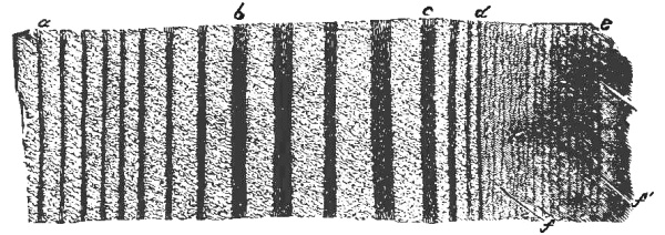
Longitudinal section of the wall
magnified. The dark stripes parallel and close together
are horn-tubes; the lighter surface between the tubes
represents the intertubular horn. Notice that the
horn-tubes are of various diameters. The space between
a and b represents the small tubes of the
outer, darker horn of the principal (middle) layer of the
wall; the space between b and c the lighter,
inner horn of the wall; c, d, the horn
separating the wall proper from the horny leaves; d,
e, the horny leaves (keraphyllous tissue), on which
can be seen fine, parallel, vertical stripes; in the
horn-leaf at f, f′, are seen fissures passing
obliquely upward and outward towards the wall.
The Frog (Figs. 31, h,
35, k, l, 38 and
39), secreted by the velvety tissue covering the plantar cushion
and presenting almost the same form as the latter, lies as a wedge between
the bars and between the edges of the sole just in front of the bars,
with both of which structures it is intimately united. Its horn is
quite soft and very elastic. The median lacuna or cleft
of the frog (Fig. 31, l) divides it into
two branches (Fig. 31, i), which pass
backward and outward into the horny bulbs (Fig. 31, k).
In front of the median lacuna the two branches unite to
form the body of the frog (Fig. 31, h),
which ends in a point, designated the point, apex, or summit of
the frog. On the upper surface of the frog, directly over the median
cleft of the lower surface, there is a small projection called the
frog-stay (Figs. 35, l, 38
and 39, b), which fits into the median
cleft of the plantar cushion. Besides, the upper surface of the frog
shows many minute openings, similar to but smaller than those of the
sole and coronary groove, for the reception of villi. In unshod hoofs
the frog, sole, bars, and bearing-edge of the wall are on a level; that
is, the plantar surface of such hoofs is perfectly flat.
Fig. 41.
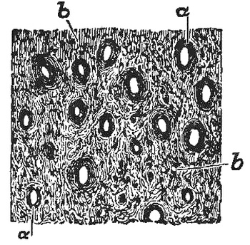
Cross-section of the wall, magnified:
a, horn-tubes;
b, intertubular horn.
The minute structure of the horn can scarcely be considered in
detail in an elementary treatise such as this is. However, a few of the
most important facts are as follows:
If we carefully examine a transverse section of the horn of the wall
(Fig. 41), sole, or frog, we will see with the naked eye,
though much better with a magnifying glass, many minute points quite close to
one another, and greatly resembling the small openings which we have seen
in the coronary groove of the wall and on the upper surface of the
horny sole and frog. If, now, we examine a longitudinal section of the
wall (Fig. 40) or sole, we will see a number of fine, dark
stripes which are straight, parallel, quite close to one another, of different
widths, and which are separated by bands of lighter horn also of
different widths. A thin section or slice of the wall taken at right
angles to the direction of these dark lines (Fig. 41) shows
us that the minute points that are visible to the naked eye, when held up to
the light or moderately magnified, prove to be small openings (Fig. 41, a).
Since these openings, shown in Fig. 41, represent the
dark lines shown in Fig. 40, because an opening is found wherever
there is a dark line, we must regard all dark lines seen in longitudinal
sections of wall, sole, and frog as hollow cylinders or tubes,
though they are not always hollow, but are often filled with loosely
adjusted, crumbling, broken down horn-cells. The dark edges of the
openings (a) consist of thick layers of horn-cells (tube-walls).
The entire structure is called a horn-tube, and the
lighter-colored masses of horn (Fig. 41, b)
between the tubes are known as intertubular horn.
With the exception of the horny leaves of the
wall and bars, all the horn of the hoof is composed of horn-tubes and
intertubular horn.
The horn-tubes of the wall, sole, and frog always run downward and
forward parallel to the direction of the wall at the toe,—that is,
in a direction parallel with the inclination of the hoof as a whole.
Although the wall, sole, and frog differ from one another considerably
with respect to the size and number of the horn-tubes, the quality
of the intertubular horn, and the thickness and strength of the
horn-cells, these differences are only of subordinate interest or
importance to the horseshoer; but he who desires to learn more of this
matter is referred to the work of Leisering & Hartmann, “Der Fuss des
Pferdes in Rücksicht auf Bau, Verrichtungen und Hufbeschlag,” eighth
edition, Dresden, 1893. This book also treats of the variations in the
quality of hoofs, which is very important for the practical horseshoer
to know. It, furthermore, considers the solidity and strength of the
horn of the different parts of the hoof.
With respect to solidity, two kinds of horn are distinguished,—namely,
hard and soft horn. The periople, the white line, and the
frog are soft horn structures; the middle layer of the wall and the
sole are hard or solid horn. The wall, however, is somewhat harder and
more tenacious than the sole, for the latter passes off in more or less
large flakes (exfoliates) or crumbles away on its lower surface, at least
in shod feet, while no such spontaneous shortening occurs in the wall.
Fig. 42.
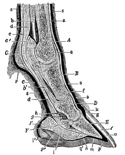
Vertical section through middle of
a forefoot, the skin and pododerm being in red. (In the
figure the direction of both long and short pasterns,
B and D, is too nearly vertical—too
steep). A, metacarpal bone (cannon); B,
os suffraginis (long pastern); C, inner sesamoid
bone (to render it visible a portion of the intersesamoid
ligament was removed); D, os coronæ (short pastern);
E, os pedis (foot-bone); F, navicular bone;
a, extensor tendon; b, suspensory ligament
of the fetlock; b′, superficial inferior sesamoid
ligament; c, perforatus tendon or flexor of the os
coronæ; c′, ring passing forward from this tendon
and encircling the perforans tendon; d, perforans
tendon; e, capsular ligament of fetlock-joint;
f, capsular ligament of coronary joint; g,
g′, capsular ligament of pedal joint; h,
synovial sheath of the perforans tendon; i, plantar
cushion and fleshy frog; i′, bulbs or glomes of
plantar cushion; i″, indicates the lowest point
reached by the plantar cushion, which in the figure is
hidden below by the frog-stay of the horny frog; k,
coronary band (red); l, podophyllous tissue (red);
m, velvety tissue of the sole (red); n,
velvety tissue of fleshy frog (red); o, wall;
p, sole; q, frog; q′, the inner half
of the frog-stay which reposes in the median lacuna of the
fleshy frog; s, hair-skin (red).
Soft horn differs from hard horn in that its horn-cells never become
hard and horn-like. It is very elastic, absorbs water quickly, and as
readily dries out and becomes very hard and brittle and easily fissured
and chapped. With respect to quality, we distinguish good and
bad horn; the former is fine and tenacious (tough), the latter coarse
and either soft and crumbling or hard and brittle. If not dried out,
all horn is elastic, though soft horn is more elastic than hard. All
horn is a poor conductor of heat.
The relative positions of the various parts of the foot are shown
in Fig. 42.
Fig. 43 represents the exterior of a well-formed foot.
Fig. 43.
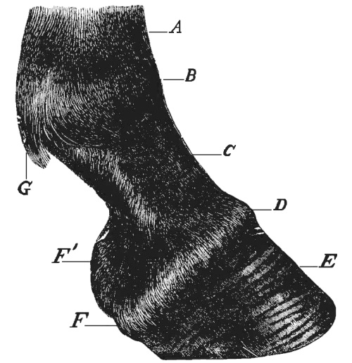
Right forefoot viewed from the side:
A, lower end of the cannon; B, fetlock-joint;
C, long pastern; D, coronet; E, hoof;
F, heel; F′ inner heel; G, foot-lock
covering the ergot.