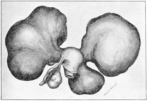
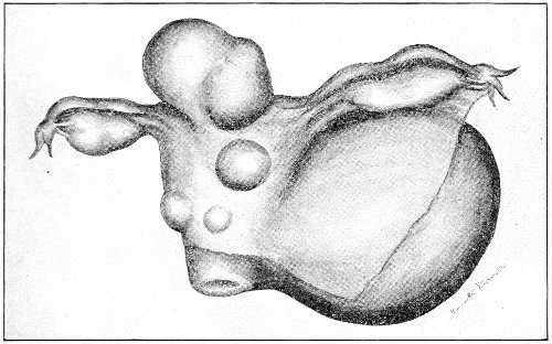
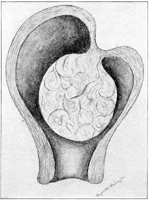
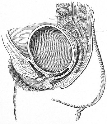
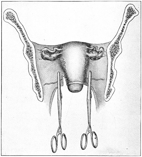
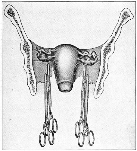
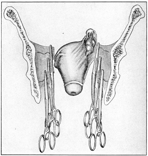
The review of a few facts about the anatomy of the
Fallopian tubes will assist in the study of the diseases
that affect these structures.
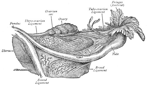
The average length of the normal Fallopian tube is 4
inches (10 centimeters). The tubes are often of unequal
length, the difference sometimes being equal to 1 centimeter.
The length of the Fallopian tube is subject to
considerable variation, and in some forms of ovarian disease
the length of the tube may be very much increased.
The uterine end of the tube varies in thickness from
2 to 4 millimeters. The outer end varies from 7 to 10
millimeters in thickness.
The narrow uterine end of the tube is called the isthmus.
The outer end, of trumpet-shape, is called the
ampulla. The canal of the tube is small. At the uterine
end, or ostium internum, it will barely admit a bristle.
Beyond the middle of the tube the canal gradually widens
to the outer opening—the ostium abdominale.
The ostium abdominale is surrounded by peculiar
luxuriant folds of mucous membrane called fimbriæ.
The fimbriæ are formed by the outward bulging of the
exuberant mucous membrane.
The Fallopian tube consists of three coats, the peritoneal,
the muscular, and the mucous.
The peritoneal coat, which invests the tube for two-thirds
of its circumference, is formed by the free border
of the broad ligament, between the folds of which the
Fallopian tube lies. Loose connective tissue attaches the
peritoneal to the middle or muscular coat.
The muscular coat consists of unstriped muscular fiber
which is continuous with that of the uterus. The muscular
fibers are arranged in two layers, an outer longitudinal
and an inner circular layer.
The inner or mucous coat, which is continuous with
the mucous membrane of the uterus, is covered with
columnar ciliated epithelium.
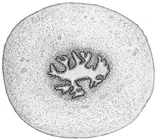
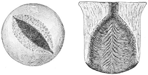
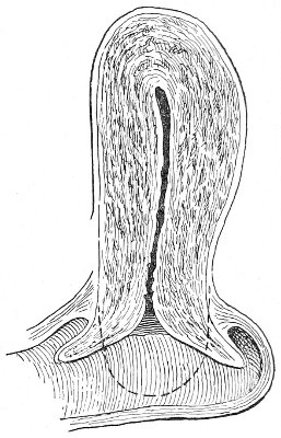
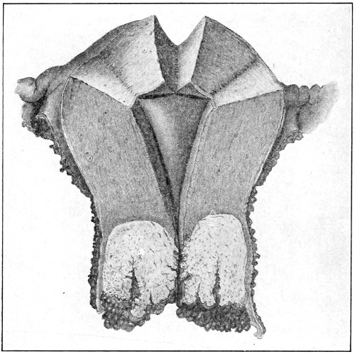
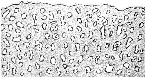
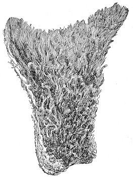
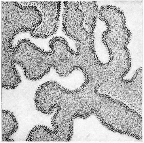
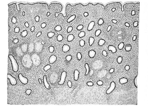
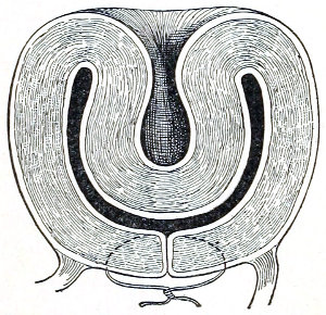
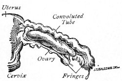
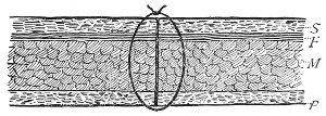
Fig. 142.—Section of the normal Fallopian tube near the uterine cornu (Beyea).
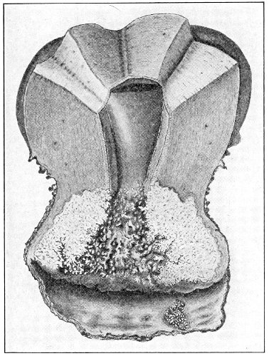
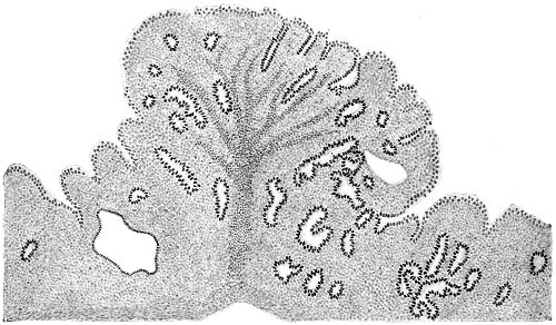
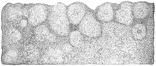
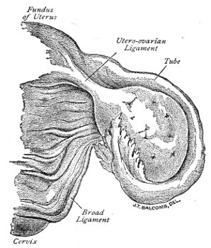
In the outer portion of the tube the mucous membrane
is thrown into longitudinal folds or plicæ. These folds
increase in thickness and in number as the ostium abdominale
is approached. The difference in the degree
of plication at the two ends of the tube is shown by
Figs. 142, 143. The folds of mucous membrane project
beyond the ostium to form the fimbriæ. Like the rest
of the mucous membrane, the fimbriæ are covered by
columnar ciliated epithelium.
The peritoneal covering does not, as a rule, extend on
to the fimbriæ. It terminates by a sharp line which
marks also the termination of the circular muscular fibers
of the middle coat of the tube. The fimbriæ are subject
to great variation in number and in distribution. Sometimes
the Fallopian tube has one or two accessory ostia
in the vicinity of the usual opening. These accessory ostia
are situated on the upper aspect of the tube and are surrounded
by more or less luxuriant fimbriæ. Occasionally
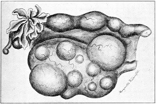
a small pedunculated tuft of fimbriæ is found on the
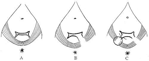
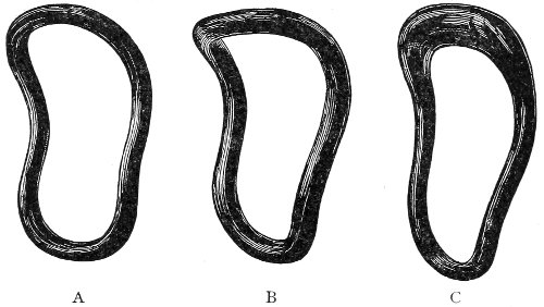
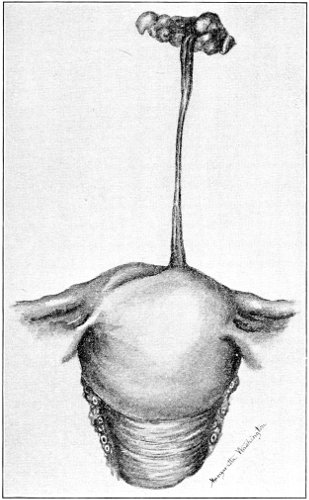
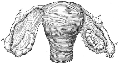
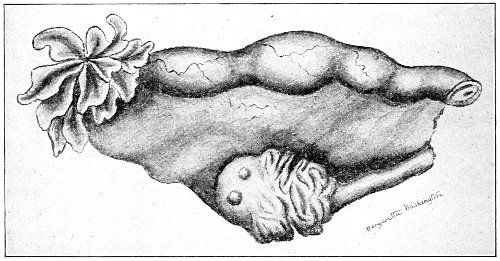
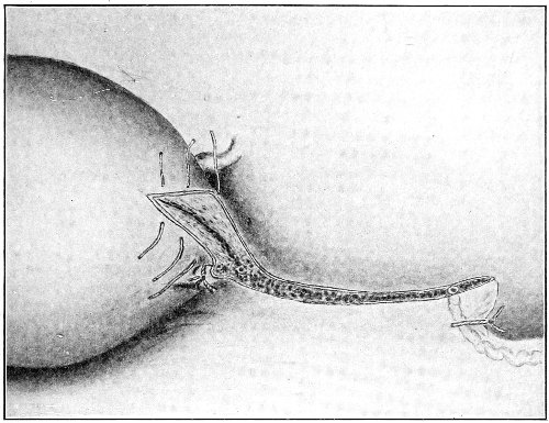
outer portion of the tube (Fig. 144, B). In some cases
there is an accessory tubal end supplied with an ostium
(Fig. 144, A).
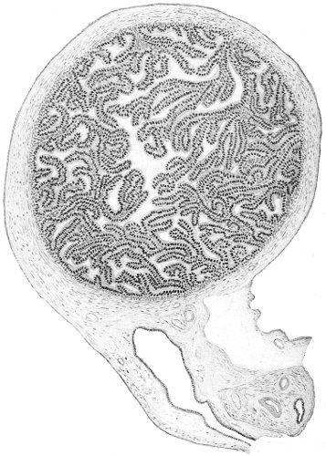
Fig. 143.—Section of the normal Fallopian tube near the abdominal ostium
(Beyea).
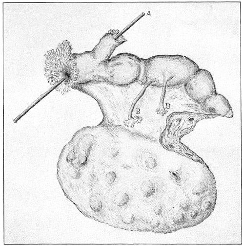
Fig. 144.—Fallopian tube and ovary: A, accessory tubal end with an ostium;
B, pedunculated tuft of fimbriæ.
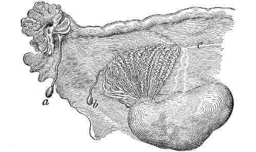
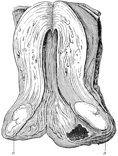
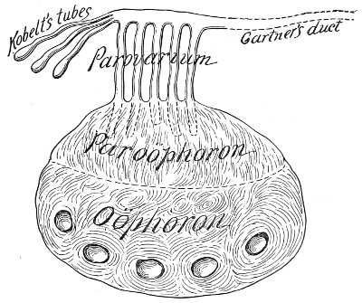
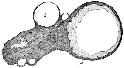
Fig. 145.—Fallopian tube, ovary, and parovarium: a, hydatid of Morgagni;
b, cyst of Kobelt’s tube; c, Gärtner’s duct.
Very often a small pedunculated cyst, about the size
of a pea, is found attached to the fimbriæ or to the outer
aspect of the tube.
These cysts are called hydatids, or cysts of Morgagni.
They are said to occur in about 8 per cent. of adults and
in 20 per cent. of fetuses. They are not pathological.
The cyst wall is composed of three coats: an external
peritoneal coat; a middle muscular coat, arranged in two
layers; and an inner mucous coat covered with columnar
ciliated epithelium. The cyst contains a clear watery
fluid.
No distinct glands, such as are found in the cervix and
the body of the uterus, have been observed in the Fallopian
tubes. The mucous crypts formed by the folds of
the mucous membrane are probably glandular in character
and secrete an albuminous fluid.
Inflammation is the disease that most usually affects
the Fallopian tubes. The condition is, as a rule, secondary
to endometritis, the mucous membrane of the tubes
becoming inflamed by direct extension from the mucous
membrane of the uterus.
The causes of salpingitis are as numerous as those of
endometritis. The most common causes of salpingitis are
sepsis and gonorrhea.
Any form of inflammation of the endometrium may
extend to the Fallopian tubes, but the septic and the
gonorrheal forms of endometritis are especially virulent,
and it is the rule in these diseases that the tubes are
affected.
The various forms of glandular and interstitial endometritis
that have already been described, and which are
due to subinvolution, laceration of the cervix, uterine
displacements, fibroid tumors, etc., may exist for a long
time without producing any perceptible disease of the
tubes. In sepsis and gonorrhea, however, the tubes become
very quickly affected after the uterine cavity has
been invaded, and for this reason these forms of endometritis
excite the greatest apprehension.
Like inflammation of other structures, salpingitis may
be either acute or chronic.
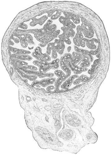
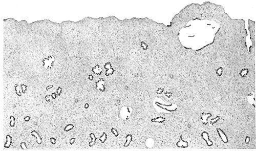
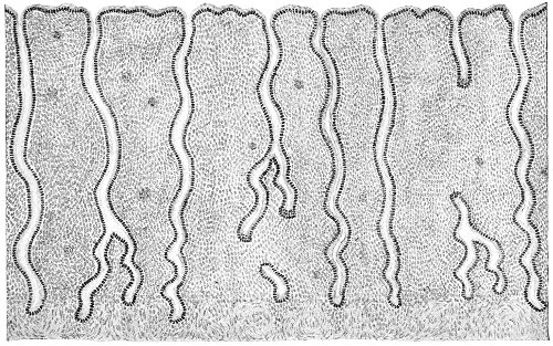
Fig. 146.—Acute septic salpingitis: section about the middle of the tube
(Beyea).
Acute Salpingitis.—In the first stages of acute salpingitis
the disease is confined to the mucous membrane
of the tube. It very quickly extends thence, however,
to the muscular and peritoneal coats, which become infiltrated
with embryonic cells characteristic of the early
stages of inflammation (Fig. 146).
If the tube is laid open, the mucous membrane is found
covered with a muco-purulent secretion. The whole
tube is soft, succulent, and friable. The friability is
such that the tube may readily be ruptured by bending.
The fimbriæ are swollen and congested. A drop of pus
is often seen exuding from the ostium abdominale.
In acute salpingitis the tube may become very quickly
(in a week or ten days) enlarged to the size of the index
finger or the thumb.
The condition that has been described is that found in
the severe cases of acute salpingitis, the result of gonorrhea
or of sepsis after labor. Opportunity is afforded to
examine such cases when the woman has been subjected
to celiotomy, or at the post-mortem when the woman has
died of acute peritonitis or sepsis.
It is probable that a good many cases of acute salpingitis
undergo resolution, and that the tube is restored to
its normal condition.
It is also probable that milder forms of acute salpingitis
occur—cases in which the disease is limited to the
mucous membrane and is merely catarrhal in character,
there being no pus, but a hypersecretion of mucus from
the tube-lining. Such cases, however, recover or pass
into a chronic form of simple catarrhal salpingitis; and
the diagnosis made by a study of the subjective and objective
symptoms cannot be confirmed by operation or
autopsy.
Resolution with perfect restoration of the Fallopian tube
to its normal condition is, of course, always to be hoped
for. In some cases a few fine peritoneal adhesions between
the tube and neighboring structures—such as the
ovary, the uterus, the anterior or the posterior surfaces
of the broad ligament, or a loop of intestine—may result
before resolution takes place, and persist after all other
traces of inflammation have disappeared. In other cases
cure may result, after a greater or less degree of permanent
damage has been done to the abdominal ostium of
the tube, by the shrinking and distortion or crumpling of
the fimbriæ. Such indications of an old, cured attack
of salpingitis are not infrequently seen during celiotomy
for other conditions.
When resolution and cure do not occur, a speedy fatal
result may take place by direct extension of the infection
from the tube to the general peritoneum, with the production
of general peritonitis. Between this extreme
and the mild forms of very localized peritonitis, marked
by a few harmless adhesions, all degrees may exist.
Sometimes a local accumulation of pus occurs in the
pelvis, walled off from the general peritoneum by rapidly
formed adhesions. In other cases a tubal abscess is
quickly formed by inflammatory closure of the abdominal
ostium and distention of the tube with pus; or the
cellular tissue of the broad ligament may become infected,
and the abscess may originate there. And,
finally, if the woman escape these dangers, one or other
of the various forms of chronic salpingitis may result,
and render her a lifelong invalid.
Chronic Salpingitis.—Salpingitis is usually seen in
the chronic form. An acute primary salpingitis must
not be confounded with an acute attack of inflammation
or with an acute exacerbation in an old chronic case. It
is rare that acute gonorrheal salpingitis is seen. The
disease is usually subacute or chronic from the beginning,
as are many of the other manifestations of gonorrhea
in woman, like gonorrheal cervicitis and endometritis.
The most frequent form of acute salpingitis met with
is the septic variety, which occurs as a result of septic
infection after a criminal abortion, a miscarriage, or a
labor. It is usually complicated by severe septic endometritis,
peritonitis, or general sepsis.
The lesions found in chronic salpingitis are numerous.
The simplest form of the disease is the chronic catarrhal
salpingitis, in which the pathological changes are confined
to the mucous membrane of the tube. The muscular
and peritoneal coats are not affected. The ostium
abdominale remains open and is of the normal shape.
The mucous membrane is congested. The folds of
mucous membrane, or the plicæ, are hypertrophied from
gradual infiltration of inflammatory products. The tube
may become somewhat enlarged and more tortuous than
normal. If the inflammatory condition extends to the
middle or muscular coat of the tube, the interstitial
form of salpingitis is produced. The wall of the tube
becomes thicker and harder. The microscope shows an
increased amount of connective tissue in the tube-wall.
As chronic salpingitis progresses the ciliæ of the lining
cells disappear.
If the disease extends through the peritoneal coat, inflammatory
adhesions take place between the tube and
neighboring structures. The tube is often found adherent
to the posterior aspect of the uterus, the broad ligament,
or the ovary.
The most usual seat of adhesions is about the abdominal
ostium. Adhesions here are caused by leakage or escape
of septic material into the peritoneal cavity. The leakage
is slow, and the gradually formed adhesions in time
close the ostium by gluing it to adjacent structures, so
that further escape of tubal contents by this opening is
stopped.
If, in such a case, the tube is freed from its adhesions,
the fimbriæ will be found in the normal position with the
ostium abdominale open.
The usual method of closure of the distal end of the
Fallopian tube is by another process. It takes place as
follows: When the inflammation reaches the muscular
coat of the tube, this coat becomes lengthened and extends
beyond the fimbriæ, which apparently retract and
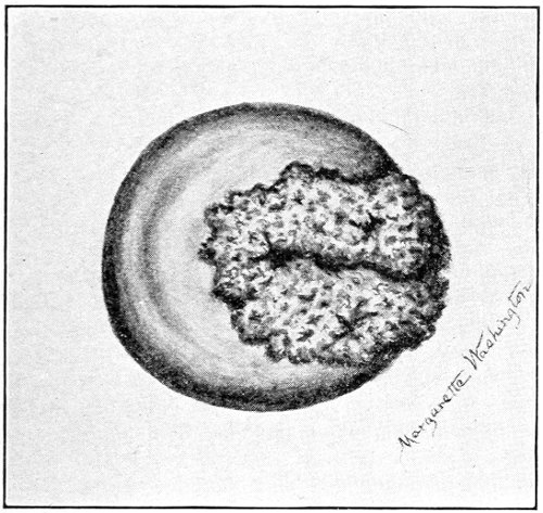
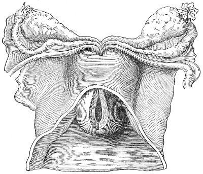
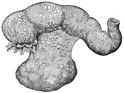
become invaginated in the tube. The opening of the
tube, instead of being flaring with protruding, diverging
fimbriæ, becomes rounded and narrow (Fig. 147).
The fimbriæ become drawn farther into the tube until
they appear to be directed inward instead of outward.
The ostium becomes narrower, and more rounded,
until the edges finally meet and unite by peritoneal
adhesions.
Tubes representing all stages of this process of closure
are often found in operating for inflammatory disease.
Closure of the abdominal ostium by any method is to
be viewed as a conservative process. It prevents leakage,
through this channel, of septic material, and consequently
diminishes the danger of peritonitis.
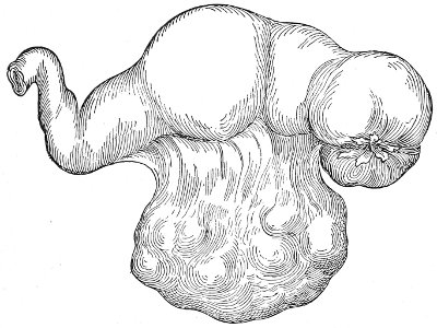
Fig. 147.—Salpingitis with partial inversion of the fimbriæ.
When the abdominal ostium has become closed, the
tubal contents and secretions may have a sufficient
passage for escape by the isthmus into the uterus, and
no further changes take place beyond slow infiltration
and degeneration of the tube-walls. The tube may become
much hypertrophied, not from distention of the
lumen, but as the result of simple inflammatory infiltration
of the mucous and muscular coats, and may attain
the size of the thumb. The walls may become much
degenerated, soft, and friable, so that the tube may easily
be cut through by a ligature or may be broken by bending.
The whole tube may become much elongated and very
tortuous, reaching a length of six or eight inches. The
isthmus of the tube, or the portion in immediate relation
to the uterus, is usually least affected. The whole tube
may become much hypertrophied, and yet the isthmus
will remain approximately of its normal size. In other
cases, however, the disease extends throughout the whole
length of the tube into the uterine horn, and the degeneration
of the tube may be such that it may readily be
broken off at its junction with the uterus.
If, after the ostium abdominale has been closed, anything
occurs to obstruct the escape of the tubal contents
into the uterus, cystic distention of the tube will take
place. Such obstruction may be produced by swelling
of the mucous membrane in the narrow isthmus; by cicatricial
contraction; or by a sharp flexure in any part of
the tortuous tube. Sometimes there are two or more
distended portions of the same tube.
When the tube is distended with pus, the condition is
called a pyosalpinx; when distended with a watery fluid,
a hydrosalpinx; and when distended with blood, a hematosalpinx.
Tubal cysts of this kind may attain large size, in some
cases equal to that of the fetal head.
The shape of the tube becomes much altered. The
greatest distention is at the distal portion, so that the
tube assumes a pear-shape. The lower portion of the
tube is restrained by the mesosalpinx and the tubo-ovarian
ligament, so that as the tube increases in length the
upper portion appears to outgrow the lower, and a retort-shaped
tumor results, or the tube may become tortuous
and folded upon itself.
As the tube enlarges the layers of the mesosalpinx
may become separated, and the tube burrows between
them until it is brought into immediate contact with the
ovary, and the retort-shaped tumor appears with the ovary
lying in the concave portion.
In some cases the ovary and the tube become adherent
by peritoneal adhesions, and the mesosalpinx, which is
wrinkled and folded between them, may be restored by
separation of the adhesions.
In other cases the mesosalpinx itself becomes much
thickened by inflammatory infiltration, and keeps the
tube and ovary separated.
In chronic salpingitis the inflammatory process usually
in time extends to the ovary, and some of the forms
of chronic ovaritis are produced.
The capsule of the ovary becomes thickened, and rupture
of the ripe ovarian follicles is prevented. Small
cysts throughout the ovary are formed in this way. Two
or more cysts may become converted into one cavity by
absorption of the intervening walls, so that cystic spaces
of larger size, equal to that of a duck-egg, may result.
Such cysts may become infected by pyogenic organisms
from the tube, and an ovarian abscess is produced.
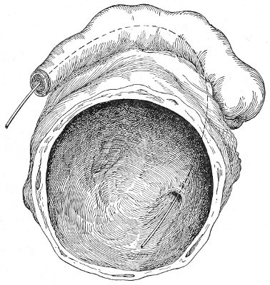
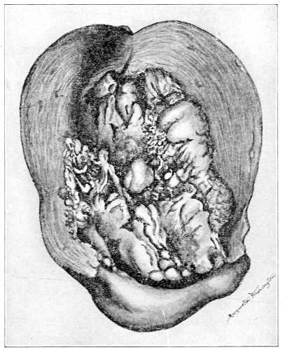
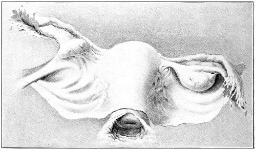
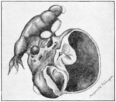
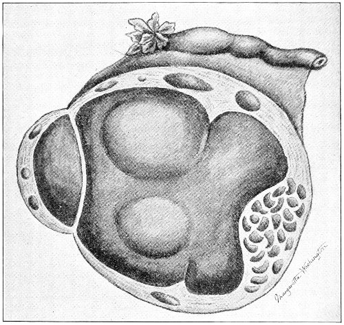
Fig. 148.—Tubo-ovarian abscess.
Tubo-ovarian Abscess.—If the tube is brought into
immediate contact with the ovary, either by agglutination
of the fimbriated end to the surface of the ovary, or
by adhesion of the side of the tube to the ovary, or by
burrowing between the layers of the broad ligament, the
tissue intervening between the cavity of the tube and
the cyst of the ovary may be absorbed or perforated, and
the two cavities will be thrown into one, forming a
tubo-ovarian abscess or a tubo-ovarian cyst (Fig. 148).
The opening between the tubal and ovarian portions of
the cyst does not usually correspond to the abdominal
ostium of the tube, but may be an adventitious opening
in the side of the tube (Fig. 148).
Pyosalpinx.—When the Fallopian tube is distended
with pus or with other fluid, its walls gradually become
thinned. In this respect the Fallopian tube differs from
the body of the uterus, in which a hypertrophy of the
muscular coat usually takes place, under the influence of
distention from the presence of retained fluid within it.
This gradual thinning of the tube-wall predisposes to
rupture or leakage and the escape of the contents into
the abdominal cavity. A pyosalpinx often becomes adherent
to the rectum, the small intestine, or the bladder.
The wall of the intestine or the bladder becomes perforated,
and the pus is discharged in this way. It seems
probable that in some unusual cases the obstruction in
the lumen of the tube is temporarily overcome, and that
evacuation takes place through the uterus, followed by
refilling of the tube. This, however, is a very unusual
occurrence, and is not frequent, as is assumed by some
writers. The evidence of such discharge is based only on
clinical observation. There is no good pathological evidence
of such an occurrence. It is probable that in most
of the reported cases the purulent or watery discharge
which escaped in a sudden gush was derived from, and
had been retained in, the body of the uterus.
The pus of pyosalpinx varies greatly in character. In
the early stages of the disease it is actively septic and
contains a variety of micro-organisms.
These organisms are the gonococcus, streptococcus,
staphylococcus, the bacillus coli communis, the tubercle
bacillus, and the pneumococcus.
In the later stages, however, these organisms become
inert, die, and disappear, so that in the majority of cases
of chronic pyosalpinx the pus is found to be bacteriologically
sterile. Observation on this subject made
by a number of investigators shows that out of 133
cases of acute and chronic suppuration of the uterine appendages
in which the pus was examined bacteriologically,
no organisms whatever were found in 82 cases; in
other words, the pus was sterile in about 61 per cent. of
the cases. The pyosalpinx in time, therefore, becomes
inert so far as any active inflammatory action is concerned,
and resembles a chronic abscess in other parts of the body.
Active inflammatory action may, however, be excited at
any time, as in other chronic abscess, by a new infection,
septic organisms entering the abscess by way of the uterine
cavity, an adherent loop of intestine, or the bladder.
The woman will then have an attack of acute septic inflammation
in the old pyosalpinx, and will be exposed to
the various dangers that were imminent during the primary
acute stages of the disease.
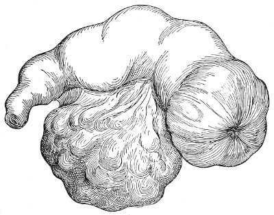
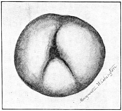
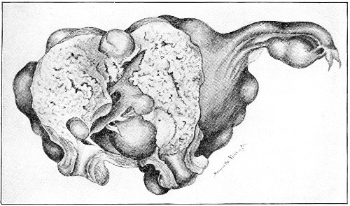
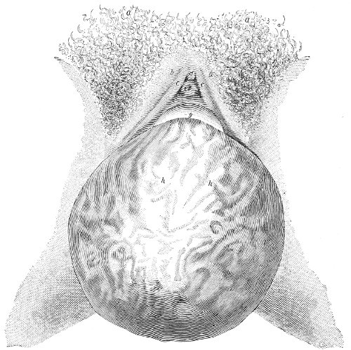
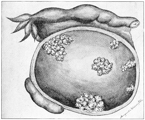
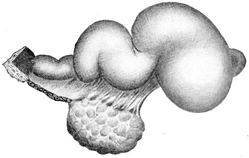
Fig. 149.—Hydrosalpinx, showing complete inversion of the fimbriæ.
It seems probable that if the woman survive the dangers
to which she is exposed from a pyosalpinx, the
tumor may in time become converted into a hydrosalpinx.
The solid constituents of the fluid become absorbed
or deposited upon the cyst-walls, and a clear
watery fluid remains. In hydrosalpinx the recesses of
the tube are often found to contain cheesy material and
cholesterin—remnants of the old purulent accumulation.
The tubo-ovarian cyst is formed in this way from a former
tubo-ovarian abscess.
Hydrosalpinx.—The fluid in a hydrosalpinx may be
colorless, slightly yellow, or brownish or chocolate
colored from the presence of blood. As the accumulation
increases, the walls of the cyst atrophy and become
very thin. The epithelium and the mucous membrane
atrophy and in time disappear, until nothing but a thin-walled
transparent cyst remains (Fig. 149). The cyst-wall
in hydrosalpinx is always thinner and more transparent
than that in pyosalpinx. On the inner wall of
the cyst delicate ridges corresponding to the plicæ or
folds of mucous membrane may be traced. There may
often be discovered, at the distal end of the retort-shaped
tumor, a slight depression that marks the position of the
abdominal ostium, while upon the inner aspect of this
depression may be found the remains of the invaginated
fimbriæ. The size of the tube in hydrosalpinx varies
from that of the little finger to a tumor as large as the
fetal head. Large hydrosalpinx tumors are very unusual,
because the fluid probably leaks slowly through the thin
cyst-wall, and because the secreting surface of the cyst
becomes destroyed by pressure. The fluid from a hydrosalpinx
is sterile, unirritating to the peritoneum, and is
readily absorbed. The cyst may rupture spontaneously
or as the result of some slight accident; the fluid will be
absorbed by the peritoneum, and only the shrivelled,
atrophied sac will remain. In old cases of this kind the
Fallopian tube is represented by an impervious cord.
Such specimens have often been found in old prostitutes
who have survived the dangers of their calling.
Hematosalpinx.—True hematosalpinx, a closed Fallopian
tube distended with blood, is a rare condition.
Tubal pregnancy is the usual cause of an accumulation
of blood in the Fallopian tube, but the term hematosalpinx
should not be applied to this condition. True
hematosalpinx occurs when, from any cause, hemorrhage
takes place into a tube that had previously been closed
by inflammatory action. Such an accident may be caused
by traumatism or by torsion of the pedicle of a tubal cyst.
Slight hemorrhages of this kind occur in pyosalpinx and
in hydrosalpinx, and cause the brownish discoloration
that is sometimes seen in the contents of these tumors.
The various forms of inflammatory disease of the tubes
that have been described under names which designate
the gross appearance of the disease are all really but different
manifestations of the same primary condition.
Gonorrheal or septic infection may produce any of the
forms of tubal disease that have been mentioned. Interstitial
salpingitis without closure of the ostium, pyosalpinx,
hydrosalpinx, hematosalpinx, tubo-ovarian abscess,
etc. are not distinct diseases, but are different manifestations
of the same disease, representing different stages of
progress or different methods of development. Several
of these different forms are often found in the same
woman. On one side there may be a hydrosalpinx, on
the other a pyosalpinx, both caused by a primary chronic
gonorrhea; the distal end of one tube may be distended
by a clear watery fluid, forming a hydrosalpinx, while the
isthmus may be distended with pus, forming a pyosalpinx;
a hematosalpinx may be formed on one side, while
a tubo-ovarian abscess exists on the other; and so through
a great variety of combinations.
Pyosalpinx with active septic contents represents the
early stages of tubal disease, or it represents a chronic
condition in which reinfection has occurred. Pyosalpinx
with sterile pus is like a chronic abscess anywhere else,
and represents a chronic form of salpingitis that had been
active and purulent in the beginning. Hydrosalpinx
represents the disease less violent and septic in the beginning,
and slow in progress; or it represents the last stages
of an old pyosalpinx; while, finally, hematosalpinx represents
a condition of salpingitis in which some accident
has befallen the cystic tube and caused hemorrhage into
its cavity.
The description given shows the progress, the dangers,
and the terminations of salpingitis.
The disease is caused by extension of inflammation
from the endometrium. The usual causes of this inflammation
are gonorrhea, or infection after a criminal abortion,
a labor, or a miscarriage. The gonorrheal salpingitis
is usually slow or insidious from the beginning.
The symptoms of the disease are often not troublesome
until many months after the primary gonorrheal infection.
The closure of the tube is slow, and it is sometimes
not until the tube becomes distended with pus that
the woman experiences much suffering and is placed in
imminent danger. There are cases, however, of acute
gonorrheal salpingitis in which the disease is virulent
and active from the beginning. Infection may traverse
the tube, reach the peritoneum through the open ostium,
and produce general peritonitis within a few days of the
primary attack of gonorrhea. In such cases it is probable
that the infection is a mixed one, other organisms
accompanying the gonococcus. In other cases the abdominal
ostium becomes quickly closed and a gonorrheal
tubal abscess is rapidly formed.
The septic variety of salpingitis, as has already been
said, is more frequently acute from the beginning. Within
ten days or two weeks after a criminal abortion, or after
a miscarriage or labor, a large tubal abscess may be
formed; or the septic organisms may pass through the
tube before the ostium has been closed, and produce within
a few days a general fatal peritonitis.
On the other hand, septic salpingitis is often slow, a
mild attack of puerperal sepsis being the beginning of
years of invalidism, of gradually increasing suffering,
until gross tubal disease is produced.
The slowest forms of salpingitis are those that result
from chronic endometritis, such as accompanies subinvolution,
laceration of the cervix, retro-displacements, or
uterine fibroid. Simple catarrhal salpingitis is often found
in these diseases; or the abdominal ostium may be closed,
and a small hydrosalpinx will be present; or the isthmus
may be sufficiently open for drainage, and no tubal distention
result. Hydrosalpinx is very often found with
uterine fibroids.
Cancer of the cervix or the body of the uterus is a frequent
cause of salpingitis, of hydrosalpinx, and of pyosalpinx.
The endometrial inflammation secondary to the
cancer extends into the tubes.
The progress of salpingitis is beset with danger.
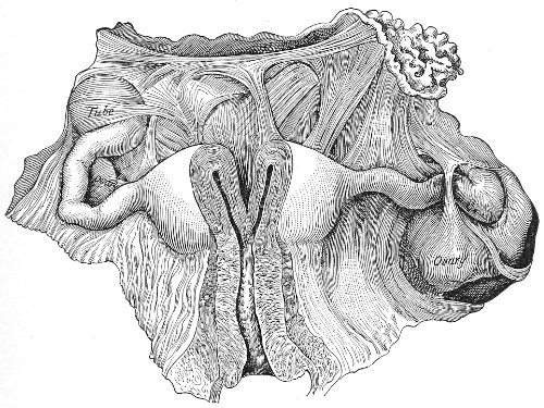
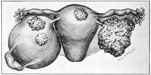
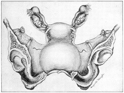
Fig. 150.—Chronic salpingitis with general adhesions of tubes, ovaries, and
uterus (Bandl).
At any time a pyosalpinx may rupture and a rapid fatal
peritonitis result. Unusual effort, vaginal examination,
or slight operations upon the cervix or body of the uterus
may cause this accident. Not infrequently, such
rupture has been produced by even gentle bimanual examination.
I have seen a fatal peritonitis occur from
rupture of a pyosalpinx during the replacement of a prolapsed
uterus.
For this reason the operator should always determine
by careful examination the presence or absence of tubal
disease in every case before performing any of the minor
gynecological operations or manipulations, such as trachelorrhaphy
or the replacement of a retroverted uterus.
Purulent disease of the tubes is a contraindication to all
such procedures, unless an immediate subsequent celiotomy
is to be performed. Great care must be exercised
in any of the less dangerous forms of salpingitis. In
any case of salpingitis, however mild, an acute attack
may be excited by reinfection or by rough manipulation.
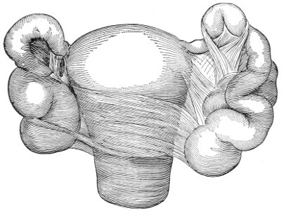
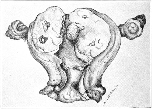
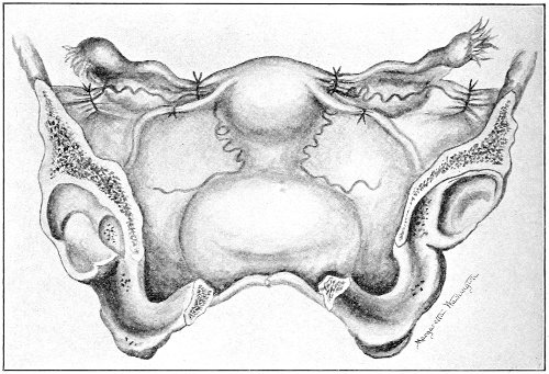
Fig. 151.—Chronic salpingitis: both Fallopian tubes are closed and adherent.
Rupture into the peritoneum is not the only danger to
which the woman is exposed in salpingitis. The gradually
formed adhesions in the pelvis impede the motion of
the pelvic intestines and may cause intestinal obstruction.
Obstruction of the ureters has occurred from pelvic
inflammation. The Fallopian tube may discharge
its contents through the bladder and produce violent cystitis,
or it may discharge through the rectum or intestine,
or adhere to the side of the vagina and discharge through
this channel; or it may be evacuated through the abdominal
parietes. Such fistulous openings rarely, if ever,
close spontaneously and permanently. Temporary closure
may occur, but the tube will refill and discharge as
before.
Fistulæ of this kind persist for many years, becoming
seats of tuberculosis or exhausting the woman by the
continuous suppuration.
If the patient escape these dangers, the disease may
become quiescent. Some of the less dangerous forms of
salpingitis are produced, until finally, when the woman
has reached middle life, a hydrosalpinx remains, or an
adherent, atrophied, cord-like remnant of the tube.
Though then freed from the various dangers that had
threatened her life, she is not restored to health, but
remains a suffering invalid.
Salpingitis may be unilateral or bilateral. It is more
likely to be unilateral in the acute cases than in the
chronic, for, as the primary focus of the disease exists in
the body of the uterus, it will extend in time to the second
tube in case only one had at first been involved. If
the endometrial disease is cured before the second tube
has been attacked, the salpingitis may remain unilateral.
Double salpingitis is especially likely to occur in those
diseases of the endometrium that are difficult or impossible
to eradicate—diseases like chronic gonorrhea, where
the infection lurks in the distal ends of the utricular
glands and defies our methods of treatment. Operators
have repeatedly removed a unilateral pyosalpinx, leaving
the second tube apparently perfectly healthy, and yet,
after the lapse of a few months, a second operation has
been necessary for the relief of a similar pyosalpinx on
the other side.
Symptoms of Acute and Chronic Salpingitis.—The
symptoms of acute salpingitis are usually obscured by
the accompanying symptoms of endometritis, ovarian
congestion and inflammation, and localized peritonitis.
The woman complains of pelvic pain and tenderness,
which are most severe in one or both ovarian regions.
There are elevation of temperature and rapid pulse. The
knees are often drawn up as in peritonitis.
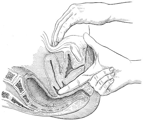
Bimanual examination reveals marked tenderness upon
pressure in the vaginal fornices. There is an indistinct
sense of fulness in the region of the tubes. If the pelvic
peritoneum and cellular tissue are involved, the whole
vaginal vault will feel full and resistant. The tissues
lying to the sides and behind the uterus are thickened
and resistant. If the woman is thin and there is not
much surrounding inflammation, it is sometimes possible
to palpate the enlarged tender tube between the vaginal
finger and the abdominal hand. Usually, however, the
tenderness is too great to permit this. The tube, from
its increase in weight, may fall below its normal level,
and may be felt lying behind the uterus in Douglas’s
pouch.
Usually, in cases of acute salpingitis, the examiner is
obliged to content himself with the determination of an
indistinct fulness and marked tenderness in the region of
the Fallopian tubes.
Before the true pathology of salpingitis was known
these cases were described as pelvic peritonitis or pelvic
cellulitis. It was supposed that the inflammation involved
the peritoneum of the pelvis or the cellular tissue
of the broad ligaments. It is true that this is often the
case, and that inflammation of these structures accompanies
the salpingitis, but it is the tubal inflammation
which is the primary disease.
The most pronounced symptom of chronic salpingitis
is pain. The pain is referred to one or to both ovarian
regions as the disease is unilateral or bilateral. It is due
not only to the salpingitis, but to the accompanying ovaritis.
The pain is continuous. It is relieved by the recumbent
posture, and is increased whenever the woman
is upon her feet or is performing any work. The pain
is increased by a jolt or sudden movement, by defecation,
often by urination and by coitus. The pain during coitus,
from direct pressure, is often so great that marital
relations are abolished. I have seen a woman with salpingitis
who was obliged to take a dose of morphine
before every act of defecation. The pain from the jolting
of a carriage often renders riding impossible.
The pain is dull and aching in character or sharp and
lancinating. It may extend down the anterior aspect of
the thighs.
The pain is very much worse at each menstrual period.
All the genital structures become congested and swollen
at this time, and such phenomena, occurring in the adherent
inflamed tubes and ovaries, often cause unbearable
pain. The dysmenorrhea in salpingitis is usually
very characteristic. It begins several days—sometimes a
week—before the bleeding appears. It starts in one or
both ovarian regions, and radiates thence throughout the
pelvis and down the thighs. It will be remembered that
the dysmenorrhea of anteflexion begins only a few hours
before the bleeding—that the pain is usually situated in
the center of the lower abdomen, in the region of the
uterus, is expulsive in character, and is relieved when
the bleeding has become well established.
The dysmenorrhea of salpingitis usually lasts throughout
the whole of the period.
The pain of salpingitis persists throughout the whole
course of the disease. It is common to all forms of salpingitis,
and seems to bear no relation to the gross character
of the lesions of the tubes. The pain and the
dysmenorrhea are often as marked in a case of salpingitis
without cystic distention as in a case of large pyosalpinx.
The pain persists after the dangerous stages of the disease
have been passed. Relief begins only with the
cessation of menstruation, when general atrophy takes
place in the genital organs.
The pain of salpingitis is often obvious from the expression
and the posture of the woman. She walks with
the body slightly flexed forward; she sits down gently
upon a chair; she protects herself, by support with the
hand, from the jolting of a carriage or a car.
The woman frequently suffers with marked exacerbations
of the pain, which occur independently of the menstrual
periods, and are caused by leakage from the tube
and the resulting local peritonitis. The woman often
describes such attacks as attacks of “inflammation of the
bowels.” They occur usually during the early stages of
the disease. Each attack, if survived, results in a more
perfect closure of the ostium abdominale, and diminishes
the risk of subsequent attacks. At these times all the
symptoms of local peritonitis are present: elevated temperature,
rapid pulse, local or general distention, and
tenderness. In any case of pyosalpinx or of old chronic
salpingitis close questioning of the patient will elicit a
history of this kind.
Acute attacks of pain, fever, and other disturbance
also occur in cases of chronic salpingitis from acute reinfection
of the diseased tube. The disease may have
been quiescent for a long time, and yet active reinfection
may take place by way of the uterine cavity or by the
passage of the colon bacillus through an adherent intestinal
wall; or infection may occur through an adherent
bladder.
Salpingitis is usually accompanied by menorrhagia.
It is impossible to determine how much of this is to be
attributed to the tubal disease. There is always an accompanying
endometritis which is sufficient to account
for it.
Sterility is the rule in cases of salpingitis. The disease
of the mucous membrane and the destruction of the ciliæ
render the passage of the ovum into the uterus difficult.
For this reason tubal pregnancy may occur in salpingitis,
impregnation and attachment of the ovum taking place
within the tube. Inflammation of the ovary, which prevents
the rupture of the ripened ovarian follicles, is another
cause of the sterility. When the abdominal ostia
are closed absolute sterility is present.
In chronic salpingitis the condition of the Fallopian
tubes is revealed by bimanual examination. The tube
usually falls below its normal level, and may be felt by
the vaginal finger lying beside the uterus, or behind it,
in Douglas’s pouch. By careful palpation the connection
of the tubal tumor with the uterus may be traced. Bimanual
examination is most satisfactory in the quiescent
stages of the disease. During an exacerbation or during
one of the acute attacks of inflammation the tenderness
prohibits thorough palpation, and the surrounding inflammatory
infiltration masks the condition of the tube. The
tube may be felt as a hard cord, or as a cystic tumor with
the ovary lying in its concavity, or as a tortuous, sausage-shaped
mass.
In old chronic cases the tube and ovary may be felt as
a hard, knot-like mass adherent to the side of the uterus
or coiled about the cornu (Fig. 151).
In nearly every case the isthmus is rendered hard and
cord-like by inflammatory infiltration. This indurated
condition of the isthmus is a feature of tubal disease that
is usually readily determined, and it is of decided diagnostic
value. The connection, by such a cord, of the
mass felt in the pelvis with the uterine cornu is the
most valuable proof that the tumor is tubal in character.
Diagnosis.—The diagnosis of chronic disease of the
Fallopian tubes must be made from a study of the history,
the symptoms, and by physical examination.
The history is always of value. Careful questioning
will usually show that the ovarian pain dates from a
criminal abortion, from an attack of fever after a miscarriage
or labor, or from a suspicious coitus. Women who
have been infected with chronic gonorrhea by their husbands
attribute the origin of the disease to their marriage.
The woman will often say that for some days
after marriage she suffered with irritation and burning
of the external genitals, with dysuria, perhaps with a
slight vaginal discharge, and that after this, very gradually,
the ovarian pain developed. She may have had one
child or a miscarriage, but with this exception is usually
sterile.
The history of attacks of local peritonitis, confining
the women to bed for several days or weeks, can also usually
be obtained.
The character and the situation of the pain and the
character of the dysmenorrhea usually point strongly to
salpingitis. The physical examination is not by any
means always satisfactory. The small flaccid tubal tumors
are often difficult to palpate, especially in fat
women, and the gross forms of the disease may be obscured
by surrounding adhesions and inflammation. The
examination, however, when taken in connection with
the history and the symptoms, will usually enable one to
make the diagnosis. Inflammatory tumors in the female
pelvis are very generally tubal in origin.
It is difficult to estimate the mortality of salpingitis.
It is certainly a frequent cause of death—not only immediately,
by some of the acute accidents that may occur,
but as a result of gradual exhaustion from prolonged suppuration.
Acute salpingitis, and the purulent forms of
the disease, should always be viewed with anxiety. As
appendicitis is the usual cause of peritonitis in man, so is
salpingitis the usual cause of this disease in the woman.
In every case of peritonitis in a woman, therefore, careful
examination of the pelvic organs should be made.
Salpingitis is an exceedingly common disease. It occurs
in all classes of society, but most frequently in the
lower walks of life. Salpingitis is the rule in prostitutes,
and in them is caused by gonorrhea or by septic infection
at criminal abortion.
Treatment.—The treatment of acute salpingitis in its
early stage should be expectant: absolute rest in the
recumbent position, vaginal douches of a gallon of hot
sterile water (100°-110° F.) two or three times a day,
small doses of saline purgatives (Rochelle salts, ʒss-ʒj
every one or two hours) until mild purgation is produced,
should be prescribed, and should be continued as required.
Relief of pain is afforded by hot fomentations
over the lower abdomen. It is best to administer no
opium, as it is very important to watch these cases closely,
and the symptoms that demand operation might be
masked by the administration of an anodyne. Examinations
should be made with great care and gentleness, and
no oftener than is necessary to determine the progress of
the disease. If the patient is progressing satisfactorily,
repeated examinations are contraindicated.
A chill followed by a rapid high elevation of temperature
(105°-106° F.) is often caused by even gentle manipulation
of the upper organs of generation in cases of
acute inflammation.
The case must be watched carefully and continuously.
In the gonorrheal and septic forms of the disease there is
great danger of extension to the peritoneum, or of the
formation of a tubal or other form of pelvic abscess that
will imperil the life of the woman.
As a general rule, it may be said that, unless there are
well-marked symptoms of extensive pelvic peritonitis,
or unless a distinct tumor can be felt in the pelvis, operation
is not indicated. As resolution undoubtedly takes
place even after severe acute attacks of salpingitis, it is
right to treat the woman with this end in view rather
than to resort to an immediate mutilating operation.
If, under the expectant plan of treatment, the patient
does not improve; if the area of pelvic tenderness increases;
if the local tympany (which may at first be
present only on one or both sides of the pelvis, and
which indicates merely local peritoneal irritation or inflammation)
extends upward; if the temperature and
pulse-rate increase; if constipation appears; if, in fact,
indications of extension of the peritonitis are present,—celiotomy
should be immediately performed. The diseased
tube or tubes should be removed, and, if necessary,
the abdomen should be drained.
Fatal peritonitis sometimes results within three or four
days after the onset of acute salpingitis. As soon, therefore,
as the physician realizes the imminence of this
complication in any case, he should not delay in removing
the source of infection.
The other acute termination of salpingitis, the formation
of an abscess in the pelvis, likewise demands operative
interference. This condition is readily recognized.
The woman has one or more chills. The temperature
becomes more elevated and the pulse more rapid. The
pelvic tenderness and pain may become more distinctly
localized to one or both ovarian regions. Defecation and
urination increase the pain. Bimanual examination reveals
an exceedingly tender mass, either indurated or
perhaps soft and fluctuating, lying to either side of, or
behind the uterus. The character, upon palpation, of
the mass depends upon the nature and extent of the peritoneal
adhesions that surround it. The diagnosis of a
pelvic abscess resulting from acute salpingitis is usually
easy.
There is some difference of opinion among operators in
regard to the best treatment for this condition. Some
advise evacuation of the abscess by way of the vagina;
others advise celiotomy, with removal of the abscess and
the Fallopian tube that caused it, followed, if necessary,
by abdominal or vaginal drainage. I prefer the latter
method of treatment, for reasons that will appear under
the consideration of the technique of operation.
Treatment of Chronic Salpingitis.—Cases of simple
chronic catarrhal salpingitis undoubtedly recover after
the cure of the endometrial disease of which the salpingitis
forms a part. The tube may be restored perfectly
to its normal condition; or there may remain an atrophic
condition of the mucous membrane; or the fimbriæ may
be left somewhat distorted, crumpled, or slightly drawn
within the tube; or there may be a few fine peritoneal
adhesions, like cobwebs, between the distal end of the
tube, the broad ligament, and the ovary. Such slight
lesions may cause no trouble beyond interfering a little
with the fecundity of the woman.
When, however, the adhesions are more extensive,
treatment for their relief may be demanded, even though
all inflammatory action has disappeared from the body of
the uterus and the tubes. Treatment in such cases is
demanded, not to cure the salpingitis or on account of
any danger that threatens the woman’s life, but to relieve
the pain caused by the results of the inflammation.
It may be necessary to perform celiotomy in order to
free or break up adhesions that bind down the ovary in
an abnormal position, or to liberate an adherent intestine,
or to replace a uterus that has been displaced by the traction
of adhesions.
The degree of suffering experienced by the woman is
the guide in advising such operative interference.
Pelvic massage has been used for the relief of pelvic
adhesions of this kind, the uterus, tubes, and ovaries being
manipulated between the fingers in the vagina and a
hand upon the abdomen. The results of this treatment
have not been encouraging.
In discussing the treatment of chronic salpingitis the
cases may be divided into two classes: those in which
palliative treatment may be followed, and those in which
operation is demanded.
There are a great number of cases of chronic salpingitis
in which there is no gross disease of the tubes, and
in which operation upon the tubes is not immediately
indicated. It is proper in such cases to try milder palliative
treatment first.
Salpingitis is always preceded, and usually accompanied,
by inflammation of the endometrium, and in
every chronic case attention should first be directed to
the cure of the endometritis.
If there is no tubal and ovarian displacement—that
is, if the ovary is not prolapsed; if the uterus has not
been retroverted; if there are no extensive tubal adhesions;
and if there is no gross disease of the tube, such
as pyosalpinx, hydrosalpinx, hematosalpinx, a thorough
curetting of the uterus, or, if necessary, a trachelorrhaphy
or an amputation of the cervix, will often relieve the
woman of her suffering, and it may not be necessary to
operate for the damaged tubes.
In all such cases, however, the operator must be very
careful to exclude active or purulent tubal disease. If he
overlooks a pyosalpinx, the curettage or the trachelorrhaphy
may be followed by an active peritoneal inflammation
that will destroy the woman.
If there is ovarian or uterine displacement, we cannot
expect relief until these conditions have been treated, and
such treatment usually requires celiotomy.
The pain and dysmenorrhea of chronic tubal disease
may be relieved by rest in the recumbent position during
the menstrual period; by the administration of saline
laxatives (the pain is always increased by constipation);
by vaginal douches of large quantities of hot water (one
gallon at 110° F.) administered two or three times a day
in the recumbent posture; and by applications of Churchill’s
tincture of iodine to the vaginal vault, and the use
of the glycerin tampon. The directions for this treatment
have been given under the preparatory treatment
of laceration of the cervix.
Such treatment is only palliative: it relieves the pain,
but it will not cure well-established chronic salpingitis.
In many cases the woman experiences little, if any,
relief from this treatment. In other cases, though the
pain may be very much relieved while she is taking treatment,
yet it returns as soon as the treatment is stopped,
and she becomes unwilling to lead the life of an invalid
under constant medical care, with but little prospect of
relief until the menopause is reached. It is then necessary
to consider operation.
The second class of cases referred to—those in which
immediate operation is demanded, and in which it is
dangerous to delay and useless to try the palliative treatment—includes
a great variety. Such cases are—the
gross forms of tubal disease, hydrosalpinx, hematosalpinx,
and pyosalpinx; salpingitis with prolapsed and adherent
tube and ovary; salpingitis with retrodisplacement
of the uterus; all the milder forms of salpingitis
which have resisted palliative treatment.
The operative treatment of salpingitis usually demands
celiotomy. Some operators, however, prefer to reach the
uterine appendages by way of the vagina.
The details of the operative technique of salpingo-oöphorectomy
will be given in a subsequent chapter. As
a rule, the operation of celiotomy for salpingitis should
always be immediately preceded by thorough curetting
of the uterus and, if necessary, by trachelorrhaphy or
an amputation of the cervix.
After the abdomen has been opened the operation consists
in freeing adhesions, rendering patulous the abdominal
ostium of the tube, replacing the uterus, and, if
necessary, removing the tube and ovary on one or on
both sides.
Removal of the tubes and ovaries—salpingo-oöphorectomy—is
usually necessary. In pyosalpinx this operation
should always be performed. If the woman is young and
is very anxious to have children, every attempt should be
made to save, at any rate, one tube and ovary. Remarkable
cases of conception have occurred after conservative
operations upon badly diseased tubes.
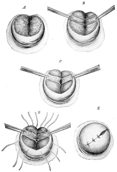
The adhesions about the abdominal ostium may be
broken and the imprisoned fimbriæ freed; or if the ostium
is firmly closed, an incision may be made in the
wall of the tube, the peritoneum stitched to the mucous
coat, and a new ostium produced. In one case conception
followed such an operation in which the ovary was
sutured in the artificial opening made in the tube. Conception
has occurred after both tubes had been amputated
at the uterine cornua.
In all such conservative operations, however, the
woman should be told of the probability of failure and
the probable necessity for a subsequent radical operation.
The successful cases show the possibilities of surgery,
but, unfortunately, they are exceptional. Sterility usually
continues, the pain is usually unrelieved, and a second
radical operation becomes necessary.
Such conservative operations upon badly diseased
tubes should be performed, therefore, only when the
woman is young and anxious for children. Whenever
the abdominal ostium is closed and the ovary is adherent,
it is safest to perform a complete salpingo-oöphorectomy.
This is always indicated when the woman is near the
menopause or when immediate certain relief is demanded
from prolonged suffering.
In some cases the question arises as to whether both
tubes should be removed when only one is grossly diseased.
In the early stages of chronic pyosalpinx it often
happens that but one tube is found diseased, while the
other is apparently perfectly healthy or is only slightly
adherent. Experience has shown that in a great many
cases of tubal disease in which only one tube was removed,
the second tube has become similarly affected,
often within a short time, and a second operation has
been required. This disaster is not likely to occur if the
endometrial disease is eradicated by thorough curetting
at the time of the first operation. But in some forms of
salpingitis, as the gonorrheal, the infection is so deeply
seated in the distal ends of the utricular glands that the
most vigorous curetting fails to remove it, and the second
tube will become infected from the original focus in
the uterus.
So common is such occurrence that many women,
profiting by the experience of their friends, request the
operator to remove both tubes, even though he finds but
one diseased. The advice already given in regard to conservative
operation applies here also. It is safest in all
forms of pyosalpinx to remove both appendages. In the
less serious forms of salpingitis—hydrosalpinx and adherent
tubes without cystic distention—there is less danger
of recurrence, and the unilateral operation may be
more safely performed. The importance of thorough
treatment of the endometritis at the same time is emphasized
by these considerations.
In many cases in which double salpingo-oöphorectomy
is performed it is often advisable to remove the uterus at
the same time. The uterus may be amputated at any
convenient point of the cervix, or it may be completely
removed at the vaginal junction. This operation ensures
more certain and speedy relief from suffering, and is
attended by but little, if any, greater mortality than the
simple salpingo-oöphorectomy. The uterus without the
tubes and ovaries is a useless structure. The operation
is advisable if the uterus is retroverted and adherent,
when the uterus is large and subinvoluted, when the disease
of the endometrium is severe and is likely to persist—in
any case, in fact, in which the physician fears that
the uterus may be a subsequent source of trouble.
Pus in the female pelvis, to which condition the vague
term of pelvic abscess has been applied, is usually the
result of salpingitis producing a pyosalpinx, of ovarian
abscess, or of suppuration of an ovarian cyst, very often
a dermoid. The disease may also occur from infection
of a broad-ligament hematoma or from a pelvic hematocele
caused by a ruptured tubal pregnancy.
Following these conditions the cellular tissue of the
pelvis may become affected, so that the purulent accumulation
may make its way between the layers of the
broad ligament or in some other part of the pelvis.
Before the days of modern abdominal surgery these
accumulations of pus were evacuated through the vagina,
the rectum, or the abdominal wall, according to the direction
in which the abscess seemed to point or in which it
seemed to be most accessible. The sinuses thus formed
often persisted for years or during the remaining life of
the woman. There were many theories in regard to the
origin of the suppuration, it being impossible to determine
its true nature without opening the abdomen.
Now we know that the great majority of such pelvic
abscesses originated in septic infection of the Fallopian
tubes, and that infection of the pelvic cellular tissue
was secondary.
There are, however, rare cases in which the suppuration
occurs primarily in the cellular tissue of the pelvis,
without any involvement whatever of the tubes or
ovaries. Such an accumulation of pus is usually found
in the cellular tissue of the broad ligaments; it sometimes
occurs in the utero-vesical tissue, and rarely in the
tissue back of the cervical neck.
The cause of such suppuration is usually infection, by
way of the lymphatics, from the uterus, or by the passage
of septic organisms directly through the uterine wall.
The condition is most frequently the result of puerperal
sepsis. I have on one occasion seen it occur in connection
with extensive venereal ulceration of the external
genitals. It seems probable that a pelvic lymphatic
gland, becoming infected, may break down and suppurate,
forming the starting-point of the abscess.
The symptoms of this form of pelvic abscess are those
characteristic of any other kind of suppuration in the
pelvis.
The purulent accumulation may be detected by bimanual
examination. It usually bulges into the vagina
at the lateral fornices or before or behind the cervix.
The abscess-mass is in close relationship with the uterus.
In this respect it differs from a simple tubal or an ovarian
abscess, in which cases a distinct separation of the tubal
or ovarian tumor from the uterus may be determined, at
any rate, before the pelvic cellular tissue has become involved.
If the abscess bulge in the anterior vaginal fornix, it
is very probably of neither tubal nor ovarian origin, as
tubal and ovarian abscesses lie to the side of, or behind,
the uterus.
The sense of fluctuation is often difficult or impossible
to determine. The infiltration of the surrounding structures
gives to the mass a dense hard feeling that obscures
fluctuation. To the experienced finger, however, this
indurated condition of the tissues is characteristic of
pelvic suppuration, as is the sense of fluctuation elsewhere.
The treatment of pelvic suppuration of this nature is
evacuation by way of the vagina. The incision should
be made into the most prominent part of the mass.
When made into the lateral fornices, the operator should
remember the position of the ureters and the uterine
arteries. The ureters lie a little over half an inch from
the cervix. In every case it is safest to make the incision
close to the cervix and to work carefully into the abscess-cavity.
The pus should be evacuated, and a double drainage-tube
should be introduced for subsequent washing.
In most cases, however, the physician cannot determine
with any certainty that the abscess is simply confined
to the pelvic cellular tissue and did not originate in
the Fallopian tube. If there is any doubt of this kind,
celiotomy should be performed and the true nature of the
condition determined. If a pyosalpinx or an ovarian
abscess is present, as is usually the case, the condition
may be dealt with as has already been advised. If the uterine
adnexa are healthy, the abdomen may be closed and
a subsequent vaginal incision may be made.
Indiscriminate evacuation of collections of pus in the
pelvis by way of the vagina has resulted in a great deal
of harm. The abscess, being usually of tubal origin,
often persists indefinitely. Intestine, ureters, bladder,
and blood-vessels have often been injured; and when subsequent
celiotomy is performed the operation is attended
with great danger from the presence of the fistulous
opening.
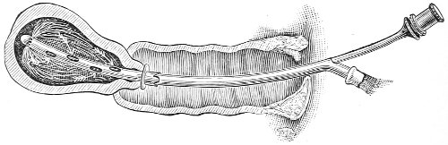
 Copyright, 1908, by W. B. Saunders Company.