DEFINITION.—Inflammation of the interior of the mouth.
The term Stomatitis is used to designate inflammatory affections of
the mucous membranes of the structures of the interior of the mouth,
including thus the mucous membrane of the lips, gums, tongue, cheek,
palate, and anatomical adnexes. Inflammatory affections of the mucous
membrane of the palate, palatine folds, and tonsils are usually
described more particularly under the heads of angina, sore throat,
and tonsillitis.
Stomatitis occurs idiopathically, deuteropathically, and traumatically.
Several varieties of stomatitis occur, sufficiently characteristic to
require separate description: viz. erythematous or catarrhal, aphthous
or vesicular, folliculous or glandular, pseudo-membranous or
diphtheritic, ulcerous, gangrenous, cryptogamous or parasitic, and toxic.
Simple, superficial, erythematous, or catarrhal stomatitis; pultaceous stomatitis.
DEFINITION.—A simple inflammation or erythema, general or partial, of
the mucous membrane of the interior of the mouth.
It occurs both in adults and in children, and may be primary or
secondary, acute or chronic. In adults and adolescents it accompanies
catarrhal and ulcerous affections of the throat, and is described,
therefore, to a certain extent, in connection with these affections.
SYNONYMS.—Ordinary or common diffuse Inflammation of the mouth;
Erythema of the mouth; Oral catarrh.
ETIOLOGY.—In many cases of catarrhal stomatitis, both in adults and
in children, the affection is of obscure origin and the cause eludes
detection. In the great majority of instances the cause lies in some
irritation of the alimentary tract, whether local or at a distance.
The local causes, which are by far the more frequent, include every
variety of topical irritation to which the oral mucous membrane is in
itself liable or to which it may be subjected. Thus, irritating
foreign substances taken into the mouth; unduly heated, unduly iced,
or unduly spiced food and drink; the excessive use or abuse of tobacco
and of stimulants; contact of acrid and corrosive acid and alkaline
mixtures; the
constitutional action of certain medicines,
particularly mercury, but likewise bromine, iodine, arsenic, antimony,
and, to a slighter extent, other medicinal substances also;
inspiration of irritating dust, gases, vapors, steam, and smoke; even
hare-lip, cleft palate, and congenital or acquired deformities of the
mouth generally,—may all be included in this category.
In the newly-born a special hyperæmia of the mucous membrane has been
cited (Billard) as the cause.
Morbid dentition is the most frequent local cause of catarrhal
stomatitis in children, but it is an occasional cause in adults
likewise. Hence it is frequent from the sixth to the thirtieth month
of life; again, between the ages of six and fifteen years, the period
of second dentition; and likewise between the eighteenth and
twenty-second years, the period for the eruption of the last molars.
Deformed, carious, and broken teeth, improper dentistry, wounds and
ulcerations of the gums, negligence in cleansing the teeth,—all these
contribute their quota as exciting causes. Nurslings occasionally
contract the affection from the sore nipples of their nurses. In some
instances they acquire it by protracted sucking at an exhausted
breast. Protracted crying, from whatever cause, sometimes induces
catarrhal stomatitis, not only in nursing children, but in older ones.
Prolonged or too frequent use of the voice, whether in talking,
reading, singing, or shouting, may be the exciting cause.
Distant irritations of the alimentary tract, exciting catarrhal
stomatitis, include stomachic and intestinal derangements of all
sorts. Poor food and lack of hygiene on the one hand, and
over-feeding, excess of spices, alcohol, and tobacco on the other, are
not infrequent exciting causes. Undue excitement, excessive mental
emotion, unrestrained passion, deranged menstruation, normal and
abnormal pregnancy and lactation, sometimes incite the affection.
Slight colds from cold feet or wet clothing give rise to catarrhal
stomatitis. It likewise presents as an extension from coryza, sore
throat, glossitis, tonsillitis, pharyngitis, and laryngitis.
Deuteropathic or secondary catarrhal stomatitis occurs in various
febrile diseases, especially the acute exanthemata—measles, scarlet
fever, small-pox; in syphilis, in pulmonary tuberculosis, and in
long-continued chronic pneumonia.
Infantile stomatitis is most frequent between the ages of two and
twelve months; the stomatitis of adolescents at the periods of
dentition; and that of adults when local sources of irritation predominate.
SYMPTOMATOLOGY.—The symptoms in catarrhal stomatitis vary in severity
with the intensity and extent of the inflammatory processes.
In the infant the subjective symptoms usually commence with
restlessness, fretfulness, and crying. Unwillingness to nurse or
inability to do so soon becomes manifest. The child may seize the
nipple eagerly with a firm grasp of the lips, but at the first suction
lets it drop away with a cry of pain and disappointment. The cause of
the pain is made evident on inspection and palpation of the interior
of the mouth. The parts are dry, glazed, hot, and tender. So hot is
the mouth at times that its heat, conveyed to the nipple in suckling,
is sometimes the first intimation of the existence of the malady.
Similar conditions often prompt an older
child to refuse the
teaspoon. This sensitiveness is observed in the tongue and on the
inner surface of the cheeks. It increases during movements of the
tongue and jaw. Deglutition becomes painful, especially when the food
tendered is rather hot or rather cold. There is a grayish-white
accumulation of partially detached epithelium on the tongue, sometimes
in longitudinal strips, sometimes in a continuous layer. Should the
stomatitis be due to dentition, the affected gums will be swollen,
hot, and painful. There is usually an augmentation of the secretions
in the mouth. Sometimes they flow from the mouth in great quantity,
inflaming the lips. These secretions acquire an increased viscidity,
so that they become adherent in clammy masses to the tongue, the gums,
and the lips. Taste thus becomes impaired, while decomposition of
these masses in sitû imparts fetor to the breath; the odor being
especially pronounced when the child awakens from a night's sleep, the
secretions having accumulated meanwhile more rapidly than they could
be discharged. When the secretions of the mouth are not excessive
there may be merely a faint mawkish odor to the breath, sweetish in
some instances, sour in others. Loss of appetite is usual. Diarrhoea
sometimes exists to a moderate degree, attended at times by gaseous
distension of the intestines.
In severe cases dependent on morbid dentition swelling of the
submaxillary glands and infiltration of the connective tissue may take
place. More or less pyrexia becomes evident. In some instances
convulsions supervene; either directly from cerebral hyperæmia, or in
reflex manner from irritation of the sensitive gingival nerves.
In the adult impairment of taste is one of the earliest subjective
symptoms. This symptom is usually accompanied or else closely followed
by peculiar viscid and sticky sensations about the tongue, gums, and
palate—sensations that excite vermicular motions of the lips and
tongue to get rid of the foreign material by expectoration or by
deglutition. The taste is usually a bitter one, and the viscid
sensations are usually due to accumulations of desquamated epithelium
upon the tongue and other structures. An unpleasant odor is sometimes
exhaled, the result of decomposition of the excessive secretions.
In the chronic form of the affection, especially as it occurs in the
adult, the alterations of taste, the saburral coatings of the tongue,
and the fetor of the breath are more marked than in the acute form.
The mucus accumulating during sleep often awakens the patient in
efforts at hawking and spitting to detach and expectorate it. These
movements are occasionally so violent as to provoke emesis. The
disagreeable odor from the mouth is almost continuous.
In uncomplicated cases there is no loss of appetite or impairment of
digestion. The presence of these symptoms is presumptively indicative
of gastric disease, usually ulcerous or carcinomatous.
The course of the disease varies according to the causes which have
given rise to it. When these subside, the stomatitis soon ceases; when
they are irremediable, the stomatitis remains incurable. No special
period can be mentioned, therefore, for its duration. It terminates,
when cured, in complete restoration of the parts to their normal
condition. There are no special complications or sequelæ.
PATHOLOGY AND MORBID ANATOMY.—The hyperæmia of the
tissues,
physiological during the entire process of dentition, is readily
provoked into a pathological hyperæmia. Whatever the origin, however,
acute catarrhal stomatitis begins, usually, with congestion and
tumefaction of the oral mucous membrane. The congestion is sometimes
preceded by pallor, as though anæmia from constriction of the
capillaries were the initial step in the phenomena. The congestion and
swelling are more rarely diffuse than circumscribed; i.e. confined
to certain portions of the tissues, especially the gums, which become
swollen and painful to contact. The surface is dry and glistening, and
the secretion diminished. The mucous membrane is raised in patches
here and there where the submucous tissues are the most lax. These
patches, irregular in size and configuration, are seen on the tips and
edges of the tongue, on the inner surface of the cheeks, at the
gingival junctions of the jaws, around the dental margins of the gums,
about the angle of the mouth, and on the palate. Sometimes the patches
coalesce—to such an extent in rare instances as to cover the entire
mucous membrane even of the palate and the gums. Their margins are
bright red, their centres yellowish. These elevated patches are due to
local accumulation of new-formed cellular elements, perhaps determined
by the distribution of capillaries or lymphatics. Intensification of
the inflammatory process around or upon them, giving rise to a more
abundant cell-proliferation, sometimes occurs; the results presenting
macroscopically in ridges or welts of a vivid red, surrounding the
patches or traversing them.
The tongue undergoes engorgement, and becomes increased in bulk;
exhibiting dentated facets along its edges and around its tip, due to
the pressure sustained from the adjoining teeth. Opposite the lines of
junction of the two rows of teeth the impression is double. The
dividing lines separating the facets project a little, and are
opalescent, grayish, or whitish, owing to increased proliferation of
epithelium. Similar dentate impressions from a like cause may be seen
on the inner surfaces of the cheeks.
The hyperæmia of the parts is soon followed by excessive production of
new cellular elements, rendering the now increased secretions turbid;
so that the surfaces of the tongue and cheeks become moist again, and
covered with a grayish-white, pultaceous form of desquamated
epithelium, but slightly adherent, and therefore readily detached by
movements of the tongue, lips, and cheeks. In some instances the
epithelium becomes raised into minute vesicles, and chiefly on the
edges of the tongue, thus presenting a sort of lingual herpes.
Excoriations, and even shallow ulcerations, may follow.
Isolated lesions occur. There may be congestion of the palate without
tumefaction, its epithelium undergoing detachment in shreds. The
congested patches at the dental margins of the gums may become
overlaid by opalescent masses of desquamated epithelium, followed by
their actual ulceration, and even by detachment of the teeth.
In children the lips may be swollen and excoriated or surrounded by an
eruption of herpes. Profuse salivation may occur in a child a few
months old when the affection becomes protracted. Febrile movement is
rare before the fifth or sixth month.
In chronic stomatitis the tumefaction is usually greater, with
distension of the capillaries and hypertrophy of some of the mucous
follicles, especially
those upon the cheeks and palate. There is
also hypertrophy of the lingual papillæ, especially those at the tip
of the tongue. Adherent to the gums and the tongue is a yellowish
tenacious mucus, composed of squamous epithelia, fat-globules,
bacteria, and the usual débris of disorganization. The saliva is
secreted in unusual quantities, and sometimes dribbles more or less continuously.
DIAGNOSIS.—Recognition of the conditions described under the head of
Pathology and Morbid Anatomy, in the presence of the symptoms
described under Symptomatology, renders the diagnosis easy.
Chronic stomatitis may be mistaken for mere indication of gastric
catarrh, which is likewise attended with loss of appetite, fetor of
breath, and coating of the tongue.
PROGNOSIS.—The prognosis is favorable in almost every instance,
recovery being almost universal in the acute form. Stomatitis of
dentition subsides with the physiological completion of that process;
stomatitis of exanthematic origin ceases with the evolution of the
eruptive disorder. In the chronic form ultimate recovery will depend
upon the permanency of the existing cause and the extent of the
inflammatory new formations.
TREATMENT.—The first indication, as a matter of course, is to obviate
the cause, whatever that may be. This, when practicable, usually
suffices to bring the malady promptly to a favorable termination.
Intestinal disturbances, whether causative or incidental, must be duly
corrected, and the administration of a saline purge is almost always
desirable. In addition, resort is made to frequent ablutions with
fresh water, warm or tepid, in sprays, gargles, or washes, as may be
most convenient or practicable. Emollients (gum-water, barley-water,
quinceseed-water), astringents (alum, tannin), and detergents (borax,
sodium bicarbonate), may be added, with opiates to relieve pain if need be.
Frequent or continuous suction of fragments of ice usually affords
prompt relief to local pain and heat. The anæsthetic properties of
salicylic acid have been utilized,1 one part to two hundred and
fifty of water containing sufficient alcohol for its solution.
1 Berthold, cited by Ringer, Handbook of Therapeutics,
10th ed., London, 1883, p. 612.
DEFINITION.—Inflammation of the mucous membrane of the interior of
the mouth, characterized by small superficial ulcers. These ulcers are
irregularly circular or oval, are not depressed below the general
surface of the mucous membrane, and support a creamy sebum or
exudation. They occupy positions known to be normally supplied with
mucous glands.
The classical description of this affection includes the initial
eruption of vesicles or groups of vesicles which rupture within a day
or two of their appearance, leaving, upon discharge of their contents,
the little superficial characteristic ulcers. Modern investigation,
however, casts some doubt upon the vesicular character of the initial
lesion, and renders it extremely probable that the reiterated
expression of this opinion has been
a simple deference by writer
after writer to the descriptions given by his predecessors. This
subject will receive further elucidation more appropriately in
describing the pathology and morbid anatomy of the disease.
Aphthous stomatitis may be either idiopathic or symptomatic, discrete
or confluent. It is often recurrent, and is sometimes epidemic.
SYNONYMS.—Aphthæ; Vesicular stomatitis; Follicular stomatitis
(Billard); Canker sore mouth.
ETIOLOGY.—Aphthous stomatitis occurs at all ages, and is most
prevalent during summer heat. In children it is most frequent from the
period of the commencement of dentition to the completion of the
eruption of the temporary teeth. It is infrequent during the fourth
year of life, and is rare after the fifth. It is most apt to appear in
pale, delicate, and scrofulous children, especially in such as are
predisposed to catarrhal and cutaneous diseases (Billard, Barthez and
Rilliet). Sometimes it seems to be hereditary (Barthez). Some
individuals are subject to frequent recurrences. Poor food,
insufficient clothing, want of due ventilation, lack of cleanliness,
and similar deprivations act as predisposing causes. Hence the disease
is apt to occur in the crowded wards of hospitals and asylums for children.
Anything that exhausts the physical forces of the adult, such as
excessive heat, overwork, anxiety, hardship and privation as in
shipwreck, and the drains of menstruation, pregnancy, and lactation,
excessive sexual intercourse, etc., may predispose to the disease.
Long-continued debility from severe constitutional maladies, with
chronic febrile conditions, such as chronic phthisis, chronic
syphilis, chronic enteritis, chronic gastritis, and from diabetes and
carcinoma, likewise acts as a predisposing cause, giving rise, during
the final stages of the systemic disease, to symptomatic aphthæ, often
of the confluent variety. Aphthous stomatitis sometimes accompanies
certain of the continued fevers, exanthematous and non-exanthematous.
As exciting causes the following may be cited: gingivitis, from morbid
dentition in children, and from neglect of the teeth, dental caries,
and dental necrosis in adults; tobacco-smoking; the local contact of
acrid substances in food or otherwise; acute gastro-intestinal
disorder from improper or tainted food. Excessive humidity of the
atmosphere is assigned as a prominent exciting cause of the disease in
some countries. This is especially the case in Holland, where it often
exists epidemically. The confluent form at these times is said to
attack parturient women principally (Ketslaer). Inundations, not only
in Holland, but in Hayti, Porto Rico, and in the United States, are
sometimes followed by an endemic of aphthous stomatitis. It is
believed that the emanations from decayed animal and vegetable matters
left ashore on the reflux of the water, produce the morbid conditions
which constitute the predisposing cause under such circumstances.
The use of certain drugs—preparations of antimony, for
example—sometimes produces a vesicular stomatitis sufficiently
analogous to aphthæ to be mentioned in this connection, and only to be
distinguished therefrom by the history of the case.
PATHOLOGY AND MORBID ANATOMY.—As has been intimated, the morbid
anatomy of aphthæ has long been described as a series of initial
vesicles2 upon the buccal, labial, gingival, or lingual mucous
membrane. Their variance from analogous cutaneous vesicles—herpes,
for instance—is attributed to anatomical differences in the
constitution of the mucous membrane and the skin. The rarity of their
detection has been accounted for by the rapid maceration of the epithelium.
2 Tardieu, Hardy and Behier, Barthez and Rilliet, Meigs and
Pepper, and many others.
The general opinion at present, however, is that the apparent vesicle
is an inflamed mucous follicle.3 Some observers contend that it is
an inflammation of the mucous membrane pure and simple (Taupin);
others consider it an inflammation, sometimes in a follicle, sometimes
in the mucous membrane (Grisolle); others, a fibrinous exudation in
the uppermost layer of the mucous membrane (Henoch). Some have
described it as the analogue of a miliary eruption (Van Swieten,
Sauvage, Willan and Bateman); others, of herpes (Gubler, Simonet,
Hardy and Behier); others, of ecthyma (Trousseau) and of acne (Worms).
3 Bichat, Callisen and Plenck, Billard, Worms, and others.
The vesicle of the primary stage, though generally vouched for, is
rarely seen by the practitioner, so rapid is the metamorphosis into
the aphthous ulcer. Its very existence is positively denied by several
authorities (Vogel, Henoch), and Vogel states that he has never, even
upon the most careful examination, discovered a real vesicle upon the
mucous membrane of the mouth—one which, upon puncture, discharged
thin fluid contents and then collapsed.
Beginning in a few instances, only, in a simple stomatitis, the
initial anatomical lesion presents as a red, hemispherical elevation
of epithelium one to two millimeters in diameter, and barely
perceptible to the touch of the finger, though described by the
patient as positively appreciable to the touch of the tongue. Believed
to have been transparent or semi-transparent at first, its summit is
usually opaque when first seen by the medical attendant, appearing as
a little white papule. Billard describes a central dark spot or
depression—the orifice of the duct of the inflamed follicle, as he
considers it. Worms and others, however, who likewise attribute the
little tumor to an inflamed follicle, have failed to recognize any
such central depression. There may be but four or five of these
papules; rarely are there more than twenty. Diffuse inflammation
between them is rare. A few new papules are seen on the second day,
perhaps a few fresh ones on the third day. Eventually, contiguous
desquamations coalesce into an irregular excoriated or ulcerated
surface. These appearances and processes may be summed up as
hyperæmia, increased cell-proliferation into circumscribed portions of
the mucous structures, with distension of the epithelium (dropsical
degeneration?), rupture, and ulceration.
This is the stage at which the local lesion usually comes under
professional notice as a superficial circular or ovoidal ulceration or
patch, with irregularly rounded edges and an undermined border of
shreddy epithelium. It is level with the surface or but slightly
tumefied, and is usually surrounded by an inflammatory areola that
gives it a slightly excavated aspect. Sometimes this is a narrow red
rim, and sometimes it is a delicate radiating arborescence of several
millimeters. Adjacent ulcerations coalesce and produce irregularly
elongated losses of substance. The floor
of the ulcer is covered
with an adherent semi-opaque or opaque lardaceous mass, sometimes
grayish-white, sometimes creamy or yellowish-white when unadmixed with
other matters; the color depending more or less upon the number of
oil-globules present, the result of fatty degeneration of the epithelium.
For a few days, three to five or more, the surface of the ulcer
increases slightly by detachment of its ragged edges, eventually
leaving a clean-cut sore, gradually reddening in color, with an
inflammatory margin indicative of the reparative process. Repair
steadily progresses by the reproduction of healthy epithelium from
periphery to centre, so that within a day or two the size of the ulcer
becomes diminished to that of a pinhead; and this is promptly covered
over, leaving a red spot to mark its site, until, in a few days more,
the color fades in its turn, and no trace of the lesion remains. The
period of ulceration is prolonged to one or more weeks in some
subjects, chiefly those of depraved constitution.
It was the uniform configuration of the initial lesions, their
invariable seat, and the central depression which he detected, that
led Billard to the opinion that the so-called eruption or vesicle was
an inflamed mucous follicle. This view was further supported by the
fact that the disease does not occur in the new-born subject, in whom
the lymphatic glands and follicles of the digestive tract are barely
developed, while it does occur after the fifth or sixth month of life,
up to which time these structures are growing rapidly, and thus
predisposing the infant to this peculiar disease by reason of the
physiological nutritive hyperæmia.
Discrete aphthæ are found principally in the sides of the frenum and
on the tip and sides of the tongue; on the internal face of the lips,
the lower lip particularly, near their junction with the gums; on the
internal face of the cheeks, far back, near the ramus of the jaw; upon
the sides of the gums, externally and internally; on the summit of the
gums of edentulous children (Billard); exceptionally upon the soft
palate; in rare instances upon the pharynx.
Confluent aphthæ appear in the same localities as are mentioned above,
and are much more frequent in the pharynx and oesophagus than are
discrete aphthæ. They are said to be found occasionally in the stomach
and in the intestinal canal.
In the confluent form of the disease the aphthæ are much more
numerous, and the individual ulcerations run into each other;
coalescing into elongated ulcers, especially upon the lower lip and at
the tip of the tongue.
SYMPTOMATOLOGY, COURSE, DURATION,
TERMINATIONS, COMPLICATIONS, AND
SEQUELÆ.—The discrete form of the affection is rarely attended by
constitutional disturbance of any gravity, and such disturbance,
slight as it may be, is much more frequent in children than in adults.
The local manifestation gradually wanes from periphery to centre in
from eight to ten days, the patches changing in color from grayish to
yellow, becoming translucent, and losing their red areola, until
nothing but dark-red spots remain to mark their site. These spots fade
in time, removing all trace of lesion.
Aphthous stomatitis of secondary origin attends conditions of serious
constitutional disturbance—circumstances under which it is incidental
and not causal.
The confluent form, unless exceedingly mild, is attended by symptoms
of
gastric or intestinal derangement—viz. coated tongue,
thirst, salivation, acid or acrid eructations, nausea, perhaps
vomiting, indigestion, and constipation or diarrhoea, as may be. The
vomiting in these instances is usually attributed to the presence of
aphthæ in the oesophagus and stomach, and the diarrhoea to their
presence in the intestines.
Severer cases present, in addition, febrile phenomena, restlessness,
loss of appetite, and unhealthy fecal discharges.
The constitutional symptoms precede the local manifestations in some
instances by a number of days.
Confluent epidemic aphthous stomatitis, as it occurs in parturient
women, is described (Guersant) as commencing with rigors, headache,
and fever. The local symptoms are very severe. Pustules form upon the
palate and pharynx. Deglutition becomes painful and difficult.
Vomiting and painful diarrhoea occur, indicating extension of the
disease to the stomach and the intestines. Typhoid conditions may
supervene, and continue as long as three weeks, even terminating fatally.
The earliest local symptoms consist in some degree of discomfort and
heat, to which severe smarting becomes added at the period of
ulceration. The little sores, no matter how minute they may be, are
exceedingly painful to the touch, even to the contact of the tongue.
Mastication thus becomes painful, and even impracticable, in the
adult; and suction at the breast or the bottle difficult and painful
in the infant. The mouth of the infant is so hot that its heat is
imparted to the nipple of the nurse, whose sensations in nursing
sometimes furnish the earliest indication of the disease. Indeed, the
heat of the child's mouth at this time, and the acridity of the buccal
secretions, are often sufficient to irritate and inflame the nipple,
and even to produce superficial excoriation. The general mucous
secretions of the mouth are usually augmented. Hypersalivation is much
less frequent.
The course of the disease is mild as a rule. The chief inconvenience
is the difficulty in alimentation consequent on the pain in
mastication and in swallowing.
The duration of the affection in idiopathic cases varies, as the rule,
from four to seven days, counting from the first appearance of the
local lesion to the complete repair of the succeeding ulceration.
Individual cases are often more protracted. Successive crops of aphthæ
may prolong the disease for many days. In confluent aphthæ the course
is slower and the disease less amenable to treatment; ulceration often
continuing longer than a week, and recovery requiring twelve or
fifteen days. The duration in consecutive cases varies with the nature
of the underlying malady. In individuals seriously debilitated by
protracted constitutional disease, as in the subjects of phthisis, the
affection may continue, with intermissions and exacerbations, as long
as the patient lives. The termination of the individual ulcerations is
in repair.
There are no special complications. The accompanying stomatitis is
usually a gingivitis simply, and is apt to be circumscribed when more extensive.
There are no sequelæ. Sometimes labial herpes or similar ulcerations
follow, which are likewise sore and painful.
DIAGNOSIS.—The isolated patches of the discrete form are usually
sufficiently characteristic to establish the diagnosis.
In
children the gums are usually seen to be congested, swollen,
moist, and glistening. Sometimes they are even sanious. This condition
is deemed of great importance in cases of small, solitary aphthæ
concealed in the sinus between gums and lips (Rilliet).
Confluent aphthæ may be mistaken for ulcerative or ulcero-membranous
stomatitis, especially when the emanations from a coated tongue exhale
a disagreeable or fetorous odor.
From thrush—with which it is most frequently confounded—it is to be
discriminated by the absence, upon naked-eye inspection, of the
peculiar curdy-like exudations to be described under the appropriate
section, and under microscopic inspection by the lack of the peculiar
thrush-fungus (Oïdium albicans).
PROGNOSIS.—Recovery is usually prompt in discrete cases, but relapses
are not infrequent. In confluent cases recovery is dependent upon the
character of the constitutional disorder by which the local disease
has been caused or with which it is associated, and is therefore much slower.
The disease is grave in certain epidemic confluent forms, such as are
described as occurring in Holland and elsewhere under conditions
alluded to. Parturient women under such circumstances occasionally
succumb to the typhoid condition into which they are thrown. When
following measles there is some danger of laryngitis, and the case
becomes grave. Oedema of the larynx is sometimes produced.
TREATMENT.—Very simple treatment suffices in the discrete form of the
disease. A mild antacid, or even an emetic, may be indicated when
there is gastric derangement or disturbance; or a mild laxative when
the patient is costive. Castor oil, rhubarb, or magnesia may be given,
followed, if need be, by an astringent if diarrhoea should occur. A
little opium may be administered if requisite. The diet should be
quite simple and unirritating. Cold milk is often the very best diet,
especially while the mouth remains quite sore.
Topical treatment in the milder cases may be limited to simple
ablutions, by rinsing or by spray, with water, cold or tepid as may be
most agreeable to the patient. A little opium may be added when the
parts are painful or tender. In severer cases an antiseptic wash may
be substituted, as the sodium sulphite or hyposulphite, thirty grains
to the ounce, creasote-water, or the like.
Demulcent washes of elm, sassafras-pith, or flaxseed are often more
soothing than simple water. Pellets of ice from time to time are quite
refreshing and agreeable. Occasional topical use of borax or alum,
applied several times a day by means of a hair pencil, soft cotton
wad, or the like, is often useful, care being taken to touch the sores
lightly, and not to rub them. If the course toward repair is retarded,
the parts may be touched lightly with silver nitrate in stick or in
strong solution (60 grains), or washed more freely, two or three times
a day, with a weaker solution, five or ten grains to the ounce of
distilled water. Cupric sulphate, ten grains to the ounce, zinc
sulphate, twenty grains to the ounce, mercuric chloride, one grain to
the ounce, or potassium chlorate, twenty grains to the ounce, may be
used as local applications, repeated at intervals of four or five
hours. Iodoform has been highly recommended of late.
The
confluent variety requires constitutional treatment adapted
to the underlying malady. Nutritious diet is often demanded, together
with tonics, such as iron and quinia, or even stimulants, wine or
brandy. Topically, cauterization with silver nitrate is more apt to be
indicated, and to be indicated more promptly than in the discrete
form. Potassium chlorate in doses of one or more grains may often be
given with advantage, at intervals of from four to two hours.
DEFINITION.—An exudative inflammation of the interior of the mouth,
due to the development upon the mucous membrane of a parasitic
vegetable confervoid growth, the Oïdium albicans (Robin).
SYNONYMS.—Stomatitis cremosa; Stomatitis pseudo-membranosa; Thrush;
Muguet of the French; Schwämmchen of the Germans.
HISTORY.—Thrush was long regarded as a pseudo-membranous variety of
stomatitis, and was likewise confounded with other varieties of
stomatitis, especially aphthæ, its differentiation from which will be
rendered apparent by a study of its etiology and morbid anatomy.
The microscopic researches of Berg4 of Stockholm upon the minute
structure of the supposed pseudo-membrane developed the fact that it
was largely composed of certain cryptogams. This growth was named
Oïdium albicans by Prof. Ch. Robin,5 by whom it had been subjected
to minute study.
4 Ueber die Schwämmchen bei Kindern, 1842—Van der
Busch's translation from the Swedish, Bremen, 1848.
5 Histoire naturelle des Végétaux parasites, Paris,
1853.
Later observers consider the oïdia in general simply transitional
forms in the life-history of fungi otherwise classified. According to
Grawitz, the O. albicans is a stage of the Mycoderma vini, his
experiments having shown that on cultivation the filaments germinate
like Torula and Mycoderma, and that the latter can be grown in the
epithelium of the mucous membrane.6
6 Ziegler, A Text-book of Pathological Anatomy and
Pathogenesis, translated by Macalister, vol. i. p. 319, London, 1883.
|
FIG. 13.
|
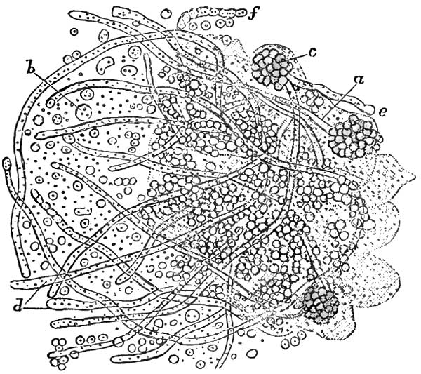
|
|
Oïdium albicans, from the Mouth in a case of
Thrush (Küchenmeister). a, fragment of a separated thrush-layer
implanted in a mass of epithelium; b, spores; d, thallus-threads
with partition walls; e, free end of a thallus somewhat swollen;
f, thallus with constriction, without partition walls. |
ETIOLOGY.—Thrush is usually a symptomatic disease, secondary to an
acid
condition of the fluids of the mouth. Athrepsia (Parrot,
Meigs and Pepper), or innutrition, is the presumable predisposing
cause. Negligence in maintaining cleanliness of the mouth and of the
articles which are placed in it is regarded as the main exciting
cause. It occurs both in the adult and in the infant, but it is much
more frequent in infancy and in early childhood. It is most frequently
encountered in asylums and hospitals for children, being often
transmitted from child to child by the nurse or by means of the
feeding-bottle. The poor health of the child seeming less accountable
for the disease than the unsanitary condition of the wards, buildings,
and surroundings, it is consequently much less frequent in private
than in public practice. It is more frequent in the first two weeks of
life than later. Seux observed it within the first eight days in 394
cases out of 402 (Simon). It is much more frequent during summer than
at any other season, more than half the cases (Valleix) occurring at
that portion of the year.
In senile subjects, in adults, and in children more than two years of
age it is cachectic, and observed chiefly toward the close of some
fatal and exhausting disease, such as diabetes, carcinoma,
tuberculosis, chronic pneumonia, enteric fever, puerperal fever,
erysipelas, chronic entero-colitis and recto-colitis, and
pseudo-membranous sore throat. It is sometimes observed in the early
stage of enteric fever.
Meigs and Pepper, apparently following Parrot, deem the central cause
to lie in a certain failure of nutrition under which the general
vitality slowly ebbs away. They are inclined7 to recognize a causal
factor in a deficiency in the supply of water in much of the
artificial food administered to young subjects. The normal acidity of
the fluids of the mouth of the newly-born (Guillot, Seux) is not
sufficiently counteracted until saliva becomes abundant. Premature
weaning, entailing, as it often does, the use of improper foods,
renders the child liable to gastro-intestinal disorders. To this add
want of care of the bottle and nipples, of the teaspoon or pap-boat,
and of the mouth itself, and the conditions are fulfilled in
fermentations of remnants of milk taking place without and within,
which produce the acid condition of the fluids and secretions of the
mouth said always to accompany and precede the development of the
disease (Gubler).
7 A Practical Treatise on the Diseases of Children, 7th
ed., Philada., 1882.
The theory of contagiousness seems established (Guillot, Berg, Gubler,
Robin, Trousseau). This has been further demonstrated by experiments
upon sheep (Delafeud), in which thrush has been implanted whenever the
animals were unhealthy, but not otherwise.
PATHOLOGY AND MORBID ANATOMY.—The mucous membrane of the mouth within
a few hours after its invasion by thrush is seen to be covered to some
extent by minute masses of a granular curdy substance adherent to the
tissues, which often bleed slightly when the substance is forcibly removed.
In children much reduced by inanition or severe disease, much of the
deposit soon coalesces into a membraniform product, grayish or
yellowish from rarefaction by the air, or even brownish from admixture
of blood. By the same time the general congestion of the mucous
membrane will have subsided into the pallor of anæmia. Though
tolerably adherent when fresh, the deposit when older often becomes
loosened spontaneously,
so that it may be removed by the finger
in large flakes without producing any hemorrhage whatever.
The characteristic masses present both as delicate roundish flakes,
isolated, not larger than a pinhead, and as confluent patches several
times as large and more irregular in outline. These masses under
microscopic inspection are seen to be composed of the filaments and
spores of a confervoid parasitic plant, the Oïdium albicans, enclosing
altered epithelia in various conditions. This parasitic growth does
not become developed upon healthy mucous membrane with normal
secretory products. Acidity of the fluids and exuberance of epithelium
are the requisites for its production, whatever be the cause. The
acidity of the fluids irritates the mucous membrane upon which they
lie. This irritation induces abnormal proliferation of epithelium,
upon which the spores of the cryptogam then germinate. Dissociated
epithelial cells become proliferated at the surface of the mucous
membrane, between which and upon which both free and agglutinated
spores accumulate. From these spores sprout out simple and ramified
filaments in compartments containing moving granular elements. (For
the minute detailed anatomy of these filaments and spores the reader
is best referred to Robin's work on Vegetable Parasites.)
It may suffice here to mention that the filaments are sharply-defined
tubercles, slightly amber-tinted, of a mean diameter of between four
and three millimeters, simple while immature and branched when fully
developed. These tubules are filled with link-like groups of elongated
cells in compartments, giving them an appearance of regular
constriction at the junctions of adjoining groups of cells.
Surrounding these tubules are groups of spheroid or slightly ovoid
spores from five to four millimeters in diameter. Each spore contains
one or two granules and a quantity of fine dust. This cryptogamic
growth is developed in the proliferated cells of epithelium. The
filaments in their further growth separate the epithelia, and even
penetrate them. Thence they penetrate the mucous membrane and the
submucosa (Parrot).
The mucous membrane beneath the growth is red, smooth, and glistening.
Papillæ are sometimes prominent. It is not excoriated unless the
growth has been removed with some violence, when, as noted, it may
bleed slightly. Duguet and Damaschino have recently encountered cases
associated with a special ulceration of one of the palatine folds; the
former in enteric fever, the latter in a primitive case. The growth is
quickly reproduced after removal—even within a few minutes when the
secretions are very acid.
The glossal mucous membrane is usually the tissue first involved, the
specks being more numerous at the tip and edges of the tongue than at
its central portion. The glands at the base of the tongue may become
invaded. From the tongue extension takes place to the lips, the
cheeks, the gums, and the palate, hard and soft. The growth is
especially prolific in the folds between lips and gums and between
cheeks and gums. Sometimes the parts mentioned become involved
successively without actual extension. In several recently reported
instances occurring during enteric fever,8 the affection began on
the soft palate, tonsils, and pharynx, and then progressed anteriorly
toward the tongue, the cheeks, and the lips.
8 Duguet, Soc. Méd. des. Hôp., Mai 11, 1883; Rev.
mens., Juin 1, 1883, p. 187.
But
there is no limitation of the disease to these structures.
The growth may cover the entire mucous membrane of the mouth. From the
mouth it may reach the lateral walls of the pharynx, and in rare
instances the posterior wall of the pharynx. The product is said to be
more adherent on the pharynx (Reubold) than in the mouth. From the
pharynx it may reach the epiglottis, and even the larynx (Lelut), in
which organ it has been seen upon the vocal bands (Parrot). It has
never been observed in the posterior nares or at the pharyngeal
orifice of the Eustachian tube. It flourishes best, therefore, upon
squamous epithelium. In infants much reduced, Parrot has seen
ulceration in the neighborhood of the pterygoid apophyses, but
attributable to the cachectic state of the child, and not to the
disease in the mouth.
In many cases—in as large a proportion as two-thirds, according to
some observers—the oesophagus becomes invaded, either in irregular
longitudinal strips or in rings, in all instances (Simon) terminating
a little above the cardia. In exceptional cases the entire mucous
surface of the oesophagus may be covered with the product (Seux). It
has been seen in the stomach (Lelut, Valleix), and is even said to be
developed there (Parrot), presenting as little yellow projections,
isolated or contiguous, from the size of millet-seeds to that of peas,
and usually located along the curvatures, especially the smaller
curvature and cardia (Simon).
In instances still more rare it is found in the intestinal canal
(Seux), even at the anus (Bouchut, Robin), and thence upon the
genitalia. In a child thirteen days old, Parrot found it in the
pulmonary parenchyma at the summit of the right lung, where it had
probably been drawn by efforts of inspiration.
The nipple of the nurse often becomes covered with the growth (Gubler,
Robin, Trousseau, Simon).
SYMPTOMATOLOGY.—In infants the earliest symptom is distress during
nursing, the nipple being seized repeatedly, and as frequently
released with cries of pain and disappointment. This cry is hoarse
when the vocal bands are involved.
The constitutional symptoms depend upon the underlying malady, and may
of course vary with its character. Thus we may have the symptoms of
simple diarrhoea, gastro-enteritis, or entero-colitis on the one hand,
and of tuberculosis and other diseases elsewhere enumerated on the
other. Cachectic children, especially in asylum and hospital practice,
lose flesh, and their skins become harsh, dry, and inelastic from loss
of fluids (Meigs and Pepper). The genitalia, the anus, and the
adjacent parts become eroded by the acridity of the discharges, and
then become covered with the growth.
The disease rarely lasts longer than eight days in strong children
that can be well cared for. It may continue indefinitely, on the other
hand, in cachectic children; that is to say, for several months or
until the patient succumbs, as may be. Death occurs usually from the
causal disease, and not as a result of the morbid condition of the mouth.
DIAGNOSIS.—In the Infant.—Examination of its mouth to detect the
cause of the child's inability to nurse reveals congestion of the
mucous membrane, intense and often livid in severe cases. It is first
noticed at the extremity of the tongue. When the congestion is general
it is darkest in the tongue. This livid congestion may extend over the
entire visible
mucous membrane, save upon the hard palate, where
it is tightly adherent to the periosteum, and upon the gums, where it
is rendered tense by the approach of erupting teeth. The papillæ at
the tip and sides of the tongue are very prominent. Sometimes the
organ is quite dry, even sanious, while it is painful to the touch.
The reaction of the secretions of the mouth is acid instead of
alkaline, and the parts are hot and very sensitive.
Two or three days later the circular milky-white or curdy spots or
slightly prominent and irregularly-shaped flakes or patches may be
seen on the upper surface of the tongue toward the tip and inside the
lips and the cheeks, especially in the grooves connecting gums and
lips and gums and cheeks. The surrounding mucous membrane is unaltered
in mild cases, and there is no evidence of other local disorder or of
any constitutional involvement. In severe cases the entire mucous
membrane is dry and deeply congested.
The affection can be positively discriminated from all others by
microscopic examination of the deposit, which reveals the presence of
the cryptogam described.
TREATMENT.—In infants, artificial nourishment, whether with milk of
the lower animals or prepared food of whatever composition, should be
given up, if possible, and a wet-nurse be supplied. If this procedure
be impracticable, the least objectionable mode of preparation of cow's
milk should be employed (and this will vary with the practice of the
physician), and the utmost circumspection should be maintained in
securing the cleanliness of the vessels in which it is prepared, the
bottle from which it is given, and the nipple which is placed in the
child's mouth. Should the sugar and casein in the milk appear to keep
up the disease, weak soups may be substituted for the milk diet until
it has subsided. Weiderhofer advises artificial nourishment, by way of
a funnel inserted in the nasal passages, in case the child should
refuse to swallow. Deglutition is excited in a reflex manner when the
milk or other fluid reaches the pharynx.9
9 Journ. de Méd. Bordeaux, Juin 10, 1883.
The local treatment should consist in careful removal of the patches
from time to time—say every two or three hours—with a moistened soft
rag. This must be done without roughness of manipulation. In addition
to this, the parts may be washed or painted every hour or so with an
alkaline solution for the purpose of neutralizing the acidity of the
fluids of the mouth. For this purpose borax is most generally used, in
the proportion of twenty grains to the ounce of water or the half
ounce of glycerin. Sodium bicarbonate or sodium salicylate may be
substituted for the sodium borate. The use of honey in connection with
the drug is calculated to promote acidity by fermentation of its
glucose, and is therefore, theoretically, contraindicated.
Adults may use washes, gargles, or sprays of solutions of sodium
borate or of sodium bicarbonate.
The constitutional treatment in each case must be adapted to the
nature of the underlying malady which has favored the local disease,
with resort in addition to the use of quinia, iron, wine, spirit, and
beef-essence. The hygienic surroundings should be made as sanitary as possible.
DEFINITION.—Inflammation of the interior of the mouth, usually
unilateral, eventuating in multiple ulcerations of the mucous membrane.
SYNONYMS.—Fetid stomatitis, Phlegmonous stomatitis, Putrid sore
mouth, Stomacace, are synonymous terms for idiopathic ulcerous
stomatitis. Ulcero-membranous stomatitis, Mercurial stomatitis
(Vogel), are synonymous terms for the deuteropathic variety of the disease.
ETIOLOGY.—The principal predisposing cause of the disease is to be
found in ochlesis; the contaminating atmosphere of crowded dwellings
and apartments insufficiently ventilated; uncleanliness; insufficiency
of proper clothing; unhealthy food, and the like. It prevails
epidemically in crowded tenements, schools, prisons, asylums, and
hospitals; in garrisons and in camps; in transports and men-of-war. It
is often propagated by contagion, but whether by infection or actual
inoculation seems undetermined. Measles is an active predisposing
cause. Feeble individuals are the most liable to the disease. It
occurs at all ages. In civil life it is most frequent between the ages
of four and ten years. Sometimes more girls are affected than boys
(Meigs), and sometimes it is the more prevalent among boys (Squarrey).
Autumn is the season of greatest prevalence.
Carious teeth, fracture and necrosis of the jaw (Meigs), and
protracted catarrhal stomatitis are among the chief exciting causes.
Irregular dentition is sometimes the exciting cause; and this may
occur at the first and second dentition or at the period of eruption
of the last molars.
PATHOLOGY.—The anatomical lesion is the destructive inflammation of
portions of the mucous membrane of the mouth, leaving ulceration on
detachment of the eschars. It usually commences as a gingivitis. At
two periods of life—namely, from the fourth to the eighth year of
life, and from the eighteenth to the twenty-fifth year—it is apt to
be ulcero-membranous, a condition asserted to be altogether
exceptional at other periods (Chauffard).
A diffuse fibro-purulent infiltration of the lymph-spaces of the
mucosa is regarded as the first step in the pathological process. This
infiltration is sufficiently abundant to compress the capillary
vessels of the tissues, and thus arrest the circulation (Cornil et
Ranvier). All those localized portions of mucous membrane from which
the circulation is cut off perish and are discharged in fragments. The
ulcers thus left are grayish, granular, and sanious, with thin,
irregularly dentated borders a little undermined, through which pus
can be expressed on pressure. The usual cryptogams of the oral cavity,
in various stages of development, are in great abundance in the
grayish detritus, which likewise contains altered red and white blood-corpuscles.
According to some observers (Caffort, Bergeron), the first evidence of
the disease is an intensely congested erythematous patch, upon which
one or more pustules present, point, and rupture promptly, leaving the
characteristic ulcerations.
For some indeterminate reason, the ulcerations are mostly unilateral,
and occur much the more frequently on the left side. The principal
primal
points of ulceration are upon the external borders of the
gums, more frequently those of the lower jaw, and upon the
corresponding surface of the cheek and lip—the cheek much oftener
than the lip. Thence ulceration may extend to the tongue, less
frequently to the palate. The ulcerative process follows the outline
of the gums, baring the bases of the teeth to a variable extent, so
that they seem elongated. On the cheek the patch of inflammation is
generally oval, the longest diameter being antero-posterior, and the
most frequent position is opposite to the last molar.
Each ulcer is surrounded by an intensely red areola, beyond which the
tissues are succulent and tumid from collateral inflammatory oedema,
often giving the ulcers an appearance of great depth; but when the
detritus is discharged they are seen to have been superficial.
Detachment of the necrosed segments of mucous membrane takes place by
gradual exfoliation from periphery to centre. Sometimes detachment
occurs in mass, usually in consequence of friction or suction. The
ulcers, gingival and buccal, bleed easily when disturbed. They may
remain separate, or may coalesce by confluence of interposing
ulcerations extending across the furrow between gum and cheek or lip.
The adjoining side of the tongue sometimes undergoes similar
ulceration from behind forward, inoculated, most likely, by contact
with adjoining ulceration. In rare instances, neglected cases most
probably, the ulceration may extend to the palatine folds, the
tonsils, and the soft palate.
SYMPTOMATOLOGY.—The affection usually begins without any
constitutional symptoms. Young infants sometimes present slight
febrile symptoms, with impairment of appetite and general languor.
Fetid breath, salivation, and difficulty in deglutition are usually
the first manifestations of the disease to attract attention. The
mouth will be found to be hot, painful, and sensitive to the contact
of food. Infants often refuse food altogether, though usually they can
be coaxed to take liquid aliment. Larger children and adults complain
of scalding sensations. They find mastication painful, and cannot chew
at all on the affected side. The salivation is excessive, the saliva
bloody and often extremely fetid. When swallowed, this fetid saliva
causes diarrhoea. The cheeks sometimes become swollen, and the
submaxillary connective tissue oedematous. Adenitis takes place in the
submaxillary, retro-maxillary, and sublingual glands of the affected
side. Sometimes the other side becomes affected likewise, but to a
less extent. The glands do not suppurate, but the adenitis may remain
as a chronic manifestation in scrofulous subjects.
The disease, left to itself, will often continue for a number of
weeks, or even months as may be, unmodified even by intercurrent
maladies (Bergeron). Long continuance may result in partial or
complete disruption of the teeth, or in local gangrene, or even in
necrosis of the alveoli (Damaschino). Properly managed, the ulcers
become cleansed of their detritus, and within a few days heal by
granulation, their position long remaining marked by delicate red
cicatrices upon a hard and thickened substratum. Repeated recurrences
are sometimes observed.
DIAGNOSIS.—The appearances of the gums and adjoining structures
described under the head of Pathology establish the diagnosis. The
usually unilateral manifestation and the peculiar fetid odor
distinguish it from severe forms of catarrhal stomatitis. From cancrum
oris it is distinguished
by the absence of induration of the
skin of the cheek over the swollen membrane, and by the succulence and
diffuseness of the tumefaction. From mercurial stomatitis it is
discriminated by the history, and by the absence of the peculiar
manifestations to be discussed under the head of that disease.
PROGNOSIS.—The prognosis is good, the disease being susceptible of
cure in from eight to ten days in ordinary cases. When due nutrition
is prevented by the pain in mastication and deglutition, and in
much-reduced subjects, the disease may continue for several weeks. It
is in these cases that detachment of the teeth takes place, with
periostitis and necrosis of the alveoli. Protracted suppuration and
failure in nutrition may lead to a fatal result, but such a
termination is uncommon.
TREATMENT.—Fresh air, unirritating and easily digestible food, the
best hygienic surroundings practicable, attention to secretions from
skin and bowels by moderate and judicious use of ablutions,
diaphoretics, and laxatives, with the internal administration of
cinchona or its derivatives, with iron and cod-liver oil, comprise the
indications for constitutional treatment.
Locally, demulcent mouth-washes are called for, containing
astringents, detergents, or antiseptics. Acidulated washes are more
agreeable in some instances. For antiseptic purposes, however, sprays
and douches may be used of solutions of potassium permanganate, boric
acid, carbolic acid, or salicylic acid. Gargles of potassium chlorate,
ten or twenty grains to the ounce, are highly recommended, as well as
the internal administration of the same salt in doses of from two to
five grains three times a day for children, and of ten to twenty
grains for adults.
If the sores are slow to heal, the ulcerated surfaces may be touched
once or twice daily with some astringent, such as solution of silver
nitrate (ten grains to the ounce), or, if that be objectionable, with
alum, tincture of iodine, or iodoform.
Prompt extraction of loose teeth and of loose fragments of necrosed
bone is requisite.
DEFINITION.—A non-contagious, deuteropathic inflammation of the
interior of the mouth, almost invariably unilateral, and characterized
by a peculiar gangrenous destruction of all the tissues of the cheek
from within outward.
SYNONYMS.—Gangrenous stomatitis; Gangrena oris; Grangrenopsis;
Cancrum oris; Stomato-necrosis; Necrosis infantilis; Gangrene of the
mouth; Gangrenous erosion of the cheek; Noma; Buccal anthrax; Aquatic
cancer; Water cancer; Scorbutic cancer; Sloughing phagedæna of the mouth.
HISTORY.—The most important work upon the subject was published in
1828, from the pen of Dr. A. L. Richter,10 whose accurate historical
account of the disease was in great part reproduced, with additions
thereto, by Barthez and Rilliet in their Treatise on the Diseases of
Infants, Paris, 1843, and quoted by nearly all subsequent writers on
the theme.
From these records it appears that the first accurate
description of the affection was given in 1620 by Dr. Battus, a Dutch
physician, in his Manual of Surgery. The term aquatic cancer,
water-kanker, bestowed on it by van de Voorde, has been generally
followed by the physicians of Holland, although van Swieten (1699)
properly designated it as gangrene. J. van Lil termed it noma, as well
as stomacace and water-kanker, and cited a number of Dutch physicians
who had observed its epidemic prevalence. The majority of more recent
observers, however, deny its epidemic character.
10 Der Wasserkrebs der Kinder, Berlin, 1828; further,
Beiträg zur Lehre vom Wasserkrebs, Berlin, 1832; Bemerkungen über
den Brand der Kinder, Berlin, 1834.
Of Swedish writers, Lund described it as gangrene of the mouth;
Leutin, under the name of ulocace. In England, Boot was the first to
write of gangrene of the mouth, and was followed by Underwood,
Symmonds, Pearson, S. Cooper, West, and others.
In France it has received great attention. Berthe11 described it as
gangrenous scorbutis of the gums; Sauvages (1816) as necrosis
infantilis. Baron in 1816 published12 a short but excellent account
of a gangrenous affection of the mouth peculiar to children; and
Isnard presented in 1818 his inaugural thesis on a gangrenous
affection peculiar to children, in which he described, simultaneously,
gangrene of the mouth and gangrene of the vulva. Then followed Rey,
Destrees (1821), Billard (1833), Murdoch, Taupin (1839), and others,
until we reach the admirable description by Barthez et Rilliet, from
which the present historical record has been chiefly abstracted.
11 Mémoires de l'Académie royale de Chirurgie, Paris,
1774, t. v. p. 381.
12 Bullétins de la Faculté de Médecine de Paris, 1816, t.
v. p. 161.
German physicians likewise have largely studied the subject. De
Hilden, A. G. Richter, C. F. Fischer, Seibert, and many others
preceded A. L. Richter, whose important contribution to the literature
and description of the disease has been so highly extolled by Barthez
and Rilliet.
In America the disease has been best described by Coates, Gerhard, and
Meigs and Pepper, all of Philadelphia.
(For extensive bibliographies the following sources should be
consulted in addition to those cited: J. Tourdes, Du Noma ou du
Sphacèle de la Bouche chez les Enfants, Thèse, Strasbourg, 1848: A.
Le Dentu, Nouveau Dictionnaire de Médecine et de Chirurgie pratique,
article "Face," Paris, 1871.)
ETIOLOGY.—Almost exclusively a disease of childhood, gangrenous
stomatitis is exceedingly rare in private practice, and very
infrequent at the present day even in hospital and dispensary
practice. Lack of hygienic essentials of various kinds,
impoverishment, long illnesses, and debilitating maladies in general
are the predisposing causes. It is sometimes endemic in hospitals and
public institutions, but rarely, if at all, epidemic. It is not
generally deemed contagious, though so considered by some writers. It
appears to have been more frequent in Holland than elsewhere, to be
more frequent in Europe generally than in the United States, and now
much less frequent in the United States than formerly. To recognition
of the predisposing causes and to their abolition and avoidance may
probably be attributed its diminished frequency all over the world.
Though attacking children only as a rule, it has been observed in
adults (Barthez et Rilliet, Tourdes, Vogel). Nurslings are not liable
to the disease. Though occurring occasionally earlier in life, the
greatest period of prevalence is
from the third to the fifth or
sixth year of age, and thence, with diminishing frequency, to the
twelfth and thirteenth years. It is probably equally frequent in the
two sexes, though the majority of authors have described it as more
frequent in females.
Healthy children are not attacked. Even in delicate children it is so
rarely idiopathic that this character is utterly denied it by many
observers. The disease which it follows, or with which it becomes
associated, may be acute or chronic. According to most writers, it
occurs with greatest frequency after measles. It follows scarlatina
and variola much less often. It is observed likewise after whooping
cough, typhus fever, malarial fever, entero-colitis, pneumonitis, and
tuberculosis. Excessive administration of mercury has been recognized
as an exciting cause, some cases of mercurial stomatitis progressing
to gangrene.
According to Barthez et Rilliet, acute pulmonary diseases, and
especially pneumonia, are the most frequent concomitant affections,
and are usually consecutive.
SYMPTOMATOLOGY, COURSE, DURATION,
TERMINATIONS, COMPLICATIONS, AND
SEQUELÆ.—The disease usually becoming manifested during other
disease, acute or chronic, or during convalescence therefrom, there
are no special constitutional symptoms indicating its onset. Hence
considerable progress may be made before its detection. The earliest
local characteristic symptom distinguishing gangrenous stomatitis is a
tense tumefaction of one cheek, usually in proximity to the mouth. The
lower lip is generally involved, thus rendering it a matter of
difficulty to open the mouth. This tumefaction in some instances
progresses over the entire side of the face up to the nose, the lower
eyelid, and even out to the ear in one direction, and down to the
chin, and even to the neck, in the other. Before the parts become
swollen externally, ulceration will have taken place to some extent in
the mucous membrane, but usually without having attracted special
attention, the subjective symptoms having been slight. A gangrenous
odor from the mouth, however, is almost always constant. Its presence,
therefore, should lead to careful investigation as to its seat and
cause. The gums opposite the internal ulcer become similarly affected
in most instances, and undergo destruction, so that the teeth may
become denuded and loosened, and even detached, exposing their
alveoli. The bodies of the maxillary bones suffer in addition in some
instances, and undergo partial necrosis and exfoliation.
It is maintained (Löschner, Henoch) that in some instances there is no
involvement of the mucous membrane until the ulcerative process has
reached it from the exterior.
The tumefied portions of the check and lip are pale, hard, unctuous,
and glistening. They are rarely very painful, and often painless. On
palpation a hard and rounded nodule one or two centimeters in diameter
can be detected deep in the central portion of the swollen cheek.
From the third to the sixth day a small, black, dry eschar, circular
or oval, becomes formed at the most prominent and most livid portion
of the swelling, whether cheek or lip. This gradually extends in
circumference for a few days or for a fortnight, sometimes taking in
almost the entire side of the face or even extending down to the neck.
As it enlarges the tissues around become circumscribed with a zone
intensely red. The internal eschar extends equally with the external
one. Eventually, the eschar
separates, in part or in whole, and
becomes detached, leaving a hole in the cheek through which are seen
the loosened teeth and their denuded and blackened sockets.
During this time the patient's strength remains tolerably well
maintained, as a rule, until the gangrene has become well advanced.
Intelligence usually remains good. Many children sit up in bed and
manifest interest in their surroundings. Others lie indifferent to
efforts made for their amusement. Some exhibit insomnia and delirium.
The pulse is small and moderately frequent, rarely exceeding 120 beats
to the minute until near the fatal close, when it often becomes
imperceptible. Appetite is often well preserved, unless pneumonia or
other complications supervene, but thirst is often intense, even
though the tongue remain moist. The desire for food sometimes
continues until within a few hours of death. Toward the last the skin
becomes dry and cold, diarrhoea sets in, emaciation proceeds rapidly,
collapse ensues and death.
Death usually occurs during the second week, often before the complete
detachment of the eschar—in many instances by pneumonia, pulmonary
gangrene, or entero-colitis. Some die in collapse, which is sometimes
preceded by convulsions. When the eschars have become detached,
suppuration exhausts the forces of the patient, and death takes place
by asthenia.
The complication most frequent is pneumonia, and the next
entero-colitis. Gangrene of the lungs, of the palate, pharynx, or
oesophagus, of the anus, and of the vulva, may supervene. Hemorrhage
from the facial artery or its branches has been noted as an
exceptional mode of death (Hueber), the rule being that the arteries
in the gangrenous area become plugged by thrombi, and thus prevent hemorrhage.
Recovery may take place before the local disease has penetrated the
cheek—indeed, while the mucous membrane alone is involved. In recent
instances, however, the disease does not subside until after the loss
of considerable portions of the cheek, and the child recovers with
great deformity, not only from loss of tissue in the cheek and nose,
but from adhesions between the jaws and the cheek.
PATHOLOGY AND MORBID ANATOMY.—Gangrenous stomatitis always involves
the cheek, almost always that portion in proximity to the mouth. It is
almost invariably unilateral. Either side seems to be equally liable.
Both sides suffer only, it is contended, when the gangrene is limited
in extent, confined to the mucous membrane, and occupies the sides of
the frenums of the lips (Barthez et Rilliet). It usually if not
invariably begins in the mucous membrane, as a phlyctenular
inflammation, which undergoes ulceration, followed by gangrene,
immediately or not for several days, and then becomes covered with a
more or less brownish-gray eschar. The ulceration of the mucous
membrane is occasionally preceded by an oedematous condition of the
cheek externally, similar to that sometimes observed in ordinary
ulcerous stomatitis; but this is not the characteristic circumscribed,
tense infiltration observed later. This ulceration is situated most
frequently opposite the junction of the upper and lower teeth.
Sometimes it proceeds from the gingivo-buccal sulcus of the lower jaw,
sometimes from the alveolar border of the gums. It extends in all
directions, and often reaches the lower lip. From three to sixteen
days may be consumed in these extensions. The
surrounding mucous
membrane becomes oedematous. The ulceration soon becomes followed by
gangrene, sometimes within twenty-four hours, sometimes not for two or
three days, and exceptionally not for several days. The ulcerated
surfaces bleed readily, change from gray to black, and become covered
with a semi-liquid or liquid putrescent detritus. They are sometimes
surrounded by a projecting livid areola, which soon becomes gangrenous
in its turn. The shreds of mortified membrane, though clinging a while
to the sound tissues, are easily detached, and often drop
spontaneously into the mouth. Meanwhile, there is abundant salivation,
the products of which pour from the mouth, at first sanguinolent, and
subsequently dark and putrescent and mixed with detritus of the
tissues. Large portions of the gums, and even of the mucous membrane
of the palate, may undergo destruction within a few (three to six)
days. The gangrenous destruction of the gums soon exposes the teeth,
which become loose and are sometimes spontaneously detached. Thence
the periosteum and bone become implicated and undergo partial
denudation and necrosis, and portions of necrosed bone become detached
if the patient survives. The characteristic implication of the
exterior of the cheek becomes manifest from the first to the third
day, but occasionally not until a day or two later. A hard,
circumscribed swelling of the cheek or cheek and lip occurs, sometimes
preceded, as already intimated, by general oedematous infiltration.
The surface is tense and unctuous, often discolored. In its central
portion is an especially hard nucleus, one to two centimeters or more
in diameter. Gangrene often takes place at this point from within
outward at a period varying from the third to the seventh day or
later. The skin becomes livid, then black; a pustule is formed at the
summit of the swelling, which bursts and discloses a blackened
gangrenous eschar from less than a line in thickness to the entire
thickness of the cheek beneath. The area of gangrene gradually
extends. The dead tissues become detached, and a perforation is left
right through the cheek, through which are discharged saliva and
detritus. Meanwhile, the submaxillary glands become swollen and the
surrounding connective tissue becomes oedematous. In some instances,
however, no change is noticeable in these glands.
Examinations after death have shown that thrombosis exists for some
distance around the gangrenous mass. Hence the rarity of hemorrhage
during the detachment of the eschar.
DIAGNOSIS.—In the early stage of the disease the main point of
differential diagnosis rests in the locality of the primitive lesion,
the mucous membrane of the inside of one cheek. Subsequently there is
the gangrenous odor from the mouth; the rapid peripheric extension of
the local lesion, which acquires a peculiar grayish-black color; its
rapid extension toward the exterior of the cheek or lip; the
tumefaction of the cheek, discolored, greasy, hard, surrounded by
oedematous infiltration, and presenting a central nodule of especial
hardness; then the profuse salivation, soon sanguinolent, subsequently
purulent and mingled with detritus of the mortified tissues. Finally,
the eschar on the exterior of the swollen cheek or lip leaves no doubt
as to the character of the lesion. From malignant pustule it is
distinguished by not beginning on the exterior, as that lesion always
does (Baron).
PROGNOSIS.—The prognosis is bad unless the lesion be quite limited
and
complications absent. At least three-fourths of those
attacked perish; according to some authorities fully five-sixths die.
The objective symptoms of the local disease are much more important in
estimating the prognosis than are the constitutional manifestations,
the vigor of the patient, and the hygienic surroundings, although, as
a matter of course, the better these latter the more favorable the
prognosis. Prognosis would be more favorable in private practice than
in hospital or asylum service.
TREATMENT.—Active treatment is required, both locally and
constitutionally. Local treatment is of paramount importance, and
alone capable of arresting the extension of the process of
mortification. The topical measure in greatest repute is energetic
cauterization with the most powerful agents, chemical and
mechanical—hydrochloric acid, nitric acid, acid solution of mercuric
nitrate, and the actual cautery, whether hot iron, thermo-, or
electric cautery. The application of acids is usually made with a firm
wad or piece of sponge upon a stick or quill, care being taken to
protect the healthy tissues as far as practicable with a spoon or
spatula. After the application the mouth is to be thoroughly syringed
with water to remove or dilute the superfluous acid. Hydrochloric acid
has been preferred by most observers.
As these cauterizations must be energetic to prove effective,
anæsthesia ought to be induced. Should ether be employed for this
purpose, hydrochloric acid or the acid solution of mercuric nitrate
would be selected of course.
In the early stages these agents are to be applied to the inside of
the cheek, so as to destroy all the tissue diseased, if practicable,
and expose a healthy surface for granulation. Should the exterior of
the cheek become implicated before cauterization has been performed or
in spite of it, it is customary to destroy the tissues from the
exterior, including a zone of apparently healthy surrounding tissue.
As the gangrene extends, the cauterization is to be repeated twice
daily or even more frequently. After cauterization the parts are
dressed with antiseptic lotions, and antiseptic injections or douches
are to be used frequently during day and night to wash out the mouth
and keep it as clear as possible from detritus.
Meigs and Pepper report beneficial results from the topical use of
undiluted carbolic acid, followed by a solution of the same, one part
in fifty of water, frequently employed as a mouth-wash. The progress
of the sloughing was checked and the putridity of the unseparated dead
tissue completely destroyed in the two cases mentioned by them, one of
which recovered quickly without perforation of the cheek. Gerhard
preferred undiluted tincture of the chloride of iron; Condie, cupric
sulphate, thirty grains to the ounce. Bismuth subnitrate has recently
been lauded as a topical remedial agent.13
13 Maguire, Medical Record N.Y., Feb. 3, 1883.
The mouth should be frequently cleansed by syringing, douching,
spraying, or washing with disinfectant solutions, such as chlorinated
soda liquor, one part to ten; carbolic acid, one to twenty.
Lemon-juice is sometimes an agreeable application, as in some other
varieties of stomatitis. Constitutionally, tonic and supporting
treatment is demanded, even in those instances where the appetite is
well maintained and the
general health apparently well
conserved. Soups, milk, semi-solid food, egg-nog, egg and wine, wine
whey, milk punch, finely-minced meat, should be administered as freely
as the state of the digestive functions will permit. If necessary,
resort should be had to nutritive enemata. Quinia and tincture of
chloride of iron are the medicines indicated. When sufficient alcohol
cannot be given with the food, it should be freely exhibited in the
most available form by the mouth or by the rectum. The apartment
should be well ventilated, the linen frequently changed, the
discharges promptly removed.
DEFINITION.—An inflammation of the interior of the mouth due to
poisoning, especially by drugs, and chiefly by mercury, copper, and phosphorus.
DEFINITION.—An inflammation of the mucous membrane of the mouth,
eventually ulcerating, the result of systemic poisoning by the
absorption of mercury.
SYNONYMS.—Stomatitis mercurialis; Mercurial ptyalism, Ptyalismus
mercurialis; Mercurial salivation, Salivatio mercurialis.
ETIOLOGY—Predisposing and Exciting Causes.—Special vulnerability to
the toxic influence of mercury, and special proclivity to inflammatory
affections of the mouth and the organs contained therein, are the
predisposing causes of mercurial stomatitis. The exciting cause is the
absorption of mercury into the tissues of the organism. The
susceptibility of healthy adults is much greater than that of healthy
children. The susceptibility of adults varies very greatly.
Constitutions deteriorated by prolonged disease, undue exposure, and
the like are much more promptly influenced in consequence. Tuberculous
subjects do not bear mercury well.
Idiosyncratic susceptibility to toxæmia by mercurial preparations is
now and then encountered in practice, and instances have been
published14 in which fatal results have ensued, after prolonged
suffering, from the incautious administration of a single moderate
dose of a mercurial drug.
14 For example, see in Watson's Practice of Physic a case
of furious salivation following one administration of two grains of
calomel as a purgative, the patient dying at the end of two years,
worn out by the effects of the mercury and having lost portions of the
jaw-bone by necrosis.
Until comparatively recent years the most common cause of mercurial
poisoning was the excessive employment of mercurial medicines, whether
by ingestion, inunction, or vapor bath. Topical cauterization with
acid solution of mercuric nitrate is likewise an infrequent, and
usually an accidental, cause of the affection. Elimination of the
mercury by way of the mucous glands of mouth and the salivary glands
proper excites the stomatitis in these instances. An entirely
different series of cases occur in artisans exposed to handling the
metal and its preparations or to breathing its vapor or its dust. In
these instances the poison may gain
entrance into the absorbent
system by the skin, the mucous membranes of the nose, mouth, and
throat, the stomach, or the lungs. No matter what care may be
exercised in cleansing the hands, it is often impossible to prevent
occasional transference of the noxious material from fingers to
throat, or to thoroughly free the finger-tips under the nails. The
avocations entailing the risks of mercurial stomatitis comprise
quicksilver-mining, ore-separating, barometer- and thermometer-making,
gilding, hat-making, manufacturing of chemicals, and exhausting the
globes employed in certain forms of electric illumination.15 The
slow absorption of mercury into the bodies of artisans induces in
addition serious constitutional nervous disturbances—tremors, palsy, etc.
15 Med. and Surg. Reporter, Philada., Dec. 30, 1883, p.
734.
SYMPTOMATOLOGY, COURSE, DURATION,
TERMINATIONS, COMPLICATIONS, AND
SEQUELÆ.—The principal subjective symptoms of mercurial stomatitis
are—characteristic fetor of the breath, sore gums and mouth,
continuous nauseous metallic brassy or coppery taste, and profuse salivation.
At first the mouth feels parched and painful, the gums tender, the
teeth, the lower incisors especially, set on edge. Soon the gums
become swollen, and when touched with the tongue seem to have receded
from the necks of the teeth, which thereby appear to be longer than
usual. The gums feel quite sore when pressed upon with the finger or
when put on the stretch by clashing the rows of teeth against each
other. This sort of soreness is often watched for in the therapeutic
administration of mercurials purposely given to "touch the gums," as
an indication that the system is under the influence of the drug. It
is, therefore, one of the earliest indications of mercurial poisoning,
but if not sought for it may elude attention until after the mouth has
become sore a little later. The pain in the mouth is augmented by
efforts of mastication and expectoration, and may be associated with
pains at the angle of the lower jaw or extending along the domain of
the third or of the third and second divisions of the distribution of
the fifth cerebral nerve. Mastication of solid food is often
unendurable. Constitutional manifestations become evident about this
time in increased heat of skin, acceleration of pulse, furred tongue,
dry mouth, great thirst, and loss of appetite. The dryness of the
mouth does not last long, but is soon followed by hypersalivation, one
of the characteristic phenomena of the disorder. The saliva secreted,
often acid in reaction, varies greatly in quantity, which is usually
proportionate to the severity of the case. It is secreted night and
day, sometimes to the amount of several pints in the twenty-four
hours—in moderately severe cases to the amount of from one to two
pints in that space of time. It is limpid or grayish, mawkish or
somewhat fetid, and reacts readily to the simplest tests for mercury.
The salivation is almost continuous, sometimes quite so. The patient
soon becomes unable to endure the fatigue of constant expectoration,
and the fluid then dribbles from his mouth or runs off in an unimpeded
slobber. When excessive, the patient's strength becomes rapidly
exhausted—in part by impoverishment of the fluids, in great measure
from the lack of refreshing sleep.
Meanwhile, the local inflammatory process extends from the gums to the
floor of the mouth and to the lips, and thence to the tongue and the
cheeks.
The salivary glands are in a state of inflammation
likewise, but rather in consequence of direct irritation in the
elimination of the poison through their channels than by extension of
the stomatitis along their ducts. The lymphatic glands of the lower
jaw become engorged and tender. Mastication, deglutition, and
articulation all become impeded mechanically by tumefaction of the tissues.
In some instances the glossitis is so great that the tongue protrudes,
thereby impeding respiration and even threatening suffocation. In some
cases oedema of the larynx has been noted, threatening suffocation
from that cause. Should the inflammatory process extend along the
pharynx to the Eustachian tubes, deafness and pains in the ears will
become additional symptoms.
The subsequent progress of unarrested mercurial stomatitis is that of
ulcerous stomatitis.
Should gangrene of the mucous membrane take place, there will be great
fetor from the mouth, and some danger of hemorrhage on detachment of
the sloughs should the process be taking place in the direction of
vessels of some calibre. Necrosis of the inferior maxilla entails
continuance of the disagreeable local symptoms until the discharge in
fragments or in mass of the dead portions of bone.
In the earlier stages of the attack the constitutional symptoms may be
sthenic. Fever, cephalalgia, and the usual concomitants of pyrexia,
however, soon give way to the opposite condition of asthenia.
Exhausted by the excessive salivation, and unable to repair waste by
eating or sleeping, the sufferer soon passes into a condition of
hopeless cachexia. Those who survive remain cachectic and feeble for a
long time—some of them disfigured for life by various cicatrices
between cheeks and jaw, by loss of teeth or of portions of the jaw-bone.
The duration of mercurial stomatitis varies with the susceptibility of
the patient, the intensity of the toxæmia, and the character of the
treatment. Mild cases may get well in a week or two; severe cases may
continue for weeks, and even months; extreme cases have persisted for
years. Even moderate cases occasionally resist treatment for weeks.
Under the improved therapeutics of the present day mercurial
stomatitis almost always terminates in recovery, especially if it
receive early and prompt attention. Neglected or improperly managed,
it may terminate in serious losses of tissue in gums, cheeks, teeth,
and bone, leaving the parts much deformed and the patient in a
permanently enfeebled condition.
Erysipelas, metastatic abscesses, inflammations, pyæmia, or
colliquative diarrhoea may be mentioned as complications which may
prove sufficiently serious to produce death, independently of the
virulence of the primary stomatitis.
PATHOLOGY AND MORBID ANATOMY.—Mercurial stomatitis is an ulcerative
process attended with an excessive flow of saliva containing mercury.
It has a tendency to terminate in destruction and exfoliation of the
mucous membrane of the gums and other tissues attacked, and eventually
in necrosis of the jaw-bone. The detritus is found, microscopically,
to consist of granular masses of broken-down tissue, swarming with
bacteria and micrococci, and containing some blood-cells and many
pus-cells. In some instances micrococci have been detected in the blood.
The disease usually begins in the gums of the lower incisors, and
extends
backward, often being confined to one side of the jaw.
The gums, first swollen and then livid, become separated from the
necks of the teeth. Their edges undergo ulceration. The ulcers are
surrounded by fungous margins, pale or red, which bleed on the
slightest contact, and some become covered with grayish-yellow
detritus. The ulceration extends in depth, destroying the supports of
the teeth, so that they become loosened and even detached. The
inflammatory process extends to the lips, the cheek, and the tongue,
which undergo tumefaction and exhibit the impressions of the teeth in
grayish opalescent lines or festoons of thickened epithelium at the
points of pressure. The glossitis may become intense. It is almost
always present, to some extent, as a superficial or mucous glossitis.
Occasionally acute oedematous glossitis has ensued, and such cases
sometimes terminate fatally. Ulceration takes place in these
structures similar to that which has taken place in the gums. If not
arrested, gangrenous destruction ensues, not only in these tissues,
but beneath them. Thus, the teeth become loosened, and even detached;
the jaw-bones themselves may become bared, necrosed, and in part
exfoliated; and the cheeks undergo partial destruction by gangrene.
Sometimes the inflammation descends to the larynx, and this may
produce oedematous infiltration of the loose connective tissue of that
structure. Sometimes it mounts the pharynx and reaches the orifices of
the Eustachian tubes. The salivary glands become swollen and discharge
great quantities of fluid, as detailed under Symptomatology. The
retro-maxillary and submaxillary lymphatic glands become enlarged by
inflammatory action.
DIAGNOSIS.—In the earliest stages the inflammation of the gums in
mercurial stomatitis cannot be distinguished from that which takes
place in other forms of ulcerative stomatitis. The fetor of the
breath, however, the profuse salivation, and the chemical reaction of
the saliva, together with the history of exposure to mercury, soon
place the nature of the case beyond doubt. Similar results following
poisonings by copper salts and by phosphorus are differentiated by the
history of the special exposure.
PROGNOSIS.—In mild cases the prognosis is favorable, provided further
exposure to the cause can be avoided. This holds good almost
invariably in cases due to over-medication with mercurials, but is far
less applicable to cases in artisans, the result of prolonged exposure
to the poisonous influences of mercury and its slow absorption. On the
whole, the affection is much less serious than formerly, both because
it can, in great measure, be guarded against by proper prophylaxis in
risky vocations, and because its treatment has been made much more
efficient. In severe cases serious results may ensue despite the most
judicious treatment, and convalescence is usually very slow, weeks
often elapsing before solid food can be chewed without pain or without
injury to the gums.
When death ensues, it may be by asthenia, erysipelas, pneumonia,
pyæmia, or colliquative diarrhoea.
TREATMENT.—Mercurial stomatitis may sometimes be prevented by the
administration of potassium chlorate during exposure. Mild cases
following the administration of mercurials often subside upon mere
withdrawal of the drug. Should spontaneous subsidence not take place,
the administration of potassium chlorate every few hours, in doses of
from
thirty to sixty grains or more in the twenty-four hours,
soon effects amelioration, which promptly terminates in recovery. The
characteristic fetor often ceases within twelve hours' use of this
drug. Should the inflammatory manifestations be severe, a few leeches
applied beneath the edge of the lower jaw, followed by a poultice
enveloping the neck to promote further flow of blood, often affords
prompt relief (Watson). Lead acetate (ten grains to the ounce of
water) and iodine (half a fluidrachm of the compound tincture to the
ounce of water) are useful as gargles and washes. When the result of
slow poisoning, elimination of the mercury by sulphur vapor baths and
the administration of small doses of potassium iodide are recommended.
Cauterization of the ulcerated surfaces is sometimes serviceable,
silver nitrate or hydrochloric acid (Ricord), or chromic acid 1:5
(Butlin, Canquil), being used for the purpose.
Opium in decided doses is indicated for the relief of pain. It may be
added with advantage to detergent and disinfective mouth-washes
(potassium chlorate, sodium borate, creasote-water, saponified
emulsion of coal-tar, tincture of cinchona, tincture of myrrh, etc.),
the use of which should form an important part of the treatment.
Watson highly recommended a wash of gargle of brandy and water, 1:4 or
5. In severe cases difficulty is encountered in maintaining effective
alimentation. When mastication is not impracticable, soft-boiled egg
and finely-chopped raw beef may be given. When the patient cannot chew
at all, resort is confined to milk, soups, and the juice of beef.
Nourishing enemata should be administered, as in all affections where
it becomes impracticable to sustain the patient by way of the mouth.
Tonics and stimulants are indicated to avoid debility from the
excessive salivation and its sequelæ—quinia, coffee, wine, and
alcohol, the first, if required, by hypodermatic injection, all of
them by enema if necessary.
Glossitis and oedema of the larynx may require the surgical procedures
often necessary when they occur under other circumstances.
Other forms of toxic stomatitis hardly require special elucidation.
Apart from the anomalies presented in monsters, there are a few
congenital abnormalities of the tongue with which it becomes the
accoucheur at least to be familiar, as their presence may interfere
materially with the nutrition of the infant, whether nursed or spoon-fed.
CONGENITAL DEFICIENCY OF THE TONGUE.—A considerable portion of the
tongue may be wanting anteriorly, comprising, in some instances, the
entire free portion of the organ. The stump then presents as a single
or a bifid protuberance of variable size. In some instances
considerable power of movement exists, and even conservation of taste.
Suction and deglutition are both practicable. When the child grows it
can speak, though with a certain amount of difficulty. A few cases are
on record, however, of ability to speak without any evidence of a
tongue above the floor of the mouth.
An instance of lateral deficiency has been observed by Chollet,16
the deficient
half being represented merely by the two layers of
the lingual mucous membrane, without any intervening muscular substance.
16 Demarquay, Dict. de Méd. et de Chir. prat., xx. p.
130.
BIFID TONGUE, separate investment of the two sides, has been
occasionally observed in connection with similar arrest of development
in the lower jaw and other organs.
DEFINITION.—An abnormal attachment or adhesion of some portion of the
tongue to some portion of the surrounding structures of the mouth.
SYNONYM.—Tongue-tie.
PATHOLOGY AND MORBID ANATOMY.—The ordinary form of tongue-tie
consists in an abnormal development of the frenum of the tongue, the
anterior vertical portion of the duplicature of mucous membrane which
connects the lower surface of the raphé of the tongue with the floor
of the mouth. The tongue cannot be extended beyond the lips. Suction
is interfered with in some cases. If not remedied spontaneously or by
surgical interference, mastication and articulation may become
seriously impeded.
Other forms of ankyloglossia, congenital and acquired, possess special
interest from surgical points of view mainly.
DIAGNOSIS.—Inspection and digital exploration readily reveal the
nature of the restriction in the movements of the tongue and the size
of the frenum.
PROGNOSIS.—The prognosis is good, the difficulty being susceptible of
relief by division of a portion of the constricting frenum. Accidents
have been reported following the operation, the occasional occurrence
of which should be borne in mind. These are hemorrhage, which is not
dangerous except in the prolonged absence of some one competent to
restrain it should it be extreme; and retroversion of the tongue, an
accident which has been known to prove fatal by occluding the orifice
of the larynx (Petit).
TREATMENT.—Slight cases rarely need operation; but when the movements
of the tongue are restricted by a very short and deep frenum its
division becomes necessary. The operation is usually performed with
scissors, the ranine arteries being protected by means of a fissured
plate of metal (Petit), such as has long been used as a handle to the
ordinary grooved director of the physician's pocket-case. The cut
should be more extensive in the lateral directions of the fold than
antero-posteriorly. After-treatment is rarely necessary, unless
annoying hemorrhage is produced by movements of suction. Compression
between the fingers, maintained for a number of minutes, suffices to
restrain the hemorrhage in most instances. When this fails, recourse
may be had to cauterization with the point of a heated iron or some
other form of actual cautery.
DEFINITION.—Hypertrophy of the tongue.
SYNONYMS.—Megaloglossia, Glossoptosis, Prolapsus linguæ, Lingua
propendula, Chronic prolapse of the tongue, Chronic intumescence of the tongue.
HISTORY.—This rare affection has been long known, the first cases on
record being in the works of Galen. Other cases have been recorded by
Celsus and Avicenna. Among more modern recorders may be mentioned
Scaliger (1570), Bartholin (1680), Benedict and Pencer; among recent
recorders, Lassus,17
Percy,18
Harris,19 Humphrey,20
Gayraud,21 W. Fairlie
Clarke,22 Bryant,23 and the French
dictionaries in present process of publication; to all of which the
reader is referred for bibliographic, descriptive, and illustrative details.
17 Mémoire de l'Institut National, 18—, an VI. t. i.
18 Dict. Sci. Méd., t. xxvii.
19 Am. Journ. Med. Sci., vol. vii., 1830, p. 17; vol.
xx., 1837, p. 15—both illustrated.
20 Trans. Med.-Chir. Soc. London, 1853, p. 113.
21 Thèse de Montpellier, No. 68, 1865.
22 Diseases of the Tongue, London, 1873.
23 "Surgical Affections of the Tongue," Guy's Hosp.
Reports, 1883, p. 102 et seq.
ETIOLOGY.—This affection is usually congenital, at least to a certain
extent, and augments with the growth of the child. It has been
attributed, on apparently insufficient grounds, to injury received
during parturition. It is probably intra-uterine in origin. Though
encountered in both sexes, the majority of recorded cases have been in
females. In summing up these observations, it appears that the
affection often attracts little or no attention until dentition is in
progress. The hypertrophy begins to augment rapidly during the second
or third year of age, or a year or two later in some cases.
The gradual increase of the congenital deformity during infancy has
been attributed to hypernutrition from local irritation produced by
habits of sucking on the organ, induced, in some subjects, by forcible
efforts at suction from a short nipple. Similarly, the rapid
augmentation of volume noted as occurring during the period of
dentition or a little later has been attributed to hypernutrition
excited by irritation suffered by the protruding organ from the lower
row of teeth. Cases commencing at this age have been supposed to be
due exclusively to tongue-sucking. In some instances, due to this
cause apparently, the deformity is associated with idiocy
(Lawson24). Convulsions, epileptic seizures, and whooping cough have
been regarded by some writers as occasional causes of the deformity.
Indeed, idiocy and cretinism are not infrequent co-associates with the
deformity (Parrot25). It has been observed likewise in anencephalous
monsters (Brissot, idem).
24 Trans. Clin. Soc. London, vol. v. p. 158.
25 Gaz. Méd. Paris, Dec. 10 and 17, 1881; Lond. Med.
Record, Mar. 15, 1882, p. 113.
SYMPTOMS, COURSE, DURATION,
TERMINATIONS, COMPLICATIONS, AND
SEQUELÆ.—The prominent symptom of macroglossia is the enlarged tongue
protruding beyond the mouth. The resemblance of the protruding tongue
of a child with macroglossia to the tongue hanging from the mouth of a
calf gave rise to the name lingua vitulina by which it has sometimes
been designated. In some instances, where the enlargement is but
moderate, the organ can be retained within the mouth. When bilateral,
the enlargement may be symmetrical, or may interest one side of the
tongue more than the other. When the enlargement is confined to the
free portion of the tongue, it interferes little with respiration and
with movements of suction. When occupying the base of the organ, it
may seriously embarrass respiration, and even produce suffocation in
some
instances (Clarke). The mouth being maintained open, saliva
dribbles away constantly except during alimentation. Thirst is often
very great in consequence of this, and of the desiccation of the
tongue and of the walls of the mouth by the unmodified air to which
they are continuously exposed. The tongue is usually free from pain.
In some subjects, although the tongue, left to itself, protruded
considerably, it has been found quite practicable to maintain it
within the cavity of the mouth by means of bandages or other
appliances secured to the back and top of the head. These bandages are
removed from time to time to give relief from the restraint and to
permit food and drink to be taken. Systematic compression, indeed, has
been induced in this way in some instances, and has produced
considerable diminution in the size of the organ—sufficient to
maintain its concealment without the aid of an appliance. When the
tongue cannot be retained within the mouth the patient becomes unable
to close the jaws. Hence saliva dribbles constantly, save when food or
drink is being taken. The protruded portion of the tongue undergoes a
livid discoloration, sometimes diffuse, sometimes disseminated. Though
sometimes remaining comparatively soft in texture, it usually becomes
hard, dry, rough, fissured, ulcerated and sanious, covered with
desiccating layers of mucus and epithelium, and marked by indentations
made by the edges of the teeth, which sometimes seem almost to
strangle it. Mastication, deglutition, and articulation often become
very difficult, and respiration also, but less frequently. The lower
lip becomes much everted. The larynx and hyoid bone become drawn
upward and forward by the weight of the organ. The configuration of
the lower jaw undergoes considerable change, and the teeth become
pressed out of position. Dislocation of the jaw from this cause has
been noticed (Chalk26).
26 Trans. Path. Soc. London, vol. viii. p. 305.
These symptoms undergo aggravation with the growth of the subject,
and, while presenting general features of resemblance in all cases,
vary considerably in individual instances. Great difficulty is
encountered, as a rule, in taking food, and mastication has to be
performed very slowly. In some instances mastication can be performed
satisfactorily by the molars, owing to a compensatory curvature of the
lower jaw, even though the anterior portions of the jaw may remain
permanently separated (Harris). Some patients get along by using their
fingers to push the bolus far
enough back to permit of its
deglutition. Some have used a cup with a long tube slightly curved to
convey fluids to the back part of the mouth for a similar purpose.
Some have been systematically fed by means of a catheter passed
through a nasal passage and thus on into the oesophagus. The
difficulties in nourishing patients reduce some of them to extreme emaciation.
Notwithstanding all these drawbacks, quite a number of cases are on
record where the patients have reached well into adult life before
being submitted to radical measures for relief. One patient is
recorded as having reached the age of eighty, having worn for some
sixty-five years a silver shield to conceal her deformity (Clarke).
PATHOLOGY AND PATHOLOGICAL ANATOMY.—The hypertrophy may involve all
the structures of the tongue, but usually implicates the muscular
tissue especially (Sédillot, Paget, Bouisson). In a case published by
W. Fairlie Clarke it was found on microscopic examination that the
papillæ as well as the mucous and submucous tissues were somewhat
enlarged and thickened, while the bundles of muscular fibre were
slightly coarser than natural. Maas reports a unilateral case of
macroglossia in a male child two months of age associated with
hyperdevelopment of the entire left side of the body.27 In some
instances the blood-vessels and lymphatics are chiefly involved
(Virchow, Billroth, Maas), two cases of which have been described by
Virchow as cavernous lymphatic tumors.
27 Arch. klin. Chir., p. 413, Bd. xiii. Heft 3.
Hilliard reports28 a congenital case from vicious growth, removed at
fourteen months of age. Microscopic sections showed the large lacunæ
filled with corpuscles, blood-pigment in different stages of
degeneration, and
the papillæ much hypertrophied. Winiwarter29
reports a congenital macroglossia associated with congenital cysts of the neck.
28 Brit. Med. Journ., Nov. 26, 1870, p. 591.
29 Arch. klin. Chir., 1874, Bd. xvi. Heft 3.
Sometimes there is very little true muscular hypertrophy, as in a case
quoted by Bryant which was presented to the Pathological Society of
London in 1872 by M. H. Arnott. In this specimen the epithelial
covering was very thick and the papillæ enlarged. The blood-vessels
were larger than usual, and there were large irregular spaces,
thin-walled and filled with blood or clear fluid. "A few vesicular
bodies which may have been enlarged lymphatics were also
present"—probably cross-sections of lymphatic vessels.
The size that may be attained even in young children seems incredible,
three and four inches protruding from the mouth in some instances. The
free portion is more bulky than the intra-oral portion. One case
reported "as thick as an arm" probably refers to the arm of the child.
As a rule, both sides of the tongue are involved; exceptionally, the
affection is unilateral.
In most instances the hypertrophy occupied the free portion of the
tongue chiefly, the base of the organ having been implicated in but a few.
|
FIG. 14.
|

|
|
Chronic Intumescence of the Tongue (Harris). |
|
FIG. 15.
|
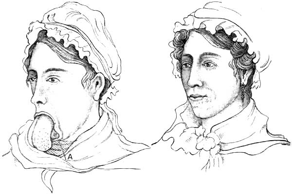
|
|
Hypertrophy of the Tongue (Harris), before
operation and after. A, odontolith. |
DIAGNOSIS.—The presence of the tongue outside of the mouth speaks for
itself (Figs. 14 and 15). The age of the patient, usually a young
child, the history of the case if it present in the adult, suffice to
differentiate macroglossia from the tumefaction of glossitis on the
one hand and from certain protruding tumors and malignant diseases on
the other. Hypertrophy of the tongue following chronic glossitis,
syphilitic or non-specific, must not be confounded with the congenital
or idiopathic affection under consideration.
PROGNOSIS.—The prognosis is good as to relief from the deformity,
provided the patient is submitted to surgical interference, and the
prognosis of the operation depends upon the procedure selected.
Sometimes additional operations are requisite to remedy the defects
the lower jaw has sustained by prolonged depression. In comparatively
young adults restoration of its position, configuration, and function
seems likely to result spontaneously after the protruding portion of
the tongue has been removed.
TREATMENT.—It has been maintained (Lassus) that the hypertrophy can
be overcome by systematic compression of the tongue, by leeching the
tongue, bandaging or strapping it, and forcibly maintaining it in the
mouth by suitable retentive appliances. While it has been admitted
that this plan may prove successful in cases of moderate enlargement
of but few years' duration and unaccompanied with change in the shape
of the lower jaw, the experience of more recent observers has been
recorded as unfavorable, at least in pronounced cases. Clanny30
succeeded in this way with a child five years of age whose tongue
protruded three inches. This plan is said to be very painful and
irritating. It requires close watching on account of the difficulty of
respiration which may ensue from thus blocking up the pharynx. It has
been advised as a useful and sometimes an essential preliminary (Syme)
to a radical procedure consisting in the excision of a V-shaped
segment. This latter operation (Boyer) has been successfully performed
by Howe, Harris, Humphry, Syme, and others.
Frederici31
extended the incisions to the very base of the tongue. It has been
performed both with knife and with scissors, the cut surfaces being
united with sutures after ligation or torsion of the bleeding vessels.
Re-enlargement ensued in one of Syme's cases operated upon in this
way,32 and likewise in a case of Gies,33 requiring subsequent
excision of the exuberant portions. Operations with the ligature,
though sometimes successful (Fergusson,34 Hodgson35), may be
followed by fatal septicæmia during the slough (Liston36), or,
failing to strangulate the tongue sufficiently, may require the
application of the knife, after all, to affect the separation
(Harris37).
30 Edinb. Med. and Surg. Journ., 1805, vol. i., cited by
Clarke.
31 Arch. gén. de Méd., 1844; Edinb. Med. and Surg.
Journ., p. 528, vol. lxiv., 1845.
32 Edinb. Med. Journ., 1857, vol. ii. p. 1057.
33 Arch. klin. Chir., 1873, p. 640.
34 Practical Surgery, London, 5th ed., p. 518.
35 Trans. Med.-Chir. Soc. London, 1858, p. 129.
36 Elements of Surgery, p. 334, Philada., 1842.
37 Am. Journ. Med. Sci., vol. vii. p. 17.
Excision with the incandescent loop of the galvano-cautery seems to be
the most suitable procedure. Valerani38 operated in this manner
without the loss of a drop of blood upon a congenital macroglossia in
a child seven months of age. Maas39 operated in this way on a child
two months of age. Fairlie Clarke, who removed a congenital
macroglossia with the écraseur in a child five months of age,
recommends operation before dentition begins40—an opinion which
appears to be justified by the belief that the pressure of the teeth
contributes to the subsequent rapid enlargement of the organ.
Nevertheless, the operation may be undertaken at any age. Several of
those already cited were performed upon adults, and Stephen
O'Sullivan41 excised the hypertrophied tongue of a female sixty-five
years of age.
38 Giornale della Reale Accademia di Turino, fasc. 1518;
London Med. Record, Sept. 15, 1876, p. 408.
39 Loc. cit.
40 Lancet, March 30, 1872, p. 432.
41 Dublin Journ. Med. Sci., Aug., 1875, p. 178.
Ignipuncture with the thermo-cautery of Paquelin has been
successfully used of late by Helferich and by von Bruns of
Tübingen.42 In the latter instance the subject was five years of
age. Fourteen punctures were made from above downward at intervals of
about one centimeter, and five were made transversely. Not a drop of
blood was lost. On the third day secondary hemorrhage occurred from
the intercommunication of three of the punctures; this was restrained
by ferric chloride, and the case went on to a favorable conclusion.
Surgical procedures must constitute our sole therapeutic reliance. The
temporary subsidence of enlargement under the influence of mercury and
the iodides seems sure to be followed, sooner or later, by
reproduction of the deformity. It is therefore a waste of time to
attempt cures by medication.
42 Centbl. f. Chir.; Med. Times and Gaz., Sept. 23,
1883.
DEFINITION.—Inflammation of the tongue.
The term glossitis is usually applied to inflammation of the tissues
of the tongue as a whole (parenchymatous glossitis), and not to those
superficial inflammations which exist associated with the different
varieties of stomatitis
and with other affections, and which
implicate the mucous membrane and its glands and papillæ only
(superficial glossitis). Superficial glossitis, however, sometimes
terminates in parenchymatous glossitis. Papillæ and glands are usually
affected together in superficial glossitis. When the inflammation
predominates in the papillæ the disease is often designated as
papillary glossitis; when in the glands, it is often termed follicular
glossitis. Superficial glossitis, again, is sometimes manifested by
the eruption of vesicles on the tongue, under which circumstance it is
often denominated vesicular glossitis, sometimes herpetic glossitis.
Glossitis is sometimes restricted to a portion of the tongue
(circumscribed glossitis), and it sometimes involves the whole of the
tongue (diffuse glossitis). Either form may be unilateral
(hemiglossitis), though both forms are more frequently bilateral.
Either form may be acute or chronic.
DEFINITION.—An inflammation of the mucous membrane of the tongue,
usually involving likewise both papillæ and glands.
SYNONYMS.—Catarrhal glossitis, Angina lingualis. Varieties:
Papillary, follicular, vesicular (herpetic and eczematous), psoriatic, ichthyotic.
ETIOLOGY.—Predisposing and Exciting Causes.—It is rarely idiopathic,
is most frequently deuteropathic, and sometimes traumatic. Superficial
deuteropathic glossitis usually occurs in connection with gastric and
gastro-enteric affections. It occurs likewise in association with
stomatitis, tonsillitis, pharyngitis, many febrile affections,
scorbutus, tuberculosis, syphilis, so-called psoriasis and ichthyosis
of the tongue, carcinoma of the tongue, and the various neoplasms of
the organ. Irregular and sharp-cornered or jagged teeth often induce
traumatic superficial glossitis. Pungent vapors, such as those of
chloride of ammonium, so much used of late years in the treatment of
nasal catarrhs, sometimes produce a superficial traumatic glossitis,
usually localized on the superior surface of the anterior portion of
the tongue. Tobacco-smoking, especially from a short-stemmed pipe,
will likewise produce it occasionally at the point where the
concentrated smoke strikes the organ. Attempts to drink liquids too
hot, too acrid, or too caustic may be mentioned as other occasional
causes. Nervous irritation, such as of the chorda-tympani nerve, is
attributed as a causal influence of unilateral vesicular glossitis,
herpetic or otherwise, and as a probable factor in other varieties of
unilateral glossitis. Eczema of the tongue may ensue as a sequel of
prolonged cutaneous eczema (De Mussy43).
43 Gaz. hebd., June 22, 1883; Med. News, Aug. 11, 1883,
p. 151.
PATHOLOGY AND MORBID ANATOMY.—Superficial glossitis, as indicated,
involves the mucous membrane, glands, papillæ, and epithelium. It is
hardly necessary to dwell upon the pathological conditions of the
lingual mucous membrane and its epithelium in gastro-intestinal and
febrile disorders, as these are described in connection with the
various diseases. Ordinarily, the epithelium increases in thickness,
and when detached, spontaneously or otherwise, exposes a red and
swollen membrane with erect papillæ. Sometimes the condensed
stratified layer of epithelium becomes dry and very hard. Under some
illy-defined conditions,
the papillæ of the tongue undergo great
hypertrophy. The filiform papillæ become elongated to several times
their normal length, and feel and look like so many hairs on the
tongue. Like many other lingual affections, this is often unilateral.
It is quite marked in some cases of influenza and other febrile
disorders, producing great annoyance in the mouth. It occurs likewise
in gastro-intestinal disorders and in disorders of the mouth and
teeth. It is evidently a deuteropathic phenomenon. In other cases the
glands of the tongue, especially at its base, become involved, forming
the follicular glossitis of some writers. In another class of cases,
most frequently syphilitic or at least quasi-syphilitic, one or more
whitish circumscribed patches are seen on the tongue, resembling such
as are left after superficial cauterization with nitrate of silver.
This condition is described as psoriasis linguæ. It is due to
condensation of layers of epithelium, which may become detached in a
few days in mass or in fragments, leaving the denuded mucous membrane
red and the papillæ erect and somewhat swollen.
When psoriasis of the tongue has existed for a long time, a further
change, and a more permanent one, takes place in the papillæ and
epithelium. This condition has been denominated ichthyosis linguæ.
Superficial ulceration takes place at the psoriatic patches, and the
repair eventually excites such a proliferation of epithelium that it
becomes quite horny to the sight and to the touch. It spreads over a
much larger extent of surface than the original psoriasis, but, like
it, leaves the unaffected portions of the tongue in an apparently
normal condition. Both affections are usually bilateral, and the
patches or series of patches most frequently symmetrical or engaging
analogous vascular territory upon the two sides.
In a case reported by Mr. Hulke44 portions of the horny substance
were habitually sliced off with a razor. Microscopic examination
"showed colossal papillæ; the indurated portion of the mass was
altogether epithelial, the lower cells being clear, transparent, and
natural, the middle ones granular, and the superficial layer felted
together into a dense opaque mass" (Clarke).
44 Medical Times, Nov. 30, 1861, p. 556.
Both of these affections are liable in about one-third of the cases to
terminate in epithelioma. Although the opinion generally entertained
classes all cases of psoriasis and ichthyosis linguæ in the category
of syphilitic affections, there is reason to doubt its accuracy.
Sangster45 has drawn up a tabular statement of 44 cases, of which 1
only occurred in a female; 23 occurred in smokers, 12 being inveterate
smokers. In but 12 instances (81/3 per cent.) was there positive
proof or strong evidence of syphilis; 30 per cent. of the whole number
eventuated in epithelioma.
45 Med. Times and Gaz., London, April 8, 1882, p. 370.
Vesicular glossitis, usually unilateral and most frequently
right-sided, has been described by Paget,46
Stoker,47 Barker,48
Hill,49 and De Mussy,50
and doubtless by others.
46 Lancet, March 11, 1865; Clarke, op. cit., p. 88.
47 Dub. Journ. Med. Sci., May 1, 1876, p. 401,
illustrated.
48 Lancet, Nov. 22, 1879, p. 764.
49 Brit. Med. Journ., Oct. 7, 1882, p. 683.
50 Gaz. hebd., June 22, 1883; Med. News, Aug. 11, 1883,
p. 151.
SYMPTOMATOLOGY,
COURSE, DURATION, TERMINATIONS, COMPLICATIONS,
AND SEQUELÆ.—Dryness of the tongue, stiffness, heat, and pain—the
latter especially during movements of the organ in deglutition and in
articulation—are the prominent subjective symptoms of acute
superficial glossitis. There are rarely any marked symptoms of febrile
disturbance unless the disorder is about to undergo extension into
parenchymatous glossitis.
DIAGNOSIS.—Redness of the tongue, prominence of the papillæ, slight
enlargement, perhaps bearing impressions made by the teeth, and pain
or sense of impediment on movement, are the main diagnostic features
of superficial glossitis.
Vesicles indicate the vesicular variety of superficial glossitis;
irregular whitish patches, the psoriatic variety; and hard, horny
patches with intervening fissures, the ichthyotic variety. A
superficial circumscribed glossitis attending the local ulcerations of
syphilis, tubercle, and epithelioma is differentiated by the clinical
history of the case.
TREATMENT.—In ordinary cases the treatment described under catarrhal
stomatitis suffices, so far as local measures are concerned. The
gastritis or gastro-enteritis requires appropriate attention, as does
any systemic malady under which the patient may be laboring. Demulcent
and astringent lotions may be applied by douche, spray, or gargle.
Local applications of weak solutions of iodine have been recommended.
In cases of considerable severity, and especially when there is reason
to expect extension into the deeper tissues, superficial scarification
of the dorsum of the tongue is advisable.
Ulcers are perhaps best treated locally by touching the edges daily
with the pencil of sulphate of copper. Any imperfect teeth in their
immediate vicinity to which the ulceration may be attributable should
be extracted or put in repair. De Mussy's case of eczema was cured
after five months' daily use of a large quantity of water-cress.
DEFINITION.—An inflammation of the tongue said to be due to parasitic
vegetation.
SYNONYMS.—Nigrities, Glossophytia, Black tongue.
Under the term black tongue two different affections have been
described, the one an epidemic erysipelatous disorder to be mentioned
under parenchymatous glossitis, and the other, now to be mentioned, a
peculiar black pigmentation due to parasitic disease seated upon and
around the hypertrophied filiform papillæ. The ordinary parasitic
vegetations found upon the tongue do not produce the affection in question.
HISTORY.—First described by H. Hyde Salter,51 and then by
Eulenburg, it has been made the subject of observation by Raynaud,52
Fereol and others,53
Lanceraux,54
Dessois,55 Hirz,56
Pasquier,57 Moure,58 and a few
others.
Outside of French literature, little had been written of it until very recently.
51 Article "Tongue," Encyclopedia of Anatomy and
Physiology, London, 1849-52, vol. iv. pp. 1159, 1160.
52 Gaz. hebd., 1869, No. 14, p. 221.
53 Gaz. des Hôp., June 29, 1875.
54 Union Méd., March 20, 1877.
55 De la Langue noire [Glossophytie], Paris, 1878, 8vo,
p. 38, illustrated.
56 Gaz. Méd., Strasbourg, 1879.
57 Bull. Méd. du Nord, 1883.
58 Revue mensuelle de Laryngologie, etc., Sept., 1883, p.
276.
ETIOLOGY.—The affection appears to be due to some fault of nutrition,
but the cause has not been determined. The fluids of the mouth always
exhibit an acid reaction. It has been seen chiefly in dyspeptics and
hypochondriacs, and has seemed in one instance (Moure) to have
followed the use of chlorate-of-potash lozenges. A case has been
recorded by Solomon Solis Cohen59 in a negro child the subject of
congenital syphilis. Mr. George Stoker60 and G. Y. Broatch61 have
each reported a case of long duration occurring in a painter.
59 The Polyclinic, Philada., July, 1884, p. 10.
60 Brit. Med. Journ., March 29, 1884, p. 602—said to be
first case recorded in England.
61 Ibid., April 19, 1884.
PATHOLOGY AND MORBID ANATOMY.—The disease is characterized by a
grayish-black or fully black discoloration on the upper surface of the
tongue, which gives it an aspect which recalls the normal appearance
of the tongue of the parrot and the giraffe, and an occasional
appearance of the organ in the ox, sheep, dog, cat, and some other
animals. The filiform papillæ are enormously elongated, so that they
closely resemble hairs, and they are described by some writers as
lying upon the surface of the tongue in confusion like that of a field
of wheat thrown down by the wind. The individual papillæ are
surrounded with a parasitic vegetable growth. Raynaud compared the
microscopic spores in his case to the microphyte of tinea tonsurans or
that of herpes circinatus. According to Malassez, they do not differ
from those found in the saburral tongue of the dyspeptic, and he
considers that their development is favored by their very arrest by
the hypertrophied papillæ. Nevertheless, the subjects of this disease
are not all dyspeptics by any means. Dessois made culture-efforts to
reproduce the disease upon his own tongue, but failed to inoculate it.
For detailed description of the disease we cannot do better than refer
the interested reader to Dessois' monograph, from whose observations,
chiefly, it appears that the discoloration begins at the central
portion of the tongue, increases gradually in extent and intensity for
three or four days, and then gradually disappears by desquamation. The
tongue is very dry while the affection is at its height. Close
examination of the parts and microscopic inspection of papillæ removed
for the purpose are said to show that the spores of the cryptogam are
first developed at the base of the papillæ, separating them from each
other. The irritation produced by the parasite causes longitudinal
hypertrophy of the papilla, and the continued growth of the parasite
produces a muff-like envelopment of the papilla; the spores at the
same time becoming insinuated between the most superficial epithelial
cells and dislocating them, so that they maintain their position
around the axis of the papilla only by means of the intervening
parasitic masses. The papilla continues to elongate and the cryptogam
to increase, until finally it invades nearly the entire length of the
papilla. This entire parasitic mass soon becomes detached, carrying
with it the epithelial cells under which it has become insinuated, and
leaves the papilla naked, save for a few cells remaining attached by
their superior borders. In the case observed by my brother, as in Mr.
Stoker's and Broatch's cases, microscopical examination of the black
filaments showed them to be composed of closely-packed epithelial
cells, overlapping one another, stained
brown, and decreasing in
intensity of color from the apex toward the base of the filament. In
some cells the nucleus was darker, and in others lighter, than the
surrounding protoplasm. The cryptogam, therefore, cannot always be
detected. Indeed, there seems reason to believe that the affection may
not be parasitic,62 although the prominence given to this feature by
French writers apparently warrants its being so considered. There may
be two kinds of black tongue—one non-parasitic.
62 Hutchinson, The Medical Press, p. 20, July 11, 1883.
SYMPTOMS.—There are no special subjective symptoms. The objective
symptoms are the peculiar dark or black discoloration of the upper
surface of the tongue and the excessively elongated filiform papillæ.
DIAGNOSIS.—The chief diagnostic feature is the black discoloration of
the tongue which has given the name black tongue to the disease.
Discrimination is requisite from discoloration by food or medicine.
PROGNOSIS.—This is favorable, the condition subsiding under
treatment, and sometimes spontaneously, though liable to recurrence.
In some instances the condition becomes chronic.
TREATMENT.—The indication is to endeavor to favor desquamation of the
papilla by means of potassium chloride or sodium borate, and to
administer alkalines, so as to render the saliva alkaline and
unfavorable for the development of the parasite. It is recommended, in
addition, to scrape the tongue with a spatula, and to douche it with a
spray of mercuric chloride, 1:500. Attention to the general health is
requisite, especially in dyspeptics and hypochondriacs. In the case of
the negro child above alluded to the discoloration of the tongue
finally disappeared under the systemic use of potassium iodide,
without topical treatment, although repeated recurrences took place at
varying intervals.
DEFINITION.—An inflammation of the tongue involving its substance as
well as the mucous membrane.
SYNONYMS.—Idiopathic glossitis. Interstitial glossitis. Erectile
glossitis (Salter), Glossomegistus (Sauvages), Paraglossia.
HISTORY.—Albeit a comparatively infrequent disease, especially at the
present day, numerous cases and collections of cases are on record
from very early times; and the affection seems to have attracted the
attention of medical writers ever since. Hippocrates, Galen, Aretæus,
Celsus, Aëtius, Avicenna, Forestus, Rivière, Schenkins, Sauvages,
Vogel, van Swieten, are referred to by more modern writers as having
described the disease. Louis, De la Malle, Lassus, J. P. Frank, Jn.
Frank, Fleming,63 Clarke, and Bryant may be cited as the most
prominent recent observers.
63 Dublin Journ. Med. Sci., 1850, vol. x.
ETIOLOGY.—Predisposing and Exciting Causes.—Glossitis is sometimes
idiopathic, sometimes deuteropathic, and sometimes traumatic. Impaired
health from over-fatigue or from exhaustive disease may be regarded as
a predisposing cause in the presence of the causes which more
frequently give rise to traumatic glossitis. Sudden or prolonged
exposure to atmospheric changes, to cold and moisture, as when working
in damp and wet localities, is often the apparent determining cause in
both
the idiopathic and deuteropathic varieties. In many
instances occurring in this way it is found that the tongue has long
been subjected to mechanical irritation from jagged teeth.
Idiopathic glossitis occurs at all ages, but has been supposed by some
observers to be more frequent in scrofulous subjects. It occurs in the
apparently healthy only after severe exposure to wet and cold, and in
convalescents from acute febrile diseases usually after some moderate
exposure to a draught of air or change of temperature. It appears to
be more imminent after influenza (Möller, Smee, Graves, Salter) than
after other febrile disorders. It has occasionally been caused by
chewing acrid plants, some of them food-plants, some of them
medicinal. In the list have been included celery, bilberries, Daphne
mezereum and Daphne laureola, aconite, and tobacco. It has been known
to follow the eating of shellfish (Watson, Salter).
Deuteropathic glossitis has occurred during the course of scarlatina,
variola, epidemic erysipelas (black tongue), scorbutus, enteric fever,
glanders, septicæmia from various causes, rheumatism, diffuse
inflammation of the connective tissue of the cervico-mental region,
herpes, syphilis, ptyalism, mercurial and other varieties of toxæmic
stomatitis, tonsillitis, pharyngitis, gastritis, and epithelioma of
the tongue. It appears to be occasionally endemic (Fleming64), and
is occasionally epidemic (Reil65). In the United States it prevailed
extensively during an epidemic of erysipelas that overran the country
from 1842 to 1846, inclusive, and was frequently reported in the
American medical journals of that period under the name of black
tongue. In some localities more than half the cases terminated
fatally, sometimes within two or three days, more frequently about the
eighth or tenth day, and occasionally still later. Traumatic glossitis
arises from a number of causes. Among these may be mentioned the
irritation of jagged edges of broken and carious teeth; wounds from
firearms and other weapons; wounds from splinters of toothpicks,
spiculæ of bone, broken pipe-stems, pins, needles, nails,
slate-pencils, and other pointed things inadvertently placed in the
mouth; wounds from the teeth during epileptic seizures and other
convulsive paroxysms; contact of the tongue with cold iron in cold
weather; the inspiration of very hot air, as in burning buildings;
burns, scalds, scalding beverages; acrid and corrosive substances
introduced by design or accident; incautious use of tobacco in bulk,
and of ammonia; incautious cauterization; concealed calculi in the
tongue; concealed bulbs of teeth; rupture of the lingual frenum; the
bites and stings of venomous insects, as the wasp, the hornet, and the
bee. For many years writers have referred to a case reported by Dupont
to the Parisian Academy of Medicine which followed a young man's
attempt to win a wager that he would bite into the body of a living
toad, and to two fatal cases reported by Ambrose Paré from drinking a
vinous infusion of sage which was subsequently found to have been
impregnated with the saliva of the toad.
64 Dub. Journ. Med. Sci., 1850, vol. x. p. 88.
65 Memorabilia Clinica (Dict. Sciences méd.), vol.
xviii.
SYMPTOMS, COURSE, DURATION,
TERMINATIONS, COMPLICATIONS, AND
SEQUELÆ.—In acute parenchymatous glossitis the local symptoms often
appear quite suddenly, usually unilaterally, even when they become
bilateral subsequently, and they increase in severity with great
rapidity. These
symptoms are, at first, distinct sensations of
heat and tumefaction in the tongue, quickly followed by stiffness and
considerable impediment in its movements, as though it were numb and
weighted down. In cases where the glossitis is an extension from
tonsillitis, these sensations begin in the root of the organ. They
commence at the root likewise, in most instances following exposure to
severe cold and moisture. In other instances the extremity of the
organ is affected first. In cases resulting from local injury the
symptoms commence at the injured portion. The local symptoms are
sometimes preceded by rigor, followed by fever, cephalalgia, and pains
in the neck and occiput. Examined at this time, the tongue is seen to
be swollen and studded with indentations due to the pressure sustained
from the teeth. At first the surface is punctated and red;
subsequently it becomes brownish or decidedly brown. Although the
organ may remain moist for several hours, it eventually becomes
excessively dry, and supports a thick adhesive coating of mucus and epithelium.
|
FIG. 16.
|
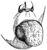
|
|
Glossitis (Liston). |
In a few hours, sometimes as few as two or three, the entire organ may
become involved in the inflammation, enlarging to such an extent as to
keep the lower jaw depressed, to fill almost the entire oral cavity or
to quite fill it, and to project like a tumor beyond the teeth and the
lips (Fig. 16). In exceptional cases the enlargement of the tongue has
been so great as to produce dislocation of the lower jaw. The soft
palate is lifted up and the epiglottis often pressed down. The latter
condition has been known to threaten suffocation. In this condition
the patient cannot breathe through the mouth, widely as it may be
forced open, and has great difficulty in breathing through the nose.
Respiration is therefore laborious. Articulation is impeded or
impossible, and deglutition difficult or impracticable. The
tumefaction and congestion are often continuous into the floor of the
mouth and the parts adjacent. The sublingual and submaxillary glands
often become swollen, tense, and painful; and the entire neck is
sometimes swollen to such a degree as to exert injurious pressure on
the jugular veins. The tongue is very hard to the touch, almost or
quite immovable, and is the seat of burning heat and pain. The pain
often extends from the root of the tongue along the glosso-pharyngeal
folds into the pharynx, and thence by way of the Eustachian tubes into
the ears, the folds just named being very much upon the stretch. When
the tongue protrudes far out of the mouth it becomes excessively dry,
fissured, sanious, and excoriated, or even ulcerated at points where
it is subjected to the pressure of the teeth. It is covered with dark
viscid secretions, which often extend beyond it and over the entire
aperture of the mouth. The epithelial coating often undergoes
desquamation, and then the organ becomes exquisitely sensitive to the
contact of food, water, or even the air. This desquamation is
sometimes in mass, in sheets peeling off like a pseudo-membrane. The
general symptoms vary in individual cases. As a rule, the face is
turgid and its expression anxious; the conjunctiva suffused,
respiration impeded, and sleep disturbed or impracticable. Saliva
dribbles externally, often in considerable quantity. The
odor
from the mouth becomes quite fetid from decomposition of the retained
products of secretion. Thirst is usually intense, though immoderate
only in some cases. Cough is more or less constant and quite
exhausting. This and the dribbling of saliva contribute with the
dyspnoea to prevent sleep. Pyrexia is often intense. The pulse is
strong and quick at first, 100-120 per minute, and there is often
marked throbbing of the temporal and carotid arteries. The skin is hot
and dry at first, but cold sweat subsequently accumulates upon the
face and neck as the dyspnoea increases. The bowels are constipated.
The urine is scanty and high colored. The impediment to the return of
blood to the heart from the head causes cerebral congestion,
drowsiness, and even threatens asphyxia. In other cases there is
intense cephalalgia, nervous irritability, restlessness, and even delirium.
The symptoms sometimes reach their acme in rather less than
forty-eight hours, and then gradually subside. More frequently they
continue on into the third or fourth day. Occasionally they are
protracted as long as the fifth or sixth or even the eighth day,
rarely longer. Resolution occasionally takes place within twenty-four
hours, however (van Swieten), though more frequently occurring from
the fifth to the seventh day. In some instances remittance or
intermittence has been noted, the cause therefor not being apparent,
although attributed to malaria.
Resolution of the inflammatory process is usually indicated by the
gradual return of moisture on the tongue and progressive detumescence
of the organ, accompanied by subsidence of the redness, heat, and
pain. Increased secretion of saliva, general perspiration, or diuresis
sometimes marks the cessation of the pyrexia.
Should the process be going to terminate in suppuration, the local
distress increases, markedly about the end of the week. The pains
become lancinating, and associated with throbbing of the lingual
blood-vessels. The swelling becomes prominent and softer at some one
point, although the sense of fluctuation is not very perceptible on
palpation, and finally the abscess bursts through the surface, unless
previously incised, and discharges a fetid pus. Suppuration always
involves a prolonged duration of the attack.
In rare instances glossitis terminates in gangrene of the tongue,
circumscribed or diffuse. This result is indicated by adynamic
symptoms on the part of the constitution, and by the livid appearance
of the parts undergoing mortification. The hemorrhage following
extensive sloughs from gangrene has been fatal in some instances.
Fleming66 calls prominent attention to a complication of glossitis,
of which he alludes to several examples. This is "an inflammation,
circumscribed or diffused, originating in the loose areolar tissue
between the genio-hyo-glossi muscles, and first manifesting itself by
a train of symptoms identical with those of ordinary glossitis, but
soon characterized by peculiar features." These features comprise
fulness under the chin like that dubbed double chin, pressure upon
which, especially near the hyoid bone, being very painful; and
suppuration, which, circumscribed or diffuse, burrows most freely
toward the base of the tongue.
66 Loc. cit., p. 91.
Chronic induration of the tongue sometimes remains unilateral,
although the acute disease has not been unilateral.
PATHOLOGY
AND MORBID ANATOMY.—It has been advanced that in
idiopathic glossitis from cold the engorgement of the vessels is
probably a consequence of vaso-dilator influence of the
glosso-pharyngeal nerve for the base of the organ, and of the
chorda-tympani for the anterior portion. An instance of herpetic
glossitis from probable irritation of the chorda-tympani nerve by an
aural polypus (Berkely Hill67) seems to lend some force to this
opinion. However engendered, there is a rapid distension of the organ
by blood, followed by infiltration of fibrin and serum into the
intermuscular connective tissue and into the planes of the connective
tissue separating the muscular fasciculi. In some instances
degeneration of muscular fibre has been observed. There is great
increase in the thickness of the coats of epithelium, beneath which
the mucous membrane is red and its papillæ erect. This coating
sometimes peels off like a false membrane. In cases extending from
tonsillitis the base of the tongue suffers most.
67 Brit. Med. Journ., Oct. 7, 1882, p. 683.
The disease usually terminates by resolution, although a slight amount
of hypertrophy, unilateral or bilateral, sometimes persists, and
occasionally to a marked degree (Wells). In instances much less
frequent suppuration ensues, usually in debilitated subjects or in
cases due to traumatism or in cases inefficiently treated. The
suppurative process is usually circumscribed and unilateral, and the
abscess points most frequently just beneath the side of the tongue;
sometimes, however, the pointing takes place at the dorsum, sometimes
at the tip. The pressure of the teeth seems to be the provocative
cause of the disposition to point at the edge of the tongue. The pus
is usually quite fetid. Sometimes the abscess is gangrenous.
Gangrene is an infrequent result of glossitis. The pressure of the
teeth, strangulating the organ at the oral outlet, seems to occasion
the failure of nutrition in instances where it occurs. The losses are
ordinarily insignificant, though appearing quite extensive while the
tongue remains swollen. Sometimes large portions drop off, and fatal
hemorrhage has resulted (Frank) in consequence. From the nature of the
organ the parts separate more readily than in almost any other
instance. Gangrenous abscess, ensuing even from very slight causes,
such as a wound with the head of a barleycorn (Ranking68), sometimes
proves fatal.
68 Provincial Med. and Surg. Journ., 1844.
In those cases of diffuse inflammation of the interconnective tissue
of the genio-hyo-glossi muscles Fleming states that the
suppuration—which, whether circumscribed or diffuse, burrows toward
the root of the tongue—absolutely dissects its extrinsic muscles and
destroys their functions; ultimately injuring the periosteum and
laying bare the inside of the inferior maxilla in the vicinity of
their attachments. When an incision is made to the parts through the
integument, the muscles will be found on palpation flabby and
detached, and their interstices filled with purulent matter, sometimes
very fetid.
DIAGNOSIS.—These is no difficulty in the diagnosis, except in the
early stage of such examples as are attributed to metastatic gout and
rheumatism. The subsidence of the peculiar pains elsewhere, and the
onset of pain in the tongue, would lead to the inference that a
glossitis of this kind was
in progress. The acuteness of the
tumefaction would distinguish it from hypertrophy of the tongue on the
one hand, and from the tumefaction attending malignant disease on the other.
Cystoma of the tongue has sometimes been mistaken for abscess due to
glossitis; but even here the history of the case should serve in most
instances as a satisfactory factor for the differential diagnosis.
PROGNOSIS.—The prognosis depends upon the gravity of the local
symptoms and the activity of the treatment. A case left to itself will
be likely to terminate fatally within five or six days. Death, indeed,
has been known to take place within forty-eight hours, even in cases
submitted to treatment. On the whole, however, the prognosis should be
regarded as favorable in the absence of specially lethal
complications. Even suppuration adds little gravity to the prognosis,
the structure of the organ being but little favorable to accumulations
of purulent material. Should an abscess become gangrenous, however,
the prognosis becomes grave at once, as it in the presence of gangrene
from pressure or other cause. Should the patient survive losses by
gangrene, there may be permanent impairment in articulation.
TREATMENT.—Superficial glossitis, as a rule, merely requires active
purgation, with the topical use of cold emollient mouth-washes
containing mucilage of slippery elm, quince-seed, or the like, to
which detergents, such as alum and borax, may be advantageously added
in the proportion of five grains to the ounce. In cases resisting this
mild treatment topical applications of glycerite of tannin twice or
thrice a day are often serviceable.
Parenchymatous glossitis demands the most active antiphlogistic
treatment. If the case be seen at an early stage of the process,
before the tumefaction of the tongue has become so great as to fill
the mouth and interfere with swallowing, a saline purge—say salts and
senna—containing some tartar emetic can be advantageously
administered to begin with. Following this, tartar emetic may be
continued in small doses every two or three hours, associated with
small doses of tincture of aconite-root (minim j-iij), according to
the condition of the pulse and the effect of medication. Should this
treatment fail to produce prompt amelioration in the local symptoms,
or should the tongue be considerably swollen when the case comes under
care, free leeching should be applied from the hyoid bone to the angle
of the jaw on each side, including the region of the hyoid bone
(fifteen to twenty-five Spanish leeches). This should be followed by
emollient cataplasms, reaching from ear to ear, to favor continuous
hemorrhagic oozings from the leech-bites. The internal antiphlogistic
treatment is indicated just the same, and if not administrable by the
mouth may be administered by the bowel; the nauseant and depressent
effects of the tartar emetic and aconite being maintained by
hypodermatic injection. Leeching the inflamed tongue itself is said to
be often prompter in producing detumescence of the organ than leeching
exteriorly, but the leech-bites are apt to add to the local
irritation; besides which, the mouth is so filled by the swollen
tongue as to leave little more than the tip accessible to the leeches
without danger of losing control of them. Venesection from the arm,
the jugular vein, or from vessels elsewhere is no longer much in
vogue, it being doubtful whether general venesection is more useful
than local bleedings. Debility, whether presenting originally or as
the result of withdrawal of blood and other
antiphlogistic
measures, may be met by the systematic use of tincture of chloride of
iron and of quinia.
Severe cases demand one or more longitudinal incisions on each side of
the raphé of the tongue, deep enough to reach nearly halfway into the
substance of the organ and carried from base to tip. Cases are on
record in which the patients themselves had in their despair cut into
their tongues in order to obtain relief from their local sufferings,
and had in this way rescued their own lives by the means most
appropriate for the purpose (Camerarius, Lusitanus69). When the
mouth is filled by the tongue, it is necessary to insert the knife on
the flat until the base of the tongue is reached, and then to turn it
and make the cuts as indicated. Copious bleeding usually follows these
incisions, often followed by marked diminution in the volume of the
organ. Deep as these cuts appear when made, they become quite shallow
before the organ has shrunk to its normal volume. Bleeding from the
ranular veins, recommended by some practitioners in preference to
incisions into the organ, is often impracticable on account of the
tumefaction preventing access to them.
69 Dict. Sciences méd., vol. xviii.
If severe hemorrhage takes place from divided vessels, the vessels may
be subjected to torsion, which is the preferable mode of management,
or to searing with some form of the incandescent cautery (hot iron,
electric cautery, Paquelin's thermo-cautery). Astringent and chemical
styptics are of little use. The method of searing is open to the
objection that secondary hemorrhage may ensue on detachment of the
eschars, but this accident is not likely to happen under circumstances
at all favorable.
In localized or circumscribed glossitis the incision to be made should
interest the swollen portion only.
Should the tongue swell again, the incisions may be repeated. Whether
the tongue require incision or not, it is good treatment to have the
patient inhale vapor from hot vinegar, alcohol, or cologne spirits to
render the parts more comfortable. Washes of weak detergent solutions
containing potassium nitrate, sodium borate, or ammonium chloride may
be used by syringe or spray to cleanse the parts and promote
detachment of the epithelial coatings on the tongue and interior of
the mouth, the accumulations of which are sometimes matters of great
annoyance. The drug last mentioned exerts in addition a special action
on the inflammatory process which is often quite serviceable. I have
seen good results follow the prolonged use of sprays of an aqueous
solution of ammonium chloride (Stuver), one drachm to the ounce, from
the steam-spray apparatus, continued for fifteen to twenty minutes at
a time and repeated every two or three hours.
In many instances the patient is unable to take food by the mouth. The
best plan under such circumstances is to pass a catheter into the
stomach through the larger of the two nasal passages, and retain it in
position unless its presence interferes too much with respiration.
Milk and stimulus can then be poured into the stomach from time to
time with the aid of a funnel passed into the outer opening of the
tube, which should be kept corked during the intervals when retained
in position. This failing or impracticable, it will be necessary to
nourish the patient with enemata.
On the appearance of abscess the same should be freely laid open. In
cases of hesitation, the true nature of the presumable abscess can be
determined
beforehand with the exploring-needle. The pus from an
abscess of this kind is rarely copious and is usually fetid. It would
be good practice to distend the sac after discharge of pus by
injecting into it a solution of carbolic acid.
In resisting or advanced cases of suppurative inflammation of the
planes of connective tissue between the muscles at the lower portion
of the root of the tongue, Fleming recommends a free incision under
the chin in the middle line, through skin and fasciæ and on through
the raphé of the muscles themselves.
In cases of gangrene, washes, douches, or sprays of carbolic acid,
chlorinated soda, hydrogen peroxide, or potassium permanganate are
indicated to relieve fetor; while the most supporting treatment by
mouth or otherwise is requisite on general principles.
Chronic glossitis, like acute glossitis, may be superficial or
parenchymatous.
Superficial chronic glossitis is usually confined to the papillæ of
the tongue, territories of which, so to speak, are mapped out on the
surface of the tongue, separated by furrows reaching to the basement
mucous membrane. In pronounced cases the dividing furrows are quite
deep, giving the organ a mamelonnated appearance, and they penetrate
into the mucous membrane (dissecting glossitis, Wunderlich), which
becomes subjected to great irritation by the retention of articles of
food in the fissures. Demarquay70 has recorded a case of this kind
in which, to relieve the intense sufferings with which the patient had
been plagued for a number of years, he had been forced to amputate the
anterior half of the organ—an operation which succeeded thoroughly.
70 Loc. cit., p. 142.
In another group of cases the surface of the swollen tongue is mapped
out in small ovoid patches, smooth, red, and glossy, from which the
papillæ have become separated without regeneration. Sometimes chronic
glossitis presents as an aphthous inflammation. Sometimes superficial
ulcers occur upon the dorsum of the tongue, irritable, indolent, and indurated.
ETIOLOGY.—Chronic superficial glossitis is in rare instances a sequel
of the acute form of the disease. Usually, however, it is encountered
as a chronic affection from the outset, so to speak, generally as a
result of long-continued irritation in connection with dyspepsia and
other gastric and gastro-intestinal disorders. It is frequently
encountered in subjects of chronic alcoholism.
The superficial ulcerations often occur at the sides of the organ,
usually in some of the depressions formed by the contact of the teeth.
SYMPTOMATOLOGY.—The symptoms are those due to a consciousness that
the tongue is too large, with occasional pain in taking acid and succulent food.
PATHOLOGY.—The
pathology does not differ from that of chronic
inflammations elsewhere. The apparent obliteration of the papillæ in
some cases is due to a filling up of the intervening furrows by
permanent deposit of new-formed cells. The excoriations and
superficial ulcerations are most frequently due to disturbance of
nutrition by pressure.
DIAGNOSIS.—The affection may be confounded with syphilitic disease of
the tongue or with papillary epithelioma, but the history of the case,
the resistance to antisyphilitic treatment, and the negative results
from microscopic examination of fragments of tissue removed for the
purpose, serve to establish the diagnosis in cases of doubt. It must
not be forgotten, however, that many cases of epithelioma begin in
chronic glossitis, non-specific as well as syphilitic.
PROGNOSIS.—Though not threatening to life, the prognosis of the
disease itself is bad. It resists treatment, being, in fact, a
complication of some obstinate or intractable gastric or
gastro-intestinal disorder, or an evidence of constitutional
dyscrasia. Cure may be expected in recent cases, following cure of the
dyspepsia or other malady upon which the chronic glossitis may be dependent.
TREATMENT.—Care to cleanse the tongue by washes, douches, or wet
cloths after each meal, in order to remove particles of food which may
have become impacted in the anfractuosities of the organ, is important
in order to avoid additional sources of irritation.
Astringents and caustics of various kinds have been extensively
employed, carefully applied to the floors of the fissures, but it is
very rarely that any benefit ensues. Demarquay71 reports good
results in one case of dissecting papillary glossitis from biweekly
applications of equal parts of chromic acid and water. Butlin reports
good results from chromic acid (1:10).
71 Loc. cit., p. 143.
Careful attention to the gastro-intestinal functions, and a thorough
change of diet, such as the adoption of the milk cure and the like,
with due attention to bathing and outdoor exercise, comprise the most
rational method of constitutional treatment. Should the secretions of
the mouth give an acid reaction with litmus-paper, alkalies are
strongly indicated, topically and systemically. Avoidance of alcohol
in all forms is often absolutely essential.
The chronic parenchymatous form of glossitis is usually circumscribed.
When diffuse or general it has usually been a sequel of acute
parenchymatous glossitis. It is not a painful disorder, and as a rule
is not associated with constitutional manifestations. The
circumscribed tumefaction usually presents as an induration upon some
portion of the side of the tongue, being most frequently directly or
indirectly due to irritation sustained from a jagged tooth. Ordinary
sensibility is much diminished, and sometimes the sense of taste
likewise. Sometimes the indurated mass is ulcerated superficially. The
enlargement of the organ is not sufficient to keep it outside the
mouth. Sometimes, indeed, the tongue,
as a whole, has undergone
atrophy, unilateral or bilateral. Chronic abscess of the tongue
sometimes supervenes, chiefly in scrofulous subjects.
PATHOLOGY AND MORBID ANATOMY.—This consists merely in interstitial
connective-tissue hyperplasia, with atrophy of muscular fibres from compression.
SYMPTOMS.—In addition to the objective symptoms of induration or
circumscribed tumefaction, the subjective symptoms may be summed up as
general hypersensitiveness to sapid and acrid substances; diminished
tactile sensibility at the part affected; slight stinging sensations
while the parts are at rest; occasional or continuous local pains; and
a sense of impediment in the movements of the tongue in articulation
and even in deglutition.
DIAGNOSIS.—Inspection reveals the swelling, and palpation its
induration. In addition, the adjacent source of irritation, a jagged
tooth or two, is seen. Abscess is recognized by special prominence at
one point of the swelling and by indistinct sense of fluctuation.
Cystic tumor is liable to be mistaken for abscess, but the
exploring-needle will solve the difficulty. Circumscribed induration
may be confounded with tumor or with epithelioma.
PROGNOSIS.—This is good, provided the source of irritation can be
removed or suppressed.
TREATMENT.—The first element in the treatment is the removal or
repair of any offending tooth, and next attention to any underlying
malady, constitutional or local. Weak solutions of iodine locally are
said to be of service. Abscesses require incision and evacuation.
Their walls should be distended with solutions of carbolic acid or be
touched with solutions of iodine, silver nitrate, or cupric sulphate,
to promote reparative inflammation.
This is a special variety of gangrenous ulcerative glossitis,
presenting as an integral phenomenon of a disease peculiar to
slaughterers, who become infected from diseased cattle, usually by
means of the knife, which they are sometimes in the habit of holding
in the mouth (Heyfelder and others). It has been described chiefly by
Heyfelder,72 Duhamel, Chavarrien d'Audebert, Felix Plata, Breschet
et Finot, and Maisonneuve.73
72 Med. Vereins Zeitung, 1834.
73 Des Tumeurs de la Langue, Paris, 1848, Thèse de
Concours.
The period of incubation occupies about one day. The tongue then
undergoes rapid tumefaction and becomes the seat of intense pain. The
points of inoculation become hard, covered with vesicles containing
bloody serum, which blacken, rupture, and leave dark, livid,
gangrenous patches of ulceration. Profound cachexia rapidly ensues
under typhoid manifestations, and death may result in less than
twenty-four hours, though usually not until sixty hours. The
prognosis, therefore, is of the gravest character.
The chief treatment consists in thorough cauterization of the
inoculated points
by means of the actual cautery, followed by
deep incisions into the body of the tongue if the glossitis be severe.
Apart from the ulcerations of the tongue incidentally mentioned in the
foregoing pages, there are two forms of ulcer, both of sufficiently
frequent occurrence in ordinary practice to require special
description: these are the tuberculous ulcer and the syphilitic ulcer.
Tuberculous Ulcer of the Tongue.—Tuberculous ulceration of the tongue
occurs in a certain number of cases of advanced tuberculosis of the
lungs or of the lungs and larynx. It has even been asserted to precede
pulmonary tuberculosis. It is most frequently observed upon the upper
surface of one side of the organ, sometimes at the tip, sometimes
farther back, and usually on the same side upon which the disease is
most advanced in the lungs or the larynx. It is often associated with
previous or subsequent tuberculous ulcerations of the palate or of the
pharynx or contiguous structures. It gradually extends, and rarely if
ever heals. It is characterized by a superficial excavation, and by
being covered with a grayish detritus entirely different from the
purulent layers seen on other kinds of ulcers. When of long standing
its base is indurated, and this may give rise at first to suspicion of
squamous-celled carcinoma. Small yellowish elevations are sometimes
observed in the reddened mucous membrane around the ulcer—an
appearance deemed sometimes characteristic of the tuberculous nature
of the lesion (Trélat74).
74 Bull. de l'Acad. de Méd., 1869, or Arch. gén. de
Méd., 1870.
PATHOLOGY AND MORBID ANATOMY.—Nodular tuberculous infiltration takes
place beneath the mucous membrane, which becomes elevated in small,
semiglobular, yellow protuberances of one or more millimeters in
diameter, around which the mucous membrane is red and swollen. The
epithelium becomes shed without undergoing renewal, and thus a little
point of superficial ulceration remains. When several such points are
sufficiently contiguous they coalesce into a single ulcer of irregular
contour, which gradually spreads without much other change.
Practically, it never heals.
SYMPTOMS.—In addition to the superficial ulceration described, and in
addition to the constitutional and local symptoms of advanced
tuberculosis of the lungs or lungs and larynx, as may be, there are no
special symptoms attending the tuberculous ulcer of the tongue. Saliva
is sometimes secreted in excess, but that is not characteristic. There
is little pain and little impediment to the movements of the tongue
until the disease has advanced.
DIAGNOSIS.—The presence in a tuberculous subject of a unilateral,
irregular ulcer of the tongue surmounted with grayish detritus and
surrounded by reddened edges, should suffice for the recognition of
its presumptive tuberculous character. It is most difficult perhaps to
differentiate from a small ulcerated squamous-celled carcinoma, and
the two indeed sometimes coexist, rendering the discrimination
extremely difficult until the advanced progress of the carcinoma
places the diagnosis beyond doubt.
In
the early stages, however, it is distinguished by lack of the
peculiar lancinating pains of carcinoma, which, however, are not
invariably attendant, and by lack of secondary involvements of the
cervical lymphatic glands. At all times it should be distinguishable
from the carcinomatous ulcer by lack of the fungus-like appearance of
the bed of the ulcer which is usual in carcinoma.
From syphilitic ulcer it is distinguishable by the history of the
case, its tendency to be unilateral, and its failure to respond to
antisyphilitic treatment. Syphilitic ulceration of the tongue may
represent the primary, the secondary, or the tertiary manifestation of
the specific disease. The former will not be discussed in this connection.
Secondary ulcers occur on the upper surface of the tongue, most
frequently at the anterior portion, as fissures, usually longitudinal,
the floors of which are ulcerated. They occur likewise at the sides,
tip, and even lower surface of the organ. They are often associated
with secondary ulceration in the mucous membrane of some portion of
the mouth. They are quite painful, especially to the contact of
pungent articles of food. Some ulcers occur as simple superficial
excoriations at some portion of the edge or tip of the tongue, giving
little evidence of any specific character.
Tertiary ulcers are usually sequelæ of gummata. They are much deeper
than secondary ulcers, sanious at bottom, often serpiginous in
configuration, and apt to extend in depth as well as in superficies,
sometimes penetrating through and through the organ. They are most
frequent in the very central portion of the tongue, or are
symmetrically disposed on either side of it.
PROGNOSIS.—The prognosis of tuberculous ulceration is bad, both as
regards tongue and patient.
TREATMENT.—The only topical treatment offering any prospect of local
cure is the bodily destruction of the ulcer and the surrounding tissue
with caustics, the best of which are the incandescent metals, or else
the excision, with the incandescent knife, of a portion of the tongue
comprising all the affected tissue. In the former case the tuberculous
process often reappears about the cicatrix; in the latter, at some
more distant point.
Tincture of iodine locally, detergent washes, and the like, often
secure a certain amount of comfort as palliatives. The same
indications prevail as in simple chronic glossitis, superficial and
deep-seated. Iodoform locally is of benefit, inasmuch as it relieves
pain and reduces collateral inflammation, but it is powerless to
arrest the onward march of the ulcerative process.
DEFINITION.—A loss of blood from the mouth.
SYNONYM.—Stomatorrhagia.
ETIOLOGY.—Hemorrhage from the mouth is usually a symptom of some
disease or injury of the mouth, tongue, gums, palate, pharynx, or
nose. It may, however, occur as one of the phenomena of scorbutus or
of hæmophilia. It is said to occur occasionally as a vicarious
menstruation. It may be slight, so as barely to tinge the saliva, or
it may be profuse enough to terminate fatally. Between these extremes
there is an infinity of gradations. As a result of disease it may be
caused by simple hyperæmia of the mucous membrane, by rupture of
dilated blood-vessels, by ulceration, by gangrene. As a result of
injury it may arise from wounds of various kinds, accidental or self-inflicted.
The gums are the most frequent source of slight hemorrhage from the
mouth. The pharynx, probably, is the next most frequent seat.
Hemorrhage from the tongue, cheeks, lips, and palate is usually
traumatic or the result of ulceration.
SYMPTOMATOLOGY, COURSE, DURATION,
COMPLICATIONS, TERMINATIONS, AND
SEQUELÆ.—The symptoms of hemorrhage from the mouth are the presence
of blood in the saliva or in the mouth itself, or in the expelled
products of expectoration, emesis, or catharsis, for sometimes the
blood is swallowed, and occasionally inhaled into the air-passages.
The course, duration, complications, and terminations of
stomatorrhagia depend upon its cause. Prolonged hemorrhage will entail
anæmia; profuse hemorrhage may terminate fatally.
DIAGNOSIS.—Careful examination of the mouth, tongue, pharynx, and
posterior nares, both by direct and by reflected light, may be
necessary to discover the source of the hemorrhage and discriminate it
from hæmoptysis and hæmatemesis.
PROGNOSIS.—The prognosis will depend upon the nature of the cause,
its susceptibility of arrest, the quantity of blood lost, and the
general health of the patient. It is grave, as a rule, in the subjects
of hæmophilia, as there is a constitutional malnutrition of the
blood-vessel system which cannot be counteracted.
TREATMENT.—Ergot or oil of turpentine internally, astringent
mouth-washes, and recumbency constitute the main features in treatment.
DEFINITION.—Departure from the physiological processes concerned in
the eruption of teeth, entailing certain local and systemic disorders.
SYNONYMS.—Dentitio difficilis, Pathological dentition, Odontitis infantum.
The correct comprehension of the subject will be facilitated by
considering its etiology, pathology, and symptomatology in connection.
Indeed, morbid dentition has been assigned so prominent a part in the
etiology of various affections elsewhere discussed that a satisfactory
consideration of its own causation would require the repetition of
much that belongs more appropriately under other titles. While in some
infants the teeth erupt so quietly that the parents are astonished by
the accidental discovery of their presence above the gum, few children
escape a greater or less amount of local and constitutional
disturbance while passing through the process of dentition. So severe
may these disturbances become that, according to the mortality-tables
of London, as cited by West,75 teething was assigned as the cause of
death of 4.8 per cent. of all children dying under one year old, and
of 7.3 per cent. of those who died between the
ages of twelve
months and three years. It is furthermore well recognized that the
period of greatest mortality among children is that of the first
dentition. Associating these facts, we see, on the one hand, that
while disorders of dentition may act a causative part in the
production of systemic diseases or aggravate morbid processes due to
ordinary causes, on the other hand they may be but one expression of
some profound constitutional disturbance; or both aberration in the
eruption of the teeth and systemic disease may be dependent upon the
influence of dyscrasia. The period is one of active organic processes;
the child is becoming fitted for a new manner of existence; and change
and development are going on throughout nervous, vascular,
respiratory, and alimentary systems. Hence there exists peculiar
susceptibility to morbid influences; and any process, physiological or
pathological, once started, goes through its stages with excessive energy.
75 Lectures on the Diseases of Infancy and Childhood,
Philada., 1860, p. 425.
Although the periods of normal eruption of the deciduous teeth vary
within extensive limits, and an invariable order in eruption is not
observed in all subjects, it may be stated as a rule that the lower
central incisors are cut in quick succession about the seventh month.
Some infants get these teeth during the fourth month (Vogel), and
others have to wait until the tenth or eleventh month, some even
longer. A few weeks after the appearance of these lower
incisors—within fourteen days in some subjects, not until nine or
more weeks in others—the central incisors of the upper jaw are cut,
and its lateral incisors shortly afterward, followed in their turn by
the lateral incisors of the lower jaw. In some instances—the
majority, according to Vogel—the eruption of the inferior lateral
incisors is delayed until the anterior molars are about to become
exposed, usually from the twelfth to the fifteenth month. Sometimes
the upper molars are cut before the lower, sometimes after them. From
the sixteenth to the twentieth, or even the twenty-fourth, month the
canine teeth are cut, and the four posterior molars follow between the
twentieth and thirtieth months, rarely delayed until the thirty-sixth
month,—completing the process of the first dentition.
From this it will be seen that the teeth erupt as a rule in pairs, and
that a longer or shorter interval of repose takes place between the
eruption of successive pairs.
Variations from the usual order beyond the limits noted above may be
considered abnormal. Numerous cases are on record both of precipitate
and of tardy dentition. Tanner cites from Haller nineteen examples in
which one or more of the central incisors have been found through the
gums at birth, and have had to be removed to prevent injury to the
mother's nipple; from Crump, a case of full dentition at birth,
reported to the Virginia Society of Dentists; and from Ashburner, a
case of a child beginning to cut its first tooth, an incisor in the
upper jaw, during its twenty-third month, the infant being very
delicate, with a large head, tumid abdomen, and peculiarly small-sized
extremities. The same author quotes from Serres cases of persons
passing through several years of life—in one instance seven—before
cutting their first teeth, and mentions on the authority of Tomes that
Boxalli and Baumes have each recorded an instance in which the patient
reached old age without a single tooth having ever appeared.
Rachitis is often the cause of tardy dentition, and in the subjects of
this diathesis
not alone are the teeth retarded in development,
but they decay early and even fall from their sockets.
The first indication of approaching dentition is the markedly
increased production of saliva. For some little time after birth the
salivary glands seem to remain wholly inactive, and until the fourth
or fifth month of extra-uterine life they furnish very little
secretion. At this period a decided change occurs. The mouth is
constantly filled with saliva, which dribbles from its corners. To
this continual slobbering, wetting the garments covering the chest,
has been attributed the bronchial catarrh which attends some infants;
and diarrhoea has likewise been referred to the swallowing of large
quantities of saliva, acting as a mild laxative by virtue of its
saline constituents.
There may be no further manifestation until the seventh month, beyond
the broadening of the dental ridge. The exact position of each tooth
is usually indicated by greater prominence of the gum above it for
some time before it comes through, its entire outline being very
distinct in the upper central incisors. As the tooth approaches the
surface the gum becomes hot, shining, tense, and tumid, often painful.
A slight amount of catarrhal stomatitis is almost invariable. There is
some elevation of temperature; flushing of the cheek may occur; the
child is restless, peevish, and fretful; its sleep may be broken; it
may cry out with pain; its thumb, its fingers, any hard substance it
can obtain, are thrust into its mouth to allay the irritation of the
gums. Otalgia is not uncommon, and its occurrence may be inferred from
the fact that the child pokes its thumb or finger into the auditory
canal or firmly presses the tragus down over the external meatus.
These may comprise all the disordered manifestations, local or
constitutional, or there may be in addition loss of appetite,
diarrhoea, vomiting, and the various disturbances of reflex nervous
origin to be alluded to later; or, in the not common yet not rare
instances already mentioned, there may be absolutely no appreciable
disturbance whatever.
Sometimes a disposition exists to the formation of small aphthous
ulcerations on the tongue or elsewhere in the mouth, particularly at
the duplicature of the lip and the outer surface of the alveoli.
Ulceration occurs most frequently at the tip of the tongue, probably
occasioned by friction from the new teeth. Usually there is a single
flat, round ulcer, its edges somewhat infiltrated, its bed covered
with a yellow lardaceous substance. It is extremely painful to the
touch, and thus every movement of the tongue occasions distress. It
may heal within a few days or continue for weeks. Ulcers in other
situations are less obstinate.
Occasionally—and more frequently in debilitated subjects or those
exposed to unhygienic surroundings—there is an unusual amount of heat
and swelling of the gum, which becomes excessively tender, usually
over the summit of a particular tooth—in which case there will be a
little tumor-like elevation—or around a tooth which has partially
pierced through it. Small sloughy ulcerations form in this situation.
There is great pain, and usually high fever and severe
gastro-intestinal disorder. This affection, often difficult of cure,
is termed by some writers odontitis infantum. So severe is the pain,
and so great its tendency to aggravate constitutional disturbances,
that life may be placed in jeopardy, and even fatal results ensue.
Less
severe than either of the forms just described, and yet far
more intense than the mild stomatitis which many authors regard as
physiological, is an aggravated form of catarrhal stomatitis sometimes
attendant upon morbid dentition, in which there is swelling of the
submaxillary glands and infiltration of the adjacent connective
tissue. In this case there is usually considerable pyrexia.
The constitutional disturbances of reflex nervous origin occasioned by
morbid dentition are of the most varied character, both in their
degree of gravity and in the manner and locality of their
manifestation. Doubtless the extensive ramifications of the great
vagus nerve, and its connections both of origin and distribution with
the exquisitely sensitive fifth nerve, as well as with the facial
nerve and with the sympathetic system, will explain why the irritation
should now be seated in the gastro-intestinal tract, giving rise to
vomiting and diarrhoea (gastritis, gastro-enteritis, enteritis,
entero-colitis, cholera infantum); now in the respiratory tract,
provoking cough more or less severe, or even a well-marked bronchitis;
now manifest itself in various cutaneous eruptions (urticaria, eczema,
impetigo, lichen, prurigo, herpes); and now accumulate in the
cerebro-spinal axis, manifesting its presence by slight spasms
(dysuria, muscular twitchings), or discharging with terrific force in
some of those convulsive seizures which are the dread of mothers and
the cause of much anxiety to physicians.
The mechanical causation of diarrhoea and bronchitis, insisted upon
particularly by Vogel, has already been alluded to. While this may be
one element, most certainly the nervous factor is too important to be
disregarded. Bronchitis, not attributable to ordinary exposure, occurs
coincidently with teething even in children who have been protected
against wetting of the chest; and the fact that more purely nervous
phenomena, and especially the dreaded brain symptoms, are usually
absent in children who have an excessive flow of saliva, and
particularly if there be also a moderate diarrhoea, would conduce to
the belief that nervous irritation, discharging itself in this manner,
does not accumulate in the centres.
Doubts have been expressed whether dentition can give rise to
convulsions in perfectly healthy children, although its rôle as an
exciting cause in predisposed subjects is admitted (Hillier). That
dentition alone, in the absence of any other predisposing or exciting
influence, will provoke any of the disorders with which it is
associated may be doubted in view of the fact already cited, that in
some infants there are no untoward occurrences. But there seems to be
no valid reason for separating the disturbances purely in the domain
of the nervous system from the other pathological processes originated
or aggravated by morbid dentition. Doubtless predisposition often
determines the direction and severity of the reflected phenomena; and
in the same manner reflected irritation may bring an organ within the
influence of the ordinary disease-producing cause.
The convulsive phenomena associated with dentition may take the form
of general eclampsia or spasms of particular groups of muscles. These
latter are very common—according to Vogel, universal—and vary in
intensity from that slight contraction of the facial muscles which
sends the mother into raptures of delight over the heavenly smile of
her sleeping
babe to the distressing seizure of laryngismus
stridulus. Sometimes the child may sleep with its eyes half open, the
eyeballs directed upward, and only the white sclerotic to be seen
through the gap between the lids, "producing an appearance which is
unnatural and alarming to the laity."
The attacks of general eclampsia are usually sudden. The child has
been to all appearances perfectly healthy, when, without warning,
there occurs a series of tetanic spasms like a succession of electric
shocks. The individual eclamptic shock cannot be distinguished from an
epileptic seizure. These convulsions sometimes continue for several
days, but frequently they cease after a few minutes. They may pass off
and leave nothing to testify to their occurrence; very frequently they
occasion permanent distressing lesions. Partial, so-called essential
paralyses, squint, or even idiocy, are cited among their sequelæ;
infants subject to repeated convulsions while cutting successive teeth
have eventually perished from cerebro-spinal meningitis; death has not
infrequently been an immediate result. In these graver cases teething
is probably but one of the morbid influences at work.
Purulent otitis media follows dentition in some infants, usually, if
not invariably, of a scrofulous diathesis. At the clinic of the
Jefferson Medical College Hospital fully one-third of all the cases of
otorrhoea in children are said to be so occasioned.
Blennorrhoeal conjunctivitis is a rare complication of teething, and
when it occurs usually accompanies the eruption of the upper molars
and canines (eye teeth). It is attributed to direct extension of the
gingival inflammation by continuity through the antrum of Highmore and
the nasal passages. By some it is said to occur only in strumous
subjects. It is unilateral, and is not contagious, so that there is no
cause for alarm concerning the unaffected eye. The lids soon swell
enormously and the eyeball is exposed with difficulty. There is
considerable pain. The secretion is more mucous, translucent, and
stringy than in genuine blennorrhoea. The eyeball always remains
intact and the prognosis is always favorable (Vogel). Milder forms of
catarrhal conjunctivitis are not very uncommon.
Thus far, we have considered only the process of the first dentition.
Before the shedding of any of the deciduous teeth, the first permanent
molars inaugurate the second dentition, appearing in position at about
the sixth year. Next, displacing their temporary predecessors, come
the central incisors, between the sixth and eighth years, the inferior
pair generally preceding the superior ones. The lateral incisors are
cut between the seventh and ninth years; the anterior bicuspids
between the ninth and tenth years; the posterior bicuspids between the
tenth and eleventh years; the canines between the eleventh and
thirteenth years; the second molars between the twelfth and fourteenth
years; the third molars, or wisdom teeth, between the seventeenth and
twenty-first years as a rule, occasionally much earlier, sometimes later.
The eruption of the permanent teeth does not usually occasion any very
great amount of distress; nevertheless, it sometimes acts both as a
predisposing and as an exciting cause of various disorders, local and
systemic. The various forms of stomatitis, tonsillitis, sore throat,
gastro-intestinal derangements, febrile disturbances, bronchitis,
internal rhinitis, diseases
of the eye, of the ear, of the skin,
chorea, epilepsy, etc., have all been noted as accompanying, if not
occasioned by, the second dentition. Ashburner76 records, among
other similar instances, that of a lad twelve years old who presented
a marked case of chorea, and after three months' continuance of the
twitchings fell into a violent epileptic fit, from difficulty in the
eruption of the second pair of permanent molars of the upper jaw. The
use of the gum lancet relieved the convulsion, and there was no return
of the chorea.
76 On Dentition and some Coincident Disorders, London,
1834, cited by Tanner.
Quite frequently, the eruption of the inferior dentes sapientiæ
occasions great and protracted suffering, especially when they appear
very close to or partially under the coronoid processes. Considerable
irritation is occasioned, in which the gums and adjacent tissues
participate. Inflammation may result and extend to the fauces;
mastication becomes impossible; severe odynphagia is excited.
Suppuration may ensue, and then the pus burrows in various directions,
finding exit at points more or less remote, internal or external.
Among the consequences of the eruption of a wisdom tooth into a
crowded arch, White77 cites fistulæ, necrosis, exostosis, ulceration
and sloughing of the soft tissues, cystic and other tumors, ankylosis
of the jaw, amaurosis, otalgia, otorrhoea, deafness, facial paralysis,
hemicrania, oesophagismus, tonsillitis, erysipelas, aphonia, hysteria,
neuralgia, chorea, epilepsy, tetanus, death.
77 "Pathological Dentition," extract from annual supplement
to the Obstet. Journ. of Great Britain and Ireland, April, 1878.
DIAGNOSIS.—The age of the child and the appearances already described
will afford a basis for diagnosis so far as the local manifestations
in the mouth are concerned. The diagnosis of local disorders at a
distance, or of systemic disturbances of whatever character, can be
made out only by careful consideration of all the attending
circumstances; and it is always to be borne in mind that while the
process of dentition is to be recognized as one of the causative
factors, grave injustice might be done the little patient, and its
life perhaps endangered, by failure to recognize the presence of other
and perhaps more potent morbid influences.
In cases of chorea or epilepsy, of eye or ear troubles, or of any
morbid condition not otherwise accounted for, occurring during the
period of the second dentition, especially at the sixth, twelfth, and
seventeenth years, or until the wisdom teeth are fully erupted, it is
well to inspect the mouth and to think of dentition as the possible cause.
PROGNOSIS.—The prognosis will depend upon the character and gravity
of the associated symptoms, the presence or absence of diathesis, and
the etiological importance attached to dentition. It is impossible to
lay down a general law.
TREATMENT.—The treatment of the deuteropathic or associated disorders
is to be conducted on the general principles applicable to those
diseases; for a consideration of which the reader is referred to the
appropriate articles of this work. We have here to consider general
prophylaxis and local measures. The proper management of the child
during the period of the first dentition is a matter of great
importance, and may avert serious complications. The child should be
as much as possible in the open air whenever the weather is favorable.
The head may be daily sponged with cold water, and caps and warm
head-coverings of all kinds should be
forbidden.78 Frequent
rubbing of the gums with a crust or other hard substance, or with the
finger, is advisable; and something for the child to bite on,
preferably a silver piece, should be provided. Orris-root, calamus,
and other vegetable substances frequently given to children for this
purpose are objectionable; their fermentation is apt to lead to
thrush. The secretions must be kept active. The diet should be
carefully regulated, and cooling drinks be freely given in order that
the child may not overload its stomach by too frequent suckling in its
efforts to relieve the local heat by moisture. The mother should be
warned not to put it too frequently to the breast. Weaned children
will often be found unable to digest their ordinary food, and in that
case still greater care will be required. Slight diarrhoea does not
call for interference, and is often beneficial in relieving nervous
tension and thus averting a tendency to convulsions. Indeed, when the
bowels are not relaxed gentle aperients should be given, especially in
plethoric subjects or in those with cutaneous eruptions (Clarke).
Cutaneous eruptions do not call for treatment, and there seems to be
ground for the popular fear that they may be driven inward; at least,
cases are on record in which their disappearance under treatment, and
even spontaneously, has been followed by more or less severe convulsions.
78 Tanner after Clarke.
In cases where bronchitis can be traced wholly or in part to soaking
of the clothing, due protection of the chest by an oil-cloth or
waterproof bib may be prophylactic against future attacks. In children
who have suffered from any special set of morbid manifestations during
the eruption of one pair of teeth, similar disturbances may be
expected, and should be guarded against, in the future.
Aphthous ulcerations are usually associated with disorders of
digestion, the relief of which must be the main object of treatment.
Locally, the treatment does not differ from that of aphthous
stomatitis in general. Obstinate ulceration of the tongue may require
the use of silver nitrate. In that form of ulceration called odontitis
infantum, in addition to proper attention to the diet and secretions
and mild antiphlogistic medication, local depletion by leeches,
preferably at the angle of the jaw, is often beneficial. Some writers
advise the application of leeches directly to the gum. Potassium
chlorate internally, two grains every four hours to a child twelve
months old, is curative in the majority of cases. It may be given
dissolved in sweetened water. Solutions of borax, and, in severe
cases, of silver nitrate, may be applied locally. The use of the
lancet is contraindicated, for the cut surfaces would be liable to
ulceration.
|
FIG. 17.
|
|
|
Incision for a cuspid (White). |
|
FIG. 18.
|
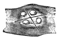
|
|
Incision for a molar (White). |
The propriety of resort to the lancet for cure of systemic
disturbances by obviating the source of local irritation is one which
deserves consideration. It can only be decided upon the indications
presented by the individual case. The knife is not a panacea for all
the disorders of childhood occurring during dentition, and its
indiscriminate use is to be discountenanced. Nevertheless, there can
be no doubt that engorged and inflamed gums demand incision for their
relief, on the same general principles of surgery applicable to
similar conditions elsewhere. Where it is probable that systemic
disease, even if not solely caused, is aggravated by the irritation
and pain of a tooth unable to make its way to the surface unaided, it
is clearly the duty of the physician to give his little patient that
modicum of relief, if not of cure, which will be afforded by a proper
incision of the
gum. It will not do merely to score the gums,
but cuts should be made deep enough to reach the presenting surface
and extend even beyond its boundaries. The developing enamel cannot be
injured unless undue force be exerted. The best instrument to employ
is a curved double-edged bistoury, so wrapped as to prevent injury to
tongue, cheek, or lips. The child should be firmly held by another
person, and in such a position that the parts may be well illuminated.
The jaws can be separated by the operator's left hand, and the fingers
so disposed as to protect the tongue and lips. Sometimes the insertion
of a small cork between the jaws will be of advantage. The cuts should
be made with special reference to the form of the presenting tooth.
James W. White79 recommends for the incisors and cuspids a division
of the gum in the line of the arch; for the molars a crucial incision,
thus X, the centre of the crown as near as can be determined
indicating the point of decussation. A cuspid partially erupted needs
severance of the fibrous ring on the anterior and posterior as well as
on the lateral surfaces (Fig. 17). All the cups of a molar may have
erupted, and yet strong fibrous bands maintain a decided resistance.
In this case White thinks that all the boundaries of the tooth should
be traced by the lancet and all such bands completely severed, or else
a crucial incision, as in the figure (Fig. 18), should be made so as
to ensure perfect release from pressure. The only contraindication to
the use of the lancet, except in ulcerative odontitis, as before
mentioned, is the existence of a hemorrhagic diathesis.
79 Op. cit.
DEFINITION.—An acute inflammation of the tonsil or tonsils; or
inflammation of the tonsil or tonsils, with inflammation of the
peritonsillar connective tissue and of the palatine folds.
VARIETIES.—When the inflammatory process is confined to the mucous
membrane the disease is erythematous, superficial, or catarrhal
tonsillitis; when it involves the lacunæ it constitutes lacunal or
follicular tonsillitis; when it involves the gland as a whole it
constitutes parenchymatous, phlegmonous, or suppurative tonsillitis.
The two latter varieties may present in combination. When the
superficial inflammatory process is a vesicular one, eventually
sheathing the surface of the organ in whole or in part with a
membranous envelope, it constitutes herpetic or membranous
tonsillitis. This variety may complicate superficial tonsillitis. When
the inflammation of the tonsil, usually superficial, is due to the
presence of a cryptogamic growth, it is a mycotic or parasitic
tonsillitis, benign or malignant (diphtheria), as may be. When the
inflammation of the tonsil is due to rheumatism, it is rheumatic or
constitutional tonsillitis.
SYNONYMS.—Inflammation of the tonsils, Amygdalitis, Quinsy, Angina
tonsillaris, Angina phlegmonosa, Phlegmonous sore throat, Cynanche
tonsillaris. Lacunal tonsillitis is more generally known as
folliculous tonsillitis (tonsillitis follicularis). Common membranous
or pseudo-membranous sore throat (angina membranosa communis) is used
as a synonym for herpetic or membranous tonsillitis (tonsillitis
herpetica seu membranosa). Mycosis tonsillaris is a synonym for
mycotic tonsillitis (tonsillitis mycotica benigna or tonsillitis
parasitica). The tonsillitis of diphtheria is sometimes termed
tonsillitis diphtheritica, tonsillitis mycotica maligna; that of
rheumatism, tonsillitis rheumatica, angina rheumatica, rheumatic sore throat.
HISTORY.—Tonsillitis was described by Hippocrates. Of recent authors,
Sauvages, Cullen, Louis for researches on the effects of
blood-letting; Bell on the specific value of guaiacum; Velpeau as to
the use of powdered alum and nitrate of silver; Bourgeoise on the use
of tartar emetic; Maingault on paralytic sequelæ; Hering on mycosis;
and the authors of the various encyclopædias and dictionaries,—may be
mentioned as chief among the numerous observers whose contributions
have been of most value. The bibliographical references appended to
the compilations
last cited will guide the student in gaining
access to the more important special observations of anomalous cases.
ETIOLOGY.—Predisposing and Exciting Causes.—Tonsillitis may be
idiopathic, deuteropathic or symptomatic, or traumatic.
The predisposing cause of idiopathic tonsillitis is usually diathetic,
and is associated with congenital or inherited vulnerability of the
organ. Of diathetic causes, scrofula is undoubtedly the most
provocative, but even rheumatism and gout are likewise so considered,
though in a far more limited degree. Acute articular rheumatism is, in
fact, sometimes preceded by rather a sharp attack of tonsillitis
(rheumatic tonsillitis), which subsides spontaneously in a very few
days, sometimes within one day, sometimes suddenly and synchronously
with the onset of the ordinary manifestations of rheumatism, though
the latter are often slight and transient, as if the force of the
attack had been spent on the tonsils. Tonsillitis, non-specific in
character, is apt to be prevalent during epidemics of scarlatina,
diphtheria, rubeola, and variola. Membranous tonsillitis is common
before and after epidemics of diphtheria. Epidemics of tonsillitis
have been recorded, but in the face of their extreme rarity it becomes
questionable whether they were not extensive examples of the
proclivity just alluded to.
Tonsillitis is more frequent in individuals with chronically diseased
tonsils than in individuals in whom these glands are healthy. Such
individuals, too, are more liable to recurrences; and such recurrences
often follow very slight provocations.
Idiopathic tonsillitis is rare in infancy. At the period of eruption
of the permanent teeth it is much more liable to occur than before
that period, and the liability increases progressively until the
second dentition has been completed. It is most frequent during the
decennium immediately following puberty—that is to say, in
adolescents and young adults—or from the fifteenth to the
twenty-fifth year. The disposition or predisposition to renewed
attacks continues marked during the decennium immediately succeeding;
after which attacks are more and more infrequent. Certain anatomical
changes occurring in the tonsils, as the rule about the fortieth year,
may diminish their proclivity to inflammation. Nevertheless, the
disease occasionally occurs in advanced age.1
1 Solomon Solis Cohen, "Abscess of the Tonsil in an
Octogenarian," Med. News, Philada., Feb. 16, 1884, p. 186.
Deuteropathic tonsillitis is quite frequent in infancy, being excited
by the infection of scarlet fever, diphtheria, measles, and small-pox,
as discussed under these headings respectively. Under similar
circumstances it occurs in the adolescent and the adult likewise. It
is also produced in carcinoma and sarcoma of the tonsil.
Rheumatic tonsillitis, a deuteropathic variety, is most prevalent
during atmospheric changes.
Herpetic tonsillitis, often a deuteropathic variety, seems sometimes
of nervous origin exclusively. It is sometimes traceable to defective
drainage. It is sometimes prevalent during epidemics of diphtheria,
when its membranous character renders it extremely liable to be
mistaken for the tonsillitis of diphtheria.
Traumatic tonsillitis occurs occasionally. The causes are—inspiration
of irritant gases, the deglutition of chemically acrid substances, the
accumulation
of calcareous concretions in the crypts or in the
lacunæ, direct and indirect gunshot and other wounds, the impaction of
fish-bones, fragments of toothpicks, cherry-stones, and other foreign
bodies, and the like.
Mycotic tonsillitis is due to the development of a cryptogam upon
tonsils probably already in a state of catarrhal inflammation in
individuals with health impaired by previous disease or unfavorable
hygienic influences.
SYMPTOMATOLOGY, COURSE, DURATION,
TERMINATIONS, COMPLICATIONS, AND
SEQUELÆ.—The onset of tonsillitis, sometimes preceded by headache and
general malaise, is often accompanied by a chill, pyrexia following
within twenty-four hours. The temperature may reach 105° F., being at
its maximum, as a rule, about the third day. It is rarely below 101°
F. The pulse is accelerated to 120 beats per minute. Simultaneously
with the constitutional symptoms local distress is usually manifested,
but either set of disturbances may precede the other by several hours
or by an entire day. Heat and soreness of the throat are early
complained of, gradually increasing in severity to actual pain. The
pain may become intense, especially during deglutition. When the
posterior palatine fold is put upon the stretch, additional pain is
referred to the ear, for this fold encloses the staphylo-salpingeus
muscle, which runs from the palate to the pharyngeal orifice of the
Eustachian tube. This pain in the ear, sometimes the principal cause
of complaint, is often premonitory of suppuration. Noises in the ears
on the one hand, and impairment of hearing on the other, often attend
extension of the inflammation in this direction, the enlarged tonsil
sometimes pressing the posterior palatine fold against the pharyngeal
orifice of the Eustachian tube.
On inspecting the throat early in the disease, one of the tonsils will
be seen to be swollen into an irregularly tumid, much-inflamed mass,
usually of a vivid red color. Occasionally both tonsils are involved
simultaneously, but this is far less frequent than involvement of the
second tonsil a few days later or after subsidence of the process in
its fellow. In many instances the inflammation affects one tonsil only.
The inflammatory process is seldom confined to the tonsil. All the
structures of the throat, even to the base of the tongue, are often
involved, and it is rarely indeed that the anterior palatine fold,
distended over the surface of the tumefied gland, escapes
inflammation. It is this stretching of the anterior palatine fold
which occasions much of the exquisite pain that forms so prominent a
subjective symptom in severe cases. The soft palate, hanging forward
in the cavity of the pharynx, is often inflamed or intensely
congested, and the uvula tumefied, elongated, and oedematous. It may
be flaccid upon the posterior part of the tongue or hang immediately
over the epiglottis or upon it, and induce painful and tiresome
efforts at deglutition and expectoration to relieve the consequent
titillation. Sometimes it adheres by viscid secretion to the side of
the swollen tonsil.
The inflamed tonsil or the tonsil and its coverings project far into
the cavity of the pharynx, often as far as the middle line, touching
its fellow when both are involved, so that ulceration sometimes ensues
at the points of contact. The posterior surface of the anterior
palatine fold sometimes becomes unfolded, as it were, in the
tumefaction of the gland, and
remains stretched over it in a
thin continuous layer without any line of demarcation. The swollen
palate projects upon the enlarged tonsil like a shelf, from which
depends the uvula, the latter being often oedematous, usually
anteriorly, but sometimes posteriorly.
Inflammation of the connective tissue about the lower jaw, especially
when at or near the articulation, often renders it difficult or even
impracticable to open the mouth sufficiently to permit direct
inspection of the parts; but it is rarely that sufficient space cannot
be made to allow partial protrusion of the tongue on the one hand, and
the introduction of a fore finger for exploratory purposes on the
other, though both of these acts are sometimes impossible. The
tumefaction of the parts impairs the freedom and ease of deglutition,
which may become so painful as to prevent the swallowing of the
saliva, which then may dribble from the mouth.
The pain experienced in swallowing is often manifested by convulsive
action of the muscles of deglutition and of the muscles of the face.
The swollen tonsils prevent the soft palate from being applied to the
surface of the pharynx, as usual in deglutition; and as the upper or
retro-nasal portion of the pharynx thus fails to be shut off from the
lower oesophageal portion, liquids are often forced up into the nasal
passages posteriorly, and are regurgitated through the nostrils, thus
rendering it impracticable, for the time, to slake thirst or to
swallow liquid nourishment.
At first sensations of dryness and pastiness in the throat are
complained of, but in a few hours these symptoms become relieved by a
more copious secretion of mucus or mucus and saliva. This secretion
soon becomes viscid, and so adherent to the parts as to be detached
only with difficulty, thus causing harassing efforts for its
dislodgment by hawking and expectoration, or equally distressing
efforts to swallow it. Should the inflammatory process extend to the
salivary glands, as is not infrequently the case, secondary ptyalism
often results, with increased distress from this source, and the
patient lies or sits with his head inclined upon the diseased or most
diseased side to favor the uninterrupted flow of saliva from the mouth.
Extension of the inflammatory process to the submaxillary glands, or
to the parotid, or to the connective tissue surrounding them, is
indicated by tumefaction externally, which is often exquisitively
sensitive to pressure.
The timbre or quality of the voice is often impaired in a peculiar
manner by the tumefaction of the throat and the immobility of the soft
palate. The voice is thick, throaty, or guttural, having a
characteristic harsh, rasping aspiration in enunciation, while
articulation is much impeded by impairment in the movements of the
jaw, palate, tongue, and lips. At times it is also painful. Speech is
sometimes indistinguishable or impossible, and the voice may even
become suppressed, so that signs and writing remain the sole means of communication.
Impairment of respiration, at least to any considerable degree, does
not occur, unless both tonsils are involved and swollen to an intense
degree—conditions under which dyspnoea may become pronounced, severe,
and even urgent, and suffocation become imminent. Painful respiration
is not uncommon in rheumatic tonsillitis.
The fever is sthenic in type. There are often severe aching pains in
the
limbs. Headache, restlessness, insomnia, nausea, and even
vomiting, may occur. The tongue is heavily coated, the breath is
fetid, appetite is impaired, and the bowels are constipated. The urine
is diminished in quantity, high-colored, and of high specific gravity.
It usually shows slight increase of urea and great diminution of
chlorides. Albuminuria occurs in rare instances.
The symptoms are proportionate to the severity of the attack. A first
attack is usually much severer than subsequent ones, and suppurative
cases more severe than those terminating by resolution. Resolution is
the usual termination, and the parts are restored to a normal
condition at the end of ten to fourteen days, sometimes earlier; in
exceptional cases not until three or four weeks. Sometimes permanent
hypertrophy of the tonsil remains.
Where the inflammatory process fails to subside, suddenly at the end
of five or six days, or a little later, or not until ten days to a
fortnight have passed, slight rigors supervene, announcing
suppuration, and the local distress is very great, with pulsation and
lancinating pains in the tonsils, until all at once the abscess bursts
and its contents are discharged with immediate relief. Sometimes the
pus or much of it is involuntarily swallowed; sometimes it is
expectorated. In exceptional instances the pus has escaped into the
larynx and suffocated the patient, usually during sleep.2 In rare
instances the abscess, having burrowed beneath the pharyngeal muscles,
may open at the external angle of the jaw or behind the sterno-mastoid
muscle. It may discharge into the epiglotto-pharyngeal fold, and
thence reach and distend the epiglottis. It has been known to descend
along the planes of connective tissue into the mediastinum or into the
lungs. Even ulceration into the maxillary and carotid arteries has
occurred, usually with fatal result, occasionally with an opportunity
to save life by ligating the carotid (Erhmann).3
2 Stokes, Med. Times and Gaz., Aug. 29, 1874, p. 251;
Littlejohn, Brit. Med. Journ., Jan. 2, 1875, p. 16.
3 Gaz. méd., Paris, 1878, p. 42.
The most frequent point of spontaneous rupture externally is at the
upper portion of the gland anteriorly, just beneath the anterior
palatine fold. Sometimes internal rupture occurs into the lacunæ.
Termination by gangrene is exceptional, and is confined to individuals
with debilitated constitutions. It is much less frequent than
formerly—as a result, perhaps, of better methods of treatment.
Metastasis is one of the methods of termination as to joints or
muscles in rheumatic tonsillitis—to lungs, brain, or
gastro-intestinal tract—as formerly occurred with much more frequency
under direct depletory treatment.
In rare cases extension of the inflammation occurs to the epiglottis,
even to the larynx, and the laryngitis may be so severe as to threaten
life from the occurrence of oedema. Diffuse inflammation of the
retro-pharyngeal connective tissue or of the connective tissue of the
neck may constitute an unpleasant complication of the disease.
In a few instances paralysis of the palate occurs as a sequel of
tonsillitis, and in exceptional cases the paralysis may also affect
the arytenoid muscles of the larynx, and even the accommodator muscles
of the eyes.
PATHOLOGY AND MORBID ANATOMY.—Tonsillitis is almost
always
associated with inflammation of the tissues surrounding the tonsil and
those contiguous to it, even in the mildest and most frequent
manifestations of the affection. Thus, inflammation of the palate
(staphyllitis) and uvula, and even of the pharynx (pharyngitis), are
anatomically included with tonsillitis in angina or sore throat.
The mildest form of the malady is a catarrhal inflammation of the
mucous membrane covering the gland, and does not extend along the
lacunæ which dip inward from the surface and divaricate toward the
interior of the organ. It is termed catarrhal tonsillitis, and, as has
been intimated, is almost always associated with catarrhal sore
throat. It is attributed to hyperæmia, with passive engorgement of the
vessels, following retrocession of blood from the cutaneous surface
after undue exposure to cold and moisture. A severer form of the
malady involves the lacunæ in addition—several or all of them. This
should be termed lacunar tonsillitis, as suggested by Wagner.
Primarily, at least, it does not involve the follicles of the tonsils
which open into the lacunæ, and is therefore incorrectly denominated
follicular tonsillitis, although it is most generally so described.
The lacunæ are involutions of the mucous membrane, and in health
furnish a slightly turbid mucoid secretion which serves to lubricate
the parts and, as is generally believed, to facilitate deglutition.
When the lacunæ are inflamed these products become pent up in them to
a certain extent, accumulate, and project in part at their orifices in
turbid creamy or curdy masses, plastered over the parts when thin in
consistence, or tightly imbedded when thick or desiccated. These
masses are usually white, but sometimes, owing to various admixtures,
they are more or less yellowish or gray or brown. They consist of
epithelium chiefly, with more or less pus and accumulation of cells
similar to those of which the follicles are composed—whether from
follicles which have become distended by proliferation of their
constituents, and have then burst, is not known. This epithelium has
often undergone fatty degeneration in part. Cholesterin is an
occasional constituent, and swarms of micrococci and bacteria abound
when the masses are not recent, especially if the inflammation is
occurring in a tonsil long the seat of chronic disease of the lacunæ.
The tonsil itself is moderately swollen and its mucous membrane
hyperæmic. If the parenchyma of the tonsil be involved likewise, as
often occurs, the swelling will be much greater, so that the gland
will project a considerable distance beyond the margins of the
palatine folds. When a hypertrophied tonsil is the seat of the
inflammation the tumefaction will be much greater than when the
inflamed tonsil has been normal.
Acute inflammation of the palatine folds often coexists, especially of
the anterior fold. The soft palate may also be engaged in the morbid
process, which may involve the uvula likewise. Pharyngitis is an
occasional accompaniment, and stomatitis quite a rare one.
Lacunar tonsillitis sometimes subsides by spontaneous evacuation of
the pent-up contents of secretion and desquamation, the parts
returning to their normal condition. More frequently a desiccation of
some of these products ensues, with permanent chronic inflammation.
Decomposition then often takes place, fouling the breath by the escape
of the gases. Butyric acid has been recognized as one of the most
prominent of these fetid gases.
Calcareous change occurs in
these contents of the lacunæ in some instances. Lacunar or follicular
tonsillitis is often associated with severe inflammation of the
peritonsillar connective tissue and the contiguous palatine fold, with
serous or cellular infiltration into these structures almost always
terminating in suppuration. This form of tonsillitis is very frequent,
and is often confounded with parenchymatous tonsillitis or with
tonsillar abscess. In these cases the abscess is in the peritonsillar
tissue or in the posterior leaflet of the anterior palatine fold. It
bursts above the tonsil at the angle between the two folds in the
greater number of cases. In some instances the suppurative
inflammation affects the anterior surface of the posterior palatine
fold, sometimes contiguously to the antero-tonsillar abscess,
sometimes independently, constituting a retro-tonsillar abscess. The
follicles in the posterior palatine fold are sometimes involved, the
thickened anterior surface of this structure becoming studded with
small projections the size of ordinary pinheads or larger, distended
with whitish-yellow contents.
In another class of cases of tonsillitis the inflammatory process may
be chiefly parenchymatous, for rarely is it wholly so. That is to say,
it may involve the glandular structure of the organ wholly or in main
part. The disease is then an adenitis, an inflammation of
gland-tissue—tonsillitis per se. It is associated with superficial
inflammation of the surrounding mucous membrane, secondarily if not
primarily, and often with inflammation of the lacunæ. In many
instances the parenchymatous inflammation is a direct extension of the
lacunar inflammation.
Parenchymatous tonsillitis may subside by resolution, or, as is quite
frequent, terminate by suppuration. A number of small abscesses may be
formed, which usually become confluent and rarely remain discrete.
Sometimes a single large abscess is formed. The confluent abscess may
discharge by several points. It is always associated with a severe
inflammation of the palatine folds and palate, especially the anterior
fold; sometimes of the adjoining half of the soft palate and the
uvula; sometimes of the entire velum and uvula. Sometimes these parts
become oedematous; sometimes suppuration ensues. Severe pharyngitis is
not uncommon. Glossitis, involving the posterior portion of the tongue
especially or exclusively, is an occasional accompaniment of
parenchymatous tonsillitis. (See GLOSSITIS.) Occasionally oedema takes
place in the epiglottis and upper margin of the larynx.
The character of the secretions varies. Sometimes these are semifluid;
sometimes soft, caseous, or pultaceous; sometimes fibrinous and
arranged in pseudo-membranes; sometimes hemorrhagic; sometimes moist
and viscid, sometimes very dry; often adherent, and always containing
cryptogams (leptothrix, Oïdium albicans, bacteria, and micrococci).
Collections of caseous products accumulate not only in the crypts of
the tonsils and in their overlying mucous membrane, but likewise in
the follicles of the palatine folds below the tonsil, and thence
toward the base of the tongue.
The submaxillary glands often undergo engorgement, and become so
tender that external manipulation is painful, and sometimes they
undergo suppuration. The tumefaction due to the swollen glands and
infiltrated connective tissue around it is frequently incorrectly
referred to the tonsil itself, rather than to the accompanying
inflamed palate, with the lymphatics
of which these glands are
in more direct anatomical connection. The tonsil is at a considerable
distance from the inflamed glands, and cannot be felt from the
exterior except under unusual circumstances.
Herpetic tonsillitis is a rare form of inflammation of the tonsil, or
rather of its investing mucous membrane, characterized by the eruption
of herpetic vesicles on its surface. The vesicles soon undergo
rupture, and the resulting ulcers coalesce and become covered with a
fibrinous exudation. The disease is usually associated with similar
vesicles upon the palatine folds and upon the soft palate, and
exceptionally with vesicles on the pharynx. (See Herpetic
Pharyngitis.) It is by some fortuitous circumstance only that it is
observed in the vesicular stage. It is confined to one side of the
throat in most instances, but may be bilateral also.
Mycosis tonsillaris has been described by a few observers. B.
Fraenkel4 has recorded three
cases, E. Fraenkel5 one,
and Bayer6
two. In these cases the disease was not confined to the tonsils, but
implicated the calciform papillæ of the tongue also, and one of
Bayer's cases some pharyngeal follicles in addition. E. Fraenkel's
case was in a male, and occupied but the right tonsil and base of
tongue. The white masses in this case were formed of spores and
filaments (Bacillus fasciculatus, Sadebeck), which are described as
penetrating some millimeters into the gland-tissue. These masses were
tenacious, and were reproduced rapidly after removal. In Bayer's
cases, both females, the same microphyte was recognized. In a female
patient observed at the Philadelphia Polyclinic7 this affection
followed rheumatic tonsillitis, diphtheria being prevalent near her
residence, which was in a very unsalubrious locality. The deposit,
confined to the left tonsil, was so firmly adherent to the mucous
membrane that the implicated portion had to be torn away to get rid of
the growth, which was twice reproduced. The fungus was in its mycelial
state, a few spores and conidiferous filaments being recognized microscopically.
4 Berlin. klin. Woch., 1873, S. 94; ibid., 1880, No.
18.
5 Zeitschrift für klin. Med., iv., 1882.
6 Rev. mens. de Laryngologie, etc., Nov., 1882, p. 329.
7 S. Solis-Cohen, The Polyclinic, March, 1884, p. 133.
Quite recently, and since the above was written, the results of an
elaborate study of this affection by Theodor Hering of Warsaw have
been published in a paper entitled "Pharynxmycosis leptothricia,"8
read before the Society of German Naturalists and Physicians. The
author collates fourteen cases, six of which were observed by himself.
He claims that the microphyte is simply the Leptothrix buccalis.
8 Zeitschrift für klinische Medicin, Bd. vii. H. 4,
1884.
The local subjective symptoms as collated by Hering vary from the
merest sense of discomfort in chronic cases to intense pain,
difficulty in speech and in deglutition, and various grades of cough
in acute ones. In some cases they are altogether wanting.
Constitutional disturbance may be entirely absent or may be presented
in various febrile or sub-febrile manifestations.
DIAGNOSIS.—The history of the attack, the appearances described, and
the symptoms narrated should ordinarily suffice for a correct
diagnosis. Still, mistakes do occur. An unsuspected tumor of the
tonsil observed for the first time during an ordinary sore throat
might be taken for an inflamed tonsil, but the progress of the case
would soon lead to its due recognition.
While tonsillitis is
infrequent after the fourth decennium, it occasionally occurs late in
life, and has been observed even in the ninth decennium; and reserve
is proper as to the cause of enlarged tonsils in the sore throats of
those advanced in life.
The deposit in follicular or lacunar tonsillitis or angina is pulpy
and not membraniform. It can be wiped from the surface with a fragment
of sponge, and does not tear from the surface in strips, as is the
case with the pseudo-membrane of diphtheria or of common membranous
sore throat. There is no abrasion of the mucous membrane beneath the
deposit. The patches are more prominent, usually more circumscribed,
and dip down into the lacunæ, or rather project from the crypts upon
the surface of the tonsil. In its physical aspect the deposit more
closely resembles that observed in the sore throats accompanying
cachectic conditions, as in chronic tuberculosis, advanced syphilis,
some forms of scarlatina, typhus and typhoid fever, extreme old age
(agine pultacée, Fr.; cachectic angina); but the existence of previous
constitutional disease and actual debility should prevent the mistake
in diagnosis. In susceptible subjects the oncoming of an attack of
rheumatic tonsillitis may often be inferred, previous to the
manifestation of local symptoms, from the existence of otherwise
inexplicable odynphagia, the pain being especially intense upon
attempts to swallow saliva. Sometimes laryngoscopic inspection at this
early stage of the disease will reveal vivid redness of the mucous
membrane in the neighborhood of the crico-arytenoid articulations.9
The value of this early diagnosis lies in the opportunity it affords
to try abortive treatment.
9 S. Solis Cohen, The Medical News, Aug. 11, 1883, p.
146.
PROGNOSIS.—The prognosis of catarrhal tonsillitis is almost
invariably favorable, except under very obviously unfavorable
conditions, the inflammatory process subsiding spontaneously within a
few days. It is favorable, as a rule, in phlegmonous tonsillitis
subsiding within ten or twelve days in most instances, even though all
the stages be completed to suppuration and discharge of the abscess.
Sometimes two or three weeks are consumed in the process. A certain
amount of reserve is requisite, nevertheless, in severe cases, in view
of the possible complications which may prevent recovery. If both
tonsils are affected to such an extent as to interfere seriously with
respiration, death by suffocation may ensue should the obstruction be
not relieved by excision of portions of the swollen glands or an
artificial opening be not made into the air-passage. Suppuration may
perforate the internal carotid or the external maxillary artery and
produce sudden fatal hemorrhage. The remembrance of such occurrences
should screen a surgeon from the imputation of carelessness should he
be unfortunate enough to incise an abscess under similar conditions.
Some cases are on record of fatal hemorrhage but a short period before
a proposed operation could have been performed.
Suffocation has ensued from discharge of the abscess into the
air-passage, usually during sleep; but it has occurred even during the
moment of speaking (Stokes).10 Such
results are accidental and exceptional.
10 Med. Times and Gaz., Aug. 29, 1874, p. 251.
Recurrences are frequent, especially in scrofulous subjects, and such
recurrences are apt to result in permanent hypertrophy and induration.
TREATMENT.—Mild cases of tonsillitis require no treatment except to
keep
the patient protected from exposure to abrupt changes of
temperature. The course of the affection both in mild cases and in
severe ones may often be materially shortened by prompt resort to the
use of guaiacum, both internally and topically. A gargle containing an
ounce each of ammoniated tincture of guaiacum and compound tincture of
cinchona to the pint, with the addition of three ounces of clarified
honey, and saturated with potassium chlorate (twenty grains to the
ounce), may be used, a drachm at a time, every two hours, hour, or
half hour, according to the urgency of the symptoms, and may likewise
be administered internally in drachm doses for an adult every two or
more hours. The beneficial effects will often be manifested within
less than twelve hours. Pellets of ice held in the mouth from time to
time often relieve pain and repress inflammation. Sodium bicarbonate
locally, in powder, affords great relief in some instances. In the
presence of marked pyrexia tincture of aconite may be given in drop
doses every hour until an impression has been made upon the heart,
when its continuance at intervals of four or more hours will be a
matter for consideration.
Guaiacum and aconite may be given with equal benefit in any form
preferred by the prescriber. At the same time saline laxatives may be
required from time to time. Regulation of the diet is often necessary.
When the tonsils are very much swollen, gargling of all kinds becomes
too painful, and therefore sprays of sedative and emollient mixtures
are to be substituted, or steam from water impregnated with volatile
substances, as benzoin, paregoric, hops, chamomile, and sage. When the
cervical glands are swollen, continuous hot and moist applications
externally afford great relief. If the suffering from the inflamed
tonsil be intense, scarification should be practised and the bleeding
be encouraged by warm water. When suppuration exists, the abscess
should be promptly evacuated by incision at the most prominent
accessible point.
Special symptoms require appropriate management on general principles.
The pain in swallowing can often be diminished by pulling on the lobe
of the ear at the moment of deglutition (Grewcock).11 In debilitated
subjects, or during epidemics of diphtheria, quinia, iron, and
supporting measures are indicated.
11 Lancet, Nov., 1882, N.Y. reprint, p. 399.
The rheumatic cases are best treated with sodium salicylate, ten to
fifteen grains every hour or two until relieved. Instituted in the
formative stage above alluded to, this treatment frequently seems to
be veritably abortive, especially when preceded by a full dose of an
alkaline purgative—say one ounce of Rochelle salts. Oil of gaultheria
may be used in small doses as an agreeable flavor to the mixture, or
in doses of ten to twenty minims, well diluted, as an adjuvant to the
salicylate, or even as a substitute for it. After subsidence of the
acute symptoms cinchonidine salicylate may be continued for a few days
in appropriate doses.
Herpetic tonsillitis requires the ordinary treatment for erythematous
tonsillitis, with additional topical treatment by sprays of alkaline
solutions, such as sodium borate or bicarbonate, five grains to the
ounce, or lime-water. Internally, small doses of mercuric chloride
will be of service, the dose varying, according to the age and size of
the patient, from one-forty-eighth to one-sixteenth of a grain every
two hours, until the stomach shows signs of irritation therefrom. In
cases of doubt as to diagnosis
from diphtheria the treatment for
diphtheria will be indicated as the safer measure. Here, again, the
mercuric chloride is sometimes equally valuable.
Mycosis of the tonsil does not seem amenable to medicinal treatment.
Thorough removal of the fungus with forceps or sharp spoons is
required, even though mucous membrane be detached with it. When this
is impracticable, ablation of the tonsil may be necessary. Raw or cut
surfaces left by any of these manipulations should be subjected to
thorough cauterization, electric cauterization being the most feasible method.
DEFINITION.—An acute inflammation of the mucous membrane of the
pharynx, whether implicating the glandular structures or not, and
usually associated with inflammation of contiguous structures.1
Acute pharyngitis may be catarrhal or erythematous, phlegmonous or
suppurative, ulcerative, herpetic or membranous, gangrenous, and erysipelatous.
1 In deference to the plan suggested by the editor of this
work, separate articles have been prepared under the heads of
Pharyngitis and Tonsillitis respectively. The two processes, however,
are so frequently associated that they should be studied together, the
more that both of them are likewise associated with extensions of the
inflammatory process to the palate, palatine folds, base of the
tongue, and other contiguous structures. The writer has always
preferred to describe these diseases under the head of sore throat,
which does not presuppose any limitation to individual anatomical
structures.
SYNONYMS.—Sore throat; Angina.
ETIOLOGY.—Acute pharyngitis may be idiopathic, deuteropathic,
traumatic, toxic, or parasitic. The predisposing cause may be
diathetic, as scrofula, rheumatism, gout, and syphilis; it may be a
depression of the vital powers from any cause, such as continued
exposure to foul air or impure water, improper diet or sedentary
occupations. There exists in some individuals a predisposition to
"catching cold," independent of any cachexia. Pharyngitis may occur at
any age, but is more frequent in the young. One attack increases
subsequent liability to the disease.
The exciting cause is usually exposure to cold and damp. Hence the
disease is more frequent at the seasons when these conditions prevail
or when sudden changes of temperature are taking place. Sudden
chilling of the body when overheated may occasion it in warm weather;
for instance, a plunge into the ocean while covered with perspiration.
The ulcerative variety, when not due to syphilis or tuberculosis, is
usually of septic origin, and is apt to occur in the debilitated
especially. The gangrenous form, which is rare, results from profound
blood-poisoning. The herpetic or membranous variety may be due to
disturbance of the trophic nervous system, and has been attributed to
mental emotion (Feron), to uterine disturbances (Bertholle), to the
contact of irritating substances and to miasmatic or fetid exhalations
(Peter). It prevails principally during epidemics of diphtheria or of
scarlet fever, and may be of cryptogamic origin. The cryptogam of
thrush is sometimes developed on the mucous membrane of the pharynx,
either primitively or as an extension of the disease from the oral
cavity. Certain conditions of the
atmosphere give rise at times
to so-called epidemic pharyngitis. Paludal or malarial pharyngitis may
arise from the same causes as malarial fevers. Pharyngitis occurs in
the various exanthemata as an essential part of the morbid process,
and is always more or less prevalent during epidemics of measles or
scarlet fever. It occurs not rarely in typhoid fever, and is an
occasional complication of pneumonia, rheumatism, herpes, pemphigus,
and other acute affections. It is one of the complications of facial
erysipelas, but erysipelatous pharyngitis may occur primarily.
Pharyngitis may be excited by the inhalation of deleterious solid,
fluid, and gaseous substances in the atmosphere which act mechanically
or chemically on the mucous membrane. Many drugs administered in
poisonous or even in medicinal doses may give rise to an attack of
inflammation of the pharynx; among them may be cited preparations of
mercury, antimony, iodine, arsenic, copper, lead, zinc, silver,
stramonium, belladonna, and most of the Solanaceæ. Traumatic
pharyngitis results from deglutition of boiling water or of acrid or
caustic substances; from inhalation of hot air, of steam, or of flame,
and is most usually associated with traumatic oesophagitis or with laryngitis.
PATHOLOGY AND MORBID ANATOMY.—Acute pharyngitis, as most commonly
encountered, is a simple erythematous inflammation of the mucous
membrane; the palate and tonsils being likewise involved. In most
instances there is simply an active hyperæmia which may subside in a
day or two. When more intense than this the mucous membrane of the
palate, tonsils, and pharynx becomes congested and swollen, uniformly
or in circumscribed areas. In some instances the submucous tissue of
the pharynx is greatly relaxed, and the mucous membrane lies upon the
substructure in thick folds. In others there is more or less oedema.
The mucous follicles, especially those of the posterior palatine
folds, are frequently swollen. There is an abnormal though not
excessive secretion of viscid mucus, clear or turbid. The uvula is
often swollen or distended with serum, and its mucous membrane is
relaxed. Sometimes it appears as though pasted to one of the folds of
the palate by viscid secretion. The posterior palatine folds may be
distended with serum, and their arched appearance thus become
obliterated. Resolution occurs gradually in some instances, quickly in others.
Phlegmonous pharyngitis exhibits a still higher grade of inflammation.
It involves the submucous structures as well as the mucous membrane,
including at times the fibrous sheaths of the muscles. It may, in
addition, involve the palate, the tonsils, the base of the tongue, and
contiguous structures. Suppuration is common, usually circumscribed,
but not infrequently diffuse in patients of enfeebled constitution.
One variety of the disease is essentially a deep-seated pharyngitis;
and this form almost always progresses to suppuration (suppurative
pharyngitis). The process becomes then, not infrequently, a diffuse
suppurative inflammation of the subpharyngeal connective tissue,
extending sometimes downward along the oesophagus, into which the pus
may be discharged by spontaneous rupture, with a result of permanent
stricture from irregular cicatrization. Sometimes the suppurative
process extends anteriorly beneath the cervical fascia, and the pus
may gravitate so as to occlude the air-passages, partly or completely,
by direct pressure; or in other instances the entrance of
the
larynx may become blocked by the tumefaction of the pharynx. When
phlegmonous pharyngitis is of traumatic origin, there will be more or
less destruction of the mucous membrane according to the nature of the
injury, whether accidental or designed, whether due to burn, scald,
inhalation of hot air or steam, or to deglutition of alkaline, acid,
or other corrosive substances. In these cases the morbid process is
rarely confined to the pharynx, but the larynx, the oesophagus, and
even the stomach, are liable to be involved. If regurgitation of hot
air or of caustic fluids takes place through the nasal passages, the
injury will of course involve those regions.
Ulcerative pharyngitis is a low form of inflammation present in sore
throat, probably dependent upon septicæmia. The tonsils are somewhat
congested and swollen, and one or more white superficial ulcers form
on their surface, or on the palate, or on the pharynx. These ulcers
are generally round or oval, and vary greatly in size. When two or
more ulcers exist, they exhibit no tendency to confluence. Healing
takes place rapidly, usually without leaving any traces of the lesion.
Membranous pharyngitis, or herpes of the pharynx, is one of the
infrequent phenomena of a not uncommon sore throat, which exhibits at
first a collection of small vesicles the size of millet-seeds or
larger, isolated here and there or clustered in groups on the palate
and uvula, less frequently on the tonsils. Herpes of the mouth and
lips sometimes coexists. These vesicles are surrounded by inflammatory
areolæ. Their contents are more or less turbid. In rare instances they
disappear without trace after a day or two. Usually they soon undergo
rupture, sometimes within a few hours, so that small ulcers are left,
which almost immediately become covered with a grayish-white
exudation. A number of patches will coalesce, forming limited sheets
of false membrane not unlike those of diphtheria. The disease is
usually confined to one side of the throat, the corresponding
submaxillary or cervical glands being affected moderately when at all
involved. The tonsil is swollen, and the mucous membrane of the palate
and the palatine folds is congested and often tumefied. There is an
abnormal secretion of viscid, ropy, turbid mucus. In a few days the
ulcers heal beneath the exudation, which becomes disintegrated and
detached, the inflammatory process subsiding by gradual resolution.
Sometimes the ulcers cicatrize without previous deposit of false
membrane. Occasionally there are at longer or shorter intervals
successive crops of vesicles, which may or may not undergo ulceration.
Gangrenous pharyngitis may supervene upon any form of pharyngitis, but
in the majority of instances its malignant character is inevitable
from the outset; so that some authors have even restricted the term
gangrenous to a form of sore throat characterized by primitive
gangrene of the pharyngeal mucous membrane originating independently
of any other malady. Whether an idiopathic disease, or whether it
follows scarlatina, measles, small-pox, dysentery, or enteric fever,
it is associated with that depraved condition of the system
denominated typhoid. At times it occurs in tuberculous phthisis. The
initial manifestations may be simply those of intense inflammation.
The tongue is covered with a dark creamy, pultaceous deposit
consisting of broken-down epithelium, pus-cells, bacteria, and
molecular débris, while similar masses are occasionally seen upon
other mucous surfaces of the mouth and throat. The tonsils,
palate, and pharynx are livid and swollen, and sometimes oedematous.
At an early period the tonsils, the palatine folds, and the posterior
wall of the pharynx become covered with dark, ashy-colored ulcers with
excavated edges. Sometimes these spots are black from the first, and
appear slightly elevated. These soon slough out with more or less of
the surrounding tissues, and the ulcers left are covered with sanious,
ichorous, fetid secretion. In some instances a delicate
pseudo-membrane has been found in the bed of the ulcer after death
(Mackenzie). The destructive process rapidly extends—sometimes to the
oesophagus in one direction or to the nares in the other. The larynx
is less frequently implicated; should it be attacked, oedema is liable
to occur. Occasionally the process is limited to the tonsil, and there
is no pharyngitis at all. Erosion of the blood-vessels may give rise
to fatal hemorrhage. In those instances where the gangrene is
circumscribed there are found, post-mortem, depressed oval or circular
patches from one-twentieth to one-half an inch in diameter, varying in
color from dark gray to absolute black. The edges are of a brownish
color and are perpendicular. The bundles of muscular fibre are laid
bare by destruction of the mucous membrane and submucous connective
tissue, but as a rule escape implication of their substance. Similar
patches have been noted in the epiglottis and the upper part of the
larynx as well as in the mouth and pharynx—in some cases, indeed, in
the trachea, the lungs, the oesophagus, the stomach, and the intestines.
Erysipelatous pharyngitis is usually an extension of erysipelas from
the facial integument, which may take place by the lips and mucous
membrane of the mouth, by the nasal fossæ, by the Eustachian tube from
the tympanum and external ear, or by the nasal fossæ from the
conjunctiva and eyelids through the lachrymal duct. When the disease
begins in the pharynx the order of communication may be reversed. The
pathological processes are the same as in cutaneous erysipelas. The
mucous membrane of the pharynx will be diffusely red or purplish and
shiny. Sometimes little bullæ are formed and become ruptured, leaving
a patch of softened whitish-yellow tissue, which is sometimes torn
from the surface beneath by the act of coughing or of deglutition. The
inability to swallow is not due to swelling of the tissues, but to
actual paresis of the muscles, probably from interstitial
infiltration, but perhaps from implication of their substance. The
cervical and submaxillary glands are rarely involved. Erysipelatous
pharyngitis usually terminates by resolution, desquamation of the
greater part of the epithelium of the mucous membrane often taking
place; but it may be followed by abscess or by gangrene. Extension may
take place to the larynx, and oedema may follow.
Exanthematous pharyngitis accompanies some cases of cutaneous
exanthemata. The pharyngitis of small-pox is occasioned by an eruption
upon the mucous membrane similar to that which appears on the skin.
Often in advance of the cutaneous eruption it occupies the inside of
the cheeks, the palate, uvula, and pharynx; sometimes the larynx as
well. Maturation occurs more rapidly than upon the skin, and there is
more or less purulent infiltration of the submucous connective
tissues. Ulceration of the larynx or trachea may ensue so severe in
character as to cause fatal termination by the local lesion.
In
measles an eruption similar to the cutaneous manifestation
occupies the air-tract from nostril to bronchi rather than the
food-passages. The Eustachian tubes may be involved, and the
inflammation is sometimes propagated along the lachrymal duct. The
throat may be affected a day or two before the external integument.
Small red points the size of a millet-seed or larger appear on the
palate, the tonsils, the posterior palatine folds, and the wall of the
pharynx. These disappear in a few days, though sometimes in bad cases
fibrinous exudation may accumulate. In other instances abscess or
ulceration takes place, chiefly in the larynx.
The pharyngitis of scarlatina develops a day or two prior to the
cutaneous eruption, the mucous membrane of the palate, tonsils, and
pharynx being deeply congested, uniformly or in patches, with slight
papulous elevations here and there. In the course of a day or two an
opalescent or milky deposit, consisting chiefly of detached epithelium
and viscid mucus, is observed on the swollen palate and tonsils. In
the anginose variety the hue of the inflamed structures is more dusky.
There is a pseudo-membranous deposit of a dirty-white, ash, or even
yellow color. It is not limited to the tonsils, but accumulates rather
on the palate, palatine folds, and posterior wall of the pharynx. The
mucous membrane beneath the patches is often ulcerated, and sometimes
gangrenous. There is much greater tumefaction of all the parts than in
simple scarlatina, the enlargement of the cervical and submaxillary
glands and the infiltration of contiguous connective tissue being so
great in some instances as to prevent the mouth from being opened. A
viscid and turbid secretion accumulates in the mouth. The nasal
secretions sometimes desiccate into firm crusts. Suppuration may
occur. Sometimes otitis media results from extension along the
Eustachian tube, and sometimes suppuration of the membrana tympani,
suppurative external otitis, or disease of the internal ear with
extension to the cerebrum. In malignant cases all the processes are
aggravated. Ulceration or gangrene soon ensues, the pseudo-membranous
deposit being dark, almost black, from extravasated blood. Oedema of
the uvula and soft palate is liable to occur, and if the larynx be
involved there may be oedema of the epiglottis and ary-epiglottic folds.
SYMPTOMATOLOGY.—Simple pharyngitis very often gives rise to but
little discomfort. There is usually more or less heat and dryness in
the parts, especially at first. There is some dysphagia, principally
from pain in swallowing, but in part from actual debility in the
muscles of deglutition. Hoarseness is not usual, and cough is
infrequent if there be no elongation of the uvula. Speech may be
embarrassed by difficulty of articulation. There is usually some
febrile movement, with acceleration of pulse and respiration. Some
cases exhibit more intense inflammatory action, with a corresponding
aggravation of the constitutional symptoms. The skin becomes markedly
heated, the body-temperature rises to 101° F. or higher, the pulse
reaching 100-120, in some instances 140, beats per minute, even in the adult.
In that variety known as rheumatic sore throat there will be in
addition pain and soreness in the neck, back, and limbs, often severe,
and increased by motion. There will be great accumulation of saliva in
the mouth because of the intense pain in swallowing it. Speech, and
even respiration, may become painful. These manifestations are
frequently followed
by the ordinary phenomena of acute articular
rheumatism, but they may subside in a few days, leaving only a general
feeling of muscular soreness or slight stiffness in one or more of the
joints. Sometimes a temporary torticollis follows.
In some cases of pharyngitis the cervical glands become swollen and
painful, but this is not common. In children the constitutional
disturbance is much greater than in adults. In malarial districts or
in subjects of malarial poisoning the manifestations may assume a
periodic character. Recovery takes place, as a rule, in from three to ten days.
In many instances the local phenomena are most prominently manifested
on one side of the throat. There will then exist great liability to
similar involvement of the other side after convalescence of a day or
two, and without care and avoidance of exposure the second attack may
be far more severe than the first.
The severe variety of pharyngitis denominated phlegmonous is often
ushered in with a decided chill, the phenomena of fever following
within twenty-four hours. The symptoms, both local and general, are of
much greater severity than in catarrhal pharyngitis, especially in
cases proceeding to suppuration.
Paralysis of the palate and other paralyses may follow either of the
forms of sore throat just described. Albuminuria sometimes results. In
extremely rare cases it is an accompaniment of the disease.
Superficial ulceration of the mucous membrane may occur in almost any
form of pharyngitis or of sore throat. Some authors have separated a
special form of ulcerative sore throat occurring in those enfeebled
from long exposure to unwholesome influences, such as nurses, hospital
attendants, etc., in whom the first symptom is pain in deglutition,
especially of saliva. The tongue is furred and the breath is
offensive. There is loss of appetite, with general lassitude,
feebleness of circulation, and more or less elevation of temperature.
Intense headache is often present. Under suitable treatment recovery
is rapid.
The ulcerative sore throats of syphilis and of tuberculosis require
separate consideration.
Common membranous pharyngitis frequently gives rise to but slight
symptoms, differing very little from those of other forms of
pharyngitis; but there may be high fever of sthenic or of asthenic
type, very often preceded by general malaise, sometimes by a decided
chill. The pain in deglutition and the local heat and dryness are
sometimes much greater than in the more ordinary forms of pharyngitis.
The distress may extend into the ear, sometimes to the nasal passages,
in rare instances to the larynx. The disease lasts for a week or ten
days, usually terminating in recovery. In occasional instances,
chiefly in children, it terminates fatally by apnoea from extension of
the membrane into the larynx. Paralytic sequelæ are not rare.
The advent of gangrenous pharyngitis is sometimes indicated by sthenic
phenomena, but usually from the first it is marked by extreme
prostration, comparable in some instances to the collapse of cholera.
There is a low type of fever. The pulse is feeble and infrequent. The
skin, especially of the extremities, is cold and blue. The eye is
glassy, the countenance haggard. The pain, as a rule, is not severe,
sensation being benumbed. The disease is often accompanied by an
irregular erythematous cutaneous
eruption. The secretions and
excretions escape by the mouth and nose, and they are extremely fetid.
There is indeed a peculiar odor, which once encountered can scarcely
be mistaken. If the lungs become affected there will be copious
hæmoptysis. In some cases the tendency to hemorrhage is general, blood
oozing or gushing simultaneously from lungs, bowels, nose, and mouth,
and sometimes extravasating beneath the skin. Sphacelus ultimately
takes place at the points of ecchymosis. Diarrhoea, abundant and
fetid, due to invasion of the alimentary tract, often sets in before
the close, and may be regarded as a sure precursor of death. Death
usually takes place from syncope, intelligence often remaining
unaffected to the last. When these cases recover a horrible amount of
deformity often remains to mark the ravages of the disease. During
cicatrization the positions of contiguous parts become very much
altered. The palate may become adherent by its sides, and by more or
less of its posterior surface, to the pharynx, sometimes resulting in
complete occlusion of the nasal portion of the pharynx.
The constitutional symptoms of erysipelas of the pharynx are those
that attend the usual manifestations of external erysipelas, the
febrile phenomena, epigastric pain, nausea, and so on, being increased
in severity. There will be great pain and difficulty of deglutition.
If there be serious oedema, symptoms of suffocation will occur.
Laryngitis will be indicated by pain referred to the larynx. The
duration of the disease varies from forty-eight hours to a week,
rarely longer. Death may occur within two or three days from oedema of
the larynx or from other causes frequently indiscernible. Resolution
usually takes place in those cases which recover. Occasionally abscess occurs.
DIAGNOSIS.—The diagnosis rests upon the conditions already described
under the heads of Pathology and Symptomatology. Under ordinary
circumstances it presents no difficulty, but during the prevalence of
epidemics of scarlatina or diphtheria even the mildest sore throat
demands careful attention and frequent inspection until the exclusion
of the graver maladies may be positively determined. The greatest
difficulty will present in cases of common membranous sore throat, for
it is sometimes impossible to make the differentiation from
diphtheria, especially as the vesicular stage is rarely seen.
Sometimes, it is said, it is possible to detect one or more of the
small ulcers left by the rupture of the vesicles; sometimes small
isolated spots of false membrane will by their transparency indicate
recent formation, and by their circular shape the previous existence
of a vesicle (Peter, cited by Mackenzie). The coexistence of cutaneous
herpes is corroborative of the diagnosis, but by no means an
infallible sign. It must not be forgotten in this connection that
membranous sore throat may predispose to an attack of diphtheria. In
gangrenous sore throat the grayish-black patches may be mistaken for
the pseudo-membranes of diphtheria, but their color is dark from the
outset, while in diphtheria they become dark only as the disease
progresses. They always represent actual death of the tissues, which
is not an essential lesion of diphtheria. Swelling of the cervical
glands is unusual. Finally, the characteristic odor of gangrene is
almost unmistakable.
PROGNOSIS.—The prognosis is favorable in catarrhal pharyngitis and in
the milder forms of the phlegmonous, non-specific, ulcerative, and
common
membranous varieties. It is unfavorable in intense
suppurative pharyngitis, though cases often get well. In gangrenous
pharyngitis the prognosis is extremely grave, but recovery is not
impossible. In traumatic pharyngitis the prognosis will of course
depend upon the nature and extent of the injury, being not unfavorable
if this be confined to the pharynx, though even in limited cases there
may be stenosis or other ill results from cicatrization. Erysipelatous
pharyngitis is of grave prognosis when the result of extension of the
disease from the face, but recovery is frequent when the pharyngeal
disease is primary.
TREATMENT.—The treatment of superficial pharyngitis is very simple.
Unless the case be so light that no special medicinal treatment seems
advisable, the patient should be confined to a bed or lounge to secure
rest, a light coverlid being thrown over the body to equalize the heat
of the surface. If a meal has recently been taken, a mild emetic is
often of service to empty the stomach and save the labors of
digestion. A gentle laxative or, if the patient be of costive habit, a
saline purge is indicated to facilitate the passage of matters already
in the intestinal canal. In cases of actual constipation a drastic
cathartic may be required. If there be considerable pain a small dose
of morphine may be advantageously combined with the aperient. If
frequent pulse or high temperature exist, especially in severe cases,
tincture of aconite, in doses of one or two drops every hour or two
hours at first, will be useful. As soon as any marked effect has been
produced the aconite may be discontinued or the intervals between
administrations lengthened. Locally, the free use of demulcent drinks,
and of pellets of ice when cold is agreeable, will relieve the pain in
the throat and sometimes repress excessive secretion. Cold compresses
to the neck anteriorly are often soothing, and sponging the entire
surface of the body with tepid water, acidulated or alcoholized, will
allay the intense heat of the skin. The diet should be light and
nutritious. Very often the emetic, rest, and regulation of diet will
constitute the entire treatment required.
When the local distress is very great, astringent lozenges (catechu,
krameria) may be allowed to dissolve in the mouth, or sprays of weak
solutions of alum or of carbolic acid may be propelled upon the mucous
membrane. Tannin, potassium chlorate, and cupric sulphate are often
used for this purpose. When the uvula is elongated or oedematous it is
often a constant source of irritation and discomfort. Scarification to
give vent to pent-up blood or puncture to allow the escape of effused
serum will afford prompt relief. Excision is never necessary.
In phlegmonous pharyngitis the treatment will necessarily be more
active. Here an early emetic is of great service. A saline laxative
may be administered every three or four hours for a day or two, each
dose containing a drop or two of the tincture of aconite, with the
addition of morphine if indicated by pain. Drop-doses of aconite at
more frequent intervals sometimes serve a better purpose. Inhalation
of steam, or of steam from water impregnated with hops,
chamomile-flowers, paregoric, compound tincture of benzoin, juice of
conium, or the aqueous extract of opium, belladonna, or conium, will
afford great relief, as will the frequent projection of sprays of warm
water, simple or slightly aromatized with cologne-water or with toilet
vinegar. Warm and moist applications externally are often very
soothing. Gargling entails too much pain to be of
service, but
medicated sprays may be used of aqueous solutions (twenty grains to
the ounce) of tannin, alum, zinc sulphate, or cupric sulphate, care
being taken to guard against the swallowing of any of these drugs.
Powders of alum, tannin, krameria, etc., diluted with liquorice,
acacia, bismuth, lycopodium, and the like, may be blown upon the
parts, and are often efficient. Sodium bicarbonate frequently affords
relief. The topical application of silver nitrate is rarely
practicable and generally unnecessary.
When the inflammatory process is of a higher grade and not likely to
yield to purely medicinal treatment, leeching or venesection may be
employed, but should not be resorted to without urgent reason. The
recognition of abscess is an indication for its immediate discharge by
incision or aspiration. In suppurative cases quinia and iron should be
given in large doses. The general treatment is like that of simple
sore throat. When liquid food cannot be swallowed, nourishment by
enema is requisite. Efforts at deglutition should be spared as much as
possible, and with this view medicines which can be administered by
inhalation, by enema, or by hypodermatic injection are to be preferred.
In pharyngeal sore throat, whether catarrhal or phlegmonous, depending
on rheumatic or gouty diathesis, salicylic acid or the salicylates
will prove useful, either alone or in conjunction with other measures.
The treatment of ulcerative pharyngitis is practically the same as
that recommended for phlegmonous pharyngitis. Antiseptic gargles may
be used locally, but as a rule the pain is so great that inhalations
of soothing vapors, as before recommended, will answer a better
purpose. When the process is very acute fragments of ice will be most
useful. Ice to the head will afford relief to pain. A little good
wine, with quinia and iron, comprises the medicinal measure requisite.
Gangrenous pharyngitis calls for the most active and supporting
treatment. Eggs, milk, cream, nutritious soups (up to the limits of
the patient's capacity for swallowing, and by enema when necessary),
quinia, tincture of the chloride of iron, and alcohol in large doses,
are indicated. Local treatment is of high importance. Agents to
destroy diseased tissue promptly and prevent the extension of the
gangrenous process, such as bromine, strong nitric or hydrochloric
acid, acid solution of mercuric nitrate, or caustic potassa, are to be
thoroughly applied, in the hope of exposing a healthy surface beneath
which will heal by granulation. When this treatment is unsuccessful or
too hazardous, as in cases where the blood-vessels are probably
involved, we can only palliate the symptoms by applying weak solutions
of acids and astringents, to which opium may be added, relying on
constitutional measures for restraining the destructive process.
Washes and sprays of potassium chlorate, eucalyptol, thymol, hydrogen
peroxide, etc., or the agents employed in common sore throat, are
often agreeable to the patient, and may be useful in restraining
fetor, but they have no direct therapeutic influence on the progress
of the disease. If the ulceration is extending into the vicinity of
the great vessels of the neck, measures for compression should be at
hand, in the use of which the nurse should be instructed, and
preparations be made to facilitate ligation of the carotid artery in
an emergency. Tracheotomy may be necessitated by oedema of the larynx.
The deformities resulting from gangrenous sore throat in cases that
recover usually require surgical treatment.
Traumatic
pharyngitis must be treated on general principles.
When due to contact of caustic or corrosive substances, an attempt may
be made to neutralize the effects by a chemical antidote, but the
physician is usually summoned too late to accomplish much in this
manner. Morphine should be given in full doses, hypodermatically.
Insufflations of morphine in powder, soothing inhalations, fragments
of ice in the mouth, cold compresses, and, where possible, oleaginous
drinks, are indicated to relieve topical distress. Rectal alimentation
should be resorted to where the difficulties of deglutition are at all
great. If symptoms of suffocation occur, tracheotomy must be
performed. The results of traumatic pharyngitis require treatment
according to their special indications.
Erysipelatous pharyngitis is to be treated by the administration, by
enema if necessary, of large doses of quinia, tincture of the chloride
of iron, brandy, and diffusible stimulants. Alimentation is to be kept
up by mouth or rectum, as may be necessary, with as much food as can
be given containing the most nutrition in the smallest bulk possible.
Locally, a strong solution of silver nitrate (sixty grains to the
ounce) should be so applied as to cover a margin of unaffected
structures. Sedative inhalations are of service. Extension to the
larynx demands scarification or tracheotomy.
When the diagnosis of common membranous sore throat can be made out
with certainty, there is nothing calling for special treatment, but
the treatment pursued in ordinary sore throat may be generally
followed with advantage. When fetor exists, as during the detachment
of patches of exudation, antiseptic and detergent sprays may be
employed. Solutions of borax, boric acid, carbolic acid, potassium
chlorate, potassium permanganate, etc. are appropriate. In some
individuals, especially strumous and tuberculous subjects, there is a
constitutional proclivity to chronicity or to the recurrence of the
peculiar manifestations. More active measures will be required in
these cases. Locally, frequent application of the dilute acids (i.e.
every day or two) affords the most satisfactory results. Internally,
iron and cinchona preparations should be administered. Opium in small
doses has a special application—not as a narcotic, but as a gentle
stimulant or nervous tonic. Nux vomica or arsenic may be employed for
a similar purpose. The diet should be highly nutritious and easily
assimilable. Unnecessary exposure should be avoided, and supporting
measures generally, hygienic, as well as medicinal, should be
persisted in. Membranous pharyngitis sometimes exhibits a tendency to
phagedæna. The treatment for gangrenous sore throat is then indicated.
It may invite an attack of diphtheria or the diagnosis may be in
doubt. In that case the prudent course is to treat it as diphtheria,
but to avoid the recommendation for diphtheria of some indifferent
remedy, during the exhibition of which a case of membranous sore
throat has recovered. When extension to the larynx occurs threatening
suffocation, tracheotomy to avert death should be performed, as in
croup or diphtheria.
The sore throats of the exanthemata, of typhoid fever, etc., are to be
treated on the general principles applicable to catarrhal or
phlegmonous pharyngitis. Oedema or tumefaction, as in malignant
scarlatina, of a sufficient extent to obstruct respiration, is to be
relieved by scarification, and when this is inefficient resort must be
had to tracheotomy. The sore
throats caused by drugs are to be
treated first by removal of the cause, and afterward according to the
special indications.
DEFINITION.—An acute ulcerative pharyngitis due to infiltration with
miliary and granular tubercle and the consequent destructive
metamorphosis.
SYNONYMS.—Acute tuberculous sore throat, Acute tuberculous
pharyngitis, Tuberculosis of the pharynx, Phthisis of the pharynx.
HISTORY.—Only of late years has tuberculosis of the pharynx been
distinctly recognized as a tuberculous disease. The tubercular sore
throat or pharyngitis described by Green of New York, and other
authors following him, is an affection of entirely different
character, and not tuberculosis at all. The chronic tuberculous sore
throat of advanced tuberculosis is likewise a different affection
clinically, though of the same histological character.
To the late Isambert2 of Paris belongs the credit of definitively
recognizing the specificity of acute tuberculous sore throat or
pharyngitis, and to him likewise the credit of indicating its
differentiation from syphilitic sore throat, with which it had long
been confounded. To B. Fraenkel of Berlin3 is likewise due the
credit of an accurate comprehension and elucidation of the clinical
and histological pathology of this disease.
2 Annales des Maladies de l'Oreille, du Larynx, etc.,
vol. xi., 1875, p. 162; Conférences cliniques sur les Maladies du
Larynx et des premières Voices, Paris, 1877, p. 219.
3 Berlin. klin. Woch., Nov., 1876; London Med. Record,
Jan. 15, Feb. 15, 1877.
ETIOLOGY.—Acute tuberculous pharyngitis is quite a rare disease. Its
predisposing causes, in all probability, are identical with those of
acute tuberculosis. Its exciting cause, in some cases at least, is
some unusual exposure to cold and wet. It is not certain that the
throat is affected before the lungs; but if this be the case, it is
certain that the lungs become affected soon afterward. The disease
occurs in young children, Isambert having recorded a case at four and
a half years of age, but it is much more frequent in adolescents and
young adults. It is impossible, as yet, to assign the reason why the
pharynx rather than other structures undergoes tubercularization in
these exceptional cases of pharyngitis. Syphilis sometimes coexists in
the adult certainly, and it may be questioned whether hereditary taint
may not be an important factor in determining tuberculosis in a region
so frequently ravaged by syphilis.
PATHOLOGY AND MORBID ANATOMY.—The local disease is essentially an
ulcerative pharyngitis or pharyngo-laryngitis, as may be, extremely
rapid in its progress, and terminating fatally within a few weeks, or
a few months at farthest. The ulcerative process usually begins on the
palatine folds or else on the lateral wall of the pharynx, thence
extending to the palatine folds, soft palate, uvula, and hard palate
in one direction, and toward the posterior wall of the pharynx in the
other. The uvula sometimes becomes thickened into a club-shaped,
gelatinous-looking mass, somewhat characteristic. Previous to
ulceration the mucous membrane is subjected to abundant infiltration
with miliary and granular tubercle just beneath the epithelial layer.
Macroscopically, these infiltrated portions of
tissue present as
irregular chagrinated groups of patches, generally confluent, which
when abundant or prominent are liable to be confounded with syphilitic
patches. Just beneath the surface the collections of tubercle project
as little semi-transparent grayish nodules, in size and form recalling
the appearance of vermicelli-seeds or fish-eggs. They steadily
increase in volume and in number, lose their translucency, and finally
undergo disintegration into lenticular ulcers with caseous bottoms and
undermined hyperæmic edges. The ulcers extend steadily in periphery
and in depth, and coalesce by necrosis of intervening mucous membrane.
Polypoid excrescences springing from the beds of the ulcers have been
described (Fraenkel). Collateral tumefaction takes place in some
instances, due, it is stated (Isambert), to infiltration of the
tissues by a gelatinous material, possibly a mucoid degeneration of
the connective tissue. The usual tendency of the disease, however, is
to incite atrophic metamorphosis of the adjacent tissues not
undergoing actual tubercularization. In many instances extension to
the upper portion of the larynx takes place; in some, extension to the
vault of the pharynx. Extension to the oesophagus, as has been
remarked by Mackenzie, and to the posterior nasal outlets, has not
been noticed. Enlargement of the cervical lymphatic glands is quite common.
Microscopic examination of the tissues of the pharynx has revealed
profuse infiltration with round cells—most frequently in the mucous
membrane and submucous connective tissue only, occasionally in the
muscular fibres likewise. The muscles sometimes undergo the fatty
degeneration, and the mucous glands both fatty and colloid degeneration.
SYMPTOMATOLOGY.—The chief and characteristic subjective symptom is
extreme pain in swallowing (odynphagia)—pain much more intense than
in other morbid processes in the same locality, and inexplicable by
the extent of the visible disease merely. This pain often extends
toward the ears. Cough, adynamic fever, rapid emaciation, and so on
are present, as in acute tuberculosis generally.
DIAGNOSIS.—It cannot be stated that the diagnosis is easy. The two
distinguishing characteristics are the exquisite pain in swallowing
and the absence of pus from the surface of the ulcers. The aspect of
the ulcers differs, furthermore, from that of syphilitic ulcers by the
lack of opalescence and of inflammatory areolæ. The gray nodules in
the affected mucous membrane are different from what is observed in
any other disease. These points, with the history of the attack, the
family history, and the probable evidence of tuberculosis in the
lungs, will usually serve to discriminate the disease from syphilis,
for which it is most likely to be mistaken. In cases of doubt
ophthalmoscopic examination of the choroid and iris may reveal
tubercle. The bacillus tuberculosis has been found in the detritus
from the ulcers (Guttman, Gurovitch). The fact must not be ignored
that syphilitic and tuberculous pharyngitis may exist together.
Febrile symptoms, typhoidal in type, in a case of supposed syphilitic
sore throat will most likely be indicative of tuberculosis.
PROGNOSIS.—The disease is rapidly fatal, apparently inevitably so. An
exceptional case has been recorded, however (Cadier4), living
several years after the diagnosis had been made by Isambert and many others.
4 Annales des Maladies de l'Oreille, du Larynx, etc.,
July, 1883, p. 136.
Death takes place by asthenia in from six weeks to six months;
occasionally within a fortnight from the apparent onset.
TREATMENT.—The little that can be accomplished in the way of
treatment is limited to improving the diet and hygienic surroundings,
with the administration of such constitutional remedial agents as are
given in acute tuberculosis, and palliative treatment of the local
suffering. For the latter purpose insufflations of iodoform and
morphine are to be recommended, two or three grains of the former with
one-fourth to one-half grain of the latter, once a day or oftener.
Such insufflations should be preceded by douches or sprays of sodium
borate or bicarbonate, to rid the parts of mucus and detritus. A drop
or two of carbolic acid, of eucalyptol, or of a solution of thymol may
be advantageously added for purposes of disinfection. Solution of
hydrogen peroxide (2 per cent. or weaker) is a very valuable agent for
use in spray or douche. It may be rendered more agreeable by the
addition of a few drops of some balsamic.
When swallowing is impracticable, nourishment by enema is indicated,
with forced feeding by means of a catheter passed through the larger
of the two nasal passages into the oesophagus.
DEFINITION.—A chronic inflammation of the mucous membrane of the
pharynx, whether implicating the glandular structures or not, and
commonly associated with similar chronic inflammation of contiguous structures.
SYNONYMS.—Chronic sore throat, Chronic angina.
Chronic pharyngitis presents in two varieties: 1, simple chronic
pharyngitis (chronic catarrhal pharyngitis, chronic catarrhal sore
throat), in which the disease does not affect, or affects but
slightly, the glandular structures of the mucous membrane; and 2,
follicular pharyngitis (granular pharyngitis, clergyman's sore
throat), in which groups of the follicular glands of the mucous
membrane are enlarged, and sometimes inflamed.
ETIOLOGY.—The predisposing causes of chronic catarrhal pharyngitis
are those enumerated under the head of the acute form of the
affection, and the exciting causes are repeated attacks of the acute malady.
The predisposing causes of chronic follicular pharyngitis are
overcrowding, and sedentary occupations; and the exciting causes are
chiefly improper use of the voice and exposure to local irritations,
mechanical and chemical, including too free use of condiments,
tobacco, and alcohol, gormandizing, and the alternations of hot food,
cold drinks, ices, and hot drinks at meals. It is not so often a
direct sequel of attacks of acute sore throat as a result of prolonged
catarrhal pharyngitis; and sometimes it appears to be chronic, so to
speak, from the outset.
Both forms of chronic pharyngitis are frequently associated with
chronic inflammations of the mucous membranes elsewhere, particularly
of the nasal passages and of the stomach, and, to a less extent, of
the genito-urinary apparatus; the entire train of phenomena, in some
instances, being
due to passive congestion dependent upon
impaired cardiac power. Similarly, it presents at times as one of the
accompaniments of exophthalmic goitre. It is often associated with
phthisis, and is sometimes found in phthisical subjects prior to the
detection of the pulmonary disease. It is sometimes coincident with
chronic cutaneous eruptions, and may depend on the same causes,
whether dietetic or nervous.
Uterine disturbances may give rise to chronic pharyngitis, probably by
reflex nervous influence, and so do other chronic and dispiriting
complaints. In like manner, depression of spirits and impairment of
bodily vigor from domestic, financial, and social chagrin provoke a
train of phenomena in which chronic pharyngitis may be a prominent manifestation.
PATHOLOGY AND MORBID ANATOMY.—Simple chronic pharyngitis is a chronic
catarrhal inflammation of the mucous membrane and submucous connective
tissue of the pharynx, with irregular hyperplasia of all the
histological elements, chiefly affecting the epithelial layers and the
most superficial strata of the submucosa. The pharynx, the posterior
surface of the palate, and the pharyngo-palatine folds are the
structures most generally implicated, but the glosso-palatine folds,
the base of the tongue, and even the anterior surface of the palate,
are sometimes involved. At an advanced stage of the affection
extension may take place to the vault of the pharynx and the posterior
nasal outlets, and in occasional instances to the larynx.
The initial hyperæmia of diffuse congestion finally leads to permanent
dilatation of tracts of capillaries varying in area and mode of
distribution, sometimes recalling the territorial outlines upon a map.
The mucous membrane is bright red in color and irregularly thickened,
sometimes into prominent welts or folds. The palate is often relaxed.
Hypersecretion takes place over the entire diseased surface, and there
is considerable desquamation of turbid epithelium, which sometimes
accumulates in masses. Glands are dilated and hypertrophied here and
there, but not in every instance, or if so indiscernibly, at least, to
the naked eye.
In some cases enlarged follicles are very prominent in the
infra-tonsillar space, between the anterior and posterior palatine
folds, and along the lateral walls of the pharynx down toward the base
of the tongue. The circumvallate papillæ may also be enlarged, and the
fungiform papillæ are sometimes very prominent and deeply congested.
In the folliculous variety of the disease the hyperplasia affects
chiefly the mucous glands and follicles, isolated or in groups,
together with zones of connective tissue surrounding them and the
epithelial investment of the mucous membrane in their immediate
neighborhood. A number of small projections, from the size of pinheads
to that of peas, mostly somewhat hemispheroidal, sometimes ellipsoidal
or quite irregular in configuration, stud the pharynx irregularly.
When clustered they are more apt to occupy the lateral angles of the
pharynx. In this locality indeed the chains of glands and their
enveloping mucous membrane sometimes present in longitudinal ridges
which simulate additional or adventitious post-palatine folds. The
projections are usually opaque, deeper in color than the surrounding
congested mucous membrane, and velvety from loss of squamous
epithelium. Sometimes they are translucent, as if filled with colloid
material, probably retained and degenerated secretion.
Very
often their contents undergo caseous degeneration, and sometimes even
calcification—a variety designated tubercular by Green, Gibb, and
others, but far different histologically from true tuberculosis of the
pharyngeal glands, which does occur occasionally in phthisical patients.
Delicate red lines of engorged capillaries usually surround the base
of these projections. There is great disposition to the accumulation
of viscid, discolored mucus on the surface of the mucous membrane. As
the disease progresses all the processes become more widely extended,
until finally nearly the entire pharyngeal and oral mucous membrane
becomes involved. The soft palate becomes relaxed and the uvula
thickened and elongated, sometimes to an extreme degree. Chronic
folliculous tonsillitis exists in many cases.
When either form of chronic pharyngitis continues for a long while
unchecked, there may result atrophy of the glandular structures and
epithelial elements generally, giving rise to pharyngitis sicca or
atrophic pharyngitis (so-called dry catarrh). There is then but scanty
secretion, and this dries rapidly upon the surface of the thin mucous
membrane, which becomes rough, inflexible, and glazed.
SYMPTOMATOLOGY.—Cough, expectoration, impairment of voice, dysphagia,
and uncomfortable sensations in the throat present in various degrees
according to the stage of the disease and the temperament of the
patient. Hemming and hawking to clear the throat often become
habitual, especially in cases associated with chronic internal
rhinitis, being provoked in many instances by secretory products which
drop into the pharynx or glide along its walls. It is sometimes
important to distinguish this habit from the cough of laryngeal or
bronchial irritation.
In cases associated with chronic gastritis the loss of appetite and
consequent emaciation accompanying the symptoms of pharyngitis
sometimes lead friends of the patient to a mistaken diagnosis of
consumption; and when, as is not infrequent, chronic bronchitis also
coexists, even the physician may be misled.
In many instances of chronic folliculous pharyngitis evidently of long
standing, and accidentally discovered at times to the surprise of the
patient, no history of the classical group of symptoms can be obtained.
DIAGNOSIS.—The diffuse congestion of the mucous membrane and the
absence of marked involvement of the follicles are, with the history
of the case, the main discriminative features in the diagnosis of
chronic catarrhal pharyngitis. The regular or irregular masses of
tissue projecting beyond the general surface of the mucous membrane
are the distinguishing characteristics of chronic folliculous
pharyngitis. The vascular network of dilated capillaries mapping the
surface into numerous irregular small areas of different sizes is not
peculiar to either variety.
PROGNOSIS.—The prognosis of chronic catarrhal pharyngitis is
favorable when no irremediable malady of body or mind exists. Much
depends on the practicability of improving the dietetic and hygienic
environment of the patient. The prognosis is likewise good in chronic
folliculous pharyngitis under favorable surroundings, so far as relief
from suffering is concerned; but the follicles, when long
hypertrophied, so rarely undergo absorption under any treatment that
their destruction becomes necessary—quite a different thing from
their cure. The enlarged follicles once destroyed, the collateral
irritative inflammation caused by
them usually subsides.
Impairment of voice, a result of the disease, may be remedied in young
subjects, who will learn to use the voice with the abdomen in
distension; but much improvement cannot be expected in old subjects
and in those in whom the disease has been produced by improper methods
of declamation, which are beyond correction.
TREATMENT.—In chronic catarrhal pharyngitis constitutional treatment
adapted to the diathetic condition is required in the first instance.
Alkaline laxatives are usually indicated by the irregularly coated
tongue and the tendency to costiveness. These may be advantageously
administered in half a pint of hot water one hour or so before meals,
with a view of washing the stomach free from accumulations of mucus,
epithelium, and retained products of digestion and decomposition, so
that its condition may be improved for the reception and digestion of
the ensuing meal. Topical medication of the throat is likewise
requisite. This should be of a soothing character. Mild astringents
are applicable, but strong astringents are often actually injurious.
Silver nitrate and cupric sulphate in stick or strong solution should
not be used; but sprays of dilute solutions (one or two grains to the
ounce of distilled water), twice or thrice a day, are often of
service. Zinc sulphate (five grains to the ounce) may be used in the
same manner. Zinc chloride (ten grains to the ounce), carefully
applied to the surface daily with a broad brush or soft cotton wad, is
a useful remedy. Tannin in ether sometimes answers admirably, a
delicate film being left for some time on the surface. Solutions of
bismuth nitrate or borate in glycerin applied locally often relieve
uneasiness. A broad flat brush is the best instrument for making these
applications, placed low in the pharynx so as to paint the entire
posterior wall by a single movement from below upward.
For home use, sprays, three or four times a day, of tar-water,
containing five or ten grains to the ounce, of sodium borate or
bicarbonate, or sodium, potassium, or ammonium chloride, or sodium,
potassium, or ammonium iodide, are soothing and efficacious, and much
superior to gargles. They are often preferred warm. Demulcent lozenges
(gelatin, acacia, althæa, glycyrrhiza) slowly dissolved in the mouth
often relieve topical discomfort.
Much more active treatment is required in chronic folliculous
pharyngitis. Judicious constitutional treatment is of great
importance. Topical medication is of equal importance. In recent cases
of moderate intensity the ordinary treatment for the catarrhal variety
sometimes suffices. In cases of long standing strong solutions of
silver nitrate (sixty to one hundred and twenty grains to the ounce),
carefully applied with the broad flat brush twice or thrice a week,
are often of great remedial effect. Iodine (one drachm to the ounce of
glycerin), alone or in combination with equal parts of carbolic acid,
applied daily, may be serviceable in cases unimproved by the silver
nitrate. Dilatation of the capillaries may sometimes be benefited by
applications of ergot (fluid extract) or ergotin (grs. x-xx to the
ounce). Enlarged follicles of long standing are rarely amenable to
astringent and alterant topical treatment. They require destruction.
The agent to be used is a matter of indifference as a rule, and,
according to the taste or resources of the practitioner, may be the
solid silver nitrate, caustic potash, London paste, zinc chloride,
or
the incandescent cautery, whether heated by fire, hot
naphtha, or electricity.
The sprays and lozenges already mentioned are useful in this variety
of pharyngitis also. They may be medicated with sedative ingredients
according to indications for the relief of pain and discomfort.
In cases resisting the plan of treatment suggested mercuric chloride
may be successfully used, both internally (gr. 1/16 two or three times
a day) and in spray, a drachm or less night and morning (one grain to
four ounces). External counter-irritation by repeated blistering over
the larynx and under the angles of the jaws is useful in some
instances. During treatment the voice should be used as sparingly as practicable.
In chronic atrophic pharyngitis the treatment, constitutional and
local, should be such as favors secretion from mucous
membranes—internally, cubeb, pyrethrum, calamus, xanthoxylum,
jaborandi, ammonium chloride; topically, sprays, four or more times a
day, of hot water, glycerin and water, ammonium chloride. Patients
sleeping with the mouth open should wear an apparatus, extemporized or
made to order, to keep the lower jaw closed in sleep.
DEFINITION.—A specific inflammation of the mucous membrane of the
pharynx or of the mucous membrane and submucous tissues, the result of
syphilis, and often associated with like disease in contiguous structures.
SYNONYMS.—Pharyngitis syphilitica, Pharyngitis specifica, Syphilitic
sore throat, Syphilis of the pharynx.
ETIOLOGY.—Contamination by syphilitic virus is the sole cause,
whether by direct inoculation or by systemic poisoning, hereditary or
acquired. Direct inoculation proceeds from primary sores on the lips,
tongue, cheek, and hard palate, themselves the result of actual
contact with sores in other individuals. Initial sores have been seen
upon the tonsils, palatine folds, pharynx, and even the epiglottis.
Direct inoculation from secondary sores may be communicated by the
tooth-brush, blow-pipe, pipe-stem, trumpet, mouth-piece of
feeding-bottle, pap-boat, or similar article previously used by an
infected individual. Uncleansed surgical instruments convey the
disease in like manner.
PATHOLOGY AND MORBID ANATOMY.—Syphilitic pharyngitis—or, more
strictly speaking, syphilitic sore throat—occurs in all varieties,
primary, secondary, tertiary, and hereditary. Secondary manifestations
are the most frequent, and primary sores the most infrequent. The
primary sore is soft in some instances, and hard in others. Phagedænic
ulceration may ensue. Secondary manifestations are usually bilateral,
and often symmetric in configuration and distribution. They appear
from a few weeks to a few months after infection, and are among the
most frequent early manifestations of secondary syphilis. The
inflammatory process begins in erythema, usually diffuse, often
punctated, sometimes in patches. It extends from above downward more
frequently than in the reverse direction, but may spread in any
direction. The lesion commences upon the soft palate and tonsils more
frequently than on
the pharynx, but may commence in any portion
of the oro-guttural cavity. Tumefaction ensues, with lividity of the
surface. The epithelial cells become distended; the resulting
opalescence, somewhat characteristic, eventually subsides into a
central opacity, the true mucous patch or condyloma latum. Mucous
patches vary in size from mere specks to large irregular surfaces,
often the result of coalescences. They sometimes become red and
granular and covered with purulent products. Microscopically (Cornil),
they consist of thickened epithelium upon a base of proliferated
lymphoid cells, which often infiltrate the deeper tissues extensively.
They may disappear in the course of a few weeks by resolution and
absorption. Sometimes suppuration occurs in small superficial
abscesses which discharge upon the surface. Several abscesses
discharging simultaneously in coalescence, an extensive ulcer may
result, which, in repair, leaves a cicatricial trace of its site. Flat
and circular bluish-white patches, due to thickening of epithelium,
appear after the first year of constitutional syphilis, and may exist
in association with the true mucous patch. They bleed readily on rough
handling, but rarely undergo ulceration.
Tertiary manifestations may present within a few months after
infection or not until many years. Gummatous infiltration of the
connective tissue, diffused or circumscribed (syphiloma), follows
diffuse or localized erythema, and then the gummata break down,
discharge by ulceration, and leave deep-seated irregular ulcers with
undermined edges and surrounded with inflammatory areolæ. These
manifestations are much more frequent in the palate than in the
pharynx, and the ulcerative process often destroys the uvula and large
portions of the palate and palatine folds. When the pharynx and
posterior surface of the palate are both ulcerated, cicatricial
adhesions are sometimes inevitable, and thus serious stricture of the
suprapalatine pharyngeal canal may ensue. The lesion may be quite
limited in extent or may involve the entire pharynx. The ravages may
become sufficiently extensive to involve the vertebra and the skull or
to perforate the large blood-vessels. Cicatrization in the pharynx is
vertical or stellate as the rule, and the peculiar pallid lustre of
the cicatrices is quite characteristic of the syphilitic lesion. In
many instances secondary and tertiary manifestations commingle.
Ulceration is then more likely to extend superficially than in depth.
Hereditary manifestations pursue much the same course as tertiary
manifestations. They usually occur before puberty, but are
occasionally delayed until after maturity. Deferred tertiary and late
hereditary manifestations sometimes present the characteristic
ulceration of the commingled secondary and tertiary disease; and this
form of ulceration is often incorrectly attributed to scrofulosis and to lupus.
SYMPTOMATOLOGY, COURSE, DURATION, COMPLICATIONS,
AND SEQUELÆ.—The
subjective symptoms of syphilitic pharyngitis are those of
erythematous and ulcerative pharyngitis of like grade, except that
there is very little pain. The course is chronic unless specific
treatment be instituted, when prompt repair may be expected unless the
general health has been much undermined. The duration is indefinite.
The manifestations subside under treatment, and recur if it is not
sufficiently prolonged. Complications occur with similar
manifestations of syphilis in adjacent or contiguous or distant
structures, as may be. The most frequent sequel in neglected cases is
cicatricial stricture.
DIAGNOSIS.—Bilateral inflammation in symmetric distribution is
very characteristic of syphilis. Irregular ulcers with undermined
borders and surrounded by inflammatory areolæ are similarly
characteristic. Acknowledged history of syphilis or the detection of
syphilitic manifestations elsewhere serves to confirm the diagnosis.
In cases of doubt a few days' treatment with specific remedies in
large doses will almost invariably serve to clear up the diagnosis.
PROGNOSIS.—The prognosis as to life is good unless the ulcerations
have become so extensive as to threaten perforation into blood-vessels
or the patient has become greatly debilitated. The prognosis as to
freedom from cicatricial adhesions and stricture is not good in the
presence of lesions which have destroyed large territories of tissue,
even under very careful management.
TREATMENT.—Specific medicines in positive doses constitute the most
effectual treatment. Mercury is indicated in secondary lesions.
Extensive ulcerative tertiary and hereditary lesions are peculiarly
susceptible to large doses (30 to 90 or more grains daily) of
potassium iodide, under the influence of which they often heal without
any local applications whatever. As soon as a positive impression has
been produced the dose may be diminished. The parts should be kept
clean and comfortable by periodic douching with sprays of alkaline
solutions, or, what is still more serviceable, with a ten-volume
solution of hydrogen peroxide diluted with one or more parts of
distilled water. The best local application to the edges of the
pharyngeal syphilitic ulcers is the solid cupric sulphate. Chromic
acid (1:8) is a serviceable local stimulant to indolent ulcers.
Necrosed fragments of bone should be removed. Should any impediment to
respiration take place during administration of the iodides, oedema of
the larynx may be suspected, and should be looked for. Professional
supervision is requisite for many months after the lesions have
healed. Cicatricial sequelæ of stricture require surgical interference.
DEFINITION.—Inflammation of the oesophagus.
SYNONYMS.—Inflammatory dysphagia, Inflammation of the gullet.
Oesophagitis may be acute or chronic. Either form may be idiopathic,
deuteropathic, or traumatic.
DEFINITION.—Acute inflammation of the gullet.
SYNONYM.—Oesophagitis acuta.
HISTORY.—Until the publication in 1829 of a thesis by J. T. Mondière
entitled Recherches sur l'Inflammation de l'Oesophage, et sur
quelques points de l'anatomie pathologique de cet organ, little study
had been devoted to acute inflammation of the oesophagus; and since
that time Mondière's researches have been largely utilized by
subsequent writers. It has been taken for granted that Galen's mention
of pain in the oesophagus1 has indicated his recognition of the
disease. Fernel2 mentions phlegmon of the oesophagus; Honkoop3
describes inflammation of the oesophagus; J. P. Frank4 describes an
oesophageal angina; and Joseph Frank5 seems to have been the first
author to use the term oesophagitis. Since the publication of
Mondière's monograph the principal systematic descriptions have been
those of Hamburger6 Von Oppolzer7
Zenker and Ziemssen8 Luton,9
and Bernheim.10
1 De locis affectis, lib. iv. cap. iii.; lib. v. cap. v.
2 De partium mortis et sympt., lib. vi. p. 277.
3 Specimen inaugurale de morbo oesophagi inflammatorie,
Lugd. Batav., 1774.
4 De curandis hominem morbis, Epitome prælectionibus
academicis dicata, Mannheim, Stuttgardt, and Vienna, 1792-1820.
5 Praxeous medica præcepta universa, Lipsiæ, 1826-32.
6 Klinik der Oesophaguskrankheiten, Erlangen, 1871.
7 Vorlesungen über specielle Pathologie und Therapie,
Erlangen, 1872; Englished in abstract by the writer in Philada. Med.
Times, 1872.
8 Handbuch der speciellen Pathologie und Therapie, 1877;
English translation, New York, 1878, vol. viii.
9 Dictionnaire de Médecine et de Chirurgie pratiques,
Paris, 1877, vol. xxiv.
10 Dict. Encyclopediques des Sciences médicales, Paris,
1880, vol. xiv.
ETIOLOGY.—Acute oesophagitis is quite a rare disease. It occurs
idiopathically, deuteropathically, and traumatically—traumatically
far the most frequently,
and idiopathically least frequently. It
is doubtful whether any special predisposing causes of oesophagitis
can be indicated. Nevertheless, infancy has been so cited by some
authors (Mondière, Billard, Behier, and Steffan). Slight idiopathic
catarrhal—or rather erythematous—oesophagitis occasionally ensues in
the adult from sudden or prolonged exposure to cold and moisture, and
under such circumstances may sometimes be regarded as rheumatic in
origin, subsiding after a few hours' continuance, to be immediately
succeeded by manifestations of articular rheumatism, acute or
subacute, as in some analogous examples of rheumatic pharyngitis.
Exceptionally, severe oesophagitis may follow a simple cold (Noveene,
cited by Bernheim), or presents as an extension of sore throat, the
result of cold (Graves11). It is induced also by the habitual use of
very hot drinks and food, and occasionally by the opposite extremes,
the use of very cold articles of food and drink (Mondière, Bourguet,
Hamburger). The abuse of tobacco and alcohol is alleged as quite a
frequent cause of mild oesophagitis, usually occurring, however, in
association with pharyngitis from the same causes.
11 Lon. Med. and Surg. Journ., 1836-37, No. 172, and
Clinical Lectures, Dublin, 1864, p. 592.
Oesophagitis sometimes follows the deglutition of irritating medicines
or moderately caustic poisonous substances not sufficiently acid to
produce veritable traumatic oesophagitis. Mercury, codeina, and
particularly tartar emetic, are cited as capable of exciting oesophagitis.
Large doses of tartar emetic, as formerly administered in pneumonia
(Laennec12), sometimes produced a peculiar form of pustulous
oesophagitis, not unlike the pustulous oesophagitis sometimes
occurring as part of the local manifestations of small-pox.
12 Traité de l'Auscultation médicale, etc., Paris, 1837,
vol. iii. p. 560, illustrated; Laboulbène, Anatomie pathologique,
and Nouveau Dict. de Méd. et de Chir., vol. xxiv. p. 370,
illustrated; Von Oppolzer, op. cit., p. 109.
Deuteropathic catarrhal oesophagitis occurs sometimes as an extension
of catarrhal pharyngitis on the one hand, and of catarrhal gastritis
on the other. It also occurs in scarlet fever, measles, and typhus
fever. It is likewise consecutive to the various diseases and surgical
lesions of the tube itself. Parasitic oesophagitis occurs as an
extension of parasitic stomatitis or thrush. Deuteropathic
circumscribed phlegmonous oesophagitis is sometimes produced by
extension of inflammation from softened caseous bronchial glands at
the bifurcation of the trachea, and by pressure from mediastinal
tumors, aneurism of the aorta, etc.
Pseudo-membranous oesophagitis is almost always deuteropathic. It has
been encountered chiefly in association with pseudo-membranous
pharyngitis or diphtheria, and with croupous pneumonia, but likewise
in enteric and typhus fever, in cholera and in dysentery, in measles,
scarlatina, and small-pox, in nephritis, tuberculosis, carcinoma, and pyæmia.
In a case of hysteria, to be mentioned later, the entire epithelial
coat of two-thirds of the oesophagus was discharged by emesis. In this
respect the local disease—in that instance at least—resembles
pseudo-membranous enteritis.
Traumatic oesophagitis is produced by the deglutition of corrosive
substances, which destroy portions of the mucous membrane in their
passage or excite a suppurative inflammatory process, or is produced
by the passage
of foreign bodies of sharp and irregular contour,
or by the passage of surgical appliances, whether used for actual
operations or for explorations merely.
In rare cases traumatic oesophagitis is the result of wounds with
firearms or other weapons.
Phlegmonous oesophagitis may be the result of disease or injury of the
interior of the oesophagus, or of its external coat by extension of
disease from tissues surrounding it. Foreign bodies, arrested in the
oesophagus or wounding it in their passage to the stomach; injuries
from surgical appliances, exploratory or operative, employed in
treatment for foreign bodies in the tube or in cases of stricture and
morbid growth; and chemical irritants swallowed by accident or
design,—are the chief causes in the former class of cases; but
pustulous and pseudo-membranous oesophagitis must likewise be regarded
as occasional causes. The causes operating upon the exterior of the
tube primarily are suppurative laryngitis and tracheitis, tumefaction
and softening of bronchial and tracheal lymphatic glands, and caries
of the spine. Isolated cases have been attributed to emboli in
connection with valvular diseases of the heart (Parenski, cited by
Daton), and to fits of anger (J. Frank and Rigal, cited by Daton). In
certain cases the cause is unassignable, and is then usually
attributed to some dyscrasia, syphilis in particular.
PATHOLOGY AND MORBID ANATOMY.—Acute oesophagitis presents both as a
diffused inflammation and a circumscribed process, annular or
irregular in contour. It does not appear from the post-mortem records
consulted that any special portion of the tube is particularly liable
to circumscribed inflammation, though the lower fourth appears most
frequently affected. The inflammatory process may be simply
superficial or erythematous, catarrhal or desquamative (Zenker and
Ziemssen), or it may be phlegmonous, and thus interest the submucous
tissues as well as the mucous membrane. This may terminate in abscess
or in diffuse suppuration with ulcerations. In occasional instances
gangrene ensues. Both in diphtheria and in croupous pneumonia,
pseudo-membranous oesophagitis is an occasional complication; and the
same process is said to have been observed in typhus fever, measles,
scarlatina, cholera, pyæmia, dysentery, tuberculosis, and carcinoma
(Von Oppolzer and others).
Pustulous oesophagitis occurs in small-pox, and occasionally follows
the use of tartar emetic in large doses.
The morbid anatomy of acute idiopathic oesophagitis, though usually
detailed in descriptions of the disease, must be known by theory much
more than by demonstration, few examples coming under the inspection
of the pathologist in time to distinguish the progressive stages of
the inflammatory process, and still fewer being revealed by
oesophagoscopy during life. The lesions most frequently observed
post-mortem are thickening, softening, and desquamation of the
epithelium, with very scanty accumulations of viscid mucus here and
there upon the mucous membrane. The mucous membrane is seen to have
suffered numbers of pinhead or slightly larger superficial
circumscribed erosions, rounded or longitudinal, and likewise a few
actual ulcerations. The follicles of the mucous membrane are often
both swollen and hypertrophied, reaching the size of peas, especially
in the upper portion of the tube.
The desquamation of the epithelium usually takes place in small
patches. One case, however, has been recorded (Birch-Hirschfeld13)
in which a young hysterical woman, after suffering three days with
acute oesophagitis without assignable cause, ejected by emesis a
membranous tube two-thirds of the entire length of the oesophagus, and
shown under examination to have been the completely detached
epithelial coat of the oesophagus, wholly normal in its upper layers
and infiltrated with agglomerated round cells in its lower layers. It
was supposed that the mass had been detached in consequence of acute
subepithelial suppuration.
13 Lehrbuch der pathologische Anatomie, Ziemssen, 1877,
p. 818; English translation, vol. viii. p. 140.
In addition to swollen and hypertrophied follicles there may be some
evidence of ulcerative destruction of these follicles. Mondière and
others declare that this folliculous inflammation and ulceration may
exist without any other change whatever in the remaining constituents
of the mucous membrane; and therefore this form of oesophagitis was
termed folliculeuse by Mondière (folliculous oesophagitis). This form
of the disease has been observed in typhus fever, in croup (Mondière),
and in tuberculosis. Ulceration of the oesophagus occurs, likewise, in
carcinoma and in oesophagitis from corrosive fluids, wounds, and other injuries.
In pseudo-membranous oesophagitis the exudation occurs usually in
small circumscribed gray-yellow or brownish flakes or stripes
distributed over different portions of the surface, more closely,
however, at the upper portion of the tube. Superficial erosions are
sometimes observed beneath these deposits, and occasionally
ulcerations, at times sufficient to give rise to severe hemorrhage
(Zenker and Ziemssen). Usually the mucous membrane is but slightly
hyperæmic. In some instances pseudo-membrane is so massed in plugs as
to occlude the cavity of the tube, as with obturators. In cases
associated with pseudo-membranous gastritis the oesophageal
manifestations are greatest in the vicinity of the cardiac extremity.
Somewhat allied to pseudo-membranous oesophagitis, and apt to be
confounded therewith, is the parasitic deposit of the Oïdium albicans
in cases of thrush; in connection with which subject this point has
already received attention.
The pustules of small-pox may interest any portion of the mucous
membrane, but are most numerous at the upper portion of the tube. They
may be discrete or confluent. The pustules from tartar-emetic
poisoning are most numerous at the two extremities of the canal. It is
contended that the so-called pustules of variolous oesophagitis are
really superficial variolous ulcers, the antecedent specific lesions
having been lenticular papules merely, with abrasion of the softened
epithelial layer.14 Variolous oesophagitis may excite an
accompanying catarrhal or pseudo-membranous oesophagitis. The local
lesions, be they pustular or ulcerous, heal without cicatrices.
14 Virchow's Deutsche Klinik, 1858, No. 31, p. 306;
Zenker and Ziemssen, op. cit., p. 146.
Phlegmonous oesophagitis presents both as a diffuse and as a
circumscribed inflammation. Examined after death, there is abundant
purulent infiltration into the submucous connective tissue, which has
undergone destruction in portions of its extent. The infiltration
pushes the folds of mucous membrane outward to the interior of the
tube, and thus diminishes its calibre considerably when the
infiltration is annular. The
mucous membrane is congested,
ecchymosed, and covered with mucus, and has usually suffered
desquamation of portions of its epithelium. Ulcerations through the
mucous membrane will have taken place in some instances. Cicatrices
mark the location of ulcers which have healed.
In the diffuse variety the oesophagus is inflamed and swollen, as a
whole, in proportion to the extent of the disease. In the
circumscribed variety the morbid appearances are circumscribed. It has
been known to continue into the stomach (Belfrage and Hederius, cited
by Zenker and Ziemssen), and to extend therefrom (Ackermann, idem).
As described by Zenker and Ziemssen, chiefly from ten autopsies by
themselves and one by Belfrage and Hederius, phlegmonous oesophagitis
begins with a submucous purulent infiltration, transforming the
areolar tissue into an apparent layer of pus, although microscopic
examination shows the bundles of connective tissue to be intact at
first. At a later period they become really destroyed, leaving mere
crevices filled with pus. The mucous membrane, but little involved,
may remain normal or may present the evidence of catarrhal
inflammation, desquamation of epithelium, congestion, and slight
deposits of mucus. The muscular coat, intact to the unaided eye, under
the microscope gives some evidence of purulent infiltrations. The pus
may finally escape through the mucous membrane, in extensive
infiltrations, at several points, which give the parts a sieve-like
appearance when the perforations are closely grouped.
Gangrene sometimes occurs as a result of intense phlegmonous
oesophagitis, but this is far more rare than the gangrene supervening
after injuries by caustic substances. Sometimes it results from
capillary embolism (Rokitansky, Virchow, cited by Luton, op. cit.).
SYMPTOMATOLOGY, COURSE, DURATION,
TERMINATION, COMPLICATIONS, AND
SEQUELÆ.—The main subjective symptoms of oesophagitis are pain and
difficulty in swallowing, with febrile phenomena superadded in severe
cases. In simple oesophagitis of mild character these symptoms may be
so slight as to be attributed to other causes or be disregarded
altogether. In most instances there is a dull, steady pain beneath the
sternum, some sense of impediment to deglutition or absolute pain in
swallowing (odynphagia), and occasional regurgitation of viscid,
glairy mucus, food, or acid products from the stomach. In severe cases
the substernal pain is more acute and more diffused, and is frequently
associated with pain between the scapulæ and to the left side. This
latter pain may be attributable to acid from the stomach. Sometimes
the pain is described as acute, especially during the passage of large
boluses, particularly if they are very hot, or even very cold. The
seat of pain, however, does not always indicate the seat of
inflammation, even though the pain be always referred to the same
locality. When the cervical portion of the tube is implicated,
tenderness may sometimes be detected by external pressure or by
special movements of the head and vertebræ.
The amount and character of the dysphagia vary greatly. Sometimes
there is a sense of impediment to the passage of food, solid or
liquid, or solid only, through and beyond the painful region. This
sensation may be accompanied or be followed at a brief interval by
regurgitation of food or mucus, or food enveloped with mucus, the
latter in some instances tinged with blood. The deglutition or the
regurgitation may be
accompanied by spasm of the muscular coat
of the oesophagus. The dysphagia is not always due to tumefaction of
the mucous membrane, but usually in part to coexisting inflammation of
the muscular coat or infiltration between the sheaths of muscular
fibres, paralyzing their efforts at contraction.
There are no subjective symptoms which permit discrimination between
desquamative catarrhal oesophagitis and folliculous oesophagitis. The
only symptom particularly indicating pseudo-membranous oesophagitis is
the expulsion of shreds of the membrane by hawking or by emesis; but a
strong inference is justifiable when the ordinary symptoms of
oesophagitis occur in cases of pseudo-membranous pharyngitis or
croupous pneumonia.
Phlegmonous oesophagitis is indicated by the presence of pus or of
dead mucous membrane in the matter regurgitated or vomited. In severe
cases there is considerable febrile reaction. In children, convulsions
may supervene from reflex irritation conveyed along the pneumogastric nerve.
The course of acute catarrhal oesophagitis is, as a rule, short, the
pain and dysphagia usually subsiding in a few days, with complete
resolution and no unfavorable sequelæ. When due to obstruction, the
course is indefinitely prolonged. Sometimes it subsides into a mild or
unsuspected chronic oesophagitis. In the symptomatic oesophagitis of
febrile diseases, the course is longer and unequal. In severer forms
and in phlegmonous oesophagitis, the disease may be protracted by
suppuration, abscess, gangrene, perforation of the oesophagus, and
other complications. It often terminates fatally—in three or four
days in some cases—sometimes under symptoms of collapse. Cases may
recover without important sequelæ, but stricture very often results
from cicatricial complications. Chronic oesophagitis is a more
frequent sequel of the phlegmonous variety than of the catarrhal. It,
in its turn, may give rise to dilatation of the oesophagus, annular or
diverticular, from detention of food and consequent pressure.
DIAGNOSIS.—The diagnosis will rest upon the interpretation of the
coexistence of a certain number of the symptoms mentioned. Idiopathic
phlegmonous oesophagitis may readily be mistaken for dorsal myelitis
by the location of the pain—the more so that the spinal disease is
occasionally attended with spasm of the oesophagus, and the myelitis
by difficulty in deglutition; but the differentiation may be
determined by the inability to produce oesophageal pain by pressure
made along the dorsal vertebræ. In deuteropathic or traumatic
phlegmonous oesophagitis, the history of the attack will indicate the
probable nature of the malady, and prevent the mistake. Diffused
oesophagitis is suspected when the general pain or the painful
dysphagia appears to extend along the entire tract of the oesophagus,
or at least a large portion of it.
Circumscribed oesophagitis is usually indicated by odynphagia at a
certain point of the tube after completing the act of deglutition. The
location of the inflammation can sometimes be determined by
auscultation of the descent of the alimentary bolus or of a swallow of
water (Hamburger), which may yield evidence to the ear of arrest or
impediment to its passage. Auscultation of the oesophagus, however, is
less useful in acute oesophagitis than in stenosis, stricture, and
mechanical obstruction. When available in oesophagitis, the normal
sound of the passage of water down
the gullet becomes masked,
and accompanied by that of regurgitative ascent of small bubbles of
air. Sometimes there is a slight friction sound during the act of
deglutition itself. In circumscribed oesophagitis, especially when
annular, as is most frequently the case, sounds are heard attributable
to marked obstruction to the descent of the bolus. Abscess cannot be
positively diagnosticated until after its rupture and the appearance
of pus in the matters regurgitated or otherwise expelled from the oesophagus.
Catheterism of the oesophagus is hardly justifiable as a method of
diagnosticating oesophagitis, though proper enough when it becomes
important to determine the locality of attendant obstruction.
It is important that inflammation of the oesophagus be differentiated
from spasm, stricture, stenosis, carcinoma, and other oesophageal
maladies; nearly all of which present the same main subjective
symptoms—pain and impediment to deglutition. The history of the case
is in itself a guide of great diagnostic value, often quite sufficient
for the purpose; but in its absence or retention other data must be gathered.
Spasm of the oesophagus is most frequent in neurotic subjects. Its
manifestations are often sudden. It is evanescent or intermittent. It
is not a febrile affection. It is often overcome in a moment by catheterization.
Stricture presents often an additional symptom of oesophagitis, the
regurgitation of mucus and food. The differentiation is made, in cases
of doubt, by the passage of the bougie or catheter.
Carcinoma of the oesophagus, while recent, may present much similitude
to oesophagitis, but as the case advances, the glandular involvements,
the cachexia, the expulsion of cancerous fragments, and the vomiting
of blood seem sufficient to prevent further confusion.
PROGNOSIS.—The prognosis is favorable in acute catarrhal
oesophagitis, the manifestations often subsiding within a few days;
sometimes, indeed, within a few hours, and that, too, without special
medication. It is therefore largely dependent on the cause of the
oesophagitis and the severity and extent of the malady. The only
unfavorable prognostications arise from the impediment to nourishment
and the complications which may ensue.
In presumptive pustulous oesophagitis from the use of preparations of
antimony, the manifestations usually subside within a few days upon
suspension of the remedy. Sometimes, however, these cases terminate fatally.
Pseudo-membranous oesophagitis is usually fatal in its significance,
and the same may be said of the pustulous or ulcero-papular
oesophagitis of small-pox.
Phlegmonous oesophagitis is of grave augury, though many cases
recover. It may prove fatal within two or three days, though life is
usually prolonged for several days, even in fatal cases. When not
fatal, abscesses are apt to form, which, discharging internally or
externally, are followed by stricture or fistulæ.
Both ulcerative oesophagitis and intense catarrhal oesophagitis may
terminate in chronic thickening of the walls of the oesophagus and in
cicatricial adhesions more or less extensive.
TREATMENT.—Mild oesophagitis requires no special treatment. The
patient should be kept within doors, and be fed on rice-water,
barley-water, and other mucilaginous articles of diet, so as to avoid
all sources of local irritation. These drinks are usually better borne
hot than cold, but sometimes cold is quite agreeable. When cold can be
well borne the frequent deglutition of pellets of ice is useful as
well as agreeable, and ice-cream becomes a medicinal article of diet.
In severe cases the measures indicated become still more requisite,
and the use of the voice should be restrained in addition. All
unnecessary efforts at deglutition should be avoided, and anodyne
medicaments (opium, hyoscyamus, belladonna) should be added to the
demulcent food or beverages. When swallowing is impracticable or very
painful, nutriment should be given by the bowel, and medicines by the
bowel or by the skin. Thirst may be allayed by retaining fragments of
ice in the mouth from time to time, by rinsing the mouth with simple
or acidulated water, by sucking the juice of acid fruits, or by
allowing compressed effervescent lozenges to dissolve slowly in the mouth.
The external application of cold compresses, continuously or in
frequent renewals, is also indicated.
Febrile phenomena require ordinary antiphlogistic medication. When
this is impracticable, the indications may be met by using the cold
bath or the wet sheet, and by administering antipyretics
hypodermatically. Traumatic oesophagitis from a foreign body requires
removal of the object if still in the oesophagus; that from swallowing
alkalies is met by the use of acidulated beverages (vinegar and water,
Orfila); that from swallowing acids, by the use of alkaline drinks, of
which the handiest is usually soap and water. As soon as they can be
procured this may be changed for lime-water and calcined magnesia.
Theoretically, the carbonates of the alkalies are indicated likewise,
but it is contended (Hamburger, Oppolzer) that the extrication of the
carbonic acid gas renders mechanical rupture of the corroded
oesophagus imminent. Subsequently, fresh water should be freely drunk,
or be injected into the oesophagus when swallowing is impracticable.
The subsequent treatment is to be instituted upon general principles.
DEFINITION.—A chronic inflammation of some of the tissues of the
oesophagus.
SYNONYM.—Oesophagitis chronica.
ETIOLOGY.—Chronic oesophagitis is sometimes a sequel of the acute
affection. More frequently it is the result of excessive use of strong
alcoholic beverages or of very hot drinks. It is said to be sometimes
the result of passive congestion in chronic pulmonary and cardiac
diseases. It follows the prolonged sojourn of foreign bodies in the
oesophagus. It exists in connection with carcinoma of the oesophagus,
with dilatation, and with stricture of the oesophagus, and with other
diseases obstructing the tube externally or internally. It is
sometimes produced by caries of the vertebræ, both scrofulous and
syphilitic, and by the pressure of aneurismal and other tumors.
PATHOLOGY AND MORBID ANATOMY.—Hypertrophy of the mucous membrane of
the oesophagus, of the submucous connective tissue, and
even of
the muscular coat, are the processes which take place in chronic
oesophagitis, especially when it has been of long continuance. This
hypertrophy, when at or near the cardiac extremity, may produce
stricture (Rokitansky and others), with subsequent dilatation of the
oesophagus from its frequent and prolonged distension by food which
should have passed on at once into the stomach.
On post-mortem examination the main evidences of disease are most
frequent in the lower third of the organ. Its folds of mucous membrane
are thick and prominent, dirty red, brownish-red, or gray, as may be,
abraded here and there, and covered with viscid muco-purulent
secretions. Abscesses and ulceration are not uncommon in cases due to
prolonged pressure or extension of disease from outside the tube. Such
ulceration has not uncommonly been the source of serious hemorrhage.
Diffuse inflammation of the peri- or retro-oesophageal connective
tissue has been noted as an occasional sequel to the inflammatory
process in the walls of the oesophagus.
SYMPTOMATOLOGY, ETC.—The symptoms of simple chronic oesophagitis are
similar in the main to those of mild acute oesophagitis, but are often
still more moderate, and therefore likely to be overlooked. In severe
cases the symptoms are chiefly those of the disease, usually stenotic,
which has excited the chronic inflammatory process. The course is
prolonged and the duration indefinite. Stricture is a frequent sequel.
DIAGNOSIS.—The diagnosis rests on the same principles and inferences
as in acute oesophagitis, the symptoms, however, being of longer
duration. The auscultatory signs of arrest or impediment in the
descent of the solid or liquid bolus are usually more definite than in
acute oesophagitis. The same differentiations are available in
excluding spasm, stricture, and malignant diseases. The use of the
sound or catheter is much more justifiable than in the acute variety.
PROGNOSIS.—The prognosis is usually unfavorable, on account of the
great liability to stricture and occlusion from organization of
inflammatory products.
TREATMENT.—Chronic oesophagitis may require both local and
constitutional treatment. The constitutional treatment will have to be
adapted to the cause of the disease. If due to obstructed circulation
in consequence of valvular disease of the heart, digitalis and
remedies of its class will be indicated. If due to obstructive
pulmonic disease, chloride of ammonium and alkaline remedies will be
indicated. Syphilitic inflammation requires the mixed treatment, with
mercuric chloride and potassium iodide or their equivalent. Iodides,
indeed, are often required in non-specific cases, and are useful
particularly in ordinary circumscribed oesophagitis. Under all
conditions alcoholic beverages should be interdicted, and so should
the deglutition of all irritating food and drink. Mild, bland, and
mucilaginous substances should be largely employed in food and drink.
The copious use of carbonic-acid waters is also recommended
(Oppolzer). Sinapisms and revulsives to the side of the cervical and
dorsal vertebræ are also recommended by some writers (Oppolzer).
The topical treatment consists in the systematic use every few days of
aqueous solutions of astringents (alum, tannin, ten to thirty grains
to the ounce) or alterants (compound solution of iodine, twenty minims
to the
ounce) passed gently over the diseased surfaces by means
of a piece of soft surgical sponge securely attached to a flexible staff.
Severe pain of rather sudden occurrence is usually attributable to
circumscribed ulceration, and is best treated by superficial
cauterization, as above, with a dilute solution of silver nitrate (ten
grains to the ounce). These remedies may be used in the form of
ointments of the same strength smeared upon a rather large flexible
bougie. To relieve pain and sense of constriction belladonna or
stramonium ointment, applied in the same manner, sometimes fulfils a
useful indication. Before making these applications attempts should be
made by auscultation to locate the seat of disease or obstruction.
After subsidence of the disease, occasional catheterization may be
practised at intervals of several weeks, in order to detect any
recommencing stenosis.
DEFINITION.—Circumscribed destruction of portions of the mucous
membrane of the oesophagus, the result of inflammatory processes.
ETIOLOGY.—Ulceration of the oesophagus occurs as a result of
inflammation of the organ, as discussed in connection with
Oesophagitis, and the cause varies with the character of the
oesophagitis, whether idiopathic, traumatic, or symptomatic of disease
elsewhere. Diseases, constitutional or local, provocative of
ulceration of the oesophagus, usually implicate some portion of either
the alimentary or the respiratory tract.
SYMPTOMATOLOGY.—The symptoms are in the main those described under
Acute Oesophagitis, particularly the expulsion of sanguinolent
products or of unmixed blood. Perforation into the trachea is
indicated by expectoration of food or drink; perforation into the
great vessels, by hæmatemesis, usually fatal; and perforation into the
mediastinum, by emphysema and purulent cellulitis. When large or
extensive ulcerations have cicatrized they occasion symptoms of
organic stricture.
PATHOLOGY AND MORBID ANATOMY.—Referring to the corresponding section
under Oesophagitis, attention may be directed here to the liability of
deep-seated ulcers of the oesophagus to perforate the gullet and
establish fistulæ with the trachea, bronchi, mediastinum, aorta, and
carotid artery, according to the locality of the lesion. These lesions
are usually necessarily fatal.
DIAGNOSIS.—The presence of blood in matters regurgitated or vomited
forms the chief diagnostic indication of ulceration of the oesophagus,
taken in connection with the usual symptoms of acute or chronic oesophagitis.
PROGNOSIS.—The prognosis is altogether dependent on the nature of the
disease which has given rise to the ulceration.
TREATMENT.—The constitutional treatment will depend on the nature of
the disease which has occasioned ulceration. Ergot and turpentine are
administered in case of hemorrhage—the former best, perhaps,
hypodermatically. Attempts are sometimes made to cauterize the ulcer
or ulcers with nitrate-of-silver stick conveyed in a covered slotted
canula, to be exposed when the fenestrum reaches the ulcerated
locality, previously
determined by catheterization, or inferred
to be reached by the sensations of the patient. The practice is
uncertain in its manipulation and questionable in its results.
DEFINITION.—A constriction of the calibre of the oesophagus, whether
spasmodic or organic in character. This definition excludes stenosis
due to pressure from without.
DEFINITION.—A contraction of the muscles of the oesophagus, of
variable duration, causing partial or complete stenosis of the gullet
and interfering with the passage of food or of food and drink to the stomach.
SYNONYMS.—Oesophagismus, Spasm of the oesophagus, Cramp of the
oesophagus, Convulsive dysphagia, Spasmodic dysphagia, Spasmodic
stenosis of the oesophagus, Spastic stricture of the oesophagus.
HISTORY.—On this subject there is little of importance in medical
annals previous to the observations of Frederick Hoffmann,15 and
little of importance subsequently save the observations of
Mondière,16 though numerous personal observations are on record, as
well as a number of excellent compilations in various monographs,
text-books, encyclopædias, and dictionaries.
15 De spasmo gulæ inferioris, Halæ, 1733; De morbis
oesophagi spasmodicis, Opera omnia, vol. iii., Geneva, 1761.
16 "Recherches sur l'Oesophagisme ou Spasme de
l'Oesophage," Arch. gén. de Méd., April, 1833.
ETIOLOGY.—Spasmodic stricture of the oesophagus is a neurosis often
hysterical. It is much more frequent in females than in males, and,
although observed in young subjects and less frequently in old ones,
is most common between the ages of twenty and fifty. It is sometimes
observed in several members of a neurotic family. It is often
associated with other evidences of neurosis, but sometimes constitutes
the sole manifestation. Sometimes the cause defies detection.
Sometimes it can be traced to a fear of strangulation, induced
primarily by some accidental impediment to deglutition or the entrance
of a foreign body. Strong mental emotion, such as the dread of
hydrophobia after having been bitten by a dog, sometimes produces the affection.
It occurs in connection with organic lesions of the oesophagus,
organic lesions of the stomach, organic lesions of the larynx and
trachea, and organic lesions of the lungs, heart, large blood-vessels,
and perioesophageal tissues, but likewise as a reflex disorder, with
lesions of distant organs, as the genito-urinary tract, the
intestines, the brain and spinal cord. Even pregnancy may produce
reflex oesophagismus. It sometimes occurs as a direct or reflex
manifestation of gout and of rheumatism. In a few instances it occurs
as one of the manifestations of tetanus and of hydrophobia.
SYMPTOMATOLOGY, COURSE,
DURATION, ETC.—The spasm may affect
the
oesophagus only, or may be associated with spasm of the muscles of the
pharynx. It is usually manifested in a sudden inability to swallow or
to complete the acts of deglutition. This may be transitory or may
continue for a number of hours. The relaxation of the spasm is
sometimes followed by the discharge of flatus and the copious
secretion of pale urine. The spasm may recur at irregular intervals or
be more or less distinctly intermittent. Sometimes it precedes every
effort at deglutition. In some instances it occurs only upon attempts
to swallow certain kinds of food, and the articles of food vary with
different patients. Cold viands sometimes produce spasm when warm and
hot food is tolerated. Consciousness of a liability to spasm increases
the dysphagia for the time being, or brings it on suddenly when this
liability had been forgotten. The spasm is sometimes painless and
sometimes painful. In some instances it is associated with partial
regurgitation of a mass of air (the globus hystericus).
The dysphagia is rarely complete, instances in which no liquids can be
swallowed being infrequent. The aliment swallowed usually passes on
into the stomach, upon relaxation of the spasm, after a certain period
of detention varying from a number of seconds to many minutes. In
cases of prolonged or persistent spasm the aliment is usually
rejected, either at once or after a time, according as the contraction
takes place at the pharyngeal extremity of the oesophagus or lower
down. When rejected after some detention in the gullet, the aliments
are usually enveloped with mucus or followed by expulsion of mucus and
of flatus.
In some subjects the pain in swallowing is severe. Sometimes it is
associated with spasm of the diaphragm (hiccough), spasm of the
air-passages, palpitation of the heart, and syncope.
The liability to spasm sometimes continues for years. Sometimes it
ceases permanently as suddenly and as unexpectedly as it began.
The seat of the spasm is referred by the patient to different regions,
which in their totality comprise the entire extent of the oesophagus.
In some patients the seat varies on different occasions. The actual
seat of any individual spasm is best determined by exploration with
the oesophageal bougie or by auscultating the oesophagus during the
passage of a bolus. It is most frequent perhaps at the upper
extremity, and then perhaps at the cardiac extremity. When habitually
low down, there is some liability to permanent distension of the
oesophagus from repeated retentions of food at the same place for
hours together. In some instances food is regurgitated from the
oesophagus after its retention for a day or even longer. When the
spasm is high up, the regurgitation may follow the act of deglutition
almost immediately.
PATHOLOGY AND MORBID ANATOMY.—The affection being usually a pure
neurosis, there is no oesophageal lesion to be cited. In some of the
few autopsies recorded, constriction has been noted without lesion of tissue.
DIAGNOSIS.—The diagnosis is based on the sudden onset of the spasm
without assignable cause, its intermittent or recurrent character, its
manifestation in advance of the effort at deglutition, the symptoms of
regurgitation, the coexistence of some of the affections mentioned in
connection with its etiology, and on the satisfactory result of
exploration with the oesophageal bougie; which differentiates the
affection from organic stricture or mechanical obstruction. In
catheterization of the oesophagus
in cases of pure spasm,
although the sound is usually arrested at the seat of constriction, it
passes onward after a few moments by sudden relaxation of the spasm.
Sometimes, indeed, the very first manipulation of this kind overcomes
the spasm permanently.
In the absence of other indications the differentiation from spasm of
organic origin rests, in great measure, on the conservation of
nutrition, cases being few in which the spasm is persistent enough to
interfere so materially with the ingestion of aliment as to produce emaciation.
PROGNOSIS.—The prognosis is usually favorable in spasm of the
oesophagus, except in cases where the underlying malady is itself a
grave one. Patients do not die of neurotic spasm of the oesophagus. In
the majority of cases it is susceptible of cure within a few weeks,
sometimes much more promptly. Even when it continues for months or for
years there is little fear of permanent injury to the general health,
inasmuch as sufficient nutriment of some kind or other can be ingested
to sustain the patient.
The duration of the affection depends upon the surroundings of the
patient, his amenability to treatment, and the existence or absence of
disease in the oesophagus or elsewhere. In cases dependent upon
diseased conditions in the oesophagus or elsewhere the character of
the disease controls the prognosis, both immediate and ultimate. Thus,
aneurism of the aorta, tuberculosis, ulceration of the larynx and
trachea, carcinoma of the stomach, tetanus, and hydrophobia present
the highest unfavorable indications. Purely neurotic cases are
extremely prone to recurrence.
TREATMENT.—The treatment to be pursued will depend upon the nature of
the case. If due to organic lesion in the oesophagus or in some other
organ, the treatment will be directed to that affection, whatever it
may be. If due to emotional disturbance, therapeutic efforts will be
directed to their suppression or removal. If purely hysterical,
appropriate constitutional remedies for that condition will be
prescribed. These comprise asafoetida, valerian, camphor, musk, oxide
of zinc, bromides, belladonna, conium, and so on, best administered in
small doses at frequent intervals.
Local treatment is almost always necessary, both for its beneficial
mechanical effects and for its emotional influence. This consists in
the systematic passage of the bougie; and it is by far the best
practice to insist upon the patient's submission to it without an
anæsthetic. In cases of intense hyperæsthesia, which are rare, and in
the initial exploratory passage of the instrument in highly excitable
or uncontrollable subjects, anæsthesia may be resorted to if there be
no contraindication. The mere passage of the bougie will often effect
immediate relaxation of the spasm. When required, the manipulation may
be repeated a few times at intervals of several days. Should the
passage of the bougie determine the stricture to be purely spasmodic,
the patient should be made to partake of food in the physician's
presence at first, and afterward under the supervision of an efficient
attendant, until it becomes evident that there is no absolute
impediment to the passage of food. The presence of the physician
during early attempts at taking ordinary food imparts such confidence
in the patient that he soon overcomes his dread of strangling and
learns to eat again as he should do. Meantime, it may be necessary
from time to time to pass the bougie just before food is taken.
In such cases it is well to smear the instrument with ointment of
belladonna, so as to deposit it more or less along the entire tract of
the oesophagus. Failing by these methods, success may follow the
occasional passage, at intervals of a few days, of a sponge probang
saturated with a very weak solution of iodine or of silver nitrate.
Counter-irritation along the course of the pneumogastric nerve or
along the spine is sometimes useful.
Electricity is sometimes employed to overcome the spasm; but
intra-oesophageal electrization of every kind is risky from the danger
of exciting fatal syncope from irritation of the pneumogastric nerve.
This objection is not applicable to percutaneous electrization, save
in a much more limited degree. Caution is requisite even with external
manipulations along the tract of the pneumogastric nerve; and such
manipulations, therefore, should not be undertaken without sufficient
familiarity with the effects of electric currents in that situation.
Taken all in all, the best results seem to follow the systematic use
of the bougie and enforced deglutition under the eye of an attendant
in whom the patient feels reliance can be placed in case the food
should "go the wrong way" or become impacted in the gullet.
DEFINITION.—Diminution in the calibre of the oesophagus in
consequence of organic alterations in its walls, whether interstitial,
cicatricial, or malignant.
SYNONYM.—Stenosis of the oesophagus.
HISTORY.—As mentioned in connection with other affections of the
oesophagus, so with organic stricture: though much more has been
written on the subject, it is to the observations and publications of
Mondière, so frequently cited, that we must credit medical literature
with a due appreciation of this topic. The last thirty years
especially have been prolific in the record of cases, and their study
has been further stimulated by the attention directed to the operation
of gastrostomy as a means of prolonging life in cases otherwise
hopelessly fatal.
ETIOLOGY.—Organic stricture of the oesophagus is occasionally
congenital. As a rule, life is rarely prolonged under such conditions,
but cases are on record in which it has been preserved to quite
advanced age. Thus, in a female who died from inanition at fifty-nine
years of age, after lifelong symptoms of stricture (Everard
Homes17), there was an annular stricture behind the first ring of
the trachea; and in a male subject who died with pneumonia at
seventy-four years of age, after lifelong symptoms of stricture,18
the stricture was found at the cardiac extremity of the oesophagus,
which was enormously dilated its entire length above the constriction.
17 Biblioth. méd., t. viii. p. 260; Michel, Dict.
Encyclopedique, t. xiv. p. 466.
18 Wilks, Path. Trans. London, xvii. p. 130; Holmes, The
Surgical Treatment of the Diseases of Infancy and Childhood, 2d ed.,
p. 137.
In the majority of cases the stricture is due to cicatricial
obliteration of more or less of the calibre of the oesophagus, the
result of losses of substance following scalds produced by caustic
substances swallowed, mostly by accident and sometimes by design.
Wounds of all kinds,
whether from the interior, as in the case
of foreign bodies and instruments of extraction, or from the exterior,
as in the case of surgical operations, wounds from firearms, and the
like, are apt in their cicatrization to give rise to this form of stricture.
Though denied by some authorities, syphilitic disease of the
oesophagus is an undoubted cause of stricture. Setting aside disputed
records of older authorities, we may cite recent cases reported by
Lancereaux, West,19 Wilks, Virchow, and Lublinski,20 the
latter-named going deeply into the bibliography, pathology, and
therapeutics of stricture from syphilis. The author could add his
personal testimony were it requisite.
19 The Lancet, 1872.
20 Berlin. klin. Woch., Aug. 20, 1883; London Medical
Record, Nov. 15, 1883, p. 489.
Stricture of the oesophagus is likewise occasioned by the presence of
papillomatous, fibroid, and other morbid growths. Carcinoma is quite a
frequent cause.
The frequent deglutition of undiluted spirituous liquors is said to
give occasion at times to stricture of the oesophagus, but in these
instances this result is usually due to precedent chronic oesophagitis
thereby excited, and terminating in infiltration and hyperplasia of
the submucous connective tissue, and sometimes great thickening of the
epithelium as well.
Males are more frequently the subjects of stricture of the oesophagus,
and early adult life the most frequent period for its occurrence,
though it may present at any age.
SYMPTOMATOLOGY.—Except in traumatic cases, the earliest symptoms,
preceded in some instances by indications of mild oesophagitis,
perhaps unnoticed or unrecognized, are occasional impediments to
deglutition of large and firm boluses, or rather a mechanical obstacle
to completion of the act of glutition occurring at intervals of a few
meals or a few days. After a while the swallowing of a large solid
bolus becomes permanently impracticable. Then, sometimes, repeated
efforts become necessary to swallow small masses of solid food; and
even to do this may require external manipulation, or at least the
additional pressure of liquids swallowed immediately after the solid
bolus. These efforts are sometimes attended with spasm, regurgitation,
and pain, and may be accompanied in addition with tracheal dyspnoea,
and with nervousness in consequence. As the disease progresses it
becomes impossible to swallow solid food, and subsequently even fluid
food in extreme cases. The bolus is then often regurgitated
immediately after its deglutition, and may be covered with mucus,
blood, pus, or fragments or detritus of ulcerated malignant growth,
according to the nature of the case. Pain and sensations of rawness
are often felt at the point of constriction, whence the pain often
radiates toward one or both scapulæ. If the tube is much dilated above
the stricture, the food may be detained in the sac for several hours,
and then be regurgitated in a softened, partially-decomposed
condition. Should the mass be so situated as to compress the trachea,
suffocative symptoms may be produced.
In stricture due to organic disease there may be dysphonia from
pressure or injury to the recurrent laryngeal nerve producing
paralysis of the vocal band. The anatomical relations of the left
recurrent nerve renders it the much more liable of the two to become
implicated. Moderate dyspnoea may result from this paralysis by reason
of the reduced space of the glottis.
PATHOLOGY AND MORBID ANATOMY.—Organic stricture of the
oesophagus is usually due to disease or structural change involving
the mucous membrane and submucous connective tissue; but the muscular
structure may become involved likewise. It may, however, be due to
abnormal laxity of the mucous membrane, permitting a fold to occupy a
position impeding the passage of the bolus.
In cases which are not carcinomatous the diminution in the actual
calibre of the tube is usually due to submucous proliferation of
connective tissue and to thickening of the mucous membrane. The
encroachment on the calibre of the tube may be quite slight, or may be
so great as to amount to almost complete occlusion.
The seat of stricture is at the upper portion of the oesophagus most
frequently, then at the cardiac extremity, at the point of crossing by
the left bronchus, and at the point of passage through the
diaphragm—all localities slightly constricted normally—but it may
occur at any portion.
In most instances the stricture is single. There may, however, be two,
three, or even four strictures. Multiple strictures are most common
after deglutition of caustic substances which have made their way
clear down into the stomach.
Syphilitic strictures are usually single, and so, as a rule, are
strictures of malignant origin. The latter are much larger in extent.
Cicatricial strictures from caustic substances may be in the form of
bands, rings, or longitudinal stripes or folds. Sometimes they are
quite extensive, and have been known to interest fully one-third of
the length of the oesophagus. The circumference, length, calibre, and
thickness of the stricture, however, vary within the most extreme
limits. Occasionally occlusion of the tube is complete.
The detention of food above the stricture usually dilates the
oesophagus, producing hypertrophy of the mucous membrane and submucous
connective tissue, followed in its turn by fatty degeneration. Atrophy
of the oesophagus may ensue below the stricture if at all tight, and
the mucous membrane becomes thrown into longitudinal folds.
DIAGNOSIS.—The diagnosis of organic stricture of the oesophagus
rarely presents difficulty. Dysphagia, spasm, and regurgitation are
quite characteristic of stricture. When the constriction is high up,
the vomiting or regurgitation of food may closely follow its
deglutition; when low down, this act may be delayed ten or fifteen
minutes, in some cases for hours. Alkaline reaction of the vomited
matters is indicative of their having failed to reach the stomach. The
presence of blood-cells, pus-cells, and cancer-cells indicates
ulceration, suppuration, and malignant disease, respectively.
Auscultation of the oesophagus during deglutition of water will
indicate the seat of stricture by revealing the ascent of consecutive
air-bubbles even when palpation with bougies fails. The passage of
oesophageal bougies or the stomach-tube into the oesophagus will often
reveal the point of stricture. Its length is estimated by the distance
of the resistance offered to the passage of the instrument; its
diameter, by the size of the largest instrument which can be passed
through it; and its consistence, by the character of the resistance.
Care is requisite in manipulating with these instruments, lest by
undue exertion of force they be passed through an ulcerated portion of
the wall of the tube or
a diverticulum. The character of the
resistance is sometimes the sole means of differentiating stricture
from stenosis due to compression of the oesophageal wall from its outside.
It sometimes happens, in individuals with impaired sensitiveness of
the epiglottis or vestibule of the larynx, that the exploratory bougie
is introduced into the air-passage instead of the gullet. The usual
premonitory phenomena of suffocation will indicate the mistake. There
is some likelihood, too, of entering the larynx in individuals with
unusually prominent cervical vertebræ and in cases of stricture at the
extreme upper portion of the oesophagus. In introducing these
instruments into the oesophagus, therefore, it is well that they be
guided along the fore finger of the disengaged hand, and passed deeply
into the throat, either to the side of the larynx or behind it. By
keeping to the side and reaching the oesophagus by way of the
laryngo-pharyngeal sinus the risk of entering the larynx may be
avoided. Before introducing the tube the case should be carefully
examined for aneurism, which by pressure sometimes gives rise to the
ordinary subjective symptoms of stricture. Should aneurism be
detected, passage of the tube would be hazardous.
PROGNOSIS.—The prognosis is in most instances unfavorable. It is
comparatively favorable in cases of moderate stricture due to causes
apparently remediable. The extent and volume of the stricture progress
more or less slowly according to the nature of its cause, and in
non-malignant cases, such as are due to the action of caustic
substances, it may last for years before the patient, if not relieved,
succumbs, as he does, from gradual inanition. In the earlier stages,
before the hypertrophied muscles above the stricture undergo fatty
metamorphosis, the increased muscular power is sufficient to force
nourishment through the stricture; but when this becomes no longer
possible progressive marasmus must ensue. Meantime, abscess may become
developed in consequence of the pressure of retained food, and
tuberculous degeneration of the lung and local gangrene may take place
in consequence of the malnutrition.
TREATMENT.—The treatment of organic stricture of the oesophagus
resolves itself into maintenance of the general health, the
administration of the iodides to promote absorption of effusions into
the connective tissue or the muscles, mechanical and operative
measures for removal of the causes of the constriction or the
strictured tissues themselves, and operations for securing artificial
openings below the point of stricture for the introduction of
nourishment (oesophagostomy and gastrostomy). Nourishment by enema is
of great value.
In carcinomatous stricture local measures are in the main
unjustifiable, as they usually entail injury which may prove very
serious. Arsenic internally is thought to retard the progress of
malignant disease when administered early and persistently. Morphine
is used hypodermically to assuage pain.
In cancerous and tuberculous disease great caution is requisite in
determining upon mechanical or surgical procedures. In cicatricial
stenosis from the effects of caustic substances, such measures may be
undertaken with much less consideration.
The local treatment consists in systematic mechanical dilatation with
bougies or mechanical dilators properly constructed. These are
employed
daily, every other day, or at more prolonged intervals,
according to the tolerance of the parts and the progressive
improvement. They are retained several moments at each introduction,
and followed by the passage and immediate withdrawal of an instrument
of larger size. It is often advisable that the final dilatation of
each series be made with a stomach-tube, so that liquid food may be
poured through it from a syphon or a small-lipped vessel, that there
may be no necessity for swallowing food for some hours thereafter.
This method is continued until it becomes evident that nothing further
is to be gained by its continuance. In cases that have been at all
successful, the introduction of the instrument should be repeated
every week or two for a long time, to prevent or retard recurrence of
the constriction, which is very liable to take place. M. Krishaber has
reported21 cases in which a tube passed through the nose was
retained from forty to three hundred and five days; and from this
success he deduces the practicability of continuous dilatation in this
manner. Billroth and Rokitansky have encountered cases in which
frequent dilatation had set up inflammation of the surrounding
connective tissue, which had caused fatal pleurisy by continuity.
21 Trans. Internat. Med. Congress, London, 1881, vol.
ii.
Forcible dilatation by mechanical separation of the sides of a double
metallic sound has been employed with success in some instances. It is
a risky procedure.
Destruction of cicatricial tissue by caustics has been attempted, and,
though successes occasionally attend the practice, it is hardly
considered sufficiently promising.
Division of the stricture by internal oesophagotomy, with subsequent
dilatation, has been practised of late years, and offers some chances
of success. Oesophagostomy and gastrostomy have been performed in some
cases of impassable stricture, and the latter operation is gaining in
favor. For surgical details, however, we must refer to works on surgery.
DEFINITION.—Carcinomatous degeneration of the oesophagus, whatever
the variety.
SYNONYM.—Cancer of the oesophagus.
ETIOLOGY.—Carcinoma is the most frequent disease of the oesophagus
that comes under professional observation. The most frequent variety
is the squamous-celled (53 out of 57, Butlin). Spheroidal-celled and
glandular-celled varieties are much less frequent. In some instances
the morbid product is a combination of the two. Colloid degeneration
is occasionally met with. Carcinoma is usually primitive. Its cause is
undetermined, but, as it is most frequent at the constricted portions
of the tube, pressure is supposed to be the exciting cause. It does
not always give rise to secondary infection. Sometimes it is an
extension from the tongue, epiglottis, or larynx, or from the stomach.
It is most frequent in males, and more so in the intemperate than in
the abstinent.
The immediate exciting cause is often attributed to local injury from
retention of foreign bodies or the deglutition of hot, acrid, or
indigestible substances.
There appears to be some disposition to carcinoma of the
oesophagus in tuberculous subjects (Hamburger), while the children of
tuberculous parents may have carcinoma of the oesophagus, and their
offspring, again, tuberculosis.
SYMPTOMS.—The earliest local symptom is slight dysphagia, with
impediment to completion of the act of glutition—an evidence of
commencing stricture. Subsequently, inverted peristaltic action is
added, an evidence of dilatation above the stricture, with partial
retention of food. At a later stage vomiting will occur, with
admixtures of pus and sanguinolent fragments of cancerous tissue.
Progressive emaciation and impaired physical endurance usually precede
these local symptoms, but actual cachectic depression may come on
quite tardily. At first there is no pain; subsequently there comes on
considerable uneasiness at some portion of the tube. Finally, there
may be severe local burning or lancinating pains, particularly after
meals. If the disease be high up, there may be pain between the
shoulders, along the neck, and even in the head, with radiating pains
toward either shoulder and along the arm. If low down, there may be
intense cardialgia and even cardiac spasm. If the trachea or larynx be
compressed or displaced, dyspnoea will be produced. If the recurrent
laryngeal nerve be compressed, there will be dysphonia or aphonia.
Perforation of the larynx will be indicated by cough, expectoration,
hoarseness, or loss of voice; of the trachea, by paroxysmal cough,
dyspnoea, or suffocative spasm; of the lungs, by acute pneumonitis,
especially if food shall have escaped, and expectoration of blood,
pus, and matters swallowed, as may be; of the pleura, by pneumothorax;
of the mediastinum, by emphysema; of the pericardium, by pericarditis;
of the large vessels, by hemorrhage. Perforation of the aorta or
pulmonary artery is often followed by sudden death from hemorrhage,
and of the lungs by rapid death from pneumonitis.
PATHOLOGY AND MORBID ANATOMY.—Primitive carcinoma is usually
circumscribed. It is most frequent at the cardiac extremity, but often
occurs where the oesophagus is crossed by the left bronchus, and
sometimes occupies the entire length of the tube. The greater
proclivity of the lower third of the oesophagus has been attributed to
mechanical pressure where it passes through the diaphragm; that of the
middle third, to pressure of its anterior wall against the left
bronchus by the bolus. It begins, either nodulated or diffuse, in the
submucous connective tissue, implicates the mucous membrane,
encroaches upon the calibre of the tube, undergoes softening and
ulceration, and becomes covered with exuberant granulations. When the
entire circumference of the oesophagus is involved stricture results,
sometimes amounting eventually to complete obstruction. Ulceration
taking place, the calibre again becomes permeable. The oesophagus
becomes dilated above the constriction and collapsed below it.
As the disease progresses the adjoining tissues become involved.
Adhesions may take place with trachea, bronchi, bronchial glands,
lungs, diaphragm, or even the spinal column (Newman22). Perforation
may take place into the trachea, usually just above the bifurcation,
or into the lungs, pleura, mediastinum, pericardium, aorta, or
pulmonary artery. Abscesses are formed, the contents of which undergo
putrefaction. There
may be involvement of the pneumogastric
nerve, with reflex influence on the spinal nerves and the sympathetic
(Gurmay23).
22 N.Y. Med. Journ., Aug., 1879, p. 158.
23 Bull. méd. de l'Aisne, 1869; Gaz. méd. Paris, April,
1872.
DIAGNOSIS.—The diagnosis will rest on due appreciation of the
symptoms enumerated and the ultimate evidence of the cancerous
cachexia. Auscultation will often reveal the location of the disease.
This may be further confirmed by palpation with the bougie, but the
manipulation should be made without using any appreciable force.
Laryngoscopic inspection and digital exploration are sufficient when
the entrance into the oesophagus is involved.
Differential diagnosis is difficult at an early stage, and often to be
based solely on negative phenomena. At a later stage it is easy,
especially when cancerous fragments are expelled. In some instances a
tumor can be felt externally. Such a tumor, however, has been known to
have been the head of the pancreas (Reid24).
24 N.Y. Med. Journ., Oct., 1877, p. 404.
Cancer of the oesophagus is liable to be confounded with chronic
oesophagitis, cicatricial stenosis, diverticulum, extraneous
compression, abscess, and non-malignant morbid growths.
PROGNOSIS.—The prognosis is unfavorable, the disease incurable. Death
may be expected in from one to two years, though sometimes delayed for
longer periods. Inanition or marasmus is the usual cause of death in
uncomplicated cases. Sometimes it takes place by hæmatemesis,
sometimes following involvement of the stomach, and sometimes wholly
unassociated with any direct disease of the walls of the stomach.
Death takes place not infrequently from perforation into adjoining
organs, and sometimes from secondary inflammation of other vital
organs, as the brain and the lungs.
TREATMENT.—There is little to be done in the way of treatment apart
from the constitutional measures indicated in carcinoma generally and
in chronic diseases of the oesophagus. The cautious use of the
stomach-tube to convey nourishment into the stomach is allowable
during the earlier stages of the disease only. It is dangerous after
ulceration has taken place, from the risk of perforating the walls of
the oesophagus, and thus hurrying on the fatal issue by injury to the
intrathoracic tissues.
When deglutition becomes impracticable or the passage of the
oesophagus absolutely impermeable to nutriment, food and alcoholic
stimuli should be administered by enema. Indeed, it is good practice
to begin to give nourishment occasionally by the bowel before it
becomes absolutely necessary, so as to accustom the part and the
patient to the manipulation. Narcotics to relieve pain are best
administered hypodermatically, so as to avoid unnecessary irritation
of the rectum.
The passage of dilators, as in stricture of cicatricial origin, is
very hazardous. They produce irritation, which hastens the softening
of the tissues, and are open to the risk of penetrating the softened
tissues and passing through the walls of the oesophagus into the
pleura, lung, or mediastinum. Fatal accidents of this nature are on record.
Gastrostomy is sometimes performed to prolong life.
DEFINITION.—Loss of motive-force in the muscular tissue of the
oesophagus, whether intrinsic or reflex in origin.
SYNONYMS.—Gulæ imbecillitas, Paralytic dysphagia, Atonic dysphagia.
ETIOLOGY.—Paralysis of the oesophagus may be caused by impairment of
function in one or more of the nervous tracts distributed to the
muscles concerned in dilating the upper orifice of the gullet or in
those concerned in the peristaltic movements which propel the bolus to
the stomach. These impairments of function may be nutritive in origin,
as in softening and atrophy of the nerve-trunk, or, as is more
frequent, they may be pressure-phenomena from extravasations of blood,
purulent accumulations, exostoses, tumors, and the like.
The paralysis may be due to disease or wounds of the nerves themselves
or of their motor roots, or of the cerebro-spinal axis, implicating
their origin, or to pressure and atrophy of a trunk-nerve in some
portion of its tract. It is likewise due to neurasthenia from
hemorrhage or from protracted disease (enteric fever, yellow fever,
cholera), or to systemic poisoning in diphtheria, syphilis, and
plumbism. It may be due to muscular atrophy or intermuscular
proliferations of connective tissue, to dilatation of the oesophagus,
and to disease in the tube. It may be due to mechanical restraint from
external adhesions of the oesophagus to intrathoracic tumors
(Finny25). It may result from sudden shock or fright. It may follow
the sudden reaction of cold upon the overheated body. It is one of the
manifestations of hysteria and of the hysteria of pregnancy.
25 Dub. Journ. Med. Sci., Oct., 1877.
SYMPTOMS.—Partial paralysis may give rise to no symptoms at all. The
earliest manifestations are those of impediment to the prompt passage
of the bolus to the stomach, repeated acts of deglutition or
additional swallows of food or drink being necessary. Large masses are
swallowed and propelled onward more readily than small ones, and
solids more readily than fluids. There is often a characteristic
gurgling attending the passage of fluids along the tube. Swallowing is
best performed in the erect posture. These symptoms increase in
severity as the paralysis increases. There is little pain or none at
all. In some cases there is no regurgitation of food; in others, this
is more or less frequent. When the paralysis is complete, deglutition
becomes impossible, and the food attempted to be swallowed is expelled
from the mouth and nose in a paroxysm of cough. Sometimes the food
enters the larynx and produces paroxysms of suffocation or threatens asphyxia.
There is more or less flow of saliva from the mouth in consequence of
the inability to swallow it; and in some cases the losses of material
from the blood are so great as to reduce the patient very rapidly.
PATHOLOGY AND MORBID ANATOMY.—Paralysis of the oesophagus may be
partial or complete. It may be associated with paralysis of the
pharynx, palate, tongue, epiglottis, or larynx; with so-called bulbar
paralysis; with general paralysis; with cerebro-spinal disseminated sclerosis.
DIAGNOSIS.—The diagnosis rests mainly on the symptoms of dysphagia,
especially when associated with paralyses elsewhere. It is
differentiated
from paralysis of the pharynx by the ability to
swallow the bolus and the apparent arrest of the bolus at some portion
of the tube. Auscultation of the oesophagus will determine the
locality of the arrest. It likewise affords presumptive evidence of an
alteration in the usual form of the bolus, which, being subjected to
compression at its upper portion only, assumes the form of an inverted
cone. The remaining auscultatory indications are similar to those of dilatation.
There is no impediment to the passage of the stomach-tube or
oesophageal sound, or to its free manipulation when within the oesophagus.
When the symptoms quickly reach a maximum, they indicate a paralysis
due to apoplexy, and so they do when the symptoms are sudden, hysteria
being eliminated. Paralysis due to gumma or other cerebral tumor is
much slower in its course.
PROGNOSIS.—In idiopathic paralysis, the local or special affection to
which it is due being curable, the prognosis is favorable, especially
if the paralysis be confined to the oesophagus. Recovery, however, is
often slow, even in curable cases. In hysterical paralysis the
prognosis is good. In deuteropathic paralysis the prognosis is much
less favorable, and will depend upon the nature of the causal
disease—apoplexy, insanity, cerebral tumor, syphilis, etc.
TREATMENT.—The treatment varies with the nature of the cause as far
as combating the origin of the disease is concerned. With regard to
the intrinsic paralysis of the oesophagus itself, strychnine and its
congeners are indicated, and may be administered hypodermatically if
the difficulty in swallowing be very great. If the paralysis be
partial, it is better to give nux vomica or Ignatia amara by the
mouth, in hopes of getting some beneficial astringent influence on the
walls of the oesophagus.
In all instances the feeding of the patient is an important element in
treatment. Masses of food arrested in the tube should be forced onward
with the sound. In some cases nourishment must be habitually
introduced through the stomach-tube and nutritive enemata be resorted to.
Electricity, though sometimes successful, is a risky agent to employ,
because, as announced by Duchenne, the use of an oesophageal electrode
is attended with some risk of unduly exciting the pneumogastric nerve
and thereby inducing syncope.
DEFINITION.—An abnormal distension of a portion of the oesophagus or
of the entire tube, whether general, annular, or pouched.
SYNONYMS.—Oesophagocele, Hernia of the oesophagus, Diverticulum of
the oesophagus.
ETIOLOGY.—Dilatation of the oesophagus is occasionally met as a
congenital affection (Hanney,26
Grisolle,27 and others). The cause
under these circumstances is obscure. Usually, however, dilatation of
the oesophagus is of mechanical origin, due to distension by food or
water above a stricture or an impacted foreign body. Presumptive
paralysis of the muscular coat in chronic oesophagitis is alleged as a
source of similar distension.
26 Edinb. Med. and Surg. Journ., July, 1883.
27 Traité Élément. de Path. int., Paris, 1883, ii. p.
358.
General dilatation is presumed to be the mechanical result of
constriction of the cardiac extremity, leading to distension of the
oesophagus by the accumulation of large quantities of liquids.
Sometimes it is due to paralysis of the muscular coat, permitting its
distension by food.
Annular dilatation is sometimes due to distension just above the seat
of a stricture. Sometimes it is due to impaction of a foreign body;
sometimes there is no mechanical impediment; occasionally it is
observed as a congenital anomaly.
Pouched dilatation (diverticulum) is usually due to retention of food
immediately above an impacted foreign body or some obstruction of
another character. Some of the muscular fibres of the oesophageal wall
become separated and spread asunder, allowing the mucous membrane to
be gradually forced through them by repeated efforts of deglutition
upon retained masses of food or drink, until finally a pouch is
formed, hernia-like, outside of the tube. Another mode of production
is said (Rokitansky28) to consist in the subsidence of tumefied
glands outside the oesophagus, after adhesions had been contracted
with the oesophagus during the inflammatory process. The shrinking of
these enlarged glands to their normal volume sometimes draws the tube
outward into a funnel-shaped sac constricted at its margin by the
muscular coat, which has receded from the pouch or has been stripped
loose. The same form of dilatation is likewise an occasional result of
rupture of the muscular coat sustained in blows or falls. It
occasionally exists, too, as a congenital defect, and this has been
attributed (Bardeleben and Billroth29) to partial closing of one of
the branchial fissures externally, while the internal opening has
remained patent.
28 Archiv. gén. de Méd., 1840, p. 329.
29 Trans. Clin. Soc. London, 1881, p. 130.
SYMPTOMATOLOGY.—The symptoms, at first, are usually those of
obstruction to the passage of food, but before this obstruction occurs
dilatation may have existed without symptoms. In some cases of
diverticulum high up, there is a tumor, usually on the left side of
the neck. Rokitansky has reported one the size of the fist situated on
the right side of the neck, and Hankel30 and others a tumor upon
each side. The tumor varies in bulk from time to time according as it
may be empty or may be distended with food, drink, or gas.
30 Rust's Mag., 1833; Dict. Encycl., loc. cit.
Food caught in the pouch can often be forced out into the pharynx by
external pressure over the tumor in the neck. The retention of food
above a constriction or in a sac is usually accompanied by some
distress after indulgence in too much food. This uneasiness becomes
relieved upon regurgitation or vomiting. Deglutition is impeded to a
less extent when the disease does not implicate the upper portion of the gut.
Complete dilatation is sometimes indicated by long addiction to habits
of rumination. In some instances this rumination is an agreeable
sensuous process. In pouched dilatation it is very often disagreeable,
the regurgitated matters being acrid, owing to acid fermentation of
the contents of the sac.
While the dilatation remains moderate there may be little dysphagia or
none at all, the muscles continuing sufficiently vigorous to propel
the food; but after the muscles become paralyzed by distension the
dysphagia gradually increases and may culminate in complete aphagia.
One
of the special indications of diverticulum is that the
regurgitation does not take place until several hours after a meal. As
the sac enlarges there may be less and less complaint of dysphagia,
because it becomes able to contain larger quantities of food. At the
same time it may so compress the main tube as to occlude its calibre
and prevent access of food to the stomach.
The symptoms of annular dilatation are similar to those of stricture
with retention of food above it, the regurgitation usually following
deglutition more quickly.
In some cases of dilatation, circumscribed and general, food is
sometimes retained for an entire day or more before it is ejected. The
decomposition of the retained food usually produces a more or less
continuous foul odor from the mouth.
The course of the affection is progressively from bad to worse, and
entails ultimate emaciation. Some patients succumb early, and some
live to advanced age. Perforation of the oesophagus ensues in some
instances, and death results in consequence of the injuries sustained
by perioesophageal structures by the escape of the contents of the
oesophagus. Perforation is indicated by sudden collapse and by
emphysema from swallowed air.
PATHOLOGY AND MORBID ANATOMY.—Dilatation of the oesophagus is either
general or partial, according as it takes place in the whole or
greater portion of the oesophagus or in a circumscribed portion.
Partial dilatation may involve the entire circumference of the canal
(annular dilatation), or it may implicate but a portion of the wall,
which becomes pouched into a sac externally (diverticulum or saccular dilatation).
|
FIG. 19.
|
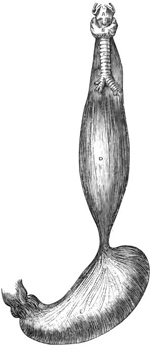
|
|
Fusiform Dilatation of Oesophagus (Luschka).
A, Larynx; B, Thyroid gland; C, Trachea; D, Oesophagus;
E, Stomach. |
General dilatation, though sometimes congenital, is, as mentioned
under Etiology, more frequently the mechanical result of distension of
the oesophagus by food or drink prevented from ready entrance into the
stomach by a constriction at the cardiac orifice. This form of
dilatation is sometimes discovered as a post-mortem curiosity. The
muscles have usually undergone great hypertrophy, and the mucous
membrane some thickening and congestion, with erosions and sometimes
ulcerations, indicative of chronic oesophagitis. In some instances all
the coats of the oesophagus have undergone hypertrophy. The dilatation
may vary from slight enlargement to the thickness of an ordinary man's
arm or larger (Rokitansky31); in rare cases, even a capacity nearly
equal to that of the stomach (Luschka32 and others). (See Fig. 19.)
31 Path. Anat.
32 Arch. für Anat., etc., March, 1868, p. 473.
The oesophagus is usually fusiform or spindle-shaped, being
constricted at those portions at which it is normally slightly
constricted. Sometimes the dilatation takes place between the lobes of
the lungs (Raymond33).
33 Gaz. méd. de Paris, 1869, No. 7, p. 91.
Annular dilatation is usually due to circumferential distension just
above a stricture. When not due to stricture its seat is usually just
above the diaphragm, where the oesophagus is normally liable to
constriction. The upper portion of the dilatation is larger than the
lower portion, and the muscular walls are usually hypertrophied.
Pouched dilatation (diverticulum) is usually formed chiefly of mucous
membrane and submucous tissue pushed through gaps in the fibres of the
muscular coat, produced by distension. It sometimes involves the
entire coat in cases in which the oesophageal wall has become adherent
to enlarged lymphatic glands, which subsequently undergo subsidence in
volume and drag the adherent portion of the wall after them
(Rokitansky). The muscular walls are then usually hypertrophied, the
mucous membrane sometimes hypertrophied, sometimes atrophied. The
diverticulum is usually located in the upper portion of the
oesophagus, just below the inferior constrictor muscle of the pharynx.
It may thus be, in part, a pharyngocele also. It may be located behind
the point of bifurcation of the trachea or where the oesophagus is
crossed by the left bronchus. Its direction may be to the left side in
the upper portion of the oesophagus, to the right side, or upon both
sides; but when situated lower down it is usually directed backward,
between the posterior wall of the tube and the spinal column. Hence
its distension with food completely blocks up the calibre of the
oesophagus. The orifice by which the oesophageal wall remains in
communication with the pouch is round or elliptic in shape and
variable in size, sometimes being about an inch in its long diameter,
sometimes much smaller. The size of the diverticulum varies; a common
size is that of a duck egg, but the size of a fist has been attained.
Sometimes the diverticulum drags the oesophagus out of position and
forms a sort of blind pouch in the direct line of its axis, so that it
becomes filled with food which fails to reach the stomach. Sometimes
there are several dilatations.
The dilatations become enlarged by retention of food, and are liable
to undergo inflammation, ulceration, and perforation.
DIAGNOSIS.—The diagnosis will depend upon the symptoms of dysphagia,
regurgitation, and so on, and upon the evidence furnished by
auscultatory indications, palpation with the oesophageal sound, and,
in some instances, the existence of a tumor in the neck, enlarging
after meals, and
from which food or mucus can be forced up into
the pharynx by pressure externally.
Stethoscopic auscultation of the oesophagus during the deglutition of
water indicates an alteration in the usual form of the gulp, which
seems to trickle rapidly in a larger or smaller stream according to
the degree of dilatation. If the dilatation be annular and located
high up, auscultation is said to give the impression of a general
sprinkling of fluid deflected from its course. The peculiar gurgle is
often audible without the aid of stethoscopy. Palpation with the
oesophageal bougie is competent to reveal the existence of a large sac
by the facility with which the terminal extremity of the sound can be
moved in the cavity. In the case of a diverticulum, however, the sound
may glide past the mouth of the pouch without entering it, although
arrested at the bottom of the sac in most instances.
In annular dilatation any constriction below it is usually perceptible
to the touch through the sound; but, on the other hand, the ready
passage of the bougie into the stomach, while excluding stricture,
does not positively disprove the existence of a circumscribed
dilatation. If high up, the dilatation may be detected externally by
its enlargement when filled with food after a meal, and the subsidence
of tumefaction when the sac is emptied by pressure from without, or by
regurgitation. If the dilatation occupy a position which exercises
compression of the trachea, dyspnoea will ensue when it is distended.
The intermittence of the tumefaction serves to differentiate the
swelling from abscess or morbid growth. From aneurism of the aorta,
which it may simulate (Davy34), it is to be discriminated by absence
of the usual stethoscopic and circulatory manifestations. The
diagnosis of congenital dilatation is based upon a history of
difficulty in deglutition dating from the earliest period of recollection.
34 Irish Hosp. Gaz., 1874, p. 129; Med. Press and
Circular, May, 1874.
PROGNOSIS.—The prognosis is not favorable in any given case unless
the cause can be removed, and not even then unless food can be
prevented from accumulating in the distended portion of the tube.
Nevertheless, cases sometimes go on into advanced age. On the other
hand, they may terminate fatally within a year (Lindau35). The
danger of perforation adds additional gravity to the prognosis, for
life may be suddenly lost by this accident. Death usually takes place
by inanition. A case of death by suffocation has been recorded,
attributed to the pressure of the distended oesophagus upon the
intrathoracic vessels (Hannay36).
35 Casper's Wochenschrift, 1840, No. 22; Arch. gén. de
Méd., 1841, p. 498; Dict. de Méd et de Chir., xxiv. p. 410.
36 Edinb. Med. and Surg. Journ., July 1, 1833.
TREATMENT.—If the dilatation be due to stricture or to an impacted
foreign body, the treatment should be directed to overcoming the one
and removing the other.
General dilatation from chronic oesophagitis requires treatment for
that disease.
Much depends upon preventing the accumulation of food in a sac or
diverticle; the best means of accomplishing which is the systematic
administration of all nutriment by means of the stomach-tube. When
this is not advisable, care must be exercised in the selection of such
food as is least likely to irritate the parts if detained in the pouch.
As far as general treatment is concerned, stimulants are usually
indicated, as the patients become much reduced. If paralysis of the
muscular coat of the oesophagus is believed to exist, the
administration of preparations of phosphorus and of strychnine are
indicated on general principles of therapeutics. Stimulation of
muscular contractility by the oesophageal electrode has been
recommended, but the prospects of success hardly justify the risks of
serious injury in the domain of the pneumogastric nerve.
It has not yet been determined whether surgical procedures are
competent to relieve dilatation. In cases of pouched dilatation high
up it would not be difficult, as suggested by Michel,37 to expose
the sac and excise it in such a manner that the sutures uniting the
walls of the oesophagus shall occupy the site of the mouth of the
diverticulum, and, thus obliterating it by cicatrization, restore the
normal path of the food from the pharynx to the oesophagus.
Gastrostomy, too, should hold out some hope of rescue, no matter what
portion of the oesophagus be dilated.
37 Dict. Encyclop., xiv. p. 465.
BY SAMUEL G. ARMOR, M.D., LL.D.
To difficulty in the physiological process of digestion the familiar
name of dyspepsia has been given, while to a merely disturbed
condition of the function the term indigestion is more frequently
applied. This distinction, difficult at all times to make, may appear
more arbitrary than real; and inasmuch as it involves no important
practical point, the author of the present article will use the terms
interchangeably as indicating functional disturbance of the
stomach—i.e. disturbance of the digestive process not associated
with changes of an inflammatory character, so far as we know.
Since it is one of the most common of all complaints from its
association with various other morbid conditions, the term is not
unfrequently vaguely employed. It is difficult, of course, to define a
disease whose etiology is so directly related to so many distinct
morbid conditions. Indeed, there are few diseases, general or local,
which are not at some time in their history associated with more or
less derangement of the digestive process. For purposes of limitation,
therefore, it will be understood that we now refer to chronic
functional forms of indigestion which depend largely, at least, on a
purely nervous element, and for this reason are not infrequently
described as sympathetic dyspepsia. Doubt has been expressed as to
whether such forms of disease ever exist, but that we encounter purely
functional forms of dyspepsia, corresponding to the dyspepsia
apyretica of Broussais, would appear to be a well-recognized clinical fact.
What the precise relation is between digestive disturbances and the
nervous system we may not fully understand, no more than we understand
how a healthy condition of nervous endowment is essential to all vital
processes. Even lesions of nutrition are now known to depend upon
primary disturbance of nervous influence. This is seen in certain skin
diseases, such as herpes zoster, which closely follows the destruction
of certain nerves. And it is well known that injury of nerve-trunks is
not unfrequently followed by impaired nutrition and failure in
reparative power in the parts to which such nerves are distributed.
Indeed, so marked is the influence of the nervous system over the
nutritive operations that the question has been considered as to
whether there are
trophic nerves distributed to tissue-elements
themselves whose special function is to keep these elements in a
healthy state of nutrition. The proof, at least, that the digestive
process is, in some unexplained way, under the immediate influence of
the nervous system, either cerebro-spinal or trophic, is both varied
and abundant. The digestive secretions are known to be the products of
living cells which are abundantly supplied with nerve-fibres, and we
can readily believe that the potential energy of this cell-force is
probably vital and trophic. At any rate, it is unknown in the domain
of ordinary chemistry. The digestive ferments, as clearly pointed out
by Roberts, are the direct products of living cells. Their mode of
action, he claims, bears no resemblance to that of ordinary chemical
affinity. It has a distinctly physiological character. Nor do they
derive their vital endowments from material substances. "They give
nothing material to, and take nothing from, the substances acted on.
The albuminoid matter which constitutes their mass is evidently no
more than the material substance of a special kind of energy—just as
the steel of a magnet is the material substratum of the magnetic
energy, but is not itself that energy" (Roberts). That this living
cell-force is partly, at least, derived from the nervous system is
clear from the well-known effects of mental emotion, such as acute
grief, despair, etc., in putting an immediate stop to the digestive
process. Experiments on the lower animals have also shown the direct
influence of the nervous system over gastric secretion. Wilson Philip
showed by various experiments on rabbits and other animals that if the
eighth pair of nerves be divided in the neck, any food which the
creatures may afterward eat remains in the stomach undigested, and
after death, when the nerve has been divided, the coats of the stomach
are not found digested, however long the animal may have been dead.
Bernard also excited a copious secretion by galvanization of the
pneumogastric, and by section of the same nerve stopped the process of
digestion and produced "pallor and flaccidity of the stomach."
Recently doubt has been thrown on these statements of Bernard and
Frerichs. Goltz concludes, from observations made on frogs, that
nerve-ganglia, connected by numerous intercommunicating bundles of
nerve-fibres, exist in the walls of the stomach, the irritation of
which gives rise to local contractions and peristaltic movements of
the stomach, and that these ganglia influence the gastric secretion.
However this may be, it still remains true that these gastric ganglia
are in connection, through the vagi, with the medulla oblongata, and
are thus influenced by the cerebro-spinal nerve-centres. And clinical
observation confirms what theoretical considerations would suggest.
Thus, strong mental impressions are known to produce sudden arrest of
secretion, and that which arrests secretion may, if continued, lead to
perversion of the same.
Impressions made upon the nerves of special sense are also known to
affect the salivary and gastric secretions. The flow of saliva is
stimulated by the sight, the smell, the taste, and even thought, of
food. Bidder and Schmidt made interesting experiments on dogs bearing
upon this point. They ascertained by placing meat before dogs that had
been kept fasting that gastric juice was copiously effused into the
stomach. Other secretions are known to be similarly affected.
Carpenter by a series of well-observed cases has shown the direct
influence of mental conditions on the
mammary secretion. The
nervous association of diabetes and chronic Bright's disease is
interesting in this connection, and the direct nervous connection
betwixt the brain and the liver has been shown by numerous
experiments. It is maintained by modern physiologists that "the
liver—indeed each of the viscera—has its representative area in the
brain, just as much as the arm or leg is represented in a distant
localized area" (Hughlings Jackson). And in harmony with this view
Carpenter long since pointed out the fact that if the volitional
direction of the consciousness to a part be automatically kept up for
a length of time, both the functional action and the nutrition of the
part may suffer. It has been described by him as expectant attention,
and it has, as we shall see, important practical bearings on the
management of gastric affections. Sympathetic disturbance of the
stomach is also connected with direct disease of the brain. This is
seen in cases of concussion. The almost immediate effects of a blow
are nausea and vomiting, and the same thing is observed in local
inflammation of the meninges of the brain.
Many forms of functional dyspepsia due to nervous disturbance of a
reflex character will be pointed out when discussing the etiology of
the disease.
ETIOLOGY.—Among the agencies affecting the digestive process in
atonic forms of dyspepsia may be mentioned—
First, predisposing causes;
Second, exciting causes.
In general terms it may be said that all conditions of depressed
vitality predispose to the varied forms of atonic dyspepsia. These
conditions range through an endless combination of causes, both
predisposing and exciting. There is not a disturbed condition of life,
extrinsic or intrinsic, that may not contribute to this end. In some
cases it may be the effects of hot and enervating climates; in others
the alterations in the elementary constituents of the blood may be
apparent; while in still others the cause may be exhausting
discharges, hemorrhages, profuse suppuration, venereal excesses,
sedentary occupations, and long-continued mental and moral emotions.
Heredity may also predispose to functional dyspepsia. Certain faulty
states of the nervous system are specially liable to be transmitted
from parent to offspring—not always in the exact form in which they
appeared in the parent, but in forms determined by the individual life
of the offspring. For obvious reasons, growing out of our modern
American civilization, the inheritance of a faulty nervous
organization is apt to spend itself upon the digestive apparatus. The
inordinate mental activity, the active competitions of life, the
struggle for existence, the haste to get rich, the disappointments of
failure,—all contribute to this end. The general tendency of American
life is also in the direction of a highly-developed and morbidly
sensitive nervous system, and functional dyspepsia is a natural
sequence of this. The symptoms of dyspepsia thus caused usually
manifest themselves at an early period of life.
Age also predisposes to weak digestion. The stomach becomes weak as
age advances, in common with all the functions of the body, and
consequent upon this weakness there is diminished excitability of the
gastric nerves, with diminished muscular action of the walls of the
stomach and deficient secretion of the gastric juice. Chronic
structural changes are
also apt to occur in advanced life. The
gastric glands become atrophied and the arteries become atheromatous,
so that with symptoms of indigestion there are often associated loss
of consciousness at times, vertigo, irregular action of the heart,
etc. These general facts have an important bearing upon the hygienic
management of dyspepsia in the aged. They require, as a rule, less
food than the young and vigorous. In times when famine was more
frequent than now it was found that the older a human being was, the
better deficiency of food was borne. Hippocrates tells us, in his
Aphorisms, that old men suffer least from abstinence. Their food
should be such, both in quantity and quality, as the enfeebled stomach
can digest. There is less demand for the materials of growth, and
consequently for animal food. Moderate quantities of alcohol,
judiciously used, are also specially adapted to the indigestion of the
aged. It has the double effect of stimulating the digestive process
and at the same time checking the activity of destructive
assimilation, which in old age exhausts the vital force. And in order
to more effectively arrest destructive metamorphosis great caution
should be taken against excessive muscular fatigue, as well as against
sudden extremes of temperature. Loss of appetite from deficient
formation of gastric juice is a common symptom in old age. This is not
often successfully treated by drugs, and yet medicines are not without
value. The sesquicarbonate of ammonium acts as a stimulant to the
mucous membrane and to the vaso-motor nerve, and in this way becomes a
valuable addition to the simple vegetable bitters. Dilute hydrochloric
acid with the vegetable bitters may also be tried. Condiments with the
food directly stimulate the action of the enfeebled stomach. The old
remedy of mustard-seed is not unfrequently useful, and pepper,
cayenne, horseradish, and curries act in a similar manner in torpid
digestion. And in cases of great exhaustion associated with anæmia
benefit may be derived from small doses of iron added to tincture of
columbo or gentian.
Nor should it be forgotten that in the opposite extreme of life the
digestive capacity is extremely limited. The infant's digestion is
readily disturbed by unsuitable alimentation. For obvious reasons it
does not easily digest starchy substances. The diastasic ferment does
not exist in the saliva of young sucking animals, at least to any
extent. No food is so suitable for early infantile life as the
mother's milk, provided the mother herself is healthy. It contains in
an easily digestible form all the constituents necessary to the
rapidly-growing young animal. Van Helmont's substitute of bread boiled
in beer and honey for milk, or Baron Liebig's food for infants, cannot
take the place of nature's type of food, which we find in milk. If a
substitute has to be selected, there is nothing so good as cow's milk
diluted with an equal quantity of soft water, or, what in many cases
is better, barley-water, to which may be added a teaspoonful of
powdered sugar of milk and a pinch of table-salt and phosphate of
lime. Lime-water may be added with advantage. Dilution of alimentary
substances is an important condition of absorption in the infant stomach.
Anæmia is a common predisposing cause of indigestion. Indeed, as a
widely-prevailing pathological condition few causes stand out so
prominent. It affects at once the great nutritive processes, and these
in turn disturb the functional activity of all the organs of the body.
Not only are the gastric and intestinal glands diminished in their
functional activity by impoverished or altered blood, but the
movements of the stomach are retarded by weakened muscular action. It
is impossible to separate altered blood from perverted
tissue-structure and altered secretion. Indigestion produced by anæmia
is difficult of treatment, on account of the complexity of the
pathological conditions usually present, the anæmia itself being
generally a secondary condition. Careful inquiry should be made,
therefore, into the probable cause of the anæmia, and this should, if
possible, be removed as an important part of the treatment of the
dyspepsia. Nothing will more promptly restore the digestive capacity
in such cases than good, healthy, well-oxidized blood. Indeed, healthy
blood is a condition precedent to the normal functional activity of
the stomach.
To these general predisposing causes may be added indigestion
occurring in febrile states of the system. The cause here is obvious.
In all general febrile conditions the secretions are markedly
disturbed; the tongue is dry and furred; the urine is scanty; the
excretions lessened; the bowels constipated; and the appetite gone.
The nervous system also participates in the general disturbance. In
this condition the gastric juice is changed both quantitatively and
qualitatively, and digestion, as a consequence, becomes weak and
imperfect—a fact that should be taken into account in regulating the
diet of febrile patients. From mere theoretical considerations there
can be no doubt that fever patients are often overfed. To counteract
the relatively increased tissue-metamorphosis known to exist, and the
consequent excessive waste, forced nutrition is frequently resorted
to. Then the traditional saying of the justly-celebrated Graves, that
he fed fevers, has also rendered popular the practice. Within certain
bounds alimentation is undoubtedly an important part of the treatment
of all the essential forms of fever. But if more food is crowded upon
the stomach than can be digested and assimilated, it merely imposes a
burden instead of supplying a want. The excess of food beyond the
digestive capacity decomposes, giving rise to fetid gases, and often
to troublesome intestinal complications. The true mode of restoring
strength in such cases is to administer only such quantities of food
as the patient is capable of digesting and assimilating. To this end
resort has been had to food in a partially predigested state, such as
peptonized milk, milk gruel, soups, jellies, and beef-tea; and
clinical experience has thus far shown encouraging results from such
nutrition in the management of general fevers. In these febrile
conditions, and in all cases of general debility, the weak digestion
does not necessarily involve positive disease of the stomach, for by
regulating the diet according to the digestive capacity healthy
digestion may be obtained for an indefinite time.
Exhaustion of the nerves of organic life strongly predisposes to the
atonic forms of dyspepsia. We have already seen how markedly the
digestive process is influenced by certain mental states, and it is a
well-recognized fact that the sympathetic system of nerves is
intimately associated with all the vegetative functions of the body.
Without a certain amount of nervous energy derived from this portion
of the nervous system, there is failure of the two most important
conditions of digestion—viz. muscular movements of the stomach and
healthy secretion of gastric juice. This form of indigestion is
peculiar to
the ill-fed and badly-nourished. It follows in the
wake of privation and want, and is often seen in the peculiarly
careworn and sallow classes who throng our public dispensaries. In
this dyspepsia of exhaustion the solvent power of the stomach is so
diminished that if food is forced upon the patient it is apt to be
followed by flatulence, headache, uneasy or painful sensations in the
stomach, and sometimes by nausea and diarrhoea. It is best treated by
improving in every possible way the general system of nutrition, and
by adapting the food, both in quantity and quality, to the enfeebled
condition of the digestive powers. Hygienic measures are also of great
importance in the management of this form of dyspepsia, and especially
such as restore the lost energy of the nervous system. If it occur in
badly-nourished persons who take little outdoor exercise, the food
should be adapted to the feeble digestive power. It should consist for
a time largely of milk and eggs, oatmeal, peptonized milk gruels,
stale bread; to which should be added digestible nitrogenous meat diet
in proportion to increased muscular exercise. Systematic outdoor
exercise should be insisted upon as a sine quâ non. Much benefit may
be derived from the employment of electric currents, and hydrotherapy
has also given excellent results. If the indigestion occur in the
badly-fed outdoor day-laborer, his food should be more generous and
mixed. It should consist largely, however, of digestible nitrogenous
food, and meat, par excellence, should be increased in proportion to
the exercise taken. Medicinally, such cases should be treated on
general principles. Benefit may be derived from the mineral acids
added to simple bitters, or in cases of extreme nervous prostration
small doses of nux vomica are a valuable addition to dilute
hydrochloric acid. The not unfrequent resort to phosphorus in such
cases is of more than doubtful utility. Some interesting contributions
have been recently made to this subject of gastric neuroses by
Buchard, Sée, and Mathieu. Buchard claims that atonic dilatation of
the stomach is a very frequent result of an adynamic state of the
general system. He compares it to certain forms of cardiac
dilatation—both expressions of myasthenia. It may result from
profound anæmia or from psychical causes. Mathieu regards mental
depression as only second in frequency. Much stress is laid upon
poisons generated by fermenting food in the stomach in such cases. It
may cause a true toxæmia, just as renal diseases give rise to uræmia.
Of course treatment in such cases must be addressed principally to the
general constitution.
But of all predisposing causes of dyspepsia, deficient gastric
secretion, with resulting fermentation of food, is perhaps the most
prevalent. It is true this deficient secretion may be, and often is, a
secondary condition; many causes contribute to its production; but
still, the practical fact remains that the immediate cause of the
indigestion is disproportion between the quantity of gastric juice
secreted and the amount of food taken into the stomach. In all such
cases we have what is popularly known as torpidity of digestion, and
the condition described is that of atony of the stomach. The two main
constituents of gastric juice—namely, acid and pepsin—may be
deficient in quantity or disturbed in their relative proportions. A
certain amount of acid is absolutely essential to the digestive
process, while a small amount of pepsin may be sufficient to digest a
large amount of albuminoid food.
Pure unmixed gastric juice was
first analyzed by Bidder and Schmidt. The mean analyses of ten
specimens free from saliva, procured from dogs, gave the following results:
| Water |
973.06 |
|
| Solids |
26.94 |
|
| Containing— |
Peptone and pepsin |
17.19 |
| |
Free hydrochloric acid |
3.05 |
| |
Alkaline chlorides |
4.26 |
| |
Ammonium chloride |
0.47 |
| |
Chlorine |
5.06 |
| |
Phosphates— |
Lime |
1.73 |
| |
|
Magnesia |
0.23 |
| |
|
Iron |
0.08 |
They proved by the most careful analyses that fresh gastric juice
contains only one mineral acid—namely, hydrochloric; since which time
Richet has been able to prove that "this acid does not exist in a free
state, but in loose combination with an organic substance known as
lucin," the chloride of lucin. And just here the curious and puzzling
question arises as to the secretion of a mineral acid from alkaline
blood. Ewald, the distinguished lecturer in the Royal University of
Berlin, tells us that "a brilliant experiment of Maly's has thrown
unexpected light upon this. There are fluids of alkaline reaction
which may contain two acid and alkaline mutually inoffensive salts,
but still have an alkaline reaction, because the acid reaction is to a
certain extent eclipsed; for instance, a solution of neutral phosphate
of soda (Na2HPO4) and acid phosphate of
soda (NaH2PO4) is
alkaline. Such a solution placed in a dialyzer after a short time
gives up its acid salt to the surrounding distilled water, and one has
in the dialyzer an alkaline fluid outside an acid fluid." He thus
proved that the acid phosphate of sodium is present in the blood in
spite of its alkaline reaction.
Lack of the normal amount of the gastric secretion must be met by
restoring the physiological conditions upon which the secretion
depends. In the mean time, hydrochloric and lactic acids may be tried
for the purpose of strengthening the solvent powers of the gastric secretion.
EXCITING CAUSES.—The immediate causes of dyspepsia are such as act
more directly on the stomach. They embrace all causes which produce
conditions of gastric catarrh, such as excess in eating and drinking,
imperfect mastication and insalivation, the use of indigestible or
unwholesome food and of alcohol, the imperfect arrangement of meals,
over-drugging, etc.
Of exciting causes, errors of diet are amongst the most constantly
operative, and of these errors excess of food is doubtless the most
common. The influence of this as an etiological factor in derangement
of digestion can scarcely be exaggerated. In very many instances more
food is taken into the stomach than is actually required to restore
tissue-waste, and the effects of such excess upon the organism are as
numerous as they are hurtful. Indeed, few elements of disease are more
constantly operative in a great variety of ailments. In the first
place, if food be introduced into the stomach beyond
tissue-requirements, symptoms of indigestion at once manifest
themselves. The natural balance betwixt
supply and demand is
disturbed; the general nutrition of the body is interfered with; local
disturbances of nutrition follow; and mal-products of digestion find
their way into the blood. Especially is this the case when the
excessive amount of food contains a disproportionate amount of
nitrogenous matter. All proteid principles require a considerable
amount of chemical alteration before they are fitted for the metabolic
changes of the organism; the processes of assimilative conversion are
more complex than those undergone by fats and amyloids; and it follows
that there is proportional danger of disturbance of these processes
from overwork. Moreover, if nitrogenous food is in excess of
tissue-requirement, it undergoes certain oxidation changes in the
blood without becoming previously woven into tissue, with resulting
compounds which become positive poisons in the economy. The kidneys
and skin are largely concerned in the elimination of these compounds,
and the frequency with which these organs become diseased is largely
due, no doubt, to the excessive use of unassimilated nitrogenous food.
Then, again, if food be introduced in excess of the digestive
capacity, the undigested portion acts directly upon the stomach as a
foreign body, and in undergoing decomposition and putrefying changes
frets and irritates the mucous membrane. It can scarcely be a matter
of doubt that large groups of diseases have for their principal causes
excess of alimentation beyond the actual requirements of the system.
All such patients suffer from symptoms of catarrhal indigestion, such
as gastric uneasiness, headache, vertigo, a general feeling of
lassitude, constipation, and high-colored urine with abundant urates,
together with varied skin eruptions. Such cases are greatly relieved
by reducing the amount of food taken, especially nitrogenous food, and
by a systematic and somewhat prolonged course of purgative mineral
waters. Europe is especially rich in these springs. The waters of
Carlsbad, Ems, Seltzer, Friedrichshall, and Marienbad, and many of the
alkaline purgative waters of our own country, not unfrequently prove
valuable to those who can afford to try them, and their value shows
how often deranged primary assimilation is at the foundation of many
human ailments. The absurd height to which so-called restorative
medicine has attained within the last twenty years or more has
contributed largely to the production of inflammatory forms of
indigestion, with all the evil consequences growing out of general
deranged nutrition.
The use of indigestible and unwholesome food entails somewhat the same
consequences. This may consist in the use of food essentially
unhealthy or indigestible, or made so by imperfect preparation
(cooking, etc.). Certain substances taken as food cannot be dissolved
by the gastric or intestinal secretions: the seeds, the skins, and
rinds of fruit, the husks of corn and bran, and gristle and elastic
tissue, as well as hairs in animal food, are thrown off as they are
swallowed, and if taken in excess they mechanically irritate the
gastro-intestinal mucous membrane and excite symptoms of acute
dyspepsia, and not unfrequently give rise to pain of a griping
character accompanied by diarrhoea. Symptoms of acute dyspepsia also
frequently follow the ingestion of special kinds of food, such as
mushrooms, shellfish, or indeed fish of any kind; and food not adapted
to the individual organism is apt to excite dyspeptic symptoms.
Appetite and digestion are also very much influenced by the life and
habits of the individual. The diet, for instance, of bodily
labor should consist largely of digestible nitrogenous food, and meat,
par excellence, should be increased in proportion as muscular exercise
is increased. For all sorts of muscular laborers a mixed diet is best
in which animal food enters as a prominent ingredient. Thus, it has
been found, according to the researches of Chambers, that in forced
military marches meat extract has greater sustaining properties than
any other kind of food. But with those who do not take much outdoor
exercise the error is apt to be, as already pointed out, in the
direction of over-feeding. It cannot be doubted at the present time
that over-eating (gluttony) is one of our popular vices. Hufeland
says: "In general we find that men who live sparingly attain to the
greatest age." While preventive medicine in the way of improved
hygiene—better drainage, better ventilation, etc.—is contributing
largely to the longevity of the race, we unfortunately encounter in
more recent times an antagonizing influence in the elegant art of
cookery. Every conceivable ingenuity is resorted to to tempt men to
eat more than their stomachs can properly or easily digest or
tissue-changes require. The injurious consequences of such
over-feeding may finally correct itself by destroying the capacity of
the stomach to digest the food.
But, on the other hand, in many nervous forms of dyspepsia the weak
stomach is not unfrequently made weaker by severely restricted
regimen, and especially is this the case with mental workers.
Theoretical and fanciful considerations sometimes lead to physical
starvation. This is apt to be the case with dyspeptics. Men who toil
with their brain rather than their muscles, whether dyspeptic or not,
require good, easily-digested mixed diet. It is a popular error to
suppose that drugs can take the place of such food, especially drugs
which are supposed to have a reconstructive influence over the nervous
system, such as iron and phosphorus. The expression of Büchner, "No
thinking without phosphorus," captivating to theoretical minds, has
gained much notoriety, and has doubtless led to the excessive use of
that drug in nervous forms of indigestion. There never was a period
when phosphorus was so universally prescribed as the present. It
enters into endless combinations with so-called nerve-tonics. Of the
injurious influence of the drug in many cases of functional
indigestion there can be no doubt; and the statement itself, so often
quoted, that "the amount of phosphorus in the blood passing through
the brain bears an exact proportion to the intensity of thought," is
calculated to mislead. T. K. Chambers, author of the excellent Manual
of Diet, makes the statement that "a captive lion, tiger, leopard, or
hare assimilates and parts with a greater amount of phosphorus than a
hard-thinking man; while the beaver, noted for its power of
contrivance, excretes so little phosphorus that chemical analysis
cannot find it in its excreta." In the wonderful adaptations and
regulative mechanisms of nature we may trust largely to the natural
law of supply and demand in maintaining a proper equilibrium. It may
be doubted, indeed, whether we require at any time more phosphorus for
brain- and nerve-tissue than can be found in such food as contains
digestible phosphatic salts. The natural demand for food grows out of
healthy tissue-change. An appetite to be healthy should commence in
processes outside of the stomach.
Food may also be introduced into the stomach in an undigestible form
from defects of cookery. The process of cooking food produces
certain well-known chemical changes in alimentary substances which
render them more digestible than in the uncooked state. By the use of
fire in cooking his food new sources of strength have been opened up
to man which have doubtless contributed immeasurably to his physical
development, and has led to his classification as the cooking animal.
With regard to most articles the practice of cooking his food
beforehand is wellnigh universal; and especially is this the case with
all farinaceous articles of food. The gluten of wheat is almost
indigestible in the uncooked state. By the process of cooking the
starchy matter of the grain is not only liberated from its protecting
envelopes, but it is converted into a gelatinous condition which
readily yields to the diastasic ferments. Roberts, in his lectures on
the Digestive Ferments, points out the fact that when men under the
stress of circumstances have been compelled to subsist on uncooked
grains of the cereals, they soon fell into a state of inanition and disease.
Animal diet is also more easily digested in the cooked than in the raw
state. The advantage consists chiefly in the effects of heat on the
connective tissue and in the separation of the muscular fibre. In this
respect cooking aids the digestive process. The gastric juice cannot
get at the albumen-containing fibrillæ until the connective tissue is
broken up, removed, or dissolved. Hot water softens and removes this
connective tissue. Hence raw meat is less easily digestible.
Carnivorous animals, that get their food at long intervals, digest it
slowly. By cutting, bruising, and scraping meat we to a certain extent
imitate the process of cooking. In many cases, indeed, ill-nourished
children and dyspeptics digest raw beef thus comminuted better than
cooked, and it is a matter of observation that steamed and underdone
roast meats are more digestible than when submitted to greater heat.
Some interesting observations have been made by Roberts on the effects
of the digestive ferments on cooked and uncooked albuminoids. He
employed in his experiments a solution of egg albumen made by mixing
white of egg with nine times its volume of water. "This solution,"
says Roberts, "when boiled in the water-bath does not coagulate nor
sensibly change its appearance, but its behavior with the digestive
ferments is completely altered. In the raw state this solution is
attacked very slowly by pepsin and acid, and pancreatic extract has no
effect on it; but after being cooked in the water-bath the albumen is
rapidly and entirely digested by artificial gastric juice, and a
moiety of it is rapidly digested by pancreatic extract."
It is a mistake, however, to suppose that cooking is equally necessary
for all kinds of albuminoids. The oyster, at least, is quite
exceptional, for it contains a digestive ferment—the hepatic
diastase—which is wholly destroyed by cooking. Milk may be
indifferently used either in the cooked or uncooked state, and fruits,
which owe their value chiefly to sugar, are not altered by cooking.
The object in introducing here these remarks on cooking food is to
show that it forms an important integral part of the work of
digestion, and has a direct bearing on the management of all forms of dyspepsia.
Haste in eating, with imperfect mastication, is a common cause of
indigestion in this country. Mastication is the first step in the
digestive
process. It is important, therefore, that we have good
teeth and that we take time to thoroughly masticate our food, for by
so doing we prepare it for being acted upon by the juices of the
stomach. Time is also necessary in order that the salivary secretion
may be incorporated with the alimentary substances. By the salivary
diastase starch is converted into sugar and albuminoids are prepared
for the action of the gastric juice. If these changes take place
imperfectly, the stomach can scarcely regain in gastric digestion what
was lost in imperfect mastication and insalivation. Haste in eating is
one of the American vices. It grows out of the temperament of our
people. We are jealous of lost time, and unfortunately this time is
too often taken from the stomach. We bolt our food with unseemly
haste, and pay the penalty in ruined stomachs. Many cases of
indigestion are greatly relieved, if not permanently cured, by simply
doubling or quadrupling the time occupied in eating.
Irregularity in the intervals between meals, such as taking one meal
only in twenty-four hours or taking food before the preceding supply
has been digested, is another fruitful source of indigestion. The
digestive process, in the natural order of change, is confused;
changes which should take place are delayed; and the results are such
as arise from excessive eating. Moreover, the stomach lacks the rest
so essential to digestion. The necessary interval, however, between
meals varies with the nature of the food taken. "Between the extremes
of the carnivoræ," says Ewald, "which feed once in twenty-four hours,
and the herbivoræ, which never have done with the business of feeding,
man holds a middle place, but not without permitting the recognition
in the course of his life of a sort of transition from the herbivora
to the carnivora. Infants should have the breast during the first
three weeks as often as they wake; after that every two hours to the
third month; then up to dentition every three hours; and later there
should be five meals in twenty-four hours." But to this general
statement there are, of course, many exceptions. Under certain
pathological conditions food should be taken in small quantities at
short intervals. This is especially the case in chronic gastric
catarrh and in feeble digestion of nervous subjects. Such patients are
not unfrequently improved by becoming again infants or herbivoræ. By
the use of an exclusive milk diet or peptonized milk gruels, given in
small quantities at comparatively short intervals of time, the stomach
may be so accommodated that it will digest without discomfort a large
amount of nourishment within a given time. To S. Weir Mitchell of
Philadelphia we are indebted for some valuable observations bearing
upon this point of forced alimentation.
To the causes of indigestion already alluded to may be added the habit
of spirit-drinking, especially the habit of taking alcohol undiluted
on an empty stomach, which rarely fails after a time to engender
dyspeptic symptoms. It is a prominent factor in the production of
chronic gastric catarrh—a condition more frequently present in
painful indigestion than any that have been named. It is one of the
most common diseases met with in practice. Indeed, all causes already
alluded to involve, sooner or later, if they are constantly operative,
irritative and catarrhal conditions of the mucous membrane of the
stomach, so that we find it difficult at times—indeed impossible—to
separate purely functional from subacute inflammatory forms of
dyspepsia. Practically, we simply study the
subject in the
relative degrees of prominence of the one condition or the other.
But, in a still more comprehensive sense, indigestion is caused by
disturbance of organs directly associated with the stomach in the
digestive process. All organs closely associated with each other in
their physiological functions are apt to become associated in morbid
action. The clinical recognition of this is a matter of great
importance in the management of gastric affections. And first in the
order of importance in such association is the liver. So closely,
indeed, are the liver and stomach functionally associated in the
process of primary assimilation that they may be considered parts of
the same great digestive apparatus. Hence disturbance of the
liver—either in the formation of glycogen, the destruction of
albuminoid matter, or the secretion of bile—is immediately
communicated to the stomach. It may be difficult to say which of these
separate and distinct functions of the liver is most at fault; that
can only be a matter of physiological inference. In the one case, for
instance, the dyspeptic may be fairly well nourished, yet his
elimination may be bad. In the other there is no failure of the
destructive and excreting functions, but those concerned in the
assimilation of fat and peptones are disordered, so that the patient
is not well nourished, so far as the fatty element is concerned. This
is the more common form, and a form not unfrequently associated with
pulmonary consumption. The liver finally becomes fatty—a condition
usually found associated with the constitutional forms of phthisis.
The pancreas is also closely associated with the stomach, and its
secretion is of essential value in the digestive process. It is to be
regretted that our precise knowledge of its diseases is in such
striking contrast with its importance in the animal economy, and yet
it can scarcely be doubted that in dyspeptic symptoms associated with
failure of digestion of starchy, albuminous, and fatty elements of
food there is disorder of the secretion of the pancreas. Hence in the
treatment of the early stages of pulmonary consumption and other
disorders associated with deficient digestion and assimilation of
fatty substances the importance of directing our attention to the
condition of the liver and pancreas, as well as to the stomach.
That morbid states of the intestinal track occupy a prominent place in
the etiology of dyspepsia is also a well-recognized clinical fact.
Indeed, constipation of the bowels is an almost universal
accompaniment of deranged digestion, and when persistent for years it
is apt to lead to the most disastrous consequences. These are mainly
in the direction of lessened elimination from the intestinal glandulæ.
The general symptomatology of deficient excretion from these glandulæ
is closely analogous to the same condition of the liver: there is
impairment of the general health; the clear florid complexion
disappears; the patient becomes of a greenish or sallow hue; the blood
is altered in quality; fatigue is experienced after the slightest
exertion; the nights are restless; and there is great tendency to
mental despondency. Moreover, constipation often precedes the gastric
symptoms. The diminished muscular activity of the intestinal track
extends to the stomach; its movements are diminished; food is not
properly mixed with the gastric juice, and by being too long retained
in the stomach in a comparatively undigested state acetous
fermentation in the saccharine and starchy articles of diet is set
up, acid eructations and a sour taste in the mouth being
commonly complained of. Dyspepsia associated with this condition of
the intestinal track cannot be relieved until the constipation is
relieved, and by overcoming the constipation the dyspeptic symptoms
often disappear.
Mention has been made of the baneful influence of certain mental
states in the production of dyspeptic symptoms. But there are forms of
indigestion due to local nervous disturbance existing elsewhere than
in the nerve-centres. This was ascribed by the older writers to what
they termed consensus nervorum, or sympathy, by which "the operation
of a stimulus is not limited to the nerves immediately irritated, but
is extended to distant parts in known or unknown connection with the
irritated nerves." An intimate acquaintance with this law of sympathy
is of the utmost importance in the study of the functional forms of
dyspepsia, for no other organ of the body is subjected to such a wide
range of reflected nervous disturbance as the stomach. Morbid
sympathetic impressions are transmitted mainly through branches of the
vaso-motor nerve of the semilunar ganglia of the abdomen, and from the
pneumogastric to the stomach. Thus, a pregnant uterus not unfrequently
produces very troublesome vomiting; some females suffer from nausea
and indigestion during each menstrual period; and the more chronic
forms of pelvic irritation, such as a flexed uterus, and endometritis,
cervicitis, or tender ovary, may be the continuous exciting cause of
most troublesome forms of nervous dyspepsia. There is also close
sympathy of the stomach with the lungs and heart through the
distribution of the pneumogastric. So also may fixed points of
irritation in any part of the nervous system be reflexly transmitted
to the stomach, giving rise to most pronounced symptoms of
indigestion. And it is evident that in all such cases but little can
be accomplished in the way of relieving the dyspeptic symptoms until
the cause upon which they depend is removed. The treatment must have
reference mainly to the removal of such cause.
Lastly, all the causes mentioned finally concur in producing
irregularities of the mechanism of digestion; and this may be done by
disturbing either the muscular movements of the stomach or in
suspending or perverting the gastric solvents, or in these two
conditions combined.
SYMPTOMS.—1st. Referable to the Stomach.—The symptoms which attend
and indicate the presence of functional dyspepsia are such as
accompany in a greater or less degree almost all cases of chronic
gastritis. Clinically, so far as the direct gastric symptoms are
concerned, it is difficult to separate them. The more prominent of the
local symptoms are—a sense of fulness and distension after meals,
discomfort during the digestive process, derangement of appetite, acid
eructations, flatulence, regurgitations of food, and sometimes nausea
and vomiting. There is seldom severe pain; the sensation is rather
that of uneasiness. Exceptionally, however, there may be pain, which
radiates from the stomach to the shoulders, and may pass down the left
arm so as to simulate angina pectoris. But it may be readily
distinguished from that complaint by coming on after food, and not
after exertion. In other cases a sense of constriction may be
accompanied by dyspnoea, arising from impeded movements of the
diaphragm from being pushed upward by the distended stomach, or there
may be heartburn, with an ill-defined sense of burning felt in the
epigastrium;
but thirst, so frequently present in chronic
gastritis, is, as a rule, absent in functional dyspepsia. These
symptoms are manifested in varying degrees of prominence in individual
cases, and some of them are rarely found present. Thus, nausea and
vomiting are not characteristic features of the chronic forms of
functional dyspepsia, and as a rule epigastric tenderness is entirely
absent. In markedly hysterical subjects or in persons whose nervous
system has been unduly excited by alcohol there may be shrinking from
the slightest touch upon pressure; but in these cases the tenderness
is not confined to the stomach, nor is it increased by deep pressure.
In some cases there is an unnatural craving for food—a symptom rarely
if ever observed in structural lesions of the stomach—and now and
then it happens that the appetite becomes depraved, especially with
hysterical patients. They crave indigestible and unnatural substances,
such as earth, chalk, and substances wholly devoid of alimentary
properties. Impairment of appetite, however, is the more common
feature of this form of indigestion.
Flatulence and eructations are generally complained of, the flatulence
being accompanied by a painful sense of fulness, affecting in equal
degree the stomach and small intestines. It is derived principally
from putrefactive or fermentative changes of the ingesta, which are
imperfectly elaborated in the stomach. The gases consist of carbonic
acid, sulphuretted hydrogen, hydrogen, nitrogen, and the hydrocarbons,
the butyric and acetic fermentations furnishing the hydrogen and
carbonic acid gas. In addition to these marsh gas is formed by a
special fermentation, the basis of which exists in the cellulose taken
with vegetable food. In excessive meteorism from paralysis of the
intestines the gas is principally nitrogen; the marsh-gas fermentation
results from the ingestion of certain easily-fermentable vegetables,
such as cabbage, cauliflower, etc.
In a certain proportion of cases regurgitation occurs from the
stomach. The liquor regurgitated may be intensely acid from the
presence of some of the fatty acids, probably butyric, lactic, or
acetic. Exceptionally, it may be insipid or brackish, constituting
what is known as pyrosis, or water-brash. The fluid is usually
tasteless and without smell, and in reaction it is neutral to
test-paper. It contains sulphocyanuret of potassium, and it has been
supposed therefore to be only saliva. The quantity thrown up may vary
from a spoonful to a pint or more. It affects females more than males,
and especially those who subsist upon coarse and indigestible food. It
is best treated by astringents—such as kino, krameria, logwood, or
tannin—administered in the intervals between digestion, so that they
may act directly on the mucous membrane. The oxide and nitrate of
silver are thought by some to be superior to the vegetable astringents.
Cardialgia is a painful condition of the stomach, usually referred to
its cardiac orifice, and is popularly known as heartburn. It is met
with in both functional and organic disease of the stomach. It is very
constantly present in chronic catarrhal gastritis, and evidently
depends upon the presence of an acid, for it is usually promptly
relieved by alkalies, such as chalk, magnesia, soda, or alkaline
saline waters. Food containing much fat, starch, or sugar should be avoided.
Nausea and vomiting are only occasional symptoms of functional
dyspepsia. When vomiting does occur it may take place at different
times
and with varying degrees of severity, differing in this
respect from the nausea and vomiting of subacute gastritis, which
takes place, if at all, soon after the ingestion of food. The time of
vomiting and the character of the matter ejected are liable to great
variation in functional dyspepsia. It may be the result of direct
irritation of morbidly sensitive gastric nerves, or it may be a reflex
phenomenon; it may follow soon after the ingestion of food, or it may
come on when the stomach is empty; the material vomited may be simply
food but little altered or an alkaline ropy mucus; it may consist in
the acid juices of the stomach or in a neutral watery fluid; or the
ingesta may have undergone fermentative and putrefactive changes from
either insufficient amount of the gastric solvent or from narrowing
(constriction) of the pyloric extremity, in which case the yeast
fungus (Torula cerevisiæ) or the Sarcina ventriculi may be found in
great abundance in the vomited matter. Vomiting of this kind usually
occurs some time after eating. The gastric juice itself checks
putrefaction; so also does the admixture of bile. In the absence of
these natural antiseptics fermentation takes place. But it would be
erroneous to suppose that the fermentative dyspepsia is the primary
disease; it is a symptom which can be permanently corrected only by
correcting the condition upon which it depends.
Among the most noticeable of the phenomena referable to other organs
than the stomach are those connected with the liver and the alimentary
canal. The tongue in dyspeptic troubles varies much in character. In
reflex sympathetic indigestion it is not unfrequently clean; in
hepatic dyspepsia it is generally thickly coated with a white or
yellow fur. The symptoms are such as pertain more especially to
chronic gastro-duodenal catarrh, such as nausea, epigastric
oppression, furred tongue, heartburn, acid eructations, flatulent
distension of the stomach and bowels, unpleasant taste in the mouth,
offensive breath, loaded urine, frontal headache, irritability, and
hypochondriasis.
Constipation, as we have seen, is an almost universal accompaniment of
functional dyspepsia, sustaining to it not unfrequently a causative
relation. It is undoubtedly one of the most common of the slighter
ailments of civilized life, and exerts a wide influence in deranging
the general health. "It is quite extraordinary how many different
derangements of health may result from imperfect action or a torpid
state of the secreting and expelling structures of the large bowel.
There may be violent and persistent nerve-pains, referred to the back,
or hip, or groin, and certain other symptoms which lead pessimist
practitioners, excelling in the discovery of neuroses, to diagnose
structural changes in some part of the spinal cord or the antecedent
state which is supposed to lead to them" (Beale). Pains in the loins
and thighs, violent lumbar pain, and certain remediable forms of
sciatica are sometimes due to imperfect excretion of the lower part of
the alimentary canal. And it is even possible that a condition of
hypochondria bordering on insanity may be brought about by
long-continued defective action of the bowels. In exceptional cases of
dyspepsia diarrhoea may be present. This is more frequently the case
when indigestion is associated with a congested state of the liver, in
which case the symptom should be regarded as curative. Excessive
irritability of the muscular walls of the stomach, superadded to weak
digestion, may also be followed by lienteric forms of diarrhoea.
Undigested
food hastily finds its way into the intestinal track,
and not unfrequently appears in the fecal evacuations.
Functional derangements of the stomach are often accompanied by pale
urate deposits in the urine. It may contain an excess of phosphates,
and in microscopical examination crystals of the oxalate of lime are
frequently found, constituting a special affection described by
Golding-Bird as oxaluria. He associated it with irritative dyspepsia,
hypochondriasis, and exhaustion of nerve-power. This form of dyspepsia
is best managed by the mineral, vegetable, and acid tonics, to which
may be added small doses of nux vomica, with the usual adjuvants of
good air and exercise, freedom from anxiety and care, cold sea-water
baths, and well-selected, generous animal diet.
Another form of dyspepsia is sometimes associated with a peculiar form
of dizziness—gastric vertigo. German writers speak of it as abdominal
dizziness, and Trousseau calls it vertigo stomicale. It is usually an
acute symptom, begins without any premonition, and is liable to be
confounded with disease of the brain. It sometimes occurs soon after a
meal, but more often when the stomach is empty (Trousseau). It
perhaps, in a majority of cases, depends upon dyspepsia, but it has to
be differentiated from organic brain disease, from cerebral anæmia,
cerebral hyperæmia, the slighter forms of epilepsy, Minière's disease,
and general nervous exhaustion and depression. But in many cases it
will be found that treatment directed against the dyspepsia cures the vertigo.
Dyspeptic patients are also liable to skin diseases, and especially is
this observed in the gastro-duodenal forms of indigestion. Disorders
of the skin, such as urticaria, erythema, lichen, eczema, and other
allied conditions, are well-recognized external indications at times
of disordered conditions of the gastro-intestinal mucous membrane.
Thus, it is a matter of common observation that the gastric symptoms
increase when the eruption on the surface disappears.
The general influence of the nervous system over the function of
digestion is perhaps the most remarkable feature of the disease, so
that disturbed innervation becomes conspicuous in its symptomatology.
The phenomenon varies in individual cases. Languor, drowsiness after
taking food, depression of spirits, irritability, hypochondriasis,
sleeplessness, palpitation, dry cough, dyspnoea, are all of common
occurrence; and the mental disturbance—the anxiety, gloom, and
sadness—is to many dyspeptics more distressing than absolute pain.
It is impossible, however, to present, in this connection, a complete
clinical history of functional dyspepsia, for the reason that it is
associated with so many separate and distinct affections, the
dyspepsia itself being symptomatic of these affections.
PATHOLOGY.—But little is known of the pathology of the purely
functional forms of dyspepsia beyond what is expressed by the terms
atony and asthenia. These express simply certain states of the system
with which atonic dyspepsia is so frequently found associated.
Pathological anatomy has shown, however, that some cases are dependent
upon, or associated with, certain appreciable alterations of the
stomach, such as atrophy of the mucous membrane or fatty degeneration
of its walls; and not unfrequently it is the seat of the so-called
amyloid or lardaceous degeneration, although this albuminoid
infiltration or cloudy
swelling is more frequently the
accompaniment of chronic inflammatory process. But Jones and Fenwick
have shown that these conditions may occur independently of
inflammation. However, upon this point we are compelled to speak with
caution. The boundary-line between functional and structural diseases
is not always clearly defined. Functional and structural troubles of
the stomach are certainly very intimately associated. Moreover,
symptoms of purely functional dyspepsia are so frequently associated
with the subacute forms of gastritis that the pathology of the disease
becomes, from necessity, doubtful and complex. It can only be studied
in connection with certain states or conditions of which functional
derangement of the stomach is a symptom readily recognized during
life. In the light of more advanced physiological and pathological
researches we may expect the limits of purely functional dyspepsia to
be much restricted.
DIAGNOSIS.—The diagnosis of atonic dyspepsia must have special
reference to its etiology. It is usually a chronic disease, and has to
be discriminated from subacute or chronic inflammation of the stomach.
This is the more difficult because many symptoms exist in common in
both varieties of indigestion. But in functional or atonic dyspepsia
the symptoms are not so continuous; there is less epigastric
uneasiness, less tenderness, less nausea or loathing of food, less
thirst, and less acidity and heartburn, less emaciation, less
cerebral and nervous disturbance, and the constitutional symptoms are
also less severe. The tongue, as a rule, is not so thickly coated, is
not so red or broad and flabby, the papillæ are less marked, the
breath less offensive, and the urine, instead of showing a condition
of lithæmia, is not unfrequently pale and sometimes neutral,
depositing oxalates and phosphates, especially in feeble, broken-down
conditions of the nervous system.
With other painful affections of the stomach, such as ulcer and
cancer, it is not likely to be confounded, especially when in these
affections pain, vomiting, and hæmatemesis are present.
TREATMENT.—The first and leading indication is to remove, as far as
possible, all causes of the disease, and this requires patient
research and much diagnostic skill. Suggestive hints of treatment may
be found in connection with the discussion of the varied etiology of
the disease. We can, in conclusion, only allude to the matter in a
very general way. Special cases must furnish their own indications of treatment.
In many cases a condition of nervous asthenia will be found
prominently present. A leading indication, therefore, irrespective of
the special determining cause, is to improve the general health of the
patient; and this is accomplished by all means which invigorate the
system generally. And first in the order of importance are diet and
regimen. It is evident that if a patient eat too much or too often, or
if he eat indigestible or unwholesome food, or lead an indolent and
luxurious life, nothing can be accomplished by way of drugs in the
relief of the disease. Excessive alimentation is, as we have seen, a
most prolific source of the disease. Tempted to excess by great
variety and by the ingenuity of culinary refinements, the stomach is
burdened beyond its capacity of digestion and beyond the actual
requirements of the system; and especially is this the case with those
who live sedentary, indoor lives. In all such cases it is absolutely
essential that the digestive organs have rest.
Better even in
cases of doubt reduce the diet for a time below the actual wants of
the system until waste products are thoroughly removed and appetite is
revived. The benefit derived in some instances from the protracted use
of purgative mineral waters is largely attributable to the restricted
regimen enforced and to the washing out of the system the waste products.
On the other hand, too great or too protracted abstemiousness may
equally impair the digestive process. In ordinary forms of atonic
dyspepsia we should seek rather, by appropriate treatment, to raise
the digestive capacity to the level of digesting good, healthy,
nutritious food, than to reduce the food to the low standard of feeble
digestion. But it is a mistake to suppose that this can be
accomplished by simply forcing food upon a stomach that lacks capacity
of digestion.
As to the kind of diet, no precise rule is suited to all cases. Within
certain limits individual experience must be consulted. But these
experiences are not always reliable. Dyspeptic patients, more than any
others, are apt to have fancies. Certain general rules, therefore,
should be insisted upon. The food should be wholesome and digestible;
it should be well cooked, well masticated, and taken at regular and
not too long intervals. The intervals of time between meals depend
upon circumstances already referred to. In some cases small quantities
of easily-digested food should be taken at short intervals. In cases
of feeble digestion of nervous subjects milk diluted in Seltzer water,
or milk and lime-water, or peptonized milk, may be taken in liberal
quantities at comparatively short intervals of time. Sometimes
isinglass, arrowroot, or ground rice may be advantageously combined
with the milk, to which tender, undone meats may be added.
Peptonized Food.—Recently the attention of the profession has been
attracted to artificially digested food. The essential acts of
digestion are known to be chemical transmutations. Albuminoid
substances are changed into peptones and starchy matters are changed
into dextrin and sugar. To Roberts, in his excellent lectures
delivered in the Lumleian course before the Royal College of
Physicians of London in 1880, we are indebted for valuable information
on the digestive ferments and in the preparation and use of
artificially-digested food; and from these lectures we shall derive
most of the information we possess at present. It has been
demonstrated that an extract of the stomach or pancreas, in water, has
to a certain extent the same powers as the natural secretions of these
organs. Hence, says Roberts, it is possible for us to subject articles
of food beforehand to complete or partial digestion. Heat
approximatively accomplishes the same thing. In the practice of
cookery we have, as it were, a foreshadowing of this art of artificial
digestion. Heat and digestive ferments alike aid gastric digestion. In
case of the lower animals the whole process has to be accomplished by
the labor of their own digestive organs.
Artificially digested food may be prepared in two ways—either by
following the gastric method with pepsin and hydrochloric acid, or by
following the intestinal method and using extract of pancreas. Both of
these plans have had special advocates. Roberts claims that the latter
yields by far the better results. "The pancreas not only acts upon
albuminous substances, but also upon starch. Pepsin, on the other
hand, is
quite inert in regard to starch. Moreover, the products
of artificial digestion with pepsin and acid are much less agreeable
to the taste and smell than those produced by pancreatic extract." The
pancreas of the pig, according to Roberts, yields the most active
preparation, but the pancreas of the ox or the sheep may be employed.
The pancreas of the calf is not active on starchy materials. A very
active extract of pancreas is now prepared, and is easily obtainable,
with directions for making peptonized milk, milk gruel, milk punch,
soups, jellies, blanc-manges, beef-tea, enemata, etc. It is important
to remember that peptonized foods do not keep well, especially in warm
weather. If a quantity sufficient for twenty-four hours be prepared at
any one time, the quantity which remains over twelve hours should be
reboiled before using. Food thus peptonized is indicated in feeble
conditions of digestion and when the derangement of digestion results
from causes pertaining to the condition of the stomach itself—i.e.
catarrhal forms of dyspepsia.
As a rule, the food should be such as will require the least possible
exertion on the part of the stomach. Raw vegetables should be
forbidden; pastries, fried dishes, and all rich and greasy compounds
should be eschewed; and whatever food be taken should be eaten slowly
and well masticated. Many patients digest animal better than vegetable
food. Tender brown meats, plainly but well cooked, such as beef,
mutton, and game, are to be preferred. Lightly-cooked mutton is more
digestible than beef, pork, or lamb, and roast beef is more digestible
than boiled. Pork and veal and salted and preserved meats are
comparatively indigestible. Bread should never be eaten hot or
fresh—better be slightly stale—and bread made from the whole meal is
better than that made from the mere starchy part of the grain. Milk
and eggs and well-boiled rice are of special value.
But to all these general dietetic rules there may be exceptions
growing out of the peculiarities of individual cases. These should be
carefully studied. The aged, for obvious reasons, require less food
than the young; the middle-aged, inclined to obesity and troubled with
feeble digestion, should avoid potatoes, sweets, and fatty substances
and spirituous liquors; persons suffering from functional derangements
of the liver should be put, for a time, on the most restricted
regimen; while, on the contrary, the illy fed and badly-nourished
require the most nutritious food that can be digested with comfort to
the patient.
The general regimen should be tonic and invigorating. The patient
should have the benefit of the best possible hygiene. Under this head
may be mentioned suitable clothing, fresh air, moderate exercise,
sunlight, baths, rest, regular hours, and the abandonment of all bad
habits. No single measure has such marked influence on the digestive
powers of the stomach as systematic, well-regulated muscular exercise
in the open air, and especially if the exercise be accompanied by a
cheerful mental state. For this reason outdoor sports are of benefit.
Hunting, fishing, boating, are known to excite the keenest appetite
for food, and the stomach will digest substances that would distress
it under other circumstances. Exhaustion, however, is to be carefully
avoided. Horseback exercise is a remedy of much value, especially in
the hepatic forms of indigestion.
The mental and moral treatment of the purely functional forms of
indigestion are amongst the most powerful means we possess. As an
etiological factor certain morbid mental states rank first, as we have
seen, in the order of importance. Grief, despondency, and despair are
effectual barriers to digestion, and in a less degree mental worry
seriously interferes with the process. It is a matter of prime
importance, therefore, that the patient's mind be pleasantly occupied,
that he should be free from all care and mental worry, and that he
especially be kept from dwelling, if possible, upon his own bodily
ailments. This is often best accomplished by travel, when practicable,
in foreign countries, where everything will be novel and new and
calculated to lead him away from himself. Get him to travel, says
Watson, in search of his health, and the chances are in favor of his
finding it. We have the authority of Sir James Johnson also for saying
that no case of purely functional dyspepsia can resist a pedestrian
tour over the Alps.
We come now to discuss the medical treatment of dyspepsia, which,
though not unimportant, is subordinate to the general hygienic
measures already referred to. General hints of treatment have been
made in connection with special causes mentioned in the text. We seek,
in a general way, by therapeutic measures—
1st. To stimulate the secreting and muscular coats of the stomach;
2d. To supply materials in which it is supposed the gastric juice is defective;
3d. To lessen abnormal irritability;
4th. To combat special symptoms or conditions which may hinder the
digestive process.
To meet these indications innumerable remedies have been recommended,
but they are of benefit only as they counteract the conditions upon
which the dyspepsia depends. For loss of appetite, if there are no
contraindications to their use, the vegetable bitters are often
useful, such as quassia, gentian, and columbo. Of these columbo is the
simplest of its class, but none more generally useful than mistura
gentianæ with soda. The Hydrastis canadensis has also peculiar claims
as a bitter stomachic. It, perhaps more than any of the bitters,
promotes gastric secretion in feeble digestion, and has at the same
time peculiar salutary effects on the enfeebled condition of the
chronically inflamed gastric mucous membrane. It is supposed also to
have a stimulating effect on the pancreatic secretion. It may be given
in the form of a fluid extract combined with glycerin and small doses
of nux vomica.
Among the specific stimulating nerve-tonics, nux vomica, or its
alkaloid, strychnia, deserves special mention. In small tonic doses it
is specially indicated in conditions of general nervous prostration
associated with a tendency to hypochondriasis. In such cases we
frequently observe pale urine, containing an excess of the phosphates.
The mineral acids are valuable additions to the bitter tonics in all
broken-down conditions of the nervous system. In administering nux
vomica care should be taken as to limitation of time and dose. The
excessive or prolonged use of the drug is apt to produce serious
general nervous disturbance, the secondary condition being often the
opposite to that for which it was prescribed. Temporary saccharine
diabetes is not unfrequently one of the results.
In atony of the mucous membrane, with morbid sensibility and slow
digestion, ipecacuanha is a remedy of much value. It was first
brought into prominent notice in connection with gastric affections by
Budd, since which time it has been more or less used by the
profession. In torpid, slow digestion, with depraved or lessened
gastric secretion, it is of undoubted value. It should be given on an
empty stomach at least half an hour before meals. The dose should be
short of producing nausea. We may commence with two to four drops of
the tincture or wine of ipecac, and gradually increase until we find
the point of tolerance; or it may be given in the form of pill in
doses of a quarter or a half grain before meals, combining it with
rhubarb in three- or four-grain doses. Ipecacuanha may be administered
at the same time we are giving the mineral acids, or mineral acids
with pepsin.
Adjuvants to Digestion.—In atony of the stomach the gastric mucous
membrane responds feebly to the stimulus of food. There is failure in
both muscular movement and gastric secretion, with slowness of
digestion as a result. To meet this condition we seek to increase the
digestive power by the addition of certain principles natural to the
digestive process—viz. the mineral acids, pepsin, and pancreatin. Of
these acids, the hydrochloric should be preferred, because it is the
natural acid of the gastric juice. Lactic, nitro-hydrochloric, and
phosphoric acids have also been used with benefit. There can be no
doubt of the efficacy of either of these preparations. They are best
given when the stomach is empty, so that they may directly act on the
relaxed atonic mucous membrane. Half an hour before or two hours after
a meal is the best time for their administration, and to be of benefit
they should be administered for a length of time. From fifteen to
twenty minims of the dilute hydrochloric or nitro-hydrochloric acid
may be given in some bitter tincture or infusion for months. An
elegant preparation may be made by adding the acid to tincture of
orange-peel and syrup of lemon. Aromatic tincture, tincture of ginger,
or glycerin may be added in some cases. It is important that remedies
administered in gastric affections should be made pleasant as possible
to the patient.
Metallic preparations are of use in some cases. If for any reason they
are preferred, the perchloride of iron is one of the very best
preparations. Arsenic and zinc may also be tried in small doses.
Pepsin and its Uses.—Of the efficacy of pepsin as an artificial
substitute for the normal solvent of the food adverse opinions have
been expressed, but in spite of the most critical scepticism as to its
action its use since first introduced into medicine has steadily
increased. It has been shown to be the natural constituent of the
gastric juice and glands, and as a natural ferment, when combined with
hydrochloric acid, it constitutes the most important solvent of the
nitrogenous portions of our diet (Habershon). There is a vast number
of different preparations of pepsin in the market, and some of them
are doubtless of little value. We ought to be quite sure that the
article is what it purports to be. The pepsina porce is the best
preparation, one grain of which, says Beale, ought to thoroughly
digest one hundred grains of boiled white of egg in three or four
hours at a temperature of 100° F. His test as to the value of pepsin
is as follows: "One hundred grains of hard-boiled white of egg, cut
into thin slices, may be placed in a wide-mouthed bottle or flask with
one ounce of water and twenty drops of dilute hydrochloric acid. One
grain of pepsin powder is to be added, and the mixture placed
before a fire at a temperature of about 100° F. The flask is to be
shaken from time to time. In about an hour the white of egg begins to
look transparent at the edges, and in about four hours it will be
completely dissolved if the pepsin is good." In cases of feeble
digestion from deficiency of gastric juice pepsin is a valuable
adjuvant to the digestive power, and may be given with advantage in
connection with the mineral acids or with ipecacuanha or capsicum
before meals.
Special Remedies.—There are certain symptoms characteristic of the
different forms and complications of dyspepsia that require special
remedies. Bismuth is often useful. It is especially indicated where
there is a morbid painful condition of the gastric nerves. The
subnitrate or carbonate of bismuth may be given in ten- or
twenty-grain doses, suspended in water by means of mucilage of acacia,
and flavored with ginger or peppermint. It should always be given on
an empty stomach. Other elegant preparations supposed to be
improvements upon these have been recommended, and may be tried.
In cases of anæmia, if there are no contraindications, iron may be
tried. If digested and assimilated, it improves the blood, and this is
often the first step in the direction of restoring functional
activity. Of the preparations of iron, none is perhaps superior to the
perchloride. The saccharo-carbonate and the ammonio-citrate are also
valuable and unirritating salts of iron, and may be given with other
tonics. Ferruginous mineral waters slightly charged with carbonic acid
are well tolerated in small doses. The free dilution favors the
action, and is frequently more acceptable to the stomach than the more
concentrated forms. From one-half to one glassful may be taken at a
time; and the use of iron in this form may be preceded or accompanied
by the administration of small doses of quinia and of the bitter
tonics. But it is a mistake to commence the treatment by the
indiscriminate use of iron, quinine, and nerve-tonics. The
contraindications to the use of iron are irritable and inflammatory
states of the mucous membrane, or dyspepsia associated with deranged
conditions of secretion, as manifested by dirty tongue and loaded urine.
When the nervous system is prominently at fault, nux vomica, arsenic,
and the nitrate and oxide of silver often prove to be valuable
remedies. Here also benefit may be derived from the lighter
ferruginous preparations; indeed, few combinations have greater
influence over the nervous system than the joint action of arsenic and
iron. Much benefit may also be derived, in special cases, from
methodical hydro-therapeutic treatment. If judiciously used it
strengthens the nervous system, stimulates the organic functions, and
increases the power of vital resistance. And in some cases of nervous
dyspepsia electricity gives good results. In all cases of nervous
prostration as much wholesome food should be taken as the stomach can
easily digest.
In hepatic forms of indigestion there is no substitute for an
occasional mercurial cathartic, for, notwithstanding adverse
criticism, clinical experience has taught the great value of this drug
upon the upper portion of the intestinal track. The mode of operation
may be doubtful, but the result is unquestionable. In functional
disturbance of the liver or morbid conditions of the upper portion of
the intestinal track, as indicated
by the loaded tongue, sallow
complexion, want of appetite, and lithæmia, no remedy will give so
much relief as a few broken doses of calomel, followed by a saline
aperient; or eight or ten grains of blue mass, with a grain of
ipecacuanha, may be administered at bedtime, followed by a saline
draught in the morning. After the bowels are thoroughly unloaded by a
mercurial, great advantage may be derived from a systematic course of
the aperient mineral waters—the Friedrichshall, the Hunyadi,
Carlsbad, or some of the mineral waters of our own country. The
hepatic form of indigestion cannot be relieved until we relieve the
congested hepatic portal system, and this is best accomplished by the
general line of treatment here indicated. The simple vegetable
bitters, with or without alkalies, may be used at the same time or
subsequent to this treatment; but they are often worse than useless
until we secure freedom of abdominal circulation. The diet should be
light and nourishing, and the patient should spend most of his time
out of doors. Horseback exercise is peculiarly advantageous.
But in many cases of the more chronic forms of dyspepsia the colon is
as atonic as the stomach, and therefore the bowels require special
attention. In colonic dyspepsia all active purgation should be
avoided, and salines, such as sulphate of magnesia, the Hunyadi and
other saline mineral waters, should be specially prohibited. The most
useful aperients in such cases are rhubarb, aloes, senna, colocynth,
or podophyllin. Few laxatives answer a better purpose than the
ordinary compound rhubarb pill. It may be improved, in special cases,
by combining with it extract of nux vomica or belladonna. When there
is no affection of the rectum to forbid its use, the watery extract of
aloes answers very well, and, unlike many cathartic substances, the
dose need not be increased, nor does it disturb the digestive process.
It may be given in one-sixth of a grain up to a grain or more, and its
purgative action may be improved by being reduced to a state of very
minute division and combining with it small doses of belladonna.
Belladonna itself is a useful remedy. According to the observations of
Harley, it "tones and tightens the longitudinal fibre, while it
relaxes the circular;" and long before this theory of its action was
suggested, Trousseau called attention to its singular efficacy in
producing easy and natural evacuations from the bowels. It is
important to observe its mode of use. It should be given in sixth of a
grain doses of the extract in the morning a half hour or hour before
breakfast. Its efficacy may be increased by combining with it small
doses of the watery extract of aloes. In colicky conditions of the
bowels two- or three-drop doses of tincture of colocynth sometimes act
wonderfully well. In obstinate constipation the free use of diluents
at the termination of digestion is often attended with excellent
results. But the hygienic and dietetic treatment of constipation is
even more important than the medicinal, such as outdoor exercise, the
cold bath, rubbing, kneading the bowels, and the use of bread made of
whole meal, oatmeal, and an abundant supply of fresh vegetables and fruits.
Nausea and vomiting, occasional symptoms of functional dyspepsia, may
be relieved by various agents, such as effervescing draughts,
lime-water, oxalate of cerium, hydrocyanic acid, creasote, ice, and alkalies.
When vomiting is dependent on fermentation or putrid action of the
contents of the stomach with development of sarcinæ, it may be checked
by carbolic acid or by creasote, or by the sulphite of soda or
sulphurous acid; and in irritable conditions of the stomach bismuth is
a valuable remedy. It may be given with alkalies or with
finely-triturated animal charcoal.
Gastric pain needs treatment appropriate to the circumstances under
which it arises. Sometimes it is relieved by regulating the ingesta or
the intervals at which it is taken; sometimes by warm carminative
stimulants or by chloric ether, ginger, or brandy. If the pain is more
constant, approaching a condition of gastralgia, hydrocyanic acid and
bismuth are more effective remedies. But it cannot be too strongly
stated, in conclusion, that in the management of the atonic forms of
dyspepsia hygienic treatment is of prime importance. The hopeful
future of medicine lies in the direction of promoting healthy
nutrition, and this is best accomplished by the careful adaptation of
food and exercise and modes of living to individual cases of disease.
Under the head of neuroses of the stomach have been variously
described the conditions indicated in the heading of this section; and
a certain amount of confusion has arisen in the use of these terms
from the fact that they represent subjective sensations common alike
to organic and functional forms of indigestion: pain, for instance, is
felt in gastritis, cancer of the stomach, and ulcer of the stomach.
Indeed, it rarely occurs independently of some disorder of digestion
or structural lesion of the stomach.
By gastralgia, considered as a distinct affection, however, we mean a
purely neuralgic condition of the sensory fibres of the stomach,
excluding inflammatory and structural changes on the one hand and
chronic forms of atonic dyspepsia on the other. The attacks are
usually periodical in character, with constricting pain in the pit of
the stomach, and the intervals are not necessarily associated with
symptoms of dyspepsia. It chiefly occurs in females of nervous
temperament at the catamenial periods.
Two forms of the disease have been described—one depending on
hyperæsthesia of the sensory fibres of the pneumogastric, the other on
hyperæsthesia of the solar plexus. This may be correct in theory, but
practically it can be of little importance to make the discrimination,
even if it were possible to do so.
Clinically, the disease is presented to us in two forms. In one the
pain is agonizing, comes on without premonition, is sometimes
intermittent or remittent in character, and conveys to the sufferer
the idea of spasm; hence it has often been described as colic of the
stomach. If not relieved by appropriate remedies, the pain may last
for hours or days. This is the acute form. In the other the pain is
more of a neuralgic character and is not so severe. There may be
varying exacerbations which may last for months or years. This is not
an unfrequent form, and may consist simply in the more acute form
becoming chronic.
ETIOLOGY.—With the limitation indicated, we have naturally to seek
the causes of the affection, says Ziemssen, in two directions: either
in the abnormal nature of the irritants to which the gastric nerves
are subjected,
or in an altered condition of the nerves
themselves, which therefore react abnormally with the normal degree of
irritation. This briefly covers the whole ground of the etiological
relations of the disease.
The predisposing causes are such as produce general depressed
vitality, embracing at the same time special conditions of extreme
nervous excitability. Some of these general conditions were pointed
out while speaking of atonic forms of dyspepsia. Indeed, the two
conditions are often associated, and practically it may be difficult
to separate them, although the connection between them is not
necessarily an invariable one. Like atonic dyspepsia, gastralgia is
apt to affect anæmic persons, and notably anæmic females at menstrual
periods. Thus, the association between gastralgia, chlorosis, and
hysteria is a matter of common observation. Of 350 cases noted by
Briquet, only 30 had no signs of gastralgia; and this observation is a
fair average expression of the experience of others.
Certain blood-poisons are also known to give rise to the disease.
Infection of the blood by malaria was observed by Niemeyer to produce
spasm of the stomach instead of the paroxysms of intermittent fever;
and in malarious regions of the United States the same observation has
been made. Gout and rheumatism are also known to sustain causative
relations to the disease. Certain idiosyncrasies also enter as a
factor into the somewhat complex etiology of the disease. Thus, some
persons suffer immediately from eating certain kinds of food and
fruits, such as shellfish, strawberries, honey, and even milk and
coffee. The pain and spasm are produced by direct contact with the
sensory fibres of the stomach; i.e. they react abnormally to normal
stimulation. But disease of the nerve-centres may enter into the
causation. This is seen by the effects of morbid growths impinging
upon nerve-trunks; their terminal branches often become extremely
irritable and painful, and this condition may be intensified by
idiosyncrasy. Excessive acidity of the stomach, seeds of fruit,
certain articles of food, the presence of worms in the stomach, and
draughts of ice-water may simply act as exciting causes to a centric predisposition.
Of the more direct causes operating upon nerve-centres, all the
depressing passions and emotions deserve special mention; so do all
causes which produce an exhausted state of innervation, such as
venereal excesses, onanism, the abuse of narcotics, etc.
But chief among the causes are those of a reflex kind. Painful
affections of the kidneys, irritable conditions of the bladder,
diseases of the liver, and, above all, morbid conditions of the female
genital organs, sustain a direct and close relation to painful and
spasmodic conditions of the stomach. It is a common accompaniment of
versions, flexions, prolapses, inflammations, erosions of the os, as
well as diseased conditions of the ovaries. When such local conditions
are associated with anæmia and hysteria, patients rarely fail to have
painful gastric complications.
SYMPTOMS.—The symptoms of gastralgia, like most of the neuroses, are
characterized by severe pain occurring in paroxysms, followed by
remissions, and sometimes by complete intermissions, again to recur
with varying degrees of severity. The pain in the acute variety is of
a violent, spasmodic character, and is referred to the epigastrium
immediately beneath the ensiform cartilage. Frequently it extends from
the epigastrium to the back and chest and into the right and left
hypochondrium.
No one has so briefly and so accurately described
the immediate attack of gastralgia as Romberg: "Suddenly, or after a
precedent feeling of pressure, there is severe griping pain in the pit
of the stomach, usually extending into the back, with a feeling of
faintness, shrunken countenance, cold hands and feet, and small,
intermittent pulse. The pain becomes so excessive that the patient
cries out. The epigastrium is either puffed out like a ball, or, as is
more frequently the case, retracted, with tension of the abdominal
walls. There is often pulsation in the epigastrium. External pressure
is well borne, and not unfrequently the patient presses the pit of the
stomach against some firm substance or compresses it with his hands.
Sympathetic pains often occur in the thorax under the sternum, in the
oesophageal branches of the pneumogastric, while they are rare on the
exterior of the body. The attack lasts from a few minutes to half an
hour; then the pain gradually subsides, leaving the patient much
exhausted, or else it ceases suddenly with eructation of gas or watery
fluid, with vomiting, with a gentle soft perspiration, or with the
passage of reddish urine."
Besides the violent paroxysmal pain referred to the stomach, symptoms
of derangement of other organs are often present. Prominent among
these are hysterical phenomena which are protean in their
manifestations, and if not recognized they are liable to mislead.
Thus, with gastric pain there may be violent palpitation of the heart,
with shortness of breath, cough, globus, hiccough, and convulsive
affections, and in a certain proportion of cases there is marked
melancholia or hypochondriasis.
The stomach is variously modified in its function. In many cases it is
entirely unaffected. The desire for food may be indeed increased, and
its ingestion may give a sense of relief. In others vomiting may be
severe, while in still others there may be merely a condition of
anorexia. The tongue is, as a rule, clean, the skin cool, the
temperature undisturbed, and there is absence of tenderness over the
epigastrium. Generally pressure relieves the pain.
DIAGNOSIS.—Functional and structural troubles of the stomach very
markedly simulate each other; therefore the diagnosis requires to be
made with great caution, and this is best done by a most rigid and
careful exclusion; and this becomes difficult because the symptoms are
mainly subjective.
It is a matter of great moment in differentiating the disease to take
into account all constitutional states which predispose to nervous
asthenia. Thus in conditions of chlorosis and hysteria the presumption
is strong that the pain is neurotic or spasmodic in character; and
this presumption is intensified if there be no accompanying
constitutional symptoms which indicate inflammatory action. We exclude
inflammatory conditions of the stomach by the frequent and complete
intermissions, by the absence of thirst, tenderness, and all febrile
movement. Moreover, the pain of inflammatory affections, unless
produced by corrosive poisons, is rarely so severe as in neuralgic
affections; nor are nausea and vomiting so uniformly present in
neurotic affections. Then the time at which the pain is experienced is
a matter of importance. In inflammatory affections it is felt
immediately on taking food. In neurotic affections it may occur when
the stomach is empty, and it is not unfrequently relieved by food. In
ulcer and cancer of the stomach pain is a common element, and, as in
gastralgia, it is referred to the epigastrium. But in gastric
ulcer the pain is rarely absent; it is of a dull, gnawing character,
is strictly localized in the centre of the epigastrium, and is
aggravated by pressure and by food. Moreover, the vomited matter often
contains blood. In cancer of the stomach the pain is not as severe and
spasmodic in character as in gastralgia, the vomiting is a more
prominent symptom, and the material vomited has the characteristic
cancerous look. Cancer is more apt to occur too in advanced life, and
it is characterized by a steady progressive emaciation.
Gastralgia may also be confounded with rheumatism of the abdominal
muscles as well as neuralgia of the inferior intercostal nerves, and
it is liable to be confounded with colic resulting from biliary
calculi. Colicky pains in the transverse portions of the colon may
also be mistaken for pains in the stomach. "It is no exaggeration to
say," says Trousseau, "that in perhaps half the cases which are called
gastralgia the affection is nothing more than cholalgia." The more
fixed the pain is to one spot, and the nearer it is to the median
line, the greater is the probability of its being gastric.
PROGNOSIS.—Notwithstanding the severe and apparently alarming nature
of the symptoms, the prognosis of gastralgia is in the main favorable,
although the prospect of a permanent and speedy cure is small. The
duration of the disease depends on the nature and persistence of the
exciting causes, and these are so often associated with an exhausted
state of innervation that speedy recovery from the disease cannot be
promised. In the simpler varieties, caused by improper food, the
disease will disappear by removing the cause, and the hysterical forms
are liable to disappear with advancing life. So also cases arising
from malaria, anæmia, chlorosis, uterine disease, rheumatism, and gout
may be relieved by removing the cause. But there are cases produced by
unknown causes, and especially cases associated with a general and
unexplained cachexia, in which the prognosis is not good.
TREATMENT.—This is both radical and palliative. The radical treatment
must have reference to the diseases which have given rise to it. If,
for instance, the gastralgia can be traced to sympathetic disturbances
of the uterine organs, no remedy can be permanently effective until
the cause is removed. Since chlorosis and anæmia are so often found
associated with it, benefit may be expected from the ferruginous
preparations in some form. Iron occupies a prominent place as a
remedial agent. The precipitated carbonate is to be preferred on
account of its peculiar influence over the nervous system, and
especially over painful neuralgic conditions. It may be given in
drachm doses, or even larger, combined with ginger or aromatic powder.
If the stomach will not tolerate it, other preparations may be tried.
Quinia is a valuable addition to iron, and it is specially valuable in
cases of suspected malarious origin. Sometimes a few large doses will
break up the paroxysmal pains as no other agent will.
In the more chronic forms of the disease arsenic is one of the most
reliable remedies we possess. It has a well-deserved reputation in the
treatment of a great variety of nervous affections, and in none more
than in the disease now under consideration. It should be given for a
length of time—three or four minims of Fowler's solution, gradually
increased and
given immediately after food—and in cases of
anæmia it should be associated with iron.
In irritable, broken-down conditions of the nervous system nux vomica,
or its alkaloid strychnia, is a useful remedy. But it is a powerful
stimulant to the spinal nerve-centre, and care should be used in the
too protracted use of the remedy or in its administration in too large
doses. It may be combined with the phosphate or the valerianate of
zinc, or either may be given separately. The nitrate and oxide of
silver have also been used with asserted success. Nitrate of silver
may be given in pill form with opium.
If there is a strong hysterical element, the bromides and
antispasmodics may be tried in connection with remedies calculated to
strengthen the nervous system. The judicious employment in such cases
of hydro-therapeutic measures is of great value. Good results are also
obtained from electricity. The constant current should be preferred.
Among palliative remedies—i.e. remedies that act directly on the
painful gastric nerves—the subnitrate of bismuth has long been
regarded with great favor. Its action is mainly local; it may be
given, therefore, in drachm doses or more three or four times a day.
If there is nothing to contraindicate its use, aconite or dilute
hydrocyanic acid may be given with the bismuth.
For the immediate relief of pain, however, there is no substitute for
opium. The subcutaneous injection of morphia will generally give
immediate relief. But there are many reasons why we should try other
palliative remedies. In a disease so painful in character a remedy
that gives such prompt relief is liable to abuse. The formation of the
opium habit should be carefully guarded against. Spirits of chloroform
may be tried, therefore, as a substitute for opium, followed by large
draughts of hot water—hot as the patient can possibly sip it. Hot
water of itself often gives immediate relief.
An important part of the treatment consists in well-regulated hygiene.
Change of air, travel, pleasant mental surroundings, together with
carefully regulated diet, are in a majority of cases more efficacious
than drugs.
Reasoning from the great functional activity of the stomach, from its
daily periodical change of blood-supply, from its extensive glandular
arrangement, and from its important relations to the functions of
vegetative and animal life, we might readily infer that it would be
frequently the seat of acute and destructive inflammation. But it is
remarkable, all things considered, how seldom that is the case.
Indeed, acute spontaneous inflammation of the stomach is almost
unknown. When it occurs it most frequently results from toxic causes.
In less severe forms, however, not attended with immediate danger to
life, it is undoubtedly a disease of frequent occurrence, and in this
more comprehensive sense the subject will be considered in the present section.
The mucous membrane alone is usually the seat of the disease, and for
this reason it has become the custom of late years to describe it as
gastric
catarrh. This may be objectionable, for the reason that
it does not include gastric inflammation of every grade of intensity.
The term catarrh is generally applied to much more simple anatomical
structures than those pertaining to the stomach. We shall consider the
subject therefore under two forms—namely, (1) Catarrhal; (2)
Erythematous gastritis.
ETIOLOGY.—Certain conditions predispose to the disease. Acute
catarrhal gastritis is specially liable to occur in those who
habitually suffer from a disordered stomach. This may arise from
functional disturbance of the digestive process on the one hand, or
mechanical obstruction on the other. Mechanical causes are widespread
in their influence. Thus, weak heart-action from any cause tends to
disturb the normal adjustment between the two sides of the
circulation—arterial and venous. An abnormal amount of blood
accumulates on the venous side of the circulation, and chronic passive
hyperæmia of the abdominal viscera is the result. The effect of this
upon the stomach is to lower its functional activity and to invite
inflammatory action. The same condition results from structural
diseases of the heart, lungs, or liver. Persons suffering from
valvular diseases of the heart, emphysema of the lungs, or cirrhosis
of the liver are strongly predisposed to diseases of the stomach.
Gastric troubles are also apt to supervene during the progress of
various diseases.
Gouty and rheumatic persons are specially prone to suffer from gastric
catarrh; and eruptive disorders, such as scarlatina, diphtheria, etc.,
tend to erythematous forms of gastric inflammation. Catarrhal
gastritis is also a very common sequence of the whole class of
malarious fevers, including yellow fever, intermittents, and
remittents. In its more acute form gastric inflammation supervenes in
the course of yellow fever; and what is observed here in an extreme
degree exists in a minor degree in all the so-called malarious fevers.
Intermittent and remittent fevers are always attended with
gastro-duodenitis and gastro-hepatitis. The degree of this
inflammatory complication determines the continued character of the
fever. Upon this point the writer has very decided views based upon a
wide field of observation in malarious regions of country. We have
lost ground in the treatment of these diseases by directing our
attention almost exclusively to the febrile and malarious, to the
exclusion of the inflammatory, elements. Quinia is inoperative in the
cure of these troublesome and often fatal complications. Indeed, it is
more than that: it is often positively injurious. Arrest the local
phlegmasia and secure freedom of abdominal circulation, and we at once
get the action of the specific remedy. It may be going too far to
affirm, as did Broussais, that gastritis sustains a causative relation
to all forms of fever, but that gastro-duodenitis is an important
secondary condition in all forms of malarious fever, complicating and
perpetuating the febrile state, there can be no doubt; and it is
equally clear that it constitutes one of the most dangerous
complications. Excessive alimentation, with the injudicious use of
tonics and stimulants, so often resorted to in the treatment of these
fevers in their early stages, only serves to intensify the local
inflammation. Abolish the congestive and inflammatory element of a
remittent, and it at once becomes an intermittent.
Mention has been made of weak heart-action as a factor in catarrhal
gastritis; also mechanical impediments to the return of blood
from the stomach to the heart. The stomach is thereby kept in a
constant state of congestion, the nutrition of the mucous membrane is
less active than in health, and its solvent juices are more sparingly
secreted. Thus in long-continued congestion produced by mitral disease
of the heart Samuel Fenwick found the formation of pepsin impaired. He
made artificial gastric juice from the mucous membrane of three males
dying of heart disease, and he found, on the average, only 2-9 grs. of
albumen were dissolved, whereas the amount digested by the mucous
membrane of persons who had died of other maladies was 4 grains. In
the cases of three females a still smaller amount of solvent power was
displayed. These facts have important bearings upon the question of
alimentation in fevers and the conditions in which there is chronic
congestion on the venous side of the circulation. Long-continued
passive hyperæmia of the stomach from any cause not only impairs its
functional activity, but strongly predisposes to inflammatory complication.
Acute erythematous gastritis is most frequently met with in children.
It is a very common form of disease in early life, and the local
nature of the malady is frequently overlooked. Few questions in
practical medicine are more embarrassing to the physician. It has been
known and described as gastric and remittent fever, as continued
typhoid, and even as acute hydrocephalus. Writers and teachers
describe and dogmatize, while practical men hesitate at the bedside.
There is little doubt but in the background of these febrile
manifestations in children there is often an acute erythematous
gastritis, which is more successfully treated by a rigid milk diet,
small doses of calomel and bismuth, mucilaginous drinks, cooling
saline laxatives, and sometimes leeches applied to the epigastrium,
than by the heroic doses of quinia so frequently resorted to.
We must not, in this connection, lose sight of the fact, so clearly
pointed out by Broussais, that inflammation of the stomach is often
secondarily repeated in the brain. The whole field of clinical
observation abounds in illustrations of this. How often, for instance,
we can trace the sick headache, the delirium, and even convulsive
movements of the voluntary muscles, to primary gastro-intestinal
irritation! In the play of the sympathies morbid irritative action is
transmitted from the organic to the cerebro-spinal nerves; and of all
portions of the abdominal viscera the stomach and upper portion of the
intestinal track are the most frequent seat of these intense morbid
sympathies. Remedies which cool the stomach and lessen inflammatory
action diminish the excitement of the brain, and vice versâ.
EXCITING CAUSES.—Among the direct exciting causes of gastric
inflammation—exclusive of acrid or corrosive poisons—the most
frequent in this country is the excessive use of alcohol. It acts most
injuriously when it is but slightly diluted and taken on an empty
stomach. And next to this pernicious habit, in the order of
importance, is the use of large quantities of food—more than the
stomach has capacity to digest, and more than is necessary for the
wants of the system. Excessive alimentation is a prolific source of
gastric inflammation. It generally manifests itself, however, in a
chronic or subacute form.
Acute erythematous gastritis, so frequently met with in children, is
often present in scarlatina. It is evidently not catarrhal in
character, for in the earlier stages there is no increased secretion
of mucus and but little injection of the mucous membrane. The changes
are observed in the deeper structures of the stomach, and principally
in the gastric tubules. They are much distended by granular, fatty,
and albuminous matter; and in this respect it is analogous to
erythematous affections of the skin with which it is associated in scarlatina.
Finally, acute gastric catarrh may be excited by all causes that
weaken the digestive power either by weakening the gastric juice or by
retarding the movements of the stomach.
ANATOMICAL CHARACTERS.—No disease requires more knowledge and caution
in determining post-mortem changes than those of the stomach. In the
first place, it presents in inflammatory conditions markedly different
degrees of intensity, with corresponding differences in anatomical
changes. Its diseases also present many special forms, and changes
take place after death which simulate morbid processes during life.
Moreover, intense vascular injections are apt to disappear in the
small superficial vessels after death. This applies to all mucous
membranes, but specially to the mucous membrane of the stomach, which
is the seat of varying amounts of blood in their physiological limits
during life. For this reason the observations of Beaumont made upon a
living subject are invested with peculiar interest. It will be
remembered that in the case of Alexis St. Martin the appearances noted
were such as belong to the milder forms of inflammation. Beaumont
noticed in this case, after indiscretions in eating or abuse of ardent
spirits, a livid erythematous redness of the gastric mucous membrane,
with, at the same time, dryness of the mouth, thirst, accelerated
pulse, and, at the height of the injection, an entire absence of
gastric secretion. At other times there was considerable muco-purulent
matter, with oozing of grumous blood, "resembling the discharge from
the bowels in cases of chronic dysentery." The fluid taken out through
the fistulous opening consisted mostly, however, of mucus and muco-pus
which showed an alkaline reaction. He describes also a condition of
ecchymosis and oozing of blood from certain red spots of the gastric
mucous membrane, and when thus limited the constitutional symptoms
experienced by the patient were correspondingly slight. Ecchymoses may
be present in large number, with exudates of false membrane, which
Beaumont describes as aphthous. Brinton also describes a severe form
of gastritis which he terms ulcerative, in which he observed
hemorrhagic erosions.
In the catarrhal form of gastritis the mucous membrane is covered with
a thick, tenacious, stringy mucus; it is softer than usual, and
generally thickened. It presents at the same time a dead-white
appearance, corresponding to Virchow's cloudy swelling—a condition
analogous to that which is observed in acute Bright's disease. Even
casts of the tubes are sometimes met with.
This inflammatory change in the substance of the mucous membrane is
especially observed in the acute erythematous form of gastritis
complicating scarlatina. In the early stage there is no increased
secretion of mucus, and at a more advanced stage the membrane may be
even paler than usual.
In cases of acute toxic gastritis intense redness is seen over the
entire
surface of the mucous membrane, followed by rapid
exudations and sloughing of portions of the membrane.
In all forms of the disease there is a tendency to extension of the
inflammation into the duodenum and small intestines. In the more
chronic forms we almost invariably encounter the condition of gastro-duodenitis.
SYMPTOMS.—The symptoms of inflammation of the stomach present wide
differences in their intensity, depending upon the degree of severity
in different cases. In acute inflammation caused by the direct action
of poisonous irritants they are pronounced and highly diagnostic. The
patient immediately complains of burning pain, referred to the
epigastrium, followed by intense thirst and vomiting. The thirst is
apt to be very great and the act of vomiting painful. The vomited
matters contain mucus, saliva, sometimes bile, and not unfrequently,
in fatal cases, black, grumous, coffee-ground material. There is
marked tenderness on pressure, the pulse is frequent and small,
coldness of the surface is marked, and hiccough is apt to occur. The
expression of the patient is anxious, the abdominal muscles rigid,
and, in fatal cases, the prostration becomes rapidly extreme. The
patient dies by asthenia. These symptoms apply to acute cases of
marked severity, usually of toxic origin.
In the milder forms of catarrhal gastritis more frequently met with
there is seldom complaint of pain. The sensation is rather that of
fulness, uneasiness, with more or less tenderness on pressure. The
symptoms are such as belong to acute indigestion and the embarras
gastrique of French authors. The phenomena may be those of a slight
bilious attack. The tongue is foul, the breath offensive, the bowels
confined, and the urine high-colored and scanty. There is also
generally a sense of fatigue, and soon secondary cerebral symptoms
supervene, such as cerebral hyperæmia, headache, vertigo, noises in
the ears, palpitation, sighing, yawning, dyspnoea, faintness, and in
severe cases marked physical and mental depression. Nausea and
vomiting are common, and if the inflammation extends to the duodenum
and liver, symptoms of gastro-hepatic catarrh manifest themselves. If
fever supervenes, urticaria sometimes complicates these attacks.
In young children the inflammation is apt to involve a general catarrh
of the whole intestinal track. Thirst is excessive, followed by
vomiting and diarrhoea. The discharges are liquid, watery, offensive,
acid, and out of all proportion to the amount of fluid absorbed by the
stomach. The pulse becomes weak and fluttering, the skin pale, the
features pinched, the eyes sunken, and the extremities cold. The
tendency is toward rapid collapse and fatal issue. The symptoms
describe what is usually known as cholera infantum. It has its
analogue in the cholera morbus of adults.
In erythematous gastritis nausea and vomiting are as general as in the
catarrhal form, but, unlike the catarrhal, pain at the epigastrium is
a prominent symptom. It comes on directly after taking food. In
phthisical cases the sensation is rather that of rawness of the
oesophagus and stomach. Thirst is a troublesome symptom; the tongue is
red or dry and glazed; tenderness of the epigastrium is marked;
diarrhoea is generally present; and, as in the catarrhal form, the
stools are fetid and unhealthy. The disease shows a marked tendency to
become chronic.
DIAGNOSIS.—In the more acute forms of the disease the symptoms
are all highly diagnostic. Vomiting, burning pain of the stomach,
tenderness on pressure, intense thirst, with frequent and small pulse,
point with almost unerring certainty to acute gastric inflammation.
But vomiting of itself, however persistent, is not evidence of
gastritis, for it may be present from many other causes. If the
vomiting be attended by headache, it may be confounded with gastric
irritability from brain disease. Thus, chronic meningitis with
persistent vomiting strongly simulates gastritis, and in the case of
children it is liable to be mistaken for it. In gastritis the nausea
is from the first a pronounced feature of the disease. Vomiting in
affections of the brain is often unattended by nausea. In gastritis
the tongue is more frequently coated or red and glazed. Diarrhoea is
also more frequently present, especially in early life. In affections
of the brain the tongue may be clean and the bowels are usually
obstinately confined. When there is much fever, gastritis may be
confounded with remittent or typhoid fever. In periods of childhood
this mistake is specially liable to occur, for there are many symptoms
in common. In all such cases the early history of the case ought to be
carefully inquired into. In gastritis we may be able to detect the
cause in any particular case. The gastric symptoms are apt to occur
suddenly, and, as already stated, are prominent from the first. In
meningitis the skin is more frequently dry; in gastric catarrh
perspirations are common. The more prominent and characteristic
symptoms of typhoid should also be carefully excluded, such as the
gradual invasion, peculiar eruption, bronchial catarrh, enlargement of
the spleen, gurgling in the right iliac fossæ, with tympanitic
abdomen. Peritonitis, with vomiting, may be mistaken for gastritis,
but the diffuse tenderness, the fixedness of position, the rigidity of
the abdominal muscles, and the tympanitic distension serve to guide us
in our diagnosis.
PROGNOSIS.—The prognosis must have reference to the cause. The more
violent forms of the disease resulting from corrosive poisons are
generally fatal. Death is apt to take place in a few hours from a
condition of collapse. The immediate cause of death is failure of
heart-action. It is also a dangerous disease in the extremes of life.
In its acute form in children it is apt to terminate fatally,
especially if it is not recognized early and judiciously treated. The
complications of the disease may also render the prognosis
unfavorable. Milder cases tend to recovery.
TREATMENT.—The most important indication of treatment, applicable to
all forms of gastric inflammation, is to secure complete or partial
rest for the inflamed organ. In dangerous cases no food should be
taken into the stomach. The patient should be nourished exclusively by
nutrient enemata. If food is permitted, it should be restricted to
milk and lime-water, administered in small quantities at short
intervals. In acute and dangerous cases, suddenly manifesting
themselves, the exciting cause should be carefully inquired into, and
speedily removed, if possible, by an emetic, or, if need be, by the
stomach-pump, if the poison be one which can be ejected; and following
this antidotes are to be administered according to the nature of the poison.
To allay the intense thirst small pieces of ice should be swallowed at
frequent intervals, or, what is often more grateful to the patient,
iced
effervescing drinks in small doses oft repeated. Injections
of water may also tend to relieve thirst. To allay vomiting the
physician is often tempted to try a great variety of remedies which
are usually worse than useless, for they aggravate rather than relieve
the distressing symptom. For the purpose of quieting the stomach opium
is the most reliable remedy we possess. It is best administered
hypodermically. Fomentations may be applied over the epigastrium.
Stimulants are, of course, contraindicated on account of their
irritating action on the inflamed membrane, but in case of rapid
tendency to death by failure of heart-action they should be
administered by the rectum or hypodermically.
In milder cases—which are much the more common—physiological rest of
the organ is also a cardinal principle of treatment. Rest of the body
is equally essential. In cases of any severity the patient should be
kept quiet in bed. For the condition of acute indigestion known as
embarras gastrique ipecacuanha in six- or eight-grain doses, given
three times within twenty-four hours, will often produce healthy
bilious stools, and in this manner accomplish the cure. One or two
grains of calomel may be added to each dose of ipecacuanha with
benefit. In all forms of catarrhal gastritis, especially if symptoms
of portal congestion are present, mild mercurial cathartics are
attended with benefit. Six or eight grains of calomel may be rubbed up
with sugar of milk and placed dry on the tongue, followed by a cooling
saline aperient. When diarrhoea is present in such cases, it should be
regarded as conservative, and encouraged by the administration of
half-grain or grain doses of calomel, combined with bismuth and
bicarbonate of soda. The diet should be restricted to milk and
lime-water or milk mixed with Vichy or Seltzer water. Demulcent drinks
should be freely given. In the slighter attacks effervescing drinks
are grateful to the patient; and if there be excessive formation of
acid in the stomach, antacids and sedatives should be administered.
Bismuth has a peculiar sedative and antiseptic effect in the milder
forms of inflammatory action of mucous membranes. It is especially
valuable in gastro-intestinal troubles of children. Its action is
mainly local surface action, and may therefore be given in liberal
doses if necessary. Children may take from five to ten grains, and
adults twenty grains or more. Hydrocyanic acid adds to its sedative
qualities, or when pain is present, with diarrhoea, opium in some form
may be added. The salicylate of bismuth is specially indicated when we
want to add to the antiseptic qualities of bismuth.
The general principles of treatment indicated here are applicable to
the so-called remittent fevers of children—namely, calomel in small
doses, combined with bismuth and bicarbonate of soda, followed by
occasional cool saline laxatives. Ipecacuanha is also a valuable agent
in correcting morbid gastro-intestinal secretions. When there is early
epigastric tenderness, with hot skin and elevation of temperature, two
or three leeches should be applied to the epigastrium, followed by
warm poultices of linseed meal. Dry cupping may also be used with
benefit; and if decided remissions occur, with suspicions of a
complicating malarious element, a few liberal doses of quinia may be
tried. In many such cases, however, it will be found unnecessary, and
not unfrequently hurtful. In acute gastro-intestinal inflammations of
children—the
temperature reaching 105° or more—no febrifuge,
in the opinion of the writer, is equal to the cool or cold bath,
repeated from time to time until there is a decided reduction of
temperature. But the gastric inflammation, rather than the fever,
should mainly claim our attention.
Great care is necessary during convalescence from acute gastric
disease, particularly as regards the hygienic management. The apparent
debility of the patient too often tempts the physician to the early
and injudicious use of tonics, stimulants, and excessive alimentation,
which, if persisted in, can scarcely fail to perpetuate a chronic form
of inflammatory action.
There is perhaps no malady more frequently met with than chronic
gastric catarrh, and none more frequently misunderstood. It comprises
many different forms of gastric derangement, which are grouped under
the general head of inflammatory dyspepsia, with many symptoms
strongly simulating ordinary functional dyspepsia. It includes, in the
author's opinion, a large number of cases of obstinate chronic
dyspepsia, which are badly managed because not recognized as of
inflammatory origin.
ETIOLOGY.—In a more or less chronic form it is frequently met with as
a result of the acute affections. Hence the etiology is mainly that of
acute gastric catarrh. It may be caused—
1. By functional disorders of the stomach.
2. By mechanical causes which interfere with the portal circulation.
3. In connection with certain constitutional states, such as gout,
rheumatism, phthisis, renal disease, certain eruptive diseases, and as
a sequence of malarious fevers.
4. By the excessive use of alcohol and other gastric irritants.
5. By errors of diet, especially excessive alimentation.
6. By decomposition of ingested aliment owing to deficiency of gastric juice.
7. By all causes that weaken the digestive power and lower the general
tone of the system.
Of all these causes, errors of diet are most apt to produce it, and to
perpetuate it when once established. And next to this, in the order of
importance, is the immoderate use of alcohol, especially by persons
whose general health and digestive power are below a healthy standard.
Such persons are apt to suffer from irritative and inflammatory forms
of dyspepsia, which, in various degrees of intensity, alternate with
the acuter forms of embarras gastrique.
The injudicious use of drugs may also be mentioned. There can be no
doubt that many transient and functional forms of indigestion merge
into the more chronic inflammatory forms of dyspepsia from the abuse
of stimulants, tonics, and purgatives. Anxious for relief, and urged
on by hope of recovery, the victims of functional dyspepsia are apt to
have recourse to every grade of quacks and to be subjected to every
form of harassing and mischievous treatment. Indeed, the use of
potential and irritating drugs, administered for all kinds of
ailments, real or imaginary, enters largely into the etiology of
chronic gastric catarrh.
Mechanical causes deserve also special consideration. These are mainly
such as offer impediment to the return of blood from the stomach
to the heart. In acute cases the congestion may be very intense.
Congestion of the same kind, but more gradual in its occurrence and
less in degree, may be present from all conditions affecting the
circulation of venous blood through the liver. General anæmia, by
producing weak heart-action, disturbs the normal adjustment between
the arterial and venous sides of the circulation. Blood accumulates in
the veins and capillaries, and morbid action propagates itself in a
direction contrary to the circulation. Hence in all conditions of
general anæmia there is tendency to dyspnoea, pulmonary oedema,
bronchorrhoea, special forms of liver disease, gastric catarrh, and
even temporary albuminuria. All mechanical obstructions to the free
transit of blood through the heart, lungs, or liver are followed by
the same results. A free secretion of mucus into the stomach is one of
the most commonly recognized. It is often vomited in large quantities.
This alkaline mucus, while it dilutes the digestive juices of the
stomach, furnishes favorable conditions for the development of low
micro-organisms, which contribute to the fermentative process. We may
not duly estimate the effects of these organisms on a mucous membrane
softened by long-continued passive hyperæmia.
Malarious fevers, from their congestive tendency, give rise to the
more acute forms of gastro-enteric inflammation. In the more chronic
forms of intermittent and remittent fevers more or less gastric
inflammation is invariably present. Indeed, in all forms of fever
gastric inflammation is a complicating element, and the recognition of
the fact has an important bearing on the treatment.
Certain constitutional diseases appear to involve special liability to
this affection, such as scrofula, phthisis, gout, rheumatism,
syphilis, and many chronic forms of skin disease; and in many cases
the cause is not apparent.
ANATOMICAL CHARACTERS.—The gross appearance of the stomach in chronic
gastritis is thus admirably described by Broussais, who faithfully
recorded what he "observed during many years in the bodies of those
who have long suffered from distaste for food, nausea, and vomiting."
These observations were made long before morbid anatomy had thrown
much light on the more minute structural changes of organs, and the
general picture will be recognized as faithful to-day: "Softening,
friability, and the reduction into a kind of gelatinous mass commonly
occurs in the region of the lower part of the larger curvature of the
stomach; and when closely examined it is perceived that it is not only
the mucous membrane that has undergone that species of decomposition,
but that the muscular has participated in it, and that the whole of
the cellular tissue which united the three membranes has entirely
disappeared. The parietes of the viscus are then reduced to a very
thin lamina of serous membrane, commonly so fragile as to tear on the
slightest handling, or even already perforated without any effort on
the part of the anatomist. The pyloric region, on the contrary, has
manifestly acquired more consistence and thickness; the mucous
membrane there presents large folds, the muscular appears more
developed, and the cellular and vascular are injected; sometimes even
a true scirrhous state is observed there. The portion of the mucous
membrane which covers this scirrhus is sometimes
ulcerated, but
that in the surrounding parts and at the border of the ulcer, far from
being softened, is, on the contrary, tumefied, indurated, and
injected. Finally, though there may or may not be ulceration of the
pylorus, it is always manifestly hypertrophied, whilst the lower part
of the great curvature is the seat of softening and atrophy."
These were the observations of the great anatomist apparent to the
naked eye. At the present time we can only confirm them by stating
that structural changes are particularly noticed in the pyloric region
of the stomach. The mucous membrane generally is vascular and covered
with a grayish, tough, transparent mucus. It is more opaque and
thicker than natural. The surface is usually changed in color: it may
be red, brown, ash-gray, slate-colored, or even black in spots. The
darkened spots are due to pigmented matter, and this is generally most
marked in the pyloric half of the stomach. It is most commonly met
with in cases of prolonged passive congestion of the stomach from
portal obstruction, and requires for its production the rupture of
capillaries in the superficial layers of the membrane and the
transformation of the hæmatin into pigment. The same condition often
produces ecchymoses and hemorrhagic erosions in spots. In other cases
the mucous membrane is strikingly uneven, being studded with numerous
little prominences separated from each other by shallow depressions or
furrows. This condition, which has been compared to granulations upon
wounds, is called mammillation. It is the état mamelonne of Louis, and
is considered by him as a sure and constant sign of inflammatory
action. Like many other structural changes, it is usually found in the
neighborhood of the pylorus. More rarely polypoid growths project from
the membrane, and little cysts also frequently appear in the mucous membrane.
Chronic inflammation tends to thickening of the mucous membrane. It
sometimes is not only greatly thickened, but acquires an extreme
degree of toughness. Exceptionally, however, the membrane, either
entire or in spots, may be abnormally thin. The thickening of the
walls of the stomach, when it involves the pylorus, gives rise to
constriction of the orifice and consequent dilatation of the stomach.
When the disease has been of long standing the interstitial tissue
between the tubules becomes thickened, the stomach is changed in its
normal structure, and the tubules themselves become confused,
compressed, and much less straight and parallel than in the normal
state. Or they may in some cases be enlarged, according to Flint, in
consequence of swelling and parenchymatous or fatty degeneration of
their epithelial cells. Microscopic examination often shows changes
such as occur in other glandular organs. The glands and tubules become
the seat of degenerative changes, such as are observed in Bright's
disease of the kidney, and they are frequently found associated in the
same case. The mouths of the gastric tubules become blocked up, while
deeper parts are dilated into cysts; and at times they are atrophied
or filled with granular fatty matter.
Many cases of persistent anæmia may be traced, according to Flint, to
this degenerative process of the gastric tubules.
The SYMPTOMS of chronic gastritis are mainly those of difficult
digestion of an aggravated kind, and are liable to be mistaken for
those of
ordinary functional dyspepsia. Some points of
distinction were referred to in the section treating of functional
dyspepsia; and while there are many symptoms in common, it is vastly
important that the two forms of the disease should be early
recognized, for they are radically distinct in their pathology and treatment.
We now speak of what is usually known as inflammatory, irritative, or
gastric dyspepsia—a persistent and aggravated form of indigestion
which has its origin in the stomach itself, in contradistinction to
dyspepsia which originates largely from causes outside of the stomach
and transmitted to it through nervous impression. The one is
functional and indirect; the other is inflammatory and direct.
The symptoms referable directly to the stomach are mainly those of
difficult and painful digestion, and are alike characteristic of all
forms of indigestion, such as loss of appetite, sense of weight and
fulness of the epigastrium, distress after taking food, acidity,
eructations of gas, etc. But chronic gastritis is more frequently
accompanied by a burning sensation in the epigastric region,
accompanied by tenderness on pressure, which is generally increased
after meals. Sometimes this tenderness amounts to actual pain, which
is increased after meals. But we are liable to be misled by pain:
gastric pain is not a characteristic symptom; subacute forms of the
disease may exist without any fixed pain; the sensation is rather that
of burning, uneasiness, and oppression of the epigastric region. The
appetite, as a rule, is greatly impaired—indeed, the sense of hunger
is rarely experienced—and nausea and vomiting frequently follow the
ingestion of food. This is especially the case when catarrh of the
stomach is associated with renal disease, portal congestion, or
chronic alcoholism. Large quantities of mucus are brought up, the
vomiting taking place usually in the morning, and on examination of
the mucus it will frequently be found to contain sarcinæ and large
numbers of bacterial organisms. When stricture of the pylorus is
present the vomiting of putrid, half-digested food usually takes place
about the termination of the digestive process.
The tongue presents characteristics peculiar to chronic inflammation
of the stomach. In some cases it is small and red, with enlarged and
red papillæ; in others, it is broad and flabby and somewhat pale; but
in either case, on close inspection, the papillæ will be found red and
enlarged, this being more apparent on the tip and edges. In children
of scrofulous habits and in older persons of tubercular tendency the
whole organ is redder than natural, the papillæ standing out as vivid
red spots.
In other cases the catarrh of the stomach extends to the mucous
membrane of the mouth. In all cases of oral catarrh the tongue,
instead of being red and pointed, is large and apparently oedematous.
It is uniformly covered with a white or dirty brownish coat, and
frequently shows the impression of the teeth upon its edges. The
secretions of the mouth are depraved, the breath heavy and offensive,
and the gums spongy and unhealthy in appearance. Acidity is also common.
Thirst is a common symptom. It is rarely absent either in the acute or
chronic form of the disease. It is most marked in the intervals
between meals and in the evenings.
It is rare in gastric catarrh of long standing that it does not extend
to the intestines, and occasionally from the duodenum to the ductus
choledochus; in which case we have the combined symptoms of
gastro-intestinal catarrh associated with jaundice. The nutritive
system becomes implicated, and patients are especially prone to
develop any diathesis to which they may be liable.
There remains a group of symptoms of great interest in the study of
gastric inflammation—important because liable to mislead as to the
real nature of the difficulty—namely, morbid conditions of the
nervous system. Few diseases have such a wide range of morbid
sympathies, and few, it may be added, are so generally misunderstood
and misinterpreted. Two main facts, as formulated by Broussais,
deserve to be restudied by the profession:
First, that irritations of the visceral parenchyma which do not
implicate their serous membranes only give rise to ill-defined
sensations, and they not painful;
Second, that most of the acute pains arising from visceral irritation
are rather referred to external parts than to the viscera themselves.
Unless the seat of very acute inflammation, mucous membranes are
remarkably free from pain, and yet the gastric mucous membrane is the
seat of a most exquisite internal visceral sense and has a wide range
of morbid sympathetic disturbances. These sympathetic phenomena are
often treated for primary neuralgias. No fact in the clinical study of
disease deserves more careful consideration than this. Absence of
pain, then, is calculated to mislead. It is often only the sensation
of uneasiness, depression and melancholy, want of appetite, thirst,
nausea, loathing of food, and derangement of the bilious and gastric
secretions, that directs our attention to the stomach. Moreover, in
gastro-enteric inflammations pain is more frequently felt in parts
sympathetically affected than in the stomach itself. "It is only when
irritations of mucous membranes are in the vicinity of the openings of
cavities that the irritations are distinctly perceptible in the seat
they occupy" (Broussais). Morbid irritative action commencing in the
stomach repeats itself in the cerebro-spinal system of nerves, and the
secondary irritation may develop a more immediately dangerous
inflammation than the primary. This is frequently observed in
children, who are specially prone to irritation of the visceral
apparatus. Many cases of primary gastric irritation terminate in acute
cerebral inflammation. Indeed, the greater number of phlegmasiæ of the
brain are only sympathetic irritations issuing from primary
inflammation of the stomach. Short of inflammation, the transmitted
irritation may merely give rise to reflex convulsions, and in adults
to sick headache, or, if long continued, to conditions of
hypochondria. Headache is a prominent symptom of gastric irritation.
It is not usually acute, but rather a sense of fulness and pressure,
sometimes felt in the frontal, at other times in the occipital,
region. Many cases commonly called cerebral hyperæmia and cerebral
anæmia are nothing more than malassimilation from chronic gastric
catarrh. This fact deserves to be specially emphasized at present, for
we are apt to consider the cerebral the primary lesion. Vertigo, as in
functional dyspepsia, is also an occasional symptom, and very commonly
patients complain of extreme degrees of sleeplessness and disturbed
dreams and nightmare.
The heart's action is often disturbed in its rhythm, and sympathetic
dyspnoea leads to suspicion of disease of the lungs. And to all these
nervous phenomena may be added unusual languor, lassitude,
irritability of temper, and a feeling of inability for either mental
or physical exertion.
But in the play of morbid sympathies it must be borne in mind that the
stomach may be secondarily affected. Irritations of all organs are
constantly transmitted to the stomach from their very commencement.
Hence the frequent loss of appetite, the thirst, the embarrassed
digestion, the deranged gastric secretion, and the altered color of
the tongue. This is markedly the case in all the malarious and
essential forms of fever. Gastric complication in these fevers is
rarely, if ever, absent, and if aggravated by the too early use of
tonics and stimulants and by harsh irritating cathartics, it becomes
too often a fatal complication.
Gastric symptoms are also associated with other constitutional
disorders, such as phthisis, renal disease, rheumatism, gout, and
almost all forms of chronic eruptive diseases.
Intestinal symptoms are rarely absent. Constipation is often
obstinate, and especially is this the case if the catarrhal condition
is confined to the duodenum. The lower down the inflammation the
greater the probability of diarrhoea, and when present the stools are
offensive and frothy; sometimes they are dry and scybalous and coated
with a tough, tenacious mucus which may form casts of portions of the
intestinal track. In other cases patients suffer from distressing
intestinal flatulence and a sense of general discomfort. Piles is a
complication frequently present without reference to complication of
the liver.
The urine is more frequently disordered than in any other form of
disturbance of digestion. The most common changes consist in an
abundant deposit of the urates; exceptionally, however—especially in
cases of long standing in which there are marked nervous symptoms
associated with defective secretion of the liver and pancreas—it may
be of low specific gravity and pale in color from the presence of
phosphates. Slight febrile movement is not uncommon.
Finally, in all cases of chronic gastric catarrh the nutritive system
becomes deeply implicated—much more so than in functional
disturbances of the stomach. Emaciation is almost constantly present,
the patient often showing signs of premature decay.
DIAGNOSIS.—The disease with which chronic gastritis is most liable to
be confounded is atonic dyspepsia, the chief points of distinction
from which have been already alluded to. In general terms it may be
said that in chronic gastritis there is more epigastric tenderness,
more burning sensation and feeling of heat in the stomach, more
thirst, more nausea, more persistent loss of appetite, more steady and
progressive loss of flesh, more acidity, more eructations of gas, more
general appearance of premature decay, and greater tendency to
hypochondriasis. And yet all these symptoms, in varying degrees of
prominence, may be present in all forms of indigestion. To the points
of distinction already mentioned, then, a few circumstances may be
added which will afford considerable assistance in coming to a correct diagnosis:
1. The length of time the disease has uninterruptedly lasted. It is
essentially a chronic disease.
2. The local symptoms are never entirely absent, as is not
infrequently the case in functional dyspepsia.
3. The uneasy sensations, nausea, oppression, or pain, as the
case may be, follow the ingestion of food. They are not so prominently
present when the stomach is empty.
4. The result of treatment. In chronic gastritis it will be found that
all the local symptoms are exasperated by the usual treatment of
functional dyspepsia.
5. Stimulants and stimulating food are not well borne. Alcohol,
especially on an empty stomach, produces gastric distress. There is
also frequently slight febrile disturbance.
Chronic gastritis, with nausea, vomiting, hæmatemesis, general pallor,
and loss of flesh, may be mistaken for cancer of the stomach. But in
cancer vomiting is about as apt to take place when the stomach is
empty as during the ingestion of food; pain is usually greater,
especially when the orifices of the stomach are involved; the
tenderness is more marked; the emaciation and pallor more steadily
progressive; the vomiting of coffee-ground material takes place more
frequently; and the disease is more rapid in its progress. The age and
sex of the patient may also aid us in our diagnosis. Cancer is more
frequently a disease of middle and advanced life, and localizes itself
oftener in the stomach of males than females. Finally, the discovery
of a tumor would remove all doubts. Hæmatemesis in chronic catarrh of
the stomach is almost invariably associated with obstruction to venous
circulation in the liver, heart, or lungs.
In rare cases it may be difficult to distinguish chronic gastric
catarrh from ulcer of the stomach. In ulcer of the stomach pain is a
more prominent and constant symptom; it is more centrally located; the
vomiting after taking food is more immediate and persistent; the
tongue may be clean; flatulence is not a constant symptom; the
appetite is seldom much affected; the bowels are generally confined;
and there is nothing characteristic about the urine.
TREATMENT.—In this, as in the more acute forms of the disease, rest
of the stomach is important. From mistaken notions of disease we are
prone to over-feed our patients, and thus seriously impair the
digestive and assimilative processes. In chronic inflammation of the
stomach a restricted diet is of prime importance. The physician should
most carefully select the patient's food, and urgently insist on its
exclusive use. This of itself, if faithfully persevered in, will often
effect a cure.
The exclusive use of a milk diet—especially skim-milk—should be
thoroughly tested. In testing it we should allow two or three weeks to
elapse before any other food is taken. At the end of that time
soft-boiled eggs, stale bread, and well-cooked rice may be added, with
an occasional chop once a day. Some patients do not tolerate raw milk
well. In such cases we should thoroughly test the peptonized or
pancreatized milk or the peptonized milk-gruel, as suggested by
Roberts. This artificially-digested milk agrees wonderfully well with
many stomachs that cannot digest plain milk. Milk, in whatever form
administered, should be given at comparatively short intervals of
time, and never in quantity beyond the digestive capacity. Better err
on the side of under- than over-feeding. Nothing should be left to the
fancy or caprice of the patient. The food should be carefully selected
by the medical adviser, and given in definite quantities at definite
times. Even the moral
effect of such discipline is healthful for
the patient. After testing milk diet for a time, we may gradually add
small quantities of rare and thoroughly minced meat. Milk, eggs, and
rare meat are more easily digested, as a rule, than starchy
substances. Farinaceous food is apt to give rise to excessive acidity.
But stale bread may be added to the milk, and, if there is tendency to
acidity, better have it toasted thoroughly brown.
In addition to the dietetic treatment of the disease, diluents,
timeously administered, are of essential service. As a rule, patients
are too much restricted from their use, under the supposition that
they dilute the gastric juice and thereby impair the digestive power.
This restriction is proper at, and for some time after, the ingestion
of food. But at the end of the first hour after taking food several
ounces of gum-water, or some mucilaginous fluid sweetened and rendered
palatable by a few drops of dilute muriatic acid, should be
administered, and repeated every hour during the digestive process.
Diluents, thus administered, are not only grateful in allaying the
thirst of the patient, but are at the same time an essential part of
the treatment. The free use of demulcents at the termination of
digestion in the stomach is especially useful.
Beyond these general principles of treatment, applicable to all
varieties of gastric catarrh, we must have reference to the varied
etiology of the disease. This, we have seen, is most complicated.
Hence the difficulty in prescribing any rules of treatment applicable
to all cases. We should seek here, as in all cases, to generalize the
disease and individualize our patient.
Chief among remedial agents may be mentioned the alkaline carbonates.
When combined with purgative salines they are specially valuable in
gastro-duodenal catarrhs associated with disease of the liver. These
are a very numerous class of cases, especially in malarious regions of
country, and when present in a chronic form lay the foundation of
widespread disorders of nutrition. No treatment in such cases is
effective until we diminish engorgements of the liver and spleen, and
nothing accomplishes this so well as the use of alkaline saline
laxatives. These may be assisted in their action by small doses of
mercurials. It was a cardinal principle among the older practitioners,
in the absence of more minute means of diagnosis, to look well to the
secretions; and what was their strength is, I fear, our weakness.
Wonderful results often follow a course of the Carlsbad, Pullna, or
Marienbad waters, taken on an empty stomach, fasting, in the morning.
While taking the waters a rigid and restricted diet is enforced. This
is an important part of the treatment. And the fact that so many
varied ailments are cured by a course of these mineral waters with
enforced dietetic regulations only shows the prevalence of
gastro-duodenal catarrhs and their relation to a great variety of
human ailments. To a certain extent the potassio-tartrate of sodium
and other saline laxatives may take the place of these waters if
perseveringly used and taken in the same way. In feebler subjects
minute doses of strychnia or some of the simple vegetable bitters may
be used in conjunction with the laxative salines.
In chronic inflammatory conditions of the gastric mucous membrane,
which frequently follow acute attacks, the protracted use of hot water
is often followed by excellent results. There can be no doubt of the
value
of hot water in subacute inflammation of mucous membranes
in any locality; and it is specially valuable in gastro-intestinal
catarrh associated with lithæmia. Hot water, laxative salines,
combined with restricted diet and healthful regimen, accomplish much
in correcting morbid conditions of primary assimilation; and by
accomplishing this many secondary ailments promptly disappear. A pint
of water, hot as the patient can drink it, should be taken on an empty
stomach on first rising in the morning, and it may be repeated again
an hour before each meal and at bedtime. A few grains of the
bicarbonate of sodium and a little table-salt may be added. In some
cases three or four drops of tincture of nux vomica or some of the
simple bitters may be taken at the same time with benefit. Alkaline
bitters are natural to the upper portion of the digestive track. No
food should be taken for a half hour or an hour after the hot water.
This treatment, to be effective, must be persevered in for a length of
time. A most rigid system of dietetics suited to individual cases
should be enforced at the same time. This is an important part of the treatment.
In irritable and morbidly sensitive conditions of the mucous membrane
the sedative plan of treatment is not unfrequently followed by good
results; and of remedies belonging to this class bismuth is the most
effective. It is specially indicated in the more irritable forms of
gastric disturbance in which there is a sense of uneasiness and pain
at the epigastrium after taking food. If there is much acidity
present, it may be combined with magnesia or a few grains of
finely-pulverized animal charcoal.
Chronic cases of long-continued inflammatory action, with intestinal
complication, are often much benefited by the use of mercurials in
small doses. The one-fifth of a grain of calomel, combined with
bismuth or the bicarbonate of sodium, may be given for weeks without
danger of salivation. Excellent results sometimes follow this
treatment. In small doses calomel is undoubtedly sedative to the
mucous membrane of the upper portion of the digestive track. In cases
of long standing that have resisted other modes of treatment the more
direct astringents have been found of great value. Of these, nitrate
of silver is to be preferred, alike for its sedative, astringent, and
alterative properties. It may be given in pill form in from
one-quarter to one-grain doses, combined with opium, a half hour
before each meal. The writer of this article can speak from much
experience of the value of this drug. It proves in many cases a
valuable addition to the hot-water and dietetic course already alluded to.
If large quantities of mucus are vomited from time to time, especially
in the morning, we may resort with benefit to the use of other
astringents, such as bismuth, oxalate of cerium, kino, and opium; and
if we have reason to suspect stricture of the pylorus in connection
with a catarrhal condition of the mucous membrane, the stomach-pump
gives the patient great relief. It should be used about three hours
after a meal, injecting tepid water, and then reversing the syringe
until the water comes out perfectly clear. Niemeyer speaks highly of
it in such cases. He says: "Even the first application of the pump
generally gives the patients such relief that, so far from dreading a
repetition of this by no means pleasant operation, they clamorously
beg for it."
The gastric catarrh of phthisis is difficult to relieve. Artificial
digestives may be tried, with dilute muriatic acid, as already
indicated; and
for the relief of pain and irritation there is no
remedy so efficacious as hydrocyanic acid, which may be combined with
bismuth and opium in case there is diarrhoea. Hot water may be also
tried, with restricted animal food.
Habitual constipation must be overcome by suitable laxatives and by
enemata. Castor oil is mild and efficient in these cases, or in cases
of unusual torpor of the muscular coat of the bowels small doses of
aloes and strychnia may be tried. The free use of diluents toward the
close of digestion favors free action of the bowels. All harsh and
irritating cathartics are to be carefully avoided.
When there is much tenderness of the epigastrium, benefit may be
derived from counter-irritation, and nothing is so effectual as the
repeated application of small blisters.
General hygienic measures are in all cases to be insisted upon. In
morbid conditions of the liver and the upper portion of the digestive
track the free supply of oxygen to the lungs is a remedy of much
power. Hence patients should live as much as possible in the open air.
They should be warmly clad, and, if not too feeble, frequent cold
baths should be resorted to.
After local irritation has been subdued by appropriate treatment,
tonics may be tried to counteract the enfeebled state of the stomach.
They are such as are appropriate for functional diseases of the
stomach. But they should be used with caution and judgment in
irritable and inflammatory forms of dyspepsia. If we attempt to force
an appetite by their use, and to crowd upon the stomach more food than
it has capacity to digest, we may intensify the trouble and thereby
add to the patient's general debility. Food and tonics fail to impart
strength because the stomach is not in a condition to digest them.
One thing should be mentioned, in conclusion, as an important item in
the treatment—namely, patience. Chronic gastric catarrh, it should be
remembered, is essentially a chronic disease, and time becomes an
important element in its cure.
DEFINITION.—Simple ulcer of the stomach is usually round or oval.
When of recent formation it has smooth, clean-cut, or rounded borders,
without evidence of acute inflammation in its floor or in its borders.
When of long duration it usually has thickened and indurated margins.
The formation of the ulcer is usually attributed, in part at least, to
a disturbance in nutrition and to a subsequent solution by the gastric
juice of a circumscribed part of the wall of the stomach. The ulcer
may be latent in its course, but it is generally characterized by one
or more of the following symptoms: pain, vomiting, dyspepsia,
hemorrhage from the stomach, and loss of flesh and strength. It ends
frequently in recovery, but it may end in death by perforation of the
stomach, by hemorrhage, or by gradual exhaustion.
SYNONYMS.—The following epithets have been employed to designate this
form of ulcer: simple, chronic, round, perforating, corrosive,
digestive, peptic; ulcus ventriculi simplex, s. chronicum, s.
rotundum, s. perforans, s. corrosivum, s. ex digestione, s. pepticum.
HISTORY.—It is only since the description of gastric ulcer by
Cruveilhier in the year 1830 that especial attention has been paid to
this disease.
In the writings of the ancients only vague and doubtful references to
ulcer of the stomach are found (Galen, Celsus). It is probable that
cases of this disease were described under such names as passio
cardiaca, gastrodynia, hæmatemesis, and melæna.
After the revival of medicine in the sixteenth century, as post-mortem
examination of human bodies was made with greater frequency, the
existence of ulcers and of cicatrices in the stomach could not escape
attention. But only isolated and curious observations of gastric ulcer
are recorded up to near the end of the eighteenth century. One of the
earliest recorded unmistakable cases of perforating ulcer was observed
by John Bauhin, and is described in the Sepulchretum of Bonetus,
published in 1679. Other cases belonging to this period were
described by Donatus, Courtial, Littré, Schenck, and Margagni.1
1 References to these and to other cases may be found in
Lebert's Krankheiten des Magens, Tübingen, 1878, p. 180 et seq.
To Matthew Baillie unquestionably belongs the credit of having first
accurately described, in 1793, the anatomical peculiarities of simple
gastric ulcer.2 At a later date he published three good engravings
of
this disease.3 Baillie's concise and admirable description
of the morbid anatomy of gastric ulcer was unaccompanied by clinical
data, and seems to have had little or no influence in directing
increased attention to this disease.
2 The Morbid Anatomy of Some of the Most Important Parts
of the Human Body, London, 1793, p. 87.
3 A Series of Engravings, accompanied with Explanations,
etc., London, 1799.
A valuable account of the symptoms of gastric ulcer was given by John
Abercrombie in 1824.4 Nearly all of the symptoms now recognized as
belonging to this affection may be found in his article. He knew the
latent causes of the disease, the great diversity of symptoms in
different cases, and the modes of death by hemorrhage, by perforation,
and by asthenia. He regarded ulcer simply as a localized chronic
inflammation of the stomach, and did not distinguish carefully between
simple and cancerous ulceration.
4 "Contributions to the Pathology of the Stomach, the
Pancreas, and the Spleen," Edinburgh Med. and Surg. Journ., vol.
xxi. p. 1, Jan. 1, 1824. See also, by the same author, Pathological
and Practical Researches on Diseases of the Stomach, etc.—an
excellent work which passed through several editions.
Cruveilhier,5 in the first volume of his great work on Pathological Anatomy, published between the years 1829 and 1835, for the first
time clearly distinguished ulcer of the stomach from cancer of the
stomach and from ordinary gastritis. He gave an authoritative and full
description of gastric ulcer from the anatomical, the clinical, and
the therapeutical points of view.
5 J. Cruveilhier, Anatomie pathologique du Corps humain,
tome i., Paris, 1829-35, livr. x. and livr. xx.; and tome ii., Paris,
1835-42, livr. xxx. and livr. xxxi.
Next to Cruveilhier, Rokitansky has had the greatest influence upon
the modern conception of gastric ulcer. In 1839 this pathologist gave
a description of the disease based upon an analysis of 79 cases.6
The anatomical part of his description has served as the model for all
subsequent writers upon this subject.
6 Rokitansky, Oesterreich. med. Jahrb., 1839, Bd. xviii.
(abstract in Schmidt's Jahrb., Bd. 25, p. 40).
Since the ushering in by Cruveilhier and by Rokitansky of the modern
era in the history of gastric ulcer, medical literature abounds in
articles upon this disease. But it cannot be said that the importance
of these works is at all commensurate with their number or that they
have added very materially to the classical descriptions given by
Cruveilhier and by Rokitansky. Perhaps most worthy of mention of the
works of this later era are the article by Jaksch relating to
symptomatology and diagnosis, that of Virchow pertaining to etiology,
the statistical analyses by Brinton, and the contributions to the
treatment of the disease by Ziemssen and by Leube.7 In 1860, Ludwig
Müller published an extensive monograph upon gastric ulcer.8
7 Jaksch, Prager Vierteljahrschr., Bd. 3, 1844; Virchow,
Arch. f. path. Anat., Bd. v. p. 362, 1853, and A. Beer, "Aus dem
path. anatom., Curse des Prof. R. Virchow in Berlin, Das einfache
duodenische (corrosive) Magengeschwür," Wiener med. Wochenschr.,
Nos. 26, 27, 1857; Brinton, On the Pathology, Symptoms, and Treatment
of Ulcer of the Stomach, London, 1857; V. Ziemssen, Volkmann's
Samml. klin. Vorträge, No. 15, 1871; Leube, Ziemssen's Handb. d.
spec. Path. u. Therap., Bd. vii., Leipzig, 1878.
8 Das corrosive Geschwür im Magen und Darmkanal,
Erlangen, 1860. Good descriptions of gastric ulcer are to be found in
the well-known works on diseases of the stomach by the English
writers, Budd, Chambers, Brinton, Habershon, Fenwick, and Wilson Fox.
ETIOLOGY.—We have no means of determining accurately the average
frequency of simple gastric ulcer. The method usually adopted is to
observe the number of cases in which open ulcers and cicatrices are
found
in the stomach in a large number of autopsies. But this
method is open to two objections. The first objection is, that scars
in the stomach, particularly if they are small, are liable to be
overlooked or not to be noted in the record of the autopsy unless
special attention is directed to their search. The second objection
is, that it is not proven that all of the cicatrices found in the
stomach are the scars of healed simple ulcers, and that, in fact, it
is probable that many are not. In consequence of these defects (and
others might be mentioned) this method is of very limited value,
although it is perhaps the best which we have at our disposal.
In 32,052 autopsies made in Prague, Berlin, Dresden, Erlangen, and
Kiel,9 there were found 1522 cases of open ulcer or of cicatrix in
the stomach. If all the scars be reckoned as healed ulcers, according
to these statistics gastric ulcer, either cicatrized or open, is found
in about 5 per cent. of persons dying from all causes.
9 The Prague statistics embrace 11,888 autopsies, compiled
from the following sources: 1, Jaksch, Prager Vierteljahrschr., vol.
iii.; 2, Dittrich, ibid., vols. vii., viii., ix., x., xii., xiv.; 3,
Willigk, ibid., vol. li.; 4, Eppinger, ibid., vol. cxvi.
The Berlin statistics are to be found in dissertations by Plange
(abstract in Virchow's Archiv, vol. xviii.), by Steiner, and by
Wollmann (abstracts in Virchow und Hirsch's Jahresbericht, 1868),
and by Berthold (1883).
The Dresden statistics are in a dissertation by Stachelhausen
(Würzburg, 1874), referred to by Birch-Hirschfeld, Lehrb. d. path.
Anat., Bd. ii. p. 837, Leipzig, 1877.
The Erlangen statistics are reported by Ziemssen in Volkmann's Samml.
klin. Vorträge, No. 15.
The Kiel report is in an inaugural dissertation by Greiss (Kiel,
1879), referred to in the Deutsche med. Wochenschr., Feb. 4, 1882,
p. 79.
So far as possible, duodenal ulcers have been excluded. Only those
reports have been admitted which include both open ulcers and
cicatrices.
It is important to note the relative frequency of open ulcers as
compared with that of cicatrices. In 11,888 bodies examined in Prague,
there were found 164, or 1.4 per cent., with open ulcers, and 373, or
3.1 per cent., with cicatrices. Here scars were found about two and
one-fourth times as frequently as open ulcers. The observations of
Grünfeld in Copenhagen show that when especial attention is given to
searching for cicatrices in the stomach, they are found much more
frequently than the figures here given would indicate.10 It would be
a moderate estimate to place the ratio of cicatrices to open ulcers at 3 to 1.
10 Grünfeld (abstract in Schmidt's Jahrb., Bd. 198, p.
141, 1883) in 1150 autopsies found 124 cicatrices in the stomach, or
11 per cent., but in only 450 of these cases was his attention
especially directed to their search, and in these he found 92 cases,
or 20 per cent., with scars. Grünfeld's statistics relate only to
persons over fifty years of age. Gastric ulcer, moreover, is
extraordinarily common in Copenhagen.
The inexact nature of the ordinary statistics relating to cicatrices
is also evident from the fact that in the four collections of cases
which comprise the Prague statistics the percentage of open ulcers
varies only between 0.81 and 2.44, while the percentage of cicatrices
varies between 0.89 and 5.42.
The statistics concerning the average frequency of open ulcers are
much more exact and trustworthy than those relating to cicatrices. It
may be considered reasonably certain that, at least in Europe, open
gastric ulcers are found on the average in from 1 to 2 per cent. of
persons dying from all causes.11
11 If in this estimate were included infants dying during
the first days of life, the percentage would be much smaller.
It is manifestly impossible to form an accurate estimate of the
frequency of gastric ulcer from the number of cases diagnosed as such
during life, because the diagnosis is in many cases uncertain.
Nevertheless, estimates upon this basis have practical clinical value.
In 41,688 cases constituting the clinical material of Lebert12 in
Zurich and in Breslau between the years 1853 and 1873, the diagnosis
of gastric ulcer was made in 252 cases, or about 2/3 per cent.
12 Lebert, op. cit., p. 196.
Of 1699 cases of gastric ulcer collected from various hospital
statistics13 and examined post-mortem, 692, or 40 per cent., were in
males, and 1007, or 60 per cent., were in females. The result of this
analysis makes the ratio 2 males to 3 females.
13 These statistics include the previously-cited Prague,
Berlin, Dresden, and Erlangen cases so far as the sex is given, and in
addition the returns of Rokitansky, op. cit.; Starcke (Jena),
Deutsche Klinik, 1870, Nos. 26-29; Lebert, op. cit.; Chambers,
London Journ. of Med., July, 1852; Habershon, Dis. of the Abdomen,
3d ed.; Moore, Trans. of London Path. Soc., 1880; and the Munich
Hospital, Annalen d. städt. Allg. Krankenh. zu München, vols. i. and
ii.
Only series of cases from the post-examinations of a number of years
have been admitted. It is an error to include isolated cases from
journals, as Brinton has done, because an undue number of these are
cases of perforation, which is a more common event in females than in
males. Thus, of 43 cases of gastric ulcer presented to the London
Pathological Society since its foundation up to 1882, 19, or 44 per
cent., were cases of perforation. In my cases are included a few
duodenal ulcers not easily separated from the gastric ulcers in the
compilation.
In order to determine from post-mortem records the age at which
gastric ulcer most frequently occurs, all cases in which only
cicatrices are found should be excluded, because a cicatrix gives no
evidence as to the age at which the ulcer existed.
The following table gives the age in 607 cases of open ulcer collected
from hospital statistics14 (post-mortem material):
| Age. |
1-10. |
10-20. |
20-30. |
30-40. |
40-50. |
50-60. |
60-70. |
70-80. |
80-90. |
90-100. |
Over 100. |
| No. of cases. |
1 |
32 |
119 |
107 |
114 |
108 |
84 |
35 |
6 |
... |
1 |
| Totals. |
33 |
226 |
222 |
119 |
7 |
From this table it is apparent that three-fourths of the cases are
found between the ages of twenty and sixty, and that the cases are
distributed with tolerable uniformity between these four decades. The
largest number of cases is found between twenty and thirty. The
frequency of gastric ulcer after sixty years diminishes, although it
remains quite considerable, especially in view of the comparatively
small number of those living after that period.
14 The sources of these statistics are the same as those of
the statistics relating to sex in the preceding foot-note. The age in
the Erlangen cases of open ulcer is given by Hauser (Das chronische
Magengeschwür, p. 191, Leipzig, 1883). It is evident that only about
two-fifths of the cases could be utilized, partly because in some the
age was not stated, but mainly on account of the necessity of
excluding scars—a self-evident precaution which Brinton did not
take.
The probability that many cases of ulcer included in the above table
existed for several years before death makes it desirable that
estimates as to the occurrence of the disease at different ages should
be made also from cases carefully diagnosed during life, although the
diagnosis must necessarily be less certain than that in the
post-mortem records. The best
statistics of this character which we possess are those of Lebert, from whose work the following table
has been compiled:
Age in 252 Cases of Gastric Ulcer diagnosed during Life by
Lebert.15
| Age. |
5-10. |
11-20. |
21-30. |
31-40. |
41-50. |
51-60. |
61-70. |
| No. of cases. |
1 |
24 |
87 |
84 |
34 |
17 |
5 |
| Totals. |
25 |
171 |
51 |
5 |
| Per cent. |
9.92 |
67.85 |
20.24 |
1.99 |
Of these cases, nearly seven-tenths were between twenty and forty
years of age—a preponderance sufficiently great to be of diagnostic
value.16
15 Op. cit., p. 199. Of these cases, 19 were fatal, and
the diagnosis was confirmed after death. All of the cases were studied
by Lebert in hospitals in Zurich and Breslau.
16 In my opinion, clinical experience is more valuable than
are post-mortem records in determining the age at which gastric ulcer
most frequently develops. In support of this opinion are the following
facts: In many cases no positive conclusions as to the age of the
ulcer can be drawn from the post-mortem appearances, and sufficient
clinical history is often wanting; a considerable proportion of the
cases of gastric ulcer do not terminate fatally with the first attack,
but are subject to relapses which may prove fatal in advanced life; in
most general hospitals the number of patients in advanced life is
relatively in excess of those in youth and middle age. By his faulty
method of investigating this question, Brinton came to the erroneous
conclusion that the liability to gastric ulcer is greatest in old
age—a conclusion which is opposed to clinical experience.
The oldest case on record is the one mentioned by Eppinger,17 of an old beggar whose age is stated at one hundred and twenty years.
17 Prager Vierteljahrschrift, Bd. 116.
The occurrence of simple ulcer of the stomach under ten years of age
is extremely rare. Rokitansky, with his enormous experience, said that
he had never seen a case under fourteen years.18 There are recorded,
however, a number of cases of gastric ulcer in infancy and childhood,
but there is doubt as to how many of these are genuine examples of
simple ulcer. Rehn in 1874 analyzed a number, although by no means
all, of the reputed cases, and found only six, or at the most seven,
which would stand criticism.19 The age in these seven cases varied
between seven days and thirteen years. In one case (Donné) a cicatrix
was found in the stomach of a child three years old. Since the
publication of Rehn's article at least four apparently genuine cases
have been reported—namely, one by Reimer in a child three and a half
years old; one by Goodhart in an infant thirty hours after birth; one
by Eröss in a girl twelve years old suffering from acute miliary
tuberculosis, in whom the ulcer perforated into the omental sac; and
one by Malinowski in a girl ten years of age.20
18 Communication to Von Gunz in Jahrbuch d.
Kinderheilkunde, Bd. 5, p. 161, 1862.
19 Jahrb. d. Kinderheilk., N. F., Bd. 7, p. 19, 1874.
20 Reimer, ibid., Bd. x. p. 289, 1876; Goodhart, Trans.
London Path. Soc., vol. xxxii. p. 79, 1881; Eröss, Jahrb. f.
Kinderheilk., Bd. xix. p. 331, 1883; Malinowski, Index Medicus,
vol. v. p. 575, New York, 1883.
Rehn does not mention Buzzard's case of perforating ulcer in a girl
nine years old (Trans. London Path. Soc., vol. xii. p. 84, 1861).
See also Chvostek's case of round ulcer in a boy (Arch. f.
Kinderheilk., 1881-82) and Wertheimber's case of recovery from
gastric ulcer in a girl ten years old (Jahrb. f. Kinderheilk., Bd.
xix. p. 79).
The mean age at which gastric ulcer develops is somewhat higher in
the male than in the female. This is apparent from the following
collection of 332 cases of open ulcer in which both age and sex are
given:21
| Age. |
10-20. |
20-30. |
30-40. |
40-50. |
50-60. |
60-70. |
70-80. |
80-90. |
90-100. |
Over 100. |
Total. |
| Males. |
9 |
33 |
44 |
39 |
37 |
20 |
5 |
1 |
... |
1 |
189 |
| Females. |
13 |
35 |
25 |
25 |
18 |
18 |
9 |
... |
... |
... |
143 |
In males the largest number of cases is found between thirty and forty
years, and in females between twenty and thirty. In males 54½ per
cent. of the cases occur after forty years of age, and in females 48.9 per cent.
21 These cases are obtained from the same sources as those
of the first table (page 483).
The relation between age and perforation of gastric ulcer will be
discussed in connection with this symptom.
The conclusions concerning the age of occurrence of gastric ulcer may
be recapitulated as follows: Simple ulcer of the stomach most
frequently develops in the female between twenty and thirty, and in
the male between thirty and forty. At the post-mortem table it is
found with almost equal frequency in the four decades between twenty
and sixty, but clinically it appears with greatly diminished frequency
after forty years of age. In infancy and early childhood simple ulcer
of the stomach is a curiosity.
We have no positive information as to the influence of climate upon
the production of gastric ulcer. The disease seems to be somewhat
unequal in its geographical distribution, but the data bearing upon
this point are altogether insufficient.
According to the returns of Dahlerup and of Grünfeld, gastric ulcer is
unusually common in Copenhagen.22 According to Starcke's
report23—which, however, is not based upon a large number of
cases—the percentage is also unusually high in Jena. Sperk says that
gastric ulcer is very common in Eastern Siberia.24 Palgrave gives a
high percentage of its occurrence in Arabia.25 The disease is less
common in France than in England or in Germany,26 and in general
appears to be more common in northern than in southern countries. The
statement of DaCosta27 coincides with my own impression that gastric
ulcer is less common in this country than in England or in Germany. I
have found 6 cases of open ulcer of the stomach in about 800 autopsies
made by me in New York.
22 Dahlerup in Copenhagen (abstract in Canstatt's
Jahresbericht, 1842) found 26 cases in 200 autopsies (13 per cent.)
made in the course of a year and a half. Grünfeld (loc. cit.) found
124 cicatrices in 1150 autopsies (11 per cent.).
23 Starke (loc. cit.) found 39 cases in 384 autopsies (10
per cent.); cf. also Müller, Jenaische Zeitschr., v. 1870.
24 Deutsche Klinik, 1867.
25 Narrative of a Year's Journey through Central and
Eastern Arabia, London, 1865.
26 Laveran and Teissier, Nouveaux Éléments de Path. et de
Clin. méd., t. ii. p. 1060, Paris, 1879; and Godin, Essai sur
l'Ulcère de l'Estomac, Thèse, Paris, 1877, p. 8.
27 Medical Diagnosis, 5th ed., Philada., 1881. Keating
expresses the same opinion in the Proc. of Path. Soc. of
Philadelphia, vol. i. p. 142.
In 444,564 deaths in New York City from 1868 to 1882, inclusive, ulcer
of the stomach was assigned as the cause of death only in 410 cases.
Little value can be assigned to these statistics as regards a disease
so difficult of diagnosis.
Gastric ulcer is more common among the poor than among the rich.
Anxiety, mental depression, scanty food, damp dwellings, insufficient
exercise, and exposure to extreme cold are among the depressing
influences which have been assigned as predisposing causes of gastric
ulcer, but without sufficient proof.
The comparative frequency of gastric ulcer among needlewomen,
maidservants, and female cooks has attracted the attention of all who
have had large opportunity for clinical observation.
Pressure upon the pit of the stomach, either by wearing tight belts or
in the pursuit of certain occupations, such as those of shoemaking, of
tailoring, and of weaving, is thought by Habershon and others to
predispose to ulcer of the stomach.28
28 Bernutz found gastric ulcer in a turner in porcelain,
and learned that other workmen in the same factory had vomited blood.
He thinks that in this and in similar occupations heavy particles of
dust collecting in the mouth and throat may be swallowed with the
saliva, and by their irritation cause gastric ulcer (Gaz. des
Hôpitaux, June 18, 1881).
Vomiting of blood has been known in several instances to affect a
number of members of the same family, but beyond this unsatisfactory
evidence there is nothing to show hereditary influence in the origin
of gastric ulcer.
In a few cases injury of the region of the stomach, as by a fall or a
blow, has been assigned as the cause of ulcer. The efficacy of this
cause has been accepted by Gerhardt,29 Lebert, Ziemssen, and others.
In many of the cases in which this cause has been assigned the
symptoms of ulcer appeared so long after the injury that it is
doubtful whether there was any connection between the two.
29 "Zur Aetiologie u. Therapie d. runden Magengeschwürz,"
Wiener med. Presse, No. 1, 1868.
That loss of substance in the mucous membrane of the stomach may be
the result of injury directly or indirectly applied to this organ
cannot admit of question. But it is characteristic of these traumatic
ulcers that they rapidly heal unless the injury is so severe as to
prove speedily fatal. Thus, Duplay30 relates three cases in which
pain, vomiting, repeated vomiting of blood, and dyspepsia followed
contusions of the region of the stomach. But these traumatic cases,
which for a time gave the symptoms of gastric ulcer, recovered in from
two weeks to two months, whereas the persistence of the symptoms is a
characteristic of simple ulcer.31
30 "Contusions de l'Estomac," Arch. gén. de Méd., Sept.,
1881.
31 In a case reported by Potain, however, the symptoms of
ulcer appeared immediately after injury to the stomach, and continued
up to the time of death (Gaz. hebdom., Sept. 12, 1856).
In the same way, ulcers of the stomach produced by corrosive poisons
as a rule soon cicatrize, unless death follows after a short time the
action of the poison. That corrosive ulcers may, however, be closely
allied to simple ulcers is shown by an interesting case reported by
Wilson Fox,32 in which the immediate effects of swallowing
hydrochloric acid were recovered from in about four days, but death
resulted from vomiting of blood two weeks after. At the autopsy the
source of the hemorrhage was found in an ulcer of the pyloric region
of the stomach. An equally striking case is reported by Williams.33
A boy who suffered severely for three or four days after drinking some
strong mineral acid recovered, so that he
ate and drank as
usual. Two months afterward he died suddenly from perforation of a gastric ulcer.
32 Trans. of the Path. Soc., vol. xix. p. 239, London,
1868.
33 The Lancet, April 9, 1842.
While, then, it would be a great error to identify traumatic and
corrosive ulcers of the stomach with simple ulcer, it is possible that
either may become chronic if associated with those conditions of the
stomach or of the constitution, for the most part unknown to us, which
prevent the ready healing of simple ulcer.
Gastric ulcer is often associated with other diseases, but it occurs
also uncomplicated in a large number of cases. Most of the diseases
with which it has been found associated are to be regarded simply as
coincident or complicating affections; but as some of them have been
thought to cause the ulcer, they demand consideration in this connection.
The large share taken by pulmonary phthisis in deaths from all causes
renders this disease a frequent associate of gastric ulcer. It is
probable that the lowered vitality of phthisical patients increases
somewhat their liability to gastric ulcer. Moreover, it would not be
strange if gastric ulcer, as well as other exhausting diseases, such
as diabetes and cancer, diminished the power of resisting tuberculous
infection. Genuine tuberculous ulcers occur rarely in the stomach, but
they are not to be identified with simple ulcer.
There is no proof that amenorrhoea or other disorders of menstruation
exert any direct influence in the production of gastric ulcer,
although Crisp went so far as to designate certain cases of gastric
ulcer as the menstrual ulcer.34 Nevertheless, amenorrhoea is a very
common symptom or associated condition in the gastric ulcer of females
between sixteen and thirty years of age.
34 The Lancet, Aug. 5, 1843.
Chlorosis and anæmia, especially in young women, favor the development
of gastric ulcer, but that there is no necessary relation between the
two is shown by the occurrence of ulcer in those previously robust.
Moreover, it is probable that in some cases in which the anæmia has
been thought to precede the ulcer it has, in fact, been a result
rather than a cause of the ulcer.
Especial interest attaches to the relation between gastric ulcer and
diseases of the heart and of the blood-vessels, because to
disturbances in the circulation in the stomach the largest share in
the pathenogenesis of ulcer has been assigned by Virchow. As might be
expected, valvular lesions of the heart and atheroma of the arteries
are not infrequently found in elderly people who are the subjects of
gastric ulcer. A small proportion of cases of ulcer has been
associated also with other diseases in which the arteries are often
abnormal, such as with chronic diffuse nephritis, syphilis, amyloid
degeneration, and endarteritis obliterans. But, after making the most
generous allowance for the influence of these diseases in the
causation of ulcer of the stomach, there remains a large number of
cases of ulcer in which no disease of the heart or of the arteries has
been found.35 Gastric ulcer develops most frequently between fifteen
and forty years of age, a period when arterial diseases are not
common. Changes in the
blood-vessels of the stomach will be
described in connection with the morbid anatomy of gastric ulcer.
35 From Berlin are reported the largest number of cases of
gastric ulcer associated with diseases of the circulatory apparatus;
thus, by Berthold 170 out of 294 cases, and by Steiner 71 out of 110
cases of ulcer. Endocarditis and arterial atheroma (present in
one-third of Berthold's cases of ulcer) form the largest proportion of
these diseases.
Chronic passive congestion of the stomach in cases of cirrhosis of the
liver, direct injury to the mucous membrane of the stomach by
parasites in trichinosis, hemorrhage into the coats of the stomach in
scorbutus and in dementia paralytica, persistent vomiting in
pregnancy, and anæmia induced by prolonged lactation, have each been
assigned as causes in a few cases of gastric ulcer, but they are not
associated with gastric ulcer in enough cases to make their causative
influence at all certain.
Galliard assigns diabetes mellitus as the cause in one case of gastric
ulcer.36
36 Clin. méd. de la Pitié, Paris, 1877, p. 77.
Rokitansky attributed some cases of gastric ulcer to intermittent fever.
Those who believe in the inflammatory origin of ulcer of the stomach
think that chronic gastritis is an important predisposing cause.
The abuse of alcohol is admitted as an indirect cause of gastric ulcer
by the majority of writers.
Lastly, burns of the skin, which are an important factor in the
etiology of duodenal ulcers, have been followed only in a very few
instances by ulcer of the stomach.
The direct causes of ulcer of the stomach, concerning which our
positive knowledge is very limited, will be considered under the
pathenogenesis of the disease.
SYMPTOMATOLOGY.—The following classes of cases of gastric ulcer may
be distinguished:
First: Gastric ulcer may give rise to no symptoms pointing to its
existence, and be found accidentally at the autopsy when death has
occurred from some other disease. This latent course is most frequent
with gastric ulcers complicating chronic wasting diseases, such as
tuberculosis, and with gastric ulcers in elderly people.
Second: Gastric ulcer may give rise to no marked symptoms before
profuse hemorrhage from the stomach or perforation of the stomach,
resulting speedily in death, occurs. Acute ulcers in anæmic females
from fifteen to thirty years of age are those most liable to perforate
without previous symptoms.
Third: Gastric ulcer may occasion only the symptoms of chronic
gastritis, or of functional dyspepsia, or of purely nervous
gastralgia, so that its diagnosis is impossible. In this class of
cases after a time characteristic symptoms may develop. Here, too,
sudden death may occur from hemorrhage or from perforation.
Fourth: In typical cases characteristic symptoms are present, so that
the diagnosis can be made more or less positively. These symptoms are
pain, and hemorrhage from the stomach, associated usually with
vomiting and disturbances of digestion.
The different symptoms of gastric ulcer will now be described.
Of all the symptoms, pain is the most constant and is often the first
to attract attention. It is absent throughout the disease only in
exceptional cases. In different cases, and often in the same case at
different times, the pain varies in its quality, its intensity, its
situation, its duration, and in other characteristics.
The kind of pain which is most characteristic of gastric ulcer is
severe
paroxysmal pain strictly localized in a circumscribed
spot in the epigastrium, coming on soon after eating, and disappearing
as soon as the stomach is relieved of its contents.
More common, although less characteristic, than the strictly localized
pain are paroxysms of severe pain, usually called cardialgic37 or
gastralgic, diffused over the epigastrium and often spreading into the
surrounding regions. This is like the neuralgic pain of nervous
gastralgia, which is not infrequent in chlorotic and hysterical
females. The pain may be so intense as to induce syncope, or even
convulsions, in very sensitive patients.
37 There is much confusion as to the meaning of the term
cardialgia. With most English and American writers it signifies
heartburn, while continental writers understand by cardialgia the
severe paroxysms of epigastric pain which we more frequently call
gastralgia.
The strictly localized pain is probably caused by direct irritation
confined to the nerves in the floor of the ulcer. In the diffuse
gastralgic attacks the irritation radiates or is reflected to the
neighboring nerves, and sometimes to those at a distance.
In most cases of gastric ulcer localized epigastric pain and diffuse
gastralgic paroxysms are combined.
The painful sense of oppression and fulness in the epigastrium which
is felt in many cases of gastric ulcer after eating is simply a
dyspeptic symptom, and is probably referable to an associated chronic
catarrhal gastritis. This dyspeptic pain is of little value in diagnosis.
Most subjects of gastric ulcer feel in the intervals between the
paroxysms a more or less constant dull pain, or it may be only a sense
of uneasiness, in the epigastrium. When sharp epigastric pain is felt
continuously, it is usually inferred that the ulcer has extended to
the peritoneum and has caused a circumscribed peritonitis, but this
inference is not altogether trustworthy.
The quality of the pain caused by gastric ulcer is described variously
as burning, gnawing, boring, less frequently as lancinating.
More important than the quality is the situation of the pain. The
situation of the localized pain is usually at or a little below the
ensiform cartilage. It may, however, be felt as low as the umbilicus
or it may deviate to the hypochondria. In addition to pain in the
epigastrium (point épigastrique), Cruveilhier called attention to the
frequent presence of pain in the dorsal region (point rachidien). The
dorsal pain, which may be more severe than the epigastric, is
sometimes interscapular, and sometimes corresponds to the lowest
dorsal or to the upper lumbar vertebræ. It is usually a little to the
left of the spine. The pain is often described as extending from the
pit of the stomach through to the back.
According to Brinton, the situation of the localized pain gives a clue
to the situation of the ulcer, pain near the left border of the
ensiform cartilage indicating ulcer near the cardiac orifice, pain in
the median line and to the right of this indicating ulcer of the
pyloric region, and pain in the left hypochondrium indicating ulcer of
the fundus. It does not often happen that the pain remains so sharply
localized as to make possible this diagnosis, even if the situation of
the pain were a safe guide.
Of the various circumstances which influence the severity of the pain
in gastric ulcer, the most important is the effect of food. Pain
usually
comes on within a few minutes to half an hour after
taking food, although it may appear immediately after ingestion or be
delayed for an hour or more. The pain continues until the stomach is
relieved of its contents by vomiting or by their passage into the
duodenum. It is unsafe to attempt to diagnose the position of the
ulcer merely from the length of time which elapses between the
ingestion of food and the onset of pain. It has sometimes been noticed
that as improvement progresses pain comes on later and later after
eating. As might naturally be expected, coarse, indigestible,
imperfectly-masticated food, sour and spirituous liquids, and hot
substances are more irritating than bland articles of diet. In some
exceptional cases the ingestion of even coarse food, instead of
aggravating, has had no effect upon the pain, or at least for the time
being has even relieved it.
External pressure usually increases the intensity of the pain of
gastric ulcer; in rare instances pressure relieves the pain.
Rest and the recumbent posture as a rule alleviate the pain of ulcer
of the stomach. The position of the patient may affect the severity of
the pain in a more striking way. It may naturally be supposed that
that posture is most agreeable which removes from the ulcer the weight
of the food during digestion. Hence it was claimed by Osborne38 that
the site of the ulcer could often be inferred from the effect of
posture on the pain. Thus, relief in the prone position would indicate
ulcer of the posterior wall; relief in the supine position, ulcer of
the anterior wall; relief on the left or on the right side, ulcer of
the pyloric or of the cardiac region respectively. As ulcer of the
posterior wall is the most frequent, relief should be obtained oftener
by bending forward or by lying on the face than in the supine
position. Experience has shown that the influence of posture on the
pain is not a safe guide in diagnosing the location of the ulcer.
38 Jonathan Osborne, Dublin Journal of Medical Science,
vol. xxvii. p. 357, 1845.
Mental emotions—particularly anxiety and anger—fatigue, even
moderate exercise, exposure to cold, and the menstrual molimen may
each cause exacerbations of pain in some cases of gastric ulcer.
Tenderness on pressure is a common symptom of gastric ulcer. A
localized point of tenderness may be discovered even when the
subjective pain is not localized. Pain sometimes follows pressure not
immediately, but after a brief interval. A fixed point of tenderness
can often be determined when the stomach is empty more accurately than
when it is full. The tender spot can sometimes be covered by the
finger's end. In searching for a point of tenderness it should be
remembered that many persons are very sensitive to pressure in the
epigastrium, and also that pressure is not without danger to those who
are the subjects of gastric ulcer. Not only may pressure induce
paroxysms of pain, but it may cause even rupture of the ulcerated
walls of the stomach.39 Hence pressure should be cautiously employed
and should not be often repeated.
39 Dalton has reported a case in which perforation of a
gastric ulcer occurred while the patient was subjected in a water-cure
establishment to kneading of the abdomen to relieve his flatulence
(Trans. N.Y. Path. Soc., vol. i. p. 263.)
In some cases of gastric ulcer pain is felt in regions at a distance
from the stomach. The most frequent of these so-called radiation
neuralgias are—neuralgia of the lower intercostal spaces, combined
sometimes with
hyperæsthesia or with analgesia of the affected
region, pain in the right shoulder (perhaps due to adhesions between
the stomach and the liver or the diaphragm), pain in the left
shoulder, and pain in the loins. In a case of ulcer reported by Traube
terminating in perforation the sole complaint, besides loss of
appetite and retching, had been difficulty in breathing and oppression
in the chest. These symptoms, which may be combined with gastralgic
paroxysms, are referred by Traube to transference of the irritation
from the gastric to the pulmonary filaments of the pneumogastric
nerve.40
40 Deutsche Klinik, 1861, No. 10. These symptoms
evidently correspond to the vagus neurosis described by Rosenbach, in
which, as the result of reflex irritation of the pneumogastric nerve
in the stomach, occur difficulty in breathing, oppression in the
chest, palpitation, arhythmical action of the heart, and epigastric
pulsation (Deutsche med. Wochenschr., 1879, Nos. 42, 43).
Sometimes the pain of gastric ulcer intermits for days or even weeks.
When the intermission is of considerable duration it is probable that
cicatrization has been in progress. It should, however, be remembered
that gastralgic attacks may continue even after cicatrization of the
ulcer is completed, probably in consequence of compression of
nerve-filaments by the cicatricial tissue. Once in a while the pain
exhibits a marked periodicity in its appearance. Thus in a case of
ulcer ending fatally from hemorrhage the pain came on but once a day,
and that with considerable regularity at the same hour. In this case
the pain was relieved by taking food.41 The pain of gastric ulcer
may be temporarily relieved by hemorrhage from the stomach, and
perhaps by division of the irritated nerve by sloughing (Habershon).
41 Case reported by Peacock, Rep. of Proceedings of London
Path. Soc., vol. i. p. 253, 1847.
The causes of the pain of gastric ulcer are not far to seek. Foremost
is the irritation of nerve-filaments exposed by the ulcerative
process. The irritation may be by mechanical, chemical, or thermic
agencies. With our present imperfect knowledge it is profitless to
discuss whether the pneumogastric or the sympathetic nerves are the
chief carriers of the abnormal sensations.42 In the next place, we
may have radiation of the irritation from these nerves to neighboring
and even to remote nerves. Furthermore, the extension of the
inflammation to the peritoneum and the surrounding parts, and the
formation of adhesions, are additional factors in some cases in
causing pain. Finally, the great differences in susceptibility to pain
manifested by different individuals is to be borne in mind.
42 Leven, without sufficient reason, distinguishes two
kinds of gastralgic attacks—the one having its point of departure in
the pneumogastric, the other in the sympathetic nerve; in the former
the pain is associated with dyspnoea and palpitation of the heart; in
the latter the pain is deeper, and is accompanied by vaso-motor (?)
troubles on one side of the body.
Next to pain, vomiting is the most frequent symptom of gastric ulcer.
There is, however, little which is characteristic of ulcer in this
symptom, unless the vomited material contains blood. In some cases of
gastric ulcer vomiting is the most marked and most distressing symptom
of the disease. It may, however, be absent during the whole course of
gastric ulcer.
Vomiting occurs most frequently after taking food, and is greatly
aggravated by an unregulated diet. Sometimes nearly everything which
is taken into the stomach is vomited. The vomiting of mucus or of a
thin fluid unmixed with food is indicative only of chronic
catarrhal gastritis. Alimentary vomiting, which is more indicative of
gastric ulcer, usually occurs not immediately after taking food, but
at the acme of a gastralgic attack caused by the food. Soon after the
stomach is emptied by one or more acts of vomiting the pain is
relieved. The act of vomiting is usually easy, and at times is hardly
more than regurgitation of the food. Sometimes the patient experiences
an excessively sour taste from the vomit.
Vomiting exhausts the patient by withdrawing nutriment, and when
persistent may even cause death from inanition. But in some cases of
gastric ulcer, especially in women, the vomiting seems to be mainly a
nervous symptom, and even when long continued may be attended by
little or no loss of flesh. Evidently, more food is retained in these
cases than might be supposed.
There are two evident causes of vomiting in gastric ulcer—namely,
chronic catarrhal gastritis, which is a frequent complication, and
direct irritation of the nerves in the ulcer. Vomiting due to
dilatation of the stomach is oftener a sequel than an immediate
symptom of gastric ulcer.
For the diagnosis of gastric ulcer hemorrhage from the stomach is the
most important symptom.
The frequency of only the larger hemorrhages can be determined with
any degree of exactness. If the blood be effused in small quantity or
slowly, it may be discharged solely with the stools and escape
detection. Such slight hemorrhages doubtless occur in most cases of
gastric ulcer. It is probable that easily-recognized hemorrhages from
the stomach occur in about one-third of the cases of gastric
ulcer.43 Hemorrhage is absent as a rule in the acute perforating
ulcer of the stomach.
43 In consequence of the uncertainty of the diagnosis in
cases of gastric ulcer which recover without hemorrhage, the estimates
of the frequency of this symptom have a very limited value, and will
vary with different observers according to their standard of diagnosis
of this disease. Lebert observed gastric hemorrhage in four-fifths of
his carefully-studied cases, and in three-fifths of his cases there
was profuse hæmatemesis. Brinton estimates that the larger hemorrhages
occur in about one-third of the cases. Müller found them in one-fourth
of the cases which he analyzed.
In most cases hemorrhage from gastric ulcer is preceded by pain,
vomiting, and disturbances of digestion. Antecedent symptoms may,
however, be absent, or may be so obscure that no suspicion of ulcer
exists until the hemorrhage occurs.
The hemorrhage may be slight, moderate, or excessive in amount
(Cruveilhier). The larger hemorrhages are those which are most
distinctive of gastric ulcer.
The blood may be vomited, or voided with the stools, or retained in
the stomach and the intestines.
As has been remarked, when the hemorrhage is scanty all the blood may
escape by the bowel. Sometimes, although much less frequently, blood
effused in large quantity is entirely evacuated with the stools. After
hæmatemesis more or less blood is discharged by the bowel, sometimes
for several days after the vomiting of blood has ceased. Blood which
has traversed the whole length of the intestinal canal acquires a
tarry consistence and a black or brownish color in consequence of the
production of dark-brown hæmatin by the action of the digestive juices
upon the hæmoglobin, and in consequence of the formation of
black sulphide of iron by the union of hydrogen sulphide in the lower
part of the intestine with the iron of the hæmatin. The passage of
these black viscid stools is called melæna. Inasmuch as we cannot
presume gastric hemorrhage to be absent simply because no blood has
been vomited, it is evidently important to examine the stools for
blood when the diagnosis of gastric ulcer is obscure, and also in
cases of gastric ulcer where there are symptoms of internal hemorrhage
not accounted for by blood vomited. It should be remembered that
certain drugs, particularly iron and bismuth, may blacken the feces.
In very exceptional cases of gastric ulcer the effusion of a large
volume of blood causes sudden death before any of the blood has been
vomited. The autopsy shows the stomach and more or less of the small
intestine distended with coagulated blood.
Hemorrhage from gastric ulcer is usually made manifest by the vomiting
of blood. The quantity of the vomited blood varies from mere traces to
several pounds. The color and the consistence of the blood depend upon
the quantity effused and the length of time that the blood has
remained in the stomach. Blood which has been acted upon by the
gastric juice is coagulated, has a grumous consistence, and acquires
by the formation of hæmatin out of hæmoglobin a dark-brown color,
often compared to that of coffee-grounds. Blood effused in small
quantity is usually vomited only with the food, and has usually the
coffee-grounds appearance. The patient's condition is not appreciably
influenced by this slight loss of blood. A little blood expelled after
repeated acts of vomiting has no diagnostic importance. Vomiting
usually occurs soon after a large gastric hemorrhage. It is the
mechanical distension of the stomach rather than any irritating
quality of the blood which causes the vomiting. Blood which is
rejected immediately after a large gastric hemorrhage is alkaline,
fluid, and of an arterial (rarely of a venous) hue. Often, however,
even with large hemorrhages, the blood remains sufficiently long in
the stomach to be partly coagulated and to be darkened in color. Ulcer
more frequently than any other disease of the stomach causes the
vomiting of unaltered blood in large quantity. But this kind of
hæmatemesis is not peculiar to simple ulcer. It may occur in other
diseases, such as gastric cancer, and coffee-ground vomiting may be
associated with ulcer.
Copious hæmatemesis in cases of gastric ulcer appears usually without
premonition, or it may be preceded for a day or two by increased pain.
Its occurrence is somewhat more common during the digestion of food
than in the intervals, but there have been cases of ulcer where the
bleeding was favored by an empty stomach and was checked by the
distension of the organ with food. The free use of stimulants and
violent physical or mental exertion may excite hemorrhage. With the
onset of the hemorrhage the patient experiences a sense of warmth and
of oppression at the epigastrium, followed by faintness, nausea, and
the vomiting of a large quantity of blood. An attack of syncope often
causes, at least temporarily, cessation of the hemorrhage. But the
thrombus which closes the eroded vessel may easily be washed away, so
that the hemorrhage often recurs and continues at intervals for
several days, thereby greatly increasing the danger to the patient.
Thus, the tendency is for
the hemorrhage from gastric ulcer to
appear in phases or periods occupying several days.
A single hemorrhage is rarely so profuse as to cause immediate death.
More frequently the patient dies after successive hemorrhages. In the
majority of cases the hemorrhage is not immediately dangerous to life,
but is followed by symptoms of anæmia, more or less profound according
to the strength of the patient and the amount of blood lost.
Prostration and pallor follow the larger hemorrhages. Dizziness,
ringing in the ears, and dimness of vision appear when the patient
attempts to leave the recumbent posture. Thirst is often a marked
symptom. The pulse is feeble and more frequent than normal. There is
often a moderate elevation of temperature (anæmic fever) after profuse
hemorrhage. The urine is pale, abundant, and sometimes contains
albumen (Quincke). After a few days anæmic cardiac murmurs can often
be heard. Under favorable circumstances these symptoms of anæmia
disappear in the course of a few weeks.
The other symptoms of ulcer, particularly the pain, are sometimes
notably relieved, and may even disappear, after an abundant
hemorrhage. They usually, however, return sooner or later. After a
variable interval one attack of hæmatemesis is likely to be followed
by others. There is much diversity in different cases as regards the
frequency of these attacks and the character of the symptoms in the
intervals. In a few cases recovery follows a single attack of gastric
hemorrhage; in other cases the hemorrhage recurs frequently after
intervals of only a few days, weeks, or months; in still other cases
hemorrhage recurs only after long intervals, perhaps of years,
although other symptoms of ulcer continue. Sometimes the disappearance
of symptoms indicates only an apparent cure, and later the patient
dies suddenly while in apparent health by a profuse gastric
hemorrhage. In the rare cases of this last variety Cruveilhier has
found sometimes that the ulcer has cicatrized except just over the
eroded blood-vessel.
The sources of the hemorrhage in gastric ulcer will be described in
connection with the morbid anatomy.
The symptoms of gastric indigestion are commonly, although not
constantly, present in gastric ulcer. They may constitute the sole
symptoms, in which case the diagnosis of the lesion is impossible. The
most important local symptoms of gastric dyspepsia are diminution,
less frequently perversion or increase, of the appetite; increased
thirst; during digestion, and sometimes independent of digestion, a
feeling of discomfort merely or of painful oppression, or even of
sharp pain, in the epigastrium; nausea; vomiting of undigested food,
of mucus, and of bile; regurgitation of thin fluids; often acid,
sometimes neutral or alkaline, flatulence, with belching of gas, and
constipation. In many cases of gastric ulcer the appetite is not
disturbed, but the patient refrains from eating on account of the pain
caused by taking food. Among the so-called sympathetic symptoms of
dyspepsia are headache, dizziness, depression of spirits, oppression
in the chest, and irregularity of the heart's action. Dyspepsia
contributes its share to the production of the anæmia and of the loss
of flesh and strength which are present in some degree in most cases
of chronic gastric ulcer.
In many cases of acute perforating ulcer, as well as in some
cases of chronic ulcer, the symptoms are either absent or they are but
slightly marked. It has been demonstrated that in many cases of
gastric ulcer the resorptive power of the mucous membrane of the
stomach is unimpaired.44
44 This is shown by the experiments of Pentzoldt and Faber,
who determined the length of time which elapsed between swallowing
gelatin capsules containing iodide of potassium and the appearance of
the iodide in the saliva (Berl. klin. Wochenschr., No. 21, 1882).
Quetsch observed rapid absorption from the stomach in two cases of
gastric ulcer (ibid., 1884, No. 23). It is believed that also the
duration of the digestive process in the stomach is often within
normal limits in cases of gastric ulcer, although exact experiments
upon this point, as they require the use of the stomach-pump, have not
been made in this disease (Leube).
The most common cause of dyspepsia in gastric ulcer is the chronic
catarrhal gastritis which usually accompanies this disease. It is
probable that the movements of the stomach may be seriously interfered
with by destruction of the muscular coat of the stomach when the ulcer
is of considerable size and is seated in the pyloric region. Adhesions
of the stomach to surrounding parts may likewise impair the normal
movements of the stomach. It is possible that ulcers, especially those
which are very painful, may cause reflex disturbance of the
peristaltic movements of the stomach and alterations in the quality or
the quantity of the gastric juice. The serious digestive disturbances
which are caused by distortions and dilatation of the stomach
resulting from cicatricial contraction of gastric ulcer are not
considered in this article.
Although Niemeyer emphasized the frequency in gastric ulcer of a
strikingly red tongue with smooth or furrowed surface, it does not
appear that any especial importance is to be attached to this or to
any other condition of the tongue as a symptom of the disease.
Increased flow of saliva is a rare symptom, which, when it occurs, is
usually associated with dyspepsia.
Constipation is the rule in gastric ulcer. The most important of the
various circumstances which combine to produce this condition is the
small amount of solid food taken and retained by the patient. The
restraint caused by gastric ulcer and gastric catarrh in the normal
movements of the stomach may diminish by reflex action the peristalsis
of the intestines (Traube and Radziejewski). The passage of large
quantities of blood along the intestinal canal is often associated
with colicky pains and diarrhoea.
Amenorrhoea is a symptom which was formerly thought to be
characteristic of gastric ulcer, although there was much discussion as
to whether it was the cause or the result of the ulcer. Amenorrhoea is
indeed common in the gastric ulcer of young women, but there is
nothing strange in this when one considers the frequency of
amenorrhoea in general, and its causation by various debilitating and
depressing influences such as are to be found in gastric ulcer.
Notwithstanding a few striking cases which have been recorded, it has
not been demonstrated that hemorrhages vicarious of menstruation take
place from gastric ulcer.
Gastric ulcer is not a febrile disease. Temporary elevation of
temperature may follow profuse gastrorrhagia and may attend various
complications, of which the most important are gastritis and
peritonitis. It has been recently claimed by Peter that the
surface-temperature of the
epigastrium is elevated in gastric
ulcer, but the observations upon this point are as yet too few for any
positive conclusions.45
45 According to Peter, the normal surface-temperature of
the epigastrium is from 95½° to 96° F. (35.3° to 35.5° C.), while in
gastric ulcer the temperature may equal or even exceed by one or two
degrees the axillary temperature. It is said to register the highest
during attacks of pain and of vomiting and after hemorrhages (Gaz.
des Hôpitaux, June 23 and 30, 1883). See also Beaurieux (Essai sur
la Pseudo-gastralgie, etc., Thèse, Paris, 1879).
The general health of the patient remains sometimes surprisingly good,
even in cases of gastric ulcer with symptoms sufficiently marked to
establish the diagnosis. But in most cases of chronic gastric ulcer
the general nutrition sooner or later becomes impaired. This cannot
well be otherwise when dyspepsia, vomiting, paroxysms of severe pain,
and hemorrhage are present, separately or in combination, for any
great length of time. In proportion to the severity and the
continuance of these symptoms the patient becomes pale, weak, and
emaciated. The face, thin, anxious, of a grayish-white color, and
marked with sharp lines of suffering, presents the appearance which
the older writers called facies abdominalis, to which even so recent
an author as Brinton attaches exaggerated diagnostic importance. A
little cachectic dropsy may appear about the ankles. While it is true
that the general nutrition is less rapidly, less continuously, and, as
a rule, less deeply, impaired in gastric ulcer than in gastric cancer,
nevertheless sometimes a cachexia develops in the former which is not
to be distinguished from that of cancer. Litten46 relates a case of
gastric ulcer which simulated for a time pernicious anæmia. In this
case the profound anæmia could not be explained by vomiting,
hemorrhage, or other symptoms of ulcer.
46 Berliner klin. Wochenschrift, Dec. 6, 1880.
Beyond determining the existence of a fixed point of epigastric
tenderness, physical examination of the region of the stomach is
usually only of negative value in the diagnosis of gastric ulcer. In
some cases of ulcer of the stomach epigastric pulsation is very
marked, and sometimes most marked during gastralgic attacks. In these
cases there may be dilatation of the aorta from paralysis of
vaso-motor nerves analogous to the dilatation of the carotid and
temporal arteries in certain forms of migraine (Rosenbach). When the
diagnosis lies between gastric ulcer and gastric cancer, the presence
of epigastric tumor is justly considered to weigh against ulcer; but
it is important to know that tumor may be associated with ulcer.
Thickening of the tissues around old ulcers and the presence of
adhesions may give rise to a tumor. A thickened portion of omentum
which had become adherent over an old gastric ulcer produced a tumor
which led to a mistake in the diagnosis.47
Rosenbach48 calls
attention to the occasional production of false tumors by spasm of the
muscular coat of the stomach around a gastric ulcer. These tumors
disappear spontaneously or yield to the artificial distension of the
stomach by Seidlitz powders—a procedure which one would not venture
to adopt if he suspected gastric ulcer. Fenwick thinks that in some
cases of gastric ulcer fixation of the stomach by adhesions can be
made out by physical exploration.
47 A. Beer, Wiener med. Wochenschrift, No. 26, 1857.
48 Deutsche med. Wochenschrift, 1882, p. 22.
The gravest symptom which can occur in gastric ulcer is the
perforation of the ulcer into the general peritoneal cavity.
Only rough estimates can be made of the frequency of this
symptom. These estimates vary from 2 to 25 per cent. From the data
which I have collected I infer that perforation into the general
peritoneal cavity occurs in about 6½ per cent. of all cases of gastric
ulcer.49
49 Miquel (Schmidt's Jahrb., Bd. 125, p. 65, 1864)
reckons the frequency of perforation at 2 per cent. Brinton's estimate
of 13½ per cent. is the one generally accepted. He found 69 cases of
perforation in 257 open ulcers collected from various sources. He
doubles the number of open ulcers, as he considers cicatrized ulcers
twice as frequent as the open. The statistics of some of the authors
to whom he refers should not be used in this computation, either
because they do not give accurately the number of cases of
perforation, or because they include under perforation all cases of
ulcer which have penetrated all of the coats of the stomach, whereas
of course only perforation into the general peritoneal cavity should
be here included. Valuable and laborious as are Brinton's researches,
his statistics upon this point, as upon many others, are inaccurate.
In 249 fatal cases of open ulcer taken from the statistics of Jaksch,
Dittrich, Willigk, Wrany (Prager Vierteljahr., vols. xcv. and
xcix.), Eppinger, Starcke, Chambers, Moore, and Lebert (loc. cit.),
I find 50 cases of perforation into the peritoneal cavity. This makes
the percentage of perforations 6½ if the open ulcers be multiplied by
3, the number of cicatrized ulcers being taken as three times that of
open ulcers (p. 482). This method of computation, which is adopted by
Brinton, is defective on account of the uncertainty as to the proper
proportion between cicatrized and open ulcers.
Lebert observed 9 cases of perforation with fatal peritonitis in his
252 cases studied clinically. He places the frequency of perforation
with peritonitis at 3 to 5 per cent., which corresponds to Engel's
estimate of 5½ per cent. (Prager Vierteljahrschrift, 1853, ii.).
As regards sex, perforation occurs two to three times oftener in the
female than in the male. This increased liability is referable mainly
to the preponderance of the acute perforating ulcer in young
women.50
50 The liability to perforation in females seems to be not
only absolutely, but also relatively, to the number of ulcers greater
than in males, although, on the contrary, Brinton holds that the
excess of perforations in females is not greater than that of ulcers.
Berthold found perforation in 3.1 per cent. of the cases of gastric
ulcer in males, and in 9.7 per cent. of the cases in females (op.
cit., p. 28).
In the female the liability to perforation of gastric ulcer is
greatest between fourteen and thirty years of age. In the male there
seems to be no greater liability to perforation at one age than at
another.51
51 Of 139 cases of perforated ulcer in females, Brinton
found that four-fifths occurred before the age of thirty-five. He
calculates the average age at which perforation occurs in the female
as twenty-seven, and in the male as forty-two. He thinks that the
average liability to perforation in both sexes decreases as life
advances, although he holds that the liability to ulcer itself
constantly increases with age.
As will be explained in considering the morbid anatomy, ulcers of the
anterior wall of the stomach perforate more frequently than those in
other situations.
As regards the symptoms which may have preceded perforation three
groups of cases can be distinguished:
In the first there has been no complaint of gastric disturbance. In
the midst of apparent health perforation may occur and cause death
within a few hours. This is the ulcère foudroyante of French writers.
It is met with more commonly in chlorotic young women than in any
other class.
In the second group of cases, which are more frequent, gastric
symptoms have been present for a longer or shorter time, but have been
so ambiguous that the diagnosis of gastric ulcer is not clear until
perforation occurs. Then, unfortunately, the diagnosis is of little
more than retrospective interest.
In the third group of cases perforation takes place in the course of
gastric ulcer, the existence of which has been made evident by
characteristic symptoms, such as localized pain and profuse
hemorrhage.
The immediate cause of perforation of gastric ulcer is often
some agency which produces mechanical tension of the stomach, such as
distension of the organ with food or with gas, vomiting, straining at
stool, coughing, sneezing, pressure on the epigastrium, violent
exertion, and jolting of the body.
With the escape of the solid, the fluid, and the gaseous contents of
the stomach into the peritoneal cavity at the moment of perforation,
an agonizing pain is felt, beginning in the epigastrium and extending
rapidly over the abdomen, which becomes very sensitive to pressure.
The pain sometimes radiates to the shoulders. Symptoms of collapse
often appear immediately or they may develop gradually. The pulse
becomes small, rapid, and feeble. The face is pale, anxious, and drawn
(facies hippocratica). The surface of the body, particularly of the
extremities, is cold and covered with clammy sweat. The internal
temperature may be subnormal, normal, or elevated; after the
development of peritonitis it is usually, but not always, elevated.
Consciousness is usually retained to the last, although the patient is
apathetic. Vomiting is sometimes absent—a circumstance which may be
of value in diagnosis, and which Traube attributes to the readiness
with which the contents of the stomach can be discharged through the
abnormal opening into the peritoneal cavity. There is usually
constipation. The respirations become more and more frequent and
costal in type. Thirst is often urgent. Suppression of urine is not an
uncommon symptom, although there may be frequent and painful attempts
at micturition. Albumen and casts may appear temporarily in the urine.
Retraction of one testicle, like that in renal colic, has been
observed (Blomfield). The patient usually lies on his back with the
knees drawn up. The abdomen is often at first hard and retracted from
spasmodic contraction of the abdominal muscles, but later it usually
becomes tympanitic, sometimes to an extreme degree. The presence of
tympanitic resonance replacing hepatic dulness in front is usually
considered the most important physical sign of gas free in the
peritoneal cavity, but this sign is equivocal. On the one hand, the
presence of adhesions over the anterior surface of the liver may
prevent the gas from getting between the liver and the diaphragm;52
and on the other hand, in cases of meteorism coils of intestine may
make their way between the liver and the diaphragm, or the liver may
be pushed upward and backward, so that its anterior surface becomes
superior and the hepatic dulness in front disappears. Physical
examination may reveal in the dependent parts of the peritoneal cavity
an accumulation of fluid partly escaped from the stomach and partly an
inflammatory exudate.53 For humane reasons one should not submit the
patient to the pain of movement in order to elicit a succussion sound
or to determine change in the position of the fluid upon changing the
position of the patient.54 There is sometimes relief from pain for
some hours before death.
52 Even without these adhesions liver dulness may persist
after perforation of the stomach, as in a case of Nothnägel's in which
for twenty-four hours after a large perforation from gastric ulcer the
abdomen was retracted and hepatic dulness was well marked (Garmise,
Ulcus Ventriculi cum peritonitide perforativa, Inaug. Diss., Jena,
1879).
53 In a case of peritonitis resulting from perforation of a
latent ulcer of the duodenum, Concato found in the acid fluid
withdrawn by aspiration from the peritoneal cavity Sarcina ventriculi
(Giorn. internaz. delle Scienze Med., 1879, No. 9).
54 Other symptoms which have been thought to be diagnostic
of pneumo-peritoneum in
distinction from meteorism, but the value of
which is doubtful, are these: In pneumo-peritoneum the respiratory
murmur can be heard by auscultation over the entire abdomen, while in
meteorism it does not extend beyond the region of the stomach
(Cantani); in the former amphoric sounds synchronous with respiration
can sometimes be heard over the abdomen (Larghi); borborygmi are
heard, if at all, distantly and feebly; the percussion note of gas
free over the liver is different from that of tympanitic intestine
(Traube); the percussion note is of the same character over the whole
anterior wall of the abdomen; the epigastric region is more elastic to
the feel than in tympanites; the distension of the abdomen is more
uniform than in tympanites; and coils of distended intestine,
sometimes showing peristaltic movement, cannot be seen or felt as in
some cases of meteorism (Howitz).
There are exceptional cases of perforation in which some of the
most important of the enumerated symptoms, such as pain, tenderness of
the abdomen on pressure, tympanites, and the symptoms of collapse, are absent.
Death sometimes occurs from shock within six or eight hours after
perforation. More frequently life is prolonged from eighteen to
thirty-six hours, it may be even for three or four days, and, very
rarely, even longer.55 When life is prolonged more than twelve hours
an acute diffuse peritonitis is usually but not always developed.
55 In the Descriptive Catalogue of the Warren Anatomical
Museum, by Dr. J. B. S. Jackson, p. 448, Boston, 1870, is described a
case of gastric ulcer in which, so far as can be judged by the
symptoms and the post-mortem appearances, the patient lived nineteen
days after perforation.
The contents of the stomach, instead of being diffused throughout the
peritoneal cavity, may be confined by a rapidly-developed
circumscribed peritonitis to a space near the stomach, or perforation
may occur into a space previously shut off from the general peritoneal
sac by adhesions. In this way circumscribed peritoneal abscesses form
in the neighborhood of the stomach. Diffuse peritonitis may be caused
either by an extension of the inflammation or by the rupture of these
abscesses into the general peritoneal cavity. The cases of
circumscribed peritonitis following perforation of gastric ulcer, with
escape of the contents of the stomach, although more protracted than
those in which the whole peritoneal surface is at once involved,
generally terminate fatally sooner or later. The symptoms are often
very obscure.
The most interesting of these peritoneal abscesses is the variety to
which Leyden has given the name of pyo-pneumothorax subphrenicus
(false pneumothorax of Cossy), the diagnostic features of which first
were recognized by G. W. Barlow and Wilks in 1845.56 Here there is a
cavity, circumscribed by adhesions, just beneath the diaphragm,
containing pus and gas and communicating with either the stomach or
the intestine. By the encroachment of this cavity upon the thoracic
space the symptoms and signs of pyo-pneumothorax are simulated. Barlow
and Leyden have diagnosed during life this affection when resulting
from perforated gastric ulcer. The points in diagnosis from genuine
pyo-pneumothorax are the presence of respiratory murmur from the
clavicle to the third rib, the extension of the respiratory murmur
downward by deep inspiration, history of preceding gastric disturbance
with circumscribed peritonitis, absence of preceding pulmonary
symptoms, rapid variations in the limits of dulness with changes in
the position of the body, absence or only slight evidence of increased
intrapleural pressure (such as bulging of the
thorax as a whole,
and of the intercostal spaces), displacement of the heart,
displacement of the liver downward, and, if necessary, the
determination by means of a manometer that the pressure in the abscess
cavity rises during inspiration and falls during expiration, the
reverse being true in genuine pneumothorax.57
56 Barlow and Wilks, London Med. Gazette, May, 1845;
Leyden, Zeitschr. f. klin. Med., i. Heft 2; Cossy, Arch. gén. de
Méd., Nov., 1879; Tillmanns, Arch. f. klin. Chirurg., Bd. 27, p.
103, 1881.
57 Schreiber has shown that this last diagnostic point,
which was given by Leyden, is not without exceptions, for the pressure
in the peritoneal cavity may sink during inspiration and rise during
expiration (as in the pleural cavity), especially when the diaphragm
takes little or no part in respiration ("Ueber Pleural- und
Peritonealdruck," Deutsches Arch. f. klin. Med., July 31, 1883).
Through the medium of subphrenic abscess, or directly through
adhesions between the stomach and the diaphragm, gastric ulcer may
perforate into one of the pleural cavities (generally the left) and
cause empyema or pneumo-pyothorax. Adhesions may form between the
diaphragm and the pulmonary pleura, so that the ulcer perforates
directly into the lung; in which case pulmonary gangrene or pulmonary
abscess is usually developed. The diagnosis of the perforation into
the lung has been made by recognizing a sour odor and sour reaction of
the expectoration, and by finding in the sputum particles of food
derived from the stomach. Sudden death from suffocation has followed
perforation of the stomach into the lung.58
58 Tillmanns (loc. cit.) has collected 12 cases of
communication between the stomach and the thoracic cavity from
perforation of gastric ulcer; all proved fatal. In Sturges's case of
recovery from pneumothorax supposed to be produced by perforation of a
gastric ulcer the diagnosis of the cause of the pneumothorax was very
doubtful (The Lancet, Feb. 7, 1874).
Perforation of gastric ulcer into the transverse colon has been
followed by the vomiting of formed feces and by the passage of
undigested food by the bowel (Abercrombie). Enemata may be vomited, so
that, as suggested by Murchison, the introduction of colored enemata
may aid in the diagnosis.
Gastro-cutaneous fistulæ are among the rare results of perforation of
gastric ulcer. In these cases food, sometimes only in liquid form,
escapes through the fistula.
The opening of gastric ulcer into the pericardium is one of the rare
causes of pneumo-pericardium.
Other varieties of perforation which are of pathological rather than
of clinical interest will be mentioned under the morbid anatomy of
gastric ulcer.
COURSE.—Few diseases are more variable in their course and duration
than is simple gastric ulcer. It is customary to distinguish between
acute and chronic forms of gastric ulcer, but this is a distinction
which cannot be sharply drawn. Those cases are called acute in which,
with absence or short duration of antecedent gastric symptoms,
perforation or gastrorrhagia suddenly causes death. But in some of
these cases the thickened and indurated margins of the ulcer found at
the autopsy show that the disease has been of much longer duration
than the clinical history would indicate. Still, there is reason to
believe that within the course of a few days ulcers may form and
perforate all of the coats of the stomach.
In the great majority of cases of gastric ulcer the tendency is to
assume a chronic course, so that the often-used term chronic gastric
ulcer is generally applicable.
The great diversity of the symptoms in different cases makes it
impossible to give a generally applicable description of the course of
gastric ulcer. It is, however, useful to designate the main clinical
forms of the disease. Thus we may distinguish—
1. Latent ulcers, with entire absence of symptoms, and revealed as
open ulcers or as cicatrices at the autopsy.
2. Acute perforating ulcers. With or without a period of brief gastric
disturbance perforation occurs and causes speedy death.
3. Acute hemorrhagic form of gastric ulcer. After a latent or a brief
course of the ulcer profuse gastrorrhagia occurs, which may terminate
fatally or may be followed by the symptoms of chronic ulcer.
4. Gastralgic-dyspeptic form. In this, which is the most common form
of gastric ulcer gastralgia, dyspepsia and vomiting are the symptoms.
Sometimes one of the symptoms predominates greatly over the others, so
that Lebert distinguishes separately a gastralgic, a dyspeptic, and a
vomitive variety. Gastralgia is the most frequent symptom.
5. Chronic hemorrhagic form. Gastrorrhagia is a marked symptom, and
occurs usually in combination with the symptoms just mentioned.
6. Cachectic form. This usually corresponds only to the final stage of
one of the preceding forms, but the cachexia may develop so rapidly
and become so marked that the course of the disease closely resembles
that of gastric cancer.
7. Recurrent form. In this the symptoms of gastric ulcer disappear,
and then follow intervals, often of considerable duration, in which
there is apparent cure, but the symptoms return, especially after some
indiscretion in the mode of living. This intermittent course may
continue for many years. In these cases it is probable either that
fresh ulcers form or that the cicatrix of an old ulcer becomes ulcerated.
8. Stenotic form. By the formation of cicatricial tissue in and around
the ulcer the pyloric orifice becomes obstructed and the symptoms of
dilatation of the stomach develop.
DURATION.—The average duration of gastric ulcer may be said to be
from three to five years, but this estimate is not of great value, on
account of the absence of any regularity in the course and duration of
the disease. In cases of very protracted duration, such as forty years
in a case of Habershon's and thirty-five in one of Brinton's, it is
uncertain whether the symptoms are referable to the persistence of one
ulcer or to the formation of new ulcers, or to sequels resulting from cicatrization.
In 110 cases (44 fatal) analyzed by Lebert59 the course was latent until the occurrence of perforation or of profuse hemorrhage in 15 per
cent., the duration was less than one year in 18 per cent., from one
to six years in 46½ per cent., from six to twenty years in 18 per
cent., from twenty to thirty-five years in 2½ per cent.
59 Op. cit., p. 235.
TERMINATIONS.—In the majority of cases gastric ulcer terminates in
recovery. The recovery is often complete. Various gastric disturbances
may, however, follow the cicatrization of gastric ulcer, especially if
the ulcer was large and of long duration. These sequential
disturbances are due to the contraction of the cicatrix, to adhesions
between the stomach and surrounding parts, to deformity of the
stomach, and especially to dilatation of the stomach by cicatricial
stenosis of the pylorus. Hence,
gastralgia, dyspepsia, and
vomiting may continue after the ulcer has healed, so that anatomical
cure of the ulcer is not always recovery in the clinical sense.
Relapses may occur after recovery, as those who have once had gastric
ulcer are more prone to the disease than are others. Not infrequently
the patient recovers so far as to be able to attend to the active
duties of life, but to avoid renewed attacks he is always obliged to
be very careful as regards his mode of living.
How often gastric ulcer ends in death it is impossible to say. It is
certain that Brinton under-estimates the number of recoveries when he
computes that only one-half of the ulcers cicatrize. Lebert reckons
the mortality from gastric ulcer as 10 per cent., which appears to be
too low an estimate. Perhaps 15 per cent. would be a more correct
estimate of the mortality.
The causes of death are perforation, hemorrhage, exhaustion, and
complicating diseases.
About 6½ per cent. of the cases of gastric ulcer terminate fatally by
perforation into the peritoneal cavity. Although this estimate can be
considered only approximative, there is little doubt but that the much
larger percentages given by most writers are excessive, and are
referable to the undue frequency with which cases of perforation of
gastric ulcer have been published. Such cases naturally make a strong
impression upon the observer, and are more likely to be published than
those which terminate in other ways.
Death from hemorrhage occurs probably in from 3 to 5 per cent. of the
cases of gastric ulcer.60 In many more cases hemorrhage is an
indirect cause of death by inducing anæmia. Unlike perforation, fatal
hemorrhage from gastric ulcer is more common in males than in
females—more common after than before forty years of age. The average
age at which fatal hemorrhage occurs is given by Brinton as
forty-three and a half years both for males and females.
60 In 270 fatal cases of open ulcer from the statistics of
Jaksch, Dittrich, Eppinger, Starcke, Chambers, Habershon, Moore, and
Lebert, I find 27 deaths by hemorrhage. Reckoning three cicatrices to
one ulcer, this would give a percentage of 31/3.
In a considerable proportion of the fatal cases exhaustion is the
cause of death. According to Lebert, death from exhaustion occurs in
about 4 per cent. of the cases of gastric ulcer. The causes of
exhaustion are the pain, hemorrhage, dyspepsia, and vomiting which
constitute the leading symptoms of the disease.
Finally, death may be due to some of the complications or sequels of
gastric ulcer.
COMPLICATIONS.—Some of the complications of gastric ulcer are
directly referable to the ulcer, others are only remotely related to
it, and others are merely accidental.
Pylephlebitis is among the most important of the complications
directly referable to the ulcer. This pylephlebitis is usually of the
infectious variety, and leads to abscesses in the liver, sometimes to
abscesses in the spleen and other organs.
As has already been mentioned, chronic catarrhal gastritis stands in
close relationship to gastric ulcer. Chronic peritonitis is a rare
complication of gastric ulcer (Moore, Vierordt). Chronic interstitial
gastritis, with contraction of the stomach and thickening of its
walls, was
associated with ulcer in a case under my observation.
In a case of ulcer under the care of Owen Rees61 this condition of
the stomach was associated with chronic deforming peritonitis
(thickening, induration, and contraction of the peritoneum) and
ascites, so that the symptoms during life and the gross appearances
after death resembled cancerous diseases of the peritoneum. Simple
ulcer and cancer may occur together in the same stomach, or cancer may
develop in an ulcer or its cicatrix. Glässer reports a case of
phlegmonous gastritis with gastric ulcer.62 Extension of
inflammation to the pleura without perforation of the diaphragm
sometimes occurs. Fatty degeneration of the heart may be the result of
profound anæmia induced by gastric ulcer.63 Embolic pneumonia and
broncho-pneumonia are occasional complications. A moderate degree of
cachectic dropsy is not very infrequent in the late stages of gastric ulcer.
61 Med. Times and Gaz., April 24, 1869.
62 Berlin. klin. Wochenschrift, 1883, No. 51.
63 Shattuck, Boston Med. and Surg. Journ., June, 1880,
vol. ciii.
Other complications, such as pulmonary tuberculosis, valvular disease
of the heart, general atheroma of the arteries, cirrhosis of the
liver, syphilis, chronic Bright's disease, waxy degenerations, and
malaria, have been considered under the Etiology, and some of them
will be referred to again in connection with the Pathology, of gastric
ulcer. In most instances when ulcer is associated with these diseases
the ulcer is secondary.
SEQUELÆ.—The most important sequelæ of gastric ulcer are changes in
the form of the stomach in consequence of adhesions and in consequence
of the formation and contraction of cicatrices. These lesions are most
conveniently described under the Morbid Anatomy. The symptoms of the
most important of these sequels—namely, stenosis of the pylorus with
dilatation of the stomach—will be described in another article.
MORBID ANATOMY.—As regards number, simple ulcer of the stomach is
usually single, but occasionally two or more ulcers are present. It is
not uncommon to meet in the same stomach open ulcers and the scars of
healed ulcers. According to Brinton, multiple ulcers are found in
about one-fifth of the cases. In one case O'Rorke found six ulcers on
the anterior wall of the stomach.64 Berthold mentions a case in
which thirty-four ulcers were found in the same stomach.65
64 Trans. of the New York Path. Soc., vol. i. p. 241.
Wollmann mentions the occurrence of over eight simple ulcers in the
same stomach (Virchow und Hirsch's Jahresb., 1868, Bd. ii. p. 126).
65 Op. cit., p. 21. It is expressly stated that these
were not hemorrhagic erosions, but deep corrosive ulcers.
The usual position of simple gastric ulcer is the posterior wall of
the pyloric portion of the stomach on or near the lesser curvature.
Ulcers of the anterior wall are rare, but they carry a special danger
from their liability to perforate without protective adhesions. The
least frequent seats of ulcer are the greater curvature and the fundus.
The table on page 504 gives the situation of 793 ulcers recorded in
hospital statistics:66
| Lesser curvature |
288 |
(36.3 per cent.) |
| Posterior wall |
235 |
(29.6 per cent.) |
| Pylorus |
95 |
(12 per cent.) |
| Anterior wall |
69 |
(8.7 per cent.) |
| Cardia |
50 |
(6.3 per cent.) |
| Fundus |
29 |
(3.7 per cent.) |
| Greater curvature |
27 |
(3.4 per cent.) |
From this table it is apparent that ulcers occupy the lesser
curvature, the posterior wall, and the pyloric region three and a half
times more frequently than they do the remaining larger segment of the stomach.
66 These statistics are collected from the previously-cited
works of Rokitansky, Jaksch, Wrany, Eppinger, Chambers, Habershon,
Steiner, Wollmann, Berthold, Starcke, Lebert, and Moore. They
represent 566 cases. So far as noted, most of the ulcers on the
posterior wall were nearer to the lesser curvature than to the
greater; those on the lesser curvature extended more frequently to the
posterior than to the anterior wall. Although not apparent from the
table, most of the ulcers of the lesser curvature and of the posterior
wall were in the pyloric region. So far as possible, cicatrices were
excluded. Pylorus and cardia in the table indicate on or near those
parts.
Occasionally two ulcers are seated directly opposite to each other,
the one on the anterior, the other on the posterior, wall of the
stomach. The most plausible explanation of this is that the ulcers are
caused by a simultaneous affection of corresponding branches which are
given off symmetrically from the same arterial trunk as it runs along
one of the curvatures of the stomach (Virchow).67
67 A. Beer, "Aus dem path. Anatom. Curse et. Prof. R.
Virchow, etc.," Wiener med. Wochenschr., Nos. 26, 27, 1857.
The ordinary size of the ulcer varies from a half inch to two inches
in diameter. The ulcer may be very minute, as in two cases reported by
Murchison, in each of which a pore-like hole was found leading into a
perforated artery from which fatal hemorrhage had occurred.68 On the
other hand, the ulcer may attain an enormous size, extending sometimes
from the cardiac to the pyloric orifice and measuring five or six
inches in diameter.69
68 Murchison, Trans. of the Path. Soc., vol. xxi. p. 162,
London, 1870.
69 In one of Cruveilhier's cases the ulcer was 6½ inches
long and 31/3 inches wide. Law describes an ulcer measuring 6 inches
by 3 inches (Dublin Hosp. Gaz., ii. p. 51).
The ulcer is usually round or oval in shape. The outline of the ulcer
may become irregular by unequal extension in the periphery, or by the
coalescence of two or more ulcers, or by partial cicatrization. Simple
ulcers, especially when seated near the lesser curvature, have a
tendency to extend transversely to the long axis of the stomach, thus
following the course of the blood-vessels. By this mode of extension,
or more frequently by the coalescence of several ulcers, are formed
girdle ulcers, which more or less completely surround the
circumference of the stomach, oftener in the pyloric region than elsewhere.
As the ulcer extends in depth it often destroys each successive layer
of the stomach in less extent than the preceding one, so that the form
of the ulcer is conical or funnel-shaped, with a terrace-like
appearance in its sloping edges. The apex of the truncated cone, which
is directed toward the peritoneum, is often not directly opposite to
the centre of the base or superior surface which occupies the mucous
membrane, so that one side of the cone may be vertical and the other
sloping. In the half of the stomach nearer the lesser curvature the
cone slopes upward, and in the lower half of the stomach it slopes
downward. The usual explanation of its conical shape is that the ulcer
exactly corresponds to the territory supplied by an artery with its
branches. Virchow finds an explanation for the oblique direction of
the funnel in the arrangement of the arteries of the stomach. These,
coming from different sources, run along the curvatures of the
stomach, and there give off symmetrically branches which run obliquely
toward the mucous membrane, so that one of these
branches with
its distributive twigs (arterial tree) would supply a part shaped like
an oblique funnel. One of the chief supports of the theory which
refers the origin of simple gastric ulcer to an arrest of the
circulation is this correspondence in shape of the ulcer to the area
of distribution of the branches of the arteries supplying the stomach.
All ulcers do not present the conical form and terraced edges which
have been described. These appearances are far from constant in fresh
ulcers, and they are usually absent in those of long duration.
The most characteristic anatomical feature of simple ulcer of the
stomach is the appearance of the edges and of the floor of the ulcer.
The edges of recently-formed ulcers (acute ulcers) are clean-cut,
smooth, and not swollen. To use Rokitansky's well-known comparison,
the hole in the mucous coat looks as if it had been punched out by an
instrument. The floor of the ulcer may be smooth and firm or soft and
pulpy. The floor and edges of fresh ulcers are often infiltrated with
blood, but they may be of a pale-grayish color. Usually no
granulations and no pus are to be seen on the surface of the
ulcer.70 In ulcers of longer duration the margins become thickened,
indurated, and abrupt; the floor acquires a dense fibrous structure.
70 In rare instances granulations may be present, as in a
case of W. Müller's, in which their presence rendered difficult the
diagnosis of simple ulcer from carcinoma (Jenaische Zeitschrift, v.,
1870). The microscope may also be required to distinguish the
irregularly thickened margins of old ulcers from scirrhous cancer.
The floor of the ulcer may be the submucous, the muscular, or the
serous coat, or, if the whole thickness of the stomach be perforated,
it may be some adjacent organ to which the stomach has become
adherent, this organ being usually the pancreas or the left lobe of
the liver or neighboring lymphatic glands.
The microscopic examination of recently-formed ulcers shows that the
tissue immediately surrounding the ulcer is composed of granular
material, disintegrated red blood-corpuscles, pale and swollen
fragments of connective-tissue fibres, and cells unaffected by
nuclear-staining dyes. The red blood-corpuscles are sometimes broken
into fragments of various sizes in about the same way as by the action
of heat. The gastric tubules are separated from each other and
compressed by infiltrated blood, and contain cells which do not stain.
Around this margin of molecular disintegration, which has evidently
been produced by the action of the gastric juice, there is often,
although not constantly, a zone of infiltration with small round
cells, probably emigrated white blood-corpuscles. These cells are most
abundant near the muscularis mucosæ and in the submucosa. Extravasated
red blood-corpuscles extend a variable distance around the ulcer,
farthest as a rule in the submucous coat. Many of the blood-vessels in
the immediate neighborhood of the ulcer appear normal; others,
particularly the arterioles and the capillaries, may be filled with
hyaline thrombi. Clumps of hyaline material may also be seen in the
meshes of the tissue around the ulcer. Fine fatty granules may be seen
in the tissue near the ulcer. The interstices of the loose submucous
tissue and the lymphatic vessels are often filled with fibrillated
fibrin and scattered blood-corpuscles for a considerable distance
around the ulcer.
In the margins of old gastric ulcers there is also a zone of molecular
necrosis. The induration and the thickening of the edges of these
ulcers
are caused by a new growth of fibrillated connective
tissue, which blends together all of the coats invaded by the ulcer.
This new tissue is usually rich in lymphoid cells, which are often
most abundant in the lymphatic channels. In the fibrous edges and base
of old ulcers are arteries which are the seat of an obliterating
endarteritis, and which may be completely obliterated by this process.
An interstitial neuritis may affect the nerve-trunks involved in the
fibrous growth. Blood-pigment may be present as an evidence of an old
hemorrhagic infiltration.71
71 The histological changes here described are based upon
the examination of typical specimens both of recent and of old gastric
ulcers which have come under my observation.
Cicatrization is accomplished by the development of fibrous tissue in
the floor and borders of the ulcer. By the contraction of this
new-formed tissue the edges of the mucous membrane are united to the
floor of the ulcer, and may be drawn together so as to close
completely the defect in the mucous membrane. The result is a white
stellate cicatrix, which is usually somewhat depressed and surrounded
by puckered mucous membrane. It is probable that small, superficial
ulcers may be closed so that the scar cannot be detected. The mucous
membrane which has been drawn over the cicatrix is intimately blended
with the fibrous substratum, and is usually itself invaded by fibrous
tissue which compresses and distorts the gastric tubules. Hauser72
has shown that the tubular glands grow down into the cicatricial
tissue, where they may branch in all directions. These new-formed
tubules are lined by clear cylindrical or cutical epithelial cells,
and may undergo cystic dilatation. Very irregular cicatrices may
result from the healing of large and irregular ulcers. When the ulcer
is large and deep and the stomach is adherent to surrounding parts,
the edges of the mucous membrane making the border of the ulcer cannot
be united by the contraction of the fibrous tissue in the floor of the
ulcer. The cicatrix of such ulcers consists of fibrous tissue
uncovered by mucous membrane. The closure of the ulcer is incomplete.
Such cicatrices are liable to be the seat of renewed ulceration.
72 Das chronische Magengeschwür, etc., Leipzig, 1883. In
the rare instances of carcinoma developing in the borders or in the
cicatrix of gastric ulcer, Hauser believes that the cancerous growth
starts from these glandular growths, which in general have only the
significance of Friedländer's atypical proliferation of epithelial
cells.
The formation and contraction of the cicatrix may cause various
deformities of the stomach. The character of these deformities depends
upon the situation, the size, and the depth of the ulcer which is
cicatrized. Among the most important of these distortions are stenosis
of the pyloric orifice, followed by dilatation of the stomach, more
rarely stenosis of the cardiac orifice, with contraction of the
stomach, approximation of the cardiac and of the pyloric orifices by
the healing of ulcers on the lesser curvature, and an hour-glass form
of the stomach, produced by the cicatrization of girdle ulcers or of a
series of ulcers extending around the stomach. These abnormalities in
form of the stomach, particularly the constriction of the orifices,
may be attended by more serious symptoms than the original ulcer.
As the ulcer extends in depth a circumscribed peritonitis, resulting
in the formation of adhesions between the stomach and surrounding
parts, is usually excited before the serous coat is perforated, so
that the gravest of all possible accidents in the course of gastric
ulcer—namely, perforation
into
the peritoneal sac—is
permanently or temporarily averted. It has been estimated that
adhesions form in about two-fifths of all cases of gastric ulcer
(Jaksch). On account of the usual position of the ulcer on the lesser
curvature or on the posterior wall of the stomach, the adhesions are
most frequently with the pancreas (in about one-half of all cases of
adhesion); next in frequency with the left lobe of the liver; rarely
with other parts, such as the lymphatic glands, the diaphragm, the
spleen, the kidney, the suprarenal capsule, the omentum, the colon,
and other parts of the intestine, the gall-bladder, the sternum, and
the anterior abdominal wall. Adhesions cannot readily form between the
anterior surface of the stomach and the anterior abdominal wall, on
account of the constant movement of these parts, so that ulcers of the
anterior gastric wall are those most liable to perforate into the
peritoneal cavity.
It is difficult to include in any description all of the various and
complicated lesions which may result from perforation by gastric ulcer
of all of the coats of the stomach. The consequences of perforation
may be conveniently classified as follows:
1. Some solid organ, usually the pancreas, the liver, or the lymphatic
glands, may close the hole in the stomach.
2. An intra-peritoneal sac shut in by adhesions may communicate
through the ulcer with the cavity of the stomach.
3. A fistulous communication may form either between the stomach and
the exterior (external gastric fistula) or between the stomach and
some hollow viscus (internal gastric fistula).
4. The ulcer may perforate into the general peritoneal cavity.
These lesions may be variously combined with each other. It is to be
noted that in the first three varieties protective adhesions are
present, and that in the last these adhesions are either absent or
ruptured.
When the pancreas, the liver, or the spleen form the floor of the
ulcer, they may be protected from extension of the ulcerative process
by a new growth of fibrous tissue extending from the floor of the
ulcer a variable depth into these organs. Sometimes, however, the
ulcerative process, aided doubtless by the corroding action of the
gastric juice, eats out large excavations in these organs. These
excavations communicate with the cavity of the stomach, and are
usually filled with ichorous pus. The pancreas, unlike the spleen and
the liver, possesses comparative immunity against this invasion by the
ulcerative process.
The situation, the form, and the extent of circumscribed peritoneal
abscesses resulting from perforation of gastric ulcer depend upon the
parts with which the stomach has contracted adhesions. Should an ulcer
on the posterior wall of the stomach perforate before the formation of
adhesions, the perforation would of course be directly into the lesser
peritoneal cavity. An interesting example of this rare occurrence has
been communicated by Chiari.73 In this case, the foramen of Winslow
being closed by adhesions, the lesser peritoneal cavity which
communicated with a gastric ulcer was filled with ichorous pus, and in
this floated the pancreas, which had necrosed in mass and had
separated as a sequestrum. That form of intra-peritoneal abscess known
as subphrenic pneumo-pyothorax has been already described under
Symptomatology. Peritoneal abscesses communicating with the stomach
may open into various places,
as into the general peritoneal cavity, into the pleural cavity, into the retro-peritoneal tissue,
through the abdominal or thoracic walls, etc.
73 Wiener med. Wochenschr., 1876, No. 13.
Gastro-cutaneous fistulæ are a rare result of the perforation of
gastric ulcer.74 The external opening is most frequently in the
umbilical region, but it may be in the epigastric or in the left
hypochondriac region or between the ribs. Fistulous communications
resulting from the perforation of gastric ulcer have been formed
between the stomach and one or more of the following hollow viscera or
cavities: the colon, the duodenum and other parts of the small
intestine, the gall-bladder, the common bile-duct, the pancreatic
duct, the pleura, the lung, the left bronchus, the pericardium, and
the left ventricle. Gastro-colic fistulæ, in contrast to
gastro-cutaneous fistulæ, are more frequently produced by cancer than
by ulcer of the stomach.75 In rare instances the peritoneum over
ulcers of the lesser curvature has contracted adhesions with the
pyloric portion of the stomach or with the first part of the duodenum.
To accomplish this it is necessary that a sharp bend in the lesser
curvature should take place. By extension of the ulcerative process
abnormal communication is established between the left and the right
half of the stomach or between the stomach and the duodenum. In either
case the right half of the stomach is often converted into a large
blind diverticulum, the digested food passing through the abnormal
opening.76 Gastro-duodenal fistulæ are more frequently with the
third than with the first part of the duodenum. In one of Starcke's
cases the stomach communicated with the colon and through the medium
of a subphrenic abscess with the left lung.77
74 Of the 25 cases of gastro-cutaneous fistula collected by
Murchison, 18 were the result of disease. In 12 of these cases the
probable cause was simple gastric ulcer (Med.-Chir. Trans., vol.
xli. p. 11, London, 1858). Middeldorpf says that among the internal
causes of the 47 cases of external gastric fistula which he tabulated,
simple ulcer of the stomach played an important rôle (Wiener med.
Wochenschr., 1860).
75 Of 33 cases of gastro-colic fistula collected by
Murchison, 21 were from gastric cancer and 9 or 10 probably from
simple ulcer. On the other hand, gastro-cutaneous fistulæ are twice as
frequently the result of simple ulcer as of cancer (Edinb. Med.
Journ., vol. iii. 1857).
76 Thierfelder has made the best study of the complicated
relations existing in these cases (Deutsches Arch. f. klin. Med.,
Bd. iv. p. 33, 1868).
77 Deutsche Klinik, 1870, No. 39. Habershon also reports
a case in which a subphrenic abscess communicated with the lung, the
stomach, and the colon, but he believes that the ulceration was
primary in the colon (Guy's Hosp. Rep., Ser. 3, vol. i. p. 109).
Four cases of perforation of gastric ulcer into the pericardium,78
with the production of pneumo-pericardium, have been reported, and two
cases of perforation into the left ventricle.79 Müller found
lumbricoid worms in a pleural cavity which had been perforated by
gastric ulcer.80 Diaphragmatic hernia may result from perforation of
the pleural cavity by gastric ulcer.81 In one instance the greater
part of the small intestines
passed through a hole in the
transverse meso-colon which had been caused by a gastric ulcer.
78 Hallin, Schmidt's Jahrb., cxix. S. 37; Säxinger,
Prager med. Wochenschr., 1865; Guttmann, Berl. klin. Wochenschr.,
1880, No. 23. Murchison mentions a specimen in the museum of King's
College, London, of a simple gastric ulcer opening into the
pericardium (Edinb. Med. Journ., vol. iii. p. 6). In a case reported
by Graves a liver abscess burst into the stomach and into the
pericardium (Clin. Lect., ii. p. 237, Dublin, 1848).
79 Oser, Wiener med. Blätter, 1880, No. 52; Brenner,
Wiener med. Wochenschr., 1881, No. 47.
80 Müller, Memorabilien, xvii., Oct., 1872.
81 Needon, Wiener med. Presse, 1869, No. 42. In a case of
Günsburg's the hole in the diaphragm was as large as the hand, and the
left pleural cavity contained the upper half of the stomach and the
spleen (Arch. f. phys. Heilk., xi. 3, 1852).
The various fistulæ which have been mentioned may be either direct or
through the medium of an abscess. While some of them are only
pathological curiosities, others, particularly the communications of
the stomach with the pleural cavity and with the lung, are
sufficiently frequent to be of practical clinical interest.
As has already been explained, ulcers of the anterior wall are the
ones most liable to perforate into the general peritoneal cavity,82
but on account of their comparative infrequency perforation occurs
oftener in other situations, particularly in the lesser curvature and
near the pylorus. Except on the anterior wall the perforation is often
brought about by the rupture of adhesions which for a time had
prevented this accident. In a considerable number of cases,
particularly of ulcers on the anterior wall, the ulcer looks as if
recently formed (acute perforating ulcer); in other cases its
thickened and indurated margins indicate long duration. Chiari83
describes a case in which rupture into the peritoneal cavity took
place through the cicatrix of an old ulcer, probably in consequence of
the distension of the stomach with gas. The hole in the peritoneum is
usually circular, smaller than the inner surface of the ulcer, and has
sharp, well-defined edges. Less frequently the edges are ragged.
Post-mortem digestion may, however, so change the borders of the
opening as to make it difficult or impossible to tell from their
post-mortem appearances alone whether perforation has occurred before
or after death. The peritoneal cavity after death from perforation is
found to contain gas and substances from the stomach. Usually within a
few hours after perforation septic peritonitis is excited, but in
exceptional cases no inflammation of the peritoneum has occurred even
when life has been prolonged twenty-four hours after perforation.
82 According to Brinton, "the proportion of perforations to
ulcers is such that of every 100 ulcers in each of the following
situations, the numbers which perforate are—on the posterior surface,
about 2; the pyloric sac, 10; the middle of the organ, 13; the lesser
curvature, 18; the anterior and posterior surface at once, 28; the
cardiac extremity, 40; and the anterior surface, 85."
83 Wiener med. Blätter, 1881, No. 3.
Emphysema of the subcutaneous, subperitoneal, and other loose areolar
tissue of the body is a rare but remarkable result of the perforation
of gastric ulcer. The emphysema is sometimes observed shortly before
death, but it attains its maximum development after death, when it may
spread rapidly over the greater part of the body. The gas consists in
part of hydrogen, as it burns with a blue flame. It is generated, at
least in great part, by fermentation of the contents of the stomach.
The gas may enter the subserous tissue at the edges of the ulcer and
thence spread, or, after perforation of the stomach, it may make its
way from the peritoneal cavity into the loose subserous connective
tissue through some place in the parietal peritoneum which has been
macerated, perhaps by the digestive action of the gastric juice.84
84 Roger (Arch. gén. de Méd., 1862) and Demarquay (Essai de Pneumatologie médicale, Paris, 1866) deserve the credit of first
calling general attention to the occurrence of subcutaneous emphysema
after rupture of the digestive tract. The following writers have each
reported a case of emphysema following the perforation of gastric
ulcers: Cruveilhier, Anat. Path., t. i. livr. xx.; Bell, Edinb.
Med. Journ., vol. vi. p. 783; Thierfelder, Deutsches Arch. f. klin.
Med., iv., 1868, p. 33; Newman, The Lancet, 1868, vol. ii. p. 728;
Poensgen, Das subcutane Emphysem nach continuitätstrennungen des
Digestionstractus, etc., Inaug. Diss., Strassburg, 1879, p. 40;
Korach, Deutsche med. Wochenschr., 1880 p. 275;
Jürgensen,
Deutsches Arch. f. klin. Med., Bd. 31, p. 441, 1882. Doubtful cases
are reported by Lefèvre, W. Mayer, and Burggraeve. The fullest
consideration of the subject is to be found in the dissertation of
Poensgen.
In two cases of sudden death from gastric ulcer Jürgensen found
gas in the veins and arteries of various parts of the body. He
believes that this gas, which certainly was not the result of
putrefaction after death, was derived from the stomach, and that it
entered during life the circulation through vessels exposed in the
borders of the ulcer, thus causing death. In one of the cases a
profuse hemorrhage preceded death, and in the other the ulcer had
perforated into the peritoneal cavity.85
85 Jürgensen does not consider whether this gas may not
have made its way into the blood-vessels after death in a manner
similar to its extension through the cellular tissue of the body in
the cases of emphysema just mentioned. In the case which he has
reported in full interstitial and subserous emphysema could be traced
from the ulcer ("Luft im Blute," Deutsches Arch. f. klin. Med., Bd.
31, p. 441, 1882).
The source of hemorrhage from gastric ulcer is from blood-vessels
either in the stomach itself or in the neighborhood of the stomach.
Hemorrhages slight or of moderate severity occur from the capillaries
and small arteries and veins in the mucous and submucous coats.
Sometimes profuse and even fatal hemorrhage comes from arteries or
from veins in the submucous coat, especially when these vessels are
dilated. Quickly-fatal hemorrhages take place from the large vessels
between the muscular and the serous coats, particularly from the main
trunks on the curvatures. After the formation of adhesions, followed
by the perforation of all of the coats of the stomach, profuse
bleeding may proceed from the erosion of large vessels near the
stomach, such as the splenic, the hepatic, the pancreatico-duodenal
arteries, the portal and the splenic veins, and the mesenteric
vessels. Bleeding may also occur from vessels in the parenchyma of
organs invaded by the ulcer. The most common source of fatal
hemorrhage is from the splenic artery, which from its position is
peculiarly exposed to invasion by ulcers of the posterior wall of the
stomach. The hemorrhage is usually arterial in origin. It may come
from miliary aneurisms of the gastric arteries or from varicose veins
in the wall of the stomach. As Cruveilhier has pointed out, an ulcer
may cicatrize except over one spot corresponding to an artery from
which fatal hemorrhage may occur. Ulcers which give rise to large
hemorrhages are usually chronic in their course. Those seated on the
middle of the anterior wall, although peculiarly liable to perforate,
are comparatively exempt from hemorrhage on account of the small size
of the blood-vessels there.
Changes in the blood-vessels of the stomach have been seen in a
considerable number of cases of gastric ulcer. Instances have been
recorded of the association with gastric ulcer of most of the diseases
to which blood-vessels are subject. An example in all respects
convincing of embolism of the artery supplying the ulcerated region of
the stomach has not been published. Probably the best case belonging
here is one of perforating ulcer of the stomach with hemorrhagic
infiltration in its walls, presented by Janeway to the New York
Pathological Society in 1871.86 In this case there was in the
gastro-epiploic artery an ante-mortem fibrinous plug which was
continued into the nutrient artery of the ulcerated piece of the
stomach. No source for an embolus could be found. In one case Merkel
found an embolus in a small artery leading to an ulcer
of the
duodenum.87 The arch of the aorta was atheromatous and contained a
thrombus. Patches of hemorrhagic infiltration existed in the stomach.
86 Trans. of the N.Y. Path. Soc., vol. ii. p. 1.
87 Wiener med. Presse, vii. p. 30, 1866.
In many cases thrombosis of the arteries, and especially of the veins
involved in the diseased tissue around an ulcer, has been observed,
and in some the thrombus was prolonged in the vessels for a
considerable distance from the ulcer. It is probable that in most of
these cases the thrombus was secondary to the ulcer. Hyaline
thrombosis of the capillaries near the ulcer is also to be mentioned.
In a certain, but not large, number of cases atheroma with
calcification or with fatty degeneration of the arteries of the
stomach has been found associated with gastric ulcer.88 Reference
has already been made to the occurrence of obliterating endarteritis
in the thickened edges and floor of gastric ulcer, where it is
doubtless secondary. In one case of gastric ulcer I found a widespread
obliterating endarteritis affecting small and medium-sized arteries in
many parts of the body, including the stomach.89
88 For cases in point see Norman Moore, Trans. of the
Path. Soc. of London, vol. xxxiv. p. 94.
89 On the posterior wall of the stomach, midway between the
greater and the lesser curvature and five inches to the right of the
cardiac orifice, was a round ulcer half an inch in diameter, with
smooth, sharp edges. In the floor of the ulcer, which extended to the
muscular coat, was a small perforated aneurism of a branch of the
coronary artery. In addition there were small, granular kidneys,
hypertrophied heart without valvular lesion, and chronic interstitial
splenitis. Small and medium-sized arteries in the kidneys, spleen,
heart, lymphatic glands, and stomach were the seat of a typical
endarteritis obliterans, resulting in some instances in complete
closure of the lumen of the vessel. The patient, who was attended by
Sassdorf, was seized during the night with vomiting of blood, which
continued at intervals for twenty-four hours until his death. The
patient was a man about fifty years of age, without previous history
of gastric ulcer or of syphilis.
In one case Powell90 found a small aneurism of the coronary artery
in an ulcer of the lesser curvature of the stomach. Hauser91 found
an aneurismal dilatation of an atheromatous and thrombosed arterial
twig in the floor of a recent ulcer. In my case of obliterating
endarteritis just referred to there was a small aneurism in the floor
of the ulcer. These miliary aneurisms in the floor of gastric ulcers
seem to be analogous to those in the walls of phthisical cavities.
Miliary aneurisms occur in the stomach independently of gastric ulcer,
and may give rise to fatal hæmatemesis, as in four cases reported by
Galliard.92
90 Trans. of the Path. Soc. of London, vol. xxix.
91 Das chronische Magengeschwür, etc., p. 11, Leipzig,
1883.
92 L'Union méd., Feb. 26, 1884. Curtis reported a case of
fatal hæmatemesis from an aneurism, not larger than a small pea,
seated in the cicatrix of an old ulcer (Med. Annals of Albany, Aug.,
1880).
Gastric ulcer is occasionally associated with waxy degeneration of the
arteries of the stomach.93 In most of these cases there were
multiple shallow ulcers. Hæmatemesis is generally absent in gastric
ulcer resulting from waxy disease of the gastric blood-vessels. As is
well known, the amyloid material itself resists the action of the
gastric juice.
93 Hauser (op. cit.) alludes to a case in which, with
waxy degeneration of the stomach, over one hundred small ulcers were
found in different stages of development, from hemorrhagic
infiltrations to complete ulcers. Cases belonging here are reported by
Fehr, Ueber die Amyloide Degeneration, Inaug. Diss., Bern, 1866;
Merkel, Wiener med. Presse, 1869; Edinger, Deutsches Arch. f. klin.
Med., Bd. 29, p. 568; Marchiafava, Atti del Accad. Med. di Roma,
iii. p. 114; and Mattei, Deutsche med. Zeitung, July 5, 1883.
Finally, varicosities of the veins of the stomach have been once in a
while found with gastric ulcer. In a large number, probably in
the majority, of cases of gastric ulcer no changes have been found in
the blood-vessels of the stomach except such as were manifestly
secondary to the ulcer.
That gastric ulcer is frequently complicated with chronic catarrhal
gastritis has been repeatedly mentioned in the course of this article.
PATHOGENESIS.—Without doubt, the most obscure chapter in the history
of gastric ulcer is that relating to its origin and to its
persistence. Notwithstanding a vast amount of investigation and of
discussion, unanimity of opinion upon these subjects has not been
reached. In view of this uncertainty it is desirable in this article
to do little more than to summarize the leading theories as to the
development of gastric ulcer.
Most observers are agreed that the digestive action of the gastric
juice has some share in the development and the progress of the ulcer,
but as to the first cause of the ulcer there are various hypotheses.
The earliest theory refers the origin of simple ulcer of the stomach
to inflammation. Since its advocacy by Abercrombie and by Cruveilhier
this theory has always had its adherents, particularly among French
writers. It is true that in stomachs which are the seat of simple
ulcer evidences of inflammation can often be found both in the
neighborhood of the ulcer and elsewhere. In recent times the
supporters of the inflammatory origin of gastric ulcer lay especial
stress upon the presence of foci of infiltration with small round
cells in the mucous and the submucous coats.94 But it is difficult
to explain by the inflammatory theory the usually solitary occurrence
and the funnel-like shape of gastric ulcer.
94 Laveran, Arch. de Phys. norm. et path., 1876, p. 443;
Galliard, Essai sur la Pathogenie de l'Ulcère simple de l'Estomac,
Thèse de Paris, 1882; Colombo, Annali univ. di Med., 1877.
The theory that gastric ulcer is of neurotic origin has also been
advocated. Some refer the origin to the secretion of an excessively
acid gastric juice under abnormal nervous influence (Günsburg), others
to vaso-motor disturbances, and others to trophic disturbances. Wilks
and Moxon compare simple gastric ulcer to ulcers of the cornea
resulting from paralysis of the trigeminus. The neurotic theory of the
origin of gastric ulcer is altogether speculative and has never gained
wide acceptance.95
95 The first to attribute gastric ulcer to nervous
influence was Siebert (Casper's Wochenschr. f. d. Heilk., 1842, No.
29, and Deutsche Klinik, 1852). Cf. also Günsburg, Arch. f. phys.
Heilk., xi., 1852; Wilks and Moxon, Lect. on Path. Anat., 2d ed.,
Philada., 1875, p. 386. Osborne in 1845 attributed gastric ulcer to
the secretion of an abnormally acid juice by a circular group of the
gastric glands (Dublin Journ. of Med. Sci., vol. xxvii. p. 357).
The view which has met with the greatest favor is that which
attributes the origin of gastric ulcer to impairment or arrest of the
circulation in a circumscribed part of the wall of the stomach, and to
a subsequent solution by the gastric juice of the part thus affected.
Rokitansky first suggested this view by assigning hemorrhagic necrosis
of the mucous membrane as the first step in the formation of the
ulcer; but it is Virchow who has most fully developed this view and
has given it its main support. The first cause of gastric ulcer,
according to Virchow, is a hemorrhagic infiltration of the coats of
the stomach induced by local disturbances in the circulation. The part
the nutrition of which is thus impaired or destroyed is dissolved by
the gastric juice.
The affections of the gastric blood-vessels to which importance
has been attached are (a) embolism and thrombosis; (b) diseases of
the coats of the vessels, as atheroma, obliterating endarteritis,
fatty degeneration, amyloid degeneration, and aneurismal and varicose
dilatations; (c) compression of the veins by spasm of the muscular
coats of the stomach in vomiting and in gastralgia; (d) passive
congestion of the stomach by obstruction in the portal circulation.
In support of this view are urged the following facts: First, it has
been proven by the experiments of Pavy that parts of the gastric wall
from which the circulation has been shut off undergo digestion;
second, hemorrhagic infarctions have been observed in the stomach,
both alone (Von Recklinghausen, Hedenius) and associated with gastric
ulcer (Key, Rindfleisch); third, the hemorrhagic infiltration in the
walls of recently-formed ulcers indicates a hemorrhagic origin;
fourth, the funnel-like shape of the ulcer resembles the funnel-shaped
area of distribution of an artery; fifth, gastric ulcers have been
experimentally produced by injecting emboli into the gastric arteries
(Panum, Cohnheim).96
96 Pavy, Philosoph. Trans., 1763, p. 161; V.
Recklinghausen, Virchow's Archiv, Bd. 30, p. 368; Axel Key, Virchow
und Hirsch's Jahresb., 1870, Bd. ii. p. 155; Rindfleisch, Lehrb. d.
path. Gewebelehre, 5te Aufl., Leipzig, 1878; Panum, Virchow's
Archiv, Bd. 25, p. 491; Cohnheim, Vorles. über allgem. Path., Bd.
ii. p. 53, Berlin, 1880.
The main objections to this view are the infrequency with which the
assumed changes in the blood-vessels have been demonstrated, the
common occurrence of gastric ulcer at an age earlier than that at
which diseases of the blood-vessels are usually present, and the
absence of gastric ulcer in the vast majority of cases of heart
disease, with widespread embolism of different organs of the body. To
meet some of these objections, Klebs97 presupposes in many cases a
local spasmodic contraction of the gastric arteries, causing temporary
interruption of the circulation; Rindfleisch and Axel Key, compression
of the gastric veins, with resulting hemorrhagic infiltration by spasm
of the muscular coat of the stomach in vomiting and in gastralgic
attacks. But these are pure hypotheses.
97 Handb. d. path. Anat., Bd. i. p. 185, Berlin, 1869.
What is actually known concerning diseases of the gastric
blood-vessels in ulcer of the stomach has already been stated under
the morbid anatomy. From this it may be inferred that the origin of
gastric ulcer in diseased conditions of the blood-vessels has been
established only for a comparatively small group of cases.
Böttcher's98 view that gastric ulcer is of mycotic origin, being
produced by micrococci, has thus far met with no confirmation.
98 Dorpater med. Zeitschr., Bd. v. p. 148, 1874.
There are those who hold an eclectic view concerning the origin of
gastric ulcer. They believe that ulcer of the stomach may be produced
by a variety of causes, such as inflammation, circulatory
disturbances, irritating substances introduced into the stomach,
traumatism, etc. The peculiarities of the ulcer are due not to any
specific cause, but to the solvent action of the gastric juice, which
keeps clean the floor and the sides of the ulcer. These clean edges
and floor, which are incident to all ulcers of the stomach, justify no
conclusion as to the cause of the ulcer. Engel99 over thirty years
ago held that gastric ulcer might
originate in various
ways—that there was nothing specific about it. Brinton was also of
similar opinion.
99 Prager Vierteljahrschr., 1853, ii.
Gastric ulcers have been produced experimentally in animals in a
variety of ways, but these experiments have not materially elucidated
the pathenogenesis of ulcer in man. Schiff by lesions of various parts
of the brain, and later Ebstein by lesions of many parts of the
central and peripheral nervous system by injections of strychnine—in
fact, apparently by any means which greatly increased the
blood-pressure—produced in the stomachs of animals ecchymoses and
ulcers. Müller by ligation of the portal vein, Pavy by ligation of
arteries supplying the stomach, likewise produced hemorrhages and
ulcers. The results of Pavy could not be confirmed by Roth and others.
Panum, and afterward Cohnheim, produced gastric ulcers by introducing
multiple emboli into the gastric arteries. Daettwyler under Quincke's
direction caused, in dogs with gastric fistulæ, ulcers of the stomach
by various mechanical, chemical, and thermic irritants applied to the
inner surface of the stomach. Aufrecht observed hemorrhages and ulcers
in the stomachs of rabbits after subcutaneous injections of
cantharidin.100
100 Schiff, De vi motorea baseos encephali, 1845, p. 41;
Ebstein, Arch f. exp. Path. u. Pharm., 1874, p. 183; Müller, Das
corrosive Geschwür im Magen, etc., p. 273, Erlangen, 1860; Pavy,
Guy's Hosp. Rep., vol. xiii., 1867; Roth, Virchow's Archiv, Bd.
45, p. 300, 1869; Panum, loc. cit.; Cohnheim, op. cit.;
Daettwyler, Quincke, Deutsche med. Wochenschr., 1882, p. 79;
Aufrecht, Centralbl. f. d. med. Wiss., 1882, No. 31.
The most interesting of these experiments are those of Cohnheim and of
Daettwyler, who demonstrated that in one essential point all of these
experimental ulcers differ from simple gastric ulcer in man—namely,
in the readiness with which they heal. To this ready healing the
gastric juice, much as it has been accused of causing the spread of
gastric ulcers in man, seems to have offered no obstacle. We know that
similar losses of substance in the human stomach heal equally
well.101 Hence it has been maintained throughout this article that
it is unjustifiable to regard all of the scars found in the human
stomach as the result of simple ulcer.
101 Portions of the mucous membrane of the stomach,
sometimes with some of the submucous coat, have been in several
instances removed with the stomach-pump, but thus far no bad effects
have followed.
It appears from these experiments, as well as from observations on
man, that it is more difficult to explain why ulcers in the stomach do
not heal than it is to understand how they may be produced. From this
point of view the observation of Daettwyler is of interest, that in
dogs which had been rendered anæmic by repeated abstraction of blood
not only did slighter irritants suffice to produce ulcers of the
stomach, but the ulcers healed much more slowly. Practically, it is
important to learn what are the obstacles to the repair of gastric
ulcers, but our positive knowledge of these is slight. It is probable
that such obstacles are to be found in constitutional causes, such as
anæmia and chlorosis, in abnormal states of the blood-vessels around
the ulcer, in catarrhal affections of the stomach, in irritating
articles of food, in improper modes of living, and in increased
acidity of the gastric juice.
DIAGNOSIS.—In many cases the diagnosis of gastric ulcer can be made
with reasonable certainty; in other cases the diagnosis amounts only
to a suspicion more or less strong, and in still other cases the
diagnosis is impossible.
The diagnostic symptoms are epigastric pain, vomiting, and
gastric hemorrhage. The characteristics of the pain which aid in the
diagnosis are its fixation in one spot in the epigastric region, its
onset soon after eating, its dependence upon the quantity and the
quality of the food, its relief upon the complete expulsion of the
contents of the stomach, its alleviation by changes in posture, and
its increase by pressure. That the pain of gastric ulcer has not
always these characteristics has been mentioned under the
Symptomatology. Vomiting without hæmatemesis is the least
characteristic of these symptoms. It aids in the diagnosis when it
occurs after eating at the acme of a gastralgic attack and is followed
by the relief of pain. Hæmatemesis is the most valuable symptom in
diagnosis. The more profuse the hemorrhage and the younger the
individual in whom it occurs, the greater is the probability of
gastric ulcer. It should not be forgotten that the blood is sometimes
discharged solely by the stools.
The simultaneous occurrence of all these symptoms renders the
diagnosis of gastric ulcer easy.102 In all cases in which
gastrorrhagia is absent the diagnosis is uncertain; but gastric ulcer
should be suspected whenever the ingestion of food is followed
persistently by severe epigastric pain and other causes of the pain
have not been positively determined. When the course of the ulcer is
latent and when the symptoms are only those of dyspepsia, the
diagnosis is of course impossible. In cases previously obscure a
diagnosis in extremis is sometimes made possible by the occurrence of
perforation of the stomach.
102 That even under the most favorable circumstances
absolute certainty in the diagnosis of gastric ulcer is not reached is
illustrated by a case reported with great precision and fulness by
Banti: A female servant, twenty-one years old, had every symptom of
gastric ulcer, including repeated hæmatemesis and the characteristic
epigastric pain. She was nourished by enemata. She died from an
ulcerative proctitis four days after the last hemorrhage from the
stomach. Only a slight catarrhal inflammation of the stomach was found
at the autopsy, without trace of ulcer, cicatrix, or ecchymosis ("Di
un Caso d'Ematemesi," La Sperimentale, Feb., 1880, p. 168). It would
seem as if there must have been an ulcer which had healed so
completely as to leave no recognizable scar.
In making a differential diagnosis of gastric ulcer, as well as of any
disease, reliance should be placed more upon the whole complexion of
the case than upon any fancied pathognomonic symptoms.
The diseases which are most difficult to distinguish from gastric
ulcer are nervous affections of the stomach. Like gastric ulcer, most
of these are more common in women than in men, and especially in
chlorotic women with disordered menstruation and with hysterical
manifestations. These nervous affections are manifold and their
leading characteristics are not yet well defined. The most important
of these affections are nervous dyspepsia, nervous vomiting, nervous
gastralgia, and gastric crises.
The leading symptoms of nervous dyspepsia, as described by Leube,103
are the ordinary symptoms of dyspepsia without evidence of anatomical
alteration of the stomach, and with the proof by washing out the
stomach that the process of digestion is not delayed. Nervous
dyspepsia is often associated with other nervous affections, and is
caused especially by influences which depress the nervous system.
Epigastric pain, and especially tenderness on pressure over the
stomach, are not common symptoms in nervous dyspepsia. Only those rare
cases of gastric ulcer in which hemorrhage from the stomach is absent
and epigastric pain is not prominent
are likely to be confounded
with nervous dyspepsia. In such cases, although the diagnosis of
nervous dyspepsia is by far the most probable, the patient may be
confined to bed and put upon the strict regimen for gastric ulcer. If
in the course of ten days or two weeks essential relief is not
obtained, ulcer may be excluded, and the proper treatment for nervous
dyspepsia with tonics and electricity may be adopted (Leube).
103 Deutches Arch. f. klin. Med., Dec. 18, 1878.
In nervous vomiting, which occurs most frequently in hysterical women,
other nervous manifestations are present; there are usually less
epigastric pain and tenderness than in ulcer; the nutrition is better
preserved; the vomiting is less dependent upon the ingestion of food
and more dependent on mental states; and there are longer intervals of
relief than in ulcer. Still, it may be necessary to resort to the
therapeutical diagnosis as in the preceding instance.
In this connection attention may be called to the importance of
searching for reflex causes of vomiting, such as beginning phthisis,
ovarian or uterine disease, cerebral disease, and pregnancy; also to
certain cases of chronic Bright's disease in which gastric
disturbances are the main symptoms.
Of all the nervous affections of the stomach, nervous gastralgia is
the one which presents the greatest similarity to gastric ulcer. Its
diagnosis from gastric ulcer is often extremely difficult, and may be
impossible. The points of difference given in the following table may
aid in the diagnosis:
| NERVOUS GASTRALGIA. |
ULCER OF THE STOMACH. |
| 1. Pain is often independent of the ingestion of
food, and may even be relieved by taking food. |
1. Pain is mostly dependent upon taking food, and
its intensity varies with the quality and the quantity of the food. |
| 2. Pain is often relieved by firm pressure. |
2. Pain is increased by pressure. |
| 3. Pain is rarely relieved by vomiting. |
3. Pain after a meal is usually relieved by vomiting. |
| 4. Fixed point of tenderness and of subjective
pain not generally present. |
4. These are often present. |
| 5. Relief is usually complete between the paroxysms. |
5. Some pain often continues between the paroxysms. |
| 6. Nutrition frequently well preserved. |
6. Nutrition usually affected. |
| 7. Usually associated with other nervous
affections, such as hysteria, neuralgia in other places,
ovarian tenderness, etc. |
7. Neuropathic states less constantly present. |
| 8. Benefited less by regulation of diet
than by electricity and tonic treatment. |
8. Benefited not by electricity, but by
regulation of diet. |
| 9. Not followed by dilatation of stomach. |
9. Dilatation of stomach may supervene. |
According to Peter,104 the surface temperature of the epigastrium is
elevated in gastric ulcer, but not in nervous gastralgia.
104 Gaz. des Hôp., June, 1883.
Probably not a single one of the points mentioned in the table is
without exception. Nervous gastralgia may be associated with gastric
ulcer, and if the ulcer is otherwise latent the diagnosis is
manifestly impossible. A diagnosis of purely functional gastralgia has
been repeatedly overthrown by the occurrence of profuse hæmatemesis.
There is no symptom
upon which it is more unsatisfactory to base
a diagnosis than upon pain. There is much difference among physicians
as regards the frequency with which they diagnose gastric ulcer in the
class of cases here described. It is probable that the error is
oftenest a too frequent diagnosis of gastric ulcer than the reverse.
Nevertheless, when there is doubt it is well to submit the patient for
a time to the proper treatment for gastric ulcer.
In several instances gastric crises have been mistaken for gastric
ulcer. These gastric or gastralgic crises, as they are called by
Charcot, by whom they have been best described,105 are most
frequently associated with locomotor ataxia, but they may occur in
connection with other diseases of the spinal cord (subacute myelitis,
general spinal paralysis, and disseminated sclerosis), and an
analogous affection has been described by Leyden106 as an
independent disease under the name of periodical vomiting with severe
gastralgic attacks. Gastric crises have been most carefully studied as
a symptom in the prodromic stage of locomotor ataxia. The
distinguishing features of these crises are the sudden onset and the
atrocious severity of the gastric pain; the simultaneous occurrence of
almost incessant vomiting; the habitual continuance of the paroxysms,
almost without remission, for two or three days; the normal
performance of the gastric functions in the intervals between the
paroxysms, which may be months apart; the frequent association with
other prodromic symptoms of locomotor ataxia, such as ocular disorders
and fulgurating pains in the extremities; and the development after a
time of ataxia. Leyden has observed during the attacks retraction of
the abdomen without tension of the abdominal walls, obstinate
constipation, scanty, dark-colored urine, even anuria for twenty-four
hours, and increased frequency of the pulse (also noted by Charcot).
Vulpian107 mentions a case in which there was vomiting of
dark-colored blood, and in which naturally the diagnosis of gastric
ulcer had been made. In the autopsies of Leyden and of Charcot no
lesions of the stomach have been found.
105 Leç. sur les Maladies du Syst. nerveux, t. ii. p. 32,
Paris, 1877.
106 Zeitschr. f. klin. Med., iv. p. 605, 1882.
107 Maladies du Syst. nerveux, p. 273, Paris, 1879.
The differential diagnosis of gastric ulcer from gastric cancer will
be considered in the article on GASTRIC CANCER.
It has already been said that a part of the symptoms of gastric ulcer
are due to an associated chronic catarrhal gastritis. Usually other
symptoms are present which render possible the diagnosis of the ulcer.
There is usually some apparent external or internal cause of chronic
catarrhal gastritis, whereas the etiology of ulcer is obscure; in
chronic gastritis gastralgic paroxysms and the peculiar fixed
epigastric pain of gastric ulcer are usually absent; in chronic
gastritis profuse hæmatemesis is a rare occurrence; and in gastritis
the relief obtained by rest and proper regulation of the diet,
although manifest, is usually less immediate and striking than in most
cases of gastric ulcer.
The passage of gall-stones is usually sufficiently distinguished from
gastric ulcer by the sudden onset and the sudden termination of the
pain, by the situation of the pain to the right of the median line, by
the complete relief in the intervals between the attacks, by the
occurrence of jaundice, by the recognition sometimes of enlargement of
the liver and of the gall-bladder, and by the detection of gall-stones
in the feces.
There is not much danger of confounding abdominal aneurism and
lead colic with gastric ulcer, and the points in their differential
diagnosis are sufficiently apparent to require no description here.
The diagnosis of duodenal ulcer from gastric ulcer will be discussed
elsewhere. The different causes of gastric hemorrhage, a knowledge of
which is essential to the diagnosis of gastric ulcer, will be
considered in the article on HEMORRHAGE FROM THE STOMACH.
PROGNOSIS.—Although a decided majority of simple ulcers of the
stomach cicatrize, nevertheless, in view of the frequently insidious
course of the disease, the sudden perforations, the grave hemorrhages,
the relapses, and the sequels of the disease, the prognosis must be
pronounced serious.
The earlier the ulcer comes under treatment the better the prognosis.
Old ulcers with thickened indurated margins containing altered
blood-vessels naturally heal with greater difficulty than
recently-formed ulcers.
Profuse hemorrhage adds to the gravity of the diagnosis. It usually
indicates that the ulcer has penetrated to the serous coat of the
stomach. A hemorrhage may exert a favorable influence, in so far as to
convince the patient of the necessity of submitting to the repose and
the strict dietetic regimen which the physician prescribes.
The severity of the pain is of little value as a prognostic sign.
Vomiting and dyspepsia, if uncontrolled by regulation of the diet,
lead to a cachectic state which often ends in death.
Little basis as there is to hope for recovery after perforation into
the general peritoneal cavity, there nevertheless have been a very few
cases in which there is reason to believe that recovery has actually
taken place after this occurrence.108
108 The most convincing case of recovery after perforation
of gastric ulcer is one reported by Hughes, Ray, and Hilton in Guy's
Hosp. Rep., 1846, p. 332. A servant-girl was suddenly seized with all
of the symptoms of perforation. Fortunately, she had eaten nothing for
four hours before the attack, and then only gruel. She was placed at
once under the influence of opium, was kept in the recumbent posture,
and was fed by the rectum. She was discharged apparently cured after
fifty-two days. Two months afterward she was again suddenly seized
with the same symptoms, and she died in fourteen hours. Shortly before
the second perforation she had eaten cherries, strawberries, and
gooseberries, which were found in the peritoneal cavity. The autopsy
showed, in addition to a recent peritonitis, evidences of an old
peritonitis. There were adhesions of the coils of the intestines with
each other and between the stomach and adjacent viscera. In the
stomach were found a cicatrix and two open ulcers, one of which had
perforated.
Other cases in which recovery followed after all of the symptoms of
perforation of gastric ulcer were present, but in which no subsequent
autopsy proved the correctness of the diagnosis, have been reported by
Redwood (Lancet, May 7, 1870); Ross (ibid., Jan. 21, 1871); Tinley
(ibid., April 15, 1871); Mancini (La Sperimentale, 1876, pp. 551,
665); and G. Johnson (Brit. Med. Journ., March 26, 1870).
Frazer's two cases, reported in the Dublin Hosp. Gaz., April 15,
1861, are not convincing. The case reported by Aufrecht (Berl. kl.
Wochenschr., 1870, No. 21) and the one by Starcke (Deutsche Klinik,
1870, No. 39), which are sometimes quoted as examples of recovery,
were cases of circumscribed peritonitis following perforation.
In an interesting case from Nothnägel's clinic reported by Lüderitz,
the patient lived sixteen days after perforation into the peritoneal
cavity, followed by all of the symptoms of diffuse perforative
peritonitis. Death resulted from pneumonia secondary to the
peritonitis. At the autopsy were found adhesions over the whole
peritoneal surface and streaks of thickened pus between the coils of
intestine. The perforation in the stomach was closed by the left lobe
of the liver (Berl. kl. Wochenschr., 1879, No. 33).
In estimating the prognosis one should bear in mind the possibility of
relapses; of a continuance of gastric disorders, particularly of
gastralgia, after cicatrization; of the formation of cicatricial
stenosis of
the orifices of the stomach; and of the development
of dilatation of the stomach.
After the worst has been said concerning the unfavorable issues of
gastric ulcer, it yet remains true that the essential tendency of the
ulcer when placed under favorable conditions is toward recovery, and
that in many cases the treatment of the disease affords most excellent
results, and is therefore a thankful undertaking for the physician.
TREATMENT.—In the absence of any agent which exerts a direct curative
influence upon gastric ulcer the main indication for treatment is the
removal of all sources of irritation from the ulcer, so that the
process of repair may be impeded as little as possible.
Theoretically, this is best accomplished by giving to the stomach
complete rest and by nourishing the patient by rectal alimentation.
Practically, this method of administering food is attended with many
difficulties, and, moreover, the nutrition of the patient eventually
suffers by persistence in its employment. In most cases the patient
can be more satisfactorily nourished by the stomach, and by proper
selection of the diet, without causing injurious irritation of the ulcer.
At the beginning of the course of treatment it is often well to
withhold for two or three days all food from the stomach and to resort
to exclusive rectal feeding. In some cases with uncontrollable
vomiting and after-hemorrhage from the stomach it is necessary to feed
the patient exclusively by the rectum.
The substances best adapted for nutritive enemata are
artificially-digested foods, such as Leube's pancreatic meat-emulsion,
his beef-solution, and peptonized milk-gruel as recommended by
Roberts.109 Beef-tea and eggs, which are often used for this
purpose, are not to be recommended, as the former has very little
nutritive value, and egg albumen is absorbed in but slight amount from
the rectum. Expressed beef-juice may also be used for rectal
alimentation. The peptones, although physiologically best adapted for
nutritive enemata, often irritate the mucous membrane of the rectum,
so that they cannot be retained. It has been proven that it is
impossible to completely nourish a human being by the rectum.110
Rectal alimentation can sometimes be advantageously combined with
feeding by the mouth.
109 Leube's pancreatic meat-emulsion is prepared by adding
to 4-8 ounces of scraped and finely-chopped beef l-2½ ounces of fresh
finely-chopped oxen's or pig's pancreas freed from fat. To the mixture
is added a little lukewarm water until the consistence after stirring
is that of thick gruel. The syringe used to inject this mixture should
have a wide opening in the nozzle; Leube has constructed one for the
purpose (Leube, Deutsches Arch. f. klin. Med., Bd. x. p. 11).
The milk-gruel is prepared by adding a thick, well-boiled gruel made
from wheaten flour, arrowroot, or some other farinaceous article to an
equal quantity of milk. Just before administration a dessertspoonful
of liquor pancreaticus (Benger) or 5 grains of extractum pancreatis
(Fairchild Bros.), with 20 grains of bicarbonate of soda, are added to
the enema. This may be combined with peptonized beef-tea made
according to Roberts's formula (Roberts, On the Digestive Ferments,
p. 74, London, 1881).
Preparatory to beginning the treatment the bowels should be emptied by
a clyster, and this should be occasionally repeated. About three to
six ounces of the tepid nutritive fluid should be slowly injected into
the rectum. The injections may be repeated at intervals of from three
to six hours. If necessary, a few drops of laudanum may be
occasionally added to the enema.
110 Voit u. Bauer, Zeitschrift f. Biologie, Bd. v.
There is universal agreement that the dietetic treatment of gastric
ulcer is of much greater importance than the medicinal treatment.
There is
hardly another disease in which the beneficial effects
of proper regulation of the diet are so apparent as in gastric ulcer.
Those articles of food are most suitable which call into action least
vigorously the secretion of gastric juice and the peristaltic
movements of the stomach, which do not cause abnormal fermentations,
which do not remain a long time in the stomach, and which do not
mechanically irritate the surface of the ulcer. These requirements are
met only by a fluid diet, and are met most satisfactorily by milk and
by Leube's beef-solution.
The efficacy of a milk diet in this disease has been attested by long
and manifold experience. By its adoption in many cases the pain and
the vomiting are relieved, and finally disappear, and the ulcer heals.
In general, fresh milk is well borne. If not, skimmed milk may be
employed. If the digestion of the milk causes acidity, then a small
quantity of bicarbonate of soda or some lime-water (one-fourth to
one-half in bulk) may be added to the milk. Large quantities should
not be taken at once. Four ounces of milk taken every two hours are
generally well borne. Sometimes not more than a tablespoonful can be
taken at a time without causing vomiting, and then of course the milk
should be given at shorter intervals. It is desirable that the patient
should receive at least a quart, and if possible two quarts, during
the twenty-four hours. The milk should be slightly warmed, but in some
cases cold milk may be better retained. In some instances buttermilk
agrees with the patient better than sweet milk. Although many suppose
that they have some idiosyncrasy as regards the digestion of milk,
this idiosyncrasy is more frequently imaginary than real. Still, there
are cases in which milk cannot be retained, even in small quantity.
For such cases peptonized milk often proves serviceable.111 The
artificial digestion of milk as well as of other articles of food is a
method generally applicable to the treatment of gastric ulcer. The
main objection to peptonized milk is the aversion to it that many
patients acquire on account of its bitter taste. The peptonization
should not be carried beyond a slightly bitter taste. The disagreeable
taste may be improved by the addition of a little Vichy or soda-water.
Peptonized milk has proved to be most valuable in the treatment of
gastric ulcer.
111 Milk may be peptonized by adding to a pint of fresh
milk, warmed to a temperature of 100° F., 5 grs. of extract pancreatis
(Fairchild Bros. and Foster) and 20 grs. of bicarb. sodii dissolved in
4 ounces of tepid water. The mixture is allowed to digest for about an
hour at a temperature of 100° F., which may be conveniently done by
placing the milk in a bowl in a pan of water maintained at this
temperature. It is then boiled, strained, and placed on ice, or when
the milk is to be taken immediately it is better not to boil it, in
order that the partial digestion may continue for a while under the
influence of the pancreatic ferment in the stomach. The milk without
boiling may be kept on ice without further digestion; and this
procedure has the advantage that the pancreatic ferments, although
inactive at a temperature near that of ice, are not destroyed. The
degree of digestion aimed at is indicated by the production of a
slightly, but not unpleasantly, bitter taste. When the digestion is
carried to completion, milk has a very bitter and disagreeable flavor.
Peptonized milk-gruel, mentioned on page 519, may also be employed.
Leube's beef-solution112 is a nutritious, unirritating, and
easily-digested article of diet. It can often be taken when milk is
not easily or
completely digested, or when milk becomes tiresome
and disagreeable to the patient. It is relied upon mainly by Leube in
his very successful treatment of gastric ulcer. A pot of the
beef-solution (corresponding to a half pound of beef) is to be taken
during the twenty-four hours. A tablespoonful or more may be given at
a time in unsalted or but slightly salted bouillon, to which, if
desired, a little of Liebig's beef-extract may be added to improve the
taste. The bouillon should be absolutely free from fat. Unfortunately,
not a few patients acquire such a distaste for the beef-solution that
they cannot be persuaded to continue its use for any considerable
length of time.
112 By means of a high temperature and of hydrochloric acid
the meat enclosed in an air-tight vessel is converted into a fine
emulsion and is partly digested. Its soft consistence, highly
nutritious quality, and easy digestibility render this preparation of
the greatest value. The beef-solution is prepared in New York
satisfactorily by Mettenheimer, druggist, Sixth Avenue and Forty-fifth
street, and by Dr. Rudisch, whose preparation is sold by several
druggists.
Freshly-expressed beef-juice is also a fairly nutritious food, which
can sometimes be employed with advantage. The juice is rendered more
palatable if it is pressed from scraped or finely-chopped beef which
has been slightly broiled with a little fresh butter and salt. The
meat should, however, remain very rare, and the fat should be
carefully removed from the juice.
To the articles of diet which have been mentioned can sometimes be
added raw or soft-boiled egg in small quantity, and as an addition to
the milk crumbled biscuit or wheaten bread which may be toasted, or
possibly powdered rice or arrowroot or some of the infant farinaceous
foods, such as Nestle's. Milk thickened with powdered cracker does not
coagulate in large masses in the stomach, and is therefore sometimes
better borne than ordinary milk.
For the first two or three weeks at least the patient should be
confined strictly to the bill of fare here given. Nothing should be
left to the discretion of the patient or of his friends. The treatment
should be methodic. It is not enough to direct the patient simply to
take easily-digested food, but precise directions should be given as
to what kind of food is to be taken, how much is to be taken at a
time, how often it is to be taken, and how it is to be prepared.
In all cases of any severity the patient should be treated in bed in
the recumbent posture, and warm fomentations should be kept over the
region of the stomach. Mental and physical fatigue should be avoided.
Usually, at the end of two or three weeks of this diet the patient's
condition is sufficiently improved to allow greater variety in his
food. Meat-broths may be given. Boiled white meat of a young fowl can
now usually be taken, and agreeable dishes can be prepared with milk,
beaten eggs, and farinaceous substances, such as arrowroot, rice,
corn-starch, tapioca, and sago. Boiled sweetbread is also admissible.
Boiled calf's brain and calf's feet are allowed by Leube at this stage
of the treatment.
To these articles can soon be added a very rare beefsteak made from
the soft mass scraped by a blunt instrument from a tenderloin of beef,
so that all coarse and tough fibres are left behind. This may be
superficially broiled with a little fresh butter. Boiled white fish,
particularly cod, may also be tried.
It is especially important to avoid all coarse,
mechanically-irritating food, such as brown bread, wheaten grits,
oatmeal, etc.; also fatty substances, pastry, acids, highly-seasoned
food, vegetables, fruit, and all kinds of spirituous liquor. The juice
of oranges and of lemons can usually be taken. The food should not be
taken very hot or very cold.
For at least two or three months the patient should be confined to the
easily-digested articles of diet mentioned. These afford
sufficient variety, and no license should be given to exceed the
dietary prescribed by the physician. Transgression in this respect is
liable to be severely punished by return of the symptoms. When there
is reason to believe that the ulcer is cicatrized, the patient may
gradually resume his usual diet, but often for a long time, and
perhaps for life, he may be compelled to guard his diet very
carefully, lest there should be a return of the disease. Should there
be symptoms of a relapse, the patient should resume at once the
easily-digested diet described above.
Medicinal treatment of gastric ulcer, although less efficacious than
the dietetic treatment, is not to be discarded. Since its advocacy by
Ziemssen the administration of Carlsbad salts or of similarly composed
salts belongs to the systematic treatment of gastric ulcer. The
objects intended to be accomplished by the use of these salts are the
daily evacuation of the contents of the stomach into the intestine by
gentle stimulation of the gastric peristaltic movements, the
neutralization of the acid of the stomach, and the prevention of acid
fermentations in the stomach. Of these objects the most important is
the prevention of stagnation of the contents of the stomach. The chief
ingredients of the Carlsbad waters are sulphate of sodium, carbonate
of sodium, and chloride of sodium. The most important of these
ingredients is sulphate of sodium (Glauber's salts), which by exciting
peristalsis propels the gastric contents into the intestine, and thus
relieves the stomach of its burden, prevents fermentation, and removes
from the surface of the ulcer an important source of irritation. The
carbonate of sodium neutralizes the acids of the stomach, but the main
value of this ingredient and of the chloride of sodium is that in some
way they correct the action of the Glauber's salts, so that the latter
may be taken in smaller quantity and without the usual unpleasant
effects of pure Glauber's salts.113 The artificial Carlsbad salts
are to be preferred to the natural or the artificial Carlsbad water.
The natural Carlsbad salts and much of those sold as artificial
Carlsbad salts consist almost wholly of sulphate of sodium. It is
therefore best to prescribe in proper proportion the leading
ingredients of these salts. A suitable combination is sulphate of
sodium five ounces, bicarbonate of sodium two ounces, and chloride of
sodium one ounce (Leichtenstern114). The relative proportion of the
ingredients may of course be varied somewhat to suit individual cases.
The salts are to be taken daily before breakfast dissolved in a
considerable quantity of warm water. One or two heaping teaspoonfuls
of the salts are dissolved in one-half to one pint of water warmed to
a
temperature of 95° F. One-fourth of this is to be drunk at a
time at intervals of ten minutes. Breakfast is taken half an hour
after the last draught. After breakfast there should follow one or two
loose movements of the bowels. If this is not the case, the next day
the quantity of the salts is to be increased, or if more movements are
produced the quantity is to be diminished until the desired result is
obtained. In case the salts do not operate, an enema may be used.
Usually, to obtain the same effect, the quantity of salts may be
gradually diminished to a teaspoonful.
113 Water from the Sprudel spring contains in 16 ounces
18.2 grains of sulphate of sodium, 14.6 grains of bicarbonate of
sodium, and 7.9 grains of chloride of sodium, and 11.8 cubic inches of
carbonic acid. Its natural temperature is 158° F. The other Carlsbad
springs have the same fixed composition and vary only in temperature
and amount of CO2.
114 The second edition of the German Pharmacopoeia contains
a formula for making artificial Carlsbad salts, so that the
ingredients are in about the same proportion as in the natural water.
The formula is as follows: Dried sulphate of sodium 44 parts, sulphate
of potassium 2 parts, chloride of sodium 18 parts, bicarbonate of
sodium 36 parts. These should be mixed so as to make a white dry
powder. The Carlsbad water is imitated by dissolving 6 grammes of this
salt in 1 liter of water (Pharmacopoeia Germanica, editio altera,
Berlin, 1882, p. 232).
According to a prescription very commonly used in Germany, the
Carlsbad salts are made by taking sulphate of sodium 50 parts,
bicarbonate of sodium 6 parts, chloride of sodium 3 parts. Dose, a
teaspoonful dissolved in one or two tumblers of warm water (Ewald u.
Lüdecke, Handb. d. Allg. u. spec. Arzneiverordnungslehre, Berlin,
1883, p. 480).
The Carlsbad salts are directed especially against the chronic gastric
catarrh which complicates the majority of cases of ulcer of the
stomach. It is well known that the most effective method of treating
this morbid condition is the washing out of the stomach by means of
the stomach-tube. The propriety of adopting this procedure in gastric
ulcer comes, therefore, under consideration. Although the use of the
stomach-tube in gastric ulcer is discarded by Leube and by Sée on
account of its possible danger, nevertheless this instrument has been
employed with great benefit in many instances of this disease by
Schliep, Debore, and others.115 No instance of perforation of an
ulcer by means of the stomach-tube has been reported, and in general
no evil effects have resulted; but Duguet cites a case of fatal
hemorrhage following washing out of the stomach.116 In view of the
great benefit to be secured by washing out the stomach, and of the
comparatively slight danger which attends the process, it seems
justifiable to adopt this procedure cautiously and occasionally in
cases of gastric ulcer with severe gastric catarrh. Of course only the
soft rubber tube should be used, and the siphon process should be
adopted.117 The stomach may be washed out with pure warm water or
with water containing a little bicarbonate of sodium (one-half drachm
to a quart of water). The occasional cleansing of the stomach in this
way can hardly fail to promote the healing of the ulcer. Recent or
threatened hemorrhage from the stomach would contraindicate the use of
the stomach-tube.
115 Schliep, Deutsch. Arch. f. klin. Med., Bd. 13;
Debore, L'Union méd., Dec. 30, 1882; Bianchi, Gaz. degli Ospitali,
March 26, 1884.
116 Gaz. des Hôp., Apr. 29, 1884. In a case of gastric
ulcer of Cornillon severe hemorrhage followed washing out the stomach
(Le Prog. méd., Apr. 28, 1883).
117 Soft rubber stomach-tubes are made by Tiemann & Co. in
New York, and are sold by most medical instrument-makers. A
description of the appropriate tube and of the method of its use is
given by W. B. Platt ("The Mechanical Treatment of Diseases of the
Stomach," Maryland Medical Journal, March 8, 1884).
Beyond the measures indicated there is little more to do in the way of
treatment directed toward the repair of the ulcer. Not much, if
anything, is to be expected from the employment of drugs which have
been claimed to exert a specific curative action on the ulcer. Of
these drugs those which have been held in the greatest repute are
bismuth and nitrate of silver. Trousseau118 devised a somewhat
complicated plan for administering bismuth and nitrate of silver in
succession for several months in the treatment of gastric ulcer. There
are few who any longer cherish any faith in these drugs as curative of
gastric ulcer. The same may be said of other drugs which have been
thought to have similar specific virtue in the treatment of gastric
ulcer, such as acetate of lead, arsenic, chloral hydrate, iodoform, etc.
118 Clinique médicale, t. iii. p. 95, Paris, 1865.
It remains to consider therapeutic measures which may be
necessary to combat individual symptoms of gastric ulcer.
The pain of gastric ulcer is generally relieved in a few days by
strict adherence to the dietetic regimen which has been laid down.
When this is not the case, it may be best to withhold all food from
the stomach and to nourish by the rectum. But this cannot be continued
long without weakening the patient, and sometimes the pain persists in
spite of the rest afforded the stomach. Undoubtedly, the most
effective means of quieting the pain of gastric ulcer is the
administration of opium in some form. Opium should not, however, be
resorted to without full consideration of the possible consequences.
When the use of this drug is once begun, the patient is liable to
become dependent upon it, and may be inclined, consciously or
unconsciously, to exaggerate the pain in order to obtain the narcotic.
When prescribing opium in this disease the physician should have in
mind the danger of establishing the opium habit. Moreover, opium
retards digestion, and is anything but an aid to the proper dietetic
regimen, which is all-important. If it is decided to give opium, it
does not matter much in what form it is administered, but the dose
should be as small as will answer the purpose. Hypodermic injections
of morphine over the region of the stomach may be recommended. Codeia
often produces less disturbance than opium or morphine. A useful
powder for the relief of pain is one containing 8 or 10 grains of
subnitrate of bismuth, 1/12
grain of sulphate of morphia, and 1/5
grain of extract of belladonna. Much of the beneficial effect
attributed to bismuth is in reality due to its customary combination
with a small quantity of morphine. Before resorting to opium in cases
of severe pain it will be well to try some of the other means for
relieving the pain of gastric ulcer, although they are less effective.
Gerhardt thinks that astringents are better than narcotics to relieve
the pain of ulcer, and he recommends for the purpose three or four
drops of solution of chloride of iron diluted with a wineglassful of
water, to be taken several times daily. Although this recommendation
is from high authority and is often quoted, sufficient confirmatory
evidence of its value is lacking. Other medicines recommended are
hyoscyamus, belladonna, choral hydrate, chloric ether, hydrocyanic
acid, bismuth, nitrate of silver, and compound kino powder. Sometimes
warm fomentations, at other times a light ice-bag over the
epigastrium, afford marked relief of the pain. Counter-irritation over
the region of the stomach has also given relief. This may be effected
with a mustard plaster or by croton oil. I have known the
establishment of a small nitric-acid issue in the pit of the stomach
to relieve the pain, but such severe measures of counter-irritation
are generally unnecessary. The application of a few leeches over the
epigastrium has been highly recommended, but this should be done
without much loss of blood. The effect of position of the body upon
the relief of pain should be determined. When the pain is due to
flatulence or to acid fermentation in the stomach, the treatment
should be directed to those states.
The most effective means of controlling the vomiting in gastric ulcer
are the regulation of the diet and, if necessary, the resort to rectal
alimentation. Absolute rest should be enjoined. Whenever small
quantities of milk, peptonized or in any other form, cannot be
retained, then exclusive rectal feeding may be tried for a while.
There have been cases of gastric
ulcer when both the stomach and
the rectum have been intolerant of food. In such desperate cases the
attempt may be made to introduce food into the stomach by means of the
stomach-tube, for it is a singular fact that food introduced in this
way is sometimes retained when everything taken by the mouth is
vomited.119 The cautious washing out of the stomach by the
stomach-tube may prove beneficial. In these cases the attempt has also
been made to nourish by subcutaneous injections of food. In a case of
gastric ulcer where no food could be retained either by the stomach or
by the rectum Whittaker120 injected subcutaneously milk,
beef-extract, and warmed cod-liver oil. The oil was best borne. The
injections were continued for four days without food by the mouth or
rectum. The patient recovered. At the best, hypodermic alimentation
can afford but slight nourishment, and is to be regarded only as a
last refuge. If there is danger of death by exhaustion, transfusion
may be resorted to.
119 Debore, L'Union médicale, Dec. 30, 1882, and Gaz.
des Hôp., April 29, 1884. For this reason Debore makes extensive use
of the stomach-tube in general in feeding patients affected with
gastric ulcer. He objects to an exclusive milk diet on account of the
quantity of fluid necessary to nourish the patient, which he says
amounts to three to four quarts of milk daily. To avoid these
inconveniences, he gives three times daily drachm viss of meat-powder
and drachm iiss of bicarbonate of sodium (or equal parts of calcined
magnesia and bicarb. sod.), well stirred into milk. This is to be
introduced by the stomach-tube on account of its disagreeable taste.
He believes that the addition of the large quantity of alkali prevents
digestion from beginning until the food has reached the intestine. He
also gives daily a quart of milk containing grs. xv of saccharate of
lime. Debore's method of preparing the meat-powder is described in
L'Union médicale, July 29, 1882, p. 160. He also uses a milk-powder
(ibid., Dec. 30, 1882; see also Le Progrès méd., July 12, 1884).
120 J. T. Whittaker, "Hypodermic Alimentation," The
Clinic, Jan. 22, 1876.
Bernutz practised successfully in two cases the hypodermic injection
of fresh dog's blood (Gaz. des Hôp., 1882, No. 64).
Krueg (Wiener med. Wochenschr., 1875, No. 34) injected 15 cc. of
olive oil twice a day subcutaneously without causing abscesses.
Menzel and Porco were the first to employ hypodermic alimentation
(ibid., 1869, No. 31).
Of remedies to check vomiting, first in importance are ice swallowed
in small fragments and morphine administered hypodermically.
Effervescent drinks, such as Vichy, soda-water, and iced champagne,
may bring relief. Other remedies which have been recommended are
bismuth, hydrocyanic acid, oxalate of cerium, creasote, iodine,
bromide of potash, calomel in small doses, and ingluvin. But in
general it is best to forego the use of drugs and to rely upon proper
regulation of the diet, such as iced milk taken in teaspoonful doses,
and upon repose for the stomach.
Hemorrhage from the stomach is best treated by absolute rest, the
administration of bits of ice by the mouth, and the application of a
flat, not too heavy, ice-bag over the stomach. The patient should lie
as quietly as possible in the supine position, with light coverings
and in a cool atmosphere. He should be cautioned to make no exertion.
His apprehensions should be quieted so far as possible. All food
should be withheld from the stomach, and for four or five days after
the cessation of profuse hemorrhage aliment should be given only by
the rectum. There is no proof that styptics administered by the mouth
have any control over the hemorrhage, and as they are liable to excite
vomiting they may do harm. Ergotin, dissolved in water (1 part to 10),
may be injected hypodermically in grain doses several times repeated
if necessary. If internal styptics
are to be used, perhaps the
best are alum-whey and a combination of gallic acid 10 grains and
dilute sulphuric acid 10 drops diluted with water. Fox praises acetate
of lead, and others ergot, tannin, and Monsell's solution. If there is
vomiting or much restlessness, morphine should be given
hypodermically. If the bleeding is profuse, elastic ligatures may be
applied for a short time around the upper part of one or more
extremities, so as to shut out temporarily from the circulation the
blood contained in the extremity. If syncope threatens, ammonia or a
little ether may be inhaled, or ether may be given hypodermically.
Brandy, if administered, should be given either by the rectum or
hypodermically. Caution should be exercised not to excite too
vigorously the force of the circulation, as the diminished force of
the heart is an important agent in checking hemorrhage. When life is
threatened in consequence of the loss of blood, then recourse may be
had to transfusion, but experience has shown that this act is liable
to cause renewed hemorrhage in consequence of the elevation of the
blood-pressure which follows it. Transfusion is therefore indicated
more for the acute anæmia after the hemorrhage has ceased and is not
likely to be renewed. It should not be employed immediately after
profuse hæmatemesis, unless it is probable that otherwise the patient
will die from the loss of blood, and then it is well to transfuse only
a small quantity.121
121 Michel transfused successfully in a case of extreme
anæmia following gastrorrhagia (Berl. klin. Wochenschr., 1870, No.
49). In a case of profuse and repeated hæmatemesis which followed
washing out the stomach Michaelis infused into the veins 350 cc. of
solution of common salt. Reaction gradually followed, and the patient
recovered. This case, which was one of probable ulcer, illustrates the
advantages of infusing a small quantity (ibid., June 23, 1884). The
dangers are illustrated by a case reported by V. Hacker, who infused
1500 cc. of salt solution in a patient in a state of extreme collapse
resulting from hemorrhage from gastric ulcer. The patient rallied, but
he died three hours after the infusion from renewed hemorrhage
(Wiener med. Wochenschr., 1883, No. 37). In Légroux's case of
gastric ulcer renewed hemorrhage and death followed the transfusion of
only 80 grammes of blood (Arch. gén. de Méd., Nov., 1880). In a case
quoted by Roussel, Leroy transfused 130 grammes of blood in a girl
twenty years old who lay at the point of death from repeated
hemorrhages from a gastric ulcer. In the following night occurred
renewed hemorrhage and death (Gaz. des Hôp., Sept. 22, 1883).
According to the experiments of Schwartz and V. Ott, the transfusion,
or rather infusion, of physiological salt solution is as useful as
that of blood, and it is simpler and unattended with some of the
dangers of blood-transfusion. The formula is chloride of sodium 6
parts, distilled water 1000.
Schilling recommends, when the bleeding is so profuse that the
patient's life is threatened, to tampon the stomach by means of a
rubber balloon attached to the end of a soft-rubber stomach-tube.122
The external surface of the balloon is slightly oiled. It is
introduced into the stomach in a collapsed state, and after its
introduction it is moderately distended with air. When the balloon is
to be withdrawn the air should be allowed slowly to escape. Schilling
tried this procedure in one case of hemorrhage from gastric ulcer,
allowing the inflated bag to remain in the stomach twelve minutes. The
hemorrhage ceased and was not renewed. Experience only can determine
whether this device, to which there are manifest objections, will
prove a valuable addition to our meagre means of controlling
hemorrhage from the stomach.
122 F. Schilling, Aerztl. Intelligenzbl., Jan. 8, 1884.
Schreiber, in order to determine the position of the stomach, was the
first to introduce and inflate in this organ a rubber balloon
(Deutsches Arch. f. klin. Med., June 5, 1877). Uhler recommends in
case of profuse gastric hemorrhage to pass a rubber bag into the
stomach and fill it with liquid (Maryland Med. Journ., Aug. 30,
1884, p. 347).
The boldest suggestion ever made for stopping gastric hemorrhage
is that of Rydygier, who advocates in case hemorrhage from an ulcer
threatens to be fatal to cut down upon the stomach, search for the
bleeding ulcer, and then resect it.123 Notwithstanding the great
advances made in gastric surgery during the last few years, Rydygier's
suggestion seems extravagant and unwarrantable.
123 Berl. klin. Wochenschr., Jan. 16, 1882.
The most effectual treatment of the dyspepsia which is present in many
cases of gastric ulcer is adherence to the dietetic rules which have
been laid down, aided by the administration of Carlsbad salts and
perhaps in extreme cases the occasional and cautious use of the
stomach-tube. If eructations of gas and heartburn are troublesome,
antacids may be employed, but they should be given in small doses and
not frequently, as the ultimate effect of alkalies is to increase the
acid secretion of the stomach and to impair digestion. The best alkali
to use is bicarbonate of sodium, of which a few grains may be taken
dry upon the tongue or dissolved in a little water.
If perforation into the peritoneal cavity occur, then opium or
hypodermic injections of morphine should be given in large doses, as
in peritonitis. Bran poultices sprinkled with laudanum or other warm
fomentations should be applied over the abdomen, although in Germany
ice-bags are preferred. Food should be administered only by the
rectum. The chances of recovery are extremely slight, but the
patient's sufferings are thus relieved. In view of the almost
certainly fatal prognosis of perforation of gastric ulcer into the
general peritoneal cavity, and in view of the success attending
various operations requiring laparotomy, it would seem justifiable in
these cases, after arousing, if possible, the patient from collapse by
the administration of stimulants per rectum or hypodermically, to open
the peritoneal cavity and cleanse it with some tepid antiseptic
solution, and then to treat the perforation in the stomach and the
case generally according to established surgical methods.124 This
would be the more indicated if it is known that the contents of the
stomach at the time of perforation are not of a bland nature.
124 Mikulicz has successfully treated by laparotomy a case
of purulent peritonitis resulting from perforation of the intestine
with extravasation of the intestinal contents. He says that the
operation is not contraindicated by existing peritonitis if the
patient is not already in a state of collapse or sepsis. The
perforation is closed by sutures after freshening the edges of the
opening (abstract in the Medical News, Philada., Sept. 6, 1884).
Both Kuh and Rydygier recommend opening the abdomen after perforation
of gastric ulcer. The borders of the ulcer are to be resected and the
opening closed by sutures (Volkmann's Samml. klin. Vorträge, No.
220, p. 12).
It is important to maintain and to improve the patient's nutrition,
which often becomes greatly impaired from the effects of the ulcer.
This indication is not altogether compatible with the all-important
one of reducing to a minimum the digestive work of the stomach.
Nevertheless, some of the easily-digested articles of food which have
been mentioned are highly nutritious. By means of these and by good
hygienic management the physician should endeavor, without violating
the dietetic laws which have been laid down, to increase, so far as
possible, the strength of his patient. Starvation treatment in itself
is never indicated in gastric ulcer. Inunction of the body with oil is
useful in cases of gastric ulcer, as recommended by Pepper.125
125 North Carolina Medical Journal, 1880, vol. v. p. 5.
In view of Daettwyler's experiments, mentioned on page 514, it
is manifestly important to counteract the anæmia of gastric ulcer.
Iron, however, administered by the mouth, disturbs the stomach and is
decidedly contraindicated during the active stage of gastric ulcer.
During convalescence, only the blandest preparations of iron should be
given, and these not too soon, lest they cause a relapse. When the
indication to remove the anæmia is urgent, and especially when the
chlorotic form of anæmia exists, it may be well to try the hypodermic
method of administering iron, although this method has not yet been
made thoroughly satisfactory. Especially for the anæmia of gastric
ulcer would an efficient and unirritating preparation of iron for
hypodermic administration prove a great boon. Probably at present the
best preparation for hypodermic use is the citrate of iron, given in
one- to two-grain doses in a 10 per cent. aqueous solution, which when
used must be clear and not over a month old. The syringe and needle
shortly before using should be washed with carbolic acid. The
injections are best borne when made into the long muscles of the back
or into the nates, as recommended by Lewin for injections of corrosive
sublimate. A slight burning pain is felt for ten minutes after the
injection. This is the method employed by Quincke with good result and
without inflammatory reaction.126 It is well to remember that
Kobert127 has found by experiment on animals that large doses of
iron injected subcutaneously cause nephritis. Other preparations of
iron which have been recommended for hypodermic use are ferrum
dialysatum (DaCosta), ferrum pyrophosphoricum cum natr. citrico
(Neuss), ferrum pyrophosphoricum cum ammon. citr. (Huguenin), ferrum
peptonatum and ferrum oleinicum (Rosenthal).128 When it becomes safe
to administer iron by the stomach, then the blander preparations
should be used, such as the pyrophosphate, lactate, effervescing
citrate, ferrum redactum. Leube recommends the following prescription:
Ferr. redact. gr. 80, Pulv. althææ gr. 60, Gelatin q. s.; make 90
pills: at first one, and afterward as many as three, of these pills
may be taken three times a day. When carefully prepared the pills are
about as soft as butter.
126 Quincke, Deutsch. Arch. f. klin. Med., Bd. xx. p. 27;
Glaenecke, Arch. f. exper. Path. u. Pharm., Bd. 17, p. 466.
127 Arch. f. exper. Path. u. Pharm., Bd. 16.
128 DaCosta, N.Y. Med. Record, vol. xiii. p. 290; Neuss,
Zeitschrift f. klin. Med., Bd. 3, p. 1; Huguenin, Correspondenzbl.
f. Schweiz. Aerzte, 1876, No. 11; Rosenthal, Wiener med. Presse,
1878, Nos. 45-49, and 1884, Jan. 20.
Various sequels of gastric ulcer may require treatment. Cicatrization
of the ulcer is by no means always cure in the clinical sense. As the
result of adhesions and the formation and contraction of cicatricial
tissue very serious disturbances of the functions of the stomach may
follow the repair of gastric ulcer. The most important of these
sequels is stenosis of the orifices of the stomach, particularly of
the pyloric orifice. Very considerable stenosis of the pylorus may be
produced before the ulcer is completely cicatrized. In three instances
a stenosing ulcer of the pylorus has been successfully
extirpated.129 The most important of these sequels
of gastric
ulcer will be treated of hereafter. Here it need only be said that
during convalescence from gastric ulcer attention to diet is
all-important. For a long time the diet should be restricted to
easily-digested food. The first symptoms of relapse are to be met by
prompt return to bland diet, or, if necessary, to rectal alimentation.
129 The successful operators were Rydygier (Berl. klin.
Wochenschr., Jan. 16, 1882), Czerny (Arch. f. klin. Chir., Bd. xxx.
p. 1), and Van Kleef (Virchow u. Hirsch's Jahresbericht, 1882, Bd.
ii. p. 383). Cavazzani cut out by an elliptical incision an old
indurated ulcer of the stomach adherent to the anterior abdominal
walls. The patient died three years afterward of phthisis (Centralbl.
f. Chir., 1879, p. 711). Lauenstein resected the pylorus
unsuccessfully for what appears to have been an ulcer of the pylorus
with fibroid induration around it (ibid., 1882, No. 9). These four
cases (three successful) are all which I have found recorded of
resection of gastric ulcer. In my opinion the resection of gastric
ulcers which resist all other methods of treatment, and especially
those which cause progressive stricture of the pylorus, is a
justifiable operation.
Ulcers of the stomach which do not belong to the category of simple
ulcer are for the most part of pathological rather than of clinical interest.
Although miliary tubercles in the walls of the stomach are more
frequent than is generally supposed, genuine tuberculous ulcers of the
stomach are not common. The most important criterion of these ulcers
is the presence of tuberculous lymphatic glands in the neighborhood,
and of miliary tubercles upon the peritoneum corresponding to the
ulcer. Sometimes miliary tubercles can be discovered in the floor and
sides of the ulcer. Tuberculous gastric ulcers, when they occur, are
usually associated with tuberculous ulceration of the intestine. In an
undoubted case of tuberculous ulcer of the stomach reported by Litten,
however, this was the only ulcer to be found in the digestive
tract.130 Tuberculous gastric ulcers generally produce no symptoms,
but they have been known to cause perforation of the stomach and
hæmatemesis. Many cases which have been recorded as tuberculous ulcers
of the stomach were in reality simple ulcers. Cheesy tubercles as
large as a pea, both ulcerated and non-ulcerated, have been found in
the stomach, but they are very rare.
130 Litten, Virchow's Archiv, Bd. 67, p. 615.
Typhoid ulcers may also occur in the stomach, but they are infrequent.
Both perforation of the stomach and gastrorrhagia have been caused by
typhoid ulcers, which, as a rule, however, produce no symptoms
distinctly referable to the ulcer.
Syphilitic ulcers and syphilitic cicatrices of the stomach have been
described, without sufficient proof as to their being syphilitic in origin.
Necrotic ulcers, probably mycotic in origin, may be found in the
stomach in cases of splenic fever, erysipelas, pyæmia, etc.
Ulceration occurring in toxic, in diphtheritic, and in phlegmonous
gastritis need not be discussed here.
Follicular and catarrhal ulcers of the stomach have been described,
but without sufficient ground for separating them from hemorrhagic
erosion on the one hand and simple ulcer on the other.
Hemorrhagic erosions of the stomach, to which formerly so much
importance was attached, are now believed to be without clinical
significance. They are found very frequently, and often very
abundantly, after death from a great variety of causes.
DEFINITION.—Cancer of the stomach is characterized anatomically by
the formation in this organ of a new growth, composed of a
connective-tissue stroma so arranged as to enclose alveoli or spaces
containing cells resembling epithelial cells. The growth extends by
invading the tissues surrounding it, and frequently gives rise to
secondary cancerous deposits in other organs of the body. The forms of
cancer which occur primarily in the stomach are scirrhous, medullary,
colloid, and cylindrical epithelial cancer. The disease develops
usually in advanced life. Rarely latent, occasionally without symptoms
pointing to the stomach as the seat of disease, gastric cancer is
usually attended by the following symptoms: loss of appetite,
indigestion, vomiting with or without admixture with blood, pain, a
tumor in or near the epigastric region, progressive loss of flesh and
strength, and the development of the so-called cancerous cachexia. The
disease is not curable. After its recognition it rarely lasts longer
than from twelve to fifteen months.
SYNONYMS.—Carcinoma ventriculi; Malignant disease of the stomach. Of
the many synonyms for the special forms of cancer, the most common
are—for scirrhous, hard, fibrous; for medullary, encephaloid, soft,
fungoid; for colloid, gelatinous, mucoid, alveolar; and for
cylindrical epithelial, cylindrical-celled or cylindrical or columnar
epithelioma, cylindrical-celled cancroid, destructive adenoma.
HISTORY.—Cancer of the stomach was known to the ancients only by
certain disturbances of the gastric functions which it produces. The
disease itself was not clearly appreciated until its recognition by
post-mortem examinations, which began to be made with some frequency
after the revival of medicine in the sixteenth century. During the
seventeenth and eighteenth centuries several instances of gastric
cancer are recorded, the best described being those observed and
collected by Morgagni (1761). During this period scirrhus was regarded
as the type of cancerous disease. It was a common custom to call only
the ulcerated scirrhous tumors cancerous.
With the awakened interest in pathological anatomy which marked the
beginning of the present century, the gross anatomical characters of
cancer and the main forms of the disease came to be more clearly
recognized. After the description of encephaloid cancer by Laennec1
in 1812,
and the first clear recognition of colloid cancer by
Otto2 in 1816, these two forms of cancer took rank with scirrhus as
constituting the varieties of cancer of the stomach as well as of
cancer elsewhere. All that it was possible to accomplish in the
description of cancer of the stomach from a purely gross anatomical
point of view reached its culmination in the great pathological works
of Cruveilhier (1829-35) and of Carswell (1838), both of whom
admirably delineated several specimens of gastric cancer.
1 Dict. des Sciences méd., t. i. and t. xii., Paris,
1812-15.
2 Otto, Seltens Beobachtungen, etc., 1816.
During this period of active anatomical research the symptomatology of
gastric cancer was not neglected. The article on cancer by Bayle and
Cayol in the Dictionnaire des Sciences médicales, published in 1812,
shows how well the clinical history of gastric cancer was understood
at that period.
Cylindrical-celled epithelioma of the stomach could not be recognized
as a separate form of tumor until the application of the microscope to
the study and classification of tumors—an era introduced by Müller in
1838.3 Cylindrical-celled epithelioma of the stomach was first
recognized by Reinhardt in 1851, was subsequently described by Bidder
and by Virchow, and received a full and accurate description from
Förster in 1858.4
3 Ueber den feineren Ban, etc., der krankh. Geschwülste,
Berlin, 1838.
4 Reinhardt, Annalen d. Charité, ii. 1, 1851; Bidder,
Müller's Archiv, 1852, p. 178; Virchow, Gaz. méd. de Paris, April
7, 1855; Förster, Virchow's Archiv, Bd. 14, p. 91, 1858.
Until the publication by Waldeyer5 in 1867 of his memorable article
on the development of cancers, it was generally accepted that gastric
cancer originated in the submucous coat of the stomach, and that the
cells in the cancerous alveoli were derived from connective-tissue
cells. Waldeyer attempted to establish for the stomach his doctrine
that all cancers are of epithelial origin. In all varieties of gastric
cancer he believed that he could demonstrate the origin of the
cancer-cells from epithelial cells of the gastric tubules—a mode of
origin which had previously been advocated for cylindrical epithelioma
by Cornil6 (1864). Waldeyer's view has met with marked favor since
its publication, but there are eminent pathologists who have not given
adherence to it in the exclusive form advocated by its author.
5 Virchow's Archiv, Bd. 41.
6 Journ. de l'Anat. et de la Phys., 1864.
It is somewhat remarkable that although in the early part of the
present century several monographs on gastric cancer appeared,7 all
the more recent contributions to the subject are to be found in
theses, scattered journal articles, and text-books. Of the more recent
careful and extensive articles on cancer of the stomach, those of
Lebert and of Brinton are perhaps most worthy of mention.8
7 Chardel, Benech, Daniel, Germain, Prus, Sharpey, Barras,
etc.
8 Lebert, Die Krankheiten des Magens, Tübingen, 1878;
Brinton, Brit. and For. Med.-Chir. Rev., 1857.
ETIOLOGY.—The data for estimating the frequency of gastric cancer are
the clinical statistics of hospitals, series of recorded autopsies,
and mortuary registration reports.
Statistics with reference to this point based exclusively upon the
clinical material of hospitals have only relative value, as they do
not
represent in proper proportion both sexes, all ages, all
classes in life, and all diseases. Statistics based upon autopsies
surpass all others in certainty of diagnosis, but they possess in even
greater degree the defects urged against hospital statistics. Not all
the fatal cases in hospitals are examined post-mortem, and gastric
cancer is among the diseases most likely to receive such examination.
Hence estimates of frequency based exclusively upon autopsies are
liable to be excessive. Estimates from mortuary registration reports,
and therefore from the diagnoses given in death-certificates, rest
manifestly upon a very untrustworthy basis as regards diagnosis, but
in other respects they represent the ideal point of view, including,
as these reports do, all causes of death among all classes of persons.
It is evident that in all methods of estimating the frequency of
gastric cancer inhere important sources of error. In general, the
larger the number of cases upon which the estimates rest the less
prominent are the errors. Such estimates as we possess are to be
regarded only as approximate, and subject to revision.
From mortuary statistics Tanchou estimates the frequency of gastric
cancer as compared with that of all causes of death at 0.6 per cent.;
Virchow, at 1.9 per cent.; Wyss, at 2 per cent.; and D'Espine, at 2½
per cent.9
9 Tanchou, Rech. sur le Traitement méd. des Tumeurs du
Sein, Paris, 1844. These statistics, which are based upon an analysis
of 382,851 deaths in the department of the Seine, are necessarily
subject to sources of error, but they do not seem to me to deserve the
harsh criticisms of Lebert and others.
Virchow, Verhandl. d. phys.-med. Gesellsch. Würzburg, 1860, vol. x.
p. 49—analysis of 3390 deaths in Würzburg during the years 1852-55.
Wyss, quoted by Ebstein in Volkmann's Samml. klin. Vorträge, No.
87—analysis of 4800 deaths in Zurich from 1872-74.
D'Espine, Echo médical, 1858, vol. ii.—mortuary statistics of the
canton of Geneva, considered to be particularly accurate.
In 8468 autopsies, chiefly from English hospitals, Brinton10 found
gastric cancer recorded in 1 per cent. of the cases. Gussenbauer and
Von Winiwarter11 found gastric cancer recorded in 1½ per cent. of
the 61,287 autopsies in the Pathological Anatomical Institute of the
Vienna University. From an analysis of 11,175 autopsies in Prague, I
find gastric cancer in 3½ per cent. of the cases.12
10 Loc. cit.
11 Arch. f. klin. Chirurg., Bd. xix. p. 372.
12 Statistics of Dittrich, Engel, Willigk, Wrany, and
Eppinger, in Prager Vierteljahrschr., vols. vii., viii., ix., x.,
xii., xiv., xxvii., l., xciv., xcix., and cxiv. Grünfeld found in 1150
autopsies in the general hospital for aged persons in Copenhagen 102
cancers of the stomach, or 9 per cent. (Schmidt's Jahrb., Bd. 198,
p. 141).
I have collected and analyzed with reference to this point the
statistics of death from all causes in the city of New York for the
fifteen years from 1868 to 1882, inclusive.13 I find that of the
444,564 deaths during this period, cancer of the stomach was assigned
as the cause in 1548 cases and cancer of the liver in 867 cases.
Probably at least one-third of the primary cancers of the liver are to
be reckoned as gastric cancers. This would make the ratio of gastric
cancer to all causes of death about 0.4 per cent. This ratio becomes
about 1 per cent. (0.93) if only the deaths from twenty years of age
upward be taken: gastric cancer hardly ever occurs under that age. It
is probably fair to conclude that in New York not over 1 in 200 of the
deaths occurring at all ages and from all causes
is due to
cancer of the stomach, and that about 1 in 100 of the deaths from
twenty years of age upward is due to this cause.
13 These statistics are obtained from the records of the
Board of Health of the city of New York. These records are kept with
great care and system.
The organs most frequently affected with primary cancer are the uterus
and stomach. In order to determine the relative frequency of cancer in
these situations, I have compiled the following table of statistics
from various sources:14
| Primary Cancers. |
Stomach. |
Uterus. |
| 11,131 |
in Vienna |
10 per cent. |
31 per cent. |
| 7,150 |
in New York |
25.7 per cent. |
24.2 per cent. |
| 9,118 |
in Paris (Tanchou) |
25.2 per cent. |
32.8 per cent. |
| 1,378 |
in Paris (Salle) |
31.9 per cent. |
32 per cent. |
| 587 |
in Berlin |
35.8 per cent. |
25 per cent. |
| 183 |
in Würzburg |
34.9 per cent. |
19 per cent. |
| 1,046 |
in Prague |
37.6 per cent. |
33.3 per cent. |
| 889 |
in Geneva |
45 per cent. |
15.6 per cent. |
| 31,482 |
total |
21.4 per cent. |
29.5 per cent. |
From this table it appears that in some collections of cases the
uterus is the most frequent seat of primary cancer, while in other
collections the stomach takes the first rank. If the sum-total of all
the cases be taken, the conclusion would be that about one-fifth of
all primary cancers are seated in the stomach, and somewhat less than
one-third in the uterus. Even if allowance be made for the apparently
too low percentage of cases of gastric cancer in the large Vienna
statistics,15 I should still be inclined to place the uterus first
in the list of organs most frequently affected with primary cancer,
and to estimate the frequency of gastric cancer compared with that of
primary cancer elsewhere as not over 25 per cent.
14 Vienna cases: Gurlt, Arch. f. klin. Chir., Bd. xxv. p.
421—statistical analysis of 16,637 tumors observed in the three large
hospitals of Vienna from 1855 to 1878. New York cases: see preceding
foot-note. Paris cases: Tanchou, op. cit., and Salle, Étiologie de
la Carcinose, Thèse, Paris, 1877, p. 145 et seq.—fatal cases in
Paris hospitals, 1861-63. Berlin cases: Lange, Ueber den Magenkrebs,
Inaug. Diss., Berlin, 1877—post-mortem material. Würzburg cases:
Virchow, loc. cit., and Virchow's Archiv, Bd. 27, p. 430. Prague
cases: reference given above—post-mortem material. Geneva cases:
D'Espine, loc. cit.
15 That this percentage is too low is apparent from the
fact that the number of cases of gastric cancer is only twice that of
primary cancer of liver in Gurlt's statistics.
The liability to gastric cancer seems to be the same in both sexes. Of
2214 cases of gastric cancer which I have collected from hospital
statistics, and which were nearly all confirmed by autopsy, 1233 were
in males and 981 in females.16 This makes the ratio of males to
females about 5 to 4. This difference is so slight that no importance
can be attached to it, especially in view of the fact that in most
hospitals the males are in excess of the females.
16 My statistics regarding sex are obtained from Prager
Vierteljahrschr., vols. xvii., l., xciv., xcix., cxiv.; Lange, op.
cit.; Katzenellenbogen, Beitr. zur Statistik d. Magencarcinoms,
Jena, 1878; Leudet, Bull. de l'Acad., t. 29, p. 564; Gussenbauer and
V. Winiwarter, loc. cit.; Lebert, op. cit.; Habershon, Diseases
of Abdomen, Philada., 1879; and Ann. d. Städt. Allg. Krankenh. zu
München, Bd. i. and ii.
If to these accurate statistics be added collections of cases from
heterogeneous sources, including mortuary statistics (Brinton, Louis,
D'Espine, Virchow, Gurlt, Welch), there results a total of 5426 cases,
with 2843 males and 2583 females, the two sexes being more evenly
represented than in the more exact statistics given in the text. In
this collection of cases Gussenbauer and V. Winiwarter's cases only up
to the year 1855 are included, as the subsequent ones are doubtless in
great part included in Gurlt's statistics. According to Brinton,
gastric cancer is twice as frequent in males as in females.
The following table gives the age in 2038 cases of gastric
cancer obtained from trustworthy sources and arranged according to
decades:17
| Age. |
10-20. |
20-30. |
30-40. |
40-50. |
50-60. |
60-70. |
70-80. |
80-90. |
90-100. |
Over 100. |
| Number of cases. |
2 |
55 |
271 |
499 |
620 |
428 |
140 |
20 |
2 |
1 |
| Per cent. |
0.1 |
2.7 |
13.3 |
24.5 |
30.4 |
21 |
6.85 |
1 |
0.1 |
0.05 |
From this analysis we may conclude that three-fourths of all gastric
cancers occur between forty and seventy years of age. The absolutely
largest number is found between fifty and sixty years, but, taking
into consideration the number of those living, the liability to
gastric cancer is as great between sixty and seventy years of age.
Nevertheless, the number of cases between thirty and forty years is
considerable, and the occurrence of gastric cancer even between twenty
and thirty is not so exceptional as is often represented, and is by no
means to be ignored. The liability to gastric cancer seems to lessen
after seventy years of age, but here the number of cases and the
number of those living are so small that it is hazardous to draw
positive conclusions.
17 The sources of the statistics for age are—Dittrich
(160), Prager Vierteljahrschr., vol. xvii.; D'Espine (117), loc.
cit.; Virchow (63), Virchow's Archiv, Bd. 27, p. 429; Leudet (69),
loc. cit.; Lange (147), op. cit.; Katzenellenbogen (60), op.
cit.; Gussenbauer and Von Winiwarter (493 cases up to 1855), loc.
cit.; Lebert (314), op. cit.; Habershon (76), op. cit.; Gurlt
(455), loc. cit.; Trans. N.Y. Path. Soc., vol. i. (41); and
Trans. London Path. Soc., vols. i.-xxxiv. (43). The results
correspond closely to those of the smaller statistics of Brinton and
of Lebert.
Cancer of the stomach in childhood is among the rarest of diseases.
Steiner and Neureutter18 failed to find a single gastric cancer in
2000 autopsies on children. Cullingworth19 has reported with
microscopical examination a case of cylindrical-celled epithelioma in
a male infant dying at the age of five weeks; it is probable that the
tumor was congenital. It is not certain whether Wilkinson's20
often-quoted case of congenital scirrhus of the pylorus in an infant
five weeks old was a cancer or an instance of simple hypertrophy.
Kaulich21 cites a case of colloid cancer affecting the stomach,
together with nearly all the abdominal organs, in a child a year and a
half old, but whether the growth in the stomach was primary or
secondary is not mentioned. The case which Widerhofer22 has reported
as one of cancer of the stomach secondary to cancer of the
retro-peritoneal glands in an infant sixteen days old seems from the
description to be sarcoma. Scheffer23 has reported a case of large
ulcerated encephaloid cancer of the fundus, involving the spleen, in a
boy fourteen years old. Jackson24 has reported an interesting case
of encephaloid cancer in a boy fifteen years old in whom no evidence
of disease existed up
to ten weeks before death. These cases,
which are all that I have been able to find in children, are to be
regarded as pathological curiosities.25
18 Prager Vierteljahrschr., vol. lxxxix. p. 77.
19 British Med. Journ., Aug. 25, 1877, p. 253.
20 London and Edinburgh Month. Journ. of Med., 1841, vol.
i. p. 23.
21 Prager med. Wochenschr., 1864, No. 34.
22 Jahrb. f. Kinderheilk. Alt. Reihe, Bd. ii. Heft 4, p.
194.
23 Jahrb. f. Kinderheilk., xv. p. 425, 1880.
24 J. B. S. Jackson, Extracts from the Records of the
Boston Society for Medical Improvement, vol. v., Appendix, p. 109,
Boston, 1867.
25 Mathien (Du Cancer précoce de l'Estomac, Paris, 1884)
has recently analyzed, chiefly from a clinical point of view, 27 cases
of gastric cancer occurring under thirty-four years of age. Of these,
3 were under twenty and 14 were between twenty and thirty years. He
also emphasizes the error of considering cancer of the stomach as
exclusively a disease of advanced life.
Such statistics as we possess would make it appear that gastric
cancer, as well as cancer in general, is somewhat less common in the
United States than in the greater part of Europe.26 These
statistics, however, are too inaccurate, and the problems involved in
their interpretation are too complex, to justify us in drawing any
positive conclusions as to this point. It is certain that cancer is
not a rare disease in the United States.
26 Of 1000 deaths in New York in 1882, 19.3 were from
cancer. The statistics on this point from some of the large European
cities are—Geneva, 53 deaths from cancer per mille; Frankfort, 47.6;
Copenhagen, 33.2; Christiania, 29; London, 28.7; Paris, 27; Edinburgh,
25.4; Berlin, 22.4; St. Petersburg, 15; Amsterdam, 12. These
statistics are obtained from the Forty-fourth Annual Report of the
Registrar-General (for 1881), London, 1883; from Preussische
Statistik, Heft lxiii., Berlin, 1882; and from Traité de la
Climatologie médicale, Paris, 1877-80, by Lombard, in whose excellent
work will be found much information on this subject.
To judge from statistics in this country and in England, the
death-rate from cancer is undergoing a rapid annual increase. Whereas
in New York in 1868 this death-rate was only 12.6 per mille, in 1882
it was 19.3. In England and Wales in 1858 the deaths from cancer per
1,000,000 persons living were 329, and in 1881 they were 520. It seems
probable, as suggested in the above report of the Registrar-General,
that this apparently increasing large death-rate is due to increased
accuracy in diagnosis. It may be also that decrease in infant
mortality and prolongation of life by improved sanitary regulations
may account in part for this increase. From this point of view Dunn
makes the paradoxical statement that the cancer-rate of a country may
be accepted as an index of its healthfulness (Brit. Med. Journ.,
1883, i.).
It is said on good authority that in Egypt and Turkey gastric cancer
and other forms of cancer are infrequent.27 A similar infrequency
has been claimed for South America, the Indies, and in general for
tropical and subtropical countries; but all of these statements as to
the geographical distribution of cancer are to be accepted with great
reserve, as they do not rest upon sufficient statistical information.
27 Hirsch, Handb. d. Historisch-geographische Pathologie,
Bd. ii. p. 379, Erlangen, 1862-64.
I have analyzed the frequency of gastric cancer among negroes upon a
basis of 7518 deaths among this race in New York, and I find the
proportion of deaths from this cause about one-third less than among
white persons.28 It has been stated that cancer is an extremely rare
disease among negroes in Africa.29 The admixture with white blood
makes it difficult to determine to what degree pure negroes in this
country are subject to cancer.
28 According to the Ninth Census Report of the United
States, in the census year 1870 the deaths from cancer among white
persons were 13.7 per mille, and among colored persons only 5.7 per
mille; but it is well known that the registration returns upon which
the vital statistics in these reports are based are very incomplete
and unsatisfactory.
29 Bordier, La Geographie médicale, Paris, 1884, p. 464.
Livingstone speaks of the infrequency of cancer among the negroes in
Africa.
The question as to what rôle is played by heredity in the causation of
gastric cancer belongs to the etiological study of cancer in general.
Probably in about 14 per cent. of the cases of cancer it can be
determined that other members of the family are or have been affected
with the disease.30
The influence of inheritance, therefore,
is apparent only in a comparatively small minority of the cases. As
suggested long ago by Matthew Baillie, this hereditary influence is
better interpreted as in favor of a local predisposition (embryonic
abnormality?) in the organ or part affected than in favor of the
inheritance of a cancerous diathesis. It has been claimed by D'Espine,
Paget, and others that cancer develops at an earlier age when there is
a family history of the disease than when such history is absent.
30 This statement is based upon the collection of 1744
cases of cancer analyzed with reference to this question. Of these, a
family history of cancer was determined in 243 cases. The cases are
obtained from statistics of Paget and Baker, Sibley, Moore, Cooke,
Lebert, Lafond, Hess, Leichtenstern, Von Winiwarter, and Oldekop.
There is extraordinary variation in the conclusions of different
observers upon this point. Velpeau asserted that he could trace
hereditary taint in 1 in 3 cancerous subjects; Paget, in 1 in 4;
Cripps, in 1 in 28. My conclusions agree with those obtained at the
London Cancer Hospital (Cooke, On Cancer, p. 11, London, 1865).
The most remarkable instance of inherited cancer on record is reported
by Broca (Traité des Tumeurs, vol. i. p. 151, Paris, 1866): 15 out
of 26 descendants over thirty years of age of a woman who died in 1788
of cancer of the breast were likewise affected with cancer. As is well
known, Napoleon the First, his father, and his sister died of cancer
of the stomach.
It may be considered established that cancer sometimes develops in a
simple ulcer of the stomach, either open or cicatrized. It is most
likely to develop in large and deep ulcers with thickened edges, where
complete closure by cicatrization is very difficult or impossible. It
is difficult to prove anatomically that a gastric cancer has developed
from an ulcer, and hence such statements as that of Eppinger, that in
11.4 per cent. of cancers of the stomach this mode of development
existed, are of no especial value.31 No etiological importance can
be attached to the occasional association of cancer with open or
cicatrized simple ulcers in different parts of the same stomach. Of
the comparatively few cases in which strict anatomical proof has been
brought of the origin of cancer in simple gastric ulcer, probably the
most carefully investigated and conclusive is one studied and reported
by Hauser.32 It is, however, by no means proven that Hauser's view
is correct, that cancer develops from the atypical epithelial growths
often to be found in the cicatricial tissue of gastric ulcer. In a few
instances both the clinical history and the anatomical appearances
speak decisively for the development of cancer in a simple gastric
ulcer;33 and the establishment of
this fact is of clinical importance.
31 Prager Vierteljahrschr., vol. cxiv.
32 Das chronische Maqengeschwür, Leipzig, 1883, p. 61.
See also Heitler, "Entwicklung von Krebs auf narbigen Grunde in
Magen," Wien. med. Wochenschr., 1883, p. 961. It seems to me that at
present there is a tendency to exaggerate the frequency with which
cancer develops from gastric ulcer.
33 A particularly satisfactory case of this kind is
reported by Lebert, op. cit., p. 503.
Many other factors in the causation of gastric cancer have been
alleged, but without proof of their efficacy. This is true of chronic
gastritis, which was once thought to be an important cause of gastric
cancer, and is even recently admitted by Leube to be of influence.34
Certainly the majority of cases of cancer of the stomach are not
preceded by symptoms of chronic gastritis. Although in a few instances
gastric cancer has followed an injury in the region of the stomach,
there is no reason to suppose that this was more than a coincidence.
34 In Ziemssen's Handb. d. spec. Path. u. Therap., Bd.
vii. p. 134, Leipzig, 1878.
Few, if any, at present believe that depressing emotions, such as
grief, anxiety, disappointment, which were once considered important
causes of cancer, exert any such influence. Cancer of the stomach
occurs as
frequently in those of strong as in those of weak
constitution—as often among the temperate as among the intemperate.
If, as has been claimed (D'Espine), gastric cancer is relatively more
frequent among the rich than among the poor, this is probably due only
to the fact that a larger number of those in favorable conditions of
life attain the age at which there is greatest liability to this
disease. No previous condition of constitution, no previous disease,
no occupation, no station in life, can be said to exert any causative
influence in the production of gastric cancer.
It will be observed that the obscurity which surrounds the ultimate
causation of gastric cancer is in no way cleared up by the points
which have been here considered and which are usually considered under
the head of etiology. It is impossible to avoid the assumption of an
individual—and in my opinion a local—predisposition to gastric
cancer, vague as this assumption appears. All other supposed causes
are at the most merely occasional or exciting causes. The attempts to
explain in what this predisposition consists are of a speculative
nature, and will be briefly considered in connection with the
pathenogenesis of gastric cancer.
SYMPTOMATOLOGY.—We may distinguish the following groups of cases of
gastric cancer:
First: Latent cases, in which the cancer of the stomach has produced
no symptoms up to the time of death. Many secondary cancers of the
stomach belong to this class. Here also belong cases in which a cancer
is found unexpectedly in the stomach when death has resulted from
other causes. I have found a medullary cancer, slightly ulcerated, as
large as a hen's egg, seated upon the posterior wall and lesser
curvature of the stomach of a laboring man suddenly killed while in
apparent health and without previous complaint of gastric disturbance.
These cases, in which life is cut short before any manifestation of
the disease, are without clinical significance, save to indicate how
fallacious it is to estimate the duration of the cancerous growth from
the first appearance of the symptoms.
Second: Cases in which gastric symptoms are absent or insignificant,
whereas symptoms of general marasmus or of progressive anæmia or of
cachectic dropsy are prominent. Cases of this class are frequently
mistaken for pernicious anæmia, and occasionally for Bright's disease,
heart disease, or phthisis. It is difficult to explain in these cases
the tolerance of the stomach for the cancerous growth, but this
tolerance is most frequently manifested when the tumor does not invade
the orifices of the organ.
Third: Cases in which the symptoms of the primary gastric cancer are
insignificant, but the symptoms of secondary cancer, particularly of
cancer of the liver or of the peritoneum, predominate. In some, but
not in all, of these cases the primary growth is small or has spared
the orifices of the stomach.
Fourth: Cases in which the symptoms point to some disease of the
stomach, or at least to some abdominal disease; but the absence of
characteristic symptoms renders the diagnosis of gastric cancer
impossible or only conjectural.
Fifth: Typical cases in which symptoms sufficiently characteristic of
gastric cancer are present, so that the diagnosis can be made
with reasonable positiveness.
It is not to be understood that these groups represent sharply-drawn
types of the disease. It often happens that the same case may present
at one period the features of one group, and at another period those
of another group. Nor is it supposed that every exceptional and
erratic case of gastric cancer can be classified in any of the groups
which have been mentioned.35
35 In the thesis of Chesnel may be found many curious
clinical disguises which may be assumed by cancer of the stomach, such
as simulation of Bright's disease, heart disease, phthisis, chronic
bronchitis, cirrhosis of the liver, etc. (Étude clinique sur le
Cancer latent de l'Estomac, Paris, 1877). Layman (Med. Annals
Albany, 1883, p. 207) reports a case of gastric cancer in which
extra-uterine foetation was suspected.
A typical case of gastric cancer runs a course about as follows: A
person, usually beyond middle age, begins to suffer from disordered
digestion. His appetite is impaired, and a sense of uneasiness,
increasing in course of time to actual pain, is felt in the stomach.
These symptoms of dyspepsia are in no way peculiar, and probably at
first occasion little anxiety. It is, however, soon observed that the
patient is losing flesh and strength more rapidly than can be
explained by simple indigestion. He becomes depressed in spirits. The
bowels are constipated. Vomiting, which was usually absent at first,
makes its appearance and becomes more and more frequent. After a while
it may be that, without any improvement, the vomiting becomes less
frequent, comes on longer after a meal, but is more copious. In the
later periods of the disease a substance resembling coffee-grounds and
consisting of altered blood is often mingled with the vomit. By this
time the patient has assumed a cachectic look. He is wasted, and his
complexion has the peculiar pale yellowish tint of malignant disease.
Perhaps there is a little oedematous pitting about the ankles. During
the progress of the disease in the majority of cases an irregular hard
tumor can be felt in the epigastrium. While one or another of the
symptoms may abate in severity, the general progress of the disease is
relentlessly downward. Within six months to two years of the onset of
the symptoms the patient dies of exhaustion.
Too much stress should not be laid upon any so-called typical course
of gastric cancer. This course is modified by many circumstances, such
as the situation of the cancer, its size, its rapidity of growth, the
presence or absence of ulceration, the existence or non-existence of
secondary tumors, the presence of complications, and the individuality
of the patient. It is necessary, therefore, to consider in detail each
of the important symptoms of gastric cancer. But in thus fixing
attention upon individual symptoms one must not lose sight of the
clinical picture as a whole. It is not any single symptom which is
decisive; it is rather the combination, the mode of onset, and the
course of the symptoms, which are of most importance in diagnosis.
Impairment of the appetite is the rule in gastric cancer. Anorexia is
sometimes a marked symptom before pain, vomiting, and other evidences
of gastric indigestion are noted. There is often a special distaste
for meat. The appetite may be capricious; it is very rarely even
increased. There are exceptional cases in which the appetite is
preserved throughout the greater part or even the whole course of the
disease. This seems to
be more frequent with cancer of the
cardia than with cancer of other parts of the stomach. Loss of
appetite is a much more common symptom in gastric cancer than in
gastric ulcer. In cancer, as well as in ulcer, the patient sometimes
refrains from food less on account of disrelish for it than on account
of the distress which it causes him.
Pain is one of the most frequent symptoms of cancer of the stomach. If
the pain begins early in the disease, and continues, as it often does,
with increasing severity, it renders gastric cancer one of the most
distressing affections. The pain is usually felt in the epigastrium,
but it may be more intense in the hypochondria. It is sometimes felt
in the interscapular region, the shoulders, or even in the loins.36
With cancer of the cardia it is often referred to the point of the
xiphoid cartilage or behind the sternum. In general, however, there is
so little correspondence between the site of the cancer and the exact
locality of the pain that no weight can be attached to the situation
of the pain in diagnosing the region of the stomach involved in the
growth. Nor does any import attach to the quality of the pain, whether
it is described as burning, gnawing, dull, lancinating, etc. Severe
gastralgic paroxysms occur, although less frequently than in gastric ulcer.
36 The pain in cases of gastric cancer may be felt in parts
of the body remote from the stomach. Thus, in a case of cancer of the
cardia reported by Minot the pain was felt, not in the epigastrium,
but in the left shoulder, the back of the neck, and the pharynx. In
several instances the pain has been interpreted as of renal origin. In
a case of gastric cancer reported by Palmer each attack of vomiting
was invariably preceded by pain in the middle of the shaft of the left
humerus (Extr. fr. the Records of the Boston Soc. for Med.
Improvement, vol. iv. p. 217).
The pain is usually aggravated by ingestion of food, although it may
not become severe until the process of digestion is far advanced.
Pain, however, occurs independently of taking food, and is
occasionally a marked symptom when there are no evidences of
dyspepsia. There can be no doubt that the cancer, as such, produces
pain by involvement of the nerves of the stomach, but there is no
specific cancerous pain, such as has been described by Brinton and
other writers. There is usually tenderness on pressure over the
stomach, and this tenderness is often over the tumor, if such can be felt.
In general, it may be said that the pain of gastric cancer, as
contrasted with that of simple gastric ulcer, is often less dependent
upon taking food, less intense, less circumscribed, less paroxysmal,
less often relieved by vomiting; but there is so little constancy
about any of these points that no reliance is to be placed upon any
peculiarity of the pain in the diagnosis of gastric cancer.
The observation of several cases of gastric cancer without pain as a
marked symptom leads me to emphasize the fact that absence or trifling
severity of pain throughout the greater part or the whole of the
disease, although exceptional, is not extremely rare. The frequency of
painless gastric cancers is given by Lebert as 25 per cent., and by
Brinton as 8 per cent., of the whole number. For many reasons,
numerical computations as to the frequency of this and of other
symptoms of gastric cancer are of very limited value.37 Absence of
pain is more common in
gastric cancers of old persons and in
cancers not involving the orifices of the stomach than it is at an
earlier period of life or when the gastric orifices are obstructed.
37 Gastric cancer cannot be considered as a disease with
uniform characters. It is irrational to group together cancers of the
pylorus, of the cardia, of the fundus, of the curvatures, cancers hard
and soft, ulcerated and not ulcerated, infiltrating and circumscribed,
and to say that pain or vomiting is present in so-and-so many cases of
cancer of the stomach. There is not a sufficient number of recorded
cases in which the symptoms are fully described with reference to the
peculiarities of the growth to enable us to apply to gastric cancer
the numerical method of clinical study with valuable results. The
great discrepancy between Lebert's and Brinton's statistics as to the
frequency of painless cancers of the stomach illustrates the present
inadequacy of the numerical method, which is misleading in so far as
it gives a false appearance of exactness.
The functions of the stomach are almost invariably disordered in
gastric cancer. Sometimes, especially in the early stages, this
disorder is only moderate, and is manifested by the milder symptoms of
indigestion, such as uneasy sensations of weight and fulness after a
meal, nausea, flatulent distension of the stomach relieved by
eructation of gases, and heartburn. With the progress of the disease
the uneasy sensations become actually painful; watery fluids, and
sometimes offensive acrid fluids and gases, are regurgitated; and
nausea culminates in vomiting. The breath is often very fetid. The
eructation of inflammable gases has been observed in a few cases.
The most troublesome symptoms of indigestion occur with those cancers
which by obstructing the pyloric orifice lead to dilatation of the
stomach. Cases of gastric cancer in which the distressing symptoms of
dilatation of the stomach dominate the clinical history are frequent.
These symptoms are in no way peculiar to cancer of the stomach, but
belong to dilatation produced by pyloric stenosis from whatever cause,
and will be described in the article on DILATATION OF THE STOMACH.
Various causes combine to impair the normal performance of the gastric
functions in cancer of the stomach. Chronic catarrhal gastritis is a
factor in not a few cases. The destruction by the cancer of a certain
amount of secreting surface can be adduced as a sufficient cause only
in exceptional cases of extensive cancerous infiltration. Of more
importance is interference with the peristaltic movements of the
stomach, particularly in the pyloric region, where the cancer is most
frequently situated. As already mentioned, dilatation of the stomach
is a most important cause of indigestion in many cases. Of great
interest in this connection is the discovery by Von den Velden38
that as a rule (to which there are exceptions) the gastric juice in
cases of dilatation of the stomach due to cancer contains no free
hydrochloric acid, and that this gastric juice has comparatively
feeble digestive power, as proven by experiments. As this alteration
of the gastric juice interferes particularly with the digestion of
albuminous substances, it is explicable why many patients with gastric
cancer have an especial abhorrence for meat.
38 Deutsches Arch. f. kl. Med., Bd. 23, p. 369.
During the progress of the disease the dyspeptic symptoms may improve,
but this improvement is usually only temporary. In exceptional cases
of gastric cancer dyspeptic symptoms, as well as other gastric
symptoms, may be absent or not sufficiently marked to attract attention.
Hiccough, sometimes very troublesome, has been observed not very
infrequently during the later periods of the disease.
There is nothing noteworthy about the appearance of the tongue, which
is often clean and moist, but may be furred or abnormally red and dry.
In the cachectic stage, toward the end of the disease, aphthous
patches
often appear on the tongue and buccal mucous membrane.
An increased flow of saliva has been occasionally observed in gastric
cancer as well as in other diseases of the stomach. Thirst is present
when there is profuse vomiting.
Vomiting usually appears after other symptoms of indigestion have been
present for some time. It may, however, be one of the earliest
symptoms of the disease. At first of occasional occurrence, it
increases in frequency until in some cases it becomes the most
prominent of all symptoms. Vomiting may occur in paroxysms which last
for several days or weeks, and then this symptom may improve, perhaps
to be renewed again and again, with remissions of comparative comfort.
There are rare cases of gastric cancer in which the first symptom to
attract attention is uncontrollable vomiting, accompanied often with
pain and rapid emaciation. Such cases may run so acute a course that a
fatal termination is reached within one to two months.39 In these
cases, which have been interpreted as acutely-developed gastric
cancers, it is probable that the cancer has remained latent for weeks
or months before it gave rise to marked symptoms.
39 For example, Andral relates a case in which death took
place thirty-seven days after the onset of the symptoms, these being
obstinate vomiting, severe gastralgic paroxysms, marasmus, and, about
ten days before death, profuse black vomit. There was found a fungoid
tumor the size of a hen's egg projecting into the cavity of the
stomach near the pylorus. In this situation the walls of the stomach
were greatly thickened by colloid growth (Arch. gén. de Méd., June,
1823). Here may also be mentioned the fact that in several instances
pregnancy has been complicated with gastric cancer. Here the
uncontrollable vomiting which often exists has been referred to the
pregnancy, and has led to the production of premature labor.
The situation of the cancer exerts great influence upon the frequency
of vomiting and the time of its occurrence after meals. When the
cancer involves the pyloric orifice, vomiting is rarely absent, and
generally occurs an hour or more after a meal. As this is the most
frequent situation of the cancer, it has been accepted as a general
rule that vomiting occurs at a longer interval after eating in cases
of gastric cancer than in cases of simple ulcer. But even with pyloric
cancer the vomiting may come on almost immediately after taking food,
so that it is not safe to diagnose the position of the cancer by the
length of time between eating and the occurrence of vomiting. As the
cancer in its growth obstructs more and more the pyloric orifice, the
vomiting acquires the peculiarities of that accompanying dilatation of
the stomach. The vomiting comes on longer after a meal—sometimes not
until twelve or twenty hours or even more have elapsed. It may be that
several days elapse between the acts of vomiting, which then present a
certain periodicity. The patient then vomits enormous quantities
containing undigested food, mucus, sarcinæ, and gaseous and other
products of fermentation. Sometimes, especially toward the end of the
disease, the vomiting ceases altogether. This cessation has been
attributed to reopening of the pyloric orifice by sloughing of the
growth. It is not necessary to assume such an occurrence, as a similar
cessation of vomiting sometimes occurs in dilatation of the stomach
due to persistent stenosis of the pylorus. Cessation of vomiting in
these cases is by no means always a favorable symptom.
Next to pyloric cancer, it is cancer involving the cardiac orifice
which is most frequently accompanied by vomiting. Here the vomiting
occurs often immediately after taking food, but there are exceptions
to this rule.
If in consequence of stenosis of the cardiac
orifice the food does not enter the stomach, it is shortly
regurgitated unchanged or mingled simply with mucus. It is this
regurgitation rather than actual vomiting which in most common and
characteristic of cardiac cancer. Even in cases in which the passage
of an oesophageal sound reveals no obstruction at the cardiac orifice
it sometimes happens that food, including even liquids, is
regurgitated almost immediately, as in a case reported by Ebstein in
which cold water was returned at once after swallowing.40 In these
cases Ebstein with great plausibility refers the regurgitation to
reflex spasm of the oesophagus induced by irritation of a cancer at or
near the cardia through contact of food or liquids, especially when
cold, with its surface.
40 "Ueber den Magenkrebs," Volkmann's Samml. klin.
Vorträge, No. 87, p. 21.
When the cancer is seated in other parts of the stomach and it does
not obstruct the orifices, vomiting is more frequently absent or of
only rare occurrence. Vomiting is absent, according to Lebert, in
one-fifth, according to Brinton in about one-eighth, of the cases of
gastric cancer. Absence of vomiting is sufficiently frequent in
gastric cancer to guard one against excluding the diagnosis of this
disease on this ground alone.
Although in many cases the vomiting of gastric cancer can be explained
on mechanical grounds by stenosis of the orifices, this is an
explanation not applicable to all cases. Mention has already been made
of spasm of the oesophagus as a cause of regurgitation of food in some
cases of cardiac cancer. A similar spasm of the muscle in the pyloric
region may explain the vomiting in certain cases in which during life
there were symptoms of pyloric stenosis, but after death no or slight
stenosis can be found. There is reason also to believe that atony of
the muscular coats of the stomach may cause stagnation of the contents
of the stomach and dilatation of the organ. In exceptional cases of
gastric cancer in which the stomach is so intolerant as to reject food
almost immediately after its entrance a special irritability of the
nerves of the stomach must be assumed. It is customary to refer this
form of vomiting to irritation of the ulcerated surface of the cancer
by analogy with a similar irritability of the stomach observed in some
cases of simple gastric ulcer. But there is little analogy between the
ulcerated surface of a cancer in which tissues of little vitality and
irritability are exposed and the surface of a simple ulcer in which
the normal or slightly altered tissues of the stomach are laid bare.
Finally, in the existence of chronic catarrhal gastritis is to be
found another cause of vomiting in many cases of gastric cancer.
The presence of fragments of the cancer in the contents removed by
washing out the stomach with the stomach-tube has been observed by
Rosenbach41 in three cases of gastric cancer, and utilized for
diagnostic purposes. A cancerous structure could be made out in these
fragments by the aid of the microscope. Hitherto, the presence of
particles of the tumor in the vomited matter has been considered as
hardly more than a curiosity, and I have not been able to find a
well-authenticated instance in which such particles in the vomit have
been recognized by microscopical examination. According to Rosenbach,
the fragments of the tumor in the washings from the stomach can be
recognized by the naked eye by the red, reddish-brown, or black specks
on their surface, due to recent or old hemorrhages which have aided in
the detachment of the fragments.
By this means such particles
are distinguished macroscopically from bits of food. By employing
soft-rubber tubes and the syphon process there is no danger, in
washing out of the stomach, of detaching pieces of the normal mucous
membrane, which, moreover, can be distinguished from the fragments of
the tumor by the aid of the microscope and usually by the naked eye.
It remains to be seen how frequently such fragments of the tumor are
to be found in the fluids obtained by washing out the stomach. It is
not probable that they will be found so often as Rosenbach
anticipates. According to the experience of most observers, they are
very rarely present. They would naturally be most readily detached
from soft, fungoid, and ulcerating cancerous growths. In this
connection may also be mentioned the occasional separation of bits of
the tumor by the passage of the stomach-tube in cases of cancer of the
cardia. The eye of the tube as well as the washings from the stomach
should be carefully examined for such particles.
41 Deutsche med. Wochenschr., 1882, p. 452.
The habitual absence of free hydrochloric acid in the gastric fluids
in dilatation of the stomach due to carcinoma of this organ was noted
by Von der Velden.42 He found in eight cases of dilatation due to
cancer of the pylorus that the fluids removed by the stomach-pump were
free from hydrochloric acid, whereas in ten cases of dilatation due to
other causes, such as cicatrized simple ulcer of the pylorus, free
hydrochloric acid was only temporarily absent from the gastric juice.
Von der Velden therefore attributes to the presence or the absence of
free hydrochloric acid in the gastric juice in these cases great
diagnostic importance. The observations which have followed Von der
Velden's publication are not yet sufficient to justify us in drawing
positive conclusions in this matter. Recently, Kredel43 has reported
from Riegel's clinic seventeen cases of simple dilatation in which
free hydrochloric acid was only exceptionally and temporarily absent
from the gastric fluids, and nineteen cases of cancerous dilatation in
which, with very rare exceptions, free hydrochloric acid was
continuously absent. Cases, however, have been observed by Ewald,
Seeman, and others in which free hydrochloric acid has been found in
stomachs dilated from gastric cancer. It is to be noted that free
hydrochloric acid is absent from the stomach in other conditions than
in gastrectasia due to cancer; of which conditions the most important
are fever, amyloid degeneration of the stomach (Edinger), and some
cases of gastric catarrh. Free hydrochloric acid is also usually
absent during the first twenty minutes to an hour after a meal. We
have not sufficient information as to the presence or absence of free
hydrochloric acid in cases of gastric cancer without dilatation of the
stomach. To Von der Velden's symptom no pathognomonic value can be
attached, but it may prove, in connection with other symptoms, an aid
in diagnosis. The presumption is against gastric cancer if free
hydrochloric acid be found continuously in a dilated stomach. Less
importance can be attached to the absence of free hydrochloric acid
unless the observations extend over several weeks and fever and
amyloid degeneration are excluded.
42 Deutsches Arch. f. klin. Med., Bd. 23, p. 369, 1879.
43 Zeitschrift f. klin. Med., Bd. 6, p. 592, 1884.
The tests for free hydrochloric acid are most satisfactorily applied
to the fluids withdrawn by the stomach-pump. After a sufficient
quantity for examination has been withdrawn the syphon process may be
substituted. Tests may also be applied to vomited material,
although here the admixture of secretions from the nose, mouth, and
throat may render the results less conclusive. Edinger's method of
swallowing bits of sponge enclosed in gelatin capsules and attached to
a string, by which they can be withdrawn, may also be employed. The
sponge should be free from sand, deprived of alkaline carbonates by
hydrochloric acid, and rendered perfectly neutral by washing in
distilled water.
For clinical purposes the most convenient tests are those which depend
upon certain changes in color produced in reagents which enable us to
distinguish inorganic from organic acids. In the gastric juice the
only inorganic acid which comes into consideration is hydrochloric
acid, and the most important organic acid is lactic.
1. Saturated aqueous solutions of tropæolin, marked in the trade OO
(Von Miller, V. d. Velden). The solution should be perfectly clear and
of a lemon-yellow color. This solution is colored red by the addition
of hydrochloric acid even in very dilute solution (0.01 per cent.). A
similar change in color is produced by lactic acid in somewhat less
dilute solution (0.06 per cent.), but the red color produced by lactic
acid disappears upon shaking with ether, while that produced by
hydrochloric acid remains, unless the acid was present in very minute
quantity. Tropæolin is therefore a very delicate test for free acid in
general, but it does not distinguish so well as some other tests
hydrochloric from lactic acid.
2. Aqueous solution of methyl-violet (an aniline dye) in the strength
of 0.025 per cent. (Witz, Maly). The solution should be of a violet
color, and in a test-tube should allow the light to pass readily
through it. The addition of hydrochloric acid in dilute solution
changes the violet to a blue color, in stronger solution to a greenish
tint. With lactic acid in stronger solution methyl-violet gives a
similar but less distinct reaction. Methyl-violet, while a less
delicate test than tropæolin, is better adapted for distinguishing
hydrochloric from lactic acid.
3. Ferric chloride and carbolic acid test (Uffelmann). Mix 3 drops of
liquor ferri chloridi (German Pharmacopoeia, specific gravity 1482), 3
drops of very concentrated solution of carbolic acid, and 20 ccm. of
distilled water. The addition of even very dilute solutions of lactic
acid (0.05 per cent.) changes the amethyst-blue color of this
test-fluid to a yellow color, with a shade of green. Dilute solutions
of hydrochloric acid produce a steel-gray, and stronger solutions a
complete decolorization of the fluid. When both hydrochloric and
lactic acids are present the effect of the lactic acid predominates
unless only a mere trace of it is present. This is therefore a good
test for lactic acid. It is necessary to prepare the test-fluid fresh
each time before using.
4. It is well to test the digestive power of the filtered fluid from
the stomach by suspending in the fluid a floccule of washed fibrin and
keeping the fluid at a temperature of about 100° F. If free
hydrochloric acid be present in moderate quantity, in a short time the
fibrin will begin to be dissolved, but if the acidity be due to
organic acid the fibrin will be dissolved very slowly or not at all.
In applying these various tests the fluids from the stomach should be
filtered and the filtrate used. It is best not to rely upon a single
test, but to employ them in combination. The fluids may be mixed in a
test-tube. The reaction is sometimes most distinct when the fluids are
allowed
to mingle upon a white porcelain dish. It is sometimes
of advantage to concentrate the mingled fluids by evaporation. The
fluid obtained by the stomach-pump five or six hours after a meal is
the most suitable for diagnostic tests. The presence of peptones and
of dissolved albumen makes the tests less delicate for the gastric
fluids than for simple aqueous solutions of the acids.44
44 For further information on this subject consult Von der
Velden, loc. cit.; Uffelmann, Deutsches Arch. f. klin. Med., Bd.
26, p. 431; Edinger, ibid., Bd. 29, p. 555; and Kredel, loc. cit.
It is important to distinguish between the slight and the copious
hemorrhages of gastric cancer.
The admixture of a small quantity of blood with the vomit, giving to
the latter the so-called coffee-grounds appearance, is a very common
occurrence in gastric cancer. Melænamesis, as the vomiting of brown or
black substance resembling coffee-grounds is called, is estimated to
occur in about one-half of the cases of cancer of the stomach. It is
observed particularly in the cachectic stage, in which it is not rare
for some brown or black sediment to be almost constantly present in
the vomit. The brown or black color is due to the conversion by the
acids of the stomach of the normal blood-coloring matter into
dark-brown hæmatin.
The presence of blood in the vomited matter can generally be
recognized by the naked eye. By the aid of the microscope red
blood-corpuscles, more or less changed, especially decolorized red
blood-corpuscles (the so-called shadows), can usually be detected.
Sometimes only amorphous masses of altered blood-pigment can be seen.
The spectroscope may also be employed, in which alkaline solutions of
hæmatin produce an absorption-band between C and D, usually reaching
or passing D. The presence of blood-coloring matter can also be
readily detected by the production of hæmin crystals.45 The slight
hemorrhages are in most cases the result of ulceration of the cancer,
by which process a little oozing of blood from the capillaries is produced.
45 Hæmin crystals may be produced by boiling in a test-tube
a little of the suspected fluid or sediment with an excess of glacial
acetic acid and a few particles of common salt. After cooling, a drop
from the lower layers will show under the microscope the dark-brown
rhombic crystals of hæmin in case blood-coloring matter was present in
not too minute quantity.
Copious hemorrhages from the stomach are not common in gastric cancer.
They occur probably in not over 12 per cent. of the cases (Lebert).
According to Lebert, they are more liable to occur in males than in
females. Blood vomited in large quantity is either bright red or more
or less darkened in color according to the length of its sojourn in
the stomach. Following profuse hæmatemesis, some dark, tarry blood is
usually passed by the stools, constituting the symptom called melæna.
Copious hemorrhages from the stomach hasten the fatal termination and
may be its immediate forerunner. Cases of gastric cancer have been
reported in which death has occurred from gastrorrhagia before there
has been time for any blood to be either vomited or voided by stool.
As might naturally be expected, patients with gastric cancer do not
usually rally as readily from the effects of gastric hemorrhage as do
most patients with simple ulcer. Profuse gastric hemorrhage, if it
occur, is most common in the late stage of gastric cancer, but I have
known a
case of cancer of the stomach in which copious
hæmatemesis was the first symptom, with the exception of slight
dyspepsia.46
46 In a case of cancer of the lesser curvature observed by
Laborie fatal hæmatemesis occurred before there had been any distinct
symptoms of gastric cancer (Bouchut, Nouv. Éléments de la Path.
gén., ed. 3, p. 288).
Profuse hæmatemesis is more common with soft cancers than with other
forms. The source of profuse hemorrhage is in some large vessel eroded
by the ulcerative process. The same vessels may be the source of the
bleeding as have been enumerated in connection with gastric ulcer.
Cancers situated near the pylorus or on the lesser curvature are the
most likely to cause severe hemorrhage.
While it is true that coffee-grounds vomiting is most common in
cancer, and profuse hæmatemesis is most common in ulcer of the
stomach, it is important to remember that either disease may be
attended by that form of hemorrhage which is most common in the other.
Dysphagia is one of the most important symptoms of cancer of the
cardia. Dysphagia is sometimes one of the first symptoms to attract
attention, but it may not appear until late in the disease. It is
usually accompanied with painful sensations near the xiphoid cartilage
or behind the sternum, or sometimes in the pharynx. The sensation of
stoppage of the food is usually felt lower down than in ordinary cases
of stenosis of the oesophagus. Stenosis of the cardia can be
appreciated by the passage of an oesophageal bougie, but it is
important to bear in mind that dysphagia may exist in cases of cancer
of the cardia in which the oesophageal bougie does not reveal evidence
of stenosis. Dysphagia may be a prominent symptom in cancer occupying
parts of the stomach remote from the cardia.47 The dysphagia here
considered is not likely to be confounded with the difficulty in
swallowing which is due to weakness or to aphthous inflammation of the
throat and gullet, which often attends the last days of gastric cancer.
47 A case in point has been reported by J. B. S. Jackson.
The cancer occupied the pyloric region (American Journ. of Med.
Sci., April, 1852, p. 364).
From a diagnostic point of view the presence of a tumor is the most
important symptom of gastric cancer. In the absence of tumor the
diagnosis of gastric cancer can rarely be made with positiveness. A
tumor of the stomach can be felt in about 80 per cent. of the cases of
cancer of the stomach (Brinton, Lebert). With all of its importance,
it is nevertheless possible to exaggerate the diagnostic value of this
symptom. It is by no means always easy to determine whether an
existing tumor belongs to the stomach or not, and even if there is
proved to be a tumor of the stomach, there may be difficulty in
deciding whether or not it is a cancer. Many instances might be cited
in which errors in these respects have been made by experienced
diagnosticians. The value of tumor as a diagnostic symptom is somewhat
lessened by the fact that it often does not appear until comparatively
late in the disease, so that the diagnosis remains in doubt for a long
time. It is to be remembered also that tumor is absent in no less than
one-fifth of the cases of gastric cancer.
In order to understand in what situations cancers of the stomach are
likely to produce palpable tumors, it is necessary to have in mind
certain points concerning the situation and the relations of this organ.
The stomach is placed obliquely in the left hypochondrium and the
epigastric regions of the abdomen, approaching the vertical more
nearly
than the horizontal position. The mesial plane of the
body passes through the pyloric portion of the stomach, so that,
according to Luschka, five-sixths of the stomach lie to the left of
this plane. The most fixed part of the stomach is the cardiac orifice,
which lies behind the left seventh costal cartilage, near the sternum,
and is overlapped by the left extremity of the liver. The pyloric
orifice lies usually in the sagittal plane passing through the right
margin of the sternum, and on a level with the inner extremity of the
right eighth costal cartilage. The pylorus is less fixed than the
cardia. When the stomach is empty the pylorus is to be found in the
median line of the body; when the stomach is greatly distended the
pylorus may be pushed two and a half to three inches to the right of
the median line. The pylorus is overlapped by a part of the liver,
usually the lobus quadratus or the umbilical fissure. About two-thirds
of the stomach lie in the left hypochondrium covered in by the ribs,
and to the left and posteriorly by the spleen. The highest point of
the stomach is the top of the fundus, which usually reaches to the
left fifth rib. The lowest point of the stomach is in the convexity of
the greater curvature to the left of the median line. The lower border
of the stomach varies in position more than any other part of the
organ. In the median line this border is situated on the average about
midway between the base of the xiphoid cartilage and the umbilicus,
but within the limits of health it may extend nearly to the umbilicus.
The lesser curvature in the greater part of its course extends from
the cardia downward to the left of the vertebral column and nearly
parallel with it. The lesser curvature then crosses to the right side
on a level with the inner extremity of the eighth rib, and in the
median line lies about two and a half fingers' breadth above the lower
margin of the stomach. The lesser curvature and the adjacent part of
the anterior surface of the stomach are covered by the left lobe of
the liver.
It follows from this description that only the lower part of the
anterior surface of the stomach is in contact with the anterior
abdominal walls. This part in contact with the anterior abdominal
walls corresponds to a part of the body and of the pyloric region of
the stomach, and belongs to the epigastric region. The remainder of
the stomach is covered either by the liver or by the ribs, so that in
the normal condition it cannot be explored by palpation.
It is now evident that tumors in certain parts of the stomach can be
readily detected by palpation, whereas tumors in other parts of the
organ can be detected only with difficulty or not at all. Cancer of
the cardia cannot be felt by palpation of the abdomen unless the tumor
extends down upon the body of the stomach. Cancers of the fundus, the
lesser curvature, and the posterior wall of the stomach often escape
detection by palpation, but if they are of large size or if the
stomach becomes displaced by their growth, they may be felt. Cancerous
tumors of the anterior wall or of the greater curvature are rare, but
they can be detected even when of small size, unless there are special
obstacles to the physical examination of the abdomen. Cancerous tumors
of the pylorus can be made out by palpation in the majority of cases
notwithstanding the overlapping of this part by the liver. The pyloric
tumor may be so large as to project from beneath the border of the
liver, or the hand may be pressed beneath this border so that the
tumor can be felt, or, what is most frequently the
case, the
weight of the tumor or the distension of the stomach drags the pylorus
downward. The pylorus may, however, be so fixed by adhesions
underneath the liver, or the liver may be so enlarged, that tumors of
this part cannot be reached by palpation.
The situation in which cancerous tumors of the pylorus can be felt
varies considerably. The usual situation is in the lower part of the
epigastric region, a little to the right of the median line, but it is
almost as common for these tumors to be felt in the umbilical region,
and it is not rare for them to appear to the left of the median
line.48 Brinton states that the tumor is in the umbilical region
more frequently in the female sex than in the male, in consequence of
the compression exercised by corsets. Occasionally pyloric cancers
produce tumors in the right hypochondrium. Exceptionally, pyloric
tumors have been felt as low as the iliac crest or even in the
hypogastric region.
48 According to Jackson and Tyson, pyloric cancers are felt
more frequently to the left than to the right of the median line.
Cancers of the stomach do not usually attain a very large size.
Sometimes they form visible protuberances. An important criterion of
cancerous tumors of the stomach is their gradual increase in size by
progressive growth.
The consistence of cancerous tumors of the stomach is nearly always
hard, as appreciated by palpation through the abdominal walls. The
surface of the tumor is usually nodulated or irregular, but
exceptionally it is smooth. The tumor may be movable or not, but in
the majority of cases it is rendered immovable by adhesions. Mobility
of the tumor, however, does not exclude the presence of adhesions. The
tumor sometimes follows the respiratory movements of the diaphragm,
especially when it is adherent to this structure or to the liver, but
more frequently the tumor is not affected or but slightly affected by
the movements of the diaphragm. If the tumor is not fixed by
adhesions, it may change its position somewhat according to the
varying degrees of distension of the stomach or in consequence of
pressure of intestine distended with gas or feces. In consequence of
these movements or of an overlying distended colon the tumor may even
disappear temporarily. It is possible that the tumor may lessen or
disappear in consequence of sloughing of the growth.49 It is not
rare for a certain amount of pulsation to be communicated to the
growth by the subjacent aorta. This pulsation is most common with
pyloric tumors.
49 Symptoms which have been considered as diagnostic of
sloughing of stenosing cancers of the pylorus are diminution in the
size of the tumor, alleviation of the vomiting, hemorrhage,
replacement of obstinate constipation by diarrhoeal stools which often
contain blood, increased pain after eating, and rapid progress of
cachexia.
The percussion note over the tumor is usually tympanitic dulness.
Sometimes there is very little alteration over the tumor of the normal
tympanitic note belonging to the stomach; on the other hand,
exceptionally there is absolute flatness over the tumor.
It is often of assistance in determining that a tumor belongs to the
pylorus to find dilatation of the stomach. An abnormal fulness of the
epigastric and umbilical regions may then be observed, and through the
abdominal walls, if thin, may be seen the peristaltic movements of the
stomach. Other signs and symptoms aid in the diagnosis of dilatation
of the stomach, and will be described in connection with this disease.
It is to be noted that what one takes to be the primary tumor of
the stomach is not so very rarely a secondary cancerous mass in the
stomach or in adjacent lymph-glands or in the peritoneum. Such nodules
may also increase the apparent size of the original tumor. As has been
pointed out by Rosenbach,50 spasm of the muscular coat near a cancer
or an ulcer of the stomach may produce a false tumor or enlarge a real tumor.
50 Deutsche med. Wochenschr., 1882, p. 22.
The cancer, instead of appearing as a circumscribed tumor, may
infiltrate diffusely the gastric walls, and so escape detection. When
the greater part or the whole of the stomach is the seat of this
diffuse cancerous infiltration, a sense of abnormal resistance may be
appreciated by palpation in the epigastric region. In these cases the
stomach is often much shrunken in size. The outlines of the thickened
organ can sometimes be made out, but the physical signs do not suffice
for the diagnosis of cancer.
With cancer of the cardia there is usually more or less atrophy of the
stomach, which is manifested by sinking in of the epigastric region.
Sometimes the tumor eludes discovery on account of special obstacles
to the physical examination of the abdomen, such as a thick layer of
fat in the abdominal walls or a large quantity of ascitic fluid. Every
aid in the physical examination of the abdomen should be resorted to.
The patient should be examined while lying on his back with the utmost
possible relaxation of the abdominal walls. If necessary, he should
also be examined while standing or in the knee-elbow position.
Sometimes a deep inspiration will force down a previously concealed
tumor. The emptying of a dilated stomach by means of a stomach-tube
will sometimes bring to prominence a gastric tumor.
The inflation of the stomach by the development in it of carbonic acid
gas may render valuable assistance in the diagnosis of tumors of this
organ and of surrounding parts. This method has been recommended by W.
Ph. H. Wagner among others, and especially by Rosenbach.51 From 20
to 30 grains of bicarbonate of soda and from 15 to 20 grains of
tartaric acid may be introduced into the stomach. The soda, dissolved
in lukewarm water, may be given first and followed by the acid in
solution, or, better, the mixed powders may be swallowed in the dry
state and followed by a tumblerful of water. Some persons require a
larger quantity of the powder in order to inflate the stomach.
Occasionally the introduction of the effervescing powder fails to
produce any appreciable distension of the stomach. This negative
result may be due to the escape of the gas into the intestine in
consequence of incontinence of the pylorus—a condition which
Ebstein52 has observed and described especially in connection with
pyloric cancer. When this pyloric insufficiency exists the resulting
tympanitic distension of the intestine is a hindrance to palpation of
tumors of the stomach. Failure to secure distension of the stomach is
not always due to this cause. It may be necessary to make repeated
trials of the effervescing mixture. It is well to have a stomach-tube
at hand to evacuate the gas if this should cause much distress.
51 W. Ph. H. Wagner, Ueber die Percussion des Magens nach
Auftreibung mit Kohlensäure, Marburg, 1869; O. Rosenbach, Deutsche
med. Wochenschr., 1882, p. 22.
52 W. Ebstein, Volkmann's Samml. klin. Vorträge, No.
155.
In some respects simpler and more easily controlled is the method of
distending the stomach by injecting air into it through a
stomach-tube, as recommended by Runeberg.53 For this purpose the
balloon of a Richardson's spray apparatus may be attached to a
soft-rubber stomach-tube. In this way the desired quantity of air can
be introduced and at any time allowed to escape through the tube.
53 J. W. Runeberg, Deutsches Arch. f. kl. Med., Bd. 34,
p. 460, 1884.
When the stomach has been inflated the contours of tumors of the
pylorus often become surprisingly distinct in consequence of the
changes in the position and the shape of the stomach. When the tumor
is fixed by adhesions, it may be possible to follow the contours of
the stomach into those of the tumor. False tumors produced by spasm of
the muscular walls of the stomach may be made to disappear by this
distension of the organ. This procedure enables one to distinguish
between tumors behind and those in front of the stomach, as the former
become indistinct or disappear when the stomach is inflated. By
bringing out the contours of the stomach the relations of the tumor to
surrounding organs may be rendered for the first time clear.
Assistance in diagnosis may also be afforded by distension of the
colon with water or with gas or with air, per rectum, in order to
determine the course of the colon and its relations to abdominal
tumors (Mader, Ziemssen, Runeberg). A manifest contraindication to
distension of the stomach or of the colon with gas exists if there is
a suspicion that the coats of these parts are so thinned by ulceration
that they might rupture from the distending force of the gas. There
have been no cases recorded where such an accident has happened.
Only in exceptional cases are the bowels regular throughout the course
of gastric cancer. Constipation is the rule, and not infrequently
there is obstinate constipation. This is to be expected when the
patient eats little and vomits a great deal, or when there is stenosis
of the pylorus. In cancer, as in many other diseases of the stomach,
the peristaltic movements of the intestine are inclined to be sluggish.
Occasional diarrhoea is also common in gastric cancer, being present,
according to Tripier,54 at some period or other in over one-half the
cases. Constipation often gives place to diarrhoea during the last
months or during the last days of life. In other periods of the
disease diarrhoea not infrequently alternates with constipation. In
rare cases diarrhoea is an early symptom, and it may be present
exceptionally throughout the greater part of the disease. The
irritation of undigested food sometimes explains the diarrhoea. When
diarrhoea is persistent there probably exists catarrhal inflammation
of the large intestine, or in some instances there may be diphtheritic
and ulcerative inflammation of the colon, causing dysenteric symptoms
during the last stages of cancer of the stomach.
54 "Étude clinique sur la Diarrhée dans le Cancer de
l'Estomac," Lyon Méd., 1881, Nos. 40, 41, 42.
Black stools containing altered blood occur for some days after
profuse gastric hemorrhage. It is important to examine the stools for
blood, as bleeding may occur from cancer of the stomach without any
vomiting of blood.
There is no change in the urine characteristic of gastric cancer.
Deposits of urates are not uncommon. If there be profuse vomiting or
frequent washing out of the stomach, the urine often becomes alkaline
from fixed
alkali.55 The amount of urea is diminished in
consequence of the slight activity of the nutritive processes of the
body. Rommelaere attaches unmerited diagnostic importance to this
diminution of urea. A similar diminution of urea occurs in other like
states of depressed nutrition.
55 According to Quincke, when the acid in the stomach is
not hydrochloric acid, but organic acid resulting from fermentation,
then vomiting and washing out the stomach do not reduce the acidity of
the urine (Zeitschrift f. klin. Med., Bd. 7, Suppl. Heft, p. 25).
Albuminuria does not belong to the history of gastric cancer, although
a small quantity of albumen may be present in the urine as in other
anæmic and cachectic conditions. A larger quantity of albumen may be
due to parenchymatous and fatty degeneration of the kidney or to
chronic diffuse nephritis, which are infrequent but recognized
complications of gastric cancer. There is often an excess of indican
in the urine, to which, however, no diagnostic significance can be attached.
The urine in gastric cancer sometimes contains an excess of aceton, or
at least of some substance which yields aceton upon the application of
various tests. This so-called acetonuria is present without any
symptoms referable to it, so far as we know. Allied to this so-called
acetonuria is that condition of the urine in which it is colored
burgundy-red upon the addition of ferric chloride in solution
(Gerhardt's reaction). It is not positively known what substance
imparts this last reaction to the urine. V. Jaksch, who has studied
the subject industriously, believes that the red coloring substance is
diacetic acid, and he proposes to call the condition diaceturia. Fresh
urine, which shows in a marked degree Gerhardt's reaction, often has a
peculiar aromatic, fruity odor, as has also the expired air.
Gerhardt's reaction has been studied mostly in diabetic urine, but it
occurs sometimes in cases of gastric cancer and in a variety of
diseases. This so-called diaceturia may be associated with a peculiar
form of coma, but it is oftener observed without any symptoms
referable to it56 (see page 555).
56 The various tests for aceton in the urine are not
altogether satisfactory. They are to be found in an article by Von
Jaksch in the Zeitschrift f. klin. Med., Bd. viii. p. 115. For
English readers a good abstract of an article by Penzoldt on these
tests and on acetonæmia in general is to be found in The Medical
News of Philadelphia, Aug. 9, 1884, p. 162, but this does not
consider the corrections and additions to be found in V. Jaksch's
article cited above. Acetonuria has been observed especially in
diabetes mellitus, fevers, carcinoma, and dyspepsia.
The substance which produces Gerhardt's reaction is to be
distinguished from other substances which may be present in the urine
and give a red color with ferric chloride—first, by the fact that
boiling the urine in a test-tube for five or six minutes destroys the
first-named substance, or causes the red color to disappear in case
this has been produced by ferric chloride; and, secondly, by the fact
that ether extracts the substance from acidified urine, and that the
red color produced in the ether extract by ferric chloride (it may be
necessary to first neutralize the acid) fades away in the course of a
few days (V. Jaksch, Zeitschrift f. Heilkunde, Bd. iii. p. 17).
Urines which respond to Gerhardt's reaction in a marked degree yield
aceton on distillation, but aceton or an aceton-yielding substance may
be present in considerable quantity without response of the urine to
Gerhardt's test.
Disorders of nutrition embrace an important group of symptoms, such as
loss of flesh and strength, impoverished blood, and cachectic color of
the skin. Emaciation and debility are sometimes the first symptoms of
gastric cancer to attract attention, and often the first symptoms to
arouse anxiety. More frequently these symptoms of disordered nutrition
first appear after dyspeptic ailments or pain have existed for several
weeks or months. It may aid in the diagnosis of gastric cancer to
weigh the patient
from time to time, as carcinoma is generally
attended by progressive loss of weight.
The patient frequently becomes morose and depressed in spirits. His
strength fails, sometimes disproportionately to the loss of flesh.
There is no disease in which emaciation becomes more extreme than in
cases of gastric cancer.
In many cases profound anæmia develops, and sometimes in such a degree
that this symptom cannot be regarded always as simply co-ordinate with
the other disorders of nutrition, but is to be regarded rather as an
evidence of some special disturbance of the blood-forming organs. The
blood may present the same changes as are observed in pernicious
anæmia, such as extreme reduction in the number of red
blood-corpuscles (to one million or even half that number in a cubic
millimeter) and manifold deformed shapes of the corpuscles
(poikilocytosis). In extreme cases the proportion of hæmoglobin in the
blood may be reduced to 50 or 60 per cent. of the normal quantity.57
There is occasionally a moderate increase in the number of white
blood-corpuscles. In one case of gastric cancer I observed a
leucocytosis in which there was one white to twenty red
blood-corpuscles without enlargement of the spleen.58
57 The granular disintegrating corpuscles
(Zerfallskörperchen of Riess) may also be found in the blood in
considerable number. Leichtenstern has observed that toward the end of
life the relative proportion of hæmoglobin in the blood may be
increased, sometimes rapidly, and may even exceed the normal limit.
This is due to concentration of the blood in consequence of the loss
of water. In such cases the tissues appear abnormally dry and the
blood thick and tarry at the autopsy (Ziemssen's Handb. d. spec.
Path. u. Therap., Bd. viii. 1te Hälfte, p. 344).
It seems to me proper to distinguish two kinds of anæmia in gastric
cancer—a simple anæmia, which is present in the majority of cases,
and can be explained by the development of the cancer and the
disturbance of the gastric functions; and a pernicious anæmia, which
is present only in exceptional cases, and has the typical symptoms of
progressive pernicious anæmia.
58 In a case of large medullary cancer of the stomach
reported by H. Mayer there was one white to fifty red
blood-corpuscles. The spleen was not enlarged (Bayer, Aerztl.
Intelligenzblatt, 1870, No. 21). A similar case is related by Lebert,
in which, however, the spleen was enlarged (op. cit., p. 481).
To the pallor of anæmia is added often a faded yellowish tint of the
skin which is considered characteristic of the cancerous cachexia. At
the same time, the skin is frequently dry and harsh, and may present
brownish spots (chloasma cachecticorum). The pallid lips, the pale
greenish-yellow color of the face, the furrowed lines, and the pinched
and despondent expression make up a characteristic physiognomy, which,
however, is neither peculiar to gastric cancer nor present in all
cases of the disease. There is no cachectic appearance which is
pathognomonic of cancer; and in this connection it is well to note
that there are cases of gastric ulcer, and particularly of
non-cancerous stenosis of the pylorus, in which all of the symptoms
described as peculiar to the cancerous cachexia are met with.
Nevertheless, the weight of these symptoms in the diagnosis of gastric
cancer should not be underestimated. There is no disease in which
profound cachectic symptoms so frequently and so rapidly develop as in
gastric cancer.
The profound nutritive disturbances of gastric cancer are referable
partly to the cancer as such, and partly to the impairment of the
functions of the stomach. It is impossible to separate the effects of
these two sets of causes, and distinguish, as some have done, a
cachexia of cancer
and a cachexia of inanition. It is the
combination of these causes which renders the cachexia of cancer of
the stomach so common, so rapid in its development, and so profound as
compared with that of cancer in other situations. The relation of
cancer in general to cachexia need not here be discussed, save to say
that there is the best ground for believing that the cachexia is
directly dependent upon the growth and metamorphoses of the primary
cancer and its metastases, and that there is not reason to assume any
dyscrasia antedating the cancerous formation.
While the failure of the general health and the gastric symptoms in
general develop side by side, it is especially significant of gastric
cancer when the symptoms of impaired nutrition are more pronounced
than can be explained by the local gastric disturbance. When, however,
as sometimes happens, gastric symptoms are absent or no more than can
be explained by anæmia and marasmus, then in the absence of tumor a
positive diagnosis is impossible. Such cases of gastric cancer during
life often pass for essential or pernicious anæmia. Otherwise,
unexplained symptoms of anæmia with emaciation and debility,
particularly in elderly people, should lead to a careful search for
gastric cancer.
Finally, it is necessary to add that there are exceptional cases of
gastric cancer in which there is no emaciation, and in which the
general health appears to be astonishingly well preserved. In most of
these cases death occurs either from some accident of the disease or
from some complication.
Slight or moderate oedema about the ankles is a common symptom during
the cachectic stage of gastric cancer. This oedema is due to hydræmia.
This cachectic dropsy in rare cases becomes excessive and leads to
anasarca, with serous effusion in the peritoneal, pleural, and
pericardial sacs. Such cases are liable to be mistaken for heart
disease, particularly as a hæmic murmur often coexists, or for
Bright's disease. Ascites may be the result not only of hydræmia, but
also of cancerous peritonitis or of pressure on the portal vein by
cancer. Many cases of gastric cancer associated with ascites have been
falsely diagnosed as cirrhosis of the liver, and sometimes the
distinction is extremely difficult or impossible.
During the greater part of the disease the pulse is usually normal;
toward the end it is not infrequently rapid, small, and compressible.
In consequence of weakness and anæmia any exertion may suffice to
increase the frequency of the pulse, and may induce palpitation of the
heart and syncope.
As might be expected as the result of anæmia, hæmic murmurs in the
heart and blood-vessels are not rare in gastric cancer.
Epigastric pulsation is often very prominent in cases of gastric
cancer, as it may be in various other conditions. This pulsation is
sometimes of a paroxysmal nature.
Venous thrombosis is not a rare complication in the last stages of
gastric cancer. It is most common in the femoral and saphenous veins,
and is rapidly followed by painful oedematous swelling of the affected
extremity. Thrombosis of the subclavian and axillary veins is much
less frequent. When it occurs there are the same symptoms of
phlegmasia alba dolens in the upper extremity as have been mentioned
for the lower. Lebert has recorded a case of thrombosis of the right
external jugular
vein.59 The thrombosis is the result of
marasmus, and therefore may occur in other gastric diseases besides
gastric cancer, so that this symptom has not all the diagnostic
importance for gastric cancer claimed by Trousseau. Being an evidence
of great weakness of the circulation, marantic thrombosis in cancer of
the stomach is of grave prognostic import.
59 Op. cit., p. 394.
The temperature is often normal throughout the course of gastric
cancer. Febrile attacks, however, are not uncommon in this disease.
Elevation of temperature may occur without any complication to explain
it. During the second half of the disease there may be either
irregular febrile attacks or a more continuous fever, which is,
however, usually of a light grade, the temperature not generally
exceeding 102°. Slight chills may be experienced. Lebert describes a
light and a hectic carcinomatous fever.
There may be subnormal temperature with collapse during the last days
of life, and in general anæmia and inactivity of nutritive processes
tend to produce a low temperature.
Dyspnoea on slight exertion may be present in gastric cancer as a
result of anæmia or of fatty heart. In a few cases of gastric cancer
have been observed symptoms pointing to a reflex vagus neurosis, such
as paroxysms of dyspnoea, oppression in the chest, and palpitation of
the heart, but these symptoms are less common in gastric cancer than
in some other diseases of the stomach. Watson60 relates a case of
gastric cancer in which increasing dyspnoea and palpitation were such
prominent symptoms that he was led to diagnose fatty heart with portal
congestion as the sole trouble. At the autopsy the heart and lungs
were found healthy, but there was extensive cancer of the greater
curvature of the stomach. He subsequently ascertained that there had
been symptoms pointing to gastric disease.
60 Sir T. Watson, Lectures on the Principles and Practice
of Physic, vol. ii. p. 471, Philada., 1872.
The various complications of gastric cancer which affect the
respiratory organs will be considered later.
Depression of spirits, lack of energy, headache, neuralgia,
sleeplessness, and vertigo are functional nervous disturbances which
are often the result of disordered digestion from whatever cause, and
are therefore not uncommon in gastric cancer. The theory that these
symptoms are due to the absorption of noxious substances produced in
the stomach and intestine by abnormal digestive processes is
plausible,61 and more intelligible than reference to some undefined
sympathy between the digestive organs and the nervous system.
61 This theory is elaborated by Senator ("Ueber
Selbstinfection durch abnorme Zersetzungsvorgänge, etc.," Zeitschrift
f. klin. Med., Bd. 7, p. 235).
The intelligence is generally not impaired in the course of gastric cancer.
Considerable interest belongs to coma as a symptom of cancer of the
stomach, and more particularly to the occurrence of coma with the
peculiar characters which have been described by Kussmaul as
distinguishing diabetic coma.62 The most distinctive feature in
Kussmaul's group of symptoms is the accompaniment of the coma by a
peculiar
dyspnoea in which, without evidence of disease of the
lungs or air-passages, the respirations are strong and deep and often
attended with a groaning sound in expiration. The breathing is either
normal in frequency or oftener moderately increased. The pulse is
usually small and frequent. The temperature is not much elevated, and
sometimes is much below the normal. Sometimes the coma is preceded by
a period of excitement, with restlessness, and perhaps with screaming.
Gerhardt's reaction in the urine may or may not be present. When it is
present in a marked degree there is often an aromatic, chloroform-like
odor to the breath and to the fresh urine. The patient may come out of
the coma, but in the vast majority of cases the coma terminates fatally.
62 Deutsches Arch. f. klin. Med., Bd. 14, p. 1.
It is now known that this dyspnoeic coma is not confined to diabetes
mellitus, but that it occurs also in gastric cancer and in various
other diseases.63 Its occurrence in gastric cancer is rare. In this
disease it does not usually appear until anæmia is far advanced, but
it may occur in cases of cancer in which the patient's general health
and nutrition are still fairly good. I recently made the post-mortem
examination of an elderly man, fairly well nourished, who was found in
the streets comatose and brought in this condition to Bellevue
Hospital, where he died in about twelve hours. While in the hospital
his breathing was increased in frequency, forcible, and deep. His
temperature was normal. The urine contained a small quantity of
albumen, but no sugar. No previous history could be obtained. Uræmic
coma was suspected. At the autopsy was found a large, soft, ulcerated
cancer of the lesser curvature and posterior wall of the stomach near
the pylorus. The kidneys, brain, heart, and other organs were
essentially healthy.
63 Von Jaksch was the first to describe this form of coma
in cancer of the stomach (Wien. med. Wochenschr., 1883, pp. 473,
512). He adopted the term coma carcinomatosum, and more recently coma
diaceticum. L. Riess has reported seventeen cases of this coma
occurring in a variety of diseases, such as pernicious anæmia, gastric
cancer, gastric ulcer, tuberculosis, which all had in common profound
anæmia. He proposes the term dyspnoeic coma (Zeitschrift f. klin.
Med., Bd. 7, Suppl. Heft, p. 34, 1884). Senator has described two
cases of gastric cancer with this coma. He uses the terms dyscrasic
coma and Kussmaul's group of symptoms (ibid., Bd. 7, p. 235). In the
cases described by Litten under the name coma dyspepticum, dyspnoea
was absent, but Gerhardt's reaction in the urine was present. In
Litten's cases structural disease of the stomach was not supposed to
be present. The patients recovered from the coma (ibid., Suppl.
Heft, p. 81).
We possess no satisfactory explanation of this form of coma. In
diabetes it is considered to be due to the presence in the blood of
some intoxicating agent. For a time this agent was thought to be
aceton; it is now believed by Von Jaksch to be diacetic acid. Much
stress has been laid upon the aromatic, fruity odor of the breath and
of the fresh urine, and upon the presence of some substance in the
urine which imparts to it a burgundy-red color upon the addition of
liquor ferri chloridi (Gerhardt's reaction. See changes in the urine,
page 551). Although the whole aceton question is at present in a very
confused state, there is no proof that aceton or its allies possesses
the toxic properties assumed by this theory;64 and it is certain
that dyspnoeic coma may occur in diabetes and in other diseases
without the presence of Gerhardt's reaction in the urine. It is also
true that this reaction often occurs without any clinical symptoms
referable to it. Riess and Senator believe that in non-diabetic
cases anæmia is the most important factor in the production of this
coma.65
64 Frerichs, Zeitschrift f. klin. Med., Bd. 6, p. 3.
65 Riess refers the coma to the anæmia as such, whereas
Senator thinks that, in consequence of the depraved nutrition of the
body resulting from the anæmia, some toxic substance is developed
which enters the circulation.
Coma, probably belonging to this same variety, may occur in gastric
cancer without the peculiar dyspnoea which has been described. There
is reason to believe that this dyspnoea is not a necessary symptom of
the so-called diabetic coma.
Chronic Bright's disease terminating with uræmic coma is an occasional
but not frequent complication of gastric cancer.
Coma and other cerebral symptoms may be produced by secondary
cancerous tumors in the brain.
Stupor deepening into coma may develop during the often-prolonged
death-agony of gastric cancer.
The distribution, origin, and frequency of cancerous growths secondary
to gastric cancer are most conveniently considered under Pathological
Anatomy. Symptoms referable to certain localizations of these
secondary cancerous deposits, however, are so common, and so
interwoven with the clinical history of cancer of the stomach, that it
is desirable to consider some of these symptoms in the present connection.
Cancer of the liver is the most important of these secondary cancerous
growths. It is estimated to be present in nearly one-third of the
cases of gastric cancer, but by no means in all these cases does it
produce symptoms. As a rule, the earlier hepatic cancer forms in the
course of gastric cancer the more likely is it to be attended by
symptoms. The most important symptoms of secondary cancer of the liver
are enlargement of the liver, peritoneal exudation, and persistent
icterus. When nodular growths can be felt in the free border or
surface of the liver, the diagnosis is generally easily established.
Sometimes the liver remains of normal size or is even contracted, and
then the diagnosis is difficult or impossible. Ascites or exudative
peritonitis is present in about one-half of the cases of cancer of the
liver. Jaundice is less frequently present. It is only persistent
jaundice which aids in the diagnosis of hepatic cancer.
The various combinations of gastric cancer with secondary hepatic
cancer may be clinically grouped as follows:
1. Symptoms of gastric cancer with latent hepatic cancer.
2. Symptoms of gastric cancer followed by symptoms of hepatic cancer.
3. Symptoms both of gastric cancer and of hepatic cancer present when
the case comes under observation.
4. Symptoms of hepatic cancer with latent gastric cancer.
5. Symptoms of hepatic cancer followed by symptoms of gastric cancer.
6. Both hepatic and gastric cancer latent. Symptoms of anæmia and
marasmus, or of chronic exudative peritonitis, or of chronic pleurisy.
From this grouping it is evident that the existence of secondary
hepatic cancer may aid in the diagnosis of cancer of the stomach, or
may mislead, or may be without influence. The greatest assistance in
diagnosis is rendered when the physical signs and the symptoms of
hepatic
cancer develop some time after the appearance of gastric
symptoms which may previously have been equivocal. Much more difficult
to diagnosticate are the cases of hepatic cancer accompanied or
followed by gastric symptoms, inasmuch as cancer of the liver, whether
primary or secondary, may be attended with marked disturbance of the
gastric functions, including hæmatemesis. In these cases, unless a
tumor of the stomach can be discovered, a positive diagnosis of
gastric cancer is impossible. In view of the infrequency of primary
cancer of the liver, however, there will be in many of these cases a
strong probability in favor of primary cancer of the stomach. When it
is remembered that over one-third of the cancers of the liver are
secondary to cancer of the stomach, it is evident that in cases which
appear to be primary hepatic cancer very careful attention should be
given to the exploration of the stomach. But even then diagnostic
errors will often be unavoidable.
Cancer of the peritoneum secondary to cancer of the stomach may
produce no symptoms, and so pass unrecognized. The diagnosis of
peritoneal cancer is readily made when, after the recognition of
gastric cancer, secondary cancerous nodules in the peritoneum can be
felt through the abdominal walls or through the vagina. There are
cases of gastric cancer in which the symptoms are all referable to
secondary cancer of the peritoneum. Cancer of the peritoneum is
usually attended with fluid exudation in the peritoneal cavity. The
chemical and the microscopical examination of this fluid withdrawn by
paracentesis may aid in the diagnosis of cancerous peritonitis.
Whereas in dropsical accumulations in the peritoneal cavity the
quantity of albumen in the fluid is usually less than 2½ per cent., in
cancerous peritonitis there is usually from 3 to 4 per cent. of
albumen, the percentage rarely falling as low as 2½ per cent., but
sometimes being as high as from 5 to 6 per cent. The percentage of
albumen in ordinary peritonitis is usually over 4.66 Clumps of
cancer-cells are sometimes to be found by microscopical examination of
the fluid. These cells are large, epithelioid in shape, and often
contain vacuoles and fatty granules. It is only when these cells are
arranged in clumps or as so-called budding cells, and when they are
present in abundance, that they are diagnostic. They are to be sought
especially in fibrinous coagula. They are present only when the
cancerous alveoli actually communicate with the peritoneal cavity.67
The development of cancerous nodules in the margins of an opening made
in the abdominal walls by a trocar is also evidence of cancerous
disease of the peritoneum. The same thickening and retraction of the
mesentery and omentum may occur in cancerous as in tuberculous
peritonitis. In both the exudation is often hemorrhagic.
66 The conditions under which the estimation of the
quantity of albumen in the peritoneal exudation may prove of
diagnostic aid are fully considered by Runeberg (Deutsches Arch. f.
klin. Med., Bd. 34, p. 1). Here also are given methods for making
this analysis for clinical purposes.
67 The literature on this subject is as follows: Foulis,
Brit. Med. Journ., July 20, Nov. 2, 1878; Thornton, ibid., Sept.
7, 1878; Quincke, Deutsches Arch. f. klin. Med., Bd. 30, p. 580;
Ehrlich, Charité Annalen, vii. p. 226; Brieger, ibid., viii.
Importance has been attached to enlargement of the supraclavicular
lymphatic glands in the diagnosis of cancer of the stomach, but there
are so many causes of enlargement of these glands that not much
significance can be attached to this symptom, which, moreover, is
absent in most
cases. Still, under certain circumstances this
glandular enlargement may aid in the diagnosis. The same remarks apply
to enlargement of the inguinal glands, which is a common occurrence in
case cancer involves the peritoneum. One must not mistake abnormal
prominence of the lymphatic glands in consequence of emaciation for
actual enlargement.
Gastric cancer much less frequently than gastric ulcer causes
perforation of the stomach. Of 507 cases of gastric cancer collected
by Brinton, perforation into the general peritoneal cavity occurred in
17 (31/3 per cent.).68 In two cases of gastric cancer reported by
Ellis perforative peritonitis was preceded by symptoms supposed to be
only those of ordinary dyspepsia, hemorrhage and vomiting being
absent.69 Various fistulous communications like those described
under gastric ulcer may be the result of perforation of gastric
cancer, but with the exception of gastro-colic fistula they are much
more frequently produced by ulcer than by cancer. In 160 cases of
gastric cancer collected by Dittrich, gastro-colic fistula existed in
6 (3¾ per cent.).70 In 507 cases collected by Brinton this fistula
existed in 11 (2.17 per cent.). In Lange's 210 cases gastro-colic
fistula existed in 8 (3.8 per cent.). Of 33 cases of gastro-colic
fistula collected by Murchison, 21 were caused by cancerous
ulceration.71 The symptoms characteristic of fistulous communication
between the stomach and the colon are the vomiting of fecal matter and
the passage of undigested food by the stools. These symptoms are not
present in all cases, so that a diagnosis is not always possible.
Fecal vomiting is influenced by the size of the opening between the
stomach and the colon. With great obstruction at the pylorus, fecal
vomiting, as might be expected, is absent or infrequent, while the
passage of undigested food by the bowels is common. Under these
circumstances vomiting is sometimes relieved after the establishment
of the fistula. Aid may be afforded in the diagnosis of gastro-colic
fistula by the introduction into the rectum or into the stomach of
colored or other easily recognizable substances, and determining their
presence in the vomit or in the stools in consequence of their escape
by the unnatural outlet. V. Ziemssen has determined in a case of
gastro-colic fistula due to cancer the escape into the stomach of
carbonic acid gas artificially generated in the rectum, with failure
to obtain distension of the colon.72 A number of instances of
gastro-cutaneous fistula due to gastric cancer have been recorded, but
this form of fistula is much less common than gastro-colic fistula,
and much less frequently the result of cancer than of ulcer of the
stomach. Subcutaneous emphysema may precede the formation of the
fistula. Other gastric fistulous communications resulting from cancer,
such as with the pleura, the lungs, the small intestine, are too
infrequent to merit consideration under the symptomatology of the disease.
68 Loc. cit. Lange (op. cit.) records in 210 cases of
gastric cancer 12 perforations into the peritoneal cavity (5.7 per
cent.).
69 Extr. fr. the Rec. of the Boston Soc. for Med.
Improvement, vol. iii. p. 116, and vol. iv. p. 109.
70 Prager Vierteljahrsch., vol. xvii.
71 Edinb. Med. Journ., vol. iii. p. 4, 1857.
72 Deutsches Arch. f. kl. Med., Bd. 33, p. 237. He
recommends for extreme distension of the colon in an adult the
introduction, by means of a tube passed up the rectum, of a solution
of about 5 drachms of sodii bicarb. and 4½ drachms of tartaric
acid—injected not all at once, but in three or four doses at
intervals of a few minutes, the tube being cleaned in the intervals by
the injection of three ounces of water, so as to avoid generation of
gas in the tube. The generation of a smaller quantity of gas would
suffice for the purpose here in view.
As a rule, patients with gastric cancer die from gradual
exhaustion. In a condition of extreme emaciation and feebleness the
patient sinks into a state of collapse, accompanied often with stupor,
sometimes with mild delirium. The death-agony is prolonged frequently
from twelve to twenty-four hours, and sometimes even longer. On the
other hand, death may occur somewhat suddenly in the last stages of
gastric cancer, and without satisfactory explanation.
Death from copious gastric hemorrhage does not occur probably in more
than 1 per cent. of the cases of cancer of the stomach.
In the rare cases of death from perforation of the stomach the patient
is sometimes so exhausted at the time of perforation that the
occurrence of this accident remains unrecognized in the absence of any
complaint of characteristic symptoms.
The coma which sometimes leads to the fatal termination of gastric
cancer has already been sufficiently considered.
Finally, death may be the result of certain complications more or less
dependent upon the cancer. Of these the most important are suppurative
peritonitis and pulmonary complications, particularly oedema, terminal
pneumonia, and embolism of the pulmonary artery.
DURATION.—It is evidently impossible to determine the exact duration
of a cancer of the stomach. Doubtless in all cases there is a period
of growth of the tumor before it produces symptoms, and the duration
of this latent period can never be determined. When symptoms appear
they are often at first so mild as to be readily overlooked, and so
ambiguous that even if recognized they are not clearly referable to
the cancer. Gastric symptoms may have preceded, perhaps for years, the
development of the cancer, so as to lead to the assumption of a longer
duration of the cancer than is really the case. Estimates, therefore,
of the duration of gastric cancer can be only of limited value.
From 198 cases Brinton73 estimates the average duration of gastric
cancer as about twelve and a half months, the maximum duration as
about thirty-six months, and the minimum as one month. From 36 cases
Katzenellenbogen74 estimates the average duration as eighteen
months, the maximum as five years and five months, the minimum as one
month. From 112 cases Lebert75 makes the average duration fifteen
months and the maximum four years. In 4 per cent. of the cases Lebert
found the duration less than three months, in 62 per cent. between six
and eighteen months, in 42 per cent. between six and twelve months, in
17 per cent. between three and six months, and in the same number of
cases between eighteen months and four years.
73 Loc. cit.
74 Op. cit.
75 Op. cit.
Estimates of several years' duration (such as nine years in the case
of Napoleon) are to be received with scepticism. In these cases
symptoms of gastralgia or of dyspepsia or of gastric ulcer have
preceded the development of the cancer. It has already been mentioned
that cancer may develop in a simple ulcer of the stomach.
Mathieu,76 from an analysis of 27 cases of gastric cancer occurring
under thirty-four years, found the average duration in early life to
be only three months. In only 2 out of 19 cases did the duration
exceed one year. Although this analysis is based upon too small a
number of cases, there
seems to be no doubt that gastric cancer
pursues a more rapid course in early life than it does in old people.
76 Du Cancer précoce de l'Estomac, Paris, 1884, p. 40.
COMPLICATIONS.—Some of the complications of gastric cancer have been
mentioned under Symptomatology. Jaundice may appear in the course of
gastric cancer from a variety of causes, such as catarrhal
gastro-duodenitis, impaction of gall-stones in the common bile-duct,
and pressure on the bile-duct by cancerous growths in the pancreas, in
the portal lymphatic glands, or in the liver itself. Pylethrombosis,
which is likely to be suppurative, is a rare complication. In a case
of cancer of the anterior wall and greater curvature of the stomach
reported by Wickham Legg77 the symptoms seem to have been mostly
referable to a complicating suppurative pylethrombosis. Simple and
cancerous pylethromboses also occur. Other forms of peritonitis than
the cancerous may complicate gastric cancer, such as suppurative,
sero-fibrinous, and chronic proliferative peritonitis. Catarrhal
enteritis, and particularly diphtheritic colitis, are not infrequent
complications, especially in the later stages of the disease. Chronic
diffuse nephritis, both in the form of the large and of the small
kidney, is a rare complication of cancer of the stomach. Hydrothorax,
sero-fibrinous pleurisy, and emphysema may develop either with or
without cancerous invasion of the pleura. Pericarditis is much less
common; it is most likely to occur with cancer of the cardia.
Pyo-pneumothorax, abscess, and gangrene of the lung may result from
perforation of the pleura or of the lung by gastric cancer. Oedema of
the lungs, splenization, and pneumonia, involving usually the lower
lobes, are common in the last days of gastric cancer. Emboli derived
from venous thrombi are sometimes carried into the pulmonary artery or
its branches. Although much has been written as to the exclusion of
tuberculosis by cancer, no such law exists. Both old and fresh
tubercles have been repeatedly observed in cases of gastric cancer.
Reference has already been made to the frequent development of aphthæ
in the mouth, pharynx, and oesophagus in the final stage of gastric
cancer. Fatty degeneration of the heart may develop in gastric cancer
as in other anæmic states. Phlegmasia alba dolens has already been
mentioned. It is not probable that insanity is to be regarded as more
than an accidental complication of gastric cancer; still, it has been
noticed in several cases—for instance, of Dittrich's 160 cases, 5
patients were insane, 2 with violent mania. Amyloid degeneration has
been present in some cases. Purpura hæmorrhagica has been present in a
few instances in the later stages (cachectic purpura). Chronic
catarrhal gastritis and dilatation of the stomach are less
complications than a part of the disease. The relation of cancer to
simple ulcer of the stomach has already been considered. The various
secondary cancerous deposits are most conveniently considered under
the Morbid Anatomy. It is to be remarked that many of the
complications of gastric cancer—as, for instance, pneumonia and
peritonitis—may have a very obscure clinical history, as they often
occur when the patient is greatly prostrated.
77 St. Bartholomew's Hosp. Rep., vol. x. p. 236.
MORBID ANATOMY.—The following table gives the situation of the tumor
in 1300 cases of cancer of the stomach:78
| Pyloric region. |
Lesser curvature. |
Cardia. |
Posterior wall. |
The whole or the greater part of the stomach. |
Multiple tumors. |
Greater curvature. |
Anterior wall. |
Fundus. |
| 791 |
148 |
104 |
68 |
61 |
45 |
34 |
30 |
19 |
| 60.8% |
11.4% |
8% |
5.2% |
4.7% |
3.5% |
2.6% |
2.3% |
1.5% |
From this table it appears that three-fifths of all gastric cancers
occupy the pyloric region, but it is not to be understood that in all
of these cases the pylorus itself is involved. In four-fifths of the
cases the comparatively small segment of the stomach represented by
the cardia, the lesser curvature, and the pyloric region is the part
affected by gastric cancer. The lesser curvature and the anterior and
the posterior walls are involved more frequently than appears from the
table, inasmuch as many cancers assigned to the pyloric region extend
to these parts. The fundus is the least frequent seat of cancer. In
the cases classified as involving the greater part of the stomach the
fundus often escapes.
78 These cases are collected from the following sources:
Lebert, op. cit.; Prague statistics of Dittrich, Engel, Wrany, and
Eppinger, loc. cit.; Habershon, op. cit.; Katzenellenbogen, op.
cit.; and Gussenbauer and V. Winiwarter, loc. cit. Gussenbauer and
V. Winiwarter assign to the class of cancers involving the whole
stomach all cases which they found designated simply as carcinoma
ventriculi without further description. This produces in their
statistics an excessive number of cancers under this class. I have
preferred, therefore, to estimate in their collection of cases the
number of cancers involving the whole stomach, according to the
percentage for this class obtained from the other authors above
cited.
As was shown by Rokitansky, it is the exception for cancer of the
pylorus to extend into the duodenum, whereas cancer of the cardia
usually invades for a certain distance the oesophagus.
The varieties of carcinoma which develop primarily in the stomach are
scirrhous, medullary, colloid, and cylindrical epithelial
carcinoma.79 The distinction between scirrhous and medullary cancer
is based upon the difference in consistence, the former being hard and
the latter soft. Cylindrical-celled epithelioma cannot be recognized
as such by the naked eye. It presents usually the gross appearances of
medullary cancer. Soft cancer (including both cylindrical-celled
epithelioma and medullary carcinoma) is the most frequent form of
gastric cancer. Next in frequency is scirrhous cancer, and then comes
colloid cancer, which, although not rare, is much less frequent than
the other varieties.
79 I have not been able to find an authentic instance of
primary melanotic cancer of the stomach, although this form is
included by most authors in the list of primary gastric cancers. It is
known that most cases formerly described as melanotic cancers are
melanotic sarcomata, which originate usually in the skin or the eye
and are accompanied frequently with abundant metastases. Secondary
melanotic tumors have been several times found in the stomach. They
were present in 7 out of 50 cases of melanotic cancer (or sarcoma)
analyzed by Eiselt, although out of 104 cases not a single primary
melanotic cancer occurred in the stomach (Prager Viertaljahrschr.,
vol. lxxvi. p. 54). The list of secondary melanotic sarcomata of the
stomach might be still further increased. Of course gastric cancers
colored by pigment from old blood-extravasations should not be
confounded with melanotic tumors.
As all degrees of combination and of transition exist between the
different forms of cancer, and as a large number of cancers of the
stomach are of a medium consistence and would be classified by some
observers as scirrhous and by others as medullary, statistics as to
the relative frequency of the different varieties have very little
value. Moreover, in most statistics upon this point there is no
evidence that simple fibrous growths have not been confounded with
scirrhous cancer, and as a rule
little or no account is taken of
cylindrical-celled epithelioma, which is a common form of gastric
cancer—according to Cornil and Ranvier, the most common.80
80 For any who may be interested in such statistics I have
collected 1221 cases of gastric cancer, of which 791 (64.8 per cent.)
were medullary, 399 (32.7 per cent.) scirrhous, and 31 (2.5 per cent.)
colloid. 22 cases described as epithelial have been included with the
medullary; 29 cases described as fibro-medullary, and 1 as
fasciculated, have been included with the scirrhous. The cases are
from the previously-cited statistics of Lebert, Dittrich, Wrany,
Eppinger, Gussenbauer, and V. Winiwarter, and from Fenger (Virchow u.
Hirsch's Jahresbericht, 1874, Bd. i. p. 312).
Cancer of the stomach may grow in the form of a more or less complete
ring around the circumference of the stomach, or as a circumscribed
tumor projecting into the cavity of the stomach, or as a diffuse
infiltration of the walls of the stomach. The annular form of growth
is observed most frequently in the pyloric region. Cancerous tumors
which project into the interior of the stomach are sometimes broad and
flattened, sometimes fungoid in shape, but most frequently they appear
as round or oval, more rarely irregular, crater-like ulcers, with
thickened, prominent walls and ragged floor. The free surface of the
tumor presents sometimes a cauliflower-like or dendritic appearance,
which characterizes the so-called villous cancer. Diffuse cancerous
infiltration is seated oftenest in the right half of the stomach, but
it may occupy the cardiac region or even the entire stomach.
The relation of the cancerous growth to the coats of the stomach
varies in different cases. The tumor usually begins in the mucous
membrane and rapidly extends through the muscularis mucosæ into the
submucous coat. In this lax connective-tissue coat the tumor spreads
often more rapidly than in the mucous membrane, so that it may appear
as if the cancer originated in the submucosa. The mucous membrane,
however, is usually invaded, sooner or later, over the whole extent of
the tumor. The dense muscular coat offers more resistance to the
invasion of the tumor. Cancerous masses, however, penetrate along the
connective-tissue septa between the muscular bundles, which often
increase in number and size. In the muscular coat thus thickened can
be seen the opaque white fibrous and cancerous septa enclosing the
grayish, translucent bundles of smooth muscular tissue. Often,
however, the whole muscular coat beneath the tumor is replaced by the
cancerous growth, and can no longer be recognized. The serous and
subserous connective tissue, like the submucous coat, offers a
favorable soil for the growth of the tumor, which here appears usually
in the form of large and small nodules projecting from the peritoneum.
Adhesions now form between the stomach and surrounding parts, and
opportunity is offered for the continuous growth of the cancer into
these parts. In the manner described the tumor grows in all
directions, sometimes more in depth, sometimes more laterally,
sometimes more into the interior of the stomach.
Ulceration occurs in all forms of gastric cancer.81 The ulceration
is caused either by fatty degeneration and molecular disintegration of
the surface of the tumor or by the separation of sloughy masses.
Doubtless the solvent action of the gastric juice aids in the process.
The softer and
the more rapid the growth of the cancer, the more
extensive is likely to be the ulcer. Such ulcers are usually round or
oval in shape, but their contours may be irregular from the
coalescence of two or more ulcers or from serpiginous growth. The
edges are usually high, soft in consistence, and often beset with
polypoid excrescences. The floor is generally sloughy and soft, and
often presents warty outgrowths. The edges and floor may, however, be
hard and smooth. In the more slowly-growing scirrhous and colloid
cancers the ulcers are more likely to be superficial. Partial
cicatrization of cancerous ulcers may take place. The development of
cicatricial tissue may destroy the cancerous elements to such an
extent that only by careful microscopical examination can the
distinction be made between cancer and simple ulcer or fibroid
induration. The examination of secondary cancerous deposits in
adjacent lymphatic glands or other parts becomes, then, an important
aid in the diagnosis.
81 Ulceration was present in 60 per cent. of Lebert's
cases, and in 66½ per cent. of Gussenbauer and V. Winiwarter's pyloric
cancers.
Suppuration has been known to occur in gastric cancers, but it is
extremely rare.
Each form of gastric cancer has certain peculiarities which require
separate consideration.
Medullary carcinoma grows more rapidly than the other varieties of
cancer. It forms usually soft masses, which project into the stomach
and are prone to break down in the centre and develop into the
crater-like ulcers already described. All of the coats of the stomach
are rapidly invaded by the growth. The consistence of the tumor is
soft, the color upon section whitish or reddish-gray, sometimes over a
considerable extent hemorrhagic. Milky juice can be freely scraped
from the cut surface of the tumor. The so-called villous cancer and
the hæmatodes fungus are varieties of medullary carcinoma. Medullary
carcinoma is more frequently accompanied by metastases than the other
forms. In consequence of its tendency to deep ulceration medullary
cancer is more liable to give rise to hemorrhage and to perforation
than is scirrhous or colloid cancer. The continuous new formation of
cancerous tissue in the floor of the ulcer and the formation of
adhesions, however, greatly lessen the danger of perforation into the
peritoneal cavity.
Histologically, medullary cancer is composed of a scanty stroma of
connective tissue enclosing an abundance of cancerous alveoli filled
with polyhedrical or cylindrical epithelial cells. The stroma is often
richly infiltrated with lymphoid cells, and contains blood-vessels
which often present irregular dilatations of their lumen.
Waldeyer describes with much detail, for this as for the other forms
of gastric cancer, the origin of the tumor from the gastric tubules.
According to his description, a group of gastric tubules, ten to
twenty in number, sends prolongations downward into the submucous
coat. These tubular prolongations are filled with proliferating
epithelial cells, which make their way into the lymphatic spaces of
the surrounding tissue and give origin to the cells in the cancerous
alveoli. A small-celled infiltration of the surrounding connective
tissue accompanies this growth of the tubules.
The tissue beneath and at the margins of medullary cancer may be
predominantly fibrous in texture and contain comparatively few
cancerous alveoli. This scirrhous base is often exposed after the
destruction of the greater part of the soft cancer by ulceration and
sloughing. It is
probable that many of the scirrhous cancers are
formed in this way secondarily to medullary cancer (Ziegler).
Cylindrical-celled epithelioma presents the same gross appearances and
the same tendency to ulceration and to the formation of metastases
which characterize medullary cancer. The consistence of cylindrical
epithelioma may, however, be firm like that of scirrhus. Not
infrequently the alveoli are distended with mucus secreted by the
lining epithelium, and then the tumor presents in whole or in part
appearances similar to colloid cancer.
Upon microscopical examination are seen spaces resembling more or less
closely sections of tubular glands. These spaces are lined with
columnar epithelium. Often in certain parts of the tumor the alveolar
spaces are filled with cells, so that the structure is a combination
of that of ordinary cancer and of epithelioma. The stroma is generally
scanty and rich in cells, but it may be abundant. Cysts may be present
in this form of tumor, and in one case I have found such cysts nearly
filled with papillary growths covered with cylindrical epithelium, so
that the appearance resembled closely that of the so-called
proliferous cysto-sarcoma of the breast.
The origin of cylindrical epithelioma from the gastric tubules is
generally accepted, and is more readily demonstrable than the similar
origin claimed for the other forms of gastric cancer.
Scirrhous cancer assumes often the form of a diffuse thickening and
induration of the gastric walls, particularly in the pyloric region,
where it causes stenosis of the pyloric orifice. Scirrhus may,
however, appear as a circumscribed tumor. Irregular hard nodules
frequently project from diffuse scirrhous growths into the interior of
the stomach. Scirrhous cancer and medullary cancer are often combined
with each other.
The dense consistence of scirrhous cancer is due to the predominance
of the fibrous stroma, the cancerous alveoli being relatively small in
size and few in number.
Colloid cancer generally appears as a more or less uniform thickening
of the gastric walls. All of the coats of the stomach are converted
into the colloid growth. Nearly the whole of the stomach may be
invaded by the new growth.82 The tumor has a tendency to spread to
the omenta and to the rest of the peritoneum, where it may form
enormous masses, but it rarely gives rise to metastases in the
interior of organs. Colloid cancer may, however, form a circumscribed
projecting tumor in the stomach, and in rare instances it causes
abundant secondary colloid deposits in the liver, the lungs, and other parts.
82 In a case reported by Storer the whole stomach, except a
little of the left extremity over an extent of about an inch, was
converted into a colloid mass in which no trace of the normal coats of
the stomach could be made out. The colloid growth replacing the
gastric wall measured seven-eighths of an inch in thickness in the
pyloric region. Digestion was less disturbed in this case than in most
cases of gastric cancer (Boston Med. and Surg. Journ., Oct. 10,
1872). In Amidon's case (reported in the Trans. of the N.Y. Path.
Soc., vol. iii. p. 38) there seems to have been an equally extensive
colloid metamorphosis of the stomach.
Colloid cancer presents, even to the naked eye, an exquisite alveolar
structure, whence the name alveolar cancer as a designation of this
tumor. Bands of opaque white or gray connective tissue enclose
alveolar meshes which are filled with the gelatinous, pellucid colloid
substance. This colloid material is thought to be produced by a
colloid transformation of the epithelial cells in the alveoli, but the
same transformation seems to occur also in the stroma. Few or no
intact epithelial cells may be found in the alveoli. Colloid
metamorphosis may take place in all forms of gastric cancer, but it is
particularly common in cylindrical epithelioma. Colloid cancer may
originate in the peritoneum unconnected with any glandular structures.
It occurs often at an earlier age than other forms of cancer. Deep
ulceration rarely attacks colloid cancer.
Flat-celled epithelioma is found at the cardiac orifice and as a
metastatic growth in other parts of the stomach. Originating in the
oesophagus, it may extend downward into the stomach. By noting whether
the structure is that of squamous or of cylindrical epithelioma it is
often possible to determine whether a tumor at the cardiac orifice
originates in the oesophagus or in the stomach.
Secondary cancer of the stomach, although rare, is not such a
curiosity as is often represented. Without aiming at completeness, I
have been able to collect 37 cases of secondary cancer of the stomach,
of which the larger number will stand critical examination.83 Of
these cases, 17 were secondary to cancer of the breast, 8 to cancer of
the oesophagus, 3 to cancer of the mouth or nose, and the remainder to
cancer of other parts of the body. The large number of cases secondary
to cancer of the breast is explained by the large statistics relating
to mammary cancer which were consulted. Gastric cancer is more
frequently secondary to cancer of the oesophagus than to cancer of any
other part. In this category of course are not included cases of
continuous growth of oesophageal cancer into the stomach, but only
metastatic cancers of the stomach. A part at least of the gastric
cancers secondary to cancer of the alimentary tract above the stomach
I refer, with Klebs, to implantation in the mucous membrane of the
stomach of cancerous particles detached from the primary growth in the
oesophagus, pharynx, or mouth. This view is supported by the absence
in some cases of any involvement of the lymphatic glands. The
secondary deposits in the stomach conform in structure to the primary
growth. They are usually situated in the submucous coat, where they
form one or often several distinctly circumscribed tumors. The
secondary tumors may or may not ulcerate. They rarely produce symptoms.
83 These cases are from Dittrich, 2 (the remainder of his
cases I rejected); Cohnheim, 1; Petri, 2; Klebs, 3; Lücke, 1; Weigert,
1; Coupland, 1; Cruse, 1; Hausmann, 1; Bartholow, 1; Oldekop, 5; Edes,
1; V. Török and V. Wittelshöfer, 8; Grawitz, 4; Haren Noman, 5.
So-called melanotic cancers, cancers involving only the serous coat of
the stomach, and those extending by continuous growth into the
stomach, are not included in this list.
Primary cancers may be present at the same time in different organs of
the body; for instance, in the uterus and in the stomach.84 The
possibility of multiple primary cancers is to be borne in mind in
considering some of the apparently secondary cancers of the stomach,
as well as in determining whether certain cancers are secondary to
gastric cancer or not. Here the microscopical examination is often
decisive.85
84 Case of A. Clark's (Trans. N.Y. Path. Soc., vol. i. p.
260), and a similar one reported by J. B. S. Jackson in Extr. from
Records of the Boston Soc. for Med. Improvement, vol. i. p. 335.
85 The subject of multiple primary cancers is considered by
Kauffmann (Virchow's Arch., Bd. 75, p. 317), and by Beck (Prager
med. Wochenschr., 1883, Nos. 18 and 19). V. Winiwarter reports a
cancer of the stomach in a patient who died one year seven and a half
months after extirpation of a cancer of the nose. He regards the case
as one of multiple primary cancer.
Gastric cancer often causes important secondary changes in the
coats and the lumen of the stomach. In the neighborhood of the tumor
are often found hypertrophy of the muscular coat and fibrous
thickening of the submucous coat. Polypoid hypertrophy of the mucous
membrane near the cancer is not rare. Not only near the tumor, but
over the whole stomach, chronic catarrhal gastritis usually exists.
The most important alterations are those dependent upon obstruction of
the orifices of the stomach. This obstruction may be caused either by
a tumor encroaching upon the orifice or by an annular thickening of
the walls of the orifices. Even without apparent stenosis, destruction
of the muscular layer at or near the pylorus may be an obstacle to the
propulsion of the gastric contents into the duodenum. As a result of
obstruction of the pyloric orifice the stomach becomes dilated,
sometimes enormously, so as to occupy most of the abdominal cavity.
The walls of the dilated stomach, particularly the muscular coat, are
usually thickened, but exceptionally they are thinned. Sometimes with
pyloric stenosis the stomach is reduced in size. This occurs
particularly when a scirrhous growth extends diffusely from the
pyloric region over a considerable part of the stomach. Obstruction of
the cardiac orifice or in the oesophagus leads to atrophy of the
stomach, although here also there are exceptions. Above the
obstruction the oesophagus is often dilated. An existing obstruction
may be reduced or removed by ulceration or sloughing of the tumor.
Both dilatation and contraction of the stomach may attend gastric
cancer without any involvement of the orifices of the stomach in the
cancerous growth. The cavity of the stomach may be so shrunken by
scirrhous thickening and contraction of the gastric walls that it will
hardly contain a hen's egg. Irregular deformities in the shape of the
stomach, such as an hour-glass shape and diverticular recesses, may be
caused by gastric cancer.
Changes in the shape of the stomach and the weight of the tumor may
cause displacements of pyloric cancers, so that these tumors have been
found in nearly all regions of the abdomen, and even in the true
pelvis.86 Such displaced cancers usually contract adhesions with
surrounding parts.
86 Lebert, op. cit., p. 420.
It is not necessary to dwell upon the formation of adhesions which may
bind the stomach to nearly all of the abdominal organs, most
frequently to the liver, the pancreas, the intestine, and the anterior
abdominal wall. Adhesions of pyloric cancers are found in at least
two-thirds of the cases, and probably oftener.87
87 Gussenbauer and V. Winiwarter found adhesions recorded
in 370 out of 542 pyloric cancers. In considering the propriety of
resection of gastric cancers it has become a matter of importance to
know in what proportion of cases adhesions are present. I agree with
Ledderhose and with Rydygier in believing that adhesions are present
oftener than appears from Gussenbauer and V. Winiwarter's statistics.
The fact that adhesions are not noted in post-mortem records of
gastric cancer cannot be considered proof of their absence. Little has
been done in the study of gastric cancer from a surgical point of
view. Metastases and adhesions were absent in only 5 out of 52 cases
of pyloric cancer in which either pylorectomy or exploratory
laparotomy was performed (Rydygier).
Cancer of the stomach in the majority of cases is accompanied with
metastases in other parts of the body. In 1120 cases of gastric
cancer secondary cancers were present in 710, or 63.4 per cent., and
absent in 410, or 36.6 per cent.88 In about two-thirds of the cases,
therefore, secondary deposits were present.
88 These cases are from Habershon, op. cit.; Lebert, op.
cit.; Trans. N.Y. Path. Soc., vol. i.; and Gussenbauer and Von
Winiwarter, loc. cit.
In order to determine the relative frequency of the secondary deposits
in various organs of the body, I have constructed the following table,
based upon an analysis of 1574 cases of cancer of the stomach in which
the situation of the metastases were given:89
| Lymphatic glands. |
Liver. |
Peritoneum, omentum, and intestine. |
Pancreas. |
Pleura and lung. |
Spleen. |
Brain and meninges. |
Other parts of the body. |
| 551 |
475 |
357 |
122 |
98 |
26 |
9 |
92 |
| 35% |
30.2% |
22.7% |
7.8% |
6.2% |
1.7% |
0.6% |
5.8% |
89 These cases include, in addition to those cited in the
preceding foot-note, those of Dittrich (Prager Vierteljahrschr.,
vol. xvii.), Wrany (ibid., vols. xciv. and xcix.), Katzenellenbogen
(op. cit.), and Lange (op. cit.). Metastases in the intestine
formed only a small number of those under the heading peritoneum,
omentum, and intestine, but as they were all included together in
Gussenbauer's large statistics, the intestinal metastases could not
well be placed separately. In 673 cases the peritoneum and omentum
were cancerous in 21.7 per cent.
Secondary cancerous deposits are probably even more frequent in the
lymphatic glands than appears from the table. In 1153 cases of gastric
cancer in which the situation of the affected lymphatic glands is
specified, the abdominal glands, and chiefly those near the stomach,
were the seat of cancer in 32½ per cent. In Lange's 210 cases the
cervical glands were affected in 4.3 per cent. In other statistics
this percentage is much smaller. In nearly one-third of the cases
there are secondary cancers in the liver. These may attain an enormous
size in comparison with the tumor of the stomach. Cancer of the
peritoneum and of the omentum is found in about one-fifth of the cases
of gastric cancer. The spleen is rarely involved, except by continuous
growth of a cancer of the fundus or in cases of widespread
distribution of cancer through the aortic circulation. Cancer of the
liver increases the liability to metastases in the lungs, but the
latter may be present without any cancerous deposits in the liver.
Secondary cancers may be present in the suprarenal capsules, the
kidneys, the ovaries, the heart, the thoracic duct, the bones, the
skin, etc. In an interesting case reported by Finlay90 the
subcutaneous tissue of the trunk was thickly studded with small
nodules, of which two were excised during life and found to be
cylindrical epitheliomata. This led to the diagnosis of a primary
tumor of the same nature in the stomach or in the intestine. At the
autopsy was found a cylindrical epithelioma of the stomach which had
not given rise to characteristic symptoms. Secondary cancer of the
intestine is rare if the deposits in the peritoneal coat be
excepted. Several cancerous ulcers or multiple cancerous nodules may
be found along the intestinal tract, involving the mucous and the
submucous coats.91 These metastases seem best explained by the
theory of implantation of cancerous elements which have been carried
from the primary growth in the stomach into the intestine. In some of
the cases the idea of multiple primary cancers may also be entertained.
90 Trans. Path. Soc. London, vol. xxxiv. p. 102.
Unfortunately, in Röseler's case of multiple skin-cancers with an
ulcerated cancer of the stomach no microscopical examination of the
skin-nodules was made. The interpretation of this case is therefore
doubtful (Virchow's Archiv, Bd. 77, p. 372).
91 Cases in point are recorded by Wrany (loc. cit.), Blix
(Virchow u. Hirsch's Jahresbericht, 1876, ii. p. 207), Lange,
Katzenellenbogen, and Lebert.
It is not rare for gastric cancer to cause secondary deposits in the
stomach itself. Sometimes it is difficult to decide which of two or
more cancers in the stomach is the primary growth, as in Ripley's case
of ulcerated cancer of the cardiac orifice with a similar growth
around the pyloric orifice.92 It is probable that in very rare
instances multiple primary cancers may develop in the stomach.
92 J. H. Ripley, Trans. N.Y. Path. Soc., vol. iv. p. 121.
Maurizio has also reported a case of scirrhous cancer of the cardia
with scirrhous cancer of the pylorus (Annal. univ. di Medicina,
Oct., 1869). A similar case was observed by Barth (Gaz. hebdom.,
1856, No. 24, p. 424).
Cancerous metastases are produced by the transportation of cancerous
elements by the lymphatic current or by the blood-current. In a number
of instances the portal vein or some of the branches which help to
form it have been found plugged with a cancerous mass which may or may
not be organized.93 The cancer in these cases has burst through the
walls of the vessel into the lumen, where it may grow both in the
direction and against the direction of the current. On serous
surfaces, and probably also, although rarely, on mucous surfaces,
secondary cancers may develop from cancerous particles detached from a
parent tumor and scattered over the surface as a kind of seminium.
93 Cases of this kind have been reported with especial
fulness by Spaeth (Virchow's Archiv, Bd. 35, p. 432), Acker
(Deutsches Arch. f. kl. Med., Bd. 11, p. 173), and Audibert (De la
Généralisation du Cancer de l'Estomac, Paris, Thesis, 1877).
Mention has already been made of the invasion of parts adjacent to the
stomach by the continuous growth of gastric cancer. In this way
lymphatic glands, the liver, the pancreas, the omenta, the transverse
colon, the spleen, the diaphragm, the anterior abdominal wall, the
vertebræ, the spinal cord and membranes, and other parts may be
involved in the cancerous growth.
Under the head of Complications reference has already been made to
various lesions which may be associated with gastric cancer. As
regards the manifold complications caused by perforation of gastric
cancer, in addition to what has already been said the article on
gastric ulcer may be consulted. In general, the various fistulous
communications caused by gastric cancer are less direct than those
produced by gastric ulcer. The wasting of various organs of the body
in cases of gastric cancer may be found on post-mortem examination to
be extreme. Habershon mentions a case in which the heart of a woman
forty years old weighed only 3½ ounces after death from cancer of the
pylorus. As in other profoundly anæmic states, the embryonic or
lymphoid alteration of the marrow of the bones is often present in
gastric cancer.
PATHENOGENESIS.—The problems relating to the ultimate causation and
origin of gastric cancer belong to the pathenogenesis of cancer in
general. Our knowledge with reference to these points is purely
hypothetical. It will suffice in this connection simply to call
attention to
Virchow's doctrine, that cancer develops most
frequently as the result of abnormal or of physiological irritation,
hence in the stomach most frequently at the orifices; and to
Cohnheim's theory, that cancer as well as other non-infectious tumors
originate in abnormalities in development, more specifically in
persistent embryonic cells. According to the latter view, gastric
cancer develops only in those whose stomachs from the time of birth
contain such embryonic remnants. These unused embryonic cells may lie
dormant throughout life or they may be incited to cancerous growth by
irritation, senile changes, etc. According to Cohnheim's theory, the
orifices of the stomach are the most frequent seat of cancer on
account of complexity in the development of these parts.
For a full consideration of these theories the reader is referred to
the section of this work on General Pathology.
DIAGNOSIS.—The presence of a recognizable tumor in the region of the
stomach outweighs in diagnostic value all other symptoms of gastric
cancer. The detection of fragments of cancer in the vomit or in
washings from the stomach is of equal diagnostic significance, but of
rare applicability. The discovery of secondary cancers in the liver,
in the peritoneum, or in lymphatic glands may render valuable aid in
diagnosis. Of the local gastric symptoms, coffee-ground vomiting is
the most important. The relation between the local and the general
symptoms may shed much light upon the case. While anorexia,
indigestion, vomiting, and epigastric pain and tenderness point to the
existence of a gastric affection, the malignant character of the
affection may be surmised by the development of anæmia, emaciation,
and cachexia more rapid and more profound than can be explained solely
by the local gastric symptoms. The value to be attached in the
diagnosis of gastric cancer to the absence of free hydrochloric acid
from the contents of the stomach must still be left sub judice. The
age of the patient, the duration, and the course of the disease are
circumstances which are also to be considered in making the diagnosis
of gastric cancer. These symptoms of gastric cancer have already been
fully considered with reference to their presence and absence and to
their diagnostic features.
It remains to call attention to the differential diagnosis between
gastric cancer and certain diseases with which it is likely to be
confounded. The points of contrast which are to be adduced relate
mostly to the intensity and the frequency of certain symptoms. There
is not a symptom or any combination of symptoms of gastric cancer
which may not occur in other diseases. Hence the diagnosis is reached
by a balancing of probabilities, and not by any positive proof.
Notwithstanding these difficulties, gastric cancer is diagnosed
correctly in the great majority of cases, although often not until a
late stage of the disease. Errors in diagnosis, however, are
unavoidable, not only in cases in which the symptoms are ambiguous or
misleading, but also in cases in which all the symptoms of gastric
cancer, including gastric hemorrhage and tumor, are present, and still
no gastric cancer exists. Cases of the latter variety are of course rare.
In the absence of tumor the diseases for which gastric cancer is most
liable to be mistaken are gastric ulcer and chronic gastric catarrh.
In the following table are given the main points of contrast between
these three diseases:
| GASTRIC CANCER. |
GASTRIC ULCER. |
CHRONIC CATARRHAL GASTRITIS. |
| 1. Tumor is present in three-fourths of the cases. |
1. Tumor rare. |
1. No tumor. |
| 2. Rare under forty years of age. |
2. May occur at any age after childhood. Over
one-half of the cases under forty years of age. |
2. May occur at any age. |
| 3. Average duration about one year, rarely
over two years. |
3. Duration indefinite; may be for several years. |
3. Duration indefinite. |
| 4. Gastric hemorrhage frequent, but rarely
profuse; most common in the cachectic stage. |
4. Gastric hemorrhage less frequent than in cancer,
but oftener profuse; not uncommon when the general health is but little impaired. |
4. Gastric hemorrhage rare. |
| 5. Vomiting often has the peculiarities of
that of dilatation of the stomach. |
5. Vomiting rarely referable to dilatation of the
stomach, and then only in a late stage of the disease. |
5. Vomiting may or may not be present. |
| 6. Free hydrochloric acid usually absent
from the gastric contents in cancerous dilatation of the stomach. |
6. Free hydrochloric acid usually present
in the gastric contents. |
6. Free hydrochloric acid may be present or absent. |
| 7. Cancerous fragments may be found in the washings
from the stomach or in the vomit (rare). |
7. Absent. |
7. Absent. |
| 8. Secondary cancers may be recognized in the liver, the
peritoneum, the lymphatic glands, and rarely in other parts of the body. |
8. Absent. |
8. Absent. |
| 9. Loss of flesh and strength and development of
cachexia usually more marked and more rapid than in ulcer or in
gastritis, and less explicable by the gastric symptoms. |
9. Cachectic appearance usually less marked and of
later occurrence than in cancer; and more manifestly dependent
upon the gastric disorders. |
9. When uncomplicated, usually no appearance of cachexia. |
| 10. Epigastric pain is often more continuous, less
dependent upon taking food, less relieved by vomiting, and less
localized, than in ulcer. |
10. Pain is often more paroxysmal, more influenced
by taking food, oftener relieved by vomiting, and more sharply
localized, than in cancer. |
10. The pain or distress induced by taking food is
usually less severe than in cancer or in ulcer. Fixed point
of tenderness usually absent. |
| 11. Causation not known. |
11. Causation not known. |
11. Often referable to some known cause,
such as abuse of alcohol, gormandizing, and certain diseases, as
phthisis, Bright's disease, cirrhosis of the liver, etc. |
| 12. No improvement or only temporary
improvement in the course of the disease. |
12. Sometimes a history of one or more previous
similar attacks. The course may be irregular and intermittent.
Usually marked improvement by regulation of diet. |
12. May be a history of previous similar attacks.
More amenable to regulation of diet than is cancer. |
The diagnosis between gastric cancer and gastric ulcer is more
difficult than that between cancer and gastritis, and sometimes the
diagnosis is impossible. The differential points mentioned in the
table are of very unequal value. An age under thirty, profuse
hemorrhage, and absence of tumor are the most important points in
favor of ulcer; tumor, advanced age, and coffee-ground vomiting
continued for weeks are the most important points in favor of cancer.
As cancer may have been preceded by ulcer or chronic gastritis for
years, it is evidently unsafe to trust too much to the duration of the
illness. As has already been said, it is best to place no reliance in
the differential diagnosis upon the character of the pain. Any
peculiarities of the vomiting, the appetite, or the digestion are of
little importance in the differential diagnosis. Cachexia is of more
importance, but it is to be remembered that ulcer, and even chronic
gastritis in rare instances, may be attended by a cachexia
indistinguishable from that of cancer. Cases might be cited in which
very decided temporary improvement in the symptoms has been brought
about in the course of gastric cancer, so that too much stress should
not be laid upon this point. Enough has been said under the
Symptomatology with reference to the diagnostic bearings of the
absence of free hydrochloric acid from the stomach, of the presence of
cancerous fragments in fluids from the stomach, and of secondary
cancers in different parts of the body.
One must not lose sight of the fact that the whole complex of
symptoms, the order of their occurrence, and the general aspect of the
case, make an impression which cannot be conveyed in any diagnostic
table, but which leads the experienced physician to a correct
diagnosis more surely than reliance upon any single symptom.
In the early part of the disease there may be danger of confounding
gastric cancer with nervous dyspepsia or with gastralgia, but with the
progress of the disease the error usually becomes apparent. What has
already been said concerning the symptomatology and the diagnosis of
gastric cancer furnishes a sufficient basis for the differential
diagnosis between this disease and nervous affections of the stomach.
Chronic interstitial gastritis or fibroid induration of the stomach
cannot be distinguished with any certainty from cancer of the stomach.
Fibroid induration of the stomach is of longer duration than gastric
cancer, and it is less frequently attended by severe pain and
hemorrhage. Sometimes a hard, smooth tumor presenting the contours of
the stomach can be felt, but this cannot be distinguished from diffuse
cancerous infiltration of the stomach.
Non-malignant stenosis of the pylorus is of longer duration than
cancer of the pylorus. The symptoms of dilatation of the stomach are
common to both diseases. Cicatricial stenosis is the most common form
of non-malignant pyloric stenosis. This is usually preceded by
symptoms of gastric ulcer which may date back for many years.
Non-malignant stenosis more frequently occurs under forty years of age
than does cancer. The diagnosis between malignant and non-malignant
stenosis of the pylorus is in some cases impossible.
Although the surest ground for the diagnosis of gastric cancer is the
appearance of tumor, there are cases in which it is difficult to
decide whether the tumor really belongs to the stomach, and even
should it be
established that the tumor is of the stomach, there
may still be doubt whether or not it is cancerous.
The diagnosis between cancerous and non-cancerous tumors of the
stomach, such as sarcoma, fibroma, myoma, etc., hardly comes into
consideration. The latter group of tumors rarely produces symptoms
unless the tumor is so situated as to obstruct one of the orifices of
the stomach. Even in this case a positive diagnosis of the nature of
the tumor is impossible.
Of greater importance is the distinction between cancerous tumors of
the stomach and tumors produced by thickening of the tissues and by
adhesions around old ulcers of the stomach. Besides the
non-progressive character of the small and usually indistinct tumors
occasionally caused by ulcers or their cicatrices, the main points in
diagnosis are the age of the patient and the existence, often for
years, of symptoms of gastric ulcer antedating the discovery of the
tumor. The long duration of symptoms of chronic catarrhal gastritis
and of dilatation of the stomach is also the main ground for
distinguishing from cancer a tumor produced by hypertrophic stenosis
of the pylorus.
Tumors of organs near the stomach are liable to be mistaken for cancer
of the stomach. The differential diagnosis between gastric cancer on
the one hand, and tumors of the left lobe of the liver and tumors of
the pancreas on the other hand, is often one of great difficulty.
Tumors of the liver are generally depressed by inspiration, whereas
tumors of the stomach are much less frequently affected by the
respiratory movements. The percussion note over tumors of the liver is
flat, while a tympanitic quality is usually associated with the
dulness over tumors of the stomach. Light percussion will often bring
out a zone of tympanitic resonance between the hepatic flatness and
the dulness of gastric tumors. Gastric tumors are usually more movable
than hepatic tumors. By palpation the lower border of the liver can
perhaps be felt and separated from the tumor in case this belongs to
the stomach. Most of the points of distinction based upon these
physical signs fail in cases in which a gastric cancer becomes firmly
adherent to the liver. The basis for a diagnosis must then be sought
in the presence or the absence of marked disturbance of the gastric
functions, particularly of hæmatemesis, vomiting, and dilatation of
the stomach. On the other hand, ascites and persistent jaundice would
speak in favor of hepatic cancer. There are cases in which the
diagnosis between hepatic cancer and gastric cancer cannot be made.
This is especially true of tumors of the left lobe of the liver, which
grow down over the stomach and compress it, and which are accompanied
by marked derangement of the gastric functions. The frequency with
which cancer of the stomach is associated with secondary cancer of the
liver should be borne in mind in considering the diagnosis.
There are certain symptoms which in many cases justify a probable
diagnosis of cancer of the pancreas, but this disease can rarely be
distinguished with any certainty from cancer of the stomach. The
situation of the tumor is the same in both diseases. With pancreatic
cancer the pain is less influenced by taking food, the vomiting is
less prominent as a symptom, and anorexia, hæmatemesis, and dilatation
of the stomach are less common than with gastric cancer. Of the
positive symptoms in
favor of cancer of the pancreas, the most
important are jaundice, fatty stools, and sugar in the urine. Of these
symptoms jaundice is the most common.
Should there be any suspicion that the tumor is caused by impaction of
feces, a positive opinion should be withheld until laxatives have been given.
Mistakes may occur as to the diagnosis between gastric cancer and
tumors of the omenta, the mesentery, the transverse colon, the
lymphatic glands, and even the spleen or the kidney. Encapsulated
peritoneal exudations near the stomach have been mistaken for gastric
cancer. Where a mistake is likely to occur each individual case
presents its own peculiarities, which it is impossible to deal with in
a general way. Of the utmost importance is a careful physical
exploration of the characters and relations of the tumor, aided, if
necessary, by artificial distension of the stomach or of the colon by
gas (see page 549). No less important is the attentive observance of
the symptoms of each case. In doubtful cases fluids withdrawn from the
stomach by the stomach-tube should be carefully examined for cancerous
fragments, and the gastric fluids may be tested for free hydrochloric
acid by methods already described.
Pyloric cancers which receive a marked pulsation from the aorta
sometimes raise a suspicion of aneurism, but the differential
diagnosis is not usually one of great difficulty. Gastric cancer when
it presses upon the aorta may simulate aneurism, not only by the
presence of pulsation, but also by the existence of a bruit over the
tumor. The tumor produced by aneurism is generally smoother and
rounder than that caused by cancer. The pulsation of an aneurism is
expansile, but the impulse of a tumor resting upon an artery is
lifting and generally without lateral expansion. The impulse
transmitted to a tumor resting upon the abdominal aorta may be
lessened by placing the patient upon his hands and knees. Sometimes
the tumor can be moved with the hands off from the artery, so that the
pulsation momentarily ceases. A severe boring pain in the back,
shooting down into the loins and the lower extremities, and not
dependent upon the condition of the stomach, characterizes abdominal
aneurism, but is not to be expected in gastric cancer. With aneurism
gastric disorders and constitutional disturbance are much less
prominent than with cancer of the stomach.94
94 In a case of pulsating pyloric cancer observed by
Bierner the symptoms were much more in favor of aneurism than of
cancer. The cancer had extended to the retro-peritoneal glands, which
partially surrounded and compressed the aorta. There were marked
lateral pulsation of the tumor, distinct systolic bruit, diminution of
the femoral pulse, and severe lancinating pain in the back and sacral
region. With the exception of vomiting, the gastric symptoms were
insignificant. The patient was only thirty-three years old (Ott, Zur
Path. des Magencarcinoms, Zurich, 1867, p. 71).
Spasm of the upper part of the rectus abdominis muscle may simulate a
tumor in the epigastric region. The diagnosis is made by noting the
correspondence in shape and position between the tumor and a division
of the rectus muscle, the superficial character of the tumor, the
effect of different positions of the body upon the distinctness of the
tumor, the tympanitic resonance over the tumor, and, should there
still be any doubt, by anæsthetizing the patient, when the phantom
tumor will disappear. Spasm of the rectus muscle has been observed in
cases of cancer of the stomach.
Attention is also called to the possibility of mistaking in
emaciated persons the head of the normal pancreas, or less frequently
the mesentery and lymphatic glands, for a tumor.95 As emaciation
progresses the at first doubtful tumor may even appear to increase in
size and distinctness.
95 In the case of the late Comte de Chambord the diagnosis
of gastric cancer was made upon what appeared to be very good grounds.
No cancer, however, existed, and the ill-defined tumor which was felt
during life in the epigastric region proved to be the mesentery
containing considerable fat (Vulpian, "La dérnière Maladie de M. le
Comte de Chambord." Gaz. hebd. de Méd. et de Chir., Sept. 14,
1883).
It is sufficient to call attention to the danger of mistaking, in
cases where the gastric symptoms are not prominent and no tumor
exists, gastric cancer for pernicious anæmia, senile marasmus, or the
chronic phthisis of old age. In some of these cases the diagnosis is
impossible, but the physician should bear in mind the possibility of
gastric cancer in the class of cases here considered, and should
search carefully for a tumor or other symptom which may aid in the diagnosis.
The possibility of mistaking gastric cancer accompanied with
peritoneal exudation for cirrhosis of the liver or for tubercular
peritonitis is also to be borne in mind.
The diagnosis of the position of the cancer in the stomach can usually
be made in cases of cancer of the cardia or of the pylorus. The
symptoms diagnostic of cancer of the cardia are dysphagia,
regurgitation of food, obstruction in the passage of the oesophageal
bougie, and sinking in of the epigastric region in consequence of
atrophy of the stomach. It has already been said that catheterization
of the oesophagus does not always afford the evidence of obstruction
which one would expect. Cancerous stenosis of the cardia is to be
distinguished from cicatricial stenosis in this situation. The
diagnosis is based upon the history of the case, which is generally
decisive, and upon finding fragments of cancer in the tube passed down
the oesophagus.
That the cancer is seated at the pylorus is made evident by the
situation of the tumor (see p. 561) and by the existence of dilatation
of the stomach. There are many more causes of stenosis of the pylorus
than of stenosis of the cardia, so that, notwithstanding the absence
of tumor, cancer of the cardia is often more readily diagnosticated
than cancer of the pylorus.
The greatest difficulty in diagnosis is presented by cancers which do
not obstruct the orifices of the stomach. Many of these cancers run an
almost latent course so far as the gastric symptoms are concerned, and
in case they produce no recognizable tumor and are unattended with
hemorrhage, the difficulties in their diagnosis are almost insurmountable.
In general, a diagnosis of the particular form of cancer which is
present cannot be made, nor is such a diagnosis of any practical
value. In very exceptional cases such a diagnosis might be made by the
examination of secondary subcutaneous cancers96 or of fragments
found in the fluids obtained from the stomach.
96 As for example, in Finlay's case, already referred to
(p. 567). It is not safe to trust implicitly in this criterion, as the
subcutaneous tumors may be of a different nature from the tumor of the
stomach, as in an interesting case observed by Leube (op. cit., p.
125).
Although the diagnosis of gastric cancer can generally be made before
the death of the patient, unfortunately a positive diagnosis in the
early stages of the disease is usually impossible. Should resection of
cancer
of the stomach become a legitimate operation in surgery,
it will be of the utmost importance to make the diagnosis in an early
stage of the disease. Only those cases are suitable for resection in
which there are no secondary deposits, the general health of the
patient is in fair condition, and extensive adhesions have not been
formed. It was to be hoped that the ingenious instrument devised by
Mikulicz for exploring the interior of the stomach by electrical
illumination would prove a valuable aid in diagnosis. The gastroscope
in its present construction, however, has proved of little value.97
It is, moreover, difficult to manipulate, and is not free from danger
to the patient. We may be permitted, however, to hope for improvement
in this direction.
97 Mikulicz has observed with the gastroscope in a case of
pyloric cancer immobility of the pylorus and absence of rugæ in the
mucous membrane of the pyloric region (Wiener med. Wochenschr.,
1883, No. 24). It does not seem probable that there can be anything
peculiar to cancer in these appearances.
In cases in which there is reasonable suspicion of the existence of
gastric cancer, and in which there is proper ground to contemplate
resection of the tumor, it is justifiable to make an exploratory
incision into the abdomen. It can then be decided whether or not
cancer exists, and whether the case is suitable for operation. When
this incision is made with all of the precautions known to modern
surgery, it is attended with little or no danger,98 and it should
not be made except by surgeons who are practically familiar with these precautions.
98 Of 20 exploratory incisions for tumor of the stomach
performed by Billroth, not one had ended fatally (Deutsche med.
Wochenschrift, 1882, ii.).
PROGNOSIS.—There is no proof that cancer of the stomach has ever
ended in recovery. It may be admitted that partial cicatrization of
gastric cancer may occur. We have, however, no sufficient reason to
believe that cancer of the stomach has ever been completely destroyed
by any process of nature or by any medicinal treatment.
A successful resection of a cancer of the pylorus by Billroth in
January, 1881, made a great sensation in the medical world. Since that
time the operation has been performed successfully ten times, and with
fatal issue twenty-seven times. A radical cure has not, however, been
effected, although life has been prolonged for a year and a half after
the operation.99 The possibility of permanent cure of gastric cancer
by extirpation must be admitted. Enthusiasm over this possibility,
however, is seriously lessened by the fact that a radical cure is not
to be expected unless the operation is undertaken when the tumor is of
small size, has produced no distant metastases, is free from many
adhesions, and the patient is not greatly prostrated. In view of the
difficulty of diagnosis in the early stages it is not likely that
these favorable conditions can be fulfilled except in the rarest
instances. Metastases may already exist when the tumor is small and
before it has given rise to any symptoms.100 Pylorectomy, moreover,
will probably be successful in the hands of only comparatively few
surgeons. It is therefore but a feeble glimmer of hope
which is
now admitted to the hitherto relentlessly fatal forecast of this disease.
99 Several of the patients are still living (1884), but, so
far as I can learn, no patient has survived the operation more than a
year and a half.
100 Birch-Hirschfeld relates a case in which a
non-ulcerated cancerous tumor not larger than a silver half-dollar was
found in the pyloric region of the stomach of a woman who died from
injury. The tumor had given rise to no symptoms. Nevertheless,
numerous metastases existed in the lymphatic glands of the omentum and
of the lesser curvature (Jahresb. d. gesellschaft f. Natur u. Heilk.
im Dresden [1882-83], 1883, p. 37).
TREATMENT.—Even up to the present time various drugs have been
vaunted as effecting a radical cure of cancer of the stomach. Some of
these, such as mercury, are positively harmful; others, such as
conium, belladonna, and condurango, are often palliative; but not one
has been proven to be curative. Since its recommendation by Friedreich
in 1874, condurango has enjoyed the greatest vogue. The few
observations in which, under the use of this agent, tumors, real or
apparent, of the stomach have lessened in size or disappeared, admit
of other interpretations than as cures of gastric cancer. There is,
however, considerable testimony as to the virtues of condurango as a
stomachic. In some cases it relieves the pain, vomiting, and
indigestion of gastric cancer, but in many cases it is employed
without benefit. The drug which passes by the name of condurango in
the market is a very variable preparation. According to Friedreich's
directions, decoction of condurango is prepared as follows: Macerate
oz. ss of cort. condurango for twelve hours with fluidounce xij of water;
then boil down to fluidounce vj and strain. The dose is a tablespoonful
two or three times daily. The decoction of condurango may be combined
with syr. aurantii cort.
While all specific treatment of gastric cancer is to be abandoned,
much can be done for the relief and comfort of the patient. The
treatment is symptomatic.
In general, the indications are similar to those in gastric ulcer. It
is not necessary, however, to restrict the diet to the same extent as
in gastric ulcer. The patient's tastes may be consulted to a
considerable extent. Still, it will be found, as a rule, that the
patient is most comfortable when his diet is confined to
easily-digestible substances, such as milk, beef-juice, Leube's
beef-solution, rare beefsteak, and other articles mentioned under the
treatment of gastric ulcer.
The pain of gastric cancer will usually require the administration of
opium in some form. There is manifestly not the same objection to the
employment of narcotics in a necessarily fatal disease like cancer as
in ulcer of the stomach. Opium may be given in pill form or as the
tincture or deodorized tincture, or often most advantageously as
hypodermic injections of morphia, to which atropia may be added.
Vomiting is sometimes controlled by regulation of the diet,
particularly by iced milk. For this symptom also opium or morphia is
often necessary. In addition, the customary remedies for relief of
vomiting, such as bits of ice, iced champagne, soda-water, hydrocyanic
acid, oxalate of cerium, creasote, may be tried. Cold or hot
applications to the abdomen and mild counter-irritants, such as
mustard plaster or turpentine stupes, sometimes afford relief. If the
vomiting be incoercible, it may be well to administer food for a short
time exclusively by the rectum, and in case of stenosing cancer of the
cardia this method of administering food may be the only one possible.
Acid eructations and heartburn are often relieved by the antacids, as
bicarbonate of sodium, lime-water, or calcined magnesia. Against
fermentative processes in the stomach have been recommended salicylate
of sodium, creasote, carbolic acid, and the alkaline hyposulphites.
Charcoal tablets are as useful as, and less likely to disagree than,
other antifermentatives.
In view of V. d. Velden's
investigations, already mentioned, the administration of dilute
hydrochloric acid in an hour after a meal is indicated.
Excellent results have been obtained by regularly washing out the
stomach in cases of gastric cancer, particularly in pyloric cancer
with dilatation of the stomach. By this procedure pain, indigestion,
and vomiting are often greatly relieved, and the patient experiences a
renewed sense of well-being. Unfortunately, the benefit is only
temporary. The syphon process is most conveniently employed.
Contraindications to the use of the stomach-tube are copious
gastrorrhagia and great weakness of the patient.
When constipation is not relieved by washing out the stomach, enemata
should be employed. Drastic purgatives should not be given.
For diarrhoea opium may be given, particularly in the form of small
enemata of starch and laudanum.
Scanty hemorrhage in the form of coffee-grounds vomiting requires no
treatment. Copious hæmatemesis is to be treated according to the
principles laid down under the treatment of hemorrhage from gastric ulcer.
Discussion of the surgical treatment of gastric cancer of course does
not belong to this work. The opinion entertained by the physician as
to the propriety of surgical interference in gastric cancer is not,
however, a matter of indifference, for cases of gastric cancer come
first into the hands of the physician, and generally only by his
recommendation into those of the surgeon. So long as the physician
stands absolutely powerless before this disease, his general attitude
as to the propriety of surgical interference should not be one of
hostility. Experience only can determine the justification of surgical
operation in cases of gastric cancer. As yet, it is too soon to
express a positive opinion as to the value of resection of gastric
cancer. Of 37 published resections of cancer of the pylorus, 27 died
from the effects of the operation, and of the fatal cases 18 within
the first twenty-four hours. These results are certainly not
calculated to awaken much enthusiasm for the operation. Still, it
would be wrong to draw definite conclusions from the existing
statistics of resection of the cancerous pylorus, partly because the
number of operations is as yet too small, partly because the operation
has been done when it was certainly unwarrantable according to the
best judges (Billroth, Czerny), and chiefly because the number of
operators in proportion to the number of operations is too great. For
the 37 published operations there have been 27 operators. Ovariotomy
was not considered a justifiable operation until the excellent results
of individual operators were obtained. It is probable that to an even
greater extent resection of the pylorus will become the specialty of
certain operators. Therefore, before concluding as to the value of
resection of cancer of the stomach it is necessary to await the
results of individual surgeons in a series of cases.101
101 Already, from this point of view, the operation appears
more hopeful. Czerny has performed 6 resections of the stomach with
only 2 fatal results; 4 of the operations were pylorectomies for
cancer. Billroth has performed the operation 8 times with 3 fatal
results (Wiener med. Wochenschrift, 1884, Nos. 17 and 29).
So much, however, is now certain, that with our present means of
diagnosis the number of cases suitable for extirpation is very
small.102 A
radical cure is to be expected only in the rarest
instances, so that the value of the operation will depend chiefly upon
the condition of the patient after its performance. As regards this
point, the results in the successful cases have been encouraging. In
several instances the terrible sufferings of the patient have given
place to months of comparative health and comfort.
102 Billroth at the eleventh session of the Congress of
German Surgeons said that he was amazed at the number of resections of
the pylorus which had been performed. Out of 50 to 60 cases of gastric
cancer, only 1 appeared to him suitable for operation.
In cases of extreme cancerous stenosis of the pylorus which are not
suitable for resection Wölfler proposed forming a fistulous
communication between the stomach and the small intestine
(gastro-enterostomy). The results of the operation have not been
encouraging. Out of six cases in which this operation has been
performed, only two patients lived after the operation.
For the same condition Schede proposed making a duodenal fistula
(duodenostomy), but I am not aware that the operation has been performed.
The results of gastrostomy for relief of cancerous stenosis of the
cardia or of the oesophagus have not been encouraging.103
103 Of 76 cases of gastrostomy for the relief of cancer of
the oesophagus or of the cardia, only 14 lived over thirty days
(Leisrink and Alsberg, Arch. f. klin. Chir., Bd. 28, p. 760, 1882).
Little clinical interest attaches to non-cancerous tumors of the
stomach. They are comparatively rare and usually unattended by
symptoms. Even should a tumor be discovered, there are no means of
determining the nature of the tumor; and if symptoms are produced by
the tumor, the case will probably be diagnosticated as one of cancer.
It is necessary, therefore, in the present work to do little more than
enumerate the different forms of non-cancerous tumor of the stomach.
The most common of benign gastric tumors are polypi projecting into
the interior of the stomach. These are usually so-called mucous or
adenomatous polypi, being composed of hypertrophied or hyperplastic
elements of the mucous membrane with or without new growth of
submucous tissue. They may be present in large number (one hundred and
fifty to two hundred in a case of Leudet's). Their development is
usually attributed to a chronic catarrhal gastritis, so that a
gastritis polypora has been distinguished. These polyps are important
only when they obstruct one of the orifices of the stomach, in which
case they may cause even fatal stenosis. This occurrence is very rare.
Benign adenomata appear less frequently as growths in the submucous
coat of the stomach (Winiwarter).
Myomata and myosarcomata, projecting sometimes as polyps either into
the gastric or the peritoneal cavity, may attain a very large size, as
in a case reported by Brodowski in which a cystic myosarcoma of the
stomach weighed twelve pounds.104
104 Virchow's Archiv, Bd. 67.
Sarcoma, either as a primary or a secondary tumor of the stomach, is
rare. Two cases of secondary lympho-sarcoma of the stomach (primary of
the retro-peritoneal glands) without gastric symptoms have come under
my observation. In a similar case reported by Coupland the symptoms
resembled those of gastric cancer.105
105 Trans. London Path. Soc., vol. xxviii. p. 126.
In connection with gastric ulcer mention has already been made
of the occurrence of miliary aneurisms in the stomach, which may be
the cause of fatal hæmatemesis.
Sometimes the mucous membrane is studded with little cysts, as in a
case reported by Harris.106
106 Am. Journ. Med. Sci., April, 1869.
Fibromata and lipomata are very rare.
Foreign bodies in the stomach, particularly balls of hair, have been
sometimes mistaken for tumors, particularly cancer, of this organ.
Schönborn removed successfully a ball of hair from the stomach by
gastrotomy.107 Before the operation the tumor was considered to be a
movable kidney.
107 Arch. f. kl. Chirurg., Bd. xxix. p. 609.
Hemorrhage from the stomach is a symptom, and not a disease. It is a
result of a great variety of morbid conditions in the description of
which it receives more or less consideration. Already the
symptomatology and treatment of hemorrhage from the stomach have been
considered in connection with its two most important causes—namely,
gastric ulcer and gastric cancer. It remains to give a summary of the
etiology and diagnosis of gastric hemorrhage.
Hemorrhage from the stomach is also called gastrorrhagia. The term
hæmatemesis is not synonymous with gastric hemorrhage, for blood may
be vomited which has simply been swallowed or has passed from the
intestine into the stomach.
ETIOLOGY.—The causes of gastric hemorrhage are as follows:
1. Ulcer of the Stomach.—Simple gastric ulcer is the most frequent
cause of abundant hemorrhage from the stomach. Tuberculous gastric
ulcers, typhoid gastric ulcers, and the ulcers of phlegmonous
gastritis are extremely rare causes of hemorrhage. Hemorrhagic erosion
of the stomach, which by many writers is assigned an important place
in the etiology of gastric hemorrhage, is not an independent
affection, and in my opinion is without any clinical significance.
2. Cancer of the Stomach.—(Non-cancerous tumors of the stomach hardly
deserve mention in this connection, so infrequently are they the cause
of gastric hemorrhage.)
3. Traumatism (mechanical, chemical, thermic).—a. Acting from
without the stomach: severe injury to the abdomen, as by a blow or a
fall; penetrating wounds of the stomach.
b. Acting from within the stomach: foreign bodies, particularly
sharp-pointed ones; corrosive poisons, as acids and alkalies; other
toxic inflammatory irritants; and very hot substances. Here should
also be mentioned injury from an inflexible stomach-tube and
aspiration of mucous membrane with the stomach-pump.
4. Diseases of the Gastric Blood-vessels.—a. Aneurism of the
arteries of the stomach. Miliary aneurisms have been found by Galliard
and others as a cause of profuse and even fatal hemorrhage from the
stomach. Especially in obscure cases should careful search be made for
miliary aneurisms.
b. Varices of the veins are a not unimportant cause of gastric
hemorrhage. They are most frequently associated with chronic passive
congestion of the stomach, but they may be found without any
apparent disturbance of the circulation.
c. Degenerations of the vessels, particularly fatty and atheromatous
degeneration of the arteries. Probably gastric hemorrhage in
phosphorus-poisoning is to be attributed to fatty degeneration of the
arteries. Amyloid degeneration of the blood-vessels is a doubtful
cause of hemorrhage.
5. Active Congestion of the Stomach.—Here is usually placed gastric
hemorrhage as a result of severe inflammation of the stomach (as acute
catarrhal gastritis), although in these cases the inflammatory
alteration of the vascular walls is an equally important factor.
With more probability the so-called vicarious hemorrhages from the
stomach are to be assigned to active congestion. Sceptical as one is
inclined to be as regards vicarious hemorrhages of the menses, the
occurrence of such hemorrhages, although rare, must be admitted.
Doubtful, however, are alleged cases of gastric hemorrhage taking the
place of suppressed hemorrhoidal bleeding or of epistaxis.
6. Passive Congestion of the Stomach.—This embraces an important
group of causes of gastric hemorrhage. This hemorrhage is the result
of venous congestion caused by some obstruction to the portal
circulation. The obstruction may be—
a. In the portal vein itself or its branches within the liver, as in
pylethrombosis, cirrhosis of the liver, tumors, such as cancer or
echinococcus cysts, compressing the portal vein, occlusion of
capillaries in the liver by pigment-deposits in melanæmia, and
dilatation of the bile-ducts in the liver from obstruction to the flow
of bile. Next to ulcer and to cancer of the stomach, cirrhosis of the
liver is the most frequent and important cause of gastrorrhagia.
b. In the pulmonary blood-vessels, as in pulmonary emphysema,
chronic pleurisy, and fibroid induration of the lungs.
c. In the heart in consequence of uncompensated valvular and other
diseases of the heart.
For evident reasons, obstruction of the pulmonary or of the cardiac
circulation is much less likely to cause gastric hemorrhage than is
obstruction in the portal vein or the liver.
Possibly, gastric hemorrhage which is caused by violent acts of
vomiting may be caused by venous congestion of the mucous membrane of
the stomach. In support of this view, Rindfleisch advances the idea
that the veins in the muscular layers of the stomach, in consequence
of the thinness of their coats, are much more likely than the arteries
to suffer from the compression of the muscle during its contraction.
The occasional occurrence of gastric hemorrhage during pregnancy has
also been attributed to passive congestion of the stomach.
7. Acute Infectious Diseases—namely, yellow fever, acute yellow
atrophy of the liver, relapsing fever; less frequently cholera,
typhoid fever, typhus fever, diphtheria, erysipelas, and the
exanthematous fevers, small-pox, measles, and scarlet fever.
The cause of gastric hemorrhage in these diseases is not understood.
The usual explanation attributes the hemorrhage to dissolution of the
blood-corpuscles and secondary alteration of the walls of the
blood-vessels. Plugging of the vessels with micro-organisms has been
found
in only a few instances. The gastrorrhagia of acute yellow
atrophy of the liver has been attributed to dissolution of the blood,
not only by some infectious agent, but also by constituents of the
bile, and also to obstruction of the portal circulation by destruction
and occlusion of capillaries in the liver.
8. Other Constitutional Affections.—a. Hemorrhagic
diatheses—namely, scorbutus, purpura, and hæmatophilia. Strictly
speaking, a hemorrhagic diathesis exists in other affections of this class.
b. Malaria. Here we may distinguish, first, periodical malarial
hemorrhages from the stomach which are cured by quinia; second,
pernicious gastric malarial fever, of very grave prognosis; and third,
hemorrhages in malarial cachexia due to extreme anæmia. Cases which
have been described as malaria with scorbutic complications belong
mostly to the last variety. Mention has already been made of gastric
hemorrhages attributed to malarial pigmentation of the liver.
c. Profound Anæmias. The most important affections in this category
are progressive pernicious anæmia, leucocythæmia, and
pseudo-leucocythæmia, including the so-called splenic anæmia.
d. Cholæmia. The hemorrhage is attributed to dissolution of the
blood-corpuscles by the action of the biliary salts.
Gastric hemorrhage is a rare event in Bright's disease, occurring more
especially with small kidneys. In one such case I found that the fatal
hemorrhage was due to the bursting of a miliary aneurism of a small
artery in the submucous coat. Probably in all similar cases the
hemorrhage is referable to disease of the vascular walls.
9. Neuropathic Conditions.—Although ecchymoses in the mucous membrane
of the stomach can be experimentally produced by injury of various
parts of the brain and spinal cord, there is no proof that gastric
hemorrhage which is of any clinical importance is referable to
structural diseases of the nervous system. The occasional occurrence
of gastric hemorrhage in progressive paralysis of the insane, in
tuberculous meningitis, in epilepsy, is to be attributed to other causes.
In lack of a better explanation, however, the gastric hemorrhages
which have been occasionally observed in hysterical women may be
classified here. These constitute not the least important class of
gastric hemorrhages. The hemorrhages from the stomach in chlorosis
belong partly here and partly to anæmia.
10. Melæna Neonatorum.—Although in some cases ulcers have been found
in the stomach or duodenum, and in others a general hemorrhagic
diathesis exists, it must be said that the etiology of this grave
disease is still very obscure.
11. Bursting of Aneurisms or of Abscesses from without into the Stomach.
12. Idiopathic Causes.—Under this unsatisfactory designation are
included cases which are aptly described by Flint1 in the following
words: "Hemorrhage sometimes occurs from the stomach, as from the
bronchial tubes, the Schneiderian membrane, and in other situations,
without any apparent pathological connections, neither following nor
preceding any appreciable morbid conditions. It is then to be
considered
as idiopathic." A person in apparent health has
suddenly a hemorrhage, often profuse, from the stomach, which is
followed only by symptoms immediately referable to the hemorrhage. The
hemorrhage is naturally the source of great anxiety. Ulcer or cancer
of the stomach or some other grave disease is usually suspected. But
the patient develops no further symptoms, and often never has another
hemorrhage. Whatever hypotheses one may construct for these cases of
so-called idiopathic hemorrhage, the recognition of the clinical fact
of their occurrence is important.
1 Austin Flint, A Treatise on the Principles and Practice
of Medicine, 5th ed., p. 513, Philada., 1881.
Inasmuch as hemorrhage from the stomach is usually made manifest by
the vomiting of blood, it is important to bear in mind that gastric
hemorrhage is not the only cause of hæmatemesis. Blood may gain access
to the stomach by being swallowed in cases of hemorrhages from the
nose, mouth, throat, bronchi, lungs, and oesophagus. Blood may also
enter the stomach from the duodenum in cases of simple ulcer of the
duodenum or of typhoid ulcers situated in the upper part of the intestine.
The SYMPTOMS of hemorrhage from the stomach have already been
described in connection with ULCER OF THE STOMACH.
MORBID ANATOMY.—As is evident from the enumeration of the causes of
gastric hemorrhage, the lesions found after death are manifold. A
description of these lesions, however, does not belong here. It is
necessary, however, to say a few words concerning the demonstration of
the source of the hemorrhage.
The hemorrhage is arterial, venous, or capillary in origin.
Ulcerations from the stomach into the heart, which have been mentioned
in connection with gastric ulcer, are too infrequent to come into
consideration in this connection. If the bleeding is from a large
artery or vein or from a medium-sized aneurism or varix, the
demonstration of its source is not difficult. Often, however, in cases
of fatal gastric hemorrhage the search for the source of the
hemorrhage has proved fruitless. It is supposed that in many such
cases the hemorrhage is due to diapedesis, and not to rupture of a
blood-vessel (rhexis). Doubtless, small gastric hemorrhages,
particularly those into the tissues of the stomach, are often the
result of diapedesis, but in cases of profuse hemorrhages from the
stomach where the source of the hemorrhage is not demonstrable after
death, the convenient assumption of hemorrhage by diapedesis, in my
opinion, plays too important a rôle. In most cases of profuse
gastrorrhagia the symptoms point to a sudden outpouring of blood into
the stomach; and our knowledge of diapedesis does not warrant the
belief that the red blood-corpuscles can escape through the unsuffused
walls of the vessels with that combined rapidity and abundance which
would be necessary to explain the sudden and profuse hemorrhage. In
these cases hemorrhage by rhexis is altogether more probable even when
ruptured vessels cannot be demonstrated. It often requires a
long-continued and careful search to find a small vessel which is
ruptured. In the case above mentioned of fatal hæmatemesis from
miliary aneurism over an hour of continuous searching was required to
find the pinhole perforation in the mucous membrane in the bottom of
which lay the small aneurism. Chiari2 has reported a fatal gastric
hemorrhage due to rupture of a submucous vein. The erosion of the
mucous membrane was not larger than a hempseed. The examination of the
mucous membrane is often much impeded by the
closely-adherent
mucus and blood, which have to be carefully removed. Injection of the
vessels of the stomach with fluid may aid in finding a ruptured
vessel. It is not intended to assert that in all cases of fatal
gastric hemorrhage a painstaking search would reveal the source of the
hemorrhage, but it is believed that cases of fatal gastric hemorrhage
would less frequently be reported with negative anatomical result as
hemorrhages by diapedesis, or by exhalation, or as parenchymatous or
capillary hemorrhages, if such a search were made.
2 Prag. med. Wochenschr., 1882, No. 50.
DIAGNOSIS.—Undoubtedly, small hemorrhages into the stomach often
occur which are not recognized, and in the absence of vomiting even
larger hemorrhages may escape detection unless a careful examination
of the stools be made. The patient may die from abundant gastric
hemorrhage before any blood has been vomited or has passed by the bowels.
When there is doubt whether the black color of the stools is due to
blood or to the administration of iron or of bismuth, it generally
suffices to add water to the stools. If blood be present, the water
will acquire a reddish color. Should doubt still remain, then the
microscope, the spectroscope, or the test for hæmin crystals may be
called into requisition (see p. 545).
Whether black, tarry stools are produced by hemorrhage from the
stomach or by hemorrhage in the upper part of the intestine can be
decided only by the clinical history. If hæmatemesis be likewise
present, the presumption is strongly in favor of gastric hemorrhage.
The diagnosis, however, between hemorrhage from duodenal ulcer and
that from gastric ulcer is very difficult, and can rarely be
positively made.
The mere inspection of the vomit is generally sufficient to determine
whether it contain blood or not. Color more or less resembling that of
altered blood may be produced in the vomit by iron, bismuth, red wine,
various fruits, such as cranberries, and by bile. When a careful
inspection by the physician leaves doubt as to the presence of
blood—which will rarely be the case—then here also recourse may be
had to the microscope, spectroscope, and test for hæmin crystals.
Sometimes blood is swallowed and then vomited by hysterical females or
by malingering soldiers or prisoners for purpose of deceit. In such
cases there are generally no evidences of acute anæmia or of gastric
disease. The blood of some animals can be distinguished by the
microscope from human blood. When suspicion of deceit exists, there
are generally various ways of entrapping the patient.
When blood is vomited by nursing infants the possibility of its coming
from the breast of the mother is to be thought of.
Sometimes blood from the nose or throat is swallowed, particularly
when the bleeding occurs during the night. This blood may subsequently
be vomited. The inspection of the nose or throat will generally reveal
the source of the hemorrhage in such cases.
The diagnosis between hemorrhage from the oesophagus and that from the
stomach must be based upon the clinical history. The oesophagoscope,
however, has been successfully employed for diagnostic purposes.
Several cases have been reported of fatal hemorrhage from varices of
the oesophagus. Such hemorrhage cannot be distinguished from gastric hemorrhage.
Much more frequently arises the question whether the hemorrhage is
from the stomach or from the lungs. Sometimes the decision of this
point
is very difficult, and it may even be impossible,
especially when the physician is obliged to trust only to the
statements of the patient or his friends. Difficulty in the diagnosis
results mainly from the fact that coughing and vomiting of blood are
often associated with each other. With hæmoptysis blood may be
swallowed and then vomited, and with hæmatemesis more or less coughing
occurs. The diagnosis is to be based upon the points contained in the
following table:
| HÆMOPTYSIS. |
HÆMATEMESIS. |
| 1. Usually preceded by symptoms of pulmonary or
of cardiac disease. Bronchial hemorrhage, however, without evidence
of preceding disease, is not rare. |
1. Usually preceded by symptoms of gastric or of
hepatic disease, less frequently by other diseases (see Etiology). |
| 2. The attack begins with a tickling sensation in
the throat or behind the sternum. The blood is raised by coughing.
Vomiting, if it occurs at all, follows the act of coughing. |
2. The attack begins with a feeling of fulness in
the stomach, followed by nausea. The blood is expelled by vomiting,
to which cough, if it occurs, is secondary. |
| 3. The blood is bright red, fluid or
but slightly coagulated, alkaline, frothy, and frequently
mixed with muco-pus. |
3. The blood is dark, often black and grumous,
sometimes acid, and usually mingled with the food and other
contents of the stomach. |
| If the blood has remained some time in the
bronchi or a cavity, it becomes dark and coagulated. |
If the blood is vomited at once after its
effusion, it is bright red and alkaline, or it may be
alkaline if it is effused into an empty stomach. |
| 4. The attack is usually accompanied and followed
by localized moist râles in the chest, and there may be other
physical signs of pulmonary or of cardiac disease. |
4. After the attack the physical examination of
the lungs is usually negative, but there are generally symptoms
and signs of gastric or hepatic disease. |
| Bloody sputum continues for some time, often
for days, after the profuse hemorrhage ceases. |
Black stools follow profuse hæmatemesis. |
As it is important that the patient should be as quiet as possible
during and for some time after the hemorrhage, any physical
examination which disturbs the patient, such as percussing the
posterior part of the chest or palpating the abdomen, should be avoided.
The diagnosis of the many causes of gastric hemorrhage belongs to the
description of the various diseases which have been enumerated under
the etiology.
PROGNOSIS.—It is exceptional for gastric hemorrhage to prove
immediately fatal. According to Fox, such an occurrence is more
frequent with cirrhosis of the liver than with ulcer or cancer of the
stomach. The ultimate result of the hemorrhage depends greatly upon
the previous condition of the patient. If this condition was good, he
often rallies from the most desperate prostration immediately
following the hemorrhage. A previously enfeebled patient is of course
more likely to yield to the further anæmia and exhaustion caused by
profuse hemorrhage. Although the symptoms of gastric ulcer and of
cirrhosis of the liver are sometimes improved after hemorrhage from
the stomach, nevertheless this hemorrhage can never be regarded as a
welcome event.
For the treatment of gastric hemorrhage see ULCER OF THE STOMACH.
DEFINITION.—By dilatation of the stomach is understood a condition in
which the stomach is abnormally large and is unequal to the
performance of its normal functions. It will be observed that this
definition of dilatation of the stomach includes an anatomical
disturbance and a physiological disturbance. A stomach which, although
unusually large, performs its functions perfectly well is not, in the
clinical sense, a dilated stomach. The most characteristic functional
disturbance in dilatation of the stomach is delay in the propulsion of
the gastric contents into the intestine in consequence of inability of
the muscular coat of the stomach to perform the work imposed upon it.
This muscular insufficiency, whether primary or secondary, necessarily
involves disorder of the digestive and absorptive powers of the stomach.
SYNONYMS.—Dilatatio ventriculi; Gastrectasia. It has been proposed to
call the early stages of the disease insufficiency of the stomach
(Rosenbach). The condition described by Chomel as dyspepsia of liquids
is undoubtedly dilatation of the stomach.
HISTORY.—Dilatation as a disease of the stomach is not mentioned by
writers of antiquity. Fabricius ab Aquapendente in 1623 was among the
first to record an observation of dilatation of the stomach. During
the following century cases of dilatation of the stomach were recorded
more as curiosities than as of clinical interest. Most cases were
attributed to eating or drinking inordinate quantities. In 1743,
Widman clearly recognized stenosis of the pylorus as a cause of
gastric dilatation. In the works of Van Swieten, Morgagni, Lieutaud,
and J. P. Frank, during the latter half of the last century, different
causes of dilatation of the stomach are recognized. The last writer
especially distinguishes clearly between dilatation due to stenosis
and that due to atony. The data as to the symptoms of the disease were
still very imperfect. In 1833, Duplay1 published an important
article in which the main points in the causation and symptomatology
of dilatation of the stomach are clearly described. After this time
the important works on the practice of medicine or on diseases of the
stomach contain, in the main, correct descriptions of the disease
under consideration. Since the publication in 1869 of Kussmaul's
memorable article2 on the treatment of dilatation of the stomach by
the
stomach-pump, much greater attention has been paid to this
disease than ever before, so that the literature upon this subject
during the last fifteen years is very considerable. In 1875, Penzoldt
published an excellent monograph upon dilatation of the stomach.3
1 Arch. gén. de Méd., Ser. 2, t. iii. pp. 165, 523.
2 Deutsches Arch. f. kl. Med., Bd. vi., 1869. Kussmaul
first employed the stomach-pump in dilatation of the stomach in 1867,
and in that year he reported a successful result (Schmidt's Jahrb.,
Bd. 136, p. 386).
3 Die Magenerweiterung, Erlangen, 1875. To this work I am
indebted for most of the historical data in the text.
ETIOLOGY.—Dilatation of the stomach is the result of inability of
this organ to propel its contents into the intestine within the normal
space of time. In the performance of this mechanical work three
factors are involved—namely, the muscular force of the stomach, the
quantity and quality of the gastric contents, and the size of the
opening between the stomach and the intestine. All causes of
dilatation of the stomach may be referred to abnormalities of one or
more of these factors.
The most important group of causes is represented by stenosis of the
pyloric orifice or of the adjacent part of the stomach or of the
intestine.4 Most cases of hypertrophic dilatation of the
stomach—that is, dilatation with hypertrophy of the muscular walls of
the stomach—are produced by causes belonging to this group.
4 Dilatation of the stomach in consequence of intestinal
obstruction below the duodenum is so rare that no further attention is
given to the subject in the present article. The term pyloric stenosis
is often used in the course of the article to include any obstruction
to the passage of the contents of the stomach into the intestine,
whether the obstruction be in the duodenum, the pyloric orifice, or
the pyloric region.
The most frequent cause of pyloric stenosis is carcinoma, either in
the form of a diffuse infiltration of the gastric walls in this region
or as a tumor projecting into the cavity of the pyloric portion of the
stomach. Next in frequency are cicatricial growths resulting from
simple ulcer involving the pyloric region. Much less frequent are
similar cicatricial stenoses of the pylorus resulting from ulcers
produced by swallowing corrosive poisons. Simple hypertrophy of the
coats of the stomach in the pyloric region, particularly of the
fibrous and muscular coats, is an occasional cause of dilatation.
Obstruction of the pylorus by mucous polypi or by hypertrophic folds
of mucous membrane is so rare as to have little practical interest.
Likewise, stenosis caused by sarcomata, fibromata, myomata, lipomata,
and cysts need be mentioned only for the sake of completeness.
Narrowing of the pyloric orifice may be caused also by pressure from
outside of the stomach, as by tumors, particularly cancer, of the
liver and of the pancreas, and by the contraction of fibrous adhesions
and thickenings resulting from perigastritis. Obstruction of the
duodenum by tumors growing in its walls, by cicatrices resulting from
ulcers, and by external pressure may also cause dilatation of the
stomach. According to Barker, compression of the duodenum by a
wandering right kidney may induce dilatation of the stomach. The mere
association of dilatation of the stomach and movable right kidney,
however, cannot be considered proof that the former is caused by the
latter, for the subjects of movable kidney (most frequently women with
flabby abdominal walls who have borne many children) are often also
favorable subjects for atonic dilatation of the stomach.
Sometimes with dilatation of the stomach the pyloric orifice is found
abnormally small, without any thickening or other appreciable change
in the walls of the pylorus. These cases in adults have been described
by
Landerer under the name of congenital stenosis of the
pylorus, but his conclusions are not free from doubt.5 Congenital
stenosis, and even complete occlusion of the pylorus, has been
observed in infants.6
5 Ueber angeborene Stenose des Pylorus, Inaug. Diss.,
Tübingen, 1879. In the ten cases studied by Landerer the patients were
all adults, mostly in advanced life. In only one case is it mentioned
that indigestion existed from childhood; the clinical history in all
is incomplete. With the exception of one case there was no marked
hypertrophy of the muscular coat of the stomach, such as is usually
found with benign pyloric stenosis and would naturally be expected
with a stenosis existing since birth. The pyloric orifice varied from
1½ cm. to 2 mm. in diameter. Some of the specimens had been in alcohol
for a considerable time. In my opinion, Landerer has not brought
forward sufficient proof that in these cases stenosis of the pylorus
existed since birth.
6 Wünsche, Jahrb. d. Kinderheilk., viii. 3, p. 367.
Andral, Förster, and Bull have found congenital stenosis and atresia
of the pylorus.
Spasm of the pyloric muscle, which, according to Kussmaul, may be
referable to erosions, ulcers, and inflammations of the adjacent
mucous membrane, can be admitted only as a hypothetical explanation of
some cases of dilatation of the stomach.
Somewhat problematical, although not improbable, is the production of
stenosis of the pylorus or of the duodenum by torsion and by
displacement of these parts. Dilatation of the stomach is sometimes
associated with scrotal hernia, particularly with that containing
omentum or transverse colon. This dilatation Kussmaul explains by the
production of a sharp bend between the movable first part and
comparatively fixed second part of the duodenum, in consequence of the
dragging downward of the stomach by the displaced omentum or
transverse colon. In a similar way Kussmaul believes that the weight
of an over-distended stomach may produce stenosis, and by this
mechanism he explains the occasional occurrence of symptoms of
complete pyloric obstruction when a large quantity of material has
accumulated in an already dilated stomach, and the prompt relief of
these symptoms when the burden of the stomach is removed either by
vomiting or by the stomach-tube.7
7 Another explanation given by Kussmaul, and likewise based
upon experiments on the cadaver, is that when the stomach is
over-distended it may rotate upon its own axis, so that the pylorus
acquires a sagittal direction and impinges against the first part of
the duodenum. This rotation of the stomach, however, can occur only
when the abdominal walls are flabby (Kussmaul, "Die Peristaltische
Unruhe des Magens," Volkmann's Samml. klin. Vortr., No. 181).
The manner in which stenosis of the pylorus causes dilatation of the
stomach is sufficiently obvious to require no especial explanation. It
is, however, important to know that stenosis of the pylorus may be
compensated, so that even a very considerable degree of obstruction of
this orifice may exist without any dilatation of the stomach. The
obstruction may be completely counteracted by hypertrophy of the
muscular coat of the stomach, particularly of that in the right half
of the organ. Leube suggests that this increased muscular force, by
increasing the peristaltic movements, may also hasten the digestion
and absorption of the food, so far as these processes take place in
the stomach.8 The timely removal of the contents of the stomach by
vomiting may also prevent over-distension of the organ. Another
compensatory circumstance may be the reduction of the quantity of
solid and liquid food taken by the patient. Conditions are often
present, however, which oppose the development of these
compensatory circumstances. Such conditions are feebleness of the
patient, degeneration of the muscular coat of the stomach, chronic
catarrhal gastritis, insufficient secretion of gastric juice, and
delayed absorption, causing stagnation and fermentation of the food in
the stomach.
8 Leube, in V. Ziemssen's Handb. d. spec. Path. u. Ther.,
Bd. vii. 2te Hälfte, p. 211, Leipzig, 1878.
Dilatation of the stomach may occur without any obstacle to the
evacuation of the gastric contents into the intestine. The cases of
so-called atonic dilatation of the stomach belong to this class. The
degree of dilatation in these cases is rarely so great as when the
dilatation is caused by stenosis. The cause of gastric dilatation in
the absence of stenosis is not always clear, so that a variety of
hypotheses, more or less probable, have been broached to explain these
obscure cases.
Dilatation with unobstructed outlet of the stomach must be referable
either to abnormalities in the quantity or quality of the contents of
the stomach or to weakness of the muscular walls of the stomach. In
most cases both of these causes are combined, and it is not easy to
separate their action.
Abnormal gastric contents may be the result of improper ingesta or of
disturbances in gastric digestion. Although in former times the
frequency of excessive eating and drinking as a cause of dilatation of
the stomach was doubtless exaggerated, nevertheless the efficacy of
this cause cannot be doubted. Dilatation of the stomach is said to be
common in people who live almost exclusively upon a vegetable diet and
therefore require large quantities of food. The habitual drinking of
large quantities of beer may cause dilatation of the stomach. The
occasional association of a dilated stomach with diabetes is referred
to the inordinate appetite and thirst which characterize this disease.
If the food reaches the stomach imperfectly masticated, the process of
digestion is delayed, and as a result the stomach may become dilated.
Indigestible food, particularly that which readily ferments in the
stomach, may be an indirect cause of the disease under consideration.
A similar rôle may be played by swallowing foreign substances either
by accident or by design. It is not proven that dilatation of the
stomach may be referable to exhaustion of its muscular power by the
abuse of agents which at first excite peristalsis, such as emetics,
purgatives, alcoholics, tobacco, spices, etc. Equally doubtful is the
production of dilatation by the misuse of narcotics, such as opium,
which restrain peristalsis.
Of great importance in the production and continuance of gastrectasia
are all circumstances which cause stagnation and fermentation of the
contents of the stomach. These abnormalities of the gastric contents
are referable both to muscular and to chemical insufficiency of the
stomach, but in this connection it is desired to call attention
especially to chemical insufficiency, although in the production of
gastric dilatation this becomes always associated with muscular
insufficiency. In this way chronic catarrhal gastritis is operative in
the causation of gastric dilatation. In consequence of insufficient
secretion of normal gastric juice and of delayed absorption, the food
remains abnormally long undigested in the stomach, and fermentative
changes, with the development of gas, occur. No less important,
however, is the impairment of the muscular power of the stomach in
chronic gastritis. Stagnation and fermentation of the contents of the
stomach occur also in functional or atonic dyspepsia, which is to be
reckoned as a cause of dilatation of the stomach.
Naunyn9
emphasizes especially the importance of abnormal fermentations in the
stomach (alcoholic, butyric acid, lactic acid, acetic acid
fermentations) both as a cause and as a result of dilatation of the
stomach. Ulcer and cancer of the stomach may cause dilatation by
interfering with the normal digestive processes.
9 Deutsches Arch. f. kl. Med., Bd. 31.
We come now to the third and final group of causes of dilatation of
the stomach—namely, those included under weakness of the muscular
walls of the stomach. In the last analysis all causes of gastric
dilatation come under this heading, for even with pyloric stenosis and
with excessive contents a stomach will not dilate so long as its
muscular power is equal to the proper performance of the work which is
demanded. In this connection, however, reference is had especially to
those cases in which impairment or restraint of the muscular movements
of the stomach may be regarded more or less directly as the primary
cause of dilatation of the stomach.
Clearest of comprehension are those cases in which the muscular power
of the stomach is impaired by organic changes in the muscular coat.
Here may be mentioned partial destruction of the muscular coat,
particularly of that in the pyloric region, by ulcers and by cancers.
Thus, ulcers and cancers which in no way obstruct the outlet of the
stomach may cause dilatation of the organ. Inflammatory infiltration
(inflammatory oedema) of the muscular coat has been adduced as a cause
of its weakness in chronic catarrhal gastritis and in peritonitis.
Whether this is the proper explanation or not, there is no doubt that
the muscular coat of the stomach may become paretic in cases of
chronic catarrhal gastritis, as well as the subjacent muscle in
inflammations of other mucous membranes, as in laryngitis or in
cystitis. Our knowledge of the relation between degeneration of the
muscular coat of the stomach and gastrectasia is very imperfect. Fatty
and colloid degeneration of the muscle of dilated stomachs is probably
to be interpreted as a secondary change. It is probable that amyloid
degeneration may be a cause of atonic dilatation of the stomach.10
Oedematous infiltration of the coats of the stomach in cases of
cirrhosis of the liver, pulmonary emphysema, cardiac disease, and
Bright's disease has been assigned as a cause of gastric dilatation,
but without satisfactory evidence. Chronic interstitial gastritis
(cirrhosis of the stomach) is more frequently a cause of contraction
than of dilatation of the stomach.
10 Edinger, ibid., Bd. 29.
The restraint of the muscular movements of the stomach by adhesions
and by dragging downward of the organ in hernia may cause dilatation.
Relaxation of the abdominal walls, as in women who have borne many
children, by removing the normal support of the stomach, has been
thought to cause dilatation.
Atony of the muscular walls of the stomach may be a part of general
muscular weakness and impaired nutrition. Here belong cases of
adynamic dilatation of the stomach secondary to typhoid fever,
cholera, tuberculosis, anæmia, chlorosis, cachexia, senile marasmus,
neurasthenia.
Whether primary paralysis of the stomach can occur or not is wholly
uncertain. We have no positive knowledge as to the occurrence of
paresis of the stomach in consequence of organic or functional changes
in the peripheral or central nervous system. Nor does our meagre
information as to the relation between the nervous system and the
muscular movements of the stomach justify the construction of any
hypotheses as to this point.
For the sake of clearness the various causes of dilatation of the
stomach according to the foregoing classification may be recapitulated
as follows. Some of the more doubtful and of the rarer causes are
omitted:
A. Stenosis of the Pylorus or of the Duodenum.
1. Cancerous;
2. Cicatricial;
3. Hypertrophic (of pylorus);
4. From external pressure;
5. Congenital (of pylorus)?;
6. From torsion of duodenum?
B. Abnormalities in Contents of Stomach.
1. Ingesta:
a. Excessive;
b. Imperfectly masticated;
c. Indigestible.
2. Stagnation and fermentation in consequence of chemical
insufficiency of the stomach, as in chronic catarrhal gastritis and functional dyspepsia.
C. Impairment of Muscular Force of Stomach.
1. Organic changes in muscular coat:
a. Partial destruction by ulcers and cancers;
b. Inflammation, as in chronic catarrhal gastritis and peritonitis;
c. Degenerations (fatty, colloid, amyloid);
d. Oedema?;
e. Cirrhosis of stomach.
2. Mechanical Restraint:
a. By adhesions;
b. By weight of herniæ.
3. Impaired Nutrition and General Muscular Weakness, Adynamic
dilatation from typhoid fever, tuberculosis, anæmia, etc.
4. Paresis from neuropathic causes?
As a rule, not a single one, but several, of the above-mentioned
causes are operative in the production of dilatation of the stomach,
and it is often impossible to say which is the primary cause. The
various gastric functions are so dependent upon each other that if one
is disturbed the others also suffer. If, for instance, atony of the
muscular coat of the stomach exists, then in consequence of enfeebled
peristalsis the secretion of gastric juice is insufficient, the food
is not thoroughly mingled with the gastric juice, and the absorption
of the products of digestion in the stomach is interfered with; in
consequence of which the accumulating peptones still further hinder
the digestive process. The pylorus remains contracted for an abnormal
length of time, as it naturally is closed until the process of
chymification in the stomach is far advanced, and this process is now
delayed. The stagnating contents of the stomach readily ferment, and
the irritating products of fermentation induce a chronic
catarrhal gastritis, which further impairs the functions of the mucous
and muscular coats of the stomach. Thus, in a vicious circle one cause
of dilatation induces another. To assign to each cause its appropriate
share in the production of the final result is a matter of difficulty,
and often of impossibility. From this point of view the dispute as to
whether in atonic dilatation the most important factor in causation is
chemical insufficiency of the stomach (impaired secretion of gastric
juice, fermentations) or mechanical insufficiency (weakened muscular
action, stagnation), appears of little practical importance.
Of the causes of non-stenotic dilatation of the stomach, the first
place is to be assigned to chronic catarrhal gastritis and to atonic
dyspepsia, as this term is understood by most English and American writers.
As regards frequency, gastric dilatation is a common result of cancer
of the pylorus. It is less frequently caused by simple ulcer. Other
forms of pyloric stenosis than the cancerous and the cicatricial are rare.
Opinions differ as to the frequency of non-stenotic or atonic
dilatation of the stomach according to the manner in which one
interprets the cases. Non-stenotic dilatations which are comparable in
degree to those produced by stenosis are rare. The lesser grades of
atonic dilatation, however, are not rare; but here arises the
difficulty of distinguishing these cases from mere chemical or
mechanical insufficiency of the stomach, which often represents the
early stage of the process. Hence it has been proposed to discard
altogether the term dilatation, and to substitute that of
insufficiency of the stomach. But this latter term is applicable to
many affections of the stomach other than dilatation. A typical case
of atonic dilatation of the stomach is a well-defined disease, and
because it is difficult to diagnosticate its early stages is not
sufficient reason for discarding altogether the designation.
Gastrectasia may develop at any age. It is most frequent in middle and
advanced life. The largest number of cases of atonic dilatation is met
with between thirty and forty years of age. The disease is rare in
childhood.11 The disease occurs in all classes of life. Atonic
dilatation seems to be comparatively more frequent in private practice
and among the favorably situated than in hospitals and among the poor.
Kussmaul says that the largest contingent of patients is furnished by
persons who lead a sedentary life and eat and drink a great deal.
11 Kundrat and Widerhofer mention no case of stenotic
dilatation of the stomach in children. They say, however, that atonic
dilatation due to over-feeding, and particularly to rachitis, is not
infrequent in children. Widerhofer reports a case of very large
dilatation of the stomach in a girl twelve years old. The cause of the
dilatation was not apparent, and the clinical history was imperfect
(Gerhardt's Handb. d. Kinderkrankh., Bd. iv. Abth. 2). Lafage
(Thèse, Paris, 1881) reports a case of gastric dilatation at ten
years, and another at sixteen years of age. R. Demme (abstract in
Berl. kl. Wochenschr., 1883, No. 1) reports a case of large
dilatation of the stomach in a boy six and a half years old.
Cicatricial stenosis was suspected. Pauli (De Ventriculi
Dilatatione, Frankfurt, 1839) reports an enormous dilatation of the
stomach, believed to be due to congenital stenosis.
SYMPTOMATOLOGY.—Inasmuch as dilatation of the stomach is usually
secondary to some other disease, the symptoms of the primary disease
have often existed a long time before those of dilatation appear.
The subjective symptoms of gastric dilatation are for the most part
directly referable to disturbances of the functions of the stomach.
These
subjective symptoms alone do not suffice for a positive
diagnosis of the disease. Of the greatest diagnostic importance are an
examination of the vomit and a careful physical exploration of the stomach.
The appetite with dilatation of the stomach may be normal, diminished,
increased, or perverted. In the majority of cases the appetite is
diminished, and there may be complete anorexia. Sometimes the appetite
is increased even to voracity, which is explicable by the small amount
of nutriment which is absorbed. Polyphagia may therefore be a result
as well as a cause of dilatation of the stomach.
Often there is excessive thirst in consequence of the small quantity
of fluid absorbed.
Dilatation of the stomach in itself does not usually cause sharp
epigastric pain, although it is often associated with painful diseases
of the stomach.
There is usually in the region of the stomach a sense of fulness and
weight, which is often distressing and may be accompanied with dull
pain.
Heartburn and eructations of gas and of bitter or of acid fluids are
frequently present. The gas is often odorless, but sometimes it is
very offensive. It may contain sulphuretted hydrogen. In a number of
cases—which, however, are exceptional—the gas has been found
inflammable, burning usually with a colorless flame (hydrogen), but
rarely, as in a case from Frerichs' clinic, with a bright
yellowish-white flame (hydrocarbons). Detonation upon setting fire to
the gas has been noted. The analysis of the inflammable gas has shown
oxygen and nitrogen in approximately the same proportion as in the
atmosphere, in addition to large quantities of carbonic acid and of
hydrogen, also marsh gas, and in Frerichs' case olefiant gas in small
amount.12 The oxygen and nitrogen are doubtless simply swallowed,
but the carbonic acid and hydrogen are the result of abnormal
fermentations in the stomach. The origin of the hydrocarbons in the
gas is not clear, but they are probably also produced by fermentation
within the stomach.
12 One of the analyses in Frerichs' case gave carbonic
acid, 17.40; hydrogen, 21.52; marsh gas, 2.71; olefiant gas, traces;
oxygen, 11.91; nitrogen, 46.44. In another analysis were found marsh
gas, 10.75, and olefiant gas, 0.20. Sulphuretted hydrogen was also
present (Ewald, in Reichert und Du Bois-Reymond's Archiv, 1874, p.
222).
One of the most frequent symptoms, although not a constant one, of
dilatation of the stomach is vomiting. This symptom often presents
characters which, if not pathognomonic of dilatation, at least raise a
strong presumption in favor of its presence. The act of vomiting is
sometimes accomplished with such ease that it is hardly more than
regurgitation; at other times the act is accompanied with violent and
exhausting retching. A feature particularly characteristic of
dilatation of the stomach is the abundance of the vomited material. In
no other disease is such an enormous quantity evacuated from the
stomach at one time. Blumenthal relates a case in which the vomited
material amounted to sixteen pounds. Such large quantities can
accumulate in the stomach of course only when a considerable time
intervenes between the acts of vomiting. The vomiting of gastric
dilatation does not generally occur until some hours after a meal. It
often presents a certain periodicity, occurring, for instance, at
intervals of two or three days, and followed usually by temporary
relief. It is often observed that as the stomach
becomes larger
and larger the vomiting becomes less and less frequent, but at the
same time more abundant. Especially toward a fatal termination of the
disease the walls of the stomach may become so paralyzed and
insensible, and the patient so feeble, that the vomiting ceases
altogether. Another valuable diagnostic sign furnished by the vomit is
the presence of undigested food which has been taken a considerable
time, it may be many days, previously.13 If the morning vomit
habitually contains undigested food which has been eaten the previous
day, gastric dilatation either exists or is almost sure to develop.
13 Ritter relates the case of a man who vomited
cherry-pits, although he had not eaten cherries for over a year
(Canstatt's Jahresbericht, 1851, iii. p. 260)!
The vomited matter is almost always in a condition of fermentation. If
the vomit be allowed to stand in a vessel, it will separate into three
layers—an upper, frothy; a middle, of turbid fluid, usually yellowish
or brownish in color; and a lower layer, composed of solid particles,
mostly alimentary débris. The vomit often emits an extremely offensive
odor. The reaction is nearly always acid. Different kinds of
fermentation—alcoholic, acetous, lactic acid, and butyric acid—are
present, usually in combination with each other. The microscope
reveals, besides undigested and partly-digested food, crystals of
fatty acids, sarcinæ ventriculi, fungus-spores, and various forms of
bacteria, particularly rod-shaped ones. The connection between sarcinæ
and fermentative processes is not understood. There is no evidence
that sarcinæ are capable of causing fermentation. Of greater
importance is the recognition by the microscope of the spores of the
yeast-fungus (Torula cerevisiæ). These spores are rarely absent, and
their constant presence is evidence that fermentation is in progress.
Fermentation often exists in undilated stomachs, but, as has already
been mentioned, it is an important factor in the production of
dilatation, so that its early recognition, if followed by proper
treatment (washing out the stomach especially), may ward off the
development of dilatation. The article on GASTRIC CANCER is to be
consulted with reference to the habitual absence of free hydrochloric
acid from the stomach in cases of cancerous dilatation. If cancer or
ulcer of the stomach exists, blood is frequently present in the vomit,
but even in the absence of ulcer or cancer or other demonstrable
source of hemorrhage the vomit in cases of dilatation of the stomach
may exceptionally contain blood, even for a considerable length of
time. If the dilatation be due to pyloric stenosis, bile is not often
found in the vomited material.
It has already been mentioned that vomiting is not a constant symptom
of dilatation of the stomach. It remains to add that vomiting may be
present without any of the distinctive features which have been
described. Gastric dilatation, especially in its early stages, is
often accompanied by attacks of acute indigestion (embarras gastrique)
after some indiscretion in diet.
Constipation is an almost constant symptom of dilatation of the
stomach. This is naturally to be expected when so little substance
passes from the stomach into the intestine. The constipation is also
to be explained in part by the absence of the usual reflex stimulus
which the stomach during digestion normally exerts upon intestinal
peristalsis, for the constipation is usually much relieved when the
overweighted stomach is systematically washed out.
Occasionally, attacks of diarrhoea occur in cases of dilatation
of the stomach. The diarrhoea may perhaps be explained by the sudden
discharge of a large quantity of fermenting material from the stomach
into the intestine.
With marked dilatation of the stomach, especially when there is
profuse vomiting, the urine is often considerably diminished in
quantity. Particularly in cases treated by systematic washing out of
the stomach, but also in other cases, especially with abundant
vomiting, the acidity of the urine is often much reduced. The reaction
may be even continuously alkaline (Quincke). Crystals of phosphate of
magnesium have been occasionally found in the alkaline urine of
gastrectasia (Ebstein). The urine is prone to deposit abundant
sediments. It often contains an excess of indican.
The patient may suffer from attacks of dyspnoea and of palpitation of
the heart in consequence of flatulent distension of the stomach.
The general condition of the patient will of course depend chiefly
upon the character of the primary disease and upon the severity of the
gastric symptoms. A moderate degree of dilatation may exist without
much disturbance of the general health of the patient. But as the
disease progresses and the food stagnates more and more in the
stomach, finally to be rejected by vomiting, the patient cannot fail
to lose flesh and strength. In extreme cases of gastrectasia, even
without organic obstruction, the patient may be reduced to a degree of
emaciation and of cachexia indistinguishable from that of cancer. As
in so many other gastric diseases, the patient is usually mentally
depressed and hypochondriacal. His sleep is disturbed. He suffers much
from headache and vertigo. He feels incapable of physical or mental
exertion. The skin is dry and harsh; the extremities are cold. Toward
the last, cachectic oedema about the ankles can often be recognized.
Kussmaul was the first to call attention to the occurrence of tetanic
spasms in cases of dilatation of the stomach.14 This symptom has
been observed almost exclusively in an advanced stage of the disease
when the patient has become anæmic and weak. The spasms come on
chiefly after attacks of profuse vomiting or after evacuating large
quantities by the stomach-tube. The spasms may be preceded by a sense
of pain or distress in the region of the stomach, by dyspnoea, by
numbness of the extremities, or by great prostration. The tetanic
spasms affect especially the flexor muscles of the hand and forearm,
the muscles of the calves of the legs, and the abdominal muscles. The
spasm may be confined to one or more of these groups of muscles, or
there may be general tetanic contraction of the muscles of the body.
Sometimes typical epileptiform convulsions with loss of consciousness
occur. With general tetanic spasms the pupils are usually contracted,
and often irresponsive to light. Sometimes there is abnormal
sensitiveness upon pressure over the contracted muscles. The spasms
may last for only a few minutes, or they may continue for several
hours, or even for days. After their disappearance the patient is left
extremely prostrated. Although tetanic spasms increase the gravity of
the prognosis, they are not necessarily fatal.
14 Deutsches Arch. f. kl. Med., Bd. vi. p. 481.
Kussmaul considers that these spasms are analogous to those occurring
in cholera, and are referable to abnormal dryness of the tissues in
consequence of the extraction of fluid. This view is supported by the
usual
occurrence of the spasms after profuse vomiting or after
washing out the stomach. Another explanation, which is perhaps more
applicable to the epileptiform attacks, refers the convulsions to
auto-infection by toxic substances produced in the stomach by abnormal
fermentative and putrefactive changes (Bouchard).15
15 Laprevotte, Des Accidents tétaniformes dans la
Dilatation de l'Estomac, Thèse, Paris, 1884, p. 48.
Coma, with or without the peculiar dyspnoea of diabetic coma, is a
rare occurrence in gastrectasia. (For a description of this form of
coma see page 205.)
The temperature in gastric dilatation is generally unaffected.
Penzoldt, however, saw two cases with moderate rise of temperature in
the evening, which could not be explained by any complication. On the
other hand, abnormally low temperature with slow pulse has been
observed (Wagner).
Essential to the diagnosis of gastric dilatation is the physical
examination of the stomach.
If the stomach be markedly dilated, inspection may reveal an abnormal
prominence of the abdominal walls in the epigastric region and
extending a variable distance below the level of the umbilicus. This
prominence is most marked on the left side. When the abdominal walls
are sufficiently thin and relaxed, sometimes the outline of the
greater curvature between the umbilicus and pubes, less frequently
that of the lesser curvature, can be made out. Sometimes the
peristaltic waves of the stomach can be perceived through the thin
abdominal walls. By pressure or by passing the hand across the abdomen
gastric peristalsis may sometimes be excited. The peristaltic
movements of the stomach, however, are rarely perceived except when
the dilatation is due to stenosis and the muscular coat of the stomach
is hypertrophied. The peristaltic waves generally pass from left to
right, rarely in the opposite direction as well. Careful attention to
the situation, direction, and extent of these waves is necessary to
distinguish them from similar peristaltic movements of the
intestine.16 The diminution in size of the abdominal prominence
caused by a dilated stomach after profuse vomiting or after washing
out the stomach may aid in the diagnosis.
16 Kussmaul says that vigorous peristaltic movements of the
stomach may be perceptible through the abdominal walls even when there
is no dilatation of the stomach. Under these circumstances he
attributes the peristaltic commotion to an independent neurosis of the
stomach ("Die Peristaltische Unruhe des Magens," Volkmann's Samml.
klin. Vorträge, No. 181).
An important aid in bringing out the contours of the stomach is the
artificial distension of the organ by the generation within it of
carbonic acid gas, as first suggested by Frerichs. For this purpose
20-30 grains of bicarbonate of sodium and 15-20 grains of tartaric
acid, each dissolved in a little lukewarm water, may be given one
after the other. If the stomach be much dilated and relaxed, it may be
necessary to give much larger quantities of the powders (up to 2-2½
drachms of the soda and a corresponding quantity of the acid). It is
well to have a stomach-tube at hand in order to withdraw the gas in
case unpleasant symptoms develop. Sometimes the stomach fails to
become distended by this procedure. This is due in some cases to the
escape of the gas through the pyloric orifice into the intestine—a
condition designated by Ebstein as incontinence of the pylorus. It may
be that sometimes the gas produces such firm
contraction of the
gastric walls that the stomach does not expand by the pressure of the
gas. Instead of generating gas, a similar result may be produced by
simply injecting air into the stomach through a stomach-tube, as
recommended by Oser and by Runeberg. The air may, however, escape
along the side of the tube.
Palpation of the distended stomach yields an elastic resistance which
has been compared to that of an air-cushion. By palpating carefully
from above downward the greater curvature can sometimes be
appreciated, but in general it is difficult to distinguish it from the
transverse colon. Peristaltic movements may perhaps be appreciated by
palpation when they are not evident on inspection. If a dilated
stomach contains considerable fluid and the abdominal walls are
yielding, fluctuation may be perceived by palpation. The lowest level
at which this fluctuation can be felt may afford a clue as to the
position of the lower border of the stomach, but not much dependence
can be placed upon this sense of fluctuation unless many sources of
error in its interpretation are excluded.
By pressing gently and repeatedly against the abdominal walls a
splashing sound can usually be heard in cases of well-marked
dilatation of the stomach (bruit de clapotement). This sound can best
be brought out when the patient is in the recumbent position, with
relaxed abdominal walls, by palpating near the left border of the
ribs. The same sound can be produced by shaking the patient (bruit de
glou-glou). This sound may be heard at a considerable distance from
the patient, to whom it may be a source of much annoyance. In itself
this splashing sound is without diagnostic significance, as the
condition for its production—namely, the simultaneous presence of gas
and liquid in the stomach—exists often in healthy persons. By paying
attention, however, to the time at which this sound can be produced
after eating or drinking, and to its greater or less constancy, some
diagnostic importance can be attached to this sign. If the splashing
sound can be usually produced two hours or longer after the ingestion
of liquid or six hours after an ordinary meal, it generally indicates
that dilatation of the stomach exists.17 It is necessary to exclude
somewhat similar sounds which may be produced in the intestines,
particularly in the transverse colon and cæcum. The series of gurgling
sounds which may sometimes be produced in the intestine by palpation
are not likely to be confounded with the single splashing sound
produced in the stomach, but under certain circumstances a splashing
sound may be produced in the transverse colon which cannot be
distinguished from the gastric sound. If a dilated stomach contains a
very large quantity of fluid, the splashing sound cannot readily be produced.
17 Baradat, Étude sur le Bruit de Clapotement stomacal,
Thèse, Paris, 1884. Baradat says that this bruit is also diagnostic of
dilatation when it can be produced by palpating below a horizontal
line passing through the junction of the ninth and tenth ribs, but it
is evident that motion might be transmitted to the stomach even when
its greater curvature is above this line by palpating below the level
of the umbilicus.
Leube has pointed out that the end of the stomach-tube (of course the
hard tube), after it has been inserted into the stomach, can often be
felt through the abdominal walls.18 He says that if the tube can be
felt below a horizontal line passing through the anterior superior
spinous processes of the ileum, dilatation of the stomach may be
positively diagnosed,
and the existence of dilatation is
probable if the tube can be felt much below the umbilicus. No force
should be used in trying to make the end of the tube palpable. Of
course if the abdominal walls are thick or very tense the tube cannot
be felt. Leube's method is simple and convenient, and applicable to
many cases. As will be explained hereafter, a position of the lower
border of the stomach even quite as low as Leube's rule demands cannot
be considered by itself positive evidence of dilatation.19
18 Deutsches Arch. f. kl. Med., Bd. xv. p. 394.
19 According to Oser, there is a possible source of error
in Leube's method—namely, that the sound may slide along the greater
curvature of the stomach, and even reach the pylorus, so that the end
may be felt higher than the lowest point of the stomach. Considerable
objection has been made to Leube's method on the ground of its danger,
but this objection is based on the assumption that greater energy is
employed in pressing forward the sound than Leube recommends (Oser,
article "Magenerweiterung" in Eulenburg's Real Encyclopädie, Bd.
viii., 1881).
The length to which the stomach-tube can be inserted before meeting
resistance may evidently give some idea as to the size of the stomach.
The attempt, however, to establish any general law with reference to
this point has not proven successful.
In many cases valuable information as to the size of the stomach is
afforded by percussion. Over the greater part of the stomach the
percussion note is tympanitic, sometimes with a metallic quality. Over
the most dependent part of the stomach a dull sound is produced on
percussion in case a sufficient quantity of fluid or solid material is
contained in the viscus. In the upright position, therefore, in
percussing over the stomach from above downward, the tympanitic note
gives place to a dull sound, and this zone of dulness will change with
the position of the patient, so that in the recumbent posture the
dulness may entirely disappear. In order to prove that the region of
dulness belongs to the stomach, Piorry caused the patient to drink a
large quantity of water (a pint to a quart). In a healthy empty
stomach this quantity of water suffices to produce a zone of dulness
which does not descend below the level of the umbilicus. If, however,
the dulness be produced below the level of the umbilicus, it is
inferred that dilatation exists. Penzoldt's modification of Piorry's
method gives more certain results.20 By withdrawing the fluid from
the stomach by the stomach-tube the dulness may be made to disappear,
and by injecting more fluid the dulness may be made to reappear at
will. By noting the lower limit of the dulness thus produced the
position of the lower border of the stomach may be determined. The
farther this lies below the umbilicus the greater, presumptively, is
the degree of dilatation. The artificial distension of the stomach
with gas may also aid in determining its limits by percussion. It must
be said that in general the separation of the lower limits of the
stomach from the transverse colon by means of percussion is a matter
of great difficulty, and sometimes is impossible.
20 Penzoldt, op. cit., p. 48.
Upon auscultation over a dilated stomach sometimes a fine crackling or
sizzling sound, like that audible upon first uncorking a bottle of
soda-water, can be heard.21 This is referable to the fermentation
which is in progress in the stomach. Eichhorst says that a similar but
finer crackling sound can be heard over a stomach in which carbonic
acid gas is
being artificially generated, and that this sound
abruptly ceases when the ear passes below the limits of the greater
curvature.22
21 Pauli was the first to record this phenomenon (De
Ventriculi Dilatatione, Frankfurt, 1839).
22 Handb. d. spec. Path. u. Ther., Bd. i. p. 750, 1883.
The displacement of neighboring organs by a dilated stomach does not
generally give rise to important physical signs. As the tendency of a
dilated stomach is to sink down in the abdomen, there is not usually
much displacement of the thoracic viscera. If, however, the fundus of
the stomach be distended with gas, the heart may be pushed upward,
and, being pressed against the chest-wall, its apex-beat may be more
forcible and diffused than normal. The tympanitic stomach may impart a
metallic quality to the cardiac sounds.
MORBID ANATOMY.—Considerable degrees of dilatation of the stomach are
easily recognized by post-mortem examination. In extreme cases the
stomach occupies all of the anterior region of the abdomen, covering
over the intestines and extending down to the pubes or even into the
true pelvis. Many cases are recorded in which the stomach was capable
of holding six to twelve pints. Godon23 describes under the name
ventriculi hydrops a hardly credible case in which it is said that the
stomach contained ninety pounds of fluid! In the ordinary cases of
gastrectasia the lower border of the stomach is found somewhere
between the umbilicus and the pubes, frequently about a hand's breadth
below the level of the umbilicus.
23 Diss. de Hydrops Ventriculi, London, 1646. This
celebrated case is described with much detail. For three years the
abdomen was enormously distended, but the patient, a woman, never
vomited. The affection was supposed to be dropsy of the peritoneum.
Death occurred in a condition of extreme marasmus. The pylorus was the
seat of a hydatid cyst which extended into the duodenum. The stomach,
which was enormously distended, contained ninety pounds of fluid, in
which floated a great number of hydatid cysts, some of which were
ruptured. The anterior wall of the stomach was adherent to the
parietal peritoneum. The two orifices of the stomach were drawn close
to each other. The length of the stomach equalled a Paris ell.
The fundus, being the most dilatable part of the stomach, is in most
cases disproportionately dilated in comparison with the pyloric
region. This excessive dilatation of the fundus is most noticeable in
gastrectasia due to stenosis. In most cases of dilatation the pylorus
sinks down somewhat in the abdomen, but in consequence of the
distension of the lower segment of the stomach the long axis of the
organ is more nearly transverse than normal. If the pylorus be fixed,
the lesser curvature may be drawn down in its middle so as to acquire
a hooked shape. The lesser curvature, which should be covered by the
liver, may be found considerably below its normal level. The dilated
fundus may extend from the left hypochondrium into the left iliac
region.24
24 Fogt reports a case in which an enormously dilated
stomach occupied a scrotal hernia of the left side. He refers to two
other similar cases (Aerztl. Intelligenzbl., 1884, No. 26).
More or less dilatation of the oesophagus is associated with marked
dilatation of the stomach. If dilatation of the stomach be due to
obstruction in the upper part of the intestine, then the pyloric
orifice and the intestine on the proximal side of the obstruction will
be found dilated.
The walls of a dilated stomach may be hypertrophied, and such cases
are called hypertrophic dilatation; or the walls may be of normal
thickness or may be thinned, and these cases are called atrophic or
atonic dilatation. In general, the thickness of the gastric walls in
gastrectasia
depends upon that of the muscular coat. As a rule,
in cases of pyloric stenosis the muscular coat of the stomach is
hypertrophied. This hypertrophy affects chiefly the muscle of the
pyloric region. The gastric walls in stenotic dilatation may, however,
be of normal thickness or even atrophied. In non-stenotic dilatation
the muscular coat may be either hypertrophied or atrophied, but it
rarely attains the thickness observed in cases of gastrectasia due to
obstruction. Maier and others have repeatedly observed fatty and
colloid degeneration of the muscular fibres of dilated stomachs.25
More frequently, however, no degenerative change has been found in the muscle.
25 Deutsches Archiv f. klin. Med., Bd. vi. p. 480;
Landerer, op. cit. Maier designates as colloid degeneration a
peculiar homogeneous, glistening appearance of the muscular fibres.
This change is not such as would usually be called colloid, but this
term is loosely used to designate a great variety of pathological
changes. The form of muscular hypertrophy in gastrectasia is chiefly
the numerical.
The mucous membrane in dilatation of the stomach is usually in the
condition of chronic catarrhal gastritis. Although there are various
statements as to atrophy of the gastric tubules and degeneration of
the epithelial cells in the tubes in cases of gastric dilatation,
satisfactory histological investigations of the mucous membrane of the
stomach in this disease are wanting.26
26 For satisfactory studies of this nature it is desirable
that alcohol or some preservative fluid should be injected into the
stomach immediately after death.
Atrophy of various abdominal viscera—particularly of the spleen,
which is usually small in this disease—has been attributed to the
pressure of a dilated stomach. This atrophy, however, is probably in
many cases only a part of the general emaciation and anæmia.
While well-marked cases of dilatation of the stomach cannot be
mistaken on post-mortem examination, it is important to add that the
pathological anatomist cannot always decide whether or not dilatation
of the stomach exists in the clinical sense. The following
considerations will make this evident: In the first place, the stomach
is a very variable organ as regards its size, so that it is impossible
to set definite limits, and say that a stomach exceeding these is
necessarily dilated, while a stomach not exceeding these limits is
normal. In the second place, it belongs to the clinical definition of
dilatation of the stomach that the organ is insufficient for the
performance of its normal functions. This insufficiency cannot be
determined at the post-mortem table. To determine, therefore, whether
stomachs which fall within certain not easily definable limits of size
are pathologically dilated or not, it is necessary to correct and
complete the results of the post-mortem examination by a knowledge of
the clinical history.27
27 To deny all value to post-mortem examination in the
determination of dilatation of the stomach, as has been done, is
absurd. In the majority of cases this examination affords satisfactory
evidence, but for some cases a reservation like that in the text must
be made. Rosenbach in an able article shows the error of regarding
dilatation of the stomach too exclusively from the anatomical point of
view ("Der Mechanismus und die Diagnose der Mageninsufficienz,"
Volkmann's Samml. klin. Vorträge, No. 153).
DIAGNOSIS.—A considerable degree of dilatation of the stomach can
generally be diagnosticated without difficulty by means of the
symptoms and physical signs which have been described. The most
important diagnostic features relate to the character of the vomiting
and to the physical signs, together with the information afforded by
the use of the
stomach-tube. The diagnostic characters of the
vomiting are the large quantity rejected, its occurrence several hours
after a meal, its periodicity with long intervals, the temporary
relief afforded, the presence of undigested food taken a considerable
time previously, and the existence of fermentation. Washing out the
stomach will also afford evidence of stagnation of food. The time
generally occupied in the digestion of an ordinary meal is not over
six to seven hours, so that in health the contents of the stomach
removed by the stomach-tube at the end of this time should usually be
free from undigested food. There are of course individual
idiosyncrasies with reference to the time occupied in digestion, so
that implicit reliance cannot be placed on this diagnostic test.
Delayed digestion is in itself no evidence of the existence of
dilatation, but the establishment of the presence of this symptom may
confirm other points in the diagnosis.
Simple inspection, palpation, and percussion of the abdomen are
sometimes, although rarely, sufficient for the diagnosis of dilatation
of the stomach. Various devices have already been described which aid
in the physical examination of the stomach, such as the administration
of effervescing powders, the introduction of the stomach-tube, and
Piorry's and Penzoldt's methods of determining the lower border of the
stomach.28 It
is not necessary to repeat here the diagnostic
evidence afforded by physical examination. Excellent service as these
devices often perform, it must be confessed that they do not always
answer the purpose intended. The artificial distension of the stomach
with gas does not enable us always to distinguish intestine from
stomach. If the abdominal walls are thick or very rigid, this method,
like most of the others, is of little or no assistance. Then, as
already mentioned, the administration of the powders may fail to
produce any distension of the stomach, and may possibly mislead by
causing distension of intestine. Moreover, the artificial tympanites
may cause the patient much discomfort. The method of determining the
lower border of the stomach by Piorry's or Penzoldt's method is not
always conclusive. If the stomach be much dilated, it may take a very
large quantity of water to produce an appreciable zone of dulness. If
the transverse colon be distended with feces, it will not be easy to
separate the dulness of the stomach from that of the colon. Moreover,
loops of intestine containing feces or gas may lie over the anterior
surface of the stomach. The use of the stomach-tube simply for
diagnostic purposes is, for various reasons, not always practicable.
With due recognition of the important additions during the last few
years to our means of exploring the stomach, it must be admitted that
we are still far from any positive and universally applicable method
of determining the size and position of this organ during life. This
admission is the more necessary in view of the extravagant claims
which have been made for various more or less complicated contrivances
for physical exploration of the stomach.
28 Several other methods have been suggested for
determining the size and position of the stomach, but they have not
found general acceptance. Thus, Schreiber attaches a soft rubber
balloon to the end of a stomach-tube, and after its insertion in the
stomach inflates it (Deutsches Arch. f. kl. Med., Bd. 19). In
Neubauer's method the long end of a syphon-tube communicating with the
stomach is bent upward, and a glass tube is placed in the bent
portion. The fluid will evidently stand at the same level in the tube
as in the stomach in case the atmospheric pressure in both is the
same. The atmospheric pressure in the stomach is produced by using a
double tube or by passing a second tube into the stomach (Prager med.
Wochenschr., 1879). Purjesz attached a manometer to a stomach-tube,
and thought that he could fix the position of the cardia by noting the
moment when in the passage downward of the tube the negative pressure
changed to positive, but Schreiber has shown that the manometer may
indicate negative pressure even after the tube has entered the stomach
(Deutsches Arch. f. kl. Med., Bd. 33, p. 425). It has been asserted
that by means of auscultatory percussion of a stomach artificially
distended with gas the boundaries of the organ can be determined.
Leichtenstern considers the metallic quality of the tone heard over
the stomach under these circumstances more or less characteristic,
while Skamper compares the characteristic tone to that produced by
tapping with the finger on the dorsal surface of the hand of which the
valar surface is placed against the external ear (Inaug. Diss.,
Berlin, 1879, p. 30). It has been claimed that the sound as of water
dropping into a large cavity, which can be heard when the patient is
drinking, can no longer be heard when the auscultating ear passes
beyond the greater curvature (V. Bamberger). Wunderlich suggests the
possibility of feeling the arteries of the greater curvature through
the abdominal walls. Ferber calls attention in cases of gastrectasia
to a strip of dulness, with absent vocal and respiratory sounds,
corresponding to the posterior inferior border of the left lung. This
dulness, which is produced by material in the most dependent part of
the dilated stomach, disappears when the patient assumes the
knee-elbow position (Deutsche Zeitschr. f. prakt. Med., 1876, No.
42). When it is impossible by other methods to distinguish the lower
portion of the stomach from the transverse colon, it has been proposed
to distend the colon with water, with gas, or with air injected
through a tube passed into the rectum. Penzoldt (op. cit.) found
that the length of a tube (hard) or bougie reaching the bottom of the
stomach, estimating from the upper incisor teeth, should be in a
normal stomach at least 5 cm. less than that of the vertebral column
(occiput to coccyx), and at the most not much more than one-third of
the length of the body (1:2.8-1:3.3). In three cases of gastrectasia
he found the length of the tube inserted into the stomach considerably
more than one-third of the length of the body (1:2.4); in one of the
cases this length even exceeded that of the vertebral column, and in
the others it nearly equalled the length of the vertebral column.
Rosenbach's method of determining the elastic and contractile power of
the stomach is ingenious, but hardly of practical utility. By
injecting air into the stomach through a bulb apparatus attached to
the end of a stomach-tube, he is able to tell when the point of the
tube passes beneath the surface of fluid in the stomach by hearing on
auscultation a characteristic moist bubbling sound. Elevation or
depression of the level of the fluid can be determined by withdrawing
or by pushing forward the tube. That quantity of fluid which,
introduced into an empty stomach, causes no elevation, or perhaps
causes a depression, of the level of the food in the stomach,
indicates the utmost limit of the elastic and contractile forces of
the stomach (Rosenbach, Volkmann's Samml. klin. Vorträge, No. 153).
The determination of the position of the lower border of the stomach
does not in itself enable us to infer positively the size of the
organ. It may be taken as a general rule that if the lower border of
the stomach be found persistently below the level of the umbilicus,
the stomach is dilated; but there are many exceptions to this rule.
Sometimes an otherwise normal stomach preserves in adult life the
vertical position which it had in the foetus, so that its lowest point
may be below the umbilicus. According to Kussmaul, a vertical position
of the stomach is a predisposing cause of dilatation. Occasionally a
stomach has a looped shape, so that without any dilatation of the
organ the lowest point may fall below the level of the umbilicus. It
is, moreover, a clinical fact established by the experience of many
observers that the lower border of the stomach may be found below the
level of the umbilicus without the existence of any symptoms of dilatation.
The uncertainty of the anatomical diagnosis of dilatation of the
stomach in some cases makes it all the more necessary, as has been
repeatedly urged in the course of this article, to make a careful
study of the evidences of disordered gastric functions. The symptoms
of most importance in determining whether the condition called
insufficiency of the stomach is present or not are fermentation of the
gastric contents and the
persistent presence of undigested food
in the stomach beyond the limits of normal digestion. It is true that
these symptoms may be present without any dilatation of the stomach,
but they are likely to lead to dilatation if unchecked, and, what is
of practical importance, they require essentially the same treatment
as dilatation.
A differential diagnosis between chronic catarrhal gastritis and
atonic dyspepsia on the one hand, and the early stages of dilatation
of the stomach on the other, cannot be made with any positiveness.
Of course, with our present means of diagnosis the confounding of
dilated stomachs with ascites, ovarian cysts, pregnancy, hydatid cysts
(of each of these errors there are recorded instances), is inexcusable.
PROGNOSIS AND COURSE.—The prognosis of dilatation of the stomach
depends first of all upon the nature of the primary disease causing
the dilatation. The prognosis of cancerous dilatation is as
unfavorable as possible. In dilatation due to non-cancerous stenosis
the prognosis is in general more favorable. Life may be prolonged
sometimes for many years, and the patient's condition greatly
benefited by proper treatment. A permanent cure of stenotic dilatation
is not impossible, but it is rarely to be expected. Even if
temporarily relieved, the symptoms of dilatation are likely sooner or
later to return and to lead to a fatal termination. The progress of
the disease depends upon the degree and the stationary or advancing
character of the stenosis. In the article on GASTRIC ULCER mention has
been made of the cure of a few cases of desperate gastric dilatation
due to cicatricial pyloric stenosis by means of resection of the
diseased pylorus.
In general, the prognosis is more favorable in dilatation without
stenosis. If the degree of dilatation be only moderate, a permanent
cure may often be effected by proper treatment. If, however, the
dilatation be considerable, while the symptoms may be relieved or even
made to disappear for a time, relapses are prone to occur, and a
permanent cure is rarely obtained. Undoubtedly, Kussmaul's publication
in 1869, in giving to us a most valuable method of treatment, at the
same time raised extravagant expectations of the frequency with which
dilatation of the stomach can be cured. Too often the treatment with
the stomach-tube proves only palliative and not curative.
The course of dilatation of the stomach is chronic. The mode of death
is usually by inanition, very rarely from rupture of the stomach.
TREATMENT.—Reference to the causation of dilatation of the stomach
will show that there is considerable scope for the prophylactic
treatment of dilatation not referable to stenosis or incurable organic
disease. Thus, the correction of the habits of eating or drinking
inordinate quantities, or of imperfectly masticating the food in
consequence of haste or bad teeth or vicious custom, may avert the
development of gastric dilatation. Of especial importance is the
timely treatment of cases of dyspepsia or of chronic catarrhal
gastritis which are accompanied with fermentation or delayed
digestion—conditions in which the stomach-tube is of great service.
Of the means at our disposal for meeting the causal and the
symptomatic indications of dilatation of the stomach, the most
important by far is the use of the stomach-tube for the purpose of
emptying and of washing out the stomach. The introduction of this
procedure by Kussmaul in 1867 marked a new era in the treatment of
gastric disorders.
By washing out the stomach we accomplish three important things:
first, we remove the weight which helps to distend the organ;
secondly, we remove mucus and stagnating and fermenting material which
irritates and often inflames the stomach and impedes digestion; and,
thirdly, we cleanse the inner surface of the stomach and obtain the
beneficial influence of the direct application of water, to which
various medicinal substances can be added. It is probable that in
removing the fermenting contents of the stomach we also remove a
possible source of self-infection of the system (see page 596).
By accomplishing these things we may possibly also enable the stomach
to regain its lost elasticity and muscular contractility. But unless
the normal elastic and contractile powers of the stomach are restored,
the treatment with the stomach-tube, indispensable as it is for the
relief of symptoms, is only palliative and not curative. Whether or
not this restoration of the stomach to its normal functional activity
is to be expected depends chiefly upon the cause and the degree of the
dilatation. Unfortunately, as has already been stated under Prognosis,
the permanent cure of dilatation of the stomach due to organic
stenosis, although possible, is not to be expected, and the number of
cases in which largely dilated stomachs can be restored to their
normal volume or made to perform permanently their normal functions is
small. There remains, however, a considerable number of curable
cases—to be sure, not always easily diagnosticated—in which the
muscular coat of the stomach has not been seriously damaged and in
which the dilatation is generally only moderate. Furthermore,
excellent results are obtained by the use of the stomach-tube in the
cases which have been designated insufficiency of the stomach, and
which are closely allied to dilatation—in fact, often represent its
early stage. As has already been mentioned, the most important
criteria of this so-called insufficiency are the fermentation of the
contents of the stomach and the presence therein of undigested food
after the period required for normal digestion (six to seven hours for
an ordinary meal).
There are two principal methods of washing out the stomach—one by the
stomach-pump, the other by the siphon process. The stomach-pump is the
older method, and still has its advocates. The pump used by Kussmaul
is the Wyman pump, described by Bowditch in the American Journal of
Medical Sciences, vol. xxiii. p. 320, 1852. This (which is also
called the Weiss pump), as well as other forms of stomach-pump,
consists in principle simply of an aspirating syringe having at its
anterior extremity two openings communicating with the barrel of the
syringe. These openings can be alternately opened and closed by means
of an arrangement of valves. Through one opening, which is made to
communicate with an incompressible tube inserted into the stomach (the
other opening being now closed), the gastric contents are drawn into
the barrel of the syringe. This opening is now closed, and through the
other opening the contents of the syringe are discharged through a
tube externally. In a similar way fluid can be drawn into the syringe
and pumped into the stomach.
In the siphon process the outer end of the tube inserted into the
stomach is connected with a piece of elastic tubing about three and a
half feet long, in the free end of which is inserted the extremity of
a medium-sized glass funnel. A single elastic tube about six feet long
may also be used.
When the funnel is elevated, water which has
been poured into it will run into the stomach. If now, before the
water has all run out, the funnel be depressed below the level of the
stomach, the fluid contents of the stomach will flow out through the
tube according to the principle of the siphon. Figs. 20 and 21 will
make clear the mode of operation of this process. (The tube shown in
these figures is the Faucher tube, commonly used in France, and
consisting, with the funnel, of one piece. A longer tube than that
shown in the figure should be used.)
|
FIG. 20.29
|
FIG. 21.29
|
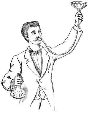
|
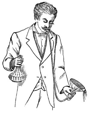
|
29 From Souligoux, De la Dilatation de l'Estomac, Paris,
1883.
Another convenient but somewhat more complicated method of employing
the siphon process is according to Rosenthal's principle, and is
represented in Fig. 22. To the outer end of the stomach-tube is
attached a Y-shaped glass tube, one arm of which is connected with an
elastic tube running to an irrigator, while the other arm is connected
with the discharging tube. Through the irrigating tube water runs into
the stomach, the discharging tube being compressed. If the discharging
tube be opened while the fluid is flowing from the irrigator, and if
then, after the establishment of a column of water in the discharging
tube, the irrigating tube be compressed or the stopcock of the
irrigator be closed, a siphon communicating with the stomach is formed
and empties this organ of its fluid contents.
|
FIG. 22.30
|
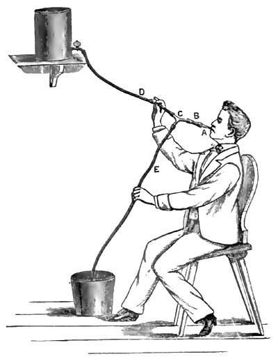
|
30 From Leube, in Ziemssen's Handb. d. spec. Path. u.
Therap., Bd. vii.
In the siphon process the tube inserted into the stomach may be an
incompressible hard-rubber tube like that employed with the
stomach-pump, but by far the simplest, most convenient, and safest
form of stomach-tube is the soft, flexible, red rubber tube,
resembling the Jacques catheter, but of course larger and longer.31
This soft tube can inflict no
injury, and in most cases it is
readily introduced. Generally, the patient himself can best manipulate
the introduction of the tube. After the tube is introduced into the
pharynx, the patient, who should be in a sitting posture, makes
repeated acts of swallowing, by means of which, accompanied by
directing and gently pushing the tube with the fingers, the tube
passes along the oesophagus into the stomach. Often at first the
nervousness and inexperience of the patient occasion some trouble, but
after a little practice he generally succeeds in introducing the tube
without discomfort or difficulty. Before its introduction the tube
should be anointed with a little vaseline or some similar substance.
In an adult the tube is introduced for a length of at least 20 to 25
inches, and in cases of dilatation of the stomach of course for a
greater distance. Whatever form of stomach-tube be used, it is
important that the tube should be at least 30 inches long, and should
be provided with one, and preferably with two, large eyes at its
distal extremity.
31 Such a tube (marked 19 A) is made by Tieman & Co. of New
York, and is to be had of most surgical instrument-makers. (For a
fuller description of the tube and the mode of its employment see
article by W. B. Platt, "The Mechanical Treatment of Diseases of the
Stomach," Maryland Medical Journal, March 8, 1884.)
Oser's tube is 2 meters long, and is made of mineralized rubber. There
are two sizes. The smaller has a lumen of 8 mm. The thickness of the
wall is 2½ mm. In the larger tube the lumen is 10 mm., and the
thickness of the wall 3 mm. He generally uses the smaller tube.
Faucher's tube is 1½ meters long. The external diameter of the tube is
10 to 12 mm. The walls are of such thickness that the tube can be bent
without effacing its lumen. At one extremity is a lateral eye with two
orifices. To the other extremity is adapted a funnel with a capacity
of about 500 grammes.
Although the stomach-pump has the advantage of more completely
evacuating the stomach and of removing coarser solid particles than is
possible with the siphon, nevertheless its disadvantages—namely, the
possibility of inflicting injury to the mucous membrane of the
stomach,32 the expense and greater complexity of the instrument, and
the circumstance that it should be used only by the physician—in
contrast with the advantages of the siphon—namely, its cheapness,
simplicity, safety, and possible employment by the patient or his
attendants—have led to the general adoption of the latter process.
Only the soft-rubber stomach-tube should be left to the employment of
the patient.
32 A number of cases have been recorded in which pieces of
the mucous membrane of the stomach have been detached by the
stomach-pump. Although as yet no serious effects have followed this
accident, the possibility of its occurrence can certainly not be
regarded with equanimity.
Sometimes the flow through the siphon is interrupted by occlusion of
the eye of the stomach-tube by a solid mass or by some cause not
always clear. As already mentioned, it is desirable that there should
be two openings at the gastric extremity of the tube. When the flow is
interrupted the position of the tube in the stomach may be
changed, or the patient may be directed to cough or to exert the
pressure of the abdominal muscles, or more water may be allowed to run
into the stomach in order to displace an occluding mass in the tube.
It is, however, well for such cases to have, if possible, a
stomach-pump and an incompressible tube in reserve. Moreover, as is
apparent from the foregoing statement of the advantages of the
stomach-pump, there are cases in which this instrument is much more
useful than the siphon, so that one cannot decide unconditionally in
favor of one instrument over the other.
The stomach-tube should be secured so that there can be no possibility
of its being swallowed entirely. A string may be attached to the
distal end of the tube. Leube33 has reported an instance in which
the whole tube disappeared into the stomach, and Jackson34 has also
narrated a case in which an insane patient swallowed the stomach-tube.
In both cases the tube was subsequently rejected by vomiting.
33 Deutsches Arch. f. klin. Med., Bd. 33, p. 6.
34 Extracts from the Records of the Boston Society for
Medical Improvement, vol. vi. p. 261.
For washing out the stomach after the greater part of the contents
have been withdrawn, about a pint of tepid fluid is allowed to slowly
run into the stomach, and is then siphoned out. This process is to be
repeated several times. In general, tepid water suffices for washing
out the stomach, but it is often better to use, at least a part of the
time, a 1 to 2 per cent. solution of bicarbonate of sodium, which
facilitates the removal of mucus. The artificial and the natural Vichy
and Carlsbad waters are also excellent for this purpose. Various
additions are also made to the water with the view of counteracting
fermentative changes in the stomach. For this purpose perhaps the best
agents are salicylate of sodium (1 per cent. solution) or resorcin (2
per cent. solution). Other substances which have also been recommended
are carbolic acid, permanganate of potassium, hyposulphite of sodium,
creasote, benzine. Simple water, however, accomplishes about all that
is possible, and many are satisfied to use it without any medication.
As regards the frequency with which the stomach is to be washed out,
one is to be guided by the symptoms and the effect obtained by the use
of the stomach-tube. As a general rule, it suffices to wash out the
stomach once a day, and often the process need be repeated only every
second or third day.
Opinions are divided as to the best time of day to select for washing
out the stomach. Kussmaul recommends the morning before breakfast, and
the majority have followed his advice; others prefer the evening.
There is much, however, in favor of washing out the stomach about half
an hour before the principal meal of the day. The best opportunity has
been offered for the digestion and absorption of the food taken at the
previous main meal, and the stomach is placed in the best possible
condition for the reception of more food.
The habitual washing out of the stomach is not without its drawbacks.
We often remove, as has been pointed out especially by Leube, not only
noxious substances from the stomach, but also the completed products
of digestion. To withdraw from the nourishment of the body this chyme
which the stomach has laboriously manufactured cannot be a matter of
indifference. Still, with the weakened absorptive powers of the
stomach,
and its inability to properly propel its contents into
the intestine, it is a question how much of this chyme would
eventually be utilized for nutrition. Another point is worthy of
attention. The relief which the patient experiences when his
overloaded stomach is freed of its burden, and the knowledge that this
method of relief is always at hand, may make him careless in the
observance of the dietetic rules which are of great importance in the
treatment of this disease. It is well, therefore, not to wash out the
stomach oftener than is necessary, nor to continue the habitual use of
the stomach-tube longer than is required.
There are contraindications to the use of the stomach-tube. In very
rare instances the attempt to introduce the tube causes the patient so
much distress, produces such violent spasm of the pharyngeal and
adjacent muscles, or induces so much retching and vomiting, or is
attended with such prostration or even syncope, that this method of
treatment has to be abandoned. Great weakness, recent gastric
hemorrhage, ulcer of the stomach in most cases (see page 523), often
cancer of the cardia or of the oesophagus, and aneurism of the aorta,
are contraindications to the use of the stomach-tube.
If we group together the results obtained by the use of the
stomach-tube in gastric dilatation, we shall find cases in which no
benefit results; cases which are benefited, but are obliged to
continue the use of the stomach-tube throughout life; cases in which
recovery is slow and gradual; cases with more or less speedy relief or
apparent cure, but followed by relapses; and cases of prompt relief
and permanent cure.
The regulation of the diet is never to be neglected in cases of
dilatation of the stomach. Here the guiding principles are that little
fluid should be taken, and that the food should be small in bulk,
nutritious, easily digestible, and not readily undergoing
fermentation. The patient should drink as little water as possible,
and should therefore avoid whatever occasions thirst. It is hardly
practicable to carry out the plan of giving water mostly by the
rectum, as has been proposed. In most cases milk is useful, but an
exclusively milk diet is not generally well borne on account of the
quantity of fluid required. Leube's beef-solution is often
serviceable. Soft-boiled eggs and tender meats are to be allowed,
particularly the white meat of fowl and rare beefsteak, especially
that prepared from scraped and finely-chopped beef, as recommended in
the treatment of gastric ulcer (page 521). Fatty, saccharine, and
amylaceous articles of food—hence most vegetables and fruits—are to
be avoided on account of their tendency to undergo fermentation in the
stomach. Alcohol in any form is usually detrimental. If gastric
symptoms, particularly vomiting, be very urgent, or if food introduced
into the stomach affords little or no nourishment, as in some cases of
tight pyloric stricture, then rectal alimentation is to be resorted to.
An important indication is to restore the tone and contractile power
of the muscular coat of the stomach. For this purpose electricity, in
the form both of the constant and of the faradic current, has been
beneficially employed. The best results are reported from the use of
the faradic current. Both poles may be applied over the region of the
stomach. The application of electricity to the inside of the stomach
by means of electrodes attached to stomach-tubes or bougies is a more
difficult procedure, but has its advocates. Uniformly good
results are not obtained by the use of electricity in gastric
dilatation, but there can be no doubt that in some cases decided
benefit follows this method of treatment.
Nux vomica, particularly its alkaloid strychnia, has been much
employed with the view of stimulating the muscular power of the
stomach. Strychnia is given either internally or hypodermically.
Hypodermic injections of ergotin have also been used for the same
purpose. It has been hoped to increase the contraction of the stomach
by cold applications to the abdomen, as by ice-bags applied
immediately after washing out the stomach. The benefit derived from
these various attempts to increase the tonicity of the gastric muscle
is not very apparent.
A belt or bandage around the abdomen in order to support the stomach
sometimes makes the patient feel more comfortable; in other cases it
aggravates the symptoms.
In many cases digestion is promoted by giving dilute hydrochloric acid
with or without pepsin. About ten drops of dilute hydrochloric acid
may be given half an hour to an hour after each meal.
When the stomach is systematically washed out, the individual symptoms
of dilatation of the stomach will rarely require special treatment.
The sensation of fulness and weight in the stomach, the eructations,
the vomiting, and the constipation are generally relieved, at least
temporarily, by washing out the stomach. The appetite is improved, and
an increase in weight is usually soon noticeable.
If heartburn and eructations of gas continue troublesome, an antacid,
such as bicarbonate of sodium or prepared chalk, will be found useful.
Leube, in order to relieve constipation and to increase the
peristalsis of the stomach, administers Carlsbad water (see page 522).
Not more than five or six ounces of the water need be given, and this
should be taken slowly in divided doses. A laxative pill containing
rhubarb may be given occasionally.
If anæmia be the cause or a prominent accompaniment of dilatation of
the stomach, iron may be administered in a form as little disturbing
the digestion as possible, as the effervescing citrate or the lactate,
or arsenic in the form of Fowler's solution may be tried. In general,
however, all drugs which impair the appetite or digestion are to be
withheld. The digestion and the general condition of the patient are
often benefited by massage.
Resection of the pylorus in cases of cancerous and of cicatricial
stenosis of this orifice has been performed in several instances. The
subject, as regards its medical in distinction from its surgical
bearings, has already been discussed in connection with cancer of the
stomach (see page 577). Here it may be added that the propriety of
resection is less open for dispute in cases of non-cancerous pyloric
stenosis than it is in cancer of the pylorus.
Remarkable results have been reported by Loreta in cases of
cicatricial stenosis of the pylorus. After performing gastrotomy he
inserts his fingers through the constricted pyloric orifice and
forcibly dilates the stricture.35 To judge from experience in
divulsing strictures in other parts of
the body, it does not
seem probable that a permanent cure can be often effected by this bold
and dangerous procedure.
35 Loreta has performed this operation successfully no less
than nine times (The Lancet, April 26, 1884).
Under the name acute dilatation of the stomach36 have been described
cases in which it has been supposed that a more or less suddenly
developed paralysis of the muscular coat of the stomach exists. But
the propriety of the term acute dilatation, and the very existence of
an acute paralysis of the stomach, are, to say the least, questionable.
36 The literature pertaining to the subject of acute
dilatation of the stomach is to be found in Poensgen, Die Motorischen
Verrichtungen des Menschlichen Magens, Strasburg, 1882, p. 95.
As causes of this so-called acute dilatation of the stomach have been
assigned injuries, particularly those affecting the abdomen, surgical
operations involving the peritoneum, acute inflammations of the mucous
and of the peritoneal coats of the stomach, acute fevers, especially
during convalescence, and overloading the stomach with food or with liquids.
The symptoms which have been chiefly emphasized are severe abdominal
pain, tympanitic distension of the stomach, and absence or cessation
of vomiting if this has previously existed. It will be noted that
inability to vomit under these circumstances implies not only
paralysis of the stomach, but also that of the abdominal muscles.
The prognosis depends on the character of the primary disease causing
the alleged paralysis.
If there be acute distension of the stomach with inability of the
organ to expel its contents either externally or into the intestine,
the stomach-tube may be employed to evacuate the gas and other
material present.
In a case described by Hilton Fagge37 as acute dilatation of the
stomach the symptoms of dilatation appeared suddenly and ran an acute
course, but the autopsy showed that the dilatation was doubtless of
much longer development than the symptoms indicated. In a case
reported by Nauwerk38 of extreme dilatation in consequence of
hypertrophic stenosis of the pylorus, after ten months of
insignificant dyspeptic symptoms there suddenly appeared, after excess
in eating, symptoms of dilatation of great severity, which continued
until a fatal termination at the end of three months. Thus it appears
that chronic dilatation of the stomach may cause little disturbance
for a considerable time and then run a rapid course.
37 "On Acute Dilatation of the Stomach," Guy's Hosp.
Rep., xviii. p. 4, 1873.
38 Deutsches Arch. f. kl. Med., Bd. xxi. p. 573.
DEFINITION.—Cirrhosis of the stomach is characterized by thickening
of the walls of the greater part or of the whole of the stomach in
consequence of a new growth of fibrous tissue, combined usually with
hypertrophy of the muscular layers of the stomach. The cavity of the
stomach is usually contracted, but sometimes it is of normal size or
even dilated.
SYNONYMS.—Fibroid induration of the stomach; Hypertrophy of the walls
of the stomach; Chronic interstitial gastritis; Sclerosis of the
stomach; Plastic linitis.
HISTORY.—The writings of the seventeenth and eighteenth centuries
contain many records of extremely contracted stomachs with uniformly
and greatly thickened walls (Butzen, Löseke, Storck, Portal, Lieutaud,
Pohl, etc.). In the works of Lieutaud and of Voigtel may be found
references to many such cases.1 Doubtless, some of these cases were
examples of cirrhosis of the stomach, but in the absence of
microscopical examination it is not possible to separate these from cancer.
1 Lieutaud, Historia anat.-med., t. i. p. 8, Venet.,
1779; Voigtel, Handb. d. Path. Anat., Bd. ii. p. 450, Halle, 1804.
Here it may be mentioned that Diemerbroeck's case, which is so often
quoted to prove that polyphagia instead of causing gastric dilatation
may produce hypertrophy of the muscular coat of the stomach, with
contraction, was probably an instance of cirrhosis of the stomach.
Andral2 was the first to describe fully and systematically
hypertrophy of the walls of the stomach. He attributed the lesion to
chronic inflammation. He erroneously supposed that scirrhus of the
stomach was only hypertrophy of the gastric walls. Cruveilhier3
distinguished between scirrhous induration and hypertrophy, which he
considered to be a final result of the irritation accompanying chronic
diseases of the stomach. Rokitansky's4 description of fibroid
induration of the stomach, although brief, is accurate. He says that
the process usually involves the whole
stomach, and that it
originates in an inflammation of the submucous connective tissue. This
inflammation may occur either independently or in combination with
gastritis mucosa. Rokitansky emphasizes the error of confounding the
disease with scirrhous cancer. Bruch5 made an elaborate study of
hypertrophy of the walls of the stomach, which he considered to be the
final stage of various chronic diseases of the stomach. Fibrous or
scirrhous cancer he considered to be nothing but this hypertrophy.
2 Précis d'Anat. path., Paris, 1829.
3 Anatomie pathologique, Paris, 1830-42.
4 Lehrb. d. Path. Anat., Wien, 1855-61.
5 Zeitschr. f. rat. Med., Bd. vii., 1849.
The best descriptions of cirrhosis of the stomach have been furnished
by English writers, by most of whom it is properly regarded as an
independent disease. Brinton6 first employed the names cirrhosis of
the stomach and plastic linitis. Excellent descriptions of the disease
have been given by Hodgkin, Budd, Brinton, Habershon, H. Jones, Wilks,
Quain, and Smith.
6 Diseases of the Stomach.
While in former times cirrhosis of the stomach was confounded with
cancer, in recent times it has not been separated by many from chronic
catarrhal gastritis. In German systematic works the disease receives,
as a rule, only passing mention in connection with chronic catarrhal gastritis.
ETIOLOGY.—Cirrhosis of the stomach is rare, but it is not so
exceptional as to be without any clinical importance. I have met with
three cases at post-mortem examination.
The disease is more frequent in men than in women. A considerable
number of cases have occurred between thirty and forty years of age,
but the greatest frequency is after forty. At an earlier age than
twenty the disease is very rare.
The causation of cirrhosis of the stomach is obscure. Nearly all
writers upon the subject have emphasized the abuse of alcohol as an
important cause in this as in other diseases of the stomach.
Intemperance cannot, however, be the only cause; and here, as
elsewhere, it is not easy to say what importance is to be attached to
it as an etiological factor. In only one of the three cases which I
examined post-mortem could it be determined that the patient was an
immoderate drinker, and in one case intemperance could be positively
excluded. Other cases have been recorded in which the abuse of spirits
could be positively excluded. In one of my cases syphilis existed, as
was established by the presence of gummata in the liver. In some cases
the disease has been attributed to cicatrization of a gastric ulcer.
In a case reported by Snellen the disease followed an injury to the
epigastric region.7
7 Canstatt's Jahresbericht, 1856, iii. 302.
Cirrhosis of the stomach, as well as cancer, ulcer, and most other
chronic structural diseases of this organ, is usually associated with
chronic catarrhal gastritis. There is, however, no proof of the
prevalent idea that chronic catarrhal gastritis is the cause of the
enormous new growth of fibrous tissue which characterizes typical
cases of this disease.
SYMPTOMATOLOGY AND DIAGNOSIS.—The symptoms of cirrhosis of the
stomach are not sufficiently characteristic to warrant a positive
diagnosis. Sometimes the disease pursues a latent course. Like cancer
of the stomach, it may put on various disguises. Thus, in a case of
cirrhosis of the stomach reported by Nothnägel8 the symptoms were
typically those of progressive pernicious anæmia. Association
with ascites or with chronic peritonitis may lead to a false
diagnosis. Thus, in one of the cases which I examined after death, and
in which there was chronic peritonitis with abundant fluid exudation,
the disease during life was diagnosticated as cirrhosis of the liver.
Most frequently, however, cirrhosis of the stomach is mistaken for
gastric cancer, from which, in fact, it can rarely be positively diagnosticated.
8 Deutsches Arch. f. kl. Med., Bd. 24, p. 353.
The symptoms are usually those of chronic dyspepsia, which sooner or
later assumes a severity which leads to the diagnosis of some grave
structural disease of the stomach, usually of cancer.
Indigestion, loss of appetite, oppression in the epigastrium,
vomiting, are the common but in no way characteristic symptoms of
cirrhosis of the stomach. There may be severe gastralgia, but in
general the disease is less painful than either ulcer or cancer of the
stomach. The inability to take more than a small quantity of food or
of drink at a time, with the sense of fulness which even this small
quantity occasions, has been considered somewhat characteristic of
cirrhosis of the stomach, but this symptom is too inconstant, and
occurs in too many other affections of the stomach, to be of much
service in diagnosis. The symptoms of dyspepsia are often of much
longer duration than in cancer, existing sometimes for many years (up
to fifteen years), but on the other hand there have been cases in
which the clinical history of gastric cirrhosis was as rapid in its
progress as cancer. Moreover, cancer may be preceded by dyspeptic
symptoms of long duration, but long duration is the exception with
cancer and the rule with cirrhosis of the stomach.
As the disease progresses the patient loses flesh and strength, and
usually dies in a condition of marasmus. Blood is rarely present in
the vomit, but in a few cases the vomiting of coffee-ground material
has been noted.
By physical examination sometimes a tumor in the region of the stomach
can be felt. Under favorable circumstances it can sometimes be
determined that this tumor is smooth, elastic, tympanitic on
percussion, and presents more or less distinctly the contours of the
stomach. By administering effervescing powder it may be possible to
obtain further evidence that the tumor corresponds in its form to the
stomach. The diagnosis of contraction of the cavity of the stomach is
not easy. Some information may be afforded by noting the length to
which the inflexible stomach-tube can be passed. The quantity of water
which can be poured into the stomach until it begins to run out of the
stomach-tube may also bring some confirmatory evidence as to the
existence of contraction of the stomach.
Even should the physical signs suffice to determine that the tumor is
the thickened and contracted stomach, still cancer cannot be excluded,
for this also may grow diffusely in the gastric walls and may cause
contraction of the cavity of the stomach. With our present means of
diagnosis, therefore, the most which can be said is, that a special
combination of favorable circumstances may render probable the
diagnosis of cirrhosis of the stomach, but a positive diagnosis is impossible.
MORBID ANATOMY.—In most cases of cirrhosis of the stomach the stomach
is contracted. The cavity of the stomach has been found not larger
than would suffice to contain a hen's egg, but such extreme
contraction is very rare. When the stomach in this disease is found
dilated, either the thickening involves only or chiefly the walls of
the pyloric portion, or the morbid process probably began there and
was followed by dilatation.
In typical cases the walls of the entire stomach are thickened, but
frequently the thickening is most marked in the pyloric region. The
walls may measure an inch and even more in thickness. The thickened
walls are dense and firm, so that often upon incision the stomach does
not collapse.
Upon transverse section the different coats of the stomach can be
distinguished. The mucous membrane is least affected, being sometimes
thickened, sometimes normal or atrophied. The muscularis mucosæ is
hypertrophied, and is evident to the naked eye as a grayish band. The
submucous coat is of all the layers the most thickened, being
sometimes ten to fifteen times thicker than normal. It appears as a
dense white mass of fibrous tissue. The main muscular coat is also, as
a rule, greatly hypertrophied; the grayish, translucent muscular
tissue is pervaded with streaks of white fibrous tissue prolonged from
the submucous and subserous coats. This last coat resembles in
appearance the submucous coat, which, however, it does not equal in
thickness, although it is, proportionately to its normal thickness,
much hypertrophied. The free peritoneal surface usually appears opaque
and dense.
To the naked eye it is apparent that the new growth of fibrous tissue
is most extensive in the submucous coat, which it is probably correct
to regard as the starting-point of the disease. The hypertrophy of the
muscular layers is also in most cases an important element in the
increased thickness of the gastric walls.
Microscopical examination9 shows sometimes a nearly normal mucous
membrane. The tubules, however, are usually more or less atrophied. In
the case reported by Nothnägel tubules could be found only in the
pyloric region of the stomach. The essential lesion is the new growth
of fibrillated connective tissue pervading all of the coats of the
stomach. In an interesting case reported by Marcy and Griffith,10
which was believed to be caused by an extensive cicatrized ulcer, a
new formation of smooth muscular tissue was found not only in the main
muscular tunic and the muscularis mucosæ, but also throughout the
submucosa. This peculiarity was probably referable to the
cicatrization of the ulcer.
9 Microscopical examination is always necessary for a
positive diagnosis of cirrhosis of the stomach. In a case which I
examined post-mortem of double ovarian cancer, with multiple secondary
deposits in the peritoneum and with chronic peritonitis, the stomach
presented the typical gross appearances of cirrhosis, but here and
there were to be found nests of cancer-cells in the prevailing new
growth of fibrous tissue in the walls of the stomach.
10 Am. Journ. of the Med. Sci., July, 1884, p. 182.
Not infrequently adhesions exist between the stomach and surrounding
organs. Exceptionally, a diffuse growth of fibrous tissue may invade
the greater part of the peritoneum, particularly the visceral layer,
and cause a thickening similar to that existing in the stomach. In
such cases ascites is usually a marked symptom.
PROGNOSIS.—The prognosis of cirrhosis of the stomach is grave. The
disease runs a chronic course, and usually terminates in death by
asthenia. There is no reason to believe that the stomach can ever be
restored to its
normal condition. Still, cases have been
reported in which it has been supposed that cirrhosis of the stomach
has terminated in recovery.11 The diagnosis, however, in such cases
must remain doubtful.
11 Lesser, Cirrhosis Ventriculi, Inaug. Diss., Berlin,
1876; Smith, "Cirrhosis of the Stomach," Edinb. Med. Journ., 1872,
p. 521.
TREATMENT.—The treatment is symptomatic, and is to be guided by the
general principles developed in previous articles concerning the
regulation of the diet and the administration of remedies.
The various causes of stenosis of the pylorus have already been
mentioned under DILATATION OF THE STOMACH, and the most important of
these causes have received full consideration in connection with ULCER
and with CANCER OF THE STOMACH.
Only one of the varieties of pyloric stenosis can claim consideration
as an independent disease. This variety is the so-called hypertrophic
stenosis of the pylorus (Lebert) or fibroid degeneration of the
pylorus (Habershon12). Under the name of hypertrophic stenosis have
been described cases in which the stenosis was due to hypertrophy of
only one of the coats of the stomach, usually either the submucous or
the muscular coat, sometimes only the mucous coat. In most cases,
however, all of the coats of the stomach are involved, and the lesion
is similar to that of cirrhosis of the stomach, but it is confined to
the pylorus or to the pyloric region. In such cases there is new
growth of fibrous tissue, most marked in the submucous coat, and
hypertrophy of the muscular coat. The appearance of the pylorus in
some instances of hypertrophic stenosis has been not inappropriately
compared to that of the cervix uteri.
12 Habershon, On Diseases of the Abdomen, London, 1862;
Lebert, Die Krankh. d. Magens, Tübingen, 1878; Nauwerk, Deutsches
Arch. f. klin. Med., Bd. 21, 1878.
In the majority of cases the change here described is the result of
cicatrization of a gastric ulcer, and some believe that all cases of
so-called hypertrophic stenosis or fibroid degeneration of the pylorus
are referable to ulcer, although it may be very difficult to discover
the cicatrix of the ulcer. It is certainly not always possible to
detect either ulcer or cicatrix, so that it seems proper to regard the
hypertrophic stenosis in such cases as constituting an independent affection.
The symptoms are those of dilatation of the stomach, sometimes
preceded by evidences of chronic catarrhal gastritis. The thickened
pylorus can sometimes be felt during life as a small, cylindrical,
usually movable tumor, either stationary in progress or of very slow growth.
In most cases the diagnosis of organic stenosis of the pylorus can be
made. Cancer may sometimes be excluded by the long duration of the
symptoms and the stationary character of the tumor if a tumor can be
felt. The exclusion of ulcer is more difficult and hardly possible,
for ulcer may have existed without producing characteristic symptoms.
The prognosis and treatment have been considered under DILATATION OF
THE STOMACH.
Atrophy of the stomach may be the result of stenosis of the cardia or
of the oesophagus. The stomach may participate with other organs in
the general atrophy attending inanition and marasmus. The walls of a
dilated stomach may be very thin.
Especial importance has been attached in recent years to degeneration
and atrophy of the gastric tubules. The glands of the stomach may
undergo degeneration and atrophy in various diseases of the stomach,
such as chronic catarrhal gastritis, phlegmonous gastritis, cirrhosis
of the stomach, and cancer of the stomach. Parenchymatous and fatty
degeneration of the glandular cells of the stomach occurs in acute
infectious diseases, as typhoid fever and yellow fever, also as a
result of poisoning with phosphorus, arsenic, and the mineral acids.
It is claimed by Fenwick that atrophy of the stomach may occur not
only as a secondary change, but also as a primary disease attended by
grave symptoms. Fenwick has described a number of cases in which the
gastric tubules were atrophied without thickening of the walls of the
stomach and without diminution in the size of the cavity of the
stomach—cases, therefore, which cannot be classified with cirrhosis
of the stomach.13 He attributes in many cases the atrophy of the
tubules to an increase in the connective tissue of the mucous
membrane, and draws a comparison between atrophy of the stomach and
the atrophic form of chronic Bright's disease.
13 The Lancet, 1877, July 7 et seq.
In 1860, Flint14 called attention to the relation between anæmia and
atrophy of the gastric glands. He expressed the opinion that some
cases of obscure and profound anæmia are dependent upon degeneration
and atrophy of the glands of the stomach. Since Flint's publication
cases have been reported by Fenwick, Quincke, Brabazon, and Nothnägel,
in which lesions supposed to be due to pernicious anæmia have been
found after death associated with atrophy of the gastric tubules.15
Nothnägel's case, which has already been mentioned, was one of
cirrhosis of the stomach.
14 A. Flint, American Medical Times, 1860. Further
contributions of Flint to this subject are to be found in the New
York Medical Journal, March, 1871, and in his Treatise on the
Principles and Practice of Medicine, p. 477, Philada., 1881.
15 Fenwick, loc. cit.; Quincke, Volkmann's Samml. klin.
Vorträge, No. 100 (case b); Brabazon, British Med. Journ., 1878,
July 27 (without microscopical examination!); Nothnägel, Deutsches
Arch. f. kl. Med., Bd. 24, p. 353.
The symptoms which have been referred to primary atrophy of the
stomach are severe anæmia and disturbances of digestion, such as
anorexia, eructations, and vomiting. The digestive disturbances are
often not greater than are frequently observed in cases of severe anæmia.
In my opinion, the existence of atrophy of the stomach as a primary
and independent disease has not been established. In many cases which
have been described as primary atrophy the histological investigation
of the stomach has been very defective. Degeneration and atrophy of
the gastric tubules secondary to various diseases of the stomach and
to certain general diseases is an important lesion when it is
extensive, and must seriously impair the digestion, and consequently
the nutrition, of the patient.
These anomalies, so far as they have not received consideration in
previous articles, are of more anatomical than clinical interest, and
therefore here require only brief mention.
The stomach may have an hour-glass shape in consequence of a
constriction separating the cardiac from the pyloric half of the
organ. This constriction is sometimes congenital,16 sometimes caused
by cicatrization of a gastric ulcer, and sometimes caused by spasmodic
contraction of the muscle, which may persist after death, but
disappears when the stomach is artificially distended. Hour-glass
shape of the stomach has been diagnosed during life by administering
an effervescing powder according to Frerichs' method.
16 A careful study of the congenital form of hour-glass
contraction of the stomach has been made by W. R. Williams ("Ten Cases
of Congenital Contraction of the Stomach," Journ. of Anat. and
Physiology, 1882-83, p. 460).
Foreign substances of hard consistence which have been swallowed
sometimes cause diverticula of the stomach.
Sometimes the fundus of the stomach is but little developed, so that
the organ is long and narrow like a piece of intestine.
The stomach may be variously distorted by external pressure, as from
tumors and by adhesions.
The loop-shaped stomach and vertical position of the stomach have been
already considered in connection with DILATATION OF THE STOMACH
(page 602).
In transposition of the viscera the stomach is also transposed. In
such a case difficulties may arise in the diagnosis of pyloric cancer,
as in a case described by Légroux.
The stomach may be found in hernial sacs. Mention has already been
made of the presence of dilated stomachs in scrotal hernia. More
frequently the stomach is found in umbilical hernias. In diaphragmatic
hernia the stomach is found more frequently in the thorax than is any
other abdominal viscus. In 266 diaphragmatic hernias collected by
Lascher17 the stomach was found either wholly or partly in the
thorax in 161 cases. The clinical consideration of diaphragmatic
hernia, however, does not belong here.
17 Deutsches Arch. f. kl. Med., Bd. 27.
Furthermore, the stomach may be displaced by tumors, enlargement of
neighboring organs, tight-lacing, adhesions, and the weight of
hernias. These displacements, however, are generally inconsiderable
and of little importance.
In a case described by Mazotti18 the stomach, of which the pyloric
portion was fixed by adhesions, was twisted around its long axis.
Death was caused by uncontrollable vomiting.
18 Virchow und Hirsch's Jahresbericht, 1874, ii. p. 249.
Sufficient attention has already been given to perforation of the
stomach in consequence of diseases of its walls, such as ulcer,
cancer, abscesses, and toxic gastritis.
A healthy stomach may be ruptured by violent injury to the abdomen
even when no external wound is produced. An example of rupture of the
stomach from this cause is that sometimes produced when a person has
been run over by a heavy vehicle.
It has been claimed that a stomach with healthy walls may burst in
consequence of over-distension of the organ with solids or with gas.
The older literature is especially rich in reports of so-called
spontaneous rupture of the stomach. Most of these cases were examples
of perforation of gastric ulcer. In a case of apparently spontaneous
rupture of a stomach which had become abnormally distended with gas,
Chiari19 found that the rupture was through the cicatrix of a simple
ulcer in the lesser curvature. It is hardly conceivable that rupture
of the healthy stomach from over-distension can occur so long as the
orifices of the organ are unobstructed.
19 Wiener med. Blätter, 1881, No. 3.
Lautschner20 reports a case of spontaneous rupture of the stomach in
a woman seventy years old with an enormous umbilical hernia which
contained the pyloric portion of the stomach. After drinking eight
glasses of water and two cups of tea and eating meat, she was seized
with vomiting, during which the stomach burst with a report which was
audible to the patient and to those around her. She passed into a
state of collapse and died in thirteen hours. A rent several
centimeters long was found in the posterior wall of the stomach.
Lautschner thinks that the pylorus was bent in the hernial sac so as
to be obstructed. In the walls of the stomach he found no evidence of
pre-existing disease.
20 Virchow und Hirsch's Jahresbericht, 1881, ii.
There is no satisfactory proof of the possibility of the occurrence of
rupture of a stomach with healthy walls except as a result of external violence.
The symptoms and treatment of rupture of the stomach are those of
perforation of the stomach, and have already been described. The
prognosis is fatal.
That the subject of gastromalacia should still occupy so much space in
medical works the purpose of which is mainly clinical proves that many
physicians still cling to the belief that this process may occur
during life. It is, nevertheless, certain that the condition which,
according to the ordinary and traditional use of the term, is
designated gastromalacia, is always a post-mortem process and is
without the slightest clinical significance. So long as the
circulation of the blood in the walls of the stomach is undisturbed,
self-digestion of this organ cannot occur. No one doubts
that
parts of the gastric walls in which the circulation has been arrested,
and which are exposed to the gastric juice, undergo self-digestion, as
has already been set forth in the article on GASTRIC ULCER. To
describe cases of this nature under the name of gastromalacia,
however, is misleading, and can cause only confusion, for the
long-continued discussion as to whether gastromalacia is a vital or a
cadaveric process applied certainly to a different conception of the
term. In some of the cases which have been published, even in recent
years, in support of the vitalistic theory of gastromalacia, and in
which it has been proven that perforation of the stomach occurred
during life, the solution of continuity took place through parts of
the gastric walls in which the circulation had already been
obstructed, particularly by extensive hemorrhagic infiltration. Some
of these cases are probably also examples of perforation of gastric
ulcer or of rupture of cicatrices from over-distension of the stomach,
in which post-mortem digestion of the edges of the ulcer or of the
cicatrix obscured the real nature of the process. The subject of
gastromalacia should be relegated wholly to works on physiology and on
pathological anatomy.
NATURE.—The term indigestion in its most common meaning refers to
gastric indigestion only. This limitation has arisen from the fact
that gastric digestion has been more thoroughly understood than
intestinal digestion, and because the symptoms, flatulence, acidity,
eructations of gas, pyrosis, and vomiting of unaltered food, are
readily referred to the stomach as their source. Intestinal digestion
has not been well known until within a recent date, and its phenomena
in disease have been mistaken for other pathological conditions.
From the important and complex function of the intestinal juices, and
the very great share they take in the solution of food, there must be
many phases of departure from the normal state. The processes of
intestinal digestion are more intricate than those of gastric
digestion, of a higher grade, and the chemical reactions are more
numerous, depending upon the participation of the bile, the pancreatic
juice, and the succus entericus; while intestinal absorption is a more
complex act than that of gastric absorption.
A brief review of the physiology of intestinal digestion will be of
aid in making clear its pathology.
The object of all digestion is to make such a solution of the ingesta
that they may pass through animal membrane and so enter the system.
Mechanical disintegration and simple solution do something toward
this, but for substances insoluble in water a more thorough change is
brought about by ferments which convert insoluble into soluble
compounds.
The process of digestion begins in the mouth. Mastication breaks up
the masses of food; the saliva softens them, dissolves soluble
substances, as salt and sugar, and thus the pleasures of the palate
are enhanced. The ferment ptyalin acts upon starch (boiled starch
being more rapidly altered than unboiled), and changes it to dextrin
and grape-sugar, both of which are diffusible through animal membrane,
entering lymph-spaces and blood-vessels. The greater part of the
saliva secreted is swallowed with the food or in the intervals of
eating. The amount formed in twenty-four hours varies from 1500 gm.
(Bidder and Schmidt) to 700 gm. (Tuczek). It must therefore serve some
ulterior purpose in the stomach. Ewald1 says that saliva converts
starch into sugar in acid as well as in alkaline and neutral
solutions. But Langley2 asserts that the ferment of
saliva is
destroyed by the hydrochloric acid of the gastric juice. The longer
food is subjected to mastication and insalivation, the more thorough
is the mouth digestion and the better prepared is the mass for the
action of the gastric and intestinal juices. It is asserted that fatty
matters are emulsified to a certain extent by the alkaline ferments of
the saliva.
1 Lectures on Digestion, New York, 1881, p. 37.
2 "On the Destruction of Ferments in the Alimentary Canal,"
Journal of Physiology, London, Jan., 1882, p. 246.
When the food enters the stomach the nitrogenous (albuminoid) elements
are attacked by the gastric juice, the bundles of muscular fibre are
broken up, the fibrillæ are reduced to a granular mass, but not
completely dissolved (Frerichs), the fat-globules are freed from their
envelopes of connective tissue, milk is coagulated, and the casein is dissolved.
"The tangible, practical object of this change is to form out of a
little-diffusible body (albumen) one easily diffusible (peptone),
which is capable of absorption through animal membrane in a higher
degree than ordinary albumen" (Ewald). Peptone is formed out of
ordinary albumen, as grape-sugar is formed out of starch, by taking up
water; it is therefore the hydrate of albumen.
The more tardy the digestion in the stomach the more highly charged
with acid is the gastric juice. According to Wright, the degree of
alkalinity of the saliva is in proportion to the acidity of the
stomach fluids, and Bence Jones has observed that during the excretion
of acid in the stomach the total alkalinity of all alkaline digestive
fluids is increased. The lesson is thus learned that a too careful
preparation of food, so as to shorten and lessen gastric labor,
diminishes the activity of the gastric juice as well as that of all
other digestive fluids.
Intestinal digestion begins when the softened mass passes through the
pylorus. This mass (chyme) is composed of (1) the products of gastric
digestion which have not been absorbed—peptone, dextrose, levulose,
peptonized gelatin, with mucus and gastric juice; (2) all matters
which have escaped digestion—the starch of vegetable substances,
dissolved gelatin and albumen which have not been peptonized, and some
unaltered muscle-structure; and (3) fat, fatty acids, and cellulose
upon which neither saliva nor gastric juice has had any influence (Ewald).
This complex semi-fluid mass with an acid reaction enters the duodenum
and comes in contact with fluids and ferments destined to work
remarkable changes in its composition. The first of these fluids is
the bile, which is alkaline and composed of the glycocholate and
taurocholate of sodium, cholesterin, soaps, etc., phosphates and
carbonates of lime and sodium, chlorides of potassium and sodium,
bile-pigment, etc. The outflow of bile is excited by the contact of
the chyme with the orifice of the bile-duct. When the alkaline bile is
mingled with the acid mass in the duodenum, it neutralizes its
acidity, precipitates the peptones, and therefore stops all further
action of the gastric juice. Fats containing free fatty acids are
emulsified, soaps being formed by a combination of the alkalies of the
bile with the fatty acids. Lastly, bile hinders fermentation in the
intestine and acts as a purgative by exciting peristalsis. Absorption
is probably also favored by bile, as it has been found that emulsified
fats pass more readily through an animal membrane which has been wet
with bile.3
3 Ewald thinks this result is doubtful: in animals killed
during digestion he has found an acid reaction in the contents of the
intestine beyond the opening of the bile-duct, with no precipitation
of the albumen (op. cit., p. 82).
As far as we now know, the function of the bile is to neutralize
the acidity of the duodenal contents, and thus pave the way for the
action of a digesting fluid of much greater potency and of much higher
function.4
4 In order still further to demonstrate the necessity of
bile-action as a preparation for pancreatic digestion, it may be
mentioned that in artificial experiments, with a heat equal to that of
the body, if antiseptics analogous to gastric juice and bile are not
used, there is a too rapid change from alkalinity to acidity, and
consequently all of the starch is not converted into sugar before it
develops lactic acid with putrefactive disorganization. A deficiency
of bile, therefore, is a cause of intestinal indigestion (Bartlett,
op. cit., pp. 12, 13).
This fluid, the pancreatic juice, is composed of inorganic salts,
albuminoids, and certain specific ferments, and has an alkaline
reaction. It has a threefold operation upon the softened mass with
which it now comes in contact: 1. The starch of vegetable matter,
which has been only slightly acted on up to this time, is now rapidly
converted into grape-sugar by a peculiar diastatic ferment more active
than any other known ferment. 2. Albuminous matters (proteids) which
have escaped digestion in the stomach are changed into a soluble and
absorbable pancreas—peptone. Trypsin is the active ferment in this
case (Kühne), and it is only in alkaline or neutral solutions that the
albuminoids are readily dissolved. The necessity of neutralization by
the alkaline bile is thus demonstrated. 3. A ferment distinct from the
others splits the fats into fatty acids and glycerin, and emulsifies
them so that they can be taken up by the lacteals lower down.
Experiments made by mixing albuminates with pancreatic gland-extract,
under favorable conditions, show after a certain time the presence of
leucin, tyrosin, hypoxanthin, and asparaginic acid. In a feebly
alkaline or neutral solution a faint putrefactive odor is soon
noticed, with the development of bacteria; ammonia, sulphuretted
hydrogen, hydrogen, and carbonic acid—evidences of the putrefaction
of albumen—are also detected.
It is difficult to tell when normal digestion in the intestines ends
and putrefaction begins. The conclusion is, that the normal action of
pancreatic juice (trypsin) gives origin to bodies met with in the
ordinary putrefaction of albumen.5 This thin border-line between
normal intestinal digestion and the decomposition of the intestinal
contents has an important bearing on the facts of intestinal indigestion.
5 Ewald, op. cit., p. 92.
The intestinal juice performs a minor but independent part in
digestion. It converts albuminous matter into peptone, and hydrated
starch into sugar. Its function is therefore supplementary to that of
the gastric and pancreatic secretions.6
6 Ewald, op. cit., p. 103; also, "The Functions of the
Intestinal Juice," Charles L. Dana, Med. News, Philada., July 15,
1882, p. 59.
When food enters the mouth the process of digestion begins, and all
the activities of the glands concerned in digestion are probably at
once set in motion. Mastication excites, by reflex action, pancreatic
secretion; the acid chyme touches the orifice of the common bile-duct
and stimulates the outflow of bile; the neutralized chyme next invites
pancreatic digestion. For the integrity of intestinal digestion it is
required that mastication and stomach digestion should be normally performed.
The intestinal movements which are so necessary to digestion by making
successive changes in the position of the intestinal contents are
controlled by nervous arrangements, but may occur independently
of the central nervous system. The ganglia of Auerbach and of Meissner
in the intestinal wall are sufficient for the development of
peristaltic waves. The irritation of the mucous membrane by food,
hyperæmia, and the pouring out of digestive juices, and intestinal
movements, are parts of one process. Paralysis by section of the
splanchnic leads to hyperæmia of the intestinal vessels and increased
peristalsis; stimulation of the splanchnic causes anæmia of the
intestinal wall and arrest of movement. Local cold by producing anæmia
brings about the same result.
The products of digestion as they pass toward the jejunum consist of
diffusible peptones, sugar, emulsified fats and oils, and substances
which have escaped digestion, as fragments of muscular fibre,
starch-corpuscles, connective tissue, hairs, or other foreign matters.
The bowel contains also carbonic acid, hydrogen, nitrogen,
sulphuretted hydrogen, and marsh gas. The mass, alkaline or neutral in
the duodenum and jejunum, becomes acid in the ileum from the
putrefaction of albumen and fermentation. The peptones and sugar pass
by osmosis into the blood-vessels of the portal system and thence to
the liver. In the liver the sugar is converted into glycogen
(carbohydrate), and stored in the liver-cells until needed for the
maintenance of animal heat and for the nutrition of the tissues. The
peptones are used in part to supply the nitrogenous waste of tissue,
but much of the albuminoid matter is broken up in the liver into
glycogen and urea, the latter of which is excreted by the kidneys as
waste matter.
The minute granules of oil in emulsion are taken up by the epithelial
cells covering the villi; thence they enter the adenoid tissue of the
villi on their way to the lymphatic radicles, the lacteals. From here
the passage is open to the underlying lymphatic vessels and to the
larger abdominal lymph-vessels and the thoracic duct beyond.
Intestinal digestion is not completed and the body does not receive
its pabulum until the products of digestion have reached the liver and
the thoracic duct.
ETIOLOGY.—It is usually said that intestinal dyspepsia is more common
in women than in men, but the contrary is the rule. Some of its most
common causes—over-eating and the eating of indigestible food—are
especially vices of men.
It is more frequent between the ages of forty and fifty, but no age is
exempt. Infants at the breast, children of any age, adults, and old
men and women are alike subject to it. Men in middle life begin to
suffer from the imprudence and carelessness of youth and from the
anxiety and cares of business. The indulged children of rich parents
and improperly bottle-fed infants frequently suffer.
Heredity and idiosyncrasy have a certain influence in determining the
prevalence of intestinal dyspepsia. The distaste for and inability to
digest vegetables, fruits, and fats are often peculiarities of family
history. The occurrence of cases in the same family is often explained
by improper food, bad cooking, and irregular hours, to the evil
influences of which all the members are similarly subjected.
All conditions of the organism which result in a depraved or altered
blood-supply, as anæmia, primary and secondary rachitis, chronic
syphilis, and continued febrile diseases, are causes of intestinal
indigestion.
The connection of the indigestion of fats with the
strumous diathesis and with phthisis is undisputed. J. Hughes Bennett
traced the origin of phthisis to defective fat-digestion; strumous
indigestion and the indigestion of fat are synonymous terms.
Debilitating influences, such as bad air, want of cleanliness and
outdoor exercise, impair functional activity in the intestines as
elsewhere. Sexual excesses, but especially masturbation, have a
special influence for evil in this direction.
The influence of the mind upon the digestion of starch and fats is
even greater than upon gastric digestion, for no other reason perhaps
than that the former is a more complex function and less easily
relieved than the latter. Prolonged or excessive mental labor does not
do so much harm as mental worry, over-anxiety, and the strain and
overwork of business. Professional men—lawyers, physicians, and
clergymen—who become over-burdened with responsibilities, and who
sympathize too much with the distresses of others, are very prone to
suffer. The careworn face with lines about the mouth and forehead is
one of the plainest signs of duodenal defect. The proper secretion of
the juices of the intestine and normal peristalsis are impossible
where brain and nerves get no rest. The too rapid mental development
of the children of the present day is a fruitful source of weakened
fat-and-starch digestion and of impaired development. So long as
children are sent to the public school at four and six years of age,
there will continue to grow up a precocious race with active brains in
feeble bodies.7 This injurious result is largely brought about by
the direct interference of premature brain-development with the
complex intestinal processes of digestion and absorption.
7 In eight of the States and Territories the minimum age
for entering the public school is fixed at four years; in seventeen
States at five years; in the others, except two, at six years. The two
notable exceptions are Alabama and New Mexico, where children do not
enter school until the age of seven.
Wealth, with ease and inactivity, and sedentary occupations,
contribute to the same end by lessening the need of food, and thus
debilitating the organs of digestion by inaction. Sedentary pursuits,
especially those in which the body is bent forward and constricted or
compressed at the waist, interfere with active function in the
intestine. This is the case in tailors, shoemakers, etc. Tight-lacing
in women and a too tight trouser-band in men are injurious.
Hot climates, especially when combined with dampness, lead to disorder
in the intestine and liver. This effect is most marked among persons
coming from colder climates, as among the English in India, who keep
up the habits of eating to which they have been accustomed at home.
The lessened demand destroys the appetite, and stimulants and
condiments are resorted to to whip up the inactive functions. The
intestine is loaded with a mass of crude, unaltered matter which can
with difficulty be disposed of. Chronic indigestion results, varied
with acute attacks of diarrhoea or dysentery. The portal system is
filled with an excess of albuminoid material which the liver is unable
to store away. The excess is got rid of by conversion into uric acid.
Lithæmia and chronic congestion and enlargement of the overloaded
liver result, with their many attendant evils.
Over-eating occasions first gastric and then intestinal indigestion by
the
entrance of unaltered food into the duodenum. Eating without
hunger often involves the taking of food which the body does not need
and which the stomach cannot digest. Diners-out rarely go through a
season without one or more internal revolts. A too-varied diet, a
dinner of many dishes, is faulty in variety as well as in excess. On
the other hand, a too great sameness in diet and the prolonged use of
one or two articles of food which are not easy of digestion, and which
have a great deal of waste, fatigue and then disorder intestinal
digestion. This is a fault into which children are often allowed to fall.
Indigestible food and an excess of starchy or fatty food conduce to
disorder of duodenal digestion. In conditions of debility and anæmia
and in the convalescence of fevers the deficiency of saliva involves
an inability to digest starch in the mouth and points to a
corresponding want in the duodenal secretions. The improper use of
alcoholic liquors, taking them on an empty stomach between meals and
in excess, tends to direct irritation of the mucous tract. Condiments
in large quantity have the same effect.
Irregularity in the hours of eating and a faulty distribution of the
amount of food disturb the perfect working of the mechanism of
digestion. Very light breakfasts and very late and large dinners are
injurious. The habit, now quite general in cities, of deferring the
breakfast proper until midday, leaves the system too long—fifteen to
sixteen hours—without proper food and weakens digestive activity.
Intestinal indigestion is very common among Americans who have lived
abroad and adopted European customs.
Another cause which is unfortunately very common is the imperfect
mastication and insalivation of food, due to too great haste in
eating, to defects in the teeth or gums, or to a deficiency of saliva.
The saliva no doubt sometimes possesses a feeble diastatic power,
although abundant in amount. Carnivorous animals bolt their food, but
vegetable-eaters must masticate. Slow mastication transforms starch
into sugar, and at the same time excites secretive activity in the
glands of the digestive tract, especially in the pancreas. The more
thoroughly this preliminary function is performed the better
preparation is there for the subsequent acts of digestion.8
8 "The familiar act of chewing is seldom a subject of
reflection, yet it throws into motion a more complicated system of
levers, accompanied by a drain of fluids from more curiously adapted
apparatus, than the arts can parallel" (Leared, On Indigestion,
London, 1863, p. 3).
The chewing of tobacco, a wretched habit which is much less common now
than formerly, and to a less extent the habit of smoking, are causes
of deficient, altered, or depraved saliva, and secondarily of altered
pancreatic secretion. The thin smoker grows fat when he abandons the weed.
The normal functions of the intestines are interfered with and
indigestion is set up by constipation. Every one has felt the activity
in digestion which accompanies the regular habit of defecation, and
the torpor and oppression which depend upon an unemptied colon. "There
is a concert of action in virtue of which the whole muscular apparatus
of the digestive tube sympathizes with that of the large intestine.
This concert of action, which induces pathological states, is the
reason why in the
physiological state a regular contraction of
the whole intestinal tube, including the stomach, is the consequence
of the regular contraction of the large intestine."9
9 Trousseau, "Les Dyspepsies," L'Union médicale, tome
xi., 1857, p. 313.
An excess of acid in the stomach would enfeeble the solvent power of
the intestinal fluids by antagonizing neutralization by the alkaline
bile; the same effect follows any cause which prevents the outflow of
the bile, as the plugging of the common bile-duct by mucus and
epithelium in catarrh or by an impacted gall-stone. The emulsification
of fats is incomplete and decomposition in the intestine follows. The
antagonism of the saliva and the gastric juice, of the gastric juice
(or the chyme) and the bile, must preserve their delicate and nice
adjustment in order for digestion to be properly performed.
Diseases of the pancreas seriously embarrass digestion in the
intestine. Lesions of this organ, as catarrh of the duct, cancer,
fatty degeneration, etc., may result in impaired emulsification of
fats, fatty diarrhoea, and wasting.
Intestinal indigestion accompanies hyperæmia and catarrh of the
intestinal mucous membrane, diseases of the heart, lungs, and liver,
and all other causes which impede portal circulation.
SYMPTOMS.—Intestinal indigestion cannot be so clearly pictured as
that of gastric dyspepsia. This is owing to the frequent concurrence
of the two conditions, the gastric symptoms taking precedence of the
others. The more complex nature of the intestinal function is another
reason, intestinal indigestion having more modifications in its
phenomena. In the stomach there is only one active secretion; in the
intestine there are three, all participants in the act of solution. An
alteration in the quality or quantity of one of these—the bile, for
example—would lead to different symptomatic results than would follow
another defective secretion, as that of the pancreas, for instance.
Clinical study has not yet fully differentiated the forms of
indigestion due to these several deficiencies. But there are certain
well-defined symptoms associated with intestinal disorders which are
distinguished by their seat, time of their appearance, and their
character from analogous symptoms connected with the stomach.
Intestinal indigestion may be acute or chronic. The latter is the more
typical and more common form.
When a sudden attack of indigestion in the intestine results from the
entrance into the duodenum of food in such a state that it cannot be
digested, the result is the rapid development of pain, flatulence,
borborygmi, and frequently of fever, ending in diarrhoea, with the
escape perhaps of the offending matter: a condition then exists which
may be called acute or subacute intestinal catarrh or acute intestinal
indigestion. One name would be as correct as the other. Slight acute
forms are marked by a coated tongue, loss of appetite, headache, pains
in the limbs, distress in the epigastrium or right hypochondrium,
flatulence, and constipation. These might be accompanied by symptoms
indicating a disorder of the liver functions—light-colored stools,
slight jaundice, lithates in the urine. But intestinal indigestion
alone can cause these symptoms without the condition of so-called
biliousness being present. The local symptoms are due to the presence
in the intestine of an
imperfectly-altered mass and the
development of gas; some of the general symptoms are reflex; others,
as headache and lassitude and pain in the limbs, come from the
absorption into the blood of the gases, particularly sulphuretted
hydrogen. The participation of the stomach in acute attacks of this
sort modifies the symptoms as here described. Such attacks are apt to
recur at intervals.
If the causes which bring about acute disorder in the intestine are
allowed to continue, the intervals between the acute or subacute
attacks diminish, and there is in time a fixed state of chronic
intestinal dyspepsia in which the partly-altered food coming from the
stomach is not properly prepared for absorption. Instead of digestion
there is decomposition; the transition is easy from the one to the
other of these states. The symptoms connected in this case with the
digestive organs are pain, occurring from two to six hours after
eating, in the right hypochondrium, the epigastrium, or the umbilical
region, due to distension of the intestine with gas. This pain is
dull, not always fixed, lasts from one to three hours, and is
accompanied by tenderness on pressure over its seat.
Tympanites, borborygmi, and a sensation of fulness in the abdomen
accompany the pain or may exist without it. Gaseous accumulations in
the intestine, the cause of these symptoms, have an independent
source, being produced by decomposition in the gut itself, and are not
due to the descent of gases from the stomach through the pylorus. What
is a physiological and temporary condition becomes in disease a
distressing symptom of long duration. In intestinal indigestion the
gut is nearly always inflated with gas, which in its movement produces
rumbling noises. In acute indigestion it is rapidly formed in large
amount, and by the stretching of the wall of the bowel and pressure on
nerve-filaments causes intense pain—colic. In the chronic form the
distension excites uneasy sensations, prevents sleep, and may be so
great as to cause dyspnoea by pushing the diaphragm upward. When the
small intestine is distended the greatest swelling may be about the
umbilicus, or the abdomen may be evenly rounded. When the colon is
chiefly or solely inflated, its outline across the upper part or at
the sides of the abdomen can be easily made out.
Constipation is a common feature. It is produced by a loss of
contractility of the intestinal wall. The more direct causes are
over-distension of the gut and disturbance in the circulation and
innervation of its walls. The stools are hard and dry, and are
expelled with difficulty. Sometimes they are coated with shreds or
films of mucus, the product of a chronic catarrh of the mucous
membrane of the colon, or mucus from the small intestine is intimately
mixed with the mass. Diarrhoea may alternate with constipation. The
passage of unaltered food, as fragments of meat, vegetables, or fruit,
clearly shows the extent to which indigestion exists. By the
microscope particles of food which have escaped complete
disintegration may be detected. The stools vary in color. Very
dark-green or black discharges show an excess of bile; light-yellow or
gray slate-colored, a deficiency. Stools of the latter character are
highly offensive in odor.
Hemorrhoids are often present, being due to the sluggish portal
circulation and to the pressure of hard fecal masses in the rectum.
The appetite is not impaired, as a rule, but it may be fitful or
irregular. A bad
taste in the mouth, and a swollen, relaxed, and
coated tongue may exist without any decided gastric disease.
The symptoms of disorder of the nervous system are more marked than in
gastric dyspepsia. This results not so much from the depressing
influences of pain as from the peculiar malnutrition of the
nerve-tissue. In order to have furnished to the blood the pabulum out
of which the nerve-elements are reconstructed the digestion of fat
must be normally performed. Lecithin, which is found conspicuously in
the brain and nerves, is a complex fat containing phosphorus and
nitrogen.10 Anæmia and waste follow directly from interference with
the digestion and absorption of fats and starch in the intestine, but
the most delicate, the most easily-disorganized solid of the body, the
nerve-tissue, is the first to feel and to manifest its want of natural
supply. And so the dyspeptic whose intestine is at fault becomes
depressed in spirits, hypochondriacal, absorbed in the contemplation
of his sufferings, analyzing them and referring them to the most
serious organic changes. There are sleeplessness, disturbing dreams,
the habit of waking at a fixed hour, dizziness, uneasy sensations or
pain in the head, and disturbances of the special senses, as buzzing
in the ears, muscæ volitantes, and attacks of blindness. Headache
assumes often the form of hemicrania; it may be in the forehead or
about the eyes. Attacks of vertigo and sensations as if the ground
were rising beneath the feet accompany intestinal flatulence.
Confusion of thought, loss of the power of application, and mental
inertia are frequent sources of anxiety. Paralysis has been noted as
following indigestion. Epileptiform convulsions and milder epileptic
attacks can be traced to undigested matter in the intestine.11
Various modifications of general sensibility also happen: there are
pains in the back and limbs, hyperæsthesia, and anæsthesia. An
inaptitude for exertion, especially for mental labor, forces the boy
to give up school and college life. Successful careers are abandoned
by men who at the cost of neglecting all the rules of health have
succeeded for a brief period in passing their fellows in the race.
Sudden attacks of fainting have been noted, with very grave collapse.
These are the effect upon the nervous centres of the absorption of
sulphuretted hydrogen which has been evolved in large quantities in
the intestine.12 The daily occurrence of vertiginous and other
morbid sensations, with melancholia, may be due to the daily toxic
absorption of gas from the intestine.
10 Fothergill, Indigestion and Biliousness, New York,
1881, p. 76.
11 Chambers, The Indigestions, London, 1867, pp.
305-307.
12 Tyrell, case of a man with eructations smelling of
sulphuretted hydrogen who had vertigo and sudden collapse; symptoms
relieved by purgative (Pacific Med. and Surg. Journ., May, 1882, p. 539).
The action of the heart is disturbed as in stomach indigestion.
Irritability of the heart and palpitation are in part due to anæmia
and in part to mechanical pressure and reflex influences. The nervous,
anæmic, thin dyspeptic has among his chief troubles a throbbing heart,
which keeps him awake at night and fixes his attention upon this organ
as the seat of his disease. The general circulation is languid; cold
hands and feet and cold sweats testify to this, and the irregularity
or suppression of catamenia follows upon the irregular blood-supply.
The urine is usually high-colored, has an abnormally high density, is
acid, and on cooling deposits lithates, uric acid, and oxalate-of-lime
crystals. The urine is most heavily loaded with sediment when
digestion has been recently completed. Therefore, the morning urine
after a heavy dinner of the night before contains the largest amount
of lithates. Albuminuria is occasionally a symptom of indigestion in
the bowel. The eating of cheese or pastry in excess may cause it.13
Seminal emissions at night frequently occur. The action and reaction
upon each other of this perversion of the sexual function, the
indigestion, and the mental disorder, reduce the poor sufferer to a
most pitiable condition of despondency and prostration.14
13 Warburton Begbie's Works, Sydenham Society's
Publications, 1882, p. 359.
14 The writer has observed cases in which an exaggeration
of the sexual instinct in men of middle age was associated with
intestinal indigestion.
Anæmia is one of the earliest indications of impaired nutrition. It
precedes loss of flesh and the wrinkled and dry condition of the skin
which may be a marked symptom in cases of long standing. Various
eruptions appear on the skin. In the strumous dyspepsia of children
the white, almost waxy, skin is covered with dry scales, which may be
seen over the whole body from head to foot. No symptom is more
characteristic of intestinal indigestion and of imperfect fat
digestion and absorption than this. Eczema and psoriasis, pityriasis,
impetigo, and porrigo decalvans are forms of skin eruption seen.
Closely allied to the symptoms caused by indigestion in the intestine
are those due to functional disorder of the liver. The liver completes
the work which the intestine has begun. It receives directly from the
intestine blood laden with the products of digestion, and further
transforms them into substances to be used in the economy. The
symptoms which result from disturbances in the performance of these
functions are, as has been said, closely connected with the symptoms
of intestinal indigestion. This association is shown by the tendency
among older writers to trace all such symptoms to the liver, the terms
bilious and biliousness including all the phenomena of derangement of
the function of digestion in the intestine, as well of the function of
the liver. Later writers excluded the part of the liver to a great
extent in giving rise to the so-called bilious symptoms. Recent
physiological study has shown how closely the intestine and the liver
are associated in health and in disease. When the liver is implicated
in indigestion the symptoms which follow are due either to a
deficiency of the secretion of bile, and the resultant disturbance of
digestion in the intestine, or to a derangement in the transformation
in the liver of the products of albuminoid digestion. When the
disorganization of the peptones is imperfectly performed in the liver,
instead of urea there is a production of lithates and lithic acid,
constituting the condition called lithæmia. The lithates pass into the
urine and are deposited. The occurrence of this urinary sediment after
excesses and imprudences in diet is well known. The continuance of
lithæmia leads to the development of symptoms more or less
characteristic. These are a loss of appetite and coated tongue,
flatulence, oppression after eating, and constipation. The nervous
system is soon disturbed, and often to a marked extent. Vertigo,
headache, disturbances of the special senses, sleeplessness at night,
drowsiness during the day, annoy the patient and induce extreme
hypochondria. He is worried, moreover, with numbness and tingling in
one or both arms or in the
legs, and hence spring fears of
paralysis. The heart is disturbed in action, and is irregular and
feeble. Emaciation in previously corpulent persons is not unfrequent.
COURSE, TERMINATION,
AND SEQUELÆ.—Acute dyspepsia in the bowel lasts
from a few hours to a day or two, and ends in leaving the patient as
well as before. A diarrhoea of indefinite duration may follow.
Chronic intestinal indigestion in infants and young children often
continues until the diet is changed to one suited to the powers of
digestion. In adults interference with so important a function cannot
but have the most serious results. While the progress is slow, lasting
many years, there is a steady march from bad to worse.
The character and conduct are so altered by the disease that a man may
be said to be just what his digestion makes him. Amiability under the
daily goad of intestinal dyspepsia is an impossibility. The
irreconcilables, the men out of joint with the world, are living
witnesses of the antagonism and disaffection within their intestines.
The deterioration in health paves the way for many diseases, and there
is hardly an organ in the body which may not ultimately become the
seat of organic change.
In the young, phthisis is frequently the ultimate result of the
malassimilation and malnutrition; in men beyond middle life
degenerative changes in the intestine, liver, and kidneys close the
series of morbid changes which began in the intestine. Thomas N.
Reynolds attributes bronchitis and phthisis in part to the local
influence of septic matter carried by the portal and lacteal vessels
to the lungs in cases of intestinal dyspepsia, with constipation and
septic fermentation of the ingesta.15
15 Paper read before section of Practical Medicine at
meeting of Am. Med. Assoc. in 1883.
In many cases business and professions are abandoned, and men become,
under the influence of despair and complete absorption in their
symptoms, intellectual and moral wrecks, burdens to themselves and to
all around them. In this stage the primary cause, the dyspepsia, is
lost in the exaggerated prominence of the nervous symptoms.
DIAGNOSIS.—The acute variety is known by the seat of the abdominal
symptoms, the pain, distension, and movement of gas not being in the
stomach, but in the intestines. The pain is like colic; the abdomen is
sensitive to the touch; tympanites is general and may be very great.
If vomiting occurs, the symptoms continue after the stomach is empty.
Diarrhoea may quickly come on, and is followed by relief. The fever
may be quite high. There is no sleep, but restlessness, and in
children delirium. They may also have convulsions.
In the chronic form the history of the case and the study of the
causes are of great value in formulating an opinion. The persistent
abuse of the pleasures of the table sooner or later develops
intestinal indigestion. Inquiry into the mode of life, hours of
eating, manner of eating, kinds of food taken, etc. gives important
information. The teeth are defective, and mastication and insalivation
are neglected. There is distress in the pit of the stomach or in the
right hypochondrium, beginning about two hours after eating and
lasting from four to six hours; intestinal distension with gas, either
in the small intestine or colon, with borborygmi and constipation, is
generally present. The nervous symptoms are characteristic: they are
depression of spirits, irritability, sleeplessness, vertigo, and
headache. The man is more completely altered mentally than in gastric
dyspepsia. The urine contains lithates in excess; anæmia and
emaciation progress rapidly. Seminal emissions and weight and
heaviness about the loins are present.
The following symptoms distinguish gastric dyspepsia, and do not occur
in intestinal indigestion unless the stomach is at the same time
involved: pain or weight in the epigastrium immediately after eating,
vomiting of unaltered food, of food in a state of acid fermentation,
eructations of ill-tasting or bad-smelling gas or of acid fluid,
water-brash, and heartburn. Loss of flesh may not take place to any
extent even in very bad forms.
The diagnosis of differences in the forms of indigestion due to
defects in the pancreatic, biliary, or intestinal secretions is not at
present a matter of precise knowledge. A pancreatic indigestion would
be followed, it might be supposed invariably, by fatty stools; but
such is not the case, since degeneration of the pancreas and closure
of the duct have occurred without fatty evacuations from the
bowels.16 Moreover, ulceration of the duodenum is followed sometimes
by fat in the stools. Still, if the symptoms of intestinal indigestion
include rapid wasting and fatty diarrhoea, we may conclude that the
pancreas is at fault. The fat varies in appearance and amount. It may
be seen as oil-drops passed alone or with fecal matter, or as lumps of
fat, pale yellow and tallow-like. Glycosuria17 bears some relation
to pancreatic diseases, and therefore may be an aid in diagnosis.
16 Ewald, op. cit., p. 95; D. S. Haldane, "Cancer of
Pancreas," Edin. Monthly Journ. of Med. Sci., xix. 1854, p. 77; J.
S. Bartrum, "Scirrhus of Pancreas and Stomach," Assoc. Med. Journ.,
1855, p. 564; DaCosta, "Primary Cancer of Pancreas," Proc. Path. Soc.
Philada., 1857, vol. i., 1860, p. 8; S. W. Gross, "Primary Cancer of
Head of Pancreas," ibid., vol. iii., 1871, p. 94.
17 Bright, "Cases and Observation connected with Diseases
of the Pancreas," Med.-Chir. Trans., vol. xviii. p. 1.
A deficient excretion of bile is indicated by a whitish or yellowish
coating of the tongue, with loss of appetite and bad taste in the
mouth. The stools are scanty, dry, slate-colored or white, and
offensive in smell. The urine contains lithates. The complexion is
pale or muddy. The nervous system is much deranged. The patient is
languid, often irritable and hypochondriacal. He complains of
headache, and is dull and drowsy after eating. The heart's action is
unsteady, intermittent, or frequent. It is impossible to recognize
indigestion due solely to a deficiency of the intestinal juice or to
feeble peristalsis, granting that such forms exist.
PROGNOSIS.—A fatal result does not follow directly from intestinal
indigestion. Its complications and results are frequently the causes
of death. Treated early and with decision, a cure can be expected.
Everything depends upon the extent to which the patient submits to the
strict directions of his physician; his whole life must be made
subordinate to the plan of treatment.
When the general health has become profoundly altered there is less
chance to do good. Discouraging symptoms are anæmia, debility,
coexisting gastric dyspepsia, an inherited hypochondriacal tendency,
or the strumous diathesis in children.
When the disease has so far progressed that the patient is unable to
rouse himself to the point of wishing to be well, only the most
severe measures directed to the control of an irresponsible person can
save him from ruin. In organic disease of the pancreas, intestine,
liver, or heart the result will depend upon the nature and curability
of the lesion.
TREATMENT.—Acute intestinal indigestion due to the presence of
undigested food and gas in the intestine is treated by relieving
present distress and procuring a free movement from the bowels. A
large enema or a quick cathartic followed by an opiate—hypodermic
injection of morphia, paregoric, or other preparation—may give early
relief. A strict diet, warm poultices over the abdomen, and an anodyne
may be needed for several days after.
The integrity of intestinal digestion depends upon the normal
performance of all the preceding stages of digestion. Perfect
insalivation, mastication, and gastric digestion are necessary to a
proper action of the intestinal juices. The first rule of treatment in
the chronic form is to examine into the condition of the mouth and
teeth—to insist upon a slow and thorough mastication of food,
especially of starchy food. Mastication is under the control of the
individual, and he refuses to exercise this salutary means of
prevention and cure at his own risk. The habit of chewing on both
sides should be cultivated. All habits which waste and weaken the
saliva should be given up, as smoking, chewing, and needless
expectoration. Where the teeth are imperfect they should be attended
to; false teeth should replace absent ones. All means should be used
for improving gastric digestion: complete solution of food here means
easier work for the intestine, and sometimes the cure of intestinal
indigestion by removal of its cause. The rules which more directly
bear upon the subject of intestinal dyspepsia are these: All the
causes which have acted to bring about the disease should be removed.
A change from a hot climate to a cooler and dry one will sometimes
have an immediate good effect. Especially is this the case if travel
is combined with change of scene. The substitution of exercise for
inertia, of fresh for confined air, and the abandoning of occupations
and habits of dress which hinder the freedom of movement of the
abdominal muscles are of the highest importance. The patient should be
made to cultivate pleasure instead of work if his mind has been
overtaxed in his profession or business. Relaxation of the strained
energies is indispensable to recovery. This rule is as applicable to
school-children as it is to the overworked adult, man or woman. The
benefits of travel, with change of scene and air, cannot be
overestimated. Pedestrian tours in the mountains for young men, a trip
to Europe for men and women in middle life, will secure the best
results. For men who work much with their brains nothing is more
conducive to aiding intestinal digestion than manual labor in the
garden or workshop as a recreation. Exercise on horseback is
pleasurable and improves a sluggish abdominal circulation. Rowing is
good for younger men if it is confined to the field of pleasure, and
is not made a task. For very feeble persons, especially for women,
massage serves the purpose of exercise. The Swedish movement cure
expands the thorax and abdomen, hastens the circulation, and quickens
all the functions of nutrition and secretion.
A course of treatment would be incomplete without suggestions as to
bathing. Life at the seashore would be of little service without the
daily plunge in the surf. Still salt-water bathing is better for
children and
delicate women. The reaction should be thorough to
secure the best results. It is much to be regretted that hydrotherapy
is not available and is not made use of more generally. A well-managed
establishment where appropriate regimen and good and sufficient food
could be combined with the renewal of the tissues by bathing would be
of great advantage in all forms of indigestion. Nearly all of the
benefit derived from the Hot Springs of Arkansas in chronic cachectic
diseases follows upon the immediate improvement of the digestion and
nutrition. The Warm and Hot Springs of Virginia have an equally good
effect upon torpid abdominal functions. The Russian bath, the very hot
bath, the cold plunge, the cold douche to the back or abdomen, and the
cold pack to the abdomen, are means which may be employed at home for
inducing a revolution ending in reform in the state of the digestive organs.
Irregularity in the hours of eating is of so much injury that rules
must be given to enforce uniform habits. Instead of the light
breakfast and heavy dinner, a good breakfast, a midday dinner, and a
light tea are to be preferred. It is of much value to regulate the
appetite according to the needs of the body and to avoid excess in
everything. In this disease eating too little or starvation to a
moderate degree gives that rest to the intestine which is necessary to
its restoration to health.
The selection of the food should not be left to the patient; the
dietary should be chosen for him with a view to lessening intestinal
labor. In general terms, this should consist of a moderate amount of
albuminoid food of the most digestible kind, and of farinaceous food
and fats in an acceptable and digestible form. In other words, as the
intestine digests proteids, starch, and fats, no exclusive diet can be
devised which will secure a perfect result. In each case the
examination of the stools and experience with different articles of
diet must be made the means for determining upon a suitable regimen.
The exclusive milk diet is the best starting-point in feeding a
patient suffering from acute or chronic indigestion. In addition to
the fact that milk has all the elements of a perfect food, it contains
sugar and fat in the most favorable condition for absorption; the
casein of milk alone requires transformation into peptone. The
pancreatic juice has the greatest activity in its effect upon milk, as
is easily demonstrated by the artificial digestion of milk by
pancreatic extract. In milk, therefore, we find a most easily
digestible and most highly nutritious food for such cases. Instead of
milk with cream, skimmed milk will be found sometimes to serve better
the purpose of an exclusive diet, because it has less fat and because
larger quantities can be taken without distaste or a sense of
repletion. Koumiss may be added to the milk diet; it is digestible,
palatable, and nutritious.
The peculiar and very active diastatic ferment of pancreatic juice
converts starch into sugar very readily. Farinaceous articles of diet
can be added to milk with advantage. Digestion takes place more slowly
and more thoroughly in consequence, and an additional article of
nutriment is obtained. Thus, milk can be diluted with a thick gruel of
barley or oatmeal, or some of the best of the various artificial foods
can be stirred in. To the milk diet may be added animal broths or
soups prepared with vegetables, animal jellies, or some of the
ready-prepared beef-essences. They may not in themselves be highly
nutritious, but they contain at least
the salts of meat, and act
as stimulants to the appetite and to the secreting glands.
Such a rigid diet cannot be kept up for a very long time without
change; the appetite craves variety. Therefore solid albuminoid food
in small quantity may be added to milk and farinaceous diet.
Sweetbread boiled in milk, without dressing of any kind, is well
suited for a beginning of animal diet. Oysters for some palates make
an agreeable variety without putting much strain upon the digestive
powers. They should be eaten uncooked, as cooking in any way renders
them less digestible, and for greater precaution the hard part, or the
adductor muscle which serves to keep the two shells together, should
be removed. Fish boiled or plainly cooked and eaten without sauce is
very easily digested. White-fleshed fish which has but little fat
incorporated with the muscle-fibre is to be preferred. The patient may
have eggs uncooked or slightly boiled, but one to two daily will be as
much as he can well digest. The meat of poultry and game, especially
that from the wings and breast, may be given even in a very feeble
state of the digestive organs. When a more solid or satisfying diet is
craved the patient may have beef or mutton cooked rare. Tripe and
rabbit are suitable to some cases.
Bread, one day old and made light and porous, need not be denied the
patient. Toast disagrees with some. To many, well-made biscuits or
crackers are agreeable.
Vegetables should be given in small quantities, as the intestine is
almost solely the seat of their digestion, and excess will tax too
much a function which should be allowed as much rest as possible. The
green vegetables contain less starch, and are therefore to be
preferred. Lettuce, cabbage, kale, spinach, and celery come under this
class, but even these are to be given to patients under treatment in
moderation, with the intention of pleasing the palate rather than for
purposes of nutrition. Macaroni and rice are easily digested.
Fruit contains very little nitrogenous matter and much water, and
therefore has but little nutritive value, but it may be given to
relieve the tedium of a restricted diet of milk or broths. Grapes,
oranges, figs, strawberries, blackberries, raspberries, and peaches
are the most digestible. But fruit should never be given as food.
Fruit-juices, especially if acid and fresh, are unobjectionable.
Coffee should not be allowed; its effect upon the nervous system is
sufficient objection to its use. Tea can be given diluted largely with
milk; cocoa, racahout, and broma are nutrient and make pleasant
drinks. Sugar need not be excluded if used sparingly, and butter and
oil in moderation may be permitted.
If the stomach is not disturbed thereby, wine may be taken with food
to excite appetite. But except in the case of those who have always
taken it, and cannot do without it, it is better to dispense with
alcohol altogether. A red wine well diluted with water is a pleasant
addition to the meal. Old wine is to be preferred to new, as being
softer and less alcoholic. Good pure American wine from California or
Virginia will answer the purpose when reliable French wine cannot be secured.
The value of mineral waters in the treatment of indigestion is great,
but without proper dietetic regimen they can accomplish but little.
The good results following a visit to Saratoga, Bedford, or other
watering-place are due to other causes than the waters. In
combination, however,
with the advantages of change of scene,
air, good hours, and a simple diet the mineral waters aid in bringing
about a cure. They are especially useful in men who drink too little
water at home, in gouty and plethoric subjects, and in states of acid
stomach digestion and chronic constipation. The alkaline and saline
waters are the best, the former containing a notable proportion of the
carbonate or bicarbonate of sodium, potassium, or lithium—the latter
having neutral salts in considerable quantity.
The articles of food to be avoided are pastry, cheese, much butter or
fat, meat richly dressed or over-cooked, pork, veal, lobsters, crabs,
vegetable matter in excess, very cold or very hot fluids or solids.
All of the methods so far described are designed to accelerate the
circulation in the abdominal organs and in the tissues generally, to
quicken the secretory and nutritive processes, and to give to the
intestinal secretions the foods which they can most readily digest. In
the event of such means failing to accomplish the desired end, is
there any direct stimulus which can be brought to bear on the
intestinal glands concerned in digestion? The pancreas being the most
active and most important of these, it would be desirable to have some
agent which could excite its gland-structure to greater activity.
Sulphuric ether has been found to have this effect; it may be given
before meals. The salivary secretion begins pancreatic digestion (the
digestion of starch), and therefore its outflow should be stimulated
at the same time; but thorough mastication does this usually without
need of further aid.
When all plans fail to secure a thorough digestion in the intestine,
and unaltered food and fat are passed, while the patient grows thinner
and feebler daily, artificially-digested food may be given. The
intestine is thus relieved of labor, and time is given for a
restoration of activity by rest and an improved tone of all the
tissues and organs. Rice, bread, baked flour, potatoes, or barley may
be given in combination with malt extract, which converts starch into
grape-sugar and dextrin.
As pancreatic juice acts both upon proteids and starch, an extract of
the pancreas has a more general application than an extract of the
stomach—pepsin. Roberts of Manchester has given full directions for
the method of digesting food by pancreatic extract. Beef, milk, and
the farinacea may be digested, the albuminoid substances being changed
into peptones, the starchy matters into dextrin and sugar, capable of
being absorbed readily with but little or no further alteration in the
digestive tract.18 Pancreatic emulsion (Dobell) is another method of
giving fat emulsified.
18 Fresh pancreatic extract is made by cutting into small
pieces the pancreas of the pig (which is the best), the ox, or sheep.
The pancreas of the calf yields an extract which acts only on
albuminous substances, but not on starchy matters. The divided pieces
of the pancreas, well freed from fat, are put in a well-corked,
wide-mouthed bottle with four times their weight of dilute alcohol
(one part of rectified spirit to three parts of water). The mixture
should be agitated once daily: at the end of a week the mixture is
filtered through paper until it is clear. A well-made liquid extract
of pancreas is made and sold by Metcalfe of Boston, and a solid
extract by Fairchild Bros. & Foster of New York. No doubt improvements
will be made in the processes of manufacture of these extracts, and
better results will in time be obtained from their use.
A less successful way of gaining the same object is by administering
the pancreatic extract internally. The difficulty lies in conveying
the extract (the ferment of which is destroyed by the acid gastric
juice) through the stomach in safety. This chemical danger is thought
to be obviated by giving the extract one to two hours after eating
with a
protecting guard of an alkali, the bicarbonate of sodium;
but the mechanical difficulty of securing direct transit through the
stomach to the intestine early enough and in quantity enough to digest
the duodenal contents is as great as the chemical obstacle. It is very
doubtful whether this method of use can be of any real service.
In those cases in which the form of indigestion is due to, or is
associated with, a deficient hepatic secretion—a condition indicated
by offensive and light-colored stools and other symptoms—it is
advisable to stimulate the liver to increased secretion. It is
probable that the same remedies which excite a flow of bile do at the
same time stimulate the pancreas. The best of these are euonymin,
sanguinarin, iridin, ipecacuanha, colocynth, jalap, podophyllin,
sodium sulphate, and potassium sulphate. Sodium benzoate, ammonium
benzoate, and the salicylate of sodium are also powerful hepatic
stimulants. As one of the purposes of the bile is to create the
alkaline medium necessary for pancreatic digestion, the administration
of an alkaline19 solution in full doses, as in the form of mineral
water, when gastric digestion is finished, may make amends for the
lack of bile. A combination of an antiseptic and the alkali may to
some extent supply the deficiency still better, as the bile is the
antiseptic of the intestinal canal.
19 The waters of Ems, Vichy (Grande Ville or Hôpital
Springs), Vals, or Bilin may be used for this purpose.
Atony of the intestinal wall leads to flatulence, colics, and
constipation, and would be a cause of indigestion if none other
existed. It is to be treated by the general rules already given, by
electrical stimulation of the abdominal muscles with the faradic
current, or by the effort to stimulate the intestinal wall more
effectively with the galvanic current. Strychnia in small doses should
be given for some time. In women of relaxed muscular fibre with
enlarged abdomens an elastic belt may be worn with advantage.
After the special aids to the parts concerned in digestion, tonics are
called for to combat the general want of tone and anæmia. The
saccharated iron, the carbonate, potassio-tartrate, lactate,
pyrophosphate, or the ferrum redactum may be given. The syrup of the
iodide of iron is the best form for children. The bitter tonics are
inadvisable except for loss of appetite in cases where the stomach is
not disordered. Quinia is available in a large number of cases in
which malarial influence plays a part. Strychnia is a good general
tonic, and may be prescribed combined with mineral acids, particularly
with the dilute hydrochloric acid.
The special symptoms which call for treatment are flatulence,
abdominal pains, and constipation. All the remedies already described
are directed toward their relief. But sometimes they appear in so
exaggerated a form as to need immediate attention. The many remedies
for colic and tympanitic distension which have the property of
relieving spasm and absorbing gas find application in these
conditions. Constipation is not to be treated by laxatives if it can
be avoided. But the bitter waters, Friedrichshall, Pullna, Hunyadi
Jânos, and Rakoczy, by exciting bile outflow, are sometimes of
undoubted curative value.
The form of dyspepsia called strumous, as it occurs in children of
anæmic appearance with dry skin covered with minute scales, and with
bad breath and light ill-smelling stools, demands a very thorough and
persevering treatment. The advantages of climate must be
sought—seashore in summer with bathing, dry and moderately warm air
in winter. Outdoor life in the sun, with active exercise, is to be had
at the expense of education within-doors. Study is not good for
children of this class except when health is made paramount to it.
Cod-liver oil, either pure, in phosphatic emulsion, or in the
pancreatic emulsion, is a necessity. Malt extract with the
hypophosphites is beneficial. The food must be carefully selected, and
the child educated to a varied diet, including fats. The skin should
be anointed daily with cocoanut oil, olive, cottonseed oil, or
cod-liver oil.
SYNONYMS.—Costiveness, Fecal retention, Fecal accumulation, Alvine
obstruction, Obstipation. Ger. Koprostase, Stuhlverstopfung,
Hartleibigkeit, Kothstanung. Fr. Constipation, Paresse du ventre,
Échauffement. It. Constipazione. Older synonyms: Constipatio vel
obstipatio alvi; Alvus tarda, dura, adstricta; Tarda alvi dejectio;
Obstipatio alvarina; Stypsis; Coprostasis (Good).
NATURE AND DEFINITION.—The act of defecation is almost wholly due to
the working of an involuntary mechanism which may be set in play by
the will, and is in part dominated by it, but which is frequently
independent and uncontrolled by volition. Deep inspiration, closure of
the glottis, downward pressure of the diaphragm, and contraction of
the abdominal muscles are accessory, but not essential, to the
expulsion of feces from the rectum. In certain persons, and
occasionally in all persons, especially in diseases where the fecal
mass is in a semi-fluid or fluid form, the strongest effort of the
will cannot resist the expulsive contractions of the rectal muscle.
The sphincter is kept in a state of tonic contraction by a nervous
centre situated in the lumbar portion of the spinal cord. The fecal
mass, supported by the bladder and the rectum, does not at first touch
the sphincter; the rectum is usually empty; but when the column has
been well driven into the rectum peristaltic action is excited in the
rectal walls and the sphincter is firmly pressed upon. The lumbar
sphincter centre is now inhibited, and the ring of muscle opens, the
accessory and voluntary muscles contract, and the expulsive act is
completed. In the well-ordered and healthy individual the rectal walls
and the sphincter do not receive the maximum of irritation from
pressure of the advancing column but once in twenty-four hours. The
habit of having one movement in each day is, it may be believed, in
accordance with the natural and physiological demand, although both
the number and the hours of evacuating are fixed to a great extent by
education. The habit once established, the mechanism of expulsion
recurs at the same hour and entirely without the direction of the
will. If the desire be resisted, it will be most apt not to return
until the same hour on the next day.
Defecation depends for its normal character upon the healthy
functioning of the organism, but especially upon the normal processes
of digestion. The character of the rectal contents as to composition
and consistence, and the time of the arrival of the mass at the
sphincter, are
regulated by the taking of food at stated hours
and by its normal digestion and absorption. Unaltered or
partly-changed remains of the ingesta pass down the bowel, mingling
with the secretion from the intestinal glands and with mucus and
epithelium. As this mass passes into and through the colon, being
propelled by regular peristaltic waves, it acquires odor from the
development of a substance which is a final product of the
putrefaction of albumen.1 Gradually the more fluid elements are
absorbed, and in the descending colon a less fluid or semi-solid
consistence of the feces is reached. A healthy digestion and
assimilation, with active and regular contractile movements of the
muscular walls of the small and large intestines, are essential to
normal defecation.
1 Ewald, Lectures on Digestion, New York, 1881, p. 106.
Constipation may be defined to be that condition in which there is a
prolonged retention of the feces or in which they are habitually
expelled with difficulty or in insufficient quantity.2 While there
are individual peculiarities due to habit or nature, the custom with
most persons of having one movement in the twenty-four hours would
cause any longer retention of the rectal contents to be considered
constipation. The limits between health and disease are not well
defined, and a failure to evacuate the bowels for several days need
not be considered pathological nor require medical interference. In
persons otherwise in good health such an occurrence due to neglect,
change of habit or diet, as in travelling, would cause no interruption
to health or comfort. Nature brings relief sooner or later and
re-establishes order and regularity. In many cases constipation is a
primary disease and the cause of many secondary disturbances, but it
is often the effect or the symptom of various acute and chronic
diseases. It may be acute or chronic.
2 Cases of constipation due to mechanical obstruction from
changes in the wall of the intestine or to exterior pressure will not
be considered in this article.
In long-continued constipation the intestinal contents are so retarded
in their progression along the canal that they undergo a too early and
too complete absorption of their fluid portion. In time there are an
accumulation and impaction of dry fecal masses in the rectum, sigmoid
flexure, descending transverse colon, or cæcum. An obstacle is thus
created which may ultimately close the tube entirely and cause
intestinal obstruction.
ETIOLOGY.—1. Constipation occurs most frequently in advanced life. It
is the effect of loss of peristaltic force and of a diminution of
sensibility in the lower bowel, and is associated with general
functional inactivity and with muscular degeneration and obesity.
Infants are more subject to constipation than children of one year and
over. In many instances this is due to artificial feeding with cow's
milk, condensed milk, and the patent foods so largely used, or with
any diet unsuited to the digestive organs. Imperfect digestion of
casein or other food, the filling of the bowel with a dry mass
difficult to propel, and the consequent catarrhal state of the mucous
membrane, are causes of both constipation and diarrhoea. Feeble,
delicate children with imperfect muscular development, and children
born rachitic, scrofulous, or syphilitic, are generally constipated.
2. Women are prone to constipation much more than men. False modesty,
which imposes restraint upon young girls, and their ignorance
of
the necessity of regularity, their habits of indoor life, and
avoidance of exercise, are largely the causes of this. But the
anatomical structure and physiological life of the woman offer another
explanation. At every menstrual period the uterus enlarges and
exercises a greater compression upon the rectum. A tender and enlarged
ovary (and at the menstrual epoch the ovary is always tender and
enlarged) exercises an inhibiting action upon the muscles which bring
the feces in contact with it in their downward passage. In the married
woman recurring pregnancies lead to the habit of constipation from the
long-continued pressure upon the colon, sigmoid flexure, and rectum,
from the extreme stretching of the abdominal muscles, and from the
paralyzing effect of compression during labor. The relaxed condition
of the pelvic and abdominal organs after labor offers no resistance to
the distension of the rectum and sigmoid flexure. The cessation of the
catamenia is accompanied with constipation, nervousness, and a feeling
of ill-defined apprehension when the bowels are moved, or abdominal
pains deter many persons, chiefly women, from habits of regularity.
All uterine and ovarian derangements by mechanical or reflex means
bring about the same result. Chlorosis and anæmia in girls are almost
invariably associated with constipation.
3. Hereditary influence shows itself very markedly in the tendency to
constipation which is seen in many members of the same family. This is
probably more often apparent than real, and is the result of neglect
of the proper attention to the wants of children and of the
perpetuation of vicious habits of taking purgatives.
4. The habits of life and the occupation of the individual have much
to do with the causation of constipation. Those who lead active
outdoor lives are generally regular in their daily movements, but
persons of sedentary pursuits or who work in constrained
attitudes—lawyers, clerks, tailors, shoemakers, and seamstresses—are
predisposed to constipation. Intellectual work, not only from the
muscular inactivity which it entails, but from the diversion of energy
to the nerve-centres, develops the constipated habit as well as
indigestion. Men who are overworked in business, employés in banks,
government offices, shops, etc., bring on the habit from the hurry
incident to their occupations. Luxurious and enervating habits of
life, over-eating and sloth, with the over-indulgence in alcohol and
tobacco, have the same effect. All the influences which deteriorate
health, such as bad ventilation and over-heating of rooms, foul air,
want of cleanliness of the person, indigestible food, imperfect
mastication, tight-lacing in women, compression of the abdominal
organs in men, can be said to share in bringing it about. Servants,
especially women, are constipated more frequently than their masters.
This is due to ignorance and neglect, and sometimes to excessive
tea-drinking and irregularity in eating.
5. Neglect to establish or continue a habit of daily regularity in
defecation leads to the accumulation in the rectum of masses of feces.
Resisting the desire to empty the bowel interrupts the necessary
reflex acts, and finally the muscular excitability and response to the
presence of feces are entirely wanting. The continued contact of fecal
matter with the mucous membrane wears out its susceptibility; the
over-distension of the rectum enfeebles the power of its muscular
wall, as is the case when all hollow muscular organs—stomach, heart,
bladder—are overstretched. Thus a
neglect to answer the demand
for a daily movement and the failure to completely empty the rectum
will gradually develop constipation in a person who has before been
perfectly well regulated. In childhood failure to teach and to insist
upon good habits is the cause of much of the trouble of after-life.
6. Acute and chronic diseases of the brain and spinal cord bring about
constipation. Meningitis, encephalitis, and myelitis, senile dementia
and softening, have it as a symptom at some time or other. In
encephalitis and myelitis there is an interruption of motor
nerve-currents. In meningitis and tetanus the muscular walls of the
bowel and the abdominal muscles are in a state of tonic contraction.
7. The use of aperients is an important agent in developing the
constipated habit by over-stimulating and wearing out muscular
activity. The idea that a daily movement is a necessity, and that an
occasional purgative is useful in relieving the system of morbid
matter which would otherwise induce disease, is the chief source of
this hurtful custom. The traditional meaning attached to the term
biliousness implied the resort to cathartics for its relief, and it is
much to be regretted that with our more advanced knowledge the effort
should be made to revive the use of this term, which was wellnigh
abandoned. More ignorance and erroneous treatment has hung upon the
theory of biliousness than upon any other doctrine of medicine within
the past thirty years: it is well for physicians to condemn it and to
resist its reintroduction into scientific phraseology.3 If the term
bilious as applied to diseases were abandoned, much good would come of
it. The general use of purgative mineral waters has added to this
evil. Among the better classes these waters play the same part as the
liver regulators and vegetable pills do among laborers and
servant-maids. Both gratify the innate love for self-medication by a
resort to cathartics for the slightest ailment. At first the injurious
effects are not apparent, but in time the reflex function is not
brought into activity except by artificial aids. The intestinal and
rectal muscles must be whipped into action, their normal contractile
power being lost.
3 For an excellent and dispassionate statement of the
reasons for abandoning the theory of the influences of bile as a cause
of disease, and the use of the term bilious, consult The Bile,
Jaundice, and Bilious Diseases, by J. Wickham Legg, chaps. viii. and
xxix. The Hippocratic and Galenical belief has been transmitted with
but little alteration through Stoll, Andrie, Abernethy, and Copland to
the writers of to-day on biliousness.
8. Certain vegetable and mineral substances taken either intentionally
or by accident constipate the bowels. Chief among these stand opium
and its preparations. All opium-eaters are constipated. Lead which is
accidentally taken into the system by workers in metals, painters,
etc. invariably produces obstinate constipation. The use of tobacco in
excess has the effect of deranging digestion and causing constipation
in many persons, but this result is occasional only.
9. Chronic diseases of the lungs and heart, by enfeebling the muscular
movements which take part in defecation, as well as by the general
feebleness and the chronic intestinal catarrh and indigestion which
they create, are causes of constipation. Chronic diseases of the
liver, especially cirrhosis, are also causes. Constipation accompanies
obesity, for in very fat persons the abdominal walls have but little
power of contraction; the
muscle-layer is thin and flaccid.
There is also in such persons in advanced life an accumulation of fat
in the mesentery and around the colon. The muscle of the bowel is in a
state of fatty degeneration, and atony and dilatation of the gut follow.
10. Painful affections about the rectum and anus deter persons from
yielding to the desire for defecation. Fissure of the anus is the
principal one of these, but fistula, hemorrhoids, and local eczema
have a similar influence. A simple rigidity or spasmodic stricture of
the anal sphincter creates constipation.4
4 Kunemann, De la Constipation compliqués de Contraction
du Sphincter anal, et de son Traitement par la Dilatation de l'Anus,
Paris, 1851.
11. Constipation is a symptom in chronic cachexiæ and wasting
diseases, in the convalescence of acute exhausting illness, as typhoid
fever and pneumonia, or in persons bedridden from any cause. Defective
nutrition and degeneration of the muscle-fibre of the intestine
explain these cases. In some of them, with improved nutrition,
regeneration takes place with a return of contractility.
12. Disorders of the digestive system have constipation as a
consequence and a symptom. The reflex sympathy between the movements
of the stomach and of the intestines brings this about in gastric
diseases.5 It occurs in gastric cancer and ulcer, in acute and
chronic gastritis, in dilatation of the stomach, and in pyloric
stricture. The small amount of ingesta entering the duodenum in these
diseases diminishes the bulk of fecal matter. In acute intestinal
catarrh diarrhoea is the rule, but the bowels may be constipated in
intense inflammation and ulceration of the mucous membrane, as is
often the case in typhoid fever. In chronic intestinal catarrh
constipation is more common in the mild forms than diarrhoea. The
thickening and irritation of the mucous membrane lead to a diminution
of reflex excitability and loss of elasticity and contractility in the
muscular coat. Hence, except in cases where the inflammation is low
down or where ulcers have formed, constipation is a more frequent
symptom than diarrhoea. The alteration in the quantity and character
of the intestinal secretions in chronic catarrh is stated to be an
important element. This is to some extent true. Mucus, which is the
chief product of this condition, leads to indigestion and fermentation
of the intestinal contents and to increased irritation of the mucous
membrane. The evolved gas distends the bowel and weakens its
contractile power. The fecal mass when it reaches the rectum has an
excess of mucus within it or around it which makes its expulsion more
difficult. But the diminution or absence of bile does not constipate.
In simple jaundice diarrhoea is not uncommon, and an excess of bile
does not of necessity cause diarrhoea.6
5 Leube, in Ziemssen's Cyclopædia, vol. vii. p. 211.
6 Legg, op. cit., p. 271.
The effects of the modifications of the pancreatic secretion are not
well known. Pancreatitis is attended by constipation. Fatty diarrhoea
is believed to follow occlusion of the pancreatic duct by pancreatic
calculi and chronic catarrh of the duct. Peristalsis is lost in
peritonitis from the muscular coat being infiltrated with serum and
paralyzed, but tuberculous peritonitis is frequently accompanied by diarrhoea.
13. Loss of fluids by abundant perspiration, by diuresis, diabetes and
lactation, increases the dryness of the bowel contents and hinders
free
evacuations. This is observed as a result of the arrival in
a tropical climate and in very hot weather in any climate. The profuse
sweats accompanying phthisis, acute rheumatism, intermittent fever,
and unusual exercise cause constipation. Another explanation which
applies to this has been offered by Good and Eberle, who ascribe
constipation to the excessive action of the absorbents in the small
intestine, by which the fluid portion is too rapidly and too
thoroughly removed.7 Exercise by promoting activity of the functions
in general may induce constipation in this way. In spermatorrhoea the
stools are infrequent. An insufficient amount of water taken with food
is another cause.
7 Dick, Braithwaite's Retrospect, xvii. p. 152.
14. Food which has but little waste to be got rid of—as milk or
beef—leaves a small residuum to be propelled along the intestine, and
therefore in one sense is constipating. Insufficient food acts in the
same way. An indigestible diet in excess, especially vegetable food, a
large part of which is insoluble, constipates by filling the bowel
with matter which cannot be got rid of, and chronic catarrh results.
The stones and seeds of fruits, as cherry- and plum-stones, raspberry-
and currant-seeds, husks of corn and oats, produce acute or chronic
constipation with serious symptoms. Intestinal worms (generally
lumbricoids) when in large numbers cause obstruction of the bowel;8
and various foreign substances taken by caprice or to take the place
of food have produced the same result: among these stick cinnamon,9
sawdust,10 and clay (among the clay-eaters of the South) have been
mentioned. Magnesia, insoluble pills, and other medicines sometimes
form concretions in the bowel. Enteroliths and accidental concretions
form in the intestinal canal and are sources of obstruction. Any
foreign body is a nucleus around which concentric layers of phosphate
of lime are deposited, and thus a hard calculus is formed. Gall-stones
may pass into the canal and there accumulate in such numbers as to
interfere with the passage of the fecal matter.
8 Copland, Medical Repository, vol. xvii. p. 243.
9 Ware, Boston Med. and Surgical Journal, 1858, vol.
lviii. p. 501.
10 Bonney, ibid., 1859, vol. lix. p. 39.
PATHOLOGICAL ANATOMY.—In cases where constipation has lasted many
years no alteration of the parts involved may be found. When lesions
do occur the pathological anatomy includes changes in the
position,11 calibre, and in the walls and contents of the
intestines. The most common displacement is that of the transverse
colon, which is depressed in its centre; the acute angle of the
descending part may reach as far down as the hypogastrium. The cæcum
sometimes lies in the centre of the abdomen. Dislocations of the
intestines are congenital, due to anomalies of intra-uterine
development, in which case they become causes of death in newly-born
children from obstruction, or if insufficient to cause death they
establish habitual and incurable constipation; or constipation may
bring about displacement by the greater weight of a portion of the
bowel constantly loaded with fecal matter.
11 Vötsch, Koprostase, Erlangen, 1874.
The sigmoid flexure is usually the seat of the greatest dilatation;
its expansion may be a cause or a consequence of constipation.12 It
may reach a maximum of distension when it fills the entire abdominal
cavity, compressing all the abdominal organs and pushing the stomach,
liver,
and intestines into the thorax. In a case of this kind
the circumference of the dilated part was twenty-seven inches.13 The
descending colon may be distended with the sigmoid flexure, or the
whole colon may be dilated from the upper part of the rectum to the
cæcum;14 the same thing happens rarely in the small intestine. In
one case, in which there was an accumulation of feces in the sigmoid
flexure, the large intestine presented itself as two immense cylinders
lying side by side, extending from the epigastrium to the pelvis.15
Each was about five and a half inches in diameter, and together they
filled the abdominal cavity. The circumference of the stretched colon
varies from ten to thirty inches. Pouches forming little rounded
tumors are seen on the outer surface of the colon; they are sometimes
hernial protrusions of the mucous membrane through the muscular coat
(Wilks and Moxon), or if large they are dilatations of the pouches of
the colon.16
12 Trastour, "De la Dilatation passive de l'Iliaque, et de
ses conséquences," Journal de Méd. de l'Ouest, 1878-79, tome xii. p.
165.
13 Dupleix, Le Progrès médicale, Paris, 1877, tome v. p.
953.
14 Peacock, "Fatal Constipation, with Excessive Dilatation
of the Colon," Tr. Path. Soc. London, vol. xxiii. p. 104.
15 Lewitt, Chicago Med. Journ., vol. xxiv., 1867, p.
359.
16 Gay, "Sacculated Colon, Prolonged Constipation," Tr.
Path. Soc. London, vol. v. p. 174.
The colon is sometimes much lengthened. But little weight can be
attached to this anomaly, as there is a difference in the length of
the colon in different nations and individuals, depending upon the
character of the food, being longer in those who eat largely of
vegetable food.17
17 Ziemssen's Cyclopædia, vol. vii. p. 606.
The mucous membrane is normal or hyperæmic, or is in various stages of
chronic catarrh. Proctitis may exist with follicular ulcers; ulcers
form in the cæcum, sigmoid flexure, and in the bends of the colon;
perforations and peritonitis rarely occur. Chronic peritonitis has
resulted from the stretching of the bowel from retained and hardened
feces; adhesions may form which ultimately cause death by obstructing
the canal. The walls of the intestines are in long-standing cases much
thinned. There are many reasons to believe that fatty degeneration of
the smooth muscular fibre takes place, in consequence of which it
loses its contractile power and atrophies. This lesion is most common
in advanced life, and accompanies fatty accumulation and degeneration
elsewhere. Its results would be constipation, distension of the bowel
with gas, and sometimes symptoms of intestinal obstruction.18 A
thinned and dilated bowel may easily be lacerated under unusual
stimulation, as from a purgative. In a case recently seen by the
writer such an accident, rupture of the colon and death from
peritonitis, occurred from the effects of an active purge taken to
bring on abortion. Hypertrophy of the wall, especially of the muscular
coat, coexists with dilatation, and is most common in the upper part
of the rectum and sigmoid flexure. It is caused by overwork in
expelling fecal accumulations. The walls never become as much
thickened as in constipation from organic stricture.
18 Cases are recorded of death with symptoms of intestinal
obstruction in which no lesion was found beyond a dilated colon; as,
for example, in British Medical Journal, April, 1879, p. 621.
Collections of fecal matter may be found in any portion of the colon,
but more frequently in the rectum, sigmoid flexure, descending or
transverse colon, or cæcum. They lie within the intestinal tube,
partly or wholly occluding it, or within lateral pouches, forming
tumors which are sometimes quite large. In this last form there is no
obstacle to the free passage of feces along the canal. Fecal
accumulations occur as small round, oval, or irregularly-shaped lumps
(scybalæ), and are often covered with layers of transparent semi-fluid
mucus, puriform mucus, or mucus in filaments. The small concretions
vary in density; they may be so hard as to resist the knife, and may
be mistaken for gall-stones; larger masses, semi-solid or solid, are
most commonly seen in the rectum and sigmoid flexure. Here the
collection may reach an immense size. In one case fifteen quarts of
semi-solid, greenish-colored fecal matter were removed at the
autopsy.19 In two other cases the weight of the feces found in the
bowel was thirteen and a half20 and twenty-six pounds21
respectively. The whole colon from the anus to the cæcum may be filled
with such a mass, as in a case mentioned by Bristowe, where the colon
"was completely full of semi-solid olive-green colored feces. The
small intestines were also considerably distended, ... and were filled
throughout with semi-fluid olive-green contents."22
19 Peacock, Tr. Path. Soc. London, vol. xxiii. p. 104.
20 Lamazurier, Archives générales, Paris, 1824, t. iv. p.
410.
21 Chelius, Heidelberg Med. Ann., 1838, vol. iv. p. 55.
22 Bristowe, "Diseases of Intestines and Peritoneum,"
Wood's Library, New York, 1879, p. 21.
The color of these collections is black, reddish, deep green, or
yellow. In composition the scybalæ, concretions, and larger masses
consist of fecal matter, with unaltered vegetable fibre; they may be
composed partly of skins of grapes, cherry-stones, biliary calculi,
hair, woody fibre, magnesia, or other foreign substances. Where fecal
concretions long remain in the intestine they acquire a hardness like
stone, and can with the microscope only be distinguished from mineral
matter.23 Hemorrhoidal tumors, anal fissures, perirectal abscesses,
fistulæ communicating externally or with the gut, are found in
connection with constipation. Abscess of the iliac fossa has been
observed in the same relationship.24
23 A remarkable case is recorded (Dictionnaire de
Médecine, Paris, 1834, t. viii. p. 435) in which an ulcerating cancer
of the fundus of the uterus had opened communication and formed
adhesions with the small intestine, from whence the feces passed into
the uterus and out through the vagina. The large intestine, totally
occluded, contained petrified fecal matter.
24 Richet, "Abscess of Iliac Fossa," Revue de
Thérapeutique médico-chirurgicale, 1876, p. 563.
SYMPTOMS.—In persons who have a daily movement an occasional
interruption of two to four days may take place without local or
general signs of inconvenience. It is often asserted by patients that
one day's omission induces suffering, and recourse is immediately had
to laxatives. This may be justified sometimes, but in the majority of
cases no actual suffering follows a very rare and short
constipation.25 If, however, symptoms do occur after a constipation
of one to three days, there is a sense of fulness and heat about the
rectum which is greater after stool; when the bowels are moved, it is
with effort (provided that no enema or purgative has been taken), and
the bulk of the expelled mass is much greater
than usual, being
moulded and hardened from its longer retention in the rectum. The
margins of the anus are tender, and the unsatisfied feeling after
stool is due to distension of the hemorrhoidal veins and oedema of the
tissues around them—a condition which ends in painful or bleeding
hemorrhoids. There are signs of impaired digestion, loss of appetite,
a coated tongue, oppression after eating and flatulence, and
distension of the abdomen. Headache is apt to be present, with
flushing of the face and general discomfort or irritability of temper.
These phenomena may all disappear within two or three days by a
spontaneous stool or by the use of a purgative.
25 Some interesting remarks in connection with the idea
that constipation is not necessarily hurtful, and is in some cases
beneficial, may be found in a pamphlet by C. I. Harris, Is our
Physiology of the Large Intestine correct, and is Constipation in
certain cases as Injurious as is supposed? London, 1878.
Acute symptoms of a violent nature are sometimes developed in persons
who have been constipated a long or short time, in consequence of
attempts at purgation or from the accumulation of indigestible food.
Violent paroxysmal pains in the abdomen and efforts at stool are soon
followed by symptoms of intestinal obstruction and serious collapse.
Quick relief follows a free movement from the bowels obtained by an
enema, or if not so relieved the case may terminate fatally.
A frequent recurrence of fecal retention from the causes mentioned
will in time develop the constipated habit. Distension of the rectum
increases its capacity and destroys its sensibility and expulsive
power. The colon above the point of stoppage is distended with gas and
weakened. The bowels are rarely moved spontaneously, and finally are
never emptied without artificial aid. The literature of medicine
contains many extraordinary records of prolonged fecal retention,
ranging from a few weeks to many months.26
26 Am. Journ. Med. Sci., Philada., 1846, p. 260 (three
months and twenty-two days); Renaudin, Dict. des Sci. méd., t. vi.
p. 257 (four months); Strong, Am. Journ. Med. Sci., Oct., 1874, p.
440 (eight months and sixteen days); Valentin, Bull. des Sci. méd.,
t. x. p. 74 (nine months); Staniland, London Med. Gaz., vol. xi. p.
245 (seven months); Dublin Hosp. Reports, vol. iv. p. 303 (eight
months); Inman, Half-Yearly Abst. Med. Sci., vol. xxxi. p. 275 (two
years); Devilliers, Journ. de Méd., 1756, t. iv. p. 257 (two years);
J. Chalmers, Med. Gaz., London, 1843, vol. xxi. p. 20 (three years);
Philada. Med. Museum, 1805, vol. i. p. 304 (fourteen years).
The evacuations in chronic constipation are harder and more dry than
they should be; they are passed in masses of various sizes, and in
color are brown, black, dark-green, or yellow. Sometimes a coating of
mucus is on the outside, and sometimes streaks of blood, or there is
an intimate admixture of mucus, giving a slimy, gelatinous appearance
to the mass. Semi-digested food, as partly-altered milk, meat, or
vegetable matter, is seen, and quite frequently there is an
intercurrent diarrhoea which alternates with costiveness.
The local symptoms about the pelvis and anal opening and in the lower
extremities come from the pressure of accumulations of feces. Thus,
compression of the iliac veins delays circulation in the lower
extremities; cold feet or oedema of the feet and ankles and varicose
veins follow. If the pressure is on the ilio-hypogastric and
ilio-inguinal nerves, there are neuralgic pains in the groin and over
the crest of the ilium. The sciatic and crural nerves may be the seats
of pain. Varicocele is the effect of weight upon the spermatic veins.
Erections and seminal emissions in men follow pressure on the pubic
veins and prostatic portion of the urethra. Retention of urine also
may come from the latter cause. If the kidneys and ureters are
compressed by fecal tumors in the descending or transverse
colon, nephritic pain, albuminuria, or retraction of the testicle,
with delay in the escape of urine, may happen. Icterus and its
consequences are owing to pressure on the common bile-duct; the liver
and other organs may be displaced and the aortic circulation
obstructed by fecal compression. In women the retention of fecal
matter in the rectum is the source of special symptoms; it contributes
largely to the occurrence of cervical anti-flexion in the soft,
pliable, growing uterus of girlhood (Thomas), and unites with
retroversion in women who have borne children to produce great suffering.
An unnatural state of the digestive system, as a cause or result, is
the invariable accompaniment of chronic constipation. The appetite is
wanting; the tongue is coated, and may be pale, soft, and indented by
the teeth. Distress follows eating; the abdomen is distended with gas
and is hard; all the evidences of gastric or intestinal indigestion
may be found. Nutrition is imperfect, as is shown in loss of flesh and
in the signs of functional disorder to be next described.
The nervous system is soon deranged; sleep is unrefreshing, restless,
and disturbed by dreams. There are headache and mental and physical
indolence. The patient speaks of being giddy, faint, and nervous.
Disturbance of vision (muscæ volitantes), of hearing (tinnitus
aurium), and alarming attacks of dyspnoea and cardialgia may
occur.27 Heart-palpitations and profuse perspirations are the effect
of excitement or effort of any kind. Chilliness or violent chills can
be traced to this cause also. In women hysteria, disturbed menses,
anæmia, and chlorosis accompany constipation.
27 C. C. Melhose, Hufeland's Journal, 1841, xcii., Stuch
iv. p. 105.
Nervous symptoms are very common in the young, and it is doubtful
whether they are consequences of constipation or whether they form a
part of a general state of malnutrition and anæmia. Hypochondria is
undoubtedly closely connected with the constipated habit, and the
failure to secure a daily movement becomes the subject of unceasing
thought and anxiety. Hallucinations and sudden loss of consciousness,
aphasia,28 and delirium, have been found to depend upon fecal
accumulation.29 The absorption of fluids and gases from
too-long-retained and decomposing feces may explain such cases. The
nerve-centres soon show the effect of the supply of altered or
contaminated blood.30 It is probable that the marked nervous
symptoms are more due to this cause than to reflex influences.
28 Mattei, "Aphasia cured by relieving Constipation,"
Bull. de l'Acad. de Méd., Paris, t. xxx., 1864-65, p. 870.
29 Pulitzer, Wien. med. Presse, 1866, x. p. 439. Case.—A
man æt. 42, with sleeplessness, hypochondriasis, hallucinations, and
one attack of sudden loss of consciousness; symptoms relieved by
removing a large quantity of fetid fecal matter from bowels. Also
Dujardin-Beaumetz, serious nervous symptoms due to constipation
(Bullétin de Thérap., Paris, t. 89, 1875, p. 179).
30 Bell, Lancet, London, 1880, i. 243-283.
A coincidence exists between dislocation of the colon and various
states of mental disturbance. Ten cases of suicide were seen by Vötsch
in which there were displacements of the colon. Laudenberger of
Stuttgart found that in ninety-four autopsies of insane persons there
were anomalies of position of the transverse colon in one-seventh of
the number (Vötsch).
Fever is not infrequently due to constipation. During the course of
typhoid and other fevers an unusual elevation of temperature is often
traced to a neglect to have the bowels emptied. But very high
temperature sometimes depends upon constipation alone, and is at once
reduced by removing the cause. This may occur in the course of chronic
diseases or in health, especially in children.31 The temperature
rises from normal to 104° F., and even higher, and immediately drops
to normal when the bowels are moved. When a sudden rise in temperature
comes with acute constipation, the influence must be a reflected one
from the mucous surface to the heat-centre.
31 F. Barnes, "On the Pyrexial Effects of Constipation,"
Med. Press and Circular, 1879, N. S. xxviii. p. 477. Also, C. H.
Jones, Lancet, London, 1879, ii. p. 229—a case in which there was a
temperature of 104.1°, pulse 180, and delirium due to scybalæ in
bowel; Cabot and Warren, "High Temperature from Constipation," Boston
Med. and Surg. Journ., 1880, ciii. p. 1571.
The urine is dark-colored and scanty, loaded often with urates, or it
may be limpid and of a very low specific gravity. The escape from the
bladder and through the ureters may be obstructed by compression, as
already mentioned. Suppression of urine has occurred, and been
relieved by removing large fecal collections.32 In women catamenial
irregularity and dysuria are generally associated with constipation.
Disturbances in pelvic circulation and local pressure of a distended
rectum explain these conditions.
32 Barnwell, Cincin. Med. News, 1875, vol. viii. p.
353—female æt. 45. Had no movement for five days; suffered with
tympanites; severe pain in right iliac region, with persistent
vomiting; tumor in same region; complete suppression of urine. At the
end fifth day passed large quantity of apple-peelings and fecal
matter. Return of flow of urine; passed two gallons in ten hours.
The skin is often parched, sallow, and is sometimes covered with
eruptions, as acne, psoriasis, eczema, erythema, or prurigo. Injuries,
wounds, and cracks of the skin heal slowly.
RESULTS AND COMPLICATIONS.—The lateral pouches of the colon, most
commonly at the sigmoid flexure, become distended, and deeper pouches
are formed, where fecal matter is retained.33 This need not
interfere with the regular daily movements. Fecal tumors are thus
formed, the nature of which is often not recognized. The colon may be
distended so as to fill a large part of the abdomen. The pressure of
hardened feces brings about ulceration of the mucous membrane,
perforation and extravasation of the contents into the abdominal
cavity, with fatal peritonitis. Abscesses in the perirectal tissues,
with fistulæ,34 anal fissures, hemorrhoids, prolapse of the rectum,
varices of the prostate gland and bladder, owe their origin to fecal
collections, especially in advanced life.
33 Long, Med. Times and Gazette, 1856, vol. ii. p. 286.
34 Bannerot, C., Du Phlegmon pelvi-rectal inférieure et de
la Fistule de l'Anus consécutive causées par la Constipation, Paris,
1880.
Intussusception has been attributed to the weight of a mass of feces.
Typhlitis and perityphlitis may come from retention in the cæcum.
Pressure upon the viscera brings about derangements in their
functions, many of which have already been described. From straining
at stool a hernia, hæmoptysis, or cerebral hemorrhage may happen.
Cases have been reported of death from rupture of an aneurism of the
aorta while at stool, and J. F. Hartigan met with a case of
spontaneous rupture of the aorta, where the vessel was apparently but
little diseased, occurring in a man aged sixty during the act of
defecation.35
35 Hartigan, Tr. Med. Soc. District of Columbia, vol. i.
No. 3, 1874, p. 55. See also same number for a valuable paper on
spontaneous rupture of aorta, by J. J. Woodward.
The effects upon the general system are those connected with
malnutrition. The health may be profoundly altered and death occur
from secondary diseases. Many general symptoms are due to the
retention in the blood of excrementitious matters or to their
reabsorption.36
36 Sterk, "Ueber den schudlichen einfluss der chronischen
Stuhlverhatten auf den Gesamur organismus," Wien. med. Presse,
xxii., 1881, p. 330 et seq.
DIAGNOSIS.—The diagnosis of constipation is not difficult except in
hysterical women, who select this as one of their subjects of
deception. Primary must be distinguished from secondary constipation,
the last being a symptom of some general or local disease. The history
of the case and the predominating symptoms will be guides to a
decision, but constipation should be regarded as a symptom until it is
proved to be otherwise. The tendency is to look upon it and to treat
it as a distinct malady; important organic changes elsewhere may thus
be overlooked. Simple habitual constipation may be mistaken for
constipation due to lesions in the wall of the intestine or to closure
from the external pressure of tumors.
Slowly-developed symptoms of obstruction may come from polypoid
growths or benign tumors in the rectum, colon, cæcum, duodenum, and
ileum. They are usually found in the rectum. The diagnosis can only be
made when the growth is in the rectum or when the tumor is expelled
from the bowel. Cancerous obstruction is accompanied by cachectic
changes, by the presence of an abdominal or rectal tumor, the passage
of blood and mucus, and violent rectal or abdominal pain. Primary
cancer in the small intestine appears in the form of lymphoma; it
readily ulcerates, and rather widens than narrows the channel of the
bowel.37
37 Wilks and Moxon, Path. Anat., Philada., 1875, p. 417.
Stricture of the bowel is most commonly found low down in the rectum
or sigmoid flexure, within reach of the finger or exploring bougie. If
high up, it can only be diagnosed by exclusion and by its slow
progression from bad to worse. Syphilis or dysentery has nearly always
preceded the development of stricture.
Tumors in the abdomen or pelvis compress the colon, and while they are
small they may be overlooked; sooner or later they grow so as to be recognized.
The presence of gall-stones as obstructions may not be detected until
they are passed. The previous occurrence of attacks of hepatic colic,
followed by jaundice, gives rise to the suspicion that gall-stones are
in the intestine if they have been carefully looked for in the stool
but never found.38 Enteroliths give no indication by which they
could be known to be in the bowel.
38 In a case seen by the author three separate attacks of
typhlitis occurred in a young woman suffering from chronic
constipation. After the last attack she passed from the bowel several
dark, irregularly-shaped concretions. The largest of these was a
gall-stone covered with fecal matter. Since this time—two years
ago—there has been no recurrence of inflammation and the constipation
is much better.
All forms of constipation from organic modification of the walls grow
worse and have no remissions; some rapidly progress toward a fatal
termination. Simple constipation is subject to improvement and
relapses due to the character of the food, climate, exercise, etc. The
etiology is an important guide.
Stercoral tumors may be known by their position and character as
ascertained by physical examinations and by their history. They are
found in the iliac, lumbar, or hypochondric regions, and sometimes in
other parts of the abdomen. The most common seat is in the sigmoid
flexure and descending colon. They are nodulated, movable, painless,
can be made to change shape or are indented by pressure, and have a
doughy feel. Exploration of the rectum, by detecting impaction, will
make the diagnosis clear when the obstruction is low down. The
distension of the abdomen above the point of obstruction is limited at
first to the region of the colon; but if the colon is much dilated
with gas or is displaced, the enlargement becomes more central and
more general. On percussion the sound is of a dull tympanitic quality,
and never absolutely dull even in cases of great fecal
accumulation.39
39 Case referred to by Guttmann (Physical Diagnosis,
Sydenham ed., p. 360), in which the sound was dull tympanitic over two
large fecal tumors which weighed when removed at the post-mortem six
kilogrammes (sixteen pounds).
Fecal tumors40 are preceded by habitual constipation, and are most
common in elderly people; they are changed in position and size or
made to disappear by cathartics or rectal injections. Persistent
treatment will bring away scybalæ which by their color and consistence
show that they have long been in the canal. But the free movement of
the bowels and the non-disappearance of the tumors are no proof that
they are not fecal.
40 Tumeurs stercorales, Paris, Thèsis No. 240, 1878.
Fecal accumulations have been mistaken for ovarian tumors,41
cancerous tumors of the mesentery, uterine fibroids, and retro-uterine
hæmatocele. Fecal tumors in the transverse colon have been taken for
enlargement of the liver and spleen. In one instance obstruction of
the bowel from fecal impaction was supposed to be a strangulated gut
in a patient suffering from hernia: an operation was performed, the
patient dying in sixteen hours afterward.42 Ovarian tumors in their
early stages are sometimes thought to be fecal.43
41 Jas. Y. Simpson, Med. Times and Gazette, London, 1859,
vol. ii. p. 549.
42 Thomas Bryant, Med. Times and Gazette, London, vol.
i., 1872, p. 303.
43 J. B. Brown, Lancet, London, 1850, vol. ii. p. 48.
Fecal impaction in the rectum, with ulceration and bloody and mucous
stools, may for a time be called cancerous ulceration. Sacculated
scybalæ cannot be distinguished from submucous tumors even by the hand
pressing on them in the rectum.44
44 H. R. Storer, Gynæcological Journ., 1869, vol. i. p.
80.
The history of each individual case, a full knowledge of etiological
factors, and a careful physical examination will in most instances
lead to a proper diagnosis.
PROGNOSIS.—The result of treatment depends upon the age. Although in
infancy constipation is very common, cure is the usual result where a
mixed diet begins to be taken in childhood. At from one to fourteen
years of age regular movements can usually be secured, unless there is
a radical defect in the organization of the child. In young girls at
puberty and after, if constipation once is established it is apt to
become inveterate, associated as it is with imperfect development and
with uterine displacements. In middle life in men the result depends
upon the cause and upon attention to the physician's counsel. If
intestinal catarrh or atony is the cause, a persistent subordination
of the life of the individual to the object in view will generally end
in cure. In women who have borne
children the hope of relief
depends upon the duration of the malady before treatment. It is a
dispiriting task to attack a constipation of many years' standing in
women with relaxed abdomens, uterine prolapsus or retroversion, and
general debility. In old age the causes are generally such as cannot
be removed. The bowels can be moved when the occasions demand, but
there is very little expectation of establishing a spontaneous habit
of regular fecal movements.
At every age and from whatever cause perseverance and hope on the part
of the patient and doctor are the chief elements of success. In
neglected cases the worst results may happen: dilatation of the colon,
ulceration, fecal impaction and obstruction, perforation; or in milder
cases chronic indigestion, hypochondria, etc.
TREATMENT.—The physician can render great service by giving to
parents advice which will prevent constipation in children. He should
insist upon the importance of habits of regularity in defecation. At
the period of puberty in young girls this is of even greater moment,
and no opportunity should be lost for pointing out the danger of
neglect. As a prophylactic measure in adults counsel should be given
suited to the occupation. To persons leading sedentary lives the
necessity of exercise ought to be made clear. In the trades little can
be done, but in the case of literary men and those who read or write
for many hours prevention is easier than cure. Daily exercise, walking
or riding, frequent bathing with active sponging and friction of the
surface, especially over the abdomen, will be of much service.
Avoiding constrained positions where pressure is brought to bear upon
the abdomen, as in bending forward to write, is quite an important
item. Among ignorant people advice of this kind is rarely attended to,
but even here the doctrine of regularity should never cease to be
preached. Active business-men, especially young men, need emphatic
teaching. They cannot plead ignorance for the habitual and persistent
neglect of the simplest rules of health of which they are in this
country so often guilty. The symptoms of indigestion which are
precursors of constipation should receive due attention, and a mode of
life and dietary suited to a complete digestion of the food will favor
the timely and proper expulsion of waste matter.
Acute constipation in a previously healthy person, lasting for one to
three days, does better without interference. No harm attends
temporary inaction of the bowel, and if a spontaneous stool takes
place at the end of this time it is a sign of a healthful and vigorous
condition. After this the normal regularity is restored. The habitual
clearing out of the bowel by a purgative pill or dose of mineral water
whenever such a state of matters occurs creates the necessity for the
interference. The man who never lets himself go over a day without an
action is miserable if he misses his purgative and its effects.
In the onset of acute diseases the custom of giving a preliminary
purgative is generally unnecessary, often injurious. It disturbs the
rest which such cases need; it produces exhaustion in some diseases,
as pneumonia, pleurisy, and rheumatism; it irritates the mucous
membrane when irritation involves danger, as in intestinal catarrh and
typhoid fever.
When it is desirable to empty the bowel in acute constipation a
warm-water enema for adults and children is the best means. When a
laxative is necessary in case of a failure of the enema, one mild in
its operation
should be chosen—a compound rhubarb pill, one to
five grains of calomel, a teaspoonful of Rochelle salts, or half a
bottle to a bottle of the solution of the citrate of magnesia or the
tartro-citrate of sodium. For children calomel, in doses of one-third
of a grain to one grain, is one of the most certain and least
objectionable. One grain of powdered rhubarb can be added to this for
a more active effect.
Under such circumstances as a blocking up of the bowel with a mass of
partially digested or undigested food, fruit-stones, skins, or other
foreign bodies, where the symptoms are violent pain, tympanites, and
vomiting, the best method is to give large enemata of warm water
through a long rectal tube passed as high up as possible, and to
administer calomel in doses of one to three grains, repeated every two
to three hours until the bowels are moved. Cold can be applied to the
abdomen to diminish tympanites and prevent inflammation. Should the
constipation not yield and the pain, vomiting, and tympanites augment,
the case will then be considered one of intestinal obstruction, and be
treated as such.
When called upon to treat chronic constipation, the physician should
remember that it is not the symptom, but its causes, to which he
should direct attention. Constipation is so often a symptom, a
complication, of other diseased states that its management is a matter
of secondary importance. Moreover, its causes are so peculiar to the
individual and depend upon so many variable habits of life that each
case asks for special study. The cure is only to be found by learning
the particular cause—the habit of neglect, hurried eating, the use of
aperients, uterine displacement, or any of the many causes enumerated.
The digestion and all that concerns it is of primary importance, and
to it attention should be at once directed. The stomach and intestinal
digestion should be examined separately, and the relative power to
digest different articles of food determined. A diet, then, should be
selected, not with a view to correcting the constipation, but as to
its suitability to the digestive capacity of the patient. No system of
diet can be fixed upon as suited to every case: the aim is to secure
normal digestion and absorption and normal peristalsis. Many trials
may have to be made before a proper dietary can be chosen. When there
is indigestion of fats and malnutrition, with pale offensive stools
containing much mucus, an exclusive nitrogenous and easily digestible
diet—such as is advised in the article on INTESTINAL
INDIGESTION—should be prescribed. In constipation connected with
membranous enteritis a similar system of diet is proper. The drugs
given should be those which aid intestinal digestion, and reference
must be made again to this subject, already treated of. Many cases of
constipation can only be cured by this treatment; the routine
treatment by purgatives and a diet of vegetables and fruits would
aggravate and not relieve. A course of exclusive milk or skim-milk
diet, if persevered in for some weeks, will cure cases of constipation
of this kind without the use of laxatives. Of course a purgative must
sometimes be given if enemata fail, but the least irritating one
should be selected.
The mineral waters best suited to constipation depending upon
intestinal catarrh are in this country those of the Rockbridge Alum
Springs and Capon Springs (Va.), the California Seltzer Springs, and
the milder
waters of Saratoga. The most suitable from Europe are
the waters of Apollinaris, Vichy, Buda, Vals, Ems, Salzbrunn, Selters,
Mt. Doré, and Kissengen. The warm baths of Virginia (Warm Springs, Hot
Springs) are useful in increasing the activity of the skin and in
giving relief to the catarrhal state. A month spent at the Warm
Springs, with a daily bath the natural heat of which is 98°, will work
a complete transformation in the abdominal circulation. This should be
conjoined, of course, with a properly-regulated diet and exercise.
Another month spent at the Rockbridge Alum Springs will complete the
restoration of the bowel to a normal state. It is much to be regretted
that the really valuable mineral springs of Virginia lack so many of
the comforts which the invalid requires. In cases where it is more
convenient a stay at the Arkansas Hot Springs is to be suggested, and
for obstinate cases of intestinal catarrh with sluggish circulation,
obesity, and gouty tendencies these springs are to be preferred. A
season at some of the mineral baths of Europe, as Aix-la-Chapelle,
followed by the strict regimen of the grape cure (as at Bingen,
Durkheim, Vevay, Montreux, or Meran), is a rational mode of treatment
which offers an almost certain prospect of cure.
If the case is one of atony of the colon due to impaction of the
rectum and dilatation of the rectum and colon, without gastric or
intestinal indigestion, a quite different regimen is required. The
constitution and mode of life are the guides to the general plan to be
followed. Sedentary pursuits are to be given up as far as possible.
Long vacations and travel must be insisted on, with active exercise by
walking and riding; also cold bathing or sponging, with brisk friction
of the whole body. Sea-bathing is useful both as an exercise and for
the effect upon the sluggish peripheral circulation, but the slothful
life at the seashore, with over-indulgence in eating and drinking, is
a source of more harm than good. Warm baths, and cold douches to the
abdomen, compresses of cold water or of alcohol, the cold douche to
the spine while in the hot bath, are all beneficial. Massage for
women, children, and feeble persons takes the place of exercise. The
kneading of the muscles over the abdomen can be combined
advantageously with an effort to accelerate the passage of the
contents of the colon by manipulation in the direction of movement.
The interrupted electrical current, used for the purpose of developing
the feeble abdominal muscles, is a source of much advantage.45 But
to be of service it should be persevered in for months, the patient
himself making the application under the direction of the physician.
In addition, the introduction of one insulated electrode into the
rectum, while the other is in contact with the abdominal muscles or
along the line of the large intestine, has been advised. The Swedish
movement cure may be a useful aid in some cases. The movements
exercise the muscles of expulsion. These are deep inspiration, flexion
and extension of thighs or trunk, twisting the trunk, pressure on the
abdomen and colon, stroking in the direction of fecal movement.
45 S. T. Stern, "Die faradische Behandlung der Obstipation
und der nervosen Enteropathie," Centralblatt für Newenheil, 5
Jahrg., Mai, 1882, p. 201; also, I. Althaus, "Treatment of Obstinate
Constipation by Faradization of the Bowel," Lancet, London, 1867,
ii. 606.
In the relaxed condition of the abdomen in women who have borne
children or in old persons the wearing of an abdominal support
sometimes gives help and comfort.
The best diet for cases of atony of the colon and rectum is one which
is easily digested and has a moderate amount of waste, as a full colon
will stimulate muscular action. Various articles are suggested with a
view to excite peristalsis by irritation of the mucous surface, but as
such substances are in themselves insoluble and innutritious, it is
unwise to resort to them. The following list includes the foods
suitable to such cases: Fresh vegetables, as spinach, raw or stewed
tomatoes, lettuce, kale, salsify, peas, asparagus, kohlrabi, and other
summer vegetables; in winter canned vegetables, if well prepared, take
their place. Among fruits, fresh fruit in general, especially grapes,
peaches, and oranges; dried fruit, as figs, raisins in small quantity,
stewed prunes, and baked or stewed apples, can be tried.
Too much vegetable matter is harmful, as the bowel is filled with an
excess of waste, much of which is undigested food; the quantity must
be regulated by the appearance of the stools and by the success of the
regimen. If the blockade continues obstinately, the vegetable diet
should be reduced. The microscope in many cases can alone decide the
amount of undigested vegetable matter. Meats are all advisable in
moderation. The least digestible, as ham and veal, are to be avoided.
Graham-flour bread, brown bread, or bran bread are better than bread
made of the best bolted flour. The first is more digestible, and bran
bread46 is thought to increase peristalsis, but this is a doubtful
effect. Oatmeal well boiled, fine hominy, corn meal, or cracked wheat
with milk are pleasant and digestible. A cup of café au lait at
breakfast or before breakfast is the best morning drink;47 it has a
laxative influence. Tea is thought to have the opposite effect. Milk
at breakfast answers well for those who take it with relish. An orange
on rising in the morning is a pleasant remedy.
46 "The Efficacy of Bran Bread in relieving Despondency ...
dependent on an Irregular and Constipated State of the Bowels,"
Journ. Ment. Sci., London, 1858-59, v. 408-411.
47 "Treatment by Café au Lait," Gaz. des Médecins prat.,
1840, No. 4, p. 13.
Certain drugs are called for to aid these measures in giving tone
directly or indirectly to the weakened bowel muscles. Strychnia stands
first, but it woefully disappoints one who trusts much in the
theoretical arguments for its use. In fact, it may be said of all
drugs given for constipation that they stand in a very subordinate
rank to the measures already discussed. They should be thought of
last, not first, and but little confidence should be put in the
vaunted value of new drugs. Strychnia can be combined in anæmia and
debility with the dried sulphate or carbonate of iron, and with quinia
or arsenic,48 or in feeble digestion with dilute hydrochloric acid
and pepsin. Belladonna was advised by Trousseau as a stimulant to
unstriped muscular fibre, and it can well be given with strychnia;
ipecacuanha and atropia are approved of in conjunction.49 A pill of
ergot, belladonna, and strychnia would answer the indication of a
feeble peristalsis. DaCosta has suggested giving one drop
of the
fluid extract of belladonna with compound tincture of gentian or
cinchona three times daily after meals. The sulphate or valerianate of
zinc, oxide of zinc, extract of valerian or gentian, capsicum, or
black pepper can be tried in pill form with belladonna and strychnia.
48 Bartholow thinks arsenic overcomes constipation when due
to deficient secretion and dryness of the feces (Mat. Med., New
York, 1879, p. 129).
49 Legros and Onimus, Journal de l'Anat. et de la Phys.,
t. vi. pp. 37 et 163. Ringer says one grain of ipecacuanha taken while
fasting each morning will relieve constipation from torpor
(Therapeutics, New York, 1882, p. 438).
These remedies are slow-acting, and in the mean while the bowels must
be moved artificially, methodically, and taught to act at stated
hours. For this purpose a small enema of cool or cold water at the
same hour every day after breakfast does well. It is irrational to
distend the bowel, already weakened by distension, with large enemata
of warm water. Recourse should not be had to this until all hopes of
effecting a cure are gone, or only as an occasional remedy in impacted
accumulations where the mass must be softened before it can be
removed. If the enema does not in time empty the colon sufficiently,
laxatives will have to be taken with some regularity until the habit
is created. A tumblerful of water with or without a teaspoonful of
salt, or a tumblerful of any alkaline water charged with carbonic
acid, taken on rising in the morning, may prove effective. A
tablespoonful of sweet oil at night acts well as a lubricator and
softens the feces. If these more simple means fail, it becomes
unfortunately necessary to give a purgative drug: any one of this
class can be combined with strychnia, belladonna, vegetable tonics,
and iron. Those to be preferred are aloes, colocynth,50 and
podophyllin. The compound podophyllin pill or a pill of one-sixth of a
grain of belladonna and podophyllin at night or three times daily, the
pill of aloes and myrrh, or the Lady Webster pill, are well-approved
forms of administration. A compound rhubarb pill acts well if taken
after dinner.
50 A few drops of the Prussian tincture of colocynth
several times daily is advised by Ringer (Therapeutics, New York,
1882, p. 642).
If one desires to select a purgative which will probably increase the
outflow of bile, selection can be made from the following drugs:
podophyllin, aloes, rhubarb, colchicum, euonymin, colocynth, calomel,
jalap, sodium sulphate, potassium sulphate, cream of tartar; and among
the rarer alkaloids iridin, sanguinarin, physostigma, and juglandin.
These, according to Rutherford, Vignal, and Dodds, increase the
secretion of bile in fasting animals. Ox-gall and pig-gall are
laxatives only; they have no effect on the liver, but can be added to
other purgatives in pill forms.
Salines largely diluted may be given to strong adults: Epsom or
Rochelle salts quite early in the morning, a solution of sulphate of
magnesia with dilute sulphuric acid, to which dried sulphate of iron
may be added, are quite popular; and of the bitter waters, Hunyadi
Jânos, Friedrichshall, or Pullna water serves the purpose. One grain
of sulphate of quinia added to a saline will increase its effect. The
milder laxative waters are to be preferred to the bitter waters. The
Saratoga waters, Congress, Geyser, Hathorn, answer the purpose taken
early in the morning, or among the European springs those of
Kissengen, Plombières, Marienbad, Homburg, Seltzer, or Leamington in
England, are not too active in their effects. In atonic constipation,
the form now under consideration, the laxative chalybeate waters are
indicated where there is anæmia or debility. These are represented by
the Columbian, Pavilion, Eureka, and Excelsior Rock among the Saratoga
waters, and by the Bedford Springs water.
It is well to administer a number of drugs in rotation in
habitual constipation, as the susceptibility to a particular drug is
lost after continued use. Increase of the dose is the usual method to
offset this result, but it is irrational to meet exhaustion by
over-stimulation. Rest of the part stimulated by using a remedy which
brings about the result in a different way is the wiser course. The
dose should be gradually reduced, tempting the bowel to act more and
more without aid. Among the laxatives which can be borne in mind in
alternating treatment the following list includes some which can be
used with advantage: the fluid extracts of rhamnus (buckthorn) and
cascara sagrada; alum, which is called for in certain forms of atony;
sulphur in the form of confection or sulphur with guaiacum51 (half a
drachm of each in powder at night); the wine of colchicum (five drops
or more three times daily), advantageously used in gouty or rheumatic
persons; the infusion or tincture of euonymus; the tincture of
benzoin; senna in fluid extract and in the compound powder of liquorice.
51 Fuller, Lancet, London, April 23, 1864, p. 459.
Infants and children should be cured of constipation without
purgatives if possible. Attention to the diet of the infant, and close
inspection of the stools to see the effect of the food given, will
guide to a proper system of feeding. Breast-milk is the best remedy;
next, a food which most nearly resembles mother's milk—cow's milk
properly diluted with barley-water, oatmeal-water, or
rice-water—stands first. Condensed milk, given in barley- or
oatmeal-water, is a second and excellent substitute in cities.
Antacids prevent a too rapid coagulation of the casein and the
formation of curdy lumps. Lime-water with milk or bicarbonate of
potassium or of sodium may be administered with the food or before it.
The quantity of food must be lessened until the child can digest all it takes.
The infant should be taught to empty the bowel at the same hour daily
by always placing it at this hour in a position favorable to and
suggestive of defecation. Dilating the sphincter at the same time with
the soap suppository or the small end of a Davidson's syringe, or just
touching the margins of the anus, will excite the necessary reflex
movement. If defecation is painful, examine the inner edge of the anus
for small cracks or for eczema ani.52 Over-stretching the sphincter
with the finger in cases of rigid or spasmodic contractions will
sometimes produce permanent relief.
52 Betz, "Eczematous Proctitis," Memorabilien, iv., Dec.
28, 1859, S. 190.
In children the question of diet is equally important. Most cases of
constipation in them originate in intestinal catarrh from improper
diet and over-feeding. Strict rules of diet should be rigidly
enforced, and each case receive special study in order to determine
upon the best dietary. The minutest details of the child's life, its
habits and surroundings, are to be controlled so as to secure the best
possible influences for health. Feeble development and muscular
inertia must be remedied by change of climate and tonics—iron,
strychnia, and cod-liver oil. When other methods fail to give early
relief, a purgative may be needed. Rhubarb, magnesia, calomel,
Friedrichshall or Hunyadi water, given in milk, the compound liquorice
powder, the compound anise
powder, are better than the more
active cathartics.53 Habitual administration of laxatives to
children ought to be regarded as a confession that the case is
incurable; it is a last resort, for which necessity is the only argument.
53 The compound anise powder, a non-officinal preparation
in use in Washington, is a convenient form of administration: heavy
calcined magnesia, 360 grs.; rhubarb powdered, 180 grs.; oil of anise,
40 minims; stronger alcohol, one fluidrachm. The bicarbonate or fluid
magnesia is also a good preparation. Ringer knows nothing so effectual
in bringing back the proper consistence and yellow color to the
motions of children as podophyllin. Dissolve one grain of the resin in
one drachm of alcohol, and of this give one or two drops on a lump of
sugar twice or three times a day (op. cit., p. 458). Bouchut
suggests the same solution, with simple syrup as a menstruum.
In old persons tonics should be combined with the laxatives, as
strychnia, iron, quinia, gentian with aloes, colocynth, rhubarb, or
podophyllin. The rectum should always be examined, as impacted fecal
masses will often be found there.
SYNONYMS.—Enterodynia, Tormina, Colicodynia, Colalgia, Dolor colicus,
Passio colica, Spasmus intestinorum, Ileus spasmodicus, Spasmus
ventriculi, Neuralgia mesenterica; Spasm of the bowels, Cholick or
Cholick Colic, and Pain in the Belly; Ger., Das Banchgrimmem, Die
Kolik; Fr., La colique.
HISTORY.—Colic is described by Hippocrates.1 He recommended the use
of emetics and gave other sound advice regarding the treatment of the
affection. Galen2 administered sedatives, as opium and henbane, and
he advised them to be combined with carminatives. Aretæus3 speaks of
the pain of colic extending to the back, limbs, and testicles, and
also states that when affecting the sides of the body it may be
confounded with pleurisy, hepatitis, or splenitis. Alexander4 points
out the differential diagnosis of the disease and directs a proper
course of treatment. Cupping, friction of the extremities, and dry
fomentations were recommended by Celsus;5 and internally he
advocated a mixture of poppy, anise, pepper, etc. Aëtius6 describes
the affection. Serapion7 and
Avicenna8 treated of colic more
clearly than any previous writers had done, and advised narcotics
administered by the mouth and rectum. Atony of the bowels is given as
a cause of the disease by Haly Abbas,9 and
Alsaharavius adds10 to
the etiology a hot intemperament, indurated feces, and poisonous
medicines. Rhazes11 directs the administration of emetics when the
colic is due to indigestion.
1 De Affect., xv.
2 De Med. Sec., loc. ix.; iv., de loc. Affect., vi. 2.
3 Morb. Acut., ii. 6; Chron., ii. 8.
4 Lib. x. 1.
5 Medicina, Libri octo, iv. 14.
6 Lib. iii. 1, 29.
7 iii. 32.
8 iii. 16, 4.
9 Pract., vii. 28.
10 Ibid., xvii. 2, 12.
11 Divis. 69.
NATURE AND DEFINITION.—Enteralgia is the name given to intestinal
pain which is independent of indigestion and of inflammation or other
organic change in the wall of the bowel, and corresponds to gastralgia
and other visceral neuralgias. It involves the nerves which pass to
the intestine along the line of attachment of the mesentery, and which
are derived from the superior mesenteric plexus, with a prolongation
from the junction of the right pneumogastric nerve with the coeliac
plexus.12
12 The very extensive distribution of terminal
nerve-filaments in the intestine is an explanation of the frequency
and severity of attacks of intestinal pain. "We may form some estimate
of the extent to which the nervous system of the intestines is
developed from the fact that about one hundred ganglia belonging to
the submucous and over two thousand to the myenteric plexus are to be
found in one square inch of the intestine of the rabbit" (Frey,
Histology, New York, 1875, p. 493).
The pain of enteralgia is not spasmodic, and is not accompanied
by flatulence, borborygmi, or other signs of indigestion and gaseous
distension of the bowels.
Colic, on the other hand, applies to intestinal pain accompanied by
indigestion, distension of the bowel with gas, or the contact of
irritating ingesta. The pain is spasmodic, and is relieved by the
passage of gas and other contents from the bowel. The pain is due to
the local irritation of the richly-gangliated plexus of nerves seated
in the submucous layer and which extends from the pylorus to the anus.
At present enteralgia must be considered from its symptoms and from
post-mortem examinations as a pure neurosis of the sympathetic system.
Opportunities are rarely offered for studying the post-mortem
appearances of the disease, from the fact that when idiopathic it
seldom ends fatally. Out of forty-nine autopsies on patients who had
suffered from colic due to lead-poisoning, only one was found with any
change of the abdominal ganglia of the sympathetic. Ségoud found the
ganglia and some of the fibres of the sympathetic hypertrophied and
indurated,13 and "in recent times Kussmaul and Maier have published
an example of sclerosis of the coeliac and superior cervical
ganglia."14
13 Ségoud, Essai sur la Névralgie du Grand Sympathique,
Paris, 1837.
14 M. Rosenthal, "Diseases of the Nervous System," Wood's
Library, New York, 1879, vol. ii. p. 265.
The pathology of enteralgia due to a vitiated state of the system, a
morbid condition of the tissues of the intestines, the presence of
irritating ingesta, or to reflexion from other organs, differs in no
wise from a neuralgia of other parts arising from constitutional,
local, or reflex causes. Pain will likewise manifest itself here in
consequence of deleterious substances circulating in the blood, as in
Bright's disease, rheumatism, gout, or lead-poisoning. The terminal
nerve-fibres of the intestines are irritated in attacks of colic by
substances or food within the alimentary canal; gases are generated
from the decomposition of the ingesta. The consequent dilatation of
the gut produces loss of tone and abolition of the contractile power
of the muscular coat. Constipation and pain from pressure exercised on
the neighboring nerves will be the result.
Obstinate constipation, and even symptoms resembling ileus, may arise
from a portion of the intestine thus distended becoming bent upon
itself, the sharp angular flexure interrupting or completely
obstructing the passage of the feces.15
15 F. H. Hamilton, Med. Gaz., New York, 1880, vii. p. 3.
ETIOLOGY.—Enteralgia may be either idiopathic or symptomatic. The
causes can best be considered by dividing them into general and reflex.
Under the head of general causes may be mentioned an inherited
neurotic temperament, particularly in individuals of a hypochondriacal
tendency. Females are far more apt to suffer from this affection than
males, on account of their more impressionable nature and greater
liability to nervous diseases in general. Hereditary tendencies,
overtaxing the mental powers during the developmental period of youth,
and later in life excessive mental labor and anxiety of business
affairs, are causes. It may occur in the cachexia developed during the
course of many chronic diseases, as diarrhoea, rheumatism, gout,
phthisis, cancer, Bright's disease, etc. Various morbid conditions of
the blood are followed by
enteralgia, as anæmia arising from
prolonged lactation, masturbation, or venereal excesses, and the
presence of various blood-poisons, as syphilis, malaria, lead, copper,
and arsenic. Other causes are living in cold, damp climates, with
sudden changes of temperature and chilling of the body. Persons
addicted to the use of tobacco or alcohol and to dissipations of
various kinds may become predisposed to enteralgia. Idiosyncrasy is a
predisposing cause.
Enteralgia is often secondary to ovarian, uterine, or other distant
disease. Also, owing to intimate sympathetic relations, pain is often
felt in the bowels as a result of disease situated in some of the
solid abdominal viscera, as the liver, spleen, and pancreas. In the
same way, organic affections of the brain and spinal cord, especially
acute myelitis and spinal sclerosis and lesions of the vertebral
bones, excite intestinal pain. Emotion may also bring it about. The
application of cold to the feet or catching cold in general is
followed by pain which is due to reflex influence.
There may be a predisposition to colic from hereditary influence and
the neurotic temperament. A feeble digestion is a source of constant
risk. Much of the pain occurring in the course of dysentery, catarrh
of the bowel, invagination, fecal impaction, and other structural
affections is of the nature of colic. The most frequent by far of the
local causes is the direct irritation of the terminal nerve-fibrils by
substances within the alimentary canal and by over-distension of the
bowel with contained gas. Some of these irritants are partly-digested
and indigestible articles of food; food taken cold or in excessive
quantity; the decomposition of food and consequent distension of the
bowel by gas. Acid drinks and alcohol have the same effect.
Constipation with scybalæ may produce colic in an otherwise healthy
person. A morbid state of the intestinal secretions, either as regards
quantity or quality, is said to have a like result, but this is a
doubtful cause. Foreign bodies within the canal, as fruit-stones,
various concretions, worms, and gall-stones if of large size, may
produce pain during their passage through the bowel. Cathartic
medicines may be enumerated as among the local causes, and also
various poisonous drugs. Lesions of any sort seated in the intestinal
wall, as ulcers and neoplasms, induce paroxysmal pains.
SYMPTOMS.—The pain of enteralgia occurs in attacks which come on
slowly and continue for a variable time—some hours or days. The pain
is situated about the umbilicus, and is relieved by deep pressure,
although at the same time there may be hyperæsthesia of the skin. The
intensity of suffering ranges from a dull heavy pain to one which is
acute and lancinating. Retraction of the abdomen is common, but there
may be tympanites. No signs of indigestion may appear, but eructations
of tasteless gas, or even borborygmi, may be complained of.
The duration of an attack of enteralgia is variable and depends to a
great extent upon the cause. Usually, when the symptoms are severe,
the duration is short. It may pass off in less than an hour, or a
succession of paroxysms continue to recur, and the attack will be kept
up for several days, weeks, or even a month. One seizure predisposes
to another, and each is liable to be more severe than its predecessor.
The malady may end gradually or as rapidly as it was ushered in.
Attacks are often mitigated, or even terminated, by the occurrence of
some other morbid condition—by a profuse sweat, the discharge of
lochia,
the menses, or some secretion which has been checked.
The development of rheumatism or gout may relieve it. Attacks often
terminate with vomiting, belching, and more especially with movements
of the bowels and discharge of flatus. A free emission of pale,
colorless urine is sometimes followed by relief. This occurs in
hysterical cases, and the disease is generally accompanied with some
uterine disorder and with tenderness along the spine.
The attack of intestinal colic may be developed suddenly and with full
intensity, or it may be preceded a short time, usually a few hours,
and rarely much longer, by prodromic symptoms. These are nausea, a
sensation of weight in the epigastrium, anorexia, eructations,
tympanites, rumbling, and slight griping pains. The patient is
irritable and restless, his bowels are usually confined, and urination
is often rendered painful by the distended bowels pressing upon the bladder.
These symptoms, which are in truth but a part of the attack, being
only of a lower grade, increase in severity and the pain becomes more
acute and distinctly paroxysmal. It is of a sharp cutting, twisting,
or most frequently griping character, and is in the earlier part of
the attack usually referred to the umbilicus or to one of the iliac
fossæ, and sometimes radiating thence in different directions.
Generally it becomes concentrated about the umbilicus. The
exacerbations of pain vary in degree of severity, in duration, and in
frequency of recurrence, while the intervals may afford complete
relief or merely a remission of the acute suffering. The sufferer
either lies quietly upon his abdomen or upon one side or the other,
with his body bent forward and thighs flexed, or he is restless and
writhes in pain, groaning and crying out from the intensity of
suffering. He may seek relief by trying a variety of positions and by
pressure applied with the hands or some solid object against the
abdomen. Anxiety and pain are depicted in his features. His face and
extremities are cool and covered with a clammy sweat. The pulse is
small, hard, and generally slower than normal. Breathing is sometimes
oppressed, as a result of spasmodic contraction of the abdominal and
thoracic muscles. Often the muscles of the hands, calves of the legs,
and the feet are similarly affected. The tongue is moist and as a rule
clean. The bowels are constipated—at times so much so as to amount to
obstruction—or, again, diarrhoea and tenesmus may be present.
Other abdominal symptoms are nausea or vomiting, eructation of gas,
and borborygmi. Micturition is at times urgent and painful, and the
testicles are frequently retracted.
The abdomen is occasionally tender, but pressure generally affords
relief. It is distended with gas, especially over the large bowel.
Palpation often reveals lumps or knots situated within the abdominal
walls and due to spasmodic contraction of its muscles, particularly of
the recti. In thin persons the constricted and dilated coils of the
intestines can also be distinguished as nodular masses which rapidly
alter in shape and position.
The severity of attacks of colic varies from a few slight griping
pains felt at intervals to a seizure of such intensity that the
patient suffers agony and presents symptoms of an alarming nature.
Fortunately, these grave cases are comparatively seldom observed. In
them the local and general symptoms are aggravated. Pain is more
intense and
constant, having less marked intervals of relief,
and the patient may pass into collapse, with a small, rapid, and
wavering pulse, restricted respiration, shivering and chilliness, and
a cool, clammy skin. The abdomen is greatly distended; hiccough comes
on, also stercoraceous vomiting, tenesmus, and in the worst cases
involuntary stools and suppression of urine. Nervous symptoms have
been observed, as dizziness or fainting, and finally delirium and
convulsions may end the scene.
VARIETIES.—Attention has been called to a distinct form of enteralgia
due to the effects of alcohol.16 It affects steady drinkers, and is,
as a rule, met with only during the hot months. The presence of
undigested food within the alimentary canal or exposure to cold may
act as exciting causes of the disease. The development is usually
gradual. Dull pains, felt at first over the abdomen, become later more
continuous and fixed about the epigastrium or umbilicus. Sometimes
they are complained of more over the bladder. Bilious vomiting and
hiccough are prominent symptoms; the patient is thirsty, and the
liquids taken only promote vomiting; and the bowels are constipated.
There is restlessness, and sleep is obtained with difficulty.
Paralysis at times supervenes, and affects the lower as well as upper
extremities. In the worst cases delirium and even convulsions occur.
The disease nearly always ends in recovery in from three to ten days.
Severe intestinal and gastric pain occurs in opium-eaters, but
especially in women who use morphia in excess. The suffering is always
greater when the opiate is reduced, but can only be cured by breaking
up the bad habit.
16 Colica bacchanalium, J. H. Claiborne, Med. Monthly,
New York, 1855, p. 227.
A variety of colic which occurs epidemically17 in some of the
inter-tropical countries has been studied and described by different
observers. It is known by various names, such as colique sèche;
colique végétale; rachialgie végétal; colic of Poitou, of Devonshire,
of Madrid, of Java, of Surinam; colique nérveuse; endémique des Pays
Chauds (Fonssagrives); endemic colic, dry colic, bilious colic,
nervous colic; girafy; dandy, etc. Ségoud called it a neurosis of the
great sympathetic, and attributed the disease to the effect of
cold.18 The symptoms resemble, in many respects, those of lead
colic, and at one time they were erroneously considered identical with
it.19 Thompson and Chisholm,20 after an
experience with the
disease in the West Indies, assert that such is not the case.
Epidemics which occurred in Madrid and several of the Spanish
provinces, and many hundred cases witnessed among the French soldiers
by other observers, were ascertained not to be due to lead-poisoning.
17 Colique végétale, or endemic and epidemic colic, did not
escape the notice of the older writers. It is mentioned by Aretæus;
Paulus Ægineta describes an epidemic of the disease which extended
through Italy and a large part of the Roman empire, and he states that
attacks were often followed by paralysis. François Citois gives an
account of an epidemic which raged at Poitou in 1616. Monson Smith
described the disease in 1717. In 1724 it appeared in Devonshire,
England, and presented the characteristic symptoms and sequelæ. In
more recent times epidemics of the disease have occurred in nearly all
the countries of Europe. It prevailed in the northern part of France
and in Belgium from 1853 to 1859, inclusive. It has likewise visited
the Caribbee Isles, Cayenne, Guadaloupe, Madagascar, India, the West
Indies, some of the provinces of Spain, Java, the west coast of
Africa, the Antilles, Senegal, New Zealand, Brazil, and various other
localities. The disease occurs in all seasons, but is more prevalent
in summer and in the commencement of autumn.
18 Ségoud, Essai sur la Névralgie du Grand Sympathique,
Paris, 1837.
19 The disease frequently appeared on board of French
war-vessels, and was regarded by Lefèvre (Recherches sur les Causes
de la Colique sèche) as due to lead-poisoning. He asserts that lead
is more used in French ships than in those of other nations, and
accounts for its being epidemic in tropical countries because a warm
climate aids in developing the affection, and because there men drink
more freely of water. (See also Dutrouleau, Arch. gén., 1855, "Mal
des Européens dans les Pays Chauds.")
20 Quoted by Oppolzer, Wien. med. Woch., Bd. xvi., 1867,
p. 724.
The affection is not accompanied with any constant lesion. Pascal21
made post-mortem examinations in six cases and found the mucous
membrane of the duodenum a little reddened: the gall-bladder contained
thick bile, and in a few cases there was congestion of the sympathetic
ganglia. The attack is usually marked by certain prodromic symptoms.
The patient complains of malaise, loss of appetite, a load in the
epigastrium, embarras gastrique, dull pains in the colon, borborygmi,
and cramps or tingling in the limbs. For the first few days the bowels
generally move several times daily. The stools are difficult, painful,
and of a black or dark-green color, offensive odor, and accompanied
with the discharge of flatus. As the disease progresses the bowels
become constipated and the discharge of gas ceases. After a few days
the pain is more severe and radiates to the lumbar region, the
testicles, or the thighs. It is seated principally in the epigastrium,
in the line of the transverse colon, or it may involve the whole
abdomen. Movement aggravates the pain, while pressure often relieves
it. The tongue is large, trembling, and coated white or yellow; the
breath is fetid, the saliva viscid, and mouth sticky. Anorexia is
complete; there are hiccough, nausea, vomiting of undigested food and
mucous or bilious matters, and constipation with hard black stools.
The patient is restless and sleepless. The abdomen may be distended or
retracted, and micturition is often painful and the urine
high-colored. The pulse is generally slower than in health, but
becomes accelerated when the attack is over. The skin is pale, the
conjunctiva often stained with bile, and in the later stages oedema of
the lids and emaciation come on. The effect upon the nervous system
may manifest itself by amaurosis, deafness, delirium, mania, coma,
epileptiform convulsions, or paralysis. The paralysis affects the
extensors of the hand, arm, and leg, or it may become general and end
in death.
21 "Recherches anatomico-pathologique sur la Colique dite
de Madrid," Rec. de Mém. de Méd. mil., Paris, 1826, xix. pp.
98-113.
The duration of the disease is from eight to fifteen days, but in some
cases it becomes chronic. Relapses frequently occur. At times
intermittent or remittent symptoms develop, and occasionally the
affection is complicated with enteritis or peritonitis. The prognosis
depends very much upon the character of the epidemic, and the most
serious cases are those accompanied with either cerebral symptoms or
peritonitis.
The paralysis sometimes passes off in a few days, but oftener lasts
indefinitely. Emetics, purgatives, and anodynes are recommended in the
treatment of the disease, and frequently a change of climate is
necessary in order to recover fully from the affection.
DIAGNOSIS.—The diagnosis of enteralgia usually presents some
difficulty even when the symptoms are well marked. That the disease is
a true neuralgia is apparent from the periodical recurrence of the
pain, its sharp and darting character, from the sudden cessation
followed by complete relief, and from the absence of symptoms of
indigestion. Affections bearing a certain resemblance to enteralgia
are to be excluded.
In lumbo-abdominal neuralgia the pain is unilateral and extends around
to the back. Tender spots can usually be detected by pressure on
the umbilical or hypogastric regions or on the spinous processes of
the vertebræ. In dermalgia the soreness is superficial, and light
pressure gives more pain than deep compression, while nervous and
hysterical symptoms are constantly associated with this form.
Gastralgia is more frequent than enteralgia, and the pain is located
about the ensiform cartilage. In myalgia of the abdominal parietes
pressure causes pain, as do also movements of the body, coughing,
sneezing, etc. Rheumatic pains would likely be felt in other muscles.
In ileus the pain is more continuous, the tenderness localized; there
is constipation of a most obstinate character, and vomiting of
stercoraceous matter. The patient has an anxious expression and a
rapid, feeble pulse. In renal calculus the pain is situated in the
course of the ureter and shoots down to the pubes and thighs. There is
frequent desire to urinate, accompanied by a scanty discharge of
urine, and a copious flow of urine is followed by cessation of pain.
The maximum of pain in hepatic colic is situated in the right
hypochondrium, and is often reflected into the shoulder of the same
side. Icterus may also be expected. Colic arising from lead-poisoning
is usually associated with sufficiently characteristic symptoms to
render the diagnosis easy. When syphilitic the pain is apt to be most
severe at night.
In catarrh of the bowel the skin is hot and dry, the pulse
accelerated, and other indications of a symptomatic fever are
presented. The pain is more constant, more localized, and pressure
causes it to be increased. Tenderness on pressure is not invariably
met with, but the rule holds good that when deep pressure increases
the pain inflammation rather than enteralgia is indicated.
If colic is due to indigestible food, a sensation of weight will be
complained of at the epigastrium, griping pains occur at short
intervals, with flatulence, vomiting, and later diarrhoea. If the
attack be wind colic, the abdomen is enlarged by tympanitic
distension; borborygmi and belching occur. If it is the result of
accumulation of feces, there would be a previous history of
constipation, and the lump of feculent matter can be located by
palpation and percussion.
PROGNOSIS.—The prognosis of idiopathic enteralgia is favorable, the
disease generally terminating after a variable period. Attacks are
very apt to recur, and each one will, in all probability, prove more
severe than the preceding. In symptomatic enteralgia the prognosis
will depend upon the nature of the fundamental disease.
Colic terminates favorably in nearly every case. Death has rarely
occurred from complications, as convulsions, and rupture of the bowel
has been observed from great distension.
TREATMENT.—If attacks of enteralgia are associated with hysterical
symptoms, it would be proper to employ antispasmodic sedatives. The
compound spirit of ether is very useful in these cases, especially if
they are accompanied with flatulence. Rubbing the spine with
stimulating or anodyne liniments frequently promises well. The
hypodermic dose of morphia gives relief more quickly than can be got
in any other way, but in pure enteralgia with frequently recurring
paroxysms care must be taken not to create the demand for the remedy
by giving it often. The radical cure of the disease is a more
important matter than the treatment of the acute attacks.
If the cause of enteralgia be located in some of the solid
viscera or in the nervous system, remedies should be addressed to
these parts, and the reflex nervous impressions allayed by the
bromides or other nervous sedatives. The valerianate of zinc in doses
of one grain three or four times a day is extremely useful in
neuralgia from reflex irritation of the female pelvic organs.
Constitutional remedies are indicated in the cases due to a morbid
condition of the blood—iodide of potassium and mercury if a
syphilitic poison or if of a rheumatic or plumbic origin; colchicum if
gouty; quinia if malarial; and iron when an anæmic state is presented.
H. C. Wood states that alum is used with success; being of service
when there is no lead in the primæ viæ, it must act in some other way
than as a chemical antidote. Arsenic is very highly recommended in
idiopathic enteralgia. Excellent results have been reported from the
use of nitrate of silver,22
hydrocyanic acid, belladonna,23 and
iodoform. Change of climate and travel may have to be resorted to in
obstinate cases.
22 Nauman, Deutsche Klinik, Bd. iii., 1851, p. 388.
23 Lancet, vol. i., 1867, p. 81.
The objects of treatment in colic are to relieve suffering and
terminate the attack as soon as possible. For this purpose the various
anodyne and antispasmodic remedies, as opium, hyoscyamus, chloral,
ether, chloroform, Indian hemp, and camphor, may be employed.
Carminatives are most useful, alone or combined with anodynes and
cathartics. Oleum cajuputi often affords immediate relief. Warm teas
of chamomile, ginger, valerian, or peppermint sometimes do good. When
the suffering is very acute nothing acts so promptly as a hypodermic
injection of morphia, either alone or combined with atropia.
The surface of the body should be kept warm, and hot applications to
the abdomen assist in relieving pain. The heat may be conveniently
applied by means of poultices, hot salt- or sand-bags, or rubber
bottles filled with hot water. Cold,24 used externally, is more
grateful in some cases, and is preferable if there be much tympanites.
Sinapisms, turpentine stupes, and stimulating and rubefacient
liniments answer well in some cases, and should be tried. Anodyne
applications to the spine occasionally do good, and cold, applied by
means of the spinal ice-bag, is recommended by Ringer.
24 Roux, Journal de Médecine, Paris, 1765, p. 48.
In order to treat the disease successfully and bring the attack to an
end, its cause should be ascertained if possible, and remedies
directed to its removal. If it be the result of indigestion, the
contents of the stomach should be removed by emetics. Drastic or
powerful cathartics will only tend to aggravate the disease, and on
that account mild laxatives are to be preferred in all cases. Castor
oil, calomel, pil. rhei comp., senna, etc. may very properly be prescribed.
In flatulent colic means should be directed to expel the gas.
Anodynes—preferably chloroform—and carminatives should be
administered by the mouth, and enemata containing turpentine or
asafoetida injected into the rectum. In severe cases a clyster with
ten or fifteen drops of liquor ammoniæ is said to do good. If
distension of the bowel be so great as to threaten rupture, it may
with propriety and safety be relieved by puncturing the colon with an
exploring-needle or a fine trocar. In cases of flatulent colic where
the gas arises from the decomposition of food
remedies to check
fermentation, such as alkalies, creasote, carbolic acid, or the
sulphites, would be indicated. Good sometimes results from pressure
and massage of the abdomen. Rubbing with etherized oils and
electricity have also been used successfully. If the attack be from
fecal impaction, it may be possible to liquefy and remove the mass by
using saline cathartics, as sulphate of magnesium, aided by large oily
or mucilaginous clysters, which in obstinate cases should be injected
through a flexible rubber tube passed up the rectum as far as
possible. Injections of an infusion of tobacco are now seldom used for
this purpose, on account of the dangerous symptoms which often
supervene. If pain or tenderness be present at the seat of impaction,
cathartics should be used very cautiously or not at all, and opium
given instead. The administration of this drug, by relieving pain and
allaying spasmodic action, is often followed by free catharsis.
Persons subject to attacks of colic may diminish the intensity, or
even prevent the recurrence, of the disease by employing during the
intervals such remedies as would be indicated in their individual
cases. Phosphate of sodium has been highly recommended for preventing
the recurrence of attacks of colic. R. N. Taylor states25 that his
experience with the use of the drug is quite extensive, and he found
the treatment uniformly successful. Thirty grains dissolved in a
glassful of water may be taken three times a day—preferably before
meals—and this quantity should be reduced if it causes any irritation
of the stomach. The bowels should be regulated and strict attention
given to diet. A milk diet is of course best in obstinate cases. Any
article of food known to disagree must be excluded, and tea, coffee,
and alcoholic drinks should also be prohibited.
25 Med. Herald, Louisville, 1880-81, ii. p. 348.
SYNONYMS.—Enteritis, Catarrhal enteritis, Mucous enteritis,
Endo-enteritis, Ileo-colitis, Entero-colitis, Diarrhoea. Older
synonyms: Chordapsus, Cauma enteritis, Enterophlogia, Enterophlogosis,
Colica acuta seu inflammatoria, Ileus inflammatorius, Enteralgia
inflammatoria, Febris intestinorum seu Iliaca inflammatoria, Colique inflammatoire.
HISTORY.—It is interesting to start at the fountain-head of the two
streams of inquiry—the clinical and the anatomical—and to follow
each in its widely-diverging wanderings until they unite to give to
the phenomena of intestinal inflammation a just interpretation.
The symptom diarrhoea was fully described by the earliest writers in
medicine.1 The symptomatic differences between diarrhoea, dysentery,
and lientery and the different forms of diarrhoea (bilious, watery,
etc.) were given in detail by the Greek and Roman physicians. The
Arabians had a much more elaborate classification of the fluxes.
Avicenna made seven varieties of simple diarrhoea. European writers
followed closely in these footsteps. Sennert made twelve and Sauvages
twenty-one varieties of diarrhoea, depending upon as many different
causes, as undigested food, worms, the bile, etc. Many recent writers
have adhered closely to the older authors in their method of treating
of diarrhoea, regarding it as a disease and dividing it into varieties
based on the causes or on the appearances of the stools. Among them
may be mentioned Cullen (1789), Good (1825), Tweedie (1841), G. B.
Wood (1852), Trousseau (1865), and Habershon (1879).
1 J. J. Woodward, Med. and Surg. Hist. of the War, Part
2, Medical Volume, foot-note, p. 273 et seq.
It was only after many years of laborious investigation that the
appropriate lesion was affixed to a symptom so well understood and
described in its clinical aspects. The first conception of abdominal
and intestinal inflammation had no relation to diarrhoea. Under the
name [Greek: eileos], Hippocrates described abdominal symptoms of
intestinal obstruction and inflammation. For Sennert (1641)
inflammation of the intestines meant peritonitis. Bonet (1679),
Hoffman (1710), and Boerhaave (1758) included under this head
peritonitis, ileus, and all febrile and painful abdominal affections.
Sauvages (1763) and Morgagni (1779) gave in detail the symptoms of
peritonitis and called the disease intestinal inflammation—enteritis.
In 1784, Cullen made an advance in subdividing
enteritis into e.
phlegmonodoea and e. erythematica—the one involving the entire wall
of the intestine and the peritoneum, the other the mucous membrane
lining the intestine. John Hunter (1794) first fixed the place of
peritonitis as a distinct affection from inflammation of the mucous
membrane of the intestines.2
2 J. Hunter, A Treatise on the Blood, Inflammation, and
Gun-shot Wounds, London, 1794, p. 284.
Up to this time constipation was the chief symptom of enteritis. The
meeting of the streams, the affixing the symptom diarrhoea to its
appropriate lesion, was brought about hypothetically at first by J.
Carmichael Smith in these words: "I think it is probable (for we can
have no positive evidence of the fact) that in diarrhoeas from
catching cold the villous or interior coat of the stomach is sometimes
slightly inflamed."3
3 Paper read Jan. 8, 1788, Med. Communications, London,
vol. ii., 1790, p. 168.
On the Continent enteritis soon after this was limited in its meaning
by Pinel (1798) to inflammation of the mucous membrane of any part of
the intestines. He gave the name catarrhal diarrhoea to the same
condition. A still further restriction of its meaning was made by
Broussais (1821), who defined enteritis to be an inflammation of the
mucous membrane of the small intestine; he gave the name colitis to
the same disease in the colon. This distinction was adopted by Rostan
(1826), Andral (1836), C. H. Fuchs (1846), G. B. Wood (1852),
Wunderlich (1856), Grisolle (1865), Flint (1866), and Aitkin (1868).
According to the views of some authors, chiefly English, as Copland
(1844), Bristowe (1871), Roberts (1874), Habershon (1879), enteritis
includes inflammation of the serous as well as of the mucous coat of
the intestines.
Niemeyer (1864), Jaccoud (1869), Leube (1875), Bartholow (1880), and
most German and French authors prefer the name intestinal catarrh as
applied to inflammation of the mucous coat; inflammation of the serous
coat is peritonitis; the word enteritis is abandoned as involving a
pathological error.
NATURE AND CLASSIFICATION.—Catarrh of the intestines is an
inflammation of the mucous membrane of the intestinal tract. There are
various peculiarities of the catarrhal process due to the anatomical
structure of the parts involved, the presence of open glands,
lymphatic follicles, etc. This disease is to be distinguished from
inflammation of the serous coat of the intestine (peritonitis). The
two are quite distinct in their etiology, pathological anatomy, and
symptomatology, although they have been often confounded under the
same name, enteritis.4 As so much confusion prevails as to the
proper meaning of enteritis, it is best to abandon the word altogether.
4 For cases called enteritis in which the lesions of
peritonitis were found, see Hamilton, Edin. Med. Journ., vol. ii.,
1857, p. 304; also Breed, Chicago Med. Examiner, Oct., 1869, p.
579.
Diarrhoea is still regarded by some authors (J. J. Woodward) as
synonymous with intestinal catarrh; by others it is considered
separately as a disease distinct from catarrh. Habershon describes the
lesions of catarrhal diarrhoea and mucous enteritis almost in the same
words.5 It is an unscientific method to take one symptom of a
pathological state, to
erect it into a disease, subdividing it
into varieties which are but differences in the intensity of its
manifestation, and to assign to it no fixed lesion. Diarrhoea is in
reality but a sequence and symptom of hyperæmia or inflammation of the
intestinal mucous membrane.
5 Such a method of treating the subject involves a
repetition, with an inversion, of the same description. Thus,
catarrhal diarrhoea has as its lesion mucous enteritis; mucous
enteritis has for its symptom (catarrhal) diarrhoea.
ETIOLOGY.—Intestinal inflammation is more prevalent in the Northern,
Middle, and Western than in the Southern States. There is no relation
between the distribution of malarial and intestinal diseases: in some
regions where malarial disease is rife there is very little disease of
the intestines. Limited areas in Lower Mississippi, Eastern Kentucky,
Eastern North Carolina, etc. have a special predisposition to diseases
of this class.6
6 F. A. Walker, Statistical Atlas, 1874, table v. p. 3;
also plates xlii. and xlv.
During the Civil War diarrhoea and dysentery were more frequent and
fatal in the central region than in the Atlantic and Pacific regions.
It is difficult to compare the relative liabilibity of the native and
foreign-born populations in the United States to intestinal disease.
Inasmuch as children, among whom the bulk of such cases occur, bear
such a small proportion to the adult foreign population, allowing for
differences due to this cause, statistics show that the foreign-born
race has a very distinct predisposition to these forms of disease.7
The Swedes, Norwegians, and Danes have a marked susceptibility to
intestinal diseases; the English and Welsh have the same tendency; but
the Irish have a comparative immunity. The colored race is more prone
to intestinal than to malarial diseases in the Middle States, but
there is the reverse susceptibility in the Northern and Southern States.
7 The number of children under ten to 1000 native
population is 306; number of children under ten in 1000 foreign
population, 47 (F. A. Walker, "Relations of Race and Nationality to
Mortality in the United States," Statistical Atlas, 1874, p. 213).
Under ten years more males than females have enteritis, in the
proportion represented by the figures 362 and 299. After ten the
predisposition of the two sexes is about the same.
The summer is the season when diarrhoea is most prevalent and most
fatal. June, July, and August are the months in which the greatest
number of cases occur and in which there is the highest mortality. The
extraordinary death-rate in these months in cities is of course due to
the influence of summer heat on children, and the death-rate from
diarrhoea and entero-colitis is chiefly among infants under one year.
But among adults the same rule holds. The highest monthly mortality
from acute diarrhoea among the U.S. troops (white) between 1861 and
1866 was 147, in July, 1862; the next highest was 114, in August,
1862. June and September were after these the most fatal months.
According to the census of 1870, the most fatal month is August.
Elevation of temperature in the summer months is the cause of the
prevalence of intestinal catarrh and of its great mortality among
infants and children. The number of deaths bears a direct ratio to the
degree of heat, the highest death-rate occurring in seasons of unusual
high temperature.8 The effect of excessive or prolonged heat is to
arrest or weaken the digestive processes; undigested masses in the
stomach or the intestines act as foreign bodies and produce inflammation.
8 Among the numerous publications bearing on this subject,
those contained in The Sanitary Care and Treatment of Children and
their Diseases (Boston, 1881) are of especial value. S. C. Busey's
article contains much valuable matter on the relation of summer heat
to illness and mortality among children.
Sudden changes of temperature from cold to heat or from heat to
cold develop diarrhoea. If the air is at the same time saturated with
moisture, the effect of a change in temperature is greatly
intensified. In the first hot days of June there is on the Atlantic
coast, especially in cities, a rapid increase in the number of cases
of gastro-intestinal disturbance. A larger number of children are
taken ill in June than in August. The child in time becomes habituated
to heat, and if not attacked early runs less risk of illness in the
later months. A sudden or unusual exposure to low temperature, as in
lying on damp ground, leads to the same result. The check to
perspiration after violent exercise is especially provocative of
diarrhoea. In these instances the congestion and consequent
inflammation of the mucous membrane are brought about through the
effect of heat or cold upon the peripheral nervous system. Cold may
act more directly by the driving of suddenly-cooled blood from the
surface of the body to the interior. External burns belong to the same
category, as they lead to extensive inflammation, sometimes to
ulceration, of the duodenal mucous membrane through reflex influence.
A case is reported of a boy aged twelve years who after an external
superficial burn of the left thigh was taken with profuse diarrhoea
which ended fatally in three hours (Ziemssen).
Contamination of the atmosphere with emanations the result of the
overcrowding of many human beings together, as in prisons, camps, or
asylums, especially where decomposition of organic matter is going on,
is of great influence in causing diarrhoea. Persons living in
badly-ventilated houses, or in houses improperly drained where the air
is vitiated by escaping gas from sewer-pipes, are especially prone to
be attacked. But sewer-gas, per se, does not cause diarrhoea any more
than it causes diphtheria or scarlatina.9 It is a step backward to
hang upon this ready explanation all our doubts and our ignorance of
the origin of disease. The specific germ of the zymotic diseases may
be conveyed in the gases from sewers, but there are other and more
direct modes of communication which should receive equal attention.
9 Longstaff (Brit. Med. Journ., London, 1880, vol. i. p.
519) believes that summer diarrhoea has a specific poison which is
intimately connected with the process of putrefaction, and that the
infective material has its source in the public sewers.
Children are much more liable to intestinal inflammation than adults.
This is due to the greater susceptibility of the mucous membrane in
them to congestion and catarrh from external influences and from
direct irritation. In infants fed upon an unsuitable diet—cow's milk
or other substitutes for mother's milk—this susceptibility is much
increased. The age most liable to attack is under one year, or from
the first to the second year, when, in consequence of dentition,
weaning, and a change from a diet chiefly or almost wholly liquid to
one of solids, there is a great liability to a disturbance of the
normal equilibrium. Intestinal catarrh forms almost one-third of the
total number of the affections of childhood. According to the census
of 1870, 761 out of every 1000 deaths from diarrhoea, dysentery, and
enteritis occurred under the tenth year. In old age a similar
predisposition exists, and a mild attack will in old persons induce
more serious symptoms than in middle life. Epidemics of diarrhoea
among the aged in asylums and hospitals are not uncommon.
Temperament and idiosyncrasy are causes of differences in
predisposition. Many persons in consequence of taking cold invariably
have diarrhoea, while others as invariably have nasal catarrh or
bronchitis. Certain articles of food, as oysters and eggs, lead always
in some persons to intestinal disturbance. An exaggerated sensibility
of the mucous membrane to particular impressions is the cause of this peculiarity.
Previous attacks of intestinal inflammation render the individual
liable to recurrences from very slight causes. The suppression of the
menses and of hemorrhoidal discharges and the healing of eruptions are
said to be followed by serious diarrhoea, but such an occurrence is
probably more often a coincidence than a result.
Sedentary life, by enfeebling muscular movement and by inducing
indigestion and constipation, brings on diarrhoea. Constipation
impairs the muscular tone of the bowel, and hardened fecal
accumulations act as irritants which sometimes provoke acute catarrhal
processes—diarrhoea and dysentery. Insufficient clothing in children
and in adults makes the skin more susceptible to changes of
temperature and conduces to intestinal congestion. Smoking in excess
and the use of narcotics and stimulants are mentioned as debilitating
causes which pave the way for disease in the intestine; the habitual
use of the stronger liquors, by keeping up chronic engorgement of the
mucous membrane, is undoubtedly a potent cause. Occupations which
involve deprivation of fresh air and sunlight, and all trades which
enfeeble the individual, make him liable to all digestive disorders. A
feeble constitution, debility from disease, from over-fatigue, or from
loss of sleep, or any perturbing influence, puts the body in a state
favorable to indigestion and diarrhoea.
The eruptive fevers are accompanied more or less by gastro-enteric
catarrh. In scarlet fever, measles, and variola there is a state of
equilibrium between the skin and the intestinal mucous membrane. When
the morbid manifestation does not normally appear upon the skin there
is a transference of irritation to the intestine. The administration
of purgatives in the early periods of scarlet fever and measles
delays, sometimes prevents, the outburst of the eruption on the skin.
The intestinal catarrh of the eruptive fevers has sometimes the
significance of an exanthem and sometimes of a secondary complication.
In measles it is more frequently the former; in scarlatina and variola
it comes later as a complication.
Uræmia, malarial infection, chronic suppuration, pyæmia and
septicæmia, cancerous and strumous disease of the mesenteric glands,
scurvy, tuberculosis, Bright's disease, and chronic wasting diseases
in general, are conditions in which diarrhoea appears as a result of
the defective nutrition of the vessels of the intestinal wall and
their liability to dilatation and hyperæmia, or from the presence in
the blood of septic matter.10
10 For experiments relating to the production of intestinal
catarrh by injections of irritating or putrid matter into the blood
consult Traité clinique et expérimentelle des Fièvres dites
essentielles, Gaspard et Bouillaud; also, Path. anat., Lebert, tome
ii., Texte, Paris, 1861, p. 205.
The ingestion of a larger quantity of food than the stomach and
intestines are able to soften, and the taking of food essentially
indigestible or improperly prepared by cooking, are causes of the
passage of masses of food more or less unaltered along the intestinal
tract. Hyperæmia
follows the mechanical irritation of the mucous
surface. When articles of food are in a partial state of putrefaction,
so that the antiseptic properties of the gastric juice cannot be
quickly enough brought into play, there is a rapid fermentation in the
stomach, with the development of symptoms of gastric and subsequently
of intestinal catarrh. Unripe fruit, vegetables composed of hard
tissue, as early potatoes, cucumbers, pineapples, and cherries, by
their indigestible nature, are frequent causes. Oysters, crabs, fish,
and lobsters often occasion acute diarrhoea in consequence of being in
an unfit condition for food. Cheese has been known to produce violent
illness with symptoms of intense intestinal irritation; these effects
are due to some poisonous substance, hitherto undiscovered, developed
in the course of putrefaction. New coffee causes diarrhoea; six months
is usually the time before coffee grown in Ceylon reaches the European
and American markets; by this time it does not have this effect.11
11 J. Stevenson, "Medical Notes from Ceylon," Edin. Med.
Journ., Feb., 1862, p. 693.
The irritant and caustic poisons, as mineral acids, caustic alkalies,
corrosive sublimate, arsenic, oxalic acid, tartar emetic, and carbolic
acid, kindle an intense inflammation of the mucous membrane of the
stomach, duodenum, and of the lower portion of the intestinal canal.
Softening of the coats of the intestines from corrosion, with
perforation, is not an infrequent result.
Drastic purgatives act as irritant poisons in producing acute
hyperæmia of the mucous coat with excessive transudation of serum; or,
in other words, an acute catarrh. A discharge of vitiated bile or an
excess of bile is given by recent12 as well as by older writers as a
provoking cause of diarrhoea. The proper relationship is the reverse
of this: an intestinal catarrh the result of irritant action upon the
mucous surface entails a more active outflow of bile, just as some
cathartics by irritating the duodenum excite the gall-bladder to empty
itself.13 Impacted fecal masses are direct irritants, exciting
inflammation (typhlitis, dysentery); putrefactive changes in
long-retained fecal collections have an additional power of
irritation. Foreign bodies accidentally or purposely swallowed,
intestinal parasites, the pus from an abscess which bursts into the
intestine, likewise are excitants of disease. Tubercle nodules,
typhoid ulcers, cancer, or other neoplasms in the wall are surrounded
by areas of inflammation.
12 Roberts, Th. and Pract. Medicine, Am. ed., Philada.,
1880, p. 160.
13 "The propositions which are the foundation of the whole
theory that bile can cause diarrhoea, and that its absence leads to
costiveness, cannot be looked upon as proved" (J. Wickham Legg, On
the Bile, Jaundice, and Bilious Diseases, New York, 1880, p. 661).
Alcohol taken in excess, as in a debauch, leads to acute
gastro-intestinal catarrh. The stomach symptoms are the earliest to
develop and are the most prominent. Habitual alcoholic indulgence is a
more common source of chronic than of acute intestinal catarrh.
The influence of unwholesome drinking-water as a cause of diarrhoea
has been carefully examined by Woodward.14 Turbid or muddy water
holding inorganic matters in suspension, he concludes from the
evidence, is not a source of disease, and the injurious effects of
such waters have been grossly exaggerated. Water containing inorganic
substances in solution produce diarrhoea, and are purgative if the
dissolved matters have purgative properties. Limestone-water may
produce
temporary disturbance of the bowels, but is wholesome.
Carbonate and sulphate of lime and magnesium in solution are more
cathartic, but not as much so as selenitic waters which contain an
excess of sulphate of lime. The salts of sodium and potassium in the
waters of Colorado, New Mexico, and Utah are still more liable to
produce diarrhoea.
14 Med. and Surg. History of the War, Part 2, Medical
Volume, p. 599 et seq.
Water contaminated with organic matters of vegetable origin, which are
found in states of decomposition in marshes and stagnant pools, does
not, in the opinion of Woodward and Parkes, have very great influence
in the production of diarrhoea or dysentery. Impurities from
decomposition of animal matters are unhealthful. This is especially
true of water impregnated with soakage from privies and sewers; and
yet epidemics of diarrhoea cannot as often be clearly traced to this
source as can outbreaks of typhoid fever. Parkes says water
contaminated with three to ten grains per gallon of putrescent animal
matter may be hurtful.
Contusions and injuries of the bowel by sudden pressure or shock to
the abdominal wall may lead to intestinal inflammation. The large
intestine is more exposed from its size and position to such injuries.
Pressure upon the bowel by a tumor, as an enlarged or retroverted
uterus, may cause diarrhoea, the source of which may be overlooked.
Early-morning diarrhoea from a displaced womb is of frequent occurrence.
Emotional influence, as sudden fright or grief, will produce sudden
diarrhoea. Lesions of nerve-centres—corona radiata, optic thalamus,
or corpus callosum—induce hyperæmia, softening, and ulceration of the
mucous membrane of the small intestine.15
15 Rosenthal, "Diseases of the Nervous System," Wood's
Library, New York, 1879, vol. ii. p. 266.
Minute organisms (bacteria) are thought by some observers to be the
cause of diarrhoea, especially of a zymotic form, which prevails in
the summer months. In accordance with this theory, the dejecta from
infected persons are the vehicle of the contagious poison which by
air- and water-contamination infects others.16
16 Wm. Johnston, Lancet, London, 1878, vol. ii. p. 397;
also, Brit. Med. Journ., London, 1879, p. 81; also, G. E. Paget, "On
the Etiology of Zymotic Diarrhoea," Brit. Med. Journ., Nov. 19,
1881, p. 819.
PATHOLOGICAL ANATOMY.—A description of the morbid anatomy of acute
intestinal catarrh includes the changes which are observed (1) in the
exterior appearances of the intestines, (2) in their contents, and (3)
in the condition of their mucous lining.
1. The external appearances of the intestines depend upon the degree
of distension of the tube, the character of the contents, and the
presence or absence of inflammation of the serous coat. Great
distension of the colon, of the cæcum, and of the small intestines is
met with in acute intestinal catarrh of some duration, and is due to
relaxation of the muscular coat. The colon usually presents the
greatest distension. The calibre of the tube may be lessened by strong
contraction of the muscular layer in acute intestinal inflammation of
great intensity with early and fatal termination. The color of the
exterior varies with the tension of the wall, the color of the
contents, and the amount of vascular injection. If the bowel is much
distended with gas, the color is pale; the mingling of bile with the
feces causes a yellowish or brownish color; if blood is in the tube a
dull red hue is given to the walls. If the intestine is congested or
inflamed, the vessels are outlined distinctly and can be seen in
different layers. The areas of external redness generally correspond
to internal hyperæmic patches. The serous membrane shows arborescent
congestion at the mesenteric attachment or is inflamed from
perforation; the signs of peritonitis are most marked in the
neighborhood of the irregularly-shaped, round, oval, or pin-point
openings in the gut. The abdominal cavity may contain fecal matter,
food, medicines, or worms which have passed through the perforation.
2. The intestinal contents, instead of being homogeneous, of
pale-yellow color, and pea-soup-like appearance in the small
intestine, brown and more condensed in the lower part of the large
intestine, may present various changes. The fluid is usually increased
in quantity, and is thinner than normal in the colon: the color is
greenish from the bile, very pale from the closure of the bile-duct,
red or black from blood. The odor is absent from excess of serum, or
very offensive from decomposition due sometimes to the closure of the
common bile-duct and the want of bile. Shreds or masses of mucus may
float in the liquid. Undissolved pills or drugs, as bismuth,
accumulated seeds, skins of fruits or vegetables, parasites, or
foreign bodies are seen. Epithelial cells, the débris of digestion,
micrococci, and bacteria are visible under the microscope.
3. Inflammation involving the mucous membrane of the whole intestinal
canal is rarely or never met with. The nearest approach to generalized
catarrh of the bowel is found in eruptive fevers, especially measles.
Inflammation extending throughout the whole length of either the small
or large intestine alone, and affecting all parts equally, is also
rare. The ileum is the part of the small intestine most frequently the
seat of disease, but the ileum is rarely affected alone. Inflammation
is more frequently limited to the colon than to the small intestine.
The most common form of intestinal inflammation is ileo-colitis, where
the lower part of the ileum and a part of the colon, sometimes of
considerable extent, are inflamed. The duodenum is sometimes the seat
of a local inflammation, but this rarely happens except in the case of
external burns; duodenitis is most frequently an extension of catarrh
from the stomach, but the pathological anatomy of the duodenum
presents some peculiarities which will be described hereafter.
(a) Hyperæmia of the intestinal mucous membrane may exist without
inflammation. The engorgement of the veins by mechanical retardation
in disease of the liver, heart, or lungs does not constitute catarrh,
although it is sooner or later followed by catarrhal processes,
usually of a chronic nature. Gravitation of blood to the most
dependent parts in cases of long illness distends the vessels, and
post-mortem hypostasis leads to the passage of serum and coloring
matter into the meshes of the mucous and submucous tissue. In fatal
cases of acute diarrhoea sometimes no lesion has been observed. The
hyperæmic membrane pales after death, as does the skin in scarlatina
and erysipelas.17 The presence or absence of hyperæmia is therefore
no positive proof of the previous existence or non-existence of
inflammation. To constitute inflammation there must be other changes
besides hyperæmia, as oedema, softening, and infiltration with
cell-elements.
17 It is difficult to recognize post-mortem hyperæmia in
the mucous membrane of the mouth or throat where intense inflammation
has been seen in life.
A degree of vascular turgescence visible to the naked eye is
nearly always present in the mucous and submucous tissues which have
been the seat of catarrh. It is usually found in the lower part of the
ileum, the cæcum, sigmoid flexure, and other parts of the colon. The
redness is diffused over a surface of several feet in length or is
circumscribed in patches of varying size. When vessels of small size
are distended with blood, red branching lines are seen (arborescent or
ramiform injection) which have their starting-point in the insertion
of the mesentery. When the capillary system is engorged a fine
interlacing network can be discovered, which gives to the membrane a
more uniform red color. Parallel lines or bands of redness extend in a
transverse direction across the axis of the canal corresponding to the
folds of mucous membrane in the small and large intestine.
The shades of color depend upon the intensity and duration of the
congestion. In acute mild forms the color is light red; in more
intense grades the membrane is more vivid or purplish. Brown and
slate-colored tints show a passage into the chronic stage. A black hue
occurs in gangrenous inflammation. Minute dots (speckled redness) are
due to minute extravasations, and ecchymotic irregular patches are
sometimes seen.
Bile-staining of the mucous surface is met with; this cannot be
removed by washing. In metallic poisoning the redness is more vivid
and the mucous membrane is eroded.18
18 For colored plates illustrating hyperæmia and
inflammation of the intestines see Carswell, Path. Anat., London,
1838, plate ii. figs. 1 and 2. These are beautiful representations of
(1) ramiform vascular injection passing into (2) capilliform
injection, which becomes (3) uniformly red, or from its intensity (4)
ecchymotic or hemorrhagic. See also Annesley, Morbid Anatomy,
London, 1828, plates x. (Fig. 2), xiii., xxii., xxiv., and xxv. Many
of these are illustrations of peritonitis as a complication of
enteritis. See also Kupferlafelnzer, Lesser, Ueber die entzundung und
Verschwarung der Schleimhaut des Verdauungskals, Berlin, 1830, tab.
iv. fr. 3; also J. Hope, Illustrations of Morbid Anatomy, London,
1834, figs. 116, 118, 124, and 125. These plates are wonderful in
their truthfulness and execution.
(b) Acute oedema and increase in the cell-elements in the inflamed
parts give rise to swelling and to softening, so that the mucous
membrane seems to be easily scraped off. This is not always the case,
as no loss of firmness of the reddened tissue is often found. In the
small intestine the villi, which in health are not seen, become
enlarged, giving a "plush-like" or velvety appearance to the mucous
membrane; they are sometimes club-shaped from epithelial accumulations
on their free extremities.
(c) When the small intestine is examined the solitary glands, which
in the normal state are barely visible, are so enlarged that they
appear as rounded prominences. They are described as looking like
grains of mustard-seed on a red ground, and are the size of pinheads.
When they are distinctly seen it may be concluded that they are
enlarged. In children the glands are enlarged when there has been
slight or no diarrhoea.
Peyer's patches are also tumefied, and are more distinct from being
elevated above the surface, but they have not in intestinal catarrh as
great a relative enlargement when compared with the solitary glands as
in typhoid fever. The interfollicular substance of the patch may
hypertrophy without any increase in the size of the follicles; a
reticulated
appearance is then given to the gland. The color of
the swollen follicles in recent inflammation is translucent from
oedema; later they are gray and opaque. These changes are more marked
at the lower end of the ileum, because the isolated follicles and
Peyer's patches are more numerous there.
(d) Catarrhal ulcers—erosions—are produced by the loss of
epithelium or from a process of vesicle-forming and rupture, as in
stomatitis. They may enlarge, undermine, and coalesce, thus reaching
quite a large size. They may deepen and perforate the wall of the
bowel, causing peritonitis, or they may heal, forming cicatrices which
in contracting may narrow the canal.
Small follicular ulcers are found on the mucous surface. They result
from the breaking down of the exposed wall of the closed follicle from
over-distension. The ulcer is either on the apex of the dome of an
isolated follicle or is within the area of a Peyer's patch. Sometimes
several ulcers may be seen on the surface of the swollen patch.19
The ulcerative process is sometimes very rapid. In the case of a child
aged eight years, with no previous intestinal disease, who died in the
Children's Hospital, Washington, in June, 1882, after a two days'
illness with watery discharges and rapid prostration, the solitary
glands throughout the ileum were many of them enlarged. As many as a
dozen small ulcers were seen at the apices of the enlarged follicles.
19 See photograph facing page 302 of Med. and Surg.
History of the War.
In the large intestine the same lesions are found, but in a more
advanced stage, especially in the cæcum and descending colon. Enlarged
solitary glands of the size of a pinhead or small bird-shot are
scattered along the canal.
Follicular ulcers20 are found in the large intestine, occupying the
summit of the enlarged follicles and involving a large extent of
mucous surface. Pigment-deposits are seen which give rise to the
appearances described as occurring in chronic intestinal catarrh.
(e) The mucous surface is covered, especially in the areas of
redness, with an adhesive, opaque mucus of neutral or alkaline
reaction and of yellow, red, or brown hue, depending upon the relative
amount of bile or blood. It is composed of mucus-corpuscles,
epithelium-cells of cylindrical and prismatic form, pus-cells, and
sometimes blood-corpuscles. Vibrios and bacteria also are seen.
The chief distinction between the lesions of acute intestinal catarrh
and typhoid fever are these: In typhoid fever the number of Peyer's
patches involved is larger; there will be a chain of enlarged glands
from the ileo-cæcal valve throughout the ileum, those nearest the
ileum being the most altered. Near the valve there is usually some
ulceration, so that the gland acquires a ragged appearance. In catarrh
of the bowel there is a more irregular distribution of enlarged
glands; they project less above the surface, and if ulcerated have one
or two spots of erosion. In typhoid fever the Peyer's patch has the
most prominence. In catarrh of the bowel the solitary glands are the
most enlarged. In catarrh the large intestine may be the seat of the
most advanced lesion; in typhoid fever, except with rare exceptions,
the lesions in the ileum are most advanced.
(f) The mesenteric glands are enlarged, but not so uniformly
so or to the same extent as in typhoid fever. The stomach is sometimes
found inflamed, the mucous membrane being reddened, thickened, or
softened. The liver, spleen, and kidneys are normal or present
accidental conditions of disease. In the respiratory organs pulmonary
congestion, pleurisy, and pneumonia are found. The heart contains
clots which are fibrinous or soft and red; they are found on both
sides, but when one cavity alone is filled the right is the common
seat. The brain is usually normal; fluid may be in the subarachnoid
space, and thrombi in the cerebral sinuses.
PATHOLOGICAL HISTOLOGY.—In the genesis of catarrh of the intestinal
mucous membrane the first effect of the exciting cause is an
over-distension of the capillaries and small vessels; this congestion
is most marked in the meshwork of vessels around the closed follicles.
A transudation of serum takes place into the mucous layer, and in more
marked congestion into the submucous layer also; these tissues become
more or less oedematous and swollen. Transudation of serum into the
intestinal tube follows. From over-stretching the walls of minute
vessels may rupture and small extravasations take place, staining the
tissue red. These subsequently become black pigment-spots. Post-mortem
extravasations are due to decomposition of the wall of the vessel.
Rupture of vessels on the surface leads to escape of blood into the
bowel, which is mixed with the transuded serum. The proper secretion,
intestinal juice, is diminished as a result of these changes, but an
excess of the mucus with which the mucous membrane is always coated
immediately follows. The origin of the mucus is not to be sought for
in the activity of the glands alone, but in the transformation of the
protoplasm of the epithelial cells.21 The varying proportions of
serum, mucus, and blood cause the stools to be serous, mucous, slimy,
or bloody, hence the terms serous, mucous, and bloody diarrhoea.
21 Rindfleisch, Path. Histology, Sydenham ed., vol. i. p.
412.
Hypernutrition, swift life of the mucous membrane, the result of
continued excess of blood, entails the increase of the cell-elements.
Lymphoid cells accumulate in the submucous layer, especially where
these cells are normally most numerous. There is a saturation of the
membrane with an excess of plasma. Cells also appear in increasing
numbers in the interfibrillary spaces of the mucous membrane, which
increase its bulk, and the follicles of Lieberkühn appear as if pushed
apart. Lymph-corpuscles accumulate in the meshes of the closed
follicles, which are distended and project above the surface as
described. Multiplication of the cells within the follicle (follicular
suppuration) causes over-stretching and the wall bursts, forming the
first stage of the follicular ulcer. The appearance upon the
epithelial surface of an increased number of loosened cells, which are
sometimes epithelial in character and at other times resembling
pus-cells (epithelial and purulent catarrh), is believed to be due to
a rapid manufacture and exfoliation of epithelial elements, and to
constitute one of the essential features of catarrh. Desquamation of
the epithelium in catarrh of the bowel, even in that of Asiatic
cholera, has been called in question by Woodward, who thinks that the
stripping off of epithelium is cadaveric.
SYMPTOMS.—Owing to the difference in the intensity and extent of
the catarrhal process there is every possible variation in the
symptoms of intestinal inflammation. No one symptomatic picture will
properly represent all cases, and with a view to greater convenience
and exactitude of illustration a division may be made into mild and
severer forms.
Under the head of mild forms can be included all cases of intestinal
catarrh which by their short duration and benignant character point to
a mild degree of inflammation. They correspond to the following
anatomical states: hyperæmia of the mucous membrane of parts of the
small or large intestine, or of parts of both simultaneously; slight
or moderate swelling of the membrane from serous saturation;
transudation of serum into the canal; increase of lymphoid cells in
the mucous and submucous tissues; and increased manufacture of
epithelial cells, but without any marked tumefaction or ulceration of
the closed follicles. The termination is by resolution, which is
reached in a few days usually, and the membrane is rapidly and
entirely restored to the normal state. Between the normal condition of
the mucous membrane, with its recurring periods of physiological
hyperæmia, and the hyperæmia with exaggerated secretion and
peristalsis which leads to diarrhoea, there is no well-defined
border-line. Diarrhoea may be regarded as the most certain sign of the
catarrhal process. Whenever the frequency and fluidity of the stools
are such as to be regarded as pathological, some stage or other of
catarrhal inflammation may be assumed to exist.
In a large number of mild forms the onset is sudden. After a meal of
indigestible food or an unusual excess pain will be felt in the
abdomen, recurring in paroxysms, which start in the neighborhood of
the umbilicus and radiate throughout the abdomen. The pain is
accompanied by borborygmi, and is succeeded sooner or later by a
desire to go to stool. The first one or two movements, which follow
each other in quick succession, are more or less consistent or
moulded, but in a short time diarrhoea is established by frequent
discharges of watery fluid, containing perhaps some undigested
fragments of food, which may have been the exciting cause of the
illness by mechanical irritation. Each stool is preceded by colics,
griping pains in the abdomen, which are relieved by the evacuation. An
attack beginning in this way and from such causes may cease in a few
hours, and be unattended by any general symptoms if proper precautions
are taken. A slight dryness and coating of the tongue, with loss of
appetite and occasional griping pains or a tendency to looseness of
the stools, may continue for a day or two. Indiscretions in diet or
other imprudences, as fatigue, may prolong the mildest attack during
one or more weeks, but the character of the illness is here due not to
the nature of the disease, but to the addition of fresh causes which
delay the natural progress toward recovery.
Severer forms either begin suddenly, as in the milder forms just
described, or are preceded for a time by symptoms of gastric or
intestinal indigestion. The patient may have complained of distress
after eating, flatulence, colicky pains, distension of the abdomen and
tenderness on pressure, loss of appetite, with a general feeling of
ill-health—symptoms which point to the existence of a condition of
the mucous membrane of the gastro-intestinal canal favorable to the
action of an exciting cause.
A feeling of chilliness ushers in the attack. This is accompanied by
fever, which at first, and sometimes throughout, is of a marked
remittent type.
The griping pains, colics, which at first are
infrequent and dull, now recur at short intervals and become sharper.
They are sometimes attended with vomiting of food or of a greenish
fluid. The intensity of suffering may be so great as to cause pallor
of the countenance, a feeling of faintness, and coldness of the
surface with sweating. The paroxysm usually precedes a movement. The
more severe pains extend to the lower extremities and the scrotum.
Movement of gas in the intestines produces rumbling, gurgling, or
splashing sounds, called borborygmi. They are paroxysmal, lasting a
few moments, or are coincident with pain, and frequently are the
immediate precursors of an evacuation. The cause for their production
is the quick propulsion of the fluids by strong peristaltic action
from one part of the bowel to the other or the rapid movement of gas
within the bowel. Relief is obtained both from the pain and from the
sense of distension by expulsion of flatus.
Tympanites is closely connected with the symptoms just described. An
excess of gases within the bowel is not primarily a result of the
inflammation of the mucous membrane, but is an early phenomenon due to
the decomposition of indigestible food in its transit through the
intestine. Later, the gases are developed very readily by the
decomposition of even the most digestible articles of food, the mucus,
which is the product of the catarrh, acting as a ferment.
The distension of the intestinal canal produces an intumescence of the
abdomen which is commonly uniform, but may be greater in some portions
of the tract than in others. Thus the transverse and descending colon
are more projecting and more distinctly outlined than other portions
of the canal.
Sensibility of the abdomen to pressure exists along the line of the
colon or over a considerable area. But no defined limitation of the
affected part can usually be made by the location of pain to the
touch. If there is any local tenderness, it is over the descending
colon. In one form of enteritis—typhlitis—the localization of the
inflammation in the cæcum produces subjective pain and pain on
pressure in a restricted region—a peculiarity which results no doubt
from the early intensity of the inflammation and the implication of
the connective tissue behind the bowel. But this is not true of
inflammation of any other part of the intestinal canal.
A sensation of soreness on movement, as in turning in bed, standing,
or walking, is not uncommon, even when the attack is of no great
gravity. The patient on standing bends forward to relieve tension, and
he may feel nervous when the bed is shaken.
Diarrhoea is the most important symptom, as it is directly related to
catarrh. The number of evacuations varies from one or two to twenty or
more in the day. In cases of medium intensity there are from six to
ten in twenty-four hours, the interval between the movements being two
to three hours during the day and somewhat longer at night. The
matters passed in quantity range from two ounces to a pint; the
average is about four fluidounces. This, however, is subject to great
variations, depending upon the intensity of the disease; the more
choleriform the attack the greater the amount of fluid passed. The
weight of the evacuations varies from five ounces to forty pounds in
twenty-four hours; this increase does not depend upon the greater
quantity of water only, but the solid constituents are in greater amount.
The normal brown color, which is due to hydrobilirubin, changes
as the movements become thinner to yellowish-brown or pale yellow from
dilution of the fecal matter with water. An excess of serum or mucus
renders them colorless. A greenish-brown, greenish-yellow, or green
hue is due to the presence of bile. The rapid descent of the contents
of the bowel delays or prevents the reabsorption of bile,22 or the
fluid is expelled before the usual transformations in color take
place.23 The bile-pigment is also absent in duodenal catarrh from
closure of the opening of the ductus choledochus.
22 L. Brunton, "On the Action of Purgative Medicines," The
Practitioner, London, June, 1874, p. 403.
23 The reaction of bile-pigment with nitric acid, which
does not take place in the contents of the colon or in normal feces,
is seen in the green stools of acute intestinal catarrh, especially in
children.
The coloration of the stools further depends upon the character of the
food and drink and upon the drugs given. From an exclusive milk diet
the discharges are pale or contain undigested whitish lumps of casein.
The preparations of bismuth and iron give a black color and the
sulphate of copper a dark-green hue. A green or greenish-brown tint is
observed after the use of calomel, and while the experiments of the
Edinburgh committee demonstrated that no increase of bile follows its
administration in dogs, yet the opinion is still general that the
green stools contain an excess of bile.
Blood appears in three forms in the stools: as a coffee-ground or
black powder from hemorrhage in the stomach or upper bowel; as a
reddish fluid with small coagula in flakes, which come from intense
congestion or ulceration of the intestine; or an abundant hemorrhage
may result from deep ulceration in the duodenum or elsewhere.
The characteristic odor of the feces is altered in several ways. As
the movements become less solid they acquire a nauseous or sour smell,
due essentially to the volatile products formed in connection with the
decomposition of fatty matters.24 When very thin and containing
little or no feculent matter the discharges lose odor, as in cholera,
or they become excessively offensive—cadaveric—in intense and fatal
inflammation and in ulceration of the bowels. After exposure to the
air the stools of diarrhoea undergo decomposition and develop
offensive smells more rapidly than in health. The absence of bile,
whether there is diarrhoea or not, gives rise to a peculiar and
unpleasant odor, showing that this secretion is to some extent an
antiseptic. The escape of fetid gas from the anus is rarely an
accompaniment of a decomposed state of the rectal contents. In
children the stools are more variable in quantity, color, and odor
than in adults, and are more readily affected by the ingesta.
24 Guttmann, Physical Diagnosis, Sydenham Soc. ed., p.
404. The odor of normal feces is due to a substance isolated by
Briequer, called scatol, which is a final product of the putrefaction
of albumen (Ewald, Lectures on Digestion, New York, 1881, p. 106).
In the diarrhoea of old persons the discharges are thin, yellow,
offensive, and often frothy.
The disorder of the digestive apparatus is attended with other
symptoms. The tongue is normal in some cases; in others red at the
point and edges with a central whitish coat, or the surface is red,
polished, and dry. Marked change in the appearance of the tongue is
due to a complicating gastric catarrh. There is thirst, with loss of
appetite, and a tendency to
nausea and vomiting in children and
feeble persons. The breath has a peculiarly offensive odor (spoken of
as fecal) in some instances.
Fever is not always present. In cases of moderate severity it occurs
in the beginning of the illness, but declines rapidly under treatment
by rest and diet. The course of fever does not conform to any type
even in severe cases, although it so nearly resembles that of typhoid
fever in its first week as to lead to mistakes in diagnosis. The
height of the fever and its duration are measures of the extent of the
lesions and their gravity. Sudden outbursts of fever point to some
complication. In catarrh of the bowel due to cold the fever is higher
than when indigestion is the cause. In very feeble persons, in
children, and in any case from neglect and improper feeding the
body-heat may be very high.
The urine is diminished and high-colored. Very little disturbance of
the nervous system is seen except in young and old patients; some
headache and restlessness are all that may be observed. Moderate
delirium at night accompanies very acute attacks. In children
convulsions are not unusual in the onset and at the end of the attack.
In the aged exhaustion from the illness soon lapses into stupor or coma.
Paraplegia and contraction of the muscles of the extremities are
referred by some observers to gastro-intestinal inflammation.25
25 Potain, Le Praticien, Paris, 1879-80, p. 88.
In uncomplicated mild cases of intestinal catarrh there is a movement
toward recovery after a few days' illness. The stools become less
frequent, smaller, and more consistent. In a week to ten days the
tongue cleans, the thirst ceases, the appetite returns, the tympanites
and pain diminish. The fever declines, and ceases before the diarrhoea
is completely arrested. There are always more or less emaciation and
loss of strength from the fever and arrest of nutrition. The liability
to relapse is great, and the patient by indiscretions reproduces the
same symptoms, thus prolonging the attack for several weeks. Acute
intestinal catarrh may pass into the chronic form by a disappearance
of fever and amelioration of all the abdominal symptoms. The patient
begins to take solid food, gains strength and flesh, but complete
recovery does not come. The diarrhoea recurs at variable intervals as
the result of indulgences in a mixed diet, over-exercise, or exposure
to cold, and in time we have some degree of chronic catarrh
permanently established.
Very mild cases may be prolonged by the neglect of the patient to
consider his painless diarrhoea of sufficient moment to need attention.
In inflammation of the more intense kind the picture is somewhat
different. The prodromes are longer and the general symptoms more
severe. Restlessness, a sense of prostration, delirium, and high fever
mark the early stages and continue for a longer time. The patient
loses flesh and strength quickly. The features express anxiety and
illness, the skin is hot and dry, and the thirst great. Vomiting is
repeated. Borborygmi, the tension of the abdomen, pain, and
sensibility to pressure are all intensified. The stools are at first
yellow and thin, but change much from day to day. They may be green or
very thin and dark or grayish, and are sometimes very offensive in
odor. Blood and mucus may be seen in them, being slimy or grumous and
bloody. When the patient is very weak the discharges are involuntary;
the tongue is coated white, with bright red tip and edges, and is
often dry.
The severer forms last from three to six weeks. After a tedious
period of alternate improvement and relapse the illness becomes
chronic or the patient dies from asthenia, perforation and
peritonitis, or some other complication.
In the most intense varieties which find examples among Europeans and
Americans in intertropical countries, or result from acute mineral
poisoning and from rapidly-progressing cases of acute ulceration of
the intestinal wall, especially in children, there is a sharper and
more violent invasion. The strength is reduced in a very short time,
and there is rapid emaciation; the features assume an anxious
expression; the complexion is leaden or livid; the skin is cold and
clammy; the pulse is small, weak, and rapid; the breath comes quick
and short, and is frequently complicated with hiccough. In the early
stages vomiting occurs, due to a concurrent gastritis; in cases of
poisoning vomiting is incessant.
The pain in the abdomen is intense, and less paroxysmal than in other
forms. The abdomen is tympanitic and excessively tender to the touch,
and the knees are drawn up to relieve the tension of the abdominal
muscles. Thin, black, or reddish stools are passed every few moments.
As the attack progresses the urine is suppressed, the voice becomes
whispering, and collapse is developed. This is marked by cold
extremities, dyspnoea, feeble and finally imperceptible pulse. Death
may end the scene in a few hours or the patient may rally and recover slowly.
Choleriform diarrhoea occurs chiefly in children during hot weather.
VARIETIES DUE TO SEAT.—The symptoms and progress of acute catarrh of
the intestines present numerous differences depending upon the seat of
the inflammation. The symptomatology already given is that of the most
common form (ileo-colitis), in which the lower part of the ileum and a
considerable portion of the colon are simultaneously involved. Many
cases no doubt occur in which the disease is limited and in which
early recovery is the rule. The pathological anatomy of cases of
generalized catarrh is better known, as they form the bulk of the
fatal cases.
I. Acute Duodenitis.—The most common form of duodenitis is that in
which the inflammation spreads by continuity of tissue from the
stomach to the duodenum, as in acute gastric catarrh after a debauch.
The prominence of the gastric symptoms disguises the intestinal
lesion, unless the catarrh, as is frequently the case, extends into
and obstructs the common bile-duct and its branches, and suddenly
develops icterus with clayey stools and altered urine. Besides
icterus, a careful isolation of symptoms will show that some cannot be
attributed to the stomach: there is a dull pain seated in the right
hypochondrium, extending to the right shoulder or shoulder-blade,
which is increased by pressure upon the region of the duodenum. As the
gastric symptoms improve there is no change in the icterus, which
continues for some days or weeks longer. The connection between burns
of the integument and ulcer of the duodenum is well known. Symptoms of
perforation, with death, may be the first sign of this lesion, but
vomiting of blood, icterus, purging of blood, indigestion, and
cardialgia occur from duodenal ulcers.
The typical acute duodenitis described by authors as an independent
affection is of rare occurrence. An epidemic of duodenitis26
has been reported where many persons were simultaneously attacked, all
the cases having had the same traits—headache, pain in the line of
the duodenum at the left edge of the right hypochondrium, pain in the
first and second lumbar vertebræ, constipation, jaundice, slow pulse,
and mental depression. Gangrenous inflammation of the duodenum27 has
been once seen, and produced a chill, a severe sense of weight and
pain in the epigastrium, retching and eructations of gas, tenderness
on pressure, frequent pulse, and high temperature. There was obstinate
constipation, with dyspnoea, death ensuing in a few days. At the
autopsy gangrenous inflammation of the duodenum was found, which ended
abruptly twelve inches from the pylorus. There was a large gall-stone
in the gall-bladder.
26 McGaughey, Philada. Med. Times, Aug. 1, 1872, ii. p.
407; also, T. N. Reynolds, Detroit Clinic, June 7, 1882, p. 181.
27 Eskridge, Philada. Med. Times, Feb. 15, 1879, ix. p.
239.
A fatal case of duodenitis is recorded28 in which the following
symptoms were observed: sudden and severe pain in the right
hypochondrium, increased by pressure; rigors, vomiting and purging of
a green flocculent fluid, and later of blood; jaundice, fever,
delirium, collapse, and death. The pylorus and two-thirds of the
duodenal mucous membrane were much inflamed and the orifice of the
bile-duct closed.
28 Die Krankheiten des Duodenums, Mayer, quoted by Leube
in Ziemssen's Cyclopædia, Am. ed., vol. vii. p. 373.
II. Acute Ileitis, Acute Jejunitis.—When the ileum, with or without
the jejunum, is the seat of catarrh, diarrhoea may not be present,
provided the inflammation is slight and there is no increase of colon
peristalsis. The symptoms then are borborygmi, pain and fulness about
and below the umbilicus or between it and the right ileum, especially
after eating, and the general symptoms arising from indigestion and
malnutrition. Fever is slight or absent; there are malaise and loss of
strength. The feces give important indications. They contain unaltered
bile and fragments of muscular fibre and starch-granules in excess of
the quantity found in health. An increased quantity of mucus, diffused
evenly in a fluid evacuation, or globules of mucus stained with bile,
or bile-stained epithelium, denote inflammation confined to the small
intestine. A larger amount of indican in the urine than is normally
present is a sign of the same lesion.29
29 These conclusions are based upon the results of one
thousand examinations of feces made by H. Nothnägel, and reported in
Zur Klinik der Darmkrankheiten; Zeitschrift für klin. Medicin, iv.,
1882, p. 223.
Intense inflammation of the small intestine may exist without
diarrhoea or other symptoms betokening the real nature of the attack.
Flint30 mentions having met with three such instances, and
Goodhart31 records thirteen cases of enteritis with marked lesions
in which no diagnosis had been made before death. Rilliet and Barthez
report twenty-four autopsies in children with intestinal lesions in
which no symptoms had been observed.32
30 Clinical Medicine, Philada., 1879, p. 280.
31 Guy's Hospital Gazette, Sept., 1878, p. 98 et seq.
32 Maladies des Enfants, Paris, 1861, tome i. p. 748.
III. Acute Colitis.—Fifty years ago colitis was synonymous with
enteritis, and not with dysentery, as at a more recent date.33 The
older
signification expressed the fact that inflammation in the
colon is essential to diarrhoea. Later the term was used synonymously
with dysentery.34
33 Journal général de Médecine, Paris, 1825, t. xci. p.
18.
34 Tweedie, System of the Practice of Medicine, 1841.
The colon is a conducting tube; the contents are composed of matters
unabsorbed in the small intestines. At first the mass entering the
colon is fluid, but by the gradual absorption of its watery part it
approaches solidity. The discharge from the rectum of a fluid shows
that the propulsion through the large intestine is so rapid that the
process of drying does not take place, or that from inflammation of
the colon there is an excess of fluid transudation from the intestinal
wall. The superficial position of the colon, its great size and
length, expose it to the action of external cold, to blows, etc.
In catarrh limited to the colon there are essentially the same
symptoms as in ileo-colitis, inasmuch as the inflammation of the colon
gives to that form its characteristic features—borborygmi, diarrhoea,
and tympanites. When the disease in the colon preponderates or exists
alone, the pain and tenderness are more superficial and confined to
the line of the large intestine. The distended colon projects and the
abdominal swelling is not so uniform. If the attack is subacute or
mild, the stools contain normal feces mixed with a great deal of
mucus; when the inflammation is in the sigmoid flexure, pure mucus is
passed. Blood mixed with mucus and tenesmus accompany inflammation low
down. Blood may, however, come from intense inflammation of the
ascending and transverse colon without disease of the lower bowel.35
35 In a case seen by the writer of colitis terminating
fatally from perforation of the transverse colon this point was
illustrated. A woman aged fifty was taken with diarrhoea in August,
1873. In November the symptoms became worse: tongue dry and red;
abdominal pain; tympanites; frequent stools, ten to seventeen in
twenty-four hours; quantity large, of a yellow or brownish-red color
with floating flakes. General symptoms grew worse; blood in stools
from time to time. November 24, sudden cessation of discharges from
the bowels, and the following day sudden collapse and death. Autopsy:
Descending colon and sigmoid flexure comparatively healthy. Transverse
colon adherent to stomach; deposits of lymph on colon and small
intestines; fluid and feces in the peritoneal cavity. The mucous
membrane of the ascending and transverse colon in a state of black
pulpy disintegration. In the transverse colon the walls were thinned
by ulceration and easily torn; gangrenous appearance of mucous coat;
perforation of the colon wall below greater curvature of the stomach.
IV. Proctitis.—The rectum may be the seat of simple catarrh, which
differs in its symptoms from catarrh of other portions of the canal.
By many this form is called simple, non-infective dysentery. But as it
is a form of intestinal catarrh, it is right that it should be
considered in connection with colitis. The first indication of its
onset is a frequent desire to go to stool, with an unsatisfied feeling
after each effort. Normal fecal matter is first expelled in solid
form, coated with mucus which may be streaked with blood. Soon,
however, the discharges consist of jelly-like mucus, alone or mixed
with blood. A small quantity of this is passed with tenesmus at short
intervals. The patient complains of a burning feeling in the rectum
and a constant and irresistible desire to strain. The same spasmodic
contraction may involve the bladder.
This affection rarely assumes a serious form. It usually ends in
recovery spontaneously or under treatment by the cessation of the
mucus and blood and the discharge of normal fecal matter.
DIAGNOSIS.—A combination of the symptoms described as belonging to
inflammation of the small and large intestine gives the most common
form of intestinal catarrh, ileo-colitis. This union is
diagnosed by the following symptoms: fever; general distension of the
abdomen; paroxysmal pains starting from the umbilicus, but having a
general distribution; noisy movements of gas; diarrhoea, the stools
being large, thin, stained more or less with bile, containing more or
less mucus intimately mixed with fluid matter and with particles of
partially-digested or unaltered food. It is possible in many cases to
recognize the part of the intestinal canal which is the seat of
disease from differences in symptoms which have already been
described. But great care in observation is needed, combined with a
minute inspection and microscopical examination of the stools, to
arrive at accurate and well-founded conclusions.
Acute follicular ulceration may be thought to have begun if after a
week or more of illness thin and sometimes putrescent stools are
passed containing small blood-coagula, with mucus and pus.36 This
opinion would be confirmed by an increase in abdominal tenderness and
the persistence of the diarrhoea or tendency to relapse
notwithstanding careful treatment and diet. The transition of the
disease into the chronic form would give additional support to this
view of the nature of the lesion.37
36 "The intestinal mucous membrane, especially that of the
small intestine, scarcely ever produces pus without ulceration"
(Virchow's Cellular Pathology, Philada., 1863, p. 492).
Some or all of the symptoms of acute intestinal catarrh are, however,
found in other diseases. It is well, therefore, to devote some
attention to differential diagnosis, giving a résumé of the salient
points of distinction.
Typhoid fever in many of its features resembles intestinal catarrh,
and in many cases is confounded with it. Until within quite recent
times the symptoms of typhoid fever were grouped under the names
gastro-enteritis and follicular enteritis. In the first week of the
illness there is reasonable ground for delay in making a positive
diagnosis. Etiological data are here of great help. The occurrence of
the symptoms in children under two years and in adults beyond fifty
years points strongly to intestinal catarrh. Spring and early summer
are the seasons for diarrhoea; typhoid belongs to late summer and to
autumn. A sudden onset after errors in diet or exposure to cold, with
the early development of pain in the bowels, rumbling of gas,
diarrhoea, would be easily recognized as a local disorder. In typhoid
fever there is a less sudden onset, with prodromal debility, anæmia,
indigestion, and nocturnal fever. To these symptoms the diarrhoea,
which is attended with little or no pain, plays a very subordinate
part. In many cases of mild typhoid the development is sudden, with
rigors. A week's study of the temperature, if no rose-spots appear,
will be needed before the diagnosis can be made. There is not much
difficulty in making the distinction when the attack has reached its
second week. At this period in catarrh of the bowel the high fever,
with regular morning remissions and evening exacerbations, is not
constant, as in typhoid fever; there is tenderness on pressure over
the abdomen and gurgling, but no great meteorism; sibilant râles are
not heard in the chest; there are no rose-spots; rarely cerebral
symptoms except insomnia; and delirium is uncommon. The spleen is not
enlarged. The prostration is proportioned to the diarrhoea, and is by
no means as
great as at the same period in enteric fever. The
colicky pains preceding and accompanying the stools are a more marked
feature of intestinal catarrh; they are absent in enteric fever or
have a feeble intensity.
In children between the ages of two and seven years there are certain
peculiarities which augment the difficulties of diagnosis. Intestinal
catarrh in them is accompanied by an abundant, frequently painless
diarrhoea, by tympanites, cerebral disturbances, a dry and coated
tongue, with sordes on the lips and gums, and by a rapidly-developed
anæmia, emaciation, and exhaustion. Typhoid fever in children of this
age is generally benignant; vomiting is more common than in adults;
high grades of meteorism are infrequent; tenderness of the cæcal
region is determined with greater difficulty; and severe nervous
phenomena and fatal intestinal complications rarely occur.38 In
other words, in young children intestinal catarrh by its severity and
enteric fever by its benignity more nearly approach each other than in
adults; in many instances the diagnosis must be undecided until late
in the attack.
38 Consult "Diseases of Children," Henoch, Wood's
Library, New York, 1882, p. 300.
Typhoid fever can of course be known if rose-spots, a splenic tumor,
or the characteristic delirium are manifested, or if the fever-curve
conforms to the type; but in children all these symptoms may be
negative; even the fever has great variability. If fever is continued
beyond ten days, and is accompanied by progressive anæmia and
emaciation and debility, the attack is enteric fever if all local
causes of fever can be excluded. There is no minimum limit to the
temperature in typhoid fever, and no matter how low the maxima of the
fastigium may be, typhoid fever cannot be excluded.39
39 Johnston, "On the Diagnosis of Mild Cases of Typhoid
Fever," Am. Journ. Med. Sci., Oct., 1875, p. 372; also, "On the Mild
Forms of Continued Fever in Washington," Am. Journ. Med. Sci., Oct.,
1882, p. 387.
The large watery stools and the absence of tenesmus mark the
difference between diarrhoea and dysentery. Blood may be present in
colitis, owing to a high grade of inflammation and to ulceration.
Simple catarrh of the rectum, proctitis, is not readily distinguished
from infective dysentery. Small mucous and bloody stools may be
catarrhal. In the present state of our knowledge dysentery would be
known by marked tenesmus, by the grave general symptoms, the reddish
fluid stools with flocculi, and by its occurrence in epidemic form.
Enteralgia presents the following features which distinguish it from
intestinal catarrh: The tongue in enteralgia is clean or coated white,
but with no red tip and edges; the appetite is capricious, but not
lost; the bowels are constipated; the pain bears no relation to the
ingestion of food or drink, as in enteritis. Fever is accidental, and
there are other nervous phenomena. In lead colic there is no fever,
tympanites, nor diarrhoea. In rheumatism of the abdominal walls the
pain is superficial and sharp, not griping, and is increased by
movements of the trunk. The digestive system is in no way disordered.
From peritonitis intestinal catarrh is distinguished by a less degree
of illness and by its usually favorable result, by diarrhoea, a
greater freedom in movement, and by a less degree of suffering on
palpating the abdomen. Tympanites, constipation, great tenderness on
pressure over the abdomen, and a small, quick pulse, point to peritonitis.
PROGNOSIS.—A simple intestinal catarrh of the form first
described involves no danger, and if treated by rest and diet soon
recovers. The more severe form, beginning as a primary disease, when
rationally treated ends in recovery in most instances.
Unfavorable predisposing causes are—a hot climate (India, the West
Indies, and intertropical climates in general); very hot weather of
the summer and autumn months; a very early or very advanced age; the
contaminated atmosphere of prisons, camps, etc.; all bad hygienic
influences; and previous or coexisting illness. When diarrhoea occurs
as a complication of the acute infectious diseases it has a special
gravity. In typhoid fever, scarlatina, measles, acute tuberculosis,
etc. it adds another element of illness and danger. During the
progress of chronic general diseases (malaria, scurvy, tuberculosis)
it becomes an obstinate and sometimes a fatal complication. Among the
exciting causes mineral poisons induce the most dangerous form of
intestinal catarrh. Unfavorable symptoms occurring during the course
of acute diarrhoea are the early development of high temperature,
cerebral disturbance, great sensibility to pressure over the abdomen,
thin and bloody or highly offensive stools, involuntary discharges,
and very rapid emaciation and loss of strength.
TREATMENT.—There have been many fluctuations of opinion as to the
relative value of modes of treatment in this disease. Various
therapeutic measures have been suggested which, after enjoying favor
for a time, have been abandoned, and revived after long periods of
disfavor. Venesection was alternately recommended and forbidden.
Emetics and evacuants, mercurials, diuretics, diaphoretics, have been
in turn warmly supported and vigorously opposed. Opium, belladonna,
the various astringents, and cinchona-bark have run through many
changes of favor. Individual drugs give curious evidence of
inconstancy. Oxide of zinc, suggested by James Adair in 1785 and by
Hendy in 1784, after a hundred years of weak approval is commended
highly by Penrose (1863), Brakenridge, and Mackey (1873),40 and by
more recent writers. Acetate of lead, which dates back to Paracelsus,
had varying fortunes of repute and disrepute. In the end of the
seventeenth century it had a name for curing diarrhoea, but in the
following century it was spoken of by Boerhaave as a deceitful and
destructive poison, and Cullen in 1789 said that hardly any one then
thought of using lead internally.41 In 1799 it had warm advocates in
this country; among them, Thomas Ewall of Washington, who wrote in
1808. Since then it has come into very general use and favor, which it
still holds.
40 J. J. Woodward, op. cit., p. 776.
41 Ibid., op. cit., p. 780 et seq.
In view of the many changes of faith in systems of treatment and in
drugs, we have no right to assume that we have as yet reached the
perfection of treatment. In fact, experience brings the conviction
that our systems are quite imperfect and that drugs fail in our hands
when they are most needed.
The prophylactic treatment is of importance, especially in children,
delicate persons, and in those suffering from disease or predisposed
by idiosyncrasy to intestinal catarrh. Directions must be given by the
physician as to the food for children appropriate to their age and
digestive capacity. Summer heat and city life being so fatal to them,
they should
be sent to the cool climate of the seashore or
mountains during the first and second years of life. In warm weather
laxatives should not be given to children, except with precautions
against their acting too freely; the same rule applies to the aged.
Too great care in diet in older children predisposes to indigestion
and diarrhoea. Variety in food is of service, therefore. It is not
well to give children food prepared so as to do away with the
necessity for mastication and for active gastric movements. The
stomach gains strength by exercise. All reasonable care should be used
not to take food in excess of the individual's power of digestion.
Unripe fruit, stale vegetables or fruits, cheese, pork, shellfish
which are not absolutely fresh, are among the aliments which may
produce diarrhoea, and are to be avoided. Many people have to be told
what food is unsuited to them, and certain articles of food in
individual cases invariably excite diarrhoea. Alcohol is often to be
blamed for diarrhoeas which are attributed to indigestible food, and
frequent recurrences of intestinal catarrh can only be prevented by
abandoning stimulants altogether. The bad effects of sudden changes in
temperature are warded off by wearing flannel next to the body. This
is an important rule for adults as for children. Even in summer thin
flannel or gauze gives protection. Well-ventilated rooms, good
house-drainage, personal cleanliness, with all other hygienic aids,
are means of prevention. Persistent disinfection of sources of air-
and water-contamination should be practised, especially in hot
weather. In the country the open privies and wells need frequent
clearing out. Cases of fatal diarrhoea are met with in elevated
regions where the continued low temperature renders it improbable that
heat could have anything to do with their causation.42 Water should
not be used which could in any way be tainted with soakage from
privies, barnyards, or other places where animal decomposition is
going on.43 Avoiding the use of cathartics in the onset of acute
illness, the nature of which is not known, is a useful prophylactic
measure. A fatal diarrhoea may result from injudicious purging in such
cases. Care in the use of laxatives should be observed in the chronic
wasting diseases—tuberculosis, rachitis, cancer, etc.
42 The yearly occurrence of typhoid fever and diarrhoea at
seashore hotels shows that there is great danger in crowding persons
together and saturating the soil with the excreta. In the summer of
1882 in a boarding-house in the mountains of Maryland, where the
temperature was never above 75°, there were three fatal cases of
diarrhoea in children, and several others of diarrhoea and dysentery
which recovered.
43 The drinking-water supplying a country boarding-house
visited by the writer passed through iron pipes imbedded in the
manure-heap of a barnyard.
The selection of a plan of treatment for intestinal catarrh will
depend upon the nature and cause of the symptoms. The diagnosis of the
case is incomplete and the treatment irrational until the indications
furnished by etiology have been obtained.
If cold has been the exciting cause, the patient should be confined to
bed. In the beginning a full dose of pilocarpin, hypodermically,44
or of the fluid extract of jaborandi by the mouth, may cause a
powerful diversion from the bowel to the skin. A hot-water or vapor
bath has the same object in view. Hot fomentations or mustard
poultices can be next applied to the abdomen. This should be succeeded
by a febrifuge
mixture containing the tincture of aconite-root,
to which an opiate (the deodorized tincture of opium or morphia) is to
be added if there is much pain or diarrhoea. A hypodermic injection of
morphia given on the first day of the attack immediately after a hot
bath will give a quiet night and diminish the intensity of the
illness. The subsequent treatment is that common to all the acute forms.
44 Atropia can be given with pilocarpin to diminish its
effect on the heart; atropia is the antidote for pilocarpin. (See
Schuk, Centralb. f. d. med. Wissen., Bd. 20, 1882, p. 357; also,
Frohnmüller, Med.-Chir. Centralb., July 14, 1882.)
If summer heat has been the cause in adults or children, artificial
cooling of the temperature of the room by the evaporation of ice-water
or by one of the refrigerating machines yet to be perfected meet the
indication. If there is much body-heat (thermic fever), cold sponging,
the application of cold to the head, or the giving of pounded ice to
satisfy the intense thirst, are all advisable. Such cases are
benefited by a change of climate when the acute symptoms subside. The
form of diarrhoea due to malaria is to be treated by quinia and change
of air to a more healthful climate. Iron, with quinia or arsenic, is
needed in obstinate cases.
Intestinal catarrh which proceeds from the presence of undigested food
or hard fecal lumps in the bowel is benefited by early removal of the
irritating cause. It is not often that substances of this kind are
retained when the stools are large and frequent. The peristalsis is
here as active as it need be, and no good, but only harm, can come
from over-stimulating the contractile muscles. In those instances
where there is a distinct history of the taking of indigestible food,
especially fruit with seeds or skins, and where the efforts at stool
are frequent, ineffectual, and accompanied by colic and borborygmi, or
where scybalæ are found floating in the fluid passed, a large enema of
warm water given slowly will excite the bowel to successful expulsive
efforts. If this does not give a certain amount of prompt relief, a
moderate dose of castor oil, calomel, Rochelle or Epsom salts ought to
be prescribed, and repeated after some hours until a free fluid or
semi-fluid stool results; one or two doses will usually suffice. If
the inflammation is localized in the cæcum (typhlitis), as indicated
by local pain, tenderness on pressure in the right iliac or right
lumbar regions, constipation, flexing of the right thigh on the trunk,
and vomiting, a purgative should not be given, nor should prolonged
efforts be made to empty the bowel by injection through long rectal
tubes. If there is doubt as to whether typhlitis or undigested food
and fecal impaction is the cause of the local pain, it is better to
err on the safe side, and not to give a purgative unless the case is
seen in the onset before the more pronounced symptoms appear; then
calomel or castor oil may be tried once, but not repeated in case of failure.
As the diarrhoea of Bright's disease is salutary, no effort should be
made to arrest it. Its periodical recurrence prolongs life. In
tuberculosis the special character of the diarrhoea must be
considered, and every effort must be made to control it. In the
eruptive fevers an early diarrhoea, as in scarlet fever, does harm; it
delays or prevents the normal development of the eruption. In the
later stages it is of service sometimes, as in measles, when it leads
to a rapid fall of temperature. The course of action depends upon the
nature of the specific disease and upon the time of the appearance of diarrhoea.
There are certain principles, founded on the knowledge derived from
pathological study and from the experience of the past in the
treatment
of intestinal catarrh, which guide us to a treatment
which is more or less rational in all cases.
Rest is essential to the cure of the inflamed intestine, but absolute
inertia of the bowel is undesirable, even injurious. The retention of
fluids, transuded serum, bile, intestinal juices, and partly-altered
food is hurtful. Decomposition sets in and gas is developed, which by
distending the bowel causes great suffering and increases the
inflammation. The movements of the intestine are not entirely under
control; the patient must be fed; digestion and assimilation involve
the activity of inflamed parts. The stomach can be made to do most of
the work, but the sympathy of action is so close between the stomach
and intestines that one cannot function without the other being
excited into activity.
The first rule of treatment is to put the patient to bed and to keep
him in a horizontal position. Even in mild cases time will be saved by
resorting to absolute rest at once. If the attack is at all severe,
the bed-pan should be used; the effort to rise and the straining at
stool exaggerate peristaltic movement, increasing the frequency of the
evacuations. Additional rest can be given to the intestines by
applying a flannel binder around the trunk, compressing the abdomen;
broad strips of adhesive plaster could be used for the same purpose.
In cases where the diarrhoea, tympanites, and griping pain are not
relieved by other measures this suggestion may be of service.
In order to lessen intestinal hyperæmia and allay suffering,
counter-irritants and soothing external applications are employed.
Local blood-letting, although in vogue during more than two centuries,
has fallen into disuse. Recent authors still continue to advise the
application of leeches to the anus in order to deplete the portal
circulation,45 but it is a decided objection to this remedy that the
fluid stools irritate the leech-bites and cause much discomfort.
Sinapisms or turpentine stupes may be of some service apart from the
relief which they give to pain. Blisters might be more generally used
than they are when the tenderness on pressure is confined to the
colon. In intense inflammation they should always be tried. Hot
poultices of flaxseed meal or hot fomentations of any sort applied
over the entire abdomen have a soothing and beneficial effect. A
flannel compress saturated with alcohol and covered with gutta-percha
cloth makes a most agreeable application.
45 Niemeyer, Practice of Medicine, Intestinal Catarrh.
The directions for diet should be carefully and explicitly given. In
the onset of the attack entire deprivation of all food for twenty-four
or forty-eight hours is expedient. To relieve thirst, cracked ice,
carbonic-acid water, Apollinaris, Seltzer, or Deep Rock water can be
ordered; barley- or rice-water is slightly nourishing and relieves
thirst, but all liquids should be given in moderation. When it becomes
necessary to give food, the stomach must be made to do the work of
digestion, and, as far as possible, of absorption also. Such
substances are to be chosen as are converted in the stomach into
peptones, and which do not require contact with the intestinal juices
for their absorption.
The peptones transformed in the stomach from nitrogenous alimentary
principles are highly soluble and diffusible. Milk is better suited to
the conditions of intestinal catarrh than any other nitrogenous food.
It is palatable, relieves the thirst, and can be taken for a long time
without
aversion. By removing the cream, the fat, which would
require intestinal digestion, is partly got rid of. Skimmed milk does
not produce a feeling of distaste and what is called biliousness, as
does milk unskimmed. In cases where there is gastric catarrh the milk
can be made more digestible by adding an equal quantity of
barley-water or rice-water. The casein is then more slowly acted on by
the gastric juice and more thoroughly digested. Milk should be given
in small quantities at short intervals, as in this way the stomach
performs the entire work more thoroughly. If a large quantity is
given, a portion of it passes into the intestine unaltered. Buttermilk
contains less fatty matter than skimmed milk, and is a pleasant
substitute for it. Koumiss, if it could be properly prepared, would be
an excellent food for diarrhoea. Even the imperfect imitations are
retained and digested when other aliments fail. The whey of milk
contains lactin, salts, a little casein, and fatty matter. It may be
made by adding to milk rennet, sherry or other wine, cream of tartar,
tamarind-juice, or alum. Milk-whey is slightly nourishing, and is said
to be sudorific; when prepared with wine it is a mild stimulant well
suited to the cases of children.
Where it is desired to give as little work to the digestive organs as
is possible, milk and other foods can be given already partly
digested, as peptonized milk prepared according to the formulæ of
Roberts and Fothergill.46 Eggs are changed quickly in the stomach.
Egg albumen is more easily digested by artificial gastric juice than
by pancreatic extract (Roberts). A solution of egg albumen boiled in
the water-bath is swiftly and entirely transformed by pepsin and
hydrochloric acid. Raw eggs have been thought to be the most
digestible, but Roberts found that a solution of egg albumen when raw
was very slowly acted on by pepsin and acid, but after being cooked it
was rapidly and entirely digested. Eggs are best given, therefore,
boiled slightly at a slow heat; when an egg is plunged in boiling
water the white sets hard, leaving the yelk soft. The albumen of the
white and the yelk should be equally cooked throughout.
46 J. M. Fothergill, Indigestion and Biliousness, New
York, 1881, p. 63 et seq. See also quote to article on CHRONIC
INTESTINAL CATARRH.
Beef-tea is said by the chemist to possess little nutritive value;
practical experience convinces the physician that it supports life.
Peptonized beef-tea may be substituted when thought best. Animal
broths thickened with rice, barley, or with peptonized gruel, as
advised by Fothergill, or with the addition of vermicelli, are
valuable aids when the palate is capricious. Raw beef is not as
digestible as when the tendinous and aponeurotic structures of the
muscular fibre have been softened, disintegrated, and converted into
the soluble and easily-digested form of gelatin by cooking.47
Scraped raw beef, when the pulp is removed from much of the connective
tissue, is easily digested by children as well as by adults.
47 Ibid., op. cit., p. 47.
In most cases of acute intestinal catarrh the patient can be well
sustained by a diet consisting of one or other of the aliments
described. For the largest number milk alone—that is, skimmed milk or
milk diluted with barley-water, rice-water, or Seltzer water—is all
that is necessary to support strength during the attack. Although
starch after deglutition is
acted on in the intestine only, it
becomes desirable sometimes to give farinaceous food in some form or
other; milk may be undigested and animal broths may become
distasteful; the palate craves some change. In this case a blanc mange
made after the formula of Meigs and Pepper is as well suited to adults
as to children,48 the proportion of cream and arrowroot being made
larger for adults. Sago49 and
tapioca50 can be tried to tempt the
palate. The flour of the Egyptian lentil51 is made into a gruel
also. Most of the patent foods for infants and invalids contain starch
in some form or other. Racahout is one of the pleasantest and best of
these. Nestle's food contains baked biscuits of wheat flour ground to
a powder. Liebig's food is made of wheat flour, malt flour, and a
little bicarbonate of potassium. Revalenta Arabica is an attractive
name for the flour of Arabian lentil with barley flour. Any of these
may be advantageously employed in cases of some duration and in the
later stages of convalescence.
48 Meigs and Pepper, Diseases of Children, Philada.,
1870, p. 304.
49 Put half an ounce of sago into an enamelled saucepan
with three-quarters of a pint of cold water, and boil gently for an
hour and a quarter. Skim when it comes to the boil, and stir
frequently. Sweeten with a dessertspoonful of sifted loaf sugar. If
wine be ordered, two dessertspoonfuls; and if brandy, one
dessertspoonful.
50 Half an ounce of the best tapioca to a pint and a
quarter of new milk. Simmer gently for two hours and a quarter,
stirring frequently; sweeten with a dessertspoonful of sifted sugar.
51 Take three tablespoonfuls of lentil flour, a
salt-spoonful of salt, and one pint of water. Mix the flour and salt
into a paste with the water and boil ten minutes, stirring (Food for
the Invalid, Fothergill and Wood, New York, 1880).
The diet for convalescence should be controlled by the physician until
the patient has been well for at least two weeks. Liquid preparations
give place to fine hominy, corn meal or oatmeal porridge, with milk.
Then bread or crackers may be given, the intervals between the meals
increasing to three or four hours. Raw oysters, sweetbreads, tender
rare steak or mutton finely divided and well masticated, rice, and
ripe peaches, succeed the simpler diet. Much saccharine, starchy, or
fatty food is to be avoided for at least two weeks after entire recovery.
When the indications derived from the study of the cause have been
acted on, and the patient has been placed under a rigid discipline of
rest and diet, the treatment of symptoms comes next in order.
In mild cases, where the cause has been irritating ingesta, diet may
relieve the symptoms in a short time without medicine. If diarrhoea
with slight colicky pains and flatulence continue after a few hours, a
mixture holding in suspension subnitrate of bismuth, with five drops
of the deodorized tincture of opium in each dose, or a pill of lead
and opium, will suffice in a short time to give relief. In severer
attacks the fever heat may mount to a high point, giving great
distress to the patient. If a temperature of 103° to 104° F. is
reached—which is not unusual in children—a warm bath is a sedative
and antipyretic remedy, or a bath of 95° can be gradually cooled down
to 75° or 65° F.—a procedure which will bring down the body-heat two
or three degrees. A substitution for the bath is sponging with cool or
cold water, to which vinegar or bay rum may be added; or towels wrung
out of cold water can be applied to the trunk and extremities (Ringer)
with a very happy effect.
Quinia can be used antipyretically in full doses, dissolved in dilute
hydrochloric acid. Pills, especially the sugar- or gelatin-coated
pills,
should not be given, as they irritate the mucous membrane
whether they are dissolved or not. In diarrhoea quinia pills often
pass unaltered.
Flatulence, eructations of gas, and borborygmi are controlled by
strict diet according to the rules given. Bismuth subnitrate or
subcarbonate unites with sulphuretted hydrogen and absorbs it. The
alkalies, sodium and potassium bicarbonate, sodium hyposulphite, the
aromatic spirits of ammonia, either relieve acidity or prevent
fermentation and the development of gas. A satisfactory formula for
the early stages of intestinal catarrh is one containing bismuth
subcarbonate, sodium bicarbonate, aromatic spirits of ammonia in water
or cinnamon-water. When the abdominal distension is great enough to be
a cause of distress, external cold—dry cold—is the best, applied
with a rubber bag filled with cracked ice or ice-water; it causes
absorption of gas. Abdominal compression with a bandage may be of some
service also. Mineral acids, especially the dilute hydrochloric acid,
by affording aid to the digestion prevent acid fermentation.
Diarrhoea is the central symptom and the best standard by which to
measure the intensity of the catarrh and its progress. But it is only
a symptom, and the mind ought to be directed to the lesion and not to
it. Having the cause in view, the object in all cases is to allay the
inflammation. This done, the diarrhoea decreases, then ceases. Shall
the effort be made to check the discharges, or shall they be allowed
to continue? The evacuant plan of treatment has been advocated, on the
ground that the purgative, by increasing intestinal secretion,
relieves the congestion of the intestinal blood-vessels and leaves the
membrane in a better state than before.52 But inasmuch as a
purgative only acts by bringing about an intestinal hyperæmia and
catarrh, there is no good reason for, and many reasons against,
treatment by evacuation.
52 Woodward, op. cit., pp. 727, 728.
A preliminary purgative, as has already been stated, is necessary to
expel undigested food and scybalæ, but for the purpose of increasing
intestinal or biliary secretion and diminishing engorgement of the
vessels this method is unsuccessful and unnecessary. When irritating
substances have been removed (and this is done usually without the
physician's aid by the spontaneous expulsive movements of the bowel)
the effort to check the discharge and to give rest is one and the
same. Opium is the one invaluable remedy which we cannot do
without.53 As little of it should be given as is necessary to
relieve the intensity of the symptoms. The aim should not be to stop
the pain and check diarrhoea, but to take the edge off the sharp agony
and to lengthen the interval between the stools. Thus gradually the
spasms of peristalsis cease, and there is a diminution, and finally
cessation, of the fluid accumulation in the bowel. The diarrhoea is
relieved entirely in a period ranging from an hour after the giving of
the first dose to one week, according to the severity of the attack.
Opium is given in pill form, in the deodorized tincture, Dover's
powder, or one of the salts of morphia may be preferred. Any of these
may be combined with antacid and antifermentative mixtures, relieving
the colic, gaseous distension, and diarrhoea. If opium is combined
with,
or followed by, evacuants, its effects are thwarted, and
it might as well not be given at all.
53 The objections urged against opium, that it increases
thirst and nervousness, causes a retention of fermenting products,
produces opium intoxication, and that it is a routine practice to give
it, and does not cure the inflammation, may be valid, but we cannot do
without opium, nevertheless.
It is the custom to combine astringents with opium, but in acute cases
of short duration it is a question whether astringents do not do more
harm than good. When good does come from the combination, it is the
opium which acts promptly and decidedly. The astringent lags behind,
and in cases of some duration and severity supplements the work of the
active partner. Bismuth is classed under this head, although it is not
an astringent. Its action is mechanical; much that is taken is passed
from the bowel as the black sulphide, which appears as a black
granular powder in the fluid stool. This is no proof that it may not
have been of service in its transit.54 After death, when large doses
have been given, it has been found lining the whole intestinal
canal.55 The subnitrate or subcarbonate can be given in powder on an
empty stomach in doses of five to twenty grains alone or in
combination with opium, or it can be dispensed with alkalies in water.
The enormous doses (one hundred and fifty to nine hundred grains
daily), as given by Monneret, are useless or hurtful. The value of
bismuth is based on empirical grounds only, but it is irrational to
load the bowel with an insoluble powder which if retained must cause
irritation. As the discoloration of the stools is an objection to
bismuth when it is desired to study their character for diagnosis,
oxide of zinc may be substituted for it, as the latter is an absorbent
of acids and gases.56 Gubler has insisted upon combining it with
bicarbonate of sodium to prevent the formation of the irritating
chloride of zinc in the stomach.57 One of the oldest and most
popular remedies tor diarrhoea is lime in the form of the carbonate or
lime-water. The officinal mistura cretæ is perhaps more generally used
for children than any other remedy. Lime-water is added with advantage
to milk when given to adults as well as children. Carrara-water, made
by dissolving the bicarbonate of lime with an excess of carbonic acid,
is less nauseous than liquor calcis, and may be mixed with an equal
part of milk.58 Chalk and its preparations are less beneficial than
bismuth as astringents, but may be used merely for their antacid effect.
54 Headland asserted that bismuth was insoluble, but it has
been detected in the liver, in milk, in urine, and in the serum of
dropsy by Orfila, Sewald, Bergeret, and Mayençon (Materia Med.,
Phillips, vol. ii. p. 81).
55 Levick, Am. Journ. Med. Sci., July, 1858, p. 101.
56 Bonamy, "Du Traitement des Diarrhées rébelles par
l'Oxyde de Zinc," Bull. gén. de Thér., t. xcii., 1877, p. 251; also,
J. Jacquier, De l'Emploi de l'Oxyde de Zinc dans la Diarrhée, Paris,
Thèsis, 1878, No. 118.
57 Gubler, Principles of Therapeutics, Philada., 1881, p.
25.
58 Phillips, Materia Medica, vol. ii. p. 105.
The sugar of lead is a valuable astringent, because unirritating and
sedative to the mucous membrane. With opium in pill form, in doses of
one to three grains, it checks diarrhoea if the inflammation has not
lasted long and is not extensive. If there are cases where the bile is
passed in quantity, it is especially called for, as it is the only
astringent which diminishes the flow of bile.
The mineral acids—dilute hydrochloric, nitric, and sulphuric
acids—are given with some success. The first aids gastric digestion,
and in small doses with pepsin can be directed after food irrespective
of other treatment. The great repute which it has enjoyed in the
diarrhoea of typhoid is no doubt due to the improved digestion and
assimilation
which follow its use. The acid principle is what is
lacking in the gastric juice in fever and debility.59 In all cases
of intestinal catarrh rapidity of gastric digestion should be sought
for. Nitric acid is of doubtful utility. Without an opiate in
combination there is little reason to hope for any result from its
use; all the suggested formulæ contain opiates.60 Dilute sulphuric
acid is thought to be more astringent than the others. If it has any
efficacy, it is due to the local astringent or alterative effect by
contact with the inflamed surface. Much testimony is to be found in
its support in cases tending to become chronic and where astringents
combined with opiates have failed after some days' trial. It should be
administered in doses of five to twenty drops in the form of mixture
with mucilage or some aromatic, as lavender and cardamom. An opiate
should not be combined with it if it is desired to test it fairly. It
would be called for when the stools are pale, abundant, watery, and alkaline.
59 Manassein, Virchow's Archiv, lv., 1872, p. 451.
60 The favor in which nitric acid is held is due to the
advocacy of nitrous acid by Hope ("Observations on the Powerful
Effects of a Mixture containing Nitrous Acid and Opium in curing
Dysentery, Cholera, and Diarrhoea," Edin. Med. and Surg. Journ.,
vol. xxvi., 1826, p. 35). Nitrous acid, the same as the fuming nitric
acid of the shops, is a reddish-yellow fluid highly charged with
nitrogen trioxide. Hope said that ordinary nitric acid did not produce
the same effects, and yet nitric acid is now given with the belief
that it is of service.
Calomel is of ancient repute as a remedy in the early stages of
diarrhoea. According to recent views, it acts as a sedative to the
gastro-intestinal mucous membrane and checks fermentation. It should
be given in small doses (one-twelfth to one-eighth of a grain to
children, one-fourth to one-half of a grain to adults); it should not
be continued for more than two or three days. In combination with
Dover's powder it acts well, but it is doubtful which of the two
remedies should receive the greater praise for the resulting
improvement. A very small dose of the bichloride of mercury has been
found beneficial by Ringer for clayey, pasty stools or straining
stools containing slime and blood. His formula is—Hydrarg. bichloridi
gr. j; Aquæ fluidounce x; a teaspoonful frequently during the day. The
gray powder is not as much thought of now as formerly; it is not so
good for the early stages of diarrhoea as calomel, but may be tried as
an alterative when the stools are green and offensive.
In the vegetable materia medica there are many and ancient remedies.
Tannin represents a large class, and there is nothing more than fancy
in preferring to it kino, catechu, hæmatoxylon, or blackberry-root.
Tannin is precipitated in the stomach as an inert tannate; gallic acid
is to be preferred for this reason, and also for its pleasant taste
and less irritating effect on the mucous membrane. It is well borne by
children, even in large doses, when given with water and syrup. It is
to be hoped that the unsightly and unsavory combinations of the
astringent tinctures with chalk mixture will be soon given up. They
are given chiefly to children, who are repelled by the sight, and
still more by the taste, of such compounds. The syrup of krameria is
the least objectionable, and catechu and krameria are made into
troches which are sometimes available.
Ipecacuanha is said by Bartholow to be extremely serviceable in the
diarrhoea of teething children with greenish stools containing mucus
or blood. He prescribes it with bismuth and pepsin.
Recently some favor has been paid to coto-bark and its active
principle, cotoin. The latter is advised to be given in the following
formula:
| Rx. |
Cotoinæ, |
gr. j; |
| |
Aquæ distillat. |
fluidounce iv; |
| |
Alcohol, |
gtt. x; |
| |
Syrupi, |
fluidounce j. |
A tablespoonful every hour. Five to eight drops of the fluid extract
of coto are given. It is said to have a speedy and certain effect in
acute diarrhoea.61
61 Coto-bark was imported into Europe from Bolivia in 1873,
and was called quinquina coto. Wittstein of Munich and Julius Jobst of
Stuttgart made the first analyses (Neues Repertorium für Pharmacie,
xxiv. and xxv.). Von Gietl (idem, xxv.) first concluded from
experiments that it was of use in diarrhoea. Cotoin and paracotoin
were separated by Jobst. It has been found successful in the treatment
of diarrhoea in Germany and of cholera in Japan (Baelz, Centralb. f.
d. med. Wissen., 1878, xvi. p. 482). Cotoin sometimes disturbs the
digestion to a marked degree. Paracotoin may be used hypodermically.
Salicin,62 ergot, guarana, have all been spoken of by enthusiasts as
possessing valuable properties in diarrhoea.
62 Lawson, "Diarrhoea and its Treatment at the London
Hospitals," Med. Times and Gaz., vol. ii., 1868, p. 122; Bishop,
"Salicin in Diarrhoea and Dysentery," Southern Med. Rec., vol. iv.,
1874, p. 585; "Comparative Value of Opium and Salicin in Diarrhoea and
Dysentery," Detroit Review of Med. and Pharm., vol. x., 1875, p.
387.
Alum is not often prescribed. Sulphate of copper is fitted for cases
in danger of passing into the chronic stage. Sulphate of zinc might be
more generally ordered than is the case. The sulphate of iron and the
fluid preparations of iron—tincture of the chloride, solution of the
pernitrite, and persulphate—are astringents, and could be tried if
other remedies fail. The effect of nitrate of silver is to constrict
vessels, to coagulate and disinfect excretions, and to form an
adherent protecting membrane (Phillips). It occupies the next place to
lead, and is suited to a subacute stage when acute symptoms have
subsided. It is warmly recommended by William Pepper and others.63
The oxide of silver has been preferred by some writers.64 For the
protracted diarrhoea of children, in whom follicular ulcers form so
rapidly, the nitrate of silver is of special value. To adults it is
administered in a pill freshly made in doses of one-eighth to one
grain. A solution in distilled water with syrup answers well for
children, the dose varying from one-twentieth to one-fourth of a grain.
63 J. Maggregor, "On the Internal Use of Nitrate of Silver
in Inflammation of the Intestines," Lancet, 1841, vol. ii. p. 937.
64 Lane, Med.-Chir. Rev., July, 1840, p. 289 et seq.;
Eyre, The Stomach and its Difficulties, London, 1852.
The theory of the germ origin of diarrhoea has naturally brought into
notice antiseptic remedies. Carbolic acid,65 creasote,66
naphtha,67 sulpho-carbolate of
calcium,68 salicylic acid,69 and
chlorine-water have each been advocated. Practice does not support
their claim to be considered remedies for intestinal inflammation.
65 Habershon, Lancet, London, 1868, vol. i. p. 7; C. G.
Rothe, Berliner klin. Wochenschrift, 1871, p. 527.
66 Southern Med. and Surg. Journ., vol. ii., 1846, p. 583;
ibid., vol. iii., 1847, p. 147; London Med. Gaz., vol. ix., 1849,
p. 254; ibid., vol. xii., 1851, p. 235.
67 Gaz. des Hôpitaux, 1849, p. 46.
68 Tr. Obstet. Soc. Lond., vol. xii., 1870, p. 12.
69 W. Wagner, Kolbe's Journ. für prakt. Chemie, Bd. xi.,
1875, S. 60.
Treatment by the rectum may be employed when medicines are
rejected by the stomach or when it is desired to bring the drugs into
more direct contact with the inflamed colon. Opiates, astringents, and
alteratives are employed in this way. Laudanum in two to four ounces
of warm water or in warm milk or starch-water can be thrown into the
rectum, the fluid being allowed to remain. The injections are to be
given often enough to relieve pain and lessen the number of
discharges. With the laudanum, or without it, the mineral astringents
can be used by enema. Acetate of lead or sulphate of zinc is to be
preferred. The objection that but a small portion of the inflamed
surface is reached by the fluid is a valid one, and therefore those
cases are most benefited where the catarrh is in the lower colon and
rectum. Ringer70 says that it is not at all necessary for the fluid
to reach that part of the intestine which is the seat of the catarrh;
the impression made on one part is communicated to the other by
sympathy. It was the practice with O'Beirne,71
Hare,72 and others
to inject fluid by a long flexible tube passed beyond the sigmoid
flexure. This method is advocated and employed in Europe by Mosler,
Winterinz, and Monti. Quite recently Dulles has drawn attention to
irrigation of the large intestine as a means of treating inflammation
of the colon, according to the plan of Alois Monti of Vienna.73
Henoch has tried with partial success in children the throwing into
the rectum of a large quantity of water holding in solution acetate of
lead, alum, or tannin. His method contemplates medication above the
sigmoid flexure; a part of the fluid escapes, while the rest remains
five or ten minutes in the bowel.74 Monti says as much as two pints
can be injected into the bowel of a nursing child—for older children
twice this quantity.
70 Therapeutics, New York, 1882, p. 99.
71 New Views of the Process of Defecation, Washington,
1834, p. 85.
72 E. Hare, "On the Treatment of Tropical Dysentery by
means of Enemata of Tepid Water," Edin. Med. and Surg. Journ., vol.
lxxii., 1849, p. 40.
73 Dulles, "Irrigation of the Colon," Philada. Med. News,
Aug. 19, 1882, p. 199. The patient is placed on the side, back, or on
belly, with the hips elevated. A large flexible catheter if a child, a
stomach-tube if an adult, is inserted into the rectum. The tube is
connected with a reservoir of water elevated above the patient. The
rectum is first distended with water, and the tube is gradually made
to follow the course of the bowel until it finds its way into the
descending colon. Thus the water may be made to distend the whole of
the colon to the cæcum. The fluid remains from a few minutes to half
an hour.
74 Henoch, Diseases of Children, Am. ed., New York, 1882,
p. 206.
Messemer75 reported three cases (one child and two adults) treated
in this way with the most striking success. His object at first was to
cleanse the rectum, but warm water did not check the diarrhoea. Cold
water was tried, and (probably by reflex influences) diminished
rapidly the number of the discharges. And Ewald76 has imitated
Messemer's method with results which are surprisingly good. He
injected 200 and 300 cc. of cold water, which was expelled by pressure
on the abdomen; 50 cc. were then thrown in and allowed to remain. He
has used the treatment in a large number of cases in children. The
question as to the ability to force water thrown into the rectum
through the sigmoid flexure and distend the colon has been settled by
the experiment of Mosler in a case where there was a cæcal fistula.
Water injected into the
rectum traversed the colon and escaped
through the fistula in two minutes.77
75 J. B. Messemer, "Cold-Water Enemata as a Therapeutic
Agent in Chronic Diarrhoea," American Journal of the Med. Sci., vol.
lxxvi., 1878, p. 133.
76 Lectures on Digestion, New York, 1881, p. 149.
77 Berlin. klin. Woch., No. 45, 1873, p. 533. Woodward,
in discussing the claims of Battey of Georgia to priority in the
discovery of the permeability of the entire alimentary canal by enema
(see paper by Battey in Virginia Med. Monthly, vol. v., 1878, p.
551), quotes from A. Guaynerius, who lived in the fifteenth century,
from J. M. de Gradibus (1502), Sennertus (1626), and from others among
the older writers to show that it was well known that suppositories
and enemata introduced into the rectum are sometimes thrown up by the
mouth. He mentions experiments by Alfred Hall (1845), G. Simon (1873),
and F. Köster (1874) which demonstrated that large quantities of water
may be forced from the rectum into the stomach. (See Woodward, op.
cit., foot-note, p. 836.)
When ulcers are thought to be present, the remedies of particular
value are nitrate of silver, bismuth or turpentine, and the mineral
acids, given in conjunction with a rigid system of diet.
In hemorrhagic diarrhoea ice externally or ice-water injections,
opium, acetate of lead in large doses (ten to fifteen grains), gallic
or tannic acid, and ergot are the appropriate remedies.
Some modifications of treatment are required for the choleraic form
(in children, cholera infantum); the danger here is imminent from the
drain of water and collapse. For the vomiting of the early stages,
pounded ice eaten freely, potassium or sodium bromide in ice-water,
and counter-irritants over the abdomen, with cold sponging or cold
baths and ice to the head if there is much body-heat. Brandy, whiskey,
or coffee in full doses is called for early. Iced coffee can be given
to children. Spirit of camphor in five-drop doses every ten minutes
aids in averting collapse. Small doses of calomel every hour or two
may benefit nausea and vomiting. Arsenic is said to do well for
vomiting and profuse watery diarrhoea. For adults, morphia
hypodermically is perhaps the best remedy for the vomiting and
purging; even for children, minute doses given in this way are best
for alarming illness. Hypodermic injections of ether have also been suggested.
For the relief of duodenitis means are used to relieve the digestion
of the want of the biliary and pancreatic secretions. Nitrogenous food
is to be taken, but no fats or starch. Counter-irritation over the
epigastrium and right hypochondrium by a blister or iodine is of
direct service. If icterus accompany duodenitis and catarrh of the
bile-ducts, all treatment must be directed to the duodenum. For
ileo-colitis and colitis the rules already given apply.
ETIOLOGY.—Chronic intestinal catarrh has many of the same causes as
the acute form; it is the expression of a large number of different
pathological states and complicates many general and local diseases.
It is very common in children under two years of age, and is
associated with change in diet in weaning and with the irritability of
all the tissues during dentition. It is also a frequent disease in old
persons, being due to imperfect mastication, the weakness of
digestion, portal congestion, the gouty diathesis, and other
causes.1 Men have the disease more frequently than women. Hereditary
influence and idiosyncrasy predispose to chronic catarrh of the bowel
as to catarrh of the bronchi. Bad hygiene, want of cleanliness with an
unhealthy condition of the skin, constant breathing of foul air due to
want of proper ventilation, animal decomposition, or overcrowding
predisposes to chronic diarrhoea. The chronic diarrhoeas among
soldiers in camps,2 among the inmates of prisons, workhouses, and
asylums, are examples of these influences. Overwork, especially mental
overwork with anxiety, and privation of sleep act in the same
direction. In the chronic constitutional diseases and in many chronic
diseases of organs diarrhoea sooner or later appears, and very
generally is the immediate cause of death. In phthisis pulmonum,
whether tubercular or not, simple catarrh of the bowel is nearly
always present.
1 La Diarrhée chez les Viellards, Paris, Thèsis, 1865,
No. 112. See also works of Durand-Fardel and Charcot and Loomis.
2 According to the statistics prepared in 1871 by T. B.
Hood of the U.S. Pension Office, chronic diarrhoea was the disease for
which a pension was granted in 20 per cent. of all cases of disability
from disease and in 75 per cent. of all the diseases of the digestive
system (Report of Commissioner of Pensions, 1871).
During the course of chronic Bright's disease, more frequently in the
cirrhotic form, lesions are developed in the intestine which cause
obstinate diarrhoea. The discharge of urea into the intestine, and its
conversion into carbonate of ammonium, which acts as an irritant to
the mucous membrane, is the reason of the diarrhoea in this disease,
according to Luton and Treitz;3 and in so far as the discharge
represents the escape of urea by the bowel, it may be regarded as
salutary. In gout, especially in old persons, periodical diarrhoea
gives relief. Chronic gouty subjects assert that they are not
benefited by colchicum until it has purged them. The
lithic-acid
diathesis, pyæmia, septicæmia, scurvy,4 diabetes, leucocythæmia,
Addison's disease, and syphilis5 have diarrhoea during some part of
their progress. The malarial cachexia is often attended with a
diarrhoea which quinia alone will relieve; this symptom may occur
periodically or be constant.
3 A. Luton, Des Séries morbides, Affections urémiques de
l'Intestin, Paris, Thèsis, 1859, No. 38, p. 45; also, Treitz, "Ueber
urämische Darmaffectionen," Prager Vierteljahrschrift, Bd. 64, 1859,
S. 143.
4 See testimony as to the influence of scurvy in promoting
diarrhoea (Woodward, Med. and Surg. History of the War, Part 2,
Medical Volume, p. 638).
5 A. Trousseau, "Comments on a Case of Syphilitic Diarrhoea
cured by Mercury," Clinique méd., Paris, 1868, t. iii. p. 123.
Disease of the liver, heart, or lungs, by retarding the circulation in
the portal system, causes venous stasis and catarrh in the
gastro-intestinal mucous membrane. The chief conditions which bring
this about are tumors compressing the mesenteric veins, cirrhosis of
the liver, tumors pressing on the ascending vena cava, valvular
disease of the right and left heart, fatty degeneration or dilatation
of the heart, cardiac debility from chronic exhausting diseases,
fibroid phthisis, chronic pneumonic phthisis, chronic pleurisy, and
pulmonary emphysema.
An unsuitable diet may not set up an acute catarrh, but may slowly
induce changes of a chronic nature in the mucous membrane. This is the
case in infants fed upon artificial food instead of breast-milk, or
when the digestion is overtaxed after weaning. In adults food
difficult of digestion and over-eating bring about the same result.
Alcohol, spices, and condiments, if taken in excess, and the habitual
use of purgatives, lead to chronic inflammation of the intestine.
Foreign bodies, such as fecal concretions, gall-stones, stones of
fruit, bones, coins, and pins, by remaining in contact with the mucous
membrane for a length of time, determine inflammation and
ulceration.6
6 Lothrop, "Case of a Child in whom seventeen plum-stones,
three cherry-stones, and seven small bones were impacted In the cæcum
and ileum for a year. Inflammation, ulceration, and perforation of the
bowel resulted" (Buffalo Med. and Surg. Journ., March, 1882, p.
346).
Neglect in the treatment of acute catarrh, the prolongation of an
acute attack, from its intensity and the incurability of the lesions,
establish chronic disease.
All chronic lesions of the bowel are complicated with chronic catarrh,
as chronic tubercular ulcer, neoplasms in the wall, pressure of a
tumor from without, etc.
PATHOLOGICAL ANATOMY.—The alterations in the intestines in chronic
catarrh involve the walls to a much greater extent than in the acute form.
The intestinal tube is dilated, contracted, or irregularly dilated and
contracted. When the calibre is increased the walls are thinned.
Hypertrophy and hardening of the tunics, chiefly of the muscular and
submucous tissue, are accompanied by a narrowing of the canal, and
this change, most common in the rectum and sigmoid flexure, sometimes
involves a very considerable extent of the colon. Lebert records the
case of a woman who had diarrhoea for six weeks; constipation and
vomiting with abdominal tenderness ensued. There was hypertrophy of
all the coats of the stomach and of the ascending colon and rectum.
The rectum was so narrowed by the thickening of its wall that a female
catheter could not be passed through it.7 In chronic catarrh the
mucous membrane
of the colon and of the lower part of the ileum
is the seat of the most characteristic lesions. The colon was alone
the seat of disease in 9 out of 99 cases analyzed by Woodward; in the
remaining 90 the two were involved together. In these the lesions in
the colon (especially in the rectum and sigmoid flexure) were always
more advanced and more serious than in the small intestine.
7 Lebert, Path. Anat., t. ii. pp. 247, 248; another case,
Laboulbène, Anat. path., Paris, 1879, p. 194.
Gastric catarrh may by extension become duodenal catarrh, and from the
duodenum the disease may extend into the common bile-duct and its
branches. The duodenitis and catarrh of the ducts may persist, and
become chronic after the stomach lesion is gone.
The mucous lining of the intestine is changed in color, form,
thickness, consistence, and in the appearance of its glandular
structures. The shades of color in chronic inflammation are dark red,
livid, brown, drab, or slate-color, light blue, and greenish. The
coloration is not uniform. Dark-red and gray spots are seen on a pale
and uniform red ground; grayish streaks and patches are mingled with
red or whitish areas, giving rise to a mottled or marbled
appearance.8 In some cases red predominates, in others the
slate-color.9 Dark-brown, almost black, patches are also seen. These
different tints depend upon the intensity and character of the
inflammation and the stage which it has reached. Diffused bright-red
discolorations belong to acute inflammation, and are rarely seen in
the chronic form. Acute hyperæmic patches appearing in the midst of a
slate-colored membrane indicate intercurrent acute attacks. Dark-red
streaks or spots are caused by extravasations. Brown and slate-colored
areas represent the changes in old extravasations or pigment-deposits
where inflammation or ulceration has existed. Where the inflammation
is progressing toward the destruction of tissue the membrane is
dark-purplish or black in color, mottled with patches of dull reddish
hue and minute spots of bright red. Black dots are seen in the small
and large intestine isolated or in close proximity. They are due to
minute specks of black pigment deposited in the apices of the villi,
in the centres of solitary glands, in rings around them, or in the
glands of a Peyer's patch. The juxtaposition of pinhead black points
gives rise to the shaven-beard appearance10 of the mucous membrane.
Dark streaks or wavy lines of pigment are also seen.11
8 For illustration of color of mucous membrane in chronic
inflammation see the following illustrations in color: Med. and Surg.
Hist. of the War, Part 2, Medical Volume—plates facing pp. 308, 518,
and 520; also, Illustrations of Morbid Anatomy, J. Hope, London,
1834, figs. 128, 129.
9 For an excellent illustration of slate-color of chronic
inflammation, with supervening acute inflammation and hemorrhagic
patches, see Carswell, Path. Anat., Plate ii. fig. 4; also, Lebert,
Path. Anat., t. ii. Pl. cxiv. fig. 7.
10 For an excellent illustration of this change see Med.
and Surg. Hist. of the War, volume cited, plates facing pp. 298, 304;
also, Atlas d'Anatomie path., Lancereaux, Paris, 1871, Pl. iii.
figs. 3 and 4.
11 See colored plates Med. and Surg. History of the War,
volume cited, facing p. 308.
In the ileum the mucous folds are obliterated or swollen and
thickened. Obliteration of the folds occurs in connection with a
dilated intestine; when the intestine is contracted they are elevated,
tortuous, and close together. The villi are hypertrophied, looking
often like minute polypi. The mucous coat is usually thickened,
measuring from one-fifth to one-fourth of an inch. It is softened, and
more easily scraped off,12 but quite
frequently there is
induration instead of softening. The solitary glands of the ileum are
hypertrophied and appear scattered over the mucous surface as small
rounded elevations. They are quite numerous or a few only may be seen.
A ring of vascular injection usually surrounds each enlarged follicle.
Peyer's patches may be unchanged or from swelling of the follicles are
more prominent than is normal, but relatively the enlargement of the
solitary glands is greater. In chronic catarrh the follicles acquire
greater size than in the acute form. The apices of the solitary glands
in the small intestine may be broken down, leaving small follicular
ulcers, with swollen rings around them formed of the undestroyed and
hypertrophied gland-structure. Here and there one or two of the
follicles in the Peyer's patch may have its centre indented by
ulceration. These changes are usually in the lower part of the ileum
near the cæcum.
12 The mucous membrane is often tumefied and softened in
cases where there are thickening and contraction of the intestine with
great reduction of its calibre (Elliot Coues, Med. and Surg. Rep.,
Philada., 1863, vol. x. p. 207).
In the colon the enlarged solitary glands are in greater number, and
are dotted about more closely in the descending colon and sigmoid
flexure.13 When there is ulceration the large intestine has many
more ulcers than the ileum, and they are more numerous in the lower
part of the colon. They appear as sharply-punched openings, and give
to the mucous surface a honeycombed look; their diameter varies from
one-tenth to one-fourth of an inch. Large ulcers formed by the
confluence of smaller ones measure from one-fourth to one inch in
diameter; they may be so deep as to have the muscular tunic for their
base, and quite often the bottom of the ulcers is black. A ragged or
uneven appearance is given to the surface by the ulcers being close
together. Such extensive destruction sometimes takes place that no
normal mucous membrane seems to be left. Perforating ulcers are
occasionally seen in the large intestine or ileum. Perforation
occurred in two of Woodward's ninety-nine cases. Healing ulcers14
are found by the side of others which are growing. Healed follicular
ulcers are known by a puckered, stellated appearance15 of the mucous
membrane, which is pigmented and of a slate-gray or marbled-brown
color16 if the process has been long completed. Larger dense
cicatrices, pigmented also, mark the site of more extensive ulcerations.
13 Illustration, Kupfertafeln
zu DR. LESSER über die
Entzündung und Verschwärung du Schleimhaut des Verdauungskanales,
Berlin, 1830, Bei Enslin, Tab. ii. fig. 4.
14 Illustration, J. Hope, Illustrations of Morbid
Anatomy, figs. 168, 169.
15 Illustration, Med. and Surg. Hist. of the War, tom.
cit., p. 528.
16 Illustration, Cruveilhier, Anat. path., xxx.
livraison, Pl. iii.; also, J. Hope, Illustrations of Morbid Anatomy,
figs. 128, 129.
Besides follicular ulcers the mucous membrane is disorganized by
ulcers which have their origin in a loss of epithelium and superficial
erosion. These deepen and may attain considerable size.
In addition to the lesions already described, the inflamed and
ulcerated surface is sometimes covered with a pseudo-membranous layer
of greenish-yellow color.
Chronic catarrh of the duodenum is the cause of dilatation of the
vessels and thickening of the coats. A varicose condition of the veins
may give rise to hemorrhage without ulceration.17 The orifice of the
common bile-duct and the ducts for some distance are narrowed by
swelling of their lining membrane.18 Ulcer of the upper part of the
duodenum
is found rarely in connection with external burns and
chronic Bright's disease. It is more common in men between thirty and
forty years of age. An embolized artery is the starting-point of the
lesion. The mucous layer, deprived of its nutrition in a limited area,
dies or is destroyed by the acid gastric juice; a round ulcer with
terraced edges is left, its base being the muscular or serous coat.
Adhesions may form between the peritoneum near the ulcer and the
liver, gall-bladder, or pancreas, or an opening may take place
posteriorly in the right seventh intercostal space or into the
peritoneal cavity. Cicatrization of the ulcer may lead to narrowing of
the canal of the bowel or of the pancreatic and common bile-ducts.19
17 G. Coulon, Bull. de la Soc. Anat. de Paris, 1879, p.
690.
18 Duodenitis was made much of by Broussais and his
followers, and a great deal has been written about it. (See Boudin,
Paris, Thèsis, 1837, No. 76, Essai sur la Duodenite chronique.)
19 Ziemssen's Cyclopædia, Amer. ed., vol. vii. p. 404.
Suppuration in the wall of the duodenum is still rarer. One case only
is on record of pus being found in the intestinal wall.20
20 Picard, Bull. de la Soc. Anat. Paris, t. xv., 1840-41,
p. 393. See also microscopic view of suppuration in wall of duodenum,
Thierfelder, Atlas d. path. Histologie, 2 lief., Tab. x. fig. 6.
Chronic proctitis or inflammation of the rectum may exist by itself.
The mucous membrane is swollen, hyperæmic, and the walls may be
indurated and thickened so as to reduce the calibre of the tube.
Superficial erosions or deep ulcers and perforation are due to the
retention of hard fecal matter in the distended pouches of the rectum.
Inflammation in the tissue around the rectum (periproctitis) is
excited by inflammation of the wall without perforation; abscesses
form and burst externally or into the bowel, thus establishing fistulæ.
PATHOLOGICAL HISTOLOGY.—The essential primary feature of chronic
catarrh is the increase and persistence of cell-accumulation in the
reticular connective tissue of the mucous and submucous layer. After
an acute or subacute attack some time elapses before the large number
of cell-elements are disintegrated or absorbed. By remaining they
offer a constant invitation to causes of irritation, hence the
necessity for prolonged care in diet after acute attacks. Fresh
causes—imprudence in eating, etc.—induce additions to the number of
cells, and the tissue becomes overcharged with active elements of
growth. Hypertrophy of tissues results, the mucous and submucous
tissues thicken, and the glandular structures are stimulated to a
condition of morbid activity. The glands of Lieberkühn elongate, the
number of acini increases, and the contained cells multiply.
Accidental closure of the gland-openings from outside pressure or
over-accumulation of the contents leads to cyst-formations. The
lymph-cells in the reticular tissue of the closed follicles undergo
rapid increase; the follicle is over-distended, projects above the
surface, bursts, and a small ulcerating cavity is left. This is now
enlarged by the breaking down of the remaining tissue of the follicle,
then of the submucosa. The overhanging roof of mucous membrane,
deprived of its nutrition, sloughs off at the edges and the exposed
ulcerated surface is increased. By the confluence of two or more
burrowing ulcers more extensive destruction of the mucous and
submucous layers is brought about. The large deepened ulcers have the
mucous layer for their base. Cicatrization of the ulcers takes place
by the formation of cicatricial tissue at their base; the excavation
is filled up partially, by contraction the edges are brought together,
and the tissue solidifies. No villi or epithelium covers these cicatrices.
There are rarer pathological changes. In catarrh of long
duration cysts are found, especially in the large intestine. They are
about the size of a small pea, projecting above the mucous surface.
They have a yellow or amber color, and contain a jelly-like yellow
fluid which can be pressed out. They originate in the distension of
the dilating glands of Lieberkühn; according to Woodward, they find a
favorable nidus for their growth in the softening tissue of the
solitary glands; hence the cystic forms are seen occupying the
interior of follicles undergoing disintegration.21 The presence of
the glands of Lieberkühn in the interior of the closed follicles has
been observed by other pathologists, some of whom are unable to
explain so remarkable a lesion.22 Around the edges or in the midst
of healing ulcers in the intestine granulation-like excrescences or
polypoid growths are observed as a rare lesion. These seem to be
projections from undestroyed islands of mucous membrane, being
surrounded by the ulcerated surface. The minute polypi originate in a
process of growth of the undestroyed mucous tissue. Cicatricial
contractions around their bases give them peculiar forms; they are
club-shaped, simple, or branched. This lesion has been described by
Johann Wagner,23 Carl
Rokitansky,24 and J. J. Woodward25 from
original specimens. According to Woodward, the growths (pseudo-polypi
he calls them) are composed of a central portion of connective tissue
continuous with the submucous connective tissue of the intestine and a
peripheral portion of diseased mucous membrane. The central connective
tissue was filled with large and numerous cells, and the glands of
Lieberkühn in the mucous covering were elongated and branched and
showed evidences of an active hyperplasia.
21 J. J. Woodward, op. cit., pp. 570, 571.
22 A. Laboulbène, Anat. path., Paris, 1879, p. 186.
23 "Einige Formen von Darmgeschwüren; iii., die
Dysenterische Darmverschwärung," Med. Jahrb. des k. k. öst. Staates,
Bd. xi., 1832, S. 274.
24 "Der dysenterische Prozess auf dem Dickdarme und der ihm
gleiche am Uterus, vom anatomischen Gesichtspuncte, beleuchtet,"
ibid., Bd. xxix., 1839, S. 88.
25 "Pseudo-polypi of the Colon," Am. Journ. Med. Sci.,
Jan., 1881, p. 142.
Polypi of the colon have been seen and figured by other pathologists,
but they were not connected with ulceration and cicatrization of
ulcers. Luschka26 saw the mucous membrane from the ileo-cæcal valve
to the end of the rectum covered with polypi, club-shaped, the size of
a hempseed or bean, and made up of glandular tubes simple or
branched. Other cases have been described by Lebert, Heuriet, and
others,27 in which polypi were distributed in the rectum, colon,
cæcum, or about the ileo-cæcal valve. This is the condition described
by Virchow as colitis polyposa.
26 Virchow's Archiv, vol. xx. p. 133.
27 Heuriet, Bull. de la Soc. Anat. Paris, t. xlviii.,
1873, p. 250; Tr. N.Y. Path. Soc., vol. ii., 1877, p. 172. For
illustration of multiple polypi of colon and rectum, Lebert, Path.
Anat., tome ii., Pl. cxxii. figs. 1 and 2; granular elevations in
ileum, idem, Pl. cxxi. fig. 1. Also, mucous polypi of rectum,
Thierfelder, Tab. xiii. figs. 3, 3a, 3c; also, intestinal polypi
of rectum, Lancereaux, Atlas Path. anat., 1871, Paris, Pl. iv. fig.
4. Polypi are rarely seen in the small intestine; see Böttcher,
"Polypöses Myom des Ileums," Archiv der Heilkunde, xi. Jahrgang,
1870, p. 125.
Atrophy of the wall of the intestine, chiefly of the mucous layer,
supervenes upon catarrh. It is confined to certain areas, the rest
being normal or in a state of chronic catarrh. It is found in 80 per
cent. of the cases examined either in the large or small intestine.
The cæcum is the most frequent seat; next in the order of frequency it
is seen in the ascending
colon, the lower part of the ileum, the
other parts of the colon, and is least common in the upper part of the
ileum. In children the lesion is more common in the small intestine
than in adults. The changes in the mucous membrane of the colon are a
diminution in its thickness and disappearance of the glands of
Lieberkühn. The mucous layer is reduced to one-fifth of its normal
size, and no trace of the glands may be left; a layer of connective
tissue with imbedded round cells is all that remains. The surface of
the membrane is irregular and colored with yellowish pigment. In the
ileum the villi are shrunken, with few cells; in some cases they
disappear altogether. The muscular tunic may share in the atrophy.28
28 Nothnägel, "Zur Klinik der Darmkrankheiten," iii.
Abtheilung, Darmatrophie, Zeitschr. f. klin. Med., Berlin, 1882, iv.
p. 422; Virchow, "Ueber den Gang der amyloiden Degenerationen,"
Virchow's Archiv, Bd. viii. S. 364; E. Neumann, "Neue Beobachtungen
über amyloide Degeneration," Deutsche Klinik, Bd. xii., 1860, S.
337, 353, and 373; Lambl, "Ueber amyloide und colloide Degeneration im
Allgemeinen und die des Darmsinsbesondere," Beob. und Studien (aus
dem Prager Kinder-Spitale), Prag., 1860, S. 319; Frerichs, "Diseases
of the Liver," New York, 1879 (Wood's Library), vol. ii. p. 180; M.
G. Hayem, "Note sur la Dégénérescence amyloide du Tube digestif,"
Compte Rend. des Séances de la Soc. de Biologie, Nov., 1865, 4me
Série, t. ii. p. 191; also, Gaz. méd. de Paris, t. xxi. p. 99.
Lardaceous (amyloid or waxy) degeneration of the intestinal mucous
membrane is met with in chronic catarrh. The small arteries of the
villi and submucous layer, the muscular and other tissues, are
infiltrated with a new material allied to fibrin. The membrane to the
eye is paler than normal. When iodine is applied, a characteristic red
staining of the infiltrated parts is noticed. This lesion is a cause
of diarrhoea and of hemorrhage,29 from the greater permeability and
greater fragility of the arteries. It is also associated with
follicular ulceration, and is probably a cause of disintegration of
the mucous membrane.30
29 T. Grainger Stewart, "On Hemorrhage from Waxy or Amyloid
Degeneration," Br. and Foreign Med.-Chir. Rev., vol. xli. p. 201.
30 Frerichs, "Diseases of the Liver," New York, 1859
(Wood's Library), vol. ii. p. 180; also, E. Aufrecht, Berl. klin.
Woch., 1869, p. 315.
The abdominal organs present other lesions in chronic intestinal
catarrh, few of which have any distinctive character. The peritoneum
shows signs of old or recent inflammation. The former is subacute or
chronic, and is recognized by the adhesions of opposed surfaces in a
limited area, frequently corresponding to the seat of intense
intestinal inflammation. Fatal perforations are delayed or prevented
by these adhesions. General peritonitis with soft lymph or
sero-purulent effusion is found with perforation. The mesenteric
glands may be enlarged. The liver is larger or sometimes smaller than
normal, and its tissue is softened and may be fatty. Abscess of the
liver31 is a very rare result of chronic intestinal catarrh, with
ulceration. The gall-bladder is usually filled with bile. The spleen
is small and firm in texture; less commonly it is soft and friable.
The pancreas is healthy. The kidneys are large and pale; the cortical
substance is relatively increased and the tubules contain granular epithelium.
31 It occurred in 4 per cent. of Woodward's cases of
chronic follicular ulceration. See case reported by the writer in
which the ulcers healed before the death of the patient from hepatic
abscess (Maryland Med. Journ., March 15, 1883, p. 562).
In the thorax the heart is flabby, pale, and small; clots are found in
the right and left side extending into the pulmonary artery and aorta.
Sudden death has been attributed to cardiac thrombosis. That coagula
do form in the heart during life is shown by the sudden
occurrence of cerebral embolism with aphasia.32
32 The writer has seen one case of this kind occurring
during the effort at stool in a patient who was very feeble and very
anæmic from chronic intestinal inflammation with ulceration.
Pneumonia is the most frequent pulmonary lesion; it may be single or
double. It was found in 18 of the 99 cases of Woodward, and in 21 out
of H. A. Allen's 41 cases. Inflammation of the pleura is not
infrequent. The brain and its membranes may be congested, and fluid is
found in the subarachnoid space, in some instances in cases which have
had a sudden termination. The cornea is ulcerated, and the eye
destroyed by opening of the anterior chamber in a small proportion of
cases. The sloughing process begins in the lower part of the cornea
and in the sclerotic.
SYMPTOMS.—When chronic intestinal catarrh succeeds the acute form,
the transition is marked by the disappearance of fever and an
amelioration of all the symptoms, with apparent recovery. The patient
begins to go about, but diarrhoea returns whenever there is any
unusual fatigue or excess in eating. In some cases there is no
improvement in the diarrhoea, but in the general symptoms only. When
the malady is chronic from the beginning, the onset is characterized
by symptoms of indigestion and occasional diarrhoea, which become more
and more pronounced according to the severity of the illness.
Mild forms of catarrh have constipation, or diarrhoea alternates with
a normal or constipated state of the bowels. The form in which
constipation is continuous is associated with mechanical stasis from
liver and heart lesions and with the gouty and uric-acid diathesis.
The mucous membrane is in a state of passive hyperæmia, an excess of
mucus being the product of the inflammatory process. Mucus coats the
lining tunic, lessens its irritability, interferes with digestion and
absorption, and acts as a ferment, exciting decomposition in the food.
The bowel is atonic and is distended with contained gas; there is
great feebleness of peristaltic contractions.
Intestinal indigestion and constipation are therefore the symptoms of
this form. The signs of indigestion occur one to three hours after
eating, according to the location of the maximum of catarrh and the
time taken by the food to reach this point. They are a sense of
fulness or distress in the abdomen from gaseous distension, slight
colicky pains, and a rumbling of gas, which may be prolonged during
several hours after a meal. The constipation is indicated by the
spontaneous passage of dry masses or scybalæ coated with layers of
mucus which are clear or cloudy, or the mucus may be intimately mixed
with solid matter. Pure mucus is also expelled without fecal matter.
There may be no stool without a purgative, and then softened matter
with scybalæ and mucus is passed. Altered mucus in the form of
membraniform shreds or cylinders occasionally pass in the so-called
membranous enteritis.
The abdomen is full and not sensitive to pressure. The tongue is
coated and usually pale and flabby. In appearance the patient exhibits
a general want of tone; the skin is white or muddy, the muscles are
soft, and the expression indicates the depression of spirits, the
lassitude, and the inertia which he feels. Hemorrhoids are very likely
to
exist. Such a condition may last for months or years without
much change. Under proper treatment recovery may take place, but if
entirely neglected or improperly treated the disease inevitably
becomes worse.
In a second mild form there is no permanent disease, only an
impressionability of the mucous membrane of the bowel to causes which
induce hyperæmia and excessive secretion ending in diarrhoea. Exposure
to cold, fatigue, or slight indiscretions in diet may bring it on;
even an emotional cause may do it. The attacks last one or several
days, and may at times assume some gravity. This predisposition to
diarrhoea lasting through a lifetime is analogous to the tendency to
inflammation of the nasal and other mucous membranes.
In severer and typical forms the symptoms point to permanent lesions.
The tongue is smooth, shining, or glazed, sometimes with a central
brown streak, but it may be also pale and covered with a white coat.
The appetite is diminished or lost, or it is capricious, craving
unsuitable food. If there is no gastric catarrh, the chief distress
does not come until some hours after eating, but the taking of food
sometimes excites pain and brings on an evacuation of the bowels
through reflex influence.
There may be slight tympanites or a retracted abdomen. The imperfect
digestion of food and the fermentation of the intestinal contents
develop gas which keeps the abdomen distended and causes slight pains
and borborygmi. Pain may, however, be entirely absent. A feeling of
abdominal soreness is not unusual; it is increased by coughing,
sneezing, or any sudden movement. Sensibility to pressure is usually
wanting; when it exists it is found along the line of the colon in
most cases.
The diarrhoea is the characteristic symptom. The number of the stools
varies from one to eight in twenty-four hours; four is about the
average number. They occur usually in the early morning hours, from
two to six o'clock, but food may at any time bring on peristaltic
contraction, so that a motion after every meal is not uncommon. In
quantity there is much variability; two to four ounces of fluid matter
is the rule perhaps, but a very much larger amount than this is often
passed with each evacuation. As a rule, the larger the quantity of
fluid the more extensive is the catarrh and the more advanced are the
lesions. The matters passed are composed chiefly of fecal matter
varying in consistence from a solid or semi-solid mass to a watery
fluid. In the soft stool, like thickened gruel, the consistence is due
to the presence of mucus; in the thinner evacuation water is the chief
element. Liquid stools are sometimes frothy. In color the dejecta are
brown, yellow, red, green, slate-color, or white. They may be colored
dark by medicines, as iron or bismuth, or by blood. The presence of
blood gives a pinkish, bright-red, dark-brown, or black color,
depending upon the amount of blood and the changes it has undergone in
the bowel from a longer or shorter retention. Blood, when it comes
from the rectum, is in bright-red streaks or small coagula. When its
source is higher up, it is much altered, being dark and granular.
Coffee-ground sediment in a fluid stool is blood from the upper part
of the intestine or stomach. The spreading of an ulcer may open a
vessel of some size, and a fatal hemorrhage follow. A yellow tint like
that of a child's movement may arise from a mixture of pus and fecal
matter. Pus, as a milky or creamy fluid which may be
streaked
with blood, is a frequent appearance in disease of the rectum and
sigmoid flexure. The odor of the stools is fecal, sour, or sickening
from fermentation or offensive from decomposition. In bad forms of
chronic diarrhoea in children the black watery stools have a most
offensive odor.
Mucus is the most constant ingredient of the diarrhoeal stool, and is
in itself a sufficient evidence of catarrh, as it is not seen in
normal stools except as a temporary phenomenon. It is present in
flakes in watery evacuations, giving a jelly-like character to the
fluid if it is in excess, or it is mixed with semi-solid feces. Pure
mucus may be passed, if the catarrh is low down, in clear, glairy, or
opaque masses. The frog's-egg or boiled-sago particles supposed by
Niemeyer and others to point to follicular ulceration, and by Traube
to be the swallowed bronchial secretion, are said by Virchow to be
partly-digested starch, and also by Nothnägel to be of vegetable
nature.33 The stools may contain small yellow or brownish masses
which are mucoid in nature, being yellow from bile-staining. Pavement
epithelial cells are found in the coating of mucus around a hard fecal
lump. Cylindrical epithelium is passed uncolored or stained with bile.
These cells are separated, and are deformed and shrunken, with a
granular protoplasm and indistinct nucleus. Goblet-cells are also
seen. Round cells in the form of mucus-corpuscles or giant-cells are
mixed with shreds of mucus or float in the thin stools. Crystals of
triple phosphate, of neutral phosphate, oxalate of lime, and other
lime salts, and of cholesterin, are also seen.
33 Virchow's Archiv, v. S. 329; Nothnägel, "Zur Klinik
der Darmkrankheiten," Zeitschrift für klin. Med., iii., 1881, p.
241.
Micrococci and bacteria have no pathological importance; they are seen
in different diseases and in health.
Unaltered food may be expelled (lientery) by rapid peristaltic
movements. But the microscope will detect what cannot be seen by the
eye—unaltered starch-granules, filaments of meat-fibre, or fat in
drops or in needle-shaped or feathery crystals.
The nervous system is disturbed after a certain time. There is
languor, with depression of spirits, mental weariness, and inaptitude
for work of any kind. The patient is querulous, morose; his sleep is
restless, but sometimes profound until disturbed by the demand to
empty the bowel. Melancholia is attributed to this as to other
diseases of the abdomen, but their influence in producing insanity is
doubtful.34
34 Griesinger, Mental Path. and Therap., Am. ed., New
York, 1882, pp. 137, 138.
No decided symptoms are exhibited in the respiratory and circulatory
systems. Even slight exertion will cause shortness of breath and
increased frequency or palpitation of the heart. This irritability of
the heart is a marked feature of the disease.35
35 DaCosta found that of 200 cases of irritable heart, 61
were in patients who had suffered from or still had diarrhoea (Am.
Journ. Med. Sci., vol. lxi., 1871, p. 37).
The appearance of the urine is normal; it may be abundant, with
phosphatic deposit, or it is scanty and high-colored. In bad cases
albumen and casts have been found.36
36 Alonzo Clark, "Proc. of New York Path. Soc.," Med. and
Surg. Reports, vol. ix., 1862-63, p. 312.
The symptom indicative of atrophy of the mucous membrane is believed
by Nothnägel to be the persistent passage of one soft, unformed stool
daily. Mucus and fat, which diminish the consistence of the stool, are
excluded by microscopical examination. One stool daily shows
that there is no exaggerated peristalsis which hurries the food along
so rapidly that water cannot be absorbed.37
37 Nothnägel, "Zur Klinik der Darmkrankheiten,"
Zeitschrift für klin. Med., iv., 1882, p. 422.
PROGRESS AND TERMINATION.—As the disease advances it is marked by
progressive emaciation and debility. The skin gradually acquires great
pallor, indicating profound anæmia. The loss of flesh is very rapid.
An exacerbation of the disease will in a few days cause the cheeks to
grow hollow, the eyes to appear sunken with dark rings around them. In
all cases of long duration the progress is intermittent; frequent
relapses occur from which the patient may rally and regain a moderate
degree of flesh. Recovery may take place in weeks or months—eighteen
months is the average duration38—but in most instances a tendency
to a recurrence of the diarrhoea from cold, fatigue, or indiscretions
in diet will continue throughout life. An incomplete recovery may take
place by the cessation of the diarrhoea and formation of a stricture
from the healing of an intestinal ulcer. In the worst cases there is
no rule as to the rapidity or regularity of the march of the disease.
38 C. H. Ralfe, Seamen's Hospital, London: see Aitken's
Handbook of Treatment, New York, 1882, p. 116.
When the advance is toward a fatal issue the emaciation progresses
until it becomes extreme; nothing but skin and bone are left. The
cuticle is hard and dry, pale or brownish in color. The muscular
strength is so reduced that the patient is unable to move from bed.
The voice may sink to a whisper. The nervous depression and moroseness
assumes a more marked character. Among the soldiers during the Civil
War who had undergone great privations with insufficient or improper
diet the mental phenomena were those of dementia.39 In them the
disease was modified also by the symptoms of scurvy and malarial poisoning.
39 W. Kempler, "Entero-Colitis," Am. Journ. of Med. Sci.,
vol. lii., 1866, p. 337.
Fever is seen in the late stages; it is nocturnal at first, and later
assumes the features of hectic. The pulse grows more frequent and
thready; aphthous deposits appear on the inside of the mouth and
pharynx. Toward the end the discharges may become more frequent and
very abundant; they are more fluid, lighter colored or black, with
floating particles of blood and mucus. There may be a loss of odor or
they may have a cadaveric smell.
Death takes place in a few weeks or after years of alternate suffering
and relief. The immediate causes of death are exhaustion, marasmus
from starvation, collapse from perforation of an intestinal ulcer and
consecutive peritonitis, syncope from sudden exertion, pneumonia, or
acute pulmonary congestion, pleurisy, or subarachnoid effusion in the
brain, with coma and convulsions.
COMPLICATIONS.—General dropsy results from the hydræmia and languid
circulation of the late period of the disease. Other causes of this
symptom are coincident diseases of the liver, kidney, or the malarial
cachexia. Oedema of one extremity follows a thrombus in the crural
vein. Chronic bronchitis and pulmonary phthisis are sometimes met
with. Acute pneumonia40 and acute pulmonary congestion are
occasional
causes of death. Peritonitis may occur with and
without perforation. The latter variety begins as a local inflammation
of subacute or chronic nature, and spreads slowly until it becomes
general. Extensive adhesions and abundant accumulations of serous,
sero-purulent, or purulent fluid in the abdominal cavity result.
40 Pneumonia (8 double and 12 single) occurred in 20 out of
41 fatal cases reported by Harrison Allen, Tr. Path. Soc. Philada.,
1867, vol. ii. p. 161.
The causal conditions which have been enumerated may be considered as
complications. They are tuberculosis, Bright's disease, cirrhosis of
the liver, abdominal tumors, scurvy, tubercular and other neoplasms in
the wall of the intestine; attacks of intercurrent, intermittent, or
remittent fever arise from the malarial influence to which the
intestinal catarrh is due. These fevers and rheumatism and
pseudo-rheumatism are complications in soldiers from the exposure to
malarial influences and to cold and dampness.41
41 Woodward, op. cit., p. 495.
Ulceration of the cornea, escape of the aqueous humor, and collapse of
the eye were observed in quite a number of cases occurring among
soldiers.42
42 Elliot Coues, Med. and Surg. Reporter, Philada., 1863,
vol. x. p. 207, and H. Allen, Tr. Path. Soc. Philada., 1867, vol.
ii. p. 161.
SEQUELÆ.—The alteration of structure from long-standing inflammation
leaves the mucous membrane prone to recurrence of inflammation.
Chronic intestinal indigestion and permanent malnutrition come from
the same cause. The glandular and lymphatic structures of the
intestine and the mesenteric glands are so changed by disease that
they imperfectly perform their function. Tabes mesenterica is the
ultimate phase of this change. Constipation succeeds chronic
diarrhoea, and is due to atony of the muscular wall from
long-continued distension, and probably from degeneration of the
muscular structure. A more serious cause of constipation, and
sometimes of intestinal obstruction, is found in stenosis of the bowel
from the healing of the ulcers of long-standing chronic catarrh.
Stricture is more common in the colon, sigmoid flexure, and rectum.
How frequently such a result follows the cicatrization of intestinal
ulcers is not definitely known. Woodward concludes from a careful
search of books and pathological museums that stenosis from this cause
is very rare.43 Syphilis is the most common cause of ulcer. Local or
general peritonitis leads to the formation of adhesions or fibrous
bands uniting neighboring links of intestine. By the contracting of
these narrowing of the intestinal canal may result. Paralysis,
hemiplegia, paraplegia, etc. have been found to follow upon diarrhoea
of long standing.44
43 Woodward, op. cit., p. 504.
44 Potain, "Parésie des Membres inférieurs ayant succedé à
un Catarrhe gastro-intestinal," Rev. de thérap. Med.-Chir., Paris,
1880, xlvii. p. 562; "Paralysis spinale sécondaire à une Diarrhée
chronique," Journ. des Conn. méd. Prat., Paris, 1880, 3, S. ii. p.
57.
DIAGNOSIS.—The mild form of chronic catarrh of the intestines
associated with constipation has been confounded with hepatic
disorders, and the obscure symptoms attending it have been attributed
to excess or diminution of bile, and medicines to regulate the liver
have been given accordingly. In the absence of lesions in the liver,
in cases where symptoms such as have been described have preceded
death, the opinion is not justified that disease of this organ has
existed. On the contrary, alteration in the mucous membrane is almost
always found, which points to the true nature of the disease. The
diagnosis is based upon the accompanying
gastric catarrh and
upon the symptoms of intestinal indigestion and malnutrition. Greater
sensibility to pressure over the right hypochondrium and along the
line of the colon, pain one to two hours after eating, with distension
of the abdomen, the passage of well-formed and somewhat indurated
feces mixed or coated with mucus, are symptoms peculiar to these mild forms.
The tendency to diarrhoea from cold, indigestible food, etc. which
marks the second form of mild catarrh is easily recognized.
The characteristic symptom of the severe form is the persistent
diarrhoea. Paroxysmal pains, tympanites and rumbling of gas,
tenderness on pressure over the colon, the alternate periods of
improvement and relapse, with the constitutional signs of impaired
nutrition and progressive anæmia and debility, point out the nature
and the seat of the lesion with sufficient clearness. It is futile to
attempt to distinguish chronic intestinal diarrhoea from chronic
dysentery. The lesions of the two conditions are essentially the same;
it depends upon the fancy as to which name is given to the lesions
described here under the title chronic intestinal catarrh. A greater
amount of blood and mucus in the stool with tenesmus would more
properly be called dysenteric, but the same case may present at one
time diarrhoeal, at another dysenteric, symptoms.
Primary must be distinguished from secondary diarrhoea. Therefore the
liver, heart, and lungs must be examined to discover diseases which
might cause portal congestion. Any constitutional malady may be a
cause and an explanation: tuberculosis or pulmonary phthisis stands
first in its influence; next, chronic Bright's disease, septicæmia,
scurvy, syphilis, and gout are attended by intercurrent diarrhoea. If
all general disease can be excluded and the morbid process be located
in the intestine alone, its cause may be known by studying the habits,
occupation, and diet of the patient. Foreign bodies—hardened feces,
gall-stones, fruit-stones, etc.—are possible causes which the history
of the case may point to.
Having located the disease in the intestine and decided upon its
primary or secondary nature, it remains to determine more precisely
(a) the locality of the lesion, and (b) the stage of the
inflammatory process.
(a) In what part of the intestinal canal is the disease located? It
must be remembered that in typical and fatal cases the large intestine
is the home par excellence of the lesions of chronic catarrh, and that
the lower part of the ileum is often associated in the morbid
processes, but limited areas of the small or large intestine are
affected in mild forms which yield readily to treatment.
Icterus, clay-colored stools, and bile in the urine show that the
catarrh is in the duodenum and involves the opening of the common
bile-duct. The absence of diarrhoea, with flatulence and colics,
limits the area of inflammation to the duodenum. Symptoms of duodenal
indigestion accompany this form of catarrh; the failure of bile to
neutralize the acid chyme impairs the effect of the pancreatic
secretion. Fats are not digested and there is fatty diarrhoea. To this
may be added tenderness in the right hypochondrium, and pain and
oppression in the epigastrium and to the right one hour after eating.
There may be wasting and hypochondriasis.
The lower part of the duodenum below the opening of the
bile-duct, the jejunum, and the ileum can be taken together as forming
the small intestine. Chronic catarrh of the small intestine is
attended with pain about the umbilicus, which comes on immediately or
in one hour after taking food. Tympanitic distension gives a full,
rounded prominence to the abdomen, which is more central than lateral,
and greater below the umbilicus than above it. It is accompanied by a
sense of oppression, which is greater after eating. Inability to
digest food consisting largely of starch or sugar, as well as
tardiness in the digestion of all foods, with resulting loss of flesh,
are signs of intestinal indigestion. There may be no diarrhoea; if
there is, important help to diagnosis can be gained by examining the
stools. They contain undigested or partly-altered meat-fibre and
starch-granules, discoverable only by the microscope. The discharges
are soft and pulpy from an intimate admixture of mucus. To the naked
eye no mucus is visible, but a thin layer under the microscope shows
clear islets of pure mucus, or mucus may only be detected by the
adhesion of the covering-glass to the slide. Bile-stained epithelium
and globules of stained mucus are seen in the liquid stools from
catarrh of the small intestine and of the ascending colon. There is
the characteristic reaction and play of color on testing for
bile-pigment. These are evidences that the stool with the bile has
been hurried along the ileum and colon, and expelled before the
transformation in the coloring matter has had time to take place.45
45 "II. Abtheilung, Diagnostische Bemerkungen zur
Localisation der Catarrhe," Zeitschrift für klinische Medicin,
Berlin, 1882, iv. p. 223.
In catarrh of the large intestine there is sensitiveness to pressure
along the line of the colon; the distension of the abdomen is not
uniform, depending upon the prominence of the transverse or descending
colon. The pains are more severe and precede the stools, which are
more frequent and larger than in catarrh of the ileum. The discharges
are pulpy or watery. Globules of mucus are visible to the naked eye,
and mucus is intimately mixed with fecal matter.
If the lower half of the colon is chiefly the seat of the disease,
pure mucus coats the more solid stool and is in its substance. With
catarrh limited to the descending colon scybalæ are imbedded in mucus.
From the sigmoid flexure and rectum larger masses of mucus, without
fecal matter or with it, are expelled. Pure lumps of mucus, mixed or
stained with red blood and without fecal matter, indicate catarrh of
the rectum—proctitis.
(b) The stage of the process of inflammation is diagnosed by the
condition of the patient, the course of the disease, and the character
of the stools. As long as there is a pulpy fecal diarrhoea, with no
blood, pus, or fragments of tissue and no marked emaciation or fever,
and with a tendency to improvement under favorable conditions, there
is every reason to believe that there is no ulceration.
In follicular ulceration the course of the disease is essentially
chronic, and is marked by periods of improvement under careful
treatment, with exacerbations and relapses from slight causes of
irritation. There is progressive emaciation and debility, with fever
of hectic character, which is worse in the later stages. The abdomen
may be retracted. The movements are frequent and liquid, and are
without odor or fetid. They
contain mucus, glassy-gray or green,
pus-cells imbedded in masses of mucus, blood in small amount, but
sometimes abundant, and shreds of the tissue of the mucous membrane.
This last is an important aid to diagnosis.
The higher the ulcer the less marked is the diarrhoea. The lower its
situation the greater is the frequency of the stools and the more
liable are they to be accompanied by tenesmus and to contain blood and
pus. Toward the last, ulceration is accompanied by rapid emaciation,
fever, sweats, a feeble circulation, a dry tongue, great thirst, and
oedema of the feet and ankles. Death takes place by gradual
exhaustion, more rarely from perforation and peritonitis or from
intestinal hemorrhage.46
46 Nothnägel, "Die Symptomatologie der Darmgeschwüre,"
Klinische Vorträge Volkmann, No. 200, Aug. 24, 1881.
Duodenal ulcer is with difficulty recognized during life.47 The
following are the symptoms which have preceded death from this lesion:
Profuse hemorrhage from the bowel, vomiting of food as well as blood,
icterus, dysphagia, hiccough, oppression in the epigastrium after
eating, attacks of cardialgia with tenderness on pressure in the right
hypochondrium, and sudden death with symptoms of collapse. If these
symptoms follow an extensive burn of the skin, they are easily
referred to a duodenal ulcer.
47 W. L. Loomis, "Perforating Ulcer of Duodenum and Sudden
Death." For two years the patient had suffered with dyspepsia and
epigastric pain after eating, was gouty, and had lost flesh. Autopsy:
atheroma of arteries, beginning cirrhosis of kidneys, walls of stomach
thickened, perforated ulcer one inch below pylorus (Med. Record New
York, 1879, vol. xv. p. 188; also Boston City Hospital Report,
1882, p. 374).
Tuberculous ulcers are distinguished from follicular ulcers by the
history of hereditary predisposition, the existence of pulmonary
tuberculosis, higher fever, and more rapid emaciation and debility.
A cancerous ulcer may be the cause of bloody stools; it is usually
within reach of the finger in the rectum; the mass exercises pressure
upon the prostate, and at times occludes the bowel, causing
obstruction. The cachexia and rapid decline are not seen in catarrhal ulceration.
PROGNOSIS.—Chronic catarrh of the intestine is most fatal in
children. Among infants artificially fed, when the illness develops
and continues during hot weather, the mortality is very great.
Recovery in the young is rendered less probable if chronic diarrhoea
is associated with rickets, scrofula, or tuberculosis.
If the catarrh in adults is a complication of some previously existing
constitutional disease, as Bright's disease or scurvy, or is connected
with lesions of the liver, spleen, heart, or lungs, there is less hope
of cure. In old persons this disease has a special gravity.
The longer the disease has lasted before treatment is begun, and the
longer it continues without being influenced by treatment, the more
unfavorable will be the prognosis. Discouraging symptoms are an
uninterrupted loss of flesh and strength, lientery, hectic fever,
relapses notwithstanding care in diet, and the signs of
ulceration—blood, pus, and tissue-shreds in the stools, with an odor
of decomposition.
Favorable promises may be based upon a hearty willingness of the
patient to submit to the strictest regimen and to subordinate his life
to the plans of treatment, the absence of other diseases, early
improvement in his general condition and local symptoms under rest and
diet. A
complete cure cannot be assumed to exist unless the
patient has passed one or more years without a relapse.
TREATMENT.—As chronic intestinal catarrh is a complication of so many
conditions, the prevention of it becomes a matter of great importance
and of very general application. All rules for preserving
health—temperance in eating and drinking, bathing, exercise, good
ventilation, the avoidance of overwork, both mental and physical—are
so many means for escaping an intestinal catarrh which may present
itself as an indigestion with constipation or as a diarrhoea.
The special liability of infants and children, and to a less extent of
very old persons, and the greater dangers they run, call for the most
careful selection of appropriate diet at these periods of life.
Where there is hereditary predisposition, idiosyncrasy, chronic
diseases of organs, or constitutional diseases, an easily-digested
dietary should be supplemented by precautions against chilling of the
surface by the wearing of flannel underclothing and woollen socks.
The etiology of each case may at once suggest a line of treatment.
Among the causes which point to appropriate measures are—the
continued presence in the bowel of indigestible or undigested food,
constant exposure to cold or to changes of temperature, chronic
cardiac disease and portal congestion from any cause, chronic
cachexiæ, as syphilis, malaria, tuberculosis, or Bright's disease, the
crowding together of individuals in prisons, asylums, etc.
1. The mildest form of intestinal catarrh characterized by intestinal
indigestion and constipation or by the passage of fecal matter more or
less solid, mixed or coated with mucus, is best treated by a diet such
as is advised for intestinal indigestion, bathing with friction,
outdoor life, exercise on horseback or by walking, pleasurable
occupations, and travel. Iron if there is anæmia, and strychnia if
there is a sluggish capillary circulation, with cold hands and feet,
are available and useful in many cases. Massage and the Swedish
movement treatment find useful application in feeble men and in women
who are not strong enough for outdoor exercise. To aid the digestion
the liquor pancreaticus as advised by Roberts, a teaspoonful one hour
and a half to two hours after each meal, with ten to twenty grains of
the bicarbonate of sodium, is, theoretically at least, to be warmly
recommended. The Rockbridge alum water, a small glass three times
daily between meals, has astringency enough for the hyperæmic membrane
and is of good service without increasing constipation.
Purgatives should be avoided as much as possible. An enema of cool
water, not more than a tumblerful, taken each morning after breakfast
if persevered in, may do all that is needed in this direction. If it
fails to empty the bowel completely, a larger enema of warm water—one
pint—holding in solution sulphate of zinc or alum in the strength of
one grain to three or four ounces, can be thrown high up with a rubber
tube once daily. This acts upon the mucous surface, constringes mildly
the congested vessels, and when expelled brings away the retained
fecal matter. These astringent rectal injections offer promise of cure
in many obstinate cases where the colon is chiefly the seat of
disease. Belladonna is advised for cases of this kind in combination
with strychnia for the constipation. Mild laxatives are often
necessary. Bedford mineral water, Hunyadi water, or other salines and
the less active vegetable
cathartics, can be given alternately.
Ipecacuanha has had quite a reputation in combination with purgatives
in intestinal catarrh. Aloin pills empty the bowel without much
attendant irritation.
2. The form of catarrh which shows itself in a tendency to diarrhoea
from indiscretions in diet or from exposure to cold must receive
prophylactic treatment. Especially in this form is it important to
improve the activity of the skin by bathing and friction, and to lead
a temperate life in all things, regulating the diet according to the
rules already stated. Tonics are called for in such cases. Fowler's
solution of arsenic (one drop before each meal, Ringer), the
potassio-tartrate or the tincture of the chloride of iron, dilute
sulphuric acid, nitro-muriatic or hydrochloric acid, are efficient in
improving digestive activity or in opposing the anæmia which is nearly
always present. Quinia is indicated in malarial anæmia with a
disposition to loose bowels. Quassia or other vegetable bitters can be
given if the appetite is languid; the bitters are, as a rule, of
little benefit, and may do harm if diarrhoea exists. Strychnia with
quassia or columbo stimulates the appetite and the gastric digestion.
When intercurrent attacks of diarrhoea come on with coated tongue,
flatulence, distress about the umbilicus after eating, bismuth given
on an empty stomach in full doses is serviceable. Small doses of
morphia or of opium in some form can be added to the bismuth if there
is much pain or when the stools are frequent.
In this and in other forms of intestinal catarrh mineral waters are
profitably employed. They are best taken at their sources; and here,
as in the case of sea-bathing, the benefit is largely due to the
change of air and scene and to the more simple mode of life. Any of
the watering-places where alkaline-saline waters or ferruginous waters
are found may be of benefit. A trip to Europe and a stay at Carlsbad
will break up many an obstinate case of chronic abdominal disorder;
but other beneficial waters in Europe are Tarosp, Rohitsch, Marienbad,
Kissengen, and Plombières.
In this country the comfort and conveniences of the summer hotels and
climate are as much to be considered as the chemical composition of
the waters. For milder forms of catarrh with constipation a season at
Saratoga, with a life of temperance there, is a wise procedure.
Bedford Springs, Pa., offer the same advantages in part, but the
waters are best suited to catarrh with constipation. Many of the
Virginia springs benefit health-seekers who do not place too much
reliance upon the virtues of the waters, and who trust to the value of
pure air, exercise, diversion, and rest.
3. All plans of treatment for the more severe form of catarrh with
chronic diarrhoea (follicular enteritis) must be based upon a
knowledge of the lesions. Bearing in mind the alterations in the
mucous and submucous tissues, it is clear that no treatment can be
successful which is not carried out with the most careful attention to
details, and which is not continued for some time after all the
symptoms of the disease have ceased. The complete resolution of
hypertrophied glandular tissue, the scattering of cell-accumulations,
and the healing of ulcers can only be secured in this way. It is best
to present the whole case before the patient, so as to enlist in the
task his intelligent co-operation.
Directions for the guidance of cases of this kind must include every
detail of the patient's life. The question of residence is of
importance to begin with. A cool and dry climate is better than a wet
and warm one, and where other means fail change of climate is
sometimes the only cure. A sea-voyage, a residence in a
mountain-region, will oftentimes promptly and effectually cure an
obstinate diarrhoea. House-drainage and ventilation should be examined
into and improved. The occupation may have developed the disease; in
overwork may lie the origin and the cause of its continuance. Rest
from work is therefore in some instances the one thing needed. In all
cases the energies and the brain should not be overtaxed. The bath to
keep the skin active can be combined with friction. The hot bath, as
hot as can be borne, is the best. It is a stimulant, not a depressant,
as is the tepid bath, and it is safer than the cold bath.
The cold sitz bath or the application of cold compresses diminishes
abdominal plethora, and is wisely advised in strong persons who are
not depressed or chilled by external cold. Sea-bathing is another
hydro-therapeutic measure which is of unquestioned advantage in all
forms of intestinal catarrh.
Permanent baths have been found very serviceable in many chronic
diseases, and there are many reasons for advising them in obstinate
diarrhoeas. There can be no better means for bringing to bear a strong
and continued influence upon the intestinal mucous membrane. The
patient should be kept in the warm bath for one, two, or three weeks,
according to his strength and the effect upon the disease. Systematic
hot bathing under the direction of a physician at Richfield, Sharon,
the Hot Springs of Virginia or Arkansas, is an invaluable aid.
Rest in the recumbent position for cases where the symptoms indicate
marked tissue-alteration is very often the most important part of the
treatment. Rest and diet are alone necessary to cure many cases, and
without these combined means relief is often impossible. The rest
should be absolute, the patient using a bed-pan and lying down all the
time. The contraindications for this method are a slight diarrhoea
which yields to other treatment, and loss of strength and appetite
from the deprivation of air and exercise.
If rest is not advisable, or does no good after a fair trial of two to
four weeks, outdoor life in fair weather by driving or walking slowly
can be suggested. A long drive will bring back a diarrhoea which has
taken many weeks to relieve.
The rules for diet must be clearly given and strictly enforced. An
exclusive milk diet should have a trial in every case. Skimmed milk
can be taken in larger quantities and with less repulsion, and is
therefore to be preferred. The exclusive milk diet can be varied with
buttermilk, koumiss, or wine-whey; and fruit-juices, as orange-juice,
lime-juice, or tamarind-water, please the patient without doing harm.
In the case of adults as well as children the milk is made more
digestible by diluting it with barley- or rice-water or by adding
transformed farinaceous food to milk in the form of Mellin's food and
other foods of this class.
Animal broths, as chicken-soup and beef-tea, are well digested if
properly made and given in small quantities. Raw meat scraped, beef or
mutton rare and thoroughly masticated, the breast of poultry, game,
broiled fish, raw oysters, raw or very slightly boiled eggs, or
sweetbread, are foods from which selection can be made to add variety
to the dietary.
Saccharine, starchy, and fatty foods are to be
given as little as possible. Vegetables may be added to the list as
the condition improves. Rice and fine hominy (grits) are to be thought
of first, as being easily digested and nourishing. Good wine in
moderation is not hurtful; the red wines diluted with water are the
best, but good port, tokay, and whiskey well diluted find application
in particular cases.
Whatever food be given, it should be taken in the quantities and at
hours prescribed by the physician, who by careful inspection of the
stools judges of the necessity of changes in his regulations and of
the success of his treatment.
The further treatment of chronic diarrhoea has for its object by the
aid of drugs to change the anatomical state of the mucous membrane.
Manifestly, the choice depends upon the state of this tissue. In the
earlier stages the increased vascularity and hypersecretion call for
mild astringents or for medicines which are believed empirically to
oppose these conditions. When drugs can be dispensed with, it is
better to do so; they should always be made subordinate to the careful
regimen already described.
Bismuth in large doses (ten to thirty grains) is a safe and
efficacious remedy in this stage. Nitrate of silver in pill form
(one-sixth to one-fourth of a grain) has the endorsement of Wm. Pepper
and many other practitioners. It should be continued for two or three
weeks at least, but it may be given in small doses during several
months, with intermissions, without danger of silver staining.48
48 A case is recorded of silver staining of the skin after
four weeks' administration (Woodward, op. cit., p. 780).
A routine administration of any drug or class of drugs is
reprehensible, and from the numerous remedies which are advocated in
chronic diarrhoea selection can be made for trial in the course of
intractable cases. The list would include sulphate of copper
(one-fourth to one-half a grain), the liquid preparations of iron
(liquor ferri nitratis, tinct. ferri chloridi), dilute nitric and
sulphuric acids, gallic acid and other vegetable astringents, oxide or
sulphate of zinc, alum, precipitated phosphate of calcium, salicin,
corrosive sublimate (1/100 gr. every hour), the Indian bael-fruit,
etc. No remedy should be abandoned until it has been continuously
given for one or more weeks.
The Rockbridge (Va.) alum water is markedly astringent, is not
unpleasant, and may be used as a substitute for water with advantage.
In fact, there is no better way of introducing in quantity a mild
astringent into the intestine than by the drinking of this water.
Cold-water rectal irrigation has a sedative and astringent influence,
and when properly used is of great advantage to both children and
adults.49 The patient should be placed in the proper position, and
the water made to enter the rectum as high up as possible. The number
of stools lessens almost immediately after this treatment, peristalsis
being inhibited thereby.
49 A long rectal rubber tube, such as advised by
Surgeon-General Wales, U.S.N., serves this purpose well.
To the water used in irrigation astringents may be added in small
doses. Sulphate of zinc, sugar of lead, or alum may be given in this
way in the strength of one grain to four or six ounces of water. This
method of treatment promises more and is more rational than the
internal administration of drugs.
Opium and its preparations should be avoided except to control
frequent or watery discharges or to relieve pain, but it is not often
that this is called for if wiser measures are first employed. Any of
the remedies spoken of may be given in the form of suppositories with
greater advantage often than by the mouth.
In that more severe class of cases called follicular ulceration, in
which the follicles are known to be ulcerated from a prolongation of
the illness, the obstinacy of the diarrhoea, the character of the
discharges, and the effect upon the general health, other measures are
to be adopted. The diet should be most strictly regulated and the
digestive power of the patient carefully studied. Cod-liver oil is
added with advantage to other foods if there is a lack of nutrition.
Aids to gastric digestion are called for.
The intestinal lesion is to be reached through the stomach or the
rectum. Nitrate of silver in small doses is more especially
applicable, and is to be preferred to all other drugs in this stage.
It is to be given in small doses and for several weeks.
Turpentine and copaiba have something in their favor in ulceration.
Ergot has been suggested, and where there is much hemorrhage from the
bowel may be prescribed.
Irrigations with solutions of nitrate of silver seem to be a direct
and certain remedy in cases where ulceration has existed for a long
time. Two and a half to three pints of distilled water, holding in
solution five grains of nitrate of silver, should be thrown up the
rectum as high as possible with a rubber tube; the effort should be
made to secure immediate exit to the fluid. This procedure is to be
repeated after the bowels are moved—once every day or every other day
if the rectum becomes irritable.50
50 See case reported by the writer to the Medical Society
of the District of Columbia, and published in the Maryland Medical
Journal, March 15, 1883, p. 562.
SYNONYMS.—Cholera nostras, Sporadic cholera, European or English
cholera, Spasmodic cholera, Cholera biliosa, Passio cholerica,
Cholerhagia, Trousse-galant, Die Gallenruhr, Brechruhr.
DEFINITION.—An affection of the gastro-intestinal mucous membrane
characterized by violent abdominal pain, nausea, and sudden, violent,
and incessant vomiting, and by purging of a watery fluid containing
little albumen and bile; attended with spasms of the muscles of the
abdomen and extremities, a pinched and sunken countenance, pallor,
cyanosis, and coldness of the surface of the body; a feeble and rapid
pulse, oppressed respiration, and great restlessness; dryness of the
tongue, great thirst, and diminished or suppressed urinary secretion
and a state approaching collapse, which may rarely prove fatal, but
is, as a rule, followed by reaction.
HISTORY.—The term cholera has been in use since the time of
Hippocrates, but he confounded with it every disease which seemed to
him to come from acridity or corruption of humors, as colics and
meteorism with constipation.1 He well described cholera morbus in
saying that "it is a disease which appears in summer, due to
imprudence in eating, at the same time as intermittent fever."2 If
Celsus be correct in deriving the name from [Greek: cholê] "bile," and
[Greek: reô] "I flow," it is more applicable to the disease now under
consideration than to the Asiatic disease, as it is the bile which is
absent in the colorless rice-water discharges of Asiatic cholera.
Trallian and Ruysch, however, ascribe it to [Greek: cholêra] the
rain-gutter of a house.
1 Append. au Traité du reg. les Maladies aigues, 19, ii.
p. 495, ed. Littré.
2 Epidémies, lib. v., ed. Littré, 71, p. 247.
In the Old Testament mention is made of a disease resembling cholera
morbus.3 Its true pathogeny was known to Galen, and it was
accurately described by Celsus,4 and
Aretæus5 mentions the nature
of the discharges and its frequency among young people and children.
3 Hist. Méd. des Maladies epidémiques, Paris, 1825.
4 Lib. iv. cap. 2.
5 Lib. ii. cap. 5.
The first mention of epidemics was in the sixteenth century. Various
epidemics in 1695, 1717, and 1718 in Germany were probably cholera
morbus. Forestus6 reports seven observations from 1559 to 1565 of
attacks due to indigestible food or drastic medicines. F. Hoffman,7
J. Frank,8 and L. Rivière speak of the benignity of the
disease as contrasting it with its apparently dangerous symptoms.
6 Opera Omnia, Rothomagi, 1633, "De stomachi affectibus,"
lib. xxviii.
7 Medicina rationalis systemica, t. iv. pt. 3, 1734.
8 Praxeos medicæ universæ præcepta, Leipzig, 1826, p.
43.
Sydenham's9 description of the epidemics in England in 1669-72 is
the earliest account of the disease in modern literature, and it was
he who gave it the name cholera morbus.
9 Sydenham Soc. edition, vol. i. p. 163.
NATURE.—There prevails at the present time a great diversity of
opinion in regard to its nature; the want of uniformity in the
appearances presented by post-mortem examinations may in some measure
account for this. The present state of our knowledge, derived both
from pathological anatomy and a study of the symptoms, will not
warrant a positive opinion in regard to it.
Niemeyer,10 in common with most German and some French authors,
considers cholera morbus to be a variety of gastro-intestinal catarrh.
Leube11 thinks it a variety of gastric catarrh with simultaneous
inflammation of the intestines and running a peculiar course. It is
certainly not identical with the specific Asiatic disease, although in
some cases the symptoms and morbid anatomy are exactly similar, and
any differentiation is impossible. By some it is believed that cholera
morbus is due to surviving germs implanted by previous epidemics of
Asiatic cholera.
10 Pract. Med., 1879, vol. i. p. 480.
11 Ziemssen's Cyclopædia, New York, 1876, vol. vii. p.
146.
The slight changes found in some fatal cases would lead to the belief
that the effect of the exciting cause is something more than a
mechanical irritation of the gastro-intestinal mucous membrane.
The sudden onset, rapid development of symptoms, and dangerous
collapse justify the theory that there must be some previous change in
the individual or some peculiar result of food-decomposition. The
nervous system may be so enfeebled by prolonged heat that an irritant
quickly destroys its equilibrium and brings about vaso-motor paralysis
of the intestinal vessels and abundant serum transudation. Or the
irritation may be specific, depending upon the development of poison
germs in food which has been subjected to heat influences. There is a
close relationship between cholera morbus and cholera infantum in
their etiology, symptoms, and pathology.
ETIOLOGY.—Predisposing Causes.—The disease is more common in the
tropics, but is not confined to any climate. In temperate latitudes it
is more likely to occur in July and August, when the variation of
temperature between day and night is great, although the other months
of summer and autumn are not entirely exempt. It is said to be more
frequent and fatal in Southern Europe than in the northern and
temperate climates. In periods immediately preceding and following
epidemics of Asiatic cholera many persons are attacked, although there
is great liability to errors in diagnosis at these times.
It occurs more frequently in youth and adolescence than in advanced
life, and males seem to be more liable to attacks than females, but
difference in occupation may assist in this predisposition. Persons
endowed with an extreme sensibility of the nervous system and who are
subject to frequent attacks of intestinal catarrh are much more liable
to the disease. The exhaustion of the nervous system by heat, which is
the
probable explanation of the phenomena of cholera infantum,
has no doubt much to do with the development of cholera morbus. Mental
anxiety or overwork in summer increases this nerve-exhaustion and
renders the termini of nerves and the centres very susceptible to
peculiar irritation.
Exciting Causes.—It is probable that the cause of most attacks is a
septic material generated in the fermentation and decomposition of
food. This poison acts as an irritant upon the gastric and intestinal
nerves and gives rise to excessive peristaltic movements and vomiting.
Hence the quality of the food is an element of more importance in the
causation than the mere quantity ingested; and herein may reside the
chief difference between cholera morbus and Asiatic cholera, the
latter being due to a specific, imported, or acclimated poison which
invariably produces the same specific form in those exposed to its
action.12 Unripe fruits, partially cooked or decaying meats and
vegetables, shellfish and fish some time from the water, may produce
the disease in those predisposed to it. The intemperate use of
ice-water and other cold drinks after a full meal or when the body is
exhausted by heat and fatigue, exposure to showers at the close of a
hot day, or passing from a heated room into damp cellars and
outbuildings, are frequent exciting causes.
12 "Bias the pugilist, naturally a great eater, had a
sudden choleraic attack after having eaten of succulent food"
(Hippocrates, lib. v. p. 247, ed. Littré).
At times there exists a certain condition of inactivity of the
digestive organs when the gastric juice is not secreted in sufficient
quantity, and perfectly sound food may undergo fermentation and set up
an attack.
The offensive exhalation from a filthy alley which had been recently
cleaned was the exciting cause of a fatal epidemic in a London
school,13 and Levier recounts an epidemic caused by the
drinking-water during the winter in Berne.14
13 Lond. Med. and Surg. Gaz., 1829, iv. p. 375.
14 Schweiz. Zeitschr. f. Heilk., iii., 1864, p. 140.
Nervous disturbance from other diseases may act as a cause. Leube
reports a case of intermittent fever which was followed by an annual
attack of cholera morbus preceded by febrile symptoms.15
15 Leube, Ziemssen, 1876, vol. vii. p. 148.
Malaria, sewer-gas, and sudden and powerful mental emotions are
credited with the causation of some attacks.
PATHOLOGICAL ANATOMY.—In a few cases an examination of the body has
revealed no phenomena sufficient to account for the symptoms, even
when they have been the most severe during life. In these cases either
the inflammation has not passed the first stage of development and the
resulting hyperæmia has disappeared after death, or the irritation of
the gastro-intestinal nerves has been sufficiently intense to cause
death before the alimentary tract has undergone any consequent
structural change.
Usually, however, there are evidences of a general gastro-intestinal
catarrh: the mucous membrane is congested throughout and denuded of
epithelium. The solitary glands are enlarged and Peyer's patches
swollen and prominent. The blood is thickened and dark in color, and
the serous membranes dry, sticky, and covered with desquamated
epithelium. Indeed, the appearances may be identical with those
observed in true Asiatic
cholera. The kidneys are congested,
sometimes enlarged, and the tubules devoid of epithelium. In
protracted cases the general muscular system shows a beginning of
granular degeneration.
In no case, however, can a positive diagnosis between Asiatic cholera
and cholera morbus be made from post-mortem appearances.
SYMPTOMS.—The attack is usually sudden in its onset, but in some
cases is preceded by nausea, thirst, loss of appetite, and slight
general distress for some hours, or it may come on in the course of
some gastro-intestinal disturbance. Frequently it is developed during
sleep, particularly after midnight, the patient being aroused by a
feeling of pressure at the pit of the stomach, which is followed by
nausea and violent and incessant vomiting with intense pain, the
contents of the stomach being ejected with great force.
The matters first vomited consist mainly of the food last eaten,
little altered or mixed with gastric mucus and tinged with bile. In a
certain proportion of cases the amount of bile is increased, although
it is difficult to judge of the relative proportion by the color and
taste of the vomited liquid. The general belief that the liver is
implicated and the bile secreted in morbid quantity rests upon
conjecture alone, and has no solid basis. After a time only yellow,
brown, or greenish mucus, with more or less bile, is ejected, and in
protracted cases hiccough is most distressing.
Following the vomiting or at the same time with it purging comes on,
and it is usually preceded by borborygmi. In rare cases there is no
vomiting, but only intense pain in the bowels and copious alvine
discharges from the beginning to the end of the attack. The stools in
the beginning are normal in color, but soon become pulpy or
semi-fluid. As they increase in quantity they become watery,
consisting of blood-serum with mucus, cast-off epithelium and
pus-cells, and are nearly odorless, and sometimes resemble very
closely the discharges of Asiatic cholera, but almost invariably
retain the yellow or green color of the bile. Colorless rice-water
discharges are observed in undoubted cases of cholera morbus outside
of any epidemic influence. The discharges are acrid and irritating,
and the neighboring parts become red and excoriated.
At the same time there is intense burning or tearing pain in the
abdomen, generally centring at the umbilicus, great thirst and painful
contractions of the muscles of the abdomen and extremities,
particularly in the calves of the legs, and of the flexors of the
thighs, forearms, fingers, and toes. In the beginning there may be
tympanites, but this soon disappears, and the abdomen becomes
retracted and the muscles drawn up into knots. The cramps usually come
on after each act of vomiting and purging, but they may appear
spontaneously. Abdominal tenderness is either wanting or slight. As
the transudation continues the thirst becomes intense, the tongue
cold, dry, and coated, and the tissues shrivelled from loss of water.
The skin is cold, clammy, or covered with a viscid sweat, and the
surface of the body is cyanosed, violet, or in the extremities it may
have a marbled appearance. The nose is pointed, the eyes dark and
sunken, and there is a general appearance of collapse.
The mind may be clear throughout, but in protracted cases there is
great nervous prostration. The patient becomes dull and lethargic,
passing into stupor after great restlessness and jactitation. The
voice is faint
or whispering, the breath cold, and the
respiration sighing. The pulse in the beginning may be depressed, but
soon becomes rapid and often imperceptible, and there is great
præcordial anxiety.
As the blood becomes thickened the urine is highly colored, small in
quantity, and it may be suppressed. An examination shows traces of
albumen, casts and desquamated epithelium, and a decrease in the
amount of urea and salts. In the last stages there may be a slight
rise in temperature, but it has no definite course and it is usually
absent. In collapse the temperature of the surface of the body sinks
below normal, but the temperature of the interior may rise as high as
101° or 102° F., as shown by the thermometer in the rectum or
vagina.16
16 London Hosp. Reports, 1856, vol. iii. p. 457.
PROGRESS AND TERMINATION.—But, fortunately, the course of the disease
tends toward recovery in the large majority of cases. The discharges
gradually decrease in quantity, the intervals are longer, the
appearance becomes more natural, and a profuse perspiration is
followed by a refreshing sleep. The surface becomes warmer, the pulse
slower and more full, and the skin regains its normal color.
If the case has been a severe one or if it occurs in a person much
enfeebled by disease, it pursues a different course. The discharges
become almost uninterrupted, and at last are passed involuntarily. The
cramps are almost continuous or are convulsive, the pulse grows
rapidly weaker and is finally lost, coma succeeds stupor, and death
follows in collapse.
The duration of the disease varies from a few hours to two or three
days; death has occurred within twelve hours.
Recovery is generally complete after a few hours; and this rapid
return to the normal condition shows that there have been no textural
change of organs. Sometimes great emaciation, irritability of the
stomach, and slight diarrhoea persist for a few days, or symptoms of a
general gastro-enteritis may supervene.
DIAGNOSIS.—In making the diagnosis of cholera morbus it is necessary
to carefully differentiate it from epidemic cholera and the effects
produced by irritant poisons, such as the metallic salts, poisonous
fungi, etc.
Occurring during an epidemic of Asiatic cholera, it is not possible to
make a diagnosis, as the symptoms of cholera morbus and of mild cases
of the Asiatic disease are identical. From severe cases it is to be
distinguished by the absence of antecedent diarrhoea, by the presence
of bile in the vomited matters, and by the color and fecal odor of the
stools. The nausea and abdominal pain are more marked, while the
dyspnoea, cyanosis, and shrunken condition of the skin are less
marked. The mortality of cholera morbus is slight, whilst about
one-half of those attacked with epidemic cholera die.
In irritant poisoning the vomiting follows quickly after the ingestion
of a meal or poisonous matter; it continues for some time before
purging begins, and is out of all proportion to the diarrhoea. The
vomited matters contain blood and mucus and are never serous in
character. Corrosive poisons may cause redness, charring, or
ulceration of the mouth and throat and a burning sensation in the
stomach. The pain over the stomach is more constant and severe,
particularly in the intervals of
vomiting, and there may be
abdominal tenderness and bloody discharges. The expression is more
anxious and the pulse rapid and weak.
Elaterium and tartar emetic will bring on vomiting and purging which
resemble the symptoms of cholera morbus. Choleriform attacks due to
uræmia simulate cholera morbus. The distinction is to be made by the
previous history—pain and purging being relatively less prominent in
uræmia—by the presence of albumen and casts in the urine, and by the
early tendency to coma.
Acute peritonitis, attended by copious vomiting and purging, has been
mistaken for cholera morbus, and the true nature of the affection only
revealed by the autopsy.
PROGNOSIS.—As a rule, cholera morbus occurring in persons otherwise
healthy ends favorably in a few hours. Cases of secondary fever, with
gastro-intestinal catarrh, may prolong the attack from a few days to
two weeks. Should treatment have no effect in lessening the vomiting
and purging, and should the evidences of heart-failure become
apparent, a fatal result may be feared. Death has occurred within
twelve hours, and the mortality is 3 per cent. of uncomplicated
cases.17
17 Bartholow, Pract. Med., New York, 1880, p. 58.
Cases occurring in the course of other diseases possess a special gravity.
TREATMENT.—During the summer months, and particularly in August and
September, when the hot days are succeeded by cool nights, iced drinks
should be used in moderate quantities; the diet should be light,
nutritious, and easy of digestion. Unripe fruits and articles of food
liable to fermentative changes should not be indulged in.
Exposure to the night air, particularly after a full meal, should be
especially avoided, and the clothing ought to be so arranged that
additions may be made as night approaches. Slight attacks of
indigestion should not be neglected, and any irregularity of the
bowels must receive immediate attention.
The period when the physician is called upon to prescribe for an
attack of cholera morbus is usually when the stomach has been emptied
of food and the patient is vomiting incessantly, purging, and writhing
in pain. If vomiting has not occurred and violent epigastric pain is
the only symptom, the stomach should be emptied by an emetic of hot
water and mustard repeated until the overcharged organ is completely
emptied. Partially-digested food in a state of acid fermentation will
thus be got rid of, and the sufferings may be immediately but not
wholly relieved.
If spontaneous vomiting has expelled the food, and the matters vomited
are green and watery, while pain and frequent stools with muscular
cramps, heart feebleness, and threatening collapse are the symptoms
presented, the remedy par excellence is a hypodermic injection of
sulphate of morphia (gr. 1/8 to
1/3) with sulphate of atropia (gr.
1/120 to
1/100). If
one dose is not followed by decided mitigation of
suffering, the injection is to be repeated in a half hour or an hour,
not giving above one grain of morphia in divided doses. At the same
time, and while waiting for the full effect of the narcotic, efforts
can be directed to giving ease to the muscular spasms and pain by
brisk friction with stimulating lotions or by mustard poultices to the
abdomen and extremities. The morphia will be the best and quickest
stimulant which can be used; it will therefore be useless in most
cases to administer brandy, camphor, chloroform,
or other
remedies of that sort. Waiting and giving nothing by the mouth is the
wiser course. In twenty minutes to half an hour the most perfect bien
être succeeds to the previous agony and exhaustion. In some cases the
vomiting, purging, and cramps cease more gradually, and six hours will
pass before the patient is at ease. The intense thirst is best treated
by the giving of cracked ice sparingly at first, more freely later.
Nothing substitutes morphia hypodermically with success, but in some
instances or when the stomach is not very irritable it may be
necessary to give medicine by the mouth. In this case chloroform (xv
to xxx drops), chlorodyne (x to xx drops), or spirits of camphor (v to
x drops) every quarter or half hour in ice-water may be directed.
Chloroform and camphor can be combined with the deodorized tincture of
opium in ten- to twenty-drop doses. Time is wasted in expecting relief
from remedies which are inevitably rejected as soon as taken; it is
only when the stomach is very tolerant that it is judicious to begin
with them.
The weakness of the heart's action must be combated by brandy or
whiskey, given by the mouth with pounded ice or administered
hypodermically. A considerable quantity of brandy or diluted alcohol
may be introduced by repeated injections beneath the skin. Iced
champagne may be tried with good effect. H. C. Wood quotes Hall18 as
recommending hypodermic injections of chloral in the cold stage of
cholera. Five to eight grains in twenty minims of distilled water can
be thus given, and repeated at intervals of fifteen to twenty minutes
until some effect is perceived.
18 Lancet, May 2, 1874.
If vomiting persists after the other symptoms—pain and muscular
spasms—are relieved, it is due to the intense gastric hyperæmia;
giving nothing which is not necessary is the wiser plan. Carbolic
acid, hydrocyanic acid, bismuth, bromide of sodium, or small doses of
calomel are remedies which meet the indication. Food should be
withheld as long as possible; then iced barley-water, followed by milk
and lime-water in very small quantities at short intervals, will test
the power of the stomach to retain and digest food.
The summer affections of the intestines in children are chiefly of a
diarrhoeal character. Diarrhoeal attacks, as is well known, are much
more frequent and severe in the summer months than in other portions
of the year. Moreover, the diarrhoea of the summer season occurs
chiefly among children under the age of two and a half years, and is
much more common and fatal in the cities than in the country. In the
large cities this malady has heretofore been the annually-recurring
scourge of infancy, but of late years its prevalence has been in some
degree diminished and its severity controlled by the establishment of
health boards and the enforcement of sanitary regulations. Still, it
remains an important disease in all our cities, and one that largely
increases the aggregate mortality. The truth of this statement is
shown by the statistics of deaths taken at random from the mortuary
records of any large city. Thus, in New York City during 1882 the
deaths from diarrhoea reported to the Health Board, tabulated in
months, were as follows:
| |
Jan. |
Feb. |
Mar. |
Apr. |
May. |
June. |
July. |
Aug. |
Sept. |
Oct. |
Nov. |
Dec. |
| Under five years. |
34 |
32 |
50 |
50 |
72 |
231 |
1533 |
817 |
362 |
195 |
68 |
35 |
| Over five years. |
14 |
15 |
14 |
20 |
15 |
19 |
131 |
149 |
84 |
55 |
31 |
24 |
Therefore, in 1882—and the statistics of other years correspond in
this particular—it is seen that nine times as many deaths of children
under the age of five years occurred from diarrhoea during the five
months from June 1st to October 31st as in the remaining seven months
of the year. It is also seen, in corroboration of the statement that
diarrhoea due to hot weather is chiefly a disease of infancy and early
childhood, that during these same five months, which embrace the
summer season, the number of deaths from diarrhoea under the age of
five years was seven and a half times greater than the number over
that age. These statistics agree with the general experience of
physicians in city practice. The summer diarrhoea would indeed be
comparatively unimportant were its death-rate as low in the first five
years of life as subsequently.
The following statistics show how great a destruction of life this
malady causes even under the surveillance of an energetic health
board; and before this board was established it was much greater, as I
had abundant opportunities to observe. The last annual report of the
New York Board of Health was made in 1875, since which time weekly
bulletins have been
issued. The deaths from diarrhoea at all
ages in the three last years in which annual reports were issued were
as follows:
| |
1873. |
1874. |
1875. |
| January |
94 |
43 |
46 |
| February |
84 |
34 |
52 |
| March |
97 |
40 |
58 |
| April |
114 |
47 |
45 |
| May |
95 |
61 |
89 |
| June |
220 |
144 |
157 |
| July |
1514 |
1205 |
1387 |
| August |
967 |
1007 |
1012 |
| September |
424 |
587 |
608 |
| October |
213 |
255 |
185 |
| November |
87 |
105 |
57 |
| December |
53 |
56 |
50 |
Thus, in these three years the aggregate deaths from diarrhoea during
the months from June to October inclusive, in which months the summer
diarrhoea prevails, were 9885, while in the remaining seven months the
number was only 1407. How large a proportion of these deaths in the
warm season occurred in children we may infer from remarks made by the
Health Board in regard to another year. In their annual report for
1870 the board state: "The mortality from the diarrhoeal affections
amounted to 2789, or 33 per cent. of the total deaths; and of these
deaths 95 per cent. occurred in children less than five years old, 92
per cent. in children less than two years old, and 67 per cent. in
those less than a year old." Every year the reports of the Health
Board furnish similar statistics, but enough have been given to show
how great a sacrifice of life the summer complaint produces annually
in this city.
What we observe in New York in reference to this disease is true also,
to a greater or less extent, in other cities of this country and
Europe, so far as we have reports. Not in every city is there the same
proportionate mortality from this cause as in New York, but the
frequency of the summer diarrhoea and the mortality which attends it
render it an important disease in, I believe, most cities of both
continents. In country towns, whether in villages or farm-houses, this
disease is comparatively unimportant, inasmuch as few cases occur in
them, and the few that do occur are of mild type, and consequently
much less fatal than in the cities.
The comparative immunity of the rural districts has an important
relation, as we will see, to the hygienic management of these cases.
ETIOLOGY.—In the causation of this disease two distinct factors are
recognized—the one atmospheric, the other dietetic.
The prevalence and severity of the summer diarrhoea correspond closely
with the degree of atmospheric heat, as may be inferred from the
foregoing statistics. In New York this disease begins in the month of
May—earlier in some years than in others—in a few scattered cases,
commonly of a mild type. Cases become more and more numerous and
severe as the weather grows warmer until July and August, when the
diarrhoea attains its maximum prevalence and severity. In these two
months it is by far the most frequent and fatal of all the diseases in
cities. In the middle of September new patients begin to be less
common, and in the latter part of this month and subsequently new
cases do not occur, unless under unusual circumstances which favor the
development of this malady. In New York a considerable number of
deaths
of infants occur from the diarrhoea in October. October
is not a hot month in our latitude—its average temperature is lower
than that of May—and yet the mortality from this disease is
considerably larger in the former than in the latter month. This fact,
which seems to show that the prevalence of the summer diarrhoea does
not correspond with the degree of atmospheric heat, is readily
explained. The mortality in October, and indeed in the latter part of
September, is not that of new cases, but is mainly of infants, as I
have observed every year, who contract the disease in July or August
or earlier, and linger in a state of emaciation and increasing
weakness till they finally succumb, some even in cool weather.
The fact is therefore undisputed, and is universally admitted, that
the summer season, stated in a general way, is the cause of this
annually-recurring diarrhoeal epidemic, but it is not so easy to
determine what are the exact causative conditions or agents which the
summer weather brings into activity. That atmospheric heat does not in
itself cause the diarrhoea is evident from the fact that in the rural
districts there is the same intensity of heat as in the cities, and
yet the summer complaint does not occur. The cause must be looked for
in that state of the atmosphere engendered by heat where unsanitary
conditions exist, as in large cities. Moreover, observations show that
the noxious effluvia with which the air becomes polluted under such
circumstances constitute or contain the morbific agent. Thus, in one
of the institutions of this city a few years since, on May 10, which
happened to be an unusually warm day for this month, an offensive odor
was noticed in the wards, which was traced to a large manure-heap that
was being upturned in an adjacent garden. On this day four young
children were severely attacked by diarrhoea, and one died. Many other
examples might be cited showing how the foul air of the city during
the hot months, when animal and vegetable decomposition is most
active, causes diarrhoea. Several years since, while serving as
sanitary inspector for the Citizens' Association in one of the city
districts, my attention was particularly called to one of the streets,
in which a house-to-house visitation disclosed the fact that nearly
every infant between two avenues had the diarrhoea, and usually in a
severe form, not a few dying. This street was compactly built with
wooden tenement-houses on each side, and contained a dense population,
mainly foreign, poor, ignorant, and filthy in their habits. It had no
sewer, and the refuse of the kitchens and bed-chambers was thrown into
the street, where it accumulated in heaps. Water trickled down over
the sidewalks from the houses into the gutters or was thrown out as
slops, so that it kept up a constant moisture of the refuse matter
which covered the street, and promoted the decay of the animal and
vegetable substances which it contained. The air in the domicils and
street under such conditions of impurity was necessarily foul in the
extreme, and stifling during the hot days and nights of July and
August; and it was evidently the important factor in producing the
numerous and severe diarrhoeal cases which were in these domicils.
In another locality, occupied by tripe-dealers and a low class of
butchers who carried on fat- and bone-boiling at night, the air was so
foul after dark that the peculiar impurity which tainted it could be
distinctly noticed in the mouth for a considerable time after a night
visit. In the street where
these nuisances existed and in
adjacent streets the summer diarrhoea was very prevalent and
destructive to human life. Murchison states that twenty out of
twenty-five boys were affected with purging and vomiting from inhaling
the effluvia from the contents of an old drain near their school-room.
Physicians are familiar with a similar fact showing this purgative
effect of impure air—that the atmosphere of a dissecting-room often
causes diarrhoea in those otherwise healthy.
The exact nature of the deleterious agent or agents in foul air which
cause the diarrhoea, whether they be gases or organisms, has not been
fully determined; but at a recent meeting of the Berliner Med.
Gesellschaft, A. Baginsky made a report on the bacilli of cholera
infantum, which he states he has found both in the dejections and in
the intestinal mucous membrane in the bodies of those who have
perished with this disease. In the stools, along with numerous other
organisms, Baginsky states that he found masses of zoögloea, and the
same organisms he detected on the surface of the small intestines, and
could trace their wanderings as far as the submucous tissue.1 But it
is evidently very difficult to determine whether such organisms
sustain a causative relation to diarrhoea or spring into existence in
consequence of the foul secretions and decomposing fecal matters which
are present.
1 Allegem. Wien. Mediz. Zeitung, Nov. 6, 1883.
The impurities in the air of a large city are very numerous. Among
those of a gaseous nature are sulphurous acid, sulphuric acid,
sulphuretted hydrogen; various gases of the carbon group, as carbonic
acid, carburetted hydrogen, and carbonic oxide; gases of the nitrogen
group, as the acetate, sulphide, and carbonate of ammonium, nitrous
and nitric acids; and at times compounds of phosphorus and chlorine
(Parkes). A theory deserving consideration is that certain gaseous
impurities found in the air form purgative combinations. D. F.
Lincoln, in his interesting paper on the atmosphere in the Cyclopædia
of Medicine, writes in regard to sulphuretted hydrogen: "When in the
air, freely exposed to the contact of oxygen, it becomes sulphuric
acid. Sulphide of ammonium in the same circumstances becomes a
sulphate, which, encountering common salt (chloride of sodium),
produces sulphate of sodium and chloride of ammonium. The sulphates
form a characteristic ingredient of the air in manufacturing
districts." The sulphates, we know, are for the most part purgatives,
but whether they or other chemical agents exist in the respired air in
sufficient quantity to disturb the action of the intestines, even
where atmospheric impurities are most abundant, is problematical and uncertain.
Again, the solid impurities in the air of a large city are very
numerous, as any one may observe by viewing a sunbeam in a darkened
room, which is made visible by the numerous particles floating in it.
These particles consist largely of organic matter, which sometimes has
been carried a long distance by the wind. The remarkable statement has
been made that in the air of Berlin organic forms have been found of
African production. Ehrenberg discovered fragments of insects of
various kinds—rhizopods, tardigrades, polygastrica, etc.—which,
existing in considerable quantity and inhaled in hot weather, when
decomposition and fermentation are most active, may be deleterious to
the system. Monads, bacteria, vibriones, amorphous dust containing
spores which
retain their vitality for months, are among the
substances found in the air of cities. The well-known hazy appearance
of the atmosphere resting over a large city like New York when viewed
from a distance is due to the gaseous and solid impurities with which
the air is so abundantly supplied—impurities which assume importance
in pathological studies, since minute organisms are now believed to
cause so many diseases the etiology of which has heretofore been
obscure. With our present knowledge we must be content with the
general statement that impure air is one of the two important factors
which cause summer diarrhoea, without being able to state positively
which of the elements in the air are most instrumental in causing this
result. But the theory is plausible that minute organisms rather than
chemical products are the chief cause. Henoch of Berlin, writing upon
this subject, calls attention to the disease known as intestinal
mycosis, its prominent symptom being a severe diarrhoea produced by
eating diseased meat containing a fungus. He believes that "a portion
of the fungus not destroyed by the gastric juice settles upon
different parts of the intestine, and there produces its effects;" and
he adds, "At present, however, we can regard the mykotic theory of
cholera infantum only as a very probable hypothesis. There is no doubt
that high atmospheric temperature increases the tendency to
fermentation dyspepsias which is present in imperfectly-nourished
children at all seasons, and causes them to appear not only
epidemically, but also in an extremely acute form which is not
frequent under ordinary circumstances. This would lead to the
conclusion that, in addition to the heat, infectious germs are
present, which, being developed in great masses by the former, enter
the stomach with the food." The fungus theory of the causative
relation of atmospheric heat to the diarrhoea of the summer season as
thus explained by Henoch commands the readier assent since it comports
with the well-known facts relating to the etiology of the summer
complaint. This disease, as we have seen, is most prevalent and fatal
under precisely those conditions of dense population, filthy domicils
and streets, and atmospheric heat which are favorable for the
development of low organisms.
In those portions of our cities which are occupied by the poor, more
than anywhere else, those conditions prevail which render the
atmosphere deleterious. One accustomed to the pure air of the country
would scarcely believe how stifling and poisonous the atmosphere
becomes during the hot summer days and close summer nights in and
around the domicils in the poor quarters of the city. Among the causes
of this foul air may be mentioned too dense a population, the
occupancy of small rooms by large families, rigid economy and
ceaseless endeavor to make ends meet, so that in the absorbing
interest sanitary requirements are sadly neglected. Adults of such
families, and children of both sexes as soon as they are old enough,
engage in laborious and often filthy occupations. Many of them seldom
bathe, and they often wear for days the same undergarments, foul with
perspiration and dirt. The intemperate, vicious, and indolent, who
always abound in the quarters of the city poor, are notoriously filthy
in their habits and add to the insalubrity by their presence. Children
old enough to be in the streets and adults away at their occupations
escape to a great extent the evil effects of impure air, but the
infantile population always suffer severely.
Every physician who has witnessed the summer diarrhoea of infants is
aware of the fact that the mode of feeding has much to do with
its occurrence. A large proportion of those who each summer fall
victims to it would doubtless escape if the feeding were exactly
proper. In New York City facts like the following are of common
occurrence in the practice of all physicians: Infants under the age of
eight months, if bottle-fed, nearly always contract diarrhoea, and
usually of an obstinate character, during the summer months. The
younger the infant, the less able is it to digest any other food than
breast-milk, and the more liable is it therefore to suffer from
diarrhoea if bottle-fed. In the institutions nearly every bottle-fed
infant under the age of four or even six months dies in the hot months
with symptoms of indigestion and intestinal catarrh, while the
wet-nursed of the same ages remain well. Sudden weaning, the sudden
substitution of cow's milk or any artificially-prepared food in place
of breast-milk in hot weather, almost always produces diarrhoea, often
of a severe and fatal nature. Feeding an infant in the hot months with
indigestible and improper food, as fruits with seeds or the ordinary
table-food prepared in such a way that it overtaxes the digestive
function of the infant, causes diarrhoea, and not infrequently that
severe form of it which will be described under the term cholera
infantum. Many obstinate cases of the summer complaint begin to
improve under change of diet, as by the substitution of one kind of
milk for another or the return of the infant to the breast after it
has been temporarily withdrawn from it. It is a common remark in the
families of the city poor that the second summer is the period of
greatest danger to infants. This increased liability of infants to
contract diarrhoea in the second summer is due to the fact that most
infants in their second year are table-fed, while in the first year
they are wet-nursed. Such facts, with which all physicians are
familiar, show how important the diet is as a factor in causing the
summer complaint.
Occasionally, from continued ill-health, the milk of the mother or
wet-nurse does not agree with the nursling. Examined with the
microscope, it is found to contain colostrum. Under such circumstances
if a healthy wet-nurse be employed the diarrhoea ceases. It is very
important that any woman furnishing breast-milk to an infant should
lead a quiet and regular life, with regular meals and sleep. In the
Louisville Med. Journal, Aug. 19, 1882, R. B. Gilbert relates
striking cases in which venereal excesses on the part of wet-nurses
were immediately followed by fatal diarrhoea in the infants which they suckled.
One not a resident would scarcely be able to appreciate the difficulty
which is experienced in a large city in obtaining proper diet for
young children, especially those of such an age that they require milk
as the basis of their food. Milk from cows stabled in the city or
having a limited pasturage near the city, and fed upon a mixture of
hay with garden and distillery products, the latter often largely
predominating, is unsuitable. It is deficient in nutritive properties,
prone to fermentation, and from microscopical and chemical
examinations which have been made it appears that it often contains
deleterious ingredients. If milk be obtained from distant farms where
pasturage is fresh and abundant—and in New York City this is the
usual source of the supply—considerable time elapses before it is
served to customers, so that, particularly in the hot months of July
and August, it frequently has begun to undergo
lactic-acid
fermentation when the infants receive it. That dispensed to families
in the morning is the milking of the previous morning and evening. The
common result of the use of this milk in midsummer by infants under
the age of ten months is more or less diarrhoea.
The ill-success of feeding with cow's milk has led to the preparation
of various kinds of food which the shops contain, but no dietetic
preparation has yet appeared which agrees so well with the digestive
function of the infant as breast-milk, and is at the same time
sufficiently nutritive.
In New York City improper diet, unaided by the conditions which hot
weather produces, is a common cause of diarrhoea in young infants, for
we meet with this diarrhoea in infants who are bottle-fed at all
seasons; but when the atmospheric conditions of hot weather and the
use of food unsuitable for the age of the infant are both present and
operative, this diarrhoea so increases in frequency and severity that
it is proper to designate it the summer epidemic of the cities.
Several years since, before the New York Foundling Asylum was
established, the foundlings of New York, more than a thousand
annually, were taken to the almshouse on Blackwell's Island and
consigned to the care of the pauper-women, who were mostly old,
infirm, and filthy in their habits and apparel. Their beds, in which
the foundlings were also placed alongside of them, were seldom clean,
not properly aired and washed, and under the beds were various
garments and utensils which these pauper-women had brought with them
as their sole property from their miserable abodes in the city. With
such surroundings, the air which these infants breathed day and night
manifestly contained poisonous emanations; while their diet was
equally improper, for it was prepared by these women from such milk
and farinaceous food as were furnished the almshouse. When assigned to
duty in the almshouse, this service being at that time a branch of
Charity Hospital, I was informed that all the foundlings died before
the age of two months; one only was pointed out as a curiosity which
had been an exception to the rule. The disease of which they perished
was diarrhoea, and this malady in the summer months was especially
severe and rapidly fatal. The unpleasant experiences in this
institution furnished additional evidence, were any wanting, that foul
air and improper diet are the two important factors in causing the
summer diarrhoea of infants. Since that beneficial charity, the New
York Foundling Asylum, in East Sixty-eighth street, came into
existence, providing pure air and, for a considerable proportion of
the foundlings, breast-milk, many of these waifs have been rescued
from death.
I have already stated that this disease occurs, with an occasional
exception, under the age of two and a half years. The following table
embraces all the cases that came to one of the city dispensaries
during my service between the months of May and October, inclusive:
| Age. |
Cases. |
| 5 months or under |
58 |
| 5 months to 12 months |
212 |
| 12 months to 18 months |
174 |
| 18 months to 24 months |
93 |
| 24 months to 36 months |
36 |
| Total |
573 |
After the third year the liability to the summer complaint so rapidly
diminishes that comparatively few are affected by it. It is seen
from the above statistics that by far the largest number of cases
occur during the period of first dentition; hence the prevalent
opinion among families that dentition causes the diarrhoea. It is the
common belief among the poor of New York that diarrhoea occurring
during dentition is conservative, and should not be checked. They
believe that an infant cutting its teeth suffers less, and may be
saved from serious illness, if it have frequent stools. Every summer I
see infants reduced to a state of imminent danger through the
continuance of diarrhoea during several weeks, nothing having been
done to check it in consequence of this absurd belief. The progressive
loss of flesh and strength and wasting of the features do not excite
alarm, under the blinding influence of this theory, till the diarrhoea
has continued so long and become so severe that it is with difficulty
controlled, and the patient is in a state of real danger when the
physician is first summoned. The following statistics, which comprise
cases occurring during my service in one of the city dispensaries,
show the preponderance of cases during the age when dental evolution
is occurring:
| |
Cases. |
| No teeth and no marked turgescence of gums |
47 |
| Cutting incisors |
106 |
| Cutting anterior molars |
41 |
| Cutting canines |
40 |
| Cutting last molars |
20 |
| All the teeth cut |
28 |
| Total |
282 |
It so happens that the period of dental evolution corresponds with
that of the most rapid development and the greatest functional
activity of the gastric and intestinal follicles, and the
predisposition which exists to diarrhoeal maladies at this age must be
attributed to this cause rather than to dentition.
SYMPTOMS.—The summer diarrhoea of infants commonly begins gradually
with languor, fretfulness, and slight febrile movement. The diarrhoea
at first usually attracts little attention from its mildness. The
stools, while they are thinner than natural, vary in appearance, being
yellow, brown, or green. Infants with milk diet are apt to pass green
and acid stools containing particles of undigested casein. The tongue
in the commencement of the attack is moist and covered with a slight
fur. At a more advanced stage it may be moist, but is often dry, and
in dangerous forms of the malady, accompanied by prostration, the
buccal surface is red and the gums more or less swollen and sometimes
ulcerated. Vomiting is common. It may commence simultaneously with the
diarrhoea, especially when food that is unusually indigestible and
irritating to the stomach has been given, but more frequently this
symptom does not appear until the diarrhoea has continued a few days.
I preserved memoranda of the date when vomiting began in the cases
treated in two consecutive summers, and found that ordinarily it was
toward the close of the first week. When it is an early and prominent
symptom it appears to be due to the presence in the stomach of
imperfectly digested or fermented and acid food, which, when ejected,
gives a decidedly acid reaction with appropriate tests. It contains
coagulated casein and undigested particles of whatever food has been
given. In many patients the progressive loss of flesh and
strength is largely due to the indigestion and vomiting by which the
food, which is so much required for proper nourishment, is lost.
Emesis occurring at a late stage of the summer complaint is often due
to commencing spurious hydrocephalus, which is not an infrequent
complication, as we will see, of protracted cases. Perhaps when a late
symptom it may sometimes have an uræmic origin, for the urine is
usually quite scanty in advanced cases. It seems probable, however,
that deleterious effects from non-elimination of urea are to a
considerable extent prevented by the diarrhoea.
The fecal evacuations may remain nearly uniform in appearance during
the disease, but in many patients they vary in color and consistence
at different periods. In the same case they may be brown and offensive
at one time, green at another, and again they may contain masses of a
putty-like appearance, the partly-digested casein or altered
epithelial cells. The stools sometimes consist largely of mucus, with
or without occasional streaks of blood, indicating the predominance of
inflammation in the colon. This is the mucous diarrhoea of Barrier.
The stools are sometimes yellow when passed, but become green on
exposure to the air from chemical reaction due to admixture with the urine.
The character of the alvine discharges is interesting. In addition to
undigested casein I have found epithelial cells, single or in clusters
(sometimes regularly arranged as if detached in mass from the villi),
fibres of meat, crystalline formations, mucus, and occasionally blood,
as stated above. In one instance I observed an appearance resembling
three or four crypts of Lieberkühn united, probably thrown off by
ulceration. If the stools are green, colored masses of various sizes,
but mostly small, are also seen under the microscope.
The pulse is accelerated according to the severity of the attack. The
heat of the surface is at first apt to be increased, though but
slightly in ordinary cases; but when the vital powers begin to fail
from the continuance of the diarrhoea the warmth of the surface
diminishes. In advanced cases approaching a fatal termination the face
and extremities are pallid and cool, and the pulse gradually becomes
more frequent and feeble. The skin is usually dry, and, as already
stated, the urinary secretion diminished. In severe cases attended by
frequent alvine discharges the infant does not pass urine oftener than
once or twice daily. The imperfect action of the skin and kidneys is noteworthy.
Protracted cases of the summer complaint are apt to be complicated by
two cutaneous eruptions—erythema extending over the perineum and
frequently as far as the thighs and lower part of the abdomen, due to
the acid and irritating character of the stools; and boils upon the
forehead and scalp. The latter sometimes extend to the pericranium,
and in case of recovery leave permanent cicatrices. This furuncular
affection of the scalp has seemed to me useful in consequence of the
external irritation which it causes, since it occurs at a time when,
on account of the feeble heart's action and languid circulation,
passive congestion of the vessels of the brain and meninges is liable
to be present.
Patients who are weak and wasted in consequence of protracted
diarrhoea, remaining almost constantly in the recumbent position,
often have an occasional dry cough which continues till the close of
life. It is due to hypostatic congestion in the lungs, usually limited
to the posterior and
inferior portions of the lobes, extending
but a little way into the lungs. It is the result of prolonged
recumbency with feeble heart's action and feeble pulmonary
circulation. Infants reduced by chronic diseases, lying day after day
in their cribs with little movement of their bodies, are very liable
to this passive congestion of depending portions of their lungs,
toward which the blood gravitates, and into which but little air
enters in consequence of their distance and position and the feeble
respirations. The hyperæmia which results is of a passive character, a
venous congestion, and the affected lobules have a dusky-red color.
This congestion, continuing, soon results in pneumonitis of the
catarrhal form, subacute and of a low grade, for pulmonary lobules in
which the blood remains stagnant soon exhibit augmented
cell-proliferation, perhaps from the irritating effects of the
elements of the blood now withdrawn from the circulation.
I have made or procured a considerable number of microscopic
examinations in these cases of hypostatic pneumonia, and the
solidification of the pulmonary lobules has been found to be due to
the exaggerated development of the epithelial cells in the alveoli,
together with venous congestion. The affected lobules, whether in the
stage of hypostatic congestion or the more advanced stage of
hypostatic pneumonitis, when examined at the autopsy, were somewhat
softer than in health, of dark color, and many of the lobules could be
inflated by strong force of the breath; but in protracted cases the
alveoli in central parts of the inflamed area resisted insufflation.
The lung in hypostatic pneumonia, even when it is inflated, still
feels firmer between the fingers than normal lung.
Hypostatic pneumonia is so common in hospitals for infants that some
physicians whose observations have been chiefly in such institutions
have almost ignored other forms of pulmonary inflammation. Billard,
many years ago, wrote: "... The pneumonia of young children is
evidently the result of stagnation of blood in their lungs. Under
these circumstances the blood may be regarded as a kind of foreign
body." Of all the chronic and exhausting diseases of infancy, no one
has, according to my observations, been so frequently complicated by
hypostatic pneumonia as the disease which we are considering, although
it does not usually give rise to any more prominent symptom than an
occasional cough. Limited to a small and almost immovable part of the
lung, it does not ordinarily accelerate respiration or render it
painful, and the cough is also apparently painless.
When progressive loss of flesh and strength has continued several
weeks, and the patient is much exhausted, another complication is apt
to occur, known as spurious hydrocephalus or the hydrocephaloid
disease, the anatomical characters of which will be described in the
proper place. The commencement of spurious hydrocephalus is announced
by gradually increasing drowsiness, perhaps preceded by a period of
unusual fretfulness. Vomiting and rolling the head are occasional
early symptoms of this complication. As the drowsiness increases the
pupils become less sensitive to light than in their normal state, and
are usually contracted. When the drowsiness becomes profound and
constant, the pupils remain contracted as in sound sleep or in opium
narcotism. The functional activity of the organs is now also
diminished, the vomiting ceases, the stools become less frequent, the
buccal surface dry, and the urine more
scanty, while the pulse
is more frequent and feeble. Spurious hydrocephalus either continues
till death, or by stimulation the patient may emerge from it. When
profound the usual result is death.
Although the summer complaint in its commencement may be promptly
arrested by proper hygienic and medicinal treatment, if it continue a
few weeks the anatomical changes which occur are such that recovery,
if it take place, is necessarily slow and gradual. Improvement is
shown by better digestion, fewer stools and of better appearance, less
frequent vomiting, a more cheerful countenance, and the absence of
symptoms which indicate a complication. Many recover after days of
anxious watching and perhaps after many fluctuations.
Death may occur early from a sudden aggravation of symptoms and rapid
sinking, or the attack may be so violent from the first that the
infant quickly succumbs; but more frequently death takes place after a
prolonged sickness. Little by little the patient loses flesh and
strength, till a state of marked emaciation is reached. The eyes and
cheeks are sunken, the bony projections of the face, trunk, and limbs
become prominent, and the skin lies in wrinkles from the wasting. The
altered expression of the face makes the patient look older than the
actual age. The joints in contrast with the wasted extremities seem
enlarged and the fingers and toes elongated. The stools diminish in
frequency from diminished peristaltic and vermicular action, and
vomiting, if previously present, now ceases. A feeble, quick, and
scarcely appreciable pulse, slow respiration, and diminished inflation
of the lungs, sightless and contracted pupils, over which the eyelids
no longer close, announce the near approach of death. The drowsiness
increases and the limbs become cool, while perhaps the head is hot.
The infant no longer has the ability to nurse, or if bottle-fed the
food placed in the mouth flows back or is swallowed with apparent
indifference. So low is its vitality that it lies pallid and almost
motionless for hours or even days before death, and death occurs so
quietly that the moment of its occurrence is scarcely appreciable.
ANATOMICAL CHARACTERS.—Since the prominent and essential symptoms of
the disease which we are considering pertain to the digestive
apparatus, it is evident that the lesions which attend and
characterize it are to be found in this part of the system. Lesions
elsewhere, so far as they are appreciable to us, are secondary and not
essential. I have witnessed a large number of autopsies of infants who
have perished from the summer complaint, chiefly in institutions, and
they have been sufficiently marked and uniform to enable us to
designate it an entero-colitis. Several years since I preserved
records of the autopsical appearances in the intestinal catarrh of
infants, most of the cases being of summer diarrhoea. The number
aggregated eighty-two. Since then I have each summer witnessed
autopsies in the institutions in cases of this disease, and the
lesions observed were the same as in the eighty-two cases.
The question may properly be asked: Can inflammatory hyperæmia of the
intestinal mucous membrane be distinguished from simple congestion if
there be no ulceration and no appreciable thickening of the intestine?
It is possible that occasionally I have recorded as inflammatory what
was simply a congestive lesion, but I do not think I have incorporated
a
sufficient number of such cases to vitiate the statistics. In
a large proportion of the cases there was evident thickening of the
intestinal mucous membrane or other unequivocal evidence of
inflammation. The following is an analysis of the eighty-two cases:
The duodenum and jejunum presented the appearance of inflammatory
hyperæmia in 12 cases. The hyperæmia was usually in patches of
variable extent or of that form described by the term arborescent. In
51 cases the duodenal and jejunal mucous membrane was pale and without
any other appearance characteristic of catarrh or inflammation. In the
remaining 19 cases the appearance of the duodenum and jejunum was not
recorded, so that it was probably normal. On the other hand, in the
ileum inflammatory lesions were present as a rule. In 49 cases I found
the surface of the ileum distinctly hyperæmic, and in that portion of
it nearest the ileo-cæcal valve, including the valve itself, the
inflammation had evidently been the most intense, since in this
portion the hyperæmia and thickening of the mucous membrane were most
marked. In 16 cases the surface of the ileum appeared nearly or quite
normal; in 14 hyperæmia in the small intestines in patches, streaks,
or arborescence was recorded, but the records do not state in which
division of the intestines they were observed.
Billard, with other observers, has noticed the frequency and intensity
of the inflammatory lesions in entero-colitis in the terminal portion
of the small intestines, and the thickening in many cases of the
ileo-cæcal valve, and he asks whether the vomiting which is so common
and often obstinate in this disease may not be sometimes due to
obstruction to the passage of fecal matter at the valve in consequence
of the hyperæmia and swelling, but has not observed any retained fecal
matter above it, such as we find in any part of the colon, or any
other appearance which indicated sufficient obstruction to cause
symptoms. Still, it seems not improbable that the reason why the
inflammatory lesions are more pronounced at and immediately above the
valve than in other parts of the small intestine is that the fecal
matter, so commonly acid and irritating in this disease, is somewhat
delayed in its passage downward at this point.
Small superficial circular or oval ulcers were observed in the ileum
in 4 cases, in 2 of which they were found also in the lower part of
the jejunum. In 1 case the records state that ulcers were in the
jejunum, but do not mention whether they were also in the ileum. In 1
case, in which there was much thickening of the ileum next to the
ileo-cæcal valve, many small granulations had sprouted up from the
submucous connective tissue, so that the mucous surface appeared as if
studded with small warts.
Softening of the mucous membrane was also apparent in certain cases.
The firmness of its attachment to the parts underneath varied
considerably in different specimens. I was able in cases in which
there was considerable softening to detach readily the mucous membrane
with the nail or handle of the scalpel within so short a period after
death that it was probable that the change of consistence was not
cadaveric. In some cases the vessels of the submucous tissue were
injected and this tissue infiltrated.
In all the cases except one lesions were present indicating
inflammation
of the mucous membrane of the colon. In 39
hyperæmia, thickening, and other signs of inflammation extended over
nearly or quite the entire colon; in 14 the colitis was confined to
the descending portion entirely or almost entirely; in 28 cases the
records state that inflammatory lesions were found in the colon, but
their exact location is not mentioned. In 18 of the autopsies the
mucous membrane of the colon was found ulcerated.
Therefore, according to these statistics—and autopsies which I have
witnessed that are not embraced in them disclosed similar
lesions—colitis is present, almost without exception, in cases of
summer diarrhoea, associated with more or less ileitis. The portion of
the colon which presents the most marked inflammatory lesions is that
in and immediately above the sigmoid flexure—that portion, therefore,
in which any fermenting fecal matter has reached its greatest degree
of fermentation, and consequently contains the most irritating
elements, and where, next to the caput coli, it is longest delayed in
its passage downward.
The solitary glands of both the large and small intestines and Peyer's
patches undergo hyperplasia. In cases of short duration, and in parts
of the intestine where the inflammatory action has been mild, the
solitary glands present a vascular appearance, like the surrounding
membrane, and are slightly enlarged. The enlargement is most apparent
if the intestine be viewed by transmitted light, when not only are the
glands seen to be swollen, but their central dark points are distinct.
If a higher grade of intestinal catarrh or a catarrh more protracted
have occurred, the volume of these follicles is so increased that they
rise above the common level and present a papillary appearance.
Peyer's patches are also distinct and punctate. The enlargement of
Peyer's patches, like that of the solitary glands, is due to
hyperplasia, the elementary cells being largely increased in number.
The small ulcers which, as we have seen from the above statistics, are
present in a certain proportion of cases in the mucous membrane of the
colon, and more rarely in that of the small intestine when the
inflammation has been protracted and of a severe type, appear to occur
in the solitary glands and in the mucous membrane surrounding them.
While some of these glands in a specimen are simply tumefied, others
are slightly ulcerated, and others still nearly or quite destroyed.
The ulcers are usually from one to three lines in diameter, circular
or oval, with edges slightly raised from infiltration. Rarely, I have
seen minute coagula of blood in one or more ulcers, and I have also
observed ulcers which have evidently been larger and have partially
healed. The ulcers are more frequently found in the descending colon
than in other portions of the intestines. When ulcers are present they
commonly occur in the descending colon, or if occurring elsewhere they
are most abundant in this situation.
According to my observations, these ulcers are found chiefly in
infants over the age of six months—during the time, therefore, when
there is greatest functional activity and most rapid development of
the solitary glands. Peyer's patches, though frequently prominent and
distinct, have not been ulcerated in any of the cases observed by me.
The appendix vermiformis participates in the catarrh when it occurs in
the caput coli, its mucous membrane being hyperæmic and thickened. In
certain rare cases the inflammation is so intense that a thin
film of fibrin is exuded in places upon the surface of the colon. It
is apt to be overlooked or to be washed away in the examination. The
rectum usually presents no inflammatory lesions, or but slight lesions
in comparison with those in the colon. It usually remains of the
normal pale color, or but slightly vascular even when there is almost
general colitis. Hence the infrequency of tenesmus.
As might be expected from the nature of the disease, the secretion of
mucus from the intestinal surface is augmented. It is often seen
forming a layer upon the intestinal surface, and it appears in the
stools mixed with epithelial cells and sometimes with blood and pus.
The mesenteric glands in cases which have run the most protracted
course and end fatally are found more or less enlarged from
hyperplasia. They are frequently as large as a pea or larger, and of a
light color, the color being due not only to the hyperplasia, but in
part to the anæmia. Occasionally, when patients have been much reduced
from the long continuance of the diarrhoea, and are in a state of
marked cachexia at death, we find certain of these glands caseous.
The condition of the stomach is interesting, since indigestion and
vomiting are so commonly present. I have records of its appearance in
59 cases, in 42 of which it seemed normal, having the usual pale color
and exhibiting only such changes as occur in the cadaver. In the
remaining 17 cases the stomach was more or less hyperæmic, and in 3 of
them points of ulceration were observed in the mucous membrane.
All physicians familiar with this disease have remarked the frequency
of stomatitis. In protracted and grave cases it is a common
complication. The buccal surface in these cases is more vascular than
natural, and if the vital powers are much reduced superficial
ulcerations are not infrequent, oftener upon the gums than elsewhere.
The gums are apt to be spongy, more or less swollen, bleeding readily
when rubbed or pressed upon. Thrush is a common complication of the
summer complaint in infants under the age of three or four months, but
is infrequent in older infants. Occurring in those over the age of six
or eight months, it has an unfavorable prognostic significance,
indicating a form of summer diarrhoea which commonly eventuates in death.
The belief has long been prevalent in the past that the liver is also
in fault. The green color of the stools was supposed to be due to
vitiated bile. But usually in the post-mortem examinations which I
have made I have found that the green coloration of the fecal matter
did not appear at the point where the bile enters the intestines, but
at some point below the ductus communis choledochus in the jejunum or
ileum. The green tinge, at first slight, becomes more and more
distinct on tracing it downward in the intestine. It appears to be due
to admixture of the intestinal secretions with the fecal matter.
I have notes of the appearance and state of the liver in 32 fatal
cases. Nothing could be seen in these examinations which indicated any
anatomical change in this organ that could be attributed to the
diarrhoeal malady. The size and weight of the liver varied
considerably in infants of the same age, but probably there was no
greater difference than usually obtains among glandular organs in a
state of health. The following was the weight of this organ in 20
cases:
| Age. |
Weight. |
|
Age. |
Weight. |
| 4 weeks |
5 ounces. |
|
10 months |
6¾ ounces. |
| 2 months |
3½ ounces. |
|
13 months |
6 ounces. |
| 2 months |
3½ ounces. |
|
14 months |
9 ounces. |
| 4 months |
5 ounces. |
|
15 months |
6 ounces. |
| 5 months |
6½ ounces. |
|
15 months |
7½ ounces. |
| 5 months |
9 ounces. |
|
15 months |
9½ ounces. |
| 7 months |
4½ ounces. |
|
16 months |
6 ounces. |
| 7 months |
6 ounces. |
|
19 months |
4½ ounces. |
| 7 months |
6¼ ounces. |
|
20 months |
9¼ ounces. |
| 9 months |
8 ounces. |
|
23 months |
15 ounces. |
In none of these cases did the size, weight, or appearance of this
organ seem to be different from that in health or in other diseases,
except in one in which fatty degeneration had occurred, but this was
probably due to tuberculosis, which was also present. In most of these
cases the liver was examined microscopically, and the only noteworthy
appearance observed was the variable amount of oil-globules in the
hepatic cells. In some specimens the oil-globules were in excess, in
others deficient, and in others still they were more abundant in one
part of the organ than in another. Little importance was attached to
these differences in the quantity of oily matter.
Hypostatic congestion of the posterior portions of the lungs, ending
if it continue in a form of subacute catarrhal pneumonia and giving
rise to an occasional painless cough, has been described in the
preceding pages. The character of the cough in connection with the
wasting might excite suspicions of the presence of tubercles in the
lungs; but tubercles are rare in this disease, and when present I
should suspect a strong hereditary predisposition. They occurred in
only 1 of the 82 cases.
The state of the encephalon in those patients in whom spurious
hydrocephalus occurs is interesting. In protracted cases of the
diarrhoea the brain wastes like the body and limbs. In the young
infant, in whom the cranial bones are still ununited, the occipital
and sometimes the frontal bones become depressed and overlapped by the
parietal, the depression being of course proportionate to the
diminution in size of the encephalon. The cranium becomes quite
uneven. In older children, with the cranial bones consolidated, serous
effusion occurs according to the degree of waste, thus preserving the
size of the encephalon. The effusion is chiefly external to the brain,
lying over the convolutions from the base to the vertex. Its quantity
varies from one or two drachms to an ounce or more. Along with this
serous effusion, and antedating it, passive congestion of the cerebral
veins and sinuses is also present. This congestion is the obvious and
necessary result of the feebleness of the heart's action and the loss
of brain substance.
DIAGNOSIS.—The occurrence and continuance of diarrhoea in the warm
months, without any apparent cause except the agencies which hot
weather produces, indicate this disease. The exciting cause of the
attack may be the use of some indigestible and irritating substance,
dietetic or medicinal, as fruits with their seeds or a purgative
medicine; but if it continue after the immediate effects of the agent
have passed off, it is proper to attribute the diarrhoea to the summer season.
In the adult abdominal tenderness is an important diagnostic symptom
of intestinal catarrh, but in the infant this symptom is lacking or is
not in general appreciable, so that it does not aid in diagnosis. When
the
diagnosis of the disease is established, the symptoms do not
usually indicate what part of the intestinal surface is chiefly
involved, but it may be assumed that it is the lower part of the ileum
and the colon. The presence of mucus or of mucus tinged with blood in
the stools shows the predominance of colitis.
PROGNOSIS.—Although this disease every summer largely increases the
death-rate of young children, most cases can be cured if the proper
hygienic and medicinal measures be early applied. It is obvious, from
what has been stated in the foregoing pages, that cholera infantum is
the form of this malady which involves greatest danger. Except in such
cases there is sufficient forewarning of a fatal result, for if death
occur it is after a lingering sickness, with fluctuations and gradual
loss of flesh and strength. Patients often recover from a state of
great prostration and emaciation, provided that no fatal complications
arise. The eyes may be sunken, the skin lie in folds from the wasting,
the strength may be so exhausted that any other than the recumbent
position is impossible, and yet the patient may recover by removal to
the country, by change of weather, or by the use of better diet and
remedies. Therefore an absolutely unfavorable prognosis should not be
made except in cases that are complicated or that border on collapse.
The most dangerous symptoms, except those which indicate commencing or
actual collapse, arise from the state of the brain. Rolling the head,
squinting, feeble action or permanent contraction of the pupils,
spasmodic or irregular movements of the limbs, indicate the near
approach of death, as do also coldness of face and extremities and
inability to swallow. It is obvious also that in making the prognosis
in ordinary cases we should consider the age of the patient, the state
of the weather, the time in the summer, whether in the beginning or
near its close, and the surroundings, especially in reference to the
impurity of the air, as well as the patient's condition.
This is the most severe form of the summer complaint. It receives the
name which designates it from the violence of its symptoms, which
closely resemble those of Asiatic cholera. It is, however, quite
distinct from that disease. It is characterized by frequent stools,
vomiting, great elevation of temperature, and rapid and great
emaciation and loss of strength. It commonly occurs under the age of
two years. It sometimes begins abruptly, the previous health having
been good; in other cases it is preceded by the ordinary form of
summer diarrhoea. The stools have been thinner than natural and
somewhat more frequent, but not such as to excite alarm, when suddenly
they become more frequent and watery, and the parents are surprised
and frightened by the rapid sinking and real danger of the infant.
The first evacuations, unless there have been previous diarrhoea, may
contain fecal matter, but subsequently they are so thin that they soak
into the diaper like urine, and in some cases they scarcely produce
more of a stain than does this secretion. Their odor is peculiar—not
fecal, but musty and offensive, and occasionally almost odorless.
Commencing simultaneously with the watery evacuations or soon after is
another
symptom, irritability of the stomach, which increases
greatly the prostration and danger. Whatever drinks are swallowed by
the infant are rejected immediately or after a few moments, or
retching may occur without vomiting. The appetite is lost and the
thirst is intense. Cold water is taken with avidity, and if the infant
nurse it eagerly seizes the breast in order to relieve the thirst. The
tongue is moist at first, and clean or covered with a light fur, pulse
accelerated, respiration either natural or somewhat increased in
frequency, and the surface warm, but the temperature is speedily
reduced in severe cases. The internal temperature or that of the blood
is always very high. In ordinary cases of cholera infantum the
thermometer introduced into the rectum rises to or above 105°, and I
have seen it indicate 107°. Although the infant may be restless at
first, it does not appear to have any abdominal pain or tenderness.
The restlessness is apparently due to thirst or to that unpleasant
sensation which the sick feel when the vital powers are rapidly
reduced. The urine is scanty in proportion to the gravity of the
attack, as it ordinarily is when the stools are frequent and watery.
The emaciation and loss of strength are more rapid than in any other
disease which I can recall to mind, unless in Asiatic cholera. In a
few hours the parents scarcely recognize in the changed and melancholy
aspect of the infant any resemblance to the features which it
exhibited a day or two before. The eyes are sunken, the eyelids and
lips are permanently open from the feeble contractile power of the
muscles which close them, while the loss of the fluids from the
tissues and the emaciation are such that the bony angles become more
prominent and the skin in places lies in folds.
As the disease approaches a fatal termination, which often occurs in
two or three days, the infant remains quiet, not disturbed even by the
flies which alight upon its face. The limbs and face become cool, the
eyes bleared, pupils contracted, and the urine scanty or suppressed.
In some instances, when the patient is near death, the respiration
becomes accelerated, either from the effect of the disease upon the
respiratory centres or from pulmonary congestion resulting from the
feeble circulation. As the vital powers fail the pulse becomes
progressively more feeble, the surface has a clammy coldness, the
contracted pupils no longer respond to light, and the stupor deepens,
from which it is impossible to arouse the infant.
In the most favorable cases cholera infantum is checked before the
occurrence of these grave symptoms, and often in cases which are
ultimately fatal there is not such a speedy termination of the malady
as is indicated in the above description. The choleriform diarrhoea
abates and the case becomes one of ordinary summer complaint.
ANATOMICAL CHARACTERS.—Rilliet and Barthez, who of foreign writers
treat of cholera infantum at greatest length, describe it under the
name of gastro-intestinal choleriform catarrh. "The perusal," they
remark, "of anatomico-pathological descriptions, and especially the
study of the facts, show that the gastro-intestinal tube in subjects
who succumb to this disease may be in four different states: (a)
either the stomach is softened without any lesion of the digestive
tube; (b) or the stomach is softened at the same time that the
mucous membrane of the intestine, and especially its follicular
apparatus, is diseased; (c) or the stomach is healthy,
while
the follicular apparatus or the mucous membrane is diseased; (d) or,
finally, the gastro-intestinal tube is not the seat of any lesion
appreciable to our senses in the present state of our knowledge, or it
presents lesions so insignificant that they are not sufficient to
explain the gravity of the symptoms.
"So far, the disease resembles all the catarrhs, but what is special
is the abundance of serous secretion and the disturbance of the great
sympathetic nerve.
"The serous secretion, which appears to be produced by a perspiration
(analogous to that of the respiratory passages and of the skin) rather
than by a follicular secretion, shows, perhaps, that the elimination
of substances is effected by other organs than the follicles; perhaps,
also, we ought to see a proof that the materials to eliminate are not
the same as in simple catarrh. Upon all these points we are
constrained to remain in doubt. We content ourselves with pointing out
the fact."2
2 Maladies des Enfants.
On the 1st of August, 1861, I made the autopsy of an infant sixteen
months old who died of cholera infantum with a sickness of less than
one day. The examination was made thirty hours after death. Nothing
unusual was observed in the brain, unless perhaps a little more than
the ordinary injection of vessels at the vertex. No marked anatomical
change was observed in the stomach and intestines, except enlargement
of the patches of Peyer as well as of the solitary and mesenteric
glands. Mucous membrane pale. In this and the following cases there
was apparently slight softening of the intestinal mucous membrane, but
whether it was pathological or cadaveric was uncertain, as the weather
was very warm. The liver seemed healthy. Examined by the microscope,
it was found to contain about the normal number of oil-globules.
The second case was that of an infant seven months old, wet-nursed,
who died July 26, 1862, after a sickness also of about one day. He was
previously emaciated, but without any marked ailment. The post-mortem
examination was made on the 28th. The brain was somewhat softer than
natural, but otherwise healthy. There was no abnormal vascularity of
the membranes of the brain, and no serous effusion within the cranium.
The mucous membrane of the intestines had nearly the normal color
throughout, but it seemed somewhat thickened and softened; the
solitary glands of the colon were prominent. The patches of Peyer were
not distinct.
In the New York Protestant Episcopal Orphan Asylum an infant twenty
months old, previously healthy, was seized with cholera infantum on
the 25th of June, 1864. The alvine evacuations, as is usual with this
disease, were frequent and watery, and attended by obstinate vomiting.
Death occurred in slight spasms in thirty-six hours. The exciting
cause was probably the use of a few currants which were eaten in a
cake the day before, some of which fruit was contained in the first
evacuations. The brain was not examined. The only pathological changes
which were observed in the stomach and intestines were slightly
vascular patches in the small intestines and an unusual prominence of
the solitary glands in the colon. The glands resembled small beads
imbedded in the mucous membrane. The lungs in the above cases were
healthy, excepting hypostatic congestion.
Since the date of these autopsies I have made others in cases
which terminated fatally after a brief duration, and have uniformly
found similar lesions—namely, the gastro-intestinal surface either
without vascularity or scantily vascular in streaks or patches,
sometimes presenting a whitish or soggy appearance and somewhat
softened, while the solitary glands were enlarged so as to be
prominent upon the surface. In cases which continue longer evident
inflammatory lesions soon appear which are identical with those which
have already been described in our remarks on the ordinary form of the
summer diarrhoea.
During my term of service in the New York Foundling Asylum in the
summer of 1884, an infant died after a brief illness with all the
symptoms of cholera infantum, and the intestines were sent to William
H. Welch, now of Johns Hopkins Hospital, for microscopic examination.
His report was as follows: "I found undoubted evidence of acute
inflammation. There was an increased number of small, round cells
(leucocytes) in the mucous and submucous coats. This accumulation of
new cells was most abundant in and around the solitary follicles,
which were greatly swollen. Clumps of lymphoid cells were found
extending even a little into the muscular coat. The epithelial lining
of the intestine was not demonstrable, but this is usually the case
with post-mortem specimens of human intestine, and justifies no
inferences as to pathological changes. The glands of Lieberkühn were
rich in the so-called goblet-cells, and some of the glands were
distended with mucus and desquamated epithelium, so as to present
sometimes the appearance of little cysts. This was observed especially
in the neighborhood of the solitary follicles. The blood-vessels,
especially the veins of the submucous coat, were abnormally distended
with blood. I searched for micro-organisms, and found them in
abundance upon the free surface of the intestine in the mucous
accumulations there, and also in the mouths of the glands of
Lieberkühn. Both rod-shaped and small round bacteria were found. I
attach no especial importance to finding bacteria upon the surface of
the intestine. The general result of the examination is to confirm the
view that cholera infantum is characterized by an acute intestinal inflammation."
NATURE.—Cholera infantum appears from its symptoms and lesions to be
the most severe form of intestinal catarrh to which infants are
liable. The alvine discharges, to which the rapid prostration is
largely due, probably consist in part of intestinal secretions and in
part of serum which has transuded from the capillaries of the
intestines. That the intestinal mucous membrane sometimes presents a
pale appearance at the autopsy of an infant who, previously well, has
died of cholera infantum after a sickness of twenty-four or
forty-eight hours, is perhaps due to the great amount of liquid
secretion and transudation in which the inflamed surface is bathed.
Moreover, it is, I believe, a recognized fact that the hyperæmia of an
acutely-inflamed surface when of short duration frequently disappears
in the cadaver, as that of scarlet fever and erysipelas. The early
hyperplasia of the solitary and mesenteric glands, and the hyperæmia
and thickening of the surface of the ileum and colon in those who have
survived a few days, indicate the inflammatory character of the malady.
The opinion has been expressed by certain observers that cholera
infantum is identical with thermic fever or sunstroke. There is indeed
a resemblance to thermic fever as regards certain important symptoms.
In cholera infantum the temperature is from 105° to 108°; in sunstroke
it is also very high, often running above 108°. Great heat of head,
contracted pupils, thin fecal evacuations, embarrassed respiration,
scanty urine, and cerebral symptoms are common toward the close of
cholera infantum, and they are the prominent symptoms in sunstroke.
Nevertheless, I cannot accept the theory which regards these maladies
as identical, and which removes cholera infantum from the list of
intestinal diseases. In cholera infantum the gastro-intestinal
symptoms always take the precedence, and are, except in advanced
cases, always more prominent than other symptoms. It does not commence
as by a stroke like coup de soleil, but it comes on more gradually,
though rapidly, and it often supervenes upon a diarrhoea or some error
of diet. In the commencement of cholera infantum the infant is not apt
to be drowsy, and it is often wide awake and restless from the thirst.
Contrast this with the alarming stupor of sunstroke. Sunstroke only
occurs during the hours of excessive heat, but cholera infantum may
occur at any hour or in any day during the hot weather, provided that
there be sufficient dietetic cause. Again, intestinal inflammation is
not common in sunstroke, while it is the common or, as I believe, the
essential lesion of cholera infantum. These facts show, in my opinion,
that the two maladies are essentially and entirely distinct.
Nevertheless, cases of apparent sunstroke sometimes occur in the
infant, and if the bowels are at the same time relaxed the disease is
apt to be regarded as cholera infantum, and if fatal is usually
reported as such to the health authorities. Cases of this kind I have
occasionally observed or they have been reported to me, although they
are not common.
With the exception of the organs of digestion no uniform lesions are
observed in any of the viscera in cholera infantum, except such as are
due to change in the quantity and fluidity of the blood and its
circulation. Writers describe an anæmic appearance of the thoracic and
abdominal viscera, and occasionally passive congestion of the cerebral
vessels. The cerebral symptoms often present toward the close of life
in unfavorable cases of cholera infantum are often due to spurious
hydrocephalus, which we have described above; but as the urinary
secretion is scanty or suppressed, cerebral symptoms may in certain
cases be due to uræmia.
DIAGNOSIS.—This form of the summer diarrhoea is diagnosticated by the
symptoms, and especially by the frequency and character of the stools.
The stools have already been described as frequent, often passed with
considerable force, deficient in fecal matter, and thin, so as to soak
into the diaper almost like urine. The vomiting, thirst, rapid
sinking, and emaciation serve to distinguish cholera infantum from
other diarrhoeal maladies.
When Asiatic cholera is prevalent the differential diagnosis between
the two is difficult if not impossible.
PROGNOSIS.—Cholera infantum is one of those diseases in regard to
which physicians often injure their reputation by not giving
sufficient notice of the danger, or even by expressing a favorable
opinion when the case soon after ends fatally. A favorable prognosis
should seldom be expressed without qualification. If the urgent
symptoms be relieved,
still the disease may continue as an
ordinary intestinal inflammation, which in hot weather is formidable
and often fatal. If the stools become more consistent and less
frequent without the occurrence of cerebral symptoms, while the limbs
are warm and the pulse good, we may confidently express the opinion
that there is no present danger.
The duration of true cholera infantum is short. It either ends
fatally, or it begins soon to abate and ceases, or it continues, and
is not to be distinguished in its subsequent course from an attack of
summer diarrhoea beginning in the ordinary manner.
TREATMENT.—Preventive Measures.—Obviously, efficient preventive
measures consist in the removal of infants so far as practicable from
the operation of the causes which produce the disease. Weaning just
before or in the hot weather should, if possible, be avoided, and
removal to the country should be recommended, especially for those who
are deprived of the breast-milk during the age when such nutriment is
required. If for any reason it is necessary to employ artificial
feeding for infants under the age of ten months, that food should
obviously be used which most closely resembles human milk in
digestibility and in nutritive properties. Care should be taken to
prevent fermentation in the food before its use, since much harm is
done by the employment of milk or other food in which fermentative
changes have occurred and which occur quickly in dietetic mixtures in
the hot months.
It is also very important that the infant receive its food in proper
quantity and at proper intervals, for if the mother or nurse in her
anxiety to have it thrive feed it too often or in too large quantity,
the surplus food which it cannot digest if not vomited undergoes
fermentation, and consequently becomes irritating to the
gastro-intestinal surface. The physician should be able to give advice
not only in reference to the frequency of feeding, but also in regard
to the quantity of food which the infant requires at each feeding.
Correct knowledge and advice in this matter aid in the prevention and
cure of the dyspeptic and diarrhoeal maladies of infancy.
Chadbourne of this city and myself made some observations in order to
ascertain how much food well-nourished infants receive daily. We
selected infants that had an abundance of breast-milk, and weighed
them before and after each nursing, so as to determine how much each
infant took during twenty-four hours. The avoirdupois ounce contains
437.5 grains, and we ascertained by careful weight and measurement,
employing the metric system for its greater accuracy, that one
fluidounce of human milk, with a specific gravity of 1.031, weighed
451.9 grains. With these data it was easy to determine the quantity of
milk in fluidounces from its weight. Our first observations related to
12 infants under the age of five weeks, 8 of which nursed twelve
times, and the remaining 4 eight, nine, nine, and eleven times
respectively, in the twenty-four hours. The quantity of milk received
by them in twenty-four hours varied considerably in the different
cases, but the average was 12.41 fluidounces. Therefore if a baby in
the first five weeks nurse every two hours, it receives only a little
more than one fluidounce at each nursing.
The next observations were made upon 15 infants between the ages of
five weeks and ten months: 8 of the 15 were under the age of six
months, and the remaining 7 were between the ages of six months and
ten months. The weighing showed that the younger took nearly the same
quantity per day, on the average, as the older infants in this group.
The average quantity received by each was twenty-four and six-tenths
fluidounces. Hence if the nursings were eight in the twenty-four
hours, three ounces were taken at each nursing; if the nursings were
twelve, the quantity each time was two ounces.
Biedert of Germany has also made similar observations in order to
determine the amount of nutriment required by infants. The results of
his weight-studies, as he designates them, were published in the
Jahrbuch für Kinderheilkunde, xix. B., 3 H. His weighing showed that
infants during their first month, if fed on cow's milk, required from
160 to 200 grammes of milk daily, and in the third month 300 grammes.
These quantities in fluid measure are 5.44 to 6.83 ounces, the
quantity required each day in the first month, and 10.22 ounces, the
quantity required daily the second month. Therefore, both my weights
and Biedert's show that infants under the age of two months assimilate
a smaller quantity of milk than is usually supposed. For infants older
than two months he estimates the quantity of milk required by infants
by their weight. He believes that the greater the weight the greater
is the amount of food which the infant needs. The method pursued by
Chadbourne and myself is more simple, and it seems to indicate with
sufficient exactness the amount of food required.
Some infants, like adults, need more food than others, so that there
can be no exact schedule of the quantity which they require at each
feeding; but while in the first and second months they do not need
more than from one to one and a half fluidounces at each feeding,
whether of breast-milk, or of cow's milk prepared so as to resemble as
closely as possible human milk, infants as they grow older and their
stomachs enlarge can take food in larger quantity, and therefore
require less frequent feeding. Under the age of two months the stomach
is so small that it cannot receive much more than one or one and a
half fluidounces without undue distension. At the age of six months it
can probably receive and digest without discomfort three ounces, and
in the last half of the first year even four ounces. Infants nourished
at the breast should be allowed to nurse every two hours in the
daytime, whatever the age, after the second month, but less frequently
at night, for frequent nursing promotes the secretion of milk, and the
milk is of better quality than when it is long retained in the breast.
If by the fifth or sixth month mothers or wet-nurses find, as is
frequently the case, that they do not have sufficient milk, other food
should be given in addition, perhaps after each second nursing or
every fourth hour. The kind of food which it is best to employ to
supplement the nursing will be mentioned under the head of curative
measures. By knowledge on the part of the mother and nurse of the
dietetic needs of the infant, and by consequent judicious
alimentation, and by measures also to procure the utmost purity of the
air, there can be no doubt that the summer diarrhoea may to a great
extent be prevented.
Curative Treatment.—The indications for treatment are—1st, to
provide the best possible food; 2d, to procure pure air; 3d, to aid
the digestive function of the infant; 4th, to employ such medicinal
agents as can be safely given to check the diarrhoea and cure the
intestinal catarrh.
The infant with this disease is thirsty, and is therefore apt to
take more nutriment in the liquid form than it requires for its
sustenance. If nursing, it craves the breast, or if weaned, craves the
bottle, at short intervals to relieve the thirst. No more nutriment
should be allowed than is required for nutrition, for the reason
stated above, and the thirst may best be relieved by a little cold
water, gum-water, or barley-water, to which a few drops of brandy or
whiskey are added.
Since one of the two important factors in producing the summer
diarrhoea is the use of improper food, it is obviously very important
for the successful treatment of this disease that the food should be
of the right kind, properly prepared, and given in proper quantity. I
need not repeat that for infants under the age of one year no food is
so suitable as breast-milk, and one affected with the diarrhoea and
remaining in the city should, if possible, at least if under the age
of ten months, be provided with breast-milk. It can be more
satisfactorily treated and the chances of its recovery are much
greater if it be nourished with human milk than by any other kind of
diet. If, however, the mother's milk fail or become unsuitable from
ill-health or pregnancy, and on account of family circumstances a
wet-nurse cannot be procured, the important and difficult duty
devolves upon the physician of deciding how the infant should be fed.
In order to solve this problem it will be well to recall to mind the
part performed in the digestive function by the different secretions
which digest food:
1st. The saliva is alkaline in health. It converts starch into glucose
or grape-sugar. It has no effect upon fat or the protein group. It is
the secretion of the parotid, submaxillary, and sublingual glands,
which in infants under the age of three months are very small, almost
rudimentary. The two parotid glands at the age of one month weigh only
thirty-four grains. The power to convert starch into sugar possessed
by saliva is due to a ferment which it contains called ptyalin.
2d. The gastric juice is a thin, nearly transparent, and colorless
fluid, acid from the presence of a little hydrochloric acid. It
produces no change in starch, grape-sugar, or the fats, except that it
dissolves the covering of the fat-cells. Its function is to convert
the proteids into peptone, which is effected by its active principle,
termed pepsin.
3d. The bile is alkaline and neutralizes the acid product of gastric
digestion. It has no effect on the proteids. It forms soaps with the
fatty acids and has a slight emulsifying action on fat. The soaps are
said to promote the emulsion of fat. Their emulsifying power is
believed to be increased by admixture with the pancreatic secretion.
Moreover, the absorption of oil is facilitated by the presence of bile
upon the surface through which it passes.
4th. The pancreatic juice appears to have the function of digesting
whatever alimentary substance has escaped digestion by the saliva,
gastric juice, and bile. It is a clear, viscid liquid of alkaline
reaction. It rapidly changes starch into glucose. It converts proteids
into peptones and emulsifies fats. While the gastric juice requires an
acid medium for the performance of its digestive function, the
pancreatic juice requires one that is alkaline. This important fact
should be borne in mind, that such a mistake as presenting pepsin with
chalk mixture, or the extractum pancreatis with dilute muriatic acid,
may be avoided.
5th. The intestinal secretions are mainly from the crypts of
Lieberkühn, and their action in the digestive process is probably
comparatively unimportant, but in some animals they have been found to
digest starch. It will be observed that of all these secretions that
which digests the largest number of nutritive principles is the
pancreatic. It digests all those which are essential to the
maintenance of life except fat, and it aids the bile in emulsifying fat.
One of the most important conferences in pædiatrics ever held convened
at Salzburg in 1881 for the purpose of considering the diet of
infants. Among those who participated in the discussion were men known
throughout the world as authorities in children's diseases, such as
Demme, Biedert, Gerhardt, Henoch, Steffen, Thomas, and Soltmann. None
of the physicians present dissented from the following proposition of
the chairman: That "all the advances made in physiology in respect to
the digestive organs of children only go to prove that the mother's
milk is the only true material which is quantitatively and
qualitatively suited to the development of the child, which preserves
the physiological functions of the organs of digestion, and under
favorable circumstances of growth unfolds the whole organism in its
completeness." All agreed that when the breast-milk fails animal milk
is the best substitute. Henoch, who was one of the conference,
expresses the same opinion in his well-known treatise on diseases of
children, as follows: "Cow's milk is the best substitute for mother's
milk during the entire period of infancy. I consider the
administration of other substances advisable only when good cow's milk
cannot be obtained or when it gives rise to constant vomiting and diarrhoea."
The many infants' foods contained in the shops were considered by the
conference, and, in the words of the chairman, "Now and evermore it is
unanimously agreed that these preparations can in no way be
substituted for mother's milk, and as exclusive food during the first
year are to be entirely and completely rejected." But, unfortunately,
we soon learn by experience that animal milk, although it is the best
of the substitutes for human milk, is, especially as dispensed in the
cities, faulty. It is digested with difficulty by young infants, and
is apt to cause in them diarrhoea and intestinal catarrh. Therefore in
the hot months its use is very apt to act as one of the dietetic
causes of the summer diarrhoea in infants exclusively fed upon it,
unless it be specially prepared so as to more closely resemble human
milk. The frequent unsatisfactory results of its use have led to the
preparation of the many proprietary substitutes for human milk which
the shops contain, and which have been so summarily discarded by the
German conference.
Woman's milk in health is always alkaline. It has a specific gravity
of 1.0317; cow's milk has a specific gravity of 1.029. That of cows
stabled and fed upon other fodder than hay or grass is usually
decidedly acid. That from cows in the country with good pasturage is
said to be alkaline, but in two dairies in Central New York a hundred
miles apart, in midsummer, with an abundant pasturage, two competent
persons whom I requested to make the examinations found the milk
slightly acid immediately after the milking in all the cows.
The following results of a large number of analyses of woman's and
cow's milk, made by König and quoted by Leeds, and of several of the
best known and most used preparations designed by their
inventors to be substitutes for human milk, show how far these
substitutes resemble the natural aliment in their chemical characters:
| |
Woman's Milk. |
Cow's Milk. |
| |
Mean. |
Minimum. |
Maximum. |
Mean. |
Minimum. |
Maximum. |
| Water |
87.09 |
83.69 |
90.90 |
87.41 |
80.32 |
91.50 |
| Total solids |
12.91 |
9.10 |
16.31 |
12.59 |
8.50 |
19.68 |
| Fat |
3.90 |
1.71 |
7.60 |
3.66 |
1.15 |
7.09 |
| Milk-sugar |
6.04 |
4.11 |
7.80 |
4.92 |
3.20 |
5.67 |
| Casein |
0.63 |
0.18 |
1.90 |
3.01 |
1.17 |
7.40 |
| Albumen |
1.31 |
0.39 |
2.35 |
0.75 |
0.21 |
5.04 |
| Albuminoids |
1.94 |
0.57 |
4.25 |
3.76 |
1.38 |
12.44 |
| Ash |
0.49 |
0.14 |
... |
0.70 |
0.50 |
0.87 |
The following analyses of the foods for infants found in the shops,
and which are in common use, were made by Leeds of Stevens's
Institute:
| |
1.
Blair's Wheat Food. |
2.
Hubbell's Wheat Food. |
3.
Imperial Granum. |
4.
Ridge's Food. |
5.
"A.B.C." Cereal Milk. |
6.
Robinson's Patent Barley. |
| Water |
9.85 |
7.78 |
5.49 |
9.23 |
9.33 |
10.10 |
| Fat |
1.56 |
0.41 |
1.01 |
0.63 |
1.01 |
0.97 |
| Grape-sugar |
1.75 |
7.56 |
Trace. |
2.40 |
4.60 |
3.08 |
| Cane-sugar |
1.71 |
4.87 |
Trace. |
2.20 |
15.40 |
0.90 |
| Starch |
64.80 |
67.60 |
78.93 |
77.96 |
58.42 |
77.76 |
| Soluble carbohydrates |
13.69 |
14.29 |
3.56 |
5.19 |
20.00 |
4.11 |
| Albuminoids |
7.16 |
10.13 |
10.51 |
9.24 |
11.08 |
5.13 |
| Gum, cellulose, etc. |
2.94 |
Undeterm'd. |
0.50 |
... |
1.16 |
1.93 |
| Ash |
1.06 |
1.00 |
1.16 |
0.60 |
... |
1.93 |
| |
Mellin's. |
Hawley's. |
Horlick's. |
Keasbey and Mattison's. |
Savory and Moore's. |
Baby Sup No. 1. |
Baby Sup No. 2. |
| Water |
5.00 |
6.60 |
3.39 |
27.95 |
8.34 |
5.54 |
11.48 |
| Fat |
0.15 |
0.61 |
0.08 |
None. |
0.40 |
1.28 |
0.62 |
| Grape-sugar |
44.69 |
40.57 |
34.99 |
36.75 |
20.41 |
2.20 |
2.44 |
| Cane-sugar |
3.51 |
3.44 |
12.45 |
7.58 |
9.08 |
11.70 |
2.48 |
| Starch |
None. |
10.97 |
None. |
None. |
36.36 |
61.99 |
51.95 |
| Soluble carbohydrates |
85.44 |
76.54 |
87.20 |
71.50 |
44.83 |
14.35 |
22.79 |
| Albuminoids |
5.95 |
5.38 |
6.71 |
None. |
9.63 |
9.75 |
7.92 |
| Gum, cellulose, etc. |
... |
... |
... |
... |
0.44 |
7.09 |
5.24 |
| Ash |
1.89 |
1.50 |
1.28 |
0.93 |
0.89 |
Undeterm'd. |
1.59 |
| |
Nestle's. |
Anglo-Swiss. |
Gerber's. |
American-Swiss |
| Water |
4.72 |
6.54 |
6.78 |
5.68 |
| Fat |
1.91 |
2.72 |
2.21 |
6.81 |
| Grape-sugar and Milk-sugar |
6.92 |
23.29 |
6.06 |
5.78 |
| Cane-sugar |
32.93 |
21.40 |
30.50 |
36.43 |
| Starch |
40.10 |
34.55 |
38.48 |
30.85 |
| Soluble carbohydrates |
44.88 |
46.43 |
44.76 |
45.35 |
| Albuminoids |
8.23 |
10.26 |
9.56 |
10.54 |
| Ash |
1.59 |
1.20 |
1.21 |
1.21 |
It is seen by examination of the analyses of the above foods that all
except such as consist largely or wholly of cow's milk differ widely
from human milk in their composition, and although some of them—as
the Liebig preparations, in which starch is converted into glucose by
the action of the diastase of malt—may aid in the nutrition and be
useful as adjuncts to milk, physicians of experience and close
observation will, I think, agree with the German conference that when
breast-milk fails or is insufficient our main reliance for the
successful nutrition of the infant must be on animal milk. Nestle's
Food, which consists of wheat flour, the yelk of egg, condensed milk,
and sugar, and which has been so largely used in this country and in
Europe, is probably beneficial mainly from the large amount of Swiss
condensed milk which it contains.
Although the preference is to be given to animal milk over any other
kind of food as a substitute for human milk, yet even when obtained
fresh and from the best dairies and properly diluted it is very apt to
disagree with infants under the age of one year, producing indigestion
and diarrhoea. The close resemblance in chemical character of cow's,
ass's, and goat's milk to human milk would lead us to expect that
either would be a good substitute for the latter. The fact that the
milk of these animals is apt to cause indigestion and intestinal
catarrh, especially in the hot months, when the digestive function of
the infant is enfeebled from the heat, must be due to the quality
rather than quantity of its constituents. The difference in quality of
the casein of human and animal milk is well known, since that of human
milk coagulates in the stomach in flakes, and that of animal milk in
firm and large masses. The German conference saw at once the
importance of the problem which confronted them—i.e. how to modify
cow's milk so that it bears the closest possible resemblance to human
milk. They even discussed the difference of the milk of different
breeds of cows, and the proper feeding and care of cows, but the most
important suggestion made—and one which has already produced good
results in this country and in Europe, and promises to be instrumental
in saving the lives of many infants who by the old method of feeding
would inevitably perish—was made by Pfeiffer of Wiesbaden. I allude
to the peptonizing of milk. The pancreatic secretion digests milk that
is rendered alkaline at a temperature between 100° and 150° F. Milk
thus treated becomes in from twenty minutes to one hour thinner,
resembling human milk in appearance, and if the peptonizing be
continued beyond a certain point, and is more complete, its taste is
decidedly
bitter. The process should be watched and the
peptonizing suspended as soon as the bitterness becomes appreciable,
for, although more advanced peptonizing so changes the milk that it is
more easily digested by the infant than when the peptonizing is
partial, yet the bitterness which is imparted to it renders it very
disagreeable as a dietetic preparation. Milk thus prepared closely
resembles human milk in appearance, and its casein is so digested that
it is either not precipitated by acids or is precipitated, like that
of human milk, in flakes. By this process a digested or an
easily-digested casein is produced, instead of the casein of ordinary
cow's milk, which produces large and firm masses in the
stomach—masses that the digestive ferments penetrate with such
difficulty that they cause indigestion, and occur in the stools in
coagula of greater or less size. Pfeiffer pointed out that when
peptonized milk is employed "the feces showed absolutely no trace of
the white cheesiness." Milk thus prepared quickly spoils, and it is
necessary to peptonize it in small quantity and often during the
twenty-four hours.
In New York City during the last year peptonized milk has been
employed largely as recommended by Pfeiffer, and with such results as
to encourage its further use. It is now used in the New York Infant
Asylum and New York Foundling Asylum. Five grains of extractum
pancreatis (Fairchild & Co.'s) and ten grains of sodium bicarbonate
are added to one gill of warm water. This is mixed with one pint of
warm milk, and the mixture, in some convenient vessel, is placed in
water kept at a temperature of 100° F. for one hour, when it is placed
upon ice to prevent further digestion. It should be tasted frequently
during the peptonizing process, and if the least bitterness be
observed the process should be suspended before the expiration of the
hour. With some specimens of milk, especially at a temperature of 115°
to 120°, a half hour or even less is sufficient. This artificial
digestion is arrested either by boiling the peptonized milk, which
destroys the ferment, or by reducing its temperature to near the
freezing-point, which renders it latent and inactive, but does not
destroy it. I need not add that placing the peptonized milk on ice is
preferable to boiling it, since we wish the ferment to continue to act
in the stomach of the infant. In the present state of our knowledge of
infant feeding, therefore, we can recommend no better substitute for
human milk than peptonized cow's milk.
Leeds recommended the following formula for peptonizing milk in his
very instructive remarks made before the New York County Medical
Association, July 16, 1884. In order that no mistake might be made, I
wrote to him for his formula, which he kindly sent me. The following
is an extract from his letter: "The formula which I ventured to
suggest for the preparation of humanized cow's milk was as follows: 1
gill of cow's milk, fresh and unskimmed; 1 gill of water; 2
tablespoonfuls of rich cream; 200 grains of milk-sugar; 1½ grains of
extractum pancreatis; 4 grains of sodium bicarbonate. Put this in a
nursing-bottle; place the bottle in water made so warm that the whole
hand cannot be held in it without pain longer than one minute. Keep
the milk at this temperature for exactly twenty minutes. The milk
should be prepared just before using."
The object is of course to provide from cow's milk a food which will
be the nearest possible approximation to healthy human milk; and this
appears to be achieved by the peptonizing process. Certainly,
what physicians have long been desiring—namely, some mode of
preparing cow's milk so that its casein will coagulate in flakes like
that of human milk—has been obtained by peptonizing.
It is a common error to expect too much of a new remedy which has a
real value, and we must not expect that all patients not in an utterly
hopeless state will begin to improve as soon as peptonized milk is
prepared for them, or that it is a full and exact substitute for human
milk, so that wet-nurses may be dispensed with. Healthy human milk is
the best of all food for infants under the age of twelve months, and
should always be preferred when it can be obtained, but we claim that
peptonized milk is a most useful addition to the dietetic preparations
for infants, probably surpassing in value the best of those in the
shops. We employ it in the belief that it affords important aid in
curing the dyspeptic and diarrhoeal maladies of infancy. Who first
formulated and recommended the process of peptonizing milk I am not
able to state, but I am informed that Roberts of Great Britain called
attention to it as a means of improving milk at a time antedating the
German conference.
Milk from healthy, properly-fed cows may be prepared without
peptonizing, so as to agree with many infants except in the warmest
weather, but is obviously less easily digested than peptonized milk.
It should be diluted as follows with water boiled so as to free it
from germs: In the first week after birth one-fourth milk with the
addition of a little sugar. The milk should be gradually increased, so
that it is one-third by the end of the fourth week, one-half by the
end of the third month, and two-thirds to three-fourths by the end of
the sixth month. After the sixth month it is still proper to add
one-fourth water, but pure milk may be given. Water increases the urination.
Before peptonizing—which, as we have seen, digests the casein to a
great extent, and changes that which is not digested so that it
coagulates in flakes in the stomach like breast-milk—was resorted to,
it was customary to use a thin barley- or oat-water in place of the
water used for diluting the milk. One heaped teaspoonful of barley
flour to two tablespoonfuls of water make a gruel of proper
consistence. A little farinaceous substance added to the milk by
mechanically separating the particles of casein tends to prevent their
coagulation in large and firm masses. This was the theory which
explained the beneficial action of the admixture. If for any reason
peptonized milk be not employed, milk prepared in the way I have
mentioned, by admixture with a farinaceous substance, is probably the
next best substitute for human milk.
It is very important to determine when and how farinaceous foods shall
be given in this disease. It is well known that infants under the age
of three months digest starch with difficulty and only in small
quantity, since the salivary and pancreatic glands which secrete the
ferments which digest starch are almost rudimentary at that age. The
artificial digestion of starch is, however, easily accomplished. Among
the last labors of the renowned chemist Baron Liebig was the
preparation of a food for infants in which the starch is digested and
transformed into grape-sugar, and thus infants at any age who are fed
with it are relieved of the burden of digesting it. The baron led the
way which has been so successfully followed since in the artificial
digestion of foods. A considerable part of the starch
in wheat
flour is converted into grape-sugar by the prolonged action of heat. I
frequently recommend that from three to five pounds of wheat flour be
packed dry in a firm muslin bag, so as to form a ball, and be placed
in water sufficient to cover it constantly and the bag kept over the
fire three or four days. During the nights the fire may go out for a
few hours. At the expiration of this time the external part, which is
wet, being peeled off, the remainder resembles a lump of yellowish
chalk. The flour grated from it gives a decided reaction of sugar by
Fehling's test. Starch is also quickly transformed into glucose by the
action of the diastase of malt, which indeed Liebig employed. If to a
gruel of barley flour, oatmeal, or other farinaceous substance, when
hot, a little of a good preparation of extract of malt, such as that
prepared by Trommer & Co. at Fremont, Ohio, which acts promptly, or by
Reed & Carnrick, be added, it becomes thinner. It is claimed that the
starch is thus quickly converted into glucose; which seems doubtful.
It is, however, so modified that it is apparently more readily
digested and assimilated. Farinaceous substances thus prepared may be
employed with peptonized or other milk. Infants frequently do better
with this admixture than when either the milk or gruel is used separately.
Of the foods contained in the shops which have been most prescribed,
and which have apparently been useful in certain cases, I may mention
those which have been prepared according to Liebig's formula, of which
there are several, the analyses of which I have given, and Nestle's
farina. In the use of those foods which contain no milk, as Ridge's
food, Imperial granum, etc., it is recommended that milk be added,
while for such as contain condensed milk, as Nestle's and the
Anglo-Swiss food, only water should be employed. The Anglo-Swiss food
contains about 60 per cent. condensed milk and about 20 per cent. each
of oatmeal and Russian wheat flour. It gives an acid reaction, unlike
Nestle's, which is alkaline. When Biedert's cream conserve was
announced great expectations were awakened from the fact that the
inventor is an authority in pædiatrics, but, unfortunately, they have
not been realized in this country. Much of Biedert's conserve when it
reaches us is spoiled, and the directions for its use are too
complicated for ordinary family use, since a different mixture is
required for each month of the infant's age. I have employed this
food, but, with Henoch, "could not convince myself that it is more
efficacious than cow's milk." I am informed that the sale of it in
this country has ceased.
Condensed milk is largely used in the feeding of infants. The milk is
condensed in vacuo to one-third or one-fifth its volume, heated to
100° C. (212° F.) to kill any fungus which it contains, and 38 to 40
per cent. of cane-sugar is added to preserve it. In the first month
one part of milk should be added to sixteen of water, and the
proportion of water should be gradually reduced as the infant becomes
older. The large amount of sugar which condensed milk, preserved in
cans, contains renders it unsuitable in the dietetic rôle of the
summer diarrhoea of infants. The sugar is apt to produce acid
fermentation and diarrhoea in hot weather. Borden's condensed milk,
freshly prepared, as dispensed from the wagons, contains, I am
informed by the agent, no cane-sugar or other foreign substance, and
on this account is to be preferred to that in the cans. It is cow's
milk of good quality, from which 75 to 79 per cent. of the water
has been removed under vacuum. The sole advantage which it
possesses—and it is an important one—is that it resists fermentation
longer than the ordinary milk.
To select the best food for the infant from this considerable number
of dietetic preparations is one of the most important duties of the
physician. If called to an infant unfortunately deprived of wholesome
breast-milk, and suffering in consequence from indigestion and
diarrhoea, what diet shall we recommend? My recommendation would be as
follows: Use cow's milk of the best possible quality and peptonized in
the manner stated above, and peptonized in small quantity at a time,
such as a pint, or, better, half a pint. This may be the sole food
till the age of five or six months. Unfortunately, in the cities the
milk that is delivered in the morning is the milking of the preceding
evening, mixed with that of the preceding morning, brought often many
miles from the farms where it is produced. Milk twelve and twenty-four
hours old, notwithstanding the use of ice around the milk-cans, is apt
to undergo some fermentative change before it reaches the nursery.
This prevents the preparation of the best quality of peptonized milk,
so that in some instances during the heated term I have found that the
peptonized milk did not agree as well as the condensed milks, like
Borden's or Nestle's food. Not a few infants suffering from diarrhoeal
maladies seem to do better if some farinaceous food properly prepared
be added to the peptonized milk than when the milk is used alone. It
is better, I think, that the starch, or a considerable part of the
starch, be converted into glucose before the admixture. This can be
done if a few pounds of wheat flour be pressed dry in a bag, so as to
form a ball, and boiled three or four days, as I have elsewhere
recommended. The flour grated from the mass gives a decided sugar
reaction to Fehling's test. For infants under the age of six months
one tablespoonful of the flour thus prepared should be mixed with
twelve tablespoonfuls of water and boiled. When it has been removed
from the fire and become tepid, a small quantity of a good extract of
malt, as Trommer's or Reed & Carnrick's, may advantageously be added
to the gruel to increase the transformation of starch and render it
more digestible. To avoid the time and trouble of preparing the food
in this manner, one of the foods contained in the shops, in which the
starch has been transformed into glucose by the employment of Baron
Liebig's formula, may be used, as Mellin's or Horlick's, instead of
the wheat flour prepared by long boiling. The older the child, the
thicker should be the gruel.
Beef-, mutton-, or chicken-tea should not be employed, at least as it
is ordinarily made, since it is too laxative. Occasionally, for the
older infants, we may allow the expressed juice of beef, raw scraped
beef, or beef-tea prepared by adding half a pound of lean beef, finely
minced, to one pint of cold water, and after allowing it to stand for
half an hour warming it to a temperature not exceeding 110° for
another half hour. By this process the albumen is preserved. Salt
should be added to it, and I am in the habit of adding to it also
about seven drops of dilute muriatic acid to facilitate its digestion.
It is chiefly for infants over the age of ten months that the
meat-juices are proper. A concentrated nutriment, prepared, it is
stated, from beef, mutton, and fruits, has lately been introduced in
the shops under the name Murdoch's Liquid Food. Young
infants
with dyspeptic and diarrhoeal symptoms can take it, and it appears to
be readily assimilated, as the quantity given at each feeding is
small. It has its advocates, and it appears to be of some service in
cases of weak and irritable stomach.
But since one of the two important factors in producing the summer
diarrhoea of infants is foul air, it is obvious that measures should
be employed to render the atmosphere in which the infant lives as free
as possible from noxious effluvia. Cleanliness of the person, of the
bedding, and of the house in which the patient resides, the prompt
removal of all refuse animal or vegetable matter, whether within or
around the premises, and allowing the infant to remain a considerable
part of the day in shaded localities where the air is pure, as in the
parks or suburbs of the city, are important measures. In New York
great benefit has resulted from the floating hospital which every
second day during the heated term carries a thousand sick children
from the stifling air of the tenement-houses down the bay and out to
the fresh air of the ocean.
But it is difficult to obtain an atmosphere that is entirely pure in a
large city with its many sources of insalubrity; and all physicians of
experience agree in the propriety of sending infants affected with the
summer diarrhoea to localities in the country which are free from
malaria and sparsely inhabited, in order that they may obtain the
benefits of a purer air. Many are the instances each summer in New
York City of infants removed to the country with intestinal
inflammation, with features haggard and shrunken, with limbs
shrivelled and the skin lying in folds, too weak to raise, or at least
hold, their heads from the pillow, vomiting nearly all the nutriment
taken, stools frequent and thin, resulting in great part from
molecular disintegration of the tissues—presenting, indeed, an
appearance seldom observed in any other disease except in the last
stages of phthisis—and returning in late autumn with the
cheerfulness, vigor, and rotundity of health. The localities usually
preferred by the physicians of this city are the elevated portions of
New Jersey and Northern Pennsylvania, the Highlands of the Hudson, the
central and northern parts of New York State, and Northern New
England. Taken to a salubrious locality and properly fed, the infant
soon begins to improve if the disease be still recent, unless it be
exceptionally severe. If the disease have continued several weeks at
the time of the removal, little benefit may be observed from the
country residence until two or more weeks have elapsed.
An infant weakened and wasted by the summer diarrhoea, removed to a
cool locality in the country, should be warmly dressed and kept indoor
when the heavy night dew is falling. Patients sometimes become worse
from injudicious exposure of this kind, the intestinal catarrh from
which they are suffering being aggravated by taking cold, and perhaps
rendered dysenteric.
Sometimes parents, not noticing the immediate improvement which they
have been led to expect, return to the city without giving the country
fair trial, and the life of the infant is then, as a rule, sacrificed.
Returned to the foul air of the city while the weather is still warm,
it sinks rapidly from an aggravation of the malady. Occasionally, the
change from one rural locality to another, like the change from one
wet-nurse to another, has a salutary effect. The infant, although it
has recovered, should not be brought back while the weather is
still warm. One attack of the disease does not diminish, but
increases, the liability to a second seizure.
Medicinal Treatment.—The summer diarrhoea of infants requires, to
some extent, different treatment in its early and later stages. We
have seen that acids, especially the lactic and butyric, the results
of faulty digestion, are produced abundantly, causing acid stools. In
a few days the inflammatory irritation of the mucous follicles causes
such an exaggerated secretion of mucus which is alkaline that the acid
is nearly or quite neutralized. In the commencement of the attack
these acid and irritating products should be as quickly as possible
neutralized, while we endeavor to prevent their production by
improving the diet and assisting the digestion. In the second stage,
when the fecal matter is less acid and irritating from the large
admixture of mucus, medicines are required to improve digestion and
check the diarrhoea, while the indication for antacids is less urgent.
Therefore it is convenient to consider separately the treatment which
is proper in the commencement or first stage, and that which is
required in the subsequent course of the disease.
First stage, or during the first three or four days, perhaps the first
week.—Occasionally, it is proper to commence the treatment by the
employment of some gentle purgative, especially when the disease
begins abruptly after the use of indigestible and irritating food. A
single dose of castor oil or syrup of rhubarb, or the two mixed, will
remove the irritating substance, and afterward opiates or the remedies
designed to control the disease can be more successfully employed.
Ordinarily, such preliminary treatment is not required. Diarrhoea has
generally continued a few days when the physician is summoned, and no
irritating substance remains save the acid which is so abundantly
generated in the intestines in this disease, and which we have the
means of removing without purgation.
The same general plan of medicinal treatment is appropriate for the
summer diarrhoea of infants as for diarrhoea from other causes; but
the acid fermentation commonly present indicates the need of antacids,
which should be employed in most of the mixtures used in the first
stage as long as the stools have a decidedly acid reaction.
Those who accept the theory that this disease is produced by
micro-organisms which lodge on the gastro-intestinal surface and
produce diarrhoea by their irritating effect are naturally led to
employ antiseptic remedies. Guaita administered for this purpose
sodium benzoate. One drachm or a drachm and a half dissolved in three
ounces of water were administered in twenty-four hours with, it is
stated, good results.3 I have no experience in the use of antiseptic
remedies.
3 N.Y. Med. Rec., May 31, 1884.
If by the appearance of the stools or the substance ejected from the
stomach, or by the usual test of litmus-paper, the presence of an acid
in an irritating quantity be ascertained or suspected, lime-water or a
little sodium bicarbonate may be added to the food. The creta
præparata of the Pharmacopoeia administered every two hours, or, which
is more convenient, the mistura cretæ, is a useful antacid for such a
case. The chalk should be finely triturated. By the alkalies alone,
aided by the judicious use of stimulants, the disease is sometimes
arrested, but, unless
circumstances are favorable and the case
be mild, other remedies are required.
Opium has long been used, and it retains its place as one of the
important remedies in this disease. For the treatment of a young
infant paregoric is a convenient opiate preparation. For the age of
one to two months the dose is from three to five drops; for the age of
six months, twelve drops, repeated every three hours or at longer
intervals according to the state of the patient. After the age of six
months the stronger preparations of opium are more commonly used. The
tinctura opii deodorata or Squibb's liquor opii compositus may be
given in doses of one drop at the age of one year. Dover's powder in
doses of three-fourths of a grain, or the pulvis cretæ comp. cum opio
in three-grain doses every third hour, may be given to an infant of
one year.
Opium is, however, in general best given in mixtures which will be
mentioned hereafter. It quiets the action of the intestines and
diminishes the number of the evacuations. It is contraindicated or
should be used with caution if cerebral symptoms are present.
Sometimes in the commencement of the disease, when it begins abruptly
from some error in diet, with high temperature, drowsiness, twitching
of the limbs—symptoms which threaten eclampsia—opiates should be
given cautiously before free evacuations occur from the bowels and the
offending substance is expelled. Under such circumstances a few doses
of the bromide of potassium are preferable. In the advanced stage of
the disease also, when symptoms of spurious hydrocephalus occur, opium
should be withheld or cautiously administered, since it might tend to
increase the fatal stupor in which severe cases are apt to terminate.
The vegetable astringents, although they have been largely employed in
the treatment of this as well as other forms of infantile diarrhoea,
are, I think, much less frequently prescribed than formerly. I have
entirely discarded them, since they are apt to be vomited and have not
proved efficient in my practice. As a substitute for them the
subnitrate of bismuth has come into use, and in much larger doses than
were formerly employed. While it aids in checking the diarrhoea, it is
an efficient antiemetic and antiseptic. It should be prescribed in ten
or twelve grains for an infant of twelve months; larger doses produce
no ill effect, for its action is almost entirely local and soothing to
the inflamed surface with which it comes in contact. It undergoes a
chemical change in the stomach and intestines, becoming black, being
converted into the bismuth sulphide, and it causes dark stools. Rarely
it gives rise in the infant to the well-known garlicky odor, like that
occasionally observed in adult patients, and which Squibb thinks may
be due to tellurium accidentally associated with the bismuth in its
natural state. For those cases in which the symptoms are chiefly due
to colitis, and the stools contain blood with a large proportion of
mucus, it has been customary to prescribe laudanum or some other form
of opium with castor oil. I prefer, however, the bismuth and opium for
such cases as are more decidedly dysenteric, as well as for cases of
the usual form of intestinal catarrh. In ordering bismuth in these
large doses it is important that a pure article be dispensed.
The following are convenient and useful formulæ for a child of one
year:
| Rx. |
Tinct. opii deodorat. |
minim xvj; |
| |
Bismuth. subnitrat. |
drachm ij; |
| |
Syrupi, |
fluidrachm ij; |
| |
Misturæ cretæ, |
fluidrachm xiv. Misce. |
Shake thoroughly and give one teaspoonful every two to four hours.
| Rx. |
Tinct. opii deodorat. |
minim xvj; |
| |
Bismuth. subnitrat. |
drachm ij; |
| |
Syrupi, |
fluidounce ss; |
| |
Aq. cinnamomi, |
fluidounce iss. Misce. |
Shake bottle; give one teaspoonful every two to four hours.
| Rx. |
Bismuth. subnitrat. |
drachm ij; |
| |
Pulv. cret. comp. c. opio, |
drachm ss. Misce. |
Divid in Chart No. X. Dose, one powder every three hours.
| Rx. |
Bismuth. subnitrat. |
drachm ij; |
| |
Pulv. ipecac. comp. |
gr. ix. Misce. |
Divid in Chart No. XII. Dose, one powder every three hours.
Cholera infantum requires similar treatment to that which is proper
for the ordinary form of the summer diarrhoea, but there is no
disease, unless it is pseudo-membranous croup, in which early and
appropriate treatment is more urgently required, since the tendency is
to rapid sinking and death. As early as possible, therefore, proper
instructions should be given in regard to the feeding, and for an
infant between the ages of eight and twelve months either one of the
above prescriptions should be given or the following:
| Rx. |
Tinct. opii deodorat. |
minim xvj; |
| |
Spts. ammon. aromat. |
fluidrachm j; |
| |
Bismuth. subnitrat. |
drachm ij; |
| |
Syrupi, |
fluidounce ss; |
| |
Misturæ cretæ, |
fluidounce iss. Misce. |
Shake bottle. Give one teaspoonful every two or three hours.
An infant of six months can take one-half the dose, and one of three
or four months one-third or one-fourth the dose, of either of the
above mixtures.
If cerebral symptoms appear, as rolling the head, drowsiness, etc., I
usually write the prescription without the opiate; and with this
omission it may be given more frequently if the case require it, while
the opiate prescribed alone or with bromide of potassium is given
guardedly and at longer intervals. Although every day during the
summer months I have written the above prescriptions, it has been
several years since any case has occurred in my practice which led me
to regret the use of the opiate; but it must not be forgotten that
there is danger in the summer complaint, and especially in cholera
infantum, of the sudden supervention of stupor, amounting even to
coma, and ending fatally. A few instances have come to my knowledge in
which, when death occurred in this way, the friends believed that the
melancholy result was hastened by the medicine. But injury to the
patient in this respect can only occur, in my opinion, through
carelessness in not giving proper attention to his condition. It is
chiefly in advanced cases, when the vital powers are beginning to
fail, when the innervation is deficient, and the cerebral circulation
sluggish, that the use of opiates may involve danger. Explicit and
positive directions should
be given to omit the opiate or give
it less frequently whenever the evacuations are checked wholly or
partially and signs of stupor appear.
Second Stage.—The summer complaint in a large proportion of cases
begins in such a gradual way that the treatment which we are about to
recommend is proper in many instances at the first visit of the
physician, who is frequently not summoned till the attack has
continued one or two weeks. The alkaline treatment recommended above
for the diarrhoea in its commencement does not aid digestion
sufficiently to justify its continuance as the main remedy after the
first few days. In a large number of instances, however, one of the
above alkaline mixtures may be given with advantage midway between the
nursings or feedings, while those remedies, presently to be mentioned,
which facilitate digestion and assimilation are given at the time of
the reception of food.
Some physicians of large experience, as Henoch of Berlin, recommend
small doses of calomel, as the twelfth or twentieth of a grain, three
or four times daily for infants with faulty digestion and diarrhoea.
To me, this seems an uncertain remedy, without sufficient indications
for its use, and I have therefore no experience with it. The following
are formulæ which I employ in my own practice, and which have been
employed with apparent good results in the institutions of New York:
| Rx. |
Acid. muriat. dilut. |
minim xvj; |
| |
Pepsinæ saccharat. (Hawley's or other good pepsin), |
drachm j; |
| |
Bismuth. subnitrat. |
drachm ij; |
| |
Syrupi, |
fluidrachm ij; |
| |
Aquæ, |
fluidrachm xiv. M. |
Shake bottle; give one teaspoonful before each feeding or nursing to
an infant of one year; half a teaspoonful to one of six months.
| Rx. |
Tinct. opii deodorat. |
minim xvj; |
| |
Acid. muriat. dilut. |
minim xvj; |
| |
Pepsinæ saccharat. |
drachm j; |
| |
Bismuth. subnitrat. |
drachm ij; |
| |
Syrupi, |
fluidrachm ij; |
| |
Aquæ, |
fluidrachm xiv. Misce. |
Shake bottle; give one teaspoonful every three hours to a child of one
year; half a teaspoonful to one of six months.
| Rx. |
Pepsinæ saccharat. |
drachm j-ij; |
| |
Bismuth. subnitrat. |
drachm ij. Misce. |
Divid in Chart No. XII. One powder every three hours to a child of one
year; half a powder to one of six months.
I have also obtained apparent benefit from lactopeptin, given as a
substitute for one of the above mixtures before each feeding or
nursing. In several instances which I recall to mind I have ordered as
much as could be placed on a ten-cent piece to be given every second
or third hour, while midway between the feedings in some instances of
considerable diarrhoea one of the mixtures of bismuth and chalk
recommended above was employed, and the result has been good.
Enemata.—It will be recollected, from our remarks on the anatomical
characters, that inflammatory lesions are commonly present in the
entire length of the colon, and that at the sigmoid flexure, where
acid and irritating fecal matter is probably longest delayed in its
passage downward, the colitis is usually most severe. Aware of this
fact, I was
led to prescribe at my first visit a large clyster
of warm water, given with the fountain or Davidson's rubber syringe,
especially in cases in which the stools showed mucus or mucus tinged
with blood. This, given with the lower part of the body raised a
little above the level of the shoulders, washes out the large
intestine and has a soothing effect upon its surface. The benzoate of
sodium may be added to the water for its antiseptic effect, as in the
following formula:
| Rx. |
Sodii benzoat. |
drachm j; |
| |
Aquæ, |
pint j. Misce. |
In occasional cases in which the stomach is very irritable, so that
medicines given by the mouth are in great part rejected, our reliance
must be largely on rectal medication, and especially on clysters
containing an opiate. Laudanum may be given in this manner with marked
benefit. It may be given mixed with a little starch-water, and the
best instrument for administering it is a small glass or gutta-percha
syringe, the nurse retaining the enema for a time by means of a
compress. Beck in his Infant Therapeutics advises to give by the
clyster twice as much of the opiate as would be required by the mouth.
A somewhat larger proportion may, however, be safely employed. The
following formula for a clyster has given me more satisfaction than
any other medicated enema which I have employed:
| Rx. |
Argent. nitrat. |
gr. iv; |
| |
Bismuth. subnitrat. |
oz. ss; |
| |
Mucilag. acaciæ, |
|
| |
Aquæ, aa |
fluidounce ij. Misce. |
One-quarter to one half of this should be given at a time, with the
addition of as much laudanum as is thought proper; and it should be
retained by the compress. It is especially useful when from the large
amount of mucus or mucus tinged with blood it is probable that the
descending colon is chiefly involved.
Alcoholic stimulants are required almost from the commencement of the
disease, and they should be employed in all protracted cases. Whiskey
or brandy is the best of these stimulants, and it should be given in
small doses at intervals of two hours. I usually order three or four
drops for an infant of one month, and an additional drop or two drops
for each additional month. The stimulant is not only useful in
sustaining the vital powers, but it also aids in relieving the
irritability of the stomach and in preventing hypostasis in depending
portions of the lung and brain, which, as we have seen, is so frequent
in advanced cases.
The vomiting which is so common a symptom in many cases greatly
increases the prostration, and should be immediately relieved if
possible. The following formulæ will be found useful for it:
| Rx. |
Bismuth. subnitrat. |
drachm ij; |
| |
Spts. ammon. aromat. |
fluidrachm ss-fluidrachm j; |
| |
Syrupi, |
|
| |
Aquæ, aa |
fluidounce j. Misce. |
Shake bottle. Dose, one teaspoonful half-hourly or hourly if required,
made cold by a piece of ice.
| Rx. |
Acid. carbolic. |
gtt. ij; |
| |
Liquor. calcis, |
fluidounce ij. Misce. |
Dose, one teaspoonful, with a teaspoonful of milk (breast-milk if the
baby nurse), to be repeated according to the nausea.
Lime-water with an equal quantity of milk often relieves the
nausea when it is due to acids in the stomach, but it is rendered more
effectual in certain cases by the addition of carbolic acid, which
tends to check any fermentative process. Perhaps also some of the
recent antiseptic medicines introduced into our Pharmacopoeia, as the
benzoate of sodium, may be found useful for the vomiting. A minute
dose of tincture of ipecacuanha, as one-eighth of a drop in a
teaspoonful of ice-water, frequently repeated, has also been employed
with alleged benefit.
Of these various antiemetics, my preference is for the bismuth in
large doses, with the aromatic spirits of ammonia, properly diluted,
that the ammonia do not irritate the stomach. Nevertheless, in certain
patients the nausea is very obstinate, and all these remedies fail. In
such cases absolute quiet of the infant on its back, the
administration of but little nutriment at a time, mustard over the
epigastrium, and the use of an occasional small piece of ice or the
use of carbonic acid water with ice in it, may relieve this symptom.
In protracted cases, when the vital powers begin to fail, as indicated
by pallor, more or less emaciation, and loss of strength, the
following is the best tonic mixture with which I am acquainted. It
aids in restraining the diarrhoea, while it increases the appetite and
strength. It should not be prescribed until the inflammation has
assumed a subacute or chronic character:
| Rx. |
Tinct. calumbæ, |
fluidrachm iij; |
| |
Liq. ferri nitratis, |
minim xxvij; |
| |
Syrupi, |
fluidounce iij. Misce. |
Dose, one teaspoonful every three or four hours to an infant of one year.
SYNONYMS.—Membranous enteritis; Infarctus (Kaempf); Diarrhoea
tubularis, Tubular looseness (Good); Follicular colonic dyspepsia,
Follicular duodenal dyspepsia (Todd); Pellicular enteritis (Simpson);
Pseudo-membranous enteritis (Cruveilhier); Pseudo-membraneuse entérite
(Laboulbène); Painful affection of the intestinal canal (Powell);
Mucous disease (Whitehead); Hypochondriasis pituitosa (Fracassini);
Fibrinous diarrhoea (Grantham); Mucous disease of the colon (Clark);
Chronic, catarrhal, or mucous diarrhoea; Colique glaireuse (of the
French); Chronic exudative enteritis (Hutchinson); Diarrhoea febrilis
(Van Swieten); Paraplexia rheumatica, Chlorosis pituitosis, Diarrhoea
pituitosa (Sauvages); Arthritis chlorotica (Musgrave); Colica
pituitosa (Sennertus); Scelotyrbe pituitosa (Perywinger); Mucositas
intestinalis colloides, Concretiones gelatiniformes intestinales
(Laboulbène); Tubular exudation-casts of the intestines (Hutchinson).
DEFINITION.—The disease is a non-febrile affection, consisting in a
peculiar, and usually persistent, morbid condition of the intestinal
mucous membrane, marked by the periodical formation of viscous,
shreddy, or tubular exudates composed chiefly of mucin, on the
discharge of which temporary amelioration of the accompanying acute
digestive and nervous symptoms occurs.
HISTORY.—Although no distinct and separate accounts of
pseudo-membranous enteritis occur in the medical writings of the
ancients, nor even in those dating up to the eighteenth century, yet
there may occasionally be detected in some of the descriptions of
certain pathological conditions grouped under such titles as colic,
passage of gall-stones, tenesmus, coeliac and pituitous affections,
diarrhoea, dysentery, etc., the peculiar features of the disease under
consideration. This confusion ruled up to a comparatively recent time.
J. Mason Good,1 writing in the first quarter of the nineteenth
century, groups the disease as a species of diarrhoea—diarrhoea
tubularis—and remarks that he had "never hitherto seen this species
classified, and not often described, although it occurred frequently
in practice."
1 Study of Medicine, 1822.
Aretæus,2 in the second century, in discussing the subject of
dysentery, speaks of alvine discharges sometimes occurring of a
substance of considerable length, in many respects not to be
distinguished from a sound piece of intestine, which he regarded as
the inner coating of the bowel.
This false interpretation of a
fact arose from the circumstance that the membranous exudate
occasionally assumes a tubular form, bearing the impress of the inner
surface of the bowel upon which it is formed, and was perpetuated up
to a comparatively recent period by successive authors. This error
befell Simpson,3
Morgagni,4 Lancisi, and
Spindler;5 the last of
whom describes the material discharged as worked up into a "materia
alba, longa, compacta."
2 Lib. ii. cap. ix.
3 Ed. Med. Essays, vol. v. p. 153, 1752.
4 31st Epistle.
5 Actis Nat. Cur., vol. v. p. 483.
Bauer6 under the title of "intestinal moles" describes in Haller's
Disputations the discharges of this disease as "concreta fibrosa
quædam pro parte pinguedine rara abducta, membranacea molarum ex utero
muliebri rejectarum formam accurate sistentia."
6 "De Moles Intestinorum," Disputationes ad Morborum,
Dresdæ, 1747, p. 463.
In the same volume Kaempf7 discourses on this subject under the
title of "infarction of the intestinal vessels," and also in a
separate treatise8 published somewhat later. In the latter he groups
the disease with others of a far different nature, their only point of
convergence being preternatural alvine discharges.
7 De Infarctu Vasorum Ventriculi, Basiliæ, 1751.
8 Abhandlungen von einer neuer methode der hartnackigsten
Krankheiten die ihren Sitz im unterleibe haben, zu heilen, Leipzig,
1784.
Subsequent authors, as a rule, fell into the same error, and it was
not until 1818 that membranous enteritis was discriminated by
Powell9 from that condition in which we recognize the presence of
gall-stones. Since then more correct views have prevailed, and the
disease has now a recognized place in nosology.
9 Trans. of Col. of Phys. London, vol. vi. p. 106.
ETIOLOGY.—As in other diseases of obscure nature, so in this, there
has been much divergence of opinion as to its cause.
The influence of age is striking, as it is rarely seen in childhood or
in persons who have passed the forty-fifth year. Of my own cases, the
youngest was forty, and the oldest fifty-four. Rilliet and Barthez10
state that membranous formations in the intestinal canal of children
are very rare; that they always occupy the summits of the folds,
rarely the intervals, of the mucous membrane; and that they are
detached in layers of greater or less extent. They are not
diphtheritic. Heyfelder11 has described similar exudations under the
name of enteritis exudatoria.
10 Traité clinique pratique des Maladies des Enfants, t.
i. p. 677, 1853.
11 Studien in Gabiete der Heilwissenschaft, p. 173.
Sex exerts as marked an influence as age, as the immense preponderance
of cases occurs in females. In an analysis of 100 cases, 4 only
occurred in males, 2 of which were children. All of my cases were
women; with the exception of two cases occurring in males, the same
experience is reported by Powell and by Copeland.
In regard to temperament, it is undoubted that the disease invades
nervous and hypochondriacal subjects oftener than others, but all
temperaments are liable in the presence of those enervative influences
that degrade physical health and impair nerve-power. All of my
patients belonged to the nervous type. Whitehead says that those of a
phlegmatic temperament, not easily excited into action, or persons
deficient in elasticity of fibre, compose all but a very small
percentage of the sufferers from this
complaint, and he had
particularly noticed that a large proportion of the women have light
flaxen hair, fair complexions, and white skins.
The determinative causes, whatever they may be, occasion perversion of
nutrition and innervation of the gastro-intestinal canal, principally,
I believe, by their action upon the ganglionic nerves presiding over
those functions originating the peculiar exudatory phenomena of this
disease. This condition of the nervous system once established, local
irritation of any sort may precipitate an attack, and hence the
multitudinous influences that have been assigned as exercising a
causative agency, as exposure to wet and cold, coarse, bad food, fecal
impaction, and the abuse of cathartic medicines, as alleged by
Grantham,12 who asserts that the use of mercury, conjoined with a
too frequent use of aperient agents, is the cause of the disease in
every case.
12 Facts and Observations in Med. and Surg., 1849, p.
205.
Farr considered the irritation of the intestinal canal owing to a
parasitic growth of a confervoid type (oscillatoria). This view is
supported by no other authority than that of himself and Bennett, as
nothing of this sort is recorded as occurring in the discharges of
patients of other observers; certainly in mine there was no parasitic
development. The presence of it in their cases may then be fairly
regarded as accidental, or at least unessential.
Habershon regarded ovarian diseases and painful menstruation in the
female, and prostatic diseases in the male, as exciting causes.
SYMPTOMS.—The most characteristic symptoms disclosing the presence of
pseudo-membranous enteritis are those arising from derangements of the
digestive organs. They are, in the beginning, vague and irregular in
occurrence, or so over-veiled by associated disorders of the
genito-urinary and nervous systems that their nature and import often
escape recognition until, weeks, and even months, of fruitless
medication addressed to these secondary phenomena having been
expended, the disease assumes such severity and presents such a
complex of peculiar symptoms that it no longer eludes identification.
The disease rarely starts as an acute affection; sometimes it is
subacute, but in the great majority of cases its course is chronic.
Its initiation is marked with symptoms of gastro-intestinal
disturbances—irregularity of the bowels, constipation and diarrhoea
alternately; and dyspeptic annoyance of one sort or
another—capricious appetite, nausea or vomiting, and pyrosis, usually
increased by liquid diet. In Dunhill's case there was almost daily
vomiting of mucus and pus streaked with blood, and occasionally pure
blood. This prominence of gastric derangement supplies an explanation
why Todd conferred upon the disease the title of follicular dyspepsia.
There is a sense of discomfort, soreness, or rawness of the abdomen,
especially along the line of the colon, and in two of my cases the
rectum was tender and raw, which augmented to decided pain in sitting
or riding, and the abdominal muscles were tense; a feeling of heat or
burning in the bowels often occurs, and almost always more or less
lassitude and mental depression. These symptoms aggravate, especially
upon indiscretions in diet, exposure to wet, or indeed under any sort
of enervative influences, at irregular intervals. Their persistence
finally induces grave disorders of nutrition, marked by the blood
becoming poor and thin, by sluggish
circulation and local
congestions in the pelvic and abdominal viscera, and loss of strength
and flesh. Yet certain patients seem to retain their flesh for a long
time, as I have seen, after suffering several years from the disease.
The depression of vital powers is still further manifested in a small,
slow, soft pulse and a temperature running below the normal standard.
The tongue is usually moist, pale, and flabby, and coated with a
pearl-white or yellowish-white coating; sometimes, however, it is raw,
red, tender, and fissured, or patchy from exfoliation of the mucous
coating. The gums and cheeks are usually pale and bloodless, and often
the seat of small roundish painful ulcers, which occasionally invade
the palate and throat. Grantham13 says that ulceration of a
phagedænic kind sometimes forms on the tonsils. The complexion usually
assumes a muddy or flavescent tint, which during the attack may deepen
to a jaundiced hue. At other times it presents a transparent or waxy appearance.
13 Op. cit., p. 204.
The skin is dry and furfy, sometimes cold and clammy, or, from
over-action of the sebaceous glands, greasy. There is a disposition,
especially on the chest, neck, and face, to papular eruptions or even
phlegmonous or carbuncular inflammation.
The urine is high-colored and loaded with abundant phosphates, which
in cooling precipitate as a heavy deposit. The bladder is often
irritable, and discharges more or less mucus. According to
Grantham,14 patients occasionally pass urine with evident traces of
albumen, and seldom containing a normal quantity of phosphates. On an
increase in fever or mental excitement a larger quantity than natural
of the lithate of ammonium is found; frequently the mucous membrane of
the bladder is found thickened in these cases.
14 Op. cit., p. 204.
The characteristic symptom, however, of this disease is the periodical
formation and discharge of mucous exudates varying in physical
appearances and frequency. The discharge may occur daily, with every
stool, or at irregular intervals—a week, month, or longer—but
usually in from twelve to fifteen days. The recurrence may be
precipitated by irregularity in diet, exposure to wet and cold, or by
excesses of any sort. The paroxysm is marked by tormina or severe
pain, which may resemble that of colic or that of the passage of a
biliary calculus, extending down the thighs or to the bladder, in the
latter case sometimes causing retention, requiring the use of the
catheter. The pain is usually referred to some part of the large
intestine. In certain cases the paroxysm is announced by chills
radiating from some point in the abdomen or even from other parts of
the body.
After the paroxysm has endured two, three, or more days—usually a
week—membranous exudates, either with a spontaneous or with an
artificial movement of the bowels, are voided; after which there is a
gradual assuagement of the local and general symptoms, but the patient
experiences a sense of exhaustion or lassitude, and the tenderness of
the abdomen and the irregularity of the bowels usually persist.
During the attack there is anorexia, but in the intervals the appetite
remains fairly good, and the alvine discharges may assume quite a
natural condition.
In the course of the disease there is more or less disturbance in the
functions of the nervous system. During the paroxysm, when the
sufferings are severe, the cast of symptoms running through the case
is of a decidedly hypochondriacal type. At times, with the expulsion
of the exudates and succeeding respite from suffering, there often
occurs a mental rebound which lifts the patient from the slough of
despair to the most hopeful anticipations of future health and
happiness. In one of my cases this transition was remarkable. This
hysterical type is common enough, and the irritability of the nervous
system is still further manifested in the occurrence of irregular
contractions of various groups of the voluntary muscles, as shown in
hysterical tetanus, general convulsions, or chorea in children, or by
paralyses of motion.
Copeland15 reports a case of a lady in whom this disease was
complicated with the severest symptoms of hysteria, occasionally
amounting to catalepsy. The paroxysms of pain recurred at intervals
between four and six weeks, followed or attended by the discharge of
large quantities of false membrane in pieces, and sometimes in perfect
tubes. The menstrual flow was painful and irregular, accompanied with
shreds of false membrane—not, however, contemporaneous with those of
the intestine. The sensory nerves are often deranged, for in some
cases there is paræsthesia—anæsthesia or hyperæsthesia—in limited
areas of the skin. There is more or less headache, neuralgic pains in
this or that nerve, or in several at the same time.
15 Dictionary of Medicine, vol. ii. p. 669.
The special senses do not escape; they manifest various forms of
functional derangement. In one of my cases there were constant buzzing
in the ears and perversion of the sense of smell, and in another the
vision was thought impaired and the services of an oculist sought.
The uterine functions are always involved in greater or less degree.
The menstruation is difficult and painful, and occasionally
accompanied with membranous discharges. In one of my cases there was a
uterine exudate, though the menopause had occurred several years
before. Leucorrhoea and cervical inflammation are common.
PATHOLOGY.—Despite the fact that the disease in question, without
being very frequent, is far from rare, little light has been shed upon
its pathology. Indeed, even its individuality as an independent and
distinct affection has been contested, although it is marked by a
complex of symptoms as peculiar and characteristic as those of any
other disease in the nosology.
There are those who maintain that the disease consists essentially in
an inflammatory condition of the intestinal mucous membrane, either of
the ordinary or of some specific type, croupous or diphtheritic.
Copeland says the formation of the membranes depends upon a latent and
prolonged state of inflammation extending along a very large portion,
sometimes the greater part, of the intestinal canal, as is evinced by
the quantity thrown off. Valleix16 dismisses the subject summarily
with the delivery of the oracular judgment that the greater number of
cases of this disease are dysenteric, and the remainder diphtheritic.
Habershon is in full accord with this view, having, as he says, seen
these membranous exudates "follow severe disease of the intestines of
a dysenteric character, and sometimes associated with a state of
chronic congestion of the liver, and often perpetuated by the presence
of hemorrhoids, polypoid
growths, etc." Wilks and Clark,17
after a full examination of the enteric exudates submitted to them,
concluded that they are true casts of the large intestines produced by
chronic inflammatory action of the mucous membrane and subsequent
exudation. Conjectures have been ventured as to the exact anatomical
structure in which the process occurs. Thus, Todd18 says that the
proximate cause of the disease is dependent upon a morbid condition of
the intestinal mucous follicles. Golding-Bird19 holds similar
language. He says: "It is probable that the follicles are the
principal seat of the disease, for we know that they sometimes secrete
a dense mucus differing little in physical qualities from coagulated
albumen or even fibrin." Livedey20 attributed the process to a
morbid secretion into the mucous crypts.
16 Guide du Médecine practicien, vol. iii. p. 10.
17 Trans. Path. Society, vol. ix. p. 230.
18 Cyclopædia of Practical Medicine, vol. ii. p. 279.
19 Guy's Hospital Reports.
20 L'Union médicale, 1868.
Among those believing in its croupous nature was Powell, who assumed
the character of the inflammation to be specific, and the exudate of
the same nature and formed in the same manner as that of ordinary
croup. This was the view entertained by Cruveilhier and Trousseau and
other French authors. Good was misled in a similar manner, as shown by
his statement that the exudation bears a striking resemblance to the
fibrous exudation thrown forth from the trachea in croup. He says,
however, that it is discharged in longer, firmer, and more compact
tubes. Serres,21 in a dissertation upon pseudo-membranous colitis,
confounds the exudate with that of thrush, muguet, and infective
dysentery. Laboulbène,22 a later writer, also remarks that there are
found in many treatises and in periodical literature a great number of
occurrences of false membranes in the dejecta. Most of these cases are
referable to dysentery, to muguet, hydatids, etc., but there remain a
certain number which are owing to different inflammatory and
non-diphtheritic affections of the digestive tube.
21 Thèse de Paris, No. 39, 1836.
22 Recherches cliniques et anatomiques sur les Affections
pseudo-membraneuse, Paris, 1861.
Whitehead, in summing up his conclusions respecting the nature of the
disease, compares it with dermic inflammation. He says: "The mucous
membrane (intestinal), like the skin (and is not the one looked upon
as an inversion of the other?), is prone under certain conditions in
certain constitutions to develop products unnatural to its functions.
It is not natural for the skin to produce eczema, neither is it
natural for mucous surfaces to produce mucus in a concrete form; that
the proximate cause of the symptoms referable to this disease is the
hypersecretion and accumulation of mucus on the free surface of mucous
membranes; such accumulations sheathe and prevent the healthy
performance of the functions natural to the part, and thus induce
immediate and remote results, the effect of such suppressed functions;
that this hypersecretion indicates a want of balance between
nerve-force and germinal matter, and that the nerve-force is perverted
by irritation."
Simpson held similar views, and regarded the disease as a chronic
pellicular or eruptive inflammation of the mucous lining of the
bowels.23 Other observers have been inclined to ignore the
inflammatory nature of the disease, at least as a primary condition,
and have sought the proximate cause in some as yet undefined
derangement of the nervous
system. Thus, Clark does not regard
the membranous exudates as the products of inflammation, properly so
called—that is, of capillary blood-stasis which has preceded their
formation—as the characteristic of such exudates is that they contain
fibrin. He says the abnormal cell-forms present arise in some other
way than by free cell-development out of an exuded blastema. Good24
asserts its dependence upon what he calls a "peculiar irritability of
the villous membranes of the large intestines, which in consequence
secrete an effusion of coagulating fibrin—fibrin mixed with
albumen—instead of secreting mucus, occasionally accompanied with
some degree of chronic inflammation."
23 Obstet. Works, Am. ed., p. 279.
24 Study of Medicine, op. cit.
Also, DaCosta doubts whether the disease is originally inflammatory at
all. "Where inflammation," he says, "occurs, is it not secondary
rather than primary, the result rather than the cause?" "Is not the
true trouble in the nervous system, in the nerves presiding over
secretion and nutrition in the abdominal viscera?"
Bennett and Byford represent the opinions of a very small minority who
regard the disease as simply an expression of uterine derangement.
MORBID ANATOMY.—As none of the cases coming under my observation
terminated fatally, no opportunity was offered to me of making
personal investigation into the anatomical changes occurring in
membranous enteritis. Such opportunities have been so rarely met with
that, indeed, it may be said that the nature of these changes is
wholly unknown.
Simpson alludes to a case of phthisis in which the patient had passed
large quantities of "membranous crusts or tubes," and in which the
mucous membrane of the colon was covered with an immense number of
small spots of a clear white color, or vesicles, which, when
punctured, discharged a small quantity of clear fluid; and also refers
to the case of Wright, in which the mucous membrane of the colon and
of the lower portion of the small intestine was studded everywhere
with a thickly-set papular eruption.
My endoscopic examinations revealed, in the living subject, the
intestinal mucous membrane of a red, verging into a scarlet color,
thickened, and denuded of epithelium in patches of varying extent.
This condition does not always invade the ampulla of the rectum, but
with the long tube I am in the habit of using it was possible in all
my cases to reach a point where it existed. The extent of diseased
surface can only be conjectured by an inspection of the exudates and
by abdominal palpation.
In most cases the exudate is restricted to the large intestines—colon
and rectum—and often to a circumscribed portion of them; but in rare
cases its length and quantity would seem to indicate that extensive
portions of the surface are covered. One of the most remarkable cases
recorded is that of a woman forty years old who had been sick for five
years with gastro-intestinal derangement. Suddenly the case became
acute, and after much suffering she passed membranous exudates three
millimeters in thickness and many centimeters long, weighing in all
three kilograms.25
25 Recueil de Mémoires de Médecine, de Chirurgie, et de
Pharmacie militaires, tome xxxvii. p. 297, 1855.
Kaempf26 gives another case, in which the length of the membranes
discharged was sevenfold greater than the stature of the
patient. In Dunhill's27 case the patient had suffered from this
disease for a long period, and during two years passed many yards of
perfect cylindrical shape, many of them several feet in length, and
sufficiently coherent to permit of their being handled, held up, etc.
In one of my cases a perfect cylinder three-quarters of a yard long
was voided.
26 Op. cit., p. 232.
27 Trans. of Path. Society of London, vol. ix. p. 188.
Laboulbène28 describes the gastro-intestinal false membrane as thin,
soft, and granular, of a more or less yellow color, slightly adherent
to the mucous membrane, and when stripped off forming a yellow
pultaceous mass. He says it is first deposited in small, irregular,
sparsely-scattered patches, located on the summits of the intestinal
folds; afterward these patches increase, and cover the folds entirely
and almost the whole calibre of the intestinal canal. The mucous
membrane, he remarks, beneath the deposit is greatly inflamed.
28 Op. cit., p. 105.
Powell believes that at times the deposit extends as high as the
duodenum, his opinion being solely based upon the clinical features of
the disease. In the first of his cases the membrane was found in
perfect tubes, some of them full half a yard in length, and certainly
sufficient in quantity, he says, to have lined the whole intestinal canal.
In examining the membranes it is always best to float them from the
fecal or other foreign material by passing the discharges in a clean
vessel containing water. Their physical characters can then be readily
studied. They are best preserved in a 10 per cent. solution of
alcohol. The exudate consists usually of a single lamina, but at
various points in certain cases several superposed laminæ may be
observed, enclosing between them particles of undigested food of
various kinds. In most cases the superficial layers are more opaque,
drier, less elastic, and friable than the deeper.
The configuration of the exudate varies greatly. The more common
variety is that occurring in loose, transparent, jelly-like masses,
like the white of an egg or glue, tinged often with various hues of
yellow. In three of my cases I noticed also the frequent occurrence of
a thin, serous, yellow discharge. In some cases the discharge
resembles pieces of macaroni, tallow, or wax; in others it assumes a
shreddy or ribbon-like form; and in a still rarer class it is tubular,
being an exact reprint of the surfaces from which detached. These
tubular pieces are, however, more or less torn and broken into smaller
fragments of an inch or two in length when discharged.
Its thickness also varies: sometimes it does not exceed that of the
thinnest film, and at others it is a quarter of an inch or more.
Its consistence ranges from that degree of loose aggregation that
permits elongation into stringy, breaking masses when fished up from
the water in which it floats, to a firmness and tenacity that will
enable it to be handled without fear of breakage.
The color differs in different cases. It is usually yellowish-white,
but this is often modified by tints dependent upon admixture with
extraneous matters from the intestinal canal—biliary coloring, blood
from the rupture of the vessels beneath the exudate, or with blood and
pus. It exhales a feculent odor.
The surfaces of the membranes are ordinarily smooth and uniform, but
sometimes reticulated. Certain observers have described the outer
surface of the tubular exudate as uniformly smooth, and the
inner as broken and flaky at some points, at others ragged and
flocculent, and in many places thrown into shallow folds, lying in
some situations across, but chiefly along, the axis of the gut.
The microscopic characters of the exudate are pretty uniform. Wilks
and Clark29 describe the surface of the tubes, examined with a
linear magnifying power of forty diameters, as exhibiting the
appearance of a gelatinous membraniform matrix traversed by a coarse
network of opaque yellow lines, studded at their points of
intersection by similarly colored rounded masses. From the larger
network proceeds a smaller secondary network, and in the recesses of
this were found, at close and regular intervals, well-defined round or
oval openings, with elevated margins, resembling in size and
appearance the mouths of the follicles of the great gut. With higher
powers the exudate was found in many cases to consist of a
structureless basement membrane, which in certain points showed a
fibrous appearance, owing doubtless to the presence of filaments of
mucin. Numerous irregular granular cells, as well as granules from the
breaking up of these cells, thickly studded the surface of the
membrane. In the specimens of Wilks and Clark the surface, besides
being marked by the opaque yellow lines and dots, presented various
foreign matters, such as bile-pigment, earthy and fatty granules,
portions of husks of seed, gritty tissues of a pear, a peculiar form
of elastic tissue, stellate vegetable hairs, and a mucedinous fungus.
Clark, in describing the fibres found between the layers of the
exudates, says that they exhibited a very distinct and regular
transverse striation, approaching in character that found in the
ligamentum nuchæ of the giraffe. Quekett and Brooke have met with the
same fibres in the feces. The transverse division depends probably
upon beginning decay. The division is sometimes so distinct and
complete as to lead, according to Beale,30 to their confounding with
confervoid growths. Farre31 actually describes the formation as of a
confervoid character.
29 Op. cit., p. 232.
30 The Microscope in Medicine, p. 194.
31 Trans. Microscopical Society.
Here and there, in my specimens, were observed scattered epithelial
cells which were occasionally gathered in patches. Small colored
masses of irregular shape, doubtless of fecal origin, were also
noticed. The cells imbedded in the matrix, according to the
above-quoted observers, consisted of two kinds—one more or less
spherical, the other more or less cylindrical. In size the spherical
cells varied from 1/2000 to
1/800 of an inch in diameter. The smaller
cells had no distinct cell-walls. Some of the larger cells were filled
with fat-granules, and represented granular cells; others had a single
or double vesicular nucleus; a few were acuminated at two opposite
points and somewhat compressed. All the other cells possessed
demonstrable cell-walls. The cylindrical cells resembled in their
general characters those which normally coat the mucous membrane of
the larger gut, but they were much more elongated, compressed, and
firmly matted together. Many of the more elongated cells were
constricted in the middle, and exhibited a nucleus on each side of the
constriction. The more or less spherical cells occupied the attached,
and the cylindrical cells the free, surface of the membranous tubes.
The perforations in the matrix were of uniform size and appearance,
surrounded by elevated margins formed of closely-grouped
cylindrical cells, and led to two kinds of pits—one short and
flask-shaped, the other long and uniformly cylindrical. The
flask-shaped pits were about one-tenth of an inch in diameter and
distinctly hollow. The wall of each pit was made up of one or two
layers of subspheroidal cells, held together by an amorphous stroma. A
few of these follicles contained a deposit which was opaque in situ,
and which when broken up was found to consist of large flattened
nuclear cells, analogous to those met with in epithelial growths.
The cylindrical pits were also for the most part hollow, about
one-sixteenth of a line in length and one-thirty-first of a line in
breadth. These walls, devoid of membrane, were composed of small, more
or less spherical cells in various stages of development, imbedded in
a gelatinous matrix.
In examining the chemical characters of the specimens obtained in my
cases the membranes were thoroughly washed, when they were nearly as
colorless as the water in which they floated. They were drained on a
sieve, and presented a gelatinous appearance, much like the white of
an egg. Their specific gravity was about that of distilled water. When
treated with strong alcohol, the membranes shrank and assumed a
striated appearance. Chemical tests of tincture of guaiacum, peroxide
of hydrogen, and others failed to show the presence of fibrin or
albumen. Treated with ether, globules of fatty matter were obtained,
which were identified by their microscopical characters and by their
reaction with osmic acid. By boiling the liquid in which the membranes
had been soaked it became faintly hazy, indicating a trace only of
albumen. Faint evidence of the presence of this body was also
presented by picric acid and Mehu's test. Treated with a weak solution
of caustic potassa and heat, the membrane dissolved, leaving a little
haziness. The liquid was then filtered, and exactly neutralized with
acetic acid, and plumbic acetate added, when a copious precipitate was
formed. Mercuric chloride and potassic ferrocyanide failed to produce
this effect. From these and other tests used the conclusion was
reached that these membranes were composed essentially of mucin.
Both the microscopical and chemical characters of the exudates of the
disease under consideration show that they are widely different in
nature from those of other diseases. They are evidently a production
of the muciparous glands (follicles of Lieberkühn) of the intestinal
canal, and consist essentially of mucin. Perroud32 concluded from
his analysis that they contain a small quantity of albumen, but are
principally formed of the same substance as that which enters into the
composition of the epidermis. The exudates of other diseases of the
alimentary mucous membrane contain albumen and fibrin, as well as
molecular or homogeneous filaments. The ordinary croupous exudate,
according to Cornil and Ranvier, always contains filaments of fibrin,
sometimes mucin and pus-corpuscles mingled with the cellular
constituents, which vary in character with the locality of the
inflammation. The filaments form a reticulum in the meshes of which
are contained the other elements.
32 Journal de Médecine de Lyon, 1864.
Diphtheritic exudates, as shown by Lehmann,33 consist of fibrin, a
large
quantity of fatty matter, and 4 per cent. of earthy
phosphates, while its structure is made up of epithelial cells united
together, which, becoming infiltrated with an albuminous substance and
gradually losing their nuclei and walls, are finally converted into
homogeneous branching masses. The cells of these masses are liable to
undergo fibrinous degeneration. The inflammation determining the
exudate is not confined to the conglomerate glands, but involves all
the textural elements of the part affected, and the material of the
membrane originates from the capillary disturbance in them.
33 Lehrbuch der Physiolog. Chemie, Leipzig, 1855.
Andrew Clark34 states that he has observed in his studies of exuded
blastema, the product of diseased action in mucous membranes, three
varieties. The first is clear, jelly-like, and imperfectly membranous.
The second is yellowish, semi-opaque, flaky, and usually membranous.
The third is yellowish-white, dense, opaque, distinctly membranous,
tough, and rather firmly adherent to the subjacent surface. The first
contains only the merest trace of albumen, and no fibrin; the second
contains an abundance of albumen, and no fibrin; the third contains
both albumen and fibrin in abundance, the latter in a fibrillated
form. They all contain the same cell-forms. Yet it is to be noticed
that in the first variety there is no evidence of transudation or
exudation; in the second, no evidence of a true exudation; and that in
the third, in which the existence of a true inflammatory exudation is
undeniable, the only additional structural element present is fibre.
34 Op. cit., p. 133.
DIAGNOSIS.—The diagnosis of membranous enteritis can never in its
advance, and rarely in its early stages, present much difficulty. Its
chronic course, irregular exacerbations, lack of febrile excitement,
the persistent derangement of the intestinal canal, the mental
depression, the gradual impairment of health, the various visceral
complications, and, lastly and chiefly, the peculiar character of the
alvine discharges,—stamp the disease with an individuality entirely
its own.
The mucous discharges of certain forms of chronic diarrhoea and the
membranous discharges of infective dysentery are all so different in
physical character, and are associated with such a different complex
of general symptoms, that they cannot be confounded with those of the
diseases in question. The peculiar irritative quickness of the pulse
of ordinary enteritis, according to Powell and Good, suffices to
differentiate this disease from membranous enteritis. The
peculiarities of the physical and chemical properties of these
exudates, already fully dwelt upon, not only distinguish them from
those of the above diseases, but also from such dejecta as may contain
fragments of undigested connective tissue, of hydatids, or of worms.
The flakes of mucus discharged from the bowels in protracted
constipation, fissura ani, and in the later stages of cirrhosis of the
liver are composed of mucus in which are found imbedded epithelial
cells from the colon and mucus-corpuscles. The microscope will also
reveal the character of the fatty discharges that may be associated
with diseases of the pancreas, liver, and duodenum. The mucous flakes
of cholera stools are composed of masses of intestinal epithelium
mixed with amorphous and granular matter, crystals of different
substances, and, according to Davaine, of parasitic forms,
particularly the Circomonas hominis.
Membranous casts from the upper part of the digestive track are,
in rare cases, passed by the bowels. One of the most curious instances
of this sort is reported by Villermé:35 A woman swallowed a
tablespoonful of nitric acid, and seventy days afterward a long
membranous exudate, one or two lines thick and of a brown color, was
discharged, which corresponded in form with the oesophagus and
stomach. The patient died a few days later.
35 Dictionnaire des Sciences médicales, tome xxxii. p.
264.
PROGNOSIS.—The prognosis of the disease as regards life is not
unfavorable, but as regards permanent restoration to health and
strength the case is entirely different. Theden36 and Hoffman37
have, however, stated that the disease is not an unfrequent cause of
sudden death.
36 Remarques et Experiences, tome ii.
37 Med. Ration., vol. v.
Abercrombie38 records a case of death from phthisis complicated with
this disease, and Wright another case in which the patient died in an
extreme state of marasmus. The acute and subacute forms are more
amenable to treatment, and the chances are correspondingly greater of
permanent recovery, though in all cases there is a strong tendency to
relapse. The chronic forms may almost be enrolled among the opprobria
medicorum when once they have made deep inroads upon nutrition and the
vital powers, and produced that condition named by Todd the pituitous
cachexia (cachexia pituitosa). These cases may, however, be alleviated
by judicious treatment, diet, and climatic changes, but repeated
relapses may be expected as the rule under slight exciting causes or
even without apparent cause. Patients under these circumstances drag
out a life of valetudinarianism, but it may be cut short at any time
by the supervention of some intercurrent disease, as phthisis, renal
degeneration, etc., or, according to Grantham, atrophy of the
intestines. Broca39 records two cases of this disease, one of which
lasted ten and the other fifteen years. Three of my cases have endured
over six years.
38 Inflamm. Affec. of Mucous Memb. of Intestines, pp.
213, 279.
39 Bullétin de la Société Anat. de Paris, 1854.
TREATMENT.—The treatment of membranous enteritis embraces medical and
hygienic measures. The medical means have for their object, first, the
removal of the membranous exudation when it has once formed; and,
second, to correct the conditions upon which its formation depends by
improving nutrition and invigorating the nervous system. The severe
sufferings of the paroxysms are greatly alleviated and the duration of
this stage cut short by freely emptying the bowels. The best means to
do this is by the injection of hot water with the long elastic bougie
three or four times a day, and to assist this with laxatives. Instead
of water, solutions of potassa, soda, and lime-water are preferred by
some practitioners. As a rule, the enemata cause considerable
discomfort, but in the end are followed by improvement in the
condition of the bowels. The best laxative is emulsion of castor oil,
but occasionally a mercurial, guarded by the extract of belladonna,
will furnish more marked relief. Powell and Copeland say that they
have employed with decided advantage a purgative consisting of the
compound infusion of gentian and infusion of senna, to which were
added ten or twenty minims of liquor potassæ. This was repeated, so
that four stools in the twenty-four hours were obtained. Clark
preferred to regulate the bowels, when needed, with rhubarb, soda, and
ipecac, conjoined or not, as required, with mercury and chalk.
Good recommends four grains of Plummer's pill every night, and the
bowels kept open by two drachms of sublimed sulphur daily. It should
always be borne in mind that all active or irritating purgatives are
harmful. The bowels by this treatment will not only be disembarrassed
of the membranous exudates, but also of any fecal collection the
retention of which would surely cause irritation, as occasionally
happens even when there is an apparent diarrhoea. This condition may
be easily determined by abdominal palpation. The relief from pain
procured by free evacuation of the intestine will be enhanced by the
employment of hot fomentations to the abdomen. Despite these means,
its severity may, however, demand the administration of narcotics. The
best form will be a hypodermic injection of a sixth or a quarter of a
grain of morphia; enemata of starch and laudanum are also beneficial.
Burrows mentions a case in which he succeeded in allaying nervous
irritation by the nightly use of thirty drops of laudanum. The patient
noticed that the habitual constipation was increased when the
accustomed narcotic was omitted. Bromide of potassium in large doses
long continued will also be found useful for the same purpose.
During the intervals of the paroxysms local medication of the bowels
and medical and hygienic measures should be had recourse to to prevent
the re-formation of the exudates by modifying the vital activities of
the intestinal mucous membrane and by restoring the general tone of
the constitutional powers. For local treatment the nitrate of silver,
sulphate of zinc, the sulphate of copper dissolved in glycerin, the
tincture of iodine, and carbolic acid cannot be over-prized. From five
to ten grains of the metallic salts, fifteen drops of tincture of
iodine, ten of the acid, administered through the long rubber tube,
are suitable doses to begin with. I am also in the habit of using
stronger solutions by mopping it on to the bowel through the
endoscopic tube. Kaempf made frequent and large injections of
decoctions of various plants—saponaria, taraxacum, etc.—which he
imagined possessed dissolvent and resolvent virtues. Cumming40
speaks highly of the efficacy of electricity.
40 Lond. Med. Gazette, 3d Series, vol. ix.
For the purpose of improving the general health the preparations of
iron are advisable, of which the best are the tincture of the
chloride, pernitrate, pyrophosphate, lactate, and potassio-tartrate.
Habershon advises infusions of the bitter tonics with hydrocyanic and
nitro-muriatic acid. I have found a combination of these acids with
henbane and infusion of serpentaria useful. I also employ hot
solutions of the latter acid as a local bath over the abdominal
region, applied with a large sponge. Clark speaks favorably of the
extract of nux vomica and astringent remedies. Simpson praises the
oleo-resins under the form of pitch pills and tar, while Clark and
others laud copaiba and turpentine. Good advises the copaiba to be
given by enema when it cannot be borne by the stomach. Brodie used
cubebs in small doses.
The alterative effects of small doses of arsenic, corrosive sublimate,
sulphate of copper, etc. may be tried in obstinate cases. Grantham in
the early stages of the complaint advises the use of ten grains of
iodide of potassium combined with one-quarter of a grain of morphia at
bed-time. He
also strongly urges the use of cod-liver oil,
which, he says, improves the strength and increases the flesh, lessens
the spasmodic pains, but does not check the discharges.
Counter-irritation of the abdominal region with tincture of iodine,
fly blisters, mustard, etc. has afforded little if any advantage.
Dunhill kept a blister open for six months without any good results.
The mineral waters of Pyrmont, Harrogate, and Carlsbad have been found
serviceable; the latter, Henoch41 says,
should be preferred before all.
41 Klinik der Unterleub. Krankheiten, p. 668.
The case will amend more speedily and surely by the adoption of those
sanitary measures, as regards clothing, diet, bathing, exercise, and
change of climate, which have such important influences upon health.
The healthy performance of the functions of the skin is of such
paramount necessity in maintaining that of the intestinal canal that
the patient should endeavor to avoid any exposure likely to lead to
checked perspiration, and should use flannel underwear and stimulate
the skin by friction with the hand or the flesh-brush. The diet should
be graded to the ability of the stomach to digest and the body to
assimilate. Our chief reliance will be upon milk, plain or peptonized,
eggs, and beef given in the various forms of acceptable preparations,
so as not to impair the tone of the stomach nor clog the appetite by
sameness. Such vegetables and fruits as agree with the patient may be
allowed. I have tried exclusive diets of milk, farinacea, and meat
without marked benefit. All stimulants, tea, and coffee should as a
rule be interdicted.
Systematic exercise in the open air and change of climate to a cool,
dry, bracing atmosphere will contribute to comfortable existence, if
not lead to recovery.
BY JAMES T. WHITTAKER, M.D.
DEFINITION.—Dysentery is the clinical expression of a disease of the
large intestine, of specific and non-specific (catarrhal) origin and
form; characterized by hyperæmia, infiltration, and necrosis
(ulceration) of its mucous membrane; distinguished by discharges of
mucus, blood, pus, and tissue-débris; and attended with griping and
expulsive pains (tormina and tenesmus).
ETYMOLOGY.—The name is compounded of the two Greek words [Greek: dys
enteron], which, though untranslatable literally into English, have
long since received the exact Latin equivalent, difficultas
intestinorum. With appropriate alteration the same name is still
employed in every civilized language in the common as well as the
classical description of the disease. The French synonym, colite,
locates the anatomical seat of the disease, while the German Ruhr and
the English flux express one of its cardinal symptoms, the frequency
(flow) of the evacuations.
HISTORY.—Ancient.—In its clinical history dysentery is one of the
oldest known diseases, the name being found in common use before the
time of Hippocrates, as in the often-quoted passage from Herodotus
(443 B.C.), who relates that it and the plague reduced the army of
Xerxes on the desert plains of Thessaly.
Fayrer informs us that in the ancient system of Hindoo medicine of the
Ayur Veda, and in the commentaries of Dhanwantari, Charaka, and
Sussutra, which carry us back nearly three thousand years, and in
later Sanskrit writers, dysentery is described by the name of atisar,
under two forms—amapake, or acute, and pakistar, or chronic; these
again are subdivided into six varieties, ascribed by those ancient
sages to changes in air, bile, phlegm, food, or to perturbations of
the emotions and passions.
Hippocrates (430 B.C.) makes frequent reference to the disease, the
nature of which he regards as a descent of the humors from the brain.
"Men of a phlegmatic temperament are liable to have dysenteries," he
says, "and women also, from the humidity of their bodies, the phlegm
descending downward from the brain."
"The disease is caused," he says more exactly in another place, "by
the overflow of phlegm and bile to the veins of the belly, producing
ulceration and erosion of the intestine." In his country, at least, it
seemed most to prevail in spring, but it was clearly connected with
the heat and moisture of this season in Greece—prime factors
everywhere in the genesis of the disease: "For when suffocating heat
sets in all of a
sudden while the earth is moistened by the
vernal showers and by the south wind, the heat is necessarily doubled
from the earth, which is thus soaked by the rain and heated by a
burning sun, while at the same time men's bellies are not in an
orderly state, nor is the brain properly dried." Of the prognosis he
observes with great acumen, "Dysenteries when they set in with fever
... or with inflammation of the liver and hypochondrium or of the
stomach, ... all these are bad. But such dysenteries as are of a
beneficial nature and are attended with blood and scrapings of the
bowels cease on the seventh or thirtieth day, or within that period.
In such cases even a pregnant woman may recover and not suffer
abortion;" whereas, "dysentery if it commence with black bile is
mortal." Galen comments upon this statement that such a discharge is
as incurable as cancer. The practitioner of our day will interpret
this assertion, which was repeated with singular unanimity by all the
writers of antiquity, with the belief that the black bile was blood,
and that such cases really were cancers. Indeed, Paulus Ægineta
distinctly says, "Dysentery arising from black bile is necessarily
fatal, as indicating an ulcerated cancer."
Thus, although dysentery is among the oldest of the known maladies,
and was recognized then as now by the same symptoms, the disease was
by no means closely defined or differentiated in ancient times. As
Ackermann long ago pointed out, many other affections were included
under the term dysentery, and some of the symptoms of true dysentery,
notably the tenesmus, were raised to the dignity of distinct diseases.
The gravity of the so-called lotura carnea, the fleshy stools, was
fully appreciated by Hippocrates, as is evidenced by the remark that
"if in a person ill of dysentery substances resembling flesh be
discharged from the bowels, it is a mortal symptom." Fleshy masses,
[Greek: xysmata], scrapings of the guts (originally epidermic
exfoliations from the bodies of gladiators, used in pills as a tonic),
were frequently alluded to by the older writers, more especially by
Aretæus, in description of the discharges of dysentery. Hippocrates
was also aware of the fact that dysentery may be a secondary as well
as a primary malady. "One may expect," he says in speaking of the
victims of gangrene, "that such patients will be attacked with
dysentery; for dysentery usually supervenes in cases of mortification
and of hemorrhage from wounds." Finally, Hippocrates recognized the
effects of emesis in relief of the disease with the remark in one of
his aphorisms that a spontaneous vomiting cures dysentery.
Celsus (25 B.C.-45 A.D.), the great encyclopædist, whose works
"constitute the greatest literary monument since the days of
Hippocrates," compiles all the information obtained up to his time;
but it is plain as regards dysentery, though he defines it in terms
that might stand in a modern text-book, that he has nothing new to add
to the knowledge of the Hippocratic school. He named the disease from
one of its most prominent symptoms, tormina (tenesmus he considered a
separate affection), speaks of the stools as being mixed with mucus
and fleshy masses, and in its treatment especially enjoins rest, "as
all motion proves injurious to the ulcer."
Aretæus (50 A.D.), of all the authors of antiquity, wrote the most
perfect and at the same time the most picturesque account of the
morbid anatomy and symptomatology of this disease. The gross
appearance of the ulcers in the intestine and the common character of
the discharges he
describes with the accuracy of the modern
pathologist and the ardor of the true clinician. He speaks of the
superficial, the deep-seated, the irritable, and the callous ulcer.
There is, he says, "another larger species of ulcers, with thick
edges, rough, unequal, callous, as we would call a knot of wood; these
are difficult to cure, for they do not readily cicatrize, and the
cicatrices are easily dissolved." Their tendency to arrest and renewal
and their general and local effects he notices at length. "There may
be a postponement of their spreading for a long time," he says,
"various changes taking place in the ulcers, some subsiding and others
swelling up like waves in the sea. Such is the course of the ulcers;
but if nature stand out and the physician co-operate, the spreading
may indeed be stopped, and a fatal termination is not apprehended, but
the intestines remain hard and callous, and the recovery of such cases
is protracted." Vivid descriptions he gives of the stools: "Sometimes
they are like chopped tallow, sometimes merely mucus, prurient, small,
round, pungent, causing frequent dejections and a desire not without a
pleasurable sensation, but with very scanty evacuations." Again, they
are "fetid like a mortification;" composed of "food now undigested, as
if only masticated by voracious teeth, ... the dejection being
discharged with much flatulence and noise; it has the appearance of
being larger than its actual amount."
Galen (164 A.D.) attempted to correct the pathology of his
contemporaries, who considered all bloody discharges dysenteric. There
are four distinct varieties of bloody stools, he claims, only one of
which, that due to ulceration of the intestine, deserves to be called
dysentery. The bilious stool he derived from melancholy, and the
fleshy stool from disease of the liver. But, though Galen regarded the
presence of blood as a necessity, he was well aware of the fact that
the stools contained ingredients other than blood. It was Galen who
first used the word scybala ([Greek: schybala], feces) to express the
small, solid masses of excrementitious matter often voided with the
stools. In his treatment of the disease he made much use of the
various drying earths, the Samian, Lemnian, Armenian, the sources of
which he made long journeys to visit in order to become better
acquainted with their properties, and which are better substituted in
our day by bismuth, chalk, magnesia, and the carbonate of iron. It is
the distinguished merit of Galen to have called special attention to
the anatomical seat of the disease. Ulceration of the intestine he
claimed as the very essence of the disease, and all the physicians of
his day, he maintained, regarded as dysenteric only such cases as are
attended with ulceration.
Galen was the exponent of the flower of Grecian, we might say of
ancient, medicine. With very few exceptions, the later writers, if
they do not obscure the original text with their speculations, are
content to simply paraphrase the observations of their predecessors,
and the subsequent contributions to the ancient history of dysentery
may be briefly summed up in a few additional notes.
Coelius Aurelianus (400 A.D.) adopted the humoralistic doctrine of
Hippocrates and regarded dysentery as an intestinal rheumatism
(catarrh) with ulceration. He seems to have been the first author to
recognize the cardinal fact that dysentery, notwithstanding the number
of its stools, should be classed with the diseases which constipate
the bowels, or, as it
was centuries later aptly put by Stoll,
"ut hanc morbis adnumeres alvum potius occludentibus," and he blames
Erasistratus for using nothing but astringents, whereas many cases of
dysentery require laxatives. It is worthy of note that Coelius
Aurelianus ascribes the first use of opium in the treatment of
dysentery to Diocles of Carystus (300 B.C.), who administered the
juice of poppies combined with galls. By the time of Galen opium was
so freely used in the treatment of the fluxes as to call for protest
against its abuse.
Alexander of Tralles (575 A.D.) is often credited as having been the
first to locate the disease in the large intestine. The truth is, he
suggested various rules by which the seat of the disease, whether in
the small or large intestine, might be definitely determined. But none
of these rules—the seat of the pain, for instance, whether above or
below the umbilicus, and the interval of time between the pain and
discharges, whether long or short—possess the least diagnostic value
or add to the attempts in this direction of previous writers—Aretæus,
Archigenes, and Galen. Like these, his predecessors, he recognized an
hepatic dysentery with discharges of bloody serum, which he attributed
with them to atony of the liver, but more boldly than they, and with
characteristic independence, he ventured to treat his patients with
fresh vegetables and fruits, damsons and grapes.
Paul of Ægina (660 A.D.) locates the disease in the rectum, and gives
a graphic account of its symptomatology. He made the mistake of many
later practitioners in regarding as a separate disease a symptom,
tenesmus, which he describes as an irresistible desire of evacuation,
"discharging nothing but some bloody humor, which is the cause of the
whole complaint, being an oedematous inflammation of the rectum which
creates the impression of feces lodged in the intestine and a desire
of evacuation." "Dysentery," he continues, "is an ulceration of the
intestines, sometimes arising from the translation of tenesmus, and
sometimes being of itself the primary affection; and is attended with
evacuations at first bilious and of various colors, then accordingly
bloody, and at last ichorous, like that which runs from dead bodies."
In curious contrast to these accurate observations is the absurd
suggestion of an obsolete therapy (Galen), that the dried dung of dogs
who had eaten bones, when drank in milk which has been curdled by
having heated pebbles put into it, is of great service; but as an
offset to this freak of fantasy is the renewed advocacy of warm milk,
fallen somewhat into disuse since the days of Hippocrates and Galen:
"And milk itself moderately boiled is an excellent thing"—a
recommendation of the milk diet which now plays such an important rôle
in the treatment of so many diseases of the alimentary canal.
Modern.—From this brief survey it is seen that the writers of
antiquity left nothing in the symptomatology of dysentery for
subsequent authors to describe. All further advance in our knowledge
of this, as of all diseases, was now rendered impossible by the
extinction of the light of science in the long night of the Middle
Ages, whose gloom deepens with succeeding centuries and whose shadows
fall close up to our own times.
The modern history of dysentery may be said to begin with Daniel
Sennertus, whose first Tractatus de Dysenteria was published at
Wittenberg in 1626. Sennert gave the deathblow to tenesmus as a
distinct disease, or as even a pathognomonic sign of dysentery,
showing that it is often present in purely local troubles, ulcers,
fissures, hemorrhoids, etc., or is due to disease of other
organs—stone in the bladder, tumors in the womb, etc. He recognized
sporadic and epidemic attacks of the disease, and described under the
terms fiens and facta forms which coarsely correspond to the catarrhal
and diphtheritic varieties of modern pathologists. Improper food,
unripe fruits, at least, cannot be the cause of dysentery, because, he
shrewdly observes, the epidemic of 1624 began in May, before the
fruits were ripe, and ceased in autumn, when they were ripe and in
daily use. Moreover, sucklings at the breast suffered with the
disease. Nor could moisture alone account for the disease, as this
epidemic occurred after an unusually hot and dry spring and early
summer. Some other cause must be invoked, and this other cause is
perhaps the occult influence of the constellations and planets—an
explanation which he afterward admits to be only an asylum of
ignorance. In the treatment of the disease the indication should be to
heal the abraded or ulcerated intestine; but since this cannot be done
unless the cause is first removed, "the abrading, eroding humor should
be evacuated and absterged, at the same time its acrimony mitigated
and corrected; then the flux should be checked by astringents, and the
pain, if vehement, lenified and removed." Purgatives should be
repeated until all vicious humors are discharged.
Sydenham colored his descriptions of the epidemic which he witnessed
in London in 1669-72 with the artistic touches of the master's hand.
"The disease sets in," he says, "with chills and shivers. After these
come the heat of the fever, then gripings of the belly, and lastly
stools. Occasionally there is no fever; in which case the gripes lead
the way, and the purging follows soon after. Great torment of the
belly and sinking of the intestines whenever motions are passed are
constant; and these motions are frequent as well as distressing, the
bowels coming down as they take place. They are always more slimy than
stercoraceous, feces being rarely present, and when present causing
but little pain. With these slimy motions appear streaks of blood,
though not always. Sometimes, indeed, there is no passage of any blood
whatever from first to last. Notwithstanding, provided that the
motions be frequent, slimy, and attended with griping, the disease is
a true bloody flux or dysentery." The efficacy of opium in its
treatment causes him to break out in praises of the great God who has
vouchsafed us a remedy of so much power. But Sydenham was too good a
practitioner not to know that all treatment must be prefaced with
laxatives. For "after I had diligently and maturely weighed in my
mind," he says, "the various symptoms which occur during this disease,
I discovered that it was a fever—a fever, indeed, of a kind of its
own—turned inwardly upon the bowels. By means of this fever the hot
and acrid humors contained in the mass of the blood, and irritating it
accordingly, are deposited in the aforesaid parts through the meseraic
arteries." The indications then were plain—viz. "after revulsion by
venesection to draw off the acrid humors by purging." It was the
frequent and successful practice of Sydenham also to drench the
patient with liquids, per os et per rectum—a mode of treatment which
both he and the learned Butler, who accompanied the
English
ambassador to Morocco, where dysentery was always epidemic, hit upon,
"neither of us borrowing our practice of the other." Butler declared
that the method of deluging the dysentery by liquids was the best. But
many attacks are cured almost on the expectant plan alone. This was
the case with the excellent and learned Daniel Coxe, Doctor of Physic,
in whom "the gripes and bloody motions ceased after the fourth
clyster. He was kept to his bed, limited to milk diet; and this was
all that was necessary in order to restore him to perfect health."
Zimmermann (1767) did not believe that improper food could be a cause
of dysentery, as in the epidemic of 1765 fresh grapes were plentifully
supplied to patients and proved an excellent remedy. He also noticed
the muscular pains (rheumatism) which had been mentioned by Sydenham
before him, and the paralyses first noticed by Fabricius in 1720, as
occurring in the course of, or as sequelæ to, the disease. It was only
contagious, he thought, in bad cases, when the stools have a cadaveric
odor. But his main and most useful contributions were in the field of
therapy. He discarded venesection entirely, was among the first to
recognize the value of ipecacuanha, and objected strenuously to opium
until the cause of the evil was expelled. Hence he was vehemently
opposed to all astringents, to the use of which he ascribes the
rheumatisms and dropsies which sometimes occur. Wines and spices were
likewise put under ban; whey he permitted, but not milk, and water
freely, but always warm. Barley-water and cream of tartar were
sufficient food and medicine for ordinary cases, while camphor and
cinchona best sustain the strength in bad cases.
Pringle (1772) observed the frequent occurrence of dysentery
coincidently with malarial fever, and was a firm believer in the
contagion of the disease. He claimed that the foul straw upon which
the soldiers slept became infectious, but maintained that the chief
source of infection were the privies "after they had received the
dysenteric excrements of those who first sicken." It is spread in
tents and in hospitals, and may be carried by bedding and clothing, as
in the plague, small-pox, and measles. Neither food nor drink
propagates the disease, he thinks, for, so far as the fruits are
concerned, he too had seen it prevail before the fruits were ripe. The
first cause of the disease is "a stoppage of the pores, checking the
perspiration and turning inward of the humors upon the bowels."
Antimony was his specific in its treatment. He was also fond of
Dover's powder in its relief, and preferred fomentations to opium,
which "only palliates and augments the cause." The best drink for
patients with dysentery was lime-water (one-third) and milk.
This period of time is made memorable in the history of dysentery, as
of nearly all internal diseases, by the contributions from direct
observation upon the dead body by the father of pathological anatomy,
John Baptist Morgagni (1779). From the days of Hippocrates down, the
seat of the disease had been, as has been shown, pretty accurately
determined, and the same acumen which enabled the clinicians to
localize the affection had inspired them, as we have seen, to define
and describe its nature. But any descriptions from actual post-mortem
examinations were not put upon record until the beginning of the
sixteenth century, when were published the posthumous contributions of
Benivieni (1506-07). In his description of the lesions of the disease
he says that "the viscera displayed
internal erosion from which
sanies was continually discharged." Nearly three centuries elapsed
before Morgagni made his anatomical studies—an interval of time void
of any contributions from pathological anatomy; and so little
attention was paid to this branch of medical science that the
descriptions of Morgagni and of his more immediate successors failed
to excite any general interest or make any permanent impression.
Morgagni himself, while he fully recognized their significance, did
not consider the ulcerations of dysentery as absolutely essential to
the disease, as many cases, even fatal ones, did not exhibit them at
all. They were not liable to be mistaken for the lesions of typhoid
fever, the ulceration of Peyer's glands, because, though they may,
they only rarely, coexist in the same subject. As to the membranous
fragments sometimes evacuated with the discharges of dysentery,
Morgagni showed that they are occasionally true fragments or shreds of
the intestinal coats, as has been maintained by the older writers,
Tulpius and Laucisius, but are far more frequently nothing else than
inspissated mucus—conceptions which subsequent studies with the
microscope have fully confirmed.
In view of the general disregard of direct observations, it is
therefore not surprising to learn that the nature of the intestinal
lesions gradually fell into oblivion or at least became underrated in
its import. But it is a matter of surprise that Stoll (1780) was able
to declare as the result of autopsies made by himself that, although
the colon is thickened and inflamed, ulcerations in dysenteries are
very rare. This distinguished author did not at all believe in the
contagion of the disease, as he had never seen it attack physicians or
nurses. It developed, he thought with the older writers, as the result
of exposure to cold during a perspiration. He emphatically insisted
upon the frequency of rheumatism as complicating the disease, and
describes in proof a number of cases of painful swollen joints during
and subsequent to the attack. It was his especial merit to have
succeeded in dispensing with the acrid bile as a cause of the disease,
maintaining that hepatic derangements were only accidental
complications, and thus disposed, but only for a time, of bilious
dysentery in so far as it was supposed to depend upon defective or
abnormal action of the liver.
But Annesley (1828) soon reinstated the liver in the pathology of
dysentery, with the exhibition of colored plates displaying abscess of
the liver in connection with the disease, as well as illustrating the
displacements and constrictions of the colon which sometimes occur in
its course.
The fourth decade of our century now brought in the anatomical
contributions of Cruveilhier and Rokitansky, to be followed later by
those of Virchow, upon which the modern morbid anatomy of the disease
is based; while the labors of the Indian physicians and of Copeland,
Parkes, and Vaidy put us in possession of the facts pertaining to its
general pathology. Fayrer has quite recently published the results of
his vast experience with dysentery in India, an important contribution
to the practical study of the disease, and Hirsch has treated
exhaustively of its medical geography. But the merit of publication of
the most complete chapter or work upon dysentery that has ever been
written anywhere belongs to, and is the especial pride of, our own
country. It constitutes the bulk of the second volume of the Medical
and Surgical History of the War of the Rebellion. It is a veritable
encyclopædia of knowledge, not
only upon the subject of which it
treats, but upon all subjects immediately or even remotely collateral
to it, and is a lasting monument to the labor and the learning of its
author, Joseph J. Woodward, Surgeon of the United States Army.
GENERAL REMARKS.—Dysentery may be a primary or a secondary disease.
As a primary disease it occurs in sporadic, endemic (often closely,
sometimes curiously, circumscribed), or epidemic form, and is either
acute or chronic, according to the nature of its symptoms and lesions.
The ancient types of sthenic and asthenic or adynamic, typhoid,
bilious, and malarial dysenteries belong rather to history than to
modern medicine. The classification of cases in general use at
present—viz. the catarrhal and croupous or diphtheritic forms—has
reference rather exclusively to the nature of the lesion, and is hence
extremely defective. Nor are the divisions (as in cholera) into
sporadic and epidemic forms much more satisfactory, in that they
indicate simply the range or extent of the disease, and by no means
define a separate array of symptoms or lesions; precisely the same
symptoms or lesions being encountered in individual cases of either
form. None of these divisions clearly indicate differences in
etiology, upon which factor alone can any acceptable division of cases
be based. Perhaps less objection may be urged against the assumption
of catarrhal and specific forms, including under the provisional term
catarrhal all the cases which cannot as yet be accounted for by the
action of a special or specific cause.
It will become apparent in the study of the etiology of dysentery that
while any of the factors invoked may suffice to produce the catarrhal
(sporadic) form, none will explain the specific (epidemic) form of the
disease; both forms may be alike in their lesion and signs, but they
differ widely in their cause. In other words, dysentery is only a
clinical, and is in no way an etiological, expression of a disease. In
this respect dysentery finds its analogue in a much grosser lesion of
the bowels—namely, occlusion, acute or chronic, which, while it
presents pretty much the same train of symptoms, may depend upon a
great variety of causes, as impaction, strangulation, intussusception,
etc. While any of the causes cited may be sufficient to excite the
catarrhal form of the disease, the same causes may stand to the
specific form only in the relation of predisposing agents. Or, as
Maclean has better put it, "It appears that many of the so-called
causes of dysentery must be regarded more as acute agents of
propagation than of causation."
As a secondary disease dysentery occurs in the course of, or as a
sequel to (not infrequently as the terminal affection of), pyæmia and
septicæmia (puerperal fever), typhus and typhoid fevers, pneumonia,
Bright's disease, variola, scarlatina, abscess of the liver (though
the order of sequence is here oftener reversed), scorbutus, marasmus
from any cause, tuberculosis, and cancer. It must not be forgotten,
however, of these latter affections that each produces its own lesions
in the large intestine, which are not to be confounded with those of
genuine dysentery.
The view that dysentery shows a periodicity of recurrence at certain
distinct intervals or cycles—three, five, or ten years—is entirely
without foundation in fact; but there is strong ground for believing
that the disease is gradually abating both in frequency and virulence
with improvements in sanitation and hygiene. Thus, Heberden shows that
the
number of deaths set down in the seventeenth century under
the titles of bloody flux and gripings of the guts was never less than
1000 annually, and in some years exceeded 4000, whereas during the
last century the number gradually dwindled down to 20 (Watson)—a
number which is certainly a misprint for 200; and Aitken states that
as a cause of death it has been decreasing since 1852. Geissler also
remarks1 that the variation in epidemics is nowhere so well
illustrated as in the case of dysentery. A noticeable reduction in the
number of cases in England began about 1850, and has continued almost
without interruption to the present time, so that now (1880) six to
eight times less cases occur than in the forties. The same diminution
has been noticed in Bavaria and Sweden. In Sweden the cases treated by
physicians in 1857 numbered no less than 37,000, with over 10,000
deaths; whereas now the number is reduced to 400-500 a year, and the
mortality has experienced a corresponding reduction from 20-30 to 6-8 per cent.
1 Periodische Schwanderungen der wichtigsten
Krankheiten.
At the same time, it is known of dysentery that it sometimes shows an
almost freakish recurrence after long intervals of time, appearing in
a place for many decades free from the disease, to establish itself
there for years as a regular endemic malady, not to disappear again
for a long series of years; in which respect, Hirsch remarks, it much
resembles malaria.
Allusion has been already made to the occasional curious
circumscription of the disease in definite localities. In fact,
dysentery, even when late to assume the proportions of a widespread
epidemic, begins, as a rule, and is confined for a time, in individual
enclosed regions—prisons, barracks, hospitals, etc.; and in the
process of dissemination it is rather characteristic of the disease to
leap over or to spare intervening territory and appear in new foci at
some distance from its original seat. A direct irradiation or linear
transmission of the disease is the exception, and not the rule. The
significance of this fact will become evident in the study of the
etiology of the disease.
Dysentery is pre-eminently a disease of army life, its victims among
soldiers numbering more than all other diseases together. Sir James
MacGrigor, Medical Superintendent of the British army, called it the
scourge of armies and the most fatal of all their diseases. Aitken
says that "it has followed the tracks of all the great armies which
have traversed Europe during the continental wars of the past two
hundred years." It decimated the French, Prussian, and Austrian armies
in 1792. In Cape Colony in 1804 every fourth man among the soldiers
was attacked with the disease, and of those attacked every fifth man
died. In Napoleon's campaign in Egypt dysentery numbered one-half more
victims than the plague; Kinglake says that 5000 men died of dysentery
alone in the war of the Crimea; and in our own country during our
Civil War from 1861-65 chronic camp dysentery was the cause of more
than one-fourth of all the diseases reported, the mortality being at
the rate of 12.36 per 1000.
Woodward relates that the dysenteries, acute and chronic, with
diarrhoeas, made their appearance in the new regiments at the
beginning of the war, and, though mild at first, quickly assumed a
formidable character. "Soon no army could move without leaving behind
it a host of the victims. They crowded the ambulance-trains, the
railroad-cars, the
steamboats. In the general hospitals they
were often more numerous than the sick from all other diseases, and
rivalled the wounded in multitude. They abounded in the convalescent
camps, and formed a large proportion of those discharged for
disability." Most of the prisoners died of this disease, and great
numbers succumbed to it on retirement to their homes after the
cessation of the war. It is the story of many a campaign, Eichhorst
says, that dysentery kills more men than the enemy's guns.
The fact that it sometimes shows itself in periodic form or with
periodic exacerbation, that it is sometimes successfully treated with
quinia, and that, as has been noticed from the days of Hippocrates
down, it prevails in greatest intensity in malarial regions, has given
rise to the view that dysentery is a malarial disease. This view,
which was strongly advocated by many of the older writers, Senac,
Fournier, Annesley, met with renewed support at the hands of many of
the surgeons in our Civil War. But wider observation has shown the
fallacy of such a view; for not only may the diseases prevail entirely
independently of each other in malarial regions, but there are regions
where one does and the other does not exist. Thus Huebner quotes from
Rollo concerning St. Lucie (West Indies), a town situated on a
mountain in the midst of a swampy country in which both dysentery and
malaria abound, while the town itself is almost free from dysentery;
and Dutrolan cites Réunion as a place where marsh fevers do not occur,
while dysentery is very common. Bérenger-Féraud2 scouts the idea of
any such connection. "Let us mention only St. Pierre de la
Martinique," he says, "where there is not a piece of marsh as big as a
hand, but where dysentery has made great ravage more than once. We
might cite also Mauritius, Gibraltar, Malta, New Caledonia—places
exempt, or almost exempt, from malaria, but often visited by dysentery."
2 Traité théorique et clinique de la Dysenterie, etc.,
Paris, 1883.
The view that dysentery is a form of typhus or typhoid fever
(Eisenmann) or scurvy needs no refutation in the light of existing
knowledge regarding the pathogenesis and pathology of these
affections. These diseases may often complicate, but can never cause, dysentery.
Dysentery is a disease which spares no age, sex, or social condition,
the seeming greater suffering of the poorer classes being due to the
filth, food, darkness, dampness—in short, to the bad sanitation—of poverty.
Though the disease is often confined exclusively to soldiers in the
midst of a civil population, examples are not wanting of an exclusive
selection of civilians or of an indiscriminate attack in every
direction. Lastly, dysentery is a disease which may recur repeatedly
in the same individual, one attack rather predisposing to than
preventing another.
ETIOLOGY.—Dysentery is an omnipresent disease. "Wherever man is,"
Ayres observed of it nearly a quarter of a century ago, "there will
some of its forms appear." But the character of the form, and more
especially the extent and severity of the disease, vary in extreme
degree with the conditions surrounding the abode of man. No one of
these conditions affects the disease so markedly as the climate. It is
the testimony of Hirsch, based upon the study of seven hundred
epidemics of the disease, that no other disease is so dependent upon
the influence of the climate. The home of dysentery is the tropical
zone. It prevails in greatest frequency
and virulence in the
tropics, and in those regions of the tropics where the characteristics
of this zone are more pronounced, diminishes in intensity in the
temperate regions, and occurs only in sporadic form farther north. At
40° latitude the line may be pretty sharply drawn; beyond it dysentery
as an epidemic is almost unknown.3
3 Shakespeare (Troilus and Cressida) cites "griping of
the guts" among the "rotten diseases of the south."
India has been from time immemorial the hotbed of this disease.
Henderson says it is perhaps more fatal to natives than all other
diseases put together, and Hutchinson, Hunter, and Tytler observe that
it causes three-fourths of the deaths among the natives of Hindostan.
In Egypt the disease is indigenous, and is, according to Frank, post
pestem maxime timendus. Greisinger reports that one-half of all the
autopsies made by him in Egypt showed dysentery as a primary or
secondary affection. It is epidemic here at all times, Roser says, and
all fatal cases of acute or chronic disease finally perish with it.
Similar testimony might be adduced from a large part of Africa, much
of Asia, the Indian Archipelago, and the West Indies. It rages
"murderously" in Peru, causing a mortality in some epidemics of 60 to
80 per cent., and occurs in this country not only in the valleys, but
in cities and provinces at the lofty elevation of 8000 to 13,000 feet.
Heat, moisture, vegetable decomposition, and sudden atmospheric change
are the distinguishing characteristics of southern climes, and the
study of the etiology of a disease incident or indigenous to these
conditions calls for an investigation of these various factors.
It is well established of dysentery that it occurs for the most part
in the hottest season of the year. Of 546 epidemics tabulated by
Hirsch, 404 prevailed in summer and fall, 113 in fall and winter, 16
in spring and summer, and only 13 in winter. Fourteen-fifteenths of
the whole number of epidemics occurred in the months of June to
September. And it is corroborative of these conclusions that of 1500
deaths from dysentery in the cities of Boston, New York, Philadelphia,
and Baltimore from 1816 to 1827, 1100 occurred in the months of July,
August, and September. In fact, the Census Reports (1860-70) of our
country show the maximum mortality in August and September, and the
minimum in January and February.
The prevalence of unusual heat may also call out an epidemic in places
where the disease usually shows itself only in endemic or sporadic
form. Thus, the severe epidemic of 1540 in England was preceded by a
heat so intense as to dry up the wells and small streams, in
consequence of which many cattle died of thirst; and the epidemics of
1583 in Germany, of 1758 in France, and of 1847 in our own country,
were characterized in the same way. Interesting in this connection is
the statement of Frick concerning the epidemic in Baltimore in 1849,
who found the cases to increase and decrease almost in proportion to
the elevation and depression of temperature. The epidemic of Weimar in
1868, where 12,000 people fell ill with the disease, illustrated the
rule when it ceased suddenly on the approach of cool weather at the
end of August.
But that heat alone is not sufficient to account for the genesis of
the disease is apparent from the occasional occurrence of it in the
tropics in the colder seasons of the year; in the colder climates,
Russia, Sweden,
and Canada; and in temperate regions during
exceptionally cool seasons, as in Plymouth in 1769, London in 1808,
Massachusetts in 1817. Moreover, the temperate zone is often
characterized by seasons of unusual heat, during the prevalence of
which dysentery may be almost unknown. Thus, during the summer of
1881, in Cincinnati, the thermometer scarcely fell below 95° F. for
weeks at a time, and was often nearly 100° during the entire night,
but the records at the Health Office show that while cases of
heatstroke were alarmingly frequent, dysentery was unusually rare
during the entire season.
That moisture cannot act more, at most, than as an occasional
predisposing cause of dysentery is sufficiently clear from the
statement of Hirsch, that of 119 epidemics, 62 commenced or were
preceded by wet and 57 by dry weather. In truth, dryness long
continued and excessive heat have already been invoked as remote
causes of the disease. But moisture, as contributing to, or being a
necessary element of, vegetable decomposition, the third
characteristic of tropical regions, is entitled to further
consideration. Annesley observed that among troops stationed in the
vicinity of rivers, canals, and places abounding with emanations from
the decay of animal and vegetable matters dysentery became extremely
prevalent and assumed a more or less malignant nature; and Baly, who
studied the disease in its famous outbreak in the Milbank
Penitentiary, remarks that "it is greatest at those seasons and in
those states of the atmosphere which most favor decomposition of
organic matter in the soil."
In Africa it has been noticed that dysentery appears with the rainy
season, to disappear only at its close; and the same observation has
been made of Bengal, while in Lower Egypt the disease follows the
inundations of the Nile. Burkhardt says of 10,000 cases that one-half
occur in wet hot seasons, two-fifths in dry hot seasons, and but
one-tenth in cold seasons. Moreover, the removal of camping-grounds to
dry localities has often arrested the disease or checked its further
dissemination. Thus, Mursinna states that the removal of the army of
Prince Henry of Hesse from Nîmes, where the disease raged fearfully,
to Leitmeritz was attended by its immediate cessation, notwithstanding
the fact that the soldiers ate large quantities of fruit. A statement
of Dillenius, quoted by Heubner, is in this connection exceedingly
instructive: "Dillenius had to march with a dysentery hospital of more
than 500 patients from July 26 to August 3, 1812, and it required four
whole days to accomplish an ordinary nine or ten hours' march. The
patients, extremely exhausted, were finally put into a sheep-shed.
Here, in the fresh air and lying on hay, they all improved very
quickly. By advice of the physician they ate for medicine the fresh
whortleberries which they themselves had picked." Werneck attributes
the exemption of the city of Halle since the end of the last century
to the draining and drying of the neighboring marshes.
On the other hand, numerous observations go to prove that dysentery is
likewise prevalent in dry sandy soils where the factors so necessary
to the production of malaria are entirely unknown. Thus, Hirsch quotes
from Harthill to the effect that dysentery never occurred among the
English troops in Afghanistan until they entered upon its thoroughly
dry and sandy plains; and from Lidell, who declared that the disease
prevailed most in Panama in March, the dry season at this place.
Again, a striking confirmation of exemption from dysentery in a marshy
region
is offered in the Antilles at Grande-Terre, "a wet,
marshy plain severely visited by malaria, but used by patients
attacked with chronic dysentery at Basse-Terre as the safest place of
refuge and recovery."
The rôle of moisture and vegetable decomposition may be, then, summed
up in the words of Annesley, that "all situations which furnish
exhalations from the decay of animal or vegetable productions under
the operation of a moist and hot state of the atmosphere will always
occasion dysentery in the predisposed subject—circumstances which,
with other causes [italics ours], combine to generate the disease."
Atmospheric vicissitudes, checking of perspiration, catching cold, are
synonyms in the present popular as in the ancient professional
conception of the genesis of dysentery. "Of the remote causes of
dysentery," Johnson says, "I need say little; they are the same in all
parts of the world—atmospheric vicissitudes." And in making this
statement the author expresses the almost universal testimony of the
Indian physicians. "Sudden change of temperature," observes
Kaputschinsky of the Trans-Caucasus, where dysentery is rife, "is in
this region no rarity. The sultry heat of noon often alternates with a
cutting cold wind, and vice versâ. In the same place is now a warm,
now a cold, now a glowing hot breeze, and such changes most predispose
to dysentery." And McMullin says of the Barbadoes that "it is a
curious fact that this disease is most prevalent where from the
immediate contiguity of mountains sudden vicissitudes of temperature
are experienced." Didelot says also of South France, "It is not the
fruits, as people still believe to-day, which act as causes of
dysentery, but the sudden variations of the air." Ruthay remarks of
the dysentery of China that the most common cause is a chill caught by
sleeping in a draught uncovered or in the open air. Metzler attributes
the exemption of Stuttgart (since 1811) from any great epidemic to the
fact that the city lies in a valley open only to the east, which
permits no contrast of hot days and cold nights; and Seeger, in
speaking of the epidemic which occurred in Ludwigsberg in 1872 (a city
of twelve thousand population, where no epidemic of any kind had
appeared since 1834, and where 870 were suddenly attacked with
dysentery) that it first broke out in Kaffeeburg in two streets
exposed to the wind, and thence spread to different parts of the city.
Exposure of the body, especially the abdomen, during sleep or when
perspiring, the sudden laying aside of flannel body-clothes, are
proceedings, Fayrer says, pregnant with danger in dysenteric regions.
A lamentable dysentery appeared, according to Trotter, on board H.M.S.
Berwick Oct., 1780, "in consequence of the hurricane on the fifth of
the month, by which the clothes and bedding of the seamen, and indeed
all parts of the ship, were soaked in water, and many of the men slept
for nights together on the wet decks overcome with fatigue and
debilitated from want of food." Fayrer also quotes from Moseley the
observation that "it often happens that hundreds of men in a camp have
been seized with the dysentery almost at the same time after one
shower of rain or from lying one night in the wet and cold."
As illustrating the conjoined operation of all these various causes,
together with filth and foul effluvia, more especially exposure to
cold, the story of dysentery was never better told than by Sir James
MacGrigor, who, in speaking of the Peninsular campaign, remarks that
"the army during June as well as July was traversing Castile, where it
was
exposed to the direct influence of a burning sun darting its
rays through a sky without a single cloud, the troops marching and
fighting during the day, and bivouacking during the night on arid,
unsheltered plains. They felt at times every vicissitude of heat and
cold. In the rapid advance they could not be regularly supplied with
food or had not time to cook it, and not unfrequently indulged in bad
wine and unripe fruit." ... The thousands of sick (chiefly from
diarrhoea, dysentery, and remittent fever) were hurried off to Ciudad
Rodrigo, the nearest hospital-station to the frontier of Portugal, a
town "composed chiefly of ruins with very narrow streets," ... and
from having been "so much the object of contest, and alternately the
site of the hospitals of all the contending armies, nearly twenty
thousand bodies were calculated to have been put into the earth either
in the town or under its walls in the course of a few months." ... "It
may easily be conceived," the author adds, "in what state cases of
dysentery must have arrived after having sustained a journey in extent
from four to twenty days, conveyed chiefly in bullock-carts or on the
backs of mules, sometimes under incessant rain for several days together."
It is really quite superfluous to cite further opinions or examples in
illustration of a fact which is so universally conceded as to be
exaggerated in its general significance. Taking cold is the common
idea of the cause of dysentery, and is always a satisfactory
explanation in a case of obscure origin in this or any disease, even
though the patient may be able to recall no possible exposure. The
physician himself contents himself only too easily with resort to this
refuge, and with further appeal to the locus minoris resistentiæ, as
the explanation of the seat of the disease, which he hopes to cure
with the aid of the vis medicatrix naturæ. But taking cold is only a
popular paraphrase for contracting a disease, and will bear no
scientific analysis of its meaning. Mere reduction of temperature will
certainly not produce a disease whose habitat is the hottest zone, nor
will a sudden chill of the surface be accepted as a sufficient cause
so long as men daily remain exempt after a sudden plunge into cold
water. Some other factor must be invoked to account for the outbreak
of specific (epidemic) dysentery.
The influence of the nervous system, the mechanical and chemical or
specific action of the ingesta and dejecta, remain to be especially
considered in the etiology of the disease.
The influence of the nervous system is more directly seen in the
production of diarrhoeas than dysenteries, but that sustained
disturbances of the emotions play an important part in the production
of dysentery is shown by the greater frequency of the disease among
prisoners of war. In the Franco-Prussian war the French prisoners
suffered more than the Germans, and the records of prison-life in our
own war, at Andersonville, Libby, and Salisbury, furnish ghastly
chapters in the history of this disease. Many other factors contribute
to the development of the disease under such circumstances—in fact,
all the cruelties of man's inhumanity to man—but the influence of the
nervous system is too plain to be mistaken. The communication between
the cervical ganglia and the sympathetic nerve-fibres which preside
over the cerebral circulation and regulate intestinal peristalsis has
been invoked (Glax) in explanation of the direct action of the brain
upon the intestinal canal. Curious in this
connection is the
claim of Savignac, who considered dysentery a disease of the nervous
system because in two cases he found spots of softening in the spinal cord.
The noxious action of irritating articles of diet has been recognized
in the production of dysentery from the earliest times. Aretæus
mentions acrid foods, and Aëtius crudities, as directly causing the
disease; and unripe fruits have been especially stigmatized from the
days of Galen down. Decomposing, fermenting food and drink cause
diarrhoea much more frequently than dysentery, but if the irritation
be severe or prolonged, or be superimposed upon a catarrhal state, a
diarrhoea, it is claimed, may pass over into dysentery. Impurities in
drinking-water were charged with causing dysentery by Hippocrates
himself, with whom Avicenna fully coincided; and the view that
epidemics of the disease are caused in this way has been abundantly
advocated ever since. So far as running water is concerned, the
researches of Pettenkofer have shown that all impurities are speedily
destroyed, for even at the distance of a few rods from the reception
of sewage the water is perfectly safe. Nor does standing water lack
the means of purification, provided it be sufficiently exposed to the
air. The observations of Roth and Lex have shown that the water of the
wells of fifteen churchyards in Berlin contained nitrates in less
quantity than the average wells in the city; and Fleck made a similar
statement with regard to the wells of Dresden. But no one in our day
would rely upon a mere chemical analysis in the detection of the
organic poisons or particles of disease. It is the physiological test
which remains the most conclusive, and the evidence in favor of the
production of dysentery by the ingestion of drinking-water poisoned by
the reception of excrementitious matter, especially the dejecta of
disease, is as positive as in the case of typhoid fever. Thus, De
Renzy found that the number of cases of dysentery "immediately
decreased at Sibsagor (India) so soon as better drinking-water was
obtained from wells deeply sunk and lined with earthenware glazed
pipes;" and Payne found that the cases of dysentery (as well as
diarrhoea and lumbrici) almost disappeared from the asylum at Calcutta
as soon as the habit of drinking water from the latrines was stopped.
In face of such facts, which might be infinitely multiplied, one would
hesitate to subscribe to the statement of Fergusson that "true
dysentery is the offspring of heat and moisture, of moist cold in any
shape after excessive heat; but nothing that a man could put into him
would ever give him a true dysentery."
The relation of the action of the dejecta must be studied from the
double standpoint of the development and the dissemination of the
disease, as originating the catarrhal form by mechanical or chemical
irritation of the intestinal mucosa, and as spreading the specific
form by direct or indirect infection.
By the time the contents of the alimentary canal have reached the
colon they have become, through absorption of their fluids, more or
less inspissated, and hence as hard, globular masses fill the sacculi
of the large intestine. Mechanical irritations by crude, indigestible
residue of any kind of food, more especially of vegetable food, or
chemical irritations, as by fermenting food, accumulate in this
region, fret the mucous membrane into a state of inflammation, even
ulceration, and produce the anatomical picture and the clinical signs
of dysentery. If there be a superadded or
pre-existent catarrhal
condition of the mucosa or a defective peristalsis of the muscular
coat, which is sluggish enough at best, the development of a
pathological state is much facilitated. And there is no doubt that the
dysentery of the tropics is increased by the bulky, indigestible,
feces-producing character of the food.
The anatomical construction of the colon may also favor these
processes by its mere abnormal length or size or by duplicatures in
its course. The protracted constipation of the insane, in whom the
transverse colon is often found elongated or displaced—to assume the
well-known M-form, for instance—may partially account for the
frequency of dysentery in these cases (Virchow), though the neglect
which comes of preoccupation of the mind, with the general inhibition
of peristalsis, is a more frequent cause of the constipation.
Wernich (1879) sums up the action of the feces, independently of a
specific cause, in attributing the dysentery of the tropics, aside
from the great changes of temperature, to (1) bad aborts, the dejecta
being deposited in all parts of the towns or into an opening made in
the floor of the hut, with which is associated total lack of personal
cleanliness; (2) to the diet, which causes a large amount of feces;
and (3) to the relaxation of the intestine in general, permitting
accumulations of infecting matter.
Upon the question of the propagation of the disease by the dejecta
rest in great measure the all-important problems of a specific virus
and of the contagiousness of the disease.
It is the almost universal opinion of those who have had the
opportunity of widest observation that epidemic dysentery arises from,
or is due to, a specific cause, a miasm, a malaria (in its wide
etymologic sense, bad air), which emanates from the soil. The
simultaneous sudden attack of great numbers under the most diverse
surroundings admits of explanation in no other way. But the precise
nature of the morbific agent is still unknown. The similarity of
epidemic dysentery to malaria would indicate the existence of a low
form of vegetable life, a schizomycete, as the direct cause of the
disease. But the proof of the presence of a specific parasite or germ
is still lacking, and though its speedy disclosure by means of the
solid-culture soils may be confidently predicted, it cannot, in the
light of existing knowledge, be declared as yet.
Especial difficulty is encountered in the study of micro-organisms in
diseases of the alimentary canal because of the myriad variety in
enormous numbers found in healthy stools. Decomposition and
fermentation both begin in the large intestine, so that the feces
swarm with the bacteria and torulæ productive of these processes.
Woodward declares that his own observations have satisfied him that "a
large part of the substance of the normal human feces is made up of
these low forms in numbers which must be estimated by hundreds of
millions in the feces of each day," bacteria, micrococci, and torulæ
being found "floating in countless multitudes along with fragments of
partly-digested muscular fibres and other débris from the food;" but
while the torulæ are increased, the other micro-organisms, bacteria,
etc., do not appear to be more numerous in the stools of dysentery
than in healthy feces.
The doctrine that dysentery depends upon parasites is very old in
medicine, and included animal as well as vegetable growths. Langius
(1659) declared that swarms of worms could be found in dysenteric
stools, and
Nyander (1760) went so far as to call dysentery a
scabies intestinorum interna; which extravagant conception would have
speedily met with merited oblivion had not his preceptor, the great
Linnæus, incorporated the Acarus dysenteriæ into his Systema Naturæ.
Sydenham about this time (1670) expressed a much clearer conviction of
the cause of the disease when he spoke of "particles mixed with the
atmosphere which war against health and which determine epidemic constitutions."
Baly (1849) first proclaimed the idea of a vegetable fungus, similar
to that described by Brittan and Swayne in cholera, as the parasite of
the disease; and Salisbury (1865) described algoid cells and species
of confervæ as occurring abundantly in all well-marked cases. Klebs
(1867) found spore-heaps and rod-like bacteria in the stools of
dysentery as in cholera, but maintained that those of dysentery were
larger and thinner than those of cholera. Hallier (1869) maintained
that although there was no morphological difference in the
micro-organisms of the stools of dysentery, typhoid fever, and
cholera, he was able by culture-experiments to develop the micrococcus
of dysentery into a special fungus, which he called Leïosporium
dysentericum. Busch (1868) demonstrated nests and colonies of
micrococci, as well as mycelium, in the villi and among the glands of
the mucous and submucous tissues in the cases of dysentery from Mexico
which he examined, but Heubner (1870) was able to disclose them in
equal numbers in preserved preparations or fresh contents of healthy
intestines. Dyer4 (1870) believes that the parasites constituting
the mildew or sweat which forms a viscous pellicle upon fruit is the
agent which directly produces and propagates the disease. Mere
immaturity of fruit gives rise only to diarrhoea. This parasite occurs
in some years more than others, which accounts for the irregularity of
occurrence of the disease. He avers that it is only necessary to clean
fruit, more especially plums, to prevent the disease. This suggestion
merits place only as a curiosity in the history of the mycology of dysentery.
4 Journal f. Kinderkrankheiten, No. 317.
More important are the results of the experiments of Rajewski (1875),
who found the lymph-spaces filled with bacteria, and who was able to
produce a diphtheritic exudation upon the surface and in the substance
of the mucous membrane of the colon by the injection of fluids
impregnated with bacteria into the bowels or blood of rabbits; but
this result was only obtained when the mucous membrane had been
previously irritated or brought into a catarrhal state by the
introduction of dilute solutions of ammonia. It remains for subsequent
investigation to confirm these highly significant conclusions, which,
when properly interpreted, may explain the action of the predisposing
and exciting causes of the disease. Rajewski's bacteria, it is
needless to state, were simply the bacteria of common putrefaction.
Lastly, Prior (1883) describes a micrococcus as the special
micro-organism of dysentery, and Koch (1883), in prosecuting his
studies of cholera in Egypt, remarks incidentally upon a special
bacillus which he encountered in the intestinal canal in dysentery,
though he is as yet by no means prepared to ascribe to it pathogenetic properties.
The question of contagion hinges upon the specificness of the disease,
and cannot be definitely determined until this problem is finally
solved. The old writers believed in the contagion of dysentery.
Helidæus
declared that he "had often seen it communicated by the
use of clyster-pipes previously used in the treatment of those
suffering with the disease, and not properly cleaned;" and Horstius
and Hildanus speak of the communicability of the disease from the
latrines contaminated by dysenteric excreta. Van Swieten maintained
that washerwomen contract it, and that physicians and nurses might be
affected. Degner saw the disease spread from street to street in
Nimeguen, while every one who came in contact with the disease became
affected. Pringle observed it spread from tent to tent in the same
way; and Tissot went so far as to declare, "Sil ya une maladie
veritablement contagieuse c'est celle ci." Ziemssen believed that the
disease is only contagious when the element of crowd-poisoning is
superadded; and Heubner states that trustworthy army surgeons in the
Franco-Prussian War frequently saw infection occur when many severe
cases were heaped together in a small space. Under these circumstances
thorough disinfection of the privies checked the spread of the
disease. But it was the universal testimony of these surgeons, as also
of our own surgeons of the Civil War, that the disease was never
transported to the civil population by any of the tens of thousands of
cases on their return to their homes.
By most modern writers dysentery is given a place, in respect to
contagion, between the exanthematous maladies, typhus and scarlatina,
which are without doubt contagious, and the purely miasmatic diseases,
malarial and yellow fevers, which are without doubt not contagious.
Dysentery is ranked with typhoid fever, which is contagious, not by
contact with the body, but with the discharges. It is not a question
in dysentery of epithelial drift or pulmonary exhalations, but of
ingestion or reception of the dejecta of the disease. By this
observation it is intended to convey the impression that dysentery,
like typhoid fever, is mostly spread in this way, but the reverse may
be true; it may be spread, like yellow fever and malaria, by poisons
in the air. But dysentery, as has been repeatedly remarked, is only a
clinical expression of a disease which may be caused in many ways; and
among these causes, least potent perhaps, but present nevertheless, is
contagion. For, not to mention the epidemics which were undoubtedly
spread in this way, as among the Allies at Valmy in 1792, among the
French in Poland in 1807, and in the hospital at Metz in 1870,
dysentery has been directly communicated by the use of clysters,
bed-pans, and privies in a most unmistakable way.
According to Eichhorst, the poison of dysentery is endowed with
extraordinary persistence of duration or tenacity of life in the
stools; for "observations are recorded where dysenteric stools have
been emptied into privies, and individuals employed to clean them out
after the lapse of ten years have been infected with the disease.
These observations go to prove, of this as of other similar affections
(typhoid fever), that the virus or microbe of the disease finds its
most favorable nidus in vaults, cesspools, sewers, etc. When the
poison is exposed to the air it is much more speedily destroyed, but
is in the mean time of course a possible conveyer of the disease."
Fayrer quotes from an anonymous writer, "whose views are as remarkable
for their force as for their originality," the rather extravagant
assertion that "if human excrement be not exposed to the air there can
be no dysentery."
Knoewenagel has recently5 opened up a new series of
reflections in his suggestion of a possible direct infection of the
large intestine per rectum, where the disease usually begins and is
mostly best expressed. He calls attention to the fact that people who
suffer with constipation indulge in longer sessions at stools and
induce in straining efforts a degree of relaxation of the rectal
mucosa. The mucous membrane at its orifice may become at the same time
abraded by hardened fecal masses, to leave open surfaces or crevices
upon which germs may lodge. Moreover, aspiration follows the efforts
at expulsion, and the air with its particles is drawn directly into
the rectum, thus affording all the conditions for immediate or direct infection.
5 Schmidt's Jahrbucher, Sept. 25, 1882.
At any rate, it must be admitted that the evidence in favor of
contagion is in some cases too strong to be ignored. A single instance
may suffice for illustration: Flügel reports that the towns of
Nordhaben and Reichenbach, containing together twenty-two hundred
inhabitants, were visited by dysentery in 1873, when nearly four
hundred people were attacked. The visit of a relative carried the
disease from Reichenbach to the daughter of an innkeeper at Tauchnitz,
and from this house the disease spread over the whole place, so that
in a short time more than one hundred people fell ill. Four to six,
sometimes as many as eleven, members of one family were successively
or simultaneously affected. The use of the same bed was the surest
means of contagion.
The duration of the poison was proven in an exquisite case, which is,
however, not entirely free from objection: Two children of an officer
were severely affected in September and October, 1872. In January,
1873, the house was vacated and occupied by a successor in office,
whereupon in April, six months after dysentery had disappeared from
the place, the wife and child of the second officer were affected with
the disease.
To sum up the etiology of dysentery in a few words, it may be said
that few chapters in medicine are so thoroughly unsatisfactory, as the
prospect of reconciling the accumulated discordant facts is very
discouraging. Because of the singular uniformity in the symptoms and
lesions the temptation is strong to look for a common cause, and to
ascribe all cases to this cause, explaining differences by degree
rather than by kind. Such a view would find solid support in the
assumption of a specific germ, and would ally dysentery with typhoid
fever, a disease which has likewise, in all cases, uniform symptoms
and lesions, and which prevails in both sporadic and epidemic form.
The advocates of this view would fix the poison of the disease in the
air and alimentary canal (but not in the blood), and explain the
existence of individual cases, as well as the prevalence of epidemics,
by meteorological conditions as affecting the growth or dissemination
of specific germs. Nor would the adoption of this view exclude the
possibility of producing the catarrhal (sporadic) cases by many kinds
of noxious germs, including those of common putrefaction. Hot air and
wet air are notorious bearers and breeders of germs, and the law of
gravity keeps them near the surface of the earth—conditions which
coincide with the prevalence of the disease in the tropics and among
individuals (soldiers) who sleep upon the ground. If the contagion of
the disease be admitted, the existence of a
contagium animatum
is implied at once, for no chemical poison has the power of propagation.
But the germ of dysentery has not been found as yet, and until it has
been found, cultivated in suitable soil, and inoculated to produce the
disease, the evidence of its existence remains merely presumptive.
So that at the present time dysentery must be regarded as a malady
which stands in closer relation to, or finds a better analogue in,
cholera than typhoid fever; for cholera is a disease which has the
same geography, has likewise nearly uniform symptoms and lesions, so
far as it leaves any, and certainly has two distinct forms of
origin—one clearly specific, cholera Asiatica, and the other
catarrhal, cholera morbus.
PATHOLOGY.—Dysentery is a local malady, but, like every local malady
if sufficiently severe, it may show constitutional effects. It is
usually gradually ushered in from a lighter form of gastro-intestinal
catarrh. After a stage of incubation which lasts from a few hours to a
few days symptoms of dyspepsia and diarrhoea set in or increase,
attended with anorexia, heartburn, nausea, eructation or borborygmi,
pain in the abdomen, and copious fluid discharges. Hereupon ensue the
pains and the discharges characteristic of the disease. Violent
griping and colicky pains (tormina) traverse the abdomen, with
sickening sensations of depression. The desire of evacuation of the
bowels (tenesmus) becomes intense and more or less constant, and the
discharge itself is attended with little or no relief. At the same
time the region of the rectum, intensely inflamed, is the seat of
intolerable burning pain, which becomes excruciating with the
introduction of a speculum or the finger.
The discharges may be copious, dark-brown, thin, and highly offensive
(bilious dysentery), may contain occasional hard round fecal casts of
intestinal sacculi (scybalæ), or may become more and more scant, until
with the most violent efforts only the minutest quantity is extruded
of mucus, generally streaked or tinged with blood (rose mucus) like
the rusty sputum of pneumonia. Later, all effort at emptying the
alimentary canal may be futile (dysenteria sicca), or the mucus may be
pure or commingled with pus to remain perfectly colorless (dysenteria
alba), or with blood in larger quantity (dysenteria rubra). In other
cases, or at other periods in the same case, the discharges consist of
fleshy masses composed of inspissated mucus or pus, blood, and
tissue-débris (lotura carnea). Sometimes, though rarely, the
discharges consist of pure blood, but oftener of a copious turbid
fluid, which on standing separates into a clear upper layer of serum
and a sediment of disintegrated lotura carnea. Or, lastly, the
sediment is composed of small round vitreous masses, evidently swollen
by maceration to look like sago-grains, which have been erroneously
supposed to represent the liberated contents of the intestinal follicles.
The general condition of the patient suffers correspondingly. There
may be fever or there may be none throughout the whole course of the
disease, but the pain and discharges quickly exhaust the strength of
the patient, and in severe or long-continued cases lead to emaciation
and profound prostration.
The skin is hot and dry; the tongue is heavily coated; the face wears
an anxious expression. The abdomen is tumid with gases, or in more
advanced cases sunken, discolored, and tender, especially in the
course
of the colon, whose thickened walls may often be felt
beneath the emaciated surface. The anus is spasmodically constricted,
or in the worst cases paralyzed, patulous, and livid or blue. Prolapse
of the rectum is common in children, and excoriation of the perineum
by the acrid discharges is not infrequent.
Finally, a typhoid state may set in or a pyæmia occur, when the
discharges may become involuntary or unconscious, and brain
symptoms—insomnia, stupor, delirium, and coma—supervene; or the
patient may linger long enough to perish by simple exhaustion or marasmus.
Under favorable hygiene the great majority of cases of catarrhal
dysentery recover without special treatment in the course of from
three to ten days, but specific dysentery has no definite duration and
but little tendency to spontaneous cure. The worst cases are often
quickly controlled by appropriate interference, and the most
surprising results may be sometimes obtained in cases of even years'
duration. On the other hand, a certain percentage of cases is
characterized by a defiance to every kind of treatment, including the
last resort, a change of climate.
An acute case of catarrhal dysentery generally subsides without
lesions, and the natural duration of the attack may be much
abbreviated by proper treatment. Specific or epidemic dysentery lasts
from two to four weeks, or, becoming chronic, continues for years or
for life, with exacerbations and remissions.
Various complications are liable to occur in the course of the
disease. Three deserve especial mention—viz. affection of the joints
(rheumatism), paralysis, and abscess of the liver. Perforation and
peritonitis, always possibilities, and deformities of the colon,
thickenings, and constrictions, are not infrequently left.
MORBID ANATOMY.—The lesions of dysentery are the ordinary signs of
inflammation of a mucous membrane and its subjacent structures. They
do not differ in any essential way from those of any mucous surface in
a state of inflammation, the minor variations being due to differences
in the anatomy and physiology of the part affected. Thus, a
description of the pathological process in inflammation of the
pharynx, bronchi, or uterus would answer upon the post-mortem table
for the same process in the large intestine, and the finer microscopic
lesions could be differentiated in any case only by the histology of
the part affected.
A slight lesion of any mucous membrane constitutes what is known as a
catarrhal process; a more grave affection, a diphtheritic process; a
more chronic inflammation, a hypertrophic or hyperplastic process.
Hence an easy distinction between sporadic and epidemic cases might be
based upon the character of the lesion found. But, as has been stated
already, it is impossible to draw a line between catarrhal and
specific cases, the same lesions being found in either form. The
difference, so far as the morbid anatomy is concerned, is wholly in
degree or stage, and not at all in kind, the specific (epidemic) form
presenting the graver lesion as a rule. So most cases of sporadic
dysentery show only catarrhal lesions, while most cases of epidemic
dysentery show diphtheritic lesions.
Catarrhal dysentery shows as its first obvious alteration a hyperæmia
of the mucous surface. It is limited exclusively to the large
intestine in the vast majority of cases, and only in rare exceptions
affects the small intestine, though cases have been mentioned as
curiosities in which the
process has reached the stomach in its
upmost prolongation. The hyperæmia is most marked, as a rule, in the
lowest parts of the large intestine, the rectum and descending colon,
but there is, as Virchow has pointed out, a peculiar predisposition to
affection at the seat of all the flexures, the iliac, hepatic,
splenic, sigmoid, where the additional element of fecal arrest or
impaction is superadded to the cause of the disease.
The catarrhal process occurs first in detached spots or streaks upon
the projecting folds or duplicatures of the mucous membrane; which
spots coalesce to form extensive surfaces. Examined by transmitted
light, these surfaces show a distinct arborescence of the vessels. Or
the disease may commence in the follicles in distinct areas of the
large intestine, and may remain confined to these structures to
constitute the variety known as follicular dysentery.
The hyperæmia of inflammation is attended with dilatation and paresis
of the vessel-walls and retardation of the circulation. The whole
process may be arrested at this stage, so that there escapes from the
vessels, at most, only serum to develop the oedema which, with the
defective nutrition from arrest of the circulation, gives rise to the
softening of the epithelial cells. These cells may be thus lifted from
their bed to constitute the process of desquamation, the fundamental
anatomical characteristic of acute dysentery, by which process the
submucous connective tissue is laid bare and the so-called catarrhal
ulcer results. Or the epithelium, but partially detached, may remain
upon the surface, "either raised in the shape of small vesicles which
contain clear serum, or it forms a grayish-white layer resembling the
mealy scurf of the epidermis—an appearance which probably induced
Linnæus to term dysentery scabies intestinorum interna" (Rokitansky).
Kelsch maintains that the inflammatory process in dysentery commences
in the delicate connective tissue between the follicles, the network
of small spindle-shaped cells with multiple nuclei becoming speedily
penetrated by a number of very small, newly-formed vessels. Where the
epithelium is desquamated the surface is covered with granulations as
after a wound. The disposition of the follicles is soon deranged, for,
instead of standing in rows like gun-barrels, they are pushed asunder
and uplifted, so as to remain at different heights. Their interior
becomes blocked with mucus or their orifices occluded, so that
retention-cysts are formed to give rise to the appearance of the bead
necklace. Soon the walls of neighboring follicles coalesce, dissolve
away, and communication is established between them. The interior of
these communicating tubes or canals is filled with vitreous mucus; the
walls are stripped of their lining cells, but their "blind extremities
contain still adherent colossal epithelial cells." Moreover, the
follicles break into each other under the proliferative budding
process, so that the end of a distorted tube may be found in the
interior of another. Where follicles are destroyed the mucous membrane
above them or in their vicinity collapses—a condition observed and
described by Colin as effrondement. The mucosa in these regions may
appear perfectly uninjured, but by "blowing upon it with a tube it is
lifted up like an ampulla to show an opening in its centre," though
more frequently the mucous membrane collapses or sinks in at the
region of destruction. The inner surface of the mucosa is rendered
additionally uneven by the elevations or protuberances caused by the
proliferations in the submucous connective tissue. The older
writers (Pringle, Hewson) regarded these projections as tumors of the
mucosa, and Rokitansky, who describes their true nature, speaks of
them as warty, tubercular (nodular) swellings or fungoid
excrescences—constituting a condition, he says, which Gély has termed
hypertrophie mamelonné. The alternate elevations and depressions thus
produced have been likened to the representations of bird's-eye views
of mountain-chains.
As a rule, occasional red blood-corpuscles are also permitted to
escape through the vessel walls in the process of diapedesis to give
to the surface mucus its characteristic tinge, and punctate submucous
hemorrhage is very frequently seen.
The pressure of the swollen, softened mucosa upon the sensitive
nerves, and the irritation of the acrid intestinal contents, are often
invoked to account for the constant desire of defecation (tenesmus)
which constitutes such an essential symptom of the disease; but both
the tenesmus and the colicky pains (tormina) precede the anatomical
changes, and are much more rationally explained by the direct action
upon the nerves of the cause of the disease, or by the derangement of
innervation effected through changes in the circulation.
An acute case of catarrhal dysentery may exhibit no further lesions,
and in the lightest cases even these may have entirely disappeared
post-mortem, so that no change at all may be observed at the autopsy.
In a more severe or protracted case the other alterations which
constitute the more complete cycle of the inflammatory process follow
the stage of hyperæmia. The arrest of circulation becomes more or less
complete, and the white corpuscles emigrate from the vessels to form
the pus-cells. Fibrin, or the elements which compose it, also escapes
to infiltrate the mucous membrane and remain upon its surface. The
pseudo-membranous or diphtheritic process is now developed, and may
vary in intensity from a mere frosting of the surface to dense
infiltration of the entire thickness. The false membrane, as well as
the mucous membrane, next suffers necrosis to form more or less
extensive sloughs. These sloughs are grayish-white when fresh,
dark-brown when stained by the intestinal contents, or greenish or
black when undergoing gangrene. They may cover patches of the mucosa
or the whole mucosa from the ileo-cæcal valve to the rectum. They soon
become soft and pultaceous, hang in flaps or festoons in the interior
of the intestinal tube, or, detached, are voided in fragments or
shreds. One such fragment nine inches long is recorded in Woodward's
exhaustive description of the pathology of this disease. Examined
under the microscope, they are seen to consist of coagulated fibrin,
red and white blood-corpuscles, epithelial cells and débris, necrotic
pieces of mucosa, and myriads of micrococci and other micro-organisms.
The fall of the sloughs leaves the dysenteric ulcer. Its edges are
irregular and ragged, its base uneven like a crater, and its surface
is more or less covered with pultaceous débris. The submucous
connective tissue may form its base, or, this structure having been
also destroyed, the muscularis may be exposed, or in more extensive
necrobiosis the peritoneum itself may be laid bare. Occasionally this
last barrier is broken down, and perforation occurs. Or an acute
peritonitis may be developed, in dysentery as in typhoid fever, by
simple extension of the inflammatory
process without
perforation. Perforation is very rare in cases of follicular
ulceration, and is by no means frequent in the diphtheritic process,
but it is the most frequent cause of peritonitis in chronic dysentery.
It may occur in any part of the colon, but does occur most frequently
in the cæcum. The resulting peritonitis is fatal as a rule, but the
danger is obviated sometimes, as in typhoid fever, by agglutination of
the gut to a contiguous structure or viscus. Perforation usually
occurs late in the disease, but it may occur very early. Thus Nägele
reports from the Franco-Prussian War a case in which perforation took
place on the fourth day, the diagnosis having been confirmed by an
autopsy. In rare cases a perityphlitis may ensue, with its natural
consequences, or periproctitis may be developed with perineal abscess,
or, finally, fistulæ may form to burrow about and discharge themselves
anywhere in or upon the surface of the abdomen, the lumbar region, or
the thigh. Bamberger describes cases of perityphlitis attending
dysentery, in some of which resorption occurred, while in others pus
was discharged upon the surface of the abdomen; and the writer of this
article once saw, in consultation with T. A. Reamy, a case of fistula
which extended from the descending colon to the vagina. Through the
opening made to discharge the pus from a fluctuating abscess pointing
in the vaginal vault an india-rubber tube could be passed for six to
eight inches. The patient finally died from marasmus.
Chronic dysentery is distinguished by the alterations which occur in
inflammation developing more gradually and extending over a longer
period of time. Under the irritative changes resulting from an altered
circulation the connective tissue undergoes marked hyperplasia, so
that the wall of the intestine becomes at times enormously thickened,
and its calibre is often correspondingly diminished. Cornil observes
that acute or subacute dysentery is characterized by infiltration of
the submucous connective tissue, followed by destruction, while in
chronic dysentery the predominant lesion is essentially a
proliferation and thickening of the connective tissue of the large
intestine. The muscular tissue also undergoes hypertrophy, and the
peritoneum becomes thickened and opaque. Sometimes the peritoneum is
covered with patches of false membrane, or agglutination occurs with
other portions of the intestine to give rise to contortions or occlusions.
Ulceration shows itself in chronic dysentery in every grade and stage
of the process, from the first denudations to old cicatrizations. In
bad cases the whole course of the colon from the ileo-cæcal valve to
the rectum may constitute one vast tract of suppuration. Blood-vessels
may be opened by the necrotic process, and copious, even fatal,
hemorrhage may ensue. When pure blood is discharged, the hemorrhage
usually occurs in this way per rhexem, but the quantities of blood
evacuated with other elements usually escape per diapedesem.
The cicatrization which results puckers the edges of the ulcers, and
may in cases of extensive or circular ulceration lead to more or less
stenosis of the intestinal tube. According to Rindfleisch, the scars
of dysenteric ulcers are very prone to contract, so that "the
liability of a subsequent stricture is directly proportionate to the
extent of the previous ulceration." The danger in these cases may be
immediate from entire, or more remote from partial, occlusion. Thus,
Bamberger records a case of
typhlitis due to impaction of feces
above a stenosis gradually developed from a dysenteric ulcer.
Although dysentery is a disease of the large intestine, its lesions
are not exclusively limited to this structure. It is always a purely
local disease at first, and, strictly speaking, continues so
throughout its course, yet it produces in severe or chronic cases
widespread and general effects. Rapid emaciation sets in, and anæmia
is soon pronounced in all the internal organs. The mesenteric glands
show signs of irritation or of absorption of specific products in
hyperæmic pigmentation and hyperplasia. The kidneys in acute cases
exhibit venous stasis, and in chronic cases may undergo parenchymatous
change. The joints are peculiarly liable to suffer in certain cases,
and the nervous system may exhibit lesions—points to be described in
the symptomatology of the disease. Should pyæmia occur, it
superimposes its own particular lesions in the serous membranes and
internal organs. All of these affections are to be regarded, however,
rather as complications than essential effects.
But the liver is found affected so frequently in dysentery as to
constitute more than a mere coincidence. Schneider has recently (1873)
reported of the results of his observations on 1400 cases of tropical
dysentery that in the 395 post-mortem examinations the liver was found
normal in but 10 cases. The abnormalities were as follows: hyperæmia
of various grades, 160; fatty degeneration, 62; abscess, 57; nutmeg
liver, 47; perihepatitis, 25; granular atrophy, 19; syphilitic
atrophy, 8; cicatrices, 6; excavation with helminth, 1.
Bérenger-Féraud (1883) reports of 411 fatal cases of dysentery
observed at Senegal that the liver appeared sound to the naked eye 98
times (23 per cent.) and diseased "undeniably" 313 times (77 per
cent.). Of the 313 cases of hepatic affection there were
found—hypertrophy, softening, or hyperæmia, 123 times (39 per cent.);
abscess, 143 times (46 per cent.); simple discoloration, 29 times (9
per cent.); atrophy or cirrhosis, 18 times (6 per cent.). Annesley
found abscess of the liver 21 times in 29 cases of dysentery; Hospel,
13 times in 25 cases; and Budd found ulceration of the large intestine
10 times in 17 cases of hepatic abscess. Gluck had the opportunity of
making 28 post-mortem examinations in 151 cases of dysentery in
Bucharest, finding abscess of the liver 16 times. All these authors
adopt the explanation first offered by Budd of direct transfer of
diseased products through the mesenteric and portal veins.
But more extensive observation has developed the fact that the
frequency of abscess of the liver in connection with dysentery is a
peculiarity of tropical climates. In the temperate and colder regions
of the North this complication is not by any means so frequent.
Frerichs declares that of 16 observations collected by Louis and
Andral, "ulcers were present in only 3, and in 2 of these cases the
ulcers were tubercular; of his own 8 cases, there was intestinal
affection in none." Gluck believes that the liver is more prone to
show suppuration when already predisposed to it by a preceding amyloid
or cirrhotic change of malarial origin. Eichhorst calls attention to
the well-known fact that abscess of the liver is especially a disease
of the tropics independently of dysentery, and the frequency of its
occurrence here may be a mere coincidence. But it must be remembered
that opportunity for post-mortem examination, upon the
results
of which these statistics are based, does not occur in the great
majority of cases of dysentery, and abscess of the liver is very often
overlooked. Thus, Schneider cites cases where persons with abscess of
the liver of the size of the head were considered simulants up to
twenty-four hours before death. Since the diagnosis of hepatic abscess
has been made so easy by aspiration, cases begin to multiply; and it
is doubtless the experience of most practitioners, in the temperate
zone at least, that the decided majority of cases of hepatic abscess
acknowledge an existing or previous attack of dysentery. Certainly,
few authors would now venture to subscribe to the view of Annesley,
that the abscess of the liver was the primary malady and was the cause
of the dysentery.
SYMPTOMATOLOGY.—Dysentery, as stated, begins, as a rule, with the
general signs of a gastro-intestinal catarrh. So frequent is this mode
of inception, and so few are the exceptions, that it is impossible to
resist the conclusion that the disease is caused by the introduction
of a noxious element into the alimentary canal. The irritation thus
induced begins at the stomach, and is rapidly propagated throughout
the whole tract of the intestine. In the course of a few days the
cause of the disease becomes strictly localized to the large
intestine, whose greater capacity and more sluggish movement fit it
for the easier reception and longer retention of noxious matter.
But specific dysentery and the more intense forms of catarrhal
dysentery occasionally exhibit distinctive symptoms from the start,
and in rarer cases the disease is suddenly announced with such
tempestuous signs as to excite the suspicion of poisoning. Thus, a
case (one of five lighter cases) is reported from the Rudolfstiftung
in Vienna (1878) where the disease closely simulated Asiatic cholera,
and where it rapidly ran a fatal course, in spite of laudanum,
soda-water, ice pills, mustard plasters, injections of amyl nitrite,
camphor, and ether, and faradization of the phrenic nerve to stimulate
the failing respiration. Finger reports similar cases from the
hospital at Prague.
Ordinarily, the peculiar pains of dysentery first proclaim the
character of the disease. The severe grinding, twisting pains,
tormina, are more or less localized in the course of the colon, and
hence surround or traverse the entire abdomen, the pains at the
epigastrium being due to spasmodic contractions of the transverse
colon. The patient in vain adopts various postures in relief or sits
with his hands firmly compressing the abdominal walls. The tormina are
more or less intermittent or remittent, and are usually experienced in
greatest severity toward evening. During their acme the face wears the
aspect of the intense suffering, which is expressed in outcries and
groans. At the same time there is upon pressure over the whole abdomen
more or less tenderness, which soon comes to be especially localized
at the cæcum or sigmoid flexure.
The tenesmus (cupiditas egerendi) is a more distressing, and certainly
more distinctive, sign of dysentery. It is the feeling of heavy weight
or oppression, of the presence of a foreign body in the rectum, which
demands instant relief. At the same time intense heat is felt in the
rectum, which the patient likens sometimes to the passage of a red-hot
iron. The desire of evacuation becomes as frequent as urgent. In
well-marked cases the patient sits at stool half an hour or an hour at
a time, straining until faint
and exhausted, leaving the commode
with reluctance, only immediately or very soon to use it again.
Great depression is felt at the stomach at the same time, with nausea,
occasionally with vomiting; and strangury, with the discharge of only
a few drops of scalding urine or blood from the bladder, adds
additional suffering to the disease. Retraction of the testicle and
prolapsus ani, especially in children, are prone to occur in severe cases.
But neither the pain nor the prostration is so characteristic of
dysentery as the stools, which, though of very varied nature, are
nevertheless distinctive. After the discharge of the intestinal
contents the first evacuations consist of mucus in the form of glairy,
stringy matter, like the white of an egg, expressed as the result of
the violent efforts at straining. The mucus may be pure or tinged with
blood, but it is usually very scant in quantity, and stands in this
regard in marked contrast with the violence of the efforts to secure
its extrusion. It is the frequency of its discharge which constitutes
an especial distress. Twenty to forty, even two hundred, times in the
twenty-four hours the patient must go to stool. In the worst cases the
patient sits at stool or lies upon the bed-pan the most of the day.
The mucus is sooner or later mingled with pus or stained with blood.
The presence of pus by no means necessarily implies the existence of
ulceration, as the apparently pure mucus always shows occasional white
blood-corpuscles under the microscope, and even extensive suppurations
occur without apparent solutions of continuity.
The presence of blood is equally characteristic of dysenteric stools.
Usually it is intimately commingled with the mucus or pus or forms the
chief element of the copious so-called bilious discharge. The
evacuation of pure blood indicates erosion of vessels low in the
colon, often in the rectum itself, though enormous quantities of blood
are sometimes voided from unbroken surfaces. Thus Lécard reports the
case of a soldier who "while sitting restless at stool lost one and a
half quarts of blood." The patient died on the fifth day of the
disease, and at the autopsy there was found "apoplectiform congestion
from the ileo-cæcal valve to the anus, but no ulcers anywhere, nor any
broken vessels."
Besides the mucus, pus, and blood, the dysenteric stools contain the
sloughs which have been torn off by violent peristalsis in cases of
the diphtheritic form. Usually they are separated in shreds and
fragments, but occasionally large sheets, even casts of a section of
the colon, are voided en masse. These were the cases considered by the
older authors to be detachments of the mucous membrane itself. As
already observed, these fragments consist for the most part of
inspissated mucus, pus, blood, and tissue-débris; but there is no
doubt that in some cases partially necrosed mucosa also enters into
their construction. One enormous tubular cast fourteen inches long,
preserved in our Army Medical Museum, was found to be "composed of
pseudo-membranous lymph in which no traces of the structure of the
mucous membrane could be detected" (Woodward).
There still remains to be mentioned the boiled-sago or frogs'-spawn
matter whose origin has given rise to such a curious mistake. Not
infrequently these vitreous-looking bodies compose the bulk of the
sediment in the stools of dysentery, and even some of our modern
authors,
unacquainted with the more searching investigations of
Virchow, have regarded them as expressed contents of intestinal
follicles. Virchow found that under the application of iodine they
always assumed a blue color, whereupon he ironically remarks that the
sago-like mucus is really mucus-like sago. They are simply granules of
starch ingested as food, to remain partially or wholly undigested.
The scybalæ, the composite matter known as the lotura carnea, and the
micro-organisms found in the stools have already received mention elsewhere.
Although the stools of dysentery are scant, as a rule, they are so
frequent as to discharge during the entire twenty-four hours a very
large quantity of albuminous matter. Oesterlen has made the curious
calculation to show that the mean daily loss of albuminates in
dysentery of moderate intensity is from 50 to 60 grammes during the
first fourteen days, and on an average about 20 grammes during the
next eight days. The total loss experienced in an attack of three
weeks' duration thus amounts to about 1000 grammes—in rough figures,
two pounds avoirdupois. The rapidity with which emaciation, hydrops,
and marasmus occur in severe cases is thus easily accounted for.
Nägele speaks of cases where patients were reduced to skeletons in
eight to fourteen days, so that the convalescence extended over six
weeks to eight months.
The alteration in the character of the secretion in dysentery is not
confined to the mucous membrane of the large intestine. On the
contrary, all the digestive juices are changed, in some cases entirely
checked. In the graver cases the saliva takes on an acid reaction and
loses its glycogenic properties; the gastric juice in the same cases
becomes alkaline and loses its peptonizing properties; while the
secretion of the bile is wholly arrested. Uffelmann, who had the rare
opportunity of studying the secretion of bile in a case of biliary
fistula, relates that during an attack of dysentery the bile ceased
entirely to flow, and only began to show itself again, at first
greenish, then greenish-brown, finally brown, during the process of
resolution on the ninth day of the disease. The anorexia, nausea, and
vomiting which so often mark the access or attend the course of
dysentery find thus easy explanation.
Should the disease continue, the general strength of the patient
becomes so profoundly reduced as to resemble the status typhosus. The
tongue, which has been hitherto thickly coated, now becomes black,
shows fissures, and bleeds, while the gums are covered with sordes.
The pulse becomes feeble, thready, or barely perceptible. The skin is
dry and harsh or scaly. The abdomen is tumid or collapsed, the anus
paralyzed, and the discharges continuously ooze out to excoriate the
perineum. While the brain is usually clear throughout the disease,
insomnia, stupor, or coma develop in the gravest cases from absorption
of disease-products (Senator), or death suddenly ensues from
heart-clot or from thrombus of the venous sinuses of the dura mater (Busey).
Pyæmia announces itself with a series of chills, followed by irregular
temperature, by the speedy occurrence of multiple abscesses in distant
organs, venous thromboses, affections of the serous membranes,
pleuritis, pericarditis, and embolic pneumonia. Gangrene of the
intestine, which may occur as early as the third day of the disease
(Nägele), is evidenced by the signs of general collapse.
COMPLICATIONS AND SEQUELÆ.—The lighter cases of dysentery, as
well as most of the grave cases, run their entire course without
complication, and often without sequelæ. But a certain percentage of
cases is attended with complications on the part of the joints
(rheumatism), of the nervous system (paralysis), and on the part of
the liver and the kidneys.
Arthritis, when it occurs, shows itself, as a rule, in the second week
of the disease (Eichhorst), or after the disease has run its course,
during the period of convalescence (Huelte). That it is not a mere
coincidence is evidenced by the fact that it is present in a large
number of cases in certain epidemics, while it is not present
independently of dysentery. Thus, Braun of Stannenheim saw in the two
epidemics of 1833-34 more than forty cases of rheumatism, and Huelte
reports ten cases observed by himself in the epidemic at Montargis in
1854. Certain epidemics are distinguished by the rarity of this
complication, while most are marked by its absence altogether. In the
epidemic at Rahden (1872), 400 cases among 3800 inhabitants, Rapmund
saw inflammation of the joints set in only six times; and the entire
absence or extreme rarity of it in later epidemics have led most
physicians to deny any connection between the diseases, or to regard
the joint affection as incident to a complicating scorbutus or
neuralgia. All authors who admit it describe the knee-joint as being
the most frequent seat of the affection, but acknowledge that it is
mostly polyarticular; while there is much difference of opinion
whether it ever presents the general signs of true
rheumatism—pyrexia, diaphoresis, or its complications on the part of
the heart. Huelte maintains that it does not, and that it is allied to
gonorrhoeal rheumatism in this respect, while Eichhorst states that it
may not only show all these signs, but may be followed in exceptional
cases by suppuration and ankylosis. It usually lasts four to six
weeks, but neither its occurrence nor its severity stands in any
relation to the intensity of the attack of dysentery. It is probably
to be regarded as a manifestation of a light pyæmia or septicæmia, as
it is a frequent manifestation of this condition in or after
scarlatina, puerperal fever, and the septic fevers of surgery.
Paralysis has been observed to occur after dysentery ever since the
days of Galen, and, disregarding the observations of ancient and of
the older periods of modern times, we find occasional records of cases
in our own days. Leyden,6 in reporting a case of paresis and
rheumatic pains following an attack of dysentery, says that although
post-dysenteric paralysis is now rarely mentioned, it was frequent in
the older reports, and claims it as an admitted fact that it occurs
not at all infrequently now. Joseph Frank quotes some observations of
this kind, and refers especially to the dissertation of Fabricius.7
These paralyses, Fabricius observes, have been seen after the
premature suppression of malignant epidemics of dysentery by opiates
and astringents. Observations were afterward recorded by Graves in
which paralysis occurred after colics and inflammation of the
intestines; and English veterinary surgeons mention the fact that
paralytic weakness of the posterior extremities of horses and cattle
follow attacks of intestinal inflammation. The paralysis, when it
occurs, is usually confined to the lower extremities, but may extend
to
and involve the upper extremities, by preference in the form
of paralysis transversa (opposite arm and leg). Brown-Séquard
attributes it to reflex contraction of the blood-vessels; Jaccoud, to
exhaustion of the nerve-centres; and Röser, to the contact of the
inflamed transverse colon with the solar plexus. Remak first suggested
the idea of an ascending neuritis—a view which would seem to be
corroborated by the paralysis observed after the experiments, by
Lewisson, of crushing the uterus, kidneys, bladder, or loop of
intestine, and which finds additional support, as Leyden remarks, in
the length of time which lapses before it appears. Landouzy8 says
that Finger found diffuse myelitis in a patient affected with
paralysis supervening upon cancer of the intestine, and that Delioux
and Savignac saw spots of softening in the cervical and lumbar region
of the cord in the case of a man affected with post-dysenteric
paralysis. Weir Mitchell suggests the possibility of other
factors—long marches, malaria, bad diet, and injuries to the
spine—in the genesis of the cases, mostly paraplegias, observed by
him; and Woodward calls attention to lead-poisoning (as by treatment)
in explanation of a certain number of cases.
6 "On Reflex Paralysis," Volkmann's Sammlungen.
7 Paralysis seu hemiplegia transversa resolutionem brachii
unius et pedis alterius exhibet, Helmstedt, 1750.
8 Des Paralysies dans les Maladies aigues.
Abscess of the liver gives rise to few distinctive symptoms, and is
mostly recognized or suspected, in the absence of positive signs, by
the persistence or obstinacy of the dysentery. The ease and impunity
with which aspiration may be performed in its recognition justifies
the use of it in every doubtful case.
Regarding complications on the part of the kidneys, Zimmerman
recognizes four classes of cases: (1) mild cases, showing no albumen
and no casts; (2) severe, long-continued cases, with putrid stools,
status nervosus, and collapse, showing albumen; (3) cases commencing
with nervous symptoms, paralysis, scanty urine, showing kidneys filled
with exudation-cells and detritus; and (4) cases of speedy renal
complication and death. To these may be added the cases of protracted
chronic dysentery with long-continued suppuration, entailing the
possibility, of really rare actual occurrence, of amyloid degeneration
and chronic parenchymatous change (Bartels).
Dysentery may be further complicated by parotitis; by venous
thrombosis (phlegmasia dolens); by diphtheritic deposits on other
mucous surfaces, which Virchow declares to be exceedingly rare; and by
hydrops, which is oftener a concomitant of the period of convalescence.
Besides the deformities of the colon, which may ensue as a consequence
of ulceration or peritonitis, a long attack of dysentery is apt to
leave a hyperæsthetic or non-resistant state of the mucous surface, so
that every imprudence in exposure or in diet begets an intestinal
catarrh or a relapse of the disease.
DIAGNOSIS.—When dysentery presents itself with its whole train of
symptoms the recognition of the disease is very easy. The tormina and
tenesmus, the peculiar discharges, the rapid reduction of strength,
leave no doubt as to the nature of the affection. The prevalence of an
epidemic of the disease will often establish the character of a case
even when all the signs are not present or when anomalies occur.
Embarrassment in diagnosis only attends the recognition of catarrhal
or isolated cases, and in these cases there may be a doubt as between
dysentery and
diarrhoea—if such a symptom can be called a
disease—or typhoid fever, cholera, or some purely local affection of
the rectum, cancer, hemorrhoids, etc. In children difficulty of
diagnosis may arise as between dysentery and intussusception.
Dysentery is differentiated from that lighter form of intestinal
catarrh whose main symptom is diarrhoea by the presence of tenesmus in
dysentery, as well as by its mucous, muco-purulent, diphtheritic, and
bloody discharges. Dysentery lasts longer than diarrhoea as a rule,
and does not yield so readily to treatment.
Typhoid fever shows from the start brain symptoms, which are absent
from dysentery; has a typical temperature-curve, whereas there may be
no fever in dysentery, or, if any, of irregular remittent type; is
often prefaced by epistaxis and attended with bronchitis, both of
which are absent in dysentery; and exhibits ochre-colored pea-soup
stools, altogether different from those of dysentery.
Cholera morbus distinguishes itself from dysentery by its sudden
onset, its profuse vomiting and discharges, its violent cramps, and
the speedy collapse.
Cancer of the rectum can be usually felt, and hemorrhoids can be
always seen, so that no difficulty should be experienced in the
recognition of these cases.
Intussusception occurs mostly in children, and has, in common with
dysentery, vomiting, mucous or bloody stools, colic, tenesmus, nervous
unrest, and prostration, so that a differential diagnosis may be
impossible for a few days. The more strict localization of an
intussusception, which may sometimes be felt as a sausage-like mass,
most frequently in the right ileum and hypochondrium, the greater
frequency and persistency of the vomiting and pain, the presence
visibly or palpably of the invaginated gut at the anus or rectum, soon
enable the careful examiner to recognize the case.
PROGNOSIS.—The prognosis of dysentery varies between extremes. Some
cases are so mild as to merit the remark of Sydenham concerning
certain cases of scarlet fever: "Vix nomen morbi merebantur." They
terminate of themselves under favorable hygiene without especial
treatment. On the other hand, no known disease has a more frightful
mortality than dysentery in some of its epidemics, especially in
army-life. It was this class of cases which Trousseau had in mind when
he called dysentery the most murderous of all diseases. Sixty to
eighty may be the appalling percentage of death in these cases.
Under favorable surroundings the average mortality of dysentery
amounts to 5-10 per cent., but is much influenced by the age and the
general condition of the patient, as well as by the complications
which occur in its course. Thus, Sydenham said of it over two hundred
years ago, "It is not infrequently fatal to adults, and still more so
to old men, but is nevertheless exceedingly harmless to infants, who
will bear it for months together without suffering, provided only
Nature be left to herself." But dysentery is by no means always
harmless to infants, for in some epidemics this period of life has
been visited with the greatest severity. Thus, Pfeilstücker reports of
Würtemberg (1873-74) that the greatest mortality, 39 per cent.,
occurred at the age of one to seven years, and the least, 4 per cent.,
at the age of thirty-one to fifty years;
and Oesterlen says of
England that dysentery causes 20 per cent. of deaths from all causes
in the first year of life.
Nor does the prevalence of a greater number of cases necessarily imply
a heavier mortality. For Bianchi reports of Rome that cases of
dysentery constituted but 2.28 per cent. of all the admissions into
the Ospedale di San Spirito in 1873, while in 1874 the percentage
increased to 3.68; yet the mortality of 1873 was 17.02 per cent.,
while that of 1874 was but 9.09 per cent.
Complications on the part of the nervous system, the status typhosus,
pyæmia, and great prostration, necessarily render the prognosis grave,
yet even these cases are not necessarily fatal. Thus, Jules Aron
reports from the epidemic at Joigny (1876) a case of recovery after
complete paralysis of the sphincter ani. The recognition and discharge
of an hepatic abscess relieves the patient from the dangers of this
complication. Peritonitis alarmingly aggravates the prognosis, and
perforation is almost of necessity fatal.
PROPHYLAXIS.—The improved sanitation of modern times has already
diminished the frequency and mitigated the severity of epidemics of
dysentery; and this fact, which is only an accidental observation as
it were, gives the clue to the means of its further prevention.
The selection of proper sites for camping-grounds, barracks, and
hospitals, the prevention of overcrowding in tenement-houses, ships,
and jails, the regulation of sewage, the care for the food and drink,
the observation of the strictest cleanliness by authoritative
control,—all these are general measures which suggest themselves in
the prophylaxis of this or any disease.
In the management of individual cases the first precaution is to
prevent the dissemination of the disease. Whether it be really
contagious or not, every case should be managed, as Bamberger
suggests, as though it were contagious; and this protection of others
secures for the individual patient the most favorable hygiene.
The bedding must be frequently changed; the windows kept open to
secure free ventilation, which, in the light of existing knowledge, is
the only true disinfectant; and all the furniture of the sick-room,
especially including the receptacles for the discharges, must be kept
perfectly clean. For this purpose the best purifier is very hot water.
The temperature of the sick-chamber, if it be subject to regulation,
should never be allowed to sink below or rise above 65-70° F.
The drinking-water should be secured, during an epidemic, from the
purest possible source, and if good drinking-water cannot be had, what
there is should be thoroughly boiled.
The discharges should properly be mixed with sawdust or some
combustible substance and burned, or if this be impracticable should
be buried in the soil a few feet below the surface, and not emptied
into water-closets or privy-vaults used by others.
Such articles of food should be abjured as have a tendency to produce
intestinal catarrh. So unripe fruits, vegetables which readily undergo
fermentation—in short, all indigestible substances—should stand
under ban. But no prohibition should be put upon ripe fruits or simple
nutritious food of any kind.
Lastly, individuals should protect themselves from catching cold. The
researches of Pasteur have disclosed the fact that certain germs
of disease will grow and multiply in the body of an animal whose
temperature is reduced, when they would not increase without it.
TREATMENT.—The first requisite in the treatment of an individual case
is perfect rest. Patients with even the lighter forms of catarrhal
dysentery should observe the recumbent posture, and cases of more
serious illness should be put to bed. Rest in bed, an exclusive diet
of milk—which should always have been boiled—and the time of a few
days is sufficient treatment for the mildest cases. Where there is
objection to milk, meat-soups, with or without farinaceous matters,
rice, barley, etc., may take its place.
A case which is somewhat more severe will require perhaps a light
saline laxative—a Seidlitz powder, a dose of Rochelle salts or Epsom
salts in broken doses—or a tablespoonful of castor oil or five to ten
grains of calomel, to effect a cure. For the relief of the pain of the
lighter cases nothing is equal to tincture of opium, of which five to
ten drops every three or four hours in a tablespoonful of
camphor-water acidulated with a few drops of nitric acid will
generally suffice; or Dover's powder in broken doses, one to three
grains, with five to fifteen grains of bismuth or soda, or both, is a
good substitute for a change.
The successful treatment of dysentery in any form depends upon a
recognition of the fact that the disease is local as to its seat, and
is probably specific as to its cause. The anodynes relieve the
effects, but the laxatives remove the cause. Consequently, the most
rational treatment of the severer cases is the irrigation of the large
intestine and the thorough flushing out of its contents. The use of
clysters in the treatment of dysentery dates from the most ancient
times, with the object, however, rather of medicating than washing out
the bowel. O'Beirne (1834) and Hare (1849) were the authors of the
irrigation treatment, which they executed by means of a long tube
introduced into and beyond the sigmoid flexure. Since Hegar has
recently shown how the whole tract of the large intestine can be
thoroughly inundated and flushed with a common funnel and rectal tube,
the practice has continually gained ground, until it is now admitted
as the most valuable method of treatment. H. C. Wood of Philadelphia,
and later Stephen Mackenzie of London, have reported a number of cases
in which irrigation of the bowel with large injections medicated with
nitrate of silver, drachm j-pint j, was attended with the most surprising
results—sometimes but a single injection effecting a cure; and the
writer of this article has reported one case almost in articulo mortis
where complete cure followed the irrigation of the bowel—on three
occasions with three pints of water containing three drachms of common
alum. This case was all the more instructive from the fact that a
relapse had occurred after very striking but only temporary relief had
been obtained with the nitrate of silver, the alum having been
substituted simply on the ground of expense.
Salicylic acid has also been extensively employed in this way with the
best effects, but carbolic acid has been discarded since the reports
of several cases of poisoning have been published. Should it become a
question of the necessity of a parasiticide, the bichloride of mercury
in extremely dilute solution, 1 to 1000, would be the agent par
excellence; but it is probable that the simple flushing of the bowel
is the chief
curative agent. The use of alum is not attended
with the dangers which have ensued from the absorption of carbolic
acid, and which might ensue from the bichloride of mercury. The water
used in the injections should always be cold. Ice-water injections
alone give at times the greatest relief. Wenzel uses injections of
ice-water in all recent and acute cases, whether slight or severe—in
bad cases every two hours. He seldom finds it necessary to use opium.
The object is to introduce as much water as possible without producing
too much pain. The large intestine of an adult holds, on an average,
six imperial pints, but in the author's experience not more than three
or four pints can be safely introduced. The patient should lie upon
the back or the left side with the hips elevated and the head low,
while the injection is slowly introduced from a funnel, fountain, or a
bulb syringe whose nozzle is thoroughly anointed with vaseline. In the
absence of a thoroughly competent assistant the operation should be
performed by the physician himself, for the proper use of an
irrigating enema is a practice which requires both judgment and skill.
When pain is experienced, the further influx of the fluid should cease
for a few minutes, when it may be resumed again and again until the
largest possible quantity is introduced. It is impossible to
over-estimate the value of this treatment in cleansing, disinfecting,
and constringing the foul and flabby surface of the whole seat of the
disease. As was said by Hare, "It changes a huge internal into an
external abscess, and enables us to cleanse the bowel of its putrid contents."
Of all the remedies which have been recommended in the relief of
dysentery besides the irrigation method, but one, ipecacuanha,
deserves the name of a specific. This remedy was first introduced into
practice in 1648 by the botanist Piso, who was led to adopt it from
the popular praise he had heard of it while travelling in Brazil. He
considered it the most exquisite gift of nature, and administered it
in infusion according to the Brazilian (subsequently known as the
French) method. Légros made three successive voyages to South America
to import supplies of the drug to France, but neither he nor the
French merchant Grènier, who had brought over more than one hundred
and fifty pounds of it, could secure its general use. Thereupon,
Grènier acquainted Adrian Helvetius, a Dutch physician practising in
Paris at the time, with the wonderful virtues of the Radix
dysenterica, who, having experimented with it first upon patients of
the lower classes, was later successful in curing the Dauphin of
France. The further use of it, by permission of the king, at the Hôtel
Dieu, enabled him to secure a monopoly of its sale and secured for him
a grant of a thousand pounds. Grènier hereupon put in his claim for a
division of the profits, and upon the refusal of Helvetius instituted
suit to obtain his rights. Justly indignant at the loss of his suit,
he revealed the secret, and ipecacuanha became common property at
once. Extravagant ideas were now entertained of its value, but in the
extreme reaction which followed every virtue of it was disavowed, so
that the drug came to be almost forgotten. From this temporary
oblivion the remedy was rescued by an English army surgeon, E. Scott
Docker, in 1858, who administered it, in combination with laudanum, in
his regiment, on the island of Mauritius, in all cases and stages of
the disease with such success that out of fifty cases he lost but one.
Although such indiscriminate use and such almost unvaried success has
not attended, and from the nature
of different cases could not
attend, its universal employment, there is no doubt of the inestimable
value of the service rendered in the restoration of the remedy in the
treatment of dysentery. Yet over twenty years elapsed before its
author received from his country, at the urgent solicitation of the
Director-General, in recognition of this service, a grant of four
hundred pounds.
Remarkable testimony as to its efficiency soon began to appear.
Ewart9 recommends ipecacuanha in every form and type of acute
dysentery, as well as in the acute attacks supervening upon chronic
cases. Its advantages are simplicity, safety, comparative certainty,
promptitude of action, decrease of chronic cases and of complications,
especially abscess of the liver, and great reduction of mortality. "It
produces all the benefits that have been ascribed to bloodletting,
without robbing the system of one drop of blood; all the advantages of
mercurial and other purgatives, without their irritating action; all
the good results of antimony and other sudorifics, without their
uncertainty; all the benefits ascribed to opium, without irritating,
if not aggravating or masking, the disease." To the objections urged
against it he replies that the nausea is only temporary and
evanescent—that vomiting is exceptional and of but short duration;
moreover, it permits nourishment and assimilation and produces sleep.
If uncontrollable sickness and vomiting occur, they are probably due
to abscess of the liver, malaria, some other cachexia, Bright's
disease, strumous mesenteric glands, hypertrophy of the spleen,
peritonitis, or extensive sloughing.
9 Indian Annals Med. Science, April, 1863.
Cunningham prefaces the treatment by a sinapism to the epigastrium and
half a drachm of the tincture of opium. Then from one to one and a
half drachms of ipecacuanha are administered in powder. It causes
considerable nausea, and vomiting occurs in one to two hours. During
the nausea copious perspiration breaks out, the pulse becomes fuller,
softer, and less frequent, the tenesmus and abdominal pains cease, and
the patient has no more stools for twelve to twenty-four hours. The
next evacuation is easy, fluid, but free from blood or mucus.
Sometimes the dose of ipecacuanha may require to be repeated.
Malun reports the results of treatment of 436 cases occurring in
twenty-one months. There were only 6 deaths, and in only 1 of these
could the remedy be fairly said to have failed. Under all other plans
of treatment the mortality has varied from 12 to 22.3 per cent.
Mr. Docker says that the mortality of dysentery in the British army
during the ten years that followed the adoption of the ipecacuanha
treatment fell from 11 to 5 per cent.
The remedy is best administered in large doses, 20 to 40 grains, and
should be repeated every four to twelve hours until permanent good
effects are secured. A dose of 15 to 30 drops of tincture of opium, or
morphia, one-fourth grain hypodermically, will best protect the
patient from too great exhaustion. The beneficial results are mostly
obtained in the acute cases, though surprising results sometimes
follow in cases of very long standing. Thus, Gayton records a case of
severe chronic dysentery of eighteen months' duration which was
entirely cured by ipecacuanha, and probably most physicians of large
practice can recall cases where the continual relapses of the
dysenteric habit have been completely broken up by
one course of
active treatment. Should the remedy fail to be of service in the
course of forty-eight hours, it should be discontinued.
Batiator, the bark of the root of the Ailanthus glandulosa; mudar, the
bark of the root of the Calotropis gigantea; and bael-fruit, more
especially in chronic cases,—have been proposed by Roberts, Duval,
Chuckerbutty, and others as substitutes for ipecacuanha, but are not
likely to soon supplant it.
Materia medica is rich in drugs whose virtues have been vaunted in
dysentery, and cases occur where the judicious physician will make his
selection according to the indications in a given case. Turpentine
internally, as well as externally in the form of a stupe (Copland),
has had advocates from ancient times; astringents, tannic acid or the
substances which contain it, kino, catechu, krameria; the acetate of
lead, nitrate of silver, etc.; antiseptics, carbolic acid, salicylic
acid, boracic acid; anodynes, opium and its preparations; and
quinia,—are among the agents most frequently employed.
Bonjean (1870) had occasion to laud the efficacy of ergotin in
checking hemorrhage and controlling the discharges in a report which
substantiates the claim of Rilliet and Lombard in the epidemic at
Geneva in 1853. Massolez had the same good results in the war of the
Crimea, as had also Andrea in the Spanish hospitals of Ceuta and Tetuan.
Clysters of nitrate of silver, 4 grains to 5 ounces of water (Duchs),
or with a few drops of tincture of opium (Berger); of ipecacuanha
(Begbie and Duckworth); of laudanum and starch (Sydenham,
Abercrombie); of the various astringents,—may be tried in obstinate,
more especially chronic, cases.
Local inspection of the rectum by means of the speculum may possibly
reveal an ulcer, which is the chief or sole cause of the tenesmus and
bloody discharge. Maury reports such a case in which the ulcer was
deep enough to hide a small sponge. In such cases topical treatment
may effect a cure.
Dilatation may suffice to overcome a stricture in the rectum, the
result of cicatricial contraction, or colotomy may be necessary in
cases more refractory or situated higher in the bowel. Post10
reported a successful colotomy, with the formation of an artificial
anus in the left lumbar region, in such a case.
10 New York Med. Record, 1879, xvi. 24, p. 260.
The food should be fluid, but nutritious, and milk best fulfils both
these requisites. Where milk cannot be tolerated it may be substituted
by soups, beef-tea, mutton-broths, chicken-soup, etc. But it should be
known of these substitutes that they contain little or no nutritious
matter on account of the insolubility of the albuminoids, and are
really only stimulants. Soft-boiled egg, thin custards, sweetbreads,
scraped or chopped raw beef—albumen thus in substance—constitute the
best food. But during the intensity of an attack the patient should
almost altogether abstain from food, both from lack of inclination for
it and from lack of ability to digest it. During convalescence the
food should be on the basis of animal diet, though ripe fruits and
fresh succulent vegetables should not be altogether withheld.
In all cases of pronounced prostration stimulants are to be freely
used, and of all stimulants alcohol is the best, as it has also
nutritive and
antiseptic properties. Alcohol is thus triply
indicated in the treatment of dysentery, but the choice of the form
and strength will be a matter of judgment in the individual case.
Where life is imperilled by hemorrhage or anæmia from any cause, a
forlorn hope is offered in transfusion, which in the hands of C.
Schmidt (1874) has proved successful in two cases.
Abscess of the liver is best treated by aspiration or hepatotomy;
rheumatism, by the salicylates; and paralysis, by the constant current
of electricity.
Obstinate cases of chronic or continually recurring dysentery are
thoroughly cured only by a sea-voyage, a sojourn at the seashore, a
mountain-excursion, or a permanent change of climate.
BY JAMES T. WHITTAKER, M.D.
HISTORY.—Typhlitis ([Greek: typhlos], blind), inflammation of or
about the head of the colon, more especially the vermiform process, is
a disease of modern recognition. Individual cases had been reported as
curiosities where foreign bodies or fecal accumulations had excited
inflammation in this part of the intestine, but it is undoubtedly to
Dupuytren1 that the credit is due of having first individualized
this disease as a separate affection. About the same time (1827)
Longer Villermay published his communications in the Archives gén.,
t. v. 246, on the diseases of the vermiform process, to be followed in
the same year by Mêlier2 and Hussar and Dance with observations on
inflammation of the connective tissue in the region of the cæcum.
These affections, which had been hitherto described as inflammatory
tumors in the right iliac region, now received from Puchelt3 the
distinct name perityphlitis.
1 Leçons oral de Cliniq. chirurg., t. iii. art. xii.
2 Arch, gén., Sept., 1827.
3 Heidelberg klin. Annal., i. 571 and viii. 524.
Perhaps the most remarkable events in the history of these affections
since this time are the contributions of Stokes and Petrequin (1837)
on the value of opium in the treatment of perforation of the vermiform
appendix, of Albers,4 who first distinguished the special form of
typhlitis stercoralis, and of Oppolzer (1858-64), who set apart,
perhaps as an unnecessary refinement in differential diagnosis, a
paratyphlitis, an inflammation of the post-cæcal connective tissue.
Matterstock5 (1880) deserves especial mention for having given such
prominence to anomalies of the vermiform appendix in the etiology of
the affection; and Kraussold6 (1881) has connected his name with the
therapy of the disease by the boldness with which he expresses his
convictions regarding the necessity of early evacuation, by incision,
of inflammatory products, as first practised by Willard Parker in 1843.
4 Beobacht. aus dem Gebiete der Pathologie, ii. 1.
5 Handbuch d. Kinderkrank., Bd. iv. p. 893.
6 Volkmann's Sammlung., No. 191.
GENERAL REMARKS.—Typhlitis, strictly speaking, is limited to
affections of the cæcum and its appendix vermiformis; perityphlitis is
mostly due to extension of the inflammation to the peritoneal envelope
of these organs; while paratyphlitis signifies an involvement of the
extra-peritoneal and post-cæcal connective tissues. Both perityphlitis
and paratyphlitis are therefore secondary processes, though they may,
in exceptional cases, arise from affections of organs other than the
cæcum, as from
perinephritis, psoitis, vertebral caries, or as
an expression of metastatic processes in pyæmia, septicæmia (puerperal
fever), typhoid fever, etc.
ETIOLOGY.—Typhlitis and its allied affections or complications show
especial predilection for the male sex and the period of adolescence.
Nearly three-fourths (733) of the whole number (1030) of cases of
perityphlitis collected from the literature by Matterstock were males,
and this proportion holds good in infancy and early youth as well as
in adolescence. The greatest number of cases, 33 per cent., occurred
at the ages of 21-30; next, 30 per cent., at 11-20; while the ratio
gradually decreases toward both extremes of life. So the opinion is
expressed with singular unanimity by all authors that these diseases
pre-eminently affect the bloom of life.
The observation that typhlitis has so often been found to arise from
disease of the vermiform process has led to a closer study of its
anatomical relations, and developed the fact that this organ is
subject to great variation in size, shape, and situation.
Normally, the appendix vermiformis arises from the posterior interior
aspect of the cæcum as a tube of the diameter of a goosequill and a
length of three to six inches, with a general direction upward and
inward behind the cæcum. It is commonly provided with a small
mesentery, which retains it in its place. Its cavity communicates with
the cavity of the cæcum by a small orifice which is at times guarded
by a valvular fold of mucous membrane, while its free closed end
terminates abruptly in a blunt point. It is commonly found filled with
mucus throughout its entire length. The existence of this superfluous
structure, which is found only in man and certain of the higher apes,
has given rise to much speculation among the anatomists and
physiologists, especially of the teleological school, as to its
possible use. It is now, however, the generally acknowledged opinion
that the appendix vermiformis is a relic or rudiment of a subsidiary
stomach in lower forms of life. The head of the large intestine, which
forms almost an additional stomach in the Gramnivora, and is three
times the length of the whole body in the marsupial koala, is very
much reduced in the Carnivora, whose food contains but little
indigestible matter, and is greatly reduced in the Omnivora, as in
man. The vermiform appendix is the shrivelled remnant of the great
cæcal receptaculum of the lower animals. In the orang it is still a
long convoluted tube, but in man it is reduced, as stated, to the size
of a quill three or four inches in length, and is often entirely absent.
Kraussold, who complains that the vermiform process has hitherto
received only step-motherly treatment at the hands of anatomists and
clinicians, undertook a series of investigations which went to show
how often and what extreme anomalies do occur. In some cases the
appendix was disposed in an exactly opposite to the normal direction,
its blind end being turned upward along the ascending colon. In one
case it was found wound about the ileum; in another, spirally turned
at its end and lightly adherent to a hernial sac. Sometimes it was
abnormally long or short, open or closed with a valve, cylindrical,
saccular, or bulbed, fixed or free, curved or bent upon itself at a
sharp angle, provided with a short mesentery, and sometimes, as
stated, it was entirely absent.
But by far the most interesting point connected with this organ was
the frequency with which it was found the seat of ulceration or
stricture
from cicatrization somewhere in its course, the result
of dysentery, typhoid fever, syphilis, and more especially of
tuberculosis. Clinicians who have been struck with the frequency with
which typhlitis has occurred in tuberculous subjects find in this
discovery a satisfactory explanation of this very remarkable coincidence.
Normally, the vermiform appendix is found filled, as stated, with a
tough vitreous mucus, but not infrequently masses of feces, foreign
bodies, intestinal worms, etc. find their way into it, where they may
remain innocuous or may excite a dangerous inflammation. This fact, in
connection with the general uselessness or superfluousness of this
structure, has led pathologists to characterize the vermiform process
with the significant appellation of a death-trap.
Two anatomical factors deserve especial emphasis in explanation of the
frequent origin of disease in the vermiform appendix. One is the
existence of the valvular fold of mucous membrane, already mentioned,
at or near the orifice of the tube in the cæcum, the clinical
importance of which was first pointed out by Gerlach. This fold is
most marked between the ages of three and twelve, and when pronounced
narrows the orifice to one-half or one-third of the whole calibre of
the tube. As a rule, this fold, and the consequent diminution in the
size of the orifice, are but little marked in the first years of life
and in old age, which accounts for the relative infrequency of
typhlitis at these periods of life.
The second mechanical factor is the deformity caused by the abnormal
anatomical position of the organ, either as a congenital defect or as
a pathological change. Matterstock quotes from Züngel, who observed in
59 cases in the Hamburg hospital whole or partial obliteration 30
times, catarrh and old fecal concretions 43 times, abnormal adhesions
12 times, and tubercular ulceration (without perforation) 11 times.
Toft claims as the result of 300 personal investigations that every
third person between the ages of twenty and seventy showed the traces
of present or past inflammation, and that actual ulceration existed in
5 per cent. of all bodies examined. Kraussold declares that this
percentage is rather too low than too high, and adds that among his
patients—who were, it should be stated, mostly phthisical—it was
remarkable how extraordinarily often the whole vermiform appendix was
the seat of an encroaching ulcer. In a number of cases cicatrices or
cicatricial alterations were found where typhoid fever or dysentery
had existed in the previous history.
Attention should at least be called to a last anatomical factor in
explanation of the frequency of ulceration and inflammation of this
structure, in that its walls are so sparsely endowed with muscular
tissue as to render it unable to empty itself of the virus or germs of
disease which enter it from the comparatively stagnant reservoir, the cæcum.
Constipation is usually invoked as a cardinal factor in the genesis of
typhlitis (typhlitis stercoralis). Speck calls attention to the
frequency of the disease in East Siberia, where the food, mostly
vegetable, contains a large amount of indigestible residue. But that
this condition cannot sufficiently account for the disease in most
cases is proved by the fact that constipation is more frequent in
advanced life and among females, in whom typhlitis with its associate
lesions is more infrequent. For the same reason a sedentary mode of
life loses force as an argument in its production. Perhaps the most
efficient cause of the condition is a local
paresis of the
muscular tissue of the cæcum produced by the irritation of intestinal
catarrh, of disease virus, of a fecal concretion or a foreign body—an
irritation which may induce first a spasmodic action, and
subsequently, as a result, a partial paralysis or a paresis. The same
condition may be brought about more directly by the presence of a
centre of irritation—viz. by reflex inhibition of innervation.
Accumulation and impaction of feces must then necessarily ensue, and
it is highly probable that this accumulation occurs in this way as a
result more frequently than as a cause of the condition. For the
symptoms of a simple accumulation of feces (coprostasis) are never so
severe, at least at the start, as to mark the onset of a genuine
typhlitis. Nor is there anything in healthy feces to induce the signs
of a severe blood-poisoning which so commonly announces the advent or
course of typhlitis.
Room is here open for the surmise that most cases of typhlitis pur et
simple are due to the presence in the cæcum of the germs or virus of
disease taken with the food or drink, and traversing innocuously the
whole length of the alimentary tract, to finally bring up in this most
stagnant part of the intestinal canal.
The rôle of pure mechanical causes cannot be ignored or underrated in
perityphlitis, understanding by this division processes which commence
in the vermiform appendix. For it is the rule to discover in the
vermiform appendix in these cases either fecal concretions or foreign
bodies. Haeckel and Buhl found concretions of meconium in a new-born
child, and fecal concretions, intestinal stones, are far more
frequently encountered than foreign bodies. In 146 accurately observed
adult cases recorded by Matterstock, fecal concretions were met with
63 times, foreign bodies 9 times, while in the other cases nothing
could be discovered; and in 49 cases among children, fecal concretions
were discovered 27 times, foreign bodies 3 times, and nothing abnormal
in the remaining cases. Not infrequently a small foreign body acts as
a centre of crystallization for feces which become superimposed in
successive layers. Hairs, as of the beard, sometimes officiate in this
way. Among other foreign bodies met with in fatal cases of
perforation, independently of feces, may be mentioned round-worms
(Faber), cherry-stones (Paterson), needles (Payne), fish-bones
(Züngel), gall-stones (Hallete), a mass of ascarides (Klebs), buttons
(Gerhardt), etc., etc. As already intimated, supposed foreign bodies
are often found on examination to be nothing else than intestinal
concretions. As to cherry-stones, which are so often accused of
producing typhlitis, Biermer and Bossard found it difficult or
impossible to force them into the vermiform process.
MORBID ANATOMY.—The lesions revealed upon the post-mortem table show
for the most part the ordinary picture of perforative peritonitis,
which is by far the most frequent cause of death. The peritoneum in
the vicinity of the perforation is found hyperæmic, swollen, necrosed,
covered with flakes of soft fibrin, or partially agglutinated to
contiguous structures. The wall of the bowel is very much thickened by
catarrhal swelling of its mucosa, proliferation of its submucous
tissue in more chronic cases, oedema of all its coats, or suppurative
processes. Not infrequently the mucous tissue is the seat of extensive
ulceration which may involve other structures of the gut or form an
abscess, even as large as a man's head, in its immediate vicinity. The
abscess may remain strictly localized or
may wander to discharge
itself into the ileum, cæcum, duodenum, and diaphragm (Bamberger) with
resultant empyema (Duddenhausen), colon (Prudhomme), bladder
(Bossard), in which case the fecal concretion became the nucleus for a
vesical stone; acetabulum (Aubry), inferior vena cava (Demaux), or
peritoneal cavity, the most frequent eventuality. Duddenhausen saw in
one case a pylephlebitis result, Von Buhl a pylephlebitis and
metastatic liver abscess, which condition, Matterstock says, is noted
11 times in 146 autopsies; and older writers speak of discharges into
the pleural sac, into the lungs, pericardium, uterus, vagina, etc. A
curious case was observed by Eichhorst in Frerichs' clinic, where pus
found escape through the umbilicus. So cases of burrowing sinuses with
abscesses at distant seats, as in the groin or lumbar region, fistulæ
with continuous discharge, and other curiosities, may be found among
the records by the curious.
In cases of more acute course the lesions are often found centred
about the vermiform appendix. The most various contortions, adhesions,
or erosions are observed in this structure. Occasionally a
constriction occludes the course of the tube, while the distal end is
dilated into a condition of hydrops. It may be found perforated in one
or in several places. The cicatrices or agglutinations of old attacks
may be encountered; it may be cut in two or three pieces
(Matterstock), or have been entirely absorbed. Kraussold records a
case of this kind in a colleague who died of typhlitis. Upon the
post-mortem table no trace of the vermiform appendix could be
encountered except a dimple on the mucous surface of the cæcum,
indicating the site of its former orifice.
SYMPTOMATOLOGY.—Typhlitis announces itself in two ways—suddenly and
insidiously. In adults the disease begins as a rule with violent
signs; in children there is often a prodromatous stage which may last
for days or for months before a positive diagnosis can be established.
There are in these cases anorexia and vomiting, constipation and
diarrhoea, colicky pains, mostly concentrated about the ileo-cæcal
region. There are at this time a disinclination to stand or walk, a
stooped posture or gait, occasionally a light icterus, a feeling of
formication or paresis in the right leg, and lastly an increased
resistance or a palpable tumor in the right ileum.
In the adult the disease is wont to begin with more tempestuous signs.
Not infrequently it is ushered in with a well-marked chill, upon which
immediately supervenes a sharp pain at the affected region. A general
collapse of strength soon follows, with fever, thirst, a husky voice,
a coated tongue, vomiting, singultus, and an expression of anxiety.
The impression of serious illness becomes apparent at once. The case
early bears the aspect of a grave infectious disease. A constant,
dull, boring, gnawing, or lancinating pain in the right iliac region
first excites the suspicion of the physician as to the real nature of
the disease. In children the pain is sometimes felt first in the
epigastrium; in three cases mentioned by Büchner, Herzfelder, and
Traube it was first experienced in the left ileum. There may be at
this time no tumor, but there is increased resistance to pressure and
exquisite tenderness to touch in the neighborhood of the cæcum. The
whole abdomen is more or less tender, and often tumid. If there should
be also gurgling from displacement of gas, doubt is excited as to the
possible existence of typhoid fever.
In the course of a few days the tumor takes shape. A typhlitis
stercoralis shows a distension of the whole ascending colon, a
sausage-shaped tumor, smooth or nodulated, along the entire right side
of the abdomen, with increased resistance also in the transverse
colon. More frequently in typhlitis—and, as a rule, in peri- and
paratyphlitis—the tumor or tumefaction is more localized about the
head of the colon. Frequently the swelling is so great as to be
visible as a protrusion or bulging of the affected region. Percussion
shows dulness, tanquam femoris, in cases of pure typhlitis, whereas in
peri-, and more especially in paratyphlitis, there is tympanites on
light and dulness only on deep percussion. Palpation or palpatory
auscultation occasionally, though very rarely, reveals a peritoneal
friction sound (Gerhardt).
The third cardinal symptom of the disease is the disturbance of
digestion, which, as stated, often precedes or attends the first
manifestation of the pain and the tumor. Anorexia, nausea, and
vomiting—which is in the last stage of the disease often substituted
by singultus—present themselves as occasional or constant signs of
the disease. Constipation remains as a rule throughout the whole
course of the disease with an obstinacy which sometimes excites
apprehension of an intestinal occlusion; or the constipation may
alternate with diarrhoea or dysenteric phenomena, more especially in
the earlier stages. The tongue is, as a rule, heavily coated, or in
typhoid states is dry, glazed, or fissured, and sordes covers the
teeth and gums.
Fever is not a necessary factor in typhlitis, but when present
distinguishes itself by its irregular range. The pulse is usually
accelerated, full, and hard; the skin is dry and harsh; the urine is
scanty and high-colored, and contains "almost without exception
unusually large quantities of indican" (Eichhorst).
Perforation, when it occurs, is usually recognized at once by the
signs of more or less immediate collapse, which quickly results in death.
The abdomen becomes suddenly distended, meteoric over its entire
surface, the normal hepatic dulness giving place to a tympanitic resonance.
Not infrequently perforation occurs as the result of an accident, as
after a push or blow upon the abdomen (Volz), heavy lifting
(Volperling), riding in a wagon (Marsh), after dancing (Cless), mere
turning of the body in bed (Langdon Downs), after emesis (Urban),
purgation (Stokes), enema (Mêlier), etc., etc. That the slightest
agitation may suffice at times to break down the last barrier of
serous tissue separating the intestinal and peritoneal cavity is shown
in the case recorded by With, where fatal perforative peritonitis
occurred after a fit of immoderate laughter.
Paratyphlitis distinguishes itself from the other forms of the disease
by its more insidious character. There is also in paratyphlitis, as a
rule, less disturbance in the alimentary canal. The cæcum in
paratyphlitis is mostly empty or is filled with gas, whose presence is
recognized by tympanitic resonance on lighter percussion. On the other
hand, paratyphlitis is characterized by the greater frequency of
pressure signs in the right lower extremity. If the subjacent iliac
and psoas muscles be implicated, the thigh is flexed upon the leg in
decubitus. Various paræsthesiæ, formication, numbness, pain, and
veritable paresis are experienced in the right leg. Dysuria,
retraction of the testicle, and priapism may also occur in this form
of the disease. Or pressure upon the iliac vein
induces
thrombosis, with oedema, milk leg. The long-continued process of
suppuration in paratyphlitis leads also at times to hectic fever or
pyæmia, with slow marasmus.
In all cases relapses are very frequent, as repeated occurrences of
the disease constitute the rule. Eichhorst records the case of a
court-officer who suffered five attacks of paratyphlitis in the short
space of one and a half years.
DIAGNOSIS.—The recognition of the disease is mostly simple. The pain,
the tumor, and the disturbances of digestion sufficiently, and for the
most part sufficiently early, distinguish the affection.
Simple impaction of feces is differentiated by the history of
constipation; by the feel of the hardened feces, which form an
elongated, nodulated, sausage-shaped tumor along the entire ascending
colon, and later in shifting along the transverse colon; by the
comparatively slight tenderness; and by the entire relief which
follows thorough irrigation of the bowel.
Cancer may be eliminated by regard of the age of the patient, the slow
development and course of the symptoms, and the gradual manifestation
of its cachexia.
Invagination is an affection for the most part of early childhood—is
marked by the sudden appearance of violent symptoms of disturbance of
digestion, vomiting, often stercoraceous, occlusion, diarrhoea, or
dysentery, with straining and discharges of blood.
DURATION.—Typhlitis and its complications have no definite duration.
A case may terminate fatally in the course of a few days or may extend
itself over months, or with its effects over years or for life. The
disease is, as a rule, much shorter in childhood than in adult life.
According to Matterstock, nearly one-half (44 per cent.) of children
succumb to the disease within the first three days. Wood records the
case of a girl aged ten who died in nine hours. The average duration
of cases of typhlitis without suppuration ranges from fourteen to
twenty-one days. The early evacuation of inflammatory products by
aspiration or incision may cut the disease short at any time, or
exacerbations and remissions may manifest themselves for months or
years—a condition especially liable to occur when burrowing sinuses
or fistulæ develop, or when passive encapsulated abscesses are aroused
into activity by some accident or indiscretion on the part of the patient.
PROGNOSIS.—A case of typhlitis stercoralis has no gravity, and should
terminate or be terminated within twenty-four to forty-eight hours
after its recognition. Neglected or unrecognized cases, however, are
not infrequently fatal from the circumscribed or more especially
diffuse peritonitis which may ensue.
Typhlitis independent of fecal impaction is always a grave affection,
requiring in every instance a very guarded prognosis.
Every form of typhlitis is more fatal in childhood than in adult life,
and any case of the disease may present grave complications or assume
a dangerous form at any time. The greater danger of childhood lies in
the greater liability to peritonitis. Most subsequent writers confirm
this statement, first made by Willard Parker, who also remarked that
suppurative processes, abscess formation, is more common in the adult.
The mortality of perityphlitis alone in childhood is 70 per cent., in
adult life 30 per
cent., so that the proportion of recoveries is
exactly the reverse of these figures at the different periods of life.
The general adoption of the opium treatment has, however, rendered the
prognosis of typhlitis far more favorable—has, in fact, reduced the
mortality in adult life from 80 per cent., the appalling figures of
the older statistics (Volz), to 30 per cent., the ratio of modern times.
The means of earlier detection and readier relief of accumulated pus
have also contributed much to reduce the mortality of typhlitis. In
1872, Bull of New York had to report of 67 cases of perityphlitic
abscess collected by him, mostly treated without operation, a
mortality of 47½ per cent., while ten years later (1882) Noyes of
Providence was able to report of 100 cases treated by operation a
mortality of only 15 percent. (Pepper7).
7 "Contribution to the Clinical Study of Typhlitis, etc.,"
Trans. Med. Soc. Penna., 1883.
The development of fistulæ or wandering abscess, the occurrence of
pyæmia and peritonitis, necessarily aggravate the prognosis of a
simple case. Perforation is fatal of necessity, yet cases are not
wanting where recovery has occurred even after this gravest of all the
accidents of the disease. Thus, Patschkrowski reports, from Frerichs'
clinic, a case of recovery after perforation, and Pepper mentions the
results of an autopsy made upon an old man who died of vesical
hemorrhage, in whom he "found that there had, at some unknown previous
time, been perforation of the appendix."
PROPHYLAXIS.—The prevention of typhlitis has reference more
especially to cases of habitual recurrence of the disease in adults,
or to the earliest, prodromatous, stage in childhood. The slightest
manifestation of pain in the right iliac region should be looked upon
with suspicion in these cases, and absolute rest enjoined at once.
Since in childhood perforation has occurred in insidious cases after
so slight an irritation as a laxative or an enema, or even after a
bath, every provocation of this kind should be avoided. Injunction is
to be put upon all solid food in all cases in the inception as well as
throughout the course of the affection, that the element of
coprostasis be not superadded to the irritation of the disease. Adults
subject to frequent recurrences or relapses will thus avoid also the
development or aggravation of an intestinal catarrh, which in other
cases of trivial import may become dangerous to them. Many cases of
typhlitis are doubtless aborted at the start by the observance of
absolute rest and abstinence from food or rigid diet at the start.
TREATMENT.—Perhaps no disease requires such careful consideration of
its cause or form, inasmuch as the different varieties call for
entirely different treatment. A typhlitis stercoralis, for instance,
requires an exclusive evacuant treatment, whereas a peri- or
paratyphlitis demands a treatment that shall put the bowels at rest.
The safest and most effective method of emptying the cæcum of impacted
feces is by irrigation of the bowels by means of the funnel syringe
devised by Hegar. The patient is put in the knee, elbow, or chest
posture, and warm water—which is the best solvent for hardened
feces—is allowed to slowly inundate the whole tract of the colon,
after the manner and with the precautions already pointed out in the
article on dysentery. Feeble or reduced patients should be supported
in this posture until as much water as possible is slowly introduced.
As a rule, a single thorough irrigation will suffice, or one or
several additional operations may be required
to secure the
desired effect. At the same time, broken doses, twenty grains, of
sulphate of magnesia may be administered every hour or two, not so
much for the purpose of exciting additional peristalsis as of turning
water into the intestinal canal from above.
The other varieties of the affection call for opium at the start, with
the double view of preventing the irregular, spasmodic, or tetanic
contraction of the muscular coat and of obviating the danger of
peritonitis. Opium is not contraindicated in these cases, even if the
element of fecal impaction be superadded, as all clinicians are
familiar with the fact that the bowels will move of themselves at
times even under its full narcotic effects.
The remedy is best given in fluid form, as in the tincture, that the
dose may be graduated in its repetition to secure its full effect
without danger. When a quick action is required, morphia
hypodermically may be preferred; yet it is to be remembered that opium
with all its active principles is of more value in the relief of
peritonitis than morphia alone. A careful watch should be kept upon
all patients treated with large or frequently-repeated doses of opium,
that its toxic effects be avoided. Not infrequently symptoms of
poisoning have supervened after a sudden relief of pain, necessitating
the use of means to keep the patient awake for a number of hours.
Hot embrocations, or poultices applied over large surfaces of the
abdomen, give great comfort to the patient, though the very opposite
treatment of an ice-bag, occasionally shifted or suspended, is more
agreeable in some cases in the inception of the disease.
So soon as a distinct doughy sensation or a more marked fluctuation
indicates the development of pus, steps should be undertaken at once
to secure its evacuation. In cases of doubt it is best to make a
tentative exploration with the needle of the aspirator, a large-sized
needle being preferred on account of the liability of occlusion with
tissue-shreds or other débris. It is quite surprising how rapidly a
case clears up at times after the evacuation of even only a drachm or
two of oedematous fluid. More frequently, however, the aspiration must
be repeated until a quantity of pus is secured and the abscess
completely discharged.
An abscess of more superficial situation, of larger size, or of
continuous formation is best relieved by free incision. As to the time
of the operation, the old rule, ubi pus ibi incisio, holds good here
as elsewhere. An early evacuation of the products of inflammation
prevents the supreme danger of perforative peritonitis or the
formation of burrowing sinuses, fistulæ, amyloid degeneration, and marasmus.
Indurated tumors are sometimes made to soften under the long-continued
use of cataplasms, and chronic thickenings of the walls of the
intestine are relieved by general tonics, mild laxatives, mineral
waters, and gentle frictions with iodine or mercurial ointments.
Perforative peritonitis calls for opium in maximum doses as a means of
facilitating possible agglutinations or encapsulations, and a forlorn
hope is offered in an early laparotomy, which the bolder surgeons are
now undertaking in the equally desperate cases of perforation by gall- or
kidney-stones, etc.
Under no circumstances should a patient affected with typhlitis leave
the bed until the last trace of inflammation has subsided, as in no
disease is there greater liability to recurrence or relapse.
BY JAMES T. WHITTAKER, M.D.
Intestinal ulcer, Ulcus intestinorum entero-helcosis ([Greek:
helchôsis], ulcer), represents a solution of continuity in the wall of
the intestine, affecting first, as a rule, its mucous coat. Ulcer of
the intestine, like ulcer of the stomach, its occasional congener and
not infrequent associate, is the expression of an insult or injury
offered to the intestinal coat in its inner exposed surface, or of a
necrobiosis the result of a local occlusion in the general
blood-supply. Hence, ulcer of the intestine may be a purely local
disease, or be the local expression of a general, so-called
constitutional, disease. While in many cases the lines differentiating
these conditions may not be distinctly drawn, as many so-called
constitutional conditions (tuberculosis, typhoid fever, etc.) are
discovered to be—at first, at least—local processes, the toxic ulcer
(arsenic, mineral acids) may be taken as a type of the local process,
acting from within, and the syphilitic ulcer as the type of the
general process, acting from without. At the same time, it must be
recognized of syphilis that an ulcer may result from the dissolution
or breaking down of a gummatous mass anywhere in the course of the
intestine, or may be the effect of infection by extension into the
rectum of syphilitic processes about the genital organs, or, lastly,
of direct introduction of the disease in perverted intercourse
(pederasty, coitus heterotopicus).
Ulcer of the intestine is occasionally, though comparatively very
rarely, observed also as the result of pressure from within or
without. Such an ulcer is properly considered of traumatic or
mechanical origin, as it is induced as the direct effect of mechanical
irritation or arrest of blood-supply. Thus, dense masses of
inspissated feces, foreign bodies, indigestible residue of food, may
fret the mucous surface into a condition of hyperæmia, and, later,
absolute ulceration. Undue retention, as behind a cicatricial
contraction, or an occlusion from whatever cause at places where the
intestinal tube normally offers resistance (at the ileo-cæcal valve,
sigmoid flexure, etc.), may lead to ulceration in the same way.
Schönlein has called attention to the paralytic condition of the
intestinal muscularis in age as a predisposing cause of mechanical
intestinal ulcer; and Virchow has noticed the same condition among the
insane, whose intense preoccupation leads to neglect of evacuation of
the bowels. Certain intestinal parasites, more especially ascarides,
are admitted as occasional causes of ulceration, and more superficial
solutions of continuity in the rectum have been noticed as the result
of too frequent or too careless use of enemata.
Curling was the first to call attention to the fact that
extensive burns of the skin are sometimes followed by ulceration of
the intestines. The ulcerative process is almost exclusively confined
to the duodenum. Various attempts have been made to explain the
intestinal ulcer consequent upon burning of the skin, but a
satisfactory explanation is lacking as yet. Leube suggests an
inhibition in the force of the circulation by reason of accumulation
of waste products in the blood, while Billroth believes the ulcer to
be the result of an embolic process. Whatever the cause, the frequency
of its occurrence makes it more than a mere coincidence. According to
Meyer, it is observed most frequently in women, and shows its first
symptoms in seven to fourteen days after the initial burn.
Aside from toxic and traumatic causes, ulcer of the intestine occurs
as the result of dysentery, typhoid fever, and tuberculosis—diseases
mentioned in the order of frequency in the production of intestinal
ulcer. The ulcers of dysentery in the large intestine, and of typhoid
fever in the small intestine, assume such prominence in these
affections—having even been erroneously considered at one time as the
cause of these maladies—that their study belongs to the history of
these diseases. The ulcer of tuberculosis is rather an accident in the
course of this affection, and is now recognized as the occasional
result of direct infection by the ingestion of tuberculous flesh, or,
far more frequently, of the deglutition of tuberculous sputum. As a
rule, the tuberculous ulcer shows itself late in the course of
pulmonary phthisis, and is the cause of the obstinate and colliquative
diarrhoea which speedily exhausts the patient. Yet cases are
occasionally met in which numerous or extensive ulcers occur in the
intestinal canal early in the history of phthisis, before any serious
damage has been inflicted upon the lungs. The tuberculous ulcer
affects, and for the most part is confined to, the same structures
which form the seat of disease in typhoid fever—viz. the solitary and
agminated glands of the ileum. When the bacilli tuberculosis are
conveyed to the intestine by means of the lymph- and blood-supply
through the mesenteric vessels, the resulting ulcer takes the shape of
the vascular arrangement; that is, the long axis of the ulcer is at
right angles to the course of the tube. Thus, if sufficiently
extensive, the ulcer may be circular or form a girdle or ring entirely
around the tube. With the tubercular ulcer or ulcers are usually found
tubercular nodules or plaques in the serous coat, which are visible to
the naked eye as opacities or milky deposits beneath the peritoneal
coat. The glands of the mesentery may be at the same time so much
increased in size as to form visible or palpable tumors in the abdomen.
The frequency with which tuberculosis affects the vermiform appendix
has already been noticed in detail in the etiology of perityphlitis.
The ulcer of the intestine which is the result of a catarrhal process,
so called, belongs to the history of chronic diarrhoea and dysentery.
The true intestinal ulcer, per se, which has its analogue in the
stomach as the gastric ulcer, ulcus rotundum, is due to the same cause
as in the stomach—viz. to arrest of circulation and erosion by the
gastric juice. It is a well-established fact in physiology that
gastric digestion is continued—is, indeed, mainly effected—in the
small intestine; hence it is not surprising to learn that an arrest of
circulation in the small intestine
is attended by the same
result. The fact that this so-called peptic ulcer is found almost
exclusively in the duodenum speaks most emphatically for this origin
of the disease. Arrest of the circulation in the intestinal wall may
be due to embolus, which, according to the observations of Nothnägel
and Parènski, is not infrequently found in the branches of the
duodenal artery; to infarction, the condition so commonly encountered
in pyæmia and septicæmia; or to thrombotic occlusion, as seen in
amyloid degeneration—a disease process which selects by preference
the vessels of the alimentary canal, along with those of the kidney
and spleen.
The duodenal resembles the gastric ulcer in form as well as origin. It
has the same appearance, in its recent stage at least, of having been
cut out with a punch, shows no inflammation, induration, or thickening
about its borders, and presents the same funnel-shape with terraced
walls, its apex below eccentrically situated, as a rule corresponding
to the situation of the artery. It is most frequently found in the
upper horizontal portion of the duodenum, but is occasionally, though
rarely, seen in the descending portion. In the further course of the
duodenum the gastric juice becomes gradually neutralized, so that
ulcers situated below the orifice of the gall-ducts are very great
exceptions. In Krauss' collection of 47 cases but 2 were found in the
lower sections of the duodenum. The intestinal like the gastric ulcer
is usually found single or alone, but occasionally two, three, or even
four ulcers are encountered. According to the tables of Morot, a
single ulcer is found in 81.8 per cent. of cases, two in 9.2 per
cent., and three and four in 4.5 per cent. each.
Duodenal like gastric ulcers are attended with the liability to
hemorrhage and perforation in equal if not greater degree. There is
also the same tendency to implication of contiguous structures. Stich
records a case of perforation of the aorta; Eichenhorst mentions the
formation of abnormal communication with the gall-bladder; and
Frerichs, a thrombosis of the vena porta in consequence of duodenal
ulcer. Lastly, the process of cicatrization may be followed by the
same disasters as occur in pyloric ulcers in consequence of
contraction and constriction. Thus, the orifices of the pancreatic or
choledochus ducts may be narrowed or completely closed, or the whole
lumen of the duodenum obliterated, with consecutive dilatation of the
stomach and oesophagus, as in the case narrated by Biermer. A very
nice question in differential diagnosis as between pyloric carcinoma
and pyloric or duodenal ulcer is sometimes raised in this way. In the
vast majority of cases it is safe, even in the absence of a palpable
tumor and without regard to the age of the patient, to decide this
question in favor of carcinoma. Cases of complete occlusion constitute
the rule in carcinoma, and the very great exception in ulcer.
It remains to be said that duodenal is much more rare than gastric
ulcer, in the ratio of 1 to 30, and that, unlike gastric ulcer, it
chiefly affects males. According to the statistics of Krauss, already
cited, the ratio of males to females is 9 to 1, and according to Trier
the ratio is 5 to 1. It occurs in greatest frequency between the ages
of thirty to forty, diminishing in frequency with advancing age.
SYMPTOMATOLOGY.—Ulcer of the intestines announces itself by symptoms
which are, as a rule, much more vague and indefinite than the same
process in the stomach. In a certain percentage of cases the symptoms
may be entirely latent, and the cause of a sudden death be
revealed only on the post-mortem table. In less severe cases the
entire symptomatology of intestinal ulcer is grouped under the term
dyspepsia, no characteristic phenomena being manifest throughout the
course of the disease.
On the other hand, a very small ulcer may give rise to the most
dangerous symptoms—hemorrhage and perforative peritonitis, which may
be even fatal in the course of a few days or hours.
Among the symptoms that appear with prominence in the course of the
disease is pain. Although cases are abundantly on record marked by the
entire absence of pain, and although pain is by no means so
universally present as in gastric ulcer, it occurs in the great
majority of cases of ulcer of the intestine. The pain of intestinal
ulcer distinguishes itself from gastric ulcer by being more
independent of the character of the food or the time of taking it. For
the most part, it occurs in attacks of colic, which are characterized
at times by their extreme severity, long duration, and obstinacy to
every means of relief. These attacks occur in paroxysms with complete
or only incomplete remissions, and are ascribed, as in gastric ulcer,
to the erosive action of the gastric juice upon exposed nerve-fibres,
the intervals of relief corresponding to the periods of exhaustion of
the nerve-centres. At the same time, in exceptional cases, a
long-continued localized tenderness to pressure may indicate the seat
of the disease.
Palpation may elicit, besides tenderness, points or regions of
induration or intumescence. Such a condition is more especially
encountered in cases of tuberculous disease, the so-called scrofula of
the intestine or the tabes mesenterica of childhood. More localized
enlargements are occasionally to be felt in the vicinity of the
duodenal or other intestinal ulcer in consequence of circumscribed
peritonitis, with its resultant agglutinations and adhesions. In this
connection caution must be exercised not to confound masses of
impacted feces with tumefactions. The history of constipation or the
administration of a light laxative will generally suffice to remove
this source of error.
Anorexia is a symptom of intestinal ulcer as a rule. The loss of
appetite may amount to a complete aversion to all food or only to the
more fatty articles of diet. A curious exception to this rule is not
infrequently seen in the unappeasable hunger of children the victims
of tuberculous ulceration. The contrast offered in the extreme
emaciation of these patients has been made the subject of frequent comment.
With this loss or perversion of appetite and defective digestion of
the food, the general condition soon begins to fail. Though cases are
occasionally met in which a bien-être has been maintained for years,
or a condition of obesity has been retained, these cases form the
exception in the history of intestinal ulcer. More or less emaciation
gradually develops as a rule, and a reduction of the general strength
that is out of all proportion in its degree to the loss of flesh. At
the same time the mental condition of the patient suffers a
degradation to the level of the sufferer with chronic dyspepsia.
The disturbances of digestion which occur in intestinal ulcer present
many varieties in degree and kind. Some patients show none or but few
of the signs, while others run the gamut, so to speak, in the
semeiology of dyspepsia. Heartburn, eructations, pyrosis, borborygmi,
flatulence, gastralgias, pseudo-anginas, nausea, and vomiting, the
familiar
phenomena of gastric or intestinal catarrh, attend at
some time or other in the course of the disease most of the cases of
intestinal ulcer.
The condition of the discharges demands notice in detail, more
especially as abnormalities in the evacuations belong among the few of
the more constant symptoms of the disease.
Diarrhoea is the rule in intestinal ulcer. The discharges consist at
first of the undigested food and the digestive juices, which have been
hurried along the alimentary canal and prematurely evacuated on
account of the increase of peristalsis caused by the irritation in the
upper part of its tract. The arrest of the digestive process leads to
early decomposition of the ingested matters, and thus imparts to the
discharges an exceedingly offensive odor. While, in exceptional cases,
constipation may be present, or even obstipation of the bowels, the
discharges are usually so abundant as to constitute a diarrhoea, which
in some cases is so frequent or profuse as to become colliquative and
speedily exhaust the strength of the patient.
An ulceration situated in the colon or rectum would furnish the
discharges characteristic of dysentery, already described in detail,
while the same process in the ileum would show the evacuations
characteristic of typhoid fever or tuberculosis.
The most characteristic ingredient of the true duodenal ulcer is
blood. As stated in the article on
HEMORRHAGE OF THE BOWELS, ulcer of
the intestine constitutes the most frequent source of this accident,
which is sometimes so grave as to destroy life in the course of a few
days or hours. The blood from an intestinal ulcer may be evacuated
both by the mouth and the anus, or may be retained in the alimentary
canal and not appear at all. Such cases constitute the condition known
and described under the heading of occult or concealed hemorrhage,
which is recognized by the rapid general collapse of the patient. When
the blood issues from a duodenal ulcer, it is intimately commingled
with the contents of the alimentary canal. The discharges in such
cases are usually black, tarry, and more or less fluid; whereas blood
from the colon or rectum still preserves its fresh red color and is
discharged separate from the feces or simply coats its exterior.
Occasionally cases are met where the blood coagulates in the interior
of the intestinal canal, to form a cast of its lumen or to accumulate
in great mass in the sigmoid flexure or rectum. In one case in the
experience of the author such an accumulation was the cause of a very
severe tenesmus, which was only relieved by the digital evacuation of
large masses of inspissated, coagulated blood.
The presence of pus would indicate lesion of the colon, as typically
shown in dysentery, as suppuration, at least with any visible
products, does not occur in ulcer of the duodenum.
DURATION.—Ulcer of the intestine has no definite duration. As in the
case of its prototype, gastric ulcer, it may speedily be covered with
cicatricial tissue and never appear again in the course of a long
life. But such a course is as unusual as in gastric ulcer. Frequent
recurrence constitutes the rule in intestinal ulcer, or a partial
recovery with frequent relapses, as in the course of ulcer of the
stomach. So ulcer of the intestine is not infrequently a lifetime
malady, with exacerbations and remissions dependent largely upon the
prudence or imprudence of the patient with regard to diet. It need
hardly be stated that ulcer of the intestine
may terminate
fatally even in the course of a few days from hemorrhage,
circumscribed and later diffuse peritonitis, or may drag out a slow
length of years, to finally destroy the patient with the general
symptoms of inanition, hydrops, and marasmus.
DIAGNOSIS.—From what has been already stated, it is seen that ulcer
of the intestine is often entirely overlooked or may be readily
confounded with other maladies of the digestive tract. Cases of
traumatic or toxic origin are generally readily recognized by the
history of the patient, and tuberculosis reveals itself by the youth
of the individual, the existence of the disease elsewhere, the gradual
emaciation, the premature senescence—in short, the general signs of
the phthisical habitus, the meteorism, and perhaps the presence of
nodular enlargements of the mesenteric glands.
The most characteristic symptom of the peptic ulcer is, as has been
stated, hemorrhage. But hemorrhage is present in only the minority of
cases, is, as a rule, occasional and transitory, and is at all times
difficult of differentiation as to its source. Blood from a gastric
ulcer may also be voided per rectum as well as per os, and the blood
from a duodenal ulcer after regurgitation may be wholly discharged by
vomiting. The absence of vomiting and the presence—more especially
the persistence—of tarry evacuations from the bowels would speak for
ulcer of the intestine. Dilatation of the duodenum, a condition of
ectasia, closure of the bile-duct with consecutive jaundice, or the
presence of fatty stools from occlusion of the pancreatic duct (a sign
not now regarded of the same value as in the days of Bright), would
also declare in favor of ulcer in the duodenum.
As between intestinal ulcer and catarrh or intestinal ulcer and
carcinoma, precisely the same rules would hold as in the case of the
stomach. A simple enteralgia would be recognized by its more frequent
occurrence among females or individuals of neurotic temperament; by
its connection with faults of diet, malaria, or exposure to cold; by
the absence of hemorrhage, diarrhoea, or peritonitis.
PROGNOSIS.—Too much caution cannot be exercised in the prognosis of
ulcer of the intestine; for even in the cases which run a perfectly
mild course the gravest, even fatal, accidents are liable to occur.
The danger of perforation in cases of typhoid fever from a single or
from one of the few ulcers that may be present imparts one of the
chief elements of gravity to this disease; and the same catastrophe
may occur at any time in dysentery or tuberculosis. The duodenal ulcer
may likewise have a sudden gravity imparted to a mild case by a
copious hemorrhage or a peritonitis, and, even though the patient
escape all possible complications, to recover with the surface of the
ulcer healed so that the loss of substance is filled in with firm
cicatricial tissue, the danger of contraction or stenosis still
remains. The ulcers of dysentery in the colon and of syphilis in the
rectum are especially liable to be followed by deformities of this
kind, while the tuberculous ulcer in the ileum not infrequently
results in a more or less complete stenosis. The ulcer of typhoid
fever in its cicatrization almost never reduces the size of the
intestinal canal.
TREATMENT.—The most valuable therapeutic means of relieving the pain
and obviating the dangers of ulcer of the intestine consist in the
regulation of the diet. The food should be light, easily
digestible, and during the acute stage of the disease as nearly fluid
in its consistency as may be. Milk would be the staple article of diet
in all cases were it not for the fact that in some cases constipation
attends its too exclusive use. The various soups, without solids,
broths, preparations of starch (sago, arrowroot, tapioca, etc.), may
sufficiently nourish the patient until the healing process shall have
commenced. Raw beef, chopped up and made into an emulsion, is perhaps
the most nutritious and least injurious of any kind of food. Bread,
potatoes and other vegetables should be ruled out altogether, because
of their liability to produce masses of feces whose inspissation may
do mechanical damage to ulcers in process of cicatrization.
Where there is failure in the general strength early resort should be
had to alcohol, which may be administered in the form of red wine (in
preference to white, because of the tannin it contains), wine-whey,
or, in more serious prostration, of sherry wine, milk punch, egg-nog
made with good whiskey or brandy.
In the worst cases, where all food irritates, feeding by the mouth may
be abandoned altogether for a time, and the strength of the patient
sustained by nutritive enemata of beef or pancreatic emulsion.
The diarrhoea should be controlled rather than entirely checked, for
fear of the greater evil of constipation. A little bismuth with
bicarbonate of sodium or oxide of zinc may suffice for the milder
cases, while in the more aggravated cases resort must be had sooner or
later to opium.
Constipation is best relieved by careful injections of warm water or
by the administration of the lighter laxatives—mineral waters,
Seidlitz powders, citrate of magnesia, castor oil, etc.
Vomiting is combated by ice, soda-water, champagne, cherry-laurel
water, and in graver cases morphia hypodermically.
Pain may be relieved by applications of hot water, cataplasms,
injections of hot water, and, when necessary, by morphia with or
without belladonna.
Hemorrhage is checked by ice internally and externally, turpentine,
ergot or preferably ergotin by hypodermic injection, and opium.
Peritonitis, more especially perforative peritonitis, calls
imperatively for the liberal use of opium.
Patients the victims of intestinal ulcer must maintain a guarded diet
for months, often for years, after all signs of the disease have
disappeared as the best prophylaxis against recurrence. Constant
vigilance is also required to avoid constipation, and the greatest
temperance exercised with regard to the use of alcohol. The author has
at the present time a patient under treatment who presents all the
symptoms of duodenal ulcer, including hemorrhage, with every
indulgence in strong drink, and in whom all symptoms disappear under
entire abstention. Sometimes a course of mineral waters, a sea-voyage,
or other change of life or scene constitutes the best means of
avoiding frequent relapse.
It need hardly be said that an ulcer in the rectum, which is readily
recognized by its attending tenesmus, calls for local treatment; and
it is equally plain that tuberculosis or syphilis requires appropriate
internal means of relief.
BY JAMES T. WHITTAKER, M.D.
GENERAL REMARKS.—Hemorrhage of the bowels occurs in both sexes,
though more frequently in the male, and at all ages, though more
frequently at the middle period of life. In the infant a form of it is
sometimes considered as a distinct affection under the head of melæna
neonatorum, and in age it sometimes shows itself as a distinct sign of
a disease characteristic of age—namely, cancer. According to the
tables of Bamberger, it is caused in the order of frequency by
dysentery, typhus fever, cancer (of the colon), mechanical injury,
poisons and foreign bodies, ulceration (tubercular, follicular), the
round ulcer of the duodenum, and aneurism; last and least frequent is
the so-called vicarious hemorrhage.
ETIOLOGY.—Hemorrhage from the intestinal canal arises from (1)
anomalies in the contents of the bowel; (2) disease of the wall of the
bowel; and (3) from general diseases.
(1) The inspissation of the natural contents of the bowel during
long-standing or habitual constipation may convert the feces into
dense masses which irritate and scratch the mucous membrane, and thus
induce hemorrhage directly by simple solution of continuity, or
indirectly as the result of extreme hyperæmia. Such hemorrhage is
nearly always slight, streaking or coating the surface of the
scybalous mass or being extruded from the anus as a small deposit of
blood during the last act of defecation; in which latter case it is
found mostly associated with hemorrhoids or fissure of the
anus—conditions which require separate description. Independent of
these conditions, the hemorrhage nearly always has its origin in the
lowest regions of the large intestine, where condensation of the feces
is naturally greatest.
Foreign bodies in the intestinal canal descended from the stomach may
also be the cause of hemorrhage in the same way. Thus, stones of
fruits, bones of fish, fragments of oyster-shell, or other substances
in no way connected with aliments (false teeth, buttons, pins and
needles, etc.), may be swallowed accidentally or purposely (as by
children or the insane) to produce intestinal hemorrhage. Drastic
cathartics (podophyllin, gamboge, etc.) and poisons (arsenic, mineral
acids) occasionally act in the same way. Thus, Tardieu reports1 the
case of a servant to whom was administered by a homoeopath veratrin
with coffee, with fatal effect in six days. At the autopsy, made by
Amussat and Reymond, the stomach and small
intestine were found
filled with a dark-brown or black bloody fluid, but there was no trace
of perforation, ulceration, or organic disease.
1 Annales d'Hygiène, July, 1854.
Under this head mention should be made also of certain parasites whose
habitat is the intestinal canal, the walls of which they perforate.
Two varieties, the Anchylostoma duodenale and the Distoma hepaticum,
are frequent causes of hemorrhage, the former from the duodenum and
jejunum, the latter from the rectum, in hot climates, more especially
in India and Egypt.
(2) Anomalies in the intestinal walls produce hemorrhage as the result
of intense hyperæmia (per diapedesin) or of actual loss of substance
(per rhexin). Copious, even fatal, hemorrhage has thus ensued from
dysenteric and typhoid processes (and even without discoverable cause)
where no ulceration or loss of substance could be discovered on
autopsy; and this accident is so frequent as the result of ulceration
in the diseases mentioned as to constitute a characteristic sign or
complication. It must be said, however, that cases of alarming or
fatal hemorrhage without apparent cause during life or lesion after
death were more frequently reported in the literature of the times
preceding our more accurate knowledge of pathology and pathogeny. Few
clinicians or pathologists would now be content with reports made
without full knowledge of the history of the case or microscopic
examination of the intestinal walls. Thus, the report to the Société
Médicale d'Emulation, April 2, 1834, by Dubois of a young man who
quickly died of intestinal hemorrhage five days after a severe
headache, and on the same occasion by Guillemot of several similar
cases, would awaken the suspicion of masked typhoid fever; and the
case of an old man aged seventy-four who died of intestinal hemorrhage
after four days' diarrhoea, reported by Husson,2 would call for a
close examination of the vessels in the intestinal walls. In fact,
Bricheteau, who reported a case from the Hôpital Neckar, was able on
autopsy to discover a rupture in a small artery of the intestines.
2 Proceedings of the Anatomical Society at Paris, 1835.
Embolic processes leading to the formation of ulceration (by
predilection in the duodenum) are often attended with intestinal
hemorrhage, which would be more constantly present were it not for the
fact that, as in the stomach, the speedy establishment of collateral
circulation prevents the consequences of complete infarction.
Besides dysentery and typhoid fever, tuberculosis and syphilis are
occasional causes of ulceration and necrosis of the intestinal walls
which may be attended with hemorrhage. Cancer of the intestine most
frequently affects the rectum, but wherever situated may show
hemorrhage as one of its signs.
The local hyperplasia of the mucous tissue which constitutes a
polypus—and which in children, in whom it most frequently occurs, is
mostly situated in the rectum—is suspected to exist or is recognized
by the frequent discharge of blood from the bowels. A far more grave
affection of the intestinal walls, likewise most frequent in
childhood, is the peculiar dislocation known as intussusception or
invagination. This condition is so commonly attended with distressing
evacuations of blood and mucus as to simulate dysentery. The
strangulation of the intussuscepted mesentery with its vessels easily
accounts for the hemorrhage in such cases.
A more extensive compression is exercised at times by tumors in the
abdominal cavity, as by pregnancy, ovarian growths, etc.,
occlusions in the course of the portal system (cirrhosis hepatis),
interference with the general circulation, as in diseases of the heart
or lungs, with intestinal hemorrhage as a consequence.
Diseases of the blood-vessels themselves, as amyloid degeneration,
aneurism, should not be omitted from the list of factors possibly
productive of this result.
(3) The general diseases attended with hemorrhage from the bowel are
characterized for the most part by more or less general disintegration
or dissolution of the blood, with the manifestation of hemorrhage in
various parts of the body—kidneys, uterus, subcutaneous tissue, etc.;
the enterorrhagia being an accidental localization, so to speak, of
the effusion. The most prolific causes of this disorganization are the
micro-organisms which "touch the life of the blood corruptibly;" and
hence the various acute infectious diseases may show in the severer
forms hemorrhage from the bowels. Under this head may be ranged
variola, which boasts even of a hemorrhagic form; typhus, yellow, and
malarial fevers; the forms of nephritis marked by uræmia, cholera,
icterus gravis, erysipelas, etc. Disintegration of the blood or
partial dissolution of its corpuscular elements occurs also in those
obscure affections which constitute the group, or are included in the
description, of the hemorrhagic diatheses, as hæmophilia, leuchæmia,
pernicious anæmia, scurvy; of any of which enterorrhagia may be a
distinct or dangerous sign.
Melæna neonatorum is the distinct name given to a hemorrhage from the
bowels which occurs a few hours or days after birth, and which is
often so profuse as to cause death at once or in a short time. In most
cases no anatomical lesions can be discovered after death, save an
intense hyperæmia of the intestinal mucosa, so that the etiology of
this affection is involved in obscurity. The various causes assigned
in its production—ulceration of the stomach or duodenum (Bohn),
embolism (Landau), fatty degeneration (Steiner), premature ligature of
the umbilical cord (Kiwisch)—answer only for individual cases. Betz
reported a case in a family subject to hæmophilia, and Trousseau once
saw twins thus affected; but that heredity cannot account for all
cases is shown by the fact that it occurs mostly in healthy children
from healthy parentage. Klebs is inclined to attribute the affection
to the action of micro-organisms, introduced perhaps as the result of
puerperal infection, but this cause can be assumed in only a small
minority of cases; at least, but a small percentage of cases coincide
with puerperal disease on the part of the mother.
The affection is fortunately rare. Eichhorst states that Hecker
observed it but once in 500 births, and Gemich but once in 1000
births. According to Rilliet, the hemorrhage is oftener
(8/14)
intestinal, rarer (4/14)
gastric, and rarest (2/14) both. It is almost
always abundant and quickly repeated, the blood being mostly pure, in
clots or masses and fluid, though it is sometimes commingled with
meconium. It usually ceases within twenty-four hours, though it may
continue for three, five, or more days. Of 23 cases reported by this
author, 12 recovered and 11 died.
MORBID ANATOMY.—Hemorrhage from the bowels, being only a symptom of
very many different conditions, is marked by lesions characteristic of
the condition in an individual case. These lesions are more
appropriately described in connection with the various diseases. Not
infrequently in these cases the intestine is distinguished by
the absence of any lesion at all; but, from whatever cause, hemorrhage
from the bowels, like hemorrhage from any other source, shows a more
or less profound anæmia of all the internal organs, and in more
chronic and protracted cases leads to fatty degeneration, more
especially of the heart.
SYMPTOMATOLOGY.—Hemorrhage from the bowels is usually readily
recognized by the discharge of blood, either pure or mixed with the
natural contents of the alimentary canal. The actual seat of the
hemorrhage may, however, only rarely be recognized by the rectal
speculum. The colicky pains, borborygmi, or sensations of fluids in
the abdomen which are occasionally experienced may not be relied upon
in fixing the seat of the effusion. Should the hemorrhage occur in
quantity, or, more especially, should the seat of the effusion be low
in the intestinal canal, the blood which escapes is more or less pure.
When the hemorrhage is higher, or when the stay of the blood in the
bowel is longer, it becomes more or less incorporated with the
contents of the bowels or altered by the intestinal juices to present
a discharge of mushy or semi-fluid consistence, of dark-brown or black
color. So-called tarry stools are thus largely composed of blood.
But serious, even fatal, hemorrhage sometimes occurs without the
escape of any blood at all. Such are the so-called cases of concealed,
occult, or internal hemorrhage, in which the nature of the malady is
only suspected or recognized by the general symptoms attending the
profuse loss of blood. Should the hemorrhage be gradual, anæmia slowly
supervenes, with hydræmia and subcutaneous dropsy. Traube reports a
fatal case of oedema of the glottis from such a cause. Sudden
hemorrhage announces itself by pallor and prostration, dyspnoea,
vertigo, and syncope. Amaurosis, tinnitus aurium, formication, emesis,
and, if the disease be high up in the intestinal canal, hæmatemesis,
are the common attendants of serious hemorrhage. In the worst cases of
sudden effusion the patient may present the appearance of complete
collapse, and the intestinal canal be found on autopsy distended with
blood throughout a great part of its course, while no blood whatever
has escaped from the rectum. In such cases, or with more gradual loss
of blood, the patient experiences a sense of increasing weakness, the
skin becomes cold and bedewed with a clammy sweat, the pulse grows
feebler, the temperature falls, and death from exhaustion more or less
speedily ensues.
DIAGNOSIS.—The presence of blood in any quantity in the stools is
readily recognized by its coarser characteristics. Ridiculous errors
have been made by mistaking the coloration produced by bismuth, iron,
logwood, etc. administered internally, or by coloring matters
introduced into the discharges for purposes of deception. The
microscope, Zeichmann's test for blood-crystals, and in extremely
doubtful or medico-legal cases the spectroscope, furnish easy means of
detecting blood in whatever quantity or character.
It is the cause and seat, rather than the existence, of the hemorrhage
that mostly cause embarrassment in differential diagnosis. Hemorrhage
from the lungs, nose, or stomach is usually readily excluded by the
absence of any evidence of disease of these organs, and the presence
of the other symptoms of any general disease attended with
enterorrhagia makes a diagnosis in most cases easy enough. Alterations
in the contents of the
bowel, the presence of foreign bodies,
are recognized by the history of the case and by careful local
examination, while a diagnosis of anomalies in the walls of the bowel
is usually reached by exclusion. In no doubtful case should local
inspection or digital examination of the anus and rectum be omitted.
TREATMENT.—As in all cases of hemorrhage, the first requisite is
absolute rest. The patient should be at once put to bed and kept
perfectly quiet. Many a case of hemorrhage in typhoid fever is
produced by arising from bed to go to stool. The bed-pan is an
absolute necessity in the management of a case of typhoid fever after
the second week of the disease. Rest is the chief agent in prophylaxis
as well as therapy.
The most effective styptic in enterorrhagia is cold. An ice-bladder
should be laid upon or suspended immediately above the abdomen during
the whole duration of the flow. The injection of ice-water into the
bowel should be practised only in cases where the hemorrhage is
believed to come from the colon. Otherwise, the peristalsis it awakens
may only aggravate the danger. Should rest and cold fail to quickly
check the hemorrhage, resort should be had at once to ergot. This
remedy, in the form of ergotin, is most effective when introduced
beneath the skin. In cases of less imminent danger the practitioner
may be content to give the remedy by the mouth.
Small doses of the simple or camphorated tincture of opium frequently
repeated speedily arrest contractions of the bowel, and at the same
time feed the brain in threatening syncope. The astringents
proper—tannic acid or its preparations, acetate of lead, alum, the
perchloride of iron—are seldom necessary or advisable, but may be
called for in obstinate or protracted cases.
To turpentine has been ascribed, from time immemorial, specific
virtues in relief of hemorrhage of the bowels, and its administration
is still a routine system with many older practitioners. It is most
effective in large doses—one drachm, with milk or in emulsion, every
hour or two until the hemorrhage ceases.
In relief of collapse, alcohol, ether, and musk are imperatively
indicated, with the external application of heat; and in the treatment
of the anæmia and hydræmia the preparations of iron, including, later,
the mineral waters which contain it. In the worst cases of sudden
alarming hemorrhage the physician should not fail to practise the
transfusion of blood or solutions of salt or soda.
Milk is the best food and drink during the attack, and after it for
some days or weeks. Chopped or scraped raw beef may substitute it
later, while all farinaceous foods are to be strictly avoided for some time.
When a mechanical impediment to the passage of the contents of the
bowel along the intestinal canal exists, the condition is known as
intestinal obstruction. The causes of this occurrence are numerous,
the symptoms urgent, the diagnosis difficult, the treatment uncertain,
and the termination, unless relieved by nature or art, speedily fatal.
There is no class of cases to which the practitioner is called more
important, or which demands on his part greater skill and judgment.
It is customary to divide the causes of obstruction of the bowels into
two great classes—acute and chronic.
In acute cases the attack is sudden, the symptoms violent, and, unless
the cause is speedily removed, life ends in a few hours or at most in
a few days. In chronic cases the causes act comparatively more slowly,
the symptoms are chronic and less urgent, and danger of death less
imminent. In this class the cause is not uncommonly spontaneously
relieved, and the individual restored to perfect health without the
aid of medicine or the surgeon's art. This result may happen in
apparently the most desperate cases.
This classification of acute and chronic obstruction is necessary for
a proper clinical study of the subject, but it should be remembered
that in practice there will be found some cases which partake of many
of the symptoms of both acute and chronic obstruction, making it
difficult to determine to which division the cases properly belong. It
will also be seen that some, at first, well-marked acute cases subside
and become chronic in character, and that (old) chronic cases of
obstruction sometimes suddenly change their nature and become acute.
Again, some of the causes mentioned as giving rise to acute
obstruction of the bowel in rare instances produce symptoms of chronic
obstruction, and some of the causes referred to as giving rise to
symptoms of chronic obstruction in rare instances provoke signs of
acute obstruction. These cases are exceptional. As a rule, the
following list embraces conditions which produce symptoms of acute obstruction:
1. Congenital malformations.
2. Impaction of foreign bodies, gall-stones, enteroliths, etc.
3. Twisting of the bowel—volvulus.
4. Internal strangulation by loops, bands, false membranes,
diverticula, mesenteric pouches, slipping of a portion of bowel into
natural or unnatural openings, diaphragmatic hernia, etc.
5. Invagination.
As a rule, the following causes produce symptoms of chronic intestinal obstruction:
1. Constipation and fecal accumulation.
2. Stricture of the bowel, sometimes cancerous.
3. Compression of the bowel from abdominal tumors.
4. Contraction of the bowel from inflammatory changes, often tuberculous.
A consideration of external hernia is, of course, not included in this
paper, but the possibility of the symptoms of intestinal obstruction
being due to this cause should never be overlooked.
Cases of congenital strictures and malformations are confined almost
wholly to the rectum and anus, and come more properly under the
province of the surgeon. Some of the cases, however, belong to the
physician, the obstruction being so slight as not to require surgical
assistance. With all of them, however, the physician should be
familiar, that he may be able to distinguish between congenital
malformation giving rise to immediate obstruction and other forms of
intestinal occlusion. He should know, too, when to seek the aid of the
surgeon. For these reasons, as well as to make the history of the
causes of intestinal obstruction as complete as possible, it has been
thought proper to include in the list congenital occlusion and
malformation of the intestine. They will be treated, however, in the
briefest possible way, and the reader is referred to works on surgery
for a more detailed account of the pathology, symptoms, and treatment.
When congenital occlusion of the colon occurs, it is almost invariably
found in the sigmoid flexure, and is due, as most congenital atresia
of the intestine, to foetal peritonitis. Congenital occlusion may be
found in any portion of the small bowel, but a frequent site is the
lower part of the ileum and the ileo-cæcal opening. The following
case1 gives an example of a form of stricture of the duodenum in
infants, with the symptoms and pathological changes. The septum is
supposed to be an unnaturally developed valve, or two valvulæ
conniventes united: "A child when born presented no unusual symptoms
for the first twenty-four hours. Vomiting then came on, and continued
with short intermissions until death, which took place some
thirty-eight hours after birth. The bowels were never relieved during
life. The only disease found was stricture of the duodenum close to
the entrance of the gall-duct, so that a probe passed down the latter
entered the duodenum immediately below the constriction. There was
nothing to indicate in what manner the constriction had occurred. On
the gastric side of the latter the duodenum was immensely
distended—so much so that at first sight it appeared like the pyloric
end of the stomach itself, and only by a more careful examination was
the distinction between the stomach and intestine detected by a ridge
running around their place of junction."
1 Quoted by Mr. Pollock in Holmes's System of Surgery,
from Pathological Transactions, vol. xii. p. 101.
Cases like this, a number of which are on record, are
instructive and of pathological interest; when, however, congenital
occlusion exists in the small intestines, no treatment can be
suggested. If the sigmoid flexure is the part involved and diagnosis
can be made, opening the intestine in the right groin and establishing
an artificial anus should be attempted.
In the development of the foetus the anal part of the bowel, beginning
below, develops upward, and the intestinal portion, commencing above,
grows downward; both portions, advancing, finally unite, making one
continuous tube. When, however, there is some interruption in the
foetal development of the intestine, and the two portions of bowel
fail to unite, we have malformation of the rectum and anus and
intestinal obstruction; or the two portions of bowel may have been
united and continuity of the intestinal track established, and
subsequent intra-uterine inflammation may obliterate the canal. Under
these circumstances a ligamentous cord represents the original tube.
The cord descends from the cul-de-sac in which the upper part of the
bowel ends to the skin about the anus, or is lost in the tissues about
the neck of the bladder. In congenital malformations the following
conditions may be found: 1st. The anal orifice may be so minute as not
to allow the feces to escape; or the aperture may be occluded by a
membrane, through which the meconium may be seen; or the anus may be
entirely absent. 2d. The rectum may be occluded by a membranous
septum, the presence of which is not suspected until symptoms of
intestinal obstruction arise, and then it is discovered by introducing
the finger or a probe; or the rectum may be entirely absent, the colon
terminating in the iliac fossa in a dilated pouch, or ending at the
top of the sacrum or stopping at any point between this and the normal
anus, the place being determined by the period of arrest of foetal
development; or, the anus being absent, the rectum may open into the
vagina, bladder, urethra, and other places. These cases belong
exclusively to the surgeon.
Intestinal obstruction may arise from the introduction, accidental or
otherwise, of foreign bodies into the stomach and bowels. Coins,
marbles, bullets, fruit-seeds, etc. are often swallowed by children,
sometimes intentionally, and if the object is round and small it
rarely gives rise to any serious trouble. The foreign body, however,
if small, may drop into the appendix vermiformis or some other
diverticulum and end in serious mischief, or if the individual has
stricture of the bowel the foreign body may be arrested by it.
Foreign bodies which are pointed or irregular in shape, swallowed by
accident or design, may give rise to dangerous and fatal symptoms, but
not unfrequently they escape per vias naturales. Thus, pins, needles,
pieces of bone, artificial plate and teeth, small pen-knives, and
other pointed or irregular-shaped bodies, have passed in this way.
Sharp-pointed bodies, as needles, sometimes make their way through the
walls of the stomach and present themselves at other and more distant
parts of the body. I have removed a needle from the calf of the leg
which
the patient had a month before accidentally swallowed.
Jugglers accidentally, in practising their calling, and insane people,
not unfrequently intentionally, introduce into the stomach all sorts
of foreign bodies, such as buckles, forks, spoons, knives, pieces of
wood, iron, bone, etc. Gross2 records the case of a juggler who let
a bar of lead ten inches long and weighing a pound slip into his
stomach. Bell of Iowa removed it by gastrotomy, and the man recovered.
Agnew3 reports a post-mortem of an insane woman in whose intestinal
canal he found three spools of cotton, two roller bandages, a number
of skeins of thread, and a pair of suspenders.
2 System of Surgery, by S. D. Gross, 6th ed., vol. ii. p.
616.
3 Agnew's Surgery, vol. i. p. 393.
The morbid appetite of some people, particularly girls and pregnant
women, sometimes induces them to swallow powdered chalk, magnesia, and
other substances, and when this practice is continued for a long time
the insoluble powder is deposited in the bowel and forms hard masses
which more or less completely obstruct the intestines.
Stony concretions or enteroliths are found generally in the cæcum or
in the sacculi of the colon, very rarely in the small intestines. They
are round or oval, and when two or more are found together they have
facets. They consist usually of carbonate of lime or magnesia or
sesquioxide of iron. Other concretions are sometimes seen composed of
starch or the felted husks of oats, called oat-stones (avenoliths),
found particularly among the poorer classes of people in Scotland.
Other vegetable remains of husks, fibres, etc. may produce the same
thing. Young and middle-aged people more frequently suffer with these
concretions than the old. Foreign bodies made up by the gradual
accumulation of hair, string, and other substances are not
unfrequently found in the stomach and intestines. The mass produced in
this way is often very large. Sometimes the foreign body is arrested
in the oesophagus. In a post-mortem reported to the writer by Fairfax
a large copper coin, accidentally swallowed a few days before, was
found lodged in the gullet. Ulceration followed, a neighboring artery
was opened, and the patient died from hemorrhage.
Impaction of the bowel by gall-stones escaping from the gall-bladder
into the bowel is by no means an unfrequent cause of fatal
obstruction. Small gall-stones, after giving rise to intense pain and
often grave symptoms during their passage through the bile-ducts, may
escape into the duodenum and be discharged through the rectum, as any
other small foreign body. If, however, there is constriction or
stricture of the bowel at any point, the small gall-stone may lodge
there, and if other stones follow and lodge, the collection may soon
be great enough to produce obstruction. A very large single stone or a
number of stones forming a coherent mass may collect in the
gall-bladder, slowly distend the dilatable biliary passages, and
escape into the bowel; or—and this is more common—an opening made by
ulceration between the distended gall-bladder and the duodenum allows
the concretion to escape into the small intestine. These stones or
aggregation of stones are sometimes three, four, or five inches in
circumference and from one to four inches long. They occur, as a rule,
in people over fifty years of age, and more commonly in women.
Brinton, in his excellent book on Intestinal Obstruction, makes the
average age in these cases fifty-three and a half
years, and
from the statistics he has gathered it will be seen that their
occurrence is four times as often in females as in males. In 41 cases
collected by Leichtenstern, 32 were women and 9 men. The site of the
impaction is always in the small intestines. In 32 cases observed by
Leichtenstern, 17 were found in the lower part of the ileum, 10
occupied the duodenum and jejunum, and 5 the middle part of the ileum.
SYMPTOMS.—Foreign substances introduced into the stomach do not
always immediately give rise to serious symptoms. It is wonderful
sometimes to see how tolerant the stomach is of their presence. Many
instances are recorded of foreign bodies remaining in the stomach for
months without producing dangerous symptoms. The mass may be
discharged by vomiting, or it may escape through the pyloric opening
into the intestine, and ultimately be discharged through the rectum,
or, lodging in the bowel, give rise to symptoms of inflammation and
obstruction. If, however, the foreign mass remains in the stomach, and
is not removed by the surgeon's art or spontaneously discharged by
ulceration, as in several rare instances has been the case, it
uniformly proves fatal.
Before the foreign body is discharged by ulceration through the walls
of the abdomen, adhesive inflammation unites that portion of the
alimentary canal, gastric or intestinal, in which the mass is lodged
with some part of the abdominal wall. By this union the cavity of the
peritoneum is protected, just as we see the peritoneal sac protected
by an effusion of lymph in hepatic abscess opening into the small
intestine. If the adhesion between the canal and abdominal wall is
imperfect, or by an undue amount of inflammation is disunited, the
foreign body or inflammatory products which surround it may escape
into the peritoneal sac and produce fatal peritonitis. Instead of
passing through the abdominal wall, the foreign substance may escape
into the bladder or vagina, or from the small intestine into the colon
or rectum. Dangerous peritonitis may follow the simple presence of the
foreign body in the alimentary canal from the obstruction it produces
when no attempt at discharge by ulceration has been made. If the size
and shape of the body permit its passage into the small intestine, it
makes its way very slowly along this tube, giving rise to occasional
attacks of colicky pains and symptoms of partial impermeability of the bowel.
At any moment the foreign body may lodge, become impacted in the
canal, and all the grave symptoms of enteritis and general peritonitis
present themselves. Symptoms of inflammation may appear, and after a
longer or shorter time suddenly disappear, as if the foreign body had
glided over some point of obstruction and again begun its descent
through the tube. Its course is always irregular, passing quite
rapidly over a portion of the intestine, then going more slowly, then
lodging for a time at some point where it is obstructed by a fold or
the contents of the bowel or by spasmodic contraction of the muscular
coat of the intestines. As the calibre of the small intestine
gradually diminishes as it approaches the cæcum, the passage of the
foreign body becomes more and more difficult as it is propelled onward
toward the ileo-cæcal valve. After a time it may reach the cæcum,
where, of all places, it is most apt to lodge; but it may continue its
course to the rectum, where it gives rise to tenesmus and a constant
desire to go to stool. Finally, spontaneously or aided by the finger
of the physician or some instrument, it is
evacuated per anum.
Not unfrequently, the foreign body can be felt through the abdominal
walls, and its course traced day after day as it makes its way along
the canal.
It is a common practice with uninformed persons to give castor oil or
some purgative medicine when a pin, needle, coin, or other foreign
substance has accidentally passed from the mouth into the stomach.
Such practice is irrational and hurtful. Experience has shown that the
larger and more solid the alvine discharges, the more likely the
foreign body is to escape by the natural outlet; and the physician
should order such a regimen and diet as will probably secure this
condition of the contents of the bowel.
Long residence of a foreign mass at any point in the intestinal canal
is certain to produce some chronic enteritis and effusion of lymph and
subsequent stricture of the bowel, or the presence of the foreign body
may produce an ulcer; and when this is healed the resulting cicatrix
may end in serious obstruction from the natural tendency of the new
material to contract. Signs of constriction of the bowel may not be
noticed for some time after the escape of the foreign body.
Obstruction from the presence of intestinal stones and concretions is
almost invariably preceded by impaired health, emaciation, or
cachectic appearance, signs of partial impermeability of the bowels,
and repeated attacks of inflammation, especially in the region of the
cæcum. It terminates sometimes by the concretion becoming encysted, by
its spontaneous evacuation, or by ulceration and perforation, or
sometimes by complete occlusion of the bowel, and death.
As occlusion of the bowel by the presence of gall-stones always occurs
in the small intestines, the symptoms are at once of the most urgent
and violent character. The signs are those of internal strangulation,
and the termination is often rapid in the extreme. Colicky, griping
pains are soon succeeded by violent agony; vomiting begins at once,
and is constant; at first bile is thrown up, and afterward feculent
matter; the pulse is small, wiry, and frequent; the belly is
retracted; the features are pinched, the extremities cold, and
prostration soon comes on, succeeded by collapse.
Evidences of disorder of the liver, symptoms of inflammation of the
peritoneum in that region, or attacks of hepatic colic sometimes
precede obstruction of the bowel by gall-stones; unfortunately,
however, for the purposes of diagnosis, these premonitory symptoms are
not invariably present.
When a portion of bowel within the abdomen is constricted, its
circulation interfered with, and the passage of the contents of the
bowel interrupted, it gives rise to acute internal strangulation. This
condition is very similar to that of external strangulated hernia. The
difference is, that one is inside and the other outside of the cavity
of the abdomen.
Twisting of the gut upon its mesenteric axis, the passage of the bowel
through some natural or unnatural opening, the encircling of one
portion of bowel by another or by bands, false membranes, etc., may
cause
internal strangulation. It may happen at any age, and
involves generally the small intestine or the more movable parts of
the large bowel—viz. the sigmoid flexure and cæcum.
Twisting, or torsion, is not an unfrequent cause of intestinal
obstruction, and may involve almost any portion of the intestinal
tube. Its most common site is the sigmoid flexure, and next in point
of frequency the cæcum. It sometimes, but rarely, involves the small
intestines, and may occur as a simple twisting of one loop of
intestines upon another. Several conditions are necessary for its
production. First, the mesentery must be elongated. This change in the
mesenteric root may have been caused by the dragging of an old and
large hernia, or the mesentery may have been lengthened by relaxation
of the abdominal walls from childbearing or by the disappearance of
fat. However caused, before torsion of the gut takes place the
mesentery is elongated, so that the two ends are approximated and
something like a pedicle formed. Second, the portion of bowel attached
to the lengthened mesentery may become filled with an enormous
quantity of fecal matter and paralyzed by the great distension. In
this paretic condition it may be displaced by the living, moving parts
around it, and become bent and twisted, or the length of bowel
belonging to the elongated portion of the mesentery may be the seat of
inflammation, and, paralysis following, it becomes without resistance
subject to the pressure and movements of the active vital parts
surrounding it. A portion of bowel with its accumulated contents
having a redundant mesentery and paralyzed by enormous distension or
by inflammation, or by both, may readily be twisted more or less
completely, and in some cases several times upon itself.
The weight of the bowel and its contents, along with the rapid
distension of the intestine above, fixes the gut in this state of
torsion and effectually prevents it from untwisting. A semi-rotation
of the paretic and distended bowel about the mesenteric axis is
sufficient to interfere with the supply and return of blood and
provoke enteritis. Indeed, the rotation is rarely sufficiently great
to produce complete obstruction, and the symptoms are frequently
rather those of inflammation than of internal strangulation. For weeks
before the final attack the patient usually has symptoms of intestinal
disorder, such as flatulence, constipation, and spells of colic, due
no doubt to the changes provoked by the elongated mesentery and bent
or curved intestine. When torsion takes place the attack is sudden and
the symptoms violent and urgent. Vomiting, meteorism, insuperable
constipation, and frequently tenesmus, are soon followed by collapse
and speedy death. The patient may die in twenty-four hours; he rarely
lives beyond the fourth day. In some cases excessive tenesmus and
bloody stools are seen in the early stages of torsion of the bowel.
The condition may be mistaken for intussusception, but can usually be
distinguished by the premonitory symptoms of twisting and by the more
rapid course, the sudden meteorism, and quick collapse of the latter.
Still another way by which displacement of intestine may occasion
obstruction to the passage of its contents is when a portion of the
intestine has a long and narrow mesentery, and around this mesentery,
which is like a pedicle, another portion of the bowel is thrown,
encircling and compressing it. The accompanying figure, taken from
Ziemssen's Cyclopædia, gives a good idea of this condition (Fig.
23). It represents a
loop of the small intestine placed around
the mesenteric pedicle of the sigmoid flexure. Leichtenstern calls
this "intertwining or knotting of two intestinal loops."
|
FIG. 23. |
FIG. 24. |
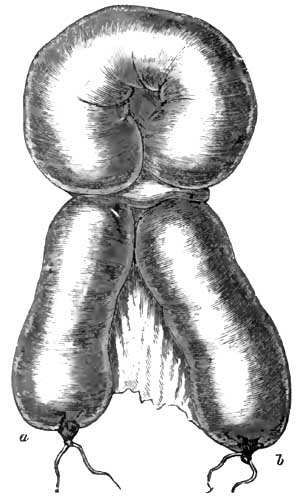 |
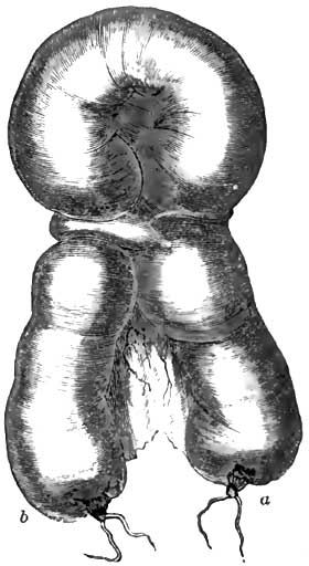 |
|
Anterior view of the strangulated intestine
and stricture. a, gastric extremity; b, rectal extremity. |
Posterior view of the strangulated intestine
and stricture. a, gastric extremity; b, rectal extremity. |
In consequence of inflammation of the peritoneum and effusion of
lymph, peritoneal surfaces are joined together, and before the lymph
is fully organized these surfaces are separated by the constant
movements of the organs and the change in the relationship of the
parts, and strings and bands of various shapes and sizes are formed in
which a portion of the intestine may become entangled and constricted.
Sometimes the bowel accidentally becomes engaged in a loop or noose of
false membrane, or becomes bound down under a band of fibrin; or, the
peritoneal surfaces of some of the organs having been joined together
or to the wall of the abdomen or pelvis, a loop of bowel may escape
into a slit or opening and become incarcerated; or a fold of bowel may
fall into a fissure in the omentum or mesentery or broad ligament of
the uterus or suspensory ligament of the liver, and become
constricted; or the appendix vermiformis may be twisted around the
intestine in such a way as to cause ligation of the tube, or, by
becoming attached to some neighboring part, it may form a loop through
which the intestine may pass and become obstructed. In the same way
the bowel may be constricted by a diverticulum. (This is well shown in
Figs. 23, 24, and 25.) Bands entangling the bowel and causing
strangulation may be attached to the fimbriated process of the
Fallopian tube or the ovary or uterus. Indeed, it is impossible to
describe in a limited space the almost infinite ways in which these
bands and strings may engage and incarcerate the intestinal tube
(Figs. 24, 25).
|
FIG. 25. |
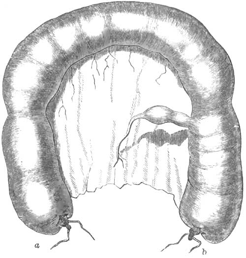 |
|
An appearance of the natural relations of the
diverticulum to the intestine. a, gastric extremity; b, rectal
extremity. |
Internal strangulated hernia may result from the bowel falling
into a pouch of the peritoneum and becoming ligated by the orifice of
the pouch, or passing into the foramen of Winslow, of which there are
three cases of strangulation recorded; or a retro-peritoneal hernia
may be formed; or, more common still, a hernia of the intestine
through the diaphragm.
In diaphragmatic hernia an opening is more frequently found in the
posterior part of this muscle. Two hundred and fifty-two cases of this
form of internal hernia have been collected by Leichtenstern, in which
the diagnosis was made in only five cases. He found the oesophageal
opening, a spot just behind the sternum, and a gap between the lumbar
and costal parts of the muscle, the weakest points in the diaphragm.
Diaphragmatic and other forms of internal hernia may exist and not
produce symptoms of strangulation either at the time of formation or
subsequently, just as we so commonly see in cases of external hernia.
When the bowel is constricted, however, and its circulation interfered
with, symptoms of internal strangulation come on, and are exactly like
the symptoms of external strangulated hernia. The attack is sudden,
the symptoms acute and urgent, and the course and termination very
rapid. Unless the constriction is relieved death may take place in
twenty-four hours; life is rarely protracted beyond three or four days.
The patient has first eructations, soon succeeded by nausea and
vomiting. The matter vomited consists of the contents of the stomach,
then of gastric fluid, bile, and the contents of the intestines. When
the last is ejected the vomiting is called fecal or stercoraceous. The
patient complains of a sense of constriction about the abdomen,
griping pains about the umbilicus, flatulence, tenesmus, and
insuperable constipation. One or two free stools from the large
intestine below the site of strangulation may be passed, but this
should not deceive the practitioner. As a rule, peritonitis soon
follows strangulation. The belly becomes tympanitic and tender, the
pulse small and wiry, and the face anxious. When gangrene supervenes
the pain subsides, the pulse becomes weak and intermittent, the
surface cold and clammy, and the patient soon dies in a state of
collapse. Slight delirium may precede death, or the mind remain
unimpaired to the end. Very often, when gangrene sets in and pain
disappears, the patient has a grateful sense of relief and is hopeful
of recovery.
One of the most frequent and important causes of intestinal
obstruction is intussusception or invagination of the bowel; by which
term is meant the protrusion or slipping of one portion of bowel into
a portion immediately adjoining.
This condition is sometimes found after death in persons old or young,
but particularly the latter, in whom during life there were no
symptoms of intestinal obstruction or intestinal trouble of any kind.
The displaced intestine in these subjects is easily reduced, is
unattended by any signs of inflammation, and is evidently the result
of spasmodic contraction of the transverse muscular fibres of the
bowel at one part, with distension and relaxation at another part, by
which, just before death, one piece of the bowel is pushed into an
adjacent piece. Not unfrequently two or more invaginations are seen in
the same subject. Flint4 counted as many as fifteen in a child who
died of typhoid fever. This invagination of the death-struggle almost
invariably involves the small intestine, and may be the protrusion of
a piece of the bowel above into a piece immediately below, or the
reverse, a portion of bowel below being pushed into a portion above.
It has been suggested that this slight and temporary intussusception
may occur during life and give rise to temporary symptoms of
intestinal obstruction, which disappear when reduction of the
displacement spontaneously takes place.
4 Practice of Medicine.
|
FIG. 26. |
FIG. 27. |
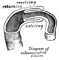 |
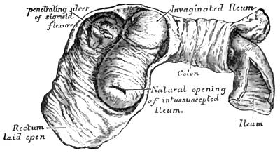 |
It will be seen by the diagrams that three successive portions of
intestine enter into the formation of an intussusception—an entering,
returning, and receiving portion. Two mucous surfaces and two serous
surfaces are thus brought into apposition. The mesentery attached to
the included lengths of bowel—viz. the entering and returning
lengths—is necessarily pulled down with the bowel in its descent, and
is also embraced by the receiving portion of the intestinal tube. The
traction excited by this portion of mesentery, thus wedged in between
the middle and inner layers of the bowel, materially alters what would
otherwise be the relationship of the parts. Fig. 26 shows simple
invagination of the ileum like the finger of a glove, in consequence
of the traction exerted. The entering or invaginated portion does not
always lie in the axis of the enveloping tube, but is more or less
curved, until very often its lower orifice is in contact with the wall
of the outer layer. The concavity of this curve looks
toward the
mesenteric edge of the invaginated portion of bowel, and the convexity
toward the opposite side of the receiving portion. The convex side of
the middle cylinder is often thrown into transverse folds or
convolutions. Intussusception, which gives rise to symptoms
characteristic of intestinal obstruction during life, is invariably
from above downward. It is doubtful whether there is on record a
single well-authenticated case of inflammatory invagination where the
lower segment of bowel protruded into the upper.
Reference to the diagrams will show that the lumen of the bowel is
diminished, and that more or less intestinal obstruction must follow
invagination. This obstruction is increased by the inflammation which
necessarily follows this condition. The large and numerous
blood-vessels of that portion of the mesentery involved in the
invagination are compressed and stretched; arterial supply, and
especially venous return, are interfered with; congestion quickly
follows, with copious inflammatory exudation; the layers of intestine
become swollen, and blood, sometimes in abundance, is poured out from
the mucous membrane. Peritonitis, limited sometimes to the invaginated
part, more often spreading to the peritoneum covering neighboring
structures, soon begins, and the contiguous serous surfaces are
agglutinated and the intussusception rendered irreducible. Lymph and
other inflammatory products are poured out freely; the coats of the
intestine become distended and thicker, and the inner and middle
layers of the invagination are separated by the deposit; the
invaginated part becomes more and more curved toward the mesenteric
border of the outer layer; and occlusion of the bowel, begun by the
invagination, is made more or less complete by the changes wrought by
inflammation (Fig. 27). That intestinal obstruction is not always
complete in intussusception is shown by the fact that fecal matter,
often in considerable quantity, is passed through the bent and
narrowed tube, the intestine retaining, at least for a time, its
contractile power. The changes produced by inflammation are chiefly
seen in the inner and middle layers of intestine, the receiving or
outer layer of the invagination often escaping any serious damage.
These changes vary with the character and duration of the
inflammation. Sometimes they are limited to an agglutination of the
opposed serous membranes, an effusion of blood and serum from the
mucous surfaces, and an enormous distension and swelling of all the
invaginated parts; or the inflammation may end in mortification of the
middle or both the inner and middle cylinders, the dead part coming
away in shreds or in large fragments, or, if the patient lives long
enough, the entire invaginated tube being discharged through the anus.
If the inflammation involves the invaginated parts unequally, strips
and shreds of the bowel are detached by ulceration and sloughing, and
may escape in the discharge from the bowels in pieces so small as to
be unnoticed; but if the intussuscepted part dies en masse, a circular
line of demarcation is formed by ulceration, and the dead segment is
detached and drops into the cavity of the bowel below, and escapes
through the rectum. It is often so complete that the inner and middle
cylinders can be recognized, and the part of the intestinal tube to
which the expelled bowel belonged can be determined. In favorable
cases the blood-vessels of the healthy bowel above and below the dead
segment pour out a circular mass of coagulable lymph, which, becoming
organized, closes the breach and
completes the intestinal tube.
To accomplish this it is necessary that the ends of the two portions
of bowel should be accurately coaptated: if they are not, some opening
may be left through which the contents of the gut may escape into the
peritoneal cavity, producing fatal peritonitis; or the new formation
may be imperfectly organized, and burst during some peristaltic
movement of the bowel or from the pressure of gas accumulating in the
gut; or the supply of lymph may be so redundant as to obstruct the
calibre of the bowel, or end in cicatricial contraction, stricture,
and obstruction. Aitken5 records four instances where the curved end
of the invaginated portion of bowel by prolonged pressure caused
ulceration and perforation of the coats of the enclosing bowel, the
invaginated portion passing through the side of the enclosing segment
and projecting into the cavity of the peritoneum.
5 Science and Practice of Medicine.
Gangrene and ulceration, however, do not always follow
intussusception. The mesenteric injuries may be sufficient to produce
congestion and exudation, and the patient survive the invagination for
weeks, and death eventually occur without sloughing or ulceration; or
spontaneous reduction of the invagination may take place and recovery
of the patient follow. The last termination must be rare, and
impossible when firm adhesion between the serous surfaces has taken
place; but that it does exceptionally occur is proved by cases where
the diagnosis of intussusception was undoubted, the invagination being
felt in the rectum or seen prolapsed through the anus. The most common
termination, if the patient survives, is mortification of the
invaginated part and separation in mass or by shreds or fragments.
Intussusception may occur in any portion of the intestinal canal, but
some points are more liable to it than others. 56 per cent. of the
cases collected by Brinton were ileo-cæcal; in 32 per cent. the small
intestine alone was involved; 28 iliac and 4 jejunal; in 12 per cent.
the colon, including its sigmoid flexure, was the part implicated.
When the rectum is involved, it usually forms the outer layer of the
invagination, the middle and inner layers being formed by the bowel
which has passed from above into it; when prolapse of the rectum
itself occurs, the mucous membrane is generally alone involved, but
along with this the muscular coat may also descend and a true
invagination of the rectum be found.
The most common variety of intussusception is the ileo-cæcal. It is in
this form that we find the greatest length of bowel involved. This
invagination begins generally at the ileo-cæcal valve, the lips of
which at first turn toward, and descend into, the cavity of the cæcum,
drawing with them the end of the ileum; in this case the valve forms
the lowest point of the invagination. If the invagination continues,
the end of the cæcum is next inverted; and if the process still goes
on, more and more of the colon is invaginated, until in some rare
cases it traverses the whole of the large intestine, appearing just
above or even protruding through the anus. In this variety the
vermiform appendix lies between the middle and inner layers of the
intussusception, and its opening, usually stretched and enlarged by
the inverted cæcum and inflammatory effusion, is found close to the
ileo-cæcal orifice. In this intussusception the cæcum and colon are
large and roomy, and the invaginated portion not so liable,
as
it is when the small intestine is alone implicated, to strangulation
and sloughing; nor is there seen in ileo-cæcal intussusception, unless
the portion of bowel involved is very short, the marked curvature of
the invaginated portion so commonly found in the small intestine. In
the ileo-cæcal form it is twisted or much convoluted rather than bent.
Another variety of ileo-cæcal invagination—very rare, however—is
where the ileo-cæcal orifice does not descend into the cavity of the
cæcum, but the lower end of the ileum passes through the valve into
the large intestine. In this instance the invaginated portion is
tightly compressed by the valve, and strangulation is speedy and
complete. An invagination may occur in the lower part of the ileum,
and the inner and middle layers pass on to the ileo-cæcal valve, and
be arrested at that point, and afterward, in consequence of violent
peristaltic action, the whole intussusception, inner, middle, and
outer layers, be invaginated into the colon. In this way the
invagination becomes doubled.
While intussusception may occur in either sex and at all periods of
life, it happens nearly twice as often in males as in females, and is
most frequently seen in childhood. Leichtenstern6 found in his
statistics of 473 cases that one-half were seen in children under ten
years old, and one-fourth of all intussusceptions occurred in children
from four to twelve months old. Invagination of the small intestine is
found almost exclusively in adults. Brinton from his records gives the
mean age of its occurrence 34.6 years. According to the same author,
the average age of ileo-cæcal invaginations is 18.57 years, and
one-half of all cases of this form of intussusception observed by him
were in children under seven years of age. Leichtenstern states that
the lower part of the ileum is the most frequent site of invagination
in the small intestine, and the descending colon and sigmoid flexure
the most common portion involved in intussusception occurring in the
large intestine.
6 Op. cit.
The mechanism of intussusception is probably not always the same. The
following is thought to be the most frequent process: A segment of
bowel becomes paralyzed by local peritonitis, some injury, diarrhoea,
or colic, and while in this state a segment of bowel above is
subjected to violent peristaltic action, and is forced into the
unresisting portion below. In this case the paretic segment forms the
outer or receiving layer of the intussusception. Leichtenstern
believes that the paretic portion is turned in and invaginated into
the normal bowel below, and that the clinical course of
intussusception and post-mortem appearance correspond with this
explanation. If such is the case, the paralyzed portion forms the
inner layer, and the active bowel below the receiving layer. Another
theory, which applies with much force to the most common of all
invaginations—viz. the ileo-cæcal—is, that as violent anal tenesmus
produces prolapse of the rectum, so prolonged and powerful tenesmus at
the ileo-cæcal opening may cause prolapse of the lips of this orifice,
and, eventually, invagination of the ileum, or of both this and the
cæcum, into the colon. When we remember that the ileo-cæcal valve is
furnished with a sphincter muscle, the analogy is complete. The idea
so long entertained that intestinal worms may occasion invagination of
the bowel has generally been abandoned. A polypoid tumor, by dragging
down the portion of bowel to which it is attached, may produce
invagination; and
Brinton's statistics give 5 per cent. of cases
of intussusception from this source. An examination of a larger number
of cases would probably show a much smaller percentage due to this cause.
A majority of cases of intussusception, however, take place suddenly,
without previous diarrhoea, colic, traumatism, or ill-health of any
kind, and probably occur without any tenesmus or paresis of a portion
of bowel. It may be that the longitudinal fibres of a segment of gut
contract, dilating and shortening a portion of the bowel; while this
part is distended a portion immediately above may be lengthened and
narrowed by contraction of the circular fibres, and violent
peristalsis going on at this moment, aided, possibly, by contraction
of the muscular wall of the abdomen, forces the upper and narrow
segment into the lower and dilated one.
At first the invagination involves a small portion of the bowel, but,
active peristaltic action continuing, it rapidly increases in size.
This increase is made at the expense of the sheath or outer layer,
which turns in to form the middle layer. The length of the
invagination varies from two or three inches to three, four, or five feet.
The symptoms of intussusception generally come on suddenly, and
indicate both intestinal obstruction and inflammation. Pain resembling
violent colic, and referred to the site of the invagination, is a
prominent symptom. The pain is intense, paroxysmal in character, but
after a time it becomes continuous. At first pressure gives relief,
but in a few hours tenderness, denoting peritonitis, appears, limited
to the invagination or spreading gradually over the whole abdomen.
Vomiting soon follows, and, with rare exceptions, is persistent. After
two or three days occasionally blood and sometimes fecal matter are
ejected from the stomach. Diarrhoea, with bloody, mucoid stools, is
rarely ever absent, and is characteristic of invagination. The patient
has from fifteen to twenty passages a day. If the large intestine is
involved, the diarrhoea is accompanied with tenesmus. Above the
obstruction gas and ingesta accumulate, and produce abdominal
distension, sometimes well marked. Generally the tumor formed by the
invagination can be felt through the abdominal wall, and is a symptom
of great importance. Meteorism and peritonitis may render the
existence of the tumor obscure or altogether prevent its recognition,
but in intussusception of the colon and at the ileo-cæcal valve the
solid cylindrical mass can usually be found, and frequently, when the
small bowel alone is implicated, a very careful and patient
examination will enable the observer to detect it. Sometimes it
changes its site, size, and shape; occasionally it can be felt in the
rectum or is seen protruding through the anus.
The urgency of the symptoms of invagination depends upon the portion
of bowel involved and the degree of constriction of the gut and its
attached mesentery. When the bowel is tightly constricted the symptoms
are acute, and the patient may die in a day or two; when the bowel is
not constricted the symptoms are chronic in character, and in the
early stages not urgent. The difference here is like that between
strangulated and incarcerated hernia.
In acute cases the attack is sudden, obstruction complete, and the
symptoms those of internal strangulation of the bowel, often followed
by collapse, which may destroy life in a few hours. These cases are
chiefly jejunal and iliac invaginations, and the higher up in
the small intestine the seat of obstruction the more violent and
urgent the symptoms. Constriction, being great, is followed by
engorgement and inflammation of the invaginated bowel, and if the
patient lives long enough gangrene ensues, by which the obstructing
mass is separated and discharged en masse or in fragments through the
anus. Not unfrequently life is saved in this way. That gangrene has
taken place and separation of the invaginated segments is in progress
are often known by the very fetid character of the evacuations and by
their admixture with blood and shreds of necrosed bowel. When the
sequestrum has been detached entire, it is often passed with
difficulty. Frequently it lodges at some point in the bowel, producing
temporary obstruction and giving rise to tenesmus and pain as it
passes along the large intestine. There is no doubt that the
continuity of the intestine above and below the neck of the
invagination has been established, and complete cures effected in the
way already mentioned. Usually, however, the patient dies from
collapse, peritonitis, or perforation of the bowel before the
obstructing mass can be removed by gangrene. Children almost
invariably die before this can take place, and adults live from the
seventh to the fourteenth day, according to the greater or less
violence and acuteness of the symptoms. When the slough has been
discharged and the continuity of intestine established, recovery is
still uncertain, and death very often happens for reasons referred to
in speaking of the separation of the sequestrum.
Separation of the invaginated portion and its expulsion, according to
Leichtenstern, in the majority of cases takes place from the eleventh
to the twenty-first day, but in chronic cases it is often delayed for
months. According to Brinton, separation of the sequestrum occurs
between the eighth and fifteenth in intussuscepted small intestine,
and between the fifteenth and twenty-second days in acute cases of
ileo-cæcal and colic invaginations.
In chronic cases of intussusception, which usually embrace the
ileo-cæcal and colic varieties, strangulation is not common and the
course of the disease is protracted. These cases often last for
several months, and the symptoms are not always well defined. At first
the pain is paroxysmal, with long intervals of ease. Vomiting
succeeds, but is not persistent; discharge of the contents of the
bowel below the seat of lesion takes place and afterward fecal matter
from above this point, because the permeability of the bowel is not
usually lost in chronic cases. Eventually the alvine discharges become
bloody, mucoid, and characteristic of intussusception; the severity of
the symptoms may gradually increase, the pain becoming greater, more
constant, the vomiting more incessant, the discharges from the bowels
more frequent, and in one, two, or three months the patient dies from
asthenia. Several authentic cases are related where the disease lasted
one or two years before terminating fatally. Very often some days
before death the pain and tenderness cease, and the operations become
free from blood and normal in character.
Constipation is a prominent symptom in all of the conditions which
give rise to intestinal obstruction, and habitual constipation or loss
of the powers provided for the advance of the contents of the
intestines not unfrequently leads to permanent occlusion of the canal.
It is impossible to fix any definite rule as a standard of health for
the number and quantity of alvine evacuations. Some individuals have a
passage from the bowels once every day; others, in the enjoyment of as
good general health, suffer from the ordinary inconveniences of
constipation if they have less than two or three daily fecal
discharges; others, again, apparently equally as well, have a movement
from their bowels once in two or three days or once a week, or even
once in two weeks. Habershon7 records the case of a "woman sixty
years old who from youth upward had had a passage from the bowels only
every six or eight days, and whose health had been perfect." A lady
under my own observation, for twenty years never had an alvine
discharge oftener than once in two weeks, and three times in her life
had passed two months without a movement of her bowels. This lady was
the mother of several children, and, although not in perfect health,
was able to attend to her ordinary household duties. Such cases are
not very uncommon, and occur, as far as I have been able to ascertain,
more frequently in women than in men.
7 On Diseases of the Abdomen, quoted by Leichtenstern in
Ziemssen's Cyc. P. of Med., vol. vii. p. 588.
The number of fecal evacuations and the quantity discharged have been
shown by Bischoff and Voit to depend, to some extent, upon the
character of the food ingested, vegetable diet producing abundant, and
animal diet scanty, stools. Doubtless, the quality of the food partly
explains the quantity of the alvine evacuation, although, to some
extent, this must depend upon the time that the feces remain in the
colon, a long residence there taking away a greater part of the watery
constituents and making the fecal mass thicker and harder; but the
variations in the number of stools in persons living on the same diet
can only be explained by the variations in the activity of the
peristaltic action in different individuals, or in the same individual
at different periods and under different surroundings.
The causes of habitual constipation are of the most varied and
diversified character, and it is not always possible in an individual
case to point out the original or primary one. Not unfrequently
several causes are in operation at the same time to produce
sluggishness of the intestinal canal and constipation. Very often it
begins with change of scene and habits, by which the daily visit to
the water-closet is interfered with, or after confinement to bed with
some temporary indisposition. It is more likely to occur in men and
women whose habits are sedentary and who are constitutionally lazy and
indolent. The feces are allowed to remain in the rectum and colon, and
every hour after the ordinary time for going to stool diminishes the
watery parts of the fecal mass and makes it harder and more
consistent. Many cases of chronic constipation, begun in this way,
have ended in dilatation and thickening of the intestine, ulceration
of the mucous membrane, and, eventually, perforation of the coats and
escape of the contents of the gut into the peritoneal cavity. Rapid
excretion of water by the kidneys, lungs, and skin produces
constipation by withdrawing a large proportion of the water from the
fecal mass, rendering it unnaturally dry and of diminished bulk. In
diabetes, constipation arises from this cause unless the patient makes
up the loss by drinking an unusually large quantity of water.
Constipation in nursing-women is explained by the loss of water in the
secretion of milk. The profuse sweating which attends malarial fever,
phthisis, and other diseases readily accounts for the constipation
which often accompanies these disorders.
Certain articles of food not necessary to mention here produce
constipation. They fail to excite peristaltic action; or articles of
diet which at first act as a stimulus to the bowels, and even provoke
temporary diarrhoea, lose their power if kept up too long—just as
certain purgative medicines lose their force if continued for too long
a period. Gradually they cease to increase the peristaltic action, and
rather add than otherwise to the inactivity of the intestines. Eating
the same kind of food day after day is very apt, sooner or later, to
result in diminished sensibility of the intestinal canal, a reduction
of the peristaltic force, and deficiency in the secretion of the
digestive juices, which in itself is a common cause of constipation.
Frequent change of diet is generally needed to supply the stimulus
necessary for that intestinal motion which relieves the bowels.
Bile is looked upon as one of the most powerful agents in stimulating
peristaltic action, and when, from any cause, mechanical or otherwise,
it is not poured into the bowel, constipation ensues. Unnatural
flexures, congenital or acquired, of the large intestine are not
unfrequently the source of chronic constipation. These flexures,
normal or factitious, favor accumulation of feces, especially in
subjects who have diminished sensibility of the bowel and a paretic
state of the muscular coat. Certain injuries and diseases of the brain
and spinal cord reduce, and sometimes altogether prevent, intestinal
activity. Hysteria, if it exist for any length of time, is generally
attended by sluggishness of the bowels, and great mental depression
(melancholia) is sometimes preceded and sometimes followed by habitual
constipation. In treating such a case it is important to make the distinction.
Temporary paralysis of the muscular coat of the bowel, followed by
symptoms of intestinal constriction, with insuperable constipation,
sometimes attends violent contusion of the abdomen. In some cases
prolonged functional weakness of the muscular coat follows the injury.
Many chronic diseases leave the bowel in a sluggish condition by the
pathological changes produced in the intestine. The function of the
muscular coat is frequently injured by the infiltration which
accompanies peritonitis. The fibres are separated by the serous
effusion which attends this inflammation; they become overstretched,
and, losing their contractility, end in paralysis and obstinate
constipation. Occlusion of the canal from this cause may last for
days, and be accompanied with tympanitis, stercoraceous vomiting, and
all the signs of internal strangulation, ending in death. Post-mortem
examinations in such cases show no stricture or unnatural diminution
in the size of the canal, but that the fatal occlusion was due to
paralysis of the muscular coat of the bowel and arrest of its power.
The normal advance of the contents of the bowel is interfered with by
any cause which lessens the contractility of the muscular coat.
Chronic
diseases which debilitate the general muscular system
affect at the same time the contractile power of the muscular coat of
the canal, and the debility and degeneration of old age are felt here,
and sometimes occasion the constipation which often accompanies this
period of life. Leichtenstern8 says that chronic intestinal catarrh
is a common factor of constipation—that when this catarrh is of long
standing it produces relaxation of the muscular coat and diminishes
the elasticity of the intestinal walls. He believes that this
pathological condition exists in a large proportion of the cases of
habitual constipation attended with mental depression, that the
hypochondriasis makes its appearance after the constipation has become
chronic, and that it is a secondary symptom. This affection is located
chiefly in the small intestine, and does not usually involve the colon.
8 Op. cit.
Probably the most common form of chronic constipation is that which
accompanies loss of sensibility and muscular inactivity of the colon
and rectum. The large bowel becomes sometimes so distended by the
accumulated fecal masses that it has been found after death to measure
ten or fifteen inches in circumference and to contain an astonishing
quantity of feces. Any part of the canal, except the last two inches
of the rectum, which is kept empty by the contraction of the
sphincters, may be occupied by the mass, but the accumulation is
greatest in the rectum, cæcum, and sigmoid flexure. At the last-named
location the distension is so great that the mass can be readily felt
through the abdominal walls. The tumor may be as large as a foetal
head, and may be mistaken for a simple or malignant tumor of the
omentum, stomach, or other organ, or for pregnancy or ovarian growth.
The dilatation may be so enormous as to push the small intestines into
the back part of the abdominal cavity and to interfere with the
function of any organ upon which it encroaches. It may press upon the
concave surface of the liver, and, arresting the flow of bile, produce
jaundice or mechanically interfere with some portion of the track of
the urinary organs and cripple their functions. When situated in a
portion of the canal not tightly attached to the abdominal walls it is
slightly movable, more or less hard and consistent, according to its
duration, for it remains often for months unchanged, sometimes giving
to the fingers the impression of a rather soft, easily-indented
swelling with a uniform smooth surface—more often feeling like a
hard, irregular, elongated, and corrugated mass of fecal balls.
Contraction here and there of bundles of the circular muscular fibres
of the gut produces the irregular, corrugated impression imparted to
the fingers. Their shape and position may sometimes be changed by
pressure through the abdominal wall. If the accumulation occurs in the
rectum, the introduction of a tube or bougie is prevented by the
impacted mass, which can be gotten away only by the fingers or by some
instrument. The colon and rectum may be dilated to their utmost
capacity with an enormous amount of feces, enough to fill a
common-sized pail, and both the patient and medical attendant be
deceived as to the sufferer's condition by the fact that he has his
daily number of stools. The semi-fluid contents of the small
intestines find their way through this mass by some irregular and
uncertain track, undermining and breaking down sometimes a lump of the
old fecal accumulation, which, if small in size or broken up, may pass
on and
escape by the anus, but if large and hard may drop into
the irregular and uncertain passage and permanently close it; then
sudden and complete intestinal occlusion takes place, with all of its
fearful consequences. If this, however, should not occur, and the
accumulation is not recognized and removed, the enormous dilatation
may go on until complete paralysis of the muscular coat is produced,
and entire stoppage of the current of feces, with permanent occlusion
of the bowel; or before this takes place ulceration may set in, partly
because of the great pressure of the fecal mass upon the mucous
membrane, and partly from the irritating character of the contents of
the bowel. Ulceration begins, most likely, at some point where
resistance is greatest, and perforation of the bowel may ensue.
SYMPTOMS.—If the accumulation occupies only a portion of the colon,
as the cæcum or sigmoid flexure, the distended part may become
displaced and twisted on its long axis. This condition scarcely ever
happens in the large intestines except at the parts mentioned. Torsion
of the cæcum rarely takes place except in persons of from forty-five
to sixty years of age, while twisting of the sigmoid flexure may
happen at any period of life. When distended and very heavy from the
weight of feces, with probably some congenital defect about its
mesenteric attachment, the sigmoid flexure may become twisted and drop
into the pelvis, producing at once symptoms of internal strangulation.
Individuals accustomed to having one or more alvine evacuations a day
are made uncomfortable by two or three days of constipation. A feeling
of distension about the abdomen, with flatulence and heat, follows
this condition, and soon afterward headache, loss of appetite, and
symptoms of indigestion supervene. If this state of the bowels
continues unrelieved, pressure upon the hemorrhoidal veins takes place
and interference with venous return, producing congestion in the lower
end of the rectum. This is attended by straining, diarrhoea, evolution
of gaseous matter, colicky pains, and possibly sympathetic disturbance
of the genito-urinary organs. When at last the hardened and enlarged
mass is discharged, it produces some pain and burning about the anus,
with possibly rupture of the mucous membrane in that region. Fissure
of the anus may thus originate. In the case of a lad aged about nine
years under my care fissure of the anus began in this way, and after
its formation the pain of defecation was so intense that he resisted
for ten days every attempt of his bowels to move. After this time he
passed every day or two one or more hardened fecal balls, but always
with such atrocious pain that he looked forward to the next attempt
with terror. This case ended in fecal impaction, which nearly proved fatal.
Not unfrequently persons who habitually go two or three days without
having a passage from the bowels are not apparently inconvenienced,
and after a time any of the discomforts ordinarily felt from
constipation are not noticed, if indeed any exist.
Generally, however, chronic constipation leads to a host of troubles
of the most varied character. There is not an organ in the body that
is not more or less influenced by it. The generation of gas in the
intestines produces a sense of fulness of the abdomen and elevation of
the diaphragm which interferes with the action of the lungs and heart.
The sufferer is oppressed, sighs, and has difficult respiration and
attacks of
palpitation of the heart. The influence of the
abdominal pressure is conducted by the sympathetic nerves to the
brain, and the patient frequently has vertigo, headache, ringing in
the ears, faintness, etc., and in consequence of the pressure upon
other nerves or of hyperæmia of the spinal cord and its membranes he
has dull aching pains in his back, groins, genitals, or extremities. I
have seen in several instances pain in the legs, coming on after the
patient has retired and lasting until morning, violent enough to
prevent sleep, at once permanently relieved by an active cathartic
after antiperiodics, alteratives, and anodynes had failed to do any good.
A patient suffering from habitual constipation usually obtains
temporary relief by the bowels acting either spontaneously or after a
dose of medicine; but, the causes of constipation continuing, the
physical discomforts and suffering continue, varied in every
conceivable way. His digestion being disturbed, appetite poor, and
assimilation imperfect, he gradually loses flesh and his complexion
becomes sallow and unhealthy. In addition to this, he soon grows
irritable and fretful, trifling affairs trouble him, he has fits of
great mental depression, and soon settles down into hypochondriasis,
his life becoming a burden to himself and a nuisance to his friends.
If the constipation ends in fecal accumulation, the worst symptoms of
mechanical obstruction may present themselves at any time, and death
of the individual follow. The practitioner should always keep this
fact in mind in treating every case of intestinal obstruction, and
search for fecal impaction by examining the rectum and the whole
length of the large intestine through the anterior abdominal wall.
Very often symptoms of impaction come on gradually in one who has been
ailing for some weeks or months, but sometimes the onset is as sudden
as in a case of acute occlusion of the intestines. The patient is
seized with pain like that of colic and an urgent desire to empty his
bowels, but all attempts to do this are futile, and the straining is
followed by great exhaustion; borborygmus, nausea, vomiting, and
possibly hiccough, soon come on, with tympanitic distension of the
belly. If the impaction is not overcome, death by collapse or from
peritonitis follows. Post-mortem examination shows enormous fecal
accumulation, peritonitis as a consequence of the obstruction,
perforating ulcer in some part of the large bowel, more often the
sigmoid flexure, or, in some cases, absolute rupture of the cæcum
itself, and escape of its contents into the peritoneal cavity.
In a report by George Pollock9 of 127 cases of intestinal
obstruction, 77 belonged to the above class; and Brinton, in his
analysis of the whole group of cases collected by him, says stricture
constitutes about 73 per cent. In 124 cases of intestinal obstruction
reported by Mr. Bryant10 from the post-mortem records of Guy's
Hospital, 47 were found to be stricture of the bowel. The above
statements show that stricture, or diminution of the calibre of the
bowel, is the most frequent cause of
intestinal obstruction, and
the subject is worthy of our earnest consideration.
9 Medico-Chirurgical Review, 1853.
10 Practice of Surgery.
While stricture of the bowel may be found in any portion of the
intestinal canal, it occurs most frequently in the sigmoid flexure and
rectum. Brinton found in 100 fatal cases of stricture 30 in the rectum
and 30 in the sigmoid flexure; only 8 cases in 100 were in the small
intestine. Brinton's statistics correspond very nearly with those of
other writers. The affection is more common in men than women, and the
average age at death is about forty-four years.
The most common cause of stricture is contraction following
cicatrization of ulcers of the mucous and submucous coats of the
intestine. The ulcer may involve the circumference of the bowel, and
the resulting cicatrix terminate in uniform constriction of its lumen,
or the ulceration may extend several inches along the side of the
intestine, ultimately causing contraction in the direction of its
longitudinal axis, marked stenosis, and kinking of the gut. When
ulceration, continuous or in patches, involves a large extent of
bowel, it may reduce the gut to a mass of indistinguishable
cicatricial tissue. Bristowe11 says he has seen the whole cæcum thus
contracted "into a channel barely capable of admitting a goose's quill."
11 Reynolds's System of Medicine.
Stricture of the intestine often follows dysentery or tubercular and
syphilitic ulceration of the bowel. Follicular or hemorrhoidal
ulceration is sometimes the beginning of a stenosis which ends in
stricture of the rectum. Stercoral ulcers of the colon are not
unfrequently the starting-point of cicatricial contraction of the
calibre of the bowel. Sometimes, but rarely, ulcers of typhoid fever
end in constriction of the intestinal tube. The diameter of the gut is
also contracted by the effects of caustic substances, by ulceration
following the lodgment of foreign bodies, and by effusion of lymph or
thickening attendant upon long-standing hernia. Very often after death
it is impossible to determine what particular kind of inflammation and
ulceration caused the stricture. Generally, the cause which provokes
the ulceration sets up chronic peritonitis, which materially aids in
producing the obstruction. Spasm of the circular muscular fibres
usually accompanies these lesions, and materially contributes in many
cases to fatal intestinal obstruction. Some authors assert that spasm
without organic change can produce acute obstruction: such an
occurrence, except possibly in the rectum, must be very rare, if
indeed it ever happens.
The most common cause of stricture is cancer. This disease may
originate in the bowel itself, or, beginning in some neighboring organ
or tissue, gradually spreads and involves the gut. It may extend
around the bowel or be infiltrated along the sides of the canal for
several inches, and may be scirrhous, medullary, or epithelial in
character. Eighty per cent. of the cases of cancer of the bowel are
situated in the rectum. Usually, but not invariably, cancerous
deposits are found in persons who have passed middle age.
An impediment to the passage of fecal matter is invariably produced in
constriction of the intestine from the above causes, and it frequently
continues until fatal occlusion occurs. The contents of the bowel
accumulate above the block, producing distension of the gut and
thickening of the muscular coats above the stricture, with contraction
and atrophy
of the portion of intestine below. Dilatation of the
bowel above the seat of lesion is sometimes great enough to cause
rupture and peritoneal extravasation, or distension and stretching of
the coats of the canal may be sufficient to interfere with its
circulation, and ulceration ensue.
Occasionally cases of stricture or well-marked circumscribed
contraction of the bowel are seen which give rise to no marked
symptoms of constriction during life. Such was the case in the
instance related by Bristowe and referred to above. These instances
are, however, exceptional in the large intestine.
Symptoms of stricture vary according to the site, cause, and extent of
the lesion. They are gradually developed, and in this respect are
unlike the symptoms of internal strangulation or of intussusception,
which are generally acute and rapid in their course. When the
obstruction in stricture is complete, progress toward death is
comparatively slow. If the stricture is seated in the small intestine,
the symptoms are often so obscure that for a long time the presence of
the contraction may not be suspected; the contents of the small bowel
are usually fluid, and in this state readily pass through the
constricted part. The more solid the contents of the bowel, the
greater the difficulty in passing a contracted and narrow orifice, and
the more conclusive and characteristic the assemblage of symptoms of
obstruction from stricture.
The history of a case of intestinal obstruction from stricture is
often instructive. For weeks or months there have been colicky pains
and intestinal disorder; possibly, in the early stages, diarrhoea, but
later marked constipation, and probably previous attacks where
constipation was for a time insuperable and death from obstruction
imminent. Hemorrhage, except in cancer or when complicated with piles,
is rare. The attack may come on suddenly, or constipation become more
and more difficult to overcome; violent peristalsis presents itself,
accompanied by pain and abdominal distension, and followed by nausea
and vomiting, the latter often being stercoraceous. During the throes
of pain—for it is paroxysmal—the outline of the distended gut can be
felt and seen through the abdominal walls if they are thin and free
from fat. Unless the stricture is relieved the patient gradually dies
from asthenia. Inflammation is often absent throughout, but enteritis
or peritonitis may come on, or perforation and peritoneal
extravasation ensue and hasten the fatal termination.
When the obstruction is in the rectum it can be felt with the finger;
if in the sigmoid flexure, it may be felt with a gum bougie or probe,
but the use of the former is unreliable, and the latter, unless
carefully employed, dangerous. Obstruction at this point, however, is
attended with marked distension of the descending and transverse
colon. If seated in the small bowel, the large intestine is flaccid
and collapsed. Careful manual exploration often enables the
practitioner to determine the site of the contraction. Weight, pain,
dulness, and fulness are usually found about the stricture, but these
signs may be of little value when the abdominal wall is thick and
unyielding, or peritonitis or tumor is present, or the contracted
portion of bowel is compressed or drawn out of its proper site.
Brinton suggests that the site of stricture may be determined by the
quantity of water which can be injected through the anus into the
bowel. Such an estimation must often be erroneous, as stricture
is rarely ever complete and fluid may be forced through the
constricted part. Indeed, Battey of Georgia has demonstrated upon dead
and living subjects that fluid may be made to pass through the entire
canal from the anus to the stomach.
Obstruction due to cancer of the rectum can be determined by digital
examination. When seated in the small intestine or higher up in the
large bowel, the presence of a painful tumor, preceded for weeks by
evidences of impaired nutrition, emaciation, and followed by
lancinating pain, cancerous cachexia, etc., will indicate the
character of the trouble.
Obstruction of the bowel is sometimes occasioned by compression or
traction exerted on the intestine by abdominal tumors or cysts.
Fibrous tumors of the uterus, ovarian cysts, hydatid growths, or
indeed any form of abdominal tumor, may by pressure on some part of
the intestinal track produce fatal obstruction. Several inches of
bowel may thus be compressed and rendered impervious, or if traction
is exerted by the tumor, which is often adherent to the bowel, the
tube may be sharply bent or twisted and its action interfered with. A
case is reported of compression of the bowel from a great accumulation
of fat about the colon. Adhesions of intestinal coils from chronic
peritoneal inflammatory changes constitute a large and important class
of cases of intestinal obstruction. This condition is known as
contraction of the bowels: 23 of the 124 cases reported by Bryant were
of this character.
The usual site of stricture is the large bowel: contraction is far
more frequently seen in the small intestine, and is caused by an
effusion of lymph following simple peritonitis or the inflammation
attending the formation of cancer or tubercle of the peritoneum. Coils
of intestine are matted together or to neighboring parts in this way
by bands of lymph or false membranes, and the action of the bowel
interfered with or obstructed. Constriction of a length of bowel may
be found after death, or a sharp, angular bend by which complete
obstruction has been produced.
Circumscribed peritonitis may produce adhesion of a portion of bowel
by bands of lymph to the uterus or its appendages, or to some part of
the large intestine, or to the abdominal wall, and the action of the
bowel become embarrassed by traction, constriction, or bending. In
consequence of the irritation following this condition, spasmodic
contraction may follow and add to the difficulty, or enteritis may
ensue; and this will especially be the case if the circulation of the
part is interfered with, and render complete what before was a partial
obstruction. Distension and fulness of the bowel above the
obstruction, with contraction and emptiness of the portion of the gut
below, are found after death in cases of contraction, just as we see
in fatal cases of stricture. The history of the case and presence of a
tumor will generally enable the practitioner to determine when
obstruction is due to the presence of some adventitious growth. When
contraction is complicated with the presence of tubercle or cancer,
symptoms attending these conditions will be present.
Obstruction of the intestines from contraction generally comes on
insidiously. The patient may date the beginning of his trouble
from an old attack of circumscribed peritonitis which probably took
place weeks or months before. He has attacks of colicky pains,
indigestion, and constipation. The last is difficult to overcome,
continuing for hours before it is relieved by medicine or the efforts
of nature. During the attack of almost insurmountable constipation
violent peristaltic movement of the bowel above the impediment may be
noticed. The patient may gradually become more and more feeble from
suffering and interference with nutrition, and die from exhaustion, or
fits of obstinate constipation may continue to recur, until finally
one of them becomes insuperable and fatal.
The symptoms of contraction closely resemble those of stricture, but
it is important to distinguish one from the other, as the treatment,
especially if surgical interference is demanded, is very different. A
rigid analysis of all of the signs will usually, but not invariably,
enable the practitioner to make the distinction.
There is an important difference between the constipation of stricture
and that of contraction. In the former the difficulty is in
defecation, emptying the large bowel, the usual site of stricture; in
the latter the difficulty is in the passage of the contents of the gut
along the narrowed and contracted small intestine, the common site of
contraction. In stricture the calibre of the bowel is diminished by
some sharply-defined mechanical impediment seated in the cavity or in
the walls of the tube; in contraction the bowel is bent or kinked by
adhesions, or coils of intestine are matted and glued together and
peristalsis interfered with. In stricture defecation is difficult and
painful; in contraction the alvine discharges are painless. In the
former blood and mucus are not unfrequently seen in the feces; in the
latter the motions are healthy. In stricture constipation alternates
with diarrhoea; in contraction looseness of the bowels is rarely seen.
In stricture distension of the abdomen is lumbar and epigastric; in
contraction the distension is less and is central and hypogastric. In
both conditions violent distinct peristaltic action is seen during a
fit of constipation, and in both the bowel above the constriction is
distended and hypertrophied. In contraction the powerful, writhing
peristalsis involves the small intestine above the impediment, and in
stricture the large bowel above the obstruction. In both stricture and
contraction inflammation of the bowel and peritoneum may supervene. In
contraction, when inflammation sets in or when enteritis and
peritonitis are absent and the attack of constipation is
insurmountable, I have noticed that the symptoms are more urgent and
rapid in their course, and danger of death from collapse greater, than
when these conditions exist in stricture.
DIFFERENTIAL DIAGNOSIS.—In every case of intestinal obstruction a
careful examination should be made for external strangulated hernia.
All of the regions of the abdomen in which hernia may occur should be
thoroughly inspected, as the symptoms of the two conditions are
identical. A small or incomplete external strangulated hernia may
easily be overlooked. An individual with an old hernia may suddenly
have symptoms of intestinal obstruction, and it may be doubtful
whether the obstruction is due to internal constriction or to the
external hernia. Diagnosis is especially difficult when the chronic
hernia is irreducible in character. If the cause
of the
impermeability is internal and below the external hernia, that portion
of intestine in the hernial tumor becomes swollen, tense, and hard,
and closely resembles the local symptoms of strangulated hernia. If
the external hernia is reducible, reduction en masse may take place
and a retro-peritoneal hernia be formed. When the case is doubtful and
urgent, an operation for strangulated hernia should be performed.
Functional obstruction of the bowel is sometimes seen, closely
simulating obstruction from one of the structural changes mentioned.
Cases of functional obstruction are seen usually in hysterical or
nervous women, and are generally recognized by the history, course,
and termination of the malady. The fact that local enteritis,
peritonitis, or typhlitis, by paralyzing a portion of the bowel, may
produce all the signs of acute and complete obstruction, should not be
lost sight of.
In cases of congenital stricture or malformation, or the presence of
foreign bodies in the intestine, or acute internal strangulation, or
twisting of a length of bowel, and generally in intussusception,
symptoms of acute obstruction are present. The individual may have
been in perfect health, and suddenly symptoms of the gravest character
set in. Intense pain, referred to some special part of the belly, is
the first sign of trouble. Nausea soon follows, and with it great
prostration; the depression of vital power approaches, and sometimes
reaches, syncope; the patient rolls and tosses in agony; his mental
distress is equally great, and if old enough he is conscious of his
danger and is anxious and despondent. Vomiting succeeds the nausea: at
first the contents of the stomach, and then those of the small
intestines, are thrown up; after a time the vomiting is stercoraceous.
The belly becomes swollen, tympanitic, and exquisitely tender; the
weight of the bed-clothes or the slightest touch of the finger upon
his abdomen is intolerable; he keeps his head and shoulders raised and
his lower limbs retracted to avoid pressure of the abdominal muscles.
Constipation is complete and insuperable. If the abdominal wall is
thin, the violent motion of the intestines can be seen and felt
through it. These painful peristaltic movements of the bowel are
paroxysmal and attended by loud rumbling or gurgling noises. The pain
gradually increases; the patient is very restless and complains of
great thirst; his pulse is small, hard, and frequent, his extremities
cool and features pinched. If not soon relieved, exhaustion comes on;
he has muttering delirium, cold clammy perspiration, hiccough,
twitching of the tendons, and death soon follows from collapse or from
peritonitis or gangrene, or from both. The average period of death is
from six to eight days. It may occur in thirty-six or forty-eight
hours, or the patient may last for two weeks.
In congenital occlusion and malformation the history of the case, the
age of the patient, and the fact that the deformity in such cases is
almost always confined to the anus and rectum, usually render the
diagnosis sufficiently easy.
Obstruction caused by foreign bodies impacted in the intestines can
generally be diagnosed. The history of the case may show that foreign
bodies have been swallowed or that the patient has been subjected to
some of the conditions which cause the formation of enteroliths. These
stony concretions are usually found in the cæcum or colon, and
frequently give rise for days and weeks to symptoms of indigestion,
emaciation,
constipation, and other evidences of bad health
before complete occlusion of the intestine takes place. Not
unfrequently, before the sudden attack of impermeability of the bowel
the patient has had repeated attacks of typhlitis, and has been
conscious for a long time of the presence of a tumor in the region of
the cæcum or colon. Possibly he has passed on some former occasion
pieces of the stony concretion.
Diagnosis of obstruction by gall-stones is often aided by the fact
that the patient has recently suffered characteristic pains of hepatic
colic and by the icterous condition of the skin. Possibly the
individual has suffered repeated attacks of hepatic trouble and has
previously passed a gall-stone. Obstruction from this cause is seen
four times as often in women as in men, and always after the middle
period of life.
In obstruction occasioned by internal hernia or the presence of
membranous bands, loops, mesenteric pouches, the symptoms are often
such as to baffle all attempts at accurate and certain diagnosis. The
onset of the symptoms is sudden and the course of the disease rapid;
prostration of vital power is extreme, sometimes amounting to syncope;
vomiting incessant and persistent; pain constant and fixed. The most
characteristic symptom of internal strangulation is the very great and
prolonged depression of vital power; it occurs generally in early
adult life.
Erichsen states that in twisting of the bowel the abdomen is unevenly
distended, it being tympanitic on one side and flattened on the other.
This condition of the bowel is usually seen after middle age. In
intussusception the principal signs are, usually, the early age of the
patient—obstruction from other causes in children being rare—the
suddenness of the onset of symptoms, the frequent desire to go to
stool, the tenesmus, and the characteristic bloody mucus discharges.
By abdominal palpation frequently the sausage-like tumor can be
recognized, and very often the intussuscepted part can be felt in the
rectum or seen protruding through the anus. When invagination involves
the upper part of the small intestine, diagnosis of the cause of
occlusion is almost impracticable.
In obstruction of the bowel from fecal accumulation, inflamed and
thickened intestine, stricture, compression and traction, and
contraction of the gut from cancerous deposit, the symptoms are
gradually presented and chronic in character. They are unlike the
signs of acute obstruction, which occur in persons apparently in
perfect health and are sudden and violent from the beginning. In
chronic obstruction of the intestine the patient has probably been
complaining for some time, with symptoms of abdominal trouble. He has
been unwell for weeks, his appetite poor, digestion disordered,
strength diminished, and bowels constipated. The last symptom is the
most distressing of all. Purgatives do not give the relief ordinarily
obtained, but add to the griping, colicky pains, nausea, and general
depression. When his bowels do act, the stool is sometimes liquid,
sometimes very hard (scybalous), or the form of the matter passed is
tape-like or pipe-like. Sometimes, in his frequent attempts at stool,
the only discharge is blood and mucus or pus. Attacks of eructation
and vomiting often take place during the progress of the disease.
Stercoraceous vomiting is, however, rare, and only seen in the later
period of the attack. Abdominal distension is slow in making its
appearance, but after a time is well marked, and due more to
tympanitis than to
constipation; the tympanitic distension is
accompanied by loud rumbling and gurgling noises in the bowels. After
a period which varies much in different cases, inflammation, suddenly
or gradually, is set up, and all the symptoms of acute obstruction are
presented, grafted on signs of chronic occlusion. We have pain,
nausea, vomiting, great distension and tenderness of the abdomen,
peristalsis plainly seen and felt if the abdominal wall is thin, the
small, frequent, wiry pulse, clammy perspiration, prostration with
hiccough, tendinous twitchings, and death, very like that following a
case of external strangulated hernia.
Obstruction due to fecal accumulation generally happens in persons who
have passed middle age, and can often be diagnosed by digital rectal
examination and palpation of the abdomen, by the presence of fecal
tumors, and the history of long-existing constipation with its
manifold consequences; previous attacks of impermeability, and relief
by discharge of enormous masses of feces.
Obstruction caused by the presence of some abdominal tumor is
generally known by the history of the case, the fact of the existence
of the tumor being known to the patient or discovered by the physician
by an examination through the abdominal walls or through the vagina or
rectum. The progress of such cases is essentially chronic, but acute
symptoms may at any time come on. Diagnosis of obstruction due to
stricture is frequently made by examination of the rectum and sigmoid
flexure, the usual sites of constriction from this cause. Complete
occlusion from stricture is almost always preceded by well-marked
premonitory symptoms.
When the obstruction is situated in the lower part of the colon and
rectum, its precise seat can be determined by digital or manual
examination or the use of a bougie or tube. When the site of
obstruction is above the sigmoid flexure, it is difficult, and
occasionally impossible, to determine its exact locality. As a rule,
when the constriction is in the small intestine the symptoms are acute
and urgent; pain is intense, vomiting comes on soon, and prostration
is early and extreme. When the large intestine is involved, except in
volvulus, the symptoms are generally chronic. In twisting of the gut
the symptoms are rapid and uncommonly severe. The higher up the
obstruction, the earlier stercoraceous vomiting begins. Above the
constriction the bowel is distended and tympanitic; below the
constriction it is generally collapsed. In obstruction of the large
intestine the outline of the tympanitic and distended gut may be
traced with the eye and hand. In constriction of the small intestine
the secretion of urine, as has been shown by Hilton, G. Bird, and
Barlow, is less than where obstruction is seated in the large bowel.
Besides rectal and vaginal examinations, which should never be
neglected in any case of intestinal obstruction, abdominal palpation
may also aid in determining the site of constriction. It should not be
forgotten, however, when a tumor is found—as, for instance, in
invagination—that the bowel may be displaced; a distended cæcum may
be pushed into and occupy the left side of the belly. Cases are not
uncommon where the symptoms are so combined and uncertain as to render
accurate diagnosis of the site of obstruction impracticable.
Very little light is thrown upon the diagnosis by pain, constipation,
or vomiting when these symptoms are considered separately. Pain is
common to many diseases of the abdomen; obstinate constipation,
lasting for
days and weeks, is often seen where there is no
mechanical obstruction; and vomiting attends many morbid conditions of
the body. But when these symptoms are combined and examined along with
the history of the case and mode of invasion, they are often
characteristic of constriction of the intestine. Pain in acute
obstruction is fixed, umbilical, and intermittent; in chronic cases it
is more diffused and increases with the distension. In acute cases
constipation is complete and insuperable; in chronic cases this
symptom gradually increases; in intussusception we have frequent
discharges of a dysenteric character, and hemorrhage, sometimes
copious, when the small bowel is involved. The bowel below the seat of
complete constriction may be full of fecal matter, and the discharge
of this spontaneously or by the aid of enemata may induce the
attendant not to regard the case as one of occlusion. Stercoraceous
vomiting, as a rule, comes on early in acute and late in chronic cases
of complete occlusion of the gut; in spasmodic ileus or impermeability
not due to mechanical occlusion feculent vomiting is only occasionally seen.
The duration of life in acute intestinal obstruction varies very much
in different cases: death may ensue in a few hours or not for ten or
twelve days; the average period is six days. The duration depends upon
the site of the constriction and the mechanical injury to the bowel;
the nearer to the pylorus the constriction, the more rapid the
progress. In volvulus involving the sigmoid flexure, when injury to
the bowel is great, the symptoms are acute in the extreme. After
peritonitis or enteritis begins, progress toward a fatal issue is very
rapid, the patient rarely living more than three or four days. In
occlusion from stricture, compression, fecal impaction, and chronic
intussusception the patient may live for weeks or even months.
The statistics of Leichtenstern show that from 5 to 10 fatal cases of
intestinal obstruction occur every year among every 100,000
inhabitants; and according to the mortuary records of England an
average of 1 death from this cause is seen in every 260 deaths.
Brinton reports 1 death from intestinal obstruction in every 280
deaths; his statement is based upon 12,000 promiscuous autopsies. The
first author states that the statistical reports of the general
hospital of Vienna inform us that out of 60 cases of ileus, 6 or 10
per cent. recovered. This report, however, is too meagre to be of much
value. From Brinton's statistics of 500 deaths from obstruction we
find that out of 100 cases, 43 are intussusception, 17 stricture, 4.8
impaction of gall-stones, 27.2 internal strangulation, and 8 torsion.
TREATMENT.—There are few conditions of the body which cause the
practitioner more anxiety and embarrassment than cases of intestinal
obstruction, and when the precise seat and nature of the occlusion are
not known the treatment is almost entirely empirical. The distinction,
however, between acute and chronic cases of obstruction of the bowels,
or of acute supervening upon chronic symptoms, can almost always be
made, and a patient investigation of the history of the case, the mode
of invasion, and a rigid analysis of all the symptoms presented will
generally enable the attendant to come to some positive conclusion as
to the cause and site of the occlusion. One fact in the treatment
which cannot be too strongly impressed upon the mind, especially of
the young practitioner, is not to use purgatives and irritating
enemata, formerly so much in vogue,
in the hope of forcing a
passage through the occluded bowel. The patient is urgently solicitous
for medicine which will open his bowels, but the use of purgatives to
overcome internal strangulation is as senseless and hurtful as when
used to overcome the constipation of external strangulated hernia.
These agents only add to the nausea, vomiting, pain, and peristalsis.
The latter is violent enough already to render coils of intestine
visible, and with every paroxysm is adding to the entanglement and
impermeability. It is said that cathartics in some instances have
unlocked the bowel in intestinal obstruction: these cases are
exceptional, and many of them were probably functional and not
structural in character. The only exception to the rule of avoiding
purgatives is as stated by Jonathan Hutchinson: "In certain cases when
impaction of feces is suspected, and in cases of stricture when
fluidity of feces is desirable."
Formerly, some of the best practitioners resorted to the exhibition of
one or two pounds of quicksilver, in the hope of overcoming intestinal
obstruction by the weight of the metal. This plan has properly been
almost if not quite abandoned. Crude mercury is very slow to reach the
obstruction, is divided into small portions by the peristalsis, which
its presence increases, and if it should finally arrive at the point
of constriction in any considerable quantity, it is more liable to add
to than overcome the difficulty.
The great remedy in intestinal constriction is opium, in large or
small and repeated doses. Its use arrests the vomiting, stops the
pain, and quiets the violent movements of the bowel. Very often by it
the intestine is preserved and the life of the individual saved. No
special dose can be prescribed: it should be administered until slight
narcosis is obtained and pain and vomiting cease. Small doses of
morphine, given hypodermically and quickly repeated, is the best plan
of exhibiting it. It may be given by the stomach, but under such
circumstances it is apt to be rejected, or if retained absorption goes
on slowly, or possibly not at all. If for any reason its hypodermic
use is impracticable, it had better be given by the rectum. Opium
lessens the danger of death from collapse: it gives nature an
opportunity to untwist the gut in volvulus, or to unroll it in
intussusception, or to cut off the invaginated part by gangrene; and
in internal hernia, morbid adhesions, strangulation by bands of lymph,
stricture, and other forms of obstruction, it diminishes violent
peristaltic action, postpones inflammatory infiltration, fixation of
the strangulated portion, and keeps the parts in better condition for
operative interference, which in many cases offers the only hope of
relief. To carry it farther than slight narcosis and arrest of the
most painful symptoms of obstruction is an abuse of the remedy. By
such abuse the symptoms will be masked and both patient and
practitioner deceived.
When obstruction is due to fecal impaction or spasm, the opium
treatment is still often indicated. Not unfrequently, after pain and
vomiting are relieved and slight narcosis kept up for some hours, the
bowels relax and spontaneous evacuation takes place. If not, discharge
of the contents of the bowel should be assisted by the administration
of castor oil, calomel, or repeated enemata of warm water. These
agents should not be used, however, as long as there is pain,
tenderness of the belly, or any evidence of peritonitis, but the opium
treatment continued until all signs of inflammation have disappeared.
It has been proposed to give
belladonna in place of opium; in
small doses and carefully watched it may be added to the opium, but
should not be substituted for it.
The local application of ice-water or pounded ice to the abdomen has
been recommended; and it is asserted that the danger of general
peritonitis is lessened, and that the strangulation itself has
disappeared, under the influence of cold. If, however, cold increases
pain and peristalsis, it should be abandoned. The local application of
moist heat or fomentations will more probably do good and give a
grateful sense of relief to the sufferer. General bleeding should
never be resorted to, and the use of leeches, except to ward off or
subdue some local inflammation, is of doubtful expediency. Blisters,
ointments, and cups are useless in such an emergency. Cracked ice,
strong coffee, and carbonated water in small quantities are valuable
in allaying thirst and nausea.
Cases are reported where obstruction of the bowels has been overcome
by the use of electricity; both the continuous and induced currents,
but chiefly the former, have been used; its value in such cases is improbable.
Abdominal taxis or massage has been earnestly recommended and
frequently practised in cases of constriction. Successful results from
this procedure have been reported. It has been attempted while the
patient was in a warm bath or under chloroform or while taking large
enemata of warm water. Abdominal traction by the use of large cups to
the belly has also been advised. We can only hope for success from
these measures in the early stages of obstruction, before inflammatory
action or fixation of the strangulation has taken place, and any
attempt of this kind should be made with tact and gentleness.
Inversion of the body has also been suggested.
The injection of large quantities of warm water into the bowels to
overcome obstruction should never be omitted before resorting to
operative interference. The author has seen this plan in five or six
instances succeed after all other means had failed. Simple warm water
should be used, introduced by means of the common Davidson or a
fountain syringe. The injection should be made slowly, with occasional
intervals of rest, to allow the fluid time to pass through the
intestinal coils. During the operation the patient should be in the
knee-elbow or Sims's left lateral position, and under the influence of
an anæsthetic. One or two gallons of water may be used. In place of
water, the bowel may be inflated with air, introduced by a pair of
common bellows to the nozzle of which a piece of India-rubber tubing
is attached. The addition of castor oil, turpentine, carbonic acid
gas, and other irritants will more likely detract from than add to the
efficacy of these measures. In chronic intussusception, or in acute
cases when fixation of invagination is believed to have taken place,
and especially when inflammation is great, gangrene threatening or in
existence, injections of air or water should not, of course, be attempted.
In invagination, when the intussuscepted part is low down in the
rectum or protruding from the anus, replacement by fingers or sound
should be tried; reduction begun in this way may be completed by
injections of air or water. The propriety of introducing the whole
hand into the rectum is very questionable. In occlusion of the gut by
compression and traction the cause should be found, and, if possible,
removed. An abdominal or pelvic tumor may be pushed out of the way of
the compressed bowel, a cyst punctured, a displaced womb replaced.
Great care should be taken to support the strength of the
patient by concentrated and nutritious food, and in the later stages
by stimulants. When the bowels are distended by gas an injection into
the rectum of ten grains of sulphate of quinia will often give marked relief.
It has been proposed to tap the distended gut with a fine trocar when
accumulation of gas is enormous, causing dyspnoea and great general
distress; temporary relief is often obtained by this plan, which is an
imitation of a common practice among veterinary surgeons. Something
more than temporary relief may, however, follow the tapping. In
twisting of the bowel, in internal strangulation by band or loop or
ring, and in some cases of invagination, the constriction is kept up
by the enormous gaseous distension. The obstruction is continued as
much by causes within as without the intestine. When tapped and the
gas drawn off, the bowel collapses, and may escape from its
constriction and return to its natural condition. Tapping is not
always certain or safe. The trocar or aspirating tube may pass between
the convolutions and no escape of gas take place, or it may be
followed by fecal extravasation into the peritoneal cavity.
Notwithstanding the risk, the plan is a valuable one, and in suitable
cases should be resorted to.
SURGICAL TREATMENT.—In cases of acute obstruction of the bowel from
bands of lymph, diverticula, internal hernia, slipping of a portion of
gut into some opening, or twisting, when the treatment suggested has
been tried and fails, laparotomy should be performed; that is, the
abdomen should be laid open, the cause of the obstruction searched
for, and, if possible, removed. In acute cases a few hours, at most
one day, may be spent in trying the medical means recommended. After
that time, if the patient is not relieved, the sooner laparotomy is
resorted to the better the chance to save life. Acute internal
strangulation of the bowel from these causes has the same symptoms,
course, and termination that acute external strangulated hernia has.
It demands the same treatment—removal of the cause of the
constriction. Delay in performing the operation in the former is as
certain to be followed by peritonitis, gangrene, and death as it is in
the latter; and the surgeon who hesitates to open the abdomen and
attempt to remove the constriction in a case of acute obstruction
after a fair trial and failure of medical measures, is as culpable as
the one who delays the operation of herniotomy for unrelieved
strangulated hernia.
In rare instances spontaneous self-reduction of external strangulated
hernia takes place; the cases are exceptional, and the fact is no
apology for postponing herniotomy. So in occasional instances acute
internal strangulation is spontaneously relieved; here too the cases
are exceptional, and the occurrence should be no excuse for delay in
laparotomy. To justify the operation it is not necessary that the
precise site and nature of the mechanical impediment should be
determined, although this can usually be done. It is only necessary to
know that the cause of the acute obstruction is not enteritis or
peritonitis, but a constriction mechanical in character, which no
medicine or manipulation or expectant treatment can relieve. When
diagnosis is clear and laparotomy is indicated to save or prolong life
in intestinal obstruction, the aid of the surgeon should at once be
invoked. Delay is fatal. Peritonitis beginning or in actual existence
makes abdominal section more dangerous and
lessens materially
the chances of recovery. To make the operation absolutely the last
resort when the bowel is injured beyond repair, when peritonitis is in
full progress, gangrene threatening, or the patient on the verge of
collapse, is a useless cruelty to the sufferer and his friends, and
only serves to bring surgery into disrepute. If the truth were known,
many of the cases of death following laparotomy should be ascribed not
to the fact that the knife was used, but to the fact that it was used
too late.
In intussusception not relieved by medical means the propriety of
abdominal section is questionable. The subjects of this condition are
usually children. Dislodging the invaginated bowel is not always
practicable, and the opium or expectant treatment may end in
spontaneous cure by the bowel righting itself or by sloughing of the
intussuscepted part. It is doubtless true that many of the so-called
cures from the latter process subsequently die from contraction of the
cicatrix at the site of the separation of the slough. In 43 cases
collected by Ashhurst of laparotomy for invagination, 13 recovered and
30 died. The record is bad, and to some extent the heavy mortality is
due to the fact that the operation was put off too long—delayed in
acute cases until sloughing had taken place, and in chronic cases
until adhesion of the invaginated parts had occurred. Indeed, some of
the cases reported were moribund when the operation was undertaken.
Recently many successful cases have been reported, and it is fair to
presume that the percentage of recoveries in the future will be
greater than they have been in the past.
In acute intestinal obstruction due to bands, internal hernia,
volvulus, or the presence of foreign bodies, as gall-stones, there is
no question that laparotomy should be performed after other measures
for relief have been employed and failed. Death in such cases is
inevitable and imminent, and operative interference should not be
postponed until peritonitis has set in. After the abdominal cavity has
been opened the distended gut can easily be found and the fingers of
the operator carried on down until the site of the constriction is
reached and the cause of the obstruction discovered. If the
constriction is due to the presence of bands or adhesions, they should
be cut or broken up and the gut relieved. If an internal hernia is
found or a portion of bowel has slipped into some fissure or pocket,
it should be withdrawn and the parts restored to their natural
position. If the cause of the obstruction is a volvulus, the bowel
should be untwisted. If a foreign body is felt impacted in the bowel
and closing it, unless it can be readily and without danger of
lacerating the coats of the gut pushed on by the fingers of the
operator until it has passed the ileo-cæcal valve, the foreign body
should be removed from the bowel by an incision and the wound in the
bowel afterward closed by sutures. If the case is one of
intussusception, the invaginated parts should be pulled out: this is
practicable where adhesions are absent or slight, but if the adhesions
are very firm, and it is impossible to restore the parts to their
natural position, the gut should be laid open above the occlusion, the
edges of the opening should be attached to the margin of the external
wound, and a fecal fistula established. If the case of acute
obstruction be due to stricture of the small intestine, which is
exceedingly rare, the gut may be laid open, and the patient recover
with fecal fistula, or entorectomy or resection of the diseased part
of the gut be resorted to. The operation of entorectomy has been
recommended by many
surgeons, and a large proportion of the
cases reported recovered. In one case by Koeberle six and a half feet
of the gut were successfully excised.
The following table by Ashhurst12 shows the results of laparotomy.
It will be seen that in 230 cases 68 recovered:
| Operations for— |
Cases. |
Result not ascertained. |
Recovered. |
Died. |
| Volvulus |
14 |
1 |
4 |
9 |
| Strangulation continuing after herniotomy or taxis |
18 |
... |
6 |
12 |
| Invagination |
43 |
... |
13 |
30 |
| Foreign bodies, impacted feces, gall-stones, etc. |
18 |
1 |
7 |
10 |
| Strangulation by bands, adhesions, or diverticula |
76 |
1 |
20 |
55 |
| Obstructions from tumors, strictures, ulcers, etc. |
28 |
2 |
7 |
19 |
| Internal hernia and ileus |
20 |
1 |
7 |
12 |
| Obstructions from other causes |
4 |
... |
1 |
3 |
| Causes of obstruction not ascertained |
9 |
1 |
3 |
5 |
| Aggregate |
230 |
7 |
68 |
155 |
12 Surgery, p. 835.
Enterotomy is an operation originally performed by Nélaton. It is done
by making an incision, preferably in the right groin, above the crest
of the ileum and parallel with Poupart's ligament. When the abdomen is
opened a coil of intestine is found and carefully stitched to the
walls of the incision. A very small opening is then made into the
bowel, and a fecal fistula established. Enterotomy is less dangerous
than laparotomy, as by it there is less interference with the
peritoneum; but no relief could be afforded by this procedure in cases
of intussusception or acute obstruction from bands, hernia, or
volvulus. It is applicable to cases of intestinal constriction when
the obstruction is about the lower part of the small or upper part of
the large intestine. It may be resorted to as a palliative measure
when exact diagnosis as to the character and site of the obstruction
is not clear, the case being otherwise hopeless, or in cases of
obstruction where severe symptoms persist and death is near, and yet
for any reason laparotomy or colotomy is inapplicable; or it may be
performed in cases of contractions after failure of patient and
persistent medical treatment. Many successful cases of enterotomy have
lately been reported, and the operation has been earnestly advocated
by Trousseau, Maunders, Wagstaffe, Bryant, and others. In chronic
constriction due to stricture or other mechanical obstructions,
malignant or otherwise, not remediable by any medical measures,
colotomy should be performed. By this operation the colon is opened
and an artificial anus established. The sigmoid flexure in the left
lumbar region is the part selected for the colotomy if the obstruction
is situated in the gut below that point. When the obstruction is
higher up in the colon or its exact site cannot be determined, the
cæcum in the right lumbar region is the part chosen. In cases of
obstruction from the mechanical pressure of tumors, the possibility of
relieving the compressed bowel by treating the tumors should of course
be considered before resorting to colotomy.
The term cancer of the intestines is used here in a clinical sense to
designate new formations in the intestinal tract the tendency of which
is to destroy life, and has no reference to the histological
characters of the tumors, inasmuch as these are, during life, for the
most part, concealed from the eye of the pathologist. It so happens,
however, that in a histological as well as in a clinical sense the
term is appropriately applied to all but a very few of the malignant
new growths that develop in the parts under consideration, if we
adopt, as seems proper, the opinion of most modern pathologists, that
cancer or carcinoma should only include those tumors "consisting of
cells of an epithelial type, without any intercellular substance,
grouped together irregularly without the alveoli of a more or less
dense fibroid stroma."
Carcinoma of the intestines appears either as cylindrical-cell cancer,
as scirrhus, or as gelatinous or colloid cancer. Scirrhous cancer of
the intestines may resemble in appearance and texture the ordinary
medullary cancer, degrees of hardness or of softness depending upon
the predominance of the stroma or of the cellular elements in the
constitution of the tumor. Rarely, and in a purely clinical sense,
cancer of the bowels may exist as a lympho-sarcoma in the small
intestine, and then through progression from the glands of the
mesentery or elsewhere. Primary intestinal sarcoma has, however, been
observed. Similarly, melano-sarcoma has been detected in the intestine
as secondary to this form of sarcoma, originating in the skin or in the eye.
Of the forms of carcinoma, cylinder-cell cancer is the most frequent.
Carcinoma gelatinosum or colloid cancer is of great relative
frequency, but it is altogether probable that here, as elsewhere, this
represents a degenerative form of ordinary carcinoma. At all events,
it is certain that it may be detected in many cases where the
essential changes reveal the ordinary glandular or cylinder-cell
variety. These forms of cancer may affect the bowel primarily or
secondarily by extension from adjacent organs and textures, or by
metastasis. Primary cancer occurs most frequently, metastatic cancer
with great rarity. The relative frequency of the different forms of
cancer is not definitely known. In the article on carcinoma in the
Dictionnaire Encyclopédique des Sciences médicales (xii. pp. 576,
577) cancer of the bowels is said to constitute about 8 per centum of
all carcinomatous new growths. Sibley1 found that primary
carcinoma occurred in the alimentary canal (exclusive of the mouth,
tongue, and the annexed organs) in 6 per centum of the cases collated
by him. Tauchou's compilations of nine thousand fatal cases of
carcinoma show that intestinal cancer was present in 4 per centum.2
These computations include cancer of the rectum. If statistics of
cancer of the bowels exclusive of rectal cancer were available, they
would show, doubtless, a much smaller proportion.
1 Medico-Chir. Transact., xlii., 1859.
2 Leube, Ziemssen's Cyclop., vii. p. 432.
Any portion of the intestinal tract is liable to be attacked by
cancer, though undoubtedly some parts of it with much greater
frequency than others. Köhler3 reported that in thirty-four cases
the cancer was situated twenty-two times in the large intestine (the
rectum excluded) and twelve times in the small intestine (nine times
in the duodenum). It is not unlikely that in the cases of duodenal
cancer the new growth extended from the pylorus. At all events,
primary cancer is seated with far greater frequency in the large
intestine, and, not including the rectum, usually in either the
sigmoid flexure or the cæcum. Grisolle4 declares the large intestine
to be four times more often affected with cancer than the small
intestine; that the sigmoid flexure is attacked as often as all the
rest of the colon taken together; and that the cæcum is still more
often affected. Where the intestinal new growth is secondary to
carcinoma elsewhere, it is usually so by extension from neighboring
parts; thus, the ileum may become implicated by contact with uterine
cancer, etc., and cancer of the stomach, liver, kidney, etc. may
invade the colon.
3 Ibid., vii. p. 431.
4 Pathologie int., 1865, ii.
Cancer of the intestines usually begins after the middle period of
life, and apparently irrespective of sex. Nevertheless, young persons
are occasionally affected, and children sometimes develop malignant
new growths of the bowels (usually sarcomatous), either primarily,
which is rare, or secondarily, by extension from other parts. The
influence of heredity seems not to be well established. There can be
no doubt that chronic irritation may act as an exciting cause of
cancer of the bowels, as it may in cancer of other parts. It has been
impossible to recognize any specific influence from especial forms of
irritation, and it is not likely that such exist. Indeed, the
etiological relations of intestinal cancer remain exceedingly obscure.
SYMPTOMATOLOGY.—Up to a certain period of development cancer of the
bowels will give no sign of its presence; indeed, cases have been
observed where, death having occurred from other causes, the existence
of the malady became apparent only at the necropsy. In all cases the
symptoms are, at first, of an indefinite character and very
inconstant. Vague abdominal pains are experienced; these gradually
tend to become referable to a certain locality and to become
associated with irregular action of the bowels. Constipation,
alternating with short intervals of diarrhoea, supervenes, and a
varying amount of meteorism is developed. These symptoms may be
attended by the signs of failing nutrition. The body gradually shows
the effects of chronic imperfect assimilation, and becomes emaciated.
The complexion slowly assumes the peculiar hue of chloasma
cachecticorum. Long before this occurs, however, the cancerous new
formation usually becomes perceptible as a more or less distinct
abdominal tumor, movable or fixed, as the part affected permits of
free movement
or is bound down to the neighboring parts either
by normal attachments or by adhesions resulting from inflammatory
processes or from the extension of the cancerous growth. When the
tumor is movable, it is generally situated in the small intestine or
transverse colon or sigmoid flexure, the other portions of the
intestinal canal being comparatively fixed. It should be mentioned,
however, that portions of the intestines normally freely movable may
become adherent to contiguous parts, as the transverse colon, with the
gall-bladder, liver, stomach, spleen, etc. etc.; the transverse colon
and small intestine, drawn down by the weight of the new growth, with
the pelvic organs, the bladder, uterus, uterine appendages, etc.; and
that, finally, different portions of the bowels may become involved in one mass.
When the duodenum is the portion implicated the tumor may escape
observation or may be indistinguishable from cancer of the pylorus. It
occasionally happens that no tumor can be discovered until the malady
is far advanced whatever part of the bowel is affected. In nearly all
cases, however, before very long the tumor will be detected wherever
situated, but it will often remain difficult, owing to its situation,
to arrive at exact conclusions as to its precise character. Usually,
it offers considerable resistance to the touch, but its features may
readily be obscured by the fecal accumulation that forms above the
constricted portion of the gut and by the gaseous distension of the
bowel. This tumor will be slightly painful to pressure, and the
patient will refer to it a spontaneous pain, usually of a dull aching,
sometimes of a stabbing, character. Percussion yields a sound of
muffled resonance, due to the tubular nature of the tumor. Cancerous
neoplasms of the bowel, and of the duodenum especially, are apt to be
associated with a distinct pulsation caused by the subjacent abdominal
aorta. This may readily be distinguished from aneurismal pulsation by
the absence of an expansile character, by the disappearance of the
impulse that may sometimes be observed when the patient is made to
kneel upon all fours, and by the occasional mobility of the cancerous
tumor. By extension and by inflammatory infiltration the tumor
frequently becomes converted into a conglomerate mass where all
determination of locality becomes conjectural. The tumor is, with very
rare exceptions, single.
The symptoms that accompany the development of these growths depend
mostly upon their position in the alimentary tract. Pain alone seems
independent of this, but is at best a most uncertain concomitant. When
the duodenum is the part affected by extension from the pylorus, the
symptoms are indistinguishable from ordinary pyloric cancer. Even
primary cancer of this part may exactly simulate pyloric cancer. The
localized pain and tumor, the vomiting after meals, the frequent
presence of blood in the vomited matters, the progressive emaciation
from starvation, the absence of abdominal distension (a result of the
constriction of the gut at its upper extremity), the gastric
dilatation,—all combine to make the diagnosis difficult.
Cancer of the duodenum in its descending part may be suspected when
signs of hepatic and pancreatic obstructive difficulties point to
implication of the ducts, through which are produced jaundice upon the
one hand, and evidences of imperfect pancreatic digestion, in the
presence of undigested fat in the stools, upon the other. In the lower
portions of the
intestines the cancer becomes more and more
associated with meteorism and fecal accumulations. Constipation
becomes steadily more obstinate, but there are occasional fluid
evacuations containing blood, pus, and mucus, often stinking
abominably. When the tumor is toward the end of the large
intestine—in the sigmoid flexure, for example—fluid discharges occur
with very great frequency at times; but these are scanty in amount and
but slightly fecal in character. In these cases one does not usually
observe the compressed, ribbon-like stools that are seen in rectal
cancer. These symptoms may precede the appearance of the tumor, when
the diagnosis will be less readily made. The constipation will at
first be more amenable to the use of purgatives. (It is said to be due
more to a loss of contractility of the bowel than to the narrowing of
its lumen.) Gradually these will lose their efficacy, and finally
complete obstruction of the lumen of the gut is effected; in which
event the symptoms of ileus will develop, with cramps and vomiting,
finally of a fecal character, and the fatal issue quickly follow. Not
unfrequently peritonitis is developed, and may be of a chronic
character or may destroy life within a day or two, or the patient may
die from exhaustion before the obstruction becomes complete.
It may happen that the integument will become involved in the
malignant process, or may become continuous with the tumor by adhesive
inflammation. In such cases an opening may be formed by suppuration,
or the lancet may secure the passage of feces through an artificial
anus, and temporary respite be obtained. Sometimes a sudden
disappearance of the symptoms of obstruction—a result due to the
softening and breaking down of the cancerous mass, restoring
temporarily the integrity of the intestinal tube—may give an
unjustifiable hope to the patient; or the same effect may follow the
establishment of a communication, by ulceration, between the bowel
above the tumor and some portion nearer to the anal orifice. The
progress of the new growth soon annuls the benefits thus gained.
Not uncommonly, particles of the cancerous mass may become detached,
and, if diligently searched for, may be discovered in the feces.
Microscopic examination may then definitely determine the nature of
the disease. It has been claimed that colloid cancer may be
diagnosticated in this manner even before the appearance of other
symptoms.5 Death may be hastened by the occurrence of metastatic
deposits in other and vital organs. Oedema of the lower extremities
(of the left extremity in cancer of the sigmoid flexure) will often be
observed as a result of the interference of the cancerous mass with
the return of blood from the extremities by pressure upon the large
veins. The combination of pain, tumor, constipation, tympanitis,
progressive wasting, and the cachexia that sooner or later supervenes,
stamps eventually most cases with unmistakable characters.
5 Charon and Ledegank, Journ. de Med.-Chir. et de Pharm.,
v. lxviii., 1879, p. 493.
The duration of intestinal cancer may extend from several months to
one, rarely two, years, the latter age sometimes being attained by
colloid cancer, the most chronic and least malignant form.
MORBID ANATOMY.—By far the most frequently encountered malignant new
growth of the bowel is carcinoma, in one or another of its forms. The
cylinder-cell epithelioma is probably the most common of
these,
and, as seen in the intestine, offers many naked-eye points of
resemblance with ordinary encephaloid carcinoma. It is soft, filled
with a milky juice, and may attain considerable size. The tumors
appear as discoid prominences of varying size and number. Later, these
may become fungoid and ulceration ensue. The growths early involve the
whole intestinal wall, and by their increase tend to obstruct the
passage of the intestinal contents. When ulcerated they present a
nodular, uneven surface, situated upon a thickened base consisting of
the infiltrated coats of the bowel. Villous prolongations (villous
cancer; the undestroyed connective-tissue stroma) may project into the
lumen of the bowel and give a peculiar tufted appearance to the part
implicated. One or more points may be invaded by cancerous growth, and
above each will be developed a dilatation of the gut (the result of
distension) containing uncertain quantities of fecal matter, upon the
removal of which the tumor will appear much smaller than it appeared
during life.
Scirrhus usually implicates the gut in its entire circumference, so
that a high degree of constriction may result from a small amount of
cancerous infiltration. It begins as small nodules or plates upon the
mucous membrane. As commonly observed, the lumen of the intestine is
narrowed by an annular band of gristly hardness. All the coats of the
bowel, with the peritoneum, become involved, and frequently the
contiguous parts are included in the cancerous infiltration, forming
an undefinable mass through which the contracted channel of the bowel
may be traced, though often impervious to any but the smallest
articles (a crow-quill, for example). The surface of the gut is
generally ulcerated, irregular, and nodular. The walls of the ulcer
are irregular and infiltrated. It will sometimes happen that the
autopsy reveals permeability of the bowel where total obstruction
prevailed during the latter days of life. This may be probably
accounted for by the disappearance of the hyperæmia that doubtless
existed during life and caused more or less turgidness of the growth.
Sometimes the connective-tissue element is less predominant, and gives
place to a more or less luxuriant cell-development; in a word,
scirrhous carcinoma is replaced by soft or encephaloid cancer. This
difference is simply one of degree, but is associated with greater
rapidity and extent of growth. Ulceration is extensive, and one may
here also often discover the villous, tufted appearance of villous
cancer, caused by the fringe-like shreds of stroma entangling cellular
elements not yet detached from the mass.
Colloid cancer, or carcinoma gelatinosum, may be associated with
either of the above-described forms as a degenerative form, or may,
apparently, develop as such from the beginning. It is a very frequent
variety of the malady. In 27 cases of intestinal cancer, colloid
cancer was present in 5, as reported by Lebert. It is most often
observed in the sigmoid flexure and cæcum, as are the other forms of
carcinoma. It is composed of a considerable mass extending around the
bowel. Ulceration is less often found here than in the other forms,
nor is there the same tendency to secondary infiltrations. By the
unaided eye an alveolar structure may be detected, and when the mass
is extensive a soft, jelly-like consistency is presented, together
with "a bright, honey-yellow color." Small deposits of the colloid
matter may be seen upon the surface. These have been described as
resembling wheals of urticaria or herpetic or eczematous
vesicles (Bristowe). The glairy fluid of colloid carcinoma oozes from
the cut surface of the tumor, bathes it, and is to be found in the intestine.
These different forms of cancer sooner or later invade neighboring
parts, as the peritoneum, mesenteric and retro-peritoneal glands, and
adjacent organs. On the other hand, the intestines may become invaded
by cancer of the peritoneum and other parts. It has even been
observed, reversing the usual order of things, as secondary to cancer
of the liver (Wilks and Moxon). Under these conditions the symptoms of
intestinal cancer will have been associated with those due to the
primary affection. Lympho-sarcoma will rarely be found as an extension
from the lymphatic glands and involving the small intestine. Melanotic
sarcoma may occur as metastatic from an original melano-sarcomatous
tumor of the skin or eyeball.
DIAGNOSIS.—In its earlier stages it is impossible to recognize cancer
of the intestines. After its symptoms have become established they may
resemble those of several disorders. Cancer of the duodenum cannot be
distinguished from that of the pylorus unless evidences of pancreatic
or biliary disturbances indicate obstruction to the passage of the
bile and pancreatic secretions. Previous to the appearance of a tumor
one must often remain in doubt. The alternations of constipation and
diarrhoea, the signs of partial obstruction, the localized pain
usually present, the gradual wasting, will arouse suspicions of
cancer, though chronic inflammatory affections of the bowels may
induce symptoms not altogether unlike these. The presence of a tumor
will supply the additional evidence necessary for a definite
diagnosis. It will be necessary to exclude fecal enlargements of the
bowels. The cancerous tumor will be somewhat painful, hard, nodulated.
A tumor due to fecal accumulation may closely simulate it, and is,
indeed, usually associated with it. By manipulation the fecal mass may
be moulded, and even displaced, and by appropriate purgative treatment
may be caused to entirely disappear. Foreign bodies, mesenteric
tumors, and other abdominal enlargements may offer physical
resemblances to intestinal cancer, but their symptomatology is usually
so different that doubt may be easily dispelled. Syphilitic gummy
infiltration, with resulting stricture, is more apt to occur in the
rectum than in other parts of the alimentary tract.
The presence of fragments of the new growths may sometimes be detected
in the stools, when microscopic examination will determine their
nature. With cylinder-cell epithelioma and glandular cancer this is
not common, but with colloid cancer much information may be gained by
examining the evacuations. According to Charon and Ledegank,6
colloid cancer of the intestine may be detected before symptoms
develop, by the presence of colloid matter in the feces. In the later
stages, however, the gelatinous change of all the histological
elements may occasion embarrassment, as at this stage the
peculiarities of the cellular structure will have been destroyed.
6 Journ. de Med.-Chir. et de Pharm., lxviii., 1879.
PROGNOSIS.—Intestinal cancer always proves fatal. Death may result
from the debility resulting from the cancerous cachexia or from
intestinal occlusion or from peritonitis. The duration of the malady
is usually not long. It runs its course in from several months to one,
rarely to two, years.
TREATMENT.—Treatment must be directed to the alleviation of the
distress caused by the disease. No curative treatment is known. When
the cancer is situated in the colon, especially in the sigmoid
flexure, the operation for artificial anus often affords great though
temporary relief. The diet should consist of such articles in the
digestion of which a large residue is not formed. Milk, eggs, soups,
etc. should compose the principal articles of food. Mild laxatives
will be required to secure the proper evacuation of the bowels, and to
relieve pain and discomfort opium is invaluable and should be freely
used. When obstruction is imminent nutrient enemata afford the most
efficient means of administering nourishment.
SYNONYMS.—Albuminoid degeneration, Waxy degeneration, Amyloid
degeneration, etc.
Lardaceous degeneration of the intestines is an affection of quite
frequent occurrence in those persons who are the subjects of a like
change elsewhere; for although it has been asserted that it may be
present as a primary affection, it almost always succeeds the same
form of degeneration in other organs. Since, for the most part, it
only makes itself manifest at an advanced stage of the disease, its
importance is usually masked by the grave constitutional condition of
the patient, whose vital forces are wellnigh exhausted by the already
advanced degenerations present elsewhere. The extensive implication of
other organs and tissues in the same degenerative process also creates
great obscurity in the symptomatology of intestinal lardaceous
disease, and is doubtless the cause of the existing dearth of definite
knowledge upon the subject.
That the intestines are comparatively frequently involved in
lardaceous disease is shown by dead-house statistics. Thus, Charlewood
Turner7 reported from the London Hospital that in 58 cases of
lardaceous disease the intestines were affected 10 times; and
Goodhart8 in 150 consecutive necropsies of lardaceous disease at
Guy's Hospital reported implication of the intestines 63 times.
7 Transactions Path. Soc. London, 1879, p. 517.
8 Ibid., p. 533.
Although the bowels do not become affected as early as several other
parts, they will almost certainly become involved should the patient's
life be prolonged; and in those cases where death is a direct result
of the degeneration the intestines share with the kidneys the chief
responsibility. It is not, however, until an advanced stage of
lardaceous degeneration that its presence in the alimentary canal is
revealed by symptoms; indeed, many cases do not, throughout life,
betray evidences of the pronounced alterations that are to be
discovered after death. Even in extreme cases there are no symptoms
that would, even with probability, be referred to lardaceous disease
of the bowels in the absence of the same degeneration in other organs
and parts. There are, then, no specific symptoms following lardaceous
degeneration of the bowels.
Where the normal functions of the intestines can no longer be properly
performed in consequences of the changes that have taken place in
them, there results a moderate diarrhoea. At first the number of
movements
may not be increased; the evacuated matters are fluid
and of a greenish or pale color. Usually, little or no pain is
experienced, though at times and in certain individuals this may be
severe and colicky. The diarrhoea is not always steadily progressive,
but may from time to time disappear. With the progress of the disease
it may become more free and persistent, and in the later stages
hemorrhage from the bowels may be superadded. This may vary in amount,
and where, as is often the case, the stomach participates in the
degeneration, hæmatemesis may also appear. These hemorrhages may be
insignificant, or may at once assume alarming proportions, and even
bring to an unexpected termination the life of the individual.
Already, at the outset of the intestinal symptoms, the general health
will have shown evidences of profound alteration, but upon the
supervention of the diarrhoea more rapid progress will be observed,
consequent upon the increased nutritive disturbance. The use of
remedies in temporarily controlling this diarrhoea may prolong for
months the life they are powerless to save.
So far as concerns the intestinal affection, there is no special
tendency toward febrile excitement. It must not be forgotten, however,
that acute inflammatory attacks of various tissues and organs
frequently arise in the course of lardaceous disease.
Though there seems to be reason to believe that mild degrees of
lardaceous degeneration may sometimes be cured, especially when
dependent on syphilis, there is but little hope of arresting its
progress at the late stage when the bowels become implicated. Indeed,
when pronounced degeneration of the bowels takes place the disease is
usually nearing the end of its course; for it is a well-settled fact
that in this degeneration extensive implications of organs may occur
without markedly reducing the patient's general condition, so long as
the kidneys and intestines remain unaffected. The cause of death is
usually to be traced to these organs. Dickinson9 found that in 35
cases where death was apparently due to renal lardaceous disorder, the
immediate result was brought about by diarrhoea in 13 cases.
Presumably, in a large proportion of these lardaceous disease of the
bowel was present.
9 Diseases of Kidney, Part ii., 1877, p. 496.
The degeneration usually affects the lower portion of the small and
the upper part of the large intestine. Occasionally it will be found
to have invaded the whole alimentary tract. As in lardaceous
degeneration generally, the process begins in the small arteries and
capillaries and veins, affecting primarily the arterial and venous
muscular coats—not, however, according to the latest authorities, the
muscular fibres themselves, but their perimysium and the cement
substance, the degeneration being one limited to the connective
tissues.10 In the mildest cases only some of the small vessels of
the mucous membrane are involved, and no naked-eye changes can be
detected. In more advanced stages the mucous membrane is pale and
shows evidence of catarrh. Thickening occurs, and as the process
advances a peculiar appearance is revealed which has been compared to
that of wet wash-leather (Wilks). The iodine test now gives the
mahogany-colored reaction of lardaceous matter, with the tissues
affected, or, if the methyl-aniline-violet test of Cornil be employed,
the lardaceous material will display a red-violet color, while
normal structures will be tinged blue-violet. It is said to be better
to make the test near a Peyer's patch, since the latter is seldom
affected by the degeneration, and brings out, by contrast, the
surrounding lardaceous material.11 This distribution of the material
cannot be considered as constant, however, since Hayem found the
patches of Peyer most frequently affected.
10 Cohnheim, Allgem. Path., 1882, p. 667.
11 Wilks and Moxon, Path. Anat., p. 404; Kyber,
Virchow's Archiv, Bd. 81, H. 1 and 2.
In more advanced stages the surface may become irregular from
glandular enlargement, and ulceration may occur. Microscopic
examination shows the lardaceous material in the vessels, and also in
the stroma of the mucous membrane and villi.12 The epithelium is not
involved. The degeneration, at first confined to the mucous membrane,
extends to the submucous tissue, the proper muscular coat of the
intestines being often implicated—so far, at least, as concerns its
connective tissue. In the more severe cases Hayem found the agminated
and solitary glands extensively involved. Fine branches from affected
vessels penetrate to the interior of the glands. In such cases the
mesenteric glands will be found implicated. The degeneration of the
vessels running through the gland structure causes disappearance of
this substance by fatty degeneration, and occasions a reticulated
arrangement of the lardaceous material, and, secondarily, ulceration.
In a similar manner ulcers may arise in any part of the affected
tract. Finally, the lardaceous material may involve the whole
thickness of the gut.
12 Eberth, Virchow's Archiv, 80, S. 138.
The diagnosis of lardaceous disease of the bowels can only be made
with certainty in the presence of pronounced albuminoid disease of
other parts in association with the symptoms of intestinal disorder.
It possesses no characteristic symptoms.
Inasmuch as the disorder invades the bowels only at a late stage of
its existence, the prognosis acquires additional gravity. It is
probable that advanced albuminoid disease is never cured; so much the
more hopeless is it when affecting this tract. If unchecked, the
diarrhoea rapidly saps the powers of life; if temporarily alleviated,
the approach of death is more gradual.
Whatever attempts are to be made to cure the disease, they must be
through the general system, and are identical with those directed
toward the cure of lardaceous disease generally. Treatment directed to
the intestines must be palliative. The diarrhoea must be combated by
appropriate diet and the administration of such remedies as protect
the surface of the mucous membrane and control the intestinal
movement. Bismuth subnitrate in large doses is therefore indicated.
Various astringents may be employed, while the use of opium often
secures most gratifying relief. It should be given in generous doses.
Preparations of the crude drug seem to answer better than its salts.
The necessity of keeping the gut free from undigestible matters that
may irritate the already badly-damaged mucous membrane is apparent.
Patients with this form of lardaceous degeneration usually show the
cachexia resulting from profound modifications of nutrition, and their
intestinal symptoms can only be regarded as links in a long
pathological chain. Hemorrhage will call for remedies that under
ordinary circumstances are employed to control bleeding from the bowels.
BY THOMAS G. MORTON, M.D.,
AND
HENRY M. WETHERILL, M.D., PH.G.
Diseases of the inferior and terminal portion of the large intestine
may be divided into primary and secondary—the former when the morbid
cause is local and independent of disease elsewhere, the latter when
it is consequent upon or incident to some other bodily affection.
Among the primary lesions may be classed congenital malformations,
prolapse of the rectum, hemorrhoids, and some varieties of new
growths; also diseases caused by local irritations, infection, or
traumatism, such as proctitis, ulceration, fissure, non-malignant
stricture, chancroidal invasion and primary syphilis, including
obstruction of the bowel by impacted feces and foreign bodies.
Thread-worms and various cutaneous eruptions about the anus may also
be included among the causes of the primary diseases of this portion
of the alimentary canal.
The secondary affections are quite numerous, and may be caused by
direct extension of disease from the colon, as in the dysentery
following typhoid fever, and follicular enteritis, or entero-colitis
of children; by contiguity, from diseases in neighboring
organs—e.g. ischio-rectal abscess causing fistula—or by changes in
the nervous or vascular supply, such as is seen in spasmodic
contraction, paralysis, epidemic dysentery, cholera, and the action of
certain remedies.
The rectum, the third or terminal portion of the large intestine, has
no sharply-defined upper limits: it is usually understood to begin at
the sigmoid flexure, opposite the left sacro-iliac symphysis; it is
from six to eight inches in length and terminates in the anus. As the
sigmoid flexure is the narrowest portion of the colon, so the calibre
of the first part of the rectum is narrower than the portion below,
where it gradually becomes more commodious, and near the anus presents
a peculiar condition of the walls which gives it a capacity for
remarkable distension. The rectum, which is somewhat cone-shaped, in
its anatomical and pathological characters retains those of the large
intestine with slight variation. Upon the upper or first part of the
rectum the duplicature of the peritoneum is continued, forming the
meso-rectum, which invests the bowel, attaching it to the sacrum.
Below this the middle portion of the rectum (extending to the tip of
the coccyx) is attached to the sacrum by connective tissue only, but
also has a peritoneal investment on the upper portion of its anterior surface.
The third or terminal part of the rectum, which is only an inch
and a half in length, and is entirely without peritoneal covering,
terminates at the anus. The circular and transverse muscular fibres,
mucous crypts, and appendages throughout the rectum are identical with
those above, except that the general muscular tunic is thicker; but
the longitudinal fibres are less distinctly aggregated into bands than
in the colon, being disposed in a more uniform manner, except that,
like the circular fibres, they are especially aggregated between the
sacculi. The fact that the meso-rectum limits the mobility of the
upper and more narrow part of the rectum has led some to locate a
third sphincter at this point, but the existence of such an organ has
not been generally admitted. Van Buren characterizes it as an organ
which "anatomy and physiology had been equally unsuccessful in
assigning either certainty of location or certainty of function."1
1 Kelsey, Diseases of the Rectum and Anus, New York,
1882, p. 20.
The anus guards the outlet of the bowel by its double sphincter
muscle, which under normal circumstances affords voluntary control,
within certain limits, over defecation. The well-known peculiarity of
the vascular supply, a sort of erectile tissue being formed by the
inferior hemorrhoidal plexus and the passage of some of the efferent
veins through the sphincter muscle, by which they are subjected to
pressure, is very favorable to the development of certain forms of
disease which will be considered among the local disorders. As
embryology has thrown considerable light upon the pathology of morbid
growths by demonstrating relationships that were previously
unsuspected, so a consideration of the development of the lower
portion of the intestinal canal may lead to a better understanding of
some of its diseases, especially those which are symptomatic or
secondary. In early foetal life the third division of the primitive
intestine, the pelvic portion, terminates in a cloaca in common with
the urachus; subsequently, about the eighth week, a partition (the
perineum) is formed which divides the cavity into two portions, the
uro-genital sinus and the anal cavity. In the mean time, at an early
period a depression occurs on the cutaneous surface at the site of the
anus, which deepens progressively until it encounters the primitive
intestine, with which it unites at the end of the fourth week, and the
continuity of the tube becomes established. It therefore is seen that
the rectum in its upper and middle portions is derived from the
internal and middle layers of the blastodermic membrane, while its
lower third, with the anus, like the buccal cavity, is formed by the
external and middle layers.
In its diseases, then, the greater part of the rectum would seem to
naturally participate in those of the large intestine, to which it
structurally belongs, while its inferior portion and the anus would
partake more in the disorders of the general cutaneous system. This
peculiarity of development also explains the difference noticed in the
vascular supply. The rectal veins are usually divided, like the rectal
arteries, into three sets—superior, middle, and inferior. They are
arranged so as to form two distinct venous systems, the rectal
returning its blood through the inferior mesenteric veins into the
portal system, the anal terminating in the internal iliac. The first
system is made up of the superior hemorrhoidal, the second of the
remaining veins.
The superior hemorrhoidal forms a venous plexus which surrounds the
internal sphincter muscle; the inferior hemorrhoidal vein also
forms a plexus, but it is subcutaneous and principally below the
inferior border of the external sphincter.
There are, however, a number of communicating branches passing along
the walls of the rectum from one plexus to the other. The internal
hemorrhoidal veins also communicate freely with the branches of the
internal iliac around the trigone of the urinary bladder by means of
small vessels, which pass through the prostate gland and seminal
vesicles. By this method of anastomosis some relief is afforded when
there is an obstruction in the portal circulation, which is such a
common cause of turgescence of these veins, often resulting in
permanent dilatation or hemorrhoids.
At the lower part, or at the junction of the middle and lower third of
the rectum, the internal circular fibres of the muscular coat of the
intestine become quite numerous, forming what is called the internal
sphincter muscle; it is nearly an inch in breadth, and completely
surrounds the lowest part of the rectum. It is about an inch above the
margin of the anus; its muscular fibres are of the involuntary or
unstriped variety; in function it assists the external sphincter in
closing the anus and preventing the involuntary escape of the contents
of the bowel.
The external sphincter lies directly under the skin and upon the
internal sphincter and the levator ani muscle; its fibres encircle the
anus: arising from the coccyx, they are inserted into the tendinous
centre of the perineum, joining the transversus perinæi, the levator
ani, and accelerator urinæ muscles. The sphincter ani is constantly in
a state of tonic contraction, but the force of its contraction may be
voluntarily increased. In the skin and superficial fascia are found
minute branches of the pudic and small sciatic nerves; in the
ischio-rectal space the internal pudic nerve; crossing about the
centre are the inferior hemorrhoidal nerves, which are distributed to
the anus and the lower portion of the rectum; the perineal nerve is
especially distributed to the anterior part of the anus.
Thus it is seen that the rectum and anus have vascular and nervous
supplies of considerable diversity and importance.
The simplest form of congenital malformation in this region consists
in an anus of insufficient size for the natural demands of the system,
but in no other manner abnormal. The most frequent variety of
imperforate anus is where complete occlusion is effected by the common
integument or by two cutaneo-mucous flaps, which owing to defective
development remain united without forming a raphé or perceptible line
of union. The rectum is not involved, and when the child strains the
contained meconium causes bulging of the part, which disappears under
slight pressure, but reappears when again free. In other cases the
occluding tissue is very firm, dense, with a disposition to pucker or
form rugæ. The sphincter muscle is rarely perfect, and though an
artificial anus may be made, years may elapse before the child can
control the evacuation. In conjunction with an imperforate anus the
colon may terminate in a cul-de-sac, or it may communicate with the
urethra, the bladder, or the vagina.
An imperforate rectum has been known to discharge at the
umbilicus, upon the face, under the scapula, upon the penis or the
anterior part of the scrotum. Sometimes, though very rarely, a common
cloaca has been found, as in fowls, common to the rectum and to the
genito-urinary organs; and still more rarely the rectum has opened in
abnormal sites upon the perineum and upon the buttocks.
The anus may be entirely absent. The rectum may be entirely absent or
it may be incomplete, terminating at various distances from the anus.
These malformations of the bowel may be associated with a perfect
anus, or with any of its imperforate forms, or with a fecal fistula.
In occlusion of the rectum the offending structure is in some cases a
hymen-like fold of mucous membrane, which, during straining, can be
recognized by the finger as a fluctuating protrusion; while in others
it consists of a mass of dense fibrous tissue which extends upward
from an inch to an inch and a half: in the former there is always
found a normal anus; in the latter there is either no trace of anus or
one in a more or less rudimentary state. In those cases where the
rectum is entirely absent the intestine terminates either in a
cul-de-sac or a fecal fistula; very rarely the rectum is replaced by a
fibro-ligamentous cord or band which springs from the colon, and,
descending toward the bladder, blends with the connective tissue of
the part. In the latter the pelvis is always in an imperfect state of
development, being much contracted in its lower diameters, and the
anus is absent; and Rokitansky and Curling lay stress upon the
non-development of the pelvis as a diagnostic guide in determining the
absence of the rectum. The passage of a sound into the bladder or
vagina is a procedure of some diagnostic value, as if its point
impinges directly against the sacrum it may be presumed that no rectum
exists. If the malformation is of such a character that the fecal
matter can find no exit, a train of symptoms ensues analogous to those
seen in the adult affected with intestinal obstruction: the infant
cries and is constantly restless, refuses food, vomits, the abdomen
distends, and death speedily ensues. A remarkable exception to this
rule was the case mentioned by Bodenhamer of a child with absence of
the rectum who was not operated on until three months after birth, and
who was apparently in perfect health. At the operation the intestine
was found three inches from the surface, and the child made a good recovery.
Although the statistics of this class of malformations are somewhat
contradictory and confusing, it is safe to state that more male than
female children are so afflicted.
The prognosis in the large majority of these cases is grave, for
unless the operator can see or feel the fluctuating protrusion, or can
recognize it after a very slight exploratory incision, he is working
totally in the dark and in close proximity to the peritoneum.
Hemorrhage, peritonitis, pelvic cellulitis, and septicæmia diminish
the chances for recovery. Indeed, the majority of these cases are
scarcely amenable to surgical treatment.
These conditions obtain most frequently at the two extremes of life,
infancy and senility, but have a very different causation in each.
Prolapse of the bowel may be partial or complete—partial when a
portion of the mucous membrane is extruded, and complete when the
entire rectum appears outside the anal orifice. A predisposing cause
in infants is found in the mobility of the bowel—in the fact that it
and the sacrum are much less curved than in the adult, and the
abdominal viscera are more voluminous: this, associated with the
undeveloped state of the muscular system, causes the weight and strain
to act directly and forcibly upon the sphincters, and the extrusion
takes place. It is often excited by allowing children to sit for a
length of time upon the chamber-vessel. It is frequently caused among
children by the presence of vesical calculi, by Oxyuris vermicularis,
diarrhoea, constipation, dysentery, polypi, and by the long-continued
acts of coughing and crying.
In adults and the aged it may be caused by loss of tone of the anus
and rectum in chronic diarrhoea and dysentery, or from the energetic
action of drastic cathartics, by urinary calculi, the long-continued
use of enemata, chronic cough, diarrhoea alternating with
constipation, stricture of the urethra, prostatic hypertrophy,
tenesmus due to the presence of polypi, and by the pressure of a
pelvic tumor. It may accompany procidentia uteri and hemorrhoids. An
incomplete, reducible prolapse consists of two or more overlapping
plications of normal-looking mucous membrane, sensitive but painless.
In these cases there is provoked a hyperplasia of much-elongated
connective tissue in the submucous space which undergoes serous
infiltration and causes an oedematous condition of the part. In a
complete prolapse the entire rectum—all of its component layers—is
protruded through the anus. In a recent case the folds of the gut are
well marked, but in one where the bowel has remained in this abnormal
condition for some time the submucous tissue becomes charged with
inflammatory deposit which effaces the plications and causes the bowel
to become pale, hard, dry, and tough; and finally pigmentation occurs
and the part assumes somewhat the character of true skin. These vary
greatly in size, from the slightest protrusion of mucous membrane to a
tumor the size of a melon. Usually they are reduced with ease, but
their reappearance is occasioned by the slightest tenesmus.
In old age the soft parts of the floor of the pelvis and the anal
sphincters lose to a great extent their tone and contractile vigor,
and the rectum, also participating in this change, is often unable to
withstand the increased thrust of the diaphragm and the compression of
the abdominal muscles during defecation; which act frequently demands
more exertion on account of a tendency to constipation in advanced life.
In these long-standing cases of senile procidentia it is a matter of
experience, verified by post-mortem dissection, that the fibres of the
sphincters and of the levatores ani muscles are flattened, pale, and
stretched beyond the possibility of contraction, while the entire
perineum is in a state of atrophy.
There are two varieties of these—the gelatinoid or soft, and the
fibroid or firm. The latter is of rare, the former of common,
occurrence, especially in children under the age of twelve years. The
fibroid polypus is only found in adults, and is composed of dense
connective-tissue elements and blood-vessels. The gelatinoid or soft
polypi are also partly composed of connective tissue and vessels, but
much finer than in the other: they contain hypertrophied follicles and
are covered with spherical epithelium. They resemble nasal polypi, but
are more dense. These growths are not malignant in character, but are
very troublesome, as they are almost always pedunculated, the stem
being from half an inch to four inches in length, which admits of the
descent of the tumor within the grasp of the sphincters during
defecation, and frequently admits of its escape from the anus. Their
presence is not free from danger, as they are very vascular, bleed
readily, and are sometimes detached by the breaking of the pedicle
during defecation. They frequently bleed spontaneously. The presence
of these abnormal growths teases the rectum and brings on tenesmus and
frequent desire to go to stool; the feces are flattened, and with them
escapes a quantity of glairy red mucus which has been compared to thin
currant-jelly. When caught in the grasp of the sphincters they often
bleed profusely, and especially is this the case with children so
affected. The presence of these bodies is accompanied with a sense of
weight and uneasiness in the bowel. They may be single or multiple;
they may be round, reniform, oval, fusiform, or irregular; they may be
smooth or villous. In size they vary from that of a marble or cherry
to that of a small hen's egg, and they are usually found about three
inches above the anus, but they vary in position from a point just
within the sphincter to one six inches up the rectum. Their presence
is usually diagnosticated without difficulty, or, if any is
experienced, a digital exploration will reveal them. A child with
functional disturbances of the bowels accompanied with frequent
hemorrhages should be examined for polypus. One of these growths
sometimes unpleasantly complicates a case of hemorrhoids.
A rectal polypus is an adenoma, consisting of dilated glands of
Lieberkühn imbedded in connective tissue, also containing nerves and
blood-vessels, and is covered with the epithelium of the bowel.
These are usually fibrous when situated below, or vascular when
situated above, the sphincter ani muscle. They are conveniently known
as external and internal piles, but in some instances it is impossible
to say whether these tumors are external or internal. In either
variety they are due to an abnormal state of the blood-vessels, and
especially of the plexus of superior, middle, and inferior
hemorrhoidal veins disposed around the lower extremity of the rectum
immediately above the internal sphincter muscle. The inferior
mesenteric and internal iliac veins receive a large portion of the
blood from this plexus, so that a very free intercommunication exists,
around the lower portion of the rectum, between the general
venous system and that of the liver. It should be borne in mind that
these veins are destitute of valves, and are situated in a very
dependent part, which is normally in a high degree of functional activity.
External hemorrhoids are found at the very verge of the anus, and,
when not irritated or inflamed, appear like movable, dependent
plications of hypertrophied skin. They appear either singly or in
groups, but it is nothing unusual to find five or six of them
together, and they are not infrequently associated with the internal
variety. These pendulous tabs of integument are very prone to
inflammation, and they then become exquisitely tender, painful tumors,
which vary in size from that of a small pea to that of a pigeon's egg.
That portion of the tumor presenting toward the anus is covered with
mucous membrane; the other is covered with integument; the former is
dark-colored, due to engorgement of its vessels. These, being composed
internally of tortuous, dilated veins which have totally lost their
normal resiliency, bleed freely on section, but after a time they
undergo the following changes: the over-distended vein, of which each
is mainly composed, either becomes obliterated by the encroachment of
inflammatory deposit or its walls give way and the contained blood
escapes; its serum is absorbed, and the tumor now consists of a
blood-clot, the remains of a vessel, inflammatory lymph, a hyperplasia
of connective tissue, mucous membrane, and integument. It undergoes a
still further change by absorption, and remains a permanent pendulous
teat of cutaneous and connective tissue, bearing no trace of vascular channels.
On account of the extremely sensitive nature of the mucous membrane
and skin of the anus, an inflamed condition of these tumors entails an
amount of suffering very disproportionate to their size: there is
torture in the act of defecation, constant tenesmus, spasm of the
sphincters, a sense of weight and heat in the perineum, and sometimes
a swollen, very painful, condition of the raphé, which stands out like a cord.
Occasionally there is a total inability to urinate, combined with a
frequent desire to do so. When an attack such as this ends in
suppuration of the tumor a radical cure is effected, but a marginal
ulcer of the anus sometimes follows. An unclean and neglectful habit
provoking constipation, sexual incontinence, over-indulgence in
highly-seasoned food or in stimulating beverages, exposure to cold and
wet, and the straining attendant upon dysuria, will provoke an attack.
No age or sex is exempt from this affection (Gross). It is claimed
that before puberty females are more subject to it than males; after
that age the reverse obtains, except during pregnancy.
Internal hemorrhoids are round, oval, or sometimes cylindroid-shaped
tumors covered by mucous membrane; they are smooth, granular, or rough
to the touch, much less sensitive and painful than the inflamed
external variety, and are situated within the rectum it may be an inch
or two above the internal sphincter muscle. They occur in groups or
scattered over the surface of the bowel. In structure they are soft,
spongy, vascular tumors composed of dilated and tortuous
blood-vessels, the veins predominating over the arteries, their
interstices scantily supplied with connective tissue, and their
covering is of mucous membrane. In color they are dark red, but when
compressed and strangulated by the sphincters they assume a dusky
purple hue. After long exposure they take on a pseudo-cutaneous appearance.
The columns of the rectum are the seat of the cylindroid pile,
which is brighter in color and much more arterial in its structure
than the ordinary variety, and bleeds very freely. Anything which
causes stasis and accumulation of blood in the hemorrhoidal plexus of
veins predisposes to this very common affection. Constipation is the
usual cause; and among others may be named diseases of the liver which
cause portal obstruction, pelvic tumors causing engorgement from
pressure, the gravid uterus, labor, prostatic hypertrophy, urinary
calculi, stricture of the urethra, stricture of the rectum, and rectal
tumors. Among other causes are horseback-riding, the erect posture,
violent cathartics, seat-worms, dysentery, diarrhoea, dyspepsia, and a
sedentary life, with a diet of rich, stimulating food.
These piles do not usually cause much suffering; they vary in size
from that of a pea to that of a pigeon's egg, and cause a sense of
weight and stuffing in the bowel; but when they are large and numerous
they cause severe pain, tenesmus, difficult defecation, spasm of the
sphincters, and prolapse of the anus. When the patient is at stool the
tumors are forced down and protrude in a bunch, surrounded and
constricted by a collar of prolapsed mucous membrane: under these
circumstances the tortuous and dilated vessels of which they are
composed give way and free arterio-venous hemorrhage takes place. In
some cases this happens at every stool, the patient losing from a few
ounces to a half pint of blood almost daily until alarmingly depleted.
Usually, the protruded piles are easily restored after a motion of the
bowels, and so remain until the next one occurs; but in other cases of
longer standing and of more gravity the sphincter loses all tone and
the piles remain constantly prolapsed. This affection is very chronic,
and the subject of it has to regulate his life with the greatest care,
as the least unusual effort or excess may provoke an exacerbation.
Excepting in the worst cases the general health is not materially
impaired. They occasionally become so strangulated as to slough off,
which effects a cure, but this is accompanied by grave constitutional
disturbance. The disease is rather rare before the age of puberty, but
is very common in both sexes in adult life, and is frequently
associated with fistula, polypus, fissure, or carcinoma of this
region. In females suffering with piles a free hemorrhage from them
sometimes takes the place of the menstrual flow. The presence of
internal piles causes a sense of weight and fulness and the sensation
of a foreign body or of feces remaining in the rectum, with
troublesome and obstinate itching about the anus. These symptoms, with
the occurrence of hemorrhage from the rupture, erosion, ulceration, or
abrasion of the dilated vessels, render the diagnosis easy. Should the
piles not protrude, they can readily be made to do so by directing the
patient to sit and strain over a vessel containing hot water. If the
piles do not appear, a digital examination should be made. Indeed, it
would be better to make one in every case of this kind.
About the margin of the anus the superficial veins are prone to great
dilatation, and when presenting form masses of a bluish color, often
very dark, covered partly by mucous membrane, partly by integument.
These are also commonly known as piles.
This is an uncommon disease, generally occurring in those advanced in
years, and consists of an hypertrophy, and sometimes of an
inflammation, of the natural rectal sacs. These pouches are quite
small in early life, and enlarge gradually as age advances, this
condition being favored by the lodgment in them of extraneous
substances, such as indurated fecal matter, inspissated mucus, the
seeds of fruit, and other undigested masses. Constipation, so usual
with the old, predisposes to this affection, as it keeps the bowel
distended with hardened feces. The pouches vary much in size, the
largest of them admitting the end of a finger. The disease is
insidious and slow, but is capable of producing intense suffering
should inflammation, suppuration, or ulceration attack them. Sometimes
as many as a dozen are involved.
The symptoms, which are rather misleading than suggestive of the
disorder, are a sensation of weight and uneasiness just within the
anus and uneasy sensations in the rectum, distressing itching, and,
after a time, pain following defecation and lasting often for hours.
The pain, which is aching and burning in character, is not confined to
the parts affected, but radiates down the thighs, toward the back, and
into the perineum. An increased secretion of mucus always exists in
these cases, but the discharge of purulent matter is uncommon, and its
presence indicates the existence of very active inflammation.
It is said that even in the worst cases no spasm of the sphincters
occurs. An exploration of the bowel with a blunt-pointed hook affords
the only reliable guide to correct diagnosis: this, as it is moved
about in the rectum, engages the rim of a sac, which may thus be drawn
down through the anus and examined.
In the absence of ulceration or syphilitic infection this is an
uncommon disease, and very many of the cases of so-called stricture of
the rectum are caused by spasm which always disappears during anæsthesia.
The affection may be described as a narrowing of the lumen of the
rectum, more or less circumscribed, by the deposition of inflammatory
lymph or fibrous tissue in the mucous, submucous, or muscular tunic of
the bowel. It may be due to traumatic causes, such as the introduction
of foreign bodies, the frequent and careless use of enema-pipes, or
the presence of sharp or irritating substances swallowed, as pieces of
shell or bone. It is said to have been caused by indurated feces, but
no cases have been published in which this causation is clearly shown.
This condition has also been brought about by various operations upon
the mucous coat of the bowel, such as the application of nitric acid
and other escharotics and the removal of portions of mucous membrane
and of hemorrhoids.
Stricture may be secondary and a result of extension of an
inflammation outside the bowel, as pelvic cellulitis; and it is
frequently caused by syphilitic deposition and by chancroidal
invasion—in the former by
infiltration, ulceration, and
cicatrization, in the latter by unnatural sexual connection, or by
infecting vaginal discharge running into the bowel.
When the stricture involves only the mucous tunic, it imparts to the
finger the sensation of a ring-like elevation or a valve-like
projection, into which the finger enters or beyond which it passes
usually without much difficulty; but when it involves the submucous
and muscular layers, as after the cicatrization of a large rectal
ulcer, the finger encounters a dense fibrous mass which in some cases
appears to have no lumen, but in others will admit only the end of the
finger. In these grave cases of long standing there occurs
considerable dilatation of the rectum above the stricture due to fecal
detention and impaction at this point, and hypertrophy of the muscular
coat of the bowel produced by long-continued straining and expulsive efforts.
Allingham2 speaks of chronic constipation as a cause, and says,
"Straining to evacuate the contents of the bowel forces down the upper
part of the rectum into the lower, causing an intussusception; it gets
within the grasp of the sphincter muscles, and this may be the
starting-point of the irritation." Stricture does not usually follow
proctitis, even when the latter is very chronic. The long-continued
pressure of the child's head in cases of delayed labor is said to have
caused stricture of the rectum.
2 Diseases of the Rectum, p. 195.
This affection is a disease of adult life, and more cases of it occur
among women than among men. "If stricture of the rectum is found in a
young woman, it is probably due to chancre cicatrices; if it is met
with in old women and men, the inference should be that it is either
caused by cancer or by syphilitic infiltration and its consequences.
Only in those cases in which no cicatricial tissue has been
formed—that is, when the contraction is due to the infiltration
alone—will the results of the antisyphilitic treatment contribute
anything toward rendering the diagnosis more certain."
Stricture of the bowel may exist for months and years without being
recognized and without causing the patient much uneasiness; more
frequently, however, there is marked uneasiness, with an increased
desire to go to stool and a sense of weight or of a foreign body in
the bowel. Violent straining accompanies the act. It is given usually
as one of the most common and reliable symptoms of this condition that
the feces are flattened, ribbon-shaped, or triangular or wire-drawn:
in true stricture, according to Allingham, this is not the case, but
the characteristic stool consists of small, irregular, broken fecal
fragments. When the contents of the bowel happen to be watery, the
loose stool is spurted out with great force. In this disease diarrhoea
alternates with constipation; the intestines become distended with
quantities of gas and feces, which provoke frequent and severe attacks
of colic; the appetite and digestion fail; the complexion becomes
sallow; the patient emaciates; ulceration sets in, and the patient
slowly sinks from exhaustion. Usually, these cases do not give rise to
much pain, and what there is, is usually referred to the back, thighs,
penis, or perineum. A discharge of mucus resembling white of egg
immediately precedes each action of the bowels. Usually, these
strictures are within two and a half or three inches of the anus, but
sometimes they have been found high up in the sigmoid flexure, and
rarely at a greater distance. A syphilitic stricture by direct
inoculation
is found just within the sphincter muscle, and
consists of an infiltration of inflammatory lymph in a circumscribed
portion of the submucous tissue. It is tight, highly sensitive,
thickened, inflamed, and bathed in pus; there are also constitutional
symptoms, as fever, anorexia, and mental irritability. The subjects of
this variety are usually women. The tissues composing strictures of
the rectum of a very chronic character are found to be gray or
bluish-white in color, of very dense fibrous structure, and creaking
under the knife when cut, as a piece of cork would do.
Besides the before-mentioned stricture, due to the contraction of a
chancroidal ulcer, is another caused by submucous gummata of the
ano-rectal region, which is very rare; and yet another, the diffuse
gumma, or ano-syphiloma of Fournier, which is the most frequent of all
causes of stricture of the rectum. The diffuse gumma is one of the
later manifestations of syphilis, and consists in "an infiltration of
the ano-rectal walls by a neoplasm of as yet undetermined structure
originally, but susceptible of degenerating into a retractile fibrous
tissue, and thus giving rise to narrowing of the intestinal calibre to
a greater or less extent."
Inflammation and suppuration in the lower part of the rectum are even
more common than the corresponding affections of the cæcum, and their
causes are quite as various. In many cases, no doubt, this affection
is traceable to ulceration (perforative or otherwise) of the mucous
membrane; in others it probably originates in the connective tissue
which surrounds the rectum (periproctitis). The rectum, still more
frequently than the cæcum, becomes involved in inflammation and
suppuration originating in the various pelvic, and even in distant,
organs. Abscesses arising in the abdominal cavity or its parietes are
peculiarly apt to gravitate into the pelvis and to communicate with
the rectum.
Proctitis in its acute form has some symptoms in common with
dysentery, but it differs from it by the absence of abdominal pain,
tenderness, and severe constitutional symptoms. The pain in proctitis
is usually referred to the sacrum and perineum, and there is
frequently dysuria from sympathetic affection of the bladder. This
disease may be acute or chronic; the latter form occurs in those
advanced in life. Frequent attempts to evacuate the bowels, with great
tenesmus, heat, weight, and fulness in the bowel, and a mucous and
bloody discharge in the absence of impaction of the rectum,
characterize the attack. Should it be protracted and severe, the
discharge will become purulent. A digital exploration should always be
made to ascertain if any foreign or irritating substance is exciting
the inflammation.
The presence in large numbers of Oxyuris vermicularis may excite
irritation and inflammation of the rectal mucous membrane, which is
sometimes very intense.3
3 Curschmann, Ziem. Encyclop., Am. ed., vol. viii. p.
848.
Inflammation of the anus and buttocks, caused by the application of
the leaves of Rhus toxicodendron after defecation, has extended into
the
rectum and produced proctitis and peritonitis.4 "In some
cases of dysentery the pathological lesions are limited to the rectum,
which would produce an apparently local inflammation very similar to
proctitis. The irritation of unnatural sexual intercourse and the
contact of gonorrhoeal poison have been known to excite intense
inflammation of the mucous membrane of the rectum, with a copious
discharge of pure pus, and accompanied by intense burning pain and
great heat of the parts involved."5
4 Case of Dunmire, Philada. Med. Times, vol. xii.
5 Heubner, Ziemssen's Cyclopæd., vol. i. p. 552.
The painful ulcer of Allingham is quite a common affection, attacking
women more frequently than men, and no age is exempt from it. Of 4000
consecutive cases of rectal and anal disease observed by Allingham,
446 presented fissure of the rectum. They are rarely multiple. Their
usual position is dorsal, although they may be found at any part of
the circumference of the anus, and just within the verge of the anus
at the junction of the skin and mucous membrane, extending upward
toward the rectum usually not more than half an inch, and appearing as
a crack or fissure, often very trifling in appearance, or a
club-shaped ulceration, the floor of which will be very red and
inflamed if it is recent, but if chronic the floor will be grayish,
with hard, well-defined margins. Sometimes there will be found at the
external extremity of the fissure a small club-shaped papilla or
muco-cutaneous polypoid growth; but this is not to be confounded with
the ordinary polypus, nor is it the cause of the fissure, but the
result of irritation caused by the latter. In other cases the external
site of the fissure is indicated by a very tender and swollen flap of
integument, which often becomes the seat of a small but very painful
fistula. The club-shaped papilla is said to indicate invariably the
existence of fissure.
Fissure of the rectum is often associated with anteversion and
retroflexion of the womb. In many of these cases the fissure will heal
spontaneously when the malposition is rectified. However treated, the
result will not be satisfactory while the uterine trouble remains
uncorrected.
Fissure is not infrequently caused by and accompanied with polypi: it
may be caused by any accident whereby the verge of the anus is torn or
superficially lacerated—by chronic diarrhoea, by violent expulsive,
straining efforts, as in labor, by the passage of very hard, dry
stools—and very frequently it is syphilitic in origin. The most
prominent symptom of this disease is pain, and this is very severe and
peculiar in character, coming on in most cases not during the act of
defecation, but twenty minutes to half an hour afterward, and is
preceded by a hot, burning, throbbing sensation at the anus: then
comes on spasmodic contraction of the sphincters, and the patient
endures agonizing pain, often for several hours, when relief is
gradually experienced, and no pain is felt until defecation again
becomes necessary. Now, it has been observed that in some cases where
the local lesion is very trifling the pain and spasm are intense and
long-continued; in other cases, where spasm and agonizing
pain
followed every act of defecation, no lesion of the anus or rectum
could be found. This led Dolbeau to consider the essence of fissure of
the anus neuralgic, and to define it as "a spasmodic neuralgia of the
anus with or without fissure." The mental depression is so much out of
proportion to the local disease that this may come within Curling's
observation, that "mental causes may produce local disease in the rectum."
This is, fortunately, a rare disease, and is peculiar and distinct
from any other form of ulceration in this region. It is not cancerous,
although bearing some resemblance to epithelioma. As it first appears
it is very like a syphilitic sore, and its situation and the character
of the pain might lead to the supposition that fissure existed. Rodent
ulcer is usually situated upon the mucous membrane, although it
occasionally invades the integument about the anus; its shape is
irregular, its edges sharp and well defined, and it does not undermine
the neighboring tissues. There is no induration about this sore, as
nature does not seem to attempt to limit it or to set up any
reparative action, and its surface is red and dry. The surrounding
tissues seem quite normal. It is very destructive, and seems to prefer
mucous membrane, although sometimes it destroys deeply. It does not
cause infiltration; it does not spread by the lymphatic system, forms
no secondary deposits, nor does it produce stricture. It may remain in
a quiescent state for some time, and a certain amount of cicatricial
tissue may form; but it never heals spontaneously, and an exacerbation
comes on which destroys in a very few hours the repair which may have
been the work of many days. This form of ulceration of the rectum is
usually considered incurable; the pain is intense, being compared to
that produced by hot iron, and of course being much aggravated by the
acts of defecation. Patients so affected die from exhaustion and pain,
although recovery may take place, I have known one case entirely cured
by complete excision. Spasm of the sphincters is a usual
accompaniment, and greatly augments the suffering of the patient. Of
the four thousand consecutive cases of rectal disease tabulated by
Allingham, only two were cases of rodent ulcer.
This condition may be caused by foreign bodies introduced into the
anus, by indigestible substances swallowed, by impaction of feces, by
pressure of tumors external to the rectum, and by intestinal
concretions. Any condition which causes loss of muscular and nervous
tone in the large intestine favors its obstruction; thus, it is not
uncommon in the aged of both sexes, but especially is this the case in
women, and in them it often follows parturition. Hysterical, nervous,
and debilitated persons are particularly prone to it. The insane, if
not carefully watched and regulated, will become the subjects of it.
Impaction of feces is a very common cause of obstruction of the
rectum, and atony of this organ is usually the primary cause, the
feces in these cases being
either very hard and dry or clayey
and tenacious. These masses are of a more or less globular shape, and,
as they irritate the bowel and produce diarrhoea, the practitioner
sometimes falls into the error of prescribing doses of opium and the
astringents, misled by the appearance of feculent fluid which oozes
around the impacting mass. The impaction occurs just above the
internal sphincter. Habitual constipation soon stretches the rectum
and robs it of expulsive force, and an accumulation of months of fecal
matter is sometimes found. The appearance of persons so affected
suggests malignant disease: they are cachectic, sallow, dyspeptic,
irritable, and nervous. Vomiting, anorexia, thirst, cough, hectic,
irregular and profuse sweating, are also among its symptoms. Cases of
melancholia and of hypochondriasis have been cured simply by the
discovery and removal of rectal impactions. This condition has been
mistaken for cancer, phthisis, intermittent fever, and enlarged
mesenteric glands. Accompanying impaction, and as a result, is
spasmodic contraction of the sphincter ani, which causes the anus to
protrude in a nipple shape and to firmly resist the introduction of
the finger. Usually, there is no discharge from the anus in these
cases. Tenesmus, a sense of weight and of a body present in the bowel,
are experienced. Young people are not often subjects of impaction.
Concretions also cause obstruction of the rectum: these are more
frequently cylindroid in shape, and sometimes have a nucleus
consisting of some firm foreign body. Wetherill reports a case of a
young adult, who had been accustomed to the daily ingestion of a
substance known as hygienic bread (this substance is made from the
husks of grain, and is very coarse: it is used to excite peristaltic
action), from whose rectum he removed a very hard ball of this
substance which was covered with mucus, but which contained no
nucleus. He reports another case in which the offending substance was
a globular mass of casein, stained with bile and covered with mucus,
and which had for a nucleus a small mass of hardened fecal matter.
Guéneau de Mussy6 reports a case in which there was an occlusion of
the rectum by a mass of magnesia, which was so firmly impacted that it
had to be removed by a mallet and chisel. A similar case occurred in
the practice of Dunlap of Norristown. Fendick7 relates an instance
of impaction by a fish-bone near the anus, causing obstruction
requiring surgical interference; which illustrates the importance of
examining carefully all cases of acute piles and threatened abscess.
6 Medical Times and Gazette, 1879, vol. ii. p. 214.
7 Lancet, 1880, vol. ii. p. 239.
These concretions often consist of animal and vegetable fibres matted
together about a nucleus, the latter consisting of the seeds of fruit,
fragments of bone or gristle, hair, small coins, or pins. "Enteroliths
may lodge in the rectal ampullæ" and cause obstruction. Indigestible
substances swallowed with the food may be arrested in the rectum, such
as grape-skins, fruit-pits, husks, and fibres, and where there already
exists stenosis of the bowel a dangerous form of obstruction may be
produced. Jones8 reports a case of chronic impaction of the rectum
by plum-stones, which gave rise to trouble in defecation, and at the
end of eighteen months produced symptoms of piles; at the end of two
years impaction occurred, and the mass was removed by the surgeon.
Hazelhurst relates
a case of impaction in a negro where two
hundred and eighty plum-stones were removed from the rectum after
having been there for a week. The records of the Pennsylvania Hospital
furnish the following interesting case of obstruction:9 "The patient
(a male) stated that twenty years before he swallowed a peach-stone.
Two years afterward he had symptoms of rectal irritation, tenesmus,
constipation alternating with diarrhoea, and liquid stools, etc. These
symptoms had continued ever since. His health had been markedly
impaired. A digital examination revealed a hard, stony mass two and a
half inches above the anus. Under ether Morton divided the external
sphincter, and with a pair of bone-forceps removed, with considerable
difficulty, a good-sized peach-stone which was lodged in the rectal
tissues. The stone was very sharp at the ends, and had evidently
lodged crosswise and become imbedded. The patient was discharged quite
well and free from all symptoms."
8 Lancet, 1856, vol. ii. p. 278.
9 Surgery in the Pennsylvania Hospital, Phila., 1880, p.
335.
Gall-stones may cause impaction or they may form the nuclei of
concretions. A case of impaction is related by Walker,10 who removed
a gall-stone from the rectum which measured three and a half inches in
its longest and one and a quarter inches in its shortest diameter;
also one by Roberts,11 in which he removed a gall-stone measuring
five inches in circumference from the rectum of a woman two weeks
after confinement. Mischievous, revengeful, insane, or intoxicated
persons sometimes force very curious foreign bodies into the rectum,
among which may be mentioned hot iron, bottles, cups, bougies, pieces
of wood, stones, a champagne flask, a goblet, slate-pencils, and the
tail of a pig with the bristles cut short. Some foreign bodies
introduced from below find their way through the sigmoid flexure and
lodge in the colon, or they may remain for a long time in the rectum.
The cæcum is the favorite resting-place of foreign bodies. Turgis12
removed by linear rectotomy a cup which had been forced into the
bowel. These foreign substances, if not promptly removed, set up
violent inflammation. Obstruction of the rectum may be caused by vast
numbers of round- or thread-worms twining themselves together in a
mass; and when this happens in children or in adults of very nervous
organization a curious train of reflex symptoms may be developed,
among which may be mentioned choreic movements, convulsions, pruritus
ani, insomnia, irritability, melancholia, and hypochondriasis.
Finally, the rectum may be obstructed mechanically by pressure exerted
from without. Such an effect might be produced by morbid growths from
the sacrum or ileum; by deposits in Douglas' cul-de-sac; by ovarian
disease; by pelvic cellulitis causing stricture of the rectum; by
vesical trouble; by ascites with hepatic disease; and by various
abnormalities of the uterus, such as inflammation, morbid growths
within or upon, simple retroversion or retroflexion, or retroflexion
of this organ in a gravid state.
10 Flint, Prac. Phys., 460.
11 Bost. Med. Journ., 1879, vol. ii. p. 116.
12 Société de Chirug., 1878.
Impaction of feces under some circumstances may give rise to extensive
sphacelus of the rectum and the contiguous parts from pressure. This
is well illustrated in the following case of a woman aged sixty-five,
who was found to have an immense distension of the abdomen from
ascites, incident to a large omental scirrhus. The patient suffered
greatly from the pressure caused by the accumulation of water, and she
was tapped. Soon after this an impaction of feces was observed, which
probably had been forming
for some time prior to her coming
under observation. A week or ten days after the tapping the impaction
was detected, but not soon enough to prevent the formation of a large
slough of the posterior and inferior part of the rectum immediately
above the anus. The submucous tissues and the skin, owing to the
greatly enfeebled condition of the patient, soon gave way, leaving a
large opening which communicated with the bowel. The tissues adjacent
were oedematous, red, and painful. The finger carried into the bowel
through the anus discovered the slough to have involved a region of at
least two and a half inches in diameter.
These are quite numerous, and they almost invariably produce much
distress and excite painful pruritus, which is augmented rather than
relieved by scratching or friction of any sort. The application of the
leaves of Rhus toxicodendron after defecation is capable of exciting
considerable inflammation upon and around the anus, accompanied by
small pearly vesicles, which, when ruptured by scratching, seem to
spread the disease wherever the contained serum flows. Eczema, when
found in the anal region, is usually due to parasitic growth. Erythema
intertrigo is caused by the friction of moist opposing surfaces, as
between the nates of stout persons, who perspire freely, and infants.
The abraded derma exudes a sero-purulent fluid which excites
troublesome pruritus. When this condition exists about the anus it
causes painful defecation and spasm of the sphincters. Erythema
chronicum occurring in this locality is frequently a sequel to chronic
eczema and chronic lichen: the skin cracks, is moist, thickens, and
the epidermis exfoliates. The proximate cause in both of these
conditions is congestion of the vascular rete of the derma. In prurigo
podicis papules appear which itch intensely, and when scratched bleed,
the summit of each papule bearing a small black scab. If not cured, in
time a true psoriasis may develop. Herpes of the anus occurs similar
to herpes at the other mucous outlets of the body, and is usually
symptomatic of slight disorder of digestion. Wetherill has seen a case
of herpes zoster, (var. proserpens,) in which the vesicles extended
from the side of the scrotum along the perineum to the verge of the
anus. This condition was accompanied with neuralgia of the rectum,
painful defecation, and spasm of the sphincters. Furunculi sometimes
form at the verge of the anus, causing spasm, pain at stool, and
occasionally marginal fistulæ. Various syphilodermata also appear in
this region. Gross was the first to describe a condition of trichiasis
of the anus—a very irritating complication to fissure—due to a
perverted recurvation of the hairs usually found in the anal region.
Villermé states that hairs have been found growing from the mucous
membrane of the rectum. The colonization of pediculus pubis about the
anus occasions a certain amount of irritation. Sarcoptes hominis is
sometimes found in this region, having been carried there by hands
infested with this parasite. The result is very distressing. The
peculiar tracks or burrows made by this little animal, and the use of
the microscope, make the diagnosis certain. The Acarus autumnalis, or
mower's mite, has been found in the skin of this part, and it is
capable of causing great distress. These do not furrow the
integument longitudinally, but burrow vertically, and may be picked
out of the summits of the wheals, where they appear as small red points.
This is a condition very different from fissure or the painful ulcer
of Allingham—much more grave, difficult to treat, and, in chronic
cases, much less hopeful of cure. It is not an uncommon affection,
Allingham's table of 4000 consecutive cases of diseases of the rectum
and anus furnishing 190 of the disease under consideration. An ulcer
of the rectum may be partly within, partly without, the internal
sphincter, but in most instances is found above that muscle, from an
inch and a half to two inches from the anus, situated dorsally.
The symptoms are unfortunately obscure and insidious, misleading not
only the patient, but also too frequently his medical adviser, and
gaining grave headway before a correct diagnosis is reached. Often the
very first symptom is a slight diarrhoea every morning as soon as the
patient rises, accompanied with a little discharge resembling
coffee-grounds; or, again, the discharge is like the white of an egg;
in some rare instances pus is formed. At this stage there is little or
no pain, but the patient suffers from tenesmus—which is not followed
by relief—and a sense of uneasiness in the part. Several stools of
this nature or streaked with blood may be passed during the earlier
part of the day, after which the patient feels partly relieved, and no
more evacuations occur until the following morning, when he again
experiences the same train of symptoms; and this repeats itself daily
for a long time. Finally, these discharges occur in the evening as
well as in the morning, then at various times during the day: his
general health begins to give way; the discharge becomes augmented in
amount and contains more blood and pus; and he suffers occasional pain
from flatulent distension. Local pain in the rectum is now felt, which
is not acute, but is very wearying, is augmented by much walking or by
long standing, and which has been described as similar to a dull
toothache. These ulcers may be multiple, and not infrequently lead to
stricture of the rectum, which condition is indicated by the
alternation of attacks of diarrhoea and constipation. As the
ulcerative process proceeds, nature makes efforts to limit the
process, which causes infiltration and thickening of the submucous and
muscular tissues, and produces narrowing of the lumen of the
intestine, which in time loses its tone and contractile power and
becomes a passive tube, utterly unfit to perform its normal duties.
The sphincters give way and the patient loses control over his
evacuations. Finally, abscesses form, which, burrowing toward the
surface, form fistulæ, and may perforate the bladder, the vagina, or
the peritoneal cavity. If one of these ulcers be examined while yet in
the acute stage, it will be found to be oval in shape, with
well-defined edges: the base will be either grayish or very red and
inflamed, the surrounding mucous membrane appearing normal. The rectal
glands will be found to be enlarged. Should the ulcer be examined at a
later stage, it will be found to be much deeper and more extensive,
with great thickening and nodulation of the mucous membrane, and
looking in places as though the latter
had been torn off. At
this stage the ulceration may be partial or may involve the entire
lower portion of the rectum. The suffering is now intense, and a
constant discharge of fetid pus and mucus takes place. The appearance
of the anus at this time suggests malignant disease: it is covered
with swollen, shiny, tender, club-shaped flaps of integument
constantly bathed in an ichorous discharge. The entire rectum and
sigmoid flexure have been involved in some cases, while in others
necrosis of the sacrum has occurred. Patients suffering from
ulceration and stricture are very liable to a low form of peritonitis,
attended by intense abdominal pain.
The causation of these ulcers of the rectum is frequently very
obscure: some are of syphilitic, others of strumous, origin. Some are
of traumatic origin, but more often the patient was in apparent health
up to the time of the appearance of the disease. The experience of
Allingham would indicate that neither chronic constipation nor
dysentery is a frequent forerunner of this malady. T. Claye Shaw,13
in an article entitled "On Some Intestinal Lesions of the Insane,"
says: "After death are found patches of ulceration sometimes so
extensive as to resemble a honeycomb network. The edges are usually
slightly raised, and perhaps hardened; but the ulcers are at other
times mere local punchings out of the mucous membrane, and there is
often a little loose gelatinous material." It is claimed that such
disorders are not infrequent among the insane.
13 St. Bartholomew's Hospital Reports, 1880.
It is also claimed that the chronic mechanical irritation from foreign
bodies, impacted feces, and the like exert a causative influence in
the formation of ulcer of the rectum. Like typhlitis, this affection
leads to chronic inflammatory changes in the immediate neighborhood
(periproctitis), with the formation of fistulæ and crater-shaped
ulcerations, and to the extensive destruction of the mucous membrane,
followed by wasting and contraction of the rectum. The healing of
these ulcers is much delayed by the fact that the ulcerated and
undermined mucous membrane is irritated by the fecal masses which are
especially apt to accumulate in the lower part of the bowel and around
the anus. We find also hemorrhoidal swelling and ulcerations, which
may be regarded as partly a cause, partly a result, of the ulcerative proctitis.
In this condition the most extensive ravages are found in the rectum
and sigmoid flexure. The causes are identical with those of catarrh of
the large intestine, if we except the follicular disease produced by
dysenteric infection. In this form of the disease, at least in its
earlier stage, the form of these ulcers is always round and
funnel-shaped, with distinct thickening of the edges of the mucous
membrane around the ulcers. These appearances may be explained by the
mode in which the follicular ulcerations originate: "The solitary
follicles become swollen, a result of catarrhal irritation, and the
cellular elements accumulate in the reticulum, giving rise at first to
nodules which project above the level of the mucous membrane: then the
newly-formed tissue-elements become necrosed in consequence of the
mutual pressure of the cells upon
each other; finally, the
apices of the follicular nodules give way and the ulcers are formed.
The surrounding mucous membrane bends over downward toward the base of
the ulcer, so that the orifices of the crypts look down into the
same."14 As the suppurative process extends, particularly in the
submucosa, and the tissue surrounding the follicles becomes destroyed,
these small ulcers coalesce to form larger ones, and the undermined
edges of the mucous membrane project over the base of the ulcers,
bleed, and become necrosed. Healing is possible by cicatrization, the
borders of mucous membrane becoming applied to the base of the ulcer
and gradually drawn together by the cicatricial tissue. Still, this
result is extremely rare if the ulcerative process has gained much
headway. When, however, a follicular ulcer of some size does heal,
cicatricial stenosis may result, followed by chronic constipation,
just as in the case of simple catarrhal ulceration. The situation of
follicular ulcerations is almost always in the large intestine, and
they vary considerably in number: sometimes only a few follicles are
thus affected, while in other cases the bowel is crowded with them.
14 Rokitansky, Path. Anat., iii. 1861, S. 226.
The anus and rectum may become the seat of chancroidal invasion. An
ulcer of this character fairly within the rectum is very rarely met
with, especially in this country, and could scarcely be produced
except by unnatural intercourse. They are of not uncommon occurrence
in the anal region, and are met with in this situation more frequently
among females than among males. Occurring among the former, they no
doubt often arise from accidental contact during normal sexual
intercourse. When this condition is found in males, it rather
indicates at least an attempt at unnatural intercourse. Of 1271 males
affected with chancroids, only 3 were found with the disease in the
anal region. Out of 388 females similarly affected, 33 were found with
chancroid of the verge of the anus. The table of Debauge gives 23
cases among 206 females having chancroid in various other situations.
The destruction of tissue in these cases may be very serious should
the nature of the ulcer not be recognized, and stricture of the rectum
or cicatricial stenosis of the anus might result. Ulceration of the
rectum may occur during chronic proctitis; it may accompany advanced
states of prolapse and procidentia of the bowel; it may attack a
stricture of the rectum and cause peritonitis by erosion. Ulceration
may accompany hemorrhoids, or it may attack them and cause dangerous
hemorrhage. Finally, a very intractable form of ulceration may follow
the clamp-and-cautery operation upon piles. When this untoward result
is seen, it is usually due to the fact that the patient has been
allowed to move about too soon. Allingham claims to have seen these
ulcerated stumps of piles even ten days after operation.
The ischio-rectal fossa is peculiarly liable to attacks of
inflammation resulting in abscess, as it is filled with much loose
connective tissue which supports a considerable amount of fat, and is
situated in a region which is constantly exposed to injury both from
within and without. It is a very vascular part, being freely supplied
by branches of the inferior
hemorrhoidal arteries and veins; the
latter, being large and destitute of valves, empty into the portal
circulation. Abscess in this region is of very common occurrence, and
may attack any one at any period of life. It occurs more frequently
among men than among women, and usually during middle life.
Abscesses in this situation may be acute or chronic. The former
variety may be caused by injury to the anus or to the surrounding
parts; by exposure to cold and wet, and particularly by sitting upon
damp seats while the body is overheated; by impaction of feces,
constipation, and straining at stool. Irritating substances swallowed
with the food, such as small pieces of bone, oyster-shell, or the
stones of fruit, may excite abscess by their presence in the rectum.
Among other causes are general debility, an impoverished state of the
blood, the scrofulous and tuberculous diatheses. The disease sometimes
occurs in quite young infants. Wetherill reports the case of an infant
attacked by an enormous ischio-rectal abscess while nursing from the
mother, who was at the time suffering from a succession of boils. Many
cases have been traced to sitting upon the outside of damp omnibuses.
Hepatic disorders, causing engorgement and stasis of the blood in the
hemorrhoidal plexus, have frequently occasioned this condition. These
abscesses are not always situated in the ischio-rectal fossa;
frequently they are subcutaneous and just outside the anus: in other
cases the starting-point may be ulceration of the mucous membrane of
the rectum, with escape of fecal matter into the areolar tissue; they
also originate in the submucous connective tissue of the rectum. The
acute abscess is sudden and very severe in its onset; the pain is
continuous, throbbing, and augmented during defecation; dysuria is
almost always present, and in some cases there is total inability to
pass water. There is local tenderness, dusky redness, and fluctuating
prominence, and, if not interfered with, a rupture of the integument
will take place and the pus will escape externally. Sometimes their
formation is accompanied with a chill or with a succession of rigors:
there is always considerable constitutional disturbance, febrile
movement, loss of appetite, and malaise. This form of abscess is
usually circumscribed and does not burrow irregularly, and sudden
relief of pain and distress is coincident with their evacuation.
Chronic rectal abscess corresponds to the cold or chronic abscess in
other situations: it is apt to occur among those who are much
debilitated or among those of the scrofulous diathesis. These
abscesses have little disposition to open spontaneously upon the
surface, but they burrow extensively in all other directions—high up
along the outside of the rectum, laterally into the tissues of the
buttock, or downward and forward into the perineum. The process of
formation may occupy many months, and sad havoc may be occasioned
before their existence is suspected. They occasion no pain nor
distress nor acute febrile movement, but may be accompanied with a
hectic condition, erratic sweatings, and rapid loss of strength. Upon
examination of the anal region in these cases a painless flat, boggy,
crepitating enlargement is the only surface-indication of the probably
extensive damage sustained by the deeper structures.
This form of abscess may be of traumatic origin, but more frequently
the inflammatory process arises in the cellular tissue of the
ischio-rectal fossa; in some cases the morbid action is due to
ulceration of the rectum.
In either case peri-rectal or
peri-anal cellulitis will be induced. When these abscesses are of
strumous origin the pus is thin, curdy, and offensive.
Both the acute and the chronic abscesses of this region are often
difficult to heal, the external opening remaining permanently
patulous, communication with the bowel resulting from internal
burrowing and erosion, with the formation of extensive sinuses in all
directions, resulting in fistulæ in ano.
This condition occurs more frequently than any other of the
abnormalities of this region, Mr. Allingham finding 1208 out of his
table of 4000 consecutive cases of diseases of the rectum and anus. He
found also that fistulæ followed rectal abscess in 151 out of 196
cases, the abscesses which healed kindly and gave no further trouble
being only 45 in number. A fistula in ano is a linear ulceration with
a patulous orifice which discharges pus: it may or may not communicate
with the bowel, and it may have more than one external opening. The
great majority of fistulæ in this region are caused by abscess, either
arising in the submucous areolar tissue of the bowel, or in the
subcutaneous connective tissue in the immediate neighborhood of the
anus, or in the ischio-rectal fossa, or in an ulcerated state of the
mucous membrane of the rectum: in other cases it is congenital, or it
may result from the presence of foreign bodies or worms in the bowel,
or from puncture of the rectum by pins, scales of shell, fragments of
bone, or other sharp substances swallowed with the food. Abscesses
leading to fistulæ have followed kicks, blows, or wounds of the anal
region: in short, anything which induces an abscess here may result in
a fistula, and as in the former more cases occur in males than
females, and more during middle age than at any other period, the same
is true as to the latter. Fistula is quite common among the phthisical
as a result of malnutrition and septicæmia, aided by the constant
succussion of the perineum produced by efforts at coughing. Of the
4000 cases previously referred to, 1208 were cases of fistula; "of
these, 172 presented more or less marked symptoms of lung trouble,
hæmoptysis, cough, or impaired resonance in some portion of the chest."
A fistula may be complete or incomplete. To be complete, it must have
two openings (it may have more)—one in the anus or rectum, and one
upon the surface. There are two forms of the incomplete or blind
fistula—one in which there exists an internal but no external
opening, and the other in which there is an external but no internal
opening. In complete fistula there may be more than one external
opening, and this is in the majority of cases not far from the anus,
but it may open in the perineum or upon any part of the gluteal
region. When the openings are multiple they usually converge to form a
common tract or sinus. The external opening presents nothing to the
untutored eye to lead to the suspicion of grave internal trouble:
frequently the vent is so minute and valvular or shielded by a thin
pellicle as to be entirely overlooked; in other cases a little teat
formed of superabundant granulations guards the entrance: there may or
may not be discoloration, elevation, or depression of the surrounding
integument, and erythema resulting from the
irritating nature of
the discharge. Inflamed and suppurating follicles in the integument
about the anus are not to be mistaken for the orifices of fistulous tracts.
The internal opening in anal fistula is situated between the sphincter
muscles, sometimes just within the anus, but oftener about half an
inch above; in rectal fistula the internal opening or openings may be
at any point above the internal sphincter. These sinuses may be very
tortuous, with pockets, blind passages, or diverticulæ, and are known
as horseshoe fistulæ when they commence at one side of the bowel and
ulcerate around it to a point opposite before making an opening.
Of the two varieties of incomplete fistulæ, by far the least frequent
is that where no internal opening exists, but where there are one or
more external orifices: these do not invariably even run toward the
bowel, but may extend off through the tissues in any direction. In the
other variety, where there exists no external evidence of disease,
considerable damage may be done before its recognition. Fistula may
coexist with hemorrhoids, stricture, ulcer, or malignant growth: it
may be a very trivial affair, with the internal but a fraction of an
inch from the external opening, or it may be long, deep, and tortuous,
with sinuses running in all directions through the buttock.
Usually, fistulæ become worse when not operated upon, but there are
cases which have healed without surgical interference—others in which
this condition has gone on for many years without getting any worse or
without the discharge increasing in amount. The fluid discharged from
a chronic fistula loses after a time much of its purulent character
and becomes serous and watery; but fresh abscess and inflammation is
apt to take place in these cases from feculent matter lodging in the
sinus. Those which burrow most readily are the internal fistulæ with
large openings, into which the feces are pushed, with the sinus
running toward the anus, because of their funnel-shape.
The presence of fistula may be suspected if there are in the anal
region abscesses which have not completely healed, or which, having
apparently done so, break out from time to time and discharge pus; or
from the existence of a circumscribed hardness or swelling
unaccompanied by an opening which varies in size and is at times
painful; or if there exist any ulcerated moist openings. To make a
positive diagnosis the tract must be explored by a probe: enter the
oiled, blunt-pointed probe gently into the external opening and let it
find its way along without force, bending the probe if necessary,
until it has traversed the sinus as far as it will go; then pass the
finger into the rectum and feel about for an internal opening or for
the point of the probe. If the finger be introduced first, the
relations of the parts are interfered with and the internal opening,
should one exist, might not readily be found. Sometimes the bottom of
the tract does not correspond in situation to the internal opening,
but extends beyond it. In those cases where no external opening
exists, the rectal speculum, aided by judicious pressure, will
discover an issue of pus from a sinus upon the mucous membrane of the rectum.
In order to illustrate the amount of damage which a small foreign body
may cause when lodged in the rectum, Wetherill relates the following
case, which occurred in his practice at the Pennsylvania Hospital for
the Insane: The patient was a middle-aged man, intelligent, and an
employé of the hospital. "Upon examination of the anal region I
found a small, tender, firm swelling, which did not fluctuate, about
an inch to the left of the anus: this had been forming for about a
week, and there was no history of painful defecation, of exposure to
damp and cold, nor of a blow or injury of the part. Without waiting
for the development of fluctuation, I made a free and deep incision
into the ischio-rectal space, and a large quantity of very fetid pus
escaped: upon introducing a large probe I found that it passed up into
the fossa to a depth of four and a quarter inches and turned but
slightly toward the bowel. Remembering the experience of Allingham,
that when the pus in these cases was very offensive there existed an
opening in the bowel, I questioned the patient again as to pain in the
bowel or painful defecation, which was answered in the negative. No
communication could be found with the finger in the bowel and a probe
in the wound, and poultices were applied, liquid diet ordered, and the
man kept in bed. The cavity was loosely filled with absorbent cotton
and the entire wound (apparently) healed slowly, but kindly, and in
about ten days after operation the patient left the house to all
appearance sound. About a week after the patient returned with the
report that he felt uneasy throbbing in the part, and that there was a
very slight discharge. Upon inspection I found in the surface-line of
the cicatrix a pinhole opening which yielded upon pressure a drop or
two of pus; upon entering a very fine probe it passed into a narrow
sinus to a depth of three and a quarter inches, but no communication
could be made with it with the finger in the bowel. Upon withdrawing
the probe it grated over something which felt like dead bone, about
two inches from the surface. I enlarged the opening, introduced a pair
of fine dressing-forceps, and withdrew a piece of the rib of a chicken
about half an inch in length and sharpened at one extremity to a fine
point. Upon making inquiry I found that he had not eaten any chicken
since the development of the abscess. He then suddenly remembered that
while he was at stool a few days prior to the formation of the abscess
he experienced a sudden pang of very acute pain in the rectum, which,
however, soon passed off. This was no doubt the moment when the piece
of chicken-bone pierced the rectum."
Hemorrhage from the rectum may be accidental, primary, or
secondary—accidental when it follows the ulceration of internal piles
or the erosion of large arterial or venous trunks during the progress
of malignant disease, or when it occurs from the rupture of a rectum
during defecation—a very rare and curious occurrence reported by M.
E. Quénu;15 primary when it occurs during, and secondary when it
occurs after, a surgical operation upon these parts.
15 Révue de Chirurg.; Practitioner, p. 29, Oct., 1882.
Hemorrhage from the rectum without any structural lesions is quite
unusual, but occasionally copious losses of blood are seen in
vicarious menstruation, and several instances have been reported.
When ligatures separate after operations upon those of broken-down
constitution very copious and dangerous bleeding may occur without any
symptoms save a "sensation of something trickling in the bowel,"
a feeling of weight and fulness in the part, with increasing weakness
and syncope of the patient, until he expresses a desire to go to
stool, when suddenly a large quantity of blood escapes.
This class of affections depends upon constitutional infection, direct
extension of disease by contiguity, by contiguity from disease in
neighboring organs, or by abnormal conditions excited by disease of
remote origin; and are frequently due to changes in the nervous and
vascular supply.
True primary syphilitic chancre of the rectum must be an extremely
rare lesion, and could have been acquired only by unnatural
intercourse. There are syphilographers who deny that the hard chancre
has ever been found within the sphincter muscles; but it certainly is
not uncommonly found in the anal region, and oftener among women than
men. The table of Jullien gives 12 instances of this lesion in males,
and 1 instance of chancre of the buttock, out of 77 cases, while among
82 cases occurring in the opposite sex, 21 were of the anus and
perineum and 4 of the buttocks. The French authorities give the
frequency of this condition in men as 1 case in every 119; in women, 1
case in every 12.
Anal chancres are easily overlooked, as they occupy the puckered folds
of the anus, which when not opened out to the fullest extent afford
perfect concealment: they may be either in the form of cracks or
slight fissures, elongated ulcerations, or firm papules. It has been
claimed that the initial lesion has provoked stricture of the rectum,
but this is not probable.
The secondary manifestations of this disease which show themselves in
the anal region are some of the syphilodermata, moist papules, mucous
patches, and moist papillomatous excrescences or condylomata. The
statistical tables of Davasse and Deville16 in regard to the
occurrence of moist papules and mucous patches in women show that out
of 186 cases they appeared about the anus in 59 and on the perineum in
40. Bassereau's17 statistics show that in men these lesions occurred
in the anal region 110 times out of 130 cases. These are, of course,
very contagious. Besides true syphilitic warts, which sometimes occur
in this region, it is quite usual to see the anus surrounded and the
entire gluteal cleft filled up with moist, offensive, papillomatous
excrescences, which remain obstinately so long as these surfaces are
permitted to rest in moist contact. Syphilitic stricture of the rectum
is one of the results of a later stage of infection, and occurs
oftener among women than men. It is stated by Jullien that of 60 cases
only 7 were men, the remaining 53 women. They are invariably formed as
follows: A gummatous deposit in the
submucosa undergoes
ulceration, and the subsequent cicatricial contraction gives rise to
the stricture. Whether the stricture will be valvular or annular
depends upon the extent of rectal mucous membrane involved in the
ulcerative process. The diffuse gummatous infiltration of the
ano-rectal tissues and the subsequent deposition of contractile tissue
are the most usual causes of these specific strictures. The lower
portion of the rectum is commonly the situation of specific
ulcerations, usually of the secondary or tertiary stage, which lead to
the formation of stricture: this occurs more frequently among women
than men, and between the ages of seventeen and thirty years. Gosselin
and Mason regard strictures as the result of chancres, and not as the
result of constitutional infection; but it is known that ulcerations
of secondary syphilis may extend upward from affections about the
anus, and also that gummata do commonly give origin to this condition.
Gummata, and strictures following them, may be distinguished from
other affections on account of the greater distance from the anus at
which they occur, chancres or chancroid ulcers being usually within
four or five centimeters of the anal orifice. Strictures due to
gummata are more apt to occur late in life, but may therefore be
easily confounded with cancer. Condylomata at the anus are often
associated with syphilitic disease of the rectum.
16 Internat. Encyclop. of Surgery, vol. ii. p. 508.
17 Loc. cit.
There are cases which yield abundant evidence of struma in the form of
enlarged glands, chronic abscess in the neck, swelling and abscess in
the vicinity of the articulations, and the like, in which ulceration
of the mucous membrane of the rectum has been found: this condition
may result in fistula in ano by erosion, permitting escape of flatus
and excrementitious products into the cellular tissue. A similar
result may follow by erosion of the bowel from a strumous abscess in
the connective tissue around the rectum; both these forms tend to the
production of that class of fistula which has no outlet at the
surface, but which has one or more openings upon the mucous membrane
of the rectum. Thus concealed (for often there is no marked surface
indication of either abscess or fistula), they may remain for a long
time unsuspected and acting as a serious drain upon the already
impaired constitution. The pus in these cases is watery, curdy, and
offensive. These chronic conditions are subject to intercurrent
attacks of acute inflammation, due to the lodgment in the abscess
cavity or the fistulous tract of fecal matter or indigestible solid
substances. Any or all of the abnormalities of this region may be
complicated by the existence of tubercular or scrofulous conditions of
the system. Tubercular ulceration of the rectum is now a
well-recognized condition. Rectal ulceration and tubercular disease of
the lungs have not been found to coexist in any marked preponderating
number of tabulated cases. The frequent association of fistula in ano
and tubercular disease of the lungs has long been recognized,
Allingham having found 172 such cases among 1208 cases of fistula in ano.
The pathology of tubercular ulceration of the rectum is graphically
described in Ziemssen's Encyclopædia as follows: "These
[ulcerations] begin in the follicular apparatus with swelling of the
individual
follicles and their vicinity from tuberculous
deposit. The newly-formed cells become caseous, the superficial layer
of the tubercle breaks down, and thus ulcerations arise of a round
funnel shape. The enlargement of these ulcerations is probably
produced by the constant formation in the base and edges of the ulcers
of new nodules, which themselves caseate and ulcerate. This process of
extension, to which these tuberculous ulcerations of the intestines
exhibit a marked tendency, takes place chiefly in a transverse
direction (girdle-shape), following the direction of the
blood-vessels. The infiltration and necrosis may advance
longitudinally, and, finally, the individual ulcerations coalesce with
each other: in this way may be explained in part the dentate
appearance of the edges of these ulcerations. The ulceration extends
also in depth, although usually the muscular coat appears to be
covered by a thick layer of connective tissue: the destruction
advances slowly in the muscularis, but in the lymphatic vessels which
pierce the muscularis less opposition is presented to the progress of
the tuberculosis; and thus it is not uncommon to find tubercles ranged
one after the other, like links in a chain, from the base of the ulcer
directly down to the serosa. Healing is extremely rare. The most
frequent situation is the lower end of the ileum, but the process may
extend upward to the stomach, or downward, involving the rectum. There
is almost invariably unmistakable signs of tuberculosis in other
organs. It is possibly never primary, but this is a still-disputed
point. It usually occurs with tuberculosis of the lungs, and when so
occurring it is always secondary. The clinical symptoms of this state
are by no means characteristic."
Dwelling upon the subject of intestinal tubercle, it has been
suggested by Klebs18 that the intestinal disease is produced by
swallowing morbid products derived from phthisical lungs—an
hypothesis supported also by the fact that tubercle in the intestines
tends so strongly to spread downward. "The seat of the deposit is the
submucous tissue or the corium of the mucous membrane: it is certainly
subjacent to the basement membrane, and not contained in the
follicles, as Creswell taught."
18 Jones and Sieveking, London, pp. 593-595.
Woodward, in his article upon diarrhoea,19 says: "The lesions,
whether mild or severe, are most generally seated in the cæcum and
colon, but more or less extensive tracts of the small intestine,
especially of the ileum, are often involved also." He continues:
"Tubercular disease of the lung was noted in nearly one-sixth of the
autopsies of fatal cases of forms of flux heretofore described.
Possibly the frequency in chronic cases may be explained by the fact
that protracted intestinal flux forms the development of lung phthisis
in the predisposed."
19 Med. and Surg. History of the War of the Rebellion, p.
266.
The rectal fissure or painful ulcer may be connected with diathetic
causes, as struma or scrofula: it is doubtful if syphilis should be
included among its causes.
The forms of cancer met with in these regions are epitheliomatous,
scirrhous, encephaloid, and colloid. Considerable diversity of opinion
has
existed as to which variety occurs most frequently; but it
is now probably a matter of absolute knowledge that the epithelial
form is the one most commonly observed, and next to that in frequency
the scirrhous form. Encephaloid and colloid are of quite infrequent
occurrence. Again, as to the sex in which this affection appears the
most often, there is much disparity existing between surgical writers:
it is usually thought to be much more prevalent among women than among
men. This is not the experience of many of the authorities upon this
subject, yet the statistics of the Hôtel Dieu, Paris, furnish
overwhelming evidence in favor of its preponderance in women.
Carcinoma of the rectum, occurring as a primary infiltration in the
rectum, probably occurs oftener in men, but there are among females so
many contiguous structures prone to cancerous degenerations, as the
uterus, the vagina, the ovaries, the Fallopian tubes, tumors and
cancerous masses occupying Douglas's cul-de-sac, that it would seem
likely that they would be more frequently the victims of secondary
cancerous deposit in the bowel or of erosion and extension of disease
by contiguity or continuity. Epithelioma in other situations attacks
men much more frequently than women. This form of disease usually
occurs in middle life and in old age, but to this general rule there
have been many exceptions; it has been seen in children. Allingham
quotes its occurrence in a lad of seventeen, and Gowland in one of
thirteen. It very rarely occurs as a secondary deposit of cancer in a
remote region or organ. In the table of 4000 cases of rectal and anal
disease, before referred to, cancer existed in 105. This disease is
usually within easy reach of the finger, except when the growth is in
the sigmoid flexure, being within an inch or two of the anus or from
two and a half to three inches above it. The epithelial form, when it
commences at the anus, is closely analogous to epithelioma of the lip:
from the anal outlet it spreads upward into the bowel, or it may be
primarily seated there. When occupying the junction of skin with
mucous membrane it is sometimes sluggish, and a long time may elapse
before it takes on aggressive action: induration, nodulation,
obstinate fissure, or fungous growth marks its inception; as the
condition proceeds, infiltration of surrounding structures takes place
and large, hard, irregular masses form, which ulcerate, split, and
form cauliflower excrescences. The rectum becomes blocked with fungoid
growths: both these and the cancer proper are very vascular, and
frequent hemorrhages occur, and an offensive muco-purulent discharge
constantly oozes from the bowel.
Scirrhus of the rectum commences as an infiltration of the submucosa,
which rapidly involves the other elements of the bowel, pushing hard
nodules upward into the lumen of the intestinal tube: these break down
and form ragged ulcers with indurated margins, and bleed profusely.
Its situation is usually not higher than three inches above the anus.
It has a tendency to invade all the neighboring organs and soft
structures, to bind them to itself in a firm, dense mass, and to form
communications with the vagina, bladder, uterus, perineum, and penis.
Abscesses and fistulæ are common complications.
These neoplasms are insidious in their onset, and when seated at some
little distance from the anus do not excite much pain at first. In the
epithelial form the anus presents an abnormal appearance: it is
inflamed and is covered with irritated, hypertrophied tags of
integument bathed in
a sanious, offensive fluid. Difficult
defecation, of which the natural form is absent, with inordinate
tenesmus, a sense of weight and fulness in the bowel, and an irritable
condition of the bladder, are among the symptoms. The feces are passed
in little, irregular lumps or broken fragments, or this state is
accompanied by or alternates with small, liquid, offensive stools. As
the diseased action proceeds, very severe local and general pain is
endured: this is of a dull, lancinating character, and affects not
only the diseased bowel, but involves the entire contents of the
pelvis, shoots down the thighs, up the back, and into the penis:
frequent and exhausting hemorrhages take place; the patient exhales a
peculiar sickening odor; his complexion becomes cachectic; his
strength fails; and death ensues, after a variable period of intense
suffering, from a few months to two or three years after the first
symptoms appeared. The modes of termination of life in these cases
are—exhaustion, secondary deposition, septicæmia, peritonitis, and
hemorrhage. Stricture of the bowel and very extensive ulceration
attend all of these advanced cases of malignant rectal disease.
The encephaloid variety is prone to very sudden and rapid breaking
down, and may destroy life within a few months. If the finger be
passed into the bowel in a case of encephaloid degeneration, it will
encounter a large soft tumor occluding the gut: this is a very
different sensation from that imparted to the finger in a case of
epithelioma or scirrhus. In the former there will be felt a
crepitating, as though due to the giving way of a moist, friable
substance; in the latter the touch will perceive dense, irregular
nodulations and ragged ulcerations having very firm margins.
The rectum may also be involved and destroyed by any of these
neoplasms in neighboring organs. A middle-aged woman who was admitted
to the Pennsylvania Hospital in a very advanced stage of epithelioma
of the cervix uteri died from hemorrhage from the rectum and uterus in
eight minutes. At the autopsy it was found that the disease had almost
separated the cervix from the body of the uterus, had involved the
cul-de-sac of Douglas, and had eroded a large opening into the rectum.
Numerous ends of large vessels were observed which had undergone erosion.
Carcinoma of distant organs does not seem to frequently involve the
rectum by secondary deposition. "In 160 cases of gastric cancer
examined at the Pathological Institution in Prague, Dittrich found
secondary cancer of the rectum only twice."20
20 Leube, Ziemssen's Cyclopæd., vol. vii. p. 235.
In these cases of carcinomatous disease originating in the bowel the
neighboring lymphatic glands are indurated and enlarged, and secondary
deposition in neighboring and distant organs is the rule.
The lower portion of the intestinal tract may become involved in
disease by direct extension from the colon, as in dysentery following
enteritis or entero-colitis. Habersham says that diarrhoea arises
generally from an irritated condition of the large intestine,
catarrhal and mucous diarrhoea from slight inflammatory disease
closely allied to ordinary coryza affecting the mucous membrane of the
large intestine. "In the diarrhoea of soldiers the lesions of the
large intestine have been either those of congestion with varying
degree of extravasation or of ulceration more or less extensive. The
colon in the former cases has invariably presented
patches of
intense congestion, and in numerous instances extravasation, the
amount and intensity varying in different subjects, in a few the whole
mucous surface of the intestine having a livid red color; in others
tracts of more or less intense congestion at irregular intervals, as
in the small intestines, would be noticed. The ileo-cæcal valve almost
invariably presented intense congestion. The rectum has uniformly
presented intense congestion, with more or less fibrinous exudation.
Frequently the presence of fibrinous exudation was a question of
doubt."21 The entero-colitis or ordinary summer complaint of infants
not infrequently causes a very troublesome form of proctitis. Besides
the ordinary causes of dysentery, Feyrer22 states that it is caused
by irritation of the solar plexus of nerves, also by the inhalation of
sewer emanations and by the ingestion of impure water.
21 Med. and Surg. History of the War of the Rebellion,
vol. ii. p. 102.
22 Times and Gazette, 1881, p. 87.
"In dysentery the anus becomes bluish-red, and is even marked with
cracks and rents; it is painful to the touch and tightly contracted.
In the later stages of severe cases it becomes large and gaping; then
the stools are generally discharged unconsciously, and the pain is
slight, paralysis of the sphincter ani having occurred. These symptoms
indicate generally that death is to be expected. In some of these
cases the pathological lesions are limited to the rectum. Dysentery
may succeed typhoid fever."23
23 Heubner, Ziemssen, vol. i. p. 552.
"Pigmentation is common after dysentery, and also after typhoid fever
when dysentery has existed. Pigment-deposits are encountered in the
large intestine in those who have suffered from repeated attacks of
acute diarrhoea or from protracted flux. They may be seated as diffuse
patches on the general surface of the mucous membrane or may be more
especially localized in the closed glands. The diffuse form of these
deposits is more frequently encountered in the large than in the small
intestine, and is apt to be more intense in the former, producing
darker and more extensive discoloration; hence the ash- and
slate-colored, greenish, and blackish tracts which are so frequently
observed in the cæcum, colon, and rectum."24 Follicular ulceration
of the rectum sometimes follows chronic dysentery and typhoid fever.
Woodward has observed that a catarrhal condition of the rectum
commonly occurs in typhoid fever cases. Referring to typhoid fever
lesions of the large intestine, Rokitansky25 says: "The ulcerative
process is by no means confined to the small intestine; we have seen
the mucous membrane of the large intestine riddled with ulcers. They
were many of them of large size, and had clean-cut, non-thickened
margins. This condition, indicating the absence of reparative action,
is not nearly so frequent as that of thickening and induration, which
generally took place to some extent in the edges of the ulcers. The
bottoms of the ulcers are commonly formed by the submucous tissue.
Sometimes the muscular fibres are completely exposed: this, however,
is generally the result of secondary advance subsequent to the
reception of the morbid action."
24 Med. and Surg. History of War of Rebellion, vol. ii.
p. 308.
25 Jones and Sieveking, p. 590.
John Harley26 calls attention to the intestinal lesions of
scarlatina as follows: "I know of no disease in which the morbid
effects are more
uniform. Scarlatina is essentially a disease of
the lymphatic system. It is attended with inflammatory action of this
system of glands, in which are included the agminated glands of the
intestine. In 28 cases examined, 8 had the solitary glands of the
large intestine enlarged and inflamed; in 1 there was acute
desquamation of the mucous membrane of nearly the whole of the large
intestine. In about half the cases the large intestine was found healthy."
26 Med.-Chir. Trans., vol. iv. p. 102.
The rectum and anus are frequently subject to attacks, by contiguity,
from diseases in neighboring organs. The most frequent of these is
doubtless that form of ischio-rectal abscess which invades the bowel,
causing fistula in ano. Various neoplasms having their seat in the
tissues or organs near the bowel may obstruct it by simple mechanical
pressure, or may cause inflammation, infiltration, and ulcerative
erosion. Mechanical encroachment by the pressure of a foreign body in
the vagina may cause grave interference with the normal functions of
the lower bowel. At the out-patient department of the Pennsylvania
Hospital in 1880, Arthur V. Meigs, assisted by Wetherill, removed a
large, hollow, glass ball-pessary from the upper end of the vagina of
an aged woman, who declared the pessary had been so placed by a
physician sixteen years before, and had remained ever since, despite
her repeated efforts to remove it with an ordinary table-fork. Upon
its extraction, which was accomplished with difficulty, a small
portion of its surface was found covered with scratches. Its presence
had caused constipation, impaction of feces, and atony of the rectum.
Interference with the rectal functions often follows tedious cases of
labor where the child's head remains long in the perineum.
"In diseases of the spinal cord and its envelopes there is a great
tendency to constipation, owing to impairment of the secretion of the
intestinal juices. The intestinal movements are usually much affected,
either in the way of increase or diminution, in the former of which
the symptom is a frequent, watery, slimy diarrhoea; as a less common
condition it is even capable of being produced reflexly. Thus in a
patient with chronic myelitis I observed the regular evacuation of a
mucous fluid mass from the intestine as often as his bed-sores were
cleansed; and the like has been seen in dogs after division of the
lumbar cord. Much more commonly there is habitual, even excessively
obstinate, constipation, of which almost all chronic spinal patients
complain. The stool is slowly discharged, dry and hard, and the
evacuation occurs only at considerable intervals and after the
application of energetic remedies. Several causes doubtless contribute
to this: diminution of intestinal secretion and peristaltic
contraction, and probably also weakness of the abdominal muscles of
compression, which is often present. If there is an extreme degree of
weakness, meteorism and accumulation of feces are other consequences.
We do not know exactly from what portions of the cord these
disturbances proceed."27
27 Erb, Ziemssen's Encyc., xiii. p. 138.
"There are also in many diseases of the cord disturbances of
evacuation caused by paresis or paralysis of the sphincter ani. In
mild cases simple difficulty in retaining stool for any length of time
exists. There may be also great disturbance of sensibility, so that
the patient does not feel the call, and even if he possess some
voluntary control, he is surprised by the discharge, of which he feels
nothing."28
28 Erb, loc. cit., p. 139.
The sphincter ani is affected in cases of myelitis, the usual symptoms
being those of paralysis; the same is true in softening of the cord or
myelo-malacia, and there is also diminished reflex excitability of the anus.
Paralysis of the bladder and rectum is often delayed in cases of slow
compression of the cord; but if the compression exists to any
considerable extent, involuntary discharges of urine and feces will be
sure to occur. A pressure-myelitis of the lumbar portion of the cord
causes paralysis of the bowel, and in the later stage of some cases of
bulbar paralysis it also occurs.
Early in the course of spinal meningitis there exists a spasmodic
condition of the sphincters: these muscles are also paralyzed in
spinal and in meningeal hemorrhage, and from the pressure of tumors
upon the cord giving rise to paraplegia. "In acute ascending paralysis
the bladder and rectum are generally quite undisturbed in their
functions. In hemiplegia and in hemi-paraplegia specialis disturbances
in the evacuation of the bladder and bowels are almost always present,
and in the acute traumatic cases it is especially common to find
severe paralytic symptoms at the beginning of the attack (complete
retention or complete incontinence, involuntary stools, etc.), which,
however, generally soon recede and give place to lighter, more
permanent trouble, as weakness of the sphincters. Sometimes these
disturbances are very significant."29
29 Ibid., loc. cit., p. 740.
The following is an extract from Gower's Diagnosis and Diseases of
the Spinal Cord:30 "The spinal cord possesses centres, situated in
the lumbar enlargement, which preside over the action of the bladder
and rectum. They are probably complex reflex centres: that for the
sphincter ani is the more simple.... But if the volitional path in the
cord is damaged above the lumbar centres, the will can no longer
influence the reflex processes: as soon as the feces irritate the
rectum they are expelled by the reflex mechanism.... If the damage to
the cord involves also the sensory tract, the patient is unconscious
of this process; if the sensory tract is unaffected, the patient is
aware of the action of the bladder or bowel, but cannot control it. It
is often said that there is permanent relaxation of the sphincters,
but this is true only when the lumbar centres are inactive or
destroyed. In this condition evacuation occurs as soon as feces or
urine enter; the urine escapes continuously instead of being expelled
at intervals.... We may, however, distinguish between the two states
of the rectum by the introduction of the finger: if the lumbar centre
is inactive, there is a momentary contraction due to local stimulation
of the sphincter, and then permanent relaxation. If, however, the
reflex centre and motor nerves from it are intact, the introduction of
the finger is followed first by relaxation, and then by gentle, firm,
tonic contraction."
30 London, 1880, p. 37.
Morton has at this time in his wards at the Pennsylvania
Hospital two cases of rectal paresis following fracture of the
vertebræ. The first, a lad aged nineteen years, while crossing the
Atlantic was struck during a gale by a spar upon the back about the
region of the lower dorsal vertebræ. From the deformity and from other
symptoms there was undoubtedly a fracture destructive to the normal
functions of the cord. Upon the arrival of the steamer, some ten days
after the accident, the lad was brought to the hospital, where he has
remained for the past eighteen months. Total palsy of sensation and
motion has continued from the time of the accident to the present day,
and extends from the navel downward: the sphincter ani is constantly
but feebly contracted; the finger, when pressed into the anus,
encounters slight resistance, which continues during and after the
simple passage of the finger; very slight pressure, however, against
the sphincter causes a very marked relaxation, which continues so long
as this is kept up. With the removal of the finger the sphincter
slowly and fairly contracts. The second case is also one of vertebral
fracture which has been in the hospital about ten months. In this
instance the sphincter is always found contracted, but it readily
yields under finger pressure, and contracts quickly and regularly in
the absence of this pressure. Constipation and rectal impaction
readily occur, and the bowel requires to be regularly emptied. In
another case, at the Episcopal Hospital, under the care of W. B.
Hopkins, there is fracture of the spine, with total palsy below the
seat of injury. The margins of the anus were found in apposition, but
in no firm contraction: mere contact of the finger appeared to have
little or no effect, but slight pressure upon the sphincter caused a
contraction, and very moderate pressure upon the anus after the
introduction of the finger produced excessive dilatation. The action
of the sphincter was in all respects very tardy.
Two recent cases (August, 1883) of fracture of the dorsal vertebræ
have been admitted into the Pennsylvania Hospital. One, a lad aged ten
years, was struck by a bale of cotton which fell upon him; the other,
a man aged thirty years, fell from a second-story window. In both
cases total palsy occurred at once upon the receipt of injury, and in
each the same conditions of the anus have obtained as before described.
In the convulsive attacks incident to epilepsy the sphincters of the
bowel and urinary bladder are relaxed and the discharges are
involuntary; probably, also, one of the first symptoms indicative of
tumor of the base of the brain is recognized in involuntary rectal
evacuations. Rectal incontinence may be also due to tabes, while atony
and constipation are sometimes noticed as a result of excessive
intellectual exertion (DaCosta). The same may be said of chronic
lesions of the brain and spinal cord. Paresis of the rectum has been
noticed as a result of chronic congestions of the heart and in hepatic
disease. Allingham has observed failing nerve-power as a cause of
rectal atony: the latter, with constipation, is one of the commonest
troubles attendant upon melancholia and the chronic forms of insanity.
Seeley of Ovid, New York, related to Morton a case of paraplegia with
rectal paresis and dysenteric symptoms from malarial poisoning in a
married lady aged thirty years. An examination revealed an immense
rectal impaction. A free administration of quinia was followed by
complete recovery in a few weeks.
Apart from those cases in which spasm is induced by the irritating
stools of diarrhoea and dysentery, or by local ulcerations, fissures
of the anus, and intestinal worms, there are those in which painful
spasm occurs, due to the presence of a vesical calculus: it is also
caused by urethral stricture, impaction of feces, irritations and
inflammations involving the bladder or the adjacent organs. Spasm is
also seen in nervous females, without constipation; also in the varied
irritations of the female genito-urinary apparatus. In cases where
spasm is due to rectal impaction retraction of the anus is frequently seen.
This most painful, distressing, and obstinate affection belongs to the
class of neuroses, and is simply functional in character, without the
least structural change in the skin or mucous membrane of the anus or
rectum: the itching may be confined to the former or it may extend
into the latter. It may be brought about by a constipated state of the
bowels, but it is more often due to derangements of the digestive apparatus.
Sufferers from pruritus ani are generally dyspeptics, although the
malady is observed in persons who are otherwise in most robust health.
Overwork, mental and physical exhaustion, have been charged with
producing pruritus, and in some instances spinal irritation seems to
have been unquestionably the exciting cause: now and again, pruritus
ani has been produced by the pregnant condition, and in some cases
this malady has persisted during the entire period of utero-gestation.
Intestinal worms, uterine maladies, rheumatism, and especially gout,
have unquestionably been exciting causes. Some years since a gentleman
having the most intense pruritus, which defied all treatment, was
suddenly relieved of all former distressing symptoms by the passage of
a small vesical calculus, the presence of which had never been
suspected, as no vesical symptoms had at any time existed. Some of the
most aggravated instances of anal pruritus are found in those far
advanced in diabetes mellitus. The nerves of the lumbar plexus in rare
instances have suffered from compression, more or less severe, from
fecal accumulations; in one case of spinal curvature detailed by
Portal31 the rectum at its upper part was so narrowed by the
encroachment of the false ribs that excessive fecal accumulation
occurred which gave rise to remote pain in the great toe.
31 Cours d'Anatomie médicale, tome iv. p. 276.
It seems to be unquestionable that there are instances of pure
neuralgia of the rectum, for in such cases the most carefully
conducted explorations have failed in demonstrating any evidences of
disease. The pain is doubtless reflex, due to depressing causes, to
atmospheric conditions, or to exposure to cold and wet. Neuralgia of
the rectum has also been noticed in malarial
poisoning, and
especially in those who have long been victims of intermittent fever.
Neuralgia in this region of the body is also due to general causes, as
witnessed in cases of rheumatism and of the gouty diathesis. Patients
now and again complain of "violent and painful pressure in the rectum,
conjoined with active pains in the perineum and anus and in the sexual
parts: these symptoms probably have a neuralgic character."32 In
many nervous disorders, but especially in spinal irritations or
inflammation, the rectum is invaded by pains of a neuralgic nature
more or less severe, which are undoubtedly only functional in
character. But 12 cases of neuralgia were observed out of 4000 cases
of rectal disease. Anstie33 says: "It needs some very powerful
irritant to set up neuralgia in any portion of the alimentary canal:
... this condition has been described by Ashton.... In one case the
patient complained of acute, paroxysmal, cutting pain extending about
an inch within the rectum.... The cause of this attack was his getting
chilled from sitting in his wet clothes." Curling says that constantly
directing the mind to this part of the body will excite congestion and
disease, possibly by producing abnormal sensations, itching, and
superficial inflammation.
32 Erb, Ziemssen's Cyclop., vol. xiii. p. 139.
33 Neuralgia and its Counterfeits, p. 130.
"In patients with piles hyperæmia of the spinal cord may become
habitual, returning regularly and frequently, and this may lead by
degrees to severer disturbance by the development of chronic
inflammation and proliferation."34 Among some of the reflex troubles
arising from rectal disease are—sterility in women, simulation of
uterine disease, pruritus ani, pseudo-sciatica, pains in the legs and
feet, and impairment of co-ordination in the muscles of defecation.
There is a case reported35 of a curious pain in the sole of a foot
caused by rectal disease; and another36 in which irritation of the
eyes was caused by hemorrhoids.
34 Ziemssen's Cyclop., vol. xiii. p. 138.
35 Med. Times and Gazette, 1868, vol. ii. p. 175.
36 Cooper, Lancet, 1862, i. p. 625.
There are some cases occasionally met with of so-called irritable
rectum. Now, a rectum may be irritable because irritated, but in some
of these instances there is no apparent cause. There occur frequent,
small stools expelled with force, but without pain: there must be
abnormal peristaltic action to cause this condition.
After death from cholera there is found congestion and a swollen state
of the mucous membrane of the rectum: in some cases the epithelium of
the entire alimentary canal is almost absent. In slow poisoning by
arsenic the bowels show ulceration, but more particularly the rectum.
After phosphorus-poisoning the large intestine has been found inflamed
and contracted to the calibre of a quill. Among the effects of copper
have been seen ulceration and a peculiar green staining of the rectum;
of lead, no marked change of the mucous membrane except, in some
instances, hardening, but the muscularis was in an advanced state of
hardening and contraction. The action of croton oil is to render the
mucosa very soft and friable. Extensive destruction of the mucous
membrane of the rectum has resulted
from poisoning by bichromate
of potassium. The mineral acids and the caustic alkalies, when not
immediately fatal, cause corrosive ulceration of the rectum; the
soluble salts of zinc, tin, bismuth, and antimony produce a like
effect. Corrosive sublimate in its action upon the large intestine
produces a dysenteric condition; similar in their effects are
colocynth, jalap, elaterium, and cantharides. Strychnia causes a deep
violet congestion; alcohol, congestion and thickening; and tobacco,
redness of the mucous membrane with great engorgement of the vessels
of the rectum. One of the results of the long-continued abuse of
morphia is a catarrhal condition of the large intestine, accompanied
with exfoliation of the intestinal epithelium. Some persons are very
susceptible to the action of jaborandi, and in such its exhibition is
followed by hyperæsthesia and dull pain in the rectum and the urethra.
It is interesting to note that an abnormal condition in the rectum may
cause extensive disease in a remote organ; thus, a stricture of the
rectum may cause abscess in the liver. Wilkes37 exhibited at the
Pathological Society a specimen in which an abscess, a diffuse,
purulent infiltration of the liver, and a gall-bladder filled with
purulent bile were distinctly traceable to the suppuration arising
from an ulcerating stricture of the rectum consisting of dense fibrous
tissue situated about four inches from the anus of a man aged
thirty-seven years. "Any form of suppurative intestinal disease seems
capable of producing hepatic abscesses of a metastatic or pyæmic
character."38 It has not been found, however, that tubercular
ulceration of the intestines has ever given rise to hepatic abscess.
37 Soc. Trans., vol. ii.
38 Darley, On the Liver, 1883, p. 536.
Examinations and explorations of the abdominal viscera by the hand
introduced into the rectum, having for their object the detection of
tumors or morbid growths, are procedures which have fallen into
merited disrepute, and are mentioned here in condemnation. However,
some years since, Morton, in consultation with (J. Forsythe) Meigs,
satisfactorily demonstrated the absence of a suspected renal calculus
by this method.
The distension of the rectum with water, either free or contained in a
rubber bag, in order to facilitate operations upon the pelvic viscera,
has been lately brought into notice by Garson at a meeting of the
British Medical Association. The most significant point brought out at
the meeting was that the water-distended rectum displaced the
distended urinary bladder upward and forward until it occupied a
position quite outside of the pelvic cavity, carrying along with it
the peritoneum both in front and behind. By this method of distension
it was found possible so to raise the peritoneum in front of the
bladder as to leave a clear working space of four centimeters between
the upper border of the symphysis pubis and the edge of the
peritoneum: this may prove useful in suprapubic lithotomy, as well as
in operations upon the uterus and its appendages during laparotomy.
FISSURE OF THE ANUS, in its true sense, is not to be confounded with
ulcerations either slight and superficial in extent, or with more
grave ulcerations involving not only the anus, but the mucous membrane
of the
rectum also. The true fissure is a mere linear crack or
abrasion or superficial ulceration. The pain accompanying this
condition is out of proportion to the length and depth of the fissure,
varying from slight uneasiness to as severe suffering as that caused
by a deep and extensive ulceration.
The treatment to be adopted in such cases is first to regulate the
bowels and to insist upon a scrupulous cleansing of the part after
each evacuation. Much sitting is to be avoided and a sedentary mode of
life is to be discouraged. Sometimes a few light topical applications
of the solid nitrate of silver will bring about a cure, or a lotion of
the same of the strength of from two to ten grains to the ounce of
water should be applied with a camel's-hair brush every other morning.
The ointment of the oxide of zinc, combined with any of the local
anæsthetics, will be found useful. As a dernier ressort in obstinate
cases an incision should be made through the fissure, together with a
thorough dilatation of the anal sphincter.
ULCER OF THE RECTUM, OR THE
GRAVER FORM OF FISSURE OF THE ANUS.—In
the milder forms of this very common and painful affection the
treatment consists in thorough cleanliness of the part; the ulcerated
surface may be cauterized with nitrate of silver, and subsequently the
application of the red precipitate ointment or the lotio nigra. Should
a vaginal discharge exist, treatment should be instituted with a view
to its arrest, as the irritating fluid coming in contact with the
ulcer would prevent its healing. The treatment of that variety of
ulceration where the disease is situated partly without the anus and
partly within the rectum, besides the use of the means enumerated
above, embraces the daily introduction of a full-sized bougie made of
wax or of yellow soap. Henry Hartshorne says: "Experience leads me to
have especial confidence in collodion, to which one-fiftieth of
glycerin has been added to lessen its constricting effect." Glycerite
of tannin and tinct. benzoin. comp. have been useful. J. C. Peters39
recommends the use of iodoform suppositories. Tarnier40 dusts the
part with the same drug in fine powder. Créquy41 has had success
with a dressing of charpie saturated in a solution of hydrate of
chloral. For the more extensive and obstinate forms of ulceration the
three methods of treatment mainly relied on are—by cauterization, by
dilatation, and by incision. The caustics usually relied upon are the
fuming nitric acid and the acid nitrate of mercury, one application of
either of these often exciting healthy granulation. This application
is to be followed by the use of an ointment consisting of thirty
grains of calomel and three grains of morphia to one ounce of lard. If
there is much spasm of the sphincters, extract of belladonna may be
added.42 During this treatment the bowels should be kept rather
loose by the use of compound licorice powder, and if much pain is
experienced an opium suppository should be used. Dilatation, first
practised by Récamier, consists of the introduction of the thumbs of
the operator into the rectum, placing them back to back, and then
forcibly separating them from each other until the sides of the bowel
can be stretched out as far as the tuberosities of the ischia. On
account of the fact that both cauterization and dilatation are not
infrequently followed by relapse, the method of operation which
includes both incision and dilatation can usually be relied
upon. The bowels should be thoroughly emptied by a laxative, and an
enema should be given three hours before the operation.
39 Philada. Med. Times, Nov. 8, 1879.
40 Bull. gén. de Thérap., Sept. 30, 1875.
41 Ashton, On the Rectum, p. 157.
42 Agnew's Surgery, vol. i. p. 418.
After full anæsthesia the ulcer may be readily exposed. The left index
finger of the operator is then carried into the rectum, and a
sharp-pointed curved bistoury is entered, three-quarters of an inch or
more from the side of the anus, to the depth of an inch or less, and
carried on toward the bowel until the point is felt by the finger
within, when it is made to puncture the mucous membrane, and then to
cut out to the surface. In some cases of deep fissure, before dividing
the sphincter the ulcer may be scraped freely or the entire ulcer may
be removed. The anus should now be well dilated with the fingers,
after which an opium suppository should be introduced and a fragment
of lint or linen well oiled be placed in the wound. The subsequent
treatment consists in keeping the patient in a recumbent position and
confining the bowels with opium. After three or four days a laxative
may be given. No dressing is necessary save attention to cleanliness.
The subcutaneous division of the sphincter has been proposed, but is
objected to on account of the liability of the extravasated blood
retained in the very loose cellular tissue of the part to form
abscess. In one case which Morton operated upon in this manner many
years ago a large abscess formed very rapidly, with serious
constitutional symptoms, which were relieved only after the freest
opening and division of the sphincter.
Should any polypi be found, their removal by ligation or by the knife
is an essential element of success in the operation; retention of
urine is not infrequent after operations upon fissure. When fissure or
ulceration makes its appearance in a syphilitic subject, it will
usually disappear under appropriate constitutional treatment.
TUBERCULOUS ULCER OF THE ANUS is best treated by the topical use of a
weak solution of chloral hydrate.
CHRONIC ULCER OF THE RECTUM.—In this condition the treatment should
include the use of anodyne and astringent lotions, suppositories or
enemata, and the internal administration of Ward's paste—i.e.
confection of black pepper.
RODENT ULCER OF RECTUM.—In this rather rare, exceeding painful, and
generally incurable malady the patient sometimes dies merely from the
exhaustion of pain. Another mode of death is by diarrhoea; another by
hemorrhage. To quote Allingham:43 "I have really nothing to offer as
regards treatment; all the various sedatives will be required in their
turn, and in the earlier stage I should recommend excision—not that I
have much hope that you will eradicate the malady, but you will remove
the pain, and for some time the sufferer will be comparatively
comfortable. I think also the application of fuming nitric acid should
be tried with the same object; one of my patients was fairly easy for
three months after I had destroyed the ulcer with the acid." Morton
accomplished the permanent healing of an undoubted rodent ulcer of the
rectum by the complete divulsion of all the involved tissues.
43 Page 30.
CANCER OF THE RECTUM.—The medical treatment of this affection is
necessarily restricted to efforts to alleviate suffering and to obtain
sleep—anodynes in the form of suppositories of pil. saponis comp. or
of morphine mixed with ext. hyoscyamus in the proportion of a half
grain of the
salt to ten grains of the extract. Enemata of warm
water are useful. D'Arpene of Elba has suggested enemata of gastric
juice. The profuse discharge of sanious pus must be met by weak
injection of sulphate of copper and opium or a very dilute solution of
chloride of zinc. Now, as to the use of bougies in these cases, it may
be laid down as a general rule that their employment may be
mischievous, yet in certain cases justifiable—indeed, absolutely
necessary. When the disease is met with in its early stage, has not
ulcerated, is within reach of the finger, and is producing much
contraction of the calibre of the gut, a gum-elastic bougie,
thoroughly lubricated, may be introduced through the contraction. In
case this produces much suffering, the attempt must be abandoned; if
not, once or twice a week is often enough. Where no extensive
ulceration exists the patient can be much helped by this procedure. F.
N. Otis44 has reported a case of malignant stricture in which he
completely divided the recto-vaginal septum, sphincter ani, and
perineum with great temporary relief to his patient. When large
portions of a malignant mass protrude from the anus, they may be
removed by the application of a paste of arsenite of copper or by the
elastic ligature, the destruction being safe and rapid. The injection
of acetic acid into these growths has been practised, but is useless
and harmful.
44 Arch. Clin. Surg., 1877.
As to operative procedure, when the morbid growth is an epithelioma
situated within a short distance of the anal opening an excision is
not only justifiable, but may be recommended. The results show that
its removal from this situation is as frequently successful as is its
removal from the lip. When a cancer completely fills the lumen of the
gut and plugs it so that the act of defecation becomes impossible,
excision of the rectum and the establishment of an artificial anus are
the two operative procedures open to the choice of the surgeon. A
decided reaction of opinion has lately taken place among members of
the profession relative to the latter of these; the few surgeons who
did practise excision of the rectum in cases of cancer were looked
upon as being rather unscientific and unsurgical. Billroth has excised
the rectum 16 times, with 4 fatal cases; Dieffenbach performed it 30
times; Lisfranc had recourse to this operation as early as 1826; while
the operation has been performed very frequently in this country, and
with success.
Morton says: "In the spring of 1882, I was consulted in the case of a
lady aged fifty-six who had suffered for two years from what were
supposed to be internal hemorrhoids. An examination revealed the
existence of a large scirrhous mass encircling about two-thirds of the
gut, on the anterior part especially, and extending upward three
inches. The patient was worn down by long suffering, but was otherwise
in fair condition of health. With the assistance of Gross and Agnew, I
excised the mass, which included the entire lower part of the rectum.
The wound healed kindly, without an untoward symptom, and the patient
was very soon enabled to leave her home for the seaside, where she
passed the summer. The disease, however, resumed, but without any of
the severe pains which she had had prior to the operation. Death
occurred from gradual exhaustion seven months afterward."
He also relates the following case of excision of the rectum: The
patient, a female aged thirty-five years, first noticed some rectal
irritation about four years ago. This was followed by
distressing pain during defecation and by hemorrhages; finally a
tumor, which was described by her medical attendant as being like an
opened umbrella, protruded, partly at first, then fully, through the
anus. The growth seemed to be spongy in character, and was very
vascular. It was removed by carrying a double-threaded needle through
the mass, and thus ligating it. Upon the seventh day after the
operation a hemorrhage occurred, which was controlled by plugging the
rectum. Although the growth was removed, the patient did not make a
good recovery; symptoms of rectal irritation continued. In September,
1884, the patient presented herself in a wretched condition, with
bearing-down pains and frequent hemorrhages. Upon digital examination
an elevated mass was readily detected upon the sacral aspect of the
bowel. By carrying the fingers beyond this mass and making traction,
it was brought down within working distance, and was excised along
with the entire lower portion of the rectum. The freshly-cut edges of
the gut were then drawn down and stitched to the mucous membrane just
above the anus. The patient made a good recovery and has a serviceable anus.
R. J. Levis45 has operated upon two cases of cancerous growth in the
rectum, removing the lower part of the bowel. The first case was that
of a man aged sixty, who made a very good recovery from the successful
operation, although three inches of the gut were removed. The second
case was that of a man aged fifty-two, who had a carcinomatous tumor
the size of a hen's egg at the right side of the rectum. The section
of gut excised was about one and a half inches in length. The patient
died of peritonitis upon the fourth day after operation. At the
autopsy there was no wound found in the peritoneum, the lowest point
of which was one inch above the end of the excised bowel.
45 Surgery in the Pennsylvania Hospital, pp. 81-83.
The etherized patient, his bladder having been emptied, should be
placed in the lithotomy position. If a male, a sound should be passed
into the bladder. An incision is then carried from the centre of the
perineum along the raphé to the anterior margin of the anus,
encircling the latter by two semicircular cuts and continuing the
division directly back to the coccyx. In the female the incision
should begin just posterior to the vagina. If the anus is not included
in the diseased mass, the external sphincter may be spared by raising
the skin and the muscle together and turning them on each side. When
the lower end of the rectum is reached the dissection should be made
entirely by the fingers or by the handle of the knife, tying vessels
as they spring. Double ligatures should be introduced through the gut
from its mucous surface outward, and, when possible, then stitched to
the skin at the margins of the wound. The bowels should be controlled
by opium for the following eight or ten days.
The other operative procedure is lumbar colotomy. This was first
advocated by Amussat in 1839, when he appeared in a treatise upon the
subject entitled On the Possibility of Establishing an Artificial
Anus in the Lumbar Region. It is denied that he ever performed this
operation. It has happened to sound surgeons and skilful operators,
when the patient has been very muscular or very fat or when the colon
has been collapsed, that they have been at length compelled to abandon
the search for the gut
and to stitch up the external wound.
Allingham states that the cause of failure often is that the colon is
searched for too far from the spine, resulting, in the opening of the
peritoneum, in the starting up into the wound of a mass of small
intestine which baffles the operator very seriously. He, having made
more than fifty dissections, has come to this conclusion: "that the
descending colon is always normally situated half an inch posterior to
the centre of the crest of the ileum (the centre being the point
midway between the anterior, superior, and posterior-superior spinous
processes)." An incision four inches in length should be made midway
between the last rib and the crest of the ileum. The incision may be
made transverse, or, better, obliquely downward and forward, as
suggested by Bryant. Allingham says that care should be taken to
preserve the original length of the incision down through all
structures, lest when the operator approaches the gut he finds himself
working in the apex of a triangle the base of which is the line of the
wound. If the surgeon has reason to expect the gut to be collapsed, an
attempt should be made to distend it with some fluid. The intestine
should be drawn well out through the wound, and a longitudinal opening
an inch in length made in it. The edges of this are to be stitched to
the edges of the skin. Fecal matter is much less likely to flow into
the wound if the sutures are passed through the intestine previous to
opening it. A weak carbolated wash is all that is required as a
dressing. In one case of stricture of the rectum from a scirrhous
mass, in which Morton performed colotomy, an immense cyst of the
kidney, which was somewhat puzzling for a moment, protruded in the
wound. After emptying the cyst the gut was readily discovered and opened.
DILATATION AND INFLAMMATION OF THE
RECTAL POUCHES.—This is a
comparatively rare condition of the rectum, called by Physick encysted
rectum, which is treated by bending the end of a probe into a hook,
passing it up into the bowel, and then withdrawing it with its
extremity resting against the surface, so as to engage and draw down
the pouches, the straining or bearing down of the patient assisting in
their extrusion; they may then be incised or cut off with a pair of
curved scissors.
LOSS OF CO-ORDINATION IN THE
MUSCLES OF DEFECATION.—In those cases
where it can be ascertained that this curious trouble is not
symptomatic, sympathetic, nor reflex, the treatment must be directed
to the building up of the general health, such as electricity, baths,
asafoetida, and iron. Regular outdoor exercise should be enforced.
SPHINCTERISMUS.—This condition, though frequently attendant upon, may
exist in the absence of, any fissure, crack, or ulcer of the anus. It
is usually associated with constipation. In its treatment magnesia and
rhubarb are to be used, and the diet must be simple, unstimulating,
and taken at regular intervals. A hot hip-bath at night, with the use
of a belladonna suppository, often proves effective. In more obstinate
cases a bougie covered with ointment of belladonna should be used
daily. In still more intractable cases the muscle must be forcibly
stretched with the fingers. In the more obstinate cases which now and
then occur it becomes necessary to completely divide the fibres of the
sphincter muscle, followed by a thorough stretching of the parts.
PRURITUS ANI.—This distressing and vexatious complaint proceeds
frequently from hemorrhoids. When caused by the presence of
seat-worms, they must be dislodged by purgatives and enemata of
quassia or of one composed of one part of carbolic acid to six parts
of sweet oil, or of turpentine and flaxseed tea. An enema of milk of
asafoetida answers a good purpose. If the pruritus is a result of the
burrowing of the itch insect, any wash, ointment, or dressing that
evolves sulphuretted hydrogen will rapidly bring relief. If caused by
other parasites, an application of ung. hydrarg. or red precipitate
ointment, followed by a warm bath, will be all that is necessary. In
some it is a symptom of dyspepsia, in others of a disordered state of
the nerves of the anus independent of local cause: in this case iron,
quinia, and arsenic should be given, and the patient should live an
outdoor life as much as possible. The remedies that have in turn been
extolled and abandoned during the treatment of this distressing
condition it would require too much space to even mention. The
following are among the best: Sulphate of zinc and alum, equal parts,
are to be placed in an earthen vessel and heated until a glassy mass
is left, which is to be dissolved in a little water and thrown into
the bowel; palm soap pressed into the anus, ointments of carbolic acid
and morphia or of bromide of potassium and cosmoline, citrine and
other mercurial ointments, and suppositories containing iodoform.
Allingham uses a bone or a metallic plug of peculiar construction, so
as to keep the anus upon the stretch at night without slipping into
the bowel. The pressure which this exerts upon the nerves and vessels
prevents the itching. Hot (not warm) water pressed against the anus
with a sponge, or ten-drop doses internally of tincture of gelsemium,
or washes of dilute hydrocyanic acid or of chloroform, or ointments of
balsam of Peru or of veratria and aconitia, or the corrosive chloride
of mercury in solution applied locally, are a few of the more reliable
among this host.
STRICTURE OF THE RECTUM
(NON-MALIGNANT).—The main modes of treatment
of non-malignant stricture of the rectum are two—by dilatation or by
linear incision (rectotomy). Dilatation may be practised by the finger
alone or by the finger covered with hollow rubber covers of various
sizes. These are open at the end, so that the guiding and recognizing
sense of touch may be left to the end of the finger. When the
stricture is out of reach of this method, either gum or metallic
bougies must be used, beginning with the smaller sizes and
deliberately and carefully adding sizes. When the lumen of a stricture
is tortuous it is best to use a long rather flexible rubber bougie
having an olive-shaped extremity. It is not necessary to leave these
bougies standing for hours in a stricture, according to the practice
of some; this merely excites inflammation. Should the stricture be
irritable, every second or third day would be sufficiently often to
attempt dilatation. In constrictions which are firm, but not
sensitive, a good plan is to insert a Molesworth elastic tube and
gradually distend it by hydrostatic pressure; Barnes's dilators have
also been successfully used. When the stricture is elastic and
re-forms itself at once after dilatation, incision should be
practised. In those strictures which are low down, the constriction
may be nicked in several places by a hernia knife, the blade of which
is guided along a finger in the bowel; when high up, a long
double-bladed knife must be employed. In syphilitic strictures, in
addition to dilatation, proper alterative treatment is
indispensable. As dilatation has often to be kept up for a long time,
the patient may be taught to practise this for himself.
PERI-ANAL AND PERI-RECTAL
ABSCESS.—When acute, and when the surgeon
is called in time, the prompt application of leeches may abort the
abscess, but usually, by the time the surgeon sees it, it is necessary
to apply hot flaxseed poultices as fast as they become cool, keeping
the patient in bed upon light diet. The rule is to open deeply and
freely so soon as the least softening under the poultices indicates
that pus is within reach. After lancing, the poultices should be
continued for a few days: then the deep wound should be packed with a
strip of oiled lint and allowed to granulate from the bottom. As the
fetor of these abscesses is horrible, they should be injected with a
solution of permanganate of potash or liq. sodii chlorinata. When the
chronic variety of this abscess is discovered, it, with all its
sinuses, diverticula, and pockets, should be laid freely open upon a
director and packed with carbolized lint. It is often very necessary
in these chronic cases to use tonic and alterative treatment, such as
cod-liver oil and iodide of iron.
GONORRHOEA OF THE RECTUM.—Undoubted cases of gonorrhoea of the rectum
in the persons of prostitutes have been observed. Burning heat and
great pain are usually felt, with a free discharge of pure pus: the
mucous membrane is always intensely inflamed. The treatment is simple;
an injection of lead-water and laudanum or of sulphate of zinc answers
a good purpose. Primary syphilitic disease of the anus and rectum is
rarely seen, but of course the treatment indicated would be similar to
that laid down for primary syphilis in its usual localities.
IMPACTION OF FECES.—This condition usually occurs among the aged and
in women after parturition and in cases of paralysis. Those persons of
sedentary habits who do not pay sufficient attention to the necessity
of a daily alvine evacuation sometimes find themselves in this
condition. Impaction occurs not infrequently among the insane, and
more frequently among women than men. Allingham states that he has
never seen a case of impaction in a young person; but Wetherill now
has under his care a most obstinate case of this disorder in a young
man whose system has been completely broken down by intemperance in drink.
In paralysis of the rectum of traumatic origin impaction is almost
certain to take place unless great care is taken to prevent it.
Diarrhoea is a very misleading element in these cases, and is a
symptom which frequently deceives those who are not on their guard.
The clayey mass of feculent matter forms a hard ball in the distended
bowel, around which the small loose passages flow. Spasm of the
sphincter ani is the usual accompaniment of impaction, and the muscle
should be gently but thoroughly dilated before means can be used to
free the canal. The finger or the handle of a tablespoon is best to
use in the dislodgment of these masses, and should be oiled before
introduction, the accumulation broken up, and then washed out by an
enema of soap, turpentine, and warm water. Purgatives and hydragogue
cathartics usually fail to give relief, and add much to the patient's
discomfort. The bowel once freed, care must be taken to prevent a
reaccumulation, which very readily takes place, as the bowel in these
cases is distended and has lost tone. To this end frequent enemata of
cold water should be used, and the patient should
take a pill of
dried sulphate of iron, sulphate of quinia, extract of nux vomica,
extract of aloes, as recommended by Allingham. The diet should be
restricted. An excellent adjunct to this treatment is the local use of
the faradic galvanic current daily after breakfast. Let the patient
lie upon a bed and apply one pole or electrode to the anus, while the
other one is passed with pressure along the course of the large
intestine. Not infrequently it happens after this has been continued
for a few minutes that a very urgent desire is felt to go to stool. In
the case mentioned of the young man affected with impaction this
treatment answered admirably well, but before its use he had to take a
small dose of croton oil daily to cause an action of the bowels, all
milder measures having proved ineffectual. It is important in these
cases to interdict a sedentary mode of life.
IRRITABLE RECTUM.—A rectum is said to be irritable when it shows an
intolerance of the presence of feces or flatus, causing frequent
desire to go to stool. From such abnormal activity of the part there
arise a burning, uncomfortable impression of fulness and a soreness of
the anus. If after examination it is found that there is no local
exciting cause, a starch enema containing forty drops of laudanum
should be given and retained. This plan of treatment will usually
afford relief. Should the irritability prove to be obstinate, examine
the rectum, and if a spot of redness or increased vascularity be
found, touch the spot with strong nitric acid.
CONCRETIONS IN THE RECTUM.—These occur less frequently than do
impactions, and differ from them in being cylindrical and nucleated,
the nuclei being such bodies as balls or tangled masses of hair, or
coins, buttons, biliary calculi, or the like. Covering these are
matted animal or vegetable fibres or hardened fecal matter. They are
not so readily dislodged as are impactions, and it is necessary to
dilate the sphincter thoroughly and remove them with a scoop. Not
infrequently these bodies are bathed in pus and are very fetid.
PROCTITIS, OR INFLAMMATION OF THE
RECTUM.—This may be either acute or
chronic, the latter being a disease of the aged. Allingham recommends
in this variety small doses of Barbadoes aloes to stimulate the bowel,
also such drugs as copaiba, turpentine, and black pepper. As an
injection in the acute form starch and laudanum, or bismuth suspended
in a mucilaginous vehicle, should be used. Probably the use of small,
smooth fragments of ice in the rectum would allay the tenesmus and
help to subdue the inflammation.
PROLAPSUS ANI AND PROCIDENTIA.—Prolapsus occurs much more frequently
in children than in adults, making its appearance at every movement of
the bowels. The child thus affected should not be allowed to sit upon
the chamber-vessel and strain, but should lie upon the side or stand,
while the nurse should be instructed to draw to one side one of the
buttocks so as to tighten the anal orifice. After the motion the
protruded part should be well washed in cold water, and afterward with
some astringent solution of oak-bark, matico, or a weak solution of
carbolic acid applied with a soft sponge. The bowel should then be
gently returned and the child be allowed to lie at rest for a while.
If there exists intestinal irritation, small doses of mercury with
chalk, with rhubarb at night, with wine of iron three times daily,
would be indicated.
The application of an anal pad and a T-bandage
will give sufficient support. If this treatment be carried
out a cure usually results in a few weeks. Some cases do not yield to
this mode, and then the surgeon has to make trial of ergotin given
hypodermically, each injection representing a grain of ergot, which is
to be thrown into the submucous tissue of the rectum every second day
for two weeks. Should relief not follow its use, cauterization is to
be tried. The cauterants usually applied are nitrate of silver, acid
nitrate of mercury, and nitric acid. Of these, nitric acid is the
best. After anæsthesia is complete every portion of the extrusion
should be touched with the acid, care being taken not to bring it into
contact with the skin; afterward the bowel is to be freely oiled and
returned. To prevent its extrusion the bowel should be filled with
soft cotton wool, a compress placed over the anus, and the buttocks
strapped tightly together with adhesive plaster. For a general
quieting effect the child should be given paregoric. About the fourth
day the adhesive plaster may be removed and a dose of castor oil
administered, which will bring away the cotton plug with the dejection.
Prolapsus and procidentia in the adult are much less manageable;
indeed, these conditions are usually very obstinate. There may exist
causes extraneous to the bowel, such as urethral stricture or enlarged
prostate or an impacted renal calculus or a calculus of the prostate.
The bowel should be searched for polypi or hemorrhoids, and the
prolapse may be cured by the removal of the irritating cause. Failing
to find any such cause, the surgeon has at his command either
cauterization or a removal of redundancy. The former may be by nitrate
of silver or acid nitrate of mercury or the actual cautery. It is well
not to apply these strong acids to the aged or those who are broken
down in health, as very destructive sloughing has followed their use
in these cases. When these are used, the same plan as that mentioned
in case of prolapse in children should be pursued. Strong carbolic
acid may be used in these cases with much less risk of sloughing than
when the acid is employed, and it may be applied oftener—indeed,
daily if desirable. Van Buren has recommended linear cauterization
with the hot iron to the mucous membrane, the bowel contracting as a
result of cicatrization. In adults generally, and especially in the
aged, all the forms of cauterization are less satisfactory as a means
of relief than either of the various modifications of Copeland's
operation, which consists in removing by ligation elliptical portions
of the mucous and submucous tissues of the prolapsed bowel. The most
satisfactory of these is either to excise two or three oval portions
of the mucous membrane with flat-curved scissors and bring the edges
together with interrupted suture, or to pinch up in several places the
redundancy in a Smith's clamp and cut off the folds in advance of the
instrument, applying to the stumps the hot iron. Allingham prefers
ligatures of horsehair in these operations, and mentions the
carbolized catgut in preference to silk. He cautions the operator not
to carry his knife into the submucous tissue, as free hemorrhage would
inevitably occur.
In old cases of prolapsus or of procidentia that are not amenable to
operative treatment much can be done to render them comfortable: the
air-dilated gum pessary will sometimes afford relief, or a pad and
T-bandage will prevent the parts becoming ulcerated by friction.
Neither gallic
nor tannic acid answers the purpose so well as
acorn flour. The frequent use of cold water to the part is always
attended with comfort, and sometimes with relief. In these old cases
of great relaxation Nélaton has used strychnia by the mouth, and Weber
(of New York) hypodermically, with fair result. Vidal has cured three
cases by the repeated use of ergotin locally, hypodermically. In order
to bring about a radical cure in these very chronic cases, very
decided means sometimes are justifiable. The late Mr. Hey of Leeds was
the first to propose a plan by which, through cicatricial contraction
and inflammatory gluing together of the various tissues composing the
bowel, the anus and sphincter muscle might be strengthened and
improved in tone; to which end he proposed to cut away the pendulous
flaps of skin around the anus. In cases where these flaps are very
redundant a cure is sometimes effected by this procedure alone. Other
cases will be benefited by the operation proposed by Dupuytren, which
consists in the removal of radiating folds of the skin and mucous
membrane at the edge of the anus. To quote from Holmes's System of
Surgery: "This operation is effected by laying hold of the fold of
skin on each side of the anus with forceps, then with a sharp curved
pair of scissors removing both skin and mucous membrane. In very
severe cases four or six applications of the scissors may be necessary."
POLYPUS OF THE RECTUM.—The polypi of the rectum are the gelatinoid
and the fibroid, but as a very rare occurrence a villous or warty
polypus has been found. Polypoid growths are very different bodies,
but they are too frequently confounded with true polypi. The only
treatment is their removal, and the safest method is by ligation of
the pedicle, and either cutting off the growth in advance of the knot
or returning it into the bowel. The patient should remain quiet until
the sloughing is complete, and his bowels must be confined, otherwise
profuse and very troublesome hemorrhage might ensue. Their removal by
the clamp and cautery is equally safe. Their removal by torsion or by
the scissors is unsafe.
The peculiar villous polypus causes great and exhausting hemorrhage. A
case was successfully treated by the application of fuming nitric acid.
FISTULA IN ANO.—In the palliative treatment of this very common
malady no great amount of relief can be afforded. Those who are aged
and feeble or those who are much broken down will find comfort to
attend the free local use of warm water, and the sinuses should be
injected with dilute solutions of one of the mineral astringents, the
strength of these not exceeding two grains to the fluidounce of water.
Cosmoline, simple cerate, ointment of the oxide of zinc, and even
fresh lard, make the patient easier, as they prevent friction of the
buttocks. One of the forms of the radical treatment consists in the
division of all the structures between the fistulous tract and the
surface. This may be accomplished either by the use of the knife or by
seton. Stimulating injections or cauterization has been known
occasionally to permanently close fistulæ in ano; but such plans of
treatment are unreliable, and usually unjustifiable. When the fistula
is not so high up in the bowel as to render the use of the knife
unsafe, this plan of treatment should be adopted. As an invariable
preliminary to all operations upon the rectum the bowels should be
thoroughly emptied and the patient should be placed under the
influence of
an anæsthetic. A flexible grooved director should
then be carried through the opening of the tract upon the surface and
along the tract to its opening in the bowel, should such exist. The
forefinger within the rectum will meet the point of the director as it
emerges from the internal opening, and the director should be pushed
onward and its extremity guided outward until it rests fairly upon the
sound integument outside, and all the included structures should be
divided along the groove of the director with a sharp-pointed curved
bistoury. Should the fore finger in the rectum not discover an internal
opening, one should be forced at the very bottom of the tract by
rotating the point of the director while making counter-pressure with
the end of the finger. Should several fistulæ be found, they should be
treated in like manner. Sometimes it will be found that the incision
is overlapped by the dusky-red flaps composing its margins, in which
case they should be trimmed off with scissors. When the tract extends
deeper than its internal opening, the latter should be ignored and the
sinus laid open to its very bottom. When no external opening exists,
one should be made, and the guide for this incision will be a point of
induration felt by the finger at some point not far from the surface.
The director entering at this point will find the tract, and should be
pushed forward as described above. These opened sinuses should be
packed with lint soaked in carbolized oil and confined by a pad and a
T-bandage. It is the practice at the Pennsylvania Hospital to apply
after division a stick of caustic potassa, allowing it to remain in
contact with the cut surfaces for several seconds, after which the
wounds are to be packed. This procedure ensures their healing from the
bottom. The bowels should be confined for a few days, after which a
dose of oil may be given. Besides attention to cleanliness and a daily
renewing of the packing no further treatment will be demanded. The
hemorrhage attending these operations is usually trifling. Should a
vessel spring, a ligature may be thrown around it. When an abundant
general oozing occurs, the rectum must be packed and a compress and
bandage firmly applied. Should the surgeon prefer the seton operation,
he should carry several threads of stout silk or a piece of rubber
cord on an eyed probe into the fistula and out of its internal
opening, and by tying their ends firmly down upon the enclosed tissues
slowly effect the same result as in the cutting operation. When the
seton used is silk, the ends should be carried through holes in a
round leaden plate or through those of an ordinary button, and tied.
These setons are to be drawn tighter every three or four days until
the division of the enclosed tissues is complete. The subsequent
treatment is the same as in the other operation.
A difference of opinion exists among surgeons as to the propriety of
operating for fistula in ano in persons afflicted with tubercular
disease of the lungs. The practice of the present day is decidedly in
favor of operating, without reference to the condition of the lungs,
provided the patient is not too much reduced in strength.
An operation for fistula in ano has been proposed and practised by
Reeves, which is a compromise between cutting and ligation. He says:
"It consisted in passing a strong and well-waxed silk ligature along
the track of the fistula into the bowel. An ordinary surgical probe
with an eye in its end carried this thread into the rectum. My bivalve
expanding speculum was previously introduced, and by its use there was
no difficulty
in seeing and seizing the ligature and bringing it
out through the anus. The probe was then withdrawn, and the ends of
the silk were wound round two strong pieces of wood which were held
between the fingers of each hand. An assistant passed a finger on
either side of the track of the fistula to steady the tissues and to
resist the traction which was put on the silk thread. The two pieces
of wood were then drawn toward me with a rapid sawing motion, and the
fistula was quickly divided, with the loss of scarcely any blood. Some
oiled lint and a pad and bandage were applied in the usual way; and
the wound healed well. No anæsthetic was administered, and although
the patient did not relish the operation, still it was quite bearable,
and what she felt most was a burning sensation, due, doubtless, to the
friction of the silk."
HEMORRHOIDS.—The treatment of this form of rectal and anal disease is
either medical or surgical according to the gravity of the case and
according to the obstinacy with which it resists local and general
therapeutic agents. An ordinary acute attack of external piles, such
as is often produced by neglect of the bowels, causing constipation,
may be treated in the following manner: The patient should rest and
avoid stimulating food and beverages. He should employ cold bathing to
the part frequently: indeed, great comfort and relief often follow
this treatment alone. An ointment of tannic acid, glycerin, and simple
cerate, or one containing calomel and extract of opium, will be found
useful. Fresh lard, cosmoline, vaseline, cold cream, ointment of the
oxide of zinc (benzoated) or an ointment containing extract of opium,
extract of galls, and extract of belladonna or stramonium, are some
among many agents that have been extolled for their relief. H. C.
Wood46 recommends enemata of solution of chlorate of potassium and
laudanum. Enemata of lime-water and linseed oil are recommended by
Agnew: "One of the very best formulas for allaying the irritation
incident to hemorrhoidal affections consists of the following
combination: Acetate of lead and tannin, of each fifteen grains;
carbonate of lead and extract of stramonium, of each thirty grains;
creasote, five drops. With a sufficient quantity of cocoa-butter mould
this into fifty suppositories."47 The internal exhibition of the
balsam of copaiba, twenty drops in capsules taken four or five times
daily, or the use of fifteen drops of liquor potassa rubbed up with
half a drachm of the balsam into emulsion, taken three times a day,
has been much extolled, as has also the confection of black pepper.
Sometimes these various means will cure a chronic or long-standing
case of piles, either internal or external. Wetherill has found that
the topical application of rectified oil of amber has cured
long-standing cases of piles. This oil should not be applied in cases
where much inflammation exists, and where the piles are internal the
best mode of bringing it in contact with them is to incorporate from
three to five minims of the oil with sufficient cocoa-butter to make a
suppository. One of these, pushed into the bowel night and morning for
a week, will not infrequently cause the piles to shrink up and finally
to disappear. The bowels should be kept open with the compound powder
of licorice. It should be remembered that magnesia irritates
hemorrhoids. Success has followed the internal use of ergotin, of the
fluid extract of hamamelis virginica, of the corn blast (ustilago
maidis), and of small doses of aloes combined with hyoscyamus. D.
Young has had good results follow the internal use of glycerin.
Chronic cases of piles have been cured by the application of ointments
containing carbonate of lead, creasote, carbolic acid, or iodoform.
Ergotin used hypodermically in the vicinity of the anus or injected
into the piles has frequently resulted in a complete cure, and the
same may be said of the injection of carbolic acid directly into the
tumors. In the application of cold water to inflamed piles it should
be borne in mind that its forcible impingement upon them in a fine
stream acts far more efficiently than the mere bathing them. Some
cases do better under the use of warm water or warm sedative and
astringent lotions. A warm flaxseed poultice mixed with laudanum is a
very comfortable application. In obstinate cases of prolapse Agnew48
recommends the use of a rectal obturator or the use of a hemorrhoidal truss.
46 Philada. Med. Times, Dec. 6, 1879.
47 Surgery in the Pennsylvania Hospital, p. 210.
48 Agnew's Surgery, vol. i. p. 445.
Those who suffer from prolapse of piles should avoid the habitual use
of cushioned seats. They should assume a semi-erect posture during
defecation, or, when this is attended with difficulty or
inconvenience, they should contrive a portable water-closet seat by
boring a hole an inch and a half in diameter through a piece of planed
board, bevelling it so as to fit the person. These means will often
prevent the extrusion of the tumors. After defecation the patient
should rest for a little while in the recumbent attitude.
The careful touching of external piles with strong nitric acid is a
mode of treatment that has been quite successful in the hands of some
surgeons. The intolerable itching of these bodies can be allayed by
touching them with tincture of aconite-root or with a concentrated
tincture of prickly-ash bark. Freezing them with the ether spray
allays the pain and itching for the time being, but these symptoms
return with redoubled energy after the effect has subsided.
Should an attack of the external variety of piles not result in
absorption, but leave an excrescence, painless but inconvenient, and
liable at any time to become inflamed, excision would be in order.
Divide the integument by an incision radiating from the anus, separate
the skin from the tumor down to its base, and after seizing it with
toothed forceps cut it off with scissors curved on the flat. Little
flaps or tabs of skin remaining after piles may be snipped off with
scissors. It is not well to operate upon external piles unless they
obstinately resist all milder treatment. There are frequently venous
enlargements containing blood-clot, and when this condition exists
proceed as follows: Pinch up the little tumor between the thumb and
finger of the left hand; transfix its base with a curved bistoury, and
cut out; at the same time, by pressure with the thumb and finger,
extrude the clot. Fill the bottom of the little sac with cotton wool,
and the operation is complete. It is not necessary in these cases to
wait until the inflammation subsides before operating.
The operative treatment of the internal variety may be by
strangulation, by the cautery, by the écraseur, and by the use of
caustics. The former of these is the safest and most convenient
method, and the one usually employed in the Pennsylvania Hospital, and
should be performed in the following manner: The lower bowel having
been thoroughly evacuated and the patient etherized, the operator
should gently but firmly stretch the sphincter. The patient should be
placed upon the side, with
the upper part of the body prone, the
hips elevated, and the thighs flexed upon the abdomen. Transfix the
largest tumor with a strong, long-handled tenaculum, cut through the
skin at the base with a knife or scissors around its external half,
and hand the hook to an assistant, instructing him to make gentle
traction. Then encircle the mass with a stout cord if the mass is not
too large, or pass a stout needle threaded with a double silk
ligature, from without inward, deeply through the base of the pile,
drawing it through the mucous membrane on the opposite side; cut loose
the needle and tie tightly, so as to completely strangulate the
included tissues on either side and leave the ends of the ligature
long. Treat all the remaining tumors in a similar manner seriatim, and
then with scissors cut away the strangulated bodies to within a safe
distance of the ligatures, the ends of which are now to be cut off
close. Place an opium suppository in the bowel, and the operation is complete.
When for any sufficiently good reason the patient will not bear the
ordinary anæsthetics, it will become necessary to modify the operation
as follows: The tumors having been well extruded by enema of warm
water or by the efforts of the patient, bend him forward over a chair
and direct an assistant to draw aside the buttocks. Then pass the
double ligatures as before indicated, but refrain from tying until all
the tumors are thus secured, as the operator will find it convenient
to draw upon the ligatures to keep the mass of piles within view and
working-distance. Then draw down each tumor, cut around its base, and
tie as before; cut off the ends of the ligatures and the greater
portion of each strangulated tumor, and return everything within the
bowel, and follow with an opium suppository. In many cases Morton has
used the nitrous oxide gas with the best results. The hook should then
be withdrawn, and each knot should be drawn more firmly down prior to
its reduplication. Following this procedure, if properly carried out,
the tumors will change color, becoming blue, thus indicating complete strangulation.
The operation by the clamp and cautery is a good method when the
hemorrhoidal tumors are small. The operation is that of Mr. Cusack of
Dublin, and the clamp employed is that invented by Mr. H. Smith of
London. This instrument is so well known that a detailed description
of it would be unnecessary. In operating with it the tumor is to be
drawn well out and the clamp applied close up to its attachment with
the bowel. Strangulation is effected by means of the screw which runs
through the shafts of the handles. This accomplished, the strangulated
portion is cut off with scissors, which should leave a stump
three-eighths of an inch long. To this stump apply the actual cautery
at a dull red heat, touching its every portion, after which unscrew
and remove the clamp and look for hemorrhage. Should any occur, touch
the bleeding point with the hot iron. Confine the patient to bed for
five or six days and give sufficient opium to confine the bowels.
After this time has elapsed administer a dose of oil. Remember that
but one pile should be clamped at one time. "The taking two piles into
the clamp at once is sure to result in hemorrhage." Do not allow the
cautery-iron to touch the clamp. After the operation return the parts
within the sphincter and cut off any tabs of redundant integument with scissors.
The removal of internal piles by means of the écraseur was the
favorite operation of Chassaignac, but it is a mode of procedure which
is now
regarded with disfavor by the best surgeons on account of
the liability to hemorrhage, and from the fact that troublesome and
injurious contractions of the anus have not infrequently followed its
use. The employment of iron or copper wire instead of the usual chain
has been recommended by those who prefer this mode of operation. The
plan adopted by Chassaignac was to pedunculate the piles by tying a
ligature around the base and drawing them down. The chain being then
applied, the strangulation and crushing off was slowly accomplished by
means of the lever of the instrument. It should take from twenty to
twenty-five minutes' crushing to accomplish this object.
For the treatment of internal piles by caustics Houston of Dublin used
strong nitric acid. A fenestrated speculum should be employed, and the
acid should be applied with a piece of wood or with a glass brush,
care being taken to limit its action to the tumors, the redundant
liquid being mopped up with a swab of lint or prepared absorbent
cotton. The entire surface should afterward be bathed in oil. The acid
is relied upon to produce a granulating surface, by the healing of
which and by the subsequent contraction a cure is sometimes achieved.
At best, this plan of treatment has proved tedious and unsatisfactory.
Chloride of zinc and caustic potassa are even more unsatisfactory
agents for this purpose than the acid, as they are very violent in
local destruction and their action is very difficult to limit. The use
of caustic potassa was last revived by Amussat, but failed to find
favor from his contemporaries, and soon fell into merited disuse. Van
Buren says: "From recent experience with the thermo-cautery of
Paquelin, I am disposed to regard it as more manageable than nitric
acid, and at least equally efficient." Allingham mentions favorably
the strong carbolic acid as a substitute for the nitric as an
application to vascular and granular surfaces. The reckless method
employed by the older surgeons of cutting off internal piles with the
knife or with scissors, without any precautions against bleeding, is
merely mentioned in condemnation. Usually no serious symptoms are to
be expected after operations for hemorrhoids, but to this general rule
there are exceptions. Morton knows of two consecutive cases of tetanus
after this operation performed in a hospital in this city, and both
terminated fatally. One of the most common occurrences after the
ligation of piles is retention of urine, generally lasting for a day
or two and requiring the use of the catheter.
HEMORRHAGE FROM THE RECTUM.—Bleeding from these parts is more usually
of a venous than an arterial character, but in some cases of
hemorrhoids the bleeding is either arterial or arterio-venous. The
latter occurs upon the detachment of a polypus, but not necessarily of
a polypoid growth. Arterial or mixed bleeding occurs in carcinoma and
in rodent ulcer, and also from the stumps of badly-occluded piles. In
cases of vicarious menstruation from the rectum the venous blood
simply oozes from the surface of the over-congested mucous membrane.
This condition should be readily diagnosed by the physical properties
of the blood and from the history of the patient. In almost all cases
of bleeding near the anus it will be possible to pick up the vessel or
the bleeding point on a tenaculum and ligate with silk, which is the
most satisfactory method to the surgeon. The rectum has been dragged
down with volsella forceps to apply a ligature to a point high up, but
in some of these cases the acupressure pin
with the twisted
suture will be found more convenient. Should hemorrhage occur after
the ligation of piles which cannot be checked by ligature, such as a
general oozing, pass all the ligatures through a hole made in the
centre of a small round sponge, then tie them across a piece of stick
(thus constructing a sort of tourniquet), and twist this around. Van
Buren cites a case in which a sudden laceration of the integument and
sphincter occurred during forcible dilatation in a case of hemorrhoids
in a very broken-down subject, with very copious hemorrhage. He passed
a sponge armed with a double ligature into the bowel, and, directing
an assistant to make traction upon the threads, the bleeding was
checked. Injecting ice-water and perchloride of iron into the rectum
will often check hemorrhage. Allingham prefers the persulphate of iron
to any other styptic for this purpose. Passing fragments of ice into
the bowel while holding a lump of ice upon the sacrum sometimes
answers a good purpose. In many cases of secondary hemorrhage from
large venous sinuses in a state of ulceration it will be impossible to
ligate, and the use of the ordinary styptics will be but the waste of
valuable time: the bowel must be tamponed as follows: Thread a strong
silk ligature through near the apex of a cone-shaped sponge, and bring
it back again, so that the apex of the sponge is held in a loop of
thread. Wet the sponge, squeeze it dry, and fill its meshes with
ferric alum or with persulphate of iron. Pass the left fore finger into
the bowel, and upon it push up the sponge, apex first, by means of a
metal rod or any other convenient body, fully five inches into the
rectum. Now fill the rectum below this with cotton-wool filled with
the styptic. The bowel having been completely filled, make traction
upon the ligatures (thus spreading out the bell-shaped sponge), while
with the other hand push up the packing. If this is carefully done no
fear of bleeding need be apprehended. In these cases the patient often
suffers from collections of flatus, which may be obviated at the time
of packing by placing a flexible catheter in the bowel and packing
around it. These plugs should remain for at least five or six days,
and frequently eight or ten days are none too long. The packing must
then be picked carefully away from the sponge. Agnew's rectal chemise
answers the same purpose. In describing its application he says:
"Through the openings at the end of the largest-sized gum catheter
pass a strong silk thread; take three square pieces of the material
usually known as mosquito-netting, placing them one on top of the
other; at the centre of these squares or pieces make an opening, and
pass the catheter through it, securing the two together by the
threads. In applying the instrument the different layers of the
chemise must be moistened with water, and afterward well filled with
the persulphate of iron. It is then conducted some distance into the
rectum on a finger previously inserted; after which it is expanded
like a parachute by packing between the catheter and its hood with
long strips of lint thrust up on the end of a bougie until the bowel
is distended on every side. The catheter will serve to conduct away
the flatus, and when, after eight or ten days, its removal becomes
necessary, this is very easily effected by drawing out the ribbon-like
pieces of lint which were used as packing." Another method is to stuff
the bowel with fragments of sponge to which threads are tied, the ends
of which, protruding from the anus, facilitate their withdrawal. In
conjunction with these procedures the patient's pelvis should be
elevated.
After excision of portions of the mucous membrane the
risk of hemorrhage will be lessened by the surgeon introducing through
the edges of each incision a few fine sutures.
Enormous quantities of blood may escape into the bowel after
operations without any external symptom being apparent until the
patient becomes pallid and weak. In other cases the patient will
complain of tenesmus and desire to go to stool, or of a sensation of
something trickling into the bowel. Upon the recognition of these
symptoms search should at once be made for internal hemorrhage.
Before taking leave of this very interesting class of diseases and of
their modes of treatment, it seems proper to introduce a few remarks
upon the subject of rectal alimentation, as it is now a
well-recognized and much-practised means of sustaining those whose
stomachs are unequal to the work which in health is so easily and
unconsciously performed. In the use of the lower bowel as an absorbent
surface of alimentary substances many failures have been reported, a
fair proportion of which, it is safe to infer, are due to the methods
employed, to the nutritive matters employed, and to the condition of
the rectum at the time. Firstly, as to the state of the rectum, it
must be empty. Wait a reasonable time, say an hour, after stool, so
that the gut may be more passive; have the patient in the recumbent
posture; direct him to resist tenesmus and to exert both the will and
the muscular power to retain the aliment. The syringe must be of hard
rubber, must be rectal-ended, and of the capacity of two fluidounces,
and perfect in action.
The preparation to be introduced, after being warmed to a temperature
of 98° or 99° F., should be very slowly injected with the syringe,
which should be also warmed and oiled. The enema must never exceed in
amount two fluidounces. If this be rejected, wait a reasonable time
and try again, using a less amount. If tenesmus proves an
insurmountable barrier to ordinary means, an opium suppository is to
be introduced three hours prior to another attempt. It has been
suggested, inasmuch as tenesmus is often relieved by the application
of cold to the rectum, to introduce the aliment in that state; but
this method is open to the objection that rectal digestion would be
much less likely to take place under this condition, as the bowel
would then have thrown upon it the additional work of warming up the
substance prior to absorbing it.
The usual errors made in applying this means of sustaining the patient
are, that the injections are too large, are too rapidly introduced,
and are not of the proper temperature. Allowing an interval of eight
hours between the enemata would afford three in the twenty-four hours,
which method has been found to offer the best results. This must be
persevered in at regular daily intervals for the patient to derive its
full benefit, and there is reason to suppose that the nervous system
gets expectant of these daily hours of support, as it does in the case
of our ordinary meal-times. An examination of the well-formed daily
stools of patients thus sustained will prove how close the analogy is
between this and digestion proper.
Next, as to the substances to be employed. The best of these are
milk, eggs, concentrated beef-extracts or beef or chicken peptones,
and brandy or whiskey of good quality. These substances may be
combined in various proportions to suit the individual requirements of
the case. A very good mixture for this purpose is two tablespoonfuls
of milk, one tablespoonful of whiskey, and an egg, using both the yelk
and the albumen. To this add a little salt. This should be well beaten
up and properly warmed.
It is well to persevere in the use of these enemata even though at
first most of them appear to be rejected, as after a time, the rectum
becoming accustomed to their presence, absorption or so-called rectal
digestion may take place. This form of alimentation should be kept in
reserve in a case of chronic illness until all other methods of
sustaining the patient prove insufficient to support life. It is not
contraindicated even in some cases of chronic diarrhoea with
persistent vomiting and loss of peptic function, advantage being taken
of the intervals between the evacuations to introduce a small and very
concentrated nutrient enema. In ordinary cases not complicated by
diarrhoea the most convenient times will be found to be about seven
o'clock in the morning, three in the afternoon, and eleven at night.
Wetherill suggests the possibility of forming with solid extract of
beef, pepsin, and pure suet a nutrient suppository which might be
retained and absorbed in some cases in which it has been found
impossible to retain the enemata. A very small addition of white wax,
he thinks, would keep these solid during warm weather; if not, the
suet might be replaced by ol. theobroma (as in ordinary
suppositories), which is probably as likely to be absorbed as the suet.
All animals, except in general the simple cell-forms constituting the
sub-kingdom of protozoa, under ordinary circumstances are more or less
liable to be infested with others, called parasites, which commonly
live at the expense of their hosts, frequently with little or no
inconvenience, but often causing discomfort and suffering even unto
death. Parasites are distinguished as external and internal, the two
being mostly of a widely different character. The former chiefly
pertain to the division of arthropoda, or animals with jointed limbs,
as exemplified by lice, fleas, and flies of the class of insects,
mites of the class of arachnides, and epizoans and isopods of the
class of crustaceans.
Internal parasites, from their usual habitation named entozoa, are
commonly observed in the intestines of animals, and hence their
distinction as intestinal worms. The name has proved to be
appropriate, for investigations have shown that most entozoa, observed
from time to time in other parts of the bodies of animals, pass part
of their life in the intestinal canal of the same or of some other animal.
By far the greater number of entozoa are peculiar animals,
constituting the chief part of the scolecides, an extensive group of
the sub-kingdom of vermes or worms. Of this group they comprise the
orders of CESTODES, or tape-worms; the ACANTHOCEPHALI,
or thorn-headed
worms; the TREMATODES, or fluke-worms; and the greater portion of the
NEMATODES, or thread-worms. Many entozoa also belong to the protozoa,
but these, so far as relates to man in a medical point of view, appear
unimportant, and will therefore not here enter into consideration.
In the course of their life entozoa undergo changes of form and
condition, and pass these in different organs of the same or of
different animals, and it may be for a brief period externally or in a
non-parasitic state. In many instances, as in the tape-worms and the
fluke-worms, the transformations accompanying the changes are of so
extraordinary a character that until their life-history was
investigated the successive metamorphoses were viewed as distinct
animals. Mostly, the entozoa pass one stage of existence within the
intestine of some animal, and another stage in different organs of
other animals. Many, perhaps most species, in each stage are peculiar
to one or a few nearly-related animals, but others of the same kind
infest a number of different animals. The animals infested by the same
parasite may be remotely as well as nearly related. Thus the Tænia
saginata, or beef tape-worm, in the mature state lives in the small
intestine of man
only, but in its juvenile or larval condition
in the flesh meat of the ox. The Tænia elliptica, the common tape-worm
of the intestine of the dog, in the larval condition lives in the
louse of this animal. The liver-fluke, Distomum hepaticum,
occasionally found in the liver of man, but of common occurrence in
the sheep, to which it proves so destructive in the affection known as
rot, in the juvenile condition lives in a little fresh-water snail of
the genus Lymneus. The guinea-worm, Filaria medinensis, which in the
mature state is found beneath the skin of man, in the larval condition
inhabits the minute crustacean cyclops of stagnant waters.
As would be reasonably supposed, entozoa commonly gain access to their
hosts through the food and drink, though in the case of aquatic
animals they also obtain entrance directly through the integument from
the surrounding medium. So long as they remain in the intestinal canal
they may occasion little trouble or inconvenience. When they are
numerous in this position or proportionately large, according to their
peculiar nature they may produce more or less suffering and even the
most serious consequences. Generally, however, it is when they occupy
other positions, to which they have migrated from the intestine, that
they induce aggravated symptoms proportioned to their numbers and the
nature of the organs they infest.
Many species of entozoa have been discovered in man, and most of them
are peculiar in kind. Many are common, and, while some are widely
extended, others are more or less restricted to certain localities.
They are variable in their frequency, largely proportioned to the
prevalence of habits which are favorable to their transmission, and
which, though under control, are more or less disregarded. Some
species are so rare in their occurrence that they seem to be
accidental, and therefore of comparatively little interest to the physician.
In general, the frequency of occurrence of intestinal worms is
proportioned to the extent of use of uncooked or insufficiently cooked
meats, the drinking of unfiltered standing waters, uncleanly habits,
and the intimacy of association with domestic animals. It therefore
follows that important prophylactics against infection by parasites
are properly-cooked food, the use of spring or freely-running water or
filtered standing water, cleanly habits, and the avoidance of intimacy
with domestic animals.
Tape-worms in the mature condition inhabit the intestines of
vertebrate animals, and are usually conspicuous for their long,
tape-like appearance and jointed character. In the juvenile or larval
state they infest the various organs, except the interior of the
intestinal canal, of both vertebrates and invertebrates, and in this
condition are so diminutive and inconspicuous that until a
comparatively recent period they for the most part remained unnoticed,
and when known their relationship with the mature forms was not recognized.
The mature tape-worm, as ordinarily observed, is a long, soft, flat,
white worm, which from its resemblance has received its familiar name.
It has a small head, succeeded by a short, more slender neck
gradually widening into the body, which is divided transversely into
segments. These, which are usually called joints or links, and also
named proglottides, are so many individuals, and finally become
separated to hold an independent existence. The tape-worm clings to
the mucous membrane of the intestine by its head, which is provided
for the purpose with suckers, and in many cases also with circlets of
hooks. The segments of the body are incessantly produced by gradual
growth and successive division of the neck, and as they enlarge they
become more distinct and develop within a bisexual generative
apparatus for each. The worm has neither mouth nor intestine, but is
nourished by imbibition from the surrounding liquid in which it lies
constantly bathed. A pair of longitudinal vessels commences in the
head and extends throughout the body, one on each side, and in some
genera is joined by a transverse vessel at the fore and back part of
every segment. The mature segments have no body-cavity, but are
occupied with a complex bisexual generative apparatus, which is
self-impregnating. Finally the uterus, usually much ramified, becomes
especially conspicuous through distension with eggs, and the rest of
the organs for the most part become atrophied. The ripe segments
successively detach themselves from those in advance, often singly and
not infrequently several linked together. In this condition, often in
lively movement, they are discharged with the feces, and thus commonly
render themselves obvious to their host. Subsequently they may
continue to live a brief period externally in a non-parasitic
condition. Ordinarily, in moist excrement, or in water or similar
materials, they will remain alive for several days.
After the discharge of the tape-worm segments, together with the eggs
which had been previously laid by the latter and those still contained
within them, any or all may be swallowed by animals feeding in places
where the infected excrement has been deposited. When the proglottides
and eggs are taken into the stomach they are digested and the embryos
or proscolices are liberated.
The embryo or proscolex of the tape-worm is a microscopic spherical or
oval body, provided at one pole with three pairs of divergent
spicules, by which it is enabled to penetrate the walls of the stomach
or intestine of its host. From these positions the embryo migrates
either directly or through the blood-vessels to some other organ, most
frequently the liver or the muscles. Having reached its destination,
it becomes fixed in position, and for a time remains comparatively
quiescent, but undergoes further development. The embryo loses its
spicules and is transformed into the larval form or scolex. In most
species of tape-worms the scolex is simple or individual in character,
and consists of a head like that of the parent or mature worm, with a
neck ending in a capacious cyst, within which the head and neck are
inverted. In this form the scolex is contained in a sac of connective
tissue induced by the presence of the parasite. Such sacs, frequently
observed imbedded in the flesh, liver, lungs, and other organs of
animals, are familiarly known as measles. In this condition the
scolices of certain tape-worms have long been known, but as their
relationship was not recognized, they were viewed as distinct species
of parasites and described as cysticerci. In other species of
tape-worms the scolex is of compound character; that is to say, the
embryo
in its further development gives rise to the production
of one or more groups of individuals in conjunction. The compound
scolex thus forms a sac or a group of sacs, the basis of hydatid
tumors. These occur of various sizes, even up to that of a child's
head, and may occupy any organ of the body. They consist of a
spherical sac or group of sacs, simple in character or containing
others, ranging in size from that of a mustard-seed to that of a
marble, or larger to that of a walnut, enclosed in an envelope of
connective tissue induced by the presence of the parasite. The sacs
are filled with liquid, and have, attached within or free and
floating, or less frequently attached without, variable numbers of
little white grains, which on examination with the microscope exhibit
the same constitution as the simple scolex above described. As in the
case of the cysticerci of measles, the scolices of hydatids have long
been known, but as their relationship with the mature forms was
unrecognized until lately, they were regarded as distinct parasites
and described as echinococci and coenuri. Sometimes the compound
scolex fails in development further than the production of the sacs,
which then constitute the so-called acephalocysts.
Measles with their occupants, when retained in the muscles or other
organs, ordinarily undergo no further development, but ultimately,
after some months to a year or two, undergo degradation. The larva or
scolex dies and atrophies; the measle degenerates, and often becomes
the focus of calcareous deposit, shrinks to a little cicatrix, and may
finally disappear. Of a more serious nature is the tape-worm embryo
which produces the hydatid tumor. With the increase of this,
proportioned to the production of sacs and scolices, it may become so
large as greatly to interfere with the function of the organ it
occupies, and according to the nature of this organ will be the
gravity of the affection.
When, however, the flesh or other parts of animals affected with
measles or hydatids containing active scolices are used as food in a
raw or insufficiently cooked state, the meats are digested in the
stomach and the scolices liberated to pursue their further
development. Passing into the small intestine, the active scolex
everts its head from its caudal sac, which atrophies and disappears,
and the parasite attaches itself to the mucous membrane, and rapidly
develops and grows into the conspicuous and familiar form of the adult
tape-worm. The duration of life of the latter while maintaining its
position in the intestine is uncertain, but under favorable
circumstances it commonly continues for years, and thus, with the
incessant production of ripe segments charged with eggs, it becomes a
constant focus of infection.
Three species of tape-worm in the mature condition are common
parasites of man, living in the small intestine. They are the Tænia
saginata, Tænia solium, and Bothriocephalus latus.
TÆNIA SAGINATA.—SYNONYMS: Tænia mediocanellata; Beef tape-worm;
Unarmed tape-worm; Fat tape-worm.
Larval condition: Cysticercus saginata; Beef measle-worm.
This, which is now regarded as the most common tape-worm of man, is
named the beef tape-worm because it is derived from the beef used as
food. In the mature condition it lives only in the small intestine of
man, and in the juvenile condition it lives in the ox. Its
frequency is proportioned to the prevalence of the custom of eating
beef in a raw or insufficiently cooked state, conjoined with the
careless habit of leaving human excrement in pastures where it is
accessible to cattle.
The mature beef tape-worm is commonly observed as a soft,
yellowish-white, thickish, band-like worm, ranging from six to twenty
feet or more in length. The head, about the size of a yellow
mustard-seed, is rounded quadrate and provided with four equidistant
hemispherical suckers. Succeeding the head is a short, slightly
narrower, flattened neck, which merges into the gradually widening and
segmented body. The segments, at first narrow fore and aft and several
times wider than the length, become successively larger,
proportionately longer, more distinct, and quadrate in outline; and
finally the length may exceed the breadth two or three times. A
full-grown tape-worm may possess twelve hundred segments and more, and
specimens are recorded as reaching a length of thirty feet. The larger
segments measure from a quarter of an inch to an inch long and from
three to four lines wide. The larger or riper segments exhibit on one
border, irregularly alternating on the two sides, at or near the
middle, a papilla in which is the external aperture of the genital
apparatus. In the fully-ripe segments the uterus, distended with eggs,
may be obscurely seen through the wall of the body, but is rendered
more visible by drying the segments, moderately compressed, between
two pieces of glass. It appears as a long, narrow, white or brownish
median line or tube, giving off laterally numerous short, transverse,
more or less branching tubes.
The worm in its usual position lies along the course of the intestine
in loose coils, and exhibits lively movements, alternately shortening
and elongating, expanding and contracting the head, and protruding and
retracting the suckers. The ripe segments spontaneously detach
themselves, and may be found scattered along the large intestine ready
to be discharged with the excrement, or, as is sometimes the case,
they may spontaneously creep from the anus. Rarely more than a single
worm infests a person at the time. The species is of rapid growth.
According to Perroncito, quoted by Cobbold, a mature worm was reared
from a beef measle, swallowed by a student, in fifty-four days.
It is estimated that the number of eggs in the mature segments of the
beef tape-worm amounts to about 35,000. As the full-grown worm may
consist of 1200 segments, and there is reason to believe these are
renewed several times annually, we learn that the whole number of eggs
produced by a single individual is enormous. The ripe segments,
attached to the parent or becoming spontaneously detached, lay their
eggs in the intestine to be discharged with the feces. When more or
less emptied they shrink and appear reduced in size, and in this
condition are expelled or spontaneously creep from the anus. If the
ripe segments are forcibly expelled and are alive, they will lay their
eggs in the feces externally. The ripe eggs are brown, oval, about
0.03 mm. long, and have a thick shell, with an outer vertically
striated envelope.
As previously intimated, the common source of the beef tape-worm in
man is the use of raw or insufficiently cooked beef affected with
measles. The ox becomes infested by swallowing the eggs, or, it may
be, even the entire segment, of a tape-worm deposited with feces in
the
pastures of cattle. The measles usually occur in the
muscles, including the heart, though they have also been noticed in
the liver and lungs. They appear, in beef, as oval, whitish bodies
from the size of a mustard-seed to that of a pea. They consist of a
sac of connective tissue containing the larval tape-worm or
cysticercus. Measles under ordinary circumstances are seldom noticed
in beef, and when they occur are commonly few in number.
According to the latest authorities—Leuckart, Cobbold, Stein, and
others—the beef tape-worm is the most common of the cestodes which
infest man. Until within about thirty years it was generally not
distinguished from the pork tape-worm, and this was accordingly
regarded as the most common human species. Since the writer distinctly
recognized the beef tape-worm within the last twenty years, all the
specimens of Tæniæ, from people of Philadelphia and its vicinity, that
have been submitted to him for examination—perhaps in all about
fifty—have appeared to belong solely to Tænia saginata. The
prevalence of this species with us is no doubt due to the common
custom of eating underdone or too rare beef, while the pork tape-worm
is comparatively rare, as with us pork is only used in a well-cooked condition.
TÆNIA SOLIUM.—SYNONYMS: The Pork tape-worm;
Solitary tape-worm; Armed tape-worm.
Larval condition: Cysticercus cellulosæ; Pork measle-worm.
Until a recent period this species was generally regarded as the most
common tape-worm of man—a view which in great measure was due to the
circumstance that the beef tape-worm was not distinguished from it. It
was called the solitary tape-worm, still expressed by the specific
name, from the impression that it rarely occurred otherwise than
single at a time in a person. This has also proved to be incorrect,
likewise due to the two kinds of tape-worms having been confounded
together; for while the beef tape-worm most commonly occurs solitary,
the pork tape-worm not unfrequently occurs with several together.
The species is now appropriately named the pork tape-worm, as
indicating its common source—pork used as food. The frequency of the
parasite is proportioned to the prevalence of the custom of using pork
in a raw or imperfectly cooked state, conjoined with that of
depositing excrement where it may be accessible to hogs. In the mature
condition the pork tape-worm is peculiar to man and lives in the small
intestine, but in the larval condition, though especially infesting
the hog, it also occasionally infests man, and lives in any organs of
the body, but mostly the muscles, liver, and lungs.
The mature pork tape-worm, as commonly seen, is a soft white, thin,
band-like worm, from five to ten feet long and about four lines where
widest. The head is spheroid, about the size of that of an ordinary
pin, and smaller than that of the beef tape-worm. It is furnished with
four hemispherical cup-like suckers, and the summit forms a blunt
papilla armed with a double circle of twenty-five or twenty-six hooks.
The neck is narrow, thread-like, about an inch long, and merges into
the segmented body, which gradually widens to the extent mentioned.
The segments, at first much wider than long, as they successively
enlarge also become more distinct and proportionately longer, so that
the more
posterior ripe ones are as long as they are wide, and
often longer, though not to the same degree as in the beef tape-worm.
The genital papilla, with its external aperture, is marginal as in the
latter. The fully-developed uterus is quite distinctive in character
from that of the beef tape-worm. The median tube is coarser, and the
lateral branches are likewise coarser, much fewer—half the number or
fewer—less branched, and less crowded. The ripe and often
spontaneously detached segments are commonly longer than broad, more
or less elliptical in outline, with truncated ends, and usually
measure about half an inch in length by about a third in breadth. The
ripe eggs resemble those of the beef tape-worm, but are usually
spheroid in shape.
The common source of the pork tape-worm is pork affected with measles
eaten in the raw or insufficiently cooked state. The hog becomes
affected with measles when it has access to human excrement containing
eggs and ripe segments of the tape-worm, which it eats with avidity.
The eggs, with their already developed embryos, when swallowed,
undergo the same series of transformations and course as those
indicated in the account of the beef tape-worm. Pork affected with
measles is much more common than beef affected in the same way, and is
frequently a subject of ordinary observation. From the difference in
habit of the hog and ox this is what might have been suspected; and
the fact that the beef tape-worm is more common than the pork
tape-worm is to be explained from the circumstance that fresh beef is
in more general use than pork, and is usually employed less thoroughly cooked.
The pork measles are commonly seen as round or oval, hard, whitish
bodies, from the size of a hempseed to that of a pea, imbedded in the
connective tissue of the muscles or flesh. The measle consists of a
sac of connective tissue enclosing the scolex or larval tape-worm,
which resembles that of the beef tape-worm, but differs especially in
the possession of a double circlet of hooks to the head, as in the
adult worm. The scolex has long been known, and was regarded as a
distinct parasite, with the name of Cysticercus cellulosæ. When fresh
pork measles are swallowed by man they are digested in the stomach,
and the cysticercus or scolex is released and passes into the small
intestine. Here, attaching itself to the mucous membrane by means of
its suckers and crown of hooks, it rapidly develops and grows into the
adult tape-worm. In this condition it lies in loose folds along the
intestine, to which it clings so tenaciously that commonly the neck
gives way when the greater part of the worm is forcibly detached by
the use of medicines. Fragments, consisting of the more mature
segments, frequently appear detached from the posterior part of the
worm, and the fully-ripe segments may be seen scattered singly in the
course of the large intestine. The isolated segments are thinner and
more translucent than those of the beef tape-worm, and in this
condition are discharged with the feces, but may also spontaneously
creep from the anus, though seldom as compared with the other species.
Experiments repeatedly made by swallowing pork measles prove that the
mature tape-worm may be developed in the course of three months. The
length of life attained by it under favorable circumstances is
uncertain, but it probably continues a dozen years or more.
The scolex of the pork tape-worm, or the cysticercus, so common in the
hog, is also less frequently a parasite of man, and in this condition
is a
more potent agent of danger than in its ordinary or mature
state. The infection is due to the introduction of eggs or mature
segments of the tape-worm into the stomach—a circumstance which may
readily occur through handling these objects and transferring them to
the mouth, or more rarely perhaps by their transference from the
intestine into the stomach through vomiting.
In the measle form the parasite may occur in any organ of the body,
but is mostly found in the muscles and subcutaneous tissue. Its
pathological significance depends on its number and position. Located
in the nerve-centres, it may occasion the most serious consequences.
Usually it occurs in small numbers and gives rise to no obvious
inconvenience, and is only accidentally detected in dissection after
death. It appears to maintain its vitality for some years, but finally
dies, and undergoes degradation. Only when it can be detected in such
position as the interior of the eye or beneath the conjunctiva can the
patient be relieved by surgical aid. Elsewhere, even if its presence
is suspected, it is ordinarily beyond the reach of medical treatment.
The writer a few years since, in dissecting the body of a colored man
to illustrate his lectures on the muscles, found two living measles,
of which one was in the diaphragm and the other in the transversalis
muscle of the abdomen, but none were detected elsewhere. The parasite
unquestionably gave no inconvenience to its host during life.
Other species of Tænia which have been observed as parasitic in the
human intestine are mostly of rare occurrence.
TÆNIA CUCUMERINA, the common tape-worm of the dog, and TÆNIA
ELLIPTICA, the common tape-worm of the cat, are very much alike in
appearance, and are regarded by many authorities as the same species.
They occur frequently in considerable numbers in these animals, living
in the small intestine. They have also been occasionally found in man,
especially children.
It is a comparatively delicate worm, chain-like in appearance, ranging
from four inches to a foot in length. The head is provided with four
suckers and a prominent rostellum armed with about sixty hooks. The
neck and anterior part of the body are thread-like. The mature
segments are elliptical in outline or like a melon-seed, whence the
name. There is a double set of sexual organs, and a genital orifice
occupies the middle of both lateral margins of the segments. The ripe
segments become readily detached and creep actively in the intestine,
and are either expelled with the feces or they spontaneously creep
from the anus. The eggs are comparatively few and measure 0.05 mm.
Late researches appear to show that the eggs adhering to the hair
about the anus or elsewhere are eaten by lice of the same animals, and
within these insects undergo further development. The dog and cat,
subsequently swallowing the lice, infect themselves with the mature
worms. Thus also persons, especially children, from too great
familiarity with these animals, directly or through their food, may
likewise become infected.
TÆNIA NANA, the Dwarf tape-worm, has been observed but once. It was
discovered by Bilharz, in Egypt, in a boy who died of meningitis. It
is a little worm, about half an inch in length, and occurred in
large numbers in the duodenum.
TÆNIA TENELLA.—This is another small species, which has been but once
observed. It is described by Cobbold, who suspects it to be derived
from measles of the sheep.
TÆNIA FLAVOPUNCTATA is also a small species, from eight to ten inches
long, with ripe joints about one millimeter long and from one and a
half to two millimeters broad. It is described by Weinland, and has
also been but once observed. A half-dozen specimens were discharged
from a healthy child, of nineteen months, in Boston, Mass.
Since the above was written the author has had the opportunity of
examining some little tape-worms which he suspects to be of the same
kind as the former. They occurred in the practice of T. V. Crandall in
Philadelphia, and were expelled from a child of three years of age
after the use of santonin. About a dozen fragments appear to have
pertained to three worms, from twelve to fifteen inches in length. The
head in all was lost. The anterior part of the body is thread-like,
the posterior part about two and a quarter millimeters wide. The width
of the joints is more than twice the length. The ripe joints are pale
brown, and are remarkable for the comparative simplicity of the
uterus, which is distended with brown eggs. A peculiarity of the worm
is the repeated but irregular alternation of fertile with sterile
joints.1
1 Amer. Journ. of Medical Sciences, 1884, p. 110.
The species is probably more common than might be supposed, and from
its small size, and perhaps harmless character, has generally escaped notice.
TÆNIA MADAGASCARIENSIS.—This species, described by Davaine, is
imperfectly known. Fragments of the worm have been twice observed in
the Comoro Islands.
BOTHRIOCEPHALUS LATUS.—SYNONYMS: Dibothrium latum;
Tænia lata; Broad tape-worm.
This tape-worm, of another genus than the preceding, is a common
parasite of man in certain localities of Europe, but has not been
found as an indigenous product elsewhere. It occurs especially in
Sweden and Russia, East Prussia, Poland, and West Switzerland. In the
latter country it prevails to such an extent that it is reported that
about one-fourth of the inhabitants of Geneva are thus infested. Among
the tape-worms submitted to the writer from time to time for
identification a few years ago was a large specimen of Bothriocephalus
latus, but it proved to have been derived from a Swede who had arrived
in this country only a few months previously.
There are many species of Bothriocephalus, which in the adult
condition mainly live in fishes. The genus is distinguished from Tænia
by many points, chiefly in the form and construction of the head, the
form of the joints and uterus, and the position of the genital
aperture, which is situated centrally on one of the broad surfaces
instead of the lateral margin.
The broad tape-worm is the largest of the tape-worms infesting
man, a full-grown specimen reaching to twenty-five feet in length with
a breadth of three-fourths of an inch, and consisting of upward of
four thousand segments. It is a soft, grayish, flat, band-like worm,
with head, neck, and segmented body holding the same proportions as in
the other tape-worms. The head is elongated, clavate, and is provided
with a long, narrow, elliptical sucker on each side. The narrower neck
is short and merges into the segmented body, which gradually widens to
half an inch or more. As the segments successively enlarge, they
increase proportionately to a greater extent in breadth, so that their
width for the most part measures from two to four times their length.
A few toward the end of the series become narrower and longer than
those in advance. In the ripe segments the uterus, distended with
brownish eggs, forms a central rosette-like group of pouches. The
genital aperture is central in the broad surface of the segments, and
is always on the same or ventral side.
The broad tape-worm inhabits the small intestine, and is usually found
single, but occasionally several together, and sometimes also in
association with one or both the other common tape-worms. The species
is also reported to be not infrequent in the dog.
Ripe portions of the broad tape-worm become detached in fragments of
variable length, to be discharged with the feces. The
partially-emptied appearance of the uteri in these fragments indicates
the laying of the eggs previous to the expulsion of the latter. The
eggs are oval, of a light-brown color, and measure about 0.07 mm.
long. The shell at one pole is furnished with an operculum or lid for
the escape of the embryo. This is developed subsequently to the
discharge of the eggs from the intestine. If the eggs are placed in
water, in the course of some months the embryos are developed and
escape from the shell. The embryo is a round or oval body furnished
with three pairs of spicules, as in that of the Tæniæ, but differs in
possessing a ciliated envelope, by means of which it freely swims
about in the water. After some days the embryo discards its envelope
and creeps about in an amoeboid manner. Further than this, until
recently, the fate of the embryo was unknown. Braun of St. Petersburg,
after determining the presence of scolices of Bothriocephalus in the
muscles, liver, and organs of generation of the pike, trout, and
eel-pout, by feeding these to cats and dogs succeeded in rearing worms
which differed in no respect, except in being smaller, from the
Bothriocephalus latus of man. Such being the case, it becomes evident
that man may ordinarily become infested with the parasite by eating
raw or insufficiently cooked fishes of the kind mentioned.
BOTHRIOCEPHALUS CORDATUS, described by Leuckart as a common species
infesting the dog in Greenland, has been reported as having once been
found in a woman. Böttger regards it as not distinct from
Bothriocephalus latus.
BOTHRIOCEPHALUS CRISTATUS.—This species, but once observed, is
described by Davaine. It was passed by a child in Paris, and the worm
was upward of nine feet in length.
SYMPTOMS OF TAPE-WORMS.—Whichever may be the species of
tape-worm infesting the human intestine, the symptoms to which it
gives rise are mainly of the same character, modified of course in
degree by the size and number of the parasites and the
susceptibilities of the patient. Clinging by means of the head to the
mucous membrane of the intestine, and involved among the valvulæ
conniventes and villi, the worm may extend in loose folds along the
greater part of the course of the intestine or lie coiled in an
elongate mass. Besides being rendered evident from time to time by the
discharge of segments or fragments, the beef tape-worm especially
sometimes introduces itself to the notice of its host through the
segments creeping from the anus. Sometimes segments of tape-worms are
vomited, especially in women; and the exhibition in this way,
especially of the pork tape-worm, is to be deplored, for should
segments be retained in the stomach the patient becomes further liable
to be affected with measles or cysticerci.
Some persons continue infested with a tape-worm a long time without
suspecting its existence and with little or no inconvenience, and
perhaps first become aware of its presence by the accidental discovery
of segments discharged from the bowels. Usually, however, the parasite
creates more or less disturbance, and not unfrequently occasions great
discomfort. The symptoms are both local and of a general nature.
Itching at the extremities of the alimentary canal and various
dyspeptic symptoms are common; uncomfortable sensations in the
abdomen, uneasiness, fulness or emptiness, feeling of movement
attributed to the worm, and colicky pains; disordered appetite,
sometimes deficient, oftener craving; paleness, discoloration around
the eyes, furred tongue, fetid breath, and sometimes emaciation;
fulness of the forehead, dull headache, buzzing in the ears, twitching
of the face, and dizziness; often uncomfortable feelings in the
abdomen increased by fasting, which are temporarily relieved by taking
a full meal. Certain kinds of food also at times appear to produce
greater uneasiness, apparently due to more than usual disturbance of
the parasite. Symptoms of a more grave character are sensations of
fainting, chorea, and epileptic fits. Others of a chlorotic and
hysterical character are not unfrequent, especially in women, who also
may suffer more or less from uterine disorder.
All the ordinary symptoms are quickly relieved by the expulsion of the
tape-worm—permanently if it is entirely removed, but temporarily, as
is frequently the case, when only the greater bulk of the parasite is
discharged and the head continues to remain securely attached to the
intestine and ready to renew its many-segmented body. The tape-worms
are capable of a wonderful amount of extension from traction without
detachment; and from the delicacy of the neck and the anterior part of
the body, and the action of medicine on the peristaltic motion of the
intestine, the posterior part of the worm, including its great bulk,
is most apt to be torn away and discharged, while the head remains. So
long as this is the case, and the worm has not been poisoned or
killed, the anterior portion grows, and thus the parasite is renewed
and accompanied by a return of all the former symptoms. Under the
appropriate treatment the evacuations of the patient should be
carefully inspected, so as to satisfy both physician and patient that
the parasite has been completely expelled. To properly examine the
evacuations, they should be repeatedly drenched with clear water, and
the sediment, after the settling
of the washings, must be
inspected. It is only when the physician has seen the head of the
parasite that he can reasonably ensure his patient a permanent cure.
TREATMENT.—To get rid of tape-worms many remedies have been employed,
though comparatively few retain a reputation for positive success.
Some act by powerfully operating on both bowels and worms, producing
the detachment and discharge of the latter without killing them, as is
often indicated in the lively movements they exhibit after their
expulsion. Others poison and kill the worms, and also cause their
detachment and expulsion from the bowel.
Before the administration of the appropriate medicine for tape-worms,
with the object of rendering it more effective it is advisable to
bring the alimentary canal into a condition which will render the
parasites most vulnerable. For this purpose fasting is to be
recommended for several days previously, and when food is used it
should be in moderate quantity, and of such a character as to leave
little residue to accumulate in the intestine. Wheat bread, the
ordinary meats, milk and coffee, are best, while the usual vegetables
should be avoided.
One of the most effective remedies is the oil of turpentine in the
dose of one or two fluidounces, made into an emulsion with white of
egg and sugar; children require about half the quantity. The large
dose is less apt to produce the usual objectionable effects of that
medicine than small ones. The only inconvenience caused by it is the
heat of the stomach, some febrile excitement, and fulness of the head
or headache lasting for one or two days. The effects are more apt to
occur when the medicine does not act as a cathartic. The oil usually
operates quickly, killing the worm and producing its discharge. If it
does not act in the course of two or three hours, a full dose of
castor oil may be given, and, if necessary to aid the action of this,
enemata may be employed. To ensure the purgative action of the oil of
turpentine it may be advantageously associated with the castor oil, of
each a fluidounce made into an emulsion.
Another and effective remedy is the root of the male fern, Aspidium
filix-mas, used in decoction or electuary. Stein of Frankfort
recommends the ethereal extract as the best preparation, and
prescribes it in doses of from seven to ten grammes, enclosed in half
the number of gelatin capsules and administered at short intervals
within half an hour. It should be taken in the morning fasting, after
taking a cup of coffee, swallowing the capsules with the aid of a
second cup. Half an hour after the capsules are taken a mixture of
castor oil, brandy, and ginger syrup, of each fifteen grammes, should
be administered. The treatment has proved all that could be desired,
and the worm, including the head, is discharged altogether, rolled
into a ball.
The bark of the pomegranate-root, Punica granatum, is also a powerful
and efficient remedy, but often proves very disagreeable from its
producing violent pains in the abdomen, with nausea and vomiting. It
also generally purges, occasioning the discharge of the worm.
Küchenmeister prefers it to any other medicine, given in the form of
decoction prepared by macerating three ounces of the fresh bark in
twelve fluidounces of water for twelve hours, and concentrating the
infusion by gentle heat to one-half. He recommends it to be taken
after fasting a day and the
administration at night of two
fluidounces of castor oil. It is to be given in three or four doses
within an hour. Should the medicine not purge, it should be followed
by another dose of castor oil.
Recently, Feraud has recommended the tannate of pelletierin, the
alkaloid of which is derived from the pomegranate-root, as the most
powerful of remedies for tape-worm, the dose for an adult being
one-half to three-fourths of a grain. The patient should fast a day on
bread and milk, and the following morning, before rising, take an
infusion of one-third of an ounce of senna. This should be followed an
hour later by half the medicine diffused in a little water, and the
patient should remain quiet in bed to avoid nausea and vomiting. Half
an hour later the rest of the medicine is to be given, followed in
another half hour by a dose of castor oil. Should there be no stool
after an hour, purgative enemata may be used. In one case twelve beef
tape-worms were discharged together measuring, collectively, fifty meters.
Kousso, the flower of Brayera anthelmintica, an Abyssinian herb, has
been of late much employed as a remedy for tape-worms, but with many
physicians of experience it has lost favor. Heller speaks of it
highly, and recommends it to be taken in the morning, an hour after
the patient has taken coffee. The dose is from half an ounce to an
ounce, and is conveniently taken in compressed balls or disks, coated
with gelatin, and swallowed at intervals in the course of an hour,
aided by mouthfuls of coffee. Any disposition to vomit should be
repressed, which is rendered easier by taking small mouthfuls of
strong coffee or pieces of ice.
Koussin, an alcoholic preparation of kousso, is also efficient, and
has the advantage over the latter that it does not occasion nausea. It
has been used in the medical clinic of Munich in the dose of 30
grains, and it has been a very rare occurrence that the result was not
all that could be desired.
The seeds of the common pumpkin, Cucurbita pepo, are extolled by many
physicians as a remedy for tape-worms; and the writer has twice had
the opportunity of observing large specimens of the beef tape-worm
which were expelled after the administration of this medicine. The
dose is an ounce of the seeds bruised into a paste and made into an
emulsion. It should be taken in the morning, fasting, and followed in
an hour or two with a full dose of castor oil.
Santonin, a principle derived from santonica, Artemisia maritima, is
reported as a remedy for tape-worms, but its efficacy has also been
denied. The dose is from two to four grains for an adult, and from
one-quarter to one-half a grain for children over two years. It is
best administered in lozenges prepared with sugar and tragacanth.
The quinia sulphate has also been recommended as an effectual remedy
both in tape- and seat-worms.
As regards the prophylaxis of tape-worms, there are some important
points to which we direct attention.
The evacuations of patients containing tape-worms, their segments and
eggs, should not be carelessly thrown away, at least in places
accessible to animals which may become infected. They should be
treated with boiling water, the heat of which is sufficient to kill
all animal parasites. The handling of living tape-worms and segments
should be avoided, as eggs
which may adhere to the hands, if
transferred to the mouth and swallowed, will produce infection.
Meats visibly infested with measles are not fit and should not be used
as food. Raw meat should altogether be discarded as food, both for the
sick and well, and all meats should be thoroughly cooked. As a rule,
meat should not be used so long as it appears red or on cutting emits
a bloody liquid. A large piece of meat requires long boiling or
roasting for sufficient heat to penetrate to the interior to destroy
any parasites that may be present. Even salted meats and hams should
be cooked to ensure against parasitic infection. It is important also
to avoid food prepared by uncleanly persons who may be infested with tape-worms.
As regards our domestic animals, which are the common source of the
infection of man with tape-worms, they should also be protected from
infection as far as possible. This is to be done by preventing them
from having access to human excrement. As Heller remarks, with this
object the barbarous custom of defecating in every place promiscuously
should be put down with a high hand.2
2 Several years since a physician of Texas sent to the
writer a piece of pork, making inquiry as to its condition, and
stating that all the pigs of his vicinity were diseased and their
flesh similarly affected. It contained a number of measles or larval
tape-worms. On giving the information and the probable cause of the
affection of the pigs, the doctor reported in return that there was
not a privy in his village. Until our people are more careful with the
raising of pigs, European governments will have reason for prohibiting
the importation of our pork.
TÆNIA ECHINOCOCCUS.—SYNONYM: Hydatid tape-worm.
Larval condition: Echinococcus; E. hominis; E. veterinorum; E.
granulosis; E. scolicipariens; E. altricipariens; E. hydatidosus; E.
multilocularis; E. cyst; Hydatid; Hydatid cyst; Acephalocyst.
This tape-worm, in its mature state the most insignificant looking of
its kind, though not strictly an intestinal worm of man, in the
juvenile condition is one of his most dangerous parasites, as being
the source of hydatid tumors. The adult tape-worm lives in the small
intestine of the dog and wolf, in some localities often existing in
these animals in thousands together. From its diminutive size it may
be readily overlooked, concealed or obscured by the villi among which
it is suspended to the mucous membrane. It is about a fourth of an
inch in length, and consists of but four segments, of which the last
alone exhibits the ripe condition. The head resembles in construction
that of the pork tape-worm, being provided with four suckers and a
prominent crown, with from thirty to fifty hooks arranged in a double
circle. The terminal ripe segment exceeds in size all the preceding
together, and before it separates from the series another is ready to
take its place. The ripe eggs contain the usual six-spined embryo as
in other tape-worms.
The mature worm is remarkable for the comparative shortness of its
life, which, according to Siebold, is about seven weeks. Apparently to
compensate for the small number of its segments, the larval form is
endowed with the power of multiplying itself to a wonderful degree.
It is only in the larval condition that the hydatid tape-worm infests
man, and in this state also it infests the ape, the ox and sheep and
other ruminants, also the horse, hog, and indeed many other animals of
the same class.
If the eggs of the tape-worm are swallowed, which may readily
happen by too free intimacy or association with infested dogs, the
liberated embryos obtain access to the intestine. Penetrating the
mucous membrane, the embryos thence may migrate to any part of the
body. From the comparative frequency of hydatid tumors in the liver we
may suspect they mostly enter the portal venous system and take the
course of the blood-current. It is, however, probable that they
migrate directly to their destination, for hydatid tumors are also
frequently seated in the neighboring organs and the abdominal walls.
The embryo tape-worm, once fixed in position, becomes the
starting-point of a hydatid tumor.
When dogs are fed on the liver, or other parts affected with hydatid
tumors, from the sheep or other animals, the scolices are liberated,
and, passing into the small intestine, are there developed into the
mature tape-worms.
Hydatid tumors occur in any of the organs of the body, but are more
frequent in the liver than in all others together. They are common in
the lungs, kidneys, spleen, omentum, and subperitoneal tissue of the
abdominal walls. They are less common in the heart, brain, spinal
canal, the pelvic viscera, and the bones. Mostly but a single tumor is
found in the same person, but occasionally several occur together in
the same or in different organs.
There are several varieties of the hydatid tumor. In man the more
common form consists of a cyst or a group of cysts enclosed in a
connective-tissue envelope induced by the presence of the parasite.
The simple cyst is produced through the transformation of the
echinococcus embryo, and the group of cysts is derived from the former
by proliferation; and hence the first has been called the parent cyst,
and the others the daughter cysts. These also in the same manner may
produce a third series, called granddaughter cysts. The parent cyst,
at first spherical, becomes modified in shape according to the space
it occupies and the resistance to which it is subjected, thus assuming
an oval, lobulated, or other form. It may increase in size to that of
a cocoanut or larger, and may remain simple, but usually is compounded
by proliferation in the production of daughter cysts. These may be few
or many up to hundreds, and range from a minute size up to that of a
walnut, and are spherical or modified in shape by mutual pressure or
other cause. The cysts are filled with a clear watery liquid of saline
taste, but without albumen.
The hydatid cysts are usually composed of an outer thick, translucent,
homogeneous, laminated, glistening, highly elastic membrane, the
ectocyst, and an inner thin, granular, and cellular layer, the
endocyst. From the endocyst originate minute buds, which become the
brood-capsules of the larval worms or scolices. These form little
groups of a few to a dozen individuals suspended within the
brood-capsules, but capable of eversion from them. The individual
scolices, which appear to the naked eye as mere white points, have the
form and construction of the head-segment of the mature Tænia
echinococcus. After death or by violence they become easily detached,
and then float free in the liquid containing them. In some cases the
echinococcus cysts develop no scolices, in which condition they
constitute acephalocysts. Occasionally the echinococcus embryo
undergoes imperfect development, constituting the multilocular hydatid
tumor, rarely found elsewhere than in the liver.
Echinococcal tumors, especially those which have many daughter
cysts, when accessible are remarkable for exhibiting a tremulous
movement when grasped by the hand and quickly tapped with the finger.
Infection through the embryonic form of the Tænia echinococcus, as the
source of hydatid tumors, is productive of the most disastrous
consequences, and has ended in the destruction of many lives both of
men and domestic animals. The parasite is not directly productive of
suffering, but its effects and dangers are proportioned to the size of
the tumor it occasions and the character and importance to life of the
organ in which the latter is situated. With the increase of the
hydatid tumor, usually of very slow growth, it encroaches upon the
surrounding parts, and if these are not displaced they become
disorganized and atrophied.
The liability and frequency of infection with the hydatid disease
appear to be proportioned to the prevalence of intimate association
with the dog. In Iceland, in which it is said every peasant owns half
a dozen dogs, which share his dwelling with him, it is also reported
that one-sixth of all the deaths are due to the hydatid parasite.
Ordinarily, the hydatid disease is beyond the reach of medical
treatment. The mercurials and potassium iodide have been recommended,
but the results are very doubtful. Apparently as an indication how
little hydatid parasites may be influenced by medicine, the following
incident will show: The writer once received for dissection the body
of an English sailor which had been injected with zinc chloride for
preservation. In the abdominal wall in the right iliac region there
was a hydatid tumor the size of a fist. On examination of the tumor it
was found full of daughter cysts, and these contained living scolices,
though the man had been dead several days and the tissues were
bleached by the zinc solution.
Favorable results in the treatment of hydatid tumors are only to be
expected through surgical means when they are accessible.
As a prophylactic measure against infection the avoidance of too
intimate association with dogs is especially to be recommended.
In concluding the chapter on Tænia echinococcus, as a prophylactic
against this and other parasites Cobbold gives the advice that "all
entozoa which are not preserved for scientific investigation or
experiment should be destroyed by fire when practicable, and under no
circumstances whatever should they be thrown aside as harmless refuse."
TÆNIA ACANTHOTRIAS.
Larval condition: Cysticercus acanthotrias.
This species has been but once observed, and only in the larval
condition or that of the scolex, which was first described by
Weinland. About a dozen specimens were found by Jefferies Wyman of
Boston in the body of a woman of Virginia who died of phthisis. They
were situated in the connective tissue beneath the skin and in the
muscles, except one, which was attached to the dura mater. The scolex
is distinguishable from that of the other human tape-worms in
possessing a triple circle of hooks. The mature form of the worm
remains unknown.
The trematodes or fluke-worms, though allied to the tape-worms, differ
in many important characters. In the mature condition, like the
latter, they are solid worms or are devoid of a body cavity or coelum,
and are with rare exceptions hermaphroditic. They are, however, never
compound, but simple or consist of single individuals, and are
provided with a mouth and alimentary canal, but this is closed or is
without an anal aperture. They have a water vascular system,
communicating with the exterior by a pore at the posterior extremity
of the body. They are commonly of flat, elliptical shape, with a
sucker-like mouth at the fore end, and with a second sucker situated
ventrally near the middle.
The fluke-worms are remarkable for their successive transformations
and course of life, and, like the tape-worms, they pass the different
stages of their existence in different animals. A number of species
have been described as infesting man, but most of them are,
fortunately, of rare occurrence.
DISTOMUM HEPATICUM.—SYNONYMS:
Fasciola hepatica; Liver-fluke.
This species, the common liver-fluke, occasionally occurs in the human
body, but is especially frequent in the sheep and other ruminating
animals, as the ox, goat, and deer, and it likewise occurs in the
horse, hog, and some other animals. It usually inhabits the liver,
occupying the bile-ducts, but is also sometimes found in the portal
and other veins and in the intestine, and more rarely in abscesses
beneath the skin. It is the cause of the affection in sheep called
rot, of which many thousands die annually.
The liver-fluke is a flat, tongue-shaped, brownish worm about an inch
long and about half as wide. It is invested with minute scale-like
spines. The head end is somewhat prolonged, and terminates in a small
oral sucker, a short distance behind which is a small ventral sucker.
The intestine is forked and much branched. The genital aperture is
situated between the oral and ventral suckers. The commonly yellowish
eggs are numerous and large, oval, and measure about 0.135 mm. long.
The common liver-fluke frequently occurs in large numbers, even
hundreds, in the liver of the sheep, obstructing the bile-ducts and
occasioning more or less destruction of the organ. The eggs pass off
with the bile into the intestine, and are discharged with the
excrement. In water the eggs are hatched, and deliver a ciliated and
freely-swimming embryo. This in favorable positions, such as marshy
pastures, obtains access to small fresh-water snails and penetrates to
the interior of their body. Here the embryo sheds its ciliated
integument and is transformed into a sporocyst. This is an elliptical
pouch containing reproductive bodies, which become developed into
individuals of more elongated form than the sporocyst, provided with a
mouth and stomach, and named redias, or nurses. The nurse penetrates
to the liver of the snail, and there develops within itself new forms
called cercarias, which resemble the parent fluke-worm, but are
provided with a long, powerful tail and have no apparent generative
apparatus. The cercaria escapes through an aperture of the nurse, and
makes its way out of the snail into the water, where it swims about
actively by means of the tail, much in the manner of a tadpole.
The cercaria after a time fixes itself to a submerged plant, becomes
encysted, shakes off its tail, and remains in a quiescent state. If in
this condition, in the feeding of sheep or other animals, the tailless
cercaria or incipient fluke-worm is transferred to the stomach, it
makes its way to the liver, and there grows and is developed into the
sexually mature worm.
Recently it has been ascertained both in England and Germany that the
juvenile state of the fluke-worm is passed especially in the little
fresh-water snail Lymneus truncatulus. As, however, the common
liver-fluke occurs in America, while the last-named species of Lymneus
does not, it is rendered probable that the juvenile condition of the
parasite also occurs in other species of snails. Incidentally, the
writer may here mention that he has found certain of our smallest
fresh-water snails, such as Planorbis parvus, frequenting meadows in
the vicinity of our rivers and creeks, swarming with nurses of several
different species of fluke-worms.
Notwithstanding the frequency of the common liver-fluke in the sheep
and other domestic animals, its occurrence has been rare in man, and
in all the cases reported it has been few in number, either single or
from two to half a dozen. In man it has been found to occupy the
bile-ducts, the portal vein, and abscesses beneath the skin.
DISTOMUM LANCEOLATUM.—SYNONYM: Smaller Liver-fluke.
This species, much smaller than the preceding, is of lanceolate form,
acute behind, smooth, and about a third of an inch long. Its suckers
are moderately large, and the bifurcate intestine is unbranched. It
infests the liver of the sheep and ox and some other animals, and not
unfrequently is found in association with the former species. It
usually does not occur in such great numbers together as in the
latter; from which and other circumstances, as the smaller size and
smooth investment, it does not produce the same serious results. Its
continuous history remains unknown, though it is probable that its
course is similar to that of the common liver-fluke. Several cases are
reported of its occurrence as a parasite in man.
DISTOMUM SINENSE.—Under this head Cobbold has recently described a
species somewhat larger than the D. lanceolatum. It occurs in the
liver of Chinese.
DISTOMUM CONJUNCTUM.—Another species described by Cobbold under this
name, originally found in the liver of an American fox, has also been
detected in man. The worm is about one-fourth of an inch long.
SYMPTOMS.—Cases of fluke-worms in the human liver have occurred so
rarely that we are not prepared to indicate with certainty what may be
the nature of the peculiar symptoms. If the parasites were numerous,
they would give rise to more or less obstruction of the bile-ducts,
with accumulation of bile, accompanied with jaundice and other
symptoms usually attendant on functional disturbance of the liver. As
in sheep, they would occasion dilatation of the bile-ducts, catarrhal
inflammation, incrustation with biliary matters, hyperplasia of the
surrounding
tissues, and more or less disorganization and
atrophy of the secretory structure.
TREATMENT.—As regards the treatment, we can say almost nothing. In
the destructive disease of rot in sheep there are no known means to
expel the parasites from the liver. If present in man, as they occur
but few in number, we may hope for their spontaneous expulsion in due
time without leaving any serious result. As a means of prophylaxis
persons should carefully avoid salads prepared from subaquatic
vegetables, like cress, which may harbor little fresh-water snails.
DISTOMUM HETEROPHYES.—This is a small species, about half a line
long, with the fore part of the body covered with minute spines, and
having a large, nearly central, ventral disk. It has been but once
observed, and was reported by Bilharz, in Cairo, as having been found,
in the post-mortem examination of a boy, in the small intestine, in
which it existed in hundreds.
DISTOMUM CRASSUM.—This is the largest of the fluke-worms infesting
man, and measures from one to three inches in length. It is
elliptical, comparatively thick, and smooth. The two suckers have
nearly the same relative size and position as in the D. hepaticum. It
inhabits the duodenum, and has been observed a number of times
infesting inhabitants of China and India.
DISTOMUM RINGERI.—A species by this name, about half an inch long,
has recently been described by Cobbold as infesting the lungs of
people in Formosa and China.
DISTOMUM OPHTHALMOBIUM.—A minute species, described under this name,
has been detected several times in the human eye.
BILHARZIA HÆMATOBIA.—SYNONYM: Distomum hæmatobium.
As a human parasite this is the most important of the fluke-worms,
being the most common and dangerous. It is apparently restricted to
Africa and Arabia, and is especially frequent in Egypt, Abyssinia, the
Cape of Good Hope, and Natal. So far as known, it is peculiar to man
and monkeys, and inhabits the veins, especially those of the portal
system, and it lives on the blood.
The blood fluke-worm is remarkable among its kind in having the sexes
distinct. The female is slender, cylindrical, and tapering toward the
ends, looking more like an ordinary thread-worm than a fluke-worm, and
is about three-fourths of an inch long. The male is about half an inch
long, but wider than the female, which it partially embraces at
maturity by doubling upon it laterally.
This parasite, of the same essential nature as the more ordinary
fluke-worms, is most probably introduced in the juvenile condition
into the stomach by drinking unfiltered standing waters, and perhaps
also by eating vegetables which grow in wet places and upon which the
young fluke-worms may be encysted. From the stomach the worms gain
access to the portal venous system, within which they undergo
development to sexual maturity. The worms, proportioned to their
number,
occasion more or less sudden and dangerous hæmaturia.
According to Bilharz, who first discovered the parasite, it also
induces inflammation of the ureters, bladder, and rectum, accompanied
with ulceration and incrustations and concretions in the same, due to
the abundant deposit of eggs in the mucous membrane. The symptoms in
the hæmaturia are obvious; all treatment fails, but the prophylaxis is evident.
AMPHISTOMUM HOMINIS.—The genus Amphistomum is distinguished from
Distomum in having the ventral disk placed at the posterior extremity
of the body.
A species has been recently described by Cobbold under the above name,
and is reported as having been observed several times in natives of
India. It is a red worm, about the fourth of an inch long, and
inhabits the cæcum and ascending colon, in which it was found in
hundreds together. The mucous membrane exhibited venous congestion and
was marked with numerous red spots resembling leech-bites, produced by
the parasites. One of the patients died of cholera.
We have too little information as to the symptoms induced by this
parasite, and of its treatment, to say anything. It is probable that
calomel, turpentine, and castor oil would be appropriate remedies.
Several other fluke-worms which have been reported as having been
found in the human body are generally viewed with doubt as to their
genuineness. Such are the Hexathyridium pinguicola, from a tumor of
the ovary; the H. venarum, said to have been found in the blood and in
the sputum of hæmoptysis; and the Tetrastomum renale, said to have
been found in the urine.
The thorn-head worms in the mature condition are comparatively robust
cylindrical worms, with a body-cavity or coelum, but devoid of mouth
and alimentary canal. They are provided with a protrusile and
retractile proboscis-like head armed with circular rows of recurved
hooks, by which they firmly cling to the wall of the intestine of
their host. The sexes are distinct. There are many species, which
mostly in the mature state live in fishes. In the juvenile or larval
condition they live in other animals, mostly crustaceans and insects.
It is doubtful whether any species naturally infests man.
ECHINORHYNCHUS GIGAS.—The great thorn-head worm is a common parasite
of the hog, living in the small intestine. It is a large white worm,
the female of which reaches a foot in length, while the male is about
one-third the size. It is doubtful whether it occurs as a human
parasite, though a worm less than the fourth of an inch found in a man
in Prague has been attributed to this species.
The nematodes, or thread-worms, are slender, cylindrical, and
inarticulate, and usually more or less tapering toward one or both
extremities.
They have a distinct coelum or body-cavity, with
thick muscular walls limited by a transparent elastic, chitinous
integument, which is sometimes more or less distinctly and regularly
transversely wrinkled. The alimentary canal extends the length of the
coelum, with the mouth at the anterior extremity, and usually an anus
at or near the posterior extremity. In some forms in the mature
condition the intestine is atrophied and the anus absent. The sexes
are distinct, and commonly the male is very much smaller than the
female. The organs of generation occupy the coelum along the sides of
the intestine. The female aperture is commonly situated ventrally near
or in advance of the middle of the body, while the male aperture is at
or in the vicinity of the anus. Mostly, the worms are oviparous, but
many are viviparous. The development is direct, and usually the
transformations are inconspicuous, so that the embryos mostly differ
but little from the parent, except in the absence of the generative apparatus.
OXYURIS VERMICULARIS.—SYNONYMS: Ascaris vermicularis; Seat-worm;
Pin-worm; Maw-worm; Maggot-worm; Thread-worm; Ascarides.
The seat-worm is the most common intestinal parasite of man, prevails
everywhere, and is peculiar to him. It is a lively, wriggling creature
which inhabits the small and large intestines and feeds on their
contents. It frequently occurs in large numbers together, and in such
cases incessantly makes its appearance, associated with multitudes of
eggs, in the evacuations.
The female, which is ordinarily seen alone in the greatest abundance,
is a white cylindrical worm tapering toward both extremities. The head
end is thickened, and is provided with three prominent labial papillæ
enclosing the mouth. The posterior end extends from the anal aperture
in a long and straight, narrow, conical, sharp-pointed tail. The
double uterine tube, distended with eggs, terminates in a vagina, the
external aperture of which is situated ventrally near the anterior
third of the body. The smaller male hardly tapers behind, but is
incurved and ends in a short, blunt, conical tail. The penis is a
single chitinous spicule, the end of which is usually seen projecting
from the cloacal aperture.
The young seat-worms, in various degrees of growth and development,
and the mature males are chiefly to be met in the lower portion of the
small intestine, while the pregnant and mature females chiefly occupy
the cæcum.
The seat-worm is exceedingly prolific, it being estimated that a
single ripe female contains from 10,000 to 12,000 eggs, and these, it
is suspected, may be renewed several times before her functions become
exhausted. From time to time the ripe females proceed along the large
intestine to the rectum, in which position they lay most of their
eggs. These are discharged, together with many of the worms, in the feces.
The eggs are ovoid in shape and about 0.05 mm. long. After they are
laid under favorable conditions the embryos are rapidly developed.
Left in water, they soon die.
The investigations of the helminthologists of the day make it appear
that it is necessary that the eggs of the seat-worm should be
swallowed
and pass through the stomach, in which the embryos are
freed, before they can undergo development to sexual maturity.
Moreover, observations go to show that infection may, and probably
ordinarily does, occur from eggs scratched from the anus and conveyed
to the mouth directly or by being applied to food from uncleanly
hands. It is evident that itching of the anus, induced by the presence
of the parasites in the rectum, often accompanied by itching of the
nose and lips, may lead to alternate scratching of the parts and the
transference of eggs from one to the other. Thus, too, uncleanly
nurses who may be infested with seat-worms after scratching may handle
food and infest children under their charge. Children are commonly
more liable to the parasites than others, no doubt from the
circumstance that they are less capable of avoiding the conditions
favorable to infection. Seat-worms prevail in all conditions of
society, but their prevalence is largely proportioned to the more or
less uncleanly habits. Persons sleeping with others infested are
liable to infection, especially if they are uncleanly and in the habit
of eating in bed. Obvious hints to avoid the parasites are obtained by
regarding the statements thus given.
SYMPTOMS.—The presence of a few seat-worms is usually attended with
no obvious inconvenience, and they may remain unnoticed unless
accidentally observed in the evacuations. The symptoms occasioned by
them are in great measure proportioned to their quantity and the
susceptibility of the patient. The most prominent symptom is excessive
itching of the anus; often trifling or even absent during the day, it
becomes very annoying and distressing in the evening or during the
night. This periodic change appears to be due to the movement of the
worms to the rectum, apparently induced by the position and repose of
the patient and the increased warmth of the body in bed. Under these
circumstances the patient attempts to relieve the incessant itching by
scratching, and often by boring with the finger in the anus. In this
way eggs become adherent to the finger-nails, under which they have
been repeatedly detected, and may thus be inadvertently transferred to
the mouth. Occasionally, some of the worms wander from the anus, and
in women may thence penetrate into the vulva. The itching of the anus
may induce more or less sexual irritation, which in the young may
further lead to onanism and its attendant evils. Other symptoms of the
presence of the parasites are itching of the nose and lips,
restlessness in sleep, grinding of the teeth, startings, twitchings,
and general nervous disturbance. When the worms are very numerous they
may produce intestinal catarrh, with discharges of mucus, pain, and
diarrhoea. In children especially they may give rise to more serious
nervous symptoms, as epileptic fits and chorea.
TREATMENT.—Generally, persons are readily relieved of seat-worms.
Epsom salt alone or with senna as a purgative, repeated once or twice,
often answers to completely expel them. Castor oil, also alone or with
a few drops of the oil of turpentine or of wormseed, is also an
effectual remedy. The tincture of aloes, in the dose of from half a
fluidounce to two fluidounces, once or twice repeated, the writer has
found to fully answer the purpose. Besides the purgatives, medicated
suppositories, in obstinate cases injections of olive oil, and enemata
of a solution of castile soap introduced by means of an elastic tube,
so as to wash out the entire length of the large intestine, may be employed.
ASCARIS LUMBRICOIDES.—SYNONYMS: Round-worm; Long round-worm;
Maw-worm; Lumbricus.
The round-worm is the largest of the nematodes which ordinarily infest
man, and is second only in frequency to the seat-worm. It is a
well-known parasite, and prevails everywhere in all conditions of
society. It is less lively in its movements than the seat-worm, and is
remarkable for possessing a peculiar disagreeable odor, which is
independent of the medium in which it lives. It inhabits the small
intestine and feeds on the contents. It also infests the hog and the ox.
The round-worm is cylindrical, reddish or brownish, and tapering
toward both extremities. The head end terminates in three prominent
labial papillæ surrounding the mouth, and the tail end is short and
conical. The female, as commonly seen, ranges from six inches to a
foot in length, and is about a fourth of an inch in thickness. The
ovarian tubes are long, thread-like, and tortuous, and, with the
shorter, nearly straight, and wider uterine tubes, contain many
millions of eggs. The genital aperture is situated ventrally near the
anterior third of the body. The male is about half the size of the
female, but is capable of considerable extension, and the tail end is
incurved. The penis consists of a pair of slender, clavate, chitinous
spicules, the ends of which protrude from the cloacal aperture at the
root of the tail.
The round-worm is exceedingly prolific, it being estimated that the
genital tubes of a large mature female contain the enormous number of
60,000,000 of eggs. The ripe eggs are laid in the intestine, and are
discharged with the evacuations in great numbers, and often in
considerable masses together. They are oval, about 0.05 mm. in length,
and are provided with a thick shell and an additional tuberculate
albuminoid envelope, usually colored by the intestinal contents.
The eggs of the round-worm after being expelled from the body are very
tenacious of life, and under ordinary favorable circumstances they may
remain in a condition capable of development for several years.
Experiments have shown that they have great power in resisting the
destructive influences of heat and cold, dryness, and the agencies of
decomposition. In water and moist earth they have been retained alive
for a year or two. When ripe eggs are placed in water the development
of the embryo is observed to proceed very slowly, and is only
completed after five or six months. The embryo while still contained
within the egg sheds its skin and becomes provided with a tooth-like
spine to the head end. The smallest examples of reputed round-worms
found in the human intestine measured only about a line in length.
The further history of the round-worm is unknown, nor has it yet been
positively ascertained in what manner man becomes infected with the
parasite. Repeated experiments, not only on the hog and other animals,
but on man himself, go to show that he is not directly infected by
swallowing the recently-laid ripe eggs. It is rendered probable that
the eggs are swallowed by some common but yet unknown minute aquatic
animal, within which the embryo may undergo further development, and
in this condition may be swallowed by man in drinking-water. In
confirmation of the view that man becomes infected in the latter way,
Davaine remarks that the "people of Paris, who drink only filtered
water, are rarely infected with the round-worm, which is
otherwise the case in the rural districts of France."
The round-worm is most prevalent in warm climates, and especially
among the less-civilized peoples. The better classes among the more
enlightened nations suffer comparatively little from the parasite, and
it is the lower classes, especially the ill-fed and uncleanly, who are
most afflicted. It is exceedingly frequent in the Orient, in Africa,
the West Indies, and Brazil.
Most commonly, only a few round-worms—one, two, three, up to a
dozen—occur together in the same person, but they often occur in
considerable number, even to several hundreds. Not unfrequently they
are found in association with seat-worms. They are more frequent and
usually occur in greater abundance in children, perhaps in a measure
due to the circumstance that they are less able to discriminate the
conditions favorable to infection and avoided on other grounds by adults.
The natural and ordinary habitation of the round-worm is the small
intestine, especially the jejunum, and it commonly only occurs in the
large intestine, mostly dead, on the way to be discharged with the
evacuations. Under disturbing circumstances, as the character of
certain irritating food, the parasite is disposed to become restless
and wander from its usual position. Not unfrequently it enters the
stomach, and thence may ascend to the mouth or nose, and perhaps the
first intimation of the presence of such an unwelcome guest is in its
expulsion from the mouth. From the pharynx the worm may enter the
larynx and trachea, or advance farther into the air-passages, giving
rise to the usual symptoms of foreign bodies in these parts.
Occasionally the parasite forces its way through the bile-ducts into
the liver and gall-bladder, creating disturbance in those organs
proportioned to the number and size of the worms and the extent of
their progress. In the liver it may occasion inflammation and the
formation of an abscess attended with all the usual symptoms of
hepatitis. It has been reported that it may penetrate the intestinal
wall and enter the peritoneal cavity, but it is generally regarded as
doubtful whether the worm can do so in a healthy state of the
intestine, but only where there may be ulceration or other similar condition.
SYMPTOMS.—The symptoms indicating the presence of the round-worm in
the intestine vary with its numbers and with the age and
susceptibility of the patient. In general, the presence of one or two
worms is unattended with any marked disturbance, and is mostly
unsuspected until the parasite is accidentally seen in the discharges.
The ordinary symptoms are disordered appetite (usually increased),
flatulence, hiccough, foul breath, dyspepsia, abdominal pains, itching
at the extremities of the alimentary canal, furred tongue, darkening
of the eyelids, and emaciation. The nervous symptoms are restlessness
in sleep, unpleasant dreams, starting in fright, grating of the teeth,
and muscular twitchings. In more aggravated cases, especially in
children, epileptic fits may occur. If the parasites are numerous,
they produce diarrhoea with copious mucus discharges, and may induce
enteritis with all its attendant symptoms. When the worms wander into
the stomach, they induce colic, nausea, retching, and vomiting, all of
which disappear with the expulsion of the parasites.
TREATMENT.—The remedies employed for seat-worms often serve to
expel the round-worm, and not unfrequently the two are discharged
together. Wormseed, or the seed of Chenopodium anthelminticum, has
been a favorite remedy for the round-worm, especially in children. The
dose in these cases is one or two scruples of the powdered seeds in
electuary with syrup or molasses, administered in the morning before
breakfast and at bedtime for three or four days. It should be followed
by calomel or other brisk cathartic. The volatile oil, in the dose of
from five to ten drops in emulsion, may be used in the same manner.
A much-extolled remedy to destroy and get rid of the round-worm is
santonin, given in doses of from one-third to one and a half grains
three or four times a day, the larger dose being used only for adults.
It should be followed by a purgative, for which a dose of castor oil
answers a good purpose.
ASCARIS MYSTAX, the common round-worm of the cat and dog, has been
reported as occasionally infesting man. It resembles the former
species, but is much smaller, commonly from one to four inches in
length, and has the head end furnished with a pair of lateral narrow,
wing-like expansions of the integument. It inhabits the small
intestine, and when present in man would no doubt induce symptoms like
those of the ordinary round-worms which infest him.
TRIOCEPHALUS DISPAR.—SYNONYMS: Long thread-worm; Whip-worm.
The long thread-worm is a not unfrequent intestinal parasite of man,
though rarely observed unless specially sought, as it ordinarily gives
rise to little or no disturbance. It is common in England, Southern
Europe, and the Orient. Davaine reports that half the cases of persons
investigated in Paris were infested with it; it also occurs in this
country. It inhabits the lower end of the ileum, the cæcum, and
vermiform appendix, and feeds on the intestinal contents. It commonly
occurs in small numbers, two or three to a dozen, occasions no evident
inconvenience, and is rarely discharged with the evacuations.
The long thread-worm is yellowish-white and cylindrical, with the
anterior half or more of the body attenuated in a hair-like manner.
The female reaches about two inches in length, has the tail end
conical, and the anus subterminal. The male is about two-thirds the
length of the former, has the thicker portion of the body enrolled,
and the tail end blunt. The eggs are laid in the intestine and
discharged with the feces. The subsequent history of the parasite and
its mode of infecting man remain unknown.
Only in cases where long thread-worms are numerous do they give rise
to trouble. According to Leuckart, Pascal gives as constant symptoms
of the presence of large numbers of the parasite, headache, redness of
the face, prominence of the eyes, small, irregular, and intermittent
pulse, and pains in the lower part of the abdomen.
The usual remedies addressed to the seat-worm and round-worm will most
probably be equally applicable to the long thread-worm.
LEPTODERA STERCORALIS.—SYNONYMS: Anguillula
stercoralis; Rhabditis stercoralis.
This is a minute nematode worm recently observed infesting
French soldiers in Cochin China. It is about half a line in length,
and inhabits the small and large intestine, and also penetrates into
the biliary and pancreatic ducts. It occurs in myriads and occasions
diarrhoea and dysentery. Another species, Leptodera intestinalis,
nearly three times as large, has been noticed in smaller number
associated with the former. The eggs of these worms are laid in the
intestines, and both together are discharged in multitudes with the
feces. They are probably introduced into man by drinking stagnant
water, and undergo complete development after passing through the stomach.
It is probable that the remedies employed in the treatment of the
familiar seat-worms and round-worms would be equally efficacious in
the expulsion of these parasites.
ANCHYLOSTOMUM DUODENALE.—SYNONYMS: Strongylus duodenalis; Dochmius
duodenalis; Sclerostoma duodenale.
This intestinal parasite, first noticed in Milan by Dubini in 1838, is
of more dangerous character than any of the nematode worms previously
described. In Europe, besides Italy, it was frequently observed among
the workmen of the St. Gothard tunnel. It is exceedingly common in
Egypt, and Bilharz found it in nearly all his post-mortem examinations
of bodies. It probably prevails to a considerable extent in most
tropical countries, including the East and West Indies and Brazil.
There is also reason to suspect, from the nature of the affection it
induces, that it may exist in the Southern States.
The Anchylostomum is a red, cylindrical worm, with the anterior
extremity tapering and recurved. The head end, somewhat enlarged,
encloses a capacious oral capsule armed with strong hook-like teeth.
The caudal extremity of the female ends in a conical point, and the
genital aperture is situated behind the middle of the body. The caudal
extremity of the male ends in a trilobate pouch, within which projects
the bispiculate penis. The female is from five lines to three-fourths
of an inch long; the male is about half the size. The eggs are oval
and measure 0.05 mm. long.
The worm inhabits the small intestine, especially the duodenum and
jejunum, clinging tenaciously to the lining membrane by means of the
armed mouth. It penetrates the mucous membrane to the submucous coat,
from which it sucks the blood that forms its food. In the position of
its attachment it gives rise to little ecchymoses. It often occurs in
large numbers, even to hundreds and thousands. The eggs are laid in
the intestine and are discharged with the evacuations. Externally, in
water, the embryo undergoes development within the egg, and then
escapes to lead for some time an independent existence. Subsequently,
it is most probable that the worm obtains access to the human stomach
by drinking standing water, and completes its development in the intestine.
The Anchylostomum proves to be a prolific source of wasting diseases
in tropical countries, and is pernicious to an extent proportioned to
the numbers infesting the intestine. By depriving the body of blood it
produces a greater or less degree of anæmia. The affection begins very
insidiously, and the general nutrition of the body may not be visibly
disturbed
until a late period. In moderate cases the disease is
indicated by general paleness of the skin and mucous membranes,
fatigue on slight exertion, and a tendency to palpitations and
quickened pulse. In more severe cases there is constantly increasing
debility, with increase of paleness, indisposition to exertion,
excessive sleepiness, and feeling of coldness. Dyspeptic symptoms
sometimes appear, and loss of appetite may alternate with ravenous
hunger. Accompanying this there is often a disposition to eat
innutritious articles, as coal, clay, wool, etc. Feeling of weight and
oppression in the epigastrium and abdominal pains are frequent. In the
advance of the affection shortness of breath appears, increased on
exertion to violent dyspnoea. Emaciation becomes obvious in the later
stage of the disease. In the worst cases the symptoms increase in
severity, the patient becomes dropsical, is attacked with profuse
diarrhoea and vomiting, and finally dies.
The severity of the affection is proportioned to the number of
parasites present and the quantity of blood they consume and cause to
be lost. Bad cases may end fatally in a few weeks, but generally the
disease lasts for months, and where the patient is provided with
abundance of good food it may continue for years.
The prognosis of the disease is rather unfavorable; if, however, the
nature of the affection is ascertained before it has greatly exhausted
the patient, and the parasites can be expelled, the result should be favorable.
We have thus far obtained but little information as to the best
treatment for Anchylostomum. Calomel and turpentine have been
recommended, and, as these are most powerful vermicides, we have
reason to believe they would prove most effectual remedies.
In regard to the prophylaxis for Anchylostomum—and we may add in
general for all parasites which gain entrance to man through
drinking-water—all stagnant or standing waters should be filtered, so
as to remove any source of infection, whether by eggs or free embryos
of parasites or of larval forms existing within minute aquatic animals
which serve as intermediate hosts to parasites. Standing waters, such
as those of puddles, ditches, marshes, and ponds, more or less swarm
with minute animals, all of which may be entirely removed by
filtration. Even the water of cisterns and wells, if supplied from the
free surface of the country, may not be free from minute animals, and
especially eggs, and therefore requires filtration to be safe. Only
spring and freely-running water of rivers and creeks and of lakes is
commonly free from microscopic animals and their eggs, and therefore
devoid of all danger in these respects.
STRONGYLUS LONGEVAGINATUS; S. bronchialis.—This nematode has been
only once satisfactorily observed. Many occurred in the lungs of a boy
in Germany, but the real cause of his death was not stated. The female
worm is about an inch long, the male about five-eighths of an inch.
Certain worms previously discovered in the bronchial glands of a case
of phthisis, and described under the name of Hamularia lymphatica, are
regarded by Cobbold as the same with the former; but the descriptions
of the two render this improbable. Treutler's drawing of Hamularia, as
copied by Leuckart, looks like an Ascaris upside down.
EUSTRONGYLUS GIGAS; Strongylus gigas; Palisade-worm;
Kidney-worm.—This worm, recorded in the catalogue of human parasites,
is doubtful as such. Pertaining to the same family as Anchylostomum,
as the common name indicates its usual habitation is the kidney. It is
the largest of the nematodes, and is a long, cylindrical red worm,
slightly tapering, and blunt at the ends. The mouth is enclosed by six
rounded labial papillæ. The caudal extremity of the male ends in an
inverted cup-like pouch, from which the penal spiculum protrudes. The
female commonly ranges from one to three feet in length and from a
fourth to nearly half an inch in thickness. The male ranges from six
inches to a foot in length and from one to three lines in thickness.
The mature parasite is common in many fish-eating mammals, from which
it is inferred that fishes are the intermediate host for the juvenile
condition of the worm. It is frequent in the wolf, dog, mink, weasel,
raccoon, otter, and seal. It also occurs in the hog, and is reported
to have occurred in the horse, ox, and man. Usually it is solitary,
and occupies one of the kidneys coiled upon itself. Under its
influence the kidney is atrophied and reduced to the condition of a
capsule of connective tissue, often containing bony spicules. It feeds
on blood and on the purulent matter resulting from the inflammation it
produces. The worm is occasionally found in other positions, as the
mesentery, the abdominal cavity, the intestine, liver, urinary
bladder, and lungs, but perhaps in most of these cases has been
derived from its usual habitation. In this country the writer has
repeatedly observed the kidney-worm in the mink, the dog, and the
wolf. In one instance in the former animal he found a female and a
male associated together in one kidney, which was reduced to the
condition of a fibrous capsule containing in its wall a large radiated
plate of bone.
The cases on record of the occurrence of this formidable parasite in
man are of very early date, and are mostly doubtful as to the
authentic nature of the worm, and are all unsatisfactory as to the
attendant phenomena.
TRICHINA SPIRALIS.—The trichina, or flesh-worm, a minute nematode, is
a common parasite of man, and from its wide prevalence and results may
be regarded as the most dangerous of all. Perhaps from the earliest
ages it has been dealing death freely and indiscriminately to our kind
without its existence having been suspected until within the last
half-century. Frequently, the affection, now named trichinosis,
produced by its presence has been so prevalent in communities as to
appear epidemic. The parasite was first discovered, and is commonly
observed, as a little worm coiled up and imbedded in the flesh of man.
In the same manner it is frequently seen in the flesh of the hog. In
the adult or mature state it lives in the small intestine of both man
and the hog, but its duration of life in this position is
comparatively brief.
Trichinosis, or the disease induced by the introduction of trichinæ
into the intestinal canal and the migration thence into the voluntary
muscles, varies in symptoms and gravity with the number, condition,
and position of the parasites and the susceptibility of the patient.
The presence of trichinæ in the alimentary canal, though often
accompanied by violent symptoms, is comparatively free from danger,
whereas in the muscular
system they not only produce the
greatest suffering, but often the most disastrous results.
Man is ordinarily infected with the trichina by eating the raw or
insufficiently cooked meat of the hog, or pork in any of its varieties
of food. Infected meat often contains immense numbers of the parasite,
a single ounce at times being estimated to contain from 50,000 to
100,000 worms.
The trichina was first distinctly noticed in the muscles of the human
body by Paget in 1835, and was described by Owen with the name it now
bears. It was subsequently observed under the same circumstances by
other investigators. In 1846 the parasite was found by the writer in
the muscles of the hog, but neither he nor others for some time
afterward suspected the significance of the discovery. In 1860, Zenker
of Dresden treated a supposed case of typhus complicated with
excessive muscular pain and oedema. On post-mortem examination the
muscles were found swarming with trichinæ, and to these the affection
altogether was attributed. Nearly at the same time the investigations
of Leuckart confirmed the relationship of the parasites as the cause
of the disease. In 1862, Friederich first diagnosticated the affection
and experimentally determined the presence of the worms in the living patient.
The trichina is also found infesting other animals of the same class
besides man and the hog, especially the rat, mouse, rabbit, cat, and
fox. Experiments further prove that mammals are generally more or less
susceptible to infection with the parasite, though some appear to
resist its extension to the muscular system, as in the case of the
dog. The horse, ox, and sheep exhibit little disposition to artificial
infection of the muscles, and hence from this circumstance and the
nature of the food of these animals they are rarely found to be
infested with trichinæ. In experiments on birds and lower classes of
animals, though trichinæ were ascertained to advance in development in
the intestine, they failed to invade the muscular system.
Ordinarily, it appears that while man is infected with trichinæ
through the hog, this animal becomes infected by eating infested rats,
mice, and cats, fragments of waste pork, and perhaps occasionally by
feeding on the excrements of infested animals.
The trichinæ occupying the muscles are immature, and it is only after
they are swallowed and the parasites are freed by digestion of the
envelopes and pass into the intestine that they undergo development to
sexual maturity. In this state the female is viviparous and gives
birth to a multitude of active embryos, which immediately commence to
migrate to the muscular system. As it is estimated that each female
may give birth to upward of a thousand embryos, it is readily
conceived to what an extent the body may become infested from eating a
few ounces of trichinous pork.
The immature or larval trichinæ are also distinguished as muscular,
and the sexually mature ones as intestinal, trichinæ, in accordance
with their position in the two principal conditions.
Muscular trichinæ vary in condition from the embryo, which works its
way among the muscular fibres or has obtained entrance into these, to
the coiled-up worm lying quiescent in a capsule imbedded among the
muscular fibres.
Infected flesh in the early state is scarcely distinguishable as such
with
the naked eye, but in old cases the trichina capsules
become imbued with calcareous matter, and are thus rendered visible as
minute white or grayish specks scattered through the red meat.
In the recent state of invasion the worms are found free among the
muscular fibres or within these. Later, they appear mostly solitary
and at rest, coiled within a fusiform mass of semi-liquid granular
matter resulting from the degradation of the muscular substance.
Subsequently, they become enclosed in an elliptical capsule,
apparently derived from the myolemma of the muscular fibre they had
entered. The capsules, situated among the bundles of sound muscular
fibres, are arranged with their long diameter parallel with the
latter. The trichina capsules commonly measure about one-fifth of a
line long, and the coiled worm within is scarcely a half-line long.
If muscular trichinæ remain with their host, after a year or more they
exhibit signs of decay. Commonly, little fat-globules appear at the
poles of the capsules, and these become the seat of calcareous
deposit. Finally, the worms die and undergo degeneration.
When meat with living trichina capsules is swallowed, the freed worms
pass into the intestine, and here in the course of four or five days
reach maturity.
The adult intestinal trichina is a minute, filiform white worm,
thicker behind and tapering forward. The female is about an eighth of
an inch long, and has the genital aperture at the anterior fourth of
the body. The male is little more than half the length of the former,
and has the caudal end provided with a pair of conical processes,
between which is the genital aperture.
The ripe female trichinæ give birth to living embryos, and continue
the function for about a month, after which they appear exhausted,
ordinarily die, and disappear from the intestinal canal. The new-born
embryos, about 1/200
of a line long, quickly leave the intestine to be
disseminated throughout the body. Penetrating the mucous membrane,
they probably enter the blood-vessels to be carried onward by the
blood-currents, and perhaps also, in part, directly migrate to their
destination in the muscles. The latter mode of progress is rendered
the more probable from the circumstance that the muscles contiguous to
the intestinal canal, as the diaphragm and those of the abdominal
walls, are commonly most abundantly infested with the parasites. In
the muscles of the limbs they are sometimes noticed to predominate
toward the extremities of the former, as if retarded in their course
by the tendinous connections.
It would appear that muscular trichinæ, to be capable of producing
infection—that is to say, of further development—must have reached a
certain stage, corresponding with the encapsulated condition, before
they are swallowed. In this stage they may remain within their host
probably for a year or two.
Children seem to suffer less in proportion to the quantity of
trichinous meat they eat than adults, and they appear less susceptible
to muscular invasion of the parasites. The difference is probably in a
measure due to the greater susceptibility of the intestinal canal and
the consequent production of more copious diarrhoea in children, with
more complete expulsion of the worms.
SYMPTOMS.—In general, the effect produced by eating trichinous meat
is proportioned to the number and condition of the trichinæ
ingested and to the susceptibility of the patient. A few of the
parasites may pursue their entire career and die within their host
without ever exhibiting any obvious evidence of their presence.
Sometimes the symptoms of trichinosis are obscure or trifling,
sometimes sufficiently well marked, but moderate, and often they are
more or less striking and violent. The period of incubation of the
affection varies from a few hours to a week or more, and the duration
of the disease also varies—both in a measure proportioned to the
number and condition of the parasites.
In mild cases of trichinosis the patient may pass through the course
of the disease without being confined to bed, and in a few weeks may
be regarded as convalescent. The majority of cases pursue a slow
course of from six or seven weeks to three or four months. A fatal
termination is frequent, and is most common from the fourth to the
sixth week, and appears mainly to be due to the loss of respiratory
power. Fatal cases rarely happen after the seventh week.
From a few hours to a few days after eating trichinous meat the
patient may be seized with dyspeptic symptoms—nausea, cardialgia,
flatulence, eructations, and vomiting. These may be accompanied with
complete loss of appetite, excessive thirst, bad taste, and fetid
breath. There is also commonly a feeling of general uneasiness, with
fulness of the forehead or headache, and feeling of weakness and
fatigue to exhaustion or complete prostration. Neuralgic pains are
felt in the abdomen and limbs, and the muscles generally are more or
less relaxed and flabby.
Violent disturbance of the alimentary canal occurs only when large
quantities of active trichinæ are taken with the food. The subsequent
symptoms, due to invasion of the muscular system, may, but do not
necessarily, accord in degree with the former.
Diarrhoea usually comes on early, and the evacuations, at first more
consistent, become thin and clay-colored, like those of typhus or like
the rice-water stools of cholera. In the severest cases the patient
may die in this stage from extreme exhaustion and with all the
appearance of cholera. Sometimes the diarrhoea subsides and gives
place to obstinate constipation.
The muscular symptoms induced by the invasion of the trichinæ may be
trifling or moderate, varying to a most violent character. They
commonly appear after a week, and later up to the sixth week. The
muscles become more or less swollen, hard and tender to the touch, or
highly painful under pressure. Motion is extremely painful, and the
patient usually lies in a helpless state with the limbs flexed—adults
on the back, children on the side. Difficulty, with pain, in chewing
and swallowing ensues, and even complete trismus, due to the presence
of the parasites in the muscles of mastication and deglutition.
Difficulty of breathing also arises from the presence of the trichinæ
in the respiratory muscles, especially the diaphragm and those of the
larynx. Even movement of the eyes is painful, due to the parasites in
the orbital muscles. Bronchial catarrh comes on early, attended with
hoarseness and asthmatic cough.
Fever may be absent in mild cases of trichinosis, but is considerable
in the severer forms, though not in the first few days. The pulse
accords with the increase of temperature.
Profuse sweating is a common symptom of the affection, commencing
early and continuing throughout. Generally there is considerable
decrease in the quantity of urine, which is highly colored.
Adults suffer with insomnia, while the reverse state prevails in
children, who commonly lie in a soporose condition. Formication and
dilatation of the pupils are frequent symptoms.
Oedema is a characteristic and pathognomonic symptom of trichinosis,
and is seldom so slight as to escape attention. It commonly appears in
the eyelids and face about the end of the first week, and may
disappear after several days, to recur after several weeks. It usually
commences in the limbs in the second week, and is more marked and
persistent, and increases, especially in severe cases.
Peritoneal and pleuritic irritation and inflammation, with bronchitis
and pneumonia, are not unfrequent complications in the more aggravated
form of trichinosis.
Most cases of the disease reaching the seventh week advance in
convalescence, while those of mild character by this time have
recovered, except from the weakness and emaciation, which remain as
evidences of serious illness.
Trichinosis in children is distinguished by greater mildness, less
danger, abundant oedema, less muscular pain, a dormant condition, and
more rapid convalescence.
The distinct recognition of trichinosis is difficult in isolated
cases, but becomes more evident where it occurs in numbers, as in an
entire family or in large portions of a community. The proof that the
patient has partaken of trichinous pork helps to establish the diagnosis.
In the beginning of severe cases of the affection symptoms of a more
or less violent gastro-intestinal catarrh are commonly present, often
associated with slight fever and almost invariably excessive
perspiration. Muscular lameness, both in mild and severe cases, is an
early symptom. The disease is distinguished from cholera by the
profuse perspiration and the peculiar muscular symptoms; from ordinary
rheumatism by the gastro-intestinal catarrh and general exhaustion.
With the appearance of oedema of the eyelids and face at the end of a
week the diagnosis becomes more certain. The further progress of the
affection is so characteristic that its distinction can scarcely
remain in doubt. The general prostration, the violent muscular
symptoms, the bronchial catarrh, the hoarseness and dyspnoea, the
profuse sweating, and the sleeplessness, render the case pretty clear.
In the prognosis of the disease no positive conclusion can be derived
from the severity or early appearance of the initial symptoms.
Commonly, the more speedily they occur and the more violent they are,
the less favorable will be the prognosis, while the later they appear,
the more propitious it is. Long-continued diarrhoea is especially
unfavorable, while a profuse diarrhoea at the beginning is to be
viewed as a fortunate event. The prognosis is more favorable in cases
in which sleep and the appetite are maintained, and in those in which
the disturbance of the respiratory organs is slight. A favorable
termination of the affection is the rule with children.
TREATMENT.—The treatment of trichinosis is not generally promising in
favorable results. No means have yet been discovered to destroy or
remove trichinæ which have migrated from the intestinal canal. While
the parasites continue within the latter we may have reasonable
hope of expelling them from the body by means of the usual remedies
for intestinal worms. Experience, however, with these remedies has not
been in accordance with expectations. In the mean time, until some
more potent vermicide is discovered applicable to the destruction and
removal of trichinæ from the intestine, we are disposed to place most
reliance on such purgatives as oil of turpentine and castor oil and
calomel and jalap. Subsequently, a good nutritive diet with wine is
recommended to preserve the life of the patient until the affection
has reached that period when the parasites become capsulated and there
is no longer danger from them as irritants.
As a preventive of trichinosis, besides the avoidance of pork or its
varied preparations of ham, sausages, etc. when it is known or
suspected they may be infested, thorough cooking of meats is a certain
means. A boiling temperature surely kills all animal parasites, but
care is requisite that large pieces of meat should be cooked
sufficiently long that the desired heat may extend to the interior
throughout. The writer may add that it was in a slice of boiled ham,
from which he had partly made his dinner, that he first discovered
trichina in the hog.
Of the nematode worms there are many species of comparatively long,
slender proportions, which constitute the family of Filaridæ. As
parasites they rarely occupy the interior of the intestinal canal,
except by way of transit, and live in most other organs and tissues of
the body of both vertebrate and invertebrate animals.
FILARIA MEDINENSIS.—SYNONYMS: Dracunculus; D. medinensis;
Medina-worm; Guinea-worm.
The Medina-worm has long been recognized as a parasite of man, and by
competent authorities has been regarded as the fiery serpent which
afflicted the children of Israel in the wilderness of Judea. It
prevails in the tropical regions of Africa and Asia, and thence has
been introduced into tropical America. It is ordinarily observed as a
long, white, cord-like worm, situated beneath the skin in any part of
the body, but mostly in the lower limbs, and especially in the
vicinity of the ankle. Though a frequent parasite, only the female is
known. In the mature condition it is nearly uniformly cylindrical, and
ranges from eighteen inches to three or four feet in length and less
than a line in thickness. The head end is rounded and furnished with a
little papillate plate, in the centre of which is situated a minute
oral aperture. The caudal end is conical and incurved. The intestinal
canal is atrophied and without an anal aperture. The coelum is mainly
occupied by a capacious uterus filled with free embryos. A generative
aperture appears also to be absent, and the young can only escape by
rupture of the parent.
Experiments made about a dozen years since in Turkestan by Fedschenko,
at the suggestion of Leuckart, have shown that when the embryos of the
Medina-worm are introduced into water containing the familiar little
crustacean cyclops, they penetrate into this, and within it undergo
transformation into the larval stage. The subsequent history of the
larval worms remains unknown, but from what we have learned of the
history of many parasitic worms it is reasonable to suppose that if
the infested cyclops is swallowed in drinking-water, it may
explain the presence of the mature worm in the human body. The young
worms, liberated from their crustacean host by digestion in the
stomach, probably enter the intestine, and thence migrate to their
destination. In the young condition, advancing to maturity, the worms
have been found in all parts of the body except within the cranium and
eyeball. They appear to migrate in the course of the least-resisting
connective tissues, along the route of the principal blood-vessels,
until they reach the surface of the body.
Usually, a single worm is found in a person, though cases occur where
several, to a dozen or more, are present. Commonly, the parasite is
solitary, though two or three may be associated together. When deeply
seated the Medina-worm ordinarily produces but little discomfort,
though in some cases its movements are accompanied with more or less
severe pain. It also gives rise to inflammation and the formation of
an abscess, in the purulent matter of which the worm lies bathed. The
removal of the worm, when accessible, by the proper surgical aid is
followed by complete relief.
It is evident that filtration of the drinking-water would be a certain
prophylaxis for the Medina-worm.
FILARIA SANGUINIS.—SYNONYMS: Filaria sanguinis hominis, Lewis; F.
sanguinolenta; F. Bancrofti, Cobbold.
Another species of Filaria, a more dangerous parasite of man and
indigenous to the tropics, is of frequent occurrence, though of
comparatively recent discovery. It has been observed in India, Africa,
Brazil, and the West Indies. It is commonly seen in the embryonic
condition, living in the blood of patients affected with elephantiasis
and certain other diseases, and is also found in the urine. In this
early condition it is a minute worm, scarcely more than the
1/100 of
an inch in length, and occurs together in immense numbers.
In the sexually mature condition the female filaria is a white
hair-like worm three or four inches in length, living in the lymphatic
vessels distally to the glands, especially in those of the lower limbs
and scrotum. The embryos after leaving the parent pass into the
lymphatic stream, and thence into the circulating blood. According to
recent observations of Manson, they enter the blood in the evening and
increase in number until midnight, after which they decrease and
disappear by morning, from which time during the day they remain
absent from the circulation. The investigations of the same authority
have shown that when the blood of infected persons is sucked by
mosquitoes these insects also imbibe the embryos, which subsequently
undergo transformation in the mosquitoes into the larval state. In
this condition the filariæ may be transferred to water, by drinking
which man may become infected with the parasites. The larvæ introduced
into the stomach appear thence to make their way to the lymphatics,
within which they undergo further development to maturity, and thus
remain a long time.
The presence of the worms in the lymphatics, with their numerous brood
in the circulating blood, gives rise to hæmaturia and chyluria. As
results of the obstruction of the lymphatic currents, the parasites
induce inflammation, suppuration, lymphatic abscesses, buboes,
lymphangiectasis,
oedema, ascites, chylous hydrocele,
elephantiasis,3 and certain cutaneous affections.
3 Several years since, with the view of ascertaining the
presence of parasitic worms, the writer examined the blood of a case
of elephantiasis under the charge of T. G. Morton, but none were
detected. From what we have since been informed of the habits of
Filaria sanguinis, the absence of the parasites may have its
explanation in the circumstance that the blood examined was withdrawn
in the daytime.
TREATMENT.—While the treatment of the affection induced by the
Filaria sanguinis is varied and uncertain, the prophylactic measures
are obvious and certain. Under favorable conditions of bright light,
high temperature, and abundant food the stagnant waters of tropical
countries are especially prolific of the minute forms of animals which
harbor parasites. It hence becomes evident that all such waters,
whether obtained from puddles, ponds, tanks, or cisterns, should be
filtered before being used for drinking. Boiling is also effectual in
destroying all the animal life of waters, and thus rendering them
innocuous so far as parasites are concerned.
Several other species of Filaria have been found in the human body,
but are little known and very rare in their occurrence.
FILARIA LOA.—This species occurs in Western Africa, on the Gaboon
River, and is perhaps more frequent than now commonly supposed. It is
an active worm, little more than an inch in length, and is usually
found beneath the conjunctiva of the eye. It probably also occupies
other positions, and a missionary on the Gaboon informed the writer
that he had extracted one from the back of one of his own fingers. Its
presence produced an intense burning pain. The negroes are reported to
extract the worm by means of a thorn. The worm has also been observed
in Brazil and the West Indies.
FILARIA RESTIFORMIS.—Under this name the writer recently described a
large Filaria reported to have been withdrawn from the urethra of a
man in West Virginia. It was obtained by C. L. Garnett, and sent,
together with an account of the case, to the Army Medical Museum of
Washington, where it is now preserved. It was a red cylindrical worm,
twenty-six inches in length, tapering at the head, and thick,
incurved, and obtusely rounded at the tail end.4
4 Proceedings of the Academy of Natural Sciences,
Philada., 1880, p. 130.
FILARIA OCULI HUMANI; FILARIA LENTIS.—A few cases are on record of
the occurrence of little worms in the aqueous humor and crystalline
lens of the human eye, to which the accompanying names have been applied.
FILARIA TRACHEALIS.—Recently some minute worms found by Rainey in the
trachea and lungs have been described under this name.
In conclusion, the writer acknowledges his indebtedness for much of
the information of this article to the articles on "Intestinal
Parasites" and "Diseases from Migratory Parasites" in Ziemssen's
Cyclopædia of the Practice of Medicine, and to Glazier's Report on
Trichina and Trichinosis.
BY ROBERTS BARTHOLOW, A.M., M.D., LL.D.
DEFINITION.—The term biliousness is used to signify a disturbance of
the gastro-intestinal digestion, with coincident excess in the
production of bile. According to the popular conception, both lay and
medical, the excess of bile is the cause of the symptoms; but when the
whole subject is carefully examined it will be found that biliousness
is made up of several factors, and that the hepatic disorder, if it
exist at all, is a mere incident.
PATHOGENY.—From the time of Galen biliousness has been regarded as a
morbid entity and the liver as the organ affected. Stoll, amongst
moderns, first revived the Galenical doctrines. Abernethy1 was
amongst English physicians the most conspicuous advocate of the
condition called biliousness, and was the apostle of blue pill and
black draught. Copland in his great dictionary2 more distinctly
formulated the views of the English school—especially that portion of
it influenced by the results of Indian practice—than had been
previously attempted, and hence his work best represents the opinions
and practice of the time amongst the English-speaking peoples. In this
country the great Rush first promulgated the notions of biliousness
which have since so dominated the medical opinion of this continent. A
large part of the United States has proved a fruitful soil for the
cultivation of theories of biliousness, since the condition known
under this name is a frequent accompaniment of malarial poisoning. To
this fact must be attributed the preponderating importance of biliary
derangements in the practice of the physicians of India also.
1 Surgical Works, London, 1811, vol. i. p. 36.
2 A Dictionary of Practical Medicine, vol. ii. p. 723.
It is a fact which will be hereafter more fully developed that
malarial infection may, and often does, derange the hepatic functions
without producing fever. The malarial poison irritates the liver, and
thus more bile is produced, but the quality deteriorates with the
increase in quantity. The functions of the liver are more disturbed
during an access of intermittent fever: the organ is swollen, the skin
is muddy, the eyes yellow, the tongue coated with a thick yellow fur,
and the urine is deeply tinged with bile-pigment.
Many of the metals employed as medicines and as poisons, as gold,
silver, antimony, arsenic, phosphorus, etc., irritate the liver
both in their entrance and in their exit from the organism, and cause
biliousness; and the same fact is true of some vegetable alkaloids and
animal poisons. The liver excretes many of these substances, and in
their passage out from the blood the hepatic cells are irritated and
an increased production of bile is a result. Improper food, indulgence
in fats, sweets, condiments, and all kinds of fermented and alcoholic
liquors, intestinal indigestion arising from any cause, and
gastro-duodenal catarrh, are the most usual and obvious pathogenic
factors. In respect to food and indigestion as etiological factors
there are several points requiring more explicit statement. When
nitrogenous elements (albuminoids) are in excess in quantity or as
respects the power to digest and convert them, immature products, of
which uric acid is the chief, accumulate in the blood. When the fats,
sugars, and starches are in excess of the requirements of the organism
or are imperfectly disposed of in the small intestines, a local
irritation of the mucous membrane is produced, and various
complicated, immature products enter the blood. With these troubles
and faults of intestinal digestion a gastro-duodenal catarrh is
usually associated. Without the production of catarrhal jaundice,
gastro-duodenal catarrh, with the forms of indigestion accompanying
it, keeps up a reflex irritation of the liver. Just as the presence of
normal chyme induces the flow of bile, so the unhealthy products of
intestinal indigestion excite an irritation of the liver. The
continued operation of this cause maintains an abnormal activity of
the liver, and more bile is produced than is easily disposed of.
SYMPTOMS.—The condition of biliousness, as now understood, is made up
of derangement of the gastro-duodenal mucous membrane, with
bile-production in excess and bile-absorption probably delayed. The
symptoms are the product of these complicated conditions. The
complexion is muddy; the conjunctivæ are yellow; the tongue is heavily
coated with a yellowish-white fur; a bitter taste persists in the
mouth; the breath is heavy in odor, even fetid; the appetite may be
keen or there may be complete anorexia; a sensation of nausea, of
heaviness, and fulness of the stomach is experienced, especially after
eating; the bowels are confined usually, but occasionally the
movements are relaxed, bilious in appearance, and cause heat and
irritation about the anus; headache is constantly present to some
extent, and there is a sense of fulness with more or less dizziness,
and singing in the ears; vision is rather blurred, and there is a
hebetude of mind; the urine is high-colored, high in specific gravity,
and deposits lithates abundantly on cooling. When these symptoms are
conjoined with hemicrania, nausea, and vomiting, the case is called
bilious sick headache, and when diarrhoea supervenes, the discharges
apparently containing much bile, it is bilious diarrhoea. The symptoms
which above all others give the character to the morbid complexus are
the muddy (bilious) complexion, the yellow-coated tongue, the yellow
conjunctivæ, and the high-colored urine. The first departure from the
normal may be scarcely observed. Gradually, owing chiefly to errors of
diet, to climatic changes, or to malarial influences, or to these
several factors combined, the affected person drifts into the
condition of biliousness above described. Besides the general malaise,
he experiences no little despondency, inaptitude for exertion, and
indeed actual weakness. Finally, he is unable to apply himself to
business, relinquishes the effort, and seeks advice.
COURSE, DURATION, AND
TERMINATION.—Those who are accustomed to
experience attacks of biliousness suffer from them at certain
intervals which may be tolerably regular—at intervals of a few days,
two, three, or four weeks—when the cause is uniform; but they may
happen very irregularly when the conditions producing them are
variable. The duration of an attack is from two days to a week or
more, according to the severity of the symptoms and to the character
of the measures instituted for relief. The termination is in a return
to the normal state. If the conditions which produced it continue,
when one attack is ended the preparations for another begin at once,
and at length sufficient derangement of the organs concerned arises to
constitute the morbid complexus of biliousness.
TREATMENT.—Prophylaxis has great importance, since the causes of the
malady are to a considerable extent, at least, preventable. Errors of
diet in respect to the use of condiments, fats, meat, pastry, etc.
must be corrected. When there is pronounced gastro-duodenal catarrh
and acid fermentation in the duodenum, the saccharine, fatty, and
starchy elements of the food must rather be excluded and lean meats
allowed. Abundant exercise, bathing, and an open-air life in general
should be directed. Whenever a malarial infection is causative a
change of climate becomes imperative. Heredity cannot, of course, be
excluded, but the tendency to hepatic derangement can be rendered
inoperative by an abstemious life.
The remedial management includes the dietetic as well as the medicinal
treatment. When the distress has reached sufficient proportions to
justify such an extreme measure, the patient should be restricted to a
diet exclusively of skimmed milk, of which he is directed to take a
gill or more every three hours. This serves a double purpose, as
aliment and as a depurative agent, for this considerable quantity of
fluid promotes the urinary excretion and the elimination of waste
products. If the case is not severe enough to allow of such an
expedient, the diet should in any event be restricted to skimmed milk
hot, milk and hot water, hot lemonade, a little chicken or mutton
broth, a bit of dry toast, etc. As a rule, although not so palatable,
hot drinks are more beneficial than cold, but if the preference is
decidedly for cold, they may be allowed. After the more severe
symptoms have subsided a little lean meat broiled may be added, and as
the cure proceeds the succulent vegetables and acid fruits may be
permitted. Abstinence from potatoes, hominy, cracked wheat, and
oatmeal should be enjoined during the convalescence of those who
suffer from habitual attacks.
Medicines may not be necessary to those who have the resolution to
adhere to skimmed milk for several days or who can abstain from food
altogether for a day. Many experienced sufferers, especially through
the South and West and in England, procure rather prompt relief from a
blue pill of ten to fifteen grains or from one to five grains of
calomel at night, followed by a Seidlitz powder, Rochelle or Epsom
salts, or phosphate of soda on the following morning. Such patients
find that no other treatment is as serviceable. They get relief from
other measures, it is true, but neither as promptly nor as
satisfactorily. It is held by the advocates of this practice that the
mercurial acts on the liver—that the surplus bile is carried off; and
they point to the peculiar stools and to
the relief experienced
in evidence of the truth of this theory. Without entering on the
argument, which would occupy too much space, it must suffice here to
state that calomel and blue pill do not increase bile-production,3
but they do stimulate the intestinal glands and increase excretion
from them. The peculiar greenish stools produced by these mercurials
do not owe their characteristic appearance to the presence of bile,
but rather to the chemical transformations of the mercury itself and
to the waste products excreted by the intestinal glands. Since the
researches of Rutherford have been published, euonymin has been much
prescribed in cases of biliousness. From three to five grains are
taken at the bed-hour, and a mild laxative in the morning. In the same
group of cholagogues are ipecac, iridin, sanguinarin, and especially
podophyllin; but the serious objection to their use is that they
stimulate the liver when this organ is in an irritable state. As
calomel and blue pill have a sedative rather than a stimulant action
on the liver, they are more useful in biliousness than are the true
cholagogues. It should be borne in mind that one-half of a grain of
calomel will have a distinct purgative action on many persons, and
that one grain will rarely need to be exceeded.
3 That calomel, the type of a mercurial purgative, does not
increase the discharge of bile has been demonstrated on dogs by Röhrig
and Rutherford, and confirmed by observation of the effects of 20
grains on Westphalen's case of biliary fistula in man—a case in
which, for a time, all the bile escaped externally, and none
apparently entered the intestine (Deutsch. Archiv f. klin. Med.,
1873, Band xi. pp. 598 and 600).
In general, notwithstanding the unquestionable utility of the
mercurial, it is better to relieve cases of biliousness by less
objectionable measures. A saline which acts at the same time on the
intestines and kidneys, as Rochelle salts, is usually effective in
bringing relief. A bottle of solution of magnesia citrate, of Saratoga
water (Congress, Hathorn, or High Rock), and of Blue Lick, the famous
sulphurous laxative of Kentucky, may remove the disorder in mild cases
if at the same time a suitable diet is enjoined. Phosphate of soda in
laxative doses, with or without Vichy water, is also a good remedy, if
somewhat slow. The warm purgatives, rhubarb, colocynth, aloes, etc.,
are useful when there is pronounced constipation.
DEFINITION.—By the term lithæmia is meant a condition of the system
in which uric (lithic) acid is produced in excess, and in which
certain derangements occur in consequence of the accumulation of this
material in the blood. Uricæmia was the term first suggested by Flint,
Sr.,4 to express this state, and subsequently lithæmia was employed
by Murchison.5 The latter has been more generally accepted. In one
of the most recent and valuable contributions to this subject by
DaCosta6 lithæmia is the term used to designate the complex of
symptoms produced by uric acid in excess.
4 The Principles and Practice of Medicine, Philada.,
1882.
5 Clinical Lectures on Diseases of the Liver, 2d ed., p.
565.
6 The Medical News, vol. ii., 1883.
PATHOGENY.—The ultimate product of albuminoid substances in the
organism prepared for final excretion is urea. That this substance is
finally formed in the liver, to be excreted by the kidneys,
seems now well established.7 In acute yellow atrophy of the liver,
with the disappearance of the proper structure of the organs urea
ceases to be produced, and instead leucin and tyrosin are excreted. In
certain states of the system characterized by deficient oxidation urea
is not sufficiently formed, and instead uric acid, a lower grade of
oxidation and a product of the disintegration of albuminoid
substances, results. An excess of urates is not always pathological.
Their excretion seems to be in a certain sense a safety-valve
function. When albuminoid matters are taken in excess of the power of
the system to convert them, or when the supply of oxygen to the blood
is deficient from any cause, urea is not formed, but uric acid and
urates are abundantly excreted by the urine.8 Imperfect digestion of
the albuminoids when they are not taken in relatively too large an
amount, and limitation below the normal of the oxidation process when
the supply of oxygen is not insufficient, will have the same effect:
in place of urea, uric acid and urates will be formed and excreted.
One of the early results of the persistent presence of an excess of
uric acid is the production of lithæmia, the morbid complexus of which
this excess is at once the cause and the proof.
7 This proposition is not universally accepted. Valmont
(Thèse de Paris, Étude sur les Causes des Variations de l'Urée dans
quelques Maladies du Foie, 1879) has carefully studied the excretion
of urea in several diseases in which the proper structure of the liver
is damaged—in atrophic cirrhosis and in cancer. As in these maladies
not all the secreting portion of the organ is destroyed, the argument
is so far weakened. His conclusions are as follows: "1. Patients with
cirrhosis or cancer of the liver who eat little excrete but little
urea. If they eat and do not absorb, or vomit or have diarrhoea, the
result is the same. When they partake largely of nitrogenous aliment
the proportion of urea rapidly increases. 2. In a cachectic or simply
anæmic patient the urea falls, apparently in proportion to the state
of the general nutrition and of the work done by the organic
functions. 3. Absolute immobility of the patient seems to have an
influence on the amount of urea excreted. 4. In sclerosis or cancer
the quantity of urea falls rapidly on the occurrence of ascites or
oedema, when a notable quantity of urea is found in the fluid. 5. The
digitalis often used in the treatment also contributed to the loss of
urea." If these conclusions are verified, the formation of urea must
depend on some other function.
8 Genevoix, Essai sur les Variations de l'Urée et de
l'Acide urique dans les Maladies du Foie, Paris, 1876.
The persons who suffer from lithæmia are usually those who indulge in
the pleasures of the table and habitually consume much meat, pastry,
and highly-seasoned and rich food of all kinds. The idle, luxurious,
and indolent, literary men of sedentary habits, men who have led
active lives, but on retiring from business have continued to indulge
in a full diet, are apt to suffer from this malady. Women are less
disposed to it, but if subjected to the same conditions may also be
similarly affected. Especially do those suffer from lithæmia who
indulge in malt liquors or in alcoholic drinks of any kind. These
substances act by deranging digestion, and thus preventing the proper
conversion of the albuminoids, by inducing congestion of the liver,
and also by interfering with the process of oxidation.
SYMPTOMS.—The symptoms of lithæmia include derangements of the
digestive organs and of the liver, of the circulation, and of the
nervous system. As these subjects suffer from gastric and
gastro-duodenal catarrh, they present the usual symptomatology of
these affections, as a sense of weight and oppression at the
epigastrium, acidity, pyrosis, a capricious—sometimes voracious,
sometimes good—appetite, a coated
tongue, a bitter taste, etc.
The bowels are irregular, sometimes constipated, occasionally relaxed,
with scybalæ. The stools may be liquid, almost black or light-yellow
and grayish. The motions are apt to be offensive, and a good deal of
offensive gas is discharged with them. Hemorrhoids are often present,
and there may be heat and irritation about the anus, and not
unfrequently intolerable itching. After meals there is much
depression, and often an insupportable drowsiness. Irregularity in the
rhythm, even intermissions, of the pulse are not infrequent.
The nervous symptoms, as DaCosta has lately insisted on, are the most
important and pronounced. The connection between oxaluria and mental
despondency has long been known, but the nature of the relation
remains undetermined. Headache, frontal and occipital, especially the
former, dizziness, tinnitus aurium, suffusion of the eyes, ecchymoses
of the conjunctiva, are usually present. Not unfrequently the subjects
of this affection experience sudden attacks of vertigo, accompanied by
dimness of vision and intense headache, and are supposed to have some
organic lesion of the brain. They are irritable, despondent, and often
intensely hypochondriacal, almost suicidal—are subject to neuralgic
attacks, and have aching in the limbs, a sense of weariness, and more
or less burning in the palms and soles.
The skin is rather dry and the complexion muddy. Urticaria is of
frequent occurrence, and sudden attacks of nausea, vomiting, and
intestinal pain coincide with the appearance of the eruption on the skin.
The urine is usually rather increased in amount, its color heightened,
its acidity above normal, and floating in it, usually visible to the
naked eye, are reddish masses composed of uric acid. More or less pain
in the back, referable to the situation of the kidneys, and sometimes
extending along the course of the ureters, is common. The bladder is
rather irritable, and the passage of the urine produces heat and
scalding. The testicles are apt to feel sore and are somewhat
retracted. On standing, the urine may deposit uric acid and the urates
copiously, or the acid may be seen to form a cloud which slowly subsides.
COURSE, DURATION, AND
TERMINATION.—The course and duration of
lithæmia are much influenced by the habits of life of the person
affected. When unopposed by treatment and no change is made in the
conditions producing it, a gradual increase in the various
disturbances takes place. After a time structural changes occur in the
liver; the organs of circulation early undergo atheromatous
degeneration; various cerebral disorders due to degenerative changes
arise; and acute intercurrent affections may terminate life. Amongst
the secondary maladies due to lithæmia are gout, diabetes, renal
calculi, and nephritic colic. If the cases are subjected to
appropriate treatment, curative results may be certainly obtained. The
prognosis, then, will be influenced materially by the moral strength
of the patient. If he is one who can surrender his appetites and live
abstemiously, a cure may be promised. The case is far different with
those who will continue the use of malt, vinous, or alcoholic drinks,
and will persist in indulging in the pleasures of the table.
DIAGNOSIS.—The differentiation of lithæmia from other affections
offers no special difficulties. From gastro-duodenal catarrh it is
separated by the
excess of uric acid in the urine only, the
other symptoms being for the most part the same. The cerebral
symptoms—the vertigo, headache, etc.—are to be distinguished from
the same due to actual disease of the brain by the previous history,
by the absence of changes seen on ophthalmoscopic examination and of
other signs of brain disease, and by the subsequent behavior. Cases of
cerebral mischief producing such effects would rapidly develop into
serious states, whereas in lithæmia there are great fluctuations, but
no apparent progress in many months. In lithæmia also there are no
changes in the fundus oculi, whereas in brain diseases choked disk,
hemorrhage into the retina, white atrophy, etc. are often discovered.
Further, in lithæmia there are no disorders of sensibility, of
motility, or of intellection, whilst these are ordinary evidences of
cerebral mischief.
TREATMENT.—Attention to diet is of the first importance. As uric acid
is an intermediate product in the metamorphosis of albumen, it might
be supposed that to diminish the quantity of this constituent of the
food would be sufficient. In some cases this suffices, but usually
attention must be given to the peculiarities of digestion
characteristic of each patient. More frequently trouble arises from
indulgence in the starchy and saccharine constituents of the diet; in
some a very considerable gastro-duodenal catarrh exists, and the
mucus, acting as a ferment, sets up an acetic fermentation in the
starchy and saccharine substances, with the necessary production of
much carbonic acid gas. If the fats disagree, the butyric fermentation
also takes place, and very irritating fat acids result. In these cases
there is usually much gas formed in the stomach and intestine, and an
immediate ratio appears to exist between the degree of mental
despondency and the quantity of gas in the intestinal canal. It
follows, then, that in cases of lithæmia the saccharine, starchy, and
fatty constituents of an ordinary diet should be omitted from the food
of such subjects. Bread should be partaken of very sparingly, and the
foods containing starch, sugar, and oil ought not to be partaken of at
all. The succulent vegetables, as lettuce, spinach, celery, cole-slaw,
tomatoes, etc., ought to be substituted. Lean fresh meats, poultry,
game (plainly cooked), fresh fish, oysters, eggs, etc. should
constitute the basis of the diet. On the other hand, there may be
those who do better on a diet of vegetables and fruit, excluding meat.
In such we may suppose the fault lies in the stomach digestion, where
the albuminoids are converted into peptones, the intestinal digestion
being active and normal. All kinds of wine and malt liquors should be
prohibited. Coffee and tea must also be relinquished. Without the
carefully-regulated diet medicines can accomplish but little; hence he
who would obtain curative results must give careful attention to every
dietetic detail.
As deficient oxidation is an important factor in developing lithæmia,
active exercise must be enjoined. The amount of exercise must be
determined by the condition of the individual and the time, regulated
as far as may be by the period after meals. As when the food prepared
for assimilation is entering the circulation oxygen is needed to
perfect the final changes, it seems clear that exercise should be
taken three or four hours after the process of digestion has begun.
Walking exercise is better than any other for this purpose, but it
should not be carried to the point of exhaustion from fatigue. Sea-air
and sea-bathing are oxidizing agents of considerable value, and are
especially useful to the
subjects of lithæmia suffering at the
same time from malarial infection.
Medicines are administered with the view to accomplish two purposes:
to correct the disorders of digestion, to promote oxidation. One of
the most useful remedies is nitric acid, five to ten minims of the
official diluted acid being given before meals. It is more especially
effective when there is an excessive production of acid. The
fermentation which produces acid and the diffusion of acid-forming
materials from the blood are alike prevented by it. The injunction to
administer it before meals must be borne in mind when these purposes
are to be subserved. Nitric acid, as well as the other mineral acids,
but in a greater degree, promotes the flow of bile. This well-known
clinical fact has been confirmed by experiments. Under the use of
nitric acid, as above advised, uric acid and the urates disappear from
the urine, being excreted as urea, and hence this remedy accomplishes
both of the objects for which medicines are administered in this
disorder. No other mineral acid can fill its place in this connection.
Alkalies possess very decidedly the power to promote oxidation. The
soda salts are objectionable, for, combining with uric acid, they form
the insoluble urate of soda. The salts of potash and lithium, on the
other hand, form soluble combinations, and they also increase
elimination. Much depends on the time at which they are administered,
as Bence Jones,9 and since Ralfe10 especially, has shown. To
increase the alkalinity of the blood and urine, they must be taken
after meals, for then the acid materials of digestion are pouring into
the blood. For the same reason, if alkalies are administered to
neutralize the acidity of the intestinal canal, they must be given
after meals. The most useful alkaline remedies are liquor potassæ,
bicarbonate of potash, Rochelle salts, citrate of lithium, etc. The
effervescing preparations of potash and of lithium are elegant and
palatable forms in which to administer these remedies. They may also
be taken dissolved in Vichy water, in our Saratoga Vichy, or in
Carlsbad or Bethesda. When the use of mineral waters is not
contraindicated in the state of the digestive organs, great good is
accomplished by the persistent use of Vichy, foreign or domestic, of
Carlsbad, and the alkaline waters of Wisconsin.
9 Lectures on Pathology and Therapeutics, by H. Bence
Jones, London, pp. 90, 280.
10 Physiological Chemistry, by Charles Henry Ralfe,
London, 1883.
The so-called cholagogues are unquestionably useful, but they become
less and less necessary according to the success achieved in the
dietetic course. Phosphate of soda is one of the most effective of
this group of medicines. As it acts as a compound, and not as a salt
of soda merely, it does not come within the prohibition against the
use of soda salts. It promotes the flow of bile and appears to remove
the catarrhal state of the mucous membrane. A teaspoonful three times
a day is the quantity usually required. Under some circumstances it
may be advantageously combined with arseniate of soda. Mercurials were
formerly almost universally used, but they have been largely
supplanted by podophyllin, euonymin, baptisin, etc., and by the
phosphate of soda above mentioned. Podophyllin is indicated when
constipation is a symptom. An efficient mode of giving it is in the
form of granules, but it must be continued without intermission for
some time or during the existence of
the lithæmia. The quantity
given should be sufficient to maintain the evacuations in a soluble
state. Good results are obtained from a combination of podophyllin
with extracts of physostigma, nux vomica, and belladonna. When
distinct torpor of the liver without constipation exists, euonymin,
combined with physostigma, may be advantageously used. For the vertigo
and hypochondriasis no remedy is more beneficial than arsenic
(Fowler's solution) in small doses kept up for some time, and it is
also distinctly curative of the catarrhal state of the mucous
membrane. When malarial infection is the cause of lithæmia, quinine
becomes indispensable.
Topical agents in some cases render important aid to the other
curative measures. A daily sponge-bath, the water made more
stimulating by the addition of sea-salt, is very useful in the absence
of sea-bathing. Friction of the hepatic region with the official
ointment of the red iodide of mercury unquestionably stimulates the
hepatic functions. General faradization and faradic and galvanic
excitation of the chylopoietic system promotes activity of the
digestive apparatus and of the organic functions in general.
DEFINITION.—By the term hepatic glycosuria in this connection is
meant a temporary glycosuria due to excessive formation of glycogen.
The liver, unduly stimulated, produces more glycogen than can be
disposed of, and hence it is excreted by the kidneys as grape-sugar.
PATHOGENY.—In the normal condition it is supposed that the glycogen
produced by the liver is converted into grape-sugar, and soon oxidized
and thus consumed. One theory of diabetes maintained that in some way
the conversion of glycogen into grape-sugar was excessive and beyond
the oxidizing power of the blood, and hence this substance was
discharged in the urine. The recent discovery by Pavy11 of glycogen
in considerable amount in the blood of all parts of the body renders
it certain that there are peculiar conditions necessary to the
formation of grape-sugar in sufficient quantity to constitute
diabetes. It is tolerably certain that an excess of acid in the
intestinal canal, diminishing thus the alkalinity of the blood, will
have as a symptom sugar in the urine. Persons disposed to the
accumulation of fat, and eating freely of sugar and starchy food, are
apt to have intestinal indigestion, and the acid produced by the
fermentation of these substances will, after its absorption, hinder
the conversion of any food-sugar. In such subjects also there may be
an increased conversion of the glycogen of the blood into sugar under
the same conditions. Such a glycosuria must necessarily be temporary
and a purely functional disorder.
11 The Lancet, vol. ii., 1883.
SYMPTOMS.—The subjects of the malady under consideration are of full
habit, even obese. They habitually consume considerable quantities of
malt liquors and a diet composed largely of the starchy and saccharine
foods. If not in malt liquors, they at least indulge freely in bread,
potatoes, pastry, cakes—in all forms of farinaceous food, fats, and
sweets. They have a keen appetite, eat largely, and drink freely of
fluids. As
a rule, these subjects are but little disposed to
physical exercise and lead rather sedentary lives. Indulgence in such
a mode of life tends to increase the accumulation of fat, weakens the
muscles, and with them the heart-muscle, and slowly induces a
gastro-intestinal catarrh accompanied by stomachal and intestinal
indigestion. At first, heaviness, oppression, and drowsiness after
meals are experienced; then acidity, pyrosis, and eructations follow;
and ultimately the evidences of intestinal indigestion—flatulence,
pain, irregular and unhealthy evacuations, etc.—come on. Meanwhile,
the appetite is not usually impaired, and the disposition to drink
fluids increases; the amount of urine voided is greater, and to rise
during the night for the purpose of emptying the bladder comes to be a
fixed habit. The urine under these circumstances is copious,
high-colored, acid, and deposits on cooling abundantly of uric acid
and urates. The amount passed in twenty-four hours will reach sixty,
eighty, or more ounces, and the specific gravity will range from 1025
to 1035. On testing in the usual way, traces of sugar, more or less
distinct, will appear,12 but not constantly, and hence repeated
examinations are necessary to determine the quantity. As a rule, the
evidence of the presence of sugar in small amount is satisfactory.
12 In testing for sugar, when the urine contains the urates
in such abundance there is danger of error. In using Trommer's,
Fehling's, or Moore's test, on heating, the urates will effect a
reduction of the copper or bismuth. It is necessary, therefore, to
separate them before applying the test. This is accomplished as
follows: The urine is evaporated to dryness on a water-bath; the sugar
in the evaporated residue is dissolved out by absolute alcohol, and
then an aqueous solution is prepared, to which the test is applied. An
experienced operator will not need to take such precautions, for,
familiar with the reactions, he can readily judge of the results.
Various affections of the skin appear in the subjects of this malady,
and urticaria, prurigo, eczema, and boils are the forms most usual.
COURSE, DURATION, AND
TERMINATION.—Slow in developing, this temporary
glycosuria is also slow in its course. It remains nearly stationary
for months, even years. Meanwhile the degenerative changes associated
with it slowly develop on all sides. The quantity of sugar does not
greatly increase, for its amount, being apparently dependent on the
quantity of acid entering the blood from the intestinal canal, must
continue nearly at the same standard. It is comparatively rare for
true diabetes to develop out of this state, although such a
termination must be regarded as a natural outcome. One reason, it may
be, why such a conclusion is not often reached is because of
intercurrent maladies. It is an important fact that acute serous—less
often parenchymatous—inflammations are very apt to occur during the
existence of even temporary glycosuria. Under appropriate management
this disorder is readily amenable to treatment. Hence the prognosis
will be favorable or not according to the skill exhibited in its treatment.
DIAGNOSIS.—This malady offers no special difficulty in diagnosis.
From gastro-duodenal catarrh and from lithæmia it is distinguished by
the saccharine condition of the urine. From diabetes it is separated
by the rate of progress, by the protracted duration of the case
without any distinct advance, and by the temporary and fugitive
character of the glycosuria.
TREATMENT.—To carefully regulate the diet is the first consideration.
The traces of sugar and the excess of urates rapidly disappear when
the
starches, sugar, and fats are withdrawn from the diet.
Indeed, the rule as to alimentation must be as rigidly enforced as in
true diabetes, but after the gastro-intestinal catarrh has subsided
the ordinary mixed diet—that before the disturbance began—may be
returned to gradually. Active exercise must be enjoined under the same
conditions and for the same purpose as in the treatment of lithæmia.
In these obese subjects, unaccustomed to movement, exercise must be
cautiously undertaken; beginning with short excursions, it must be
gradually increased. Horseback riding is an excellent expedient, but
should not take the place of walking.
The merely medical measures have a twofold direction: to remove the
gastro-duodenal catarrh; to promote oxidation of the sugar in the
blood or prevent the conversion of glycogen into grape-sugar. Vichy
water, the potash salts, and alkalies generally serve to accomplish
the latter, and phosphate and arseniate of soda, tinctures of nux
vomica, and of physostigma, bismuth, and carbolic acid, relieve the
former. Small doses of Fowler's solution (two drops ter in die), and a
minim three times a day of a mixture in equal parts of tincture of
iodine and carbolic acid, are effective remedies in gastro-duodenal catarrh.
DEFINITION.—The term jaundice has its origin in the French word
jaune, yellow. Icterus, which has come to be a more technical word, is
of uncertain Greek origin, and is much employed by French writers as
ictère. The common German name is Gelbsucht, a highly expressive
designation. Jaundice signifies a yellow discoloration of the skin
caused by the presence of bile. It is a symptom rather than a disease.
As a symptom it will receive much consideration in the pages to
follow, but there is also a functional disorder—a jaundice due to a
disturbance in the biliary functions, without evidences of structural
change—which must be discussed here. This preliminary statement of
our present knowledge of jaundice will facilitate the comprehension of
it as a symptom, and will render unnecessary explanations that will be
merely a repetition of previous ones.
CAUSES.—The theories of the causation of jaundice may be reduced to
three: 1, that it is due to a disorganization of the blood in which
the coloring matter is set free, and hence is known as hæmatogenous;
2, that the materials of the bile, which it is the office of the liver
to remove from the blood, are not so disposed of; 3, that the bile,
after being formed by the liver, is absorbed into the blood because of
an obstacle to its escape, and hence this is called hepatogenous jaundice.
The modern view of hæmatogenous jaundice had its origin in the
supposed discovery of the identity of hæmatoidin with bilirubin. If
the pigment of the blood has the same composition as the pigment of
the bile, hæmatogenous jaundice will be produced whenever hæmatoidin
is set free in the blood. Virchow13 was the first investigator to
show the close resemblance between these two pigments. Since his
observation was made an identity of hæmatoidin and bilirubin has been
maintained by Zenker, Valentiner, Kühne, and others, and as
strenuously denied by
Städeler, Preyer, Young, and others. At
the present time it appears to be established that although the blood-
and bile-pigments are closely related, they are not identical.14
Nevertheless, a hæmatogenous jaundice is still admitted to exist by
Leyden,15 Immermann,16
Gubler,17 Ponfick,18 and some others.
The existence or non-existence of this form of jaundice is, however,
of little importance in this connection, since, if it ever occur, the
malady of which it is a symptom is not an affection of the liver, but
of the blood, as phosphorus-poisoning, pyæmia, etc.
13 Archiv für path. Anat., etc., Band i. p. 370, 1847.
14 Legg, J. Wickham, On the Bile, Jaundice, and Bilious
Diseases, p. 243.
15 Beiträge zur Pathologie des Icterus, Berlin, 1866, p.
6.
16 Deutsch. Archiv für klin. Med., Band xii. p. 502.
17 Union médicale, 1857, p. 503.
18 Ziemssen's Cyclopædia, vol. ix. p. 24.
The second theory, that the bile is preformed in the blood and
separated by the liver, and that jaundice results because of the
failure of the liver to perform this office, is no longer entertained,
although largely held down to within a very recent period. As the bile
acids and bile-pigments are not to be found in the blood, chemistry
lends no support to the theory of jaundice by suppression of the
hepatic function. As they do not exist in the blood and are found in
the secretion of the liver, there can be no other view held than that
they are formed by this organ.19
19 The old doctrine of jaundice by suppression, which has
always been maintained by Harley (On Jaundice, London, 1863, p. 20
et seq.), has been again restated and strongly advocated by him in
his treatise on The Diseases of the Liver, p. 83, which was issued
in 1883. In the two following postulates he formulates his view:
"1. The biliary secretion can be actually retarded, and even totally
arrested, without alteration of hepatic tissue.
"2. When the liver strikes work and secretes no bile, the animal body
becomes jaundiced as a direct consequence thereof."
This view, he affirms, "can be made comparatively easy of absolute
proof."
The evidence on which he chiefly relies is exceedingly fallacious. It
rests on two facts: the existence of a case of jaundice in which the
ducts and gall-bladder contain no bile, but only ordinary mucus; the
appearances presented by a liver in a case of jaundice due to
obstruction of the common duct. The evidence afforded by the former is
entirely fallacious, because in an old case of jaundice with catarrh
of the bile-ducts such changes take place in the bile that it loses
all of its distinctive characteristics. This may be seen in an ancient
example of obstruction of the cystic duct, where the bile which the
gall-bladder contained is ultimately transformed into a whitish or
colorless mucus. The changes which occur in the so-called cysts of the
arachnoid are comparable, and exhibit the entire transformation of
blood-pigment, which is closely allied to bile-pigment.
The third theory of jaundice—that which refers the disease to an
absorption of the bile into the blood after it has been formed by the
liver—is the one now most generally held, and, indeed, as one of the
causes is universally held. The bile is absorbed into the blood
because an obstacle to its passage by the bile-ducts exists at some
point in their course. This is the principal, but not the only, cause
of absorption. When the pressure in the vessels falls below that in
the ducts, bile will pass toward and into the vessels. Again, it
sometimes happens that a considerable part of the bile discharged into
the intestines is reabsorbed unchanged, and enters the portal vein and
the general circulation, thus causing jaundice.
The disturbances of the liver causing jaundice are various. It
sometimes occurs without cause, and the first intimation of it is the
peculiar tint of the skin. It is certainly true that powerful emotions
are causative; thus, a violent anger has brought on an attack. In such
a case we must suppose a depression of the vaso-motor system, and such
a lowering of the blood-pressure as to favor the passage of bile into
the
veins rather than into the bile-ducts. Thus, it has been
abundantly shown that a slight difference in pressure will divert the
bile in either direction. Heidenhain20 has demonstrated that the
bile passes in the direction of least resistance, and in the case of
the considerable vaso-motor depression caused by extreme emotion the
least resistance is in the direction of the vessels. More frequently
than moral emotion is catarrh of the bile-ducts. It is not necessary
for the catarrhal swelling of the mucous membrane to close the ducts
to have the bile pass into the veins; such a degree of swelling as to
make the passage of the bile somewhat difficult suffices. A simple
hyperæmia of the mucous membrane may cause sufficient obstruction of
the bile-ducts to give rise to jaundice. Gastro-intestinal catarrh
plays an important part in the production of simple jaundice.
Frerichs21 ascertained that of 41 cases, gastro-duodenal catarrh
existed in 34. Ponfick22 considers catarrh of the ducts the
principal factor. In fact, at the present time there is but one
dissenting voice on this point.23
20 Quoted by Legg, supra, p. 253.
21 Diseases of the Liver, Syd. Soc. ed., by Murchison.
22 Ziemssen's Cyclopædia, vol. ix., supra.
23 Harley, Diseases of the Liver, 1883, p. 440 et seq.
Gastro-duodenal catarrh extends by contiguity of tissue to the mucous
lining of the bile-ducts. The catarrhal state of the mucous membrane
is produced by errors of diet, acid indigestion, indulgence in
condiments, wines, and rich foods in general. Climatic changes,
malarial infection, exposure to cold and dampness, etc. are indirectly
causative of jaundice through the intermediation of gastro-duodenal catarrh.
Formerly, obstruction of the gall-ducts was supposed to be caused
sometimes by a spasmodic contraction of the organic muscular fibre
assumed to exist in the walls of the ducts. Although the presence of
these muscular elements has been denied, Heidenhain has lately,
apparently, demonstrated them. Audigé has made observations
confirmatory of those of Heidenhain, and Dujardin-Beaumetz24 has
verified the statements of Audigé. It seems, therefore, in a high
degree probable that organic muscular elements exist in the walls of
the hepatic ducts, and that spasmodic icterus may therefore occur.
24 Bull. gén. de Thérapeutique, vol. lxxxv. p. 385,
1873.
SYMPTOMS.—Simple icterus may exist without any other obvious symptoms
than the yellow discoloration of the skin. In most cases, however, the
yellowness is preceded for a week or more by the symptoms of a
gastro-intestinal catarrh, or these symptoms accompany the jaundice.
There is much mental depression and a general malaise is experienced.
Headache, mental hebetude, a total loss of appetite, a furred tongue,
and a bitter taste, nausea and sometimes vomiting, constipation or
diarrhoea, precede or accompany the jaundice. When these symptoms
precede for some time the appearance of yellowness, it is probable
that the biliary derangement is secondary to the gastro-duodenal
catarrh, but when they occur with the jaundice it is probable that
they are due to the absence of bile from the intestine.
The yellowness first appears in the conjunctiva for a day or two
before the skin is tinted, and within forty-eight hours after the flow
of bile into the intestine has ceased. The face next becomes yellow,
then the body,
and afterward the limbs, but in some cases the
limbs remain free from discoloration. The lips do not exhibit any
change of color, but the roof of the mouth, the palate, and the mucous
membrane under the tongue are yellow. The saliva does not, as a rule,
contain bile-pigment or exhibit any changes of color unless mercurial
salivation is caused, when it becomes greenish in color and has a
bitter taste.25 A yellow tint of the sweat, especially under the
arm-pits, is common. The milk very often contains bile-pigment or is
changed in color in some way.
25 Legg, On the Bile, etc., supra.
The feces are colorless or have a grayish or clay-colored tint, and
are semi-solid, although sometimes hard and dry. In simple jaundice
diarrhoea is very often present. There may be considerable flatulence,
and more or less pain in consequence about the umbilicus, and the gas
when discharged is very offensive. The stools also, in some cases,
have an odor of decomposition, and if carefully examined particles of
food, undigested and decomposing, will be found. The feces may have a
parti-colored appearance—part whitish or grayish or clay-colored, and
part of a normal color. This condition is not difficult of
explanation. The obstruction to the flow of bile may be in a part, and
indeed in a small part comparatively, of the liver, and hence there
may be sufficient bile flow down to color the feces to a greater or
less extent. But a small amount of bile-pigment in the blood suffices
to tint the whole surface of the body.
The urine may exhibit changes in appearance before the conjunctiva
becomes yellow. It is colored in all possible degrees, from a merely
high normal hue to a deep brownish almost black tint. It may be deep
red and clear like dark brandy or brown like porter, and thick with
urates. Usually, the urine of jaundice deposits abundantly of urates,
but this fact is more especially true of those patients retaining
appetite or having a voracious appetite and indulging in a full diet
without restraint. The reaction of the urine is acid, and the specific
gravity does not often descend below 1010, and may be 1030. The amount
passed in twenty-four hours varies, but does not differ materially
from the normal. Toward the termination of some fatal cases the
quantity of urine has greatly diminished, and in a few instances was
suppressed, but in such examples other factors than hepatic disease
were concerned. More or less albumen is nearly constantly present in
the urine of jaundice, but the detection of a trace is very difficult
when the urine, as is so often the case, is cloudy. The urine should
be carefully filtered before applying the test, and a specimen for
comparison should be placed alongside of that being examined. If on
boiling no haze appears, it may be developed by dropping in some
nitric acid. The nitric-acid test, so often employed by allowing some
drops of urine to trickle down the test-tube and observing the
reaction at the point of contact, is, in the writer's experience, very
fallacious. The source of the albumen in jaundiced urine is obviously
the blood-globules. As Von Dusch first demonstrated, and Kühne26
afterward clearly confirmed, the bile acids dissolve the red
corpuscles. As the quantity of albumen in the urine is small, it is
reasonable to conclude, as suggested by Legg, that the bile acids are
not present in the blood in any considerable amount.
26 Archiv für path. Anat., Band xiv. p. 333.
When any large quantity of bile is contained in the urine, its
detection is not difficult. A strip of muslin dipped in the urine will
be stained, and the underclothing of the patient will have the
yellowish spots
caused by bile. Gmelin's test is the most
convenient. This is applied as follows: Some nitric acid containing
nitrous—which is the case of the ordinary commercial article—is put
into a test-tube, and some of the suspected urine is allowed to
trickle down the side of the tube to come in contact, but not mix,
with the acid. At the point of contact, when the urine contains
bile-pigment, first a zone of green, then blue, violet, and finally
red color, develops. As this play of colors takes place on the
instant, the attention must be sharply fixed to see the changes.
Rosenbach27 suggests this test be applied by filtering some urine
containing bile through filtering-paper and touching the paper with a
drop of nitric acid. The result is, a green circle forms at the point
of contact. The usual mode of applying Gmelin's test is to place on
the bottom of a common white plate or on a porcelain dish a thin film
of the urine, and carefully bring in contact with it a thin film of
nitric acid. The color reaction mentioned above takes place at the
margin of contact.
27 Centralblatt für die medicin Wissenschaft, 1876, p.
5.
Besides the presence of bile and albumen, and some fatty epithelium
from the tubules, there is no material change in the composition of
the urine. At one time it was supposed that the amount of urea was
greatly lessened, but later and more accurate investigations have
shown that this excretion is in greater or less quantity according to
the food taken, and bears no relation to the jaundice. On the other
hand, Genevoix28 maintains that the quantity of urea is increased in
spasmodic icterus, and in the same ratio the uric acid declines. As
regards the chlorides and other salts, there seems to be a tolerably
constant ratio in their variations with the changes of quantity of
urea and uric acid—are therefore nearly related to the amount of food taken.
28 Essai sur les Variations de l'Urée et de l'Acide urique
dans les Maladies du Foie, Paris, 1876, p. 59 et seq.
As regards the condition of the liver, there is no apparent change. In
topography, in the area of hepatic dulness, and in the dimensions of
the right hypochondrium the local condition does not deviate from the
normal in simple jaundice. There may be more or less tenderness over
the epigastrium and along the inferior margin of the liver, but there
is rarely any actual pain.
The circulation of bile in the blood and the action of the bile acids
on the red corpuscles must have an influence on the functions of
various organs. In some cases of jaundice, but by no means in all, the
pulse is slow, in a few instances descending as low as 40 per minute,
and, according to Frerichs,29 as low as 21 per minute. Usually, the
pulse-rate is not lower than 60. To observe the slowing of the heart
the patient must be recumbent, for the pulse rises to the normal or
above on assuming the erect posture and moving about. The occurrence
of fever also prevents the depression of the circulation. The slowing
of the heart is found to be due to the action of the bile acids on the
cardiac ganglia. The other elements of the bile were ascertained to
have no influence on the circulation. As the heart may be slowed by an
increase of inhibition through stimulation of the vagi or by a
paralyzing action on the cardiac muscle, it was necessary to eliminate
these effects to establish the influence of the bile acids on the
ganglia. By exclusion, and by ascertaining the effects
of the
bile acids on a properly prepared Stannius heart, Steiner and Legg
have succeeded in demonstrating this important point.30
29 Diseases of the Liver, Syd. Soc. ed., supra.
30 Archiv f. Anat. u. Physiol., 1874, p. 474; Legg, On
the Bile, etc., loc. cit.
The temperature of jaundice is normal usually, sometimes below. When a
febrile affection occurs during the course of jaundice, the rise of
temperature belonging to it is prevented in considerable part,
sometimes entirely. The depression of temperature is referred by Legg
to the lessened activity of the hepatic functions; but it seems to the
writer more satisfactory to refer it to the action of the bile acids
on the red corpuscles, the conveyors of oxygen. Röhrig31 has shown
experimentally that the injection of bile acids has this effect on the
temperature of animals.
31 Archiv der Heilkunde, 1863, p. 418.
The nutrition of the body early suffers in jaundice; more or less loss
of flesh soon occurs, and debility and languor are experienced. There
are several factors concerned in this result. The diversion of the
bile from the intestine interferes in the digestion of certain
materials; when jaundice occurs, glycogen ceases to be formed—and
this substance has an important office in nutrition and
force-evolution—and the injury done to the red blood-globules
interferes with oxidation processes.
The functions of the nervous system are variously disturbed in
jaundice. Headache, frontal, occipital, or general, is present in most
cases to a greater or less extent. Hebetude of mind and despondency
are nearly if not quite invariable, although it is not unusual to see
men with jaundice engaged in their ordinary avocations. Drowsiness is
a common symptom. More or less wakefulness at night, or sleep with
disturbing dreams, not unfrequently coincide with drowsiness during
the waking moments. In severe cases of icterus dependent on structural
changes the cholæmia may produce stupor, delirium, convulsions, etc.,
but such formidable symptoms do not belong to the simple and merely
functional jaundice.
Vision is sometimes colored yellow, or, rather, white objects appear
yellow, but this must be a rare symptom, since Frerichs never met with
an example. Murchison32 narrates a case, and the writer has seen
one. It is a fugitive symptom, rarely continuing longer than two or
three days. The term xanthopsy has been applied to it.
32 Clinical Lectures on Diseases of the Liver, New York,
1877, p. 321.
A nervous symptom of common occurrence is pruritus of the skin. This
may be so severe as to prevent sleep, and in any case is a
disagreeable and persistent affection, always worse at night. It may
appear before the jaundice so long a period as ten days, as in a case
mentioned by Graves,33 and two months in a case narrated by
Flint.34 It is most severe at the beginning of the jaundice, and
usually disappears before the jaundice ceases, but it may continue to
the end. It is not limited to any particular part of the body.
Pruritus is sometimes accompanied by urticaria, and the irritation
caused by the friction of the skin may set up an eczema. Occasionally
boils, and more rarely carbuncles, appear during the course of
jaundice. Another curious affection of the skin which occurs during
chronic jaundice is xanthelasma or vitiligoidea. First mentioned by
Rayer, this disease was afterward well described by Addison and
Gull35 under the name vitiligoidea, and they recognized two
varieties, v. plana and v. tuberosa. The plane variety is found on the
mucous membrane of the mouth, the eyelids, the palms of the
hands, and the flexures of the joints, and consists of a
yellowish-white soft eruption slightly raised above the surrounding
skin and varying in size from a pin's point to a dime in size. The
color is described as like that of a dead leaf or chamois-skin. The
tuberose variety consists of small tubercles from a millet-seed to a
pea in size. They have a yellowish color, are tense and shining, and
are placed on the ears, neck, knuckles, elbows, knees, and other
parts. Whilst the plane variety gives little if any uneasiness, the
tuberose is apt to become irritated and painful. From the pathological
point of view this eruption consists of proliferating
connective-tissue corpuscles, some of which have undergone fatty
degeneration.36 The morbid process tends to occur symmetrically, as
on the eyelids, to which it may be confined, but it usually develops
in patches, and may indeed extend over the whole body, when it is
called xanthelasma multiplex.
33 Clinical Lectures on the Practice of Medicine, 2d ed.,
by Neligan, p. 637.
34 Philada. Med. Times, 1878, p. 507.
35 Guy's Hospital Reports, 1851, p. 265.
36 Waldeyer, Archiv für path. Anatomie, etc., vol. lii.
p. 318.
The disorganization of the blood caused by jaundice sets up a
hemorrhagic diathesis. This result, however, is not usual in simple
jaundice, but belongs rather to acute yellow atrophy, sclerosis, and
other chronic affections of the liver. It will therefore be more
appropriately considered in connection with those maladies.
COURSE, DURATION, AND
TERMINATION.—When jaundice is a symptom merely,
it pursues a course determined by the peculiarities of the disease.
The duration of simple jaundice varies from one to four weeks, the
average being about three weeks. If it continues longer than two
months, suspicions may well be entertained that the case is of a more
formidable character than simple jaundice. The termination of this
form of the disease is always in health. A favorable prognosis can be
given only in the case of an accurate diagnosis. Those cases may
terminate more speedily which, being of malarial origin, are treated
by efficient doses of quinine. If delirium and coma come on, the
apparently mild case means, probably, acute yellow atrophy, which
cannot at the onset be distinguished from simple jaundice. If any
nervous symptoms occur or if hemorrhage appears, the case will prove
to be serious. A rise of temperature usually indicates mischief. When
the stools begin to exhibit the normal appearance from the presence of
bile, a satisfactory termination of the case may be soon expected. The
yellowness of the skin disappears slowly after the natural route of
the bile has been restored, and the urine is the last to lose the
pigment, as it was the first to exhibit its presence.
DIAGNOSIS.—The diagnosis of jaundice as a symptom is usually easy. It
should be remembered that jaundice cannot be detected at night by any
ordinary light, and when it is disappearing the tint varies, now being
distinct, again absent. Mental emotion when the color is fading
develops it. Browning by the sun's rays causes an appearance which
might be mistaken by a superficial observer for jaundice, but it is
only necessary to look at the parts protected and at the urine to
discover the true state of the case. The detection of bile in the
urine and the ocular evidence of its absence from the stools will be
conclusive. In some cases of jaundice the stools are golden yellow,
and in many instances they are offensive.
It is important to mark out the limits of the gall-bladder, if
it is of sufficient size to do so, for any accumulation of bile in
this sac signifies an obstruction of the ductus communis choledochus.
If the jaundice has come on after the symptoms of gastro-duodenal
catarrh, is recent, continues but two or three weeks, and then
subsides without any nervous symptoms or hemorrhage, it is a case of
simple jaundice, probably due to catarrh or spasm of the bile-ducts.
If the jaundice be preceded by attacks of severe pain, nausea, and
vomiting, and disappears after a week or two, the case is one of
hepatic calculi. If the jaundice persists months after such an attack
of acute pain, and does not disappear after a year or more, it is
probably due to an impacted calculus. The other diagnostic relations
of jaundice are more properly considered in connection with the malady
of which jaundice is a symptom.
TREATMENT.—For jaundice the symptom the treatment is included in that
of the disease. Here the treatment of simple jaundice, the functional
disorder, is to be discussed. If there is much nausea, the tongue is
heavily coated, and, especially if the seizure has followed dietetic
excesses, an emetic of ipecac may be highly serviceable. Recent
experiments have proved the accuracy of the clinical observations
which recognized the cholagogue property of ipecac, and hence the
emetic effect of this remedy is aided by its power to promote the
discharge of bile. Emetics are of course contraindicated when jaundice
is due to an impacted calculus, to malignant disease, to echinococci
or other kinds of tumor. If there is much irritability of the
gastro-intestinal mucous membrane, as shown in vomiting and diarrhoea,
small doses of calomel (1/12 to ¼ grain) three or four times a day are
highly useful. If calomel possessed the property ascribed to it of
stimulating the liver, it would be injurious; it is beneficial here
because it has a sedative effect at first, followed, when a sufficient
amount has accumulated, by an eliminant action. Such hepatic
stimulants as euonymin, sanguinarin, podophyllin, jalap, colocynth,
rhubarb, etc. have long been used in cases of jaundice with the view
that the liver is torpid and needs stimulating. It may be inquired,
however, If the bile already formed has no outlet by the proper route,
what utility can there be in making the organ produce more? The true
reason for the administration of such remedies in any case of
obstructive jaundice is to cause such downward pressure as to force
out of the duct an obstructing plug of mucus. The writer has known
this result to be accomplished by a dose of compound jalap powder when
a great variety of remedies had been employed in vain. One of the most
efficient remedies—in the writer's considerable experience the most
efficient—is phosphate of sodium, of which a drachm or more is
administered three times a day. This remedy liquefies mucous plugs and
promotes the flow of bile without harshly and rudely forcing the
biliary secretion, and it also has a marked curative effect in
gastro-duodenal catarrh. It may be given advantageously with arseniate
of soda—the latter in dose of 1/20 grain—and dissolved in a
tumblerful of Vichy water or Saratoga Vichy water, or preferably in a
wineglassful of hot water. Free use of alkaline and laxative mineral
waters is desirable, for a double purpose—to act on the liver and on
intestinal digestion, and to promote the excretion of biliary matters
by the kidneys. In this country we have a number to select from—the
Saratoga, Bethesda, Michigan, and others. Certain sulphurous waters,
as the Blue Lick of Kentucky, are highly useful in the more
chronic cases. Sulphur baths may be conjoined to the internal
administration of the waters.
Nitric and nitro-muriatic acids have long been celebrated for their
good effects in jaundice. It is the presence of the acid chyme in the
duodenum which excites the normal flow of bile, and Bernard found that
applying acid to the orifice of the common duct in the intestine has
the same effect. There is then a rational reason for the
administration of this remedy. A nitro-muriatic bath, both local and
general, was formerly more used than now. Its utility is questionable,
and the difficulties in the way of applying it great.
Recently, Gerhardt37 has proposed to faradize the gall-bladder, and
by compression with the fingers to empty it, forcing the bile into the
intestine, and thus clearing out obstructions. This seems to be very
questionable if not dangerous practice, but repeated successes will
justify it.
37 Sammlung klinische Vorträge, Volkmann, p. 112.
Regulation of the diet is of the first importance. Fats, starches, and
sweets cannot be well digested when no bile enters the small
intestine, where they undergo conversion. These substances fermenting,
much acid results, and hence if a catarrh exist it is increased. An
exclusive diet of skimmed milk, kept up for two weeks or as long as
possible, is the best mode of alimentation for this part of the
treatment. Afterward, the diet should be composed of milk, meat-broth,
lemonade, and subsequently of the succulent vegetables, acid fruits,
and fresh meat. Indulgence in malt liquors, wines, and spirits should
be strictly prohibited.
A new method of treating jaundice has been lately proposed by
Krull,38 which has the merit that no injury is done by it if no good
is accomplished. It consists in injecting into the rectum from two to
four pints of water at 60° F., which is retained as long as possible.
Each time the injection is repeated the temperature is raised a
little. Krull reports that he has uniformly succeeded, and has never
found it necessary to repeat the injection more than seven times. It
may be given twice or thrice a day.
38 Berliner klinische Wochenschrift, 1877, p. 159.
DEFINITION.—An abnormal quantity of blood in the liver, constantly
present, constitutes hyperæmia or congestion. During the period of
repose there is less, but during the period of activity more, blood
circulating in the liver, but the physiological hyperæmia is not, nor
does it contribute to, a diseased state unless abnormal conditions
continue it beyond the proper limits. The term hyperæmia, here used,
applies to a pathological state in which various structural
alterations grow out of the continual congestion of the blood-vessels
of the organ.
CAUSES.—A physiological congestion of the liver ensues when the
process of digestion is going on. The afferent vessels dilate, and not
only more blood, but various materials taken up from the foods and
products of digestion, many of them having directly stimulating
effects, also pass to the organ. Frequent and large indulgence in
food, especially if rich in quality and highly seasoned with spices,
mustard, etc., the consumption of malt liquors, wines, and alcoholic
fluids in general, the habitual use of strong coffee and tea,
gradually induce a state of hyperæmia. If to the consumption of a
large quantity of highly-stimulating food there is added the mischief
of insufficient waste, the danger of congestion of the liver is the
greater. Persons addicted to the pleasures of the table are apt to
pursue sedentary lives, and hence, besides the inappropriation of the
material digested, the process of oxidation is insufficient to burn
off the surplus. A sedentary life further tends to make the
circulation in the hepatic veins sluggish by lessening the number and
depth of the respirations, and with the obesity developed under these
conditions the propelling power of the heart is diminished by fatty
degeneration or fatty substitution of the cardiac muscle. Disease of
the semilunar ganglion, the solar plexus, and of the splanchnics under
circumstances and of a nature not now well understood may cause
dilatation of the hepatic vessels.
Suppression of a long-existing hemorrhage from piles and from the
uterine system has caused hyperæmia of the liver. Evidences of hepatic
congestion are comparatively common about the menstrual period in
consequence of the tardy appearance of the flow, of its insufficiency,
or of its sudden suppression. There is a form of jaundice known as
icterus menstrualis, and attacks of hepatic congestion are not
uncommon at the climacteric period.
The most important causes of hyperæmia of the liver are mechanical,
and consist in obstruction to the circulation in the ascending vena
cava from disease of the heart or lungs. Dilatation of the right
cavities, incompetence of the tricuspid, and stenosis of the mitral
orifice are the usual cardiac changes leading to congestion of the
liver. The same effect, to a much less extent, however, is produced by
any cause which weakens the propelling power of the heart, as
myocarditis, pericarditis, etc. Amongst the pulmonary lesions
obstructing the venous circulation are emphysema, interstitial and
croupous pneumonia, effusions into the pleura, intrathoracic aneurisms
or tumors, etc. It should not be forgotten that effusions into the
left pleura, as was demonstrated by Bartels39 and confirmed by
Roser,40 so push over the mediastinum toward the right and bend the
vena cava in the same direction, just as it emerges from the opening
in the diaphragm, that the circulation in this vessel is impeded, and
consequently congestion of the liver induced.
39 Deutsches Archiv für klin. Medicin, Band iv. p. 265.
40 Archiv der Heilkunde, Band vi. p. 40.
The influence of climate, especially of long-continued high
temperature, has been warmly disputed. On the whole, it seems probable
that in warm climates congestion of the liver is much more common.
Malarial infection is an unquestionable cause. In the section on
Jaundice it was stated that this symptom may occur without the
phenomena of fever, and, indeed, without any other disturbance of the
system. In a large proportion of cases of intermittent fever, probably
in all, more or less congestion of the liver occurs.
PATHOLOGICAL ANATOMY.—Congestion may take place in the portal
system, and be due to conditions of the gastro-intestinal mucous
membrane, or in the hepatic vein and radicles, due to obstructive
troubles in the heart or lungs. The appearances vary accordingly.
Restricting the observations to the hyperæmia, and not including
subsequent lesions, it suffices to say that the liver is somewhat
enlarged, rather darker in color than the normal, and uniformly so;
the radicles and branches of the portal vein in the liver, the trunk
of the vein itself, and the veins of the spleen, stomach, intestines,
mesentery, etc. are distended with black blood, and the tissue of the
liver rather wet, inclined to soften, and here and there marked by
minute hemorrhages from rupture of small vessels. The extravasations
of blood accompany the hepatic congestion of hot climates, and
probably are the preludes to suppurative inflammation. The portal
system the more readily suffers from a passive congestion because of
the provision for the alternate expansion and contraction of the
tunics of the vessel, scantily supplied with contractile elements. An
acute congestion of the liver produced by sudden dilatation of the
capillaries of the hepatic artery has not been described, but it would
appear to be possible.
The most important form of hepatic congestion is the mechanical,
arising from obstruction of the circulation in the heart or lungs. In
consequence of this obstruction the blood accumulates on the venous
side, and there is in consequence an ischæmia of the arterial side.
The hepatic vein becomes distended, and its terminal radicle in the
centre of each acinus—the central vein—enlarges with the increased
pressure. It follows that the minute capillaries emptying into the
central vein are also distended with blood, and finally the portal
vein and its radicles throughout are similarly affected. The same
condition of the hepatic circulation was long ago observed by
Virchow41 as a result of weakness of the muscular tissue of the
heart, and consequently diminished propelling power of the organ. On
section of the liver much black blood flows out; each central vein is
a distinct dark object in the centre of each acinus, and contrasts
strongly with the surrounding paler substance, whence the common term
for this appearance is nutmeg liver. The long-continued distension of
the central vein leads to sclerosis of its walls,42 and the
neighboring hepatic cells undergo atrophy in consequence of the
greater pressure. A relatively increased quantity of connective tissue
seems to result, but whether hyperplasia occurs is disputed. By
Talamon43 such increase of the connective tissue is denied, but
Thierfelder44 admits that there is an apparent and also in some
cases a real increase. The atrophy of the cells induces more or less
shrinking and consolidation of the liver; it is therefore smaller in
size and firmer in texture, and presents a brownish-red color. The
atrophic change in the hepatic cells is represented finally by some
brownish or black pigment, but it is rare, indeed, for all the cells
of an acinus to disappear. To this change has been applied the term
cyanotic atrophy. In some instances Liebermeister45
has found
an increase of the connective tissue of the liver; and this opinion is
confirmed by Legg.46 When this multiplication of the connective
tissue occurs, the condition of the liver is entitled cyanotic
induration. The sclerosis originating in this way is distinguished
from true cirrhosis by its less extent, irregularity, situation, and
the marked degree of hepatic congestion.
41 Archiv für path. Anat., etc., Band v. p. 289.
42 Talamon, Recherches anatomo-pathologiques et cliniques
sur le Foie cardiaque, Paris, 1881 (pamphlet).
43 Ibid.
44 Atlas.
45 Beiträge zur path. Anat. u. Klinik der
Leberkrankheiten, Tubingen, 1864, p. 209 et seq.
46 Medico-Chirurgical Transactions, vol. lviii. p. 345.
SYMPTOMS.—Hyperæmia of the liver is usually one of the complex
conditions of a morbid state, and hence is associated in its
symptomatology with the connected maladies. On the one hand associated
with gastro-intestinal disorders, on the other with cardiac and
pulmonary diseases, the symptoms must be varied accordingly. It is
necessary, however, to indicate as clearly as may be those belonging
to the hepatic circulation.
Congestion of the portal circulation is a condition to which frequent
references are made, but which is rarely clearly defined. As seen in
the West and South, it signifies a gastro-intestinal catarrh more or
less acute, with an obvious condition of biliousness, as manifested in
a faint jaundiced tint of the skin and of the conjunctivæ, uneasiness
in the right hypochondrium, with enlargement of the area of hepatic
dulness, the evacuations from the bowels being either grayish or
clay-colored, or more frequently bilious, acrid, and offensive.
The gastro-intestinal disorder which initiates the hepatic disturbance
should not be confounded with that which succeeds to congestion of the
hepatic veins. The latter invariably comes on after the obstruction at
the heart or lungs has continued for some time. There occurs in this
state very extensive hyperæmia of the gastro-intestinal mucous
membrane, and consequent disorders of stomachal and intestinal
digestion. The former is a reflex cause of disturbance, probably
through the intermediation of the solar plexus. The gastro-intestinal
irritation, by depressing the functions of the hepatic through the
solar plexus, induces a paresis of the muscular layer of the portal
system, and thus congestion ensues. Such a result is aided by high
temperature, but especially by the constitutional tendencies of some
subjects to hepatic disturbances. In such examples of hyperæmia the
symptoms consist of those belonging to gastro-intestinal catarrh,
succeeded by those referable to the liver, consisting in uneasiness,
heaviness, and fulness of the right hypochondrium, increase in the
area of hepatic dulness, soreness on pressure along the inferior
margin of the ribs and over the epigastric region, yellowness of the
conjunctivæ, a fawn color of the skin generally, and high-colored
rather scanty urine, depositing abundantly uric acid and urates. A
liver considerably enlarged and projecting one or two fingers'
breadths below the ribs may be quickly relieved and return to the
normal size on the occurrence of hemorrhage from piles or after free
watery evacuations produced by a hydragogue cathartic.
The form of hepatic congestion most usually observed is that of the
hepatic vein, caused by obstructive troubles of the heart or lungs,
and known as the nutmeg liver. The increase of size of the liver under
these circumstances may be very considerable. To determine an increase
in the area of hepatic dulness the position of the organ must be
ascertained with reference to the position of the body, whether
recumbent or erect. In the former position the liver gravitates toward
the thorax; in the
latter, downward into the abdomen. If
palpation only were employed to detect an increase in the size of the
organ, an error might readily be committed in this respect. Some
congestion may doubtless exist without an actual increase of size
recognizable by our means of investigation; there may be merely some
distension manifested by a sense of increased resistance; the liver
may project a hand's breadth below the ribs; and between these
extremes there may be all possible degrees of enlargement. When the
liver, in consequence of hyperæmia, projects below the ribs, it offers
to the sense of touch the impression of a smooth, elastic, rather
rounded surface, and not the hardness and nodular character of
sclerosis, and not the sharpness of border and hardness of texture
belonging to amyloid disease. The enlargement of the liver due to
hyperæmia is further distinguished by the fact that it varies much in
size at different times, and may be much reduced by hemorrhage from
the portal system, and increased suddenly by an attack of dyspnoea.
When the liver is enlarged by hyperæmia the patient usually has a
distinct appreciation of the fact, feels a sense of weight,
tenderness, and oppression in the right hypochondrium, and experiences
a painful dragging from the right toward the left when turned on the
left side. In some cases pain is felt in the shoulder, or, if not
pain, a feeling of weight. A slight icteroid hue of the conjunctiva,
face, upper extremities, and trunk is often present, but the stools
are not wanting in bile and the urine contains but little
pigment—facts indicating that the obstruction is limited to a small
number of ducts. If the jaundice is decided, the stools clay-colored,
and the urine loaded with pigment, a catarrhal swelling of the common
or hepatic duct exists.
As nutmeg liver is an incident in the course of the venous stasis from
cardiac or pulmonary obstructive disease, it is not unusual to find
ascites and general dropsy occur. When ascites precedes the other
manifestations of dropsy, and is relatively more important, the
hepatic congestion has led to atrophy of the cells and contraction of
the organ, or to cyanotic induration, as it has been designated in
contradistinction to cirrhotic induration.
The subjects of hepatic congestion, especially of that form of the
malady due to gastro-intestinal irritation, are apt to experience no
little mental depression, even hypochondriasis, as, indeed, is usual
in most cases of hepatic disease.
COURSE, DURATION, AND
TERMINATION.—The behavior of any case of
hyperæmia of the liver is determined, first, by the character of the
cause, and, secondly, by the extension of the mischief and the
atrophic changes which ensue. The congestion arising from
gastro-intestinal irritation is comparatively short-lived, since the
causal conditions may usually be promptly removed. It is far different
in the cases due to pulmonary or cardiac disease. If caused by a left
pleuritic exudation, the congestion will disappear as soon as the bend
in the ascending vena cava is removed by paracentesis. If, however,
produced by a permanent pulmonary obstruction, the course of the
hepatic disease is toward cyanotic induration. The same is true of
obstructive cardiac lesions. If compensation—as, for example, of a
mitral regurgitation—is not effected, the continual congestion must
lead to the ultimate lesions of the liver; but if compensation can be
brought about, the liver will be saved the irremediable
changes.
The prognosis, therapeutical and pathological, must necessarily be
dependent on the lesions of which the hyperæmia of the liver is merely
a symptom.
DIAGNOSIS.—The decision in any case of hyperæmia of the liver must
rest on the determination of the gastro-intestinal, pulmonary, or
cardiac diseases causative. When, for example, to the
gastro-intestinal disturbance or cardiac disease there are added
heaviness, uneasiness, increased area of dulness of the right
hypochondrium, a hyperæmia of the liver may be concluded to exist. The
extent to which the organ is damaged may be judged from its size, the
duration of the congestion, and the character of the determining
cause. If the area of hepatic dulness declines steadily after having
been increased, the causative conditions continuing, the shrinking is
due to atrophy. This view is confirmed if ascites has appeared and
increased out of proportion to the general dropsy.
TREATMENT.—Those cases of hyperæmia dependent on excesses in eating
and drinking require the substitution of a diet composed of lean meat,
skimmed milk, acid fruits, and such succulent vegetables as lettuce,
tomatoes, celery, etc. When there is a high degree of
gastro-intestinal catarrh, an absolute milk diet may be enforced with
great advantage. The diet, exercise, bathing, etc. enjoined in the
section devoted to lithæmia are equally applicable here. Amongst the
special plans of diet sometimes advocated in the condition of
abdominal plethora or portal congestion are the grape cure, the whey
cure, etc. Great good is accomplished by a simple diet and a course of
the Saratoga saline laxative waters—the Congress, Hathorn, Geyser,
etc. The alkaline waters of Wisconsin and Michigan, the White Sulphur
of Virginia, and others having similar properties in this country may
be employed for the same purpose. The saline laxatives, Pullna,
Friedrichshall, and other purgative salines, may be used in robust,
plethoric subjects with much portal congestion, hemorrhoids, etc.
Phosphate of soda, given in sufficient quantity to maintain a soluble
state of the bowels, is also a useful remedy. The resinous cathartics,
podophyllin, jalap, rhubarb, aloes, euonymin, iridin, baptisin, etc.,
are all useful when the indication is to unload the portal
circulation. The mercurials, formerly so much used, are now
discredited to an unwarranted degree. In an irritable state of the
gastro-intestinal mucous membrane calomel in small doses is remarkably useful.
The treatment of congestion of the hepatic vein is included in that of
the lesion causing it. In some rather exceptional cases the liver
enlarges considerably in consequence of incompetence of the mitral,
without there being any other conspicuous evidence of the lesion.
Remarkable relief is afforded to the hepatic symptoms by the
administration of digitalis. The important point in all cases due to
cardiac disease is to bring about compensation, and thus obviate the
consequences of the lesion. Remediable pulmonary affections should be
cured as promptly as possible, and the evil results of incurable
affections lessened by efforts to remove the hepatic hyperæmia.
Careful alimentation, saline laxatives, and diuretics are the most
efficient measures. It would be encroaching on the subjects of
pulmonary and of cardiac diseases to enter more minutely into the
therapeutical questions connected with a symptom of these affections.
DEFINITION.—By the term perihepatitis is meant an acute inflammation
of the serous envelope of the liver. It may be acute or chronic, very
rarely the latter, and it is usually a secondary affection, although
primary cases are not uncommon.
PATHOGENY.—Inflammation of the hepatic portion of the peritoneum may
arise by an extension of the morbid process from neighboring parts, as
in perforation of the stomach or duodenum, pleuritis of that part of
the membrane reflected from the diaphragm, etc. More frequently it
arises by contiguity from some disease of the liver itself, as chronic
interstitial hepatitis, abscess, echinococci cysts, cancer, etc. The
author has frequently (comparatively) seen perihepatitis follow the
passage of gall-stones. It is usual to find considerable organized
exudation at the hilus of the liver in the case of those who have had
several attacks of hepatic colic, and attachments to various
neighboring parts also. In those instances of secondary hepatitis
there may be more or less extensive connective-tissue formation and
compression of the hepatic substance (Budd).47
47 Diseases of the Liver; also, Bamberger, Krankheiten
des Chlylopoietic Systems, p. 495, 2d ed.
Direct perihepatitis arises from traumatic causes—from contusions of
the right hypochondrium by spent balls, blows and falls, etc.
Tight-lacing and wearing a strap to support the trousers are supposed
to excite a slow, chronic hepatitis, but the latter is more certain to
bring about such a result than the former.
SYMPTOMS.—Acute perihepatitis, if of sufficient extent, causes more
or less fever; pain is felt in the right hypochondrium, and is
increased by pressure and by deep inspirations, and in some rare
instances a friction murmur is audible synchronous with the
respiratory movements. These symptoms succeed to attacks of hepatic
colic, perforation of the stomach or intestine, and contusions of the
abdominal wall. The chronic form is not febrile; there is a feeling of
soreness instead of acute pain; pressure, the movements of the body,
respiration, etc. increase the distress, and on turning on the left
side a painful dragging is experienced. A slight degree of icterus may
be present in both acute and chronic cases.
COURSE, DURATION, AND
TERMINATION.—The course of the acute cases is
toward recovery. In two or three days the inflammation reaches the
maximum, adhesions form, and then the morbid process declines. The
whole course of an acute perihepatitis caused by external injury or by
the passage of gall-stones is terminated in a week or ten days. The
mischief done may not be limited to the adhesions formed. The large
quantity of newly-organized connective tissue may, in its subsequent
contraction, compress the common, cystic, or hepatic duct, or the
portal, or both ducts and vein. The course of the chronic cases is
determined by the causative lesion. The contraction of the new-formed
connective tissue may compress the organ and lead to sclerotic changes
which cannot be distinguished from cirrhosis. In some instances
contusions set up suppurative inflammation, and an abscess forms
between the parietal and glandular layer of the peritoneum. Such a
case will then present the phenomena of hepatic abscess.
DIAGNOSIS.—The determination of the character of the case will be
largely influenced by the history. If the attack has followed a
blow on the side or a paroxysm of hepatic colic or the symptoms of
perforation, there will be no difficulty in determining its seat and
character. In the absence of the history the differentiation must be
made between perihepatitis and pleuritis. The distinction consists in
the fact that in the former the pain and soreness are below the line
of respiratory sounds, although synchronous with them. In chronic
perihepatitis the symptoms come on in the course of the hepatic
disorder, or are consequent on a local injury, as the pressure of
stays or a band.
TREATMENT.—If the symptoms are acute and the subject robust, the
local abstraction of blood by leeches affords relief and diminishes
the violence of the disease. A bandage should be tightly applied
around the body at the level of the hypochondrium to restrain the
movements of the affected organ. A turpentine stupe may be confined in
this way, or a compress of water may be utilized to serve the same
purpose. If the pain is acute and the peritonitis due to perforation
or to the passage of calculi, the hypodermatic injection of morphia is
the most important resource.
DEFINITION.—The terms interstitial hepatitis and sclerosis of the
liver express the nature of the malady: they signify an inflammation
of the intervening connective tissue, resulting in a sclerosis—an
induration of the organ. The term cirrhosis, now so largely in use,
was originally proposed by Laennec48 because of the yellowish tint
of the granulations, from the Greek word, [Greek: chirros], yellow. As
Laennec's theory of cirrhosis was erroneous, having regarded these
granulations as new formations, the word is a very faulty one, and
hence it would be preferable to use the term sclerosis, since a
similar change in other organs is thus designated, as sclerosis of the
kidney, sclerosis of the lungs, etc. It is also called in England
gin-drinker's liver, hobnail liver. Carswell49 first described the
anatomical changes with accuracy, and illustrated them with correct
drawings. The following year Hallmann50 confirmed the truth of
Carswell's descriptions, and contributed a good account of the morbid
anatomy; and subsequently French, German, and English authorities
added new facts, which will be set forth in the further discussion of
the subject.
48 Traité de l'Auscultatlon médiate, tome ii. p. 501.
49 Illustrations of the Elementary Form of Diseases,
fasciculus 10, plate 2.
50 De Cirrhosi hep., Diss. Inaug., Berolini, 1839, quoted
by Thierfelder.
CAUSES.—Sclerosis of the liver is, conspicuously, a disease of adult
life and onward. Except the congenital example mentioned below, the
earliest age at which the disease has occurred, so far as I am able to
ascertain, is four years—a case reported by Wettergreen51 of
hypertrophic sclerosis, in which neither a syphilitic nor paludal
cause could be ascertained. Cayley52 reports a case in a child of
six; Murchison,53 Frerichs,54
Griffith,55 one each at ten. After
this period the increase relatively to age is rapid. The majority of
cases occur between thirty
and fifty years. Yet Virchow56 has
given the details of a congenital example. According to Förster, of 31
cases of cirrhosis, 16 were between forty and sixty years. The
preponderance of cases in the male sex is very decided. Of Bamberger's
51 cases, 39 were men, 12 were women; of Frerichs' 36 cases, 20
occurred in men and 16 in women—a larger proportion of women than any
other author records; of 12 cases observed by myself, only 1 was a
woman. Nationality does not affect the production of cirrhosis, except
as regards the personal habits of the people. This disease is
comparatively uncommon in wine- and beer-drinking countries, and
frequent amongst a spirit-drinking people.57 The great etiological
factor is the abuse, the habitual use, of spirits, and hence the
number of cases observed in North Germany, England, Scotland, and the
United States. Murchison affirms that he has never seen a case
produced in any other way. Even in children of tender years the abuse
of spirits can usually be traced. Nevertheless, there are instances of
the disease the origin of which cannot be referred to alcoholic
excess. The congenital cases, as that narrated by Virchow, and the
instances occurring in children and adults not given to spirits in any
form, indicate that there are other pathogenetic influences which may
bring about a sclerosis of the liver. Virchow58 was one of the first
to illuminate the subject of visceral syphilis and to demonstrate the
occurrence of sclerosis of the liver from syphilitic infection. Very
often the syphilitic cachexia coincides with alcoholic excess. There
can be no doubt that chronic malarial poisoning causes, or powerfully
predisposes to, cirrhosis. I have submitted elsewhere pathological
evidence on this point,59 and the Italian physicians, who have the
opportunity to obtain accurate data, maintain that malarial toxæmia
does bring about this state. It is probable that the overgrowth of
connective tissue is induced by the repeated congestions of the
malarial attacks, and by the obstruction due to catarrh of the
bile-ducts which so often occurs in the febrile paroxysms.
51 Hygeia, 1880, quoted by London Medical Record, March
15, 1881.
52 Transactions of the Path. Society of London, vol.
xxvii., 1876, pp. 186, 194.
53 Clinical Lectures, loc. cit.
54 Clinical Treatise, etc., Syd. Soc. ed., by Murchison.
55 Clinical Lectures, loc. cit.
56 Archiv f. path. Anat., Band xxii. p. 426.
57 Baer, Der Alcoholismus, Berlin, 1878, p. 62 et seq.
58 Virchow's Archiv, vol. xv. p. 281; also, Lancereaux,
A Treatise on Syphilis, Syd. Soc. ed.
59 Memoirs of the Sanitary Commission, medical volume.
J. Wickham Legg60 and Charcot61 nearly simultaneously discovered
that obstruction of the bile-ducts, if continued a sufficient length
of time, sets up a hyperplasia of the connective tissue of the liver.
The evidence is pathological and experimental. Thus, Legg has seen a
liver markedly cirrhotic in a case where a small cancer of the
duodenum completely obstructed the flow of bile into the
intestine.62 By tying the common duct in dogs it was found that a
hyperplasia of the connective tissue very soon occurred, and this was
followed, of course, by contraction of the new tissue and atrophy of
the hepatic cells. Closure of the hepatic vein has the same effect,
and also, as Solowieff63 has asserted, closure of the portal vein;
on the other hand, by Frerichs and others the closure of the portal is
attributed to the sclerosis.
60 On the Bile, Jaundice, etc., loc. cit., p. 351 et
seq.
61 Leçons sur les Maladies du Foie, etc., p. 231 et
seq.
62 On the Bile, Jaundice, etc., loc. cit., p. 355.
63 Arch. f. path. Anat., etc., Band lxii. p. 195.
Certain poisons, as antimony, arsenic, notably phosphorus, have the
power to set up an irritative hyperplasia of the connective tissue of
the
liver. These metals accumulate in the liver in preparation
for excretion. Wegner,64 in the study of the action of phosphorus on
dogs, rabbits, and other animals, has induced a marked degree of
sclerosis, but such results have not been observed in cases of
poisoning by phosphorus in man, except in an instance reported by Küssner.
64 Virchow's Archiv, Band lv. p. 18.
Finally, a condition of the liver corresponding in all respects to
cirrhosis has been induced by perihepatitis, by the organization of
the exudation and its subsequent contraction, and by the extension of
the morbid process from the capsule to the interlobular connective
tissue (Poulin65).
65 Étude sur les Atrophies viscérales consécutives aux
Inflammations chroniques du Sereuses, etc., Thèse de Paris, 1880.
PATHOLOGICAL ANATOMY.—Several forms of cirrhosis are recognized by
the modern French school of pathologists. According to Sabourin,66
there is an annular, a monolobular, and a multilobular form. These
differ merely in regard to the arrangement of the new connective
tissue. At the outset of the disease the liver is increased in size
and hyperæmic. Its consistence is also greater than normal. The outer
surface is at this period smooth, but on section the islets of the
parenchymatous tissue, yellowish in color, are distinctly visible
between the grayish or pale-rose tint of the intervening or
proliferating tissue. This reddish-gray material consists of fine
connective-tissue elements containing spindle-shaped cells.67 The
development of this material is such as to even exceed in quantity the
proper glandular structure. The bands of newly-formed connective
tissue extend between individual lobules (monolobular cirrhosis) or
between groups of lobules (multilobular cirrhosis). A portion of the
spindle-shaped cells form new vessels communicating with the branches
of the hepatic artery.68 Coincidently with the formation of the new
connective tissue ensues its contraction. The enlarged organ
diminishes in size from a slight degree to one-half its original
volume; especially in the left lobe is the diminution of size most
marked. On the surface it exhibits a knobbed or nodular aspect
(hobnail liver), and these knobs present through the capsule a yellow
appearance. The granulations, so called, consist of small prominences
corresponding to lobules or groups of lobules, and hence vary in size
from that of a pinhead to that of a pea.69 Between these are the
sharply-defined masses of connective tissue. On section the organ is
found to be of firm almost cartilaginous hardness, and between the
interlacing bundles of connective tissue are the small islands of
parenchymatous tissue projecting above the cut surface and having a
yellowish or brownish-yellow color. As the terminal branches of the
portal are compressed in the process of shrinking undergone by the new
connective tissue, they are destroyed. The result of this obliteration
of the portal radicles is the impaired nutrition of the lobules and
atrophy of the cells. Formerly it was held that the atrophy of the
hepatic cells was due to the compression exercised by the contracting
connective tissue, and Beale70 even maintained that the change began
in
the cells, the connective tissue contracting as the cells
receded before them. This view has been reaffirmed by Ackermann in a
paper read last year before the Congress of German Naturalists and
Physicians, but without any acknowledgment, so far as I can ascertain,
of Beale's long-before expressed opinions. In the discussion which
followed the reading of Ackermann's paper the position of its author
was supported by Aufrecht, Küssner, and others, but controverted by
Rindfleisch. It has been demonstrated by Cohnheim and Litten71 that
the lobule is nourished not only by the portal radicles, but by the
branches of the hepatic artery, which enter, by the interlobular vein,
the capillaries of the lobule, and hence the nutrition of the cells
suffers in consequence of the lessened blood-supply; but it is
probable also that more or less compression is exercised. When the
cells are destroyed, their remains may be discerned in the mass of
connective tissue as fine fat-granules or masses of pigment yellowish
or brownish in color. The peculiar appearance to which the name
cirrhosis is applied is due to the lobules or groups of lobules which
project on section above the divided surface, and are colored
yellowish by the bile-pigment, which here exists in an exaggerated
quantity. The cells themselves are not normal: they are enlarged by
compensatory hypertrophy, and they contain much bile-pigment and a
considerable quantity of fat. The compression of the capillaries,
especially their obliteration, leads to stasis of the blood and its
consequences in the whole chylopoietic system.
66 Ch. Sabourin, "Du Rôle que joue le Système veineux
sus-hépatique dans la topog. de la cirrhose du foie," Revue de
Médecine, June, 1882.
67 Förster, Lehrbuch der pathologischen Anatomie, Jena,
1873, p. 264.
68 Cornil, "Note sur l'État anatomique des Canaux biliaires
et des Vaisseaux sanguins dans la cirrhose du foie," Gaz. méd. de
Paris, 1883.
69 Charcot, Leçons sur les Maladies du Foie, etc., p.
226.
70 Archives of Medicine, vol. ii. p. 82.
71 Archiv für path. Anat. (Virchow), Band lxvii. p. 153
et seq.
SYMPTOMS.—The development of sclerosis is usually very insidious.
After some years' indulgence in spirit-drinking or affected for a
length of time with the other causes of the malady, a gradual decline
of vigor occurs. The complexion takes on a fawn color, stigmata slowly
form on the face, there is more or less yellowness of the conjunctiva,
and attacks of headache, giddiness, and even severe vertigo, are
experienced. An increasing indisposition to mental effort, some
hebetude of mind, and a gradually deepening despondency are felt. The
appetite gradually fails, becomes capricious, and only
highly-seasoned, rather odd, or unusual articles of food can be taken.
Such subjects acquire a taste for condiments, for such uncooked
vegetables as onions, celery, raw cabbage, etc., for fruits, and get a
distaste for plainly-cooked meats and vegetables, for sweets, etc. The
digestion is as capricious as the appetite: at first there are times
of appetite, again of indifference, then of disgust; some heaviness is
felt after meals; gaseous eructations, acidity, pyrosis, nausea, occur
day after day as the case advances; and ultimately morning vomiting is
regularly experienced. Nausea is felt on rising; then with much
straining and distress a little glairy mucus and a teaspoonful of bile
are brought up; after which, it may be, a little food can be taken. It
is only after the case is fully declared that these troubles of
stomach digestion become constant; previously they occur now and then
in a paroxysmal way, whilst between there is only labored digestion.
As the compression of the portal radicles maintains, by reason of the
obstruction, a constant hyperæmia of the intestinal mucous membrane, a
catarrhal state, with fermentation of the fatty, starchy, and
saccharine constituents of the food, and hence complicated products of
an irritating kind, must result therefrom. Hemorrhoids, varying in
size according to
the degree of obstruction, form, sometimes
bleeding more or less profusely, again being merely troublesome or
painful. Fissures of the anus and fistula in ano not unfrequently
complicate the case. The bowels are necessarily rather relaxed than
confined, but at the onset of the malady they may be confined,
afterward assuming more or less of the characteristics of diarrhoea.
The stools may be offensive with the products of decomposition, rather
clay-colored or golden, or brownish and almost black from the presence
of blood. In some cases the stools are parti-colored—clay-colored in
part, brownish in part—and in exceptional examples continue normal or
nearly so until near the end. As the transudations from the portal
vessels increase, the mucous membrane of the intestinal canal becomes
oedematous, and, the normal secretions being arrested, the discharges
finally consist of a watery fluid, whitish or grayish, dark-brown or
blackish, and very offensive. The decomposition of foods instead of
their proper digestion and solution, and especially the fermentation
of the starchy and saccharine constituents of the aliment taken,
produce a great quantity of gas; hence meteorism comes to be an
ordinary symptom. The accumulation of gas is greatly promoted by the
paretic state of the muscular layer and by the relaxation of the
abdominal walls consequent on the oedema of the muscular tissue. A
high degree of distress is sometimes caused by the great accumulation
of flatus; the abdomen is greatly distended and the diaphragm is
pushed up against the heart and lungs, compelling the patient at
length to sit up to breathe with ease. Of course the accumulation of
fluid may be greater, and the gas only add to the discomfort.
A very common symptom is hemorrhage. Sometimes it happens, indeed,
that this is the only evidence of the portal obstruction at first
observed. Hæmatemesis is more common than intestinal hemorrhage. Now
the blood may be large in quantity, appear little changed from its
usual coagulated state, and be brought up promptly with slight effort
of vomiting; now it is passed by stool, is in coffee-colored, granular
masses or in a tar-like, semifluid state; and again it appears in
coffee-grounds mixed with the contents of the stomach. These
variations are due to the character, seat, and extent of the
hemorrhage and to the condition of the mucous membrane.
Merely-distended capillaries, yielding, may furnish a little blood,
which, acted on by the gastric juice, forms coffee-grounds, or, if not
acted on in consequence of the failure of the gastric glands to
functionate, appears as bloody streaks mixed with mucus. Enlarged
veins, giving way, may furnish a large quantity of partly-coagulated
venous blood, charred or not as the state of the juices will
determine. In some cases hemorrhages into the submucous tissue or
thromboses of the submucous veins lead to solution of the membrane
thus deprived of its nutritional supply, and ulcers form. Two
admirable examples of this kind have been seen by the writer in which
large hæmatemesis occurred from ulcers near the pylorus. They were
round, smooth ulcers, containing coagula, and the eroded vessels
(veins) were readily seen opening into the cavity of each.
The obstruction to the portal circulation results also in an
enlargement of the spleen. There may be a simple enlargement due to
the hyperæmia merely; there may be an enlargement due to the hyperæmia
and to a resulting hyperplasia of the connective tissue; there may be
also, in
addition to the second form of enlargement, amyloid
degeneration, syphilitic hyperplasia, etc. The increased dimensions of
the spleen are by no means always made out, and authorities differ
greatly as to the proportion of cases in which the enlargement can be
detected. The organ may indeed be considerably enlarged whilst pushed
upward into the left hypochondrium by the effusion, and yet the
attempt to measure and define its dimensions may be fruitless. From a
slight increase due to the hyperæmia up to the enormous dimensions
acquired by the added amyloid material there are all possible
variations in size.
Partly in consequence of the increased blood-pressure in the vessels
of the peritoneum, and partly in consequence of the watery condition
of the blood itself, effusion takes place into the sac of the
peritoneum. Such an accumulation is known as ascites, or dropsy of the
abdomen. The time at which the effusion begins, the amount of it, and
the degree of contraction of the liver necessary to produce it, vary
in each case. Ascites may be the first symptom to announce the onset
of cirrhosis; it is more frequently amongst the later symptoms, and is
the evidence of much interference in the portal circulation. However,
it is not due wholly to hepatic disease. The blood in cirrhosis is
much reduced and watery, hence slight causes suffice to induce an
outward diffusion. Given a certain obstacle to the passage of the
blood through the liver, transudation will be the more prompt to
appear the greater the anæmia. In some cases an enormous quantity of
fluid collects: from ten to thirty pounds may be regarded as usual,
and forty to sixty pounds as exceptional, although the highest amount
just given is not rare. The fluid of ascites nearly represents the
serum of the blood. It has a straw color and is clear, but it may have
a reddish tint from the presence of blood, a greenish-yellow or brown
from bile-pigment. The solids of the serum are in the proportion of
from 1 to 3 per cent., and consist of albumen chiefly and salts, of
which sodium chloride is the principal. Hoppe's72 analysis gives
this result: 1.55 to 1.75 solids, of which 0.62 to 0.77 is albumen.
According to Frerichs, the amounts of solids ranges from 2.04 to 2.48,
and of these albumen constitutes 1.01 to 1.34.
72 Virchow's Archiv für path. Anat., etc., Band ix.
Oedema of the inferior extremities comes on after, usually—rarely
with—the ascites. If the mechanism of this oedematous swelling be as
supposed, the effusion into the areolar tissue necessarily succeeds to
the abdominal effusion. The pressure of the fluid in the cavity on the
ascending vena cava and iliac veins seems to be the principal factor;
but to this must also be added the intestinal gas, which in some
instances exerts a powerful force. The ankles have in rather rare
cases appeared swollen before the abdomen, but the detection of fluid
in the peritoneal cavity when in small quantity is not always easy.
Obese women, with much accumulation of fat in the omentum and flatus
in the intestines, have swollen feet and legs if erect for some time,
the effusion being due to pressure on the vena cava. The legs may
become enormously distended. The scrotum and penis in the male, the
vulva in the female, the buttocks and the abdominal wall, also become
oedematous, sometimes immensely. Walking grows increasingly difficult.
Warmth and moisture and the friction of the sensitive surfaces excite
vesicular and pustular eruptions where the
scrotum and labiæ
come in contact with the thighs. Urination may be impeded by the
oedema of the prepuce.
An attempt at compensation for these evils growing out of the
obstruction in the portal system is made by the natural powers.
Anastomoses of veins through minute branches are made use of to convey
the blood of the obstructed portal circulation into the general venous
system, and to this end become greatly enlarged. The interlobular
veins being obliterated by the contracting connective tissue, the
pressure in the branches and trunk of the portal vein is much
increased. Hence an outlet is sought for in the veins which
communicate between the portal and the ascending vena cava. One of the
most important of these is a vein in the round ligament, at one time
supposed to be the closed umbilical vein, but proved by Sappey to be
an accessory portal vein. Bamberger,73 however, has found the
umbilical vein pervious, and since, Hoffmann74 has demonstrated the
same fact. It is probable, indeed, that Sappey's observation is
correct for some cases. In either event, the veins of the abdominal
wall about the umbilicus communicating with the epigastric become
enormously distended, and in some advanced cases of cirrhosis form a
circle known as the caput Medusæ. Further communication between the
portal and the veins of the diaphragm takes place by means of the
veins in the coronary and suspensory ligaments. In some instances a
new route is established between the veins of the diaphragm and the
portal by means of new vessels formed in the organized connective
tissue resulting from perihepatitis. Still another channel of
communication exists between the inferior oesophageal veins, the
azygos, and the coronary, and finally between the inferior
hemorrhoidal and the hypogastric. The more completely can
communication be established between these anastomosing veins the less
severe the results of portal obstruction.
73 Krankheiten des Chylopoiet. Syst., loc. cit.
74 Quoted by Thierfelder, op. cit.
Besides these indirect evidences of portal obstruction and a
contracting organ, there are direct means of ascertaining the
condition of the liver. By the methods of physical diagnosis we may
acquire much information. On auscultation, as our Jackson75 was the
first to show, a grating or creaking like leather, or friction sound,
is audible over the right hypochondrium synchronously with the
respiratory movements or when produced by moving with the fingers the
abdominal wall on the liver. This sound is caused by the bands of
false membrane which extend between the two surfaces, and hence
indicates a secondary perihepatitis.
75 The American Journal of the Medical Sciences, July,
1850.
To ascertain the dimensions of the liver—to mark out the area of
hepatic dulness—with accuracy is a most necessary procedure. The
period of the disease is an important element in the problem. When the
new material is deposited and the congestion of the portal system
first occurs, an increase in the dimensions of the organ is observed.
This enlargement, of brief duration, must not be confounded with the
hypertrophic sclerosis, another form of the malady. So considerable is
the increase in the size of the liver that there is an evident
enlargement of the right hypochondrium, and the whole abdomen seems
fuller. The organ may be felt, on palpation, projecting one, two, or
even three fingers' breadths below the margin of the ribs, and the
left lobe extends well across the epigastrium, increasing the sense of
resistance and the area
of dulness in this direction. The
enlarged liver, as felt below the ribs, appears firmer than is
natural, is yet smooth, and the margin is sharply defined. The
duration of this period of enlargement is indefinite, but it is rather
brief, and is followed by the contracting and atrophic stage. It is
not often, indeed, that the patient presents himself during the period
of enlargement. Sometimes a perihepatitis or an unwonted tenderness in
the right side compels attention during this stage, but more
frequently it escapes notice. If perihepatitis occur, there will be
fever, pain, and tenderness, a slight icterode hue of the skin, and
possibly Jackson's76 friction sound. These symptoms, taken in
conjunction with the history of the case and the obvious enlargement
of the organ, will indicate the existence of the first stage of sclerosis.
76 The American Journal of the Medical Sciences, July,
1850, supra.
The contraction of the liver, or, as it may be expressed, the atrophy
of the hepatic cells and the consequent shrinking of the interlobular
connective tissue, goes on slowly. Several months may be occupied in
an amount of atrophy distinct enough to be recognized by the narrowing
of the area of hepatic dulness. Especially difficult is the
recognition of the contraction when ascites has fully distended the
abdomen. It may be necessary under such circumstances to postpone a
decision until tapping has removed the fluid. If the organ can be felt
by depressing the walls of the abdomen, more or less unevenness of
surface may be detected, and the inferior margin may give the
impression of hardness and sharpness of outline. At the same time, the
increased dulness of the epigastric region observed during the
hypertrophic stage will have gradually ceased because of the shrinking
of the left lobe. The liver may be undergoing the atrophic
degeneration to a marked extent and yet remain large—larger even than
normal. Such a state of things may be due to conjoint amyloid or fatty
degeneration of the organ, and, indeed, more or less fatty change
occurs in all cases of cirrhosis. The shrinking of the liver persists
until the area of dulness is not greater in area than two or three ribs.
The disturbances of function in sclerosis of the liver are not limited
to the chylopoietic system. As the secreting structure of the liver is
continually lessened in extent by the atrophy, symptoms result from
the necessary interference in the hepatic functions. These symptoms
are concerned with the liver, with the nutrition of the tissues of the
body, and with the kidneys. As regards the biliary function of the
liver, the quantity of bile acids and pigment is reduced below the
normal in proportion to the damage done to the organ. As a rule, there
is little jaundice in sclerosis, and very little bile-pigment present
in the urine. Hence there must be little produced. Instead of a
jaundiced hue of the skin, it has a fawn color—an earthy, sallow tint
eminently characteristic of a chronic affection in which the power to
produce bile is much impaired. Occasionally it happens, particularly
in the early stages of cirrhosis, that a well-marked jaundice appears
in the face and body, but this probably is due to a catarrh of the
bile-ducts. In most cases the integument presents the earthy and
sallow hue above mentioned. Graves77 appears to have been the first
to interpret aright the greater significance of this appearance of the
skin than the purely jaundiced tint. The glycogenic
function of
the liver must be impaired in the same ratio as the biliary. The
nutrition of the body suffers; the skin becomes dry and harsh; the fat
disappears; the temperature of the body, unless the conditions for
producing fever are present, is barely up to normal, if not somewhat
below; a marked degree of anæmia supervenes; and the action of the
heart becomes feeble and rapid after a period of slowness. The blood
is altered in quality, and hence hemorrhages—epistaxis
especially—occur, petechiæ and ecchymoses appear in the skin, and
stigmata are numerous about the face and nose.
77 Clinical Medicine, op. cit.
The urine in cirrhosis is high-colored because of the abundance of
pigment, and in the early stages of the disease is increased in
amount, although of lower specific gravity. When much effusion takes
place into the peritoneal sac, the compression of the renal veins by
the fluid lessens the activity of the kidneys and diminishes the
urinary flow. Much discussion has taken place over the quantity of
urea present in the urine in cases of cirrhosis, but it has been
established that the relative quantity of urea lessens in proportion
to the damage suffered by the liver.78 The urates are in excess.
78 Charcot, Leçons sur les Maladies du Foie, loc. cit.,
p. 252; also, Essai sur les Variations de l'Urée dans les Maladies du
Foie, par F. Genevoix, Paris, 1876; Des Rapports de l'Urée avec le
Foie, par A. Martin, Paris, 1877; Sur l'Urée et ces Variations dans
la Cirrhose, Thèse de Paris, Audiguier; Contribution à l'Étude du
Rôle du Foie dans la Product. de l'Urée, Reufflet.
COURSE, DURATION, AND
TERMINATION.—There are enormous variations in
the course of the disease as respects the rate of its progress. In
general, it may be said that the whole duration is from three months
to six years. The onset is often insidious, and little distress is
occasioned until effusion begins in the abdomen. In other cases there
is considerable pain in the right hypochondrium, severe disorders of
digestion and intestinal derangements, rapid emaciation, ascites, and
some intercurrent malady which terminates them, often quite
unexpectedly. The usual course is as follows: After the protracted use
of alcoholic stimulants the symptoms of gastro-intestinal catarrh
appear; there occur acidity, pyrosis, morning vomiting, and distress
after meals; the bowels are irregular, the stools rather dark and
offensive; the bodily vigor declines and the mental condition is
depressed and hypochondriacal; emaciation progresses; the skin becomes
dry, harsh, and fawn-colored; stigmata appear on the face; some
uneasiness is felt in the abdomen, through the right hypochondrium,
and about the umbilicus; presently the abdomen enlarges and the feet
and legs swell; after a time the abdominal enlargement is extreme and
the walls become thin, the genitals and thighs are greatly distended,
and the prepuce is so swollen that urination grows more and more
difficult, the penis almost disappearing in the surrounding oedema;
notwithstanding the immense size of the abdomen and lower extremities,
the chest, face, and upper extremities are wasted away; to lie down is
impossible, and only snatches of disturbed sleep are procured in the
upright sitting posture; breathing grows more and more difficult, and
a sense of suffocation is imminent; and, thus worn out by suffering
and want of sleep, the patient at last sinks into a soporose state and
dies comatose, if not cut off before by some acute serous
inflammation—pleuritis, peritonitis, peri- or endocarditis,
pneumonia, etc.
The course of any case of cirrhosis is much influenced by the
amount of damage to the hepatic cells and by the extent of the
compensatory changes in the circulation. Ulcers of the stomach or
intestine, opening vessels, or hemorrhages from the mucous membranes
may have a pronounced effect on the progress of any case. A fatal
result was determined in a case under the writer's charge by
hemorrhage from ulcers near the pylorus, which were caused by
thromboses of the stomach veins at that point. Occasionally, the
occurrence of thrombosis of the portal vein adds an embarrassing and
dangerous complication. The liver, besides the change due to
cirrhosis, may be affected by amyloid or fatty degeneration, or by
both combined. It should not be forgotten that more or less fatty
change takes place in the hepatic cells undergoing atrophy, whence the
appearance called cirrhosis. Sclerosis may be a general condition in
which several organs participate, the kidneys notably. These organs
are changed by a hyperplasia of the connective tissue, and especially
by fatty degeneration of the epithelium. In the brain the sclerosis
consists in chronic pachymeningitis, adhesions of the dura, etc., and
with these connective-tissue changes are often associated
extravasations of blood. These lesions are probably due to chronic
alcoholism rather than to the cirrhosis—are simultaneous lesions,
instead of consecutive.
The duration of cirrhosis must necessarily depend largely on the
occurrence of the complications above mentioned and on the appearance
of intercurrent diseases. The most usual intercurrent maladies are
peritonitis, pleuritis, and other serous inflammations. An attack of
cerebral (meningeal) hemorrhage may occur. Failure of the heart may be
due to fatty degeneration of its muscular tissue. Stupor, coma, and
insensibility may come on toward the close in consequence of the
retention of excrementitious matters. By Flint, Jr., these cerebral
symptoms were referred to the retained cholesterin, and hence he
designated this state cholesteræmia. Numerous experimentalists
(Pagès,79
Chomjakow,80 Von
Krusenstern,81 Koloman
Müller82)
have studied this question, and only Müller has been able to confirm
Flint's theory. The condition is more suitably designated cholæmia,
which signifies blood-poisoning from the excrementitious biliary
matters retained in the system.
79 Quoted by Legg, p. 233.
80 Quoted by Krusenstern.
81 Virchow's Archiv, Band lxv. p. 412.
82 Archiv für experimentelle Pathologie und
Pharmakologie, Band i. p. 213.
Any fully-developed case of cirrhosis can only terminate in one way,
for we possess no means of restoring the hepatic cells when once
destroyed. At the outset of the disease, before any serious changes
have taken place, it is probable it may be arrested. Proceeding to its
natural termination without complications or intercurrent affections,
death finally occurs from exhaustion. The emaciation becomes extreme,
the stomach gets to be excessively irritable, and an exhausting
diarrhoea consumes the last remains of strength. Then an oedema of the
lungs or failure of the heart or a deep coma ends the scene.
DIAGNOSIS.—Cirrhosis in its first stage is to be distinguished from
diseases which cause enlargement of the liver, and in its second or
contracting stage from diseases that induce contraction of the organ.
The history of alcoholic excess is an important means of
differentiating this
from other affections. The enlargement
belonging to alcoholism is distinguished from that due to amyloid
disease by the permanent character of the latter and by its history of
chronic suppuration, in addition to, it may be, alcoholic excess; from
cancer, by the character of the enlargement, by its permanence, by the
secondary deposits in the mesentery and elsewhere, by the severe and
persistent pain; from hydatids or echinococci cysts by the painless
enlargement of the latter, by the absence, usually, of any
interference with the hepatic functions, by the purring thrill, and by
the presence of the characteristic hooklets in the fluid withdrawn.
From the maladies characterized by the contraction of the organ it is
distinguished by the rapidity with which the case is developed in
acute yellow atrophy, and by the profound constitutional disturbance
characteristic of this form of contraction. When the liver is lessened
in size in consequence of the compression exercised by the contracting
exudation of a local peritonitis, there is a history of pain and
soreness of the right hypochondrium, followed by the symptoms of
contraction—a very different history from that of cirrhosis, in which
the local attacks of pain and distress succeed to or accompany the
symptoms of contraction. Occlusion of the gall-ducts by a calculus may
set up a slow atrophy having some points of resemblance to cirrhosis;
but in this malady attacks of hepatic colic precede the signs of
obstruction, the jaundice, and gray evacuations, and the evidences of
contraction succeed to these very characteristic symptoms; whereas in
cirrhosis paroxysms of pain followed by jaundice are not known.
Occlusion of the portal vein may also be followed by atrophy, but this
is usually due to some other affection of the abdominal organs, and
the change in the condition of the liver occurs very promptly, there
being neither the history nor the course of symptoms belonging to cirrhosis.
TREATMENT.—As the abuse of alcoholic liquors—even their habitual use
in moderation—is the chief pathogenetic factor, they should be
entirely given up. Condiments, coffee and tea, highly-seasoned animal
foods, are of less importance as causes, but are sufficiently
injurious to require them to be discontinued. The food of such
subjects should not contain fat, because the bile is necessary to its
right assimilation, and should have but a small proportion relatively
of starch and sugar, since these articles readily ferment in the
presence of an excess of mucus and in the absence of the bile. The
succulent vegetables, as lettuce, celery, spinach, etc., should be
substituted for the starchy and saccharine. A diet largely composed of
skimmed milk renders an important service both as a nutrient and a
diuretic and depurant. Lean meats, acid fruits, and the weak alkaline
mineral waters should be the basis of a proper system of alimentation.
As malarial intoxication is a cause now distinctly recognized,
patients should be removed from such influences. If this be
impracticable, the effects of the poison should, as far as possible,
be removed, especially the glandular complications. To this end, such
remedies should be employed as will affect the overgrowth of the
connective tissue, as the compound solution of iodine, the bichloride
of mercury, and the chloride of gold (or gold and sodium). Quinine
will be necessary, according to circumstances.
Do we possess any means to check the overgrowth of connective tissue
in cases of sclerosis? The writer believes that those remedies
have this power to a less or greater extent which are separated by the
liver from the blood. These are chiefly the salts of gold, silver,
copper, arsenic, and mercury (chloride), and phosphorus. The most
useful of these are the chloride of gold and sodium and the chloride
of mercury, and some phosphates. The writer has had, he thinks,
curative results in the commencement of the disease from the chloride
of gold and sodium and the phosphate of sodium. German practitioners
believe that the chloride of ammonium is a powerful alterant and
deobstruent, and prescribe it in this affection to stop the overgrowth
of connective tissue. That it does have this effect can hardly be
disputed, but the daily quantity necessary is large, the taste very
disagreeable, and the stomachal effect that of an irritant. Hence it
is by no means so effective as the chlorides above mentioned. The
chloride of gold and sodium (1/10 grain) can be given at the same time
with chloride of mercury (1/20 grain) if it is desirable to combine
their effects. The writer has seen what appeared to be cases of
cirrhosis in the first stage yield to the persistent administration of
phosphate of sodium—drachm j ter in die—and the chloride of gold and sodium.
When contraction of the liver has ensued, and hemorrhages, effusion
into the cavity of the peritoneum, and a high degree of
gastro-intestinal catarrh have occurred, the relief of the secondary
symptoms takes the first place in importance. There are but three
modes by which an effusion into the abdomen can be removed: by the
skin, by the kidneys, by the intestinal canal. Each of these may be
employed in turn. By the skin warm baths, vapor baths, digitalis
stupes, and especially the subcutaneous injection of pilocarpin, may
be employed. These alone may be sufficient in some cases—rather
rarely, however. They may all be used simultaneously or in turn to
effect the purpose. A digitalis stupe may be made to have the effect
of a vapor bath: a large one is placed on the abdomen and the body is
covered with blankets, which results in the production of abundant
sweating. The vapor bath is applied in the ordinary way, so that no
explanation is needed. If there be no contraindication in the state of
the heart, pilocarpin salts can be injected in sufficient quantity to
induce active diaphoresis. These measures proving inadequate, an
attempt should be made to dispose of the fluid by acting on the
kidneys and promoting diuresis. Amongst the diuretics in ascites,
Wilks places the resin of copaiba first. The dose ranges from two to
five grains, and it may be given in combination with gold or mercury
chloride. When this remedy increases the flow of urine, it does good,
but if the quantity of urine remains unchanged, it does no good, and
should be discontinued.
As the effusion of fluid is due to the portal obstruction, it follows
that depletion of the terminal radicles of this system will act most
directly on the origin of the troubles. Hydragogue cathartics have,
therefore, an important place in the treatment of ascites of hepatic
origin. One of the most generally efficient of these remedies is the
compound jalap powder, for whilst it produces free watery evacuations,
it also stimulates the kidneys somewhat. It is generally better to
give a full dose—one or two teaspoonfuls—in the early morning, so
that the disturbance caused by it will subside before the time for
taking food. Several free watery evacuations should be produced by it.
Sometimes the resin or extract of podophyllin is added to the compound
jalap powder to increase its activity.
Purgative combinations
of colocynth, gamboge, and resin of podophyllin are also occasionally
employed, but the most efficient hydragogue is elaterium. The
last-mentioned may act very efficiently without causing any
considerable depression, but the results obtained by it are usually
fleeting. After even a very free discharge of fluid the effusion
quickly increases, and further purgation is required. Tapping is a
palliative expedient which must sometimes be considered. With the
present improved aspirator and the antiseptic method the fluid may be
withdrawn with ease and safety. It is not necessary in any case to
remove all the fluid—merely that quantity which will relieve the
pressure on the diaphragm and on the renal vessels. The author has
seen general peritonitis result from tapping. As such a complication
will increase all the difficulties of a case, it is very desirable to
prevent it by careful application of the antiseptic method and sealing
of the punctured orifice to prevent the entrance of germs.
In the protracted cases of cirrhosis there ensues, finally, a highly
catarrhal state of the mucous membrane, the bowels become very
irritable, and frequent offensive and watery discharges occur. If
under these circumstances the abdominal effusion increases, the
remedies must consist of diuretics and diaphoretics rather than
purgatives. Indeed, an exhaustive colliquative diarrhoea may require
bismuth, copper, and other astringents, combined with opium, to
prevent the patient passing into the condition of collapse. Hemorrhage
by vomiting or by stool will demand ice, subsulphate of iron, ipecac,
ergotin in the form of subcutaneous injection especially, and other
remedies which have been found useful in gastric or intestinal hemorrhage.
Topical remedies are not without utility if used early. When the
changes in the liver are secondary to peritonitis of the hepatic
portion, the application of leeches and cups renders an important
service. At any time during the course of cirrhosis wet or dry cups
may be used with advantage whenever local pain, tenderness, and a
catching respiration indicate the extension of mischief to the
peritoneum. The tincture of iodine or flying blisters, or both in
turn, may be applied over the right hypochondrium after cups and
leeches, or at any time when local distress indicates the need of
counter-irritants. Probably the most efficient topical application
during the hypertrophic stage of cirrhosis is the official ung.
hydrarg. iodidi rubri. A piece the size of a large pea should be
thoroughly rubbed in over the hepatic region daily until some
irritation of the skin is produced. When this irritation has subsided
the applications should be renewed.
DEFINITION.—Suppurative hepatitis is an acute inflammation of the
hepatic parenchyma, terminating in suppuration. The inflammation may
be primary or due to local conditions entirely, or it may arise from
morbid processes occurring in parts or organs in anatomical relation
to the liver.
CAUSES.—Climate exercises an unquestionable influence in the
production of hepatic abscess. Those warm countries visited by
dysentery,
says Lombard,83 are almost exclusively affected by
this disease. Hirsch,84 whilst recognizing the influence of climate,
shows that the natives are not affected to the same extent as are
Europeans. Both writers maintain that hepatic abscess does not occur
frequently in the corresponding parallels of latitude in the United
States; which is true of the Atlantic border, but is not correct for
the interior continent, the valley of the Mississippi, and its
tributaries. In this vast region the conditions for the production of
hepatitis exist abundantly. The mean annual temperature, the
malaria-breeding soil, the social and personal habits of the people
(males), combine to favor the production of hepatic abscess. As the
native population and females in tropical countries are not affected,
there must be other influences to the action of which the high
temperature contributes. The rich and highly-seasoned food in which
Europeans indulge and the large consumption of alcoholic drinks are
doubtless responsible in a large measure for the occurrence of this
malady in such excessive proportions amongst them.
83 Traité de Climatologie médicale, tome iv. p. 386.
84 Handbuch der historisch-geographischen Pathologie,
Band ii. p. 300.
Sex has a remarkable influence in securing immunity against hepatic
abscess. According to the statistics of Rouis,85 of 258 cases of
hepatic abscess, only 8 were in women. He rightly enough attributes
this exemption rather to the difference in habits of the two sexes
than to any merely sexual peculiarity. In 12 cases observed by the
writer, only 1 was in a woman. In Waring's86 collection of 300 fatal
cases of tropical dysentery, only 9 occurred in women. These facts are
most conclusive regarding the relatively greater frequency of the
affection in men. As might be expected, the age at which this disease
occurs is the period of adult life, when exposure to the conditions
developing it is most likely to happen. In general, then, hepatic
abscess may be referred to the period mentioned by Rouis—from twelve
to seventy-five years of age. In my own cases the youngest was eleven
years and the oldest fifty-four years of age. It is not the
broken-down subject of mature age or the weakling of youth who is
attacked by hepatic abscess, but the more vigorous and able-bodied,
who have, because of their strength and activity, been exposed to the
manifold conditions producing it.
85 Recherches sur les Suppurations endémiques du Foie
d'apres des Observations recueilles dans le Nord de l'Afrique, par J.
L. Rouis, Paris, 1860, p. 189.
86 An Inquiry into the Statistics and Pathology of Some
Points connected with Abscess of the Liver, by Ed. John Waring,
Resident Surgeon of Travancore, 1854, p. iii.
Rouis finds that a combination of the lymphatic and nervous
temperaments seems most favorable to the production of this malady. It
is certain that those who have the bodily conditions influential in
the formation of gall-stones are not unfrequently attacked by abscess.
The passage of the calculi may induce a local peritonitis of
considerable severity; their arrest in the duct, with the result of
ulcerating through, producing peritonitis and adhesions, are
conditions eventuating in the formation of an abscess always large and
sometimes of enormous size. Under such circumstances the element of
temperament has a secondary place in the aggregate of causes.
Not very often hepatic abscess results from external blows,
contusions, and from penetrating wounds. The liver is so placed as to
glide aside when a blow is inflicted on the right hypochondrium, and
thus escapes
direct compression. An injury which elsewhere
would have but little effect may excite suppurative inflammation in
the tropical—or, as it may be entitled, the hepatic—abscess zone.
Climatic conditions, or the changed habits of Europeans in tropical
and subtropical regions, exert a distinct influence in traumatic cases.
The most important causes of hepatic abscess exist in the state of the
portal vein, hepatic artery, and the hepatic veins. In the valley of
the Mississippi and its tributaries, where abscess of the liver is a
comparatively common disease, it has been found that in a large
proportion of the cases the initial stage is an affection of the
rectum—a form of dysentery properly entitled proctitis. So far as
this vast region is concerned, the intestinal disease which precedes
abscess of the liver, and stands in a causative relation to it, is an
affection of the mucous membrane from which the inferior hemorrhoidal
veins arise. This disease, although having a dysenteric form, is not
ordinary dysentery. The onset of the disease and its symptomatic
expression are those of a mild affection of the mucous membrane of the
rectum—so insignificant in some cases as to be recalled with
difficulty. In tropical countries abscess of the liver may be
associated with dysenteric ulcerations. This relation has been
frequently observed, but is far from constant. In Waring's87 cases,
which occurred in India, 31 per cent. of the fatal cases of hepatic
abscess arose during the course of acute or chronic dysentery. De
Castro of Alexandria88 finds that dysentery is the most frequent
cause of abscess in that region, especially in the Greek hospital.
Murchison89 considers tropical abscess of the liver as secondary to
dysentery in a considerable proportion of the cases, but by no means
in all. In non-tropical countries abscess of the liver is found to
succeed to ulcerations of the stomach, the intestines, the bile-ducts,
etc. In the case of ulceration of any part of the mucous membrane from
which the portal vein receives branches a morbific material may be
conveyed to the liver. This morbific material may be some unknown
septic principle the presence of which in the liver will excite
suppurative inflammation; it may consist of an embolus having septic
power or a merely mechanical irritant; it may be micrococci or some
other living organisms, which, arrested in the portal radicles, set up
inflammatory foci, etc. There are many examples of hepatic abscess
connected with dysenteric ulcerations of the intestine in which no
embolus can be found. Admitting the presence of the embolus
originally, its disappearance is readily understood by reference to
the changes induced by suppuration. Excepting these cases there must
be many in which no embolus can be found, because none existed; an
unknown septic substance has excited the suppurative inflammation.
Emboli may be lodged in the liver from thrombi formed in the
peripheral distribution of the portal vein, or from distant parts of
the systemic circulation, as in bone diseases. There has been no
satisfactory explanation of the manner in which such emboli pass the
pulmonary capillaries to be lodged in the liver. At one time there was
supposed to be a special relation between injuries of the bones of the
head and hepatic abscess, but it is now known
that these cases
are not more numerous than those due to osteo-myelitis in any
situation. Abscesses in the lungs are greatly more frequent than in
the liver in cases of this kind. According to Waldeyer,90 whilst in
two-thirds of the cases of death from surgical diseases and injuries
there were abscesses in the lungs, in only 6 per cent. were there
abscesses of the liver. It is evident that the emboli entering the
systemic circulation are usually arrested in the pulmonary
capillaries. Klebs maintains that such emboli consist of parasitic organisms.
87 On Abscess of the Liver, supra.
88 Des Abcès du Foie des Pays chauds, et de leur
Traitement chirurgical, par le Dr. S. V. Castro (d'Alexandrie
d'Egypte).
89 Clinical Lectures, loc. cit., p. 178.
90 Virchow's Archiv für path. Anat., etc., Band xl. pp.
380, 408.
Dilatation and ulceration of the bile-ducts were the principal causes
of hepatic abscess, as ascertained by Von Baerensprung, in the Berlin
Pathological Institute. Duodenal catarrh involving the orifice of the
common duct, catarrh of the biliary passages leading to obstruction,
and plugging with a gall-stone have resulted in abscess, the initial
lesion being probably rupture of one or more of the finer tubes or
inflammation leading to suppuration.91
91 Grainger Stewart, The Edinburgh Medical Journal,
January, 1873.
Finally, a considerable proportion of cases of hepatic abscess arise
under unknown conditions. In such cases, however, it is usually found
that there has been more or less indulgence in alcoholic drinks, or
the liver has been taxed by excesses in the use of rich foods and
condiments, or exposure to extreme degrees of temperature has
occurred. In the interior valley of this continent, where hepatic
abscess is comparatively common, the causes are to be found in
malarial influences, in alcoholic indulgence, in dysenteric attacks
the product of climatic variations and improper alimentation, and in
the formation and arrest in transitu of hepatic calculi also the
result of long-continued gastro-duodenal and biliary catarrh.
PATHOLOGICAL ANATOMY.—Great differences of opinion have been
expressed as to the initial lesions in hepatic abscess. It is
probable, however, that these differences are due to the character of
the abscess. Some have their origin in the hepatic cells, others in
the connective tissue, and others still in the vessels. There may be a
number of points at which the suppurative process begins, or it may be
limited to one. Virchow92 describes the initial lesion as beginning
in the cells, which first become coarsely granular, then opaque, and
finally soften, and pus appears. Klebs, who maintains the constant
agency of septic micrococci, affirms that the changes in the cells are
due to compression exerted by the mass of these organisms distending
the neighboring vessels, and then suppuration begins on the portal
side of the lobules. Liebermeister originally held that the initial
lesion is in the connective tissue; and this view is also supported by
Köster, who brings to bear experimental data. In the walls of the
vessels of the connective tissue and about them, between the hepatic
cells, great numbers of lymphoid cells accumulate. The intercellular
spaces are also distended with plasma and round cells, and in the
vicinity of the central vein the swollen hepatic cells are pressed
together; soon pus-corpuscles appear, and the proper anatomical
elements are broken up into a diffluent mass composed of fat-granules,
pus-corpuscles, and disintegrating hepatic cells.
92 Archiv für path. Anat., etc., Band iv. p. 314.
When suppurative hepatitis arises from an embolus, or emboli, the
first step is the change in the appearance of the acini, which
are enlarged and grow softer by disintegration of their cells; then at
the centre a yellowish spot appears, and is made up of the detritus,
granules of fat, and pus. Surrounding such softening portions of the
hepatic tissue is a zone of congestion. When the morbid processes are
excited by emboli, there will be as many centres of pus-formation as
there are particles distributed by the vessels—from two or three to
fifty or more. They may be uniformly distributed through the organ or
be collected in one part. Emboli conveyed by the portal vein will be
arranged with a certain regularity and through the substance of the
liver, whilst those coming from some part of the systemic circulation
tend to form at the periphery under the capsule. Small abscesses in
close proximity unite ultimately by the softening and disintegration
of the intervening tissue.
In the so-called tropical abscess, which is the variety so frequently
met with in the interior of this country, the mode of development is
different from the embolic, above described. Owing to the deposit of
some morbific matter whose nature is now unknown, the vessels dilate
and hyperæmia of the part to become the seat of suppuration ensues.
The cells become cloudy, granular, and opaque from the deposit of an
albuminous matter in them. Within the area of congestion a yellowish
spot soon appears, surrounded by a translucent, pale-gray ring, and
here suppuration begins; the neighboring cells disintegrate and a
purulent collection is formed, which enlarges by the destruction in
succession of the adjacent portions of hepatic tissue. Whilst this
process is going on there is a border of deep congestion about the
abscess, fading off gradually into the normal tint of the hepatic
parenchyma; the walls of the abscess are rough and irregular from
projections of tissue just beginning to disintegrate, and the pus
burrows in various directions more or less deeply into the softening
parts. The size to which such purulent collections attain is largely
determined by the condition of the liver as a whole. If the organ
attacked is healthy otherwise and the general health is not
deteriorated, the area of the abscess may be limited by a well-defined
membrane and continue inactive for a long time. This limiting membrane
is of inflammatory origin, developed from the connective tissue, and
varies in thickness from a mere line to several. It was formerly
called a pyogenic membrane, because the pus discharged was supposed to
be formed by it. When such a limiting inflammation cannot take place,
the abscess continually enlarges by the softening and destruction of
the adjacent hepatic tissue, and may finally attain to enormous
proportions. The embolic abscesses vary in size from that of a pea to
that of an orange. The so-called tropical abscesses are usually
single—in three-fourths of the cases, according to Rouis;93 in 62.1
per cent., according to Waring.94 Of the fatal cases collected by
the latter author, 285 in number, a single abscess existed in 177, and
multiple abscesses in 108. In 11 per cent. there were two abscesses;
in 3.6 per cent., three; and in 5.6 per cent. there were four
abscesses. As regards the part of the liver in which abscess occurs,
the statistics show a great preponderance in favor of the right lobe.
In Waring's collection of 300 cases the right lobe was the
seat
of the abscess in 163, or 67.3 per cent.; the left lobe was affected
in 16, or 6.6 per cent.; and both lobes in 35, or 14.4 per cent. The
preponderance of cases affecting the right lobe is the more striking
when it is understood that, other parts being invaded, the right is
included with them in the morbid process. In my own cases the right
lobe was the seat of the abscess in 70 per cent.
93 Recherches sur les Suppurations endémiques du Foie,
loc. cit., p. 146.
94 An Inquiry into the Statistics and Pathology, etc.
connected with Abscess of the Liver, loc. cit., p. 125.
The contents of the abscesses are affected in character by the form of
the disease, whether embolic or tropical, by its rate of development,
by the condition of the hepatic parenchyma, by the formation of a
limiting membrane, etc. In the more chronic cases, surrounded by a
dense membrane, the pus is usually laudable or dry and cheesy; in the
acute embolic cases the pus is dark brown, ichorous or grumous, and
contains a good deal of detritus of the hepatic parenchyma; and in the
tropical cases it is of a sanguinolent, dark color, or more frequently
of a grayish purulent fluid; and in the acute forms contains much
broken-down tissue, whilst in the chronic cases, in direct ratio to
their duration, the pus approaches the laudable character. The source
of an abscess discharging from the neighborhood of the liver may be
ascertained by a microscopical examination and the discovery of the
hepatic elements (the cells) in the fluid. Bile may also be present in the pus.
The abscesses not confined by a limiting membrane constantly enlarge
by the softening and disintegration of the adjacent liver substance,
and those enclosed or encysted after a period of quiescence of
variable duration begin active efforts to establish communication
outwardly. The point to which a purulent collection in the liver tends
becomes an important element in diagnosis and in treatment. As the
abscess approaches the surface of the liver the capsule inflames, and
if adhesions are not formed more or less sloughing occurs, and the
contents are discharged into the abdominal cavity. Adhesions may form
to the parietes, an external swelling appear, and after a time
discharge take place in the right hypochondrium at some point. Pus may
escape at the umbilicus, in the right inguinal region, posteriorly at
the sacro-iliac junction, and in other situations. Adhesions may form
to the stomach, duodenum, the ascending vena cava, to the diaphragm
opening the thoracic cavity, the pericardium, or the mediastinum; and
the accumulated pus may thus find a vent. According to Waring,95 the
termination of hepatic abscess is as follows: Of 300 cases, 169, or
56.3 per cent., remained intact—that is, had not advanced beyond the
liver; 48 were evacuated by operation, or 16 per cent.; 14, or 4.6 per
cent., entered the thoracic cavity; 28, or 9.3 per cent., opened into
the right lung; 15, or 5 per cent., entered the abdominal cavity; 7,
or 2.3 per cent., opened into the colon; 1 entered the stomach; 3
entered the hepatic vein near the vena cava; 1 communicated with the
hepatic ducts, 2 with the right kidney, etc. The termination of 162
fatal cases, according to Rouis,96 was as follows: 125 proved fatal
in consequence of the extent of the abscess or of the severity of the
accompanying dysentery; 3 terminated by gangrene of the walls of the
abscess; 3 by peritonitis; 12 by opening of the abscess; 2 by rupture
of adhesions; 11 by opening of the abscess into the pleura; 2 by
intercurrent and 3 by secondary pneumonia. Notwithstanding the
differences
in the mode of expressing the conditions, the
general results are the same.
95 An Inquiry into the Statistics and Pathology, etc. of
Abscess in the Liver, loc. cit.
96 Recherches sur les Suppurations endémiques, etc., p.
149.
An abscess of the liver having discharged in a favorable way, healing
may take place. There may be such an extent of injury—the whole
secreting structure of the liver being destroyed—that repair is
beyond the power of the organism. The best results are attained when
discharge occurs by the most direct route externally; the next, by way
of the right lung; the third, by the stomach or intestine. Repair
cannot be hoped for when a large part of the normal hepatic structure
is destroyed. When the pus escapes the walls of the abscess
approximate, and union takes place by connective tissue, leaving a
radiated or a merely linear cicatrix to mark the site of the purulent
collection. So perfectly does repair take place in suitable subjects
that no trace of the lesion may remain.
Those portions of the liver outside the borders of the abscess, and
beyond the vascular derangements produced by it, may be entirely
healthy. In the cases terminating in recovery the portion of the liver
unaffected by abscess continues to functionate normally. More or less
of the liver may be destroyed; hence it follows that recovery may be
partial. According to the damage done to the proper secreting
structure of the organ will the recovery be partial, limited, or complete.
SYMPTOMS.—The existence of an abscess of the liver is determined by
systemic or general and by local symptoms, and they may be acute or chronic.
Systemic.—In acute cases the beginning of mischief may be announced
by a rigor, but more frequently this indicates the onset of
suppuration, and is one of the phenomena of the chronic form. As the
disease occurs in this country, a chill takes place suddenly in a case
which presents the usual symptoms of proctitis (dysentery) during the
course of this affection or soon after its apparent cure; then a
febrile movement occurs, and subsequently an irregular intermittent,
the rise of temperature being preceded by rigors or mere transient
chilliness. With these febrile symptoms there may be associated
uneasiness in the right hypochondrium, acute pain, or a feeling of
weight and pressure, with jaundice, etc. The fever is septicæmic,
intermittent, or remittent if it have any special type. In the
septicæmic form the rigors are severe, occur irregularly, sometimes
daily, sometimes twice a day, and at intervals of two or three days or
longer; the fever rises to a high point—104°, 105°, or higher—and
the sweats are profuse. In the intermittent form the fever usually has
the quotidian type; some slight chilliness is experienced in the early
morning as a rule, and the exacerbation occurs in the afternoon and
evening, the sweating being slight toward the morning. More
frequently, in the writer's observation, the type of fever has been
remittent, with periodical, but not regularly so, exacerbations. In
such cases the morning temperature has been at 99° or 100°, and the
evening 102° or 103°. Such a range of temperature may be present
during three or four weeks or even longer, the abscess gradually
making its way outwardly. Conclusions may be drawn from the behavior
of the febrile movement as to the character of the local affection,
with the limitations imposed by the necessary uncertainty of the data.
If the chills are decided rigors, the fever
high, and the
sweats profuse, either pyæmic abscesses or large tropical abscesses
implicating neighboring organs exist. The simple intermittent,
especially the remittent, form of fever suggests abscesses of medium
size making their way outwardly, with only partial injury to the parts
traversed. In a certain portion of the cases the type of fever changes
when a large accumulation of pus takes place; after several weeks of a
mild remittent the fever becomes irregularly intermittent with rigors,
strong exacerbations, and profuse sweats. In protracted cases the
fever assumes the typhoid aspect; there is profound adynamia, dry
tongue, sordes, diarrhoea, and the usual symptoms of this state. When
the secreting structure of the liver is destroyed to a large extent,
the condition of acholia is superadded to the typhoid state.
The pulse is irritable and quick from the beginning of the symptoms.
In a few instances a slow pulse, such as occurs in jaundice, has been
observed, but generally the number of cardiac contractions is in a
direct ratio with the body temperature. When typhoid symptoms
supervene in advanced cases the pulse becomes weak and dicrotic.
Not every case presents the symptom sweating. The chronic cases with
mild remittent fever have little more than slight moisture of the
surface, whilst the acute and pyæmic cases are characterized by
profuse sweats. If to an irregular febrile movement, preceded by
chills and followed by sweats, there is added the tendency to sweat on
all occasions—on slight exertion, on sleeping, under any
excitement—suppuration may be suspected.
General malaise, a sense of fatigue and exhaustion, and progressive
decline in flesh and strength occur. It is remarkable, however, how
some obese subjects preserve their roundness and apparent fulness of
habit. Usually, however, emaciation advances pari passu with the
progress of the suppuration. The more acute the symptoms, the more
rapid the wasting. When an encysted abscess develops in the course of
a chronic dysentery, there may be no appreciable change in the
condition of the patient properly attributable to the additional
lesion. The loss of appetite, the frequent vomiting, and often the
dysenteric troubles, contribute materially to the exhaustion and the
wasting of the tissues. The stomachal derangements may be present with
the initial symptoms, but they are usually more pronounced when the
abscess attains to considerable size.
A peculiar tint of the skin, especially of the face, is observed in
those cases without jaundice. There is an earthy or sallow hue, which
to the practised eye signifies suppuration. Jaundice is present in a
less proportion of cases. In 13 of Waring's cases the skin is said to
be sallow. In Rouis's collection icterus was present in 17 per cent.,
or 26 times in 155 patients. According to Waring, jaundice is rarely
present. In the 12 cases in my own hands actual jaundice was not
present in one, but 9 had an earthy hue or presented some yellowness
of the conjunctiva. In fact, jaundice does not have the importance as
a symptom which might, a priori, have been expected.
The mental condition of these subjects is that of depression. They
sleep poorly, are disturbed by vivid dreams of a horrifying character,
and the nocturnal sweats increase the tendency to wakefulness.
Hypochondria, or at least marked symptoms of mental depression, as
Hammond97 has shown, are present in many cases. So frequent,
indeed, seems to be the association of a depressed mental state with
hepatic abscess that in every case of the former the liver should be
carefully explored. Hammond goes so far as to say that in every case
of hypochondriasis puncture of the liver with the aspirator needle
should be practised when any symptom, however indefinite, indicates
the existence of an abscess. Besides the condition of hypochondriasis
in many cases, there may be stupor, hebetude of mind, confusion due to
acholia, cholæmia (Flint's cholesteræmia), when a large part of the
liver structure is destroyed.
97 Neurological Contributions, vol. i. No. 3, p. 68: "On
Obscure Abscesses of the Liver, their association with Hypochondria
and other Forms of Mental Derangement, and their Treatment."
Sweating has already been referred to as a phenomenon connected with
the febrile movement. It is necessary to state further that this may
vary in amount from a mere moisture of the surface connected with
sleep, or it may be a profuse diaphoresis with which the febrile
paroxysm terminates. As a systemic symptom, sweating is strongly
suggestive of suppuration, and may therefore be extremely significant,
in this connection, of suppuration in the liver. According to Waring,
of 75 cases specifically interrogated on this point, 72 presented this
symptom. Rouis refers (p. 123) to it as very constantly present,
coming on chiefly at night—sometimes generally over the body,
sometimes limited to the head, and always accompanied by an
accelerated pulse.
The urine in cases of hepatic abscess varies; it is never normal.
There may be merely an excess of urates—a symptom common enough in
all febrile affections and in suppuration. It is usually high-colored,
deficient in urea, and contains leucin and tyrosin, and not often
bile-pigment, except when jaundice is present, which, as we have seen,
is rather uncommon.
It should be borne in mind that whilst the above-described mental and
cerebral and other symptoms are often present, they are by no means
invariably so. There are cases, usually of encysted abscess, in which
no functional disturbance of any kind exists. But the systemic
symptoms are by no means so important as the local. To these we must
now direct attention.
Local.—The position, size, and shape of the liver are not without
significance, but it is strictly correct to say that an abscess of the
liver may exist without any change in the size of the organ or in its
relations to the surrounding organs. In 2 of 12 cases in the hands of
the writer there was no evidence of enlargement of the right
hypochondrium, but a difference in circumference of half an inch was
ascertained in favor of the left side. In 4 cases there was no
appreciable change in the size of the hepatic region; in one-half
there was an increase in the area of hepatic dulness. In one of the
cases in which the left side was the larger the abscess was of
enormous extent, and discharged by the stomach and intestine. The
enlargement of the liver may be very great. In one instance observed
by the author the abscess reached to the upper border of the third
rib. Rarely does the dulness extend more than two fingers' breadth
below the inferior margin of the ribs, although cases are reported in
which the enlarged organ reached to the crest of the ileum. As a rule,
the diaphragm is pushed up and the lung displaced, rather than the
dulness is extended downward. When the first tumefaction
due to
the initial congestion takes place, the organ may be much larger than
subsequently, the pus becoming encysted and the normal state outside
of the area of suppuration being restored. The purulent collection in
a large proportion of the cases taking place in the right lobe, the
extension of dulness is in the same lines as the normal. When,
however, the right lobe is the seat of abscess, or a purulent
collection forms around an impacted calculus, the swelling may appear
in the outer border of the epigastrium next the ribs, and the
increased area of dulness will be across the epigastrium and occupying
the superior portion of this region. The general experience on these
points corresponds to my own. Thus, according to Waring, there was an
evident enlargement of the liver in 90 cases, and no enlargement in
11. In most cases the increase in size gives the impression of a
fulness or hardness of the liver or of a diffused swelling or tumor of
the epigastrium. In some instances the right hypochondrium is bulged
out, the intercostal spaces widened, and the side appears to be or is
actually elevated, and occasionally enlarged veins form, as in cases
of the obstructed portal circulation of cirrhosis. In a case recently
presented at Jefferson College Hospital clinic by the author, a
globular swelling formed in the walls of the abdomen just below the
inferior margin of the ribs near the site of the gall-bladder, and was
held by an eminent surgeon to be a tumor of this locality; but it had
the history of an hepatic abscess, and ultimately proved to be one.
Rouis furnishes statistical evidence of the time when the increase in
size of the liver occurs with respect to the other symptoms. He has
noted an enlargement of the organ 73 times in 122 cases. Of 51 cases,
the liver was enlarged in 12 before suppuration, in 22 at the onset of
suppuration, and in 17 after suppuration was established. In 49
examples the liver was enlarged in 2 before any other symptom was
manifest, in 8 at the onset of symptoms, and in 39 after the symptoms
were well declared.
Fluctuation is not referred to by the writers in general, and there
are no statistical data on this symptom, so far as our observation
extends. No symptom could be more uncertain in all doubtful cases.
When a large accumulation has taken place and the parietes of the sac
are thin, fluctuation may be detected, but it cannot then be regarded
as decisive. When an abscess in the interior of the right lobe is
encysted, no fluctuation can be effected. The best mode of eliciting
fluctuation, according to Hammond, is to place the extremities of the
fingers of the left hand in the depression between the ribs over the
most prominent part of the right hypochondrium, and gently tap with
the fingers of the right hand the right border of the epigastrium. In
3 out of 12 cases this method has apparently elicited fluctuation in
my own experience. The elasticity of the hepatic structure is such
that the method of palpation, however practised, must return a
sensation nearly allied to that of fluctuation in a purulent
accumulation. It is certain, therefore, that errors of observation are
liable to occur, and hence conclusions based on an apparent
fluctuation should be accepted with caution; under any circumstances
it should be very distinct, and even then should not be acted on
unless supported by other suggestive evidence.
The uneasiness or pain felt in the right hypochondrium varies greatly
according to the position of the abscess, the degree and kind of
pressure exerted on neighboring organs, and the period of its
development. When
the peritoneal layer of the liver is
involved, there will usually be acute pain, and this happens at two
periods—when the abscess first forms from an impacted calculus or
from any cause which includes the peritoneum, and subsequently when
the pus, making its way from the liver, excites inflammation in the
peritoneal investment of the liver, of the diaphragm, or affects
ultimately the pleural membrane. In the so-called pyæmic abscesses
there is very little pain, and in the case of the large single abscess
in the interior of the right lobe there is rather a sensation of
weight or of heaviness, of dragging than of acute pain. When the
capsule of the liver is put on the stretch or the peritoneal
investment is inflamed, then acute pain may be felt. More or less pain
or local distress is, on the whole, a usual symptom. According to
Rouis,98 local pain is present in 141 out of 177 cases, or in 85 per
cent. The statistics of Waring99 closely correspond, for of 173
patients affected with this malady, in 153 there was more or less pain
referable to the affected organ. The position of the pain has some
influence in determining the seat of the malady, and often indicates
the position of the abscess. As respects the character of the pain,
there is little uniformity; in general it is a tensive, heavy,
throbbing sensation, but under the circumstances above mentioned this
may have an acute or lancinating character, as when the capsule or the
peritoneal investment of the organ becomes involved.
98 Recherches, etc., loc. cit.
99 An Inquiry, etc. into Abscess of the Liver, loc.
cit.
Besides the pain directly referable to the liver there are painful
sensations felt in the neighboring parts, of very considerable
significance. These are often described as sympathetic pains, and are
referred to the shoulder—to the right shoulder when the right lobe is
the seat of mischief, and to the left shoulder when the abscess forms
in the left lobe of the liver. Although this statement has many
limitations, it is not without diagnostic importance. Rouis
ascertained the existence of the shoulder pain in 17 per cent. of the
cases, or in 28 in a total of 163. Waring reports that this symptom
was observed in 52 in a total of 76 cases. The right shoulder seems to
be affected in about the same ratio as the right lobe of the liver in
25 times out of 26 cases, according to Rouis. The shoulder pain
appears at the same time, in a majority of cases, as the hepatic pain,
but it is very capricious. It is most frequently at the top of the
shoulder, but it may be at the end of the clavicle, in the scapula, or
extend down the arm. Its duration is very irregular, appearing
occasionally during the existence of the disease, coming on at the
outset, and lasting weeks or months, or only felt on pressure over the
liver, on coughing, or on taking a full inspiration. The character of
the pain is equally uncertain. It is usually heavy, tensive, stinging,
or may be merely a sensation of soreness or of uneasiness or of
weariness. The behavior of the shoulder pain is partly explicable by
reference to the path by which the reflex is conveyed. As Luschka100
has shown, the filaments of the phrenic nerve supplied to the
suspensory ligament and capsule of the liver, put on the stretch or
irritated, convey the impression to the cord, and it is reflected over
the sensory fibres of the fourth cervical distributed to the shoulder.
Rouis reports an instance in which the deltoid was wasted.
100 Quoted by Thierfelder, op. cit.
The decubitus of patients affected with hepatic abscess is often
extremely characteristic. To obviate the pressure on the
swollen and inflamed organ the position assumed is right
lateral-dorsal, the body inclined to the right, the right thigh flexed
on the pelvis, and the spinal column so curved as to relax the
abdominal muscles of the right side. When the pain and tenderness are
not great there may be frequent changes of position, but in repose the
lateral-dorsal decubitus is assumed. When the suppuration is well
advanced and the accumulation large, the patient keeps in that
position nearly constantly. If pressure interferes with the normal
play of the lungs, and dyspnoea is produced on assuming the recumbent
posture, the attitude taken expresses this state also: then the
decubitus is lateral and partly dorsal, but the body is raised to a
half-upright. There are many exceptions to these rules. Some lie
easiest on the back, some on the left side; but it is quite certain
that much the largest number, when uninfluenced by special
circumstances, naturally place themselves as above described.
Jaundice is amongst the rarer symptoms. Rouis finds it to be present
in 17 per cent. of the cases, Thierfelder in 16 per cent., and Waring
in somewhat less than 6 per cent. Referring to my own observation,
jaundice has rarely been present, but some yellowness of the
conjunctivæ and a faint yellow tint of the skin generally have been
evident. The peculiar aspect of the countenance connected with
suppuration has rarely been wanting. When jaundice does occur, it is
referable to two conditions—to a catarrhal swelling of the
bile-ducts, which may be coincident with the onset of the suppurative
inflammation; to the pressure of the abscess on the hepatic or common
duct, which must happen at a late period.
As an abscess of the liver forms and enlarges, pressure is exerted on
neighboring organs, producing very decided disturbances. Nausea and
vomiting, anorexia, a coated or glazed tongue, diarrhoea or dysentery,
are amongst the disorders of this kind involving the digestive
apparatus. Each of these symptoms will require examination.
There is nothing characteristic in the condition of the tongue which
does not belong to suppuration in any situation. Nevertheless, there
are some appearances that have a certain value in conjunction with
other diagnostic signs. At the onset of the suppurative inflammation
the tongue is more or less heavily coated, but as the case proceeds it
becomes dry and glazed in parts, whilst covered with a well-defined
membrane-like crust at the base and margins. This appearance is very
characteristic of the cases of suppuration, the abscess enlarging. In
a very important case observed by me lately there was a well-marked
diphtheritic-like exudation of the tongue and fauces toward the
termination of the case, the membrane forming as the pus accumulated.
This appearance was coincident with a typhoid state.
Nausea and vomiting appear with the beginning of symptoms, are
associated with the general signs of systemic disturbance, and are
especially prominent when an accumulation of pus takes place, being
due under these circumstances to pressure on the hepatic and solar
plexuses or to direct encroachment on the stomach—probably to both
causes. The frequency and persistence of the vomiting are points of
much diagnostic importance, according to Maclean101 and Fayrer,102
which I
am able to fully confirm from my own experience. The
matters ejected by vomiting consist of the contents of the
stomach—glairy mucus, the accumulation in the gall-bladder, altered
blood (coffee-grounds)—and the contents of the abscess if it
discharge by the stomach. The vomiting is most apt to occur during the
febrile exacerbation or at the time of sweating. The statistics are
conclusive as to the frequency of vomiting as a symptom. Of 84 cases
in which special reference was made to this point, in 74 nausea or
vomiting existed. In my own experience this symptom has never been wanting.
101 "The Diagnostic Value of Uncontrollable Vomiting," by
W. C. Maclean, Brit. Med. Journ., August 1, 1873.
102 Ibid., September 26, 1873.
The relation between abscess of the liver and dysentery has been much
discussed. Under the head of Causes the influence of dysentery as a
pathogenetic factor has already been examined. We have now to study
its symptomatic relations. A considerable proportion of the cases
occurring in this country have been preceded by proctitis—simple,
sporadic dysentery affecting the rectum. In India a close relationship
has been traced between ulcerations of the intestinal canal and
abscess. According to Waring, 75 per cent. of the cases have occurred
in those who were actually suffering from dysentery or recent or old
ulcerations. As observed by Rouis in Algiers, out of 143 cases there
were 128 with dysentery, or 90 per cent. Budd103 long ago maintained
that a peculiar poison generated at an open ulceration in the
intestine was the true cause. Moxon,104 Dickinson, and others have
lately reaffirmed this explanation. A case by the latter105 casts a
strong light on this question: A patient had extensive dysenteric
ulceration of the intestine and an abscess of the liver, without any
symptoms indicating their existence. Such a case teaches the
instructive lesson that dysenteric ulcerations may escape detection,
and hence the connection between abscess and the intestinal lesion
remains unknown. In a small proportion of cases—about 5 per
cent.—dysentery is a result, apparently, of hepatic abscess. Whether
the relation is admitted to exist or not, it is a curious fact that in
so many cases ulcerative disease of the intestinal canal accompanies
the hepatic affection. Hemorrhoids, prolapse of the rectum,
gastro-intestinal catarrh, etc. are produced by the pressure of an
enlarging abscess on the portal vein.
103 Diseases of the Liver, 3d ed., p. 82.
104 Pathological Transactions, 1862 and subsequently.
Numerous cases are recorded in the various volumes up to 1880.
105 Ibid., vol. xiii. p. 120.
The urine contains bile-pigment when jaundice is present, is usually
loaded with urates, and the amount of urea may be deficient when much
of the hepatic tissue is destroyed.
From the beginning of symptoms some cough is experienced: it is short
and dry, but after a time in many cases the cough is catching and
painful, and finally may be accompanied by profuse purulent
expectoration. The breathing is short and catching when by the upward
extension of the mischief the diaphragm is encroached on, and may
become very painful when the pleura is inflamed. Ulceration of an
abscess into the lungs is announced by the signs of a local
pleuro-pneumonia—by the catching inspiration, the friction sound, the
crepitant râle, the bronchophony and bronchial breathing, and bloody
sputa usually, etc. Some time before the abscess really reaches the
diaphragm, preparation is made in the lung for the discharge through a
bronchus. The author has seen
many examples of this, and a very
striking illustration of the same fact is afforded in a case by
Dickinson,106 in which an abscess holding about four ounces was
contained in the upper part of the right lobe; its walls were
irregular and not lined by a limiting membrane. It is further stated
that the "right pleura was coated with flocculent lymph, and the
cavity contained serous fluid," etc. Here, in advance of the abscess,
preparation was made for its discharge through the lung. The tendency
of an abscess of the abdomen to external discharge is manifested in
two directions: those of the upper part tend to discharge through the
lungs, those of the lower part through the natural openings below.
Abscesses of the liver come within the former rule, but it is not of
invariable application, since some discharge by the stomach or
intestine, some externally; yet a large proportion make their way
through the lungs. Another symptom referable to the pulmonary organs
in cases of hepatic abscess is singultus, or hiccough. This is a
symptom of the period of discharge rather, and is often extremely
protracted and exhausting. Pericarditis occurs in those cases in which
discharge takes place in this direction, and it may develop, as does
pleuritis, in advance of any change in the diaphragm. This preparation
of the thoracic organs for external discharge seems almost like a
conscious purpose, as if an intelligent supervision of these processes
were exercised.
106 Transactions of the Pathological Society, vol. xxxii.
p. 127.
COURSE, DURATION, AND
TERMINATION.—As the facts already given have
sufficiently shown, the course of abscess of the liver is extremely
uncertain. From the beginning to the end there may not be a single
indication of its presence. On the other hand, a well-marked case is
perfectly characteristic. Abscesses of the liver are acute and
chronic—the former of short duration, accompanying pyæmia, portal
phlebitis, and similar conditions; the latter, arising in the course
of chronic dysentery or from unknown causes, especially if encysted,
remaining latent for weeks or months. The course of an abscess is much
influenced by the direction taken by the pus in the attempt at
discharge. This portion of the subject requires careful statement and
thorough treatment, and we therefore present it somewhat in detail.
Beginning with his individual observations, the abscess in the
author's 12 cases discharged—3 externally, 5 by the lungs, and 4 by
the stomach or intestines. In Waring's107 collection of 300 fatal
cases, 169 remained intact at death, 48 were operated on;
consequently, only 83 are left for the purpose of this comparison. Of
83 cases of hepatic abscess discharging spontaneously in some
direction, 42 escaped into the thoracic cavity or by the right lung
(in 28); into the abdominal cavity (15) or stomach (1) or intestine
(7), 23; externally 2, besides in special directions to be hereafter
referred to. Rouis108 has tabulated the results in 30 cases of
abscess fatal without an operative influence. Of these, 2 discharged
externally, 17 by the thorax (15 by the lung), 5 by the stomach, 4 by
the intestine, and 2 by the biliary canals.
107 An Inquiry, etc. into Abscess of the Liver, loc.
cit., p. 131.
108 Recherches sur les Suppurations endémiques du Foie,
etc., loc. cit., p. 148.
The appearances presented when the discharge takes place through the
external parts are by no means uniform. When the epigastric or
umbilical region is the point of discharge, a globular tumor forms,
which may
be mistaken for a fibroid or fatty growth; softening
in the centre of the mass occurs, and ultimately the pus is
discharged. If the pus makes its way outwardly through the right
hypochondrium, the tumor formed is furrowed by the attachment to the
ribs, and several openings usually occur. The pus may burrow under the
skin for some distance and point in the axilla, or, making its way
along the suspensory ligament, emerge at the navel, or, descending,
appear in the lumbar region or under Poupart's ligament.
As the statistics prove, the most usual route for discharge to take
place is by the thoracic cavity, especially the right lung. Some time
in advance of an opening in the diaphragm a localized pleuro-pneumonia
occurs, adhesions form between the pulmonary and costal pleura, and a
channel is tunnelled out for the passage of the pus to a bronchus. The
discharge of pus suddenly occurs after some days of cough and bloody
expectoration. Even in favorable cases the amount is so large that the
patient has extreme difficulty in disposing of it, and in unfavorable
cases, the quantity being large, the patient's life is ended by
apnoea. In still other cases an extensive purulent accumulation may
form in the pleural cavity, the lung is compressed, and all the
phenomena of an empyema superadded to those of a hepatic abscess. In a
case reported by Westphalen109 all the bile secreted by the patient
came out by an opening in the fifth intercostal space. The empyema
thus induced may indeed be the principal lesion, as in the case of the
late Gen. Breckenridge, on whom thoracentesis was performed by Sayre
of New York, and in a case reported by Löwer.110 So far from this
being uncommon, as asserted by Thierfelder, when an abscess of the
liver approaches the diaphragm inflammatory symptoms begin on the
pleural side, and thus pyothorax may occur in advance of the
perforation of this septum.
109 Deutsches Archiv für klin. Med., 1873, Band xi. p.
588.
110 Berliner klinische Wochenschrift, 1864, p. 461.
The opening of an hepatic abscess into the pericardium is rare, since
in Waring's collection of 300 fatal cases there was not one. When it
does occur, pain is experienced about the heart; the action of the
organ becomes irregular; præcordial anxiety and oppression are felt;
suffocative attacks occur; and very soon the symptoms of pericarditis
arise. Perforation of the ascending vena cava or of the hepatic vein
happens in about 2 per cent. of the cases. When a quantity of pus is
thus turned into the circulation, disastrous results follow, not so
much from the infective nature of the pus as from the sudden increased
pressure within the vascular system and the labor imposed on the
heart, already failing.
The escape of the pus into the peritoneal cavity occurs in about 11
per cent. of the cases of spontaneous evacuation, according to Waring.
Of the 162 fatal cases collected by Rouis, 14 opened into the
peritoneum—about the same proportion as Waring gives. When discharge
takes place into the peritoneum, the patient passes into a condition
of collapse, or peritonitis is excited and rapidly proves fatal. In
rare instances the inflammatory reaction is restricted to a small
area, ulceration takes place through the abdominal parietes, and thus
discharge is effected.
An opening may be made into the intestine or into the pelvis of the
kidney. In the former case pus is discharged by stool or by vomit, and
often in enormous quantity; in the latter by the urine, frequent and
painful micturition, with much pus, being the evidence of the
accident. In either case communication may be kept up with the
abscess, and the patient be worn out with the exhausting discharge
maintained by the intercommunication between the abscess and the canal
through which discharge takes place.
Cases of hepatic abscess prove fatal without perforation. In Waring's
collection of 300 cases, 169 remained intact, in the words of the
author—that is, did not extend beyond the boundaries of the liver. Of
203 cases collected by Rouis, 96 did not extend beyond the liver.
According to Thierfelder, about one-half of the cases of hepatic
abscess perforate the liver. These statistics therefore closely
correspond, and the general conclusion is very nearly expressed in the
formulated statement of Thierfelder.
The duration of hepatic abscess cannot readily be expressed in
figures. The acute cases terminate early by reason of the various
complicating conditions. The chronic cases are much influenced in
their duration by the presence of a limiting membrane, for if this be
formed the duration will be protracted over weeks or months; and those
cases not thus confined are necessarily of shorter duration. A period
of latency may result when the extension of the morbid process is thus
hindered. Forming a conclusion from the general conduct of the cases,
it may be said that the duration of hepatic abscess is from two weeks
to six months. Of 220 cases collected by Waring, the average duration
was 39 days. Rouis fixes the average duration in 179 cases at 60 days.
Of Waring's cases, the largest number (59) terminated in from 10 to 20
days; whilst Rouis places the maximum number (104) at from 11 to 60
days, the shortest duration of any case being 10 days, and the longest
480 days.
The termination may be accelerated by the manner of discharge, as when
the abscess opens into the ascending vena cava, into the sac of the
pericardium, or into the peritoneal cavity. In my own cases, carefully
selected for these observations, death occurred in one during
discharge by the right lung, one within twelve hours after discharge
by the intestine, and one within ten days after discharge by the
stomach and intestine, the mortality of the whole being 75 per cent.
In Waring's collection of 300 fatal cases, 169 died whilst the abscess
was still intact—that is, in the liver.
The mortality from abscess of the liver is very large. In Rouis's
collection of 203 cases, 162 died, 39 recovered entirely, and 2
improved; 80 per cent., therefore, proved fatal. According to De
Castro,111 whose observations were made at Alexandria, Egypt, 93 in
208 cases died, this being 72.5 per cent. According to Ramirez,112
of 11 cases of which an account is given in his memoir, 10 died and 1
recovered—a mortality of 90 per cent. De Castro (p. 40) also gives
the results arrived at by the Medico-chirurgical Society of
Alexandria, who collected 72 cases of abscess, of which 58 died,
making the percentage of deaths 80.5. Various circumstances besides
the abscess affect the result. An early successful operation, the mode
of discharge, the amount of hepatic tissue destroyed by the
suppuration, the extent of pre-existing lesions—especially
ulcerations of the intestinal canal—are important factors in the
result. In respect to some of these we have valuable statistical data.
The discharge through the lungs is the most favorable route, next by
the parietes of the abdomen, and lastly by the intestinal canal.
One-half of those cases in which discharge is effected by the right
lung get well. This is my own experience, and it accords with the
observations of Rouis, of De Castro, and others. Rouis gives the
result in 30 cases of hepatic abscess discharging by the right lung;
of these 15 recovered. Of 25 cases observed by De Castro, discharging
by the lungs, 19 recovered. Next to the discharge by the bronchi, the
most favorable mode of exit is externally, through the parietes of the
abdomen; much less favorable is by the stomach or intestine; but still
more fatal is the discharge into the cavity of the peritoneum. When
the abscesses are multiple and due to pyæmia, the termination is
always in death. The numerous lesions besides the hepatic accelerate
the fatal issue. In the case of large single abscesses the result is
in a great measure due to exhaustion from protracted suppuration. When
in addition to the formation of a great quantity of pus there is
frequent vomiting and rejection of aliment, the failure of strength is
proportionally rapid. In favorable cases, after an abscess is
evacuated through the right lung, recovery takes place promptly. When
the discharge occurs through the abdominal wall, the process is much
slower, and often fistulous passages with several orifices, very slow
to heal, are formed. Complete recovery may ultimately take place. The
recovery will be incomplete in those cases with large loss of hepatic
substance, especially when this coincides, as it usually does, with
catarrh, ulceration, and other lesions of the intestinal tube. Again,
the recovery will be incomplete in those cases where there are
imperfect healing of the abscess site and a fistulous communication
with the exterior.
111 Des Abcès du Foie des Pays chauds, et de leur
Traitement chirurgical, loc. cit., p. 40, Paris, 1870.
112 Du Traitement des Abcès du Foie, Observations
receuilles à Mexico et en Espagne, par Lino Ramirez, M.D., Paris,
1867, loc. cit.
It is possible for the arrest and healing of a suppurative
inflammation of the liver to take place without discharge. Under such
circumstances the watery part of the pus is absorbed, the solid
constituents undergo a fatty metamorphosis, are emulsionized, and thus
absorbed, and gradually closure of the damaged area is effected by a
connective-tissue formation. We must, however, accept with caution
those examples of this process which are supposed to have occurred
because radiating cicatrices are discovered on the surface of the
liver. In a case of hepatic abscess discharging through the lung,
known to the writer, after death, which occurred fifteen years
subsequently, there was no trace of the mischief, so perfectly had
repair been effected. Radiating cicatrices are so often of syphilitic
origin that they cannot be accepted as proof of the former existence
of an abscess.
DIAGNOSIS.—He who finds the diagnosis of abscess of the liver easy
under all circumstances can have had but little experience with the
numerous difficulties in the way of a correct opinion. There are cases
so plain that the most casual inspection suffices to form a
conclusion; there are cases so difficult that the most elaborate study
fails to unravel the mystery. The maladies with which hepatic abscess
may be confounded are echinococcus of the liver, dropsy of the
gall-bladder, cancer, abscess of the abdominal wall, empyema, or
hydrothorax, etc. As regards echinococcus, the difference consists in
the slow and painless enlargement characteristic of echinococcus, and
the absence of any symptoms other than those
due to the mere
pressure of the enlarging mass. In abscess there may be no apparent
enlargement, or the increase in the area of dulness may be very great,
or after a period of increase of size there may be contraction due to
the formation of pus, and hence limitation of the inflammation;
finally, the accumulation of fluid may be sufficient to cause dulness
up to the inferior margin of the second rib. There are no
corresponding changes of size in the echinococcus cyst. Furthermore,
abscess of the liver large enough to be recognized by the increased
dimensions of the organ will be accompanied by more or less pain in
the right hypochondrium and by a septicæmic fever. On the other hand,
an echinococcus tumor is not accompanied by fever, pain, or
tenderness, and it has that peculiar elastic trembling known as the
purring tremor. The most certain means of differential diagnosis is
the use of an aspiration-needle and the withdrawal of a portion of the
fluid. The presence of pus with hepatic cells will be conclusive of
abscess, whilst a serous fluid with echinococci hooklets will prove
the existence of the echinococcus cyst.
In cases of dropsy of the gall-bladder there are no febrile symptoms,
no chills, and the tenderness when present is limited to the pyriform
body, the seat of the accumulation of fluid, and no general
enlargement of the liver can be made out. At the point of swelling
fluctuation may be detected, or if the gall-bladder is filled with
calculi the sensation imparted to the touch is that of a hard, nodular
body of an area and position corresponding to that of the
gall-bladder. Tapping the gall-bladder, an easy and safe procedure,
will resolve all doubts. When an impaction of a gall-stone is the
cause of abscess, the clinical history is eminently characteristic:
there are attacks of hepatic colic, after one of which the chills,
fever, and sweats belonging to hepatic abscess occur.
The differentiation of cancer of the liver from abscess rests on the
following considerations: In cancer there is slow enlargement, with
pain; a more or less nodular state of the organ without fluctuation;
usually ascites; no rigors; no fever and sweats. In abscess the liver
may or may not be enlarged; there are rigors, fever, and sweating, and
the surface of the organ, so far as it can be reached, is smooth and
elastic, and it may be fluctuating. Cancer happens in persons after
middle life, develops very slowly, and is accompanied by a peculiar
cachexia; abscess occurs at any period, very often succeeds to or is
accompanied by dysentery and by the usual phenomena of suppuration.
It is extremely difficult to separate an abscess in the abdominal
wall, in the right hypochondrium, or a tumor in this region, from an
abscess of the liver. The history of the case, the existence of a
dysentery or of an apparent intermittent or remittent fever before the
appearance of a purulent collection, will indicate the liver as the
probable source of the trouble. Attention has already been called to a
case in which an abscess of the liver was supposed by an eminent
surgeon to be a tumor of the abdominal wall. The history in this case
of an obstinate remittent fever, followed by the appearance of a tumor
of the hypochondrium and by a preliminary discharge at the umbilicus,
clearly indicated the nature of the trouble. In the absence of any
history of the case it is extremely difficult to fix the origin of a
suppurating tumor originating, apparently, in the depth of the right hypochondrium.
Mistakes are frequently made in the case of an abscess developing in
the convexity of the right lobe of the liver and pushing the
diaphragm up to the third, even to the second, rib, and thus producing
conditions identical with empyema of the right thorax. Such instances
of hepatic abscess are peculiarly difficult of recognition, because,
the physical signs being the same as those of empyema, the
differentiation must rest on the clinical history. In cases of empyema
proper the effusion in the chest is preceded by pain and accompanied
by an increasing difficulty of breathing; in hepatic abscess there
are, as a rule, symptoms of disturbance in the hepatic functions,
fluctuation in the hepatic region, dysentery, etc., long anterior to
any disturbance in the thoracic organs. Again, empyema may be a latent
affection, without any symptom except some obscure pain and a
progressive increase in the difficulty of breathing; on the other
hand, abscess of the liver is preceded by symptoms of liver disease
and of associated maladies. A dry, purposeless cough is present in
many cases of abscess; a painful cough with bloody expectoration
occurs when preparation is making for discharge through the lungs.
Errors of diagnosis are liable to occur in the consideration of
symptoms unquestionably hepatic in origin. Thus, the intermittent
fever accompanying some cases of hepatic colic, like the shivering
fits and fever which occur in cases of nephro-lithiasis, may be
confounded with the septicæmic fever of hepatic abscess. An attentive
examination of the attendant circumstances, especially a careful
survey of the right hypochondrium, can alone determine the nature of
the symptoms. In all doubtful cases the experimentum crucis of
puncture with the exploring-needle becomes a measure of necessity.
When all diagnostic indications are at fault, the needle of the
aspirator may decide the issue. An abundant experience has shown that
a needle of suitable size may be introduced into the right lobe
without any ill result—often, indeed, with distinctly good effects
when there is no suppuration or when pus cannot be detected. In the
present state of our knowledge it cannot be determined why puncture of
the organ should be beneficial in cases having the symptomatic type of
hepatic abscess when none exists; but of this fact there is no doubt.
TREATMENT.—As the formation of pus is coincident with or causative of
the first symptoms, it is obvious that treatment directed to prevent
an abscess can rarely succeed. Yet it is probable that now and then an
abscess just forming has been arrested and healing effected. At the
onset of symptoms some large doses of quinine, with a little morphine
(scruple j of the former and
1/8 gr. of the latter), every four or six
hours, may have a decided curative effect. During the course of the
septicæmic fever, with its chills and febrile exacerbations, quinine
in full doses and alcohol according to the conditions present are
necessary remedies. As the symptoms develop saline laxatives are
useful until the formation of pus becomes evident, when all
perturbating treatment of the intestinal canal should cease. If
dysentery be present when the hepatic symptoms arise, it should be
cured as promptly as possible; and of all remedies for this purpose,
ipecac given in the usual antidysenteric quantity offers the best
prospect of relief. For the dysentery which succeeds to abscess, and
is probably, in part at least, dependent on portal obstruction, the
mineral astringents, as copper sulphate, are the most effective
remedies. As far as practicable, after an abscess has formed the
intestinal canal should be kept quiet, for any considerable
disturbance will
endanger the escape of pus into the peritoneal
cavity. Persistent vomiting is very significant of pressure by an
enlarging abscess in the stomach, and usually signifies an abscess
associated with impacted calculus. It is important in such cases to
maintain, as far as can be done, a quiescent condition of the stomach,
for the purpose of preventing rupture into the peritoneal cavity and
to favor the nutrition which is seriously endangered by the repeated
vomiting. Effervescent soda powders are very useful; carbolic acid in
solution, or creasote-water with or without bismuth, is beneficial;
champagne, very dry and highly effervescent, has been, in the writer's
hands, remarkably efficient. As food becomes a most important need in
such cases, milk and lime-water, wine-whey, egg-nog, and similar
aliments must be given in small doses and frequently. Nutrient
enemata, prepared from eggs, milk, and beef-juice, with the materials
for digestion—acid and pepsin—may be made to supplement the stomach,
but such efforts have a very limited utility, owing to the state of
the hepatic functions and to the obstruction of the portal circuit. In
all cases it is necessary to maintain the strength by suitable aliment
and the judicious use of stimulants. The long-continued and profuse
suppuration makes an enormous demand on the vital resources of the
patient, and this must be compensated by suitable food-supplies.
As the formation of pus has taken place in most cases when symptoms
have begun, the question of highest importance is, Shall the pus be
evacuated? The statistical evidence relating to this question becomes
then an extremely valuable guide. As in almost all cases of puncture
of the liver for the evacuation of an abscess some part of the liver
substance must be passed through, it is necessary to note how far this
can be done without inflicting permanent injury on the organ. Hammond
has punctured the liver in eight cases without the presence of an
abscess, and of these not one has presented any unfavorable symptom.
The author has punctured the liver, penetrating well into the
interior, in two cases in which no abscess was discovered, but the
symptoms of hepatitis existed, with the effect to improve the
symptoms. In Condon's113 collection of 11 cases there were 8 of
abscess evacuated by the trocar, and 3 of acute hepatitis in which
abscess had not formed, but in which the puncture procured the most
decided amelioration of the symptoms. We have heretofore referred to
Hammond's experience in the puncture of the liver in cases of
hypochondriasis, this condition appearing to depend in some instances
on the presence of abscess. In a number of instances abscesses did
exist, but in many others there was no apparent lesion of the liver,
but in these cases the puncture of the organ was without any ill
result. Testimony to the same effect is given by Ramirez,114 who
asserts that he had not known a single instance in which any ill
result followed puncture of the liver. It may therefore be regarded as
certain that exploratory puncture of the organ for the purpose of
diagnosis as well as for treatment can at any time be performed with
suitable precautions in respect to the size, condition, and character
of the instrument.
113 "On the Use of the Aspirator in Hepatic Abscess," Dr.
E. H. Condon, The Lancet (London), August, 1877.
114 Du Traitement des Abcès du Foie, Observations
recueilles à Mexico et en Espagne, par Lino Ramirez, M.D., Paris,
1867, p. 65.
The authorities of most experience are agreed that, provided with the
aspirator, the abscess may be punctured as soon as a purulent
collection can be ascertained to exist. The obvious reason for tapping
the abscess is its tendency to extend in various directions,
destroying the hepatic substance. In those examples confined by a
limiting membrane, after a time of inactivity ulceration begins, and
the pus seeks an outlet in some direction. The early evacuation by a
suitable aspirator becomes then a measure of the highest necessity.
The good effects of puncture with even such a crude instrument as the
trocar is well exhibited in the statistics collected by Waring.115
In a collection of 81 cases opened by the knife or trocar there were
66 deaths, making the percentage of recoveries 18.5. In these cases
the operative procedure was a final measure, and the mischief had been
done almost if not quite in its entirety. The statistics of Waring are
concerned with a period anterior to 1850. Although they demonstrate
the value of the trocar and evacuation of the abscess, as compared
with the results of the natural course of the disease, the far greater
success of the treatment by the aspirator is shown by the statistics
of recent times. Thus in McConnell's116 14 cases, also of India, in
which the aspirator was used to empty the sac, 8 recovered and 6 died.
The statistics of Waring may also be profitably compared with those of
Condon,117 in which, of 8 cases of abscess evacuated by the
aspirator, 4 recovered, or 50 per cent. They may also be compared with
Sach's118 cases, 21 in number, of which 8 recovered, or 38 per
cent., and with the cases of De Castro119 of Alexandria, who reports
22 large abscesses operated on, the proportion of cures being 31.8 per
100, and 10 small abscesses, the proportion of cures being 70 per 100.
In a case seen in consultation with Collins, in this city, last year,
the aspirator was used by us about three months after the symptoms of
abscess declared themselves. About a quart of bloody pus was drawn off
at once, the opening sealed, and no subsequent accumulation occurred,
the patient entirely recovering, for after a year he was seen
(December, 1884) in complete health. From these data we draw the
important conclusion that early operation is desirable. This fact may
be formulated in the expression: In all cases of hepatic abscess use
the aspirator whenever the presence of pus is made out. When the
abscess is large, and especially when communication is established
with the parietes of the abdomen, a free opening, followed by the
insertion of a drainage-tube, is the proper method to pursue. If the
pus reaccumulates, it is good practice to inject the cavity with
tincture or compound solution of iodine after the pus is drawn off,
provided the dimensions of the abscess are not too great.
115 An Inquiry into the Statistics of Abscess of the
Liver, loc. cit.
116 "Remarks on Pneumatic Aspiration, with Cases of Abscess
of the Liver treated by this Method," Indian Annals of Medical
Science, July, 1872, quoted.
117 Lancet, supra.
118 Ueber die Hepatitis der heissen Länder, etc., von Dr.
Sachs in Cairo.
119 Des Abcès du Foie des Pays chauds, et de leur
Traitement chirurgical, par le Docteur S. V. de Castro (d'Alexandrie
d'Egypte), Paris, 1870, p. 59.
As regards the mode of proceeding, the following are useful rules:
Ascertain, if possible, the existence of fluctuation; locate the point
where the walls of the abscess are thinnest; insert an
exploring-needle, and if the dépôt of pus is reached substitute a
trocar having a sufficient calibre to evacuate the contents of the
abscess; observe antiseptic precautions in respect to each detail of
the operative procedure, and after the removal of the canula or
needle, if a drainage-tube is not necessary, close the
wound
antiseptically. If drainage is necessary, keep the cavity empty and
use proper solutions to prevent septic decomposition. When an abscess
of the liver is pointing, the best place to puncture is where the
abscess is most prominent and it walls thinnest, but if the
accumulation of pus is encysted and there is no attempt at effecting
an exit, the exploring-needle should be passed into the interior of
the right lobe, the most usual site of suppuration. If pus be reached,
a larger trocar may be inserted to evacuate the cavity thoroughly.
Repeated insertion of the needle-trocar is preferable when the abscess
is small, but when the accumulation is large and sufficiently firm
attachments to the abdominal parietes exist, a drainage-tube will be necessary.
In what direction soever discharge of an abscess may take place, the
general indications are to support the powers of life by food and
stimulants. The utmost quietude should be maintained. It is useful, by
the application of a firm flannel bandage, to keep the liver in its
proper position and maintain it there. When pointing of an abscess
occurs, a large flaxseed poultice is a soothing and a mechanically
supporting application.
DEFINITION.—By the term acute yellow atrophy is meant an acute
affection of the liver, characterized by rapid wasting or degeneration
of the organ, accompanied by the systemic symptoms belonging to an
acute acholia or cholæmia. It is an acute, diffused inflammation, with
atrophy of the proper gland-elements. It has been called icterus
gravis, malignant icterus, hemorrhagic icterus, malignant jaundice, etc.
HISTORY.—Cases having a more or less exact resemblance to acute
yellow atrophy have been occasionally reported from the earliest
period. Amongst English physicians, Bright120 was one of the first
to give an accurate account of the clinical history of some
well-defined cases. Rokitansky121 was really the first to define the
disease from the pathological standpoint, and it was he who designated
it acute yellow atrophy, this term being intended to signify the
nature of the objective changes. The first treatise ever published on
the disease as a distinct morbid entity was the monograph of
Horaezek,122 which appeared in 1843. Amongst the French, Ozonam in
1847 was the first to recognize and describe the disease as a distinct
affection, although Andral123 had several years before mentioned an
affection of the liver which corresponded in some of its features to
this affection. In 1862, Wagner124 asserted that many of the cases
of acute yellow atrophy were only examples of acute
phosphorus-poisoning, and that no real distinction exists between the
two affections. This statement has been warmly disputed by various
German observers, but there is no doubt a close resemblance between
the two affections.
120 Guy's Hospital Reports, 1836, vol. i. p. 621.
121 Handbook of Pathological Anatomy, Am. ed.
122 Quoted by Legg, On the Bile, Jaundice, and Bilious
Diseases, loc. cit.
123 Clinique médicale, 1839, tome ii. p. 363.
124 Archiv der Heilkunde, 1862, p. 364.
CAUSES.—There can be no doubt that acute yellow atrophy is a very
rare disease, since so few examples are found post-mortem. In the
course of a very large experience in autopsical examinations I have
met with but
one characteristic example.125 According to
Legg, it is "one of the rarest diseases known to man."
125 General Field Hospital, December, 1863.
Several theories have been proposed to explain the occurrence of this
affection, but without success. It has been ascribed to an excess in
the production of bile, to stasis in the bile, and to a sudden
saturation of the hepatic cells with biliary matters contained in the
portal vein. That these supposed causes are really influential in
producing the malady can hardly be entertained. That there is a
peculiar poison which has a causative relation to the disease is
rendered probable by the fact that a condition closely allied to this
disease is produced by phosphorus, antimony, arsenic, and other
poisons. Is it not a ptomaine generated under unknown conditions in
the intestine? Especially does the morbid anatomy of
phosphorus-poisoning nearly agree in all its details with icterus
gravis—so nearly that by many German authorities they are held to be identical.
Age has a certain influence in the causation of this disease. It is
rarely seen in early life, Lebert in a collection of 63 cases having
found only 2 before ten years of age, yet there has been a well-marked
case at three, and Hilton Fagge reports one at two and a half years of
age. Nevertheless, much the largest number occur between fifteen and
twenty-five years of age, and the maximum age may be fixed at sixty.
The influence of sex in the pathogeny is most remarkable. It is true
in Lebert's collection of 72 cases there were 44 men and 28 women, but
it is now known that he did not properly discriminate in his selection
of supposed examples of the disease. The statistics of all other
observers are opposed to those of Lebert. Thus, in Frerichs'
collection of 31 cases, carefully sifted to eliminate error, there
were 22 women and 9 men. Legg has also collected 100 cases of acute
yellow atrophy, and of these 69 were women or girls. The most active
period of life—from twenty to thirty years of age—is the usual
period for the appearance of this disease. More than one-half of
Lebert's cases occurred between fifteen and twenty-five; and of
Frerichs', two-thirds happened between twenty and thirty years of age.
In Legg's collection of 100 cases, 76 were between fifteen and
thirty-five years of age. What is the condition of women at this
period in life which renders them so susceptible to this malady? There
can be no doubt that pregnancy is the great factor. Of 69 cases
especially interrogated on this point, examined into by Legg, in 25
pregnancy was ascertained to exist. In Frerichs' collection one-half
were women in the condition of pregnancy. The period of pregnancy at
which the disease appears varies from the fourth to the ninth month,
the greatest number occurring at the sixth month. So long ago as 1848,
Virchow drew attention to the remarkable changes in the liver due to
pregnancy. Sinety126 has studied the effect of lactation on the
liver, and has ascertained the existence of fatty degeneration. There
is a form of jaundice which accompanies menstruation, as shown by
Senator,127 Hirschberg, and others. These facts indicate a certain
relationship between the sexual system of the female and the liver,
but they do not indicate the nature of the connection, if any exist,
between this condition and acute yellow atrophy.
126 De l'État du Foie chez les Femelles en Lactation,
Paris, 1873 (pamphlet).
127 Berliner klinische Wochenschrift, 1872, p. 615,
"Ueber Menstruelle Gelbsucht."
The influence of depressing emotions has been supposed to be effective
in producing this disease, but it is more than doubtful if such
a relationship exists. Lebert, however, refers 13 of his cases to this
cause, but Legg, who bases his statements on the study of 100
carefully-recorded cases, is sceptical regarding the effect of such
influences. Syphilis has in some instances appeared to be the
principal, if not the only, pathogenetic factor, and Legg128
compares the action of the virus of syphilis to the effect of
phosphorus, mercury, etc.
128 On the Bile, Jaundice, and Bilious Diseases, loc.
cit.
PATHOLOGICAL ANATOMY.—The anatomical changes occurring in this
disease indicate the existence of a systemic condition: the lesions
are not limited to the liver, but involve various other organs. The
changes in the liver should be first described, since the name of the
disease is derived from the alterations in this organ. As the name
indicates, the lesions are atrophic, but not all examples show this.
In some cases there is little change in the size of the organ; in
others the wasting is extreme; certainly in all typical examples the
liver is reduced in size. The variations in size observed are probably
due to the stage at which the inspection is made: if early, the organ
may not be reduced in size, may be even somewhat enlarged by the
deposition of new material; if later, the atrophic changes will be
more or less pronounced. When the atrophy has taken place, the size of
the liver is reduced to one-half, even to one-third, of its original
dimensions; it is then soft, almost like pulp, and cannot maintain its
shape, but flattens out on the table. The capsule is much wrinkled and
the color of the organ is yellowish, variegated by islets of reddish
or brownish-red color, these spots being somewhat depressed below the
general surface and having a firm texture. On section the boundaries
of the lobules are either lost or have become very indistinct, the
line of section being bloodless. The knife with which the sections are
made becomes greasy. In some instances ecchymoses are discovered under
the capsule, and rarely hemorrhagic extravasations in the substance of
the liver. The bile-ducts are found intact, as a rule. The greatest
change in the size of the liver is observed in the left lobe. The
duration of the disease, as has been indicated above, has a marked
influence over the size and condition of the liver. The atrophic
shrinking of the liver occurs more decidedly after the ninth day. In
general, the tissue of the liver is soft and pulpy in consistence. On
microscopic examination the most important alterations are seen to
have occurred in the hepatic cells; ultimately, these cells disappear,
being replaced by fatty and connective-tissue detritus; but before
this stage is reached important alterations have taken place in the
form and structure of these bodies: the cells become granular and
fatty, and lose their sharpness and regularity of contour, especially
at the periphery of the lobule, but ultimately all the cells within
the lobule undergo atrophic degeneration. In this atrophic
degeneration of the hepatic cells, in their fatty degeneration, and
ultimately entire disappearance, consist the real proofs of the
disease. The red islets of tissue already alluded to consist of the
fatty detritus mixed with crystals of hæmatoidin.
More or less increase of the connective tissue is noted in many of the
cases—increase of connective tissue with numerous young cells formed
around the vessels and the bile-ducts (Waldeyer129). The changes in
the
liver would surely be incomplete without some references to
the minute organisms which play so important a part in modern
pathology. Waldeyer was the first to demonstrate the presence of
bacteria in the pigment-remains of the hepatic cells. Other observers
have been unable to detect them, so that at present the parasitic
origin of this affection remains sub judice.
129 Arch. für path. Anat., 1868, p. 536, Band xliii.
Important changes also take place in the spleen, but the opinions on
this point are somewhat contradictory. Frerichs found the spleen
enlarged in most of his cases; Liebermeister, on the other hand, and
Legg,130 find that the spleen is enlarged in about one-third of the
cases. When the atrophic changes occur in the liver, more or less
swelling of the splenic veins must occur in consequence of portal
obstruction. The peritoneum, especially the omental part, is the seat
of multiple ecchymoses, and the endothelium is fatty. The mesenteric
glands are usually swollen. More or less blackish or brownish fluid,
consisting of altered blood, is usually found in the stomach, and the
same, assuming a tar-like consistence, in the large intestine.
Ecchymoses of rather small size are distributed over the stomach and
intestines. The epithelium of the stomach-glands is found granular and
disintegrating, and a catarrhal state of the gastro-intestinal mucous
membrane exists throughout. The secretions are never normal, and the
stools are wanting in bile or present a tarry appearance, due to the
presence of blood.
130 On the Bile, Jaundice, and Bilious Diseases,
supra.
Important changes take place in the kidneys. They consist essentially
in a granular and fatty degeneration of the tubular epithelium, whence
the altered appearance of the cortex. Multitudes of bacteria crowd the
pyramids. Ecchymoses also are found in the mucous membrane of the
pelvis of the kidney, in the bladder, and indeed all along the
genito-urinary tract.
The muscular tissue of the heart is in a state of acute fatty
degeneration, beginning with a granular change which may at the outset
be of very limited extent and involve but few fibres. The endo- and
pericardium are studded with ecchymoses or marked by hemorrhagic
extravasations, and the pleura presents similar appearances, but not
to the same extent.
The brain does not always show evidences of change, but in many
instances there are ecchymoses of the meninges; the walls of the
vessels are affected by fatty degeneration.
The tissues of the body are more or less deeply stained with bile. The
pathological change on which the jaundice depends has been variously
stated, but the most probable explanation is that which refers it to
mechanical obstruction of the bile-ducts, either by catarrhal swelling
or fatty degeneration of the epithelium. Notwithstanding the
prominence of the hepatic symptoms, acute atrophy of the liver is
probably only one element in a constitutional morbid complexus.
SYMPTOMS.—Acute yellow atrophy begins in two modes—the grave
symptoms preceded by mild prodromes, or the most serious symptoms
appear at the onset. The former mode is the more common. The usual
prodromes are referable to the gastro-intestinal canal, and consist of
loss of appetite, nausea, vomiting, a bitter taste in the mouth,
headache, and general malaise. Indeed, the opening attack may be much
like an
ordinary bilious seizure or acute gastro-duodenal
catarrh or a sick headache. In some cases the initial symptoms—nausea
and diarrhoea—appear to be induced by an indigestible article of
food. Jaundice never fails to be present at some period, but is
usually one of the prodromic symptoms. It has no special
characteristics by which the gravity of the approaching seizure may be
measured. It is usually rather deep, and all parts are deeply stained,
but the coloration may be limited to the body and upper extremities.
No change in pulse or temperature, except the usual depression of both
functions, is to be observed; the urine is deeply stained with
pigment, and the feces are grayish, colorless, or parti-colored.
The period of time elapsing before the serious symptoms come on is not
constant; from one week to several months have been the variations
observed. In a minority of the cases no prodromes have occurred, but
the grave symptoms have declared themselves at once. From the
appearance of the jaundice up to the onset of severe symptoms the time
has varied from two weeks to several months, but has rarely exceeded
three months. During this time there may be nothing to indicate the
gravity of the approaching symptoms; in fact, the case then, as at the
onset, seems to be one of simple gastro-duodenal catarrh associated
with catarrhal jaundice. The onset of serious symptoms is most usually
announced by dilatation of the pupil. If, therefore, in a case of
apparently simple catarrhal jaundice, especially in a pregnant female,
there should occur without apparent reason a marked and persistent
dilatation of the pupil, the possibility of the case being one of
acute atrophy should be apprehended. This symptom is not, alone, of
sufficient value to decide the character of the case, but then an
obstinate insomnia comes on, violent headache is experienced, there is
more or less confusion of mind, and jactitations or an extreme
restlessness occurs. When such pronounced nervous symptoms appear the
character of the attack is explained.
Various divisions have been proposed to mark the type of the symptoms:
thus, the icteric period embraces the prodromal symptoms with
jaundice; the toxæmic period is that stage characterized by profound
nervous disturbances. Ozonam has divided the symptoms into those of
the prodromal period and those of the serious stage, the latter being
subdivided into the symptoms of excitation and those of collapse.
There may be no prodromal period, however; without any preliminary
symptoms the patient is suddenly seized with delirium and passes into
a condition of coma and insensibility, or the first evidence of
serious illness may be convulsions. It is probable, however, that in
even the most sudden cases mild prodromal symptoms had occurred, but
were overlooked. There is much variability in the symptoms of the
toxæmic period. There are three symptoms: excitement with delirium,
sometimes delirium ferox; coma, less or more profound; and
convulsions. Legg has numerically expressed the relative frequency of
these symptoms thus: Of 100 cases of unquestionable acute atrophy, 76
had become comatose, 59 were delirious, and 32 had suffered
convulsions.131 According to the same authority, delirium and coma
were associated together in about one-half of the cases, but in
pregnant women coma often occurs alone (Legg). Usually, when
convulsions happen there has been either coma or delirium. With these
cerebral symptoms there are often present various
disturbances
of motility and sensibility, such as local convulsions, jactitations,
hiccough, extreme restlessness, paralysis of the sphincters, and
incontinence of urine and feces or retention, grinding of the teeth,
exalted sensibility of the skin, or it may be complete anæsthesia,
severe itching of the surface, etc.
131 On the Bile, Jaundice, and Bilious Diseases, loc.
cit.
During the toxæmic period, and directly dependent on the retention of
excrementitious matters in the blood, hemorrhages occur from the
mucous surfaces, from wounds, and into the various serous membranes. A
changed state of the blood being present in all cases of this disease,
the proportion in which extravasations take place is high—in about
71.3 per cent. according to Liebermeister, and 80 per cent. according
to Legg. The latter author regards these estimates as rather low. When
hemorrhage occurs in the stomach in small amount, it presents itself
as coffee-grounds or as black vomit, and in the intestine in the form
of black, tarry stools or melæna. Hemorrhage may also occur from the
surface of an ulcer, from a fresh wound, a leech-bite, etc.; but the
most usual form of extravasation of blood after the gastro-intestinal
is epistaxis or bleeding from the nose. Women who abort, as they are
very apt to do when this disease comes on, may suffer from frightful
hemorrhage, and deaths have been thus caused. Various opinions have
been expressed as to the cause of the hemorrhagic condition—by some
attributed to the changes in the composition of the blood; by others
to the alterations of the vessel walls; both factors are doubtless concerned.
During the prodromic period the temperature of the body, as in the
case of ordinary uncomplicated jaundice, is rather depressed below
normal, sometimes as much as two degrees; but when the toxæmic stage
comes on the body-heat rises to a variable extent, but usually over
100° F. In some cases no febrile movement can be detected; in others a
very considerable elevation of temperature occurs, but very rarely
attains to 105° or 106° F. The pulse becomes very rapid, in some
instances rising to 140; but without any apparent cause it may fall
suddenly to 70 or 80, and these fluctuations may take place several
times a day. The rise of temperature and a very rapid pulse may come
on in the final coma only; and immediately after death, as Legg points
out, the body-heat may attain the maximum elevation.
As the toxæmic period develops the tongue becomes dry, glazed,
fissured, sordes form on the teeth and lips, the breath becomes fetid,
and the breathing may assume the Cheyne-Stokes type. The nausea and
vomiting of the prodromal period persist, and the ominous
coffee-grounds appear in the rejected matters, or grumous
masses—clots acted on by the gastric juice—are brought up. Black,
pitch-like, or tarry stools, the result of hemorrhage, are passed
toward the end—involuntarily when liquid. When no blood is present
the stools are grayish and without bile. Constipation may be the
condition instead of diarrhoea in about one-third of the cases.
Various eruptions have been observed on the skin, such as petechiæ,
roseola, eczema, etc., but their very variety, as their occasional
appearance, indicates their accidental relationship to the disease.
The urine is much altered in character, but it is usually acid in
reaction, although it has been observed neutral or alkaline. The
specific gravity is at or nearly normal, and it has a deep-brownish or
bilious hue
due to the presence of bile-pigment. The most
important change in the composition of the urine is the diminished
quantity of urea or its entire disappearance; the phosphates, and
especially the chlorides, are also usually diminished in amount; and
albumen and leucin and tyrosin appear to a lesser or greater extent,
together with hyaline, fatty, and granular casts.
Although the observations are somewhat contradictory, it seems pretty
definitely established that the blood is more or less altered in
composition, morphological and chemical. The red corpuscles are
diminished in amount, and often deformed; the white corpuscles are
increased; and excrementitious products—urea, leucin, tyrosin, and
cholesterin—are found in greater or less quantity.
COURSE, DURATION, AND
TERMINATION.—Although, as a rule, the course of
acute atrophy is rapid, it is not invariably so. In some instances the
prodromic symptoms have continued through several months, but,
according to Thierfelder, one-half of the cases terminate in from
three to five weeks, and in only 10 per cent. is the duration
continued into eight weeks. The course of the disease is extremely
rapid in pregnant females, rarely extending beyond the second week. An
extended course of the disease is due to delay in the prodromic stage,
the toxæmic period being always absolutely and relatively shorter. In
the condition of pregnancy the danger is increased by the hemorrhages,
and the early termination is due chiefly to this factor. When the
duration of the disease is protracted and its evolution normal, the
accumulation of hepatic excrementitious matters sets up cerebral
disturbance, which becomes a pronounced feature of the case.
The termination cannot probably be otherwise than fatal. As in the
course of the disease the hepatic cells undergo solution and
disintegration, their restoration can hardly be regarded as possible,
certainly not probable. Any curative result must, then, be wrought in
the prodromic period, when the diagnosis must be viewed with some mistrust.
DIAGNOSIS.—George Harley132 maintains the singular doctrine that
acute yellow atrophy is only the "sporadic form of the contagious
jaundice of the tropics," or yellow fever. He bases his opinion on the
identity of their symptoms, pathological anatomy, mortality, and
contagious character; for he affirms that acute yellow atrophy may
exhibit contagious power in temperate climates.
132 Diseases of the Liver, Amer. ed., 1882, p. 255.
As acute yellow atrophy comes on as an ordinary catarrhal jaundice, it
is impossible to distinguish it from the latter affection during the
prodromal period. When cerebral symptoms, black vomit, and tarry
stools appear, the area of hepatic dulness very decidedly diminishes,
and leucin and tyrosin replace urea in the urine, acute atrophy may be suspected.
Acute phosphorus-poisoning, as regards its symptomatology and morbid
anatomy, does not differ from acute yellow atrophy, and many cases of
the latter have been mistaken, it is supposed, for the former. To
distinguish between them the history of the case must be carefully ascertained.
When, after the prodromal symptoms, which may not be accurately
diagnosticated, there occurs a rapid decline in the area of hepatic
dulness, hemorrhages take place from the mucous surfaces, stupor and
delirium
supervene, and urea disappears from the urine, being
replaced by leucin and tyrosin, there can be no difficulty in coming
to a conclusion: the case must be one of acute yellow atrophy.
TREATMENT.—It was formerly supposed that a case of acute yellow
atrophy must necessarily prove fatal, but this opinion must now be
modified, since examples of cure of supposed cases have been reported
from Oppolzer's clinic,133
by Lebert,134 by Harley,135 and
others. As at the onset the symptoms cannot be distinguished from a
bilious attack or from catarrhal jaundice, the treatment must be
appropriate to these states. When the serious symptoms begin, a large
dose (scruple j) of quinine should be at once administered, and half
the quantity at regular intervals to keep up the cinchonism. Phosphate
of soda, with some arseniate and such mild hepatic stimulants as
euonymin, iridin, etc., should be given to maintain a gentle aperient
action. Experience has proved that active or drastic cathartics do
harm rather than good; on the other hand, mild laxatives, especially
those having cholagogue action, seem to do good.
133 Thierfelder, op. cit.
134 Ibid.
135 Diseases of the Liver, Amer. ed., supra.
Important symptoms arising during the toxæmic period require remedies
to combat them. Nausea and vomiting, and also diarrhoea, are best
relieved by carbolic acid and bismuth in combination. Hemorrhage
requires, when intestinal, the chloride and perchloride of iron; when
from other mucous surfaces, ergotin, gallic acid, and other
hæmostatics. The depression of the vital forces should be treated by
small and frequently-repeated doses of alcohol, by quinine, by iron,
and, under some conditions, by digitalis. After the disintegration of
the hepatic cells has been produced no remedies can be of any service.
Until this occurs, however, it seems to the author well worth while to
attempt to stay the destruction by the administration of those
remedies which, by their accumulation in that organ, indicate a
special affinity for its tissue. These drugs are phosphorus, antimony,
gold, silver, and mercuric chloride. By the timely administration of
one or more of these would it not be possible to stay the progress of
the atrophic degeneration?
DEFINITION.—Poisoning by phosphorus may seem to be a toxicological
question rather than a merely hepatic disease, but as the morbid
complexus thus induced is so similar to acute yellow atrophy that the
conditions are regarded as identical by many of our German colleagues,
it is necessary to enter into some details regarding it.
PATHOGENY.—Phosphorus-poisoning occurs at any period from youth to
old age, but is most common from twenty to thirty years of age. Women
seem more inclined to effect self-destruction in this way than are
men, probably because phosphorus matches are so readily obtained.
Children may munch match-heads in a spirit of mischief. That form of
chronic poisoning seen in workmen in match-factories, and consisting
in necrosis of bone, etc., does not come within the scope of the
present inquiry.
A body poisoned by phosphorus does not exhibit a tendency to
putrefactive decomposition within the usual period. The tissues are
more or less
deeply stained by bile-pigment, and this
coloration extends to pathological fluids as well. The serous and
mucous membranes contain points of blood-extravasation, but they are
especially numerous in the serous membranes. Hemorrhages of this kind
are due to two causes—to the disorganization of the blood, and to
fatty degeneration of the arterioles. The heart is also more or less
advanced in fatty degeneration, the muscles granular, the striations
obscure or obliterated, and the whole soft and easily torn. The spleen
is usually enlarged—often, indeed, to twice its natural size. The
liver presents highly-characteristic alterations. When death occurs
early the organ is generally enlarged, infiltrated with fat, the
connective tissue undergoing hyperplasia; but in more advanced cases
atrophy has taken place, the cells have disappeared and are replaced
by fat-granules, crystals of leucin and tyrosin, connective tissue,
etc.—in fact, the changes characteristic of acute yellow atrophy. The
jaundice has been variously interpreted. As the bile-ducts in advanced
cases are found to contain no bile, but only a colorless mucus, the
advocates of a hæmatogenic jaundice hold that the jaundice is due to a
failure of the liver to excrete the biliary principles in the blood;
whilst the opponents of this view maintain the existence of an
obstruction in the ultimate ducts. Harley136 has recently brought
forward some strong facts and arguments—which we believe can be
successfully controverted—maintaining the former view. The jaundice
of phosphorus-poisoning, if Harley's opinion prove to be correct, must
be regarded as a hæmatogenic jaundice.
136 Diseases of the Liver, loc. cit.
The mucous membrane of the stomach, as might be supposed, is more or
less ulcerated or in an advanced state of catarrh, and the gastric
glands are affected by fatty degeneration.
The kidneys are affected in a similar manner to the liver; the
epithelium is fatty and sometimes detached, and the same process is
found to occur in the vessels and epithelium of the cortex.
SYMPTOMS.—Not only in the morbid anatomy, but in the symptoms, do we
find that a very remarkable resemblance exists between acute yellow
atrophy and phosphorus-poisoning. As phosphorus is usually swallowed
in bulk, some hours may elapse before the local symptoms begin, for
the contents of the stomach and the tough mucus lining the mucous
membrane may, and usually do, prevent immediate contact of the poison
with the mucous membrane. When the stomach is entirely empty the
symptoms may begin in an hour or two. The symptoms produced may be
arranged in two groups—those due to the local irritation excited by
the poison; those due to its systemic impression. In the first group
belong burning in the gullet, pain, nausea, and vomiting. According to
Lewin,137 who has collected a number of cases for analysis, vomiting
occurs in 26 out of 32 instances of poisoning. Some hours—often,
indeed, three or four days—then elapse before the systemic symptoms
begin. Vomiting, which was for the time being suspended, occurs again,
and instead of the mere contents of the stomach, containing more or
less phosphorus, blood, somewhat changed by the gastric
juice—chocolate-colored or as coffee-grounds—appears in the vomited
matters. The evacuations from the bowels may at first, as the contents
of the stomach, appear phosphorescent, and afterward exhibit the
appearances due to the presence in them
of altered blood. At
this time, if the liver be examined it will be found somewhat enlarged
and tender to pressure, and on or about the third day jaundice
appears; but it should not be overlooked that jaundice, as
Bamberger138 has shown, may be postponed to the second or third week
after the phosphorus has been taken.
137 Virchow's Archiv für path. Anat., etc., Band xxi. p.
514 et seq.
138 Legg, On the Bile, Jaundice, and Bilious Diseases,
loc. cit.
In favorable cases the area of hepatic dulness decreases and the
jaundice declines. In the fatal cases certain nervous phenomena become
prominent. There occur drowsiness, developing into coma, with
intercurrent attacks of delirium which may be of a maniacal character;
convulsions, spasmodic attacks, dilated pupils, and involuntary
evacuations. The disorganization of the blood and the fatty change in
the vessels are exhibited in the hemorrhages from the
gastro-intestinal mucous membrane. The nervous phenomena are due
chiefly to the retention in the blood of various excrementitious
matters which it is the function of the liver to separate from the
blood. Flint's theory of cholesteræmia has been so abundantly
disproved that no one upholds it at the present time, but the cerebral
symptoms are properly referred to the retention of all hepatic excrement.
The temperature in phosphorus-poisoning rises from 99° to 102° F., but
it may reach in severe cases to 103° to 105° F., and at death or
immediately afterward to 105°, even to 107° F. The same fact is true
of acute yellow atrophy. With the jaundice the pulse declines, but in
the further progress of the case, especially toward the close, the
pulse becomes rapid and small.
The changes occurring in the urine are highly significant. The amount
of urea decreases as the symptoms increase in severity, and leucin and
tyrosin take its place. If the case tends to recovery the urea again
increases in amount, but if the tendency is in the opposite direction
the quantity of urea steadily diminishes. Bile acids and bile-pigment
are found in quantity, and albumen is present in small amount.
COURSE, DURATION, AND
TERMINATION.—Phosphorus-poisoning is
necessarily an acute affection, but the duration of cases is much
influenced by the form in which the poison is taken. If in a liquid
and diffusible form, as oleum phosphoratum, the local and systemic
symptoms will develop in a few hours, but if in solid masses, as
particles of match-heads, many hours (six to ten) may elapse before
the local irritation begins. The proportion of cures in
phosphorus-poisoning varies from one-fourth to one-half of the cases.
Much depends, however, on the promptness and efficiency of the
treatment. The prognosis is the more favorable the earlier proper
measures of relief have been instituted. If the case has proceeded to
jaundice, hemorrhages, black vomit, etc. without the administration of
suitable antidotes, little can be expected from any kind of treatment.
DIAGNOSIS.—The history of any case involved in doubt is indispensable
to a correct conclusion. The phosphorescent appearance of the matters
vomited or passed by stool may make the differentiation comparatively
easy; but if the case has passed beyond this stage,
phosphorus-poisoning can be separated from acute yellow atrophy only
by the history of the case. If the fact of the administration of
phosphorus is successfully concealed, no differentiation can be made,
since
even the best authorities hold to the identity of the
toxic symptoms produced by this poison and of the morbid anatomy, with
the symptoms and lesions of acute yellow atrophy.
TREATMENT.—The poison should be evacuated as quickly as possible by
emetics and proper diluents. The best emetics are sulphate of copper,
apomorphia, and ipecacuanha, the antimonial and mercurial emetics
being unsuited, since their effects are similar to those of
phosphorus. Oleaginous protectives do not prevent, but really favor,
the absorption of phosphorus. Decoctions of flaxseed, slippery elm,
acacia, etc. are suitable demulcents and protectives. The fatty matter
in food, eggs, etc. will have an injurious effect by promoting the
solution and absorption of the phosphorus, and should hence be
excluded from the diet. The most effective antidote is oleum
terebinthinæ, and the most suitable preparation is the French acid
oil. Freshly-distilled turpentine appears to be almost if not entirely
useless. It is probable that the American oil which is old and has
been exposed to the air for many months will answer the purpose, but
it cannot be too strongly insisted on that the turpentine which has
proved to be efficient in phosphorus-poisoning is the French acid oil.
Turpentine when exposed to the air absorbs oxygen as ozone, and to
this principle are probably due the curative effects of old
turpentine. Phosphorus when acted on by this agent is converted into a
spermaceti-like substance entirely devoid of toxic power. As rapidly
as possible the poison should be acted on by the antidote, and then
the stomach should be evacuated, using, cæteris paribus, the sulphate
of copper, since this forms an insoluble phosphide with any portion of
free phosphorus, whilst at the same time it empties the stomach of its
contents. Although the immediate results of the poison may be thus
removed, the damage to the red corpuscles and to the whole mass of the
blood requires special management. The success of transfusion, as
practised by Jürgensen,139 proves that the substitution of fresh
blood may save life when the existing blood-supply is inadequate to
the performance of its proper functions. It follows that if the toxic
effects of phosphorus have continued for several days,
blood-transfusion will be necessary in those cases characterized by an
inability to recuperate notwithstanding the successful removal of the poison.
139 Berliner klinische Wochenschrift, No. 21, 1871.
For the inflammatory symptoms produced by the local action of
phosphorus, opium in some form is indispensable. This remedy is
equally valuable as a means of maintaining the vital resources and to
prevent the evil results of shock and inflammation.
DEFINITION.—Under the term carcinoma of the liver are included
primary and secondary cancer of the liver. The malady with which we
are now concerned is the primary affection, occurring in the organ
proper or in some pathological new formation connected with it.
ETIOLOGY.—Heredity is the most important factor. A careful
investigation of the reported examples demonstrates that from 15 to 20
per cent. owe their origin to hereditary influence clearly, and
probably
considerably more are indirectly derived in this way
when the immediate connection may not be demonstrable. Next to
heredity, age must be regarded as the most important pathogenetic
influence, much the largest number occurring at from forty to sixty
years of age. It is a malady of advanced life, therefore, rather than
of youth or middle age. Excluding the female organs from
consideration, it is quite certain that sex has little special
influence, and that males and females are affected about equally.
Primary cancer of the liver is comparatively infrequent, occurring in
not more than one-fourth of the cases. Frerichs collected 91 cases,
and of these 46 were secondary to cancer in organs having vascular
communication with the liver.
PATHOLOGICAL ANATOMY.—Under the term cancer of the liver are included
several distinct forms of morbid growth, but united in the
characteristic of malignancy. From the merely clinical standpoint this
characteristic is the most decisive bond of union between them, and
serves as the point of departure in the study of this affection.
Primary cancer of the liver is divisible into two forms: 1, as a
single, defined tumor; 2, as an infiltration through the whole mass of
the organ.140 Secondary cancer occurs in nodular masses, and with
extreme rarity as an infiltration. The form of cancer is really the
same; the differences in structure are only apparent, the variations
being due to the relative proportion of cells, fibres, and vessels. If
the fibrous stroma is abundant and the cells small in quantity, the
form of structure approaches scirrhus; on the other hand, if the cells
largely preponderate, the type is encephaloid; if vessels predominate,
it is called telangiectatic. The usual form in cancer of the liver is
the soft, cellular variety, encephaloid or medullary. When the
cancerous new formation is nodular, the masses vary in size from a pea
to a child's head,141 and are numerous inversely as their size. When
the cancer occurs as a solitary tumor, it may attain to enormous
dimensions. It has a spherical shape usually, protrudes from the
surface of the organ somewhat irregularly, and the overlying
peritoneum is thickened, cloudy, and adherent from a local
inflammation. The central portion, whether there be one, several, or
many nodules, is depressed, giving an umbilicated appearance to the
tumor; and this central depression is found to be soft, almost
diffluent, and full of juice. The fibrous stroma which extends through
this central soft material has a reticulated arrangement and a
shining, fibrous appearance. The cancerous masses are not confined to
these nodules, but extend into the surrounding hepatic structure, push
their way into the portal (especially the hepatic) veins, block the
ducts, and invade the lymphatic glands in the fissure of the liver.
140 Virchow, Krankhaften Geschwülste, loc. cit.; Perls,
Virchow's Archiv für path. Anat., etc., Band lvi. p. 448 et seq.;
Frerichs, A Clinical Treatise, etc., Syd. Soc. ed., loc. cit.,
vol. ii. p. 281 et seq.
141 Förster, Lehrbuch der Pathologischen Anatomie, by
Seibert, Jena, 1873.
When the cancerous new formation takes the form of an infiltration of
the organ instead of distinct nodules, the liver is usually uniformly
enlarged and its outlines preserved.142 The peritoneum is opaque,
thickened, and adherent. The organ is traversed by fibrous bands, and
the
intervening portion is a soft, juicy pulp, stained by the
imbibition of bile. In extreme cases hardly any portion of the proper
hepatic tissue remains, but is replaced by a cancerous new formation
having the same shape.
142 Perls, Virchow's Archiv, Band lvi. p. 448 et seq.
As regards the minute structure of cancer of the liver, it may be
regarded as a degeneration (cancerous) of the proper gland-cells and
of the epithelium of the bile-ducts. As cancer develops in the liver
it is to be noted that the cellular elements preponderate over the
fibrous or the stroma, and hence the new formation presents the
characteristics of softness, rapid growth, and a multitudinous
cellular hyperplasia. As regards the form of the new cells, it cannot
now be doubted that they are descendants of the secretory gland-cells
and of the epithelial lining of the ducts. According to some
observers, it is to the proliferation of the proper gland-cells that
the new formations owe their origin; according to others, to the
hyperplasia of the cells lining the ducts. As the growth of the new
formation can take place only through an adequate blood-supply, it
becomes very important to ascertain its source. There can be little
doubt that primary cancer of the liver receives its nutrient supply
through the hepatic artery, in connection with which new capillaries
form in the pathological tissue.
Secondary cancer of the liver is the usual form of the specific
manifestation. From the merely clinical standpoint the primary
affection is the more important. From the pathological point of view
the secondary implication of the liver may be a true metastasis or a
mere communication by contiguity of tissue. The most usual metastasis
occurs from epithelial cancer of the face (Schüppel), but the ordinary
communication of the new formation is from primary cancer of the
stomach, intestine, pancreas, mesentery, etc. The cancer elements, as
the author has several times verified, crowd the lymphatics and veins,
and through these channels reach the liver and other parts. As the
cancer elements in the case of secondary implication of the liver are
distributed chiefly by the portal vein, it follows that there must be
numerous secondary foci and multiple nodes. Cancerous infiltration
under these circumstances is the rarest possible form for the new
growth to take. The size and number of nodes forming in these cases of
secondary implication of the organ vary greatly—from two or three to
twenty, or a hundred, or even more. As regards the form, structure,
and ultimate behavior of the secondary formation, they do not differ
from the primary. As respects the relative proportion of stroma and
cellular elements—fibres and cells—they vary greatly, some
presenting the firm texture of a predominating fibrous stroma, others
the softness and ready diffluence of the excessive cellular
production. The latter is undoubtedly the usual condition, and when
the nodular masses are incised an abundant creamy juice exudes. With
the development of these nodules an increase in the size of the liver
takes place and the organ has an uneven and indurated feel. As the
cancerous masses develop the proper hepatic structure undergoes
atrophy, and finally little is left of the organ but the cancerous new
formation. The blood-vessels, lymphatics, and peritoneal investment
are invaded, the first mentioned most decidedly; and especially are
organized exudations the favorite seats of cancer new formations,
those, for example, about the gall-bladder and cystic duct resulting
from repeated attacks of passage of calculi.
Secondary changes take place in the cancerous nodes. As the
cells develop pressure is brought to bear on the vessels supplying
them and on each other, with the result of fatty degeneration of the
central portion, which effects the change in the form of the nodules
and in their consistence, already mentioned. The blood-supply to the
cancerous nodes in the liver is derived from the hepatic artery, as
Frerichs has determined by carefully-made injections; they also are
new formations of exceedingly delicate structure, and form a network
about the periphery of each mass or node. By reference to these
anatomical considerations it is easy to understand the failure of
nutrition of the central portions of the nodes.
Pigment cancers of the liver are rare as secondary formations, and
excessively infrequent as primary formations. They are, properly
speaking, melano-sarcomas (Schüppel). They are more often metastatic
than merely secondary—that is, transferred from different parts, as
in the case of melanotic sarcoma of the choroid—than due to
neighboring disease transferred by contiguity of tissue. This variety
of cancer, so called, takes the form of multiple nodes or of diffused
infiltration, the former more frequently; but both modes of
development may go on at the same time. The nodes vary in size from a
pea to a child's head, have a grayish, brownish, or blackish tint, and
exude on section a fluid not creamy like true cancer-juice, but rather
watery and containing black particles floating in it. In the case of
diffuse infiltration the pigment masses are thoroughly distributed
through the original hepatic tissue. In both forms the size and weight
of the organ are enormously increased. In the case of the melanotic
infiltration the whole organ is uniformly enlarged, reaching in a few
months the enormous size of twelve to twenty pounds.
Sarcomas also occur very rarely as primary growths in the liver, but
secondary sarcomas are more frequent. There are fibro-sarcoma,
lympho-sarcoma, and osteo-sarcoma as secondary deposits, the first
being very firm in consistence, the second soft and medullary, and the
last of hard, bony consistence.
SYMPTOMS.—We are especially concerned here with primary cancer of the
liver. The secondary disease is so obscured by the main and primary
lesion that a diagnosis may be impossible. Furthermore, the progress
of the original disease is that which demands immediate consideration.
As, therefore, the secondary implication of the liver is of relatively
trifling importance, and only an incident in the course of the main
disease, the matter for consideration now is primary cancer of the liver.
It is the fact that in some, even a considerable proportion, of the
cases the onset and progress of cancer of the liver are very obscure.
For some time the symptoms may be of the vaguest description. The
usual history is this: A person of forty to sixty years begins to fail
in flesh and strength, becomes sallow, has disorders of digestion,
pain and uneasiness in the right hypochondrium, and the bowels are now
confined, now relaxed. The abdomen, notwithstanding the general loss
of flesh, increases in size, and the superficial veins are enlarged;
very considerable pain is experienced in the right hypochondrium, and
often extreme tenderness to pressure is a pronounced symptom. The
pains are not limited to the hepatic region, out extend widely from
this point in all directions. On palpation the
liver is found
to be enlarged, its texture indurated, and its outline irregular and
nodular, and pain—often, indeed, quite severe—is developed by pressure.
The condition of the liver on palpation is best ascertained by
suddenly depressing the abdominal wall with the tips of the fingers
arranged in a line. Displacing thus the movable bodies in the cavity,
the liver is quickly reached, and nodules, if they exist, are readily
felt. If the new formation has developed from exudations about the
gall-bladder and cystic duct, it may be felt by suddenly depressing
the walls of the abdomen over this organ in the usual position of the fissure.
In the case of general cancerous infiltration of the organ, with the
remarkable enlargement which occurs in such cases, there will be
present an obvious distension of the right hypochondrium; the
intercostal spaces will be forced outward and the arches of the ribs
rendered more prominent; the area of hepatic dulness, both vertical
and transverse, will be increased; and the limits of dulness will move
with a full inspiration downward, and with a full expiration upward.
This mobility of cancer-nodules of the liver with the inspiratory and
expiratory changes serves to distinguish them from tumors of the
abdominal walls. Seen early, the changes in the size of a nodule or of
the liver itself may be noted from week to week,143 especially in
cases of rapidly-growing cancer.
143 Murchison, Clinical Lectures, p. 187.
As the cancerous new formations extend into the portal system within
the liver, obstruction to the portal circulation results from the
blocking of the blood-current. Also, interference in the portal
circulation arises by compression of the vessels from without, either
through the accumulation of cancer-products in the liver or by the
enlargement of the lymphatics in the fissure of the organ. In what way
soever it may be produced, the practical fact remains that ascites is
a frequent symptom, occurring in somewhat more than one-half of the
cases. The character of the fluid varies. It may be a clear serum
containing a small proportion of albumen; it may be colored by bile or
be of a deeper greenish or reddish hue; it may contain flocculi of
lymph and numerous leucocytes floating in it; and the ordinary serum
may be rendered cloudy and be filled with shreds of exudation in
consequence of peritonitis, or bloody because of hemorrhage from a
softening nodule. When the fluid is considerable in amount the
difficulty of ascertaining the condition of the liver is greatly
enhanced, and symptoms due to the interference of the fluid with the
action of various organs are introduced into the complexus of morbid
signs. Especially is the upward pressure of the ascitic fluid, and the
consequent interference in the movements of the lungs and heart, a
source of considerable distress. First, a local and afterward a
general peritonitis ensues as a consequence of the extension outwardly
of the new formations to the peritoneal layer, and its implication by
contiguity of tissue or the rupture of a spreading fungous growth and
hemorrhagic extravasation into the cavity. The peritoneal complication
is not only a serious addition to the sufferings experienced by the
patient, but it adds to the difficulties of a diagnosis. In the case
of a celebrated savant who died of cancer of the liver (seen by the
writer) there was such a pronounced peritonitis that the diagnosis
made by the attending physician was chronic peritonitis. When this
complication occurs, there takes place
a decided increase in
the local tenderness, and this increased sensibility to pressure
quickly extends over the abdomen, causing a general exquisite
tenderness. Besides this tenderness characteristic of most cases of
peritonitis, distension of the abdomen and the decubitus peculiar to
this state are obvious symptoms. It is therefore clear that the
occurrence of peritonitis not only contributes to the severity and
painfulness of the case, but seriously complicates the diagnosis.
It has been already stated that pain in the right hypochondrium is a
nearly constant symptom in cancer of the liver. With the initial
symptoms, uneasiness, heaviness, a sense of pressure in the hepatic
region are experienced, and as the case progresses more or less acute
pain develops as a rule. But there are exceptions. In cases of cancer
involving the deeper portion of the liver there may be little pain,
and in some rare cases of cancer involving the external part of the
liver—the capsule and peritoneum—but little pain is experienced. In
much the largest proportion of cases the pain is severe, and the
production of any considerable pain means the implication of the
hepatic plexus of nerves or the hepatic peritoneum. It follows, then,
that the pain in the former case is not limited to the locality of the
disease, but is more or less widely distributed through the anatomical
relations of the hepatic plexus, being felt in the epigastrium, the
walls of the chest, the shoulders, etc.
In secondary cancer of the liver, following cancer of the stomach,
vomiting is a constant symptom, but also in those cases of primary
disease in which the left lobe is especially enlarged, relatively, are
nausea and vomiting pronounced symptoms. At the onset of the malady
the appetite fails and a gastro-intestinal catarrh is set up. More or
less catarrh of the bile-ducts also ensues. The interference with
nutrition thus occasioned is enhanced in those cases in which the
obstruction of the ducts is sufficient to prevent the escape of bile
into the intestine. Jaundice is not a constant symptom, occurring in
little more than one-third of the cases. When it occurs, the peculiar
stools are present and the intestinal digestion is deranged, as in
cases of ordinary obstruction to the ducts. In two cases of cancer of
the liver occurring in the writer's practice, and examined by
post-mortem, there were calculi present in the gall-bladder; in one
case the principal calculus was egg-shaped and the size of a pullet's egg.
The nutrition rapidly fails from the beginning of cancer of the liver.
The downward pace is accelerated when the gastro-intestinal digestion
fails and vomiting occurs after taking food. The skin becomes dry and
wrinkled, and if not jaundiced has a peculiar tint, varying in depth
of color from an earthy or fawn-like hue to a deep bronze. Failure of
strength is a pronounced symptom from the beginning, and is out of
proportion to the loss of flesh. As the wasting advances the decline
of strength is accounted for, but the feeling of weakness and the
distaste for exertion which occur so early are very significant signs
of internal cancer, although they do not indicate the position of the
neoplasm. Emaciation finally becomes extreme.
The urine declines in amount as the case progresses. It is usually
very high-colored, contains bile-pigment when jaundice is present, and
other forms of pigment produced by conditions not at present known.
Sometimes albumen is present, and leucin and tyrosin rarely.
COURSE, DURATION, AND
TERMINATION.—As has been already set
forth, cancer of the liver may present so few really distinctive
symptoms as to escape recognition. Under favorable circumstances the
diagnosis may be comparatively easy. In forming an opinion it is
useful to review the whole course of the malady and draw conclusions
not only from the characteristic signs, but from the development of
the symptoms as a whole.
A case of cancer of the liver occurs usually after the middle period
of life. The person so affected begins to decline in flesh and
strength, has uneasiness in the right hypochondrium, disorders of
digestion, and begins to have a pallid or earthy hue of the
countenance. Presently, much pain is felt in the hepatic region, the
organ distinctly enlarges, and some effusion of fluid and much flatus
increase the dimensions of the abdomen. Much tenderness, often
exquisite sensibility, is produced by pressure over the liver, and
often over the whole abdomen. By careful palpation nodules can be made
out and their growth noted in those cases free from peritoneal
inflammation. The abdominal swelling and tenderness incommodes the
lungs, and a semi-erect decubitus is assumed to relieve the pressure
on them; the breathing becomes short, catching, hurried, and painful,
and sometimes a most distressing hiccough is superadded to the other
sufferings. Great wasting and weakness ensue. Jaundice appears, or the
earthy hue of the skin deepens into a bronze discoloration. The case
may be terminated by some intercurrent disease—by an attack of
pleuritis, pneumonia, by peritonitis from rupture or perforation, by
intra-peritoneal hemorrhage, by an exhausting diarrhoea. The natural
termination is by gradual failure of the powers, by marasmus, the
immediate cause of death being due to cerebral anæmia, to failure of
the heart from fatty degeneration of the cardiac muscle, from
thrombosis of the portal vein, from the development of a hemorrhagic
state, and hemorrhages from the various mucous surfaces, etc. The
duration is much influenced also by the character of the cancer,
whether scirrhous or medullary. The latter are not only more rapidly
growing, more destructive of the hepatic tissue, and more rapidly
distributed to neighboring organs, but more quickly perforate the
capsule and excite a fatal result by hemorrhage or by peritonitis. The
average duration of cancer of the liver is variously stated. Having
reference to my own personal observation, controlled by the experience
of other observers, the duration is from three to nine months, one
year being exceptional. There are cases in which the symptoms are very
acute, the progress rapid, the whole course from the initial symptoms
to the termination being completed in from two to three months. It
need hardly be observed that no case of cancer of the liver has been
cured. The invariable termination is in death. If any case has seemed
to be cured, it may be asserted with confidence that cancer of the
liver did not exist.
DIAGNOSIS.—The differential diagnosis is concerned, first, with the
existence of cancer; second, with its form. As cancer causes
enlargement of the liver in two textural conditions—namely, uniformly
smooth, and nodular—it must be differentiated from other diseases
producing similar results. Amyloid disease and echinococcus cysts
present us types of the former; cirrhosis and syphilis, of the latter.
The history of the amyloid disease and of the echinococcus cyst is
very different, and both
develop much more slowly. Amyloid
disease of the liver arises simultaneously with the same form of
degeneration in other organs, and is connected with suppurative
disease of some kind, with syphilis, with chronic malarial poisoning,
etc., and may occur at any age. Echinococcus cysts enlarge painlessly
and do not impair the vital forces; the liver is elastic, and under
favorable circumstances presents by palpation the purring-tremor
symptom. Cirrhosis may have to be differentiated at two
periods—during the time of enlargement, which, however, is rather
brief; and during the stage of contraction and nodulation. The history
in cancer and in cirrhosis is different: the age, the habits of life,
the rate of hepatic change, are opposed in the two diseases; the
diminution in size with nodulation is characteristic of cirrhosis;
enlargement with nodulation belongs to cancer. The rapid progress of
cancer, the wasting, the debility, the cachexia, serve to distinguish
it from all other affections of the liver except acute yellow atrophy
and phosphorus-poisoning; both, however, are so different in history
and development as not to require differentiation. It may be quite
impossible in latent cases to distinguish primary cancer of the liver
from secondary, but in those examples of the disease occurring in the
stomach, intestines, and pancreas there is usually an antecedent
history of the primary malady which distinctly separates it in point
of time and the character of the symptoms from the secondary
implication of the liver. Cancer of the gall-bladder, and especially
of the organized exudation about it, may not be readily separated from
cancer of the pancreas or of the duodenum. In doubtful cases the
history of attacks of hepatic colic becomes an important element in
making the differentiation.
TREATMENT.—As we are not in possession of a cure for cancer, the
treatment of cancer of the liver must be palliative. Anodynes to
relieve pain, paracentesis of the abdomen to remove accumulation of
fluid which causes distress, carbolic acid to check nausea and
vomiting, and the usual hæmostatics for hemorrhage, are the measures
most necessary. In fact, the treatment must be throughout
symptomatic—for the relief of symptoms as they arise.
DEFINITION.—By amyloid liver is meant a deposit in the cells of the
organ, in its vessels and interstitial tissue, of a peculiar
albuminoid matter called amyloid because of a superficial resemblance
to starch-granules. Various designations have been applied to this
condition of the organ; thus it has been entitled waxy liver and
lardaceous liver, because of the apparent resemblance to wax and lard respectively.
CAUSES.—There exists in the blood a peculiar material, albuminoid in
form, applied in the normal state to the structure of
tissue—dystropodextrin, as it is called by Seegen—which, when
precipitated under certain conditions not now known, assumes the
peculiar appearance with which we are now familiar under the term
amyloid. The character of the amyloid matter was first distinctly set
forth in 1858 by Virchow, who also discovered the characteristic
reaction by which it can always be detected. The reaction to iodine
gave to the material the designation amyloid, or starch-like, by which
it is chiefly known. The
circumstances inducing the deposit of
this material are by no means clearly understood. It has long been
known that suppuration, especially in connection with bone, has had a
distinct influence. Syphilis, especially the tertiary lesions
accompanied by pus-formation, has an evident causative relation.
Chronic malarial infection has a more distant and doubtful, but still
recognized, power to develop this morbid state. Of the various causes
above mentioned, the most frequent is the suppuration of pulmonary
cavities. In regard to the influence of this, however, it must be
remembered that no form of suppurative disease is so common. The
relative frequency of the association between suppurating cavities and
amyloid disease is not greater than long-standing necrosis with an
extensive sequestrum is with the same state; but the actual number of
the former is greater. Amyloid disease of the liver is most frequent
between the ages of ten and thirty, but it may occur at any age, the
period in life being determined by the operation of the causes. Thus,
Frerichs' statistics are: under ten there were 3 cases, from ten to
twenty there were 19, and from twenty to fifty there were 37 cases.
Men are, relatively to sex, more frequently attacked, and in the
proportion of three-fourths, but this difference means, of course, the
character of men's occupations and their greater liability thereby to
the accidents and diseases incident to such employments.
Besides the pathogenetic factors above mentioned, it may be well to
refer in this connection to the effect of long-standing neoplasms. It
has been found that amyloid disease is produced in some subjects by
the cachexia resulting from the slow development and persistence of
such a new formation. The special character of the neoplasm is of less
importance in respect to this condition than the constitutional
condition—the cachexia—induced by its slow growth and interference
with nutrition. Although long-standing disease, especially of a
suppurative kind, is known to be necessary to cause amyloid disease,
Cohnheim144 has lately published some facts which seem to prove that
the degeneration may occur more speedily than has been heretofore
supposed. He has shown, contrary to the previously-accepted view, that
amyloid degeneration may follow in three months after the reception of
a gunshot wound. He records three cases in which the amyloid deposits
ensued in six, five, and three months, respectively.
144 Virchow's Archiv, vol. liv. p. 271 et seq., "Zur
Kentniss der Amyloidentartung."
According to the author's observation, a peculiar somatic type is
either necessary to, or at least is greatly promotive of, the amyloid
degeneration. If, for example, the same suppurative process occurs in
a person of a blond and lymphatic type and in another of brunette and
nervo-muscular type, the former will be much more likely to suffer
from amyloid change than the latter. "The gelatinous progeny of
albuminous parents" is the mode of expression used to designate this
particular type.
PATHOLOGICAL ANATOMY.—To use the term amyloid liver is rather
misleading, since this indicates the restriction of the morbid process
to the liver, whereas it is perfectly well known to be rather widely
distributed through various organs and tissues of the body. The term
amyloid is itself confusing, since the albuminoid material so
designated is not really starch-like. The corpora amylacea, so called,
differ materially from starch-granules, and still more from the
amyloid matter. According to
Wagner,145 these substances
"have nothing in common." In the study of the amyloid deposit it has
not been possible to separate it from the tissue in which it is
imbedded; hence the published analyses of this peculiar material are
probably far from correct. However, it has been rendered probable that
the amyloid deposit has close affinities with fibrin. One of the
theories—that of Dickinson of London—assumes that this material is
fibrin deprived of the potash associated intimately with it. According
to Seegen, dystropodextrin, a material existing in normal blood,
agrees with amyloid matter in its most essential characteristics.
Although Dickinson's theory is not tenable, it has served a useful
purpose in showing the close affinity of fibrin with this pathological
product. What view soever may be entertained of its nature, it is
certain that the material to which we apply the term amyloid is of
albuminous origin. Under circumstances with which we are now
unacquainted this material is deposited from the vessels, and, instead
of undergoing organization and contributing to the structure of
tissues, remains unorganized and unappropriated. It is known that this
deposition of the amyloid material is related to the process of
suppuration and to certain cachexiæ, but the intermediate steps remain
unknown and inexplicable.
145 A Manual of General Pathology, by Prof. Dr. E.
Wagner, p. 325 et seq.
The amyloid matter is first exuded into the coats of the finest
ramifications of the hepatic artery, and therefore the first
appearance of the disease is in the middle zone of the lobules. In
this respect pathologists are agreed: that the amyloid deposits first
appear in the walls of the vessels. Wagner maintained, in opposition
to Virchow, that the exudation is limited to the vessels and does not
extend to the hepatic cells, which perish by pressure and consequent
atrophy. This point has not yet been decided. It seems most probable,
however, that the ramifications of the hepatic artery and all the
capillaries of the lobule are affected, and that the deposits in them
lead to atrophic degeneration of the cells.
In consequence of this extensive implication of the vascular system of
the liver important changes occur in the size, density, and appearance
of the liver. The organ is greatly enlarged in all its diameters. When
felt through the walls of the abdomen its outline is distinct, it is
firm, even hard, to the sense of touch, and it projects from a
finger's breadth to a hand's breadth below the margin of the ribs. The
increase of size of the amyloid liver is very great, attaining in
weight, on the average, twice that of the normal organ; but this size
may be largely exceeded in exceptional instances. In respect to shape
and outline the amyloid liver does not differ from the normal organ;
for although its dimensions are increased, its relations to the parts
adjacent are not altered. The weight of the amyloid liver may reach
ten, twelve, even sixteen pounds avoirdupois. The color of the amyloid
liver is very different from that of the normal organ: instead of
having the reddish-brown tint, it becomes grayish, yellowish, or
reddish-gray. In consistence the amyloid liver is firm and rather
elastic and doughy, and on section the margins of the incision are
well defined, even sharp. A very characteristic feature of the cut
surface is its paleness, anæmia, or bloodlessness, and scarcely any
blood is exuded, even from the large vessels. The appearance of the
incised surface of the liver has been described by comparison with
various substances: according to one, it is waxy; according to
another, it is lardaceous. A thin
section of a part of the
liver far advanced in the amyloid change is distinctly translucent,
almost transparent; but a marked difference is observable between the
amyloid matter and the lobules proper, even in the cases of extreme
deposit. The lobules are separated by an opaque yellow border, and the
centre of each is marked by a spot of a similar yellow color.
The amyloid material is remarkable for its power to resist the action
of chemical agents and putrefactive decomposition. The test originally
proposed by Virchow—iodine—continues to be the most characteristic.
Orth146 suggests a method of applying it which is very excellent in
respect to the clearness with which the reaction is shown: A large,
thin section of the affected liver is placed in a saucer of water
containing some iodine, and after the reaction has taken place is laid
on a white plate. Iodine tincture, diluted or the compound solution,
is brushed over the affected region, when the amyloid matter assumes a
deep mahogany tint and the normal tissues a merely yellowish hue. The
distinctness of the reaction may be increased by brushing over the
iodized surface some dilute sulphuric acid, when the amyloid matter
takes a deep violet, almost black, color.
146 Diagnosis in Pathological Anatomy, Riverside Press,
1878, p. 321.
Only a part of the organ—namely, the smaller vessels—may be involved
in the degeneration, and this may be restricted to patches or parts of
the organ. With the amyloid change there may be associated syphilitic
gummata, or the liver may be more or less advanced in fatty
degeneration or in cirrhosis. Those parts of the organ not invaded by
the disease are not often entirely normal; they are more or less
darkened in color by venous congestion, distinctly softer, etc. The
amyloid change is not limited to the liver, but extends to the
kidneys, lymphatic glands, the intestinal mucous membrane, etc.
SYMPTOMS.—As the amyloid change in the liver is usually coincident
with a simultaneous alteration of other organs, and as the deposits
characteristic of the affection are dependent on long-previous disease
of an exhausting kind, it is not surprising that the subjects of this
affection present the evidences of a cachexia. To the effects of a
chronic malady we have added the complications growing out of the
amyloid change in the liver, associated, as it usually is, with
amyloid degeneration of other important organs.
The symptomatic expression of amyloid liver is therefore mixed up with
various derangements that occur simultaneously, but especially with
the causes inducing the existing cachexia, with chronic suppuration of
pulmonary cavities, or in connection with diseased bone, with the
syphilitic cachexia, or with chronic malarial toxæmia. With what cause
soever the cachexia may be associated, the symptomatology of amyloid
liver is secondary to, or ingrafted on, the conditions produced by the
cachexia. The liver is enlarged in all well-marked cases from a
finger's breadth to a hand's breadth or more below the inferior margin
of the ribs; it is also firm to the touch, well defined, elastic, and
its margin rounded, but yet well defined. There is usually no
tenderness nor pain, and, without any uneasy sensations to indicate
the change taking place, the organ is found to have slowly enlarged,
sometimes to an extraordinary extent. Careful palpation may also
demonstrate an enlargement of the spleen. When the abdominal muscles
are relaxed and there is no swelling of the abdomen by flatus or
peritoneal effusion, the very considerable enlargement of
the
liver can be readily ascertained. If the effusion is not so great as
to distend the abdomen unduly, the increased consistence and
dimensions of the liver can still be made out with comparative ease.
The hepatic functions are not always sufficiently disturbed to produce
characteristic symptoms. In a small proportion—scarcely one-tenth—of
the cases does jaundice appear, and when present it is due, usually,
to enlargement of the lymphatics in the hilus of the organ, and thus
directly compressing the hepatic duct. In the writer's experience,
although jaundice has not occurred, there was present a peculiar dark
earthy or bronzed tint of the skin, significant of chronic hepatic
troubles. Obstruction of the portal circulation is rather unusual, and
the explanation is to be found in the fact that the amyloid
degeneration occurs first in the radicles of the hepatic artery. In
about one-fourth of the cases ascites is present, but in a somewhat
larger proportion hemorrhoids, blackish, tarry stools, and other
evidences of portal congestion. When the intestinal arterioles are
attacked, an intractable colliquative diarrhoea comes on; the stools
are offensive, sometimes light from the absence of bile, sometimes
dark from decomposition or the presence of blood. When the stomach
arterioles are also involved, which is usual under these
circumstances, the blandest and simplest articles of diet will pass
unchanged or simply decomposed. Blood may be vomited sometimes in
large quantity from thrombic ulcers, but the matter ejected from the
stomach when the case is well advanced is a thin, watery fluid,
faintly acid or neutral, and greenish or brownish in color.
An enlarged spleen is often present, produced by the same
conditions—by amyloid degeneration. The same change taking place in
the kidney, the urine becomes pale, abundant, of low specific gravity,
and albuminous. General dropsy supervenes in a majority of the cases
finally, due largely to the hydræmia; and of this condition ascites is
a part. In some cases enlargement of the abdomen is the first step in
the dropsical effusion, and may throughout be the most prominent, as
the author has seen. In other cases oedema of the feet and legs is the
first evidence of dropsy; in still others the dropsy is general from
the beginning.
Amyloid liver may coexist with a fairly good state of the bodily
nutrition, but if digestion and assimilation be interfered with by any
of the causes above mentioned, the strength rapidly declines and
emaciation reaches an extreme degree.
COURSE, DURATION, AND
TERMINATION.—As amyloid liver is never a
substantive affection, but secondary to some constitutional malady or
to long-continued suppuration, its course must be considered in
relation to the agency producing it. It is very silent in its origin
and progress, and causes no pronounced symptoms until it attains
considerable size and its functions are interfered with by the extent
of the deposits. The history of the affection to which it is secondary
therefore precedes the onset of the amyloid change and accompanies it
throughout. The enlarged organ, with the results of its enlargement in
altered functions of the abdominal organs, is a symptom superadded to
existing disturbances. The period elapsing in the course of a chronic
suppurative disease before the amyloid change occurs differs greatly
in different cases, and may be stated as from three months (Cohnheim's
case) to many years. Many of the cases terminate by an intercurrent
disease; others by uræmic
convulsions; a very few by hemorrhage
from the stomach or intestines; and those pursuing their course
uninterruptedly, by exhaustion.
The prognosis is very unfavorable. By some a cure at the beginning of
the morbid deposits is regarded as possible, and examples of cures
have been reported. The writer has seen supposed cases of amyloid
liver terminate in recovery. There must always remain an impression
that in such instances an error of diagnosis was committed. Those of
syphilitic origin are probably more curable, but syphiloma of the
liver may be confounded with amyloid disease, and hence the cure may
be referred to the latter.
DIAGNOSIS.—Amyloid degeneration of the liver may be confounded with
the various non-febrile enlargements of the organ. An important
element in making the differentiation is the history of suppuration in
connection with bone, with lung cavities, with constitutional
syphilis, with chronic malarial toxæmia, etc. From fatty liver,
amyloid degeneration is distinguished by the history as just sketched;
by the fatty tendencies of the body in the former, emaciation in the
latter; by the concomitant changes in the spleen, kidneys, and
elsewhere; and by the subsequent history, fatty liver terminating by a
weak heart usually, whilst the amyloid disease ends in the modes
described in the preceding paragraph. From hydatid disease, amyloid
liver is differentiated by the history, by the difference in the
physical characteristics of the enlargement, by the presence of the
purring tremor in the one, its absence in the other, and especially by
the subsequent course. In all doubtful cases the use of an
aspirator-needle and the withdrawal of some fluid containing the
characteristic hooklets of the echinococcus will serve to determine
the nature of the growth. From cancer, amyloid liver is separated by
the previous history, by the nodular character of the enlargement, by
the pain, and by the cachexia and associated derangements. Whilst
amyloid liver is secondary to suppurative diseases, cancer is usually
secondary to cancer of the stomach or other organ within the limits of
the portal circulation.
TREATMENT.—As amyloid disease owes its origin to syphilis, to chronic
malarial toxæmia, to suppuration, these, so far as they are remediable
conditions, should be cured as speedily as may be, to prevent the
development of the amyloid disease or to arrest it if begun.
Unfortunately, the condition of the liver is not recognized until the
morbid change is effected, and therefore practically irremediable.
The treatment necessarily involves that of the morbid state to which
the amyloid deposits are owing. The syphilitic disease requires iodine
and mercury; the malarial, quinine, iodine and the iodides,
eucalyptus, iron, etc., according to the state of each case; and
surgical diseases, especially necrosis of bone, should be effectively
treated by suitable surgical expedients. The cause being removed if
possible, what means, if any, can be resorted to to cause the
absorption of the amyloid matter? The only specific plan of treatment
hitherto proposed is that of Dickinson,147 based on his theory of
the constitution of amyloid matter; according to which the amyloid
deposits consist of fibrin altered by the separation of the potash and
soda salts, which have been eliminated in the pus. If this theory be
admitted, the obvious indication is to supply the alkaline materials.
The cases reported by Dickinson in which this theory was
practically demonstrated were not sufficiently improved to lend any
empirical support to this method.
147 The Pathology and Treatment of Albuminuria, p. 214
et seq.
The medicinal remedies which do any good are the iodides—notably the
iodides of ammonium, of iron, of manganese, etc., the compound
solution of iodine, and the double iodide of iron and manganese. As
the officinal ointment of the red iodide of mercury, rubbed in over
the splenic region, does so much good in chronic enlargement of the
spleen, it is probable that it will prove effective in this form of
enlargement of the liver. The writer has observed results from it in
such cases that justify him in strongly urging its employment. The
method of its application consists in rubbing perseveringly a piece of
the ointment, a large pea in size, over the whole hepatic area, and
repeating it daily until some irritation and desquamation of the skin
is produced, when it should be suspended until the parts will bear
renewed applications. Besides the topical application of the red
iodide, this remedy may be given internally with advantage without
reference to syphilitic infection. It seems to the writer probable
that bichloride of mercury may be as useful, as it is certainly more
manageable. The chloride of gold and sodium, arsenic in small doses,
and the metallic tonics, so called, may be useful carefully
administered, especially the first mentioned, which the writer
believes has some real power over the disease.
Dietetic rules are of great importance. As the hepatic functions are
much disturbed, if not entirely suspended, it is necessary to give
those foods which are converted into peptones in the stomach. As a
rule, fats, starches, and sweets are mischievous, and milk, meats,
oysters, and the nitrogenous foods best adapted to nourish the
patient. If the diarrhoea should prove exhausting, the mineral acids,
with opium, are the best remedies. Nausea and vomiting are best
relieved by carbolic acid mixture, and hemorrhages by the solution of
the chloride or subsulphate of iron.
DEFINITION.—By the term fatty liver is meant a change in the organ
characterized by the excessive quantity of fat- or oil-globules
contained in the cells of the parenchyma.
CAUSES.—The liver acts, under normal conditions, as a reservoir for
the surplus fat, which it gives out as the demand is made. It is not
only the fat brought to the liver by the blood which accumulates in
the organ, but it apparently possesses the power to transform certain
substances—albumen, for example—into fat. An important causative
element, therefore, is the quantity of fat present in the food
habitually consumed. This has been proved by the investigations of
Radziejewsky148 and others, who have shown that the fat in the food
is stored up in the normal places of deposit, one of which, of course,
is the liver. Another causative element is the formation of fat from
the albumen of the hepatic cells in consequence of diminished
oxidation. In respect to both causes the consumption of oxygen is an
important factor. The insufficient supply of oxygen
which is a
necessary result of a sedentary life leads thus, directly, to the
accumulation of fat in the liver-cells. A constitutional
predisposition is also an important factor. There are those who under
certain conditions of daily life store up large supplies of fat, and
others who under the same conditions continue lean. Women more than
men are subject to such inherited predispositions.
148 Virchow's Archiv für path. Anat., etc., Band lvi. p.
211.
Again, fatty liver occurs in the course of certain cachexiæ, notably
phthisis. In this case the obstructive pulmonary lesions interfere
with the process of oxidation, and also maintain a constant hyperæmia
of the portal system. This condition of the liver also occurs in the
cancerous cachexia, in anæmia and chlorosis of long standing, in
chronic suppurative diseases, etc. The dyscrasia of chronic alcoholism
is a very common cause of fatty liver. At the same time that
hyperplasia of the connective tissue is taking place the fat is
accumulating in the hepatic cells. So great is the accumulation of fat
in the blood that the serum presents a milky appearance. This excess
in the quantity of fat is rather due to diminished oxidation, to
lessened combustion, than to increased production. Another causative
element of the fat-production in cases of alcoholism is the
interference of alcohol with the process of digestion and assimilation.
Poisoning by phosphorus, antimony, arsenic, and other metals sets up
an acute fatty degeneration of the liver. Pregnancy, lactation, and
suppuration also have the same effect, but to a slighter and less
permanent extent.
PATHOLOGICAL ANATOMY.—Fatty liver agrees with amyloid liver in that
the fatty deposits increase the size and weight of the organ. The
surface is smooth, the peritoneal investment unaltered, and the
margins rounded. Sometimes the organ is merely increased in thickness,
sometimes in diameter. It has a greasy feel and cuts like a mass of
fatty tissue. Examined at a low temperature—below freezing—it seems
like a mass of suet, the proper structure being almost extinguished in
the fatty metamorphosis. The outline of the lobules remains distinct
even in cases far advanced in the fatty degeneration, but in the
extreme cases it is obliterated, the cut surface presenting a
uniformly yellowish or grayish-yellow tint. The fatty liver is also
wanting in blood; it is dry, and on section only the largest vessels
contain any blood. When cardiac disease of a kind to produce
congestion of the venous system exists—for example, mitral or
tricuspid lesions—the same relative decrease in the quantity of blood
in the liver is observable after death, although during the life of
the subject the opposite condition may have been present. The cause of
this bloodlessness of the fatty liver is to be sought in the pressure
exerted by the growing fat-cells.
Not all cases of fatty liver are advanced to the degree indicated in
the above description. From the normal size up to the maximum attained
by the most advanced fatty liver there are numerous gradations in the
quantity of fat and in the dimensions of the organ. Fatty degeneration
may accompany cirrhosis, in which the liver is contracted. The
deposits of fat may take place in particular areas. In cases of fatty
liver per se the deposit occurs within the liver-cells, as may be
demonstrated on microscopic examination, the initial change consisting
in the formation of granules in the protoplasm which ultimately
coalesce, thus producing
fat-globules or cells. The fatty
change in the hepatic cells proceeds in a certain methodical manner
from the cells at the periphery of each lobule to the centre. The
quantity of fat deposited in the liver in cases of fatty change is
very great. In the normal condition of the organ fat exists, according
to Perls,149 in the proportion of 3 per cent. of the weight of the
liver. When the condition of fatty liver exists the quantity of fat
rises to 40, even 45, per cent.—almost one-half. It is important to
note, as was pointed out by Frerichs, that in an inverse ratio with
the increase of fat was the quantity of water.
149 Virchow's Archiv, supra.
That more or less fatty change in the liver is not incompatible with a
normal functional activity is quite certain, but the boundary between
health and disease is by no means well defined in respect to the
quantity of fatty change in the liver-cells. The liver, within certain
limits, is a mere reservoir of the surplus fat of the body, and hence
a variable, but not excessive, amount of accumulation of fat is not
incompatible with a normal functional performance of the organ. The
limits of a merely functional state and of a diseased state are not,
therefore, very clearly defined. In certain inferior animals, as
Frerichs has shown, a fatty condition of the liver is normal.
SYMPTOMS.—The signs and symptoms of fatty liver are by no means well
defined. This state of the organ, as a rule, accompanies the general
tendency to fatty metamorphosis and deposit in the body. It is a
symptom in the course of phthisis, of chronic alcoholism, and of
various forms of metallic poisoning, but under these circumstances
there is no material change in the course of the symptoms produced by
this complication. As an independent affection it rarely, if ever,
exists alone. So far as its symptoms can be defined, they are
referable to the organs of digestion and assimilation and to the liver itself.
The appetite is generally good, but distress after eating, acidity and
heartburn, eructations of acid liquid and of certain articles of diet,
are experienced. The stools are usually rather soft or liquid, wanting
in color, whitish or pasty, and occasionally dark, almost black, owing
to the presence of blood. Hemorrhoids are usually present. The
discharges are often offensive from the decomposition of certain
constituents of the food, acid and burning because of the presence of
acetic, butyric, and other fat acids, or merely offensive because of
the formation of hydrogen compounds with sulphur and phosphorus.
Notwithstanding the derangement of the stomachal and intestinal
digestion, the deposition of fat continues in an abnormal ratio. With
the increase in body-weight a decline in muscular power takes place.
The respiration is hurried on the slightest exertion, and dyspnoea is
produced by any prolonged muscular effort. The circulation is feeble
and the pulse slow in the state of repose, but on active exertion the
pulse becomes rapid and at the same time feeble. The sleep is
disturbed by horrifying dreams, and only on assuming a nearly sitting
posture can the patient sleep with any degree of quietude.
In these cases of fatty liver a very considerable mental inquietude,
despondency, even hypochondria and melancholia, result. The relation
of insufficient hepatic excretion to the mental state is yet sub
judice, but there can be no doubt that some connection exists. From
the earliest
period hepatic derangements—as the term
hypochondria denotes—have been associated with certain disorders of
the mind. This relation certainly holds good in respect to the mental
perturbation occurring in cases of fatty liver. With a rotund
countenance and a well-nourished body there is associated very
considerable mental despondency.
Without distinct jaundice the skin has an earthen or tallow-like hue,
the conjunctiva is muddy or distinctly yellow, and now and then
well-defined jaundice appears.
The urine is rather scanty, high-colored because of the presence of
bile-pigments, and deposits urates abundantly. When jaundice
accompanies fatty liver the urine will be very dark, muddy, thick, and
will react to the usual tests for bile, urates, etc.
The area of hepatic dulness is, as a rule, enlarged in cases of fatty
liver. The deposition of fat in the cells adds to the gross size of
the organ, and hence the inferior margin extends below the border of
the ribs to a degree determined by the amount of increase in its
substance. If the liver can be felt, it is smooth, not hard and
resisting, and is free from nodules. Usually, however, owing to
deposits of fat in the omentum and in the abdominal walls, the
outlines and condition of the liver cannot be ascertained, and must
remain merely conjectural. Rather, therefore, by implication than by
direct examination can the condition of the liver be ascertained.
COURSE, DURATION, AND
TERMINATION.—The course of fatty liver, as an
element in a general change not of a toxic character, is essentially
of a chronic character. The fatty liver of acute phosphorus,
antimonial, and other forms of poisoning is acute and fatal, but it is
not these forms with which we are here concerned. Acting the part of a
reservoir of the surplus fat stored up in the body, which may be
disposed of under normal and physiological conditions, the fatty liver
becomes by careful management a normal organ again. The course,
duration, and termination will therefore largely depend on the nature
of the management pursued. A fatty liver cannot, then, be regarded as
fatal, or even as dangerous to life per se. The course and termination
will therefore be those of the associated condition.
DIAGNOSIS.—The determination of the existence of fatty liver will not
be difficult in all those cases in which this condition may properly
be suspected; for example, in phthisis, in chronic alcoholismus, in
obesity, and in cases of habitual indulgence in eating and drinking.
If in these cases the organ is distinctly enlarged, is smooth, and is
flabby in outline; if at the same time the digestion is deranged, the
stools are light in color, there are hemorrhoids, flatulence, acid
indigestion, and torpid bowels,—a fatty liver may be reasonably
suspected. The subjects of fatty liver are usually obese, and present
the characteristics typical of that condition, or they are the victims
of alcoholismus or present the evidence of habitual indulgence in the
pleasures of the table. The differentiation of fatty liver from
amyloid degeneration, from cystic disease, and from other maladies
causing enlargement of the organ is made by reference to these points
in the etiological history—by a careful study of the condition of the
organ itself and of the organs associated with it in function. As the
amyloid liver is more likely to be confounded with the fatty liver, it
should be noted that the former is an outgrowth of the process of
suppuration, that the organ
is hard in texture, and that
amyloid change occurs at the same time in other organs—conditions
opposed to those characteristic of the fatty liver. Cancer of the
liver is accompanied by a peculiar cachexia; the body wastes, and the
enlarged liver is hard and nodular instead of being smooth and flabby.
TREATMENT.—When fatty liver is a symptom merely, its treatment is
merged into that of the primary condition. Thus, in phthisis and in
the various forms of metallic and phosphorus-poisoning the condition
of the liver is quite secondary. There are cases of obesity, however,
in which the fatty change in the liver is a part of the general morbid
process, and must be treated accordingly. There are still other cases
in which, without a decided tendency to obesity, the food habitually
consumed is of a fatty or fat-forming nature. The first requisite in
the treatment of fatty liver is to amend the diet. From the time of
Hippocrates down to Mr. Banting it has been recognized that the
starchy and saccharine constituents of the food, as well as the fatty,
contribute to the formation of fat. In arranging a dietary in cases of
fatty liver this fact should be regarded. Besides excluding the fats,
saccharine and starchy substances should be cut off. The diet should
be composed of fresh animal foods, game, fish, oysters, and such
succulent vegetables as lettuce, celery, spinach, raw cabbage
(cole-slaw), etc. Amongst the articles excluded should be bread, but
the greatest difficulty is experienced in its withdrawal, many
patients declaring themselves unable to live without it. In such
instances a small biscuit (water-cracker) may be allowed, but, as far
as may be accomplished, bread should be cut off from the diet.
If there are acidity, heartburn, pyrosis, and regurgitation of acid
liquid, much good may be expected from the administration of diluted
nitric acid before meals, especially if there be considerable uric
acid in the urine. The simultaneous administration of tincture of nux
vomica will prove useful if the appetite is poor and the digestion
feeble. When the complexion is muddy, the conjunctivæ yellow, and the
tongue coated, excellent results are had from the persistent use of
phosphate of sodium. Under these circumstances also arsenic is very
beneficial. Even better results may be had from a combination of the
two agents, a teaspoonful of the pulverized phosphate being given with
one-fortieth of a grain of the arseniate of sodium. Alkalies, as
lithium citrate, solution of potassa, etc., are unquestionably useful
as remedies for obesity and fatty liver, but they must be administered
with a proper caution. Also, the permanganate of potassium has seemed
to the author to be especially valuable as a remedy for these states.
Remedies to increase the activity of the portal circulation and
diminish congestion of the hepatic vessels are useful at the outset,
but the anæmia which succeeds renders their use improper at a later
period. Amongst the hepatic stimulants of great use in those cases
characterized by whitish, pasty stools, yellow conjunctivæ, etc., are
resin of podophyllin, euonymin, baptisin, and others having the same
powers. Saline laxatives are also useful, but to a less extent. It
must be remembered, however, that these subjects are wanting in bodily
vigor, often suffer from weak heart, and always have flabby muscles,
so that they bear all depleting measures badly. The hepatic stimulant
of greatest utility in these cases is sulphate of manganese. The
writer has had excellent results from a
combination of quinine
and manganese. For the general state, which denotes insufficient
oxidation according to the chemical pathologists, permanganate of
potassium is a remedy of value, as above mentioned. The best form in
which to administer this is the compressed tablet, and the dose
usually is two grains. As chalybeate tonics are indicated, the
oxidizing power of the succinate of the ferric peroxide, the remedy so
warmly advocated by Buckler, may be utilized with advantage. The
combination of quinine, iron, and manganese in pill form, or the syrup
of the iodides of iron and manganese, or the phosphate of iron,
quinine, and strychnine, are tonics adapted to the relief of the
depression accompanying this malady.
HISTORY AND DEFINITION.—Although catarrh of the bile-ducts had been
incidentally referred to by some previous writers, notably by Stokes
of Dublin, Virchow150 was the first to treat of this condition
systematically. Amongst recent writers, Harley151 appears to be the
only one disposed to question the importance of catarrh of the
bile-ducts as a factor in the production of jaundice. Even in
phosphorus-poisoning the appearance of jaundice, at one time supposed
to be hæmatogenic in source, has been referred to a catarrh of the
bile-ducts.152 It seems probable that opinions have too decidedly
veered toward the importance of this condition as a factor in the
production of jaundice.
150 Archiv für path. Anat., Band xxxii. p. 117 et seq.
151 Diseases of the Liver, supra.
152 Wyss, Archiv der Heilkunde, 1867, p. 469 (Legg).
CAUSES.—Catarrh of the bile-ducts has been referred to all those
causes which can excite a catarrhal process in any situation. These
are systemic and local. Amongst the systemic may be placed
peculiarities of constitution or idiosyncrasy. A tendency to hepatic
disorders is a feature in certain types of constitution, and, as such
types are transmitted, the hepatic disorders seem to be inherited. In
such persons, possessing the so-called bilious nature, catarrh of the
biliary passages is not uncommon, and a special susceptibility to it
apparently exists. The atmospherical and other causes which in some
subjects will set up a catarrh of the bronchi will in the bilious type
induce a catarrh of the duodenum and bile-ducts. The malady is not
inherited; only the character of bodily structure which favors it
under the necessary conditions.
Climatic changes and certain seasons, especially the autumn, are
influential causes. Exposure to cold and dampness, the body warm and
perspiring, will set up a catarrhal process in the bile-ducts and
intestine, especially in those having the special susceptibility which
belongs to certain bodily types. Malarial miasm is an especially
active cause in malarial regions. The writer has seen many examples in
various parts of the United States within the malaria-breeding zone.
Other miasmatic agencies are not without importance. The exhalations
from the
freshly-upturned soil of some cities, the gases from
cesspools and sewers, and illuminating gas exert a causative
influence. The bad air thus made up has been happily called civic malaria.
The most influential causes of catarrh of the biliary passages are
local in origin and in action: they are the agencies which induce
catarrh of the duodenum. Disturbances of the portal circulation should
be first named. Whenever obstructive lesions of the cardiac orifices
exist, whenever the pulmonary circulation is impeded by disease of the
lungs, the portal vein is kept abnormally full, and as a necessary
result of the stasis a catarrh of the mucous membrane follows.
Congestion of the portal system may be a result of vaso-motor paresis.
The abdominal sympathetic may be the seat of various reflex
disturbances: those of a depressing kind induce stasis in the portal
system. Certain medicinal agents have this effect, and prolonged and
severe cutaneous irritation, it is probable, may act on the portal
circulation in the same way. The action of cold on the peripheral
nerves may be similarly explained.
Catarrh by contiguity of tissue is the most frequent factor. Catarrh
of the duodenal mucous membrane is the initial condition, and from
thence the process extends to the bile-ducts. Although the duodenum
may be alone affected, the usual state of things is a
gastro-intestinal catarrh, the stomach and the whole length of the
small intestine being simultaneously diseased. When the catarrhal
process is thus diffused the duodenal mucous membrane is most
deranged, probably because the acid and fermenting chyme is first
received here, and what acridity soever it may possess attacks this
part in its greatest strength. It must be remembered that the
secretion of the duodenal glands and of the pancreas and liver must
also have an abnormal character; hence those foods which in the
healthy condition of things are digested in this part of the canal
undergo ordinary putrefactive decomposition and furnish very
irritating products. This observation is especially true of the fats:
the fat acids are in the highest degree irritating. The digestive
fluid of the duodenum has a more or less pathological character,
because the catarrhal process not only interferes with the habitually
easy flow of the gland secretions, but, extending to the gland
elements themselves, gradually alter their structure.
Gastro-intestinal catarrh results from the misuse of foods and the
abuse of certain condiments and of spirits. Excess in the quantity of
starchy, saccharine, and fatty foods which undergo conversion and
absorption in the intestine, habitually consumed, decomposition of
such portions as escape proper digestion ensues, and the products of
this decomposition exercise an irritant influence on the mucous
membrane. The daily consumption of sauces and condiments and of
highly-seasoned foods has a constant irritating action; but more
influential than any other causative agency is the abuse of malt
liquors and spirit. Whilst the latter acts more on the stomach and the
liver proper, the former affect more the duodenal mucous membrane and
the bile-ducts.
To these causative agencies must be added a pathological state of the
bile itself. Under conditions not now known the bile seems to acquire
acrid properties and set up a catarrh in passing along the ducts.
PATHOLOGICAL ANATOMY.—The area affected by the catarrhal process
varies greatly. The termination of the common duct for a short space
may be the only part affected, but with this there is always more or
less,
sometimes most extensive and severe, duodenal catarrh,
followed by jaundice. The extent to which the common duct is affected
may be exactly indicated by the staining with bile, which extends down
to the point of obstruction. The catarrhal process may invade the
whole extent of the common duct, the cystic duct, gall-bladder, and
the ramifications of the tube throughout the organ. The resulting
appearances will vary accordingly.
The first change observable is a more or less considerable hyperæmia
of the mucous membrane; but this is rarely seen, because the
examination cannot be made at the time when this condition is present.
The epithelial layer is swollen, sodden, the cells cloudy, undergoing
rapid multiplication and desquamating. The cast-off cylindrical
epithelium, mucous cells, and serum make up a turbid mixture, which,
with bile, fills the smaller ducts, and may in places, especially at
the orifice of the common duct, form an obstruction sufficient to
prevent the passage of the bile; which may, however, be readily
pressed out with a little force. Especially near the end of the common
duct the mucus is apt to accumulate, and a plug of it, often tenacious
and somewhat consistent, obstructs the orifice. It is probable that
whilst catarrh is the chief cause of jaundice, it may also, by a
merely intermittent activity, cause the condition of biliousness—now
so far relieved as to permit the bile to descend into the intestine,
now so much obstruction as to prevent the escape of any considerable
part of that formed. When the common duct is the seat of the catarrhal
process, and the outflow of bile thus prevented, it accumulates in the
gall-bladder, which may be so far distended as to present a
recognizable tumor of pyriform shape through the abdominal parietes.
When the catarrhal process invades the finer ducts the appearances are
somewhat different. There are no bile-stains along the course of the
common and cystic ducts, and the gall-bladder is empty, or at most
contains only some mucus, with altered bile. The tubes at or near
their ultimate ramifications contain a turbid mucus composed of
cylindrical epithelium and lymphoid cells, and tenacious enough to
close them firmly. More or less hyperæmia of the liver-structures
proper, and consequent increased dimensions of the organ, a more or
less active catarrhal condition of the duodenal mucous membrane,
accompany the changes in the finer ducts.
SYMPTOMS.—There are marked differences in the behavior of the more
acute cases of catarrh of the bile-ducts and the chronic examples of
the same disorder. The former is held to be the most frequent cause of
jaundice, whilst the latter is an important element in the so-called
bilious state, in lithæmia, and as a secondary condition in some
cardiac and pulmonary diseases. Also, the morbid complexus of catarrh
of the bile-ducts includes the symptoms of duodenal and
gastro-duodenal catarrh.
The acute form of this disease sets in with the symptoms of
gastro-duodenal catarrh. Usually, after indulgence in too highly
stimulating food or in some article having a specially irritating
character, an attack of acute indigestion supervenes. The tongue is
more or less heavily coated, the breath heavy, the taste bitter,
pasty, or sourish, the appetite poor or actual repugnance to food,
especially to the offending articles, is experienced, and nausea, not
unfrequently vomiting, ensues. The epigastrium and the hypochondriac
regions have a heavy, overloaded, distressed,
and sore feeling;
there is some tenderness to pressure; sometimes the gall-bladder,
abnormally full, may be detected by careful palpation; and the area of
hepatic dulness will usually be increased. The abdomen is more or less
distended by gases, and eructations of offensive gases (hydrogen and
sulphur compounds, volatile fat acids, etc.) occur. Constipation
exists when the catarrhal process is limited to the duodenum, and the
stools consist of hard lumps having a light yellow, clay-colored, or
whitish appearance. When the whole extent of the small intestine is
affected, the stools will be soft, liquid, or watery, and will vary in
color from yellow to gray or white. In some cases the fecal matters
will have an offensive odor—the odor of decomposition—and
considerable discharges of very foul-smelling gas will attend the
evacuations. This symptom will occur when the intestinal digestion is
suspended and the contents of the bowel in consequence undergo
putrefactive decomposition.
During the initial period of the disorder the urine will simply be
high-colored and loaded with urates and uric acid, but when jaundice
supervenes the pigment will convert the urine into a dark,
coffee-colored, and somewhat thick liquid.
With the onset of the malady symptoms referable to the nervous system
appear. Headache, dizziness, and hebetude of mind are present, and now
and then an attack of catarrh of the bile-ducts will have the
objective signs of an ordinary migraine or sick headache. Usually,
however, as the intestinal and hepatic troubles develop, headache and
some mental hebetude come on, but when jaundice supervenes the
headache becomes more severe, and very considerable mental depression,
irritability of temper, and moroseness are experienced. Chilly
sensations, with flashes of heat, are felt at the outset, but with the
appearance of jaundice the sensation of coldness predominates. In some
cases, the intestinal catarrh being extensive, there will be, after
some preliminary chilliness, a febrile movement, but this is never of
a pronounced character, and in the slighter cases of the disease or
when the catarrhal process is limited to the bile-ducts, there is no
elevation of temperature. With the first symptoms the pulse is
somewhat quickened, but as the bile acids accumulate in the blood they
effect a decided slowing of the heart's action, the pulse falling as
low, it may be, as 50 per minute. This lessened activity of the
circulation is accompanied by corresponding reduction of temperature,
the body-heat falling a degree or more.
The most distinctive symptom of catarrh of the bile-ducts is jaundice.
In the acute or quickly-developing form above described of catarrhal
icterus the symptoms of gastro-intestinal disturbance precede the
first indication of jaundice from five to eight days. Yellowness of
the conjunctiva and of those parts of the body exposed to the air is
the first manifestation; afterward the jaundice hue becomes general.
The tint varies in depth from a faint gamboge-yellow, only discernible
in a favorable light, to a deep greenish- or brownish-yellow.
In the more chronic cases of catarrh of the bile-ducts the symptoms
are simply those of a gastro-duodenal catarrh, to which some hepatic
disturbances are superadded. Some abdominal uneasiness felt in the
epigastrium and in the right hypochondrium, especially in two to three
hours after meals; flatulence, sometimes accompanied by colic;
constipation, persistent or alternating with diarrhoea—in the one
case in hard lumps with more or less mucus adherent, in the other soft
or liquid, and in both cases having a rather golden-yellow color,
grayish or black and tar-like appearance,—such are the symptoms
referable to the intestinal canal. The disturbances in the hepatic
functions produced by the catarrhal swelling of the mucous membrane of
the ducts are further exhibited in a somewhat sallow, earthy, or muddy
complexion, yellowish tint of the conjunctiva, high-colored, acid
urine loaded with urates and phosphates. Such subjects, although
having, it may be, a keen appetite, rather lose than gain in weight:
they experience lassitude, headache, much depression of spirits, and
the mental symptoms are most pronounced during the time intestinal
digestion is going on. In fact, the morbid complexus is rather that of
intestinal catarrh; nevertheless, the slight degree of obstruction to
the outflow of bile occurring in these cases has an influence both in
the intestinal digestion and in the nutritive functions. Any degree of
obstruction, as has already been pointed out, leads to serious
structural change of the liver, and this in turn produces well-defined symptoms.
Disturbances of the hepatic functions, even jaundice, accompany the
paroxysms of malarial fever. Without the occurrence of fever,
catarrhal jaundice may come on during the course of chronic malarial
poisoning. Catarrh of the bile-ducts is the pathogenetic factor in
these cases. More especially in malarial regions, but also in
temperate and warm climates, paroxysmal attacks, with or without
jaundice, are comparatively frequent. These acute seizures occur in
those having the chronic form of the malady, and are excited by sudden
climatic changes, by excesses in eating, especially by the use of
improper articles of diet. Considerable nausea, flatulence, and
constipation or diarrhoea, weight, tension, and soreness in the right
hypochondrium and sometimes in the shoulder, chilliness, general
malaise, headache, and an increasing icterode tint of the skin,
constitute the complexus of symptoms belonging to these cases.
COURSE, DURATION, AND
TERMINATION.—Acute catarrh of the bile-ducts
with jaundice has a well-defined course—in its mildest form, with
little gastric or gastro-intestinal disturbance—lasting ten days or
two weeks; in the ordinary form, with the accompanying gastro-duodenal
catarrh, running its course in a month to six weeks. In the chronic
form, with acute exacerbations due to indiscretions in diet or to
climatic influences, the course of the disease is chequered by
vicissitudes, the result of the causes just mentioned, and its
duration must therefore be indefinite and, as a rule, protracted.
Catarrh of the bile-ducts, or catarrhal jaundice, usually terminates
in health after a period of functional derangement of the intestines
and liver. Without exhibiting any features of a special character,
some cases do not pass through this benign course: the intestinal
catarrh sets up an ulcerative process at one or more points in the
duodenum; but more especially the obstruction to the free course of
the bile caused by the catarrhal swelling of the mucous lining of the
ducts induces structural changes in the liver—an hypertrophy of the
connective-tissue elements, a sclerosis.
DIAGNOSIS.—There are but two signs which indicate the nature of the
disorder, and only one that is really distinctive. Intestinal
indigestion with slight coincident biliary derangement is one, and
jaundice is the
other. When, after the signs and symptoms of
gastro-duodenal catarrh have declared themselves, jaundice appears,
there can be no question as to the nature of the case. The diagnosis
is more difficult in the chronic cases with exacerbations due to the
exciting causes above mentioned, for the persistence of the jaundice
will suggest the occurrence of some permanent organic lesion. The
differentiation of the various kinds of jaundice has already been made.
TREATMENT.—Regulation of the diet is of the first importance. Those
foods requiring the intestinal juices for their solution and
absorption, and which cannot be properly digested when a duodenal
catarrh exists or when bile is absent, should of course be excluded
from the diet. These articles are the fats, starches, and sweets. The
mucus playing the part of a ferment, these substances are converted
into various secondary products of an irritating character. Flatulence
is caused by the evolution of carbonic acid gas and the hydrogen
compounds of sulphur and phosphorus; and acetic, butyric, and other
acids not only change the reaction of the intestinal juices, but are
directly irritating to the mucous membrane. In the acute cases a diet
of skimmed milk, taken hot and at three hours' interval, and after the
acute symptoms have subsided, in conjunction with some other aliment,
is the most appropriate mode of alimentation. Meats, fish, eggs, and
oysters are the chief articles of diet, besides the milk, during the
whole course of the more chronic cases; and to these may be added the
succulent vegetables, as lettuce, spinach, celery, raw cabbage, and
tomatoes. If, in consequence of irritability of the mucous membrane or
of idiosyncrasy, any article occasions distress, it should be omitted
from the diet.
The medicinal management includes the administration of remedies for
gastro-intestinal catarrh. The treatment of catarrhal jaundice has
been discussed. When constipation exists, saline laxatives, especially
phosphate of sodium and Rochelle salt, are useful. If there be
diarrhoea, the most appropriate remedies are bismuth, with or without
carbolic acid, Hope's mixture, oxides of zinc and silver, and other
mineral tonic astringents.
The propriety of the administration of special hepatic
stimulants—cholagogues—has been much disputed. When the disorder
consists merely in an obstruction to the outflow of bile, the utility
of stimulating the production of this secretion seems more than
doubtful. Much harm has been done by the indiscriminate use of
mercury. Its power to increase the production of bile having been
assumed, and the quantity of bile present in the feces being
manifestly less in cases of catarrhal jaundice, it followed that
mercury should be employed in this disorder. Modern experience has
quite demonstrated its inutility in the mode and for the purpose to
which it was formerly devoted. Nevertheless, good effects are had from
calomel in small doses as a sedative to the mucous membrane. When
there are nausea, headache, vertigo, and constipation present,
excellent results may be had from the
1/20 gr. to
1/10 gr. of calomel,
exhibited at short intervals until the bowels are moved. If calomel
possessed the property formerly ascribed to it, of stimulating the
hepatic functions, it would be contraindicated in catarrh of the
bile-ducts. This contraindication exists in respect to all hepatic stimulants.
If there be decided irritability of the stomach and constipation,
Seidlitz powders may be given at regular intervals. Phosphate
of sodium in drachm doses is highly useful for the double purpose of a
laxative effect and to prevent the tendency to inspissation of the
bile, which is one of the most important results of catarrh of the
bile-ducts and gall-bladder. In the more chronic cases the persistent
use of sodium phosphate is to be highly commended.
In this disease, especially as it occurs in gouty subjects, sulphate
of manganese is often decidedly serviceable. If anæmia and debility
coexist, this remedy can be combined with sulphate of iron and
sulphate of quinine—a combination which the writer has found
peculiarly effective under such circumstances. When oxidation is
deficient and the urates are present in the urine in excessive
quantity, good effects are had from the permanganate of potassium, a
tablet containing two grains being given four times a day. In the more
chronic cases the salts of silver, copper, and zinc are really very
useful, especially the oxides of silver and zinc; and of these the
former is more efficient. Better than any of those mentioned is
arsenic, as arseniate of sodium or as Fowler's solution, but the best
results are had from small or medium doses persistently used. If there
be much intestinal catarrh and consequent diarrhoea, bismuth and
aromatic powder, oxide of silver, Fowler's solution with a little
opium, Hope's mixture, etc. are appropriate remedies.
It is in catarrh of the bile-ducts that nitric and nitro-muriatic
acids have proved useful, rather than in cirrhosis and other diseases
of the liver-tissues. They prevent fermentation, promote oxidation,
and increase the activity of the assimilative functions. When there
occurs active fermentation of certain foods, and consequently
considerable flatulence, excellent results are obtained from the
members of the antiseptic group—from creasote or carbolic acid,
salicylic acid, biborate of sodium, the benzoates, etc. To these may
be added quinine, the dose of which will be determined by the purpose
for which it is prescribed. So often is catarrhal jaundice of malarial
origin that quinine becomes a remedy of high importance in the cases
occurring in the malarial-forming zone.
Certain special plans of treatment have been proposed for the cure of
catarrhal jaundice. One of the most effective of these is enemata of
cold water. By means of an irrigating apparatus the large intestine is
well distended with water once a day for several days. The first enema
has a temperature of 60° F., and subsequent injections are a little
warmer. The increased peristalsis of the bowels and the reflex
contractions of the gall-bladder dislodge the mucus lining and
obstructing the gall-ducts. When the bile flows into the intestine,
digestion is resumed and the catarrhal inflammation subsides. But with
the irrigation method may be employed other remedies, as above indicated.
Faradization of the gall-bladder has been used successfully for the
expulsion of the stored-up bile and the removal of the mucus
obstructing the ducts. It is applied by means of one moistened sponge
electrode placed directly over the gall-bladder, and the other on the
opposite side of the body and posteriorly. A slowly-interrupted
faradic current is then passed. This expedient is not suitable when
the case is acute in character.
DEFINITION.—There are two classes of concretions which may occasion
symptoms: inspissated bile and regularly-formed gall-stones.
Slowly-developing symptoms of jaundice from obstruction may arise from
the deposit of particles of inspissated bile in the hepatic ducts, or
sudden attacks of hepatic colic be due to the passage of concretions.
When biliary calculi reach the intestines, certain kinds of
disturbance may be caused by their presence there. Under the term
biliary concretions must be considered, therefore, the mechanism of
their production, their composition, the symptoms caused by their
passage through the ducts (hepatic colic), and the intestinal
disturbance due to their retention in the bowel.
Formation: Inspissated Bile.—Those concretions consisting of
inspissated bile are irregularly-shaped masses of a brownish,
greenish-brown, or reddish-brown color, friable and crumbling into a
gritty dust with slight pressure of the fingers. When recent and
before drying, they are softer, almost pultaceous, and may take the
form of the canal through which pressed. But as seen after drying they
present the appearance of a dark vegetable extract, dried and partly
pulverized. When examined as found in the gall-bladder or lodged in
the larger hepatic ducts or distributed in irregular fragments
(gall-sand) in the various hepatic passages, they present the shape,
color, and general characteristics of a partly-dried vegetable extract
roughly broken up, but still soft enough to take any shape from
pressure. The writer has seen them thus in situ accompanying
regularly-formed gall-stones in a case of gunshot wound of the liver.
These masses of inspissated bile differ from gall-stones in
composition; they consist of bile, but with a preponderance of the
coloring matter. According to Harley,153 who has given a more
correct account of these bodies than any other systematic writer,
their composition is as follows:
The contents of the solids are—
| Bile-pigment |
84.2 |
| Cholesterin |
0.6 |
| Salts (iron, potash, soda) |
15.2 |
153 The Diseases of the Liver, with and without Jaundice,
etc., by George Harley, M.D., F.R.S., Philada., 1883, p. 349.
Some years ago, before I was aware of the nature of such concretions.
I detected a number in examining the stools of a patient who had in
quick succession many attacks of hepatic colic, but as the usual form
of concretion was looked for and not found, the relation of these
bodies to the symptoms in the case was not understood. I now recognize
the value of Harley's observations on these bodies.
The biliary concretion which is properly a gall-stone has a definite
form and a more or less well-defined crystalline structure. The forms
taken are various. The most usual form is octagonal or hexagonal or
polyangular, with smooth facets, corresponding to points of contact of
other calculi. Instead of smooth facets and sharp angles, the
concretion may be studded with irregularly-shaped masses. When there
are numerous
calculi present, they have smooth surfaces and
rather sharp angles, made, not by attrition, as has been supposed, but
by deposition of the new material under pressure. When they have this
form there are many present, but the number of facets does not
indicate the number of calculi, and the absence of facets is not proof
of the absence of other calculi. The smooth opposing surfaces are not
always plane, but may be convex or concave to fit the shape of the
adjacent bodies.
Calculi may be globular, ovoid, cylindrical, and truncated cones. The
largest in my collection is egg-shaped, and nearly filled the
gall-bladder which contained it, a little mucus free from
bile-elements only being present. If a concretion forms in a duct or a
single one is present in the gall-bladder, the shape is determined by
the pressure of the walls of the duct or of the gall-bladder,
respectively. As found in the stools, and still somewhat soft, the
shape will represent the form of the common duct through which it has
been pressed. Such a soft, recently-formed gall-stone will have the
crystalline structure and chemical constitution of these bodies, and
will therefore differ from, apparently, similar masses of inspissated
bile. Although a round, ovoid, or cylindrical calculus indicates the
absence of others because there are no evidences of mutual pressure
and adaptation, a positive conclusion cannot be reached in that way,
for the gall-bladder may contain numerous calculi of long-standing,
and a recent concretion formed in a duct be discharged with the usual symptoms.
The number of calculi which may be present at any time or be produced
in the course of years ranges from one to several thousand. The number
is in inverse ratio to the size. One case154 is reported in which
7802 calculi were found in the gall-bladder, but they must have been
very minute in size. Of the specimens now in my collection, there are
230 obtained from one gall-bladder, which they entirely filled; they
are nearly uniform in size, have an average weight of two grains, and
contain four, five, and six smooth facets. Another collection of
calculi removed from a closed gall-bladder contains 45, of large size,
distending the organ and forming a tumor which projected beyond the
margin of the liver. Hepatic calculi are rarely solitary; hence if one
attack of hepatic colic occur, others may be expected.
154 Frerichs, op. cit., vol. ii. p. 499.
In color gall-stones vary from a clear white to a dark-brown, almost
black, tint. The most usual tint of the mature calculi in the
gall-bladder is that of the ripe chestnut. Long stay in the intestines
increases the depth of the color, until it becomes almost black; on
the other hand, detention in the gall-bladder has a slightly bleaching
action; but the real cause of difference of color is the presence or
absence of pigment. If composed of pure cholesterin, the color will be
whitish, opaque, or glistening and almost translucent.
In size gall-stones vary from the smallest pea up to a hen's egg. When
several hundreds are contained in the gall-bladder, they will usually
be of the dimension of a medium-sized pea. Two large solitary
concretions in my possession are respectively 2 inches and 1½ inches
in long diameter, and 1 inch and ¾ of an inch transversely. Very much
larger calculi have, however, been recorded; thus, one mentioned by
Frerichs is 5 inches in length and 4 inches in circumference. The most
frequently
encountered calculus, at least in this country, is
polyangular in shape and of the size of a large pea. Globular or ovoid
seems to be the prevailing form, and the dimensions that of a small
pea, in Germany, according to Frerichs and Von Schüppel, but this
statement must refer to the initial shape of these bodies.
Not all hepatic calculi have defined mathematical forms, but may
consist of branching cylinders composed of irregular nodular masses,
not unlike the concretions of inspissated bile. As a rule, in each
case where the calculi are multiple there is uniformity of color,
shape, and composition. This feature is well exhibited in my
collection. The calculi obtained from each subject are in one case
white, polyangular, rather unctuous, and nearly equal in size; in
another, chestnut-brown in color, polyangular in shape, and varying
slightly in size, but uniformly characteristic in shape; and in a
third, singular in number, ovoid in shape, dark-brown in color.
In composition gall-stones vary somewhat. When fresh they contain
considerable water, and at all times are hygroscopic. Dried in the
air, they are composed of—
The solids consist of—
| Cholesterin |
98 |
| Pigment |
1 |
| Inorganic or mineral matter |
1 |
| |
100 |
Such are the constituents, according to Harley, of the usual
concretion, the cholesterin calculus. But as other varieties are
encountered occasionally, it may be well to give the composition of
these. The following table by Ritter, to be found in Robin's Journal
for 1872 (p. 60), is a correct representation of the contents of
different specimens:
| Composition of Different Kinds. |
1st. |
2d. |
3d. |
4th. |
5th. |
6th. |
7th. |
8th. |
| Cholesterin |
98.1 |
97.4 |
70.6 |
64.2 |
81.4 |
84.3 |
trace. |
0 |
| Organic matter |
1.5 |
2.1 |
22.9 |
27.4 |
15.4 |
12.4 |
75.2 |
18.1 |
| Inorganic matter |
0.4 |
0.5 |
6.5 |
8.4 |
3.2 |
3.3 |
24.8 |
91.9 |
| Number of specimens |
28 |
16 |
580 |
94 |
220 |
16 |
3 |
1 |
The above may be regarded as the average composition, expressed in
round numbers. The variations from these figures will be comprehended
in two parts.
A calculus consists of three several parts: the nucleus, the body, the
rind. A calculus of small or medium size may be a nucleus for the
formation of a large one. Usually the nucleus consists of a bit of
mucus, casts of the biliary ducts (Thudicum), inspissated bile, a
blood-clot, a liver-fluke or other parasite, as a desiccated
round-worm, or some foreign body, as a seed, or, as in one reported
example, a globule of mercury.155
The central mass of mucus
may contain a large proportion of pigment or crystals of cholesterin
or lime-salts, giving it special characteristics.156 There may be
several nuclei. Fauconneau-Dufresne reports an instance in which a
pyramidal concretion contained four, and Guilbert a globular stone
with five, distinct nuclei. Such examples of calculi having multiple
nuclei are produced by the adhesion whilst in a soft state of two or
more, and the subsequent addition of material to the conjoint mass,
welding it into a single stone. A few calculi are homogeneous
throughout, composed of nearly pure cholesterin, mixed intimately with
a little coloring matter and lime salts. The cholesterin calculus will
have a somewhat translucent appearance, will be a dead white or a
yellowish-white, or present a greenish- or brownish-yellow tint
through the white. Even the white calculus, apparently composed of
nearly pure cholesterin, will be found on section to contain traces of
a nucleus. By long detention in a gall-bladder whose duct is
permanently occluded, and is therefore free of fluid, the mucus
nucleus may so shrivel as to leave a cavity which is merely stained.
One of my specimens—a solitary calculus of large size—exhibits this peculiarity.
155 Thudicum, J. L., On Gall-stones, London, 1863; also
Frerichs, op. cit., vol. ii. p. 503.
156 Cyr, Jules, Traité de l'Affection calculeuse du Foie,
Paris, 1884, p. 11 et seq.
The body consists of cholesterin, nacreous or darkened by pigment,
deposited in radiating lines or in concentric layers, or in both
together. Pigment may be intimately incorporated with the cholesterin
or deposited between the layers of this substance, pure or nearly
pure, forming an alternating arrangement.
The crust or rind usually is smooth, unctuous to the touch, firm, but
when broken with the finger-nail readily crumbles. When composed of
lime salts, or when the cholesterin is mixed with varying proportions
of these salts and of pigment, the surface is still smooth, but
thicker, firmer, and darker in color. The rind may not be smooth, but
studded with wart-like projections, or it may consist of several
layers of earthy matter separated by pigment. These layers may be very
friable, and readily crumble and fall off. In some instances the
crust, several lines in thickness, is the body of the calculus, and
the cavity contains only a light honeycomb of mucus and pigment.
The specific gravity of gall-stones composed of crystallized
cholesterin is nearly that of water. Air-dried calculi will float on
water, but the recent ones, full of moisture, sink. The relation of
the weight of the calculus to that of the bile is more important. As
the specific gravity of bile ranges from 1020 to 1026, it is obvious
that on this fluid air-dried calculi will float, but, holding in the
recent state much water, ordinary gall-stones will sink. Those
containing much mineral matter will have a correspondingly high
specific gravity—much higher than bile.
ORIGIN AND FORMATION OF
HEPATIC CALCULI.—Certain conditions are
necessary to the formation of these bodies on the part of the bile and
on the part of the gall-bladder and ducts. Constituted for the most
part of cholesterin, which exists in such small quantity in normal
bile, there must be some change in the composition of this fluid to
increase the quantity or to diminish the solubility of that
constituent. It will conduce to a better understanding of the subject
to premise the composition of the bile:
| Bile contains, in 1000 parts, |
|
| Water |
860 |
| Solids |
140 |
| The solids of bile are, |
|
| Glycocholate and Taurocholate of soda |
90.8 |
| Fat |
9.2 |
| Cholesterin |
2.6 |
| Mucus |
1.4 |
| Pigment and extractive |
28.0 |
| Salts |
8.0 |
| |
140.0 |
Normal bile is neutral or slightly alkaline in reaction. If the
reaction become acid from any cause, the constituent cholesterin is
precipitated; and this occurs the more readily the larger the
proportion of this substance held in solution. Cholesterin is an
excrementitious material found in the blood and excreted by the liver.
It represents in part, probably, the waste of nervous matter, but more
certainly of the fatty tissues in general. Conditions of the system in
which the metamorphosis of the fatty elements occurs more freely—as
obesity, advancing life, etc.—are accompanied by an increased
production and excretion of cholesterin.
So long as the neutral state or the alkalinity of the bile is
maintained, the cholesterin will be kept in solution, although its
relative proportion may be in excess of the normal. A lack of the soda
constituent of the system is one factor, but the most important is a
catarrhal state of the mucous membrane of the bile-ducts and
gall-bladder. The mucus formed plays a double rôle: it furnishes a
nucleus about which cholesterin crystallizes; it acts as a ferment and
inaugurates a process of acid fermentation which results in the
precipitation of cholesterin. When all the conditions favorable to the
separation and crystallization of cholesterin are present, any foreign
body may serve the purpose of a nucleus. The articles which have thus
served have been enumerated.
A by no means infrequent combination is that of bilirubin with
calcium; and this may constitute the nucleus or form a part of the
body or the crust of a calculus. The mechanism of its formation is not
unlike that of the cholesterin concretion. Bilirubin is soluble in
alkalies, and is precipitated from its solution by acids. It follows
that when acid fermentation takes places under the influence of mucus,
bilirubin may be precipitated in combination with calcium. The salts
of sodium and potassium are much more abundant in bile than those of
lime, but the latter much more often enter into the formation of
calculi because of their slighter solubility. Other combinations of
bile-pigments, mucus, and the salts of the bile take place, but they
are relatively less frequent. The principal lime salt is the
carbonate, and this combines in varying proportions with the bile
acids, the fat acids, and bile-pigment.
Certain physical conditions are not less important than the chemical
in the production of hepatic calculi. Accumulation of bile in the
gall-bladder, stasis, and concentration are essential conditions. If
bile remains long in the gall-bladder, it becomes darker in color and
more viscid, its specific gravity rises, and the relative proportion
of solids increases, doubtless because of the absorption of a part of
the water. The reaction—which, as has been stated, is in the fresh
state neutral or
alkaline—becomes acid in consequence of a
fermentative change (Von Gorup-Besanez) set up by the mucus. If a
catarrhal state of the mucous membrane exist, the mucus, epithelium,
and lymphoid cells cast off play the part of a ferment. The lime which
is so important a constituent of biliary concretions is not present
even in concentrated bile in sufficient amount to account for its
agency in the formation of these bodies, is furnished by the diseased
mucous membrane (Frerichs). Indeed, numerous crystals of carbonate of
lime have been seen in situ in contact with the mucous membrane in
cases of chronic catarrh. It follows, then, that catarrh of the
biliary passages has an important causative relation to that
pathological condition of the bile which precedes the formation of
calculi. In this connection we must not lose sight of the researches
made by Ord157 on the action exerted by colloids on the formation of
concretions. The mucus is the colloid; cholesterin, lime, and soda
salts are the crystalloids. These latter diffusing through the colloid
medium, the resulting combinations assume spheroidal forms. The union
of bilirubin and lime salts illustrates the same principle.
157 On the Influence of Colloids upon Crystalline Forms
and Cohesion, with Observations on the Structure and Mode of Formation
of Urinary and other Calculi, by W. Miller Ord, M.D., F.R.C.P. Lond.,
etc., London, 1879.
CAUSES.—We have here to consider the external conditions and the
general somatic influences which lead to the formation of biliary
concretions. Age has an important causative action. Besides other
agencies due to advancing life, the increase of cholesterin is an
influential factor. The less active state of the functions in general,
diminished oxidation, loss of water, and concentration of the bile are
influential factors in determining the formation of hepatic calculi in
advancing life, as the opposite conditions oppose their production in
early life. Although not unknown in infancy, at this period in life
and until twenty years of age they occur but rarely.
Fauconneau-Dufresne,158 of 91 cases,
had 4 in infants; Wolff159
had 1 in a collection of 45 cases; and Cyr,160 2 cases under ten in
a group of 558 cases. The following table illustrates the influence of
age on the productivity of gall-stones:
| AUTHORS. |
| HEIN. |
FAUCONNEAU-DUFRESNE. |
WOLFF. |
DURAND-FARDEL. |
CYR. |
| Whole No. |
395 |
Whole No. |
91 |
Whole No. |
45 |
Whole No. |
230 |
Whole No. |
558 |
| From infancy to 30 |
18 |
Before 20 |
10 |
Before 20 |
3 |
Before 20 |
2 |
Before 20 |
20 |
| From 30-70 |
377 |
From 20-40 |
13 |
From 30-60 |
42 |
From 20-30 |
28 |
From 21-30 |
208 |
| |
From 40-90 |
68 |
|
From 30-60 |
162 |
From 31-40 |
185 |
| |
|
|
From 60-90 |
38 |
From 41-50 |
91 |
| |
|
|
|
From 51-60 |
48 |
| |
|
|
|
Above 60 |
6 |
158 Traité de l'Affection calculeuse du Foie, Paris,
1851.
159 Virchow's Archiv f. path. Anat., etc., Band xx.,
1861, p. 1.
160 Traité de l'Affection calculeuse du Foie, Paris,
1884, p. 53.
Although there is a general correspondence in the results of the
observations on the age most liable, there are differences. Thus, Cyr,
whose figures represent the experiences at Vichy, makes the age of
maximum liability from twenty to forty years—distinctly earlier than
any other observer; and hence it is necessary to bear in mind the
extreme latitude of his diagnosis. Of my own collection, 30 in number,
all doubtful cases
excluded, there were 20 between thirty and
fifty years, and 10 between fifty and seventy. Of these, 22 occurred
in subjects between forty and sixty. The period of maximum liability
is about fifty years of age. Cyr refers the difference of his
statistics from those of other observers to the character of the
patients. The preponderance in the number of cases of hepatic calculi
at or about the fiftieth year is referable to the lessened activity of
the nutritive functions at this period, and to the increase in the
relative proportion of cholesterin in the blood in advanced life
(Luton161). Charcot162
maintains that after sixty biliary calculi
are more frequent, but owing to the physiological conditions then
existing the migration of these bodies is effected without notable inconvenience.
161 Jaccoud's Dictionnaire encyclopéd., art. "Voies
Biliaires;" idem., Bull. gén. de Thérap., March 15, 1866.
162 Leçons sur les Maladies du Foie, etc., p. 145.
According to most authorities, females are more liable to the
formation of gall-stones than are men. Thudicum, after an analysis of
the statistics given by the most experienced and celebrated
authorities, places the proportion at 3 to 2. Von Schüppel gives the
same figures. Cyr, whilst recognizing this estimate as true of the
great mass of observations on this point, finds that in his own cases
the preponderance of females over males was even greater, being 4 to
1—inversely to the liability of the sexes to gout; but this excess is
to be explained by the character of the subjects falling under his
observation. Women are subjected to influences which favor the
formation of these concretions, such as pregnancy, sedentary habits,
diet of a restricted character, the use of corsets, and the somatic
changes at the climacteric period.
The social state, by reason of the conditions associated with a good
position in life, has an influence in the production of calculi.
Luxurious habits and indulgence in the pleasures of the table are
important factors, and hence this malady is encountered amongst the
better class of patients in private practice rather than amongst
laboring people in the hospitals.
As the somatic conditions which exert a predisposing action, and the
social circumstances also favoring the formation of hepatic calculi,
are transmitted, heredity is by some classed among the etiological
factors, but it can only be regarded as indirect.
Malarial influences unquestionably exert a very powerful influence as
this malady occurs in this country. Paroxysms of intermittent either
induce or accompany the seizures of hepatic colic, and chronic
malarial poisoning exerts a direct causative influence through the
hepatic disturbances and the gastro-duodenal catarrh which are
associated with it. Attacks of hepatic colic are extremely frequent in
the malarial regions of the West and South. It may be, however, that
this malady is frequent rather in consequence of the diet of pork than
of climatic causes, for it is probable that indulgence in such food
plays an important part in the formation of biliary concretions
(Harley). Due allowance made for diet, climate is yet, no doubt, an
influential factor. In warm, especially in malarial, regions the
functions of the liver are taxed to compensate for the increased
action of the skin and lungs; but this organ is, besides, affected by
the poison of malaria, and to the congestion caused by it is
superadded a catarrhal state of the bile-ducts and of the duodenum. A
pathological condition of the bile itself is first induced;
then the fermentative changes set up by the mucus cause the separation
and crystallization of pigment and cholesterin.
Certain seasons favor the formation of biliary concretions, because
then the special influences which operate at all times are more active
and persistent. These seasons are fall, winter, and early spring, and
gall-stones are more numerous then in consequence of the activity of
the malarial poison, the character of the diet then employed, and the
lessened oxidation due to the more sedentary life. Climate is a factor
of some consequence, but not in the direction that might have been
supposed. Gall-stones are more common in temperate than in tropical
climates—a statement confirmed by the observation of the physicians
of India. They are, according to Harley, quite common in Russia, where
also they attain to extraordinary dimensions; but these circumstances
are not due to the climatic peculiarities of that country, so much as
to the diet habitually consumed, consisting so largely of fatty substances.
Of all the conditions which favor the production of gall-stones, none
are so influential as the bodily state and the associated dietetic
peculiarities. Those troubled with these concretions, as they have
occurred under my observation, have been either obese or have had a
manifest tendency in that direction. They have had a strong
inclination for the fat-forming foods, also for starchy, saccharine,
and fatty articles, such as bread and butter, potatoes, beans and
peas, pork, bacon, and fat poultry, etc. Harley thinks indulgence in
bacon (p. 367) is a prime factor. Thudicum rejects this notion on
chemical grounds, for obesity and the free consumption of fat cannot
be concerned in the production of these bodies, because cholesterin is
an alcohol.163 The agency of a fatty diet has been so strongly
indicated in clinical observations, and the relation of cholesterin to
the fats so obvious, that it can hardly be doubted the free
consumption of fat in food contributes directly to the formation of
calculi. An indirect relation may also be traced. A catarrhal state of
the duodenal mucous membrane existing, and the bile excluded by
swelling and obstruction of the bile-ducts, fats are decomposed, and
the fat acids, absorbed into the portal blood, contribute to those
chemical changes in the bile which result in the precipitation of
cholesterin. Beneke164 traces a connection between atheromatous
degeneration of the vessels and the formation of biliary concretions.
A general increase in the amount of fat in the body is usually
coincident with the atheromatous change, and at the same time the
relative proportion of cholesterin in the bile becomes greater.
163 A Treatise on Gall-stones, p. 214.
164 Deut. Archiv für klin. Med., Band xviii.
Indulgence in the starchy and saccharine foods plays a part in the
formation of gall-stones not less, if not more, important than the
consumption of fats. A diet of such materials is highly fattening, and
if the necessary local conditions exist they readily undergo
fermentation, and thus cause or keep up a catarrh of the mucous membrane.
Too long intervals between meals, Frerichs165 thinks, is more
influential than errors of diet in causing concretions. The bile
accumulates in the gall-bladder, and the condition of repose favors
the occurrence of those changes which induce the separation and
crystallization of cholesterin.
Obstacles to outflow of every
kind have the same effect. The largest calculus in my possession was
obtained from a case of cancer of the gall-bladder which compressed,
and finally closed, the cystic duct. Sedentary habits have the same
mechanical effect, but, as already pointed out, insufficient air and
exercise act by lessening oxidation. Corpulent persons indulging in
rich food and avoiding all physical exertion, those of such habits
confined to bed by illness or injury, the literary, the well-to-do,
self-indulgent, lazy, are usual subjects of this malady. Any condition
of things which causes a considerable retardation in the outflow of
bile will have a pathogenetic importance, especially if the causes of
chemical change, the lessened quantity of taurocholic and glycocholic
acid, and an increased quantity of cholesterin, coexist. Moral causes,
as fear, anxiety, chagrin, anger, etc., have seemed to exercise a
causative influence in some instances (Cyr).
165 A Clinical Treatise on Disease of the Liver, Syd.
Soc. ed., vol. ii. p. 511.
To the causes of retardation of the bile-flow mentioned above must be
added catarrh of the bile-ducts. This acts in a twofold way—as an
obstruction; a plug of mucus forming the nucleus. It has already been
shown that fermentative changes may be set up by the mucus, which
plays the part of a ferment, an acid state of the bile resulting.
Situation of Gall-stones, and their Destiny.—The gall-bladder is, of
course, the chief site for these bodies, but biliary concretions and
masses of inspissated bile may be found at any point in the course of
the ducts. Single stones may be impacted at any point in the cystic,
hepatic, or common duct, or masses composed of numerous small calculi
may take the form of a duct and branches, making a branching calculus
of the shape and size of the mould in which it is cast. Such casts may
be hollow, thus permitting an outlet to the bile, or they may
completely close the tube, and a cyst form, the walls of which grow
thicker with connective-tissue deposits. Stones of very large size may
be thus enclosed, Frerichs having seen one the size of a hen's egg
formed about a plum-seed, which was the nucleus. In some rare
instances the major part of the larger tubes have been filled with
inspissated bile, through which the fluid bile could only be slowly filtered.
Calculi are not often found in the hepatic duct, since they can only
lodge there in descending from the smaller tubes, and hence are too
small to become wedged in. The usual site, as has been sufficiently
explained, is the gall-bladder. At the entrance to the cystic duct and
at the terminus of the common duct in the duodenum are the points
where migrating calculi are most apt to be arrested.
Spontaneous disintegration of gall-stones sometimes occurs.
Cholesterin being dissolved off of the corners and edges, the cohesion
of the mass is impaired and it falls apart in several fragments. By
very slight mechanical injury air-dried calculi will be broken up. In
the gall-bladder two factors are in operation to effect the
disintegration of the contained calculi: the movements of the body, by
which the corners and the borders are crumbled; the solvent action of
the alkaline bile on the cholesterin. When, however, these concretions
are made up of lime and pigment, their integrity can be impaired only
by the process of cleavage; no solvent action can take place.
Various changes occur in the ducts or in the gall-bladder in
consequence of the presence of these concretions. Whilst a catarrhal
state of the mucous
membrane of the ducts is an element of much
importance in the process by which concretions are formed, on the
other hand the presence of these bodies excites catarrh, ulceration,
perforation, and, it may be, abscess of the liver. When concretions
form or are deposited in the ducts, they cause inflammatory reaction,
the walls yield, and the neighboring hepatic structures may also be
affected by contiguity. The dilatation of the tube is usually
cylindrical, much more rarely sacciform. The neighboring connective
tissue may undergo hyperplasia and a more or less extensive sclerosis
occur. More frequently the calculus ulcerates through, and an abscess
is produced which will take the usual course of that malady. Very
rarely a calculus is found enclosed in a separate sac and surrounded
by healthy hepatic tissue (Roller).166
166 Berliner klin. Wochensch., No. 42, 1879; ibid.,
Nos. 16, 17, and 19 for 1877, Fargstein.
As the gall-bladder is the usual place for the formation and storage
of gall-stones, the changes in connection with this organ are the most
important. The calculi may be so numerous or so large as to distend
the gall-bladder and cause it to project from under the inferior
border of the liver, so as to be felt by palpation of the abdominal
wall. The stones may be few in number and float in healthy bile, or
they may fill the bladder to the exclusion of fluid, the cystic duct
being closed permanently; or there may be, with one or more
concretions, a fluid composed of mucus, muco-pus, serum, and bilious
matter. The mucous membrane may be in a normal state, but this is
rare; usually it is affected by the catarrhal process, and atrophic
degeneration has taken place to a less or greater extent; the rugæ are
obliterated, the muscular layer hypertrophied. When attacks of hepatic
colic have occurred, more or less inflammation of the peritoneal layer
of the gall-bladder and cystic duct is lighted up, and organized
exudations form, changing the shape and position of the organs
concerned. It is usual in old cases of hepatic colic to find the
gall-bladder bound down by strong adhesions, the cavity much
contracted or even obliterated, the cystic duct closed, and the
neighboring portion of the liver the seat of sclerosis. Such
inflammatory exudations about the gall-bladder may become the seat of
malignant disease—of scirrhus. Several examples of this have been
reported, and one has occurred in my own practice.
The contact of a gall-stone, especially of a polyangular stone, may
cause ulceration of the mucous membrane. This is the more apt to occur
if the muscular layer of the gall-bladder is hypertrophied, especially
if certain fasciculi are thickened and overacting, leaving intervening
parts weak and yielding to the pressure of the stone forced in by the
spasmodically contracting muscles. Finally yielding, the stone and
other contents of the gall-bladder escape into the cavity of the
abdomen. Adhesions to neighboring parts may prevent rupture. Such
adhesions are contracted with the colon, the duodenum, the stomach,
and other organs. In some rare instances the closed gall-bladder has
undergone a gradual process of calcification, the mucous membrane
losing its proper structure, the muscular layer degenerating, and a
slow deposit of lime salts taking place, the ultimate result being
that the biliary concretions are enclosed in a permanent shell.
As above indicated, biliary concretions may remain where deposited for
an indefinite period. Very often they migrate from the point of
formation, the gall-bladder, into the duodenum, producing
characteristic
symptoms called hepatic colic. As the size of
the ducts increases from above downward, obviously but little vis a
tergo is needed to propel the concretions onward. The chief agency in
the migration of these bodies is the discharge of bile. Common
observation shows that the symptoms of hepatic colic usually declare
themselves in two or three hours after a meal—at that time when the
presence of the chyme in the duodenum solicits the flow of bile. The
gall-bladder contracts on its contents with an energy in direct ratio
to the amount of bile present, and with the gush of fluid the
concretion is whirled into the duct. Once there, the cystic duct being
unprovided with muscular fibres, the onward progress of the stone must
depend on the flow of bile; and, as the canal is devious, this may not
always carry the concretion into the common duct. Just behind the neck
of the gall-bladder the duct makes an angle somewhat abrupt, and here
also its folds project into the canal, so that at this point the stone
is apt to lodge; but much depends on the size and shape of the
calculus. If it pass through the cystic duct, the inflammation
resulting may close the canal, several instances of which have fallen
under my observation. The next point where stoppage of the migrating
calculus may, and frequently does, occur is the orifice of the common
duct in the duodenum. This orifice has a funnel shape, the smaller
extremity toward the intestine, the object of this being to prevent
the entrance into the duct of foreign bodies from the intestine. A
diverticulum is thereby made (Vater's) in which a concretion may
lodge, partly or wholly preventing the escape of bile into the bowel.
The various forces concerned in the propulsion of the concretion
onward from the common duct into the intestine are the discharges of
bile, the contraction of the few muscular fibres in the walls of the
duct, the respiratory movements, especially forced expiration,
coughing, sneezing, vomiting, defecation—in fact, all of those acts
in which the abdominal muscles, the diaphragm, and the sphincters are
simultaneously brought into strong contraction. The symptoms produced
by the migration and stoppage of a concretion will vary according to
the size and shape of the stone, and the consequent diminution in the
amount of bile discharged or its complete arrest. In other words, the
stone may be firmly wedged in, completely closing the canal against
the passage of bile, or it may lie loosely in the diverticulum Vateri,
acting as a sort of ball valve, now permitting a gush of bile, and now
stopping the passage-way more or less tightly.
The migration of calculi may take place by ulcerating through into
neighboring hollow organs. Usually the first step consists in stoppage
of the bile. To the accumulating bile mucus is added, and the
gall-bladder or the duct—usually the common or cystic duct—dilates,
often to a considerable extent, and, adhesions forming, discharge
ultimately takes place through some neighboring hollow organ. The
routes pursued by such fistulous communications are various. The
organs most frequently penetrated are the stomach, duodenum, and
colon, less often the urinary passages, and very rarely the portal
vein. Numerous examples of external discharge of calculi have been
reported. The most usual, as it is the most direct, is the fistulous
connection of the gall-bladder or common duct with the duodenum.
Solitary stones of immense size have been thus discharged.
Murchison167 gives references to many interesting
examples,
and the various volumes of Transactions of the Pathological Society
are rich in illustrative cases. The symptoms produced by the migration
of calculi by the natural route and by ulceration into other organs
will be hereafter considered.
167 Clinical Lectures on the Diseases of the Liver, 2d
ed., p. 487 et seq.
SYMPTOMS DUE TO THE PRESENCE OF
GALL-STONES AT THEIR ORIGINAL
SITE.—Very large calculi or numerous small ones may be present in the
biliary passages without causing any recognizable symptoms. The
migration of these bodies by the natural channel and by ulceration
into the duodenum may also be accomplished without any local or
systemic disturbance.168 That the retention of calculi may not
induce any characteristic reaction by which they may be recognized is
probably due to the fact that the gall-bladder, in which they chiefly
form, possesses but slight sensibility, and as it is in a constantly
changing state of distension or emptiness according to the amount of
bile present, it is obvious that a foreign body made up of the biliary
constituents, and having nearly the same specific gravity as the bile,
is not likely to cause any uneasiness or recognizable functional
disturbances. Furthermore, the slowness with which biliary concretions
form enables the organ to accommodate itself to the new conditions.
The lack of sensibility which is a feature of the gall-bladder, and
which I have had the opportunity to ascertain by actual puncture in an
individual not anæsthetized, is in some instances supported by a
general state of lowered acuteness of perception. There are great
differences in respect to readiness of appreciation and promptness of
response to all kinds of excitation in different individuals. To what
cause soever we may ascribe the lack of sensibility, the fact remains
that in not a few cases of gall-stones in the gall-bladder there are
no symptoms to indicate their presence. On the other hand, there are
some disturbances that have a certain significance.
168 Amongst the numerous examples of this kind to be found
recorded may be mentioned the case reported by M. L. Garnier, Agrégé
à la Faculté de Médecine de Nancy (Archives de Physiologie normale et
pathologique, No. 6, 1884, p. 176): An hepatic calculus, weighing
24.5 grammes, was discharged without any symptoms or even
consciousness on the part of the patient, a man of sixty years. He had
had colic and jaundice, but these subsided entirely, and there was no
further disturbance. As has happened in so many instances, this stone
must have ulcerated through into the bowel without causing any
recognizable symptoms.
The subjective signs are uneasiness—a deep-seated sensation of
soreness—felt in the right hypochondrium, increased by taking a full
inspiration and by decubitus on the left side. Pain or soreness,
sometimes an acute pain, is experienced under the scapula near the
angle, at or about the acromion process, and sometimes at the nape of
the neck. In one case under my observation within the past year a
patient who had had several attacks of hepatic colic, the usual
polyangular stones having been recovered, had from time to time severe
pain over the right side of the neck, shoulder, and scapula,
accompanied by a severe herpes zoster in the district affected by the
pain. This is of course an extreme example, but it is very suggestive
of the relation which may exist between hepatic disturbances and
shingles. Attacks of gastric pain coming on some time after food, and
not soon after, as is the case in true gastralgia, are usual in the
early stage of the disease—are constant, according to Cyr,169 who
quotes approvingly an observation of Leared on this point. Migraine
or sick headache and vertigo occur in many cases, but it may
well be doubted whether these symptoms are not due to the accompanying
gastro-duodenal catarrh, which is a nearly constant symptom. Acidity,
flatulence, epigastric oppression, a bitter taste, a muddy rather
bilious complexion, and constipation are symptoms belonging to catarrh
of the gastro-duodenal mucous membrane. Most of these symptoms are
rather indefinite. Some additional information may be supplied by
palpation. When the gall-bladder is distended with gall-stones, or is
in the enlarged state which occurs when the common duct is obstructed,
it may project beneath the inferior border of the liver far enough to
be felt. In thin persons a grating sound, produced by the friction of
the calculi, may be heard, the stethoscope being applied as palpation
is made over the hypochondrium. It is rare that these symptoms can be
elicited, since the calculous affection of the liver occurs for the
most part in persons of full habit, in whom the abdominal walls are
too thick to allow of the necessary manipulation. There may be also
some tenderness on pressure along the inferior margin of the ribs,
especially in the region of the gall-bladder.
169 Traité de l'Affection calculeuse du Foie, p. 71.
SYMPTOMS DUE TO THE MIGRATION OF
GALL-STONES BY THE NATURAL
CHANNELS.—A calculus passing into the cystic duct from the
gall-bladder causes the disturbance known as hepatic colic or bilious
colic, because of the jaundice which accompanies the major part of
these seizures. But jaundice is not a necessary element in these
cases; it is not until the concretion reaches the common duct that the
passage of bile into the intestine is interfered with. The
gall-bladder has a function rather conservative than essential, for
its duct may be permanently closed without apparently affecting the health.
The time when an attack of hepatic colic is most likely to occur would
seem to be determined by the flow of bile; for this, as has been
stated, is the chief factor in moving calculi along the ducts. As, no
doubt, the presence of the chyme in the duodenum is the stimulus for
the production of bile and also for the contractions of the
gall-bladder, it follows that a few hours after meals is the time when
the attacks of hepatic colic would a priori be expected. This is in
accord with experience, but there are exceptions. In one of the most
formidable cases with which the writer has had to deal—the diagnosis
confirmed by the recovery of the calculi—the most severe attacks
occurred in the early morning. According to Harley,170 colic from
the passage of inspissated bile occurs when the stomach and duodenum
are most nearly empty—from ten at night until ten in the morning—and
this he relies on as a means of diagnosis, but the exceptions are too
numerous to assign much importance to this circumstance.
170 On Diseases of the Liver, p. 354.
The onset of pain is usually sudden, but it may develop slowly from a
vague uneasiness in the region of the gall-bladder; or after some pain
and soreness at this point, accompanied by nausea, even vomiting, the
paroxysm will begin with very acute pain. The situation of the pain is
by no means constant, and usually varies in position in the same case.
The point of maximum intensity is near the ensiform cartilage, outward
and downward two or three inches, about the point of junction of the
cystic and common duct. From or about this region the pain radiates
through the epigastrium, the right hypochondrium, upward into the
chest,
backward under the scapula, and downward and inward
toward the umbilicus. In some instances under my observation the most
acute suffering was located in the right iliac region, in others in
the lumbar region, and in still others in the epigastrium. The
position of the pain may be such as to draw attention from the liver,
and thus greatly confuse the diagnosis. In a well-defined attack the
pain is intense, shooting, and boring, irregularly paroxysmal; the
patient writhes in agony, screams and groans, rolls from side to side,
or walks partly bent, holding the part with a gentle pressure or
rubbing with an agonized tension of feeling. Meanwhile the countenance
is expressive of the intensest suffering, is pallid and drawn, and the
body is covered with a cold sweat. Nausea presently supervenes, and
with the efforts to vomit a keen thrust of pain and a sense of cramp
dart through the epigastrium and side. Very considerable depression of
the vital powers occurs; the pulse becomes small, feeble, and slow, or
very rapid and feeble. The patient may pass into a condition of
collapse, and, indeed, the pain of hepatic colic may cause death by
sudden arrest of the heart's action. The cases which prove fatal in
this way are doubtless examples of fatty heart, the degeneration of
the cardiac muscle being a result of the action of the same factors as
those which cause gall-stones to form, if the relation of general
steatosis to these bodies which I have set forth prove to be true. The
pain is not continuously so violent as above expressed: it remits from
time to time, and seems about to cease altogether when a sudden access
of anguish is experienced and the former suffering is renewed, and, it
may be, more savagely than before. The pain of an attack of hepatic
colic has no fixed duration. It will depend on the size of the
calculus, on the point where impacted, and on the impressionability of
the subject. The severity of the seizures varies within very wide
limits. The attack may consist in a transient colic-like pain, in a
mere sense of soreness, in epigastric uneasiness with nausea, or it
may be an agony sufficient to cause profound depression of the powers
of life—to destroy life, indeed. The usual attack of hepatic colic is
one in which severe suffering is experienced until relief is obtained
by the exhibition of anodynes. Under these circumstances the
subsidence of the pain may be rather gradual or it may be sudden: in
the former case, as the effects of the anodyne are produced, we may
suppose that the spasm subsides and the stone moves onward, at last
dropping into the intestine: an enchanting sense of relief is at once
experienced. Very serious nervous disturbances may accompany the pain.
Paroxysms of hysteria may be excited in the hysterical; convulsions
occur in those having the predisposition to them from any cause, and
in the epileptic.
The onset of a severe seizure is announced by chilliness, sometimes by
a severe chill. Now and then the paroxysms commence with the chill,
and the pain follows. It occasionally happens that the attacks in
respect to the order in which the symptoms occur, and in their
regularity as to time, behave like an ordinary ague. In fact, there
appear to be two modes or manifestations of the attacks of hepatic
colic in malarious localities: those in which the phenomena are merely
an outcome of the passage of the calculi; those in which an attack of
intermittent fever is excited by the pain and disturbance of hepatic
colic. To the first Charcot171
has applied the phrase fièvre
intermittente hépatique. It is supposed to correspond pathogenetically
to urethral fever produced by the passage of a catheter. On the other
hand, the second form of intermittent can occur only under the
conditions producing ague. A calculus passing in a subject affected
with chronic malarial poisoning, the latent malarial influence is
aroused into full activity, and the resulting seizure is compounded of
the two factors. The truly malarial form of calculus fever differs
from the traumatic in its regular periodicity and the methodical
sequence of the attacks, which occur in the order of an intermittent
quotidian or tertian. During the attacks of hepatic colic, when
protracted and severe, a sense of chilliness or distinct chills occur,
sometimes with the regularity of an intermittent; but these differ
from the seizures which the chill inaugurates at distinct times, the
intervening period being free from disturbance.
171 Leçons sur la Maladies du Foie, p. 178.
The fever which accompanies some severe paroxysms of hepatic colic has
a distinctly intermittent character, hence the name applied to it by
Charcot. There are two forms of this calculus fever as it occurs in
malarious localities: one intermittent, coming on during a protracted
case, and immediately connected with and dependent on the passage of
the stone; the other a regular intermittent quotidian or tertian,
which determines and accompanies the paroxysm of colic. A case
occurring under my observation very recently, in which these phenomena
were exhibited and the calculi recovered, proves the existence of such
a form of the malady. In this case with the onset of the pain a severe
chill occurred; then the fever rose, followed by the sweat, during
which the pain ceased, but much soreness and tenderness about the
region of the gall-bladder, and jaundice, followed in the usual way.
At the so-called septenary periods also attacks come on in accordance
with the usual laws of recurrence of malarial fevers.
Not all cases are accompanied by fever. In many instances, probably a
majority, the pulse is not accelerated, rather slowed, and the
temperature does not rise above normal. The inflammation which follows
an attack of hepatic colic will be accompanied by some elevation of
the body-heat, and fever will occur when ulceration of the duct and
perforation cause a local peritonitis; but these conditions are quite
apart from those which obtain in the migration of calculi by the
natural channel.
Nausea and vomiting are invariable symptoms of hepatic colic. First
the contents of the stomach are brought up, then some glairy mucus
only, with repeated and exhausting straining efforts; and with the
sudden cessation of the pain there may appear in the vomit a quantity
of bilious matter, the contents of the gall-bladder liberated by the
passage of the stone into the intestine. If bile is present in the
vomit from the beginning, it may be concluded that the obstruction is
not complete.
Constipation is the rule. The abdomen may be distended with gas—is
usually, indeed, when constipation exists. Free purgation gives great
relief. The stools are composed of scybalæ chiefly at first, afterward
of a brownish offensive liquid, and when jaundice supervenes they
become whitish in color, pasty, and semi-solid. Now and then it
happens that a copious movement of the bowels takes place as the
attack is impending, but during the paroxysm no action occurs.
Jaundice is an important, but not an invariable, symptom. It comes on
within the first twenty-four hours succeeding the paroxysm, and
appears
first in the conjunctiva, thence spreading over the
body generally. The intensity of the jaundice depends on the amount of
the obstruction: if complete, the body is intensely yellow; and if
partial, the tint may be very light. The very slight degree of
obstruction which suffices to determine the flow of bile backward has
been already stated. There may be no jaundice, although all the other
symptoms of the passage of gall-stones may be present. Such is the
state of the case when a calculus enters and is arrested in the cystic
duct. Under these circumstances the natural history differs from that
which obtains when the obstruction is in the common duct and ends
abruptly by the discharge of the calculus into the intestine. After
the persistence of the symptoms of hepatic colic for a variable period
without jaundice, this sign of obstruction may appear, indicating the
removal of the stone from the cystic into the common duct. The
symptoms accompanying the jaundice—the hebetude of mind, the slow
pulse, the itching of the skin, the dark-colored urine—have been
sufficiently detailed in the section on that topic in another part of
this article.
The duration of the jaundice is different in different cases, and is
influenced by the degree and persistence of the obstruction. When the
obstruction is partial and the stone is soon removed, the jaundice
will be slight and will disappear in a day or two; on the other hand,
when the stone completely blocks the passage and is slowly dislodged,
the jaundice will be intense and will persist for ten days to two weeks.
After the paroxysm has passed, if severe, the liver will be swollen,
more or less tenderness will be developed by pressure, and in some
instances, a local peritonitis coming on, there will occur the usual
symptoms of that condition.
Although all the symptoms produced by the passage of biliary calculi
may be present, some uncertainty will always be felt unless the body
causing the disturbance is recovered from the feces. A
properly-conducted search is therefore necessary. As this is so often
done inefficiently and the calculus not found, an error of diagnosis
may seem to have occurred. Every stool should be examined in the mode
hereinafter described for a number of days after the attack until the
calculus is found. It should be remembered that only air-dried calculi
float on water. The stool, as soon as passed, should be slowly stirred
up in water sufficient to make a thin mixture, and all solid particles
removed for further examination, the thinner portion poured off, and
more water added from time to time until only solids remain at last.
It should not be forgotten that masses of inspissated bile, biliary
sand, may produce symptoms not unlike those due to gall-stones proper,
and hence all particles having the appearance of this material should
be examined chemically. Place some of the supposed bile on a white
plate and pour over it some drops of strong sulphuric acid, when the
biliverdin will take on a brilliant scarlet color.
The discharge of particles of inspissated bile causes symptoms not
unlike those due to the migration of biliary calculi, but there are
points of difference. A strongly-marked case diagnosticated biliary
calculi, and in which masses of inspissated bile were discharged in
great quantity, will furnish the symptomatology to be now described.
The onset of the paroxysms of pain is less abrupt than is the case
with gall-stones, and the attacks may occur at any time; the pain also
subsides more gradually, and hardly
ceases at any time, but
revives every now and then, so that several days, even weeks, may be
occupied with one seizure. Jaundice is less apt to follow, and indeed
well-defined jaundice rarely occurs in this affection. There is much
swelling of the liver, also considerable tenderness, and relief is
most certainly afforded by free purgation, anodynes seeming rather to
keep up the disturbance, probably by checking the hepatic secretions.
Attacks of hepatic colic may be expected to recur when a calculus with
multiple facets migrates, but the time when its associates may be
expected to move cannot be predicated on any data now available.
Single attacks may happen at intervals of weeks, months, or years. The
migration of one large stone may so dilate the ducts as to facilitate
the passage of those that remain behind, thus ensuring a recurrence of
the seizures at an early period.
IMPACTION OF CALCULI AND MIGRATION BY
ARTIFICIAL ROUTES.—The point at
which impaction takes place is an element of great importance. The
size of the calculus is far from being decisive as to the certainty of
impaction or as to the untoward results. A not unfrequent accident is
the blocking of the cystic duct at its opening, thus preventing the
influx or outgo of bile from the gall-bladder. If the stone does not
ulcerate through, in this position it does no damage, for the
gall-bladder, as has been stated, may be closed without any apparent
detriment. Just at the bend of the cystic duct, near its origin, is
the point where arrest of a calculus is most likely to take place. The
next most likely point is the duodenal end of the common duct. When
impaction occurs a local inflammation comes on, an exudation is poured
out, ulceration begins, and presently the peritoneum is reached.
Adhesions usually form with the neighboring organs, but now and then
perforation takes place, and bile, pus, and the calculus are
precipitated into the peritoneal cavity. A fatal peritonitis follows,
as a rule; but rarely the inflammation is localized, and an abscess
forms which pursues the usual course of such accumulations; or
adhesions may take place about the site of the perforation and prevent
a general inflammation of the peritoneum. In this way a very large sac
may be produced, with the ultimate result of rupture into the general
cavity, although a fistulous communication may be established with
some neighboring organ, permitting safe discharge in this direction.
A gall-stone impacted in one of the hepatic ducts or in the main duct,
ulcerating through, may form an abscess not distinguishable from other
solitary hepatic abscesses except by the presence of the concretion
causing the mischief and the absence of the usual conditions giving
rise to these accumulations of pus. It is probable that fatal
abscesses of the liver not infrequently are caused in this way in
extra-tropical countries. Adhesions forming to neighboring hollow
organs or to the external integument, such abscesses discharge,
carrying out the calculus with them. In this way may be explained the
discharge by the intestine of calculi much too large to have passed by
the natural route and unattended by the usual symptoms of hepatic
colic. These gastro-intestinal biliary fistulæ extend from the
gall-bladder and the larger ducts to the stomach, to the duodenum, and
to the transverse colon; but of these the communication with the
stomach is the least common. The adhesion of the gall-bladder or
common duct to the duodenum or colon may be direct, exudations uniting
the two parts without the intervention of an abscess cavity, or
such a sac or cavity may be interposed. In some cases the discharge of
biliary calculi is effected through these routes with so little
disturbance as to escape notice, or the symptoms may be only vague
indications of a local inflammation in the neighborhood of the liver.
Biliary fistulæ communicating externally, caused by the migration of
calculi, are comparatively common. They have the clinical history, and
are usually treated as cases, of hepatic abscess. Sometimes hundreds
of calculi are thus discharged. In such instances it may be assumed
that communication has been established with the gall-bladder. Hepatic
abscess thus due to the migration of calculi may discharge into the
pelvis of the kidney, into the ascending vena cava, or through the
lung, but these places of outlet are comparatively uncommon.
COURSES AND COMPLICATIONS.—Although symptoms cease for the time being
when the calculus passes into the duodenum, and although in most
instances no after unpleasant effects are experienced, there are cases
in which the presence of the concretion in the intestine proves to be
fruitful of mischief. Calculi of very large size—from a pigeon's to a
hen's egg—are also found in the intestine, without the occurrence of
symptoms indicative of their migration. It has been shown that this
silent migration of calculi from the liver-passages to the intestinal
is not uncommon. Hepatic concretions are distinguishable from the
intestinal by their crystalline form and by their composition. The
former are usually polyangular, and are composed of cholesterin
crystallized about a nucleus of bile-pigment, inspissated bile, or
mucus. After entrance into the intestine, lime salts and mucus are
deposited in successive layers, so that the form of the calculus is
modified and its size increased. The solitary ovoid concretion is most
frequently found in the intestine, without previous symptoms of
hepatic source, and, although increased in size in the intestine, it
retains its original shape. A specimen of this kind now in my
possession illustrates these points. It is composed of cholesterin
crystallized in radiating lines and concentric rings about a central
nucleus of inspissated bile. Around the hepatic concretion there have
formed layers of lime and mucus since it has reached the intestine,
and after drying this rind became brittle and was readily detached.
The polyangular calculus is apt to form the nucleus of a scybala-like
mass of feces; hence in the search for these bodies every such mass
should be broken up. An example of this has recently come under my own
observation. Concretions of all sizes, having reached the intestines,
as a rule pass down without creating any commotion, and are silently
discharged. But various disturbances occur in some instances.
Obstruction of the bowels is one of the results. A great may cases
have been collected by Murchison,172 as many more by
Leichtenstern,173 of impaction of the intestine produced by an
accumulation of feces about a biliary concretion. A calculus may be
retained in a fold or diverticulum of the small intestine, and may
indeed cause a loop to be formed which in turn readily twists,
becoming an immovable obstruction. This mode of obstructing the bowels
is less common than the simple impaction. It is affirmed by some
authorities, especially by Von Schüppel, that obstruction of the
bowels—impaction—is more often caused by stones that have ulcerated
through into the
intestines than by those that have descended
by the common duct; and this conclusion must be reached if jaundice
has not been present. It is not only the size of the calculus which
determines impaction, as has been stated: several may be agglutinated
in one mass, and reflex spasm of the muscular layer may be induced by
their presence in the bowel. Nevertheless, some enormous concretions
have been found in the canal, and others have been discharged without
special trouble. Hilton Fagge exhibited to the Pathological
Society174 of London two gall-stones passed with the stools,
measuring 2½ by 11/5
inches in long and short diameter, and
Fauconneau-Dufresne175 refers to concretions of the size of a hen's
egg. Mention has been made of one in the writer's possession of the
size of a pullet's egg, which, until its discharge, caused a train of
characteristic symptoms. These immense bodies may have ulcerated
through from the gall-bladder or may have grown by successive deposits
of carbonate and phosphate of lime after reaching the intestine.
172 Lectures on Diseases of the Liver, p. 573.
173 Ziemssen's Cyclopædia, vol. vii.
174 Transactions of the London Pathological Society, vol.
xix. p. 254.
175 Op. cit.
The symptoms caused by the presence of concretions in the intestines
are, when pronounced, sufficiently characteristic. At a variable
period after an attack or attacks of hepatic colic the disturbance
begins. The condition of impaction above referred to does not differ
from ordinary fecal accumulation. It is true that occasionally the
intestinal irritation due to the presence of these bodies in some
instances preceded the symptoms of impaction, but usually there is no
evidence to indicate that the stoppage of the bowel is due to anything
else than feces. The irritability manifested by the intestinal mucous
membrane when gall-stones are present varies remarkably. There may be
only some ill-defined pain which, as a rule, indicates the position of
the calculus, or it may be pain with a feeling of soreness, or it may
take the form of violent colic, with nausea, vomiting, and depression.
In my own cases pain was experienced at or near the ileo-cæcal valve,
where one was lodged, and along the descending colon, where the others
were; the pain and soreness ceased when these bodies were discharged.
In a few instances gall-stones are brought up by vomiting. The most
remarkable example of this is a case to be found in the Transactions
of the Pathological Society (vol. xii. p. 129): A woman ninety-four
years of age vomited a stone the size of a nutmeg. In the reported
examples violent pain, nausea, and much vomiting preceded the
discharge of the calculus.
Like other foreign bodies, a gall-stone may ulcerate through the
intestine, producing fatal peritonitis.
Many conditions due to the presence of biliary concretions, and which
arise during their migrations, may be viewed as complications. Many of
those produced directly have been described as a part of the proper
course of the malady; others are local and reflex, and these may with
propriety be considered as complications. First in importance are
those due to obstruction and the local inflammation.
The passage of a calculus along the duct excites an inflammation of
the mucous membrane, which by contiguity of tissue invades the
peritoneal layer if the stone is retained for a sufficient time, and
especially if it is immovably lodged. The stoppage in the flow of bile
leads to dilatation of the ducts, and a change takes place in the
character of that fluid,
owing to the admixture of mucus with
the bile and to the pouring out of a pathological secretion: it loses
the bilious appearance and becomes a merely sero-purulent fluid.
Serious changes ensue in the structure of the liver, as was first
suggested by O. Wyss and Leyden, and afterward more especially by
Wickham Legg176 and
Charcot.177 A ligature to the common duct in
animals is followed in so short a time as two weeks by hyperplasia of
the connective tissue and atrophy of the gland-elements. It has been
ascertained that similar changes ensue in man from the impaction of a
calculus in the common duct. Under these circumstances the size of the
liver, as indicated by the area of hepatic dulness, at first enlarges,
and subsequently more or less contraction, coincident with the
atrophy, ensues. When the cystic duct is obstructed the contents of
the gall-bladder increase, and become ultimately sero-purulent
(dropsy). In some instances, the walls of the abdomen being thin, a
globular elastic tumor may be felt projecting from beneath the liver.
176 St. Bartholomew's Hospital Reports for 1873. See also
Treatise on Diseases of the Liver, by the same author, loc. cit.
177 Leçons.
Angiocholitis, or inflammation of the duct, is caused by the passage,
especially by the impaction, of a calculus. The inflammation may
extend by contiguity of tissue and involve the surrounding parts.
Several cases have been examined by the writer in which the
gall-bladder and the cystic and common duct were imbedded in a mass of
organized exudation. An extension of inflammation may take place, and
be confined to the hepatic peritoneum. Heavy organized exudations will
form, adhesions be contracted to the diaphragm, to the parietal
peritoneum, and to the neighboring organs, and the capsule, thickened
and contracting, will ultimately induce changes in the structure of
the adjacent part of the liver. When the inflammation extends to the
peritoneum there are the usual systemic symptoms, and locally acute
pain, increased by the respiratory movements and by pressure, and
assuming a constrictive character; nausea and frequent vomiting, and
often a very troublesome hiccough, caused, doubtless, by implication
of some branches of the phrenic nerve; constipation, etc.
The relation of biliary colic to cancer of the biliary passages was
first noted by Frerichs, who ascertained the occurrence of gall-stones
in 9 out of 11 cases of cancer of these parts. Hilton Fagge178
reports a case of the kind, and the writer can add another from his
own observations.
178 Guy's Hosp. Rep., 1875.
The most important of the reflex symptoms are those pertaining to the
circulatory system. The action of the heart becomes irregular in
rhythm and diminishes in force. The circulation of the bile acids in
the blood causes slowing of the heart's action, as has been set forth
in the section on jaundice; but that is a direct consequence, and is
not a reflex impression. Potain was the first to show that the
structure of the heart is affected. A mitral murmur is a recognized
symptom in the icterus of gall-stones, but Potain179 has shown that
the real seat of this murmur is the tricuspid, and that the affection
of the heart is a dilatation of the right cavities. The physiological
reason for this condition of the heart is the rise of tension in the
pulmonary artery, which is secondary to irritation of the splanchnic
nerves; and to this factor is also due the reduplication of the first
sound and the accentuation of the second sound—characteristic signs
of the cardiac change in these cases.
179 Cyr, Traité de l'Affec. calc. de Foie, loc. cit.
There are certain reflex nervous troubles in cases of hepatic
colic, some of them of great importance. One of the lesser troubles is
herpes zoster. A very violent attack in the course of the distribution
of the first, second, and third cervical nerves has happened in a case
under the writer's observation. There have been reported from time to
time cases of sudden death during the paroxysms of hepatic colic, in
which a calculus lodged in Vater's diverticulum, at the intestinal
extremity of the common duct, was the cause of the accident. An
explanation of this result is to be found in the intimate nervous
communications between the liver and the heart through the solar
plexus and the large number of ganglia contained in Vater's
diverticulum. The most severe pain is felt as the calculus is passing
through the orifice of the common duct into the intestine, and here
also the spasm of the muscular fibre is most tense. The so-called
crushing-blow experiment of Goltz illustrates how intense suffering,
such as the passage of a gall-stone, can paralyze the heart through
the solar plexus. The depression of the heart's action does not always
occur on the instant, but it may be gradual—several hours, even a day
or two, being occupied in the suspension of activity. Leigh of
Liverpool180 has reported an example of death in six hours in a
female of thirty, previously in good health; Cornillon,181 another
in a female of fifty-three, who died in twelve hours from the
beginning of the paroxysm; Williamson,182 a female of fifty-one
years, who expired on the fourth day; Habershon,183 two, who died
during the paroxysms at a period not stated; and Brouardel, one which
was the subject of a medico-legal investigation. In the first case the
calculus was yet in the gall-bladder, the appearances indicating that
persistent spasms had occurred to force the calculus into the cystic
duct; in the others in which the position of the stone is mentioned,
it was engaged in the orifice of the common duct or had reached the intestine.
180 Medical Times and Gazette, 1867, vol. i. p. 248.
181 Cyr, op. cit., p. 185.
182 The Lancet (London), vol. ii. p. 780.
183 Lectures on the Pneumogastric, 3d Lecture.
In several instances sudden death has resulted from uncontrollable
vomiting induced by the paroxysms of hepatic colic. Trousseau184
mentions a case in which strangulated hernia and death ensued in
consequence of the violent vomiting brought on by the passage of a calculus.
184 Clinique médicale.
DIAGNOSIS.—Unless the distension of the gall-bladder is sufficient to
cause a recognizable tumor, gall-stones in that organ do not produce
symptoms by which they can be diagnosticated. If sudden attacks of
violent pain in the right hypochondrium, accompanied by nausea and
vomiting and followed by jaundice, have occurred from time to time,
then the presence of biliary concretions may be suspected if the
symptoms belonging to them are present in the intervals between the
seizures. The migrations of calculi produce symptoms so characteristic
that error is hardly possible. The only disorders with which an attack
of hepatic colic may be confounded are gastralgia and hepatalgia. As
regards the first, the distinction is made by the seat of pain, by the
absence of after jaundice, and by the lack of a concretion passed by
stool. As the diagnosis may depend on the finding a concretion, the
writer must again affirm the importance of a properly-conducted search
of all the stools passed for several days after the paroxysm.
Hepatalgia is diagnosticated with great difficulty, for the
pain has the same seat, the same character, but as a rule it does not
terminate so abruptly, is not accompanied by such severe vomiting and
depression, jaundice is absent, and no stone can be found in the
evacuations. Both gastralgia and hepatalgia occur in the subjects of
neurotic disturbances—in the pale, delicate, and hysterical—whereas,
as a rule, hepatic colic happens to the obese, to the persons of
active digestion addicted to the pleasures of the table.
The passage of calculi may be confounded with flatulent colic, with
the pain caused by lead and other mineral poisons, with impaction,
internal strangulation, local peritonitis, and similar causes of
sudden and violent pain. The differentiation is made by attention to
the seat and character of the pain, by the previous history, and
especially by the absence of jaundice and of a concretion. From renal
colic the hepatic is separated by the position of the pain, by the
direction taken by it, and by the retraction of the testicle, the
irritability of the bladder, and the appearance of blood in the
urine—all characteristic symptoms of the renal affection.
TREATMENT.—The treatment of biliary concretions includes the remedial
management for the calculi in position, for the paroxysms of hepatic
colic caused by the migration of these bodies, and for the results and complications.
Treatment of the Calculus State: Of Inspissated Bile.—As the
particles of inspissated bile are deposited along the larger hepatic
ducts, and form in consequence of a deficiency in the amount of
glycocholate and taurocholate of soda, two methods of treatment are to
be carried out: free purgation by an active cholagogue to wash out the
offending substance, and the exhibition of a soda salt to promote the
alkalinity of the bile and the consequent solution of the
bile-pigment. Harley's method, which he strongly urges, consists in
the administration of "one or two drachms of sulphate of soda in a
bitter infusion every morning before breakfast, or from twenty to
thirty grains of bicarbonate of soda, along with a drachm of
taraxacum-juice in a bitter infusion, every night at bedtime at
regulated intervals for a month or so, according to the constitution
of the patient and the severity of the symptoms."
As persons who suffer from inspissation of the bile are naturally
bilious, it is of the first importance in the prophylactic treatment
to regulate the diet. Indulgence in malt liquors, in fatty and
saccharine articles of food, must be forbidden. Acid fermentation in
the course of duodenal digestion should be prevented by withholding
the starches and sugars. Peptonized foods, given with an alkali, are
highly useful. Milk, fresh meat, and the succulent vegetables are the
proper constituents of a diet for these subjects. Bread is one of the
most offending articles, and should be restricted in amount as much as possible.
Next to a suitable diet, systematic exercise is a measure of the
highest utility in these cases. A daily morning sponge bath of a weak
alkaline water not only maintains the skin in a healthy state, but
also promotes the oxidation processes of the body. The alkaline
mineral waters of Wisconsin, Michigan, Virginia, and other States,
especially of the Bethesda Spring of Wisconsin, may be drunk with
great advantage to accomplish the same purpose.
We possess direct means for preventing inspissation of the
bile—remedies which act in the physiological way by increasing the
proportion of glycocholate and taurocholate of soda. Harley prefers
the sulphate and bicarbonate for this purpose, but my experience is in
favor of the cholate and phosphate of sodium, especially the latter;
for, whilst it plays the part of a soda salt, it exerts a decided
cholagogue action, thus effecting the results achieved by the combined
use of sulphate of soda and taraxacum. A cure may be confidently
looked for in this malady by the persistent use of sodium
phosphate—drachm j ter in die. It seems to act more efficiently when
given dissolved in hot water.
The paroxysms of hepatic colic due to the passage of inspissated bile
are to be treated in the same way as when this condition of things is
caused by the migration of formed calculi. The action of cholagogue
purgatives is more decidedly beneficial in the attacks due to the
passage of inspissated bile.
Biliary Calculi in Situ.—Notwithstanding their crystalline form and
firmness of texture, it is possible to effect the gradual solution of
biliary calculi. Outside of the body it is easy to dissolve a calculus
in chloroform, in Durande's remedy, etc., if time enough be given, but
the problem is a far more difficult one when the calculus is in
position in the gall-bladder or in a hepatic duct. As Trousseau185
has wisely observed, it is not safe to apply to conditions within the
body conclusions reached by experiments in the laboratory.
Nevertheless, facts are known which justify the belief that an
impression may be made on concretions in the gall-bladder. The motion
of respiration and the voluntary actions of the abdominal muscles
cause more or less attrition and breaking off of the angles and
margins of the crystals, thus permitting the solvent action of the
bile. If, however, the bile continues in the state in which it was at
the time of the crystallization of the cholesterin, it will make no
impression on this substance. We have now the means of restoring its
power to dissolve crystallized cholesterin. As a necessary
preliminary, fracture of the crystals must be effected. This may be
accomplished, when the natural forces have failed to effect it, by
manipulation of the gall-bladder through the walls of the abdomen, but
especially by faradization. Excellent results have been achieved by
this last-mentioned expedient, but no satisfactory explanation has
been made of its methodus medendi, unless we accept the mechanical
effect of the muscular movements. In applying the faradic current an
electrode is introduced into the rectum, and the other, a sponge well
moistened, is placed over the gall-bladder. An interrupted galvanic
current is indicated, the electrodes in the position just mentioned,
when a migrating calculus is stopped on its way. Such an application
has rendered important service in a few cases.
185 Clinique médicale de l'Hôtel Dieu de Paris.
Except that calculi have been found in a state of decay, their angles
and edges broken, divided by cleavage, there is no evidence that they
have undergone solution when in situ, except the clinical evidence
which consists in a disappearance of the symptoms. The remedy of
Durande, which consists in a mixture of ether and turpentine—three
parts of the former and two of the latter—has been celebrated since
the close of the last century, and is yet much employed in France,
notably at Vichy. It
is preferred by Cyr,186 who advises its
administration in capsules taken immediately before meals. Chloroform
readily dissolves calculi out of the body, and hence it has been
proposed, and indeed much used, for the purpose of effecting their
solution in the gall-bladder; but, as Trousseau urges, there is no
warrant for the belief. Corlieu,187 who first proposed its use, and
afterward Bouchut,188 maintained that chloroform does exert this
solvent action, and reported cases in confirmation; but there are so
many sources of fallacy that such evidence must be viewed with
suspicion. It has usually been administered in small doses (five
minims) three times a day for a long period. That it is beneficial by
stimulating the flow of pancreatic secretion and by allaying spasms is
probably true, but that any quantity which can be administered in
safety will act as a solvent of cholesterin concretions cannot be believed.
186 Traité de l'Affection calculeuse du Foie, p. 287.
187 Gazette des Hôpitaux, 1856, June 19.
188 Bullétin gén. de Thérap., vol. lxi. p. 49.
If solution of hepatic calculi is possible under any circumstances,
the most rational mode of effecting it would seem to be to restore
that condition of the bile which in the normal state maintains
cholesterin in the state of solution. Cholesterin is precipitated and
crystallizes about a nucleus when the glycocholate and taurocholate of
soda are deficient in amount. The agents most effective in restoring
the solvent power of the bile are the salts of soda, of which the
sulphate is preferred by Harley. In 1873 the cholate of soda was
brought forward by Schiff, who prescribed it in doses of 50
centigrammes (8 grains nearly) three times a day, to be gradually
increased until digestive or circulatory troubles arose. This remedy,
which is eminently rational from the point of view above indicated,
has apparently been of decided service in many published cases and in
the writer's experience. It will be found, however, that five grains
three times a day is as large a quantity as can be easily borne.
Another soda salt which in my own hands has proved in a high degree
effective is the phosphate. As has been explained when referring to
its use in cases of disorders due to inspissated bile, it has a
distinct cholagogue action, but the chief sources of its utility in
this affection are its chemical and resolvent powers. The usual
quantity is one drachm three times a day, dissolved in sufficient warm water.
Bile itself, in the form of inspissated ox-gall, was formerly much
used, a scruple to a drachm being given three times a day, and not
without good results. It was also prescribed with chloride of
ammonium. For the gastro-duodenal catarrh and the accompanying catarrh
of the bile-ducts this combination is sometimes useful.
I have recently proposed a new expedient for effecting the solution of
hepatic calculi. This method consists in puncture of the gall-bladder
with a fine exploring-trocar, and the injection through the canula,
after withdrawing the stylet, of a suitable solvent. Durande's remedy,
chloroform, and other solvents can be introduced in this way without
injury to the parts. I have punctured the gall-bladder, removed its
contents, and explored its interior without damaging the organ in any
way and without leaving after traces. The measure proposed offers no
special difficulties in its execution.
The Paroxysms of Hepatic Colic.—The pain of hepatic colic being
the most acute suffering known to man—in its severest form at
least—the most powerful anodynes are required. The measures employed
for relief of pain happen to be the most efficient for promoting the
expulsion of the calculus and for limiting, if not preventing, the
subsequent inflammation. As soon as the character of the seizure is
manifest a hypodermatic injection of morphine and
atropine—1/8 to ½
grain of the former and
1/200 to
1/80 grain of the latter—should be
given; ether administered by inhalation if necessary; and by the
stomach chloroform, chlorodyne, or chloral. As the stomach is usually
exceedingly irritable, the subcutaneous injection of remedies is a
precious resource: this failing or contraindicated, relief may be
given by the rectal injection of laudanum or chlorodyne. As relief is
often afforded by the act of vomiting, the attempts to empty the
stomach should be encouraged, and to this end large draughts of warm
water should be given. Hot fomentations and mustard plaster should be
applied over the right hypochondrium, and an entire warm bath may be
used if available.
Great relief is usually afforded by the action of purgatives. The
irritability of the stomach forbids the employment of drastic
purgatives, yet podophyllin resin is warmly commended by Dobell. It
must be given in small doses, and preferably dissolved in spirit.
Calomel in one-grain doses, every four hours until it purges, allays
nausea and lessens the after-uneasiness in the right hypochondrium,
but mercurial treatment given with a view to a supposed cholagogue
effect only does evil by prolonged administration, especially if
ptyalism is induced. If evidences of portal congestion are present,
such remedies as euonymin, iridin, baptisin, and others of the
cholagogue group give good results. The most effective of the remedies
of this kind is ipecacuanha, given in purgative doses: the emesis
induced by it favors the extrusion of the stone, and the powerful
cholagogue effect relieves the portal congestion. Twenty grains at
once, and repeated if need be in three hours, is a suitable quantity.
The various complications which may occur, and the results which
follow the migration of the calculus, require treatment adapted to the
conditions existing, and will be mentioned in the sections devoted to
these topics.
DEFINITION.—By occlusion of the biliary passages is meant an
obstruction, internal or external, of the hepatic, cystic, or common
duct. The causes of the obstruction are various, but the results are
quite uniform; hence the term includes a complexus of symptoms of a
very distinctive type. Occlusion may be congenital or acquired: it is
the latter with which we have especially to deal.
Stenosis signifies a narrowing which in its extremest form produces a
nearly complete obstruction; when the canal is entirely closed the
term occlusion is applied. Stenosis also may be congenital or acquired.
PATHOGENY.—The conditions producing narrowing of a hepatic duct or
its complete obstruction are numerous, and some of them complex in
their relations. As regards the ducts themselves, the interference may
be entirely within the canal, or it may affect the walls, or it
may be wholly extraneous; as, for example, when a cancer of the
pancreas encroaches on the common duct. It will be convenient to
consider the causes of stenosis and obstruction from these points of
view: 1, internal; 2, of the duct walls; 3, extraneous.
The most usual situations for the occurrence of those changes that
lead to occlusion by inflammatory adhesions are the beginning of the
cystic duct, obstruction of which is of little moment, and the end of
the common duct, which finally proves fatal.
The passage of a large polyangular calculus may cause such irritation,
abrasion of the epithelium, and subsequent inflammatory exudation as
to effect a direct union of the opposing sides of the canal. This
takes place at the beginning of the cystic duct especially, since,
owing to the spasm of the gall-bladder and the absence of muscular
fibres in the walls of the duct, the stone crushes into, without
passing through, the canal. The inflammatory exudation thus excited
may close the duct. Not unfrequently the gall-bladder, full of
calculi, is thus shut off from the liver permanently. In one instance
the writer has seen a calculus wedged into the orifice of the cystic
duct, whilst just beyond the lumen was permanently obstructed by an
organized exudation. Permanent closure of the cystic duct is of far
less consequence than of the common duct, and may, indeed, be a
conservative condition, as in the case above mentioned, where numerous
polyangular calculi may have migrated, except the closure of the passage.
The most usual point of obstruction in the course of the common duct
is the intestinal end, but various processes are employed to effect
it. The first in importance is catarrhal inflammation. This seems the
more credible when it is remembered that to a simple catarrh of the
mucous membrane is due the temporary stoppage of the duct, producing
jaundice in much the largest proportion of cases. When the epithelium
is detached and granulations spring up from the basement membrane,
adhesions of the surfaces will readily take place, and the union may
be so complete as that all traces of the duct will disappear. It is
probable that in many, if not in most, of these cases the initial
condition of the canal is that of simple catarrh, the more positive
changes in the mucous membrane arising from peculiarities in the
tissues of the individual affected, or from local injury caused by the
passage of a concretion or irritation of pathological secretions of
the duodenum.
Stenosis, and finally occlusion, of the common duct may arise from the
cicatrization of an ulcer. Such ulcers may occur in several modes.
They may result from catarrhal inflammation of a chronic type, much
new connective-tissue material forming, and in the process of
cicatrization, with the contraction belonging to it, the lumen of the
canal is so far filled up that the passage of bile is effectually
prevented. They may be produced in that state of the tissues which
accompanies certain cachectic and profoundly adynamic conditions, as
in severe typhoid fever. Such ulcers may also be due to the mechanical
injury effected by the migration of a gall-stone. In cicatrizing, a
tight stricture, impermeable to the passage of bile, may result, or
the lumen of the canal be entirely obliterated. In the latter case the
duct itself may disappear and leave no trace. An ulcer situated at the
duodenal end of the common duct and extending into the
duodenum
may also in the process of healing so contract as to render the
orifice impermeable to bile. The same effect may follow the
cicatrization of an ulcer of the duodenum in the immediate vicinity of
the orifice of the common duct.
Without the intervention of an ulcer as a means of explaining closure
of the common duct, this accident may be caused by a catarrhal
inflammation which effects denudation of the basement membrane, and
thence union may be produced by the mere contact of the
freshly-granulating surfaces. Congenital occlusion of the bile-ducts
or obstruction occurring in a few days after birth, it is probable, is
effected in this way, but no direct evidence of the process has thus
far been offered. During intra-uterine life, as at any period in
after-life, it seems necessary to the production of such changes that
a peculiar constitutional state must exist; otherwise, such a result
might happen to every case of catarrhal inflammation of the
bile-ducts. The extent of the changes is further evidence in the same
direction; for not only are the walls of the duct in permanent
apposition and adhesion, but the duct degenerates into a mere fibrous
cord, and in some instances is nearly, even entirely,
obliterated.189
189 Ziemssen's Cyclopædia, p. 589.
The cystic or common duct—the latter to be chiefly considered—may be
occluded by the retention in its lumen of some foreign body. The
impaction of a biliary calculus has already been repeatedly referred
to, but there are some additional points demanding consideration. The
larger concretions may be stopped in the neck of the gall-bladder;
those small enough to enter the canal may be arrested at its bend
behind the neck, and the very entrance of the cystic duct may be
blocked, as in a case examined by the writer.
The hepatic duct is very rarely permanently occluded. As the calibre
of this canal continuously enlarges downward, there is no point at
which a stone is likely to be arrested; nevertheless, it occasionally
happens that such an obstruction does occur. An example has occurred
under the observation of the writer, but the cause was a gunshot wound
of the liver.
The most usual, and for very obvious reasons the most important, of
the sites where occlusion occurs is the common duct and at the
termination of the duct in the small intestine, the intestinal
orifice. Just behind and to the right of its orifice the duct is
dilated into a fossa—the diverticulum Vateri; and here concretions of
a size to pass along the common duct are stopped. It is not essential
that the stone fit the canal: it may do so and prevent any bile
passing into the duodenum; it may be a polyangular body, and, though
wedged in, leave spaces through which more or less can slowly trickle.
The symptoms will be modified accordingly. Again, the diverticulum may
contain numerous concretions, which distend the canal greatly, but
through the interstices of which some bile can flow.
Other foreign bodies very rarely close the intestinal end of the
ductus communis; thus, for example, a cherry-seed, a plum-seed, a mass
of raisin-seeds, may slip into the orifice after the passage of a
gall-stone has stretched it sufficiently. A much more common cause of
occlusion is an intestinal parasite, which crawls in and is fastened.
The common round-worm is the most frequent offender, and much less
often liver-flukes find a lodgment there.
The ductus communis choledochus may be closed by agencies
acting from without. They are various, but the most common are the
carcinomata. Primary cancer of the gall-bladder and gall-ducts,
although not of frequent occurrence, is by no means rare. It develops
in connection with the connective-tissue new formations produced by
the inflammation following the migration of large calculi. A very
instructive example has been examined by the writer. The patient, a
woman aged forty-eight, had had numerous paroxysms of hepatic colic,
and after death, which followed a protracted stage of jaundice by
obstruction, a large ovoid calculus, filling the gall-bladder, was
found, and an extensive organized exudation of inflammatory origin was
the seat of carcinomatous disease involving the cystic and common
ducts and closing the lumen of both. Cancer of the pylorus, of the
duodenum, of the pancreas, of the right kidney, and of the liver
itself, not unfrequently by exterior pressure permanently occlude the
common duct. To this category of obstructing causes must be added
enlarged lymphatic glands of the transverse fissure, large fecal
accumulations, tumors of the ovaries and uterus, aneurisms of the
abdominal aorta, and especially aneurism of the hepatic artery,
several examples of which have been reported, and one has occurred in
a case seen by the writer.
The effects of obstruction are much less important when the cystic
duct is closed. The contents of the gall-bladder accumulate,
constituting the condition known as dropsy of the gall-bladder. A
catarrhal state of the mucous membrane is set up; the muco-pus formed
mixes with the bile, and the mixture undergoes fermentative changes
which further alter its character and impart to it irritating
qualities, in consequence of which the mucous membrane becomes more
decidedly inflamed, and a still more purulent fluid forms, so that
ultimately the contents of the gall-bladder are entirely purulent, and
that organ may attain to enormous size. Instead of a catarrhal
inflammation leading to suppuration, the mucous membrane may pour out
serum abundantly, the biliary contents and mucus disappear by
absorption, and finally the gall-bladder will be moderately distended
by a serous-like fluid. No further disturbance ensues, and the
gall-bladder, thus shut off from participation in the hepatic
functions, ceases to give trouble.
The results are far different when the obstruction occurs in the
hepatic or common duct, for then the bile can no longer perform its
double function of secretion and excretion—of contributing materials
necessary to digestion and assimilation, and excreting substances
whose removal is necessary to health. The liver continuing to
functionate after closure of the duct is effected, obviously the
secretion of bile continues to accumulate, and the irritation of the
mucous membrane causes a catarrhal state; mucus is poured out, and
serum escapes from the distended vessels. If the hepatic duct only is
obstructed, the dilatation will not involve the cystic duct and
gall-bladder, but as the common duct at its termination is occluded,
usually the whole system of tubes will be affected by the ensuing
changes. The alterations already described as occurring in the
gall-bladder take place in all the hepatic ducts. The bile-elements
are absorbed, and the fluid distending the whole system of hepatic
tubes becomes finally a semi-transparent serum or a very thin
sero-mucus, having in bulk a pale sea-green color. Although an intense
jaundice
coexists with the obstruction, no portion of the bile
escapes into the ducts. At the beginning of the obstruction more or
less bile is in the tubes, and then the fluid will have a distinct
biliary character; but as it accumulates, first the bile-constituents
disappear, then the mucus—which at the outset was formed freely—is
absorbed, and at last only a colorless serum remains. This fluid,
which has been examined chemically by Frerichs, is found to be
slightly alkaline, to have only 2 per cent. of solids, and to present
no trace of any biliary constituent. As the fluid accumulates the
gall-bladder and ducts dilate, sometimes to an enormous extent, the
fluid they contain amounting to several pints. The walls of the ducts
grow thinner, and may finally give way with the pressure or from
external violence, the fluid exciting an intense and quickly-fatal
peritonitis. Important changes occur in the structure of the liver
also. With the first retention of bile the liver conspicuously
enlarges, and may indeed attain to twice its normal size, but it
subsequently contracts, and may lessen in as great a degree as it had
enlarged. Changes begin in the glandular structure as pressure is
brought to bear on the cells by the enlarging ducts. The liver-cells
become anæmic and the protoplasm cloudy, but granular and fatty
degeneration does not take place. Even more important as an agency
affecting the condition of the hepatic cells is the hyperplasia of the
connective tissue, which ensues very promptly when an obstruction to
the flow of bile arises from any cause, as has been shown by Legg190
and Charcot.191 The liver on section has a rather dark olive-green
color, and is firmer in texture, owing to the increased development of
the connective tissue; the cells are bile-stained and contain granules
of coloring matter and crystals of bilirubin, and although they are at
first not altered in outline, subsequently more or less atrophy is
produced by the contraction of the newly-formed connective tissue and
the pressure made by the dilated hepatic ducts.
190 On the Bile, Jaundice, and Bilious Diseases, p. 352
et seq.
191 Leçons sur les Maladies du Foie, etc., p. 205 et
seq.
SYMPTOMS.—The symptoms produced by occlusion of the cystic duct are
not sufficiently characteristic to be diagnosticated with any
certainty. When an attack of hepatic colic has slowly subsided without
jaundice, and an elastic tumor, globular or pyriform in shape, has
appeared from under the inferior margin of the liver in the position
of the gall-bladder, dropsy of that organ may then be suspected. As
paracentesis of the gall-bladder may be performed with ease, safety,
and little pain, the diagnosis may be rendered more certain by the use
of the exploring-trocar.
Obstruction of the hepatic or common duct is accompanied by symptoms
of a very pronounced and distinctly diagnostic character. Without
referring now to the antecedent symptoms or to those belonging to the
obstructing cause, the complexus of disturbances following the
obstruction is the subject to which our attention must be directed.
The great fact dominating all other considerations is the stoppage of
the bile, whether this has occurred suddenly or slowly. Jaundice
begins in a few hours after the canal is blocked. At first there is
yellowness of the conjunctiva, then diffused jaundice, deepening into
the intensest color in two or three weeks, or, when the obstruction is
sudden and complete, in a few hours. At first the color is the vivid
jaundice tint, a citron or salmon or yellow-saffron hue, but this
gradually loses its bright appearance, grows darker, and passes
successively into a brownish, bronze-like, and ultimately a
dark olive-green, which becomes the permanent color. Under some moral
emotional influences there may be a sudden change to a brighter tint,
lasting a few minutes, but otherwise the general dark olive-green hue
persists throughout. In a few instances, after some weeks of jaundice,
the abnormal coloration entirely disappears, signifying that the liver
is too much damaged in its proper glandular structure to be in a
condition to produce bile. Such a cessation of the jaundice is
therefore of evil omen.
Pruritus, sometimes of a very intense character, accompanies the
jaundice, in most cases appears with it, and in the supposed curable
cases it has persisted after the cessation of the discoloration. The
irritation may become intolerable, destroying all comfort, rendering
sleep impossible, and so aggravating as to induce a highly nervous,
hysterical state. The scratching sets up an inflammation of the skin,
and presently a troublesome eczema is superadded. In some of the cases
a peculiar eruption occurs on the skin and mucous membranes, entitled
by Wilson192 xanthelasma. It has been carefully studied by Wickham
Legg,193 who has ascertained the character of the changes occurring
in the affected tissues, and also by Mr. Hutchinson.194 As a rule,
this eruption appears after several months of jaundice, and manifests
itself first on the eyelids, then on the palms of the hands, where it
makes the most characteristic exhibit, and after a time on the lips
and tongue. It occurs in irregular plaques of a yellowish tint
slightly elevated above the general surface, and rarely assumes a
tubercular form. As was shown by Hilton Fagge, xanthelasma occurs more
especially in the milder cases of catarrhal icterus that had been
protracted in duration, but it is also occasionally seen in the
jaundice of obstruction.
192 Diseases of the Skin, 6th ed., Lond., p. 773.
193 On the Bile, Jaundice, and Bilious Diseases, p. 317
et seq.
194 Medico-Chirurgical Transactions, vol. liv. p. 171.
According to the stage of the disease during which the examination is
made the liver will be enlarged or contracted; more or less tenderness
may be developed by pressure in the area occupied by the ducts, and a
tumor in a position to effect compression may possibly be detected.
The area of hepatic dulness will be increased in the beginning of all
the cases in which the obstruction is complete, but will remain normal
so long as the flow of bile persists despite the obstruction. When
enlarged, the liver can be felt projecting below the inferior margin
of the ribs, and with it, in most cases, the elastic globular body,
the gall-bladder. The state of the hepatic secretion, and in
consequence the duration of the obstruction, may be ascertained by
puncture of the gall-bladder and withdrawal of some of its contents
for examination. The presence of unaltered bile will indicate recent
obstruction; of serum, will prove long-standing interruption of
bile-production. The presence of concretions in the gall-bladder will
indicate the character of the obstructing cause, and an increased
amount of bile of a normal or nearly normal kind will be conclusive
evidence that the obstruction is in the course of the common duct. In
a fatal case of permanent occlusion examined by myself the cystic duct
was closed by inflammatory adhesions and the common duct was stopped
up by a calculus.
The enlarged area of hepatic dulness will, in a protracted case, not
continue. The proper secreting structure, the hepatic cells, undergo
atrophy,
and the increased connective tissue—to the
development of which enlargement of the organ is mainly
due—contracts. The ultimate result is that the liver becomes
sclerosed, and is distinctly smaller, the area of hepatic dulness
diminishing to a greater relative extent than the area of dulness due
to hypertrophic enlargement. The contraction of the liver goes on at
the rate that several months are required to make the result evident
on percussion and palpation. Not unfrequently, the contraction is too
slight to affect the percussion note of the right hypochondrium, and
then, to realize the condition of the organ, the history and rational
signs must be closely studied.
Whilst the liver thus varies in size, the gall-bladder remains
enlarged and projects from the under surface of the organ, elastic,
globular, and distinctive. The shrinking of the liver from around it
makes the impression of growing size; it may be increasing, indeed,
but more frequently the enlargement is merely apparent.
Whether the liver be enlarging or diminishing in size, its functions
are impaired, or indeed entirely suspended. As the digestive canal
receives the bile immediately on its production, it will be best to
begin with the gastro-intestinal disorders which accompany occlusion
of the bile-ducts. The appetite is either wanting entirely and food is
loathed, or an excessive or canine appetite is experienced. The latter
belongs rather to an early stage of the disorder, and comes on after
the first disturbance of the stomach belonging to the immediate
effects of the occlusion. The former is the result of long-standing
interference with the primary assimilation. The tongue is coated with
a thick yellowish fur, which, drying, is detached in flakes, leaving
the mucous membrane beneath red, raw, fissured, and easily bleeding.
The taste is bitter, and the mouth has a pasty, greasy, and unclean
feeling. There is much thirst, and as a rule the patient experiences a
keen desire for acid drinks and for fresh fruits. The stomach is
rather intolerant of food, and nausea comes on as soon as it enters
the stomach. The mucus and stomach-juice accumulating over night, in
the morning there is much retching and nausea until the acid and
rather foul contents of the organ come up. When food is retained it
causes much distress, gases of decomposition accumulate, distending
the stomach and giving prominence to the epigastrium, and eructations
of offensive gas, with some acid liquid, occur from time to time.
Similarly, in the intestines the foods undergo decomposition instead
of normal digestion; gases of putrefaction are evolved, the abdomen
generally is swollen, and flatulent colic results. Very irritating fat
acids are liberated by the decomposition of the fatty constituents of
the food, which, with the acid products of the fermentation occurring
in the starch and sugar of the diet, cause a sensation of heat and
distress through the abdomen. Usually, the bowels are torpid, but in
some cases the stools are relaxed, having the consistence and
presenting somewhat the appearance of oatmeal porridge. They may be
firm, moulded, even hard. The gas discharged and the stools are
offensive, with a carrion-like odor. Sometimes decomposing articles of
food can be detected in the stools by very casual inspection—always,
indeed, when the examination is intimate. An excess of fat is also a
characteristic of the condition induced by occlusion of the ducts,
especially when the pancreatic duct is closed, as does happen in
cancer of the head of the pancreas.
A significant change in the color of the stools takes place.
They lose their normal brownish-red tint and become yellowish or
clay-colored or white, pasty, or grayish. Sometimes the stools are
very dark, tar-like in color and consistence, or more thin like
prune-juice, or in black scybalæ. The most usual appearance of the
stools in occlusion is grayish, mush-like, and coarsely granular. The
very dark hue assumed at times or in some cases signifies the presence
of blood. A dark tint of the evacuations may be caused by articles of
food, as a greenish hue may be due to the use of spinach; a
clay-colored tint to the almost exclusive use of milk; a grayish tint
to the action of bismuth; a bilious appearance to the action of
rhubarb; and many others. When the occlusion is partial, although it
be permanent, sufficient bile may descend into the duodenum to color
the stools to the normal tint, and yet all the other signs of
obstruction be present.
The bile-pigment, not having an outlet by the natural route, by the
intestine, passes into the blood; all the tissues of the body and the
various secretions and excretions, notably the urine, are stained by
it, constituting the appearance known as jaundice or icterus. This
malady has been described (see anté), but it is necessary now to give
a more specialized account of those conditions due more especially to
the prolonged obstruction of the biliary flow. These are a morbid
state of the blood; changes in the kidneys and in the composition of
the urine; a peculiar form of fever known as hepatic intermittent
fever; and a group of nervous symptoms to which has been applied the
term cholæmia.
It has already been shown that but little pressure is required to
divert the flow of bile from the ducts backward into the blood.
Changes consequently ensue in the constitution of the blood and in the
action of the heart and of the vessels. The bile acids lower the
heart's movements and lessen the arterial tension; hence the pulse is
slower, softer, and feebler than the normal. Should fever arise, this
depressing action of the bile acids is maintained; and hence, although
the temperature becomes elevated, the pulse-rate does not increase
correspondingly. There are exceptions to this, however, in so far that
the heart and arteries are in some instances little affected, but it
is probable under these circumstances that there are conditions
present which induce decomposition of the bile acids.
The most important result of the action of the bile on the
constitution of the blood is the hemorrhagic diathesis. Soon after the
occlusion occurs in very young subjects—at a later period in
adults—the occlusion having existed for many months, in some cases
only near the end, the disposition to hemorrhagic extravasations and
to hemorrhages manifests itself. From the surface of the mucous
membranes, under the serous, in the substance of muscles, the
hemorrhages occur. Epistaxis, or nasal hemorrhage, is usually the
first to appear, and may be the most difficult to arrest. The gums
transude blood, and wherever pressure is brought to bear on the
integument ecchymoses follow. The conjunctiva may be disfigured and
the eyelids swollen and blackened by extravasations, and the skin of
the cheeks and nose marked by stigmata. Hæmatemesis sometimes occurs,
but the extravasations into the intestinal canal more
frequently—indeed, very constantly—take place in a gradual manner,
and impart to the stools a dark, almost black, tar-like appearance. In
the same way the urine may contain fluid blood and coagula, or it may
have a merely smoky
appearance from intimate admixture with the
blood at the moment of secretion.
Both the bile-pigment and bile acids exert an injurious action on the
kidneys. In cases of prolonged obstruction not only are the tissues of
the organ stained by pigment in common with the tissues of the body,
but the epithelium of the tubules, of the straight and convoluted
tubes, are, according to Moebius,195 infiltrated with pigment. In
consequence of the size and number of the masses of pigment, the tubes
may become obstructed and the secretion of urine much diminished.
Other changes occur, due chiefly to the action of the bile acids,
according to the same authority. These alterations consist in
parenchymatous degeneration. The urine contains traces of albumen in
most cases, and, according to Nothnägel,196 always casts of the
hyaline and granular varieties stained with pigment. As the
alterations in the structure of the kidneys progress, fatty epithelium
is cast off, and thus the tubules come finally to be much obstructed
and the function of the organ seriously impaired. To cholæmia then are
superadded the peculiar disturbances belonging to retention of the
urinary constituents.
195 Archiv der Heilkunde, vol. xviii. p. 83.
196 Deutsches Archiv für klin. Med., vol. xii. p. 326;
also, Harley, op. cit., p. 503.
One of the most interesting complications which arises during the
existence of obstruction of the bile-ducts is the form of fever
entitled by Charcot197 intermittent hepatic fever. Although its
character was first indicated by Monneret,198 we owe the present
conception of its nature and its more accurate clinical history to
Charcot. It does not occur in all cases. As has already been pointed
out, the passage of a gall-stone may develop a latent malarial
infection or a febrile movement comparable to that caused by the
passage of a catheter, and known as urethral fever. Charcot supposes
that true intermittent hepatic fever is septicæmic in character, and
can therefore arise only in those cases accompanied by an
angiocholitis of the suppurative variety—such, for example, as that
which follows the passage of calculi. Illustrative cases of this
fever, one of them confirmed by an autopsy, have been recently
reported by E. Wagner,199 who is rather inclined to accept Charcot's
view of the pathogeny. A remarkable case has been published by
Regnard,200 in which the angiocholitis was induced by the extension
of echinococcus cysts into the common duct. Whilst there are some
objections to Charcot's theory, on the whole it is probably true that
this intermittent hepatic fever is produced by the absorption from the
inflamed surface of the ducts of a noxious material there produced. It
may be likened to the fever which can be caused by the injection of
putrid pus into the veins of animals.
197 Leçons sur les Maladies du Foie, etc., p. 178 et
seq.
198 Cyr, Traité de l'Affection calculeuse du Foie, p.
193.
199 Deutsches Archiv für klin. Medicin, vol. xxxiv. p.
529.
200 Gazette méd. de Paris, No. 49, 1873, quoted by
Wagner, supra.
Intermittent hepatic fever, as its name implies, is a paroxysmal
fever, having a striking resemblance to malarial fever, but differs
from it in less regularity of recurrence, in the fact that urea is
below the normal amount instead of increased, and in the effect of
quinine, which in the case of malarial fever is curative, but not
curative in hepatic fever. The paroxysms are sometimes quotidian,
rarely double quotidian, tertian, quartan, and even longer, and in the
same case all of these varieties may occur; on
the other hand,
there may be entire regularity of the seizures. The severity of the
chill, the maximum temperature, and the amount of sweating vary within
considerable limits; there may be merely a slight sense of chilliness
or a severe rigor; the temperature may rise to 101° or to 104° F., and
there may be a gentle moisture or a profuse sweat. There does not seem
to be any relation between the extent and severity of the local
mischief and the systemic condition.
The period of onset of intermittent hepatic fever, and its duration
and mode of termination, are by no means readily determined. Cyr fixes
on the paroxysms of colic as the beginning, but he obviously confounds
the chill and fever caused by the passage of a calculus with the true
intermittent hepatic fever. In a carefully-observed case, the facts
confirmed by an autopsy, E. Wagner201 gives the clinical history of
a typical example of this malady: Gall-stones were found in the
duodenum, in the common and cystic ducts, but the most important one
was a polyangular stone obstructing the hepatic duct. There was an
ulcer with thickened margin at the entrance to the gall-bladder, and
the mucous membrane of the common duct near the intestinal orifice had
a smooth, cicatricial aspect of recent origin, indicating inflammatory
ulceration. The conditions favorable to the production of a morbid
material of a kind to induce septicæmic fever were therefore present.
The onset of fever occurred ten days after the last seizure, time
being thus afforded for the local changes necessary. The duration of
the fever in this case was five months, but the existence of pulmonary
phthisis with cavities will explain this apparently protracted hepatic
intermittent fever. The duration of the disease in its usual form is
uncertain, and ranges between a week and two months, or even three
months, according to Charcot.202
201 Deutsches Archiv für klinische Medicin, Band xxxiv.
p. 531, 1884.
202 Leçons sur les Maladies du Foie, p. 180.
Suspension of work by the liver necessarily involves retention in the
blood of various excrementitious matters. The attempt of Flint203 to
establish the doctrine of cholesteræmia has not been supported by the
evidence of contemporary or subsequent physiologists. This theory
denies to the other constituents of the bile any morbific action, and
concentrates those disturbances known as cholæmia on the effects of
cholesterin. As uræmia signifies not merely the presence of urea in
the blood, but of all of the toxic substances excreted by the kidneys,
so the word cholæmia comprehends all the constituents of bile having
power to derange the organism by their presence in the blood.
203 The American Journal of the Medical Sciences, 1862,
p. 349 et seq.
By cholæmia is meant those disturbances, chiefly nervous, which are
due to the presence of biliary excrementitious matters in the blood,
and not less to the effect on nutrition of the absence of bile from
the process of digestion in the intestine. As the atrophic changes
proceed in the liver, the quantity of urea and uric acid in the urine
diminishes, and presently leucin and tyrosin appear. Amongst the means
of differential diagnosis of hepatic intermittent fever from malarial
fever Charcot mentions the quantity of urea present—in the former
greatly lessened, in the latter much increased. There is, however, a
source of fallacy here not mentioned by Charcot: that is, the
variations in the amount of urea due to destruction of the hepatic
secreting structure. It follows that as changes
occur in the
kidneys, to the condition of cholæmia is superadded the derangements
belonging to uræmia.
When the occlusion has existed for some time—a variable period,
partly due to peculiarities of individual structure—there come on
certain characteristic symptoms of nervous origin: headache, hebetude
of mind, dull hearing, obscure or hazy vision, xanthopsia; somnolence
and greatly increasing stupor, leading into coma; rambling and
incoherence of mind, passing into delirium; muscular twitching,
subsultus; muscular weakness, deepening into paralysis; and finally,
it may be, general convulsions. As these derangements of the nervous
system develop, a light febrile movement supervenes, so that the whole
complexus has the typhoid type, or, as it can be more definitely
expressed, the patient thus affected lapses into the typhoid state.
COURSE, DURATION, AND
TERMINATION.—Occlusion of the gall-ducts is an
essentially chronic malady in the greatest number of cases. As a rule,
the causes of obstruction operate slowly, but to this rule there are
exceptions. Permanent occlusion may take place suddenly, as when a
gall-stone is impacted immovably in the common duct, or when a
round-worm makes its way into the duct and is firmly fixed there,
incapable of further movement.
When occlusion is once effected the gradual changes occurring in the
liver lead to slow decline of the nutrition; the bile-elements
circulating in the blood poison it and set up alterations in the
structure of the kidney, and ultimately, the brain becoming affected,
the end is reached by convulsions and coma. Although permanent
occlusion, if unrelieved, terminates in death, a small proportion of
cases get well, either in consequence of giving way of the obstructing
cause or from the opening of a new route to the intestine. Thus, a
calculus lodged in the fossa of Vater may suffer such injury to its
outer shell as to yield to the action of solvents, or, suppuration
occurring around it, the stone may be loosened and forced onward, or
ulceration may open a channel into the bowel. An incurable malady
causing the occlusion, the termination in death is only a question of
time. The duration of any case must be indefinite. There are several
factors, however, whose value can be approximately estimated. When the
obstructing cause is merely local—as, for example, a gall-stone or
the cicatrix of a simple ulcer—the duration of the case is determined
by the mere effect of the suspension of the hepatic functions. As the
eliminating action of the liver and the part played by the bile in the
intestinal digestion are necessary to life, it follows that the
complete cessation of these functions must lead to death. The rate at
which decline takes place under these circumstances varies somewhat in
different subjects. Probably two years may be regarded as the maximum,
and three months the minimum, period at which death ensues when no
other pathogenetic factor intervenes.
DIAGNOSIS.—To determine the fact of occlusion is by no means
difficult: the persistent jaundice, the absence of bile in the stools,
and the appearance of the bile-elements in the urine are sufficient.
It is far different when the cause of the occlusion is to be ascertained.
The ease and safety with which the exploring-trocar can be used in
cases of supposed obstruction of the cystic duct enable the physician
to decide with confidence points which before could only be matters of
mere
conjecture. The writer of these lines was the first to
puncture the gall-bladder and to explore, by means of a flexible probe
passed through the canula, the course of the duct.204 It is possible
in this way to ascertain the existence of gall-stones in the
gall-bladder, to find an obstruction at the entrance of the cystic
duct, to demonstrate the presence of echinococci cysts, and to remove
for microscopical examination pathological fluids of various kinds.
More recently, Whittaker and Ransohoff205 of Cincinnati have
attempted the detection of a gall-stone impacted at any point by the
introduction of an exploring-needle; and this practice has been
imitated by Harley206 of London, but without any reference to the
pioneer and prior investigation of his American colleagues. The case
of Whittaker and Ransohoff survived the exploratory puncture, but
Harley's case proved fatal from traumatic peritonitis. Notwithstanding
this untoward result, Harley persists in the advocacy of this method.
It must appear to any one familiar with the intricate arrangement of
the parts composing the anatomy of this region a most hazardous
proceeding, and hardly to be justified in view of the superior safety
and certainty of my method. To explore the interior of the
gall-bladder an aspirator-trocar is introduced; any fluid intended for
microscopical examination is then withdrawn, and through the canula a
flexible whalebone bougie is passed.
204 The Cincinnati Lancet and Clinic for 1878-79; also,
W. W. Keen, M.D., "On Cholecystotomy," The Medical News, Sept.,
1884.
205 Lancet and Clinic, 1884.
206 Lancet (London), July, 1884.
When icterus comes on in a few days after birth and persists until
death ensues by convulsions and coma, there can be no doubt regarding
congenital absence or impermeability of the common duct. Permanent
retention-jaundice, accompanied by the characteristic symptoms of that
condition immediately succeeding an attack of hepatic colic, is
probably due to impaction by a calculus. When, at or after middle
life, in a patient with a history of former attacks due to
gall-stones, there begins a fixed pain in the right hypochondrium, and
subsequently retention-jaundice, the existence of a malignant growth
in connection with the cicatricial tissue and ancient organized
exudation should be suspected; and this suspicion will be confirmed if
subsequently a tumor can be felt. If with a localized pain
slowly-developing jaundice, intestinal indigestion, fats and oils
appearing unchanged in the stools, and a condition of prostration more
than is properly referable to the derangement of the hepatic
functions, come on in a man or woman after thirty-five, cancer of the
head of the pancreas should be suspected; and this suspicion will be
confirmed if a tumor can be detected in that situation. It should not
be forgotten, however, that in emaciated subjects the head of the
pancreas may be so prominent as to be mistaken for a scirrhous growth.
A pulsating tumor of the right hypochondrium, accompanied by jaundice,
may be an aneurism of the hepatic artery. Pulsation may be
communicated to a bunch of enlarged portal lymphatic glands, which
will compress the common duct, but in this case, as the increase in
the size of the glands is due to caseous, amyloid, or cancerous
deposits, there will be found a source whence these morbid products
are derived, and will explain the nature of a tumor thus constituted.
The differentiation of hypertrophic cirrhosis from occlusion of a
slowly-forming character is by no means easy. In both jaundice
gradually appears; in both the liver is enlarged, but in hypertrophic
cirrhosis much more than in occlusion; and in the latter the
gall-bladder is full—may indeed be distended—whilst in the former it
is empty or contains but little bile. The history of the case may
indicate the nature of the symptoms. Previous attacks of hepatic
colic, and the symptoms of occlusion supervening on the last, are
highly significant of calculous occlusion.
TREATMENT.—To ascertain the nature of the occlusion is a necessary
preliminary to any exact treatment. In many cases this must remain a
mere conjecture, when, of course, the treatment is only symptomatic.
When it is probable or certain that the duct is obstructed by a
calculus, two methods may be resorted to for its removal: one method
is to break up the calculus by mechanical means; the other is to
effect its solution by chemical agents.
Fracture of an impacted calculus is not a merely fanciful expedient.
If the site of the obstruction is ascertained, an attempt may be made
to penetrate the calculus by an aspirator-needle passed through the
abdominal walls, according to the method of Whittaker and Ransohoff.
The dangers attendant on this mere puncture are great, and a fatal
result has occurred in one of the very few cases in which it has been
done. Less severe and dangerous methods for attempting the
disintegration of a calculus should be first tried, as follows: Make
firm friction with the fingers along the inferior margin of the ribs
and toward the epigastrium and umbilicus, whilst the opposite side
posteriorly is supported by the hand spread out and applied firmly. A
strong faradic current sent through the region of the gall-bladder and
ducts has in several instances seemed to do good—indeed, to remove
obstructions. A calculus impacted may be dislodged either by the
fracture of its surfaces or by the strong muscular contractions of the
abdominal walls and of the muscular layer of the duct. Most calculi
are easily broken, and when the smallest breach is made in the
external crust disintegration follows; and some calculi are so friable
as to yield to slight pressure. Furthermore, the slightest solution in
the continuity of the rind disposes the whole mass to dissolve in
suitable menstrua. Mechanical rupture is so important a step in the
process of disintegration of an impacted calculus that so serious an
operation as section of the abdomen as a preliminary to it should be
considered. The cavity exposed, the obstructed duct is found, and its
retained calculus is mashed without section of the duct. I find one
instance207 in which this was done as a subordinate part of a
cholecystotomy, and the breaking up of the stone proved to be easy of
accomplishment. It is also the method of Tait, who proposes to mash
the calculus by means of suitable forceps fitted with padded blades.
207 Harley's case, op. cit.
I have suggested a means of effecting solution of an impacted calculus
which seems, on further reflection, well worthy of consideration. The
proposal is to inject, through a canula introduced into the
gall-bladder, one of the solvents of the cholesterin calculus before
mentioned. I have already used the canula as a duct for the passage of
an exploring-sound, and have by means of it explored the interior of
the gall-bladder. It is quite as feasible to inject through the canula
a solvent, successive charges of which can be thrown in and withdrawn
by the aspirator.
That the usual solvents introduced by the stomach can effect
the solution of impacted calculi has been declared impossible by
Trousseau;208 and with this conclusion I unhesitatingly agree. I
have already discussed this part of the subject, and need now only
refer the reader to that section.
208 Clinique médicale, loc. cit.
The various causes of obstruction besides calculi do not offer an
inviting field for the exercise of therapeutical skill. Each case must
be treated according to the nature of the obstructing cause; hence to
make an accurate diagnosis is an essential preliminary to suitable treatment.
DEFINITION.—By the terms at the head of this section are meant the
various pathological processes which induce coagulation of the blood
in some part of the portal system. As the portal vein is made up of
many branches coming from the various organs of the abdominal cavity
except the kidneys, and as it empties, so to speak, into the liver, it
is obvious that various and complex derangements will ensue on the
formation of thrombi.
CAUSES.—Thrombosis of the portal vein occurs under three general
conditions: the blood is in a readily coagulable state; the action of
the heart is weak and the blood-current sluggish; the circulation
through the vein is impeded by external pressure. The coagulability of
the blood is increased in diseases characterized by an excess of its
fibrin-producing constituents, of which cirrhosis of the liver may be
mentioned as one having this peculiarity. In chronic maladies of a
depressing kind there may be simply a weak action of the heart, or the
muscular tissue of the organ may be affected by a fatty and atrophic
degeneration. The external pressure by which the blood-current through
the vein is impeded may be caused by the newly-formed connective
tissue of Glisson's capsule, by enlarged lymphatics in the hilus of
the liver, or by tumors of various kinds. The first named of these
causes of compression—atrophic cirrhosis—is most frequently acting.
Very rarely, organized exudations of the peritoneum may be so situated
as to compress the portal vein. This result can only happen when the
hepatic portion of the peritoneum is involved.
Pylephlebitis exists in two forms: the adhesive and suppurative. The
former results in changes not unlike those of simple thrombosis. The
blood coagulates in the affected part of the vessel, the clot is
organized, and the vessel ultimately forms a solid rounded cord which
is permanently occluded. The suppurative variety is so different in
its origin and in its results that it requires separate treatment, and
I therefore postpone the consideration of it to the next section.
SYMPTOMS OF THROMBOSIS AND ADHESIVE
PYLEPHLEBITIS.—It is a
remarkable fact that the biliary function of the liver is not
necessarily affected in cases of occlusion of the portal vein. It is
true, in advanced cases of cirrhosis, when the interlobular veins are
obliterated by the pressure of the contracting newly-formed connective
tissue, the functions of the liver are arrested in so far as the
damage thus caused extends. Notwithstanding the blocking of the
portal, sufficient blood reaches the hepatic cells by the anastomosis
between the hepatic artery and the interlobular veins—an anatomical
connection demonstrated by Cohnheim and Litten.209 So long as this
anastomosis continues bile will be formed, although the portal vein is occluded.
209 Virchow's Archiv, Band lxvii. p. 153, "Ueber
Circulationsstörungen in der Leber."
The most significant symptoms of thrombosis of the portal vein are the
sudden formation of ascites, which quickly assumes a very high grade,
and equally sudden passive congestion of the gastro-intestinal mucous
membrane, enlargement of the spleen, and distension of the superficial
veins of the abdominal parietes. When these symptoms succeed to
cirrhosis of the liver, or appear after the formation of a tumor in
the hepatic region, or come on in the course of phthisis or chronic
inflammation of the hepatic peritoneum, the existence of thrombus of
the portal vein may be reasonably suspected.
Coincidently with the occlusion of the portal vein the
gastro-intestinal mucous membrane becomes the seat of a catarrhal
process, and to the fluid thus produced is added a much more abundant
transudation from the distended capillaries. Nausea, vomiting, and
diarrhoea result, the rejected matters being serous, watery, and in
many cases tinged with blood. Now and then quite a severe hemorrhage
takes place, and the blood is brought up by vomiting (hæmatemesis) or
is discharged by stool. Hemorrhoids form, and, in large masses
protruding, much pain is experienced, and free bleeding may result
from rupture of a distended vein.
The veins of the abdominal parietes, which in the normal state are
invisible or at least not prominent, and which form anastomoses with
the portal, when the obstruction occurs dilate, sometimes to a
remarkable extent. The most important anastomosis is that between the
femoral and saphena and internal mammary and epigastric veins. When
the hepatic branches of the portal are closed, but the trunk remains
pervious, the parumbilical vein enlarges greatly, and, communicating
with the superficial veins of the anterior part of the abdominal
walls, forms a radiating network of tortuous veins to which is given
the striking title of caput Medusæ.
The most significant symptom of portal thrombosis is a quickly-forming
ascites. It is true, ascites is a common symptom in advanced
cirrhosis, but the rapid accumulation of fluid and the prompt filling
of the cavity after tapping distinguish that which arises from portal
thrombosis from all others. Besides its excessive extent, the ascites
presents the usual symptoms.
Due to the same cause as the enlargement of the superficial veins, the
hemorrhages, the ascites, etc., there occurs considerable hypertrophy
of the spleen in many of the cases. It sometimes happens that the new
compensatory circulation and the hemorrhages from some part in the
usual route of the portal so dispose of the blood that the
spleen does not enlarge sufficiently to be readily made out.
COURSE AND TERMINATION.—It is obvious that a condition such as that
induced by thrombosis of the portal must be comparatively quickly
fatal; but the cases vary in duration as the compensatory circulation
is more or less complete. Whilst the majority of cases terminate
within two weeks, instances of several months' duration are not
unknown, but a fatal termination, sooner or later, is inevitable in
all cases.
Coming on in the course of some chronic affection of the liver or some
obstructing cause exterior to the organ, there soon follow ascites,
nausea and vomiting, hæmatemesis, bloody stools of a liquid character,
enlargement of the spleen, distension of the abdominal veins, and the
distressing symptoms produced by an excessive accumulation of fluid in
the peritoneal cavity.
DIAGNOSIS.—As there is no symptom of thrombosis of the portal which
may not be caused by advanced cirrhosis, the diagnosis rests on the
rapid production of the attendant phenomena and their conjoint appearance.
TREATMENT.—A symptomatic treatment is alone possible. The highly
irritable and congested intestinal mucous membrane precludes the
employment of hydragogue cathartics. Salines which cause outward
diffusion from the vessels are the only cathartics which can be used
with propriety. Action of the kidneys and of the skin must be
maintained. To this end the resin of copaiba in pilular form and
pilocarpine subcutaneously may be used. If the strength of the patient
will permit, leeches around the anus can be applied, and much relief
may be expected from free bleeding. It is probable that opening a
swollen hemorrhoid would give the same kind of relief as that caused
by a free hemorrhage. In any case the benefit derived from treatment
must be merely palliative and temporary.
PATHOGENY.—Primary pylephlebitis rarely if ever occurs. On the other
hand, the secondary form is by no means uncommon; it succeeds to
ulcerative or purulent inflammation at some point in the circuit of
origin of the portal radicles. The most frequently-occurring cause is
ulceration and suppuration of some part of the intestinal tube, and
hence the most common result is multiple abscess of the liver.
Pylephlebitis has often resulted from typhlitis; from ulcers of the
large intestine, as in dysentery; from such traumatic injuries as
tying hemorrhoids; from proctitis; from ulcers of the stomach and
similar morbid processes elsewhere within the range of origin of the
portal system. The pathogeny is clear. The inflammatory or ulcerative
action extends to and involves the walls of the veins, or some morbid
material diffuses through the vein walls. In either case coagulation
of the blood in the vessel ensues, and the clot undergoes a series of
changes resulting in the formation of emboli, which, carried into the
main current, are subsequently lodged in the hepatic capillaries.
There are three steps in the morbid process: the changes in the vein
wall; the production and transformation of the thrombus; and the
formation of secondary suppurating foci in the liver.
The appearance of the tunics of the inflamed vessels varies
with the stage at which they are examined. At first the walls of the
vessels are reddish from congestion, succulent, and swollen,
infiltrated by leucocytes and inflammatory exudation and the cellular
elements undergoing proliferation. The intima especially is much
altered in its appearance and structure, becoming thick, opaque,
grayish or yellowish in color, and having adherent to it a thrombus
passing through its characteristic changes. Ulceration of the intima
then occurs, and the purulent elements, with shreds of tissue, mingle
with the degenerating blood-clot, and ultimately there remains a
purulent dépôt lined with sloughing, even gangrenous, contents. Emboli
detached from such decomposing thrombus are arrested in the vessels of
the liver, and there set up a suppurating phlebitis, ending in an
abscess formation, or a quantity of pus from the original point of
ulcerative phlebitis passes into the portal vein, and is generally
distributed through the hepatic branches, here and there foci of
suppuration being established by the deposit of decomposing emboli.
There may be numerous small abscesses irregularly distributed through
the liver, or there may be one or two larger collections of pus. Very
often the vessel whose occlusion by a suppurating embolus has caused
the mischief is destroyed, and hence no communication with the
abscess-cavity can then be traced. These abscesses are not limited by
a line of inflammatory demarcation or by a limiting membrane, but the
hepatic tissue adjacent is congested and infiltrated with pus.
Ulceration, abscesses, or purulent inflammation occurring at any point
within the area of origin of the radicles of the portal vein may
induce pylephlebitis and consequent hepatic abscess. There are two
points at which, suppuration established, secondary pylephlebitis is
most apt to occur: the cæcum; the rectum. As respects the former, the
symptoms of typhlitis precede the hepatic disturbance; and as respects
the latter, usually dysentery, or rather proctitis, is the initial
disease. In both sources of the hepatic trouble the inferior
hemorrhoidal veins are chiefly concerned—a fact explicable by
reference to the sluggishness of the circulation and the distended
condition of these veins, whence it is that thrombus is very readily
induced. Numerous instances of pylephlebitis following suppurative
lesions of the cæcum have been reported. One of the most recent, and
at the same time typical, examples of such conditions is that
published by Bradbury210 of Cambridge, England. The initial lesion
was "an ulcer the size of a split pea" situated near "the junction of
the vermiform appendix and cæcum." "The hemorrhoidal veins and the
inferior mesenteric above were filled with breaking-down clot and
pus," and "the liver contained many abscesses of various sizes, the
largest about the size of a lemon, which had burst through the
diaphragm." As is so often the case, the ulcer of the cæcum produced
no recognizable disturbance, and important symptoms were manifest only
when the emboli lodged in the liver set up suppuration, when there
occurred the usual signs of hepatic abscess. In the West and South
hepatic abscess due to pylephlebitis, induced by proctitis, with
ulceration of the rectum, is a common incident. Various examples of
this kind have fallen under my own observation. The relatively greater
frequency of this form of pylephlebitis is due to the fact above
stated, that the inferior hemorrhoidal veins are voluminous,
have a sluggish current, and are liable to over-distension by pressure
of feces and by external abdominal bands and clothing. Cases of a
corresponding character arise from suppuration and ulceration
elsewhere within the portal circuit. Thus, Bristowe211 reports a
case in which pylephlebitis resulted from an ulcer of the stomach, the
neighboring veins becoming implicated and the usual results following.
210 The Medical Times and Gazette, Sept. 27, 1884, p.
450, "Proceedings of the Cambridge Medical Society."
211 Transactions of the Pathological Society of London,
vol. ix. p. 278.
When inflammation has begun in a radicle of the portal vein, it may
proceed to the liver by contiguity of tissue, the whole intervening
portion of the vessel being affected. Probably more frequently the
intra-hepatic portion of the portal is inflamed by emboli, and the
adjacent hepatic tissue then undergoes suppuration, as has been
already set forth.
SYMPTOMS.—There being two points of disease—the primary lesion of
the peripheral vessel and the secondary results in the hepatic portion
of the portal—the symptomatology must have a corresponding
expression. The stomach, the cæcum, or the rectum, or some other organ
or tissue, being occupied by a morbid process, there will be a
characteristic complex of symptoms. Taking up the most usual primary
disturbance, a typhlitis or an ulcer of the cæcum, there will be pain,
tenderness, and possibly fever, occupying in point of time the period
proper to such a malady and an amount of disturbance of function
determined by the extent of the lesion. The symptoms caused by a
single small ulcer of the cæcum, as in the example narrated by
Bradbury, may present no characteristic features and may have little
apparent importance, and yet the lesion is productive of very grave consequences.
When from any of the causes mentioned above a thrombus forms in a vein
of the portal system in consequence of the extension of the
inflammation about it, the case, what importance soever it previously
had, now takes on new characters. The onset of the inflammation of the
vein walls and the puriform degeneration of the thrombus is announced
by a chill—a severe rigor, or chilly sensations at least. At the time
of the chill, and sometimes before it, pain is felt, significant of
the lesion in the vein. When proctitis or typhlitis precedes the
pylephlebitis, pain appropriate to the malady is a significant
symptom; but the pain which comes on with the beginning of the
inflammation in the liver is a new sign. The most frequent sites of
the pain are the right hypochondrium and the epigastrium, but it may
also be felt in the left hypochondrium or in either iliac fossa.
Unless there be diffuse peritonitis the pain is accompanied by a
strictly-localized tenderness to pressure. The situation of the pain
may afford an indication of the vein attacked, and when there are two
points at which pain is experienced, one may originate at the first
situation of the morbid action; the other will be due to pylephlebitis.
The fever succeeding the chill is decided, and in some cases may
attain to extraordinary height—a manifestation indicative of the
pyæmic character of the affection. The fever intermits or remits, with
a more or less profuse perspiration. The febrile phenomena are similar
in their objective expression to malarial fever, but there is an
important difference in respect to the periods of recurrence of the
chills. The paroxysms are very irregular as to time: there may a daily
seizure at different hours, or there may be several chills on the same
day. In other words, the
paroxysms have the pyæmic
characteristics rather than the malarial. After a time the
intermittent phenomenon ceases, and there occurs a remission merely,
the exacerbation being preceded by chilliness and succeeded by
sweating. The sweats are characteristically profuse and exhausting.
During the sweating the temperature begins to decline, and reaches its
lowest point just before the chilly sensations during the early
morning announce the onset of the daily exacerbation of the afternoon
and evening. The thermal line exhibits many irregularities until the
febrile movement assumes the remittent type, when there occur the
morning remission and nocturnal exacerbation. The maxima may be from
103° F. to 105°, even to 106°.
When the pain and chill come on, disturbances of the digestive organs
ensue. When a large vein of the portal system is occluded, the
remaining veins must be over-distended, and congestion of a part or of
all of the digestive tract will be a result. An acute gastric catarrh
is set up. The appetite is lost, the stomach becomes irritable, and
vomiting is a usual incident. Sometimes the disgust for food is
extreme, and the nausea and vomiting are almost incessant. The vomited
matters consist of a watery mucus mixed with thin bile after a time,
and now and then of a bloody mucus. Thrombosis of a stomach vein may
occur, to be followed by an acute ulcer, and from this considerable
hemorrhage may proceed, when the vomit will consist of blood. Such an
accident, happening to the mucous membrane of the intestine, will be
indicated by bloody stools if the ulceration is low down, or by
brownish, blackish, or chocolate-colored stools if higher up in the
small bowel.
The tongue has usually a characteristic coating in these cases. Large
patches of a rather heavy and darkish fur form, and, cast off from
time to time, leave a glazed and somewhat raw surface. Sometimes there
is a profuse salivary flow, but more frequently the mouth is dry. The
lips are fissured or contain patches of herpes, and the buccal cavity
may be more or less completely lined by patches of aphthæ.
Diarrhoea is a usual symptom, the stools being dark when mixed with
blood, or grayish and pasty or clay-colored when there is jaundice.
Three-fourths of the cases of pylephlebitis are free from jaundice.
This symptom may occur at the onset when the common duct is obstructed
by a calculus, but in other cases it appears when the formation of pus
in the liver exerts sufficient compression of the hepatic ducts to
prevent the passage of the bile.
When jaundice occurs, it is accompanied by the usual symptoms. The
urine, previously unchanged, is now colored by bile-pigment, and the
alterations in the renal structure and function belonging to jaundice
also take place.
It sometimes happens that the obstruction of the portal vein is
sufficient to cause enlargement of the superficial veins of the
abdomen, but the duration of the disease is usually too brief to
permit much deviation from the normal, except rarely. In the cases
characterized by the occurrence of diffuse peritonitis the abdomen
will present a swollen and tense appearance, and there will be acute
tenderness to pressure. The area of hepatic and splenic dulness is not
increased from the outset, but is evident, as respects the spleen,
soon after the obstruction at the liver, and as respects the liver
when the formation of abscesses occurs.
COURSE, DURATION, AND
TERMINATION.—The course of pylephlebitis
is compounded of the disturbance at the original point of disease, and
of the secondary inflammation at the several points in the liver where
emboli set up purulent inflammation. There are, therefore, two
distinct symptom-groups, and a short intervening period in which the
first is being merged into the second. The duration is variable, but
the extreme limits are not remote from each other, the condition of
pylephlebitis terminating in from two weeks to three months, the
shorter being the more usual. The termination is death, doubtless
invariably; for, as in true pyæmia arising from other causes, the
septic changes in the blood are such as to preclude the possibility of
a return to the normal condition.
DIAGNOSIS.—The main point in the diagnosis consists in the occurrence
of an evident local inflammation, followed by the signs of suppuration
in the hepatic region coming on subsequent to ulceration and
suppuration at some point in the peripheral expansion of the portal
system. Thus, when a proctitis with ulceration of the rectum has been
in existence for some time, there occur pain and tenderness in the
hepatic region, accompanied by an irregularly intermittent fever and
by profuse sweating, it can be assumed with considerable certainty
that emboli have been deposited in some one or more of the terminal
branches of the portal. The evidences of hepatic trouble—swelling of
the organ, jaundice, etc.—and of portal obstruction, which then
supervene, indicate with some precision the nature of the case.
TREATMENT.—Although pylephlebitis wears a most unfavorable aspect,
the possibility of a favorable result should always be entertained by
the therapeutist.
As absorption of medicaments must be slow—indeed, uncertain—by the
gastro-intestinal mucous membrane when there is portal occlusion, it
is well to attempt treatment by the skin and subcutaneous connective
tissue. Gastro-intestinal disturbance—nausea, vomiting, and
diarrhoea—should be treated by a combination of bismuth, creasote,
and glycerin—remedies acting locally chiefly. Ammonia—the carbonate
and solution of the acetate—is indicated, and should be given for the
purpose of dissolving thrombi and emboli. Corrosive sublimate,
carbolic acid, and quinine can be administered by the subcutaneous
areolar tissue. Quinine may also be introduced by friction with lard,
and in considerable quantity.
DEFINITION.—The echinococcus is the intermediate or larval stage in
the development of the Tænia echinococcus—the completed
parasite—whose chief habitat is the intestine of the dog. As the
natural and clinical history of parasites is elsewhere treated of, the
subject is here confined to the development of echinococci cysts in
the liver, its ducts, and vessels.
CAUSES.—The presence of echinococcus vesicles in the liver is due to
the migration of the embryo from the intestinal canal. As
Davaine212 has ascertained by analysis of all the recorded examples
previous to the publication of his treatise, echinococci are found in
as large a proportion in the liver as in all the other organs
combined. This statement is repeated with approval by Cobbold213 and
by Heller.214 The embryo, set free in the intestine from the food or
drink containing the ova, starts on its migration. There are several
reasons why the liver is selected for its habitat: it is the largest
accessible organ; the common duct and the portal vein offer the most
convenient roadway for reaching and penetrating its substance. The
exact route or routes of which the parasite avails itself in migrating
have not been definitely settled, although Friedreich has shown that
the portal vein is the medium of transmission of the Echinococcus
multilocularis. The comparative frequency with which the liver is
entered indicates that the portal vein is the favorite route of migration.
212 Traité des Entozoaires et des Maladies vermineuses,
etc., par C. Davaine, Paris, 1877, p. 383.
213 Entozoa, by T. Spencer Cobbold, M.D., F.R.S., London,
1874, p. 275.
214 In vol. iii of Ziemssen's Cyclopædia, p. 561.
PATHOLOGY AND SYMPTOMS.—The number of echinococci reaching the liver
varies from one to ten or twelve or more. They increase in size from
the time of their deposit in the organ, and ultimately attain to large
proportions. The rapidity of growth depends somewhat on the character
of the tissue in which imbedded, and the amount of disturbance of
function is determined by the position of the parasite in the organ.
Echinococci may be deposited in any part of the liver—in the
substance of the organ, in the ducts, or in the vessels—but the most
usual site is near the capsule, and, developing outwardly in the
direction of least resistance, impart to the outline of the organ an
irregular contour. As the echinococci develop, the adjacent parts of
the liver pressed upon undergo atrophy, but the connective tissue of
the organ contributes to the formation of the dense capsule which
envelops them. But as the increase in size is not rapid, although
continuous, if the cysts are situated at the periphery and adjacent to
the capsule, they may be present for many months without causing any
distinct symptoms. In a case occurring under my own observation last
year the only symptom which attracted attention was an enlargement of
the hepatic region, and on examination a characteristic elastic,
irregular, and painless tumor could be readily detected by sight and
touch occupying the right hypochondrium and extending into the
epigastric and umbilical regions. When the echinococci cysts impinge
on the portal vein or on the hepatic duct, there will be caused the
usual results of such pressure—ascites or jaundice, or both
conditions may occur simultaneously, with obstruction of both vein and
duct. When the cysts develop downwardly, the stomach and intestines
will be displaced, and nausea and vomiting, diarrhoea or constipation,
and, it may be, considerable pain of a colic-like character, will be
caused. An upward development of the cysts gives rise to more
pronounced disturbances. The diaphragm is pushed upward, the heart
displaced, and the lungs, especially the right, compressed.
Occasionally the diaphragm is softened and perforated by the pressure
of the enlarging cysts, and the lungs are ultimately tunnelled, the
parasites being discharged by the bronchi.
The growth of an echinococcus tumor may spontaneously cease,
and then retrograde changes take place, leading to its final
disappearance. This arrest of development may occur without any
obvious cause, but now and then such a change from the ordinary course
of tumors may be effected by an external injury, as a blow on the
abdomen, but more frequently the death of the parasite is caused by
ulceration into a bile-duct, and the entrance of bile, which is a
poison to these hydatids. It sometimes happens that, opening into a
duct of large size, the daughter and granddaughter vesicles are slowly
discharged through it into the intestine, and thus a cure is effected.
Inflammatory action occurring in the cysts, adhesions may form and
rupture into a neighboring cavity take place. Direct communication may
be established with the intestine, or the cavity of the pleura or
peritoneum be entered, with results entirely disastrous.
A necessarily fatal termination must also ensue when the hydatids
penetrate the ascending vena cava, but this accident is, fortunately,
very rare.
The passage outward through the abdominal wall is an exceedingly
uncommon but fortunate issue of echinococcus of the liver, for in this
mode the hydatids may be discharged without much difficulty.
The echinococcus vesicle is enveloped in a dense, resisting, and
elastic capsule, constructed out of the connective tissue of the part
in which it is deposited. The innermost layer of the vesicle is the
germinative (endocyst), and from its granular surface are developed
the brood-capsules and their scolices—i.e. the head with its
suckers and crown of hooklets.215 Each vesicle may contain not only
daughter, but also granddaughter, progeny, numbering from a dozen up
to many thousands, and they will vary in size from the head of a pin
to a pullet's egg. It follows that the mother vesicles must also
greatly vary in size: they range from a large pin's head to a child's
head. The vesicles or sacs contain a clear, faintly yellowish, or
opalescent fluid, neutral or slightly alkaline in reaction, and
holding in solution a large per cent. of sodium chloride, but free
from albumen. The specific gravity of the fluid ranges from 1007 to
1015, according to the quantity of sodium chloride present. Succinic
acid and also hæmatoidin are usual constituents, besides the
ingredients already mentioned.
215 Entozoa, Cobbold, p. 273 et seq., chapter viii.
Although the form of hydatid or echinococcus cyst above described is
the usual one, there is occasionally produced an anomalous development
of the parasite, which from its resemblance to colloid cancer was
supposed to have this character until Virchow216 unravelled the
mystery by demonstrating its true structure. This form of the parasite
is designated Echinococcus multilocularis. Its resemblance to colloid
cancer is the more striking because of the tendency of the interior of
the mass to undergo degeneration, to disintegrate, and to break up
into pus-sacs with greenish, cheesy, and bilious contents. An
Echinococcus multilocularis tumor is of almost stony hardness; it has
a very dense fibrous structure, intersected by cavities with thick
gelatinous contents. These minor cavities217 are sacs of
echinococci, but they depart widely from the typical form,
well-defined scolices being seldom encountered.
216 Archiv für Anat., Virchow, vol. xi. p. 80.
217 Carrière, quoted by Davaine, op. cit., p. 961.
Echinococci of the liver develop very slowly, and it is
characteristic of them to attain to very large proportions in most
cases without causing any very pronounced symptoms. There are certain
signs common to hydatids in any situation; there are others which are
due to particular circumstances.
A hydatid tumor of the liver is smooth but somewhat irregular in
outline, and elastic, when it develops downward, extending below the
margin of the ribs. If, however, it grows upward, the area of hepatic
dulness extends in that direction beyond the usual limits; the
diaphragm is pushed up, the lungs forced upward to the left and
compressed, and the heart also displaced upward toward the left. The
extension of the tumor downward, in the direction of least resistance,
is more usual. If the walls of the abdomen are sufficiently thin, the
tumor large enough, and if made up of many daughter vesicles, there
may be evoked by palpation the very characteristic sign known as
hydatid purring. To produce this effect an oscillation must be caused
by a sudden impulse communicated to the tumor on one side, the hand
resting against the other side. This sensation is likened to the
impression on the eye of the vibration of a bowl of jelly. Even when
there is a well-defined tumor this symptom is comparatively
infrequent, but if present it is pathognomonic, since no other kind of
tumor possesses the property of oscillation and elastic collision of
its several constituents.
When the tumor is so situated as to occlude the hepatic or common
duct, jaundice will be a symptom, and when the stomach is pressed upon
there will be epigastric oppression and nausea. If the vena cava is
impinged on or the portal vein, the usual results—ascites and oedema
of the lower extremities and of the scrotum—will be manifest. There
is, of course, nothing distinctive in these results.
The Echinococcus multilocularis, situated in the substance of the
liver, causes the usual disturbances of a new formation in such a
position. Much of the hepatic tissue is destroyed by its growth, and
many of the minor ducts closed. Jaundice is an early symptom—the
first, indeed, in many cases—and is also one of the most persistent.
It is present, according to Griesinger, in 10 out of 13 cases. The
usual gastro-intestinal disorders belonging to jaundice occur under
these circumstances; also the nervous disturbances of
cholæmia.218
218 Davaine, op. cit., p. 962.
Enlargement of the spleen is a very frequent symptom, being present,
according to Davaine, in 11 out of 13 cases, and, according to Heller,
in 25 out of 29 cases, in which this fact was made the subject of
direct inquiry.
Pressure on the vena cava causes oedema of the inferior extremities in
a small number of cases; and on the vena porta, ascites. There may
occur thrombosis of the portal, in which event the ascites will form
very quickly, and return as quickly after tapping.
The usually placid course pursued by echinococcus of the liver may be
much modified by inflammation and suppuration. Some external injury
may develop the inflammation. Having occurred, the clinical history
corresponds to other cases of hepatic abscess, and the reader is
therefore referred to the section on that topic for fuller information.
DIAGNOSIS.—At the outset of echinococcus of the liver the
differentiation of the tumor from other tumors, and of the
disturbances produced by it as contrasted with the effects of other
morbid growths, becomes exceedingly difficult, if not impossible. The
size, painlessness, elasticity, the purring tremor of the echinococcus
tumor, afford a sure basis for constructing a diagnosis, and as
ultimately developed they become the means of accurate differentiation
from other morbid growths of that locality. All doubt as to the nature
of a given hydatid tumor of the liver may be set at rest by the use of
the aspirator. The discovery of the characteristic hooklets of the
scolex in the fluid withdrawn from the tumor will be conclusive as to
the presence of echinococci. The hooklets may be absent, as in the
case of acephalocysts, but the fluid is characteristic in other
respects: it contains a large quantity of chloride of sodium and is
free from albumen.
Very great difficulty is experienced in diagnosticating an
echinococcus tumor developing from the upper surface of the liver,
pushing the diaphragm and lungs upward and displacing the heart to the
left. Whilst the physical signs may be, and are, usually alike when
the condition calling for diagnosis has existed for some time, there
are means of differentiating in the history of the cases and in the
initial symptoms.
The origin and growth of the echinococcus tumor are obscure and free
from constitutional disturbance; the onset of a pleuritic exudation is
marked by pain, fever, and hurried respiration and by physical signs
of a characteristic kind. It is true there are cases of so-called
latent pleurisy in which a hydrothorax forms without any well-marked
indications, but it will usually be found that some local pain,
hurried breathing, or other symptoms existed from the beginning. Those
cases of hydrothorax accompanying renal and cardiac diseases are
readily enough associated with their original cause.
Echinococcus of the liver may be confounded with abscess of the liver,
but a differentiation can be readily made by attention to a few
considerations, except in the rare condition of the Echinococcus
multilocularis which has proceeded to suppuration. In this latter
condition there are no means of differentiation, since an
abscess-formation has already occurred, nor is there any need to
attempt a distinction without the occasion of a difference.
Echinococcus differs from abscess in history, in the character of the
swelling, and in progress. Abscess of the liver is preceded by
paroxysms of hepatic colic, by inflammatory ulceration of some part of
the intestinal tract, or by local injury—traumatism. The onset of a
hydatid tumor is silent and painless. The swelling of the liver when
an abscess forms is not considerable at any time, and appears to be a
uniform enlargement of the organ, except when the pus tends to make
its way through the walls of the abdomen externally. An enlarging
echinococcus tumor is an obvious projection from the surface of the
liver at some point, and it does not have the characteristic
tenderness, the fluctuation of an abscess matured and ready to
discharge, and the constitutional disturbance; but it does have a
peculiar elasticity, and now and then may present that eminently
characteristic sign, the purring tremor. The use of the
exploring-trocar will usually suffice to clear up all doubts by the
withdrawal of the characteristic fluid of the hydatid cyst or of pus.
DURATION AND TERMINATION.—The progress of an echinococcus
tumor is exceedingly slow, and the development of symptoms produced by
its extension is early or late according to its position and to the
nature of the parts impinged on. A spontaneous cure may take place
under the rather rare circumstances of an opening into the hepatic
duct or one of its principal divisions, and the gradual discharge of
the cysts by this outlet into the intestine. Next to this mode of
termination, the most fortunate direction taken by the enlarging cysts
is through the walls of the abdomen externally. When the growth is
upward through the lungs, the symptoms belonging to empyema or
hydrothorax, with pulmonary abscess, ensue, and the termination is
fatal after a protracted course. Rupture into the peritoneal cavity is
a fatal event. Ulceration into the intestine, and the discharge of the
cysts through the route thus made, may effect a cure, but more
frequently the fistulous communication becomes a means of forming a
fecal abscess.
The result in any case of hydatids of the liver is much influenced by
the mode of treatment adopted and the period at which it is
undertaken. As these parasites can be readily reached and destroyed by
safe means, obviously the more early the diagnosis is made and the
treatment carried out, the less the injury done to the hepatic
structures and neighboring parts.
TREATMENT.—Prophylactic.—As the intestine of the dog is the natural
habitat of the Tænia echinococcus, and as the hydatid is the first
stage in the development of the ovum and the second in the
life-history of the parasite, the means of prophylaxis consist in
preventing contamination of human food and water with the dog's
excrement, which contains the ova of the parasite. In Iceland, where
hydatid disease is very prevalent, dogs and human beings living in the
same huts and obtaining their water-supply by melting the snow just
about them, contamination of food and drink must readily occur. In
this country such conditions cannot exist; nevertheless, cases of
hydatids are not infrequent. The chief, if not the only, source of
contamination is through the consumption of such uncooked vegetables
as lettuce, celery, cabbage, etc., in the folds of which the ova may
be retained, and from which an ordinary washing does not suffice to
detach them. It follows that such articles of food should be minutely
inspected and cleansed before being placed on the table.
Boiling and filtration are the means of removing impurities of this
kind from potable waters.
Therapeutical.—The remedial management of cases of Tænia echinococcus
is necessarily restricted to that stage in their development when by
increasing size the functions of organs begin to be affected. Internal
medicines given with the view to arrest the growth of the parasite are
useless. Formerly, such attempts were made and successes were claimed,
but it is now known that no medicine can act on organisms enclosed as
these are in a dense capsule. It is needless to occupy space with
therapeutical details of this kind, but mention may be made of the
agents that were supposed to be effective. Laennec held that baths of
a solution of common salt had a distinct curative effect. The internal
use of iodide of potassium and the local application of iodine paint
were believed to cure a case in St. George's Hospital, London, in the
practice of Mr. Cæsar Hawkins. Kameela was, in Iceland, supposed to
have a curative effect, but notwithstanding this the physicians of
that island resort to very heroical surgical methods in the treatment
of this affection.
The one means of relief consists in the removal of the
vesicles, either by suitable incisions or by compassing the death of
the parasite, after which the power of nature may be adequate to the
cure. In Iceland large incisions are made into the tumor at its most
prominent part, and, although accidents are not uncommon, the results
in many cases are eminently satisfactory. The accidents are shock,
hemorrhage, and especially peritonitis. Under favorable circumstances
now no procedure is more satisfactory in its results than free
incision and drainage. The tumor should be prominent, adherent all
round to the peritoneum, and the walls of the abdomen thin to ensure
complete success without accident. At the present time, so great have
been the advances in abdominal surgery, this operative procedure may
be preferable in some few cases presenting the favoring conditions
above mentioned.
Very simple expedients, however, suffice in most cases. The most
simple is puncture. This is now much practised in Iceland, and, as the
statistics show, with considerable success. Thus, Hjaltelin219
reports 100 cases cured in this way, and in his own hands this
expedient proved successful in 41 out of 50 cases operated on. In
Australia, where hydatid disease is also quite common, simple puncture
has effected a large proportion of cures,220 and is the method of
treatment usually pursued. In England puncture has the approval of
some of the best authorities.221
219 Davaine, op. cit., p. 605.
220 The Medical Times and Gazette, August, 1873, p. 164.
221 Transactions of the Clinical Society for 1872:
discussion participated in by Gull, Bryant, Greenhow, etc.
The mode of performing this operation consists in the introduction of
an exploring-trocar into the most prominent part of the tumor. It may
be withdrawn at once or be permitted to remain for a few minutes to
several hours. The dangers are suppuration in the sac and peritonitis;
but the former, although sometimes accompanied by severe
constitutional symptoms, is not likely to endanger life, and even
formidable disturbances due to the latter are usually recovered from.
The facts show that puncture very rarely indeed causes dangerous,
especially fatal, symptoms. An eruption of urticaria has been observed
to follow puncture with the trocar, and also aspiration, in a
considerable proportion of the cases, but it has no special significance.
Since the introduction of the aspirateur, puncture and withdrawal of
the fluid by means of this instrument has been practised more
frequently, and this appears to be a more effective procedure, than
simple puncture with an exploring-trocar, although in most cases the
escape of the contained fluids suffices to destroy the parasite. The
aspirateur is less likely to permit the escape of fluid into the
peritoneal cavity or the entrance of air into a vein punctured by
accident. If puncture with the trocar or aspiration be practised,
shall all the fluid be withdrawn at once? The answer to this question
may be decided by the character of the sac. Does it contain daughter
and granddaughter vesicles? If so, one puncture may not permit the
escape of much fluid; but in any event it is the practice of the most
judicious and experienced authorities222 to withdraw as much as
possible of the contents of the cysts at the first operation.
Formerly, a method practised by some French surgeons consisted in
successive tappings, a small quantity of fluid being drawn off each
time.223
There is no good reason for this method of treatment
now, and it seems to have been discontinued.
222 Transactions of the Clinical Society, loc. cit.
223 Davaine, supra.
Yet another method of treatment, but less effective than puncture or
aspiration, consists in injecting into the sac, after the removal of
its contained fluid, certain agents toxic to hydatids. A solution of
the extract of fern, alcohol, solution or tincture of iodine, and
bile, are the chief remedies thus employed. It has long been known
that bile is destructive of these parasites, and cases have occurred
of spontaneous cure in which the opening of the growing cysts into a
bile-duct has secured the entrance of bile and consequent arrest of
growth and atrophy of the hydatids. Several successful cases have been
reported in which the injection of aspidium (male fern) was the
effective agent, but the threatening symptoms produced by it, and the
comparative freedom of other methods of treatment from such
disturbances, do not recommend the injections of fern. In the case
reported by Pavy224 the extract
of fern was mixed with a solution of potassa.
224 Lancet (London), July, 1865.
Injections of iodine in solution or in the form of tincture have been
more frequently practised than of any other material. Davaine,225
who finds it less successful than simple puncture and aspiration,
recommends, as affording the best results, a dilute aqueous solution
of iodine. Alcohol, a solution of permanganate of potassium, and
various antiseptic agents have been used to some extent, but none of
them possess any advantages over more simple measures.
225 Op. cit., p. 650.
The latest proposal for the treatment of hydatid cysts, and probably
the most effective consistent with entire safety, is electrolysis.
Originally suggested by Althaus226 to those who first employed the
measure on any considerable scale, it had been mentioned thirty years
before by Budd, and appears to have been first practised in Iceland on
a single case. The first elaborate attempt to establish electrolysis
on a sound basis as a regular procedure was made by C. Hilton Fagge
and Mr. Arthur E. Durham.227 They operated on eight cases, and all
were successful. The method consists in the introduction of two
needles connected with the negative pole, and the application of the
positive—a moistened sponge—on the exterior in the neighborhood of
the hepatic region. The strength of current employed by Fagge and
Durham was that furnished by a battery of ten cells, and which by
previous trial was found to decompose a saline solution. The two
electrolytic needles, connected with wires attached to the negative
pole, were introduced into the most prominent part of the tumor about
two inches apart. The current was allowed to pass about ten minutes
usually, sometimes a little longer, the sponge on the exterior—the
positive pole—being shifted occasionally. The immediate effects are
not considerable. The tumor may be rendered somewhat more tense and
appear to be enlarged, but more frequently it becomes softer and is
lessened in size, the increase of size being due to the disengagement
of hydrogen gas, and the diminution caused by the escape of more or
less fluid. The immediate effects of the operation varied. In one case
no symptom followed, and in this the result was regarded as doubtful,
although a cure was considered probable. In the others more or less
constitutional disturbance followed, the symptoms being pain
and fever, the temperature ranging between 100° and 103° F. The
duration of the fever was from two to nineteen days, the latter in one
case only. As has been observed in some of the cases treated by
puncture or by aspiration, a rash appeared on the skin—in some
instances scarlatinous, in others of urticaria. It is a curious
circumstance that an eruption of urticaria is reported to have
appeared in one subject in whom a rupture of the sac into the
peritoneal cavity is supposed to have occurred.
226 On the Electrolytic Treatment of Tumors, etc.,
London, 1867.
227 Medico-Chirurgical Transactions, 1871, p. 1 et
seq.
Although so little change in the tumor occurs immediately after the
operation, yet it undergoes slow absorption, and ultimately
disappears. The time occupied in the disappearance of the tumor varies
from a few weeks to many months, the difference being due probably to
the situation of the growth, those occupying the substance of the
liver requiring a longer time to fill up.
Fagge and Durham report a case in which simple acupuncture was
followed by a result apparently as good as obtained by electrolysis,
and other similar experiences have been published. If the simple
introduction of a needle suffices to arrest the growth of a hydatid
cyst and induce its atrophy, of course the more complex procedures
will be abandoned.
The tendency of the treatment of hydatid cysts has constantly been
toward simplicity, and the success occurs in a direct ratio thereto.
In forming an estimate of the relative value of the methods of
treatment, the average of mortality of each plan becomes the most
important factor. Simple tapping and paracentesis, the most frequently
adopted mode of treatment, is not without immediate and remote danger.
Of 46 cases carefully tabulated by Murchison,228 there were 3 deaths
properly attributable to the operation; but the after
results—suppuration of the cyst and its consequences, peritonitis,
etc.—cannot be measured so accurately. About two-thirds of the cases
thus treated result in cure, and in a majority of these a single
operation suffices. The injection of the various substances which have
been employed for that purpose does not seem to increase the
proportion of cures, and their use distinctly enhances the dangers of
the treatment. At present, the decision as to the method of treatment
to be employed in any case should be made between simple tapping,
electrolysis, and acupuncture. Of these, the last mentioned, it can
hardly be doubted, is the method which is most desirable, for although
it has not been employed so largely as the others, thus far the
results have been better: the percentage of recoveries without
accident has been higher relatively than by other methods of
treatment. As acupuncture presents no special difficulties or dangers,
and is but little painful, it may be tried first, reserving more
formidable measures for the failures by this simple expedient.
228 Clinical Lectures on Diseases of the Liver, loc.
cit.
The Distoma hepaticum, entitled by Linnæus Fasciola hepatica, occurs
very frequently in herbivorous animals and occasionally in the biliary
passages of man.229 It is, however, less important than the
Distoma lanceolatum, which, although much smaller than the former,
occurs in much larger numbers.
229 Davaine, Traité des Entozoaires, Paris, 1877, p. 240
et seq.; also, Cobbold, Entozoa, p. 148.
Distoma hepaticum is a leech-like parasite from 25 to 30 mm. in
length, of a brownish color, smooth to the naked eye, but thickly
covered with minute spikes or spines to be seen with a low power, and
provided with a cephalic (entrance to oral cavity) and an abdominal
sucking disk, which are also organs of locomotion. The Distoma
lanceolatum owes its name to its lancet shape; it is smaller than D.
hepaticum, measuring about 8 mm. in length and half this or less in
width; it is unprovided with spines, but contains two suckers at the
side. Both parasites are hermaphrodite; the ova, according to Cobbold
(p. 166), have "an average longitudinal diameter of
1/180, whilst
their greatest transversal measurement is about
1/270." These ova are
capable of some movement, provided as they are with a ciliated envelope.
The disease known as the rot in sheep, and a peculiar cachexia
entitled by Davaine la cachexie aqueuse, are caused by the presence of
distoma. The ova gain access to man through the use of unwashed cress,
lettuce, and similar vegetables eaten in the raw state, and in
drinking-water. Fortunately, this accident is rare. The number of
reported examples collected from all sources by the indefatigable
Davaine is twelve.230
230 Ibid., p. 253 et seq.
The larger distoma passes into the common and hepatic duct and
gall-bladder, whilst the smaller (lanceolatum) enters the finer
ramifications, and, there multiplying, several consequences may ensue.
The irritation caused by their presence and development will excite a
more or less severe cholangitis, or, accumulating in sufficient
numbers, an actual obstruction will be induced, and jaundice and
structural alterations of the liver will in turn be brought on.
The DIAGNOSIS of such a malady is, in the very nature of the case,
uncertain at best, and in most cases impossible. Nevertheless, it may
be made in rare instances. The existence of the rot may cast suspicion
on the mutton and kitchen vegetables so situated as to suggest the
possibility of contamination with the ova of distoma. Definite and
conclusive information will be afforded by the presence of the ova,
still more of the more or less fully-developed parasite, in the feces
of a patient effected by the symptoms of catarrhal jaundice or
occlusion of the biliary passages. By tapping the gall-bladder
parasites may be withdrawn.
The SYMPTOMS are those common to cases of catarrh of the bile-ducts
(cholangitis), catarrhal jaundice, or occlusion of the passages, as
may be. As these have been detailed under their respective heads, it
is not necessary to repeat the observations already made.
As regards the TREATMENT, in addition to the methods of management
recommended in such cases it may be stated that the use of certain
parasiticides offers a reasonable prospect of good results. Creasote,
bichloride of mercury, thymol, eucalyptol, oil of wintergreen
(gaultheria), and similar agents are rational remedies and should be
fairly tried.
The entozoön which by its presence in the blood causes the disease
chyluria also inhabits the portal vein. In some parts of the
world—Brazil more especially—this disease is exceedingly common. It
has occurred also in two or three instances in England, and the writer
has had a case within the past year (1884) in Philadelphia. The
parasites in this case were found in immense numbers in the urine.
The blood of the portal vein sometimes is actually filled, and the
liver substance itself is penetrated, by them, but nothing is known of
the alterations they induce in these organs. When cases of hæmaturia
or chylous urine due to the Filaria sanguinis hominis occur, the
changes are not confined to the urinary organs, but often, doubtless,
involve the liver. There are no signs in the present state of our
knowledge by which the existence of these parasites in the portal vein
and liver can be determined.
Until the middle of the seventeenth century the prevalent views upon
the functions and diseases of the pancreas were vague in the extreme.
By some the organ was regarded simply as a cushion provided for the
protection of the neighboring blood-vessels and nerves; by others it
was looked upon as the seat of lesion in many very diverse diseases,
as ague, hypochondriasis, melancholia, and so on.
In 1642, Wirsung's discovery of an excretory duct demonstrated the
fact that the pancreas was a special organ, and initiated the
successful investigation of the physiology and pathology of the gland.
For many years after this, however, little progress was made, and it
is only comparatively recent investigations that have furnished
definite and reliable information upon the subject. Even now our
knowledge of the clinical and pathological features of diseases of the
pancreas is far behind that of many of the other viscera of the body,
the chief reasons for this being the uncertainty in regard to the
physiology of the gland and the rarity with which its lesions are
primary and uncomplicated.
ANATOMY AND PHYSIOLOGY.—The pancreas is a long, somewhat flattened,
narrow, acinous gland, pinkish-white in color, and of looser texture
than the salivary glands, which it otherwise closely resembles in
structure. It is hammer-shaped, measures from six to eight inches in
length, one and a half inches in breadth, and about three-fourths of
an inch in thickness, and varies in weight from three to five ounces.
The gland is situated in the upper part of the abdominal cavity; the
expanded portion, or head, lies in the concavity of the duodenum;
thence it extends transversely across the epigastric and both
hypochondriac regions on a level with the first lumbar vertebra and in
contact with the posterior abdominal wall. As it passes toward the
left it gradually decreases in size, and the narrowest part, or tail,
rests against the spleen. Behind the organ are the crura of the
diaphragm, the aorta, the inferior cava, the superior mesenteric
vessels, and the solar plexus; in front of it, the stomach and the
left lobe of the liver. Its anterior surface alone is invested with
peritoneum, being covered by the posterior layer of the lesser
omentum. The ascending portion of the head is intimately connected
with the duodenum by dense connective tissue, and at times the
descending portion, by extending backward and outward, forms an almost
complete ring around the gut; the body is loosely attached by
connective tissue to the posterior abdominal wall, and the
left
extremity and tail are joined to the left kidney and suprarenal
capsule and to the spleen by loose areolar tissue. The gland is
supplied with arterial blood by branches springing from the
pancreatico-duodenal and splenic vessels; its veins join the splenic
and superior mesenteric veins; its lymphatics communicate with the
lumbar glands; and its nerves are branches from the solar plexus. The
principal excretory duct, the canal of Wirsung, has at its widest part
the calibre of a goose-quill. It begins by the union of five small
branches at the tail, and extends transversely through the substance
of the gland from left to right, nearer the lower than the upper
border, and the anterior than the posterior surface; it is joined
throughout its course by numerous small branches from the acini, which
enter it at acute angles. In the head the duct curves slightly
downward, and as a rule opens with the ductus choledochus into the
ampulla of Vater in the second portion of the duodenum; sometimes,
however, it has a separate opening into the intestine. A second,
smaller, duct runs from the ascending portion of the head, and usually
joins the main duct, but may also open independently.
The acini of the gland are from .045 mm. to .090 mm. in diameter, and
are composed of a very thin membrane lined with pavement cells. The
thin walls of the excretory ducts are formed of connective tissue and
elastic fibres, and are lined by a single layer of small cylindrical
epithelial cells. The terminal extremities of the ducts form a
complete network around the glandular cells, resembling the
intralobular biliary canaliculi. The acini are imbedded in a mass of
adipose tissue which contains the vessels and nerves.
The topographical relation of the head of the pancreas to the ductus
choledochus is of clinical importance. As a rule (fifteen times in
twenty-two, Wyss), the bile-duct descends near the head, toward the
duodenum; frequently it runs through this part of the organ, being
either partially or entirely surrounded by the gland substance. Now,
when the bile-duct merely passes over the pancreas, any enlargement,
unless excessive, would simply push it aside, but when it passes
through the head, a comparatively slight amount of disease is
sufficient to close it entirely and cause jaundice.
It is only since the observations of Bernard in 1848 that the
prominence of the pancreatic juice as a digestive fluid has been
recognized. It fulfils several important purposes: in the first place,
it emulsifies the fatty articles of food; secondly, it converts starch
and cane-sugar into glucose; and, finally, it supplements the action
of the gastric juice upon nitrogenous materials and completes their
digestion. Each of these changes is probably brought about through the
agency of a special ferment (Danilewsky). The pancreatic juice is not
secreted continuously. According to the observations of Bernstein,
there are two separate secretory flows following each ingestion of
food—one occurring shortly after the food enters the stomach; the
other a few hours later, corresponding in time to the passage of the
food from the stomach into the intestine, the latter being followed by
a period of rest until the next meal. Both the condition of nausea and
the act of vomiting arrest the secretion. When the vagus is divided
and the central extremity of the cut nerve is irritated, the secretion
is also arrested, and remains checked
for a long time. The
arrest in each instance is attributed to reflex action of the spinal
cord and sympathetic nerve. At the same time, irritation of the mucous
membrane of the stomach caused by the presence of food increases the
flow of pancreatic juice, and so too does simple section of the nerves
which accompany the arteries. It would seem, therefore, that the gland
is under the influence of two sets of nerves from the vagus—one
inhibiting, the other exciting, its secretion.
GENERAL ETIOLOGY.—Pancreatic disease occurs more frequently in men
than in women. No period of life is exempt from it, but it is most
commonly met with in the aged. The predisposing causes are
constitutional syphilis, pregnancy, and hereditary tendency. Among the
apparent exciting causes may be mentioned the habitual over-use of
alcoholic drinks, gluttony, the excessive use of tobacco, suppression
of the menstrual flux, the abuse of purgatives, excessive and
prolonged mercurial medication, and mechanical injuries, either
prolonged pressure or blows upon the epigastrium. As a secondary
affection, disease of the pancreas is associated with chronic diseases
of the heart, lungs, liver, alimentary canal, and abdominal glands,
and the organ may be the seat of metastatic abscesses and tumors.
GENERAL SYMPTOMATOLOGY.—The objective symptoms are—rapid and extreme
emaciation of the entire body; sialorrhoea; obstinate diarrhoea with
viscid stools; fatty stools; lipuria; and the presence of masses of
undigested striped muscular fibres in the stools.
The well-established fat-absorbing and peptonizing properties of the
pancreatic juice furnish a ready explanation of the wasting of the
body which occurs when this secretion is arrested, diminished in
quantity, or altered in quality by disease. Emaciation is not a
constant symptom of pancreatic disease. A number of cases are
mentioned by Abercrombie, Claessen, and Schiff in which,
notwithstanding disease of the gland and complete closure of the duct,
revealed by post-mortem examination, the patients during life were not
only well nourished, but even moderately corpulent. In such instances
it is probable that the digestive functions of the absent pancreatic
juice are more or less adequately performed by the bile and succus
entericus. When present, emaciation is an early symptom; it is at the
same time progressive, and is usually very intense in degree, being
most marked in those cases where there is associated hepatic disease
or obstruction to the passage of bile into the intestine, where the
disease of the pancreas interferes mechanically with the processes of
nutrition by pressing upon the pyloric extremity of the stomach or
upon the duodenum, and when the organ is the seat of carcinomatous
growths. In the last-named condition, in addition to the perversion or
arrest of the secretion, the loss of flesh is attributable to the
general causes of malnutrition attendant upon carcinoma wherever situated.
Sialorrhoea, or an excessive secretion from the salivary glands, is
noticeable as a symptom of disease of the pancreas only when there is
an associated lesion of the stomach, either of a catarrhal or
cancerous nature. Under these circumstances a quantity—six or eight
fluidounces—of a colorless, slightly opalescent, and adhesive and
alkaline fluid may be expelled from the mouth at once as an early
morning pyrosis; or by frequent and repeated acts of expectoration,
following a sudden filling of the mouth with fluid, a large bulk of
thin saliva may be expelled
during the day. This hypersecretion
must not be looked upon as any indication of an especial sympathy
existing between the salivary glands and the pancreas, neither can it
be regarded as a pancreatic flux with a regurgitation of the fluid
from the duodenum into the stomach and thence through the oesophagus
into the mouth, since during the nausea that must always attend the
passage of the intestinal contents into the stomach the pancreatic
secretion is arrested, and since the liquid contains salivary, and not
pancreatic, elements.
The diarrhoea pancreatica is the least constant of all the objective
symptoms; in fact, constipation is present in many pancreatic
affections, notably carcinoma. The fecal evacuations in this condition
are frequent, thin, viscid, and contain an abundance of leucin. Under
the microscope the leucin appears either in the form of concentrically
sheathed globules, or as small crystalline rods and scales collected
together in the form of wheels or aggregated in clusters. This form of
diarrhoea may be attributed to a hypersecretion from the pancreas.
That the presence of fat in the stools is an important diagnostic
symptom of pancreatic disease is proved both by clinical and
experimental observations. The characters of these stools vary
considerably. The fat may appear mixed with the feces in small lumps,
ranging in size from a pea to a hazelnut, yellowish-white in color,
soluble in æther, and easily melted and burned. Again, after the
evacuation has become cool fat may be seen covering the fecal masses,
collected into a thick cake around the edges of the containing vessel,
or, when the feces are liquid, floating as free oil on the surface.
Finally, the fat may be in a crystalline form, the crystals being
needle-shaped and aggregated into sheaves and tufts. The quantity of
fat also varies. It may be present only in small quantities, or may
even be entirely absent from the evacuations in those cases in which
the secretion from the pancreas is simply diminished, and the amount
is greatest in those instances where there is a simultaneous arrest of
the pancreatic and hepatic secretions. It must be remembered, too,
that even in health the stools may contain fat; this occurs when an
excess of oleaginous food is consumed and after the administration of
castor oil or cod-liver oil. These conditions must be eliminated,
therefore, in estimating the value of fatty stools as a diagnostic
symptom; if, then, at the same time, coincident disease of the liver
can be excluded, the symptom becomes almost pathognomonic. The
appearance of fat in the stools may be due not only to an arrest of
the pancreatic secretion, but also to pressure upon the large
lymphatic trunks, interfering with the circulation of the chyle and
checking the absorption of fat from the intestine.
Usually, the amount of fat expelled is in direct proportion to the
quantity consumed, but occasionally the former greatly exceeds the
latter. In such cases there must be some other source for the
evacuated fat than the food; and it is probable that fat from the
adipose tissue passes into the blood, and thence through the
mesenteric vessels into the intestine. This theory would likewise
account in part for the rapid and extreme wasting, and for another
less frequently observed symptom—namely, lipuria. A case is recorded
by Clark of medullary cancer of the pancreas with nutmeg liver, and
another by Bowditch of cancer of the pancreas and liver in which
lipuria was noted. The fat was observed, after the urine had cooled,
floating about on the surface in masses or globules; differing,
therefore, from chyluria, for in this condition the fat is present in
the form of an emulsion, and gives the urine either a uniform
milk-like appearance, or, after it has been allowed to stand, rests
upon the surface in a creamy layer.
When the pancreatic secretion is arrested, most of the animal food
which has escaped gastric digestion will pass unchanged through the
intestine and give rise to another characteristic condition of the
evacuations—namely, the presence in the feces of undigested striped
muscular fibres. The amount of these fibres, and indeed their
appearance at all in any given case, will depend directly upon the
nature of the food consumed.
SUBJECTIVE SYMPTOMS.—The subjective symptoms of disease of the
pancreas are abnormal sensations in the epigastrium, and pain.
The abnormal sensations in the epigastrium are weight and pressure,
attended at times by præcordial oppression and discomfort. The feeling
of weight is usually deep-seated, may be intermittent or constant, and
is generally increased or developed by pressure. It is often
influenced by position, the assumption of the erect posture or turning
from side to side giving rise to a stretching or dragging sensation,
as if a heavy body were falling downward or moving about in the upper abdomen.
The pain may be due either to an inflammation of the peritoneum
covering the gland or to pressure upon the solar plexus, and
consequently varies in character. When it depends upon localized
peritonitis, it is constant, circumscribed, and deeply seated in the
epigastrium at a point midway between the tip of the ensiform
cartilage and the umbilicus; it is rather acute, and is greatly
augmented by pressure. The second variety occurs in paroxysms, and is
neuralgic in character, the sharp, excessively severe lancinating
pains extending from the epigastrium through to the back, upward into
the thorax, and downward into the abdomen. These paroxysms—in reality
attacks of coeliac neuralgia—are attended by great anxiety,
restlessness, and oppression and a tendency to syncope. That calculi
in the duct of Wirsung, tightly grasped at the position of arrest, may
give rise to paroxysms of pain analogous to biliary colic, cannot be
doubted, though there are no positive facts in support of this view.
PRESSURE SYMPTOMS.—When the pancreas becomes enlarged it encroaches
upon the neighboring blood-vessels and viscera, interferes with their
functions, and thus produces prominent symptoms.
The ductus choledochus from its close relation to the head of the
gland is especially liable to become obstructed, with the consequent
production of chronic jaundice and the general effects of the absence
of bile from the intestinal canal. Pressure upon the portal vein gives
rise to enlargement of the spleen; on the inferior cava, to oedema of
the feet and legs; and on the aorta, occasionally, to aneurismal
dilatation of the vessel above the point of obstruction and to
subsequent alteration in the size of the heart. By encroaching on the
stomach an enlarged pancreas may cause either displacement of the
viscus or stenosis at its pyloric extremity, attended with occasional
vomiting of large quantities of grumous, fermenting liquid, pain,
constipation, general failure of health, and the distinctive physical
signs of dilatation of the stomach. The duodenum may also be pressed
upon and more or less occluded, and pain and vomiting occur several
hours after food is taken. Occasionally hydronephrosis is
produced, the accumulation being usually in the right kidney and due
to obstruction of the corresponding ureter.
A sufficient number of cases have been collected to show that there is
an intimate connection between disease of the pancreas and diabetes
mellitus. One or other condition may take the precedence, melituria
occurring during the progress of pancreatic disease, demonstrating the
onset of diabetes, and the appearance of fatty stools in diabetes a
secondary involvement of the pancreas. Various theories have been
advanced to account for this association, but the true explanation
seems to be based upon the experiments of Munk and Klebs. By
experimenting upon dogs these observers found that extirpation of the
solar plexus produced either permanent or temporary diabetes, whereas
section of the hepatic and splanchnic nerves, removal of the pancreas,
or ligature of the duct of Wirsung was without effect. From the
intimate anatomical relation of the pancreas to the solar plexus it is
easy to understand how disease of the gland may give rise to
alterations in the nerve-structure, either by direct pressure or by
the extension of inflammation along the nerve-fibres connecting the
gland with the ganglia; and these alterations in time produce
diabetes. In the instances in which diabetes is the primary affection
the condition of the pancreas, as proved by post-mortem section, is
usually one of simple or fatty atrophy; and it may be assumed that a
lesion of the solar plexus is the cause of both diseases, the changes
in the pancreas being produced in a similar way to the atrophy of the
submaxillary gland after section of the vaso-motor nerves in Bernard's
experiments.
The same nerve-lesion may give rise to bronzing of the skin, and two
cases are recorded in which disease of the pancreas (cheesy
infiltration, cancer) was attended by this symptom.
PHYSICAL SIGNS.—To make a successful exploration of the pancreas the
stomach and colon should be as far as possible empty, and the patient
placed in a position, with the head and shoulders slightly elevated
and the thighs drawn up toward the belly, to relax the abdominal
muscles; or if necessary this relaxation must be brought about by the
administration of æther. The knee-elbow position is often preferable
to the dorsal position in practising palpation.
The condition of the gland giving rise to physical signs is one of
enlargement, affecting chiefly and primarily its head, and due
generally to the presence of some morbid growth.
Inspection reveals either a diffuse bulging of the upper third of the
abdomen to the right of the median line, or a well-defined tumor
situated beneath the right costal border, about the line of junction
of the right hypochondriac and epigastric regions. Often the
pancreatic tumor does not come in direct contact with the abdominal
wall, but presses against and thrusts forward the left lobe of the
liver, producing simply a prominence in the epigastrium. In the first
condition palpation elicits an ill-defined sense of resistance; in the
second, the fingers readily outline a tumor, which is slightly
movable, rounded in shape, firm or fluctuating, with a smooth or
nodulated surface, usually tender to the touch, and often giving a
false impulse transmitted from the aorta lying beneath; and in the
third, the smooth surface and the sharp edge of the left lobe of the
liver are easily distinguishable.
Percussion over a pancreatic tumor is commonly
dully-tympanitic, absolute flatness occurring only when it is very
large and comes directly in contact with the abdominal wall, pushing
aside the stomach and intestines.
On auscultation a blowing murmur may, in some instances, be heard over
the tumor. These murmurs are due to pressure upon the aorta, and must
be distinguished from the sound produced in aneurism of this vessel.
The various complications of pancreatic disease, such as dilatation of
the stomach, ascites, and secondary lesions of the liver, greatly
modify the physical signs, and sometimes entirely prevent an
exploration of the gland.
This is a rare disease. It occurs most frequently in males during and
after adult life, and the strumous diathesis appears to predispose to
it. Intemperance, the suppression of normal or morbid discharges, and
traumatism act as exciting causes.
ANATOMICAL APPEARANCES.—The pathological changes may be divided into
two stages. In the first the gland is deep red in color, intensely
injected with blood, greatly increased in consistence, enlarged to the
extent of two or three times its normal size, and when an incision is
made the divided lobules feel firm and crisp. The interlobular tissue
is sometimes dotted with bloody points, and the same hemorrhagic
changes may occur in the connective tissue surrounding the gland. In
this stage resolution may occur or the inflammation may pass into
suppuration. At the beginning of the second, or suppurative, stage
numerous minute collections of pus are seen scattered throughout the
gland in the interacinous tissue; these gradually collect into a
single large abscess, and at times the whole gland is converted into a
mere pus-sac, the capsule being much thickened. In other instances the
formation of pus is entirely peripancreatic. The pus is usually
inodorous and creamy, but is sometimes grayish-white or greenish in
color; it then has a faint disagreeable odor, and occasionally is very
fetid. When mixed with pancreatic juice it becomes clear and yellowish
in color, and contains numerous minute curd-like masses.
In the first stage secondary peritonitis may arise from a simple
extension of the inflammatory process, and bands of lymph are formed,
gluing the pancreas to the neighboring organs. In the second, fatal
acute peritonitis may result from the bursting of an abscess into the
peritoneal cavity. These abscesses also occasionally open into the
duodenum or stomach. Gangrene and peripancreatic sloughing occur very
exceptionally, and are probably due to extensive hemorrhagic changes.
SYMPTOMS AND COURSE.—The disease may be preceded for an indefinite
period by symptoms of impaired gastric or intestinal digestion, but
its onset is usually sudden. The attack begins with colic or
continuous
deep-seated pain, starting in the epigastrium and
extending toward the right shoulder or the back, and quickly becoming
very intense. The pain is attended by pallor of the face, great
restlessness, præcordial anxiety, dyspnoea, and faintness. The tongue
is furred or dry and red; thirst is increased; the appetite is lost;
there are frequent eructations, nausea, and constant vomiting of a
clear, greenish, viscid fluid; the vomiting produces no sense of
relief, and even increases the epigastric pain. The bowels are
obstinately constipated. The epigastric region is tense, tumid, and
excessively tender, so that it is usually impossible to elicit the
physical signs of enlargement of the gland. There is moderate pyrexia,
with evening exacerbations, and the pulse is increased in frequency.
Jaundice does not occur.
These symptoms progressively increase in severity, and reach their
maximum intensity in from three to five days. The pulse then becomes
small, compressible, and irregular, the extremities cold, the face
hippocratic, and death takes place in a state of collapse. The fatal
termination is preceded by the symptoms of acute peritonitis in the
cases which are complicated by an extension of inflammation or the
rupture of an abscess into the peritoneal cavity.
Recovery is quite possible in the early stage of the disease. On the
other hand, the course may be greatly protracted by a change in the
type of the inflammation, resulting in induration and enlargement of
the gland or in the formation of chronic abscesses. Again, when
peritonitis from extension has been confined solely to the portion of
the peritoneum that covers the gland, and has resulted in the
formation of fibrinous bands binding the pancreas to the adjacent
viscera, the symptoms of pancreatitis will on subsiding give place to
those of obstruction of the stomach, duodenum, or bile-duct.
DIAGNOSIS.—The diseases most likely to be confounded with acute
pancreatitis are biliary colic and the catarrhal form of acute gastritis.
From biliary colic it is distinguished by the absence of rigors,
jaundice, enlargement of the liver, and a tender pyriform tumor
corresponding in situation to the gall-bladder and due to its
distension with accumulated bile. The pain in both affections is
sudden in its onset, and very similar in character and distribution;
but when caused by the passage of a gall-stone it usually begins
either after a heavy meal or after some severe muscular exertion or
shaking of the body—circumstances inoperative in the production of
the pain of pancreatitis. The pain, too, in the former condition is
less severe at first, increases gradually in severity, is more
paroxysmal, is at the outset lessened by pressure, and is often
temporarily relieved by the act of vomiting. The attacks at the same
time are rarely isolated, and all doubt is removed when the pain
ceases suddenly and a calculus is discovered in the feces.
Acute gastric catarrh is almost always traceable to the ingestion of
some irritant substance, usually alcohol or food of bad quality. This
history, together with the liability of the attack to occur during the
course of chronic dyspepsia, the comparatively trifling severity of
the pain, the headache, the irregularity of the bowels, the condition
of the urine, which is either high-colored or deposits lithates
abundantly, and the tendency of the affection to become chronic, are
the points of distinction between this and the pancreatic disease.
Acute inflammation of the stomach, or gastritis proper,
resulting from corrosive poisons, presents a train of symptoms
entirely different from those of acute pancreatitis.
TREATMENT.—Absolute rest is essential. The diet should consist of
milk guarded by lime-water and of meat-broths, this food being
administered in small quantities—one to two or three fluidounces of
the milk and lime-water or half as much broth—at proper intervals. In
the early stage an effort must be made to reduce the inflammation by
the application of ice to the epigastrium or of leeches to the same
region, or preferably to the anus. The excessive pain demands the free
use of opium. The nausea and vomiting may be relieved to some extent
by directing the patient to swallow small lumps of ice, and by the
employment of iced carbonic-acid water and the effervescing draught;
and the tendency to constipation may be overcome by enemata. Later in
the course of the disease, if the epigastric tenderness permits of it,
light linseed poultices should be placed over the upper abdomen.
During the stage of collapse alcoholic stimulants and the application
of heat to the extremities are necessary. The occurrence of acute
peritonitis or other complications and sequelæ demand appropriate treatment.
In this condition the pancreas may be the seat of either acute
parenchymatous inflammation or of metastatic abscesses.
Acute parenchymatous degeneration of the muscles, kidneys, liver, and
so on is recognized as a frequent lesion in the acute infectious
diseases, particularly typhoid fever; and it is under these
circumstances, and in association always with similar changes in some
of the organs mentioned, that parenchymatous degeneration of the
pancreas takes place.
Metastatic suppurative inflammation is very rare: it has been observed
in cases of disease of the testicles after the operation of
extirpation of these organs, and occasionally in puerperal peritonitis.
ANATOMICAL APPEARANCES.—In parenchymatous inflammation the gland at
first is hardened, swollen, and reddened, and on section presents a
reddish-gray surface, with indistinctness of the glandular structure,
due to the amount of swelling of the acini. Under the microscope the
gland-cells are found to be enlarged; they contain several nuclei,
their protoplasm is infiltrated with fatty granules, obscuring the
nuclei to a certain extent, and their outline is well defined. These
alterations are most marked in the head of the gland. After a time the
hypertrophy of the cells, by pressing upon the blood-vessels, produces
an anæmic condition and the organ becomes pale; in the advanced stages
softening occurs.
Metastatic suppurative inflammation leads to the formation of a single
large abscess or to multiple minute purulent collections.
SYMPTOMS AND COURSE.—Parenchymatous degeneration gives rise to no
distinctive symptoms. Its occurrence in typhoid fever or other
infectious disease may be suspected when after prolonged hyperpyrexia
there are enlargement of the liver and spleen and albuminuria. The
appearance of jaundice (from pressure) increases the probability of
involvement of the pancreas in the general gland-change.
The development of rigors, alternating with flushing, during
the course of one of the lesions liable to be attended with metastatic
abscesses in the pancreas might suggest the formation of pus in the
gland, but an absolute diagnosis is impossible.
Several cases are on record pointing to the possibility of a
metastasis of mumps from the parotid gland to the pancreas. In these
the disappearance of the parotiditis was followed by symptoms
resembling those of idiopathic pancreatitis—namely, thirst, fever,
loss of appetite, anxiety, and burning in the epigastrium, with
deep-seated pain extending toward the right side; in addition there
was diarrhoea, with numerous, yellowish, watery stools. In one case
that resulted fatally the secondary diarrhoea suddenly ceased and the
parotid swelling reappeared. At the autopsy the pancreas was found to
be swollen, reddened, engorged with blood, and indurated. Such a
metastasis, however, must be very infrequent, and more extended
observations are necessary to establish its course and clinical features.
The first form of acute secondary pancreatitis may be a comparatively
unimportant complication of the acute infectious diseases, or,
together with the parenchymatous degeneration of other organs, may
form a distinct element in the fatal issue of these diseases.
Metastatic abscesses are prone to be followed by ulceration and the
formation of fistulous communications with the neighboring viscera.
TREATMENT.—The management of secondary inflammation of the pancreas
is regulated solely by the indications derived from the originating disease.
Inflammation of the connective tissue of the gland usually occurs
after adult life, and depends upon a variety of causes.
The secondary form, due to long-continued venous engorgement resulting
from lesions of the cardiac valves and from chronic disease of the
lungs or liver, is the most frequently observed.
Other causes are closure of the duct of Wirsung, the retained
secretion producing pressure upon the glandular tissue; the extension
of inflammation from adjacent organs, as the bile-duct when there is
an impacted gall-stone, or the stomach and duodenum, especially in
cancer and perforating ulcer, where the floor of the ulcer is formed
by the pancreas; the pressure of tumors, as aneurisms of the abdominal
aorta and coeliac axis; chronic alcoholism; and syphilis.
ANATOMICAL APPEARANCES.—The lesion may be limited to the head or to
isolated portions of the gland, or be uniformly distributed. The
general changes are a hyperplasia of the interacinous connective
tissue, with subsequent contraction and atrophy, or, in extreme
instances, entire destruction of the glandular elements proper, the
organ becoming granular and firmer and tougher than normal. A section
shows a pale surface, studded at intervals with white spots, from
which little cheese-like and fatty masses may be squeezed, and, when
there has been intense hyperæmia, with minute collections of reddish
pigment and small hemorrhagic cysts, indicating previous interstitial hemorrhages.
When the contraction causes closure of the small excretory
ducts or of the duct of Wirsung itself, the section shows secondary
cysts and beaded canals.
In exceptional instances of acquired syphilis the pancreas is the seat
of gummata or sclerosis, but in congenital syphilis hyperplasia of the
glandular connective tissue frequently occurs, being usually
associated with specific lesions of the lungs, liver, kidneys, and
general glandular system.
SYMPTOMS AND COURSE.—As chronic pancreatitis rarely attains a
sufficient degree of development to interfere seriously with the
function of the organ, the disease is usually latent, or masked by the
symptoms of the originating lesion in secondary hyperplasia, or by the
associated diseases of the abdominal viscera in alcoholism and
acquired syphilis.
When due to hereditary syphilis, the foetus is stillborn or death
takes place soon after birth, and there are no characteristic symptoms.
Occasionally, however, especially when it depends upon a complete
obstruction of the duct of Wirsung, a diagnosis may be made from the
presence of emaciation, fatty stools, and melituria, with epigastric
pain of a neuralgic character, and the discovery of a deep-seated,
dense tumor extending transversely across the epigastrium.
The duration is indefinite, and varies greatly with the cause. While a
return to the healthy condition is possible during the early stage of
the lesion, the usual course is similar to that of chronic
interstitial inflammation in other organs.
TREATMENT.—The management, when a diagnosis can be made, must be
guided mainly by the etiological indications. The restoration of the
functions of the heart, lungs, or liver when these organs are at
fault, the abstinence from alcohol in the drunkard, and an energetic
use of mercurials or iodide of potassium in syphilis, are of the first
importance in arresting the disease. A persistent course of mild
purgatives and of cathartic mineral waters is serviceable. Pain should
be relieved by belladonna or opium. The diet must be simple and
digestible, and if an arrest of the pancreatic secretion be indicated
by the appearance of fat in the stools, an effort should be made to
supply the deficiency. For this purpose pancreatin, prepared by
precipitation by alcohol from a watery extract of a calf's or pig's
pancreas, may be used.1 The pancreatin may be given in doses of from
five to fifteen grains, in the form of a pill or in capsules, and at
an interval of two hours after food is taken, or the same quantity of
pancreatin may be added to the food a few moments before it is eaten.
Probably the best substitute is a watery infusion of the gland
containing all its soluble principles. To prepare an active infusion
the pancreas must be taken from the animal during the act of
digestion. It is then freed from its surrounding fat, and macerated
for two hours in four times its weight of water at a temperature
ranging between 25° and 30° C. (58.3° and 61.1° F.). Another plan is
to beat a calf's pancreas in a mortar with six fluidounces of water
until a milk-like fluid is obtained, and strain. One-third of the
infusion obtained by either method is administered after each meal, an
entire pancreas being thus used every twenty-four hours.
1 One gramme of pancreatin is sufficient to emulsify
fifteen grammes of fatty substances, to convert eight grammes of
starch into glucose, to digest fifty grammes of fibrin, twenty grammes
of syntonine, and thirty-three grammes of boiled albumen (Raymond).
The extractum pancreatis,2 as it is now furnished to the
profession, is a very useful preparation. It may be employed to
peptonize milk, milk-gruel, and broth, or be given in combination with
bicarbonate of sodium at a fixed interval after each meal, as in the
following formula:
| Rx. |
Ext. pancreatis, |
drachm j; |
| |
Sodii bicarbonatis, |
drachm ij; |
| M. et. ft. Chart No. XII. |
S. One powder to be taken two hours after each meal.
2 That prepared by Fairchild Brothers & Foster of New York
has proved the best in my hands.
Peptonized milk is prepared by putting into a clean quart bottle 5
grains of extractum pancreatis, 15 grains of bicarbonate of sodium,
and a gill of cool water; shake, and add a pint of fresh cool milk.
Place the bottle in water not so hot but that the whole hand can be
held in it without discomfort for a minute, and keep the bottle there
for exactly thirty minutes. At the end of that time put the bottle on
ice to check further digestion and keep the milk from spoiling.
Peptonized milk-gruel is made of equal parts of any farinaceous gruel
and fresh cold milk. To a pint of this combination 5 grains of
extractum pancreatis and 15 grains of bicarbonate of sodium are added,
and the whole allowed to stand in a warm place for thirty minutes,
when the process of digestion must be arrested by placing on ice.
Peptonized broth is made in the following way: Take one-fourth of a
pound of finely-minced raw lean beef or mutton or chicken, and
one-half pint of cold water; cook over a slow fire, stirring
constantly, until it has boiled a few minutes. Then pour off the
liquor, beat the meat to a paste, and put both into a bottle with a
half pint of cold water. Add 30 grains of extractum pancreatis and 20
grains of bicarbonate of sodium; shake well, and set in a warm place
(110-115°) for three hours, shaking occasionally; then boil quickly.
Finally, strain or clarify in the usual way and season to taste.
Cancer is probably the most common of the chronic affections of the
pancreas. It is usually secondary, being due to an extension of
carcinoma of the stomach, duodenum, liver, or abdominal lymphatic
glands, but there are enough cases on record to show that it may be
primary. It has been discovered in the foetus at birth, but the vast
majority of cases occur after the age of forty. Men are more
frequently affected than women. Nothing is known as to the influence
of inherited tendency in the production of the disease, and as little
of the exciting causes, though some authors attach much importance to
prolonged pressure upon the epigastrium and to blows and contusions on
the upper part of the abdomen.
ANATOMICAL APPEARANCES.—Primary carcinoma may be either
scirrhous, encephaloid, or colloid, the first being the variety most
frequently observed.
The lesion begins in the head of the gland in the form of several
small nodules which gradually coalesce. Sometimes the whole gland
becomes involved in the new formation; again, isolated nodules may be
scattered throughout its substance, and exceptionally the growth is
limited to the tail or middle portion. When the head alone is
involved, the remainder of the gland either remains healthy, undergoes
fatty degeneration, or becomes indurated. The tumor is rounded in
outline and nodular, and varies in size, density, and color according
to the form of carcinoma present. The duct of Wirsung is ordinarily
obstructed, large retention cysts, containing a yellowish-red liquid,
are formed, and the changes already described under the head of
Chronic Interstitial Pancreatitis take place in those portions of the
gland which are free from carcinoma. The disease is very prone to
extend to the surrounding organs, particularly the neighboring
lymphatic glands, the duodenum, and the liver, rarely to the stomach.
When the contiguous organs are not directly implicated in the
carcinomatous changes, they are subjected to pressure by the tumor,
and in the case of the stomach and duodenum adhesions often form, and
are followed by perforation. There seems to be a tendency also to
infiltration of the adjacent subperitoneal connective tissue and to
hyperplasia of the fibrous tissue of the viscera, even when they are
not secondarily involved in the morbid growth, leading to narrowing of
the aorta, thickening of the walls of the stomach and duodenum, and a
sclerosis of the liver. Obstruction of the common bile-duct, with
dilatation of the gall-bladder from retention of bile, is a frequent
result of the disease.
Secondary carcinoma of the pancreas usually first appears in, and is
limited to, the head of the gland. It seldom occurs in isolated
nodules, but the growth is generally continuous with the primary
cancerous mass. The form is either scirrhous or encephaloid. Wagner
records a case of cylindrical-celled epithelioma following a simple
epithelioma of the mucous membrane of the duodenum; and a similar
instance has come under the author's own observation;3 but this
variety of morbid growth is rare. The primary growth is almost
uniformly situated in the stomach, duodenum, liver, or gall-bladder,
though occasionally it may be seated in some distant organ; in such
cases the pancreatic tumor appears as an isolated nodular mass.
3 Transactions of the Pathological Society of
Philadelphia, vol. ix. 13.
SYMPTOMS AND COURSE.—The symptoms may be divided into two
classes—namely, first, those which arise from the lesion of the gland
itself; and, secondly, those which depend upon the effect of this
lesion on the neighboring viscera.
The features belonging to the first class are general marasmus, pain,
the appearance of fat and perhaps undigested muscular fibres in the
fecal evacuations and of fat in the urine, and the physical signs of
an abdominal tumor.
Loss of flesh is one of the earliest symptoms: it is generally
progressive, and is at times so great that the spine can be distinctly
traced through the abdominal walls. Together with this emaciation
there is debility, often extreme, but sometimes not so marked as might
be expected from the degree of wasting. The skin is commonly pale and
dry, and before jaundice is
developed has the ordinary sallow
hue of cancerous cachexia. The features are pinched, and the face
wears an expression of anxiety and suffering. In cases uncomplicated
by peritoneal inflammation the temperature remains about normal, or it
may be lowered as the general exhaustion increases. The pulse is
feeble and slightly increased in frequency.
Pain is the most uniformly present and the earliest symptom. It is
always situated deep in the epigastrium, and from thence extends to
the back, to the right or left side, downward to the umbilicus or
lower part of the abdomen, and upward into the chest. It is generally
continuous, but is subject to remissions and paroxysmal exacerbations.
During the remissions sensations of distress, of burning, or of dull
pain are experienced at the pit of the stomach; during the
exacerbations, which may last several days, the pain becomes extremely
acute and lancinating and extended in distribution. The ingestion of
food and pressure upon the epigastrium have no constant effect upon
the pain. Quick movements of the body from side to side often increase
it and excite exacerbations. The suffering is greatest in the erect
posture, and on this account the patient bends his body forward so as
to relax the abdominal muscles. The paroxysmal and neuralgic character
of the pain indicates implication of the coeliac plexus.
The appearance of fat in the stools is an important symptom, unless
there be at the same time an obstruction to the passage of bile into
the duodenum, indicated by jaundice. Lipuria has been noticed in a few
cases only.
In many instances (nearly one-half of the number of recorded cases)
physical exploration reveals the signs of enlargement of the organ. At
times there is merely a sense of fulness and resistance to the touch,
and a modified tympanitic percussion note in one of the three regions
of the upper segment of the abdomen. But usually when a tumor is
present it is readily mapped out by palpation. The tumor is seated in
the epigastrium, and may extend into the right or left hypochondrium
or downward into the umbilical region. It varies much in size, is
rounded, nodulated, firm, slightly movable or fixed, and tender,
though sometimes painless, to the touch. Percussion yields dulness or
a dull-tympanitic sound. On auscultation a blowing murmur may be heard
when the tumor presses upon the aorta; and when this murmur is present
there is usually also transmitted pulsation.
The symptoms belonging to the second class arise when the adjacent
viscera become involved in the cancerous disease, or when their
functional activity is disturbed by the encroachment and pressure of
the enlarged pancreas.
From the association of a catarrhal condition of the mucous membrane
of the stomach, particularly when the pyloric orifice is obstructed,
several prominent symptoms of gastric catarrh are frequently
observed—namely, sialorrhoea, pyrosis, acid eructations, flatulence,
abnormal sensations, such as burning, weight and oppression in the
epigastrium after taking food, and increased thirst. The tongue varies
in appearance: it may be dry and covered with a brown or yellow fur,
but when the flow of saliva is increased it is peculiarly clean and
moist; and this condition is rather characteristic. The appetite is
also variable; sometimes it remains good
until the end, and
occasionally it is perverted. Hiccough in some cases is an obstinate
and annoying symptom.
Nausea and vomiting are late but moderately constant features. Their
relation to the ingestion of food is not fixed. The vomited matter may
consist of food, of glairy mucus more or less tinged with bile, of
colorless liquid, or of a fluid resembling a mixture of bran and
water. If there is marked pyloric obstruction with dilatation of the
stomach, large quantities of frothy and fermenting material containing
sarcina ventriculi are rejected at intervals. In the rare cases in
which secondary sarcoma of the viscus is developed the ejecta are
bloody or have the coffee-ground appearance, and the vomiting occurs
several hours after eating, as the new growth is generally situated at
the pylorus. When there is adhesion of the pancreatic tumor to the
stomach, with perforation, both blood and pus are vomited. Dilatation
of the stomach is attended by prominence of the epigastrium and an
extended area of gastric tympany, and in cancer of the pylorus a tumor
is often appreciable on palpation. The bowels are usually constipated.
The fecal evacuations are hard, and when the biliary secretion is
absent from the intestine they are clay-colored, and often contain
fat. When there is ulceration of the mucous membrane of the duodenum
following secondary cancer or adhesion, the stools become black and
tar-like from the presence of altered blood. Complete obstipation
occurs in mechanical obstruction of the gut from direct pressure or
from bands of lymph. Occasionally, just before death there is
diarrhoea, and there may be an alternation of vomiting and diarrhoea.
The symptoms and signs of secondary carcinoma or sclerosis of the
liver may be present, but the most commonly observed indications of
impaired hepatic function depend upon pressure-obstruction of the
common bile-duct. These are jaundice, fatty and clay-colored stools,
and the appearance of a tumor in the region of the gall-bladder.
Jaundice is a very common symptom. It occurs late in the disease as a
rule, is progressive and persistent, resisting all treatment, and is
extreme in degree, the skin becoming deep-yellow or greenish in color.
The tumor of the distended gall-bladder is pyriform in shape, firm and
elastic to the touch, yields a dull percussion sound, and occupies a
position opposite the extremity of the tenth rib on the right side of
the abdomen.
Dropsy occurs in a large proportion of cases (nearly one-half) during
the advanced stages of the disease. It is due to vascular obstruction
occasioned by the pressure of the enlarged pancreas itself or of the
secondarily degenerated coeliac glands, and finally by secondary
lesions of the liver. The dropsy appears either in the form of ascites
or anasarca, is not often extreme in degree, and is subject to
variations, disappearing and reappearing at intervals. Ascites is the
more common form, but both conditions may exist in the same patient.
It is impossible in the majority of instances to definitely fix the
date of onset of a pancreatic cancer, but the average duration of the
disease may be stated to be about one year. The uniformly fatal
termination usually takes place slowly from gradual exhaustion or with
the symptoms of an adynamic fever, but death may occur suddenly from hemorrhage.
DIAGNOSIS.—The principal features of carcinoma of the pancreas are
extreme emaciation, loss of strength, dyspepsia, pain of a neuralgic
character in the epigastrium, constipation, obstinate jaundice,
moderate ascites or anasarca, the appearance of fat in the stools,
lipuria, occasional vomiting, and the physical signs of an epigastric tumor.
These symptoms are not pathognomonic, however, and the diagnosis can
be certainly established only when it is possible to exclude primary
disease of the surrounding organs, especially of the stomach and liver.
Cancer of the stomach may be excluded by the less-marked character of
the functional disturbances of the viscus; by the absence of frequent
vomiting, hæmatemesis, and the rejection of coffee-ground material; by
the somewhat different situation and greater immobility of the tumor,
by the seat, distribution, and constancy of the pain; and by the
presence of jaundice and of fat in the stools and urine.
Diseases of the liver attended with alterations in the size of the
organ, as cancer, abscess, albuminoid and fatty degeneration,
sclerosis and hydatid tumor, have sufficiently characteristic physical
signs and symptoms to be readily distinguished from cancer of the
pancreas. On the other hand, the tumor of an enlarged gall-bladder is
often confusing. The situation of this tumor opposite the tenth rib
and its pyriform shape are important; other distinguishing points
depend upon the cause of the enlargement. In enlargement from
accumulated bile the tumor is elastic and fluctuating; from
accumulation of gall-stones, hard and nodulated, movable, painless on
palpation, and often the seat of crackling fremitus, produced by
manipulation and due to the rubbing together of several calculi; from
cancer, hard, nodular, the size of an orange, tender on pressure,
rapid in growth, preceded by attacks of biliary colic, and attended by
fistulous communications with the intestines and the passage of
gall-stones per anum.
In aneurism of the aorta or coeliac axis the tumor may present in the
epigastrium and produce analogous pressure symptoms. But the pain is
more of the character described as wearing, and is usually augmented
at night: on grasping the tumor a uniform expanding pulsation is felt
in place of the to-and-fro movement appreciable in a tumor resting
upon a healthy blood-vessel and receiving a transmitted impulse, while
the constitutional symptoms and course are quite different.
The tumor of malignant disease of the omentum, although it appears in
the epigastrium or upper part of the umbilical region, is much more
movable, and is accompanied by ill-defined symptoms very dissimilar to
those of pancreatic cancer.
In cancer of the transverse colon the mass may occupy nearly the same
position as a pancreatic growth, but the pain occurs several hours
after food is taken; vomiting is absent, and there is frequently
hemorrhage from the bowels.
Chronic pancreatitis is accompanied by symptoms simulating those of
cancer; the enlargement of the gland, however, is not so great, nor
are the indications of pressure upon adjacent organs so prominent. The
pain is less severe, the general failure in health more gradual, the
progress slower, and constipation less common.
TREATMENT.—The indications are to maintain the strength of the
patient, to provide a diet that is nutritious and at the same time
easily digested, to allay pain by the employment of narcotics, and to
relieve as far as possible the various symptoms as they arise. The
plan of
administering a calf's pancreas or extractum pancreatis
will prove serviceable when the fecal evacuations contain fat.
Nutritious and peptonized enemata may be of service in some cases.
Sarcoma of the pancreas occurs with extreme rarity. It is impossible
during life to distinguish it from carcinoma.
Tubercle of the gland is infrequently met with. Some pathologists deny
its occurrence, and believe that the cases recorded as such are merely
instances of caseous degeneration of the neighboring glands. When it
does occur, it is always secondary, the primary disease being situated
in the lungs or intestines. The alterations in the gland consist in
the development of cheesy masses or of miliary granulations in the
connective tissue between the acini. The condition gives rise to no
definite symptoms, and its diagnosis during life is impossible.
Two forms of fatty degeneration occur, either separately or
combined—namely, fatty infiltration and fatty metamorphosis.
Fatty infiltration consists of a true hypertrophy of the fat-tissue
normally existing in the gland, or of an increase and extension into
the gland of the peripancreatic adipose tissue. Yellow bands and
masses of fat-tissue appear between the acini, and by constantly
increasing in size lead gradually to a total atrophy of the cells of
the acini. The canal of Wirsung contains a fatty liquid. These changes
are found associated with fatty liver, heart, and omentum, in
drunkards especially.
Fatty metamorphosis of the gland consists of a change analogous to
fatty metamorphosis of other organs. When hyperplasia of the
interstitial connective tissue is absent, the organ is flaccid, soft,
and diminished in size; the acinous structure remains distinct, though
the acini and ducts are filled with a fatty emulsion: after this is
discharged or absorbed the gland appears as a flaccid band, and
finally becomes entirely atrophied. Fatty metamorphosis occurs in
drunkards, in diabetes, in advanced age, in cancer, phthisis, and
other wasting diseases.
Neither form of fatty disease gives rise to symptoms by which it can
be recognized during life.
This is only found in combination with amyloid change in other organs
of the body, and a diagnosis cannot be made.
Hemorrhages into the pancreas may be divided into three classes.
The most common form depends upon passive hyperæmia, the result of
chronic diseases of the heart, lungs, or liver. In this condition the
effusion of blood coexists with chronic inflammatory changes in the
interstitial connective tissue. The appearance at first is of minute
bloody points scattered throughout the areolar tissue; later, these
change into round or oval pigment masses, or spaces containing reddish
serum and surrounded by thickened, rust-colored, irregular walls.
The second class includes the rare cases of hemorrhage resulting from
the rupture of one of the large blood-vessels of the gland, and due to
some pre-existing change in the vessel walls. In these the pancreas is
enlarged, may be converted into a sac containing blood, either fluid
or coagulated or partially crystallized according to the duration of
life after the hemorrhage has taken place, and a ruptured blood-vessel
may be readily discovered on dissection.
The condition in which, without any evidence of passive hyperæmia or
gross vascular lesion, the entire pancreas become hemorrhagic,
constitutes the third class. The gland is then dark-red or violet in
color, the meshes of the interstitial tissue are filled with recent or
altered blood, and the acini are stained of a dull-gray hue. The
hemorrhage may extend to the connective tissue surrounding the gland.
Finally, the organ becomes soft, the peritoneal covering sloughs, and
fragments of broken-down gland-tissue escape into the peritoneal
cavity. These lesions are so analogous to those which attend
thrombosis occurring in other organs that their dependence upon the
same cause seems probable.
The first form of hemorrhage is unattended by special symptoms. In the
second a pulsating tumor may suddenly appear in the epigastrium, and
the ordinary indications of hemorrhage—vomiting, fainting fits, cold
extremities, feeble pulse, and general exhaustion—are present. Death
may occur suddenly or the patient may linger on for months. In the
third condition death usually occurs very suddenly, probably from
pressure upon the sympathetic ganglia. There are no symptoms, and the
rapid termination prevents the development of general peritonitis,
which would otherwise occur from the sloughing of the peritoneum.
There are no indications for treatment.
Obstruction of the excretory duct is a frequent occurrence in
pancreatic disease, and is due to two classes of causes—namely, 1st,
pressure from without; and, 2d, closure of the canal by catarrhal
swelling of its mucous membrane or by calculi.
In the first class may be placed obstruction depending upon
contraction occurring in sclerosis of the gland, upon carcinoma of the
head of the gland, upon peripancreatic adhesions and indurations, upon
the
presence of large gall-stones in the ductus choledochus,
and upon carcinoma of the pylorus and duodenum and enlargement of the
neighboring lymphatic glands.
In catarrh of the canal of Wirsung the obstruction results either from
simple swelling of the mucous membrane or from the presence of a plug
of tough mucus.
The formation of pancreatic concretions is by no means a rare event,
though these calculi are met with far less frequently than either
gall-stones or salivary concretions. They result from precipitation of
the inorganic ingredients of the pancreatic juice, and are usually
seated in the main duct, although they may be situated in the smaller
branches. They may be single or multiple, as many as twenty having
been counted in one gland. In shape they are spherical, oval, or
branched, with sometimes a smooth, at others a spiculated, surface;
their size varies from that of a minute granule to a small walnut;
they are usually white or grayish-white in color, but may be black;
and are composed of the carbonate of lime or of a combination of the
carbonate and phosphate with oxalate of lime. Coincidently with these
calculi it is common to find concretions in the kidneys and
gall-bladder.
Concretions composed of insoluble protein substances have also been
found in the pancreatic ducts (Virchow).
The most probable causes of the formation of pancreatic calculi seem
to be catarrhal conditions of the mucous membrane of the ducts and an
alteration in the chemical composition of the secretion.
Whatever the cause, the obstruction, when complete, leads to retention
of the secretion and the formation of retention cysts.
When the obstruction is situated at the duodenal extremity of the
duct, the canal and its secondary branches are either uniformly
dilated or sacculi are formed. These sacculi are round or oval, vary
greatly in size, sometimes reaching the dimensions of the fist or of a
child's head; they may be single, or several of them may be present,
differing in size and causing irregular projections of the outer
surface of the gland. When the obstruction occurs at some point in the
course of the duct, the dilatations and sacculi are found only behind
the point of occlusion. The small cysts contain a fluid resembling the
pancreatic juice; the larger, a whitish, chalky fluid, which in old
cases may contain white friable concretions composed of carbonate and
phosphate of lime, and become purulent, or be stained bright red or
chocolate-colored from the occurrence of hemorrhage. In such instances
hæmatoidin crystals can be discovered by the microscope. The interior
of the dilated ducts and of the retention cysts is lined by a single
layer of thin flat cells, with irregular edges and with oval flat
nuclei. The walls are thickened, and composed of superimposed layers
of laminated connective tissue separated from one another by flat
nucleated cells. The secreting structure of the gland undergoes
atrophy from pressure, or fatty metamorphosis takes place, and,
although the gland is increased in size from the presence of the
cysts, its functional power is lost.
In addition to causing obstruction of the duct of Wirsung and the
changes mentioned, pancreatic calculi may produce induration, atrophy,
acute inflammation, or even suppuration of the surrounding glandular tissue.
SYMPTOMS AND COURSE.—The main feature is the presence in the
epigastrium of a rounded, smooth, fluctuating, painless tumor. There
are also indications of the absence of the pancreatic secretion from
the digestive tract—notably, emaciation, general debility, and the
appearance of fat in the stools. Jaundice resulting from a coincident
obstruction of the bile-duct is a frequent symptom, and melituria has
been noted in some cases. It is probable, too, that the passage of a
calculus along the duct may give rise to pain resembling in character
and distribution the pain of hepatic colic.
The duration is indefinite. Sometimes the termination is sudden from
the rupture of a cyst into the peritoneal cavity or into the stomach
or duodenum, with hemorrhage.
DIAGNOSIS.—The absence of pain, of tenderness, and of cachexia,
together with the physical characters of the tumor, distinguishes it
from carcinoma of the gland.
Though not likely to be confounded with this disease, both hydatid
tumor of the liver and distension of the gall-bladder must be borne in
mind in making the diagnosis of a fluctuating tumor situated in the
upper third of the abdomen.
TREATMENT must be entirely symptomatic. Attention to the general
health, proper regulation of the diet, and the employment of
pancreatin or an infusion of calf's pancreas to supply the place of
the deficient pancreatic juice, are the important steps. Attacks of
pancreatic colic indicate the use of anodynes.
In two reported cases in which the cysts were very large paracentesis
for the removal of the fluid contents was resorted to, and there are
two cases on record in which the cysts were extirpated after abdominal
section. Kulenhampff of Bremen records a case of a man, thirty-nine
years of age, in whom, after a succession of severe blows upon the
abdomen, a tumor appeared in the epigastrium. An exploratory incision
was made, and a few ounces of pancreatic fluid evacuated by
aspiration. Six days afterward the abdomen was opened, the peritoneum
united to the incision, and antiseptic gauze inserted to produce
adhesive inflammation between the sac and the abdominal wall. Adhesion
taking place after four days, the cyst was opened, a liter of fluid
evacuated, a tent inserted, and an antiseptic dressing applied. For
sixteen days fluid constantly escaped in slowly diminishing
quantities, and the tumor disappeared, a fistulous tract remaining.
This completely closed under the use of tincture of iodine and nitrate
of silver at the end of the seventh week. Thiersch opened a pancreatic
cyst and evacuated three liters of chocolate-colored fluid; recovery
with a fistula followed.
From a patient supposed to be suffering from ovarian dropsy Rokitansky
partially extirpated a cyst connected with the tail of the pancreas;
death from suppurative peritonitis occurred on the tenth day.
N. Bozeman4 on December 2, 1880, successfully removed from a woman
forty-one years old a pancreatic cyst weighing, with its contents,
twenty and a half pounds. In this instance also the operation was
undertaken for the removal of a supposed ovarian tumor, the diagnosis
not being established until after the abdomen was opened.
4 New York Medical Record, Jan. 14, 1882.
BY ALONZO CLARK, M.D., LL.D.
Italian physicians in the later years of the seventeenth century and
in the early ones of the eighteenth had acquired some knowledge of the
symptoms of the disease we now call peritonitis, but known to them as
inflammation of the intestines. Indeed, it is claimed by some of the
admirers of Hippocrates that there are passages in his writings that
indicate some knowledge of the disease. But this claim will probably
be always received with many doubts as to its validity.
In confirmation of the first statement I will transcribe certain
passages from Morgagni's thirty-fifth letter: In inflammation of the
intestines "Albertini had observed the pulse to be low and rather
weak, such as you will find it to have been in general in the
foregoing letter under Nos. 9, 11, 18, and 25." He also observed the
abdomen to be tense and hard, the face and eyes to have something
unusual in their appearance. "Medical writers, indeed, agree in the
tension of the abdomen, but they add many other symptoms, which prove
beyond a doubt the intestines to be inflamed; yet they mean that
evident inflammation which all may easily ascertain, and not that
obscure disorder which we now speak of, and which few suspect"
(gangrene of the intestines). "By the same writers it is also supposed
that there is an obstinate costiveness and continual vomiting."
Morgagni refers to the assistance rendered by Albertini, Valsala, Van
Swieten, Rosa, and others in elucidating this subject. It is singular,
considering the clearness of his perception of the symptoms of
inflammation of the intestines, that he should be so greatly confused
regarding gangrene and sphacelus of the same parts. He looks on these
as the result of inflammation, and when the two classes of cases are
considered and compared, the result is a contrast and not a
resemblance. Yet he supposes that the differences are to be accounted
for by the different modes in which the same disease may be developed
in different persons.
Another thing obtrudes itself on the attention in these letters: that
while a number of post-mortem examinations are reported of those who
had died of inflammation of the intestines, of gangrene and sphacelus
of the intestines, of hepatic abscess opening into the peritoneal
cavity, there is no record of finding in the abdomen anything
corresponding to what is now known as the inflammatory effusions from
serous membranes.
Sydenham died in 1689. I have searched his works, not for
peritonitis, for the word was not in use in his day, but for some
account of inflammation of the intestines or of some disease in the
description of which symptoms are named that distinguish or belong to
peritonitis, and with the single exception of pain the search has been fruitless.
Cullen in 1775 mentions the disease, but says that so little is known
about it that he will not attempt a description of it.
Bichat died in 1802 in the thirty-eighth year of his age. I am not
able at present to lay my hand on his Pathological Anatomy; I
therefore quote from Chomel's article on peritonitis in the
Dictionnaire de Médecine to show his claim to important studies
regarding that disease: "For a long time peritonitis was confounded
under the name of inflammation du bas ventre with inflammations of the
abdominal viscera; and it is to Bichat belongs the merit of having
proved that inflammation of the peritoneum is a disease distinct, and
that it ought to be separated from enteritis, gastritis, etc., as
pleurisy is separate from pneumonia. The studies of Gasc and of
Laennec soon confirmed the opinion of Bichat, and assured to
peritonitis the important place which it ought to occupy in all
nosological tables. It has become since then a subject of numerous
observations and of interesting researches regarding the causes de sa
marche and the lesions it causes."
The references are not given by Chomel, but they are probably these:
Laennec, Histoire des Inflammations du Peritoine, 1804; and Gasc,
Dictionnaire des Sciences Méd., p. 490, 1809.
Gasc says that the twenty years next preceding his publication
witnessed the first stage of the true history of peritonitis. Walther
in 1786 had contributed some facts, and S. G. Vogel in 1795, but the
rounding off and completing their work was left for Bichat.
MORBID ANATOMY.—The first thing that strikes the observer in the
post-mortem examination of a person who has died of this disease is
the tendency of the intestines to protrude through the cut made in the
abdominal wall. This is produced by their dilatation generally, both
small and large, by gas. No gas, under these circumstances, ever
escapes from the peritoneal cavity unless there has been perforation
of the alimentary canal somewhere. While the intestines are in this
manner dilated, the stomach is small and usually empty.
On the surface of the intestines there will be found a layer of
coagulated fibrin, often very thin and delicate, requiring a scraping
of the surface of the peritoneum to demonstrate it, but commonly
obvious enough, and sometimes quite abundant. This same false membrane
can be found on the viscera covered by the peritoneal membrane, on its
anterior extension, and most at the point of contact of one coil of
the intestine with another. Incorporated with this new membrane or
lying under it will often be seen blood-spots, thin, translucent,
diffused, and having ill-defined boundaries.
The blood-vessels themselves are not remarkably congested. Here and
there may be spots where some redness remains, and the vessels are
larger than natural. But the congestion and redness, which analogy
leads us to
believe belong to the active stages of the disease,
have in great degree disappeared after death.
The peritoneal membrane itself has hardly become thickened, certainly
not in marked degree, but it has lost its lustrous surface, is, at
least in parts, of an opaline color, as if it had absorbed diluted
milk, and there is an effusion of serum or slight oedema on its
attached surface. Whatever may be the popular opinion regarding the
termination of inflammation of the bowels in mortification, whatever
the opinion of the older physicians, it is safe to say that gangrene
of the peritoneum has never been the result of uncomplicated, diffuse,
acute peritonitis. Peritonitis from strangulation of the intestine or
analogous causes is of course excepted. But in puerperal peritonitis I
have noticed a fact to which I have nowhere seen an allusion. The
parietal peritoneum is at two points in the abdomen but loosely
attached to the wall. One of these is on the anterior wall, anterior
to and a little above the iliac fossa; the other is above and below
the kidney on each side of the body. In these parts I have seen the
membrane forced off from its attachment to the walls, which with it
made a sac containing pus. Such an abscess, if the patient live long
enough, would doubtless cause the death of the membrane.
There is in almost every case of peritonitis more or less of serous
effusion, commonly not seen at first on opening the abdomen, for it
has sunk into the pelvis. It is transparent, of a yellowish hue, and
sometimes flocculi of lymph are found in it.
Whether the inflammation of the peritoneum extends to organs covered
by it is a question that has been much discussed; but it is admitted
that these organs, to a shallow depth on their surface, have an
unnatural color; and when it is remembered that the peritoneum is
nourished by vessels not exclusively its own, but running along its
attached surface, and distributed as well to the surface of the organs
it covers, it is easy to admit that to a very limited depth the organs
partake of the inflammatory disease. This supposition gives an easy
explanation of the constipation which is so prominent a feature among
the symptoms of the disease.
The manner in which the false membrane is disposed of in those who
recover is an interesting question. Forty or more years ago Vogel
described the process by which the new effusion became a living
tissue, and the manner in which blood-corpuscles and blood-vessels
were formed in it; and another author had found that the time needed
to complete this vascularization was twenty days. But now Bauer and
most of the German writers inform us that the coagulated fibrin is
converted into fatty matter and is absorbed, and that when adhesions
occur they result from the coalescence of a new formation of the
connective-tissue elements built up into granules. The question, then,
arises, Will the chemical constitution of fibrin permit its conversion
into oil? If it will, then the further question presents itself, By
what chemical action is the change effected within the body? I do not
intend to discuss these questions, but propose them by way of
expressing some doubt regarding the accuracy of this statement.
I have always supposed that the epithelial layer of the peritoneum was
pushed off by the first of the effusions in peritonitis, and that this
was one of the causes of the lustreless appearance of the membrane.
This
opinion I have never attempted to confirm or correct by
the microscope. Bauer confounds this idea. He says: "The deposition of
fibrin occurs before the endothelium presents any changes. This
fibrinous effusion encloses, primarily, hardly any cellular elements,
and only a few cast-off endothelial cells are to be found in it. The
endothelium itself is swollen and turbid; the cell-body is increased
in size; the contents are granular; multiplication of the nuclei is
apparent; the cells are, in fact, in active division. In the tissue of
the serous membrane itself, soon after the deposition on its surface,
an accumulation of indifferent (?) cells takes place, especially
around the vessels, so that the spaces between the vessels are thus
completely filled up. The fixed connective-tissue corpuscles take part
in the inflammatory process."
Delafield says: "If the autopsy is made within a few hours after
death, we find the entire peritoneum of a bright-red color from
congestion of the blood-vessels; but that is all: there is no fibrin,
no serum, no pus; epithelial cells are increased in size and number."
For this kind of peritonitis he proposes the term cellular. He finds
it in cases of local abscess of the abdominal cavity in which
inflammatory action has extended over the whole membrane, and
particularly on the omentum also, in the first two days of puerperal
peritonitis. "The ordinary form of acute peritonitis is attended with
changes in the endothelium and fixed connective tissue, and with the
production of serum, fibrin, and pus." He describes the migration of
white corpuscles of the blood through the walls of capillaries to
become pus-cells, and then says: "Minute examination shows that two
distinct sets of changes are going on at the same time: first, a
production of fibrin, serum, and pus; second, swelling and
multiplication of the endothelial cells. If the inflammation is very
intense, the pus and fibrin are most abundant; if milder, the changes
in the endothelium are more marked."
I have said above that the epithelium is early washed off by the
inflammatory effusions. In opposition—or, perhaps better, in
correction—of this idea, Delafield says: "There may be a considerable
amount of pus produced, and yet the layer of endothelium remains in
place." "If, however, the pus and fibrin are produced in large
amounts, the endothelium falls off and leaves the surface of the
peritoneum bare." The connective-tissue cells of peritoneum, he says,
undergo but little change in the first three days of the inflammation,
"but by the seventh day these cells are marked by increase in size and
number in all parts of the peritoneum."
Two or three times in my life I have met with a peculiar arrangement
of the false membrane and serum of peritoneal inflammation, of which I
do not remember to have seen a description. It is this: the serum is
enclosed or encysted in bladders, the walls of which are the false
membrane. There may be two or three layers of these bladders, one upon
another, all more or less flattened, and each holding from two to six
ounces of fluid. It would seem that in these cases the inflammatory
activity rose and fell in its progress, early reaching the point at
which coagulable lymph was effused, then falling to the stage in which
serum alone escaped. This serum lifted the false membrane irregularly,
so that several pools were formed. After this the inflammation returns
to the fibrous exudation stage, and gives to these bladders a floor
which blends with the
roof at the edges, and thus makes a
complete sac. Once more the inflammatory action is changed in its
intensity, so that the only effusion is serum; and this serum again
raises the new layer of false membrane into bladders—not always or
generally in the exact position of the first series. Still again, the
inflammation may be so changed as to make a fibrinous flow to this
second series of bladders. I am not certain that I have seen a third
series of these rare productions. They have doubtless been seen by
other persons, and may have been described. I have not been an
exhaustive reader on the subject, but I can well understand how they
may have been called hydatids on examination of the sacs without
looking at the contents. The fluid in the cysts is simply serum, with
no echinococcus sacs, and then the number of these inflammatory sacs
greatly exceeds the probable number of the fibrous sacs of hydatids.
Pus in large quantity is not often a product of simple acute diffusive
peritonitis, although it is frequently found in that form of the
disease that attends puerperal fever, septicæmia, or erysipelas. Yet I
have seen it a few times. The pus is not generally pure, but is mixed
with serum in different proportions, and there will be seen at the
same time deposits of lymph attached to the peritoneum or scales of it
floating in the fluid effusion, or both. There is reason to believe
that in the cases of this class a very large proportion are fatal in
the acute stages, but in the cases that live for a few weeks the pus
is disposed to collect in pools and become abscesses by adhesions
around them at their borders. These abscesses are disposed to find an
exit from the body. In one case four abscesses that were found in this
way in different parts of the abdominal cavity had each burrowed
toward the umbilicus, and were actually discharging their contents at
this point when I saw the case. In another case one abscess only was
formed, and in four weeks it had perforated the colon. The opening was
nearly an inch in diameter.
Kalantarians says, in eight examinations of the solar and hypogastric
plexus in persons who had died of acute peritonitis changes which he
regards as inflammatory had occurred, with subsequent opaque swelling
of the nerve-cells, ultimate fatty degeneration, brown pigmentation,
and atrophy. In chronic peritonitis the cells are often converted into
amorphous pigment matter, with increase and sclerosis of the
ganglionic connective tissue. Still, it is worthy of notice that these
changes do not express themselves in symptoms in those that recover.
ETIOLOGY.—Numerous writers have expressed a doubt whether a
spontaneous acute peritonitis ever occurs, or if it is ever primary
its occurrence in this way is very rare. Habershon1 has presented
the case with more apparent force than any other writer. He studied
the record of five hundred autopsies of peritonitis made at Guy's
Hospital during twenty-five years, but he "cannot find a single case
thoroughly detailed where the disease could be correctly regarded as
existing solely in the peritoneal serous membrane."
1 Medico-Chirurgical Trans., vol. xliii. p. 5.
This statement must be received with some caution. In twenty-five
years the records were probably made by a number of different persons,
and persons of varying views and varying capacity and judgment. It is
possible that the quotation may embrace some of the changes already
referred to as the consequences of peritonitis. It does embrace the
cases
"when inflammation of the serous membrane occurs in the
course of albuminuria, pyæmia, puerperal fever, erysipelas, etc." It
also includes "peritonitis caused by general nutritive changes in the
system," as seen "in struma, cancer, etc.," "comprising also those
cases in which the circulation of the peritoneum has been so altered
by continued hyperæmia (modifying its state of growth) that very
slight existing causes suffice to excite mischief, as in peritonitis
with cirrhosis, disease of the heart, etc."
With these explanations the statement differs widely from what it
would seem to mean without them. It is far from saying that
peritonitis always follows some abdominal lesion and is caused by that lesion.
Habershon's paper was published twenty-three years ago, and during all
these years the curative treatment of peritonitis, to which the paper
itself gave currency, has enabled us to study our cases after recovery
as well as before the sickness, and it can hardly be doubted that a
much larger proportion of the cases are primary and idiopathic than
either Louis or Habershon found reason to admit. That a large number
are produced by preceding lesions and constitutional conditions no one
will be likely to doubt.
Among the 500 post-mortem examinations of peritonitis reported by
Habershon, he found preceding disease or injury recognizable in the
abdominal cavity in 261. The following is his table, viz.:
| From hernia, of which 19 were internal obstruction |
102 |
| From injuries or operations |
35 |
| From perforation of the stomach, ileum, cæcum and appendix,
colon, etc. (other 13 mentioned with hernia, or with cæcal disease) |
43 |
| And leading to fecal abscess (2 otherwise mentioned) |
17 |
| From typhoid ulceration without perforation |
5 |
| From disease or operation on bladder and pelvis, viscera, etc. |
42 |
| From disease of the liver and gall-bladder |
11 |
| From acute disease of the colon (3 others enumerated with
perforation) |
3 |
| From disease of the cæcum or appendix (9 others previously
mentioned) |
3 |
| |
261 |
Habershon says that in the (his) second and third divisions of the
cases the causes were as follows:
| From Bright's disease |
63 |
| From pyæmia, 13; erysipelas, 5; puerperal fever, 10; with
pneumonia, 3 |
31 |
| From strumous disease |
70 |
| From cancerous disease |
40 |
| From hepatic disease |
27 |
| From heart disease |
9 |
| |
240 |
I have drawn thus liberally from Habershon's paper because it is the
only paper that I know, in any language, founded on the analysis of a
large number of cases (for five hundred post-mortem examinations is a
large number for a disease no more frequent than peritonitis), in the
belief that he dealt with facts and that his conclusions must be of
great value. He may differ with other physicians regarding what
constitutes strumous disease and in the agency of heart disease. He
may have mistaken coincidence for consequence, but the paper bears the
marks of honesty and good faith from the beginning to the end.
In Habershon's second division, under which he ranks the cases of
peritonitis caused by "a changed condition of the blood," he
ascribes 63 to albuminuria. Every physician knows how often meningitis
or pericarditis or pleurisy may occur under these circumstances,
especially in young persons; but, for myself, I cannot but express
surprise at these figures. In one capacity or another I have been
connected with large hospitals for forty-eight years, and have seen
many cases of albuminuria in private practice, and can recall but few
instances in which kidney disease, excepting cancer and other tumors,
has terminated in peritonitis. In modification of this statement,
however, it is proper to add that the hospital physician cannot know
how half the diseases he treats terminate, on account of the American
plan of interrupted service, and even less can he know of the mode of
death in cases which he sees in consultation. Even with this
admission, from my standpoint it is not easy to believe that
one-eighth of the cases of peritonitis are caused by albuminuria.
The word pyæmia used by Habershon, it seems to me, ought to be
replaced by septicæmia, and it has been by many of the profession.
Sédillot many years ago proved that laudable pus injected into the
blood-vessels of the dog produced no signs of disease, but that septic
pus, so used, was followed by grave symptoms, even death. Among the
author's cases thirteen were associated with the septic poison. He
also found five which he thinks were independent of erysipelas. One in
one hundred is a proportion hardly large enough to establish the
relation of cause and effect against the chances of concurrence.
I can make a remark with reference to the inquiry by C. Dubacy in the
October number (1881) of the American Journal of Medical Sciences,
whether diphtheria produces peritonitis. When diphtheria became
epidemic among us in 1860 or 1861 for several years, I saw a great
deal of it, but did not recognize any relation between it and peritonitis.
The relations of hernia, injuries, and operations to peritonitis need
no commentary.
Perforations of the alimentary canal may require some illustrative
statements. These occur most frequently in the vermiform appendix of
the cæcum, and are almost invariably caused by some irritating
substance imprisoned in its tube. In some cases it is a seed of some
fruit, as the orange or lemon; in others, a cherry-pit; in one that I
remember it was a small stone, such as is sometimes found in rice; in
others, a hard fecal concretion; in one, a child, a singular
formation: a strawberry-seed was the centre; around this a layer of
fecal matter, around the fecal matter a calcareous layer, on this,
again, a fecal layer, and so on to the number of six layers, the
external one being calcareous. This body was about one-fourth of an
inch in diameter, and may have been years in forming. In this
connection I may state, per contra, that I am informed that in a
pathological museum in Boston is preserved an appendix that contains,
and did contain, a large number of bird-shot, which did no mischief
except to enlarge the appendix. This was from the body of a man who
had shot and eaten many birds. My observation has led me to the belief
that a large proportion of the cases of peritonitis occurring in
children are due to perforation of the appendix.
Of the diseases of the liver producing acute diffuse peritonitis, the
foremost, I think, is abscess, single or multiple. The different modes
in which gall-stones may produce it may be illustrated by the
following
cases: (1) A lady died of acute peritonitis. At
post-mortem examination a large abscess was found, bounded above by
the liver, in other directions by adherent intestines; it contained
nearly a quart of pus: at the bottom of the sac was a single
gall-stone, very large and very black; the gall-bladder was perforated
and very much shrunken. The gall-stone had caused an ulceration of the
gall-bladder, but none of the intestines, in this respect differing
from the process known as painless transit of a gall-stone. So the
calculus caused the abscess, and the abscess caused the general
peritonitis. (2) A lady between fifty and sixty years of age had an
attack of gall-stone pains; she had had them before. In a few hours
symptoms of peritonitis were manifest, and she died. The post-mortem
examination showed the ductus cysticus was ulcerated and perforated.
Two gall-stones of large size had been formed in the gall-bladder, and
had been pushed forward into the duct about halfway to the common
duct, leaving it enlarged as they advanced. The foremost one had
caused an ulcer on the anterior or lower side of the duct, and bile
had escaped, staining all the right half of the abdominal cavity, and
throughout this half only the parts were covered with false membrane
and stained with bile.
These cases are not so very uncommon. John Freeland of Antigua had a
patient, a colored woman sixty-five years of age, who had been
suffering from intermittent fever, gastric disorder, and retching. In
one of the vomiting spells she experienced great pain, which, being
relieved by an opiate, soon returned and was attended by tympanitic
and tender abdomen. Death occurred in collapse about eight hours
later. The cavity of the abdomen was found filled with blood and bile,
the intestines inflamed and gangrenous in spots, and there was general
peritonitis. The gall-bladder was empty; the hepatic duct was
lacerated, and contained pouches in which gall-stones were encysted.
One of these bags was lacerated. This laceration was surrounded by
evidences of recent inflammation, and caused the general
peritonitis.2
2 The Medical Record, Dec. 9, 1882.
The perforations of the stomach which I have seen have been attended
by little inflammation of the peritoneum. Death has followed this
accident in twenty to thirty-six hours. There has been little pain,
little tumefaction of the bowels, little tenderness, but a sense of
sinking and a peculiar feeling at the stomach which the patient finds
it difficult to describe.
The ulcers of dysentery do at times perforate all the coats of the
colon, and yet do not with any uniformity cause general peritonitis;
but as the destructive process approaches the outer covering the
latter becomes inflamed, and lymph enough is effused to close the
opening and prevent the escape of the contents of the intestine; so
that, while perforation is not uncommon, I have rarely seen diffuse
peritonitis accompanying dysentery.
Habershon reports 5 cases in which incomplete typhoid ulcers of the
intestines caused peritonitis, and 15 from the complete perforation. I
believe that the physicians of this country and those of France have
found the complete perforation much the most common.
I do not remember to have seen fecal accumulation in the intestines
produce peritonitis at all general. I did see, years ago, a man of
middle
age in whom fecal impaction in the ascending colon had
caused destruction of all the layers of the abdominal wall on the
right side, so that the contents of the intestine were exposed to view
in a space of three inches by two. This implies that there had been
peritoneal inflammation enough to seal the intestine to the abdominal
wall on all the borders of this extraordinary ulcer. The man recovered
in about six months, and returned to his business.
The inconsiderable operation of tapping for abdominal and ovarian
dropsy has sometimes been followed by acute peritonitis. In the early
part of my professional life I met with several such cases, and have
witnessed the same from time to time since. These were mostly cases of
dropsy from cirrhosis of the liver. Habershon found 5 such cases, and
7 in the tapping of ovarian cysts.
The rupture of ovarian cysts has produced peritonitis, but in a larger
number of cases such rupture, even when the result of violence, has
not led to inflammation; but the kidney secretion has been greatly
augmented and the fluid absorbed, so that the rupture has been
beneficial rather than harmful.
Tumors, particularly those of a malignant character, are apt to grow
to the surrounding structures by adhesions the result of chronic
inflammation, but now and then they provoke an acute attack which
becomes general. Benign tumors may, in rare instances, do this. In one
case a man died of acute peritonitis, and the examination showed that
a tumor noticed before death, a very large serous cyst standing out of
the left kidney, downward-forward, was the only lesion that antedated
the inflammation.
Infiltration of urine, in any of the several ways in which it can
reach the peritoneum, is a cause of peritonitis. Pelvic cellulitis may
also be a cause, though twenty or thirty cases in succession may run a
favorable course with no secondary lesions; it is still recognized as
one of the occasional causes of peritonitis.
Among the rare causes of diffusive peritonitis is perforation of the
intestine by lumbricoid worms. In such cases the product of the
inflammatory action is apt to be sero-purulent, with but a limited
amount of fibrin. E. Marcus reports such a perforation, and it was
called by Peris ascaridophagie. The worms were apparently not found in
the peritoneal cavity, but in the intestines. The perforation had
bloodless edges, which lay quite close upon one another, as if they
had been separated by a piercing action of the attenuated extremity of
the parasite not eaten through.3
3 N.Y. Med. Journal, Jan. 27, 1883.
Lusk finds that certain vaginal injections excite a local peritonitis.
Sentey gives the details of a case in which a midwife undertook to
procure an abortion by the douche. She used a tube that was large with
a spreading mouth or opening, which probably received the neck of the
uterus in such a way as to prevent the return of the water. It was, in
consequence, forced into the uterus and through one of the Fallopian
tubes into the peritoneal cavity. By this a rapidly-fatal peritonitis
was developed. He refers to two other similar cases. It would seem
that this mode of procuring abortion can be frightfully misused,
however safe it may be in skilful hands.
There is a word still to be said regarding the difference between
peritonitis produced by wounds, operations, violence, and internal
growths, or
what, with a little liberty, may be called
traumatic causes, and that which arises spontaneously or without
recognizable cause. The first shows a tendency to limit itself to the
immediate neighborhood of the injury, and more frequently does not
become general; while the latter spreads pretty quickly over the whole
extent of the peritoneum.
SYMPTOMS.—There is, perhaps, no grave disease whose symptomatology is
more easily interpreted, in which the diagnosis is more easily made,
than the average case of acute diffuse peritonitis. Yet there are
obscure cases which it is difficult to recognize.
In a well-marked case the first symptom is pain. Chomel and even some
later writers believe that chill precedes the pain, but to the best of
my recollection it has not generally so occurred to me; and the
question arises, Have they kept the symptoms of puerperal peritonitis
separated from those of simple peritonitis?
The pain is first felt in a somewhat limited space in the abdomen, and
pretty rapidly spreads, so that it is soon felt in every part of the
bowels. It may remain greatest in the part where it first began, but
there are many exceptions to this statement. As the disease advances
the pain and tenderness become more marked, and the patient will try
to diminish the tension of the abdominal walls by lying on his back
and by bending the hip- and knee-joints, often also for the additional
purpose of lifting the bedclothes from his abdomen. Often the patient
will resist the physician's movement to examine his bowel with the
hand. In the last few hours of life the pain ceases.
The pulse in its frequency follows the advances in the disease. At the
onset it is not much accelerated, but in two or three hours it may
reach 100 to 120 in the minute. Besides becoming more frequent, it
becomes smaller in volume and more tense. Toward the end of a fatal
case it may reach 140 to 160 in the minute and be very small.
In the early hours of peritonitis the bowels begin to swell, and
percussion shows that the swelling is caused by gaseous accumulation.
This increases as the disease goes on, so that in some the bowels
become greatly distended—so much, indeed, as to diminish the thoracic
space and interfere with the respiration. As the disease advances the
tympanitic resonance may give place to dulness on percussion on the
sides and lower part of the abdomen. This is due to fluid effusion.
Before the introduction of opium in the treatment of peritonitis the
green vomit was a marked feature of the disease. It occurs in other
conditions, but rarely, and its occurrence in this disease was so
common that it was regarded as almost diagnostic. The fluid vomited is
of a spinach-green color, and the color is probably derived from the
bile; at least, I have examined it repeatedly for the blood-elements,
and have not found them. In these days this symptom of peritonitis is
not often observed.
Constipation is absolute in uncomplicated peritonitis of ordinary
severity, and I believe is caused by a temporary paralysis of the
muscular layer of the intestine. It has already been stated that the
blood-supply of the peritoneum is through vessels whose capillaries
are shared by that membrane and the tissues which it covers.
Inflammatory action in the peritoneum of average severity would
naturally extend to this muscular layer and render it inactive. When
the inflammation abates it recovers its contractile power. Thus, the
intestines become entirely insensitive to
cathartic medicines.
This fact is not observed in puerperal peritonitis, probably because
the large share which the uterus takes of the disease may act, in some
degree, as a derivative; and then, so far as I know, the muscular
layer of the intestines does not undergo the change of color and
appearance in the latter disease that has been observed in the former.
This obstinate constipation has been noticed from the first discovery
of the disease, and during forty years in the first part of this
century many physicians believed that if they could overcome it their
patients would recover. The present interpretation of this conviction
is that if catharsis, which was very rarely effected, did precede
recovery, the disease was not of a grave type—if, indeed, it was
peritonitis at all.
Sometimes peritonitis occurs in the course of a diarrhoea; then the
constipation is not at once established, but the symptoms of the two
diseases concur for one or two days, when the diarrhoea ceases.
Abdominal respiration ceases when peritonitis is established, either
because the movements of the diaphragm produce pain or because the
diaphragm is partly paralyzed, as is the muscle of the intestines.
Then the gaseous distension of the bowels obstructs the action of this
muscle. As a clinical fact it is important, and has often helped me in
a diagnosis. Another kindred fact is that all the indications of
peristaltic action cease. I have a great many times placed my hand on
the abdomen and patiently waited for a sensation that would be
evidence of intestinal movements, but did not discover any—have
placed my ear on the surface of the abdomen, and have long listened
for the gurgling which is so constant in healthy bowels, and have
listened in vain. In this respect my observations differ from those of
Battey, who reports that he has heard the friction of the newly-made
false membrane in respiration, while I concur with him in the
statement that the sensation of friction can be felt by pressure of
the ends of the fingers into the abdominal wall so as to produce
indentation. It should be said regarding the friction sound in
respiration that Battey has the support of Chomel, and he in his turn
quotes Barth and Roger; so that there may be in this sign more than I
have thus far found. (See case hereafter related.)
The temperature of the body is not, considering the extent of membrane
involved, remarkably high. I have recently attended a most
carefully-observed case in which the temperature never rose above 104°
F. It falls below the temperature of health as the disease approaches
a fatal termination.
From the time this disease was recognized as a separate and distinct
affection the countenance has fixed the attention of writers. The face
is pale and bloodless and the features pinched, and the general
expression is one of anxiety and suffering. I do not remember to have
seen a flushed face in peritonitis, although the degree of paleness
differs in different patients.
The mind is almost always clear, unless disturbed by the medicines
used in the treatment. Yet cases are recorded in which a mild, and
still more rarely a violent, delirium has been noticed. Subsultus
tendinum, and even convulsions, have been witnessed, but whether these
symptoms belong to the peritonitis or to an accompanying uræmia has
not received the attention of those who have witnessed them.
The urine is usually scanty and high-colored, but it does not often
contain either albumen or casts. This statement is presumably
untrue of the cases in which Bright's disease preceded the peritonitis
and is supposed to be the cause of it—a variety of the disease with
which I have already declared my scanty acquaintance. The urine is
often voided with difficulty, and sometimes retained, so that resort
to a catheter becomes necessary.
The symptoms of this disease are not invariable. In one case the
inflation of the bowels is only enough to be perceptible; in another,
as I have said, it becomes a distressing symptom, while in most the
bowels are obstinately constipated. A case may now and then occur in
which evacuations can be procured by cathartics. Pain is regarded by
all physicians as the most constant symptom, and it has existed in
every case that I have seen, or at least tenderness; but the late
Griscom stated to me that a man once came to his office for advice in
whom he suspected peritonitis; but the man asserted that he had no
pain, and the doctor placed his fist on the abdominal wall and pushed
backward till he was resisted by the spinal column, the man asserting
that the pressure did not hurt him; yet he died the next day, the
doctor declared, of peritonitis. This may be credible in view of the
fact that absence of pain in puerperal peritonitis is not very
uncommon. The green vomit, which was expected in all cases forty years
ago, for the most part, as I have intimated, disappears under the
opium treatment. There are persons in whom peritonitis does not
accelerate the pulse beyond 100 beats in the minute. The pain, in rare
cases, remits and recurs with some degree of regularity, in this
respect resembling intestinal colic. Andral reports such a case; I
have also witnessed it.
MORTALITY.—Up to the time when the opium treatment was adopted,
peritonitis was a fearful word; a large proportion of those attacked
by it died of it. In 1832, I began to visit hospitals as a medical
student, and for eight years, at home or abroad, was almost a daily
attendant. The number of recoveries of those that I saw in that time
can be counted on the fingers of one hand. This may be regarded as its
natural mortality, for the treatment of that day seemed to exercise
little or no control over it. (Farther on this matter will be referred
to again.)
DURATION.—Chomel believed that the disease might prove fatal in
eighteen hours, while he regards its average duration as seven or
eight days. I very much doubt whether peritonitis, not caused by
perforation, violence, or surgical operation, was ever fatal in
eighteen hours. I do not remember any case of shorter duration than
two or three days. Then, on the other hand, the period of seven or
eight days in the fatal cases appears to me too long. In the early
part of my professional life I remember to have looked for death in
three or four days. At present, in the fatal cases, life is prolonged
to double or more than double that time. In the majority of those that
recover at present the duration of the symptoms is from two days to a
week; in a few they have continued fourteen days; and lately I have
assisted in the treatment of a case in which there was little
amelioration for forty days, and yet the peritonitis was cured.
DIAGNOSIS.—When the symptoms are fully developed there are few
diseases that are more easily recognized. It is when these symptoms
are slowly or irregularly manifested, or when some other disease which
may account for many of the symptoms occurs with it or precedes it,
that there
should be any real difficulty. It is customary to
regard the danger of confounding the transit of a renal or hepatic
calculus with peritonitis as worthy of comment. But if the reader will
turn to the articles in this work which relate to these topics, he
will find the symptoms so widely different from those enumerated in
this article as belonging to peritonitis that he will be surprised
that this item in the diagnosis should have occupied so much room.
In a case already referred to, in which peritonitis followed
gall-stone pains, the transition was so marked by the rapid
acceleration of the pulse and swelling of the abdomen that each of the
three physicians in attendance at once appreciated the significance of
the change. A physician who resided in the country called on me to
report his own case. He had a little before had a very painful
affection of the abdomen which continued for three days. The pain was
paroxysmal, confined to the region of the liver, back and front, for
one day; after that there was some tenderness over most of the
abdomen, but no tympanitis. His pulse became frequent and his
temperature advanced to 103°. His physicians believed that these
symptoms justified them in treating him for peritonitis. Yet his
position in bed was constantly changed, and no one attitude long
continued—a restlessness which never occurs in peritonitis, but is
common in calculus transits. Add to this the absence of gaseous
distension and of the green vomit, the paroxysmal character of the
pain (though I remember one case in which peritoneal pain increased
and diminished somewhat regularly, but only one), and, finally, the
sudden cessation of the pain, such as often happens in calculus
transit when the calculus passes into the intestine,—it is plain that
his sufferings were caused by a gall-stone. The elevation of
temperature was the result of a long-continued worry of the nervous
system, and the abdominal tenderness came from the many times repeated
contraction of the abdominal muscles which occurs in hepatic colic.
And then, to make the diagnosis more complete, this gentleman, after
twelve or fourteen hours of pain, became jaundiced—in the end very
much so. There was no absolute constipation, and the stools were of
the color of clay from the absence of bile.
The points of difference between renal colic and peritonitis are even
better defined and easier recognized than those between it and hepatic colic.
In intestinal colic there may be some inflation of the bowels, and if
it continues a day or two there may be some tenderness; but it is for
the most part distinguished from peritonitis by the intermittent or
remittent character of the pain, by its greater severity while it
lasts, by its courting, rather than repulsing, pressure, by the
moderate acceleration of the pulse, by no or only slight elevation of
temperature (exception being made for long continuance), by the
absence of the green vomit, by the absence of the fixed position of
peritonitis, etc.
There does not seem to me any need of spending time to distinguish
gastritis or enteritis or neuralgia from peritonitis, their symptoms
are so wholly different; and if, as is said, the mucous inflammation
can penetrate all the coats of the stomach or intestine, and so cause
inflammation of the peritoneal layer, that is peritonitis, and will be
distinguished by the proper symptoms of peritonitis.
TREATMENT.—Chomel4 says: "If general peritonitis is intense, it
should be attacked by the most powerful therapeutic agents. One
should immediately prescribe a large bleeding from the arm—from 500
to 600 grammes, for example—and repeat according to the need once or
even twice in the first twenty-four hours; apply to the abdomen, and
particularly to the part of it where the pain was first felt, leeches
in large number—fifty, even a hundred—as the violence of the disease
may demand and the strength of the patient will permit." He recommends
baths, presumably tepid, and describes an apparatus by which the
patient can be put into the bath and lifted out of it without pain;
prescribes a fixed posture, gentle laxatives, mercurial frictions,
blisters; conditionally and doubtfully, paracentesis, emetics under
certain circumstances—musk, etc. under others. In the treatment of
general peritonitis there is no reference to opium. The word does not
occur, but it does in the treatment of peritonitis following
perforation. In this condition he would, among other things, give
opium à haute dose, but he does not prescribe any repetition or give
any details. It is probable that the idea was obtained from Graves,
whose first use of opium in this accident was in 1821, although its
first publication appears to have been by Stokes in 1832.
4 Dictionnaire de Médecine, 1841.
Wardell,5 who has written the latest treatise on the disease we are
considering, relies greatly on bloodletting, but falls short of Chomel
in the quantity of blood he would take—would bleed, not to withdraw a
certain number of ounces, but to produce certain effects. The
venesection is to be followed by the application of leeches—twenty,
thirty, or forty—to the abdomen; after this turpentine applications
to the bowels. After depletion, he says, opium should be given at
once: "two or three grains may be given in urgent cases." Vesication
he calls "another of our aids." He disapproves of cathartics, but when
there is accumulation in the colon would use injections. "Opium in the
asthenic form is the chief agent, and Graves and Stokes were among the
first physicians who gave it very largely." "Two or three grains may
at first be prescribed, and a grain every four or three, or even two,
hours afterward." "In perforation there is sometimes great toleration
of the drug. Murchison has known so large a quantity as sixty grains
to be given in three days with impunity." Mercurials, he thinks, are
of doubtful efficacy. In the paragraph devoted to the treatment of
puerperal peritonitis the word opium does not occur, and it is only by
a very doubtful inference that we can assume that he would ever use
it. Chomel makes no allusion to the use of opium in the same disease.
5 Reynolds's System of Medicine.
For two years (1834-36) I was connected with the New York Hospital as
house-physician or in positions by which that office is reached. The
treatment of acute diffuse peritonitis then and there was formulated
as follows: First, a full bleeding from the arm, commonly sixteen
ounces, then a dozen or more leeches to the abdomen; following this,
another bleeding or not, in the discretion of the physician. Meantime,
the patient would take half a grain to a grain of calomel every two
hours, with a little opium "to prevent the calomel acting on the
bowels," of which there was no danger, in truth. Mercurial inunction
was used at the same time. The belief was that after depletion the
most important thing was "to establish mercurial action in the
system;" in other words,
"to diminish the plasticity of the
blood." Under this plan I saw one recovery in these two years.
In 1840, I went to Vermont to give a course of lectures in the Vermont
Medical College, and while there was called by the physicians to see
with them several cases of peritonitis. I found that they were
treating the disease on the Armstrong plan; that is, bleeding freely,
and then administering a full dose of opium, as they said, "to prolong
the effects of the bleeding." In most cases there was a second
bleeding and a second administration of opium. Leeches were also used,
and irritating applications to the abdomen, and in some cases purges.
I found they were getting better results than we were in New York, and
I studied their cases as closely as I could, and reached the
conclusion that opium was the curative agent, and that it would be
safe to omit the abstraction of blood. This conviction grew in
strength with every new case, and I saw, with different physicians,
several cases, the disease being more prevalent among the mountains
there than in the city—at least that year. The idea then formed was
that to establish the narcotic effects of opium within safe limits,
and continue them by repeated administration of the drug, would cure
uncomplicated peritonitis—that a kind of saturation of the system
with opium would be inconsistent with the progress of the
inflammation, and would subdue it. There was no theory to build the
treatment on, and no explanation of the action of the drug in my mind.
What I saw of the action of two full doses of opium was the only
foundation for the idea. I had in the course of two years after those
observations in Vermont 9 cases of general acute peritonitis, 8 of
which were cured. All these were reported in succession, as they
occurred, to the medical societies and in my college lectures. The
plan was adopted by many members of these societies and by others with
whom I had opportunity of conversing on the subject, so that soon
there were several—I may say many—workers in the field; and in all
instances where the practitioner had the courage to carry out the
treatment favorable reports were returned. Not that every case of
peritonitis was cured, but the recoveries generally exceeded those
that followed any other plan ever before used. No physician tried it
with a proper understanding of its details, and with courage to
execute them, who if living does not practise it to-day.
The treatment of puerperal peritonitis is not allotted to me, and I am
very reluctent to encroach in any degree on the province of the very
competent and highly-esteemed gentleman to whom that disease was
assigned. But the history of the opium treatment is very incomplete
without the statement I am about to make, and I trust to his
generosity to forgive this encroachment; and all the more confidently
because he was not at the time acquainted with the manner in which
opium was first introduced into the treatment of puerperal fever.
After the curative action of the drug was demonstrated in general
peritonitis, I was anxious to try it in puerperal fever, of which
peritonitis forms a part. But I had no hospital and no obstetrical
practice. In 1847, I was appointed one of the physicians to Bellevue
Hospital, to which an obstetrical department was attached. After one
or two years a single case occurred and was sent to my division. I
gave her 100 grains of opium in four days, with more or less of
calomel—I have
forgotten how much. She recovered, but after
the symptoms of puerperal fever passed away she had secondary
abscesses of the lungs. These kept her ill for several weeks. At
length her recovery was complete.
In 1840 there was a very fatal visitation of puerperal fever in this
hospital, and on invitation of Vaché, who then had charge of the whole
institution, I was a daily visitor and took notes of all the cases. It
was from these notes that Vaché compiled his report of the epidemic
published in the Medical and Surgical Journal. The disease was
fearfully fatal, although every known mode of treatment was tried in
different cases, including Brenan's plan by turpentine, but all, with
one or two exceptions, with the same result. At this time the opium
plan was on its trial, and I had not acquired a confidence in it that
authorized me to try it in these cases. The time for it came in 1851.
Then a sudden, vigorous attack occurred. One woman was sent to me in
whom the disease was well advanced. I instructed my house-physician
not how much opium to give, but what effects to produce by it. I found
this woman dying the next day, and that she had taken only three
grains of opium in three doses. In three or four days seven cases were
sent me from the lying-in wards. One was returned for error in
diagnosis, and six put under treatment. Having found that prudence in
my house-physician was so much more conspicuous than courage, another
house-officer, who combined them both, was selected to be in almost
constant attendance. The instruction I gave him was in these words: "I
want you to narcotize those women to within an inch of their lives."
He did it, and saved every one of them. This gentleman is now known
over the whole land as a learned and distinguished surgeon. I feel
called upon to give his name in this connection, that he may be a
witness to the facts I state, and for the admiration with which his
nerve and prudence impressed me. One of these patients took first two
grains, then three grains, then four, and so on till she took twelve
grains of opium at a dose, the intervals being two hours. The state
into which the patient was to be brought I have denominated a state of
semi-narcotism. The quantity of the drug necessary to produce this
state varied surprisingly in different persons. One of these women was
pretty fully narcotized by four grains every two hours. She was
watched with anxiety; restoratives were kept in readiness, but nothing
was done but to suspend the administration of the medicine and to
wait. In seven hours the consciousness was fully restored, and the
improvement in her condition was wonderful. The disease seemed to be
cured. But in a few hours more the symptoms recurred, and the same
medicine was again given in three-grain doses, and again narcotism was
produced. Taught by the experience of the day before, we waited, and
when she recovered from this second narcotism her disease was
completely cured. She took no more medicine of any sort. This case was
very instructive, as it taught me that over certain cases of puerperal
fever opium has absolute control.
From the time here referred to, so long as the obstetrical service was
maintained at Bellevue Hospital, a large proportion of cases of this
fever, as they occurred, were sent to my wards, and in all these years
I have not lost faith in opium. This statement, however, requires an
explanation. Puerperal fever is a compound disease. Its great
inflammatory lesions are found in the uterus and its appendages and in
the peritoneum.
When the inflammation of the uterus is the
dominant lesion, and is purulent, opium has little or perhaps no
control over its fatality; but in the cases in which peritonitis is
the ruling lesion, if begun early, it will show its power. In this
connection I will only add that in private practice the drug has been
perhaps more curative than in the hospital. I have seen many cases in
consultation, and a decided majority have recovered. In some instances
the patient has fallen into a pleasant sleep, only broken by some
administration, and ending with her recovery. In one instance a very
eminent physician had undertaken to treat a case by the opium plan,
but he had administered the drug so timidly that for fourteen days he
had done no more than hold the disease in check. After trial, I found
that I could not induce him to give the drug in my way, and I asked
him to give me sole charge of the patient for twenty-four hours. To
this he assented, remarking, "If you cure her, doctor, I will have it
announced to the profession that she was the sickest person I ever saw
get well." In half the time allowed me I was able to establish the
opium symptoms as given farther on, and the lady slowly recovered.
The treatment of any form of peritonitis by opium permits the use of
the drug itself, or of any extract or preparation of it which contains
its narcotic qualities, but it is wise to persevere with that one
first chosen unless there is strong reason for a change. This caution
is based on the fact that we cannot change from one to another and be
certain to obtain the same drug activity. For example, we begin with
laudanum, and find what it will do. We cannot take in its place the
sulphate of morphia with the certainty that we can so graduate it as
to get precisely the same effects. Then the quantity which will be
effectual in one case may be quite inadequate for the next. The
tolerance of opium in different persons varies remarkably, and
probably the disease itself increases the tolerance in all. This will
be illustrated by some of the details of this paper.
The drug symptoms to be produced are as follows: Subsidence or marked
diminution of the pain; some or considerable tendency to sleep;
contraction of the pupils; reduction of the breathing to twelve
respirations in the minute; in the favorable cases a considerable
reduction in the frequency of the pulse; a gentle perspiration; an
itchy state of skin, or oftener of the nose; absolute inactivity of
the bowels, and after a time a subsidence of the tumor and tenderness
in them; some suffusion of the eyes.
Of these several signs of opiumism there is none more easily observed
and none more valuable than the frequency of the respiration; and
while the physician aims to reduce it to twelve in a minute, there are
chances that he will see it fall to something below that. I have often
counted it at seven, and in perhaps two cases it fell to seven in two
minutes; and yet these cases of marked oppression from opium all
recovered. In the cases in which the respiration has fallen so low
there has been considerable obtuseness of the mind; but in no case
except in the hospital patient already referred to have I seen
unconsciousness. Then the sleepiness, so long as the patient is easily
awakened, is wholly within the limits of safety.
As to the quantity of opium to be given, I have known two grains every
two hours do the work, and in other cases many times this
quantity was necessary to produce this condition of semi-narcotism.
The plan is to begin with a dose that is safe—say two or three grains
of opium or its equivalent of sulphate of morphia—and in two hours
notice its effects. If any of the opium symptoms have appeared, repeat
the dose; if none, increase by one grain, and so on at intervals of
two hours till the degree of tolerance in the patient is ascertained.
After that the case can be treated by a diminished occupation of the
physician's time—two or three visits a day. The dose is to be
increased if the opium symptoms diminish before the disease yields,
but always to be diminished or discontinued if narcotism is
approaching. The duration of the treatment will be sometimes no more
than two or three days; it may be a week, or even a fortnight, and in
one case already mentioned the symptoms persisted mildly for forty
days, and then yielded. In this case the medicine used was the
sulphate of morphia, and the enormous dose reached by steady and
graduated increase was one grain and a quarter every forty minutes in
a boy ten years old.
In some puerperal cases the doses have been so large as to require
witnesses to make the statement of them credible, and the
administration of them criminal had not the effect of each dose been
carefully studied and the amount of each measured by the action or
inadequate action of the next preceding one.
Here are the doses given a woman who fell sick October 13, 1857; the
record was made by C. H. Rawson during the treatment, and was kindly
given me two or three years ago: On the first appearance of her
disease, while the diagnosis was uncertain, 10 grains of Dover's
powder gave her a quiet night. The next day the disease was more
manifest, and she took of Magendie's solution (2 grains of sulphate of
morphine to a drachm of water) x minims every hour; growing worse, at
night she took xxx minims every hour; the next day, xl minims every
hour, and no change of symptoms. She took in twenty-four hours 32
grains of sulphate of morphia; slept, but was awakened by the
slightest noise. On the fourth day 31/3
drachms of the solution, and
opium as follows: at 4 P.M., 3 gr.; at 5 P.M., 4 gr.; at 6 P.M., 5
gr.; at 7 P.M., 6 gr., and 6 gr. hourly after 7 P.M. Sleep light.
Fifth day, in twenty-six hours took in opium and morphine the
equivalent of 208 gr. of opium. The sixth day, 212 gr. of opium; on
the seventh day, 221 gr. of opium; on the eighth, 224 gr.; on the
ninth, the same quantity; on the tenth, the same; on the eleventh, 247
gr., pulse subsiding; on the twelfth, 261 gr., other symptoms better;
on the thirteenth, 144 gr.; fourteenth day, 4 gr. hourly; slept for
the first time heavily, all other symptoms improving, bowels moved
freely, ate well, tympanites subsiding; fifteenth day, 1 gr. of opium
every two hours, and at night the last dose. Recovery was complete.
The woman denied the opium habit, and the medicines were tested by the
apothecary. Such doses can only find their justification in the
demonstrated fact that smaller doses will not produce the degree of
narcotism desired.
In Keating's edition of Ramsbotham's Midwifery a case is reported by
myself in which a woman, by pretty rapidly increasing doses, reached
forty-eight grain doses of opium, with the effect of curing her
disease and substituting a temporary active delirium.
A word of caution is probably necessary regarding the use of opium in
high doses when peritonitis and Bright's disease coexist. I have
already said that I have but scanty personal knowledge of such
a concurrence, but in Bright's disease alone I have known a large,
non-heroic dose of an opiate fatal. For example: A young man had a
felon on his finger, and did not sleep, so great was his pain. His
physician prescribed 40 drops of laudanum at bedtime. Not sleeping on
this, he took another portion of 40 drops, and in the morning he was
found in a comatose condition, and in the course of the day he died. A
post-mortem examination revealed Bright's disease, which was not
before suspected. A woman took half a grain of the sulphate of
morphine—for what reason I do not know. I was called to see her when
she was in a semi-comatose state. The time between my seeing her and
that of taking the morphine was fourteen or fifteen hours; its removal
from the body was therefore hopeless. Her limbs were swollen with
oedema, and the urine contained albumen and casts. Although the usual
means of opposing the poisonous effects of opium were resorted to,
they were of little use, and the patient died in the course of the
day. These are selected from a considerable number of similar cases
that show a similarity in their action on the brain of opium and urea.
It seems that opium precipitates the uræmic coma, yet the coma
produced by these agents combined is not so profound as that produced
by opium alone. There is in it some movement of the limbs or body or
some imperfect utterances, yet it seems to be more fatal than the coma
of opium unaided. Notwithstanding all this, I have met with several
cases of cardiac disease combined with Bright's—perhaps I should say
many—in which half a grain of morphia sulphate has been taken every
night to procure sleep with only beneficial results. This has been
observed several times when physicians have been the patients.
These facts are stated to show the hesitation and prudence that should
control the administration of opium when there is urea in the blood,
whether there is peritonitis or not; but a case in which one form of
Bright's disease preceded, and perhaps caused, peritonitis will be
more instructive: A gentleman sixty-eight years of age was attacked by
peritonitis on Thursday evening. There was a moderate chill at the
onset (this being one of the few cases in which I have witnessed this
occurrence). The diagnosis was then uncertain, and he took quieting
doses of Dover's powder, which gave him sleep. The next day the
diagnosis was easily made. The urine was examined for albumen, and
none found. It was, however, scanty. He took only six-eighths of a
grain of sulphate of morphine in the first twenty hours. It was then
increased, so that in the next twenty-four hours he took two grains of
the sulphate in divided doses—a quantity which has been greatly
exceeded in hundreds of cases with the best results; but in this case
coma was the result. At 10 A.M. on Sunday he was comatose, but not
profoundly; he could be aroused. The breaths were five in the minute,
the pulse increasing in frequency; secretion of urine next to none.
The galvanic battery was used. After seven hours, while the
respiration was growing more natural, the pulse grew more frequent and
the stupor increased. At 8.30 P.M. the breathing was fifteen in the
minute, and full and perfectly easy, but the pulse was running at 140,
and the coma unbroken, and the pupils of good size. The effects of the
opiate had passed off, but those of uræmia were profound. He died at
11 P.M. After the alarming symptoms occurred we tried to procure
another specimen of the urine for fuller examination, but
could
not. It was only after his death that we procured the evidences that
he had shown symptoms of contracted kidney for months. The urine
contained no albumen at the time of our examination, as very often
happens in that disease.
Regarding other points in the opium treatment there is little to be
said. Purgatives are entirely inadmissible. The bowels should be left
entirely at rest till they recover their muscular tone; then they will
expel first the gas, and then the feces; or if, after the inflammation
is subdued, they do not move of their own accord, injections are
admissible. I have often left the bowels absolutely inactive for
fourteen days without any recognizable consequences. If I meet a
physician who believes that leeches are essential, I yield him his
point, but never advise them. I do this because a moderate
bloodletting will do no harm, and little if any good. The same rule I
apply to irritating applications to the surface of the abdomen.
Mercurials, I think, are harmful, and therefore I object to them. As
to food, it should be milk, fresh eggs beaten up with water and
pleasantly flavored, peptones, etc. selected from among those that
leave no refuse.
The testimony of physicians who have adopted this plan within my own
circle is unanimously in favor of it. B. R. Palmer of Woodstock, Vt.,
afterward of Louisville, Ky., who was the first to test it, told me
after a few years' trial that he used to dread peritonitis as he would
dread the plague, but with opium in his pocket he met it cheerfully
and hopefully, as he did a pneumonia. Chalmers of New York, who is
known by many readers of this article, has a very extensive practice,
and he told me lately that he had not had a fatal case of peritonitis
in twenty-two years. He embraced the plan early.
Now, how did this treatment originate? From whom did the profession
adopt it? In 1836-37, I visited daily the hospitals of London,
Edinburgh, and Paris, was in frequent intercourse with the physicians
of those cities, and never saw a patient anywhere treated by opium,
and never heard the least allusion to it. I can safely appeal to any
physician who was familiar with the history of the profession before
the year 1840, or for two or three years later perhaps, to inquire
whether anything was generally known regarding this treatment of
peritonitis, or whether he himself ever heard of it. Let the inquiry
be made of Willard Parker of New York or Alfred Stillé of
Philadelphia—men of a degree of intelligence and learning that has
made them leaders in the profession—and of all the profession at that
time. I venture to assume that they were as ignorant as I was of what
Graves and Stokes had done.
The following fact is significant: In 1843, Graves published A System
of Clinical Medicine, the preface of which is dated January, 1843. In
this he says he had previously published essays, lectures, and
articles in several medical journals. In this volume he intends, he
says, "to revise what I have written, and to compress the whole within
the limits of a single volume." There is nothing in the table of
contents or explanatory headings of the several chapters of this
volume which alludes to treating peritonitis by opium. It is fair to
infer that the cases treated in 1823 had made little impression on his
mind, and that he did not think his treatment could take rank as a
discovery; and yet Stokes had made favorable mention of it eleven
years before this publication. Graves, then, did not
publish
his cases, and the first knowledge which the profession could have of
them was through Stokes's paper, published in the Dublin Journal of
Medical and Chemical Science, No. 1, in 1832. Perhaps the reason why
Stokes's paper produced so little impression on the profession may be
found in the fact that first numbers of journals of every sort have
few readers. Anyway, it was not till after the opium treatment had
attracted much attention in this country that anybody here knew that
Graves or Stokes had ever had anything to do with it. Besides, Graves
and Stokes had only used opium in cases of perforation, and they had
no plan or symptomatic guide in the use of the drug.
There is something new and strange in the following case copied from
the Medical Record of May 12, 1883, under the heading, "Operative
Measures in Acute Peritonitis:" "Dr. Reibel relates the case of a
child, eight years old, suffering from acute idiopathic peritonitis.
The disease had resisted all treatment, and the child being,
apparently, about to die, it was determined to open the abdomen with a
view to removing the fluid and washing out the peritoneal cavity with
a solution of carbolic acid. The meteorism was intense. No fluid was
found in the abdominal cavity. In prolonging the incision a loop of
the intestine was punctured, as evidenced by the escape of gas and
intestinal fluid. The wound was washed with carbolic acid and covered
with a layer of antiseptic cotton. The following day the little
patient was nearly free from pain, and was able to retain a little
milk. The temperature had fallen from 104° to 101°, and the tympanitis
was almost entirely gone. The (wounded) loop of intestine was adherent
to the abdominal wall, and there had been no escape of fluid into the
peritoneal cavity. The patient made an excellent recovery."
If the statements of this abstract are true, and the future supports
the practice pursued in this case, acute peritonitis is likely to
become a surgical rather than a medical disease. Reibel thinks that
opening the intestine in the way he did is a better plan than the
punctures with the exploring-needle to relieve the patient of the
tympanitis. But it will require more facts than one to persuade the
profession that this mishap of the scalpel can grow into a rule of
practice. (The Record finds this report in the Journal de Médecine
de Paris.)
I cannot say that I see the value of a distinction made in 1877 by
Gubler between peritonitis and peritonism. By the latter term is meant
the total of nervous and other symptoms that arise in the course of
peritonitis. Trasour has lately revived this distinction, and thinks
it important, and that a light peritonitis may be attended by a grave
peritonism. He holds that the distinction is important, because "the
treatment of peritonism consists in the administration of alcohol,
chloral, and especially of opium in large doses. Of the latter fifteen
grains may be given in twenty-four hours." "The symptoms [of
peritonism] are produced through the agency of the great
sympathetic."6
6 Med. Record, Aug. 28, 1883.
I cannot say that I have seen great effects follow small causes, but
think that, in general, the effects of peritonitis on the pulse,
strength, nervous tone, etc. are, to some extent at least, a measure
of its severity.
CONSEQUENCES OF PERITONITIS.—These are usually nothing. When recovery
takes place it is commonly complete, but cases have been known
in which the intestines have been left bound to the abdominal wall and
to each other, and so made incapable of their natural action. The
results of this are a swollen, tympanitic abdomen and impaction of the
bowels, but the general health may be very good. A woman at Bellevue
was left in this condition, yet she performed the duties of nurse in
one of the wards for some years, and finally disappeared from the
institution, and I do not know how it ended with her—probably by the
breaking up of the adhesions and a return of the bowels to their
natural condition.
In some few cases there remains new tissue, which in time is partly
broken up and remains partly attached. In this manner strings and
bands of considerable strength can be formed, and into these loops the
intestine may pass, so as to form an internal hernia of a very
dangerous character. In some bands are formed across the intestine,
which by contraction flatten the tube and obstruct the fecal movement.
There is reason to believe that such bands and bridles are formed by
local inflammation of such imperfect manifestation by symptoms that
the patient knows nothing about it. A very striking case illustrating
the possible sequence of this inflammation came under my observation
early in my professional life: A colored woman about twenty-five years
of age gave a very clear history of a peritonitis from the
consequences of which she had suffered two years before I saw her.
About six months after recovery she began to have constipation and to
suffer from small and frequent discharges of urine. The latter
gradually grew milky and to have a bad odor; the constipation grew
more and more, and at length came to be absolute for many days; then
would come a diarrhoea of some hours' continuance, after which she
would have a feeling of relief. This was her state when I saw her. She
was emaciated, and so feeble as hardly able to leave her bed. She
vomited occasionally, and her appetite for food was all gone. The
urine was heavily loaded with pus, and was ammoniacal. She died after
a few weeks. At post-mortem examination a firm membrane was found
strained across the upper strait of the pelvis, wholly separating the
abdominal cavity from the pelvic. It looked like a drum-head. The left
posterior border was drawn very tensely over the colon where it passed
into the pelvic cavity, flattening it down completely and making
stricture. To the under or lower surface the fundus of the uterus and
the base of the bladder were firmly adherent, and in this way both
were suspended. The effect of this unnatural suspension of the
inactive uterus did not seem to be noticeable, but with the bladder it
was very different: it contained three to four ounces of water,
ammoniacal and full of pus, and it could never have emptied itself.
The explanation is very simple. During the peritonitis a false
membrane was effused on the pelvic viscera in situ. When the period of
contraction which is common to all such structures came, the new
membrane was separated from the greater part of these two organs, but
not from their bases. The firm attachment to the brim of the pelvis
did the rest. So unusual a sequel of peritonitis I think deserves a
record. I should add there were no adhesions above the pelvis. Such a
structure as this, found long after the active symptoms of peritonitis
have passed, as also the bands and cords before spoken of, does not
give support to the doctrine that the false membranes are broken down
into fatty matter, and in this condition absorbed.
The possible remote effects of peritonitis are shown in a case
reported by E. A. Mearns to the Medical Record, published Sept. 15,
1883: A young man, aged nineteen, four years after he had had acute
general peritonitis was attacked with constipation, which was
absolute. He had had before occasional attacks of pain in the bowels
and constipation, which were overcome. But this was invincible. He had
the train of symptoms usual in intestinal obstruction. There was no
fever or tympanitis, and this time but little pain. He lived eight
days. There was a tangle and a constriction of the intestines at the
middle of the ileum, caused by the contraction and hardening of the
effusion of the old peritonitis, and the intestine was very much softened.
H. B. Sands reports in another number of the same journal: "The
patient was a man about thirty who had suffered from acute obstruction
for a week. No exact diagnosis was made. When the abdomen was opened
the intestinal coils were found extremely adherent one to another in
consequence of a former peritonitis. A careful search failed to
discover the nature or seat of the obstruction. The abdominal wound
was closed, and the patient died soon after."
There is no part of the gastro-intestinal canal that may not, from one
cause or another, become the seat of ulceration. The jejunum is the
part of the tube long supposed to be an exception to this rule, but
even in it one or two observers have found ulcers. These ulcers often
exist without distinctive symptoms, and may go on to cicatrization
without announcing themselves. In the stomach, however, there are
commonly indications which will admit a conjecture of their existence,
and perhaps a diagnosis. Sometimes these ulcers penetrate all the
tissues of the tube and allow the contents of the intestine to escape
into the peritoneal cavity, or they may have destroyed all but the
external layer, and some succussion, as in coughing, sneezing,
laughing, or perhaps straining at stool, may make the opening
complete, with the same results. In these cases it seems to be
inevitable that inflammation should follow, unless it has preceded,
the complete opening and sealed it up by adhesions. The tendency of
such an inflammation is to be local and limited, but when the contents
of intestines escape into the peritoneal cavity it usually becomes
general. These accidents are usually attended by the sudden
development of local pain, by rapid increase in the frequency of the
pulse, paleness, and prostration. The perforation of the vermiform
appendix is often a partial exception to this statement, for, while
the local symptoms are marked, the sympathy of the general system is
not so quickly awakened. The same can be said of perityphlitis. The
symptoms are often local for some time—a day or more; sometimes
subside, as if the disease were cured, and then return in full form.
This is produced by the tendency of the inflammation to limit itself
to the immediate neighborhood of its cause. Lymph is effused at a
short distance from the point of irritation, and seals the parts
together, so as to shut in the offending substance; and though this
substance may produce pus in contact with intestine or appendix, that
fluid is held for a time, as in abscess. It may be permanently
held in its new-made sac till it burrows into some near part, as the
intestine or bladder, or remain an abscess till opened by Willard
Parker's puncture. On the other hand, the contents of this sac may be
increased till it breaks bounds and causes extension of the peritoneal
inflammation or general peritonitis. In one particular case this
process of setting limits and breaking through them occurred in a
young lady four times at intervals of from one to two days. When the
limiting adhesions were established symptoms would subside, so as to
encourage in her physicians the hope, even the expectation, of
recovery; but again and again the fire was rekindled, and she died
eight days after the first attack. In the greater number of cases the
first breaking of the adhesions is followed by full peritonitis, and
this often by death.
The perforations of the stomach which I have seen have not been
attended by the severe pain described by most authors, but by a sudden
prostration of strength and a feeling of disquiet and sinking at the
stomach; more of collapse than of inflammation in the symptoms; no
tumefaction of the bowels; almost nothing to indicate the nature of
the accident, but a sudden new sensation in the bowels, a rapid
increase in the frequency of the pulse, it growing small as it
increases in rapidity, and a pale and shrunken countenance, and death
in from twelve to thirty hours. Then, on inspection, hardly any signs
of peritonitis are found. The peritoneal vessels are fuller and the
membrane redder than in health, and its surface covered with the
thinnest possible film of lymphy exudation, and some serum in the
deeper parts of the cavity.
These ulcerations of the stomach are not always fatal by peritonitis.
A few instances are recorded in which adhesions of the outer surface
of the organ to adjoining organs have taken place, so as to protect
the peritoneum almost wholly from the fatal contact with the gastric
fluids, and death has occurred in some other way. I have a remarkable
specimen illustrating this fact. It was taken from the body of a woman
of about middle age who had long had symptoms of dyspepsia, and had
from time to time vomited a little blood. It was not difficult to
recognize ulcer, but the extent and peculiarities of it could be
learned only by inspection. She died suddenly of copious hæmatemesis.
On examination an ulcer two and a half to three inches in its several
diameters was found, beginning near the pylorus and extending toward
the left, which in this large space had destroyed all the coats of the
stomach and exposed an inch and more of the right extremity of the
pancreas and about the same extent of the liver. The liver and
pancreas were both perceptibly eroded when exposed, and in the latter
an artery that would admit the head of a large pin was opened. The
stomach, outside of this extraordinary ulcer, was strongly attached to
the adjacent organs.
The ulcerations of typhoid fever penetrate the intestine about three
times in a hundred cases of the fever. This result is reached by the
study of a large number of cases, and appears to be pretty generally
admitted. The point where this perforation occurs is in the ileum,
near the ileo-cæcal valve—within a foot or eighteen inches of it in
the great majority of cases, although it has been known to occur
seventy-two inches above the valve, and it has been seen very rarely
in the cæcum. The fever itself may be either severe or mild. Suddenly
severe pain
sets in, oftenest in the lower part of the abdomen,
and spreads rapidly; the pulse is quickly accelerated and becomes
small; and it has been lately stated that in this and other intestinal
perforations the gases of the bowels, escaping into the peritoneal
cavity, will give resonance to percussion over the lower part of the
liver. Fetid gas found in this cavity after death is not without
importance; for example, a distinguished Senator at Washington died
not long ago of a very painful abdominal disease which his physicians
declined to relieve with opium, though the patient pleaded for it. His
family physician at home was summoned. Although the distance he had to
travel was many hundred miles, he found the patient alive and still
suffering. He at once gave morphine for the relief of the pain, but
the patient died. Now, this gentleman had diabetes a year or more
before his death, recognized by his physician at home and also by
myself. While under my observation the urine ceased to contain sugar
and its quantity became normal, but soon after this albumen was
occasionally found in it. The quantity was generally small, and casts
were only found now and then. This new disease was mild, and seemed to
be, within certain limits, manageable. He went to Washington under
injunction that he was not to let official and professional labors
bear with any weight upon him. This last sickness and the death would
naturally enough be supposed to be some new phase or consequence of
the previous illness. But, while a post-mortem examination was not
permitted, the family wished to have the body embalmed. The family
physician accompanied the embalmer, and as the latter made a cut
through the abdominal walls there was a gush of air laden with fecal
odor, and he through this opening saw the intestines covered with
false membrane. He satisfied himself that the intestine was not
opened. This fetid gas came from the peritoneal cavity. An ulcer had
perforated the intestine somewhere, and caused the death. The final
disease could be only remotely dependent on the patient's previous
illness, if at all. His impaired health may have made the ulcer possible.
All kinds of perforations in the bowels, except those of the stomach,
cæcum, and appendix, even the cancerous, have one history and the same
symptoms; and if treatment is ever successful in such occurrences it
must all be based on one set of rules—absolute rest, no pressure on
the bowels, and no movements of the muscles that will aggravate it;
food that will be wholly digested and absorbed by the stomach;
complete abstinence from cathartic and laxative medicines, and the
free administration of opium or morphine. By these means, I fully
believe, numbers have already been saved from the fatal consequences
of peritonitis caused by perityphlitis and perforation of the
vermiform appendix—some under my own observation and others under
that of my friends. A boy fourteen years of age was brought to bed by
a pain in the right iliac fossa. After a few hours his father, a
physician, desired me to see him. There was already a perceptible
fulness, with dulness on percussion, in the fossa, and some febrile
excitement. I gave a portion of morphine, and promised to call the
next morning. In the morning a message came from the father stating
that the boy was better and there was no need of further attendance.
In the evening I was recalled. The pain had returned, and had spread
over most of the bowels. He had general peritonitis. He took tincture
of opium, of which I believe the largest dose was 100
drops,
reached after three or four days of gradual but steady increase of
dose. From that point the patient got better, and the quantity of the
medicine was correspondingly reduced. There were a relapse and a
repetition of the treatment, and again the disease yielded. During
convalescence, about fourteen days from the attack, the boy, after
emptying his bladder, was suddenly pressed to continue the discharge.
Now he voided what appeared to be blood, two or three tablespoonfuls.
It was, however, pus with blood enough to color it. This purulent
discharge from the bladder continued for about three weeks, the boy
steadily recovering his health. This occurred twenty or more years
ago, and that boy is now a well-known physician. Similar cases could
be recited.
In 1850, or thereabout, I attended a physician through an attack of
typhoid fever. In the third week there was a sudden outbreak of
peritonitis. The opium treatment was resorted to, and he recovered,
and had good health for twenty years after. Peritonitis occurs rarely
in typhoid fever from any other cause than perforation, and its
occurrence in this case at this time, when perforation is more likely
to occur, renders it probable, at least, that this attack was produced
by that cause.
March 3, 1883, autopsy of Wm. Fletcher, age 59, iron-worker. On Friday
last, Feb. 23d, he was attacked with pain in the region of the right
iliac fossa; it was severe. There was no chill, but little fever, and
only slight acceleration of the pulse. His stomach was a good deal
disturbed, and the bowels were soon distended with flatus. I saw him
on the Tuesday following, with James D. Elliott. The bowels were a
good deal swollen and very resonant on percussion; pulse 84. His
stomach was still greatly disturbed, so that he retained no food, yet
there was no green vomit, but much flatulency. The movements in
respiration were particularly noticeable, being nearly or quite as
much abdominal as in health. There was a short friction sound in
inspiration, but an entire absence of the sound produced by
peristaltic action. There was no dulness on percussing over the iliac
fossa, and no pain on pressure over any part of the abdomen. I was
careful in examining the right fossa, for the first pain was there,
and it was severe; but there was no physical sign by which the
perforation could be ascertained. Still, my mind dwelt on the
probability of perforation, and I expressed my fears to Elliott
regarding it. The respiration was of natural frequency. The bowels had
not moved for two or three days.
The next day Flint was added to the consultation. The symptoms had
changed but little; the pulse was 102; no pain, no tenderness, no
peristaltic action; slight friction at one point only; the abdominal
respiration was as marked as before. Frequency of respiration, 18;
patient sleepy; pupils only slightly contracted. When we were in
consultation I again expressed my fear of perforation, but Flint
expressed the most decided opinion as to its absence, because there
was dulness to percussion over the liver. I had read his paper on the
intrusion of gas between the ribs and liver in cases of intestinal
perforation, and felt as if I were almost reproved for entertaining
the thought without this physical corroboration.
Thursday, March 1st, the stomach had become much more retentive; there
were no pain and no tenderness on pressure; pulse 109; no friction
sound, no sound of peristaltic action, no dulness on percussion over
right iliac fossa, but resonance over the whole abdomen, excepting
over the
pubes; there the resonance was not clear; over a small
space there was dulness; this was ascribed to moderate fulness of the
bladder, and, as there had been no difficulty in emptying it, nothing
was said of it. The abdominal respiratory movements were the same as before.
Friday morning, at 3 A.M., no marked change had occurred in the
symptoms, but from this time onward there was a steady sinking of the
vital powers. The pulse grew small and frequent, the hands became
cool, the breathing more frequent, and without any sudden change or
new symptom he died early in the morning. At the last visit there was
no resonance on percussion over the liver.
Autopsy, Saturday, March 3d, 2 P.M. The bowels were distended, as they
mostly are in peritonitis, but not extraordinarily. There was now
pretty free resonance over the liver. The section to open the
abdominal cavity was carefully made, with the aim of ascertaining
whether there was air or gas in the peritoneal cavity. When a
half-inch opening was made through the peritoneum, gas was forced out
through it for some seconds with an unmistakable noise. The bowels
were not opened by this cut. The bowels exposed, a very thin film of
false membrane was found on all the middle and upper portions of the
intestines, with a fringe of injection where the folds came in
contact. But two or three inches above the symphysis pubis the section
opened a collection of pus which extended downward into the pelvis.
Somewhere hereabout—neither of us could say exactly where—was found
a lump of fecal matter, not indurated, as large as a marrowfat pea,
the intestine still unopened. Search was made for the vermiform
appendix. At first it was not recognized on account of its remarkable
shortness. It was found, however, pointing directly toward the median
line of the body, and was short because a part had been separated from
the rest by slough. The end of what remained was marked by a border,
one-eighth of an inch deep, of a very dark-green gangrenous color. We
did not attempt to measure the quantity of pus. It was six ounces or
more. It was completely bounded and shut in by adhesions.
At no time during life was there resonance over the liver, but there
was some at the time of post-mortem examination before the bowels were
opened, due perhaps to the fact that at death the relaxation of the
muscles allowed the gas to rise higher than it did during life. The
unusual median position of the abscess is important in accounting for
absence of dulness, when it is usually found in slough or ulcer of the
vermiform appendix.
"A Fatal Case of Typhlitis without Recognizable Symptoms." Under this
title José M. Fisser published a case of inflammation of the vermiform
appendix causing general peritonitis in a young woman nineteen years
of age. The peculiarities of the case were that the appendix was not
perforated, and consequently there was no tumor in the right iliac
fossa—that the symptoms were all referred to the epigastrium, without
even tenderness in the fossa. She walked the floor and tossed about in
bed; the highest temperature was 103°, and the most frequent pulse was
120, and these continued but a short time. Of tympanitis there was
none till near death, and then but little. The obscurity in diagnosis
led to the publication of the case. The cause of this disease was
fecal matter, not very hard, in the appendix.7
7 Med. Record, Sept. 1, 1883.
As much has been said in this article on the diagnosis of
peritonitis, it may be well to introduce a case where that diagnosis
was conjectural, and yet quite another state existed. I visited Mrs.
H——, when her disease was advanced, twice. My impression was that
she had peritonitis, but this opinion was held with grave doubt. After
her death, Smith sent me the following record of the autopsy: "Mrs.
H—— died Friday evening at ten o'clock; next day, at three in the
afternoon, we made an autopsy. No gas or fluid in the peritoneal
cavity; the small intestines inflated almost to bursting, with
injection of the capillaries. In the left iliac region we at once
discovered a portion of the intestine almost black, and on examination
found a firm white band encircling and constricting that portion. Upon
liberating the gas the intestines collapsed, and the constricted
portion was released and easily removed. A further examination showed
that two of the epiploic appendages, coming off from the colon above
the sigmoid flexure, had united at their extreme points and formed a
loop two and a half to three inches long, and through this loop or
ring a portion of the ileum had passed, and was there constricted. The
constricted intestine was about four feet in length. This examination
has been gratifying to me. There was a small quantity of bloody serum
in the peritoneal cavity low down in the pelvis. The dark grumous
blood that passed the bowels on the second and third days can now be
accounted for, and corroborates your remark that the hemorrhage looked
like strangulation. This was at your first visit. This must be a new
cause of strangulation, and one that we could not anticipate."
There was, before I saw her, a single vomit of a suspicious fluid, but
the evidence was not strong enough to enable us to pronounce it
stercoraceous. Some of the observers noticed bloody serum in the
peritoneal cavity, and perhaps some shreds of lymph, but that was in
consequence of the strangulation.
This may occur anywhere in the broad extent of the peritoneum, and
will be more or less limited in different cases, or may be limited for
a time, and then become general. It is either acute or chronic. The
product of the diseased action may be serum or lymph or pus, or all of
them. The cause of this local inflammation is sometimes very obvious,
in other cases wholly unknown. The consequences vary all the way from
harmlessness to death; the symptoms are as variable as the
consequences, making the diagnosis easy in some cases, in others
impossible. Some cases in which it was not difficult to recognize it
have already been recorded—those caused by perityphlitis and
perforation of the vermiform appendix, for example. In such cases the
local pain, the swelling, the dulness or resonance on percussion,
depending on whether the tumor is made by inflammatory exudation or
gas, together with the general symptoms and the history, leave but
little ground for doubt regarding the character of the disease.
Perhaps one-half the local abscesses which form between the folds of
the peritoneum are recognizable during life by the local, associated
with the general, symptoms. When situated in the posterior and upper
part of the abdominal cavity, the hand gives little,
perhaps
no, assistance, as in the most widely-known case of abscess that has
been recorded in all time. While the physicians were giving to the
country hopeful reports day by day, thousands of medical men shook
their heads and spoke sadly of the prospects. The illustrious patient
was losing rather than gaining strength and flesh, his appetite poor,
his digestion poor—a strong man growing helpless—and, above all, a
pulse that for months never fell below 100. With an adequate cause of
abscess, whether there were chills or not, what else could it be?
Thus, in peritoneal abscesses that cannot be felt the general symptoms
are of great importance to the diagnosis. When abscesses tend to
discharge their contents soon or late—sometimes into the intestine,
sometimes into the bladder, sometimes externally: in such cases there
is a fair chance. Sometimes they burst into the peritoneum: such cases
are almost inevitably fatal; even opium will not cure them. The pus of
these abscesses often has the fecal odor, which it acquires by the
transmission of the intestinal gases through the intestinal walls. I
was attending, with the late James R. Wood, a young lady in whom
peritoneal abscess had been recognized. It was anterior to the
intestines. In the consultation, while we were discussing the
propriety of using the trocar, the mother became alarmed at the odor
and appearance of the urine just passed, and summoned the doctors back
to the chamber. The abscess had opened into the bladder. The urine
contained pus which gave off the fecal odor strongly. This patient
recovered. It should be added that these abscesses, as well as those
of the convex surface of the liver and those that are post-peritoneal,
sometimes pierce the diaphragm and produce empyema, or by previous
adhesion of the lung to its upper surface find a way into a bronchial
tube, and so the pus is expectorated.
The history of local fibrinous exudations is not as easily told as
that of the purulent. We find from time to time, on the peritoneum,
bands, patches, or cords of false membrane, which were produced in so
quiet a way that we can get no information regarding the time when
they were formed, and perhaps the subject of them was not aware that
anything was wrong with the bowels till he began to have the symptoms
of obstruction. These unnatural structures are formed in great
variety. The omentum is found thickened and contracted. The mesentery
and mesocolon are seen in a similar condition, causing wrinkling and
shortening of the bowels. The spleen has on its surface patches or
even plates, or one great plate, of firm fibrinous deposit, often
cartilaginous in density, sometimes calcareous; and we can rarely fix
the time of these occurrences by any symptoms. It is not always so
with the liver. We are acquainted with a perihepatitis which is acute,
attended by pain in the right side, a febrile movement, and, if the
inflammation reaches the under surface of the organ, by jaundice, and
have learned to combat this with cups and opiates, the latter in
rather free but not heroic doses, and to expect recovery in a few
days. This may leave the liver wholly or partly invested with a layer
of false membrane which may have a sequel of importance. Then, again,
we find the organ invested with a thick contractile membrane, but
cannot learn that the symptoms of perihepatitis have ever occurred.
The diseased action which produced this bad investment appears to be
analogous to that which not only covers the organ with a thinner coat
of similar new tissue, but inlays it everywhere with the same material
in cirrhosis. This also is
unattended by local pain. The
effects that may result from this encasing of the liver in a strong
contractile capsule may be illustrated by the following case (the late
Buck was the physician): The patient was an unmarried lady of middle
age who had consecrated her life to charitable works. In searching for
the suffering poor she often had to ascend several flights of stairs.
The time came when she found this fatiguing and a tax on her
respiration. She observed at the same time that the bowels were
enlarged. She called Buck, and he had no difficulty in discovering
ascitic fluid. He was surprised, as he knew that her habits were
perfectly good, and she had very little the appearance of an invalid.
Notwithstanding the proper use of the usual remedies for dropsy, the
fluid slowly increased, and at length he was obliged to draw it off.
He found it to be a clear, yellowish serum. In the course of about two
years she was tapped four times. I saw her, with Buck, after these
tappings, when the fluid had again been effused in quantity that half
filled the peritoneal cavity. The emaciation was not considerable;
there was nothing of the semi-bronzed color of the skin so common in
cirrhosis of similar duration; her appetite and digestion were not
materially impaired; the temperature was natural; the pulse was
increased in frequency only a few beats. The skin over the abdomen was
in a soft, natural state, and there was nothing that suggested a
hyperæmic or inflammatory dropsy. The liver on percussion appeared to
be reduced in size. Taking all things into account, and especially the
patient's habits and the absence cancerous cachexia, it seemed
probable that the dropsy arose from atrophy of the liver, and that the
atrophy was caused by an adventitious capsule of the organ, although
the patient had never had symptoms of perihepatitis. From this point
the fluid did not increase or diminish, but remained stationary till
she died, perhaps two years after, of some other disease. Meanwhile,
the lady resumed her favorite charity-work to a limited extent. At the
post-mortem examination the capsule was found investing nearly the
whole liver, but not materially obstructing the gall-duct. The new
membrane was thick and strong, having a thickness of at least
one-twentieth of an inch. The remaining liver structure was of natural
appearance. The organ was reduced to one-half its natural size. No
other cause of dropsy was found.
I have doubted whether any disease deserving this name really exists
independent of such low inflammatory action as may arise from the
irritations of tumors or heterologous deposits. This statement refers
to general not local peritonitis. I have never seen anything that
would lead me to believe that acute diffuse peritonitis can be
deprived of its acute character and still continue an inflammation.
With me it has always been death or cure. I have already referred to a
case in which after recovery the bowels were greatly disturbed by
tympanitis for years. But this came from adhesions: her general health
was good. I have at long intervals met with cases of ascites in which
the peritoneal membrane was redder than natural, and in which no
obstruction to the portal
circulation was discovered. This,
however, I have regarded as hyperæmia rather than inflammation.
Bauer,8 however, gives to these cases the title latent general
peritonitis, especially when after death an abnormal adhesion is found
here and there. In the cases that I have seen there was a peculiar
state of the surface of the abdomen. The skin there was more or less
scaly and dry, but I do not remember whether there were internal
adhesions. Bauer regards the diagnosis of this form of disease as
difficult, but refers to the constantly present meteorism as well as
serous fluid. I have met with three or four instances in which at the
time of puberty an abdominal dropsy has rather suddenly occurred,
lasting one to three months, and disappearing on the use of diuretics.
I have had no reason to attribute this effusion to inflammatory
action, except in one case. A lady of extraordinary symmetry and
beauty of form, in excellent health, whom I had treated for this
disorder twelve years before, applied to know whether there was
anything in that disease that would prevent her having children. She
had been married seven or eight years, and had not been pregnant. The
question then occurred to me, At the time of the dropsy could there
have been lymphy exudation that has since confined the ovaries in an
unnatural position? The question I could not answer. The treatment
which Bauer prefers for his latent peritonitis consists in "painting
with iodine, the use of diuretics, and the regulation of diaphoresis
by means of Turkish baths."
8 Cyclopædia of the Practice, etc., vol. viii. pp.
297-302.
Another form of general chronic peritonitis is, according to Bauer,
that which follows acute peritonitis. He quotes several authorities in
support of his views. I must draw on him for a description of it, for,
as I have said, practically I know nothing about it.
The symptoms of acute peritonitis are all toned down, but do not all
disappear. Vomiting occurs occasionally; tenderness is diminished, but
is quite perceptible; meteorism diminishes, but fluctuates greatly;
appetite is poor or variable; constipation alternates with diarrhoea
or is followed by dysentery; now there is a febrile heat, and then the
temperature is normal—this fever is most likely to come in the
evening; the pulse is frequent and varying; ultimately extreme
emaciation and anæmia. The most striking feature of this condition
appears to be sacculation of the fluid in the abdomen, wholly or
partially; this fluid then is not freely movable, but will give
dulness on percussion, which may contrast well with intestinal
resonance in its immediate neighborhood. When the tension of the
abdominal wall is diminished these sacs can be felt by the hand as
uneven tumors. Colicky pains occur, and in a case cited it was at one
time very severe, at another only slight. The majority of the cases
terminate, after a protracted course, fatally. Recovery may occur by
absorption or external evacuation of the fluid. He gives no special treatment.
Bauer makes still another class of cases of chronic peritonitis—those
arising in the course of old ascites; he, however, does not make much
out of it. He thinks the cases of this kind occur with cardiac and
hepatic disease, and particularly with the nutmeg liver. The symptoms,
he admits, are neither well defined nor severe, and the anatomical
changes consist "in thickening of the serous membrane by a slight
deposit of fibrin,
slight turbidity of the ascitic fluid, and a
few flakes of fibrin suspended in it." He then, strangely, gives, as
if they were illustrations of such a disease, two cases in which death
by acute peritonitis followed the last of many tappings, in one of
which a pool of pus was found encysted in front of the intestines.
Both are borrowed.
Probably most practitioners who are in the habit of making post-mortem
examinations have seen the flakes of lymph in the ascitic fluid, etc.,
but the German physicians have been the first, I believe, to regard
such cases as belonging to separate forms of disease.
William Pepper has published9 a case observed by himself and G. A.
Rex which shows non-malignant chronic peritonitis better than any I
can recall to mind. The report forms the sequel to the case of the
young woman on whom he successfully performed paracentesis of the pericardium.
9 Am. Journ. of Med. Sci., April, 1874.
This young woman began to have double pleuritic effusion, and this was
soon followed by ascites three and a half months after the operation.
From that time the ascites was better or worse, but did not wholly
leave her, and became considerable before her death. This was sudden,
she having some convulsive movements in extremis. Lesions were found
in the thoracic cavity like those discovered in the abdominal,
showing, it was believed, a special tendency in this person to plastic
exudation on the serous membranes. "The lower part of the abdomen was
found occupied by an extensive effusion. The intestines were floated
upward. There were few if any signs of inflammation of the intestinal
peritoneum, but marked changes were observed in the parietal
peritoneum and in the capsules of the liver and spleen. The
peritonitis was most marked in the upper segment of the abdomen, while
the parietal membrane presented large patches of irregular thickening.
No tubercles were found on any part of the peritoneum. The capsules of
the liver and spleen were greatly thickened, whitish, opaque, and
densely fibrous. The liver was enlarged and heavy, and so tightly
bound by its thickened capsule that its shape was somewhat altered.
"The diaphragm, especially that part of it underlying the pericardial
sacs, had undergone marked fibroid degeneration. The muscular tissue
was much atrophied; many fasciculi had evidently disappeared, while
many others were markedly narrowed, some of them shading off to a
width of less than 1/3000
of an inch, and finally disappearing
altogether. They retained, however, even in their narrowest
dimensions, their transverse striæ."
(It may be remarked, in passing, that this substitution of fibrous for
muscular tissue follows the same law that it does in the heart when
that organ is the seat of fibrosis or fibrous degeneration. Here it
was supposed to be the consequence of a low grade of inflammatory
action. Is it when it occurs in the heart?)
In the abdomen these observers found nothing which suggested the
possibility of tubercles or any obscure form of cancer. In the
pericardium, on the heart side, were found numerous small nodular
roughnesses. Irregularities of the pericardial false membrane are so
common that nothing but the close and universal adhesions would raise
any question of these relations. But tubercles would hardly be here
and nowhere else.
Delafield says that one form of the chronic disease is the
continuance of his cellular peritonitis. In this, he says, the surface
of the omentum is covered with cells which look as if they were
derived from the endothelium and connective-tissue cells, although
they differ from the normal shape of these. The new cells are for the
most part polygonal, of different size, with one or several nuclei,
and giant-cells—large granular masses filled with nuclei. Although
these new cells are produced over the entire surface of the
peritoneum, yet, as a rule, they are more numerous in little patches
here and there. These little patches may be heaped together in such
numbers as to form nodules visible to the naked eye. There is never
any stroma between these cells.
This form of peritonitis occurs most frequently with organic heart
disease, with cirrhosis of the liver, with chronic pulmonary phthisis,
and with acute general tuberculosis. In the two latter diseases he
thinks they have been improperly called tubercles.
He describes a form of chronic adhesion of peritoneal surfaces that
occurs without the intervention of fibrin, but, as he supposes, by
coalescence of the branching cells and a production from them of a
fibrillated basement substance, the fibrils crossing in all
directions. In the midst of these fibrils he finds the nuclei of these
cells. He finds also in the immediate neighborhood of these adhesions
thousands of branching cells that are attached one to another and
float free in the water, the fixed end being attached to the
peritoneum. He regards such a peritonitis with adhesions as a more
advanced stage of the forms of cellular peritonitis already described,
and the new cells are changed into membrane.
Sayre has published an extraordinary case in the Transactions of the
Pathological Society. He calls it chronic proliferative peritonitis;
it might be called more aptly the consequence of peritonitis.
A large, strong man fell from a hammock, the rope breaking, upon his
shoulders, and felt a severe pain in his stomach, and soon developed
symptoms of peritonitis. This pain never entirely subsided. The
peritonitis was recognized. About one month after he was tapped, and
240 ounces of serum were drawn. He was tapped one hundred and
eighty-seven times, and 12035/16 pounds of fluid were taken from him
during the remainder of his life. At post-mortem examination 3000 cc.
of yellow serum were found. The liver and spleen were covered by a
thick layer of false membrane, intestines were glued together in the
upper part of the abdomen, and the stomach was adherent to the lower
surface of the liver. The portal vein was contracted by this
membranous coating. There were numerous other lesions in the heart and
pleura, but these will account for the dropsy.
This man was unusually strong and hearty until 1876, when he had an
attack of double pleuro-pneumonia, and in 1878 he slipped on the front
steps and fell, but seemed to recover from the effects of this. The
fall from the swing occurred in July, 1879. He died in February,
1884.10
10 Med. Record, April 19, 1884.
This form of disease is by no means uniform in its first symptoms or
in its progress. The only things uniformly attending it are tubercles
on the peritoneum and more or less of inflammatory effusion, chiefly
lymph and serum; tumor and hardness of the bowels, general or local;
deranged function of the stomach and intestines; emaciation; and
extreme fatality.
In some cases the invasion is acute and marked—a chill followed by
fever, vomiting, early development of meteorism, and in a few days a
point or points of resistance to pressure, but not necessarily dulness
on percussion. In a few days the febrile action and the meteorism may
subside, leaving the symptoms of local peritonitis. But we have not
long to wait for a renewal of them and an evident extension of the
inflammatory action. Remission and relapse alternate at varying
intervals, until the whole extent of the peritoneal surface seems to
be involved in inflammation. With this mode of development meteorism
may not be renewed in the most common way. The lymphy product of
inflammation may so bind the intestines to the posterior walls of the
abdomen that they cannot extend forward, but are pushed upward against
the liver and diaphragm, and so encroach on the thoracic space. But
then the anterior parietes are tense and hard, and do not move in
respiration. The febrile heat may not continue more than two or three
months, but the pulse will be frequent to the end. There will be a
thinning of bowel walls, and here and there a knuckle of adherent
intestines may cause some prominence and give some resonance on
percussion. There will be also occasional vomiting, and the dejections
will be irregular—maybe only deficient or thin; there may be an
alternation of constipation and diarrhoea.
Tuberculous ulcerations of the mucous layer of the bowels is not
uncommon in tubercular peritonitis, and these ulcers have in rare
cases perforated and allowed the fecal matter to accumulate in
considerable quantity in a sac limited by previous adhesions. In all
forms of tubercular peritonitis death is caused as often by grave
complications as by what appears to be the primary disease. The
affection occurs in probably every instance in those who had at the
beginning, or had acquired in its progress, what we call the
tubercular diathesis. We are not surprised, therefore, to find on
inspection a wide diffusion of tubercles in the body, particularly on
other serous membranes, and in the lungs. Death may occur, then, from
phthisis pulmonalis or from pleurisy or meningitis, as well as from
the exhaustion and accidents of the peritoneal disease. The effusion
serum or turbid serum is very common in tubercular peritonitis, and
can be recognized by the dulness it produces in part of the cavity,
and sometimes by fluctuation. It is often sacculated, but it is not
constantly found after death, it having been absorbed before, and
perhaps long before, that event.
In other cases the invasion of the disease is stealthy and deceiving.
It comes so quietly that the patient is not conscious of any local
disorder beyond a dyspepsia and irregular action of the bowels. He has
a pulse of growing frequency, but if he knows it he ascribes it to his
dyspepsia. He is slowly losing flesh and strength; this he accounts
for in the same way. At length a perceptible swelling of the bowels
attracts his attention. At this stage the physician finds that the
swollen bowels are tympanitic everywhere or only in the upper, while
there is evidence of fluid
effusion in the more depending,
parts. He discovers some, it may be little, tenderness on pressure,
and a pulse of 85, or maybe 90, increasing in frequency toward
evening. The appetite is poor, the digestion slow, and occasionally
there is vomiting; the complexion is pale and a little dingy; the skin
of the abdomen may be dry and rough or may be natural; some colicky
pains have been or soon will be felt. From this point the disease
gradually advances. The distension of the bowels slowly increases or
they are firmly retracted; the emaciation increases; the strength
diminishes; there is often cough, which is generally dry; the bowels
are slow or diarrhoea alternates with constipation; with the distended
bowels there is always more than natural resonance on percussion,
except when there is fluid effusion, though not often the full
tympanitic sound observed in acute diffuse peritonitis. This resonance
is not equal, always, in different parts of the abdomen; the
respiration is embarrassed and almost wholly thoracic. The abdomen is
often as large as that of a female at full term of pregnancy, and
indeed the condition has been mistaken for pregnancy. This is an
inexcusable blunder in a case like that which I have in mind—a young
unmarried woman. She had no dulness on percussion in the space that
would be occupied by the gravid uterus, but rather resonance. The case
might have been a little less clear if there had been fluid effusion
in the abdominal cavity, but if this were not encysted it would flow
from one side of the abdomen to the other when the patient turned
correspondingly in bed; if it was encysted, there would be small
chances that it would have the shape and position of the gravid
uterus; if it had, there would be no chance of hearing in it the
foetal heart or feeling the foetal movements; and after all this there
remains the experimentum crucis—a vaginal examination.
At first the diagnosis is unavoidably uncertain. Some aid is found,
possibly, in the medical history of the family, in tuberculous
antecedents, yet I remember cases in which no phthisis could be found
in any living or dead member of the family on the paternal or maternal
side as far back as it could be traced. Some aid is found if the
patient himself has any of the physical or rational indications of
pulmonary phthisis, and yet there are recorded cases in which the
abdominal symptoms were the first to appear. The prominent German
physicians attach great importance to the pre-existence of a cheesy
mass or degeneration somewhere in the body as the real parent of
tubercles wherever they appear. The truth of this doctrine, I do not
think, has received anything like universal recognition; and if it
had, as this cheesy degeneration is often, perhaps commonly, only
discoverable after death, it could rarely give any assistance in
diagnosis, so that the early diagnosis is always difficult, and a very
early one often impossible. But as time goes on, and the symptoms are
better defined and show themselves one after another as they are above
described, it seems as if a careful observer could not confound it
with anything except perhaps one of the other forms of chronic
peritonitis or cancerous peritonitis. As to the latter, the cough
which exists in most cases of tubercular peritonitis will assist in
the distinction, but a physical examination much more; for a cough
does not always attend phthisis when this disease exists; for example,
I visited a daughter of one of the distinguished gentlemen of Vermont.
She had had the bowel symptoms that indicated tubercular peritonitis
for eight or ten months, and the diagnosis was not
difficult.
Remembering Louis's opinion that if tubercles invade any other part of
the body, they are likely to be found at the same time in the lungs
and in a more advanced condition, I examined the lungs, and found in
the upper part of the right a cavity so large that it could have
received a fist. I was only surprised by the fact that she did not
cough, and had not coughed. She herself assured me of that (she was
twenty-one years old); her physician, who was present at the visit,
had never heard her cough, and had no suspicion of any pulmonary
complication; but, more than all, her mother, who had walked with her,
slept with her, eaten with her, travelled with her, and from the
beginning of the illness had not been out of her company more than
twenty minutes in any twenty-four hours since the disease began, had
never heard her cough. Here, then, the nervous deviation to the
abdomen, or whatever else it may have been, had so benumbed the
sensibility of the pulmonary nerves that the alarm-bell of phthisis
had never been sounded; but the cavity, had there been any doubt
whether the bowel disease was cancerous or tuberculous, would have
almost fully settled the question. But more of the peculiarities of
cancerous peritonitis a little farther on.
The lesions of this disease (or its pathological anatomy) differ
considerably, but the differences are in the amount of tuberculous
deposit and the secondary results, not in the real nature of the
disease. Lebert has published among his plates of pathological anatomy
one which shows the peritoneum thickly sprinkled over with small
tuberculous grains, and represents each particular grain surrounded by
a little zone of inflammatory injection. There is yet no exudation,
but that would soon follow. A fibrinous exudation will soon come over
this primary deposit, and undergo a kind of organization, or at least
get blood-vessels, which in their turn can furnish the material for a
new crop of tubercles. These again provoke a new layer of fibrous
tissue, which also becomes studded with tubercles, and so on, till a
thick covering is formed over the intestines. But the same material is
interposed between their folds, separating one from another and
compressing them and diminishing their calibre; at the same time this
agglomerated mass is firmly adherent to the abdominal walls
everywhere. The new material may have a thickness of half an inch or
even more. I remember how surprised and confused I was when I made my
first inspection of such a case. The abdominal walls were cut through,
but they could not be lifted from the intestines, but were firmly
adherent to something. They were carefully dissected off and the bowel
cavity (?) exposed; there was apparently an immense tumor filling the
whole space: no intestines, no viscera, could be seen. A section was
made through this mass from above downward, and another parallel with
it and an inch distant from it, and this part removed. It appeared
like a large, hard tumor, through which the intestine made several
perforations. The new material appeared to be fibrous, with
grayish-white tubercles sprinkled in through it everywhere, and pretty
abundantly. In another case this fibro-tubercular material may occupy
one part of the abdomen, and a large serous cyst or serous cysts
another. The tuberculo-fibrous material may be found in markedly less
quantity than is so far described, till there will be no more than in
a case from Ziemssen's clinique, quoted by Bauer: "In the peritoneal
cavity about four liters of yellowish-brown, slightly turbid fluid.
Omentum
thickened, stretched, adherent to the anterior wall of
the abdomen and beset with hemorrhages; the same was true of the
parietal peritoneum; between the hemorrhages whitish-yellow and
entirely white tubercles occur, varying in size from the head of a pin
to a lentil. The intestinal serous membrane was similarly invaded. The
intestines intensely inflated; a number of ulcers on the mucous
membrane, one approaching perforation. Covering of the liver thickened
by fibrinous deposition."
The lungs and serous membranes generally will, in all probability,
show more or less of tubercular deposit, the pericardium less
frequently than the others.
The result in this affection, after it is fully established, is
believed to be uniformly fatal, and at its commencement the difficulty
of diagnosis may lead one to doubt whether his apparent success is
anything better than apparent. Still, a plan which I have relied on
is, I believe, worth announcing. As soon as the disease is recognized
the patient is put upon the use of the iodide of potassium and the
iodide of iron, in full average doses, and a solution of iodine in
olive oil is applied to the whole surface of the abdomen by such
gentle friction as will produce no pain; and after a minute or two of
such friction the oil is brushed thinly over the surface and the whole
covered with oiled silk. This dressing is repeated twice a day. The
quantity of iodine to an ounce of oil will vary considerably in
different persons; for some, seven to ten grains will be enough; for
others, thirty will be needed. The iron is to make the application
moderately irritating, and if it produce pinhead blisters or blisters
a little larger, all the better. When the application becomes painful
the oil is washed off, and the application is not renewed for two or
three days. In this manner it may be continued for two or three
months. Meantime, the patient is put upon the diet and regimen of the
consumptive, the appetite encouraged; he takes sustaining food, with
plenty of milk and cream, or cod-liver oil, as much fresh air as
possible, and friction is applied to other parts of the body with dry
flannel.
Benign tumors of the abdomen are not frequently the cause of general
peritonitis, and when they are, the grade of the disease is acute
rather than chronic. They very often provoke local inflammation and
become adherent to the neighboring structures. The same is true of
malignant growths in the abdominal cavity, except that the adhesions
are earlier formed and more likely to occur. Localized cancer, of
whatever variety, is not very prone to produce general peritonitis,
even though there be multiple developments of it. But when the disease
takes the disseminated form, and is sprinkled over the whole extent of
the peritoneum, then inflammation is almost certain to occur—not of
high grade, and yet deserving the name subacute rather than chronic. A
case which illustrates this statement has come under my observation
within the last year. I will recite it with sufficient detail to make
it intelligible.
A lady about forty years of age had, up to the summer of 1881, enjoyed
very good health, though she was never robust. At that time she felt
her strength abating and her stomach disordered. She sought
health in various places, and took professional advice in September.
It availed her little; the bowels were gradually swelling and
fluctuation could be felt. She was losing strength and flesh. There
was not a cachectic countenance, but the features were growing sharp.
She had suffered but little from pain till October. At that time she
was at the family country home. Then she began to suffer from a severe
pain in the left thigh; and this, it was noticed, increased as the
accumulation in the bowels increased, and at length her physician felt
compelled to tap her—not so much on account of great distension of
the bowel as in the hope of relieving the pain. He drew off nine
quarts of gluey, viscid fluid, and her pain was wholly relieved.
Twelve or fifteen days after this she was brought to her city home,
and her city physician, seeing that her case was a grave one, sought
the aid of a distinguished gynæcologist. She was then again tapped to
give him a more satisfactory examination. He found the ovaries
considerably enlarged and hard. They could not, however, be felt by
pressing the fingers into the pelvis from above—only by the vagina. I
saw her on the 10th of November. The fluid had again made considerable
tumefaction of the bowels, and she was again suffering great pain in
the region of the right kidney and in the leg of the same side,
together with cramps. The relief given by the first tapping induced us
to propose its repetition. It was, however, delayed till the 14th,
that the physician who had tapped her before might be present and
assist. The quantity of water drawn was again nine quarts, and again
the pains and spasms were quieted. The examination of the abdominal
fluid was interesting. It was nearly clear, reddish, of syrupy flow
and consistence, and so viscid that while a portion of it had remained
on the slide of the microscope long enough for the examination of its
constituents the thin cover became so firmly attached to the slide
that it could not be removed without breaking or long maceration. The
albumen was so abundant that the fluid was completely consolidated on
boiling. Fibrinous threads were running through it in great numbers,
and here and there was a cell of large size, round, granular, but not
plumped up with granules, with a nucleus barely less in size than the
cell itself; its outer border within, but only just within, the
boundary or wall of the cell. It was the nucleus that was granular,
for there was little room for granules between the nucleus and the
cell wall. The vial containing the fluid had been standing three or
four hours for a sediment. This in a vial four inches high occupied
the lower half, and gave nothing to the dropping-tube till the
sedimentary matter was drawn into it by suction. This matter consisted
of fibrillated fibrin in large quantity; a great number of the cells
just described, some grouped, but most separate or single. There were
pus-cells in moderate quantity, each having the amoeboid movements,
and a considerable number of red blood-corpuscles, some of natural
form, some crenate.
Immediately after the tapping the flaccid condition of the abdominal
walls admitted an examination. A solid, hard mass was found running
across the upper part of the bowels, a nodule of which was lying on
the stomach at the point of the ensiform cartilage. A harder mass of
irregular shape was also found just above the pelvis on the right
side, extending upward and to the right. This was in extent two by
three inches. The ovary, however, could not be detected by pressure
from above
downward. The diagnosis up to this time was hardly
doubtful, but these revelations made it complete, and crushed any
lingering hope of the patient's recovery.
While the pain and spasm ceased after the tapping, the oedema of the
left leg, which came on some time before the last tapping, did not
diminish. The hard spot near the right iliac fossa was tender on
pressure, but otherwise hardly painful. While the fluid did not exceed
six quarts or so, she had little pain anywhere. There were no external
glandular swellings. Her appetite was poor, and she took but little
food. She vomited very little till the end was approaching. The urine
contained a few globules of pus, some pigment matter, two or three
hyaline casts, but no trace of albumen. For sixteen days following
November 14th the patient was comfortable, but the fluid was slowly
filling the bowels again. At that time the pains already referred to
began to return. On December 5th they required another tapping, and
preparations were made for it, but vomiting, rather severe, led to its
postponement to the next day. The quantity of fluid drawn was nine and
a half quarts. It was of the same syrupy consistence as that
previously drawn, and under the microscope showed exactly the same
constituents and gave the same quantity of albumen. The next day
stercoraceous vomiting commenced, with no movement of the bowels,
except what was produced by 10 grains of calomel given on the second
day of this vomiting. That acted well and produced a temporary relief.
She after this took no food by the mouth, but milk and beef-tea were
injected into the rectum. Still, the fecal vomiting returned, and she
died on the 15th.
The post-mortem examination was made on the 17th by William H. Welch.
I could not attend it. His report is complete as to the main features
of the case, though it does not furnish an explanation of the spasms
and the oedema of the left leg, regarding which Welch was not
informed. The pain and spasm were doubtless due to backward pressure
of a diseased part on a nerve or nerves, and the oedema to a narrowing
of the iliac vein by pressure or constriction by fibro-cancerous
matter on its outer sides. "The peritoneal cavity," he says,
"contained somewhat over a gallon of clear, yellow serum. Both the
visceral and parietal layers of the peritoneum were thickened, in some
places more than in others; this was especially marked on the anterior
of the stomach and on the lower part of the ileum and in the left
iliac region. The omentum was greatly thickened and retracted into a
firm mass (or roll), which extended somewhat obliquely across the
body, more to the left than to the right. The mesentery was much
thickened and contracted, drawing the intestines backward. In a few
places only was the peritoneal surface coated with fibrin, and the
intestines were mostly free from adhesions. The coils of the lower
part of the ileum, however, were firmly matted together by organized
connective tissue in such a way that they were twisted, often at a
sharp angle, so as greatly to constrict the calibre of the gut. The
serous and muscular layers of the intestine at this point were greatly
thickened. By these causes there appeared to be a complete obstruction
at a point about six inches above the ileo-cæcal valve. By careful
dissection these coils were straightened out, so as to remove the main
cause of obstruction. The peritoneal covering of the liver was
adherent to the parietal layer.
"The surface of both the visceral and parietal peritoneum was
studded over with hundreds of small, firm, whitish nodules, generally
not larger than a pea, and often not larger than a pin's head. In some
places they had coalesced and made firm patches an inch in extent.
This same material was found in the contracted omentum in considerable
quantity. In a few places, particularly on the uterus, a blackish
pigmented deposit appeared.
"The ovaries were not adherent, but both were enlarged to the size of
a hen's egg. The outer surface of each was rough and corrugated. The
new growth was deposited on the exterior and penetrated each a quarter
to half an inch. It was of uniform white color and of firm consistence.
"The stomach wall was thickened nearly throughout its extent, but
particularly in the anterior part, where it amounted to thrice the
normal thickness. This consisted wholly of hypertrophy of the muscular
coat and increase of fibrous tissue in the peritoneal layer. This new
growth was traced, in the interlacing bands, from the surface into the
muscular coat. In the outer layer of the stomach were found three
small white nodules. The mucous membrane of the organ was healthy or a
little pale.
"The retro-peritoneal glands along the aorta were enlarged, soft, and
of a reddish-gray color. A nodule was found in the wall of the
duodenum outside the mucous membrane, and one in the Fallopian tube."
Every organ in the abdomen and chest was examined, but nothing
important found except what is here recorded. Welch concludes his
record with the following diagnosis: "Primary scirrhous carcinoma of
the ovaries. Secondary deposits in the peritoneum, in the outer layer
of the right Fallopian tube, of the stomach and duodenum, and in the
retro-peritoneal glands. Chronic peritonitis, intestinal obstruction."
This case presents to the reader so accurately the usual course of
cancerous peritonitis, and the inspection its lesions, that a treatise
on the subject is hardly called for. It often happens that cancerous
antecedents in the patient or his relatives will lend an aid to the
diagnosis, which this case did not present. To distinguish this
disease from tubercular peritonitis no question can arise except in
its dropsical form, and then the lungs in every case of the latter
that I have met with have the physical signs of tubercles, though not
always the rational indications. The pulse is much more accelerated in
the tuberculous variety. I omitted to state that the temperature of
this patient was often taken, and till the closing scene was never
found more than one or two degrees above the healthy standard, and the
morning and evening heat did not materially vary; the opposite of
both, then, would be expected in a tuberculous case. The existence of
meteorism is much more common in the tubercular disease; indeed, in
the cancerous case recited there was none of it. The duration of the
two is different—that of the cancerous kind is recorded in months,
while the tuberculous variety may continue two years. The cancerous is
more likely to be attended by alarming accidents, like the complete
obstruction of the bowels, large hemorrhages, and a sudden lighting up
of acute peritonitis. Finally, in the light of the case here recorded,
it seems probable that the examination of the abdominal fluid will
become of great importance. I have never carefully examined the fluid
of tubercular dropsy, but it does not seem probable that it will have
the syrupy
appearance, the large amount of albumen, the
abundance of fibrin-fibres, and the granular large cells with nuclei
only perceptibly less in size than the cells themselves, that were
repeatedly found in this case—found by two observers, and at every
tapping after the first.
TREATMENT cannot be curative; it therefore consists of such
administrations as will relieve pain, give sleep, improve the
appetite, increase the flow of urine if it be scanty, and relieve the
bowels if there is a tendency to constipation. It is as much the duty
of the physician to put off the fatal day, when he can, in incurable
affections as it is to cure those that will yield to his prescription
and advice. In the case just narrated opium or an opiate alone
produced such unpleasant after-effects that she was unwilling to take
it, but when the extract of belladonna was given with it she slept
pleasantly, and could take her food the next day.
Bauer, in Ziemssen's Cyclopædia of Practice of Medicine, and
Wardell, in Reynolds's System of Medicine, have each devoted a
chapter to this form of disease. They refer to the fact that the
foetus may have peritonitis before birth or be born with it, or may
have it when a few days old. They say that this form of the disease
occurs most frequently in lying-in asylums or foundling hospitals, and
that it has been supposed to depend on a syphilitic taint. They say,
too, that it follows erysipelas, scarlet fever, measles, etc. I do not
perceive that the description of either of these authors makes any
marked distinction between this and the same disease in adults, except
what may arise from the inability of the infant to describe its
sensations, and the more rapid course of the disease to a fatal
result—in some cases twenty-four hours. Having myself had no
obstetrical practice, or next to none, I have nothing to add to their
statements, and can from my own knowledge abate nothing. I therefore
refer the reader to these chapters, and to the references given by the
first of these authors, for a fuller knowledge of the matter.
Regarding the comparative exemption of children, after the first few
weeks of life, from spontaneous peritonitis, referred to by one of
these authors, I can fully confirm his statement. Though I have
assisted in the treatment of many children suffering from peritonitis,
I have difficulty in recalling to mind a single case in which the
disease was not caused by perforation of the intestine or vermiform
appendix of the cæcum, and in much the greatest frequency perforation
of the appendix.
B. F. Dawson,11 after reciting a case in which the liver had
undergone a peculiar degeneration and was attended by peritonitis
before birth, states that Sir J. Y. Simpson observed nine cases in his
own practice "and notes more than a dozen from different sources."
These cases seemed to have been caused by the ill-health of the mother
during gestation, or excessive labor, injuries, venereal disease, and
were mostly attended by grave disease; the viscera often, the liver;
but sometimes the mother was perfectly healthy, and the peritonitis
was the primary disease. Death almost always occurred in utero or
shortly after birth. In one instance the child recovered.
11 N.Y. Med. Journ., Dec., 1882.
The Med. Record takes the following from Schmidt's
Jahrbucher for Jan. 7, 1883: "Dr. Oscar Silbermann recognizes two
varieties of peritonitis in the new-born. The non-septic or chronic is
developed usually in the first third of foetal life, and is generally
syphilitic in origin. If the peritoneum covering the intestines be
involved, as well as that over the liver and spleen, various forms of
intestinal obstruction may result. Most frequently there is occlusion
of the anus, less often stenosis or complete stricture of the small
intestine. Of a number of cases of congenital occlusion of the
intestine collected by the author, all ended fatally, only one living
beyond twelve days.
"The second, acute or septic, form of peritonitis in the new-born the
author divides into two varieties, according as the peritonitis is
only a part of general infection or is the sole manifestation of the
septic poison. In either case the point of entrance of the poison is
always the navel wound. The symptoms, which need not all be present in
a given case, are vomiting, watery stools, meteorism, ascites,
abdominal tenderness, icterus, etc. The pulse and temperature may vary
in degree in different cases. A cure of the septic form is possible;
therefore the treatment should be carefully considered. The navel
wound should be cleansed, and the child is to be isolated from its
mother. To control the fever quinine may be given. Priessnitz's sheet
is of value; vomiting may be checked by chloral (one-half to one grain
in water). The strength should of course be maintained by stimulants
if necessary."
The accumulation of fluid indicated by this name has already been
referred to in its relations to several causes. There are, however,
conditions producing it which have not been considered or only
considered partially.
The most prolific source of abdominal dropsy is obstruction of the
portal circulation on its way to or through the liver. Condensation of
the liver structure in cirrhosis, with destruction of many of the
portal capillaries and compression of many more, is prominent in this
connection. The compression of the liver caused by an adventitious
external covering, referred to under the head of Local Peritonitis,
acts similarly, whether it compresses the vein at its entrance into
the liver or not, although it is not known to produce any destruction
of the portal capillaries. Some enlargements of the organ are attended
by the same result, but they are always associated with a hardening of
its structure. The disease lately called waxy liver, now often
denominated lardaceous, belongs to this class, as does that condition
in which the organ is enlarged, hardened, and fissured, regarded as
syphilitic liver. That both these diseases may have a syphilitic and
mercurial origin is not a point now under consideration. They both
harden the hepatic structure and obstruct the portal circulation,
while they may not in equal degree hinder the progress of arterial
blood. This is explained when we remember the diminished force that
propels the portal blood. Neither of these diseases produces dropsy
early in its progress, but, as I have seen it, always before it
reaches its fatal termination. Fatty liver has not, in my observation,
produced dropsy,
although I have seen livers made very large by
that disease, and the absence of dropsy when the liver has been large
has aided me in distinguishing it from the waxy disease. Cancer of the
liver in some instances does, and in others does not, produce dropsy
of the bowels. It is only certain to have this result when a tumor is
in position to press upon and obstruct the portal. Hypertrophy of the
liver, caused by mitral regurgitation or other disease of the heart,
does not generally produce dropsy, but, aided by anæmia or watery
condition of the blood, such a result is possible. In children,
however, it is not very rare to see the bowels distended by dropsy,
and to discover that the liver is enlarged at the same time. It is
common in such cases that the dropsy and the hypertrophy disappear
after a few weeks of treatment. This may occur in a child that is
anæmic, but without any disease of the heart. Such a case was brought
to me two or three months ago, and after four weeks of treatment by
tonics and diuretics the health was re-established. There is one point
in these cases of some importance. When the child lies on his back, if
the abdomen is much distended, the liver cannot be felt. It has sunk
away into the fluid, and in this position ordinary percussion cannot
ascertain its dimensions. In the July number (1840) of a quarterly
journal edited by Swett and Watson, I published an article in which I
reported the conjoined labors of the late Camman and myself on a new
method of combining auscultation and percussion, with its results,
under the heading "Auscultatory Percussion." By the method described
in that article—viz. by placing a solid stethoscope, or for that
Laennec's first stethoscope, a rolled-up pamphlet, on the chest at a
point where the liver has not fallen away from its walls, and
percussing on the abdomen from below upward—a point is reached whence
the percussion sound is brought sharply to the ear, while half an inch
below the sound is dull and distant. The lower edge of the liver is
thus easily recognized, and its upper boundary is found in a similar
manner or by ordinary percussion, so the difficulty of measurement disappears.
In such case, when the dropsy disappears and the liver recovers its
natural dimensions at the same time, the inference is that the
hypertrophy caused the dropsy, and that the hypertrophy was of the
kind called simple. The nutmeg liver is thought to have an agency in
producing dropsy, but as it is for the most part associated with
diseases that have been called dropsy-producing, its bearing on this
effusion may yet be regarded as uncertain.
It is common to speak of heart dropsy in such a way as to imply that
disease of the heart alone can produce abdominal effusion. I doubt it.
I even doubt whether the heart alone can cause the anasarca that is so
often attributed to it. In following a great multitude of heart
diseases from the time they were recognized to their termination, I
have been struck with the ease with which the patients attend to their
business, sometimes even laborious business, for years—in one
instance fifty years—with almost no complaint, and how rapidly their
condition changes as soon as albumen and casts appear in the urine. I
have been compelled by these observations to ascribe the anasarca and
oedema that makes this last stage of heart disease so distressing to
the kidneys, and not to the heart. Double pleuritic effusion is not
uncommon under these circumstances, but every physician must have
noticed the rareness of troublesome abdominal
dropsy, while
there is sometimes—perhaps often—a little effusion; and when in the
exceptional cases there has been much, it was almost always accounted
for by a dropsy-producing change in the abdominal organs, not,
perhaps, discovered during life; so that for me, while they produce
overwhelming effusions in other parts of the system, they are minor
agents in the production of ascites. Phthisis is occasionally
attended, toward its close, by oedematous legs and albuminous urine,
but I cannot report any important relation between these and
peritoneal effusion. I can say the same of chronic bronchitis. I
record this negative testimony regarding the two last-named diseases,
because I find them enumerated among the causes of abdominal dropsy.
Cancer may invade the portal vein, tumors of adjacent parts other than
those of the liver, or an aneurism may compress it and cause dropsy.
Hydatid tumors may do this. Diseases of the pelvic organs, both acute
and chronic, may produce it, but then the disease would fall into the
class of those produced by chronic or subacute peritonitis.
DaCosta thinks he has lately had a case of chronic peritonitis
attended by ascites. It was in a woman thirty years of age, who had
been thrown with force upon the frame of an iron bedstead, striking
the lower part of the bowels. Pain and tenderness followed. These were
not confined to the injured part, but extended to the whole abdomen;
and there was menorrhagia. After a time there was fluid effusion in
the peritoneal cavity, which slowly increased till her state demanded
relief from tapping. The fluid after this operation did not return.
The pain and tenderness were constant symptoms all through. She slowly
improved, and at the time the case was reported it was believed that
she would soon be discharged from the hospital. The only doubt which
DaCosta finds regarding the diagnosis is in the facts that the liver
was diminished in size and that the spleen was moderately enlarged,
and he admits the possibility that an adventitious capsule of the
liver may have caused the ascites, but believes that it was dependent
on chronic peritonitis.
Acute peritonitis subsiding into chronic, with increase of fluid
effusion, as I have already said, I am not familiar with. That
occurring in cancerous and tuberculous peritonitis has already been
considered. But in relation to these some facts regarding frequency of
occurrence, collected by Bristowe, are worth quoting. He says that in
48 cases of tubercular peritonitis, dropsy was found in 12, and that
in 22 of peritoneal cancer, 12 had more or less ascites. He further
adds, regarding cirrhosis, that of 46 cases observed post-mortem,
there was dropsy in only 20. This is not surprising, as in all the
diseased conditions of the liver that produce dropsy the anatomical
changes must reach the point at which there is considerable portal
obstruction before the effusion will occur.
The amount of fluid found in ascites varies greatly. In some it may
remain for a long time stationary at four or five quarts; in others
the suffering caused by an accumulation of nine or ten quarts will
demand its removal; and in a few cases twenty quarts have been removed
in one operation. It is in cirrhosis that the largest quantity is
found, and it is in this disease and in cancerous peritonitis that the
most frequent tappings are required. The quality of the fluid also
varies markedly: from being almost as clear and thin as spring-water
it may be almost ropy, or in color greenish or yellowish or slightly
red; it is very likely to contain
albumen; and it is probable
that a further study of its microscopic elements may enable us to
resolve doubts regarding the cause of the effusion. It very often
contains blood-corpuscles.
Bristowe finds from hospital records that ascites occurs in about
equal frequency in males and females, but, as everybody has noticed,
that hepatic dropsy is much more frequent in men than in women.
Ascites, he says, is most frequent between the ages of thirty and
fifty, and next between twenty and thirty and between fifty and sixty,
but is not uncommon above the latter age; and it occurs in children.
SYMPTOMS.—In general, ascites is easily recognized by the swollen
state of the bowels: a well-rounded swelling when the patient stands
or sits, but spread out in the flanks when he lies on his back; the
fulness of the side on which the patient may be lying, and the
flattened condition of the opposite side,—belong to this disease, and
as a group to no other. The results of percussion are significant in
the movement it causes in the fluid, and for the resonance or flatness
it produces. When the patient lies on his back, tapping with the
finger-ends on one side of the abdomen sends a wave of the fluid
across to the other side, where it is perceived as a gentle blow by
the applied fingers of the other hand. If the abdomen is not full,
this wave will be produced at the upper level of the fluid, but not
above that. If this wave cannot be sent across the body, it may be
found on either side by percussing above and feeling for it below;
percussion also teaches where the fluid is, and where it is not, by
the dull sound it produces. It is rare in ascites that the intestines
do not float on the surface of the fluid, at least from the umbilicus
upward, and there give a loud percussion sound, while toward the back,
and often toward the pelvis, it is dull, or even flat; changing the
position of the body, the resonance will be uppermost and the dulness
in the most dependent part. Then the softness or impressibility of the
abdomen till the tension becomes great is noticeable. The changed
position of the fluid as the body is turned from side to side is
important. A very small quantity of fluid can be detected in this
manner. The patient is placed on his right side and percussion is made
in the right flank: there is dulness, while in the left flank there is
resonance. The patient turns on to the left side: dulness now changes
position, and is on the left, and on the right resonance. If it is
feared that some undetected fluid remains in the pelvic cavity, the
pelvis may be raised by pillows and the same examination repeated, or
he may be placed in the knee-and-elbow position referred to by
Bristowe, and the percussion will then be made upward in the umbilical
region. In some cases the contraction of the mesentery will not allow
the intestines to rise through a large amount of fluid and float on
it; but such cases are almost confined to the cancerous and the
tuberculous varieties of the disease; and as in these the symptoms are
grave, the physician will probably have visited his patient many times
before this contraction will embarrass him. Besides, when mesenteric
contraction occurs there is a very strong probability that the omentum
will also be contracted, be rolled up, and lumpy; as this can almost
always be felt above the level of the umbilicus, he has in it an
explanation of the absence of resonance on the fluid. It has happened
that oedema of the abdominal walls or fatty accumulations there have
given a delusive though feeble fluctuation on percussion. In such
cases, if the patient make moderate pressure with the back of a small
book in
the course of the median line, that kind of wave will
be broken, while a wave in the abdominal cavity will not be prevented.
When there is considerable distension of the abdomen by fluid, weak
spots in the abdominal wall often yield and make a tumor. This is very
common at the umbilicus, where a little bladder is lifted half an inch
or more above the general curve of the abdomen. The fluid frequently
follows the track of hernias. In females it has been known to press
the anterior wall of the vagina backward and downward, so as to make
it protrude at the vulva. It has, in one of my own cases, by downward
pressure caused complete prolapse of the uterus. It is very often
attended by oedema of the lower limbs. This is accounted for by the
pressure of the abdominal fluid on the veins that return the blood
from these parts, or in cirrhosis by contraction of the ring or notch
through which the vena cava passes in the liver. If there is general
oedema, the cause will probably be found in disease of the kidneys; or
if in one limb, in pressure or thrombosis of one iliac vein. As the
disease advances the accumulating fluid forces the diaphragm upward,
diminishes the breathing room, and threatens the life still more. Then
the patient cannot lie down in bed, but spends his nights as well as
days in an easy-chair, and sleeps leaning forward on a support for his
forehead. The veins on the abdominal surface will fix attention. With
almost any large tumor in the cavity they become more or less
enlarged. But in cirrhotic dropsy this becomes more striking than in
any other affection. The enlargement is attended by a reversion of the
blood-current on the lower half of the abdomen. This is early shown by
emptying an inch or two of a vein with the finger, drawing it either
upward or downward, and noticing from which direction it is refilled
when the pressure is removed. The pelvic veins do not readily
discharge their blood by the natural channels, and by anastomosing
branches it is forced over the surface of the abdomen and into the
thoracic veins, these latter becoming in turn greatly enlarged. The
appetite is commonly poor, the digestion flatulent, the pulse
accelerated. Emaciation is gradual or rapid. The urine is commonly
scanty, and in cirrhosis of a reddish hue. The skin is apt to be dry,
particularly so in simple chronic peritonitis. The tongue has no
characteristic fur, and is often, almost always toward the close, dry.
The mind is not affected till near the end; then often the patient is
delirious, commonly mildly. Diarrhoea is not uncommon, and even
dysentery has been observed. The result is almost always unfavorable,
or, as has been said, lethal.
The diagnosis is not often difficult. When, as in chronic peritonitis
and in tuberculous peritonitis, the fluid is confined in a sac or
sacs, each particular pool will be yielding to pressure, but elastic,
and will give the percussion wave, though it may extend but a short
distance. To distinguish ovarian dropsy—ovarian cysts, as it is now
called—from ascites may require a few words. Ovarian tumors of all
kinds are found to be more prominent on one side when they rise from
the pelvis than on the other. This is not the case with ascites. The
uterus and its appendages lie in front of the pelvic intestine, and
when any of them ascend above the pelvis they must occupy the same
relative position. In other words, a large ovarian cyst must lie in
front of the intestines, while intestinal resonance should be found
behind and in the sides. But if the ovarian cyst does not occupy the
whole height of the bowels, intestinal resonance
may exist
above it, and the dulness may be found below, bounded by a portion of
a circle, and sometimes the cyst walls are resisting enough to allow
its boundaries to be ascertained by the fingers. This cyst can also be
felt in the vagina; and the uterus, instead of being pressed down, is
sometimes lifted upward, so that it cannot be reached in the vagina,
but can be felt through the abdominal walls just above the pelvic
bones. A condition more troublesome than this is when ovarian cyst and
ascites occur together. Then the posterior or lateral resonance is
lost when the patient lies on her back, but can be found on one side
when she lies on the other. In that concurrence, in dorsal decubitus
it is possible by pressure or a little blow to send a wave of the
ascitic fluid over the front of the cyst. This can be seen as well as
felt. Should the patient take the knee-and-elbow position, the
intestinal resonance may be restored in both flanks.
TREATMENT.—In opening the chapter on the treatment of ascites it is
usually said, Give principal consideration to the diseased conditions
that have caused the dropsy; in other words, cure cirrhosis, cancerous
peritonitis, tubercular peritonitis, heart disease, and the secondary
affections of the abdominal organs, release the liver from the
dangerous compression to which it is subjected, and all will go well.
But they do not inform us how these impossibilities—at least in most
cases impossibilities—are to be achieved. It is true that the
physician would not shrink hopelessly from the treatment of simple
chronic peritonitis. But this is one of the rarest causes of ascites.
A physician in a long lifetime may not have seen a case. It is true,
ascites is a symptom, always a secondary, or even a tertiary,
affection; and theoretically there can be no better advice, but
practically it cannot amount to much. Then, if the cause cannot be
removed, it remains to do our best to relieve the patient of his load
and strive to prolong his life to its utmost possible limit. In doing
this the physician will often find himself able to give gratifying
relief, and once in a great while to rejoice in a cure.
The three great emunctories, the skin, the bowels, and the kidneys,
are chiefly appealed to for relief in this as in other serous
accumulations. Most physicians prefer to use the diuretics—first,
because if they will act at all, they act so quietly and produce so
little debility that whatever can be gained by them is obtained at
small cost to the system. The form of ascites that most resists
diuretics is that which originates in cirrhosis. Often a full trial of
them, with suitable changes from time to time, is of no avail, yet now
and then the kidneys yield to persuasion and act freely. The saline
diuretics and digitalis are most in favor with some. In the early part
of the present century a pill composed of squill and digitalis in
powder, and calomel, each one grain, given three times a day, was
almost universally chosen. In place of the calomel the blue mass was
often preferred. When this prescription had produced a little ptyalism
the mercurial was omitted and the squill and digitalis continued. It
has often been observed in dropsies of all kinds that diuretics act
better after a little mercurial action is set up in the system. The
diuretic that I most frequently prescribe is made of the carbonate of
potass. ounce ss and water ounce vj; to a tablespoonful of this a
tablespoonful of fresh lemon-juice is added. This is taken every two
hours, and at the same time a dessertspoonful of the infusion of
digitalis or more is taken three times a day. This is an
old
prescription. Sometimes the old sal diureticus is used. This is the
acetate of potassium. It is not always kindly received by the stomach.
At Bellevue Hospital the following is much used: viz. infusion of
digitalis, ounce iv; bitartrate of potash, ounce j; simple syrup,
ounce ss; and water added to make a pint. This is taken pretty
freely. But it would require many pages to exhaust the diuretics. I
will only add that I have more confidence in the salts of potash and
soda, singly or combined, aided by digitalis and a mercurial, than in
any others.
The diaphoretics that are most efficient are warm water and steam. A
foot-bath long continued and frequently repeated, the patient covered
with blankets, and the water kept at 90° or warmer, are very effectual
in producing perspiration. Bricks heated or hot water in bottles, or
potatoes heated, and enveloped in damp cloths and laid alongside of
the body and limbs, form an extemporaneous vapor-bath of considerable
efficiency. A vapor-bath can be easily extemporized in the following
way: Have a kitchen vessel furnished by the tinman with a cover which
has an inch tube fitted to this and bent so as reach the floor six
feet from the fire. The pot should have a capacity of a gallon or
more, and should be kept boiling briskly. Meantime, the patient, in
his night-dress, has a double blanket brought over his shoulders from
behind, and another from before, and fastened. Now he takes a chair
(wooden), under which the steam is delivered. The blanket from behind
is kept off his body by the back of the chair, and the front one by
his knees. The steam, shut in in this way, soon brings on a sweat, and
when it is sufficiently active the front blanket is thrown off, and
the patient wrapped in the rear one and put to bed, when the sweating
can be regulated by blankets. This is better than what is called the
alcohol sweat, for in that the patient is bathed in carbonic acid gas
as well as heat. A patient is sometimes enveloped in a hot, wet
blanket with good effect. Pilocarpine has come into use lately as a
sudorific. I have witnessed its effects many times and can testify to
its certainty as a sudorific; but it is too debilitating for common
use. Digitalis has sometimes acted with extraordinary power in this
way, but there are grave risks in administering large doses.
Among the cathartics that may be used in ascites, it has seemed to me
that the milder hydragogues are safest. One ounce of Epsom salts with
a drachm of the fluid extract of senna can be taken every second or
third day for months, if need be, with little reduction of strength,
and sometimes with an increase of it. I had charge of a young man in
the hospital in whom cirrhosis was unquestionable, and dropsy at one
time extreme, in whom the abdominal veins had made furrows that would
receive the little finger, who was wholly relieved by a drastic dose
of elaterium every second day. I saw him three years after his
discharge, and then his health was good. Notwithstanding this, I
prefer the milder medicines.
Bristowe has seen no cures from either sudorifics, diuretics, or
purgatives. I have seen one or more from each of those agents, all
cirrhotic. He "has seen cures occasionally from mercury, iodide and
bromide of potash, copaiba, and a combination of fresh squills and
crude mercury." I agree with him in his statement that
counter-irritants are useless, making exception for chronic
peritonitis and the early stage of the tubercular variety. He thinks
quinia, iron, and cod-liver oil are useful.
Paracentesis in almost every case will at length become
necessary, and the question comes whether it should be practised early
or late. If it be delayed till the oppression of the breathing makes
it imperative, the walls of the abdomen will be so stretched as to
present little resistance to the reaccumulation of the fluid, and a
second tapping will be required in fifteen to twenty-five days. A
bandage is a poor substitute for muscular contraction. If, on the
other hand, the fluid is withdrawn before the muscularity is not
stretched out of the muscles, then accumulation will be less rapid and
the patient will be spared the suffering which large accumulations
cause. But tapping is not always an innocent operation. It is
sometimes followed by acute peritonitis. By the early tapping this
risk is oftener taken. Reginald Smith suggests the use of a small
canula by which only ten or twenty ounces of the fluid can escape each
hour. This mode, he thinks, removes the danger of syncope and makes
the bandage needless.
in the peritoneum is a topic on which there is little to be said. A
primary effusion of this kind probably does not occur. In
hæmatophilia, where the mucous membrane of the nose and wounds bleed
dangerously, there is no record of spontaneous bleeding into the
peritoneal cavity. The same thing can be said of that very rare
disease which has been called bloody sweat. An unmarried lady applied
to me fifteen years ago with this disorder. The blood would ooze out
at hundreds of points on the inner face of the arm; these would run
together and drop off the arm, or the same thing would occur on the
chest and in the bend of the knee. This would continue for two or
three minutes, and then cease of itself, but to recur in one or more,
rarely several, places. For years this habit continued. There was no
irregularity of the menses. I could find no visceral disease; there
was no nose-bleed. She lost strength, but only moderately. This kind
of bleeding continued for several years. She is now approaching fifty
years of age, and for the last two or three years has had no
recurrence of the bleeding. There was never anything in this case to
lead to the suspicion of peritoneal or other serous hemorrhage. In the
few similar cases on record there is the same absence of all evidence
of internal bleeding.
It has already been said that a certain amount of blood, as shown by
its corpuscles, is to be expected in cancerous ascites, and with less
uniformity in tubercular ascites, and not unfrequently in hepatic
dropsy, as well as in acute peritonitis. This may not deserve to be
called hemorrhage, on account of the moderate quantity of blood that
is effused; but aside from that which results from rupture of
blood-vessels it is about the only kind of it with which we are familiar.
Scurvy, and conditions of the blood analogous to those produced by
that disease, make it almost certain that if pleurisy or pericarditis
occurs while these conditions exist, it will be hemorrhagic. I am not,
personally, acquainted with a single instance in which peritonitis in
this condition has occurred. Copeland, however, says that hemorrhage
in peritonitis has been noticed by Broussais and others. The blood is
mixed
with the serum and stains the surface of the false
membrane, as in hemorrhagic pleurisy and pericarditis, and the disease
is of an asthenic type, "occurring in the hemorrhagic diathesis." "The
symptoms are inflammatory from the beginning, and rapidly pass into
those indicating great depression; the pulse becomes rapid, small, and
soft, death quickly supervening, with convulsions, cold and damp
extremities and surface," etc.
Copeland has himself not seen a case, and regards its occurrence as
very rare. Delafield states that "Friedreich describes two cases
occurring in patients with ascites who had been frequently tapped. He
says that both the parietal and visceral peritoneum was covered with a
continuous membrane of a diffuse, yellowish-brown color, mottled with
small and large extravasations of blood. The membrane was thickest
over the anterior abdominal wall. It could be separated into a number
of layers. These layers were composed of blood-vessels, masses of
pigment, branching cells, and fibrillated basement substance. In many
places the extravasated blood was coagulated in the shape of round,
hard, black nodules. The new membrane could be readily stripped off
from the peritoneum, and there were no adhesions between the visceral
and parietal portions of the peritoneum."
The erosions of abdominal cancer sometimes open vessels of
considerable size, causing large hemorrhage into this cavity and
sudden death.
When aneurisms of the abdominal aorta rupture, they sometimes flood
the abdominal cavity; oftener they open into the structures under the
peritoneum on the left side, and make a large flat tumor extending
from the point of rupture downward to the brim of the pelvis, and even
beyond it.
A gentleman whose health was usually good, thirty-five years of age,
felt an unwonted exhaustion and feebleness creeping over him. His
countenance became pale, his pulse rapid, growing smaller and smaller.
It seemed certain that there was hemorrhage somewhere, but until it
was noticed that the bowels were growing tumid and hard there was
nothing to guide us to its seat. Even then we were left to conjecture
regarding the bleeding vessel. This sinking continued for thirty-six
hours. After death it was found that a small aneurism had been formed
on one of the vessels of the omentum, not larger than a small walnut,
and had ruptured by a very small opening, and that it was by this
small opening that life had oozed away.
Bleedings from stabs and other wounds of the bowels, from lacerations
of the liver, spleen, uterus, and sometimes of the kidneys, should be
mentioned in this connection; but as they, for the most part, fall
into the hands of the surgeons, this is not the place to give the
details regarding them.
DEFINITION.—Tabes mesenterica may be briefly defined to be
tuberculosis of the mesenteric glands. This definition may seem too
limited, because it recognizes the identity of tuberculosis and
scrofulosis of the lymph-glands, and excludes those hyperplastic
conditions which do not certainly undergo the cheesy degeneration. It
is supported, however, by the absence of any essential difference in
the histological changes which take place in tuberculous and
scrofulous (Wagner) lymph-glands; by the frequent simultaneous
occurrence of each in the same subject; by the secondary development
of tubercles during the course of scrofulous affections; and by the
fact that the cheesy transformation is alike common to both these
conditions of new formations. Schüppel maintains that the presence of
tubercles is necessary to the production of the cheesy metamorphosis
of lymph-glands, and that "scrofulous glands are always tuberculous
glands." In this view Rindfleisch coincides, and expresses the belief
that the inflammatory and hyperplastic changes are secondary to the
formation of the tubercles. Birch-Hirschfeld asserts that cheesy
degeneration of the mesenteric glands is always accompanied by
tubercular formations.
This definition is therefore adopted as the expression of the result
of the most recent investigations. It must, nevertheless, be admitted
that a few equally competent observers deny the identity of the
tuberculous and scrofulous new formations in lymph-glands. It must
also be conceded that occasionally hyperplastic processes in the
lymph-glands undergo the cheesy metamorphosis independent of
tubercular development.
SYNONYMS.—The differences of opinion, especially among the older
authors, in regard to the nature of this disease are very distinctly
indicated in the varying significance of the numerous synonyms, of
which the following list is only a part: Atrophia mesenterica;
Atrophia infantum (Hoffmann); Febris hectica infantum (Sydenham);
Scrofula mesenterica (Sauvages); Paralysma mesentericum (Good);
Physconia mesenterica (Baumes); Mesenteritis chronica (Stewart);
Mesenteric fever, Hectic fever, Marasmus (Underwood); Carreau,
Entero-mésentérite of the French; Darrsucht der Kinder and
Gekröschwindsucht of the Germans; Tubercles of the mesentery;
Tuberculous disease of the abdomen; Phthisis mesenterica; Tabes
glandularis; Tabes scrofulosa; Macies infantum; Pædatrophia; and
Rachialgia mesenterica.
Some of these synonyms indicate the theoretical and unsupported
opinions of their authors, and others refer merely to a symptom. The
name carreau refers to a hardness of the abdomen; physconia, to the
presence of a non-fluctuating and non-sonorous abdominal tumor; and
that of entero-mésentérite presupposes a secondary origin from a
primary enteritis. Good classes it among his numerous varieties of
mesenteric turgescence, but characterizes this special form as a
scrofulous turgescence always associated with the strumous diathesis.
The terms tabes and atrophy originated when the nomenclature of
disease was derived from symptoms, and not from pathology.
HISTORY AND PATHOLOGY.—The history of tabes mesenterica is coeval
with that of scrofula and pulmonary consumption. The ancient authors
recognized the existence of a chronic disease of the mesenteric
glands, characterized by enlargement and induration, followed by
destruction of the gland-parenchyma, which was associated with
digestive disturbances, emaciation, hectic fever, and usually
terminated in death. At first, the degenerative process was regarded
as suppurative. But as the study of scrofula progressed, and frequent
observations were made of the occurrence of disease of the external
lymphatics and of the mesenteric glands in the same subject, disputes
arose as to the identity of the two affections. These controversies
led to the general acceptance of the belief that the scrofulous
degeneration of lymph-glands and the process of destruction in tabes
mesenterica were identical. Consentaneous with these investigations,
and for a long time subsequent, even down to a very late period, which
is, perhaps, not yet concluded, the relation of scrofulous disease of
the lymph-glands to pulmonary consumption was discussed and studied
with great assiduity. As the knowledge concerning these diseases
advanced, and the results of investigations were accepted, the
doctrine of the identity of the morbid processes in scrofulous disease
of the external glands and mesenteric phthisis became firmly
established. The history of scrofulosis and tuberculosis cannot be
separated. The connection and identification of the two processes have
been subjects of constant discussion from the discovery of tubercle to
the present time. Occasionally, the dividing-line seemed definitely
fixed. Then would follow the general acceptance of the doctrine of
identity. With the discovery of miliary tubercle a determined reaction
took place against this view, and for a while many regarded
scrofulosis merely as a form or stage of tuberculosis. As the
conclusions in regard to these questions changed, so did the opinions
concerning the true nature of tabes mesenterica change, until,
finally, the investigations of Rindfleisch, Schüppel, and others seem
to have established the tuberculous nature of the disease. Many
authors of a comparatively recent date have applied the term
tuberculosis to this condition, not because they knew or believed the
development of true tubercle was a constant or essential
characteristic, but because they regarded the words scrofulosis and
tuberculosis as synonymous.
Notwithstanding the obscurity in which, for so long a time, the
pathology of this disease was involved, certain facts well known to
the earliest writers have been confirmed by continuous observation
down to the present. Its secondary character has been so uniformly
recognized that some of the older authors based its origin upon the
absorption and conveyance along the lymphatic vessels to the glands of
some peccant material originating
in a primary focus of
disease. The constant coexistence with scrofulous affections and
pulmonary consumption had long ago established the direct and primary
relation of these diseases to tabes mesenterica, and authors of recent
date, though not so generally holding the opinion that it is always an
intercurrent complication of these maladies, yet maintain its
secondary development. Even Schüppel, whose investigations and
conclusions lead in the direction of an idiopathic origin, admits that
the only primary element is the tuberculosis, which finds its cause in
some peripheral irritation.
In the earlier times, as now, tuberculosis of the mesenteric glands
has been observed during every period of life from birth to advanced
old age, but then, as at the present time, the greater number of cases
were known to occur during infancy and childhood. But few cases have
been observed during the earlier months of life or before weaning.
Between the ages of two and eight years is the period of greatest
frequency. Though rarer during the later years of childhood, the older
the child the more rapid its progress to a fatal termination. Nursing
infants are not exempt, but those nursed by healthy mothers are much
less liable than the wet-nursed. Among hand-fed infants it is not an
uncommon disease, but it is much more common among the farmed-out
children. While, as has been stated, the greatest number of cases
occur in those between two and eight years of age, statistics show
that the liability to it increases from the age of two and a half
years up to the eighth, and, according to some authors, up to the
tenth year. At the latter age there is a remarkable diminution in the
number of cases. This fact is probably due to the greater prevalence
of the acute diseases of the respiratory organs and of the
exanthematous diseases among children during this period of life. Some
have attributed it to the more rapid development and increased
functional activity of the mesenteric glands. This circumstance might
afford a plausible explanation for the apparent sudden increase in
frequency after the completion of the second year because of the
independent subsistence of children at that age, and the additional
duties imposed upon the alimentary tract and its dependencies; still,
if this were so, the period of greatest frequency ought to begin at an
earlier age and more nearly correspond with the time of weaning. It
is, however, a fact that tubercularization of the mesenteric glands is
more frequently associated with chronic intestinal inflammation in
those over one year than in those under that age. This fact, together
with the greater liability of artificially-fed infants, would seem to
connect, at least in such cases, its secondary origin with some
primary irritation of the intestinal canal.
Authors are not yet agreed in regard to the relative frequency of this
disease in boys and girls, though opinions predominate in favor of the
greater number among the males. The statistics of Rilliet and Barthez
and Schmalz show a decidedly greater prevalence among boys.
The comparative frequency of tuberculosis of the mesenteric glands
cannot be determined. Louis found disease of the mesenteric glands in
one-fourth of the autopsies of persons dying of phthisis; in 100
adults dying of the same disease Lombard found tuberculosis of these
glands in 10; and in the bodies of 100 tuberculous children he found
the glands tuberculous in 34 cases. In the Hôpital des Enfants
Maladies tubercles were found in the mesentery of one-half of the
children dying of
tuberculous affections. In the bodies of
children dying of tuberculous disease in the Children's Hospital of
Washington tuberculous degeneration of the mesenteric glands has been
found in two-thirds of the cases, and without a single exception in
those dying of rickets. Authors differ also, and the statistics are
equally unreliable, in regard to the relative frequency of
tubercularization of the bronchial and mesenteric glands. The general
opinion seems to be in favor of the greater frequency in the bronchial
glands. In a majority of cases both sets of glands are found diseased.
The geographical distribution of tabes mesenterica is as universal as
that of scrofula and pulmonary phthisis. No country or climate is
exempt, yet there is no locality in which it is endemic. It has been
observed among all civilized nations, in the cold regions as well as
in the tropical countries. Wherever scrofulous and phthisical diseases
are known, there also are found cases of tabes mesenterica.
Livingstone has stated that scrofula is unknown in some regions in
Central Africa, and other travellers have made similar statements in
regard to some Indian tribes. The statistics of the Children's
Hospital of Washington show a far greater frequency among the African
race than among the whites. It belongs to no class or condition of
life, but occurs more frequently among the children of the squalid
than among the children of the affluent and well-to-do.
ETIOLOGY.—Predisposing Causes.—Modern as well as the older authors
have very generally accepted the conclusion that a constitutional
tendency or liability to this disease is its most frequent and
potential etiological factor. This predisposition may be either
inherited or acquired. The ancients called it the strumous, and the
more recent writers the scrofulous or tuberculous, diathesis. Lugol
maintained that this diathesis is begotten of old and syphilitic
fathers, and others state that children of parents nearly related and
of those broken down by disease and excesses may inherit it. That it
is transmitted by scrofulous and phthisical parents no one can doubt,
but as yet it cannot be defined to be anything more than a peculiarity
of the constitution which may exhibit abnormal reactions against
irritating influences. The scrofulous habit is believed to be
indicated by physical appearances which represent two extremes. The
erethic form is characterized by a feeble and delicate frame;
deficient muscular development; transparent, smooth, and florid skin;
light hair and blue eyes, large pupils; precocious intellect and
sanguine temperament; the torpid form, by a large head; large and
tumid upper lip; soft and flaccid flesh, bloated appearance; short and
thick neck; muscular incapacity, tumid abdomen, and sluggish
intellect. Some of these features are more frequently symptoms of the
actual disease than of the existence of a predisposition to it, and,
except so far as they may refer to a primary scrofulous or pulmonary
disease, cannot be accepted as indicative of the presence of a
constitutional tendency to tuberculosis of the mesenteric glands. A
tumid abdomen, rapid emaciation, and anæmia are far more valuable
signs of the disease of these glands.
Bad air and bad food are also important predisposing causes. They are
conditions to which the children of the poor, especially in large
cities, are constantly exposed. Insufficient protection from climatic
influences, neglect of person, and unhygienic surroundings must be
classed in the same category. It is claimed that vitiated air,
unwholesome habitation,
insufficient or improper food, squalor
and filth may cause the constitutional tendency, as they will
certainly precipitate the development of the disease in those
predisposed to it.
Exciting Causes.—The border-line between the predisposing and
exciting causes cannot be positively fixed. The presence of
tuberculosis or of some form of scrofulous disease in some other part
of the body so constantly precedes the development of tuberculosis of
the mesenteric glands, even in those who have not exhibited the
characteristic phenomena of the scrofulous diathesis, that such
affections must be regarded as exciting as well as predisposing
causes. No one can doubt the frequent infection of the mesenteric
glands in cases of pulmonary tuberculosis. The probability of systemic
infection from a single focus is universally admitted. These facts and
circumstances do not exclude the possibility of localized tuberculosis
of the mesenteric glands. Whether such exclusively local development
of tubercles ever occurs independent of the scrofulous diathesis
cannot be determined, but that the disease does find its exciting
cause in inflammatory conditions of the intestinal mucous membrane
cannot be doubted. Schüppel, who asserts the primary development of
the tubercle-formation in lymphatic glands, does not claim an
idiopathic origin, but admits the necessity of a primary peripheral
irritation in direct connection with the affected gland. The intimate
connection between diseases of the intestinal mucous membrane and of
the mesenteric glands is established beyond a doubt. Vogel and Steiner
assert that tabes mesenterica is a common result of enteritis
folliculosa. A primary inflammatory process may not contain any
element which could be classed as tubercle, yet it may excite
secondary tuberculosis of the glands. Whether such a result only
occurs in those who may have acquired or inherited the predisposition
is yet undecided. In many of the cases of tabes mesenterica
tuberculous ulcers are found in the intestines, but it cannot be
claimed that such ulcers are always the primary foci of tuberculous
development. If primary, it is not difficult to understand how the
virus may be transmitted to the glands.
It has been claimed that certain articles of food will produce the
disease. Potatoes and rye bread in large quantities and a coarse
vegetable diet have been mentioned among the exciting causes.
Deficiency in the quantity of food is a much more frequent cause than
inferiority in quality, yet there can be no doubt that any and every
article of diet that will set up catarrhal inflammation of the
intestinal mucous membrane may become a cause. Irritation of the
mucous membrane of the alimentary tract, induced by coarse,
stimulating, or imperfectly-digested food, or by the improper and
frequent use of purgative medicines, may give rise to disease of the
glands; and, even though the irritation may in itself be trivial, its
long continuance or frequent renewal may prove sufficient, especially
in those in whom the predisposition is present. Malarial and
exanthematous diseases have also been considered exciting causes, and
among the latter class measles and scarlet fever, because of the
inflamed condition of the intestinal mucous membrane which they leave,
are the most frequent. Difficult dentition and whooping cough must
also be classed in this category.
Recently attention has been called to the probable transmission of the
disease through the milk of diseased cows, but further investigation
and
more reliable data are necessary to establish this
connection. Klebs has deduced the conclusion from recent experiments
that the use of the milk of cows in advanced phthisis will always
produce tuberculosis, which begins as an intestinal catarrh and
extends to the mesenteric glands.
Some of the older authors believed that the cure of some chronic
diseases of the skin and mucous membranes and the suppression of
chronic discharges might induce tuberculosis of the mesenteric glands;
but these conditions are now known to be most frequently the initial
manifestations of the scrofulous diathesis, and the mesenteric
complications are far more likely to occur when these primary foci are
neglected and the patient is left to suffer the unabated progress of
the disease.
MORBID ANATOMY.—It is not usual to find all the glands of the
mesentery affected at once, nor of those affected all in the same
stage of disease. Newly-affected glands may be found alongside of
others in an advanced condition. In the first stage the glands are
enlarged, but rarely exceeding the size of a filbert; they are firm,
but not inelastic. This change consists in hyperplasia of the
gland-constituents. Microscopic examination shows abundant
cell-proliferation, but the cells are badly constructed and prone to
undergo retrogressive metamorphosis. The cells accumulate in clusters
without any intercellular substance, and compress the lymph-sinuses
and blood-vessels.
The second stage is characterized by the commencement of the cheesy
degeneration. The glands enlarge and coalesce in clusters, sometimes
forming large masses of hardened and inelastic glands. On section they
exhibit in the beginning foci of cheesy material imbedded in the
gland-parenchyma. In the further progress of the change the whole
gland is transformed into a homogeneous yellowish substance. In this
condition there are found on microscopic examination globular
corpuscles, nuclei, shrivelled cells, sometimes giant-cells, and most
frequently tubercles. The tubercles are usually found in the
follicular substance. Birch-Hirschfield says the cheesy formations in
secondary tuberculous mesenteric glands are only found in discrete
foci, and the tubercles occur in the follicular substance imbedded in
relatively normal tissue. The cheesy transformation is, according to
Virchow, a necrobiosis of the hyperplastic gland-elements, but
Schüppel insists that it is the result of tubercular development.
After a time the cheesy masses soften, and the glands are converted
into sacs containing a purulent fluid mixed with débris. In this
condition they are most frequently coalesced in bunches, sometimes
forming large tumors. The intervening walls may break down and the
whole bunch be transformed into one large sac filled with purulent
fluid and débris. Occasionally these masses of agglutinated glands
become adherent to the abdominal parietes or to the intestines.
Rupture of their walls may occur, and the contents may be emptied into
either the peritoneal cavity or the intestines. When communication
with the intestines takes place, it is usually through an ulcer on the
mucous surface. It is probable that the cheesy substance may sometimes
be absorbed, as Virchow thinks, by gradual softening proceeding from
the surface toward the centre.
It is believed that these degenerated glands sometimes undergo the
cretaceous transformation. Such an instance has been reported by
Carswell: "The patient, who when a child had been affected with tabes
mesenterica and also with swellings of the cervical glands,
some of which ulcerated, died at the age of twenty-one years of
inflammation of the uterus seven days after delivery. Several of the
mesenteric glands contained a dry cheesy matter mixed with a
chalky-looking substance; others were composed of a cretaceous
substance; and a tumor as large as a hen's egg, included within the
folds of the peritoneum, and which appeared to be the remains of a
large agglomerated mass of glands, was filled with a substance
resembling a mixture of putty and dried mortar, moistened with a small
quantity of serosity. In the neck, and immediately behind an old
cicatrix in the skin, there were two glands containing, in several
points of their substance, small masses of hard cretaceous matter."
Calcareous concretions have been observed by Andral and others in the
mesenteric glands in cases of chronic pulmonary disease; and
Soemmering records several observations of a tartar-like substance
found in devastated mesenteric glands in cases of rickets.
The morbid appearances in tabes mesenterica are not usually confined
to the changes in the glands. In very many cases the evidences of
disease of the peripheral glands are quite manifest, and in much the
larger number of cases pulmonary phthisis and disease of the bronchial
glands are present. The adjacent abdominal organs may also be
involved. These consecutive morbid changes are succinctly set forth in
the following notes of an autopsy taken from the records of the
Children's Hospital of Washington, D.C. The subject was a negro boy
aged ten, who had been taken sick a year previous to his death with a
bad cold and cough, followed several months afterward by enlargement
and suppuration of the cervical glands on both sides: "The body was
greatly emaciated, the lips and teeth covered with sordes. Cheesy
masses were scattered throughout the substance of both lungs. The
right lung was firmly adherent to the thoracic walls, the left
adherent at apex. The liver was enlarged and adherent to all adjacent
tissues, and contained many cheesy nodules scattered throughout its
substance and over the surface. The gall-bladder was distended with
bile. The spleen was normal in size, very dark, and filled with cheesy
masses. The pancreas contained many similar masses. The peritoneal
cavity contained a quantity of muddy fluid. The peritoneum was dark in
color, studded with tubercles, and ulcerated in a few places. The
stomach and intestines were distended with gas; the walls of stomach
thickened, the inner surface covered with a shiny mucus; in its lower
wall was one large ulcer, penetrating to the peritoneal coat and
measuring three-fourths of an inch in diameter. The peritoneal coat
was thickly studded with nodules resembling tubercles. The small
intestines were gangrenous in a few places; on the inner surface were
found fourteen ulcers, varying in size from one-fourth to one and
one-fourth inches in diameter, with elevated edges and red bases; two
penetrated the peritoneal coat. This coat contained very many
tubercles. On the mucous surface of the large intestines there were
seven large ulcers, similar in appearance to those found in the small
intestines. Some of Peyer's patches were ulcerated. The mesenteric
glands, some as large as walnuts, were filled with cheesy material,
and the mesentery was dotted over with small masses of similar matter."
In two of the reported cases of chylous effusion into the peritoneal
cavity the rupture of the lacteals was caused by degeneration of the
mesenteric glands; and in several other cases the rupture was
produced by the presence of tumors, apparently formed by the
agglomeration of numerous degenerated glands.
Several cases of fatty diarrhoea from mesenteric phthisis have been
reported. Of these the most conclusive is the case of Hall.1 The
clinical history of the case and the detection of enlarged mesenteric
glands in the umbilical and hypogastric regions placed the diagnosis
beyond a doubt. It was, however, verified by the discovery of several
vomicæ in the lungs, and of mesenteric glands "universally enlarged
and affected with strumous disease. The intestinal mucous membrane was
dotted with patches of ulceration, with here and there prominent
masses of strumous deposit on the surface."
1 Guy's Hospital Reports, vol. i., 3d Series, 1855, p.
371.
SYMPTOMATOLOGY.—It is not possible to describe a definite and uniform
clinical history of this disease. As a secondary complication of
pulmonary phthisis and scrofulous affections the preliminary symptoms
are so constantly identified with the development and progress of
these maladies that, as a rule, the initial stage cannot be recognized
by any special assemblage of symptoms. In any tuberculous or
scrofulous child the possible implication of the mesenteric glands may
be predicated upon any array of symptoms that would establish the
presence of these classes of disease. And even in the absence of the
rational and direct signs of such affections, in those exhibiting the
physical evidences of the strumous diathesis, more especially when it
is inherited, the symptoms of any trivial departure from health, such
as the catching of cold, irritation of the alimentary tract, or
protracted convalescence from any of the exanthematous or intestinal
diseases, may constitute the initial history of tabes mesenterica. In
such subjects debility and anæmia, from whatsoever cause they may
apparently result—and, in fact, any manifest lowering of the standard
of health, whether gradual or precipitate, and without assignable
cause—may mark the beginning of the process of change in the
parenchyma of the glands that will terminate in tuberculosis. The
later as well as the earlier history may be completely masked by the
symptomatology of other diseases belonging to the tuberculous class;
and so grave, as a rule, are such primary and coexisting affections
that definite recognition of this complication or localized extension
of the systemic infection becomes more a matter of skilful diagnosis
than of practical utility.
But in those cases where disease of the respiratory organs and of the
bronchial glands can be excluded the general symptomatology becomes of
paramount importance. And in view of the value of prophylactic
measures which may be employed to arrest, limit, or delay the
localized tuberculosis of these glands, the precursory symptoms may be
of special significance. This condition may be characterized by
languor and dulness or marked debility and anæmia, with loss of color,
attended with flatulence, stomachal disturbance, frequent eructations
consisting mainly of mucus, a sense of uneasiness in the abdomen after
the ingestion of food, a variable appetite, sometimes voracious and
occasionally depraved. Sometimes a dislike for fatty foods is a
prominent symptom. The tongue may be coated, the breath is usually
foul, and some have said the body emits an acid odor. If these
symptoms occur in a child of the
scrofulous diathesis, or be
directly or remotely associated with a previous gastro-intestinal
disease, or occur or persist during the convalescence of some of the
acute affections of infancy and childhood which stand in etiological
relation to this disease, they may justify a reasonable presumption of
commencing change in the mesenteric glands. This presumption will be
strengthened by emaciation, a more marked disturbance of the digestive
function, attended with fetid and occasionally whitish stools, a tumid
belly, and deep, lancinating abdominal pains of short duration,
recurring at long intervals and neither relieved nor aggravated by
pressure or an evacuation. Some have attributed special importance to
a chalky appearance and loss of consistency of the stools, indicating
the suspension of absorption by the lacteals. There may also be slight
evening fever. Later, the enlargement of the belly increases, the
emaciation becomes more marked and rapid, the appetite more variable,
sometimes very voracious, the alvine discharges more fetid or less
consistent, sometimes putty-like, and generally irregular or
constipated. The febrile exacerbations are more decided, and sometimes
chills may occur at irregular intervals. When, in addition to these
symptoms, either during the earlier or later stages, the enlargement
of the glands can be detected, the clinical picture is complete. In
consequence of the tympanitic distension of the abdomen, which usually
increases with the progress of the disease, it is impossible in a
majority of cases to detect the glandular enlargement; especially is
this true when the affected glands are separate; but, as frequently
happens during the last stage, when large tumors are formed by the
coalescence of a number of diseased glands the diagnosis may be easily
determined. In the absence of the discovery of enlarged glands the
diagnosis cannot be considered positive. They are usually most readily
found in the region of the umbilicus, and may in some cases, even when
the tension of the abdomen is very great, be detected by grasping the
abdomen with the hand and compressing it between the fingers and thumb
so that the enlarged glands will be brought in close contiguity to the
walls and be felt immediately under the fingers. If a tumor should be
present and the peritoneal cavity be free from fluid, its locality may
be recognized by a sense of resistance and circumscribed area of
diminished resonance, and then definitely outlined by palpation.
Underwood says: "Indigestion, costiveness or purging, irregular
appetite, flushed cheeks or a total loss of color, impaired strength
and spirits, remitting fever, and a hard and tumid belly, with
emaciated limbs, are amongst the more common symptoms, attending at
one period or other, of this disease."
When the diagnosis has been made out, it is not impossible to
determine the stage of the disease. The progressive intensity of the
symptoms, with rapid emaciation as a rule, bears a definite relation
to the progress of the morbid changes taking place in the glands. It
must, however, be borne in mind that children have died of tabes
mesenterica who had enjoyed excellent health up to the moment of
death, and the autopsy disclosed the condition of the glands, which
had not been suspected during life. In the case previously cited, in
which the autopsy exhibited such grave lesions of the stomach, liver,
spleen, pancreas, and intestinal mucous membrane, the clinical
phenomena were at no time commensurate with the gravity of the morbid changes.
DIAGNOSIS.—In the absence of the proof of the presence of
enlarged glands or of a tumor the diagnosis cannot be positively
determined. The enlargement and tympanitic distension of the abdomen
do not necessarily establish the existence of glandular disease, for
they are present in a great many conditions of ill-health in children.
Nor is the coexistence of a tumid belly, emaciation, and fever
sufficient, for they are found in other tuberculous and in
gastro-intestinal diseases. The discovery of enlarged discrete glands
by palpation, as before described, in connection with such
disturbances of nutrition as have been set forth, constitute the
strongest presumption in favor of tabes mesenterica. The presence of
enlarged glands unaccompanied by the ordinary symptoms of the
tuberculous or scrofulous processes is inconclusive, because the
glandular hypertrophy may be a simple hyperplasia, entirely
independent of any tendency to retrogressive metamorphosis. There is
usually some tenderness on pressure, but this may be present in any
disease of the abdominal viscera. When the glands are of sufficient
size, they may, by pressure, produce secondary derangements. Cramps in
the legs may be caused by pressure on nerves. Oedema of the legs and
dilatation of the superficial abdominal veins may result from
compression of venous trunks. "If," says Eustace Smith, "these veins
are seen to ramify on the abdominal surface and to join the veins on
the thoracic walls, tabes may be suspected in the absence of chronic
peritonitis and enlargement of the liver." Ascites may be present, but
is not a necessary result of disease of the glands.
When a tumor has been discovered by palpation, it is necessary to
determine its glandular nature. If situated about the umbilicus, in
front of the spinal column, if irregular, hard, and feeling like a
congeries of irregularly-rounded nodules, the evidence is very decided
in favor of its glandular origin. But care must be taken to exclude
tumors formed by fecal accumulations and masses attached to the
omentum. Omental tumors are usually more movable, better defined, more
superficial, and regular in form. Cancerous masses sometimes simulate
glandular tumors. The general history of the case and the age of the
patient are usually sufficient to make a diagnosis by exclusion.
Rilliet and Barthez distinguished a cancerous pancreas by the presence
of vomiting, jaundice, and abdominal pains.
The writer has many times based a conjectural diagnosis—which was
verified by a post-mortem examination—upon the presence of a tumid
abdomen, increasing emaciation, with the history of a protracted
gastro-intestinal catarrh, and an irregular febrile curve
characterized by frequent subnormal temperatures. He has also observed
a number of cases of protracted diarrhoea in children, accompanied
with extreme emaciation, notwithstanding the appetite was good,
sometimes even voracious, and the food taken was ample, nutritious,
and easily digested, in which the stools, varying from two to three,
or twice as many, daily, were whitish, leaden, or slate-colored,
sometimes semi-fluid, at other times containing lumps or masses of
putty consistence, presenting to the naked eye a greasy appearance and
to the touch a fatty feel, and at the autopsy has found only thinness
and transparency of the coat of the small intestines and degenerated
mesenteric glands.
PROGNOSIS.—The prognosis is decidedly unfavorable. So far as is known
to the writer, there is but one recorded case of recovery in which the
diagnosis was indisputable and the fact of a cure was
established by an autopsy. This was the case reported by Carswell,
before referred to. The older and some of the modern authors have
claimed many recoveries, but it must be manifest to every student of
pathological anatomy that the mistaken diagnoses must have been nearly
if not quite as numerous as the cases of cure. The writer has not
witnessed a single case of recovery, but he has observed very many
cured cases of disease which exhibited all the subjective and
objective symptoms of tuberculosis of the mesenteric glands, save and
except those by which its existence can alone be definitely and
positively established. The case of Carswell demonstrates a remote
possibility of cure by the cretaceous metamorphosis of the degenerated
glands in a subject exhibiting the scrofulous diathesis. In view of
this isolated observation, one cannot refuse to accept a similar
possibility in cases in which the disease may be localized and
confined to a few of the glands. In such cases, if recognized previous
to the formation of cheesy foci, the possibility of staying, limiting,
and perhaps occasionally curing, the disease should not be regarded as
absolutely hopeless; yet the opportunities of examining the glands in
the first stage of change has so rarely occurred that no one is
authorized to assert that the hyperplasia is the true picture of the
condition in which those in the advanced stage had its beginning; nor
has any one claimed to have witnessed the progressive stages of
resolution taking place in such glands.
The cretaceous transformation is an accepted though remote
possibility, and absorption by means of gradual softening of the
cheesy masses is perhaps a reasonable hypothesis. But even if either
of these processes was an occasional termination of the disease, it
could only lessen its gravity and prolong life, with an incomplete
recovery, in those few cases in which the tuberculous or scrofulous
changes were confined to a less number of glands than was necessary to
maintain the nutrition of the body. For while there is no serious
obstacle to the flow of chyle through the glands in the condition of
simple hyperplasia, it is completely obstructed in those transformed
into cheesy masses or purulent collections. The channels through the
glands must sooner or later be obliterated by the presence of the
abundant cell-proliferation which characterizes the initial stage of
change in this disease. For if the compression is sufficient to cut
off the supply of blood, it must prove equally destructive to the
complex system of lymph-paths. To the impermeability of the glands
must the emaciation and exhaustion which mark the course of the
disease, to a greater or less degree according to the number of glands
involved, be due.
If the investigations of Schüppel should be verified, and the primary
tubercle-formations be accepted as the initial stage of change, the
prognosis will be less favorable, but a distinct line of demarcation
may be established between two classes of cases in each of which
cheesy transformation may occur, but in one the tubercle-formations
may be primary, and in the other secondary. In the latter class the
prognosis may be more favorable, because treatment may be effective if
commenced prior to the beginning of the retrogressive metamorphosis.
COURSE, DURATION, AND
COMPLICATIONS.—When tabes is a complication of
pulmonary or bronchial phthisis, or when either of the latter
diseases appears as an intercurrent affection during the course of a
primary localized tuberculosis of these glands, the glandular
degeneration runs a more rapid course. When it appears as an extension
of external scrofulous affections or finds its cause in
gastro-intestinal irritation, its course is usually less rapid. The
number of glands involved greatly influences its duration. The
mechanical impediment to nutrition offered by a large number of
impermeable glands promotes rapid emaciation and exhaustion. The
condition of the mucous coat of the alimentary tract offers many
considerations that affect its course and duration. Follicular
enteritis hastens, and tuberculous ulceration of the mucous membrane
speedily brings, the case to a fatal termination.
Some of the older authors refer to the frequent complication of
rickets with tabes mesenterica, and the writer in numerous post-mortem
examinations of the bodies of children dying of rickets has invariably
found cheesy mesenteric glands. In view of the fact that rickets is
constantly associated with disturbance of the alimentary tract, it
should not be a surprise to find the glands in such close contiguity
to, and having vascular communication with, the diseased mucous
surface in a condition of hyperplasia. Simple hypertrophy is probably
a common complication in cases which terminate by recovery, but there
must be some element of cause, other than inflammation of the mucous
membrane of the intestines, that determines the retrogressive
metamorphosis. Several of the older authors have classed rickets in
the category of strumous diseases, and it may be that in the fatal
cases tuberculosis of the mesenteric glands is a local expression of
this diathesis.
TREATMENT.—The treatment consists, for the most part, in methods of
prevention and palliation. The tendency to disease of the lymphatic
glands in scrofulous children is so constant that it is important to
remove all sources of irritation and to combat all influences likely
to hasten or promote the localization of the constitutional condition.
All chronic discharges and diseases of the skin and mucous membrane,
the continuance of which might produce glandular complications, should
be cured as speedily as possible, slight colds should receive prompt
attention, and catarrhal inflammations of the respiratory organs
should be arrested as quickly as the resources of science will permit.
The alimentary tract demands constant and careful observation. Trivial
disorders should not be neglected: the causes should be ascertained
and removed. Digestion and nutrition should be maintained at a healthy
standard. The hygiene of person, dwelling, and sleeping apartments
merits constant and intelligent supervision.
As stated above, tabes of the mesenteric glands is so frequently
secondary to other diseases of a scrofulous nature that the danger
lies in the failure to arrest or cure such affections. It is
unfortunately too true that some of them are often beyond the
resources of medical skill, but in many cases the initial
manifestations of the strumous diathesis are either entirely neglected
or inappropriately treated. In many such cases the final and fatal
complication of mesenteric phthisis could be prevented. The treatment
of these affections belongs properly to the subjects of tuberculosis
and scrofula, to be found in other parts of this System of Medicine.
Localized tuberculosis of the mesenteric glands is so often, either
directly or indirectly, connected with catarrhal inflammations
of the gastro-intestinal mucous membrane that the cure of these
affections cannot be too strongly insisted upon as an effective method
of prevention. This is especially true with children exhibiting the
physical signs of the strumous diathesis. When it is inherited from a
diseased mother, it may be necessary to resort to artificial feeding
before the proper time for weaning has been reached. In such cases no
uniform rule can be arbitrarily followed. The condition of both mother
and child must be considered, and cases will occur which will demand
the exercise of the most cautious discretion and diligent observation.
When the disease has become established but little can be
accomplished. In such cases the treatment refers to the palliation of
symptoms and the maintenance of nutrition. Pain, when present, must be
relieved—if necessary by anodynes, either given internally or applied
in the form of cataplasms. Most often it is due to the coexisting
disease of the intestinal mucous membrane or to the ingestion of
unsuitable foods. The diet should be regulated and limited to
nutritious and easily-digested articles. Sometimes, even in cases of
advanced degeneration of the glands, great benefit may be temporarily
obtained by attention to the diet. Diarrhoea should be controlled, but
when dependent upon tuberculous ulcerations of the intestinal mucous
membrane but little can be done toward delaying the fatal termination.
When a large number of glands are affected, it will be necessary to
limit the diet to such nutrient fluids as may be absorbed from the stomach.
The medical treatment is confined to a few remedies. Faulty nutrition
is the predominant factor, and the drugs employed should be directed
to the improvement of the assimilative functions. The lacto-phosphate
of iron in the form of syrup, or the phosphates in the form of the
compound syrup, sometimes prove valuable tonics. The lacto-phosphate
may be given in combination with cod-liver oil. This latter, either
internally or by inunction, is the most valuable and universally
applicable of all remedies. The mistake is very frequently made of
giving too large quantities. Few children can digest as much as a
drachm administered three times a day. In Washington it is usually
given in the form of the phosphatic emulsion, and has proved in the
service of the Children's Hospital a valuable and effective remedy in
the nutritional disorders of children. Of the chalybeates, the syrup
of the iodide of iron is by far the most valuable; this may be given
alone or in combination with cod-liver oil. It is specially indicated
when anæmia is a marked characteristic. Some recent reports favor the
employment of pancreatized foods. The ointment of the iodide of lead
has been highly extolled as a local application to the belly. The
nature of the disease should be constantly borne in mind, and all
depressing agencies should be sedulously avoided.
A.
Abdomen, enlargement of, in rachitis,
state of, in ascites,
in cancerous peritonitis,
in cirrhosis of liver,
in cholera morbus,
in chronic peritonitis,
in dysentery,
,
in enteralgia,
in intestinal catarrh,
,
,
in tabes mesenterica,
in tape-worm,
tetanic spasm of, in dilatation of stomach,
tenderness of, in tubercular peritonitis,
,
Abdominal bandage, use of, in dilatation of stomach,
belt, use of, in constipation,
distension, in acute peritonitis,
glands, disease of,
swelling, in intestinal indigestion,
tenderness, in intussusception,
veins, abnormal anastomoses of, in thrombosis and embolism of portal vein,
prominence of, in ascites,
Abortions, frequent, significance, in diagnosis of hereditary syphilis,
Abortive treatment of acute gout,
Abscess in acute pancreatitis,
in local peritonitis,
,
of joints in gout,
of liver,
discharge of, into neighboring organs,
influence on causation of acute peritonitis,
in dysentery,
of rectum, a cause of fistula in ano,
of tonsils,
peri-anal and peri-rectal,
,
Abscesses, number, in suppurative hepatitis,
peritoneal, in perforation of simple ulcer of stomach,
seat of, in acute peritonitis,
in typhlitis and perityphlitis,
Acanthocephali, the,
Acaris autumnalis of anus,
Acetate of lead, use of, in hemorrhage from bowels,
in purpura,
in simple ulcer of stomach,
Acetonæmia in diabetes mellitus,
,
Acetone and alcohol in diabetic urine,
Acid, lactic, theory of origin of acute rheumatism,
nitrate of mercury, use of, in anal fissure and ulcer of rectum,
in cancrum oris,
salicylic, use of, in acute rheumatism,
uric, theory of origin of gout from,
,
Acini of pancreas, anatomy,
Acne complicating gout,
Aconite, use of, in acute intestinal catarrh,
in acute pharyngitis,
,
in acute rheumatism,
in parenchymatous glossitis,
in tonsillitis,
Actual cautery, use of, in cancrum oris,
Acupuncture in hydatids of liver,
Acute yellow atrophy of liver,
Addison's disease, influence on causation of chronic intestinal catarrh,
Adenomata of stomach,
Adhesions, formation of, in abscess of liver,
in gastric cancers,
peritoneal, in chronic intestinal catarrh,
Age, influence of, on causation of abscess of liver,
of acute yellow atrophy of liver,
of amyloid liver,
of ascites,
of biliary calculi,
of cancrum oris,
of carcinoma of liver,
of catarrhal stomatitis,
of cholera morbus,
of cirrhosis of liver,
of cirrhosis of stomach,
of constipation,
of diabetes mellitus,
of dilatation of stomach,
of functional dyspepsia,
,
of entero-colitis,
of fatty liver,
of fistula in ano,
of gastric cancer,
of gout,
of intestinal cancer,
of acute intestinal catarrh,
of chronic intestinal catarrh,
of intestinal indigestion,
of intestinal obstruction,
of macroglossia,
of muscular rheumatism,
,
of organic stricture of oesophagus,
of diseases of pancreas,
of parenchymatous glossitis,
of phosphorus-poisoning,
of prolapse of rectum,
of pseudo-membranous enteritis,
of rachitis,
of rheumatism,
of rheumatoid arthritis,
of scrofula,
of scurvy,
of simple ulcer of stomach,
of spasmodic stricture of oesophagus,
of aphthous stomatitis,
of stomatitis ulcerosa,
of tabes mesenterica,
of thrush,
of tonsillitis,
of typhlitis,
Agnew's rectal chemise in hemorrhage from rectum,
Air, impure, influence on causation of acute intestinal catarrh,
of rachitis,
of scurvy,
of scrofula,
vitiated, influence on causation of entero-colitis,
of tabes mesenterica,
,
Albumen in urine of jaundice,
Albuminoid degeneration of pancreas,
Albuminuria complicating gout,
in diabetes mellitus,
in intestinal indigestion,
in gastric cancer,
influence of, on causation of acute peritonitis,
Alcohol, abuse of, influence on causation of enteralgia,
of gout,
of gastric cirrhosis,
of acute chronic gastritis,
of intestinal indigestion,
of chronic oesophagitis,
of diseases of pancreas,
,
of simple gastric ulcer,
influence on causation of abscess of liver,
of biliousness,
of cirrhosis of liver,
of lithæmia,
of acute oesophagitis,
of organic stricture of oesophagus,
of chronic pharyngitis,
of acute and chronic intestinal catarrh,
,
,
use of, in acute yellow atrophy of liver,
in cholera morbus,
in cholera infantum,
in dysentery,
in functional dyspepsia,
in entero-colitis,
in hemorrhage from bowels,
in intestinal ulcerations,
in acute pancreatitis,
in acute pharyngitis,
in acute rheumatism,
Alcoholism, influence on causation of fatty liver,
Alimentation, hypodermatic, in simple ulcer of stomach,
rectal,
in organic stricture of oesophagus,
Alkalies, use of, in amyloid liver,
in diabetes mellitus,
in functional dyspepsia,
in enteralgia,
in fatty liver,
in gout,
in intestinal disorders of rachitis,
in acute intestinal catarrh,
in lithæmia,
in muscular rheumatism,
in acute oesophagitis,
in acute rheumatism,
in scrofula,
in simple ulcer of stomach,
and salicylates, combined use of, in acute rheumatism,
Alkaline carbonates, use of, in chronic gastritis,
Almond food, use of, in rickets,
Aloes, use of, in constipation,
in functional dyspepsia,
in hemorrhoids,
Alopecia, circumscribed, in rachitis,
Alum, use of, in constipation,
in enteralgia,
in hemorrhage from bowels,
in acute intestinal catarrh,
Alvine discharges of cholera morbus,
Amenorrhoea, in simple ulcer of stomach,
Ammonia, use of, in suppurative pylephlebitis,
Ammonium chloride, use of, in chronic pharyngitis,
Amphistomum hominis,
Amyloid degeneration, gastric, influence on causation of dilatation of stomach,
Amyloid degeneration of mucous membrane in chronic intestinal catarrh,
Amyloid liver,
Anæmia in chronic articular rheumatism,
in chronic intestinal catarrh,
in gastric cancer,
in intestinal indigestion,
in tabes mesenterica,
influence of, on causation of chronic gastritis,
of functional dyspepsia,
of gastralgia,
of gastric hemorrhage,
of intestinal indigestion,
of simple ulcer of stomach,
pernicious, influence of, on causation of hemorrhage from bowels,
Anæmic murmurs in acute rheumatism,
Anasarca, in cirrhosis of liver,
Anastomoses of veins, abnormal, in cirrhosis of liver,
of abdominal veins, abnormal, in embolism and thrombosis of portal vein,
Anatomy of pancreas,
of rectum and anus,
Anatomical peculiarities of scrofulous tissue,
Anchylostomum duodenale,
Aneurism of gastric vessels in simple ulcer of stomach,
Aneurisms, miliary, of stomach,
Angina,
Angiocholitis from biliary concretions,
Ankylosis following gonorrhoeal rheumatism,
from chronic articular rheumatism,
in general rheumatoid arthritis,
Annular form of dilatation of oesophagus,
,
of gastric cancer,
Anomalies in form and position of stomach,
Anodyne applications in rheumatoid arthritis,
Antimony-poisoning, as a cause of cirrhosis of the liver,
influence of, on causation of fatty liver,
Antiphlogistic treatment of acute gout,
Antiseptics, use of, in stomatitis ulcerosa,
Antispasmodics, use of, in enteralgia,
Anal fissure as a cause of hemorrhage from bowels,
fistula,
openings, abnormal,
Anus, abscess of,
imperforate,
and rectum, cancer of,
fissure of,
scrofulous and tuberculous affections of,
spasm of,
syphilis of,
ulceration of,
Aorta, aneurismal dilatation from pressure in pancreatic diseases,
Aperient, mineral waters, use of, in functional dyspepsia,
Appendix vermiformis, lesions of, in entero-colitis,
Appetite, capricious, in cirrhosis of liver,
in tabes mesenterica,
impaired, in carcinoma of liver,
in catarrh of bile-ducts,
in catarrhal stomatitis,
in cholera infantum,
in cirrhosis of stomach,
in constipation,
,
,
in dilatation of stomach,
in dysentery,
,
in chronic gastritis,
in gastric cancer,
in chronic intestinal catarrh,
in intestinal ulcer,
in simple ulcer of stomach,
in stomatitis ulcerosa,
in typhlitis and perityphlitis,
,
state of, in diabetes mellitus,
in acute rheumatism,
in muscular rheumatism,
in rheumatoid arthritis,
in scurvy,
Aphthous stomatitis,
ulcerations in morbid dentition,
Armies, prevalence of dysentery among,
,
Arseniate of sodium, use of, in jaundice,
Arsenic, effect of, on rectum,
use of, in amyloid liver,
in catarrh of bile-ducts,
in constipation,
in diabetes mellitus,
in dilatation of stomach,
in enteralgia,
in functional dyspepsia,
in gastralgia,
in hepatic glycosuria,
in chronic intestinal catarrh,
in lithæmia,
in organic stricture of oesophagus,
in pseudo-membranous enteritis,
in chronic articular rheumatism,
in rheumatoid arthritis,
Arsenic-poisoning as a cause of cirrhosis of liver,
influence of, on causation of fatty liver,
Arteries, increased width of, in rachitis,
Arthritis deformans,
gonorrhoeal,
rheumatoid,
Articular symptoms of chronic gout,
Articulations, lesions of, in acute rheumatism,
condition of, in scurvy,
Artificial feeding, influence of, on causation of entero-colitis,
production of gastric ulcer,
Ascaris lumbricoides,
mystax,
vermicularis,
Ascites,
in cancerous peritonitis,
in carcinoma of liver,
in carcinoma of pancreas,
in cirrhosis of liver,
in cirrhosis of liver, treatment,
in echinococcus of liver,
in Filaria sanguinis,
in gastric cancer,
in hyperæmia of liver,
in local peritonitis,
frequency in amyloid liver,
sudden development of, in thrombosis and embolism of portal vein,
Aspirator, use of, in abscess of liver,
in hydatids of liver,
Astringents, use of, in mercurial stomatitis,
in stomatitis ulcerosa,
Atmospheric changes, influence of, on causation of tonsillitis,
Atonic dyspepsia,
Atrophy, acute yellow, of liver,
of abdominal viscera in dilatation of stomach,
of gastric walls in dilatation of stomach,
of hepatic cells in cirrhosis,
,
of intestinal walls in chronic intestinal catarrh,
of kidneys in cirrhosis of liver,
of liver in hydatids of,
of liver from occlusion of biliary passages,
of nerve, as a cause of paralysis of oesophagus,
of oesophagus in organic stricture,
of stomach,
,
of testes in diabetes mellitus,
of tongue in chronic parenchymatous glossitis,
Auscultatory percussion in ascites,
B.
Bacteria and micrococci in deposits of tonsillitis,
influence of, on causation of acute intestinal catarrh,
significance of, in cholera infantum,
Bandage, use of, in perihepatitis,
Bands and loops, formation of, in acute peritonitis,
Bathing in biliary calculus state,
in treatment of constipation,
,
necessity of, in lithæmia,
value of, in scrofula,
Baths, use of, in diabetes mellitus,
in chronic intestinal catarrh,
,
in intestinal indigestion,
in gout,
in gonorrhoeal rheumatism,
cold, use of, in acute gastritis,
hot, use of, in hepatic colic,
in muscular rheumatism,
in rachitis,
in rheumatoid arthritis,
hot sand, use of, in local treatment of rheumatoid arthritis,
local vapor, use of, in rheumatoid arthritis,
mineral, use of, in rheumatoid arthritis,
mud, in rheumatoid arthritis,
warm and cold, use of, in acute intestinal catarrh,
salt, in rachitis,
Beading of ribs in rachitis,
Beef-essences, use of, in intestinal indigestion,
Beef-tea, use of, in entero-colitis and cholera infantum,
in rachitis,
Beef tape-worm,
Belladonna, use of, in cancerous peritonitis,
in constipation,
,
in enteralgia,
in spasmodic stricture of oesophagus,
Benzoate of sodium, use of, in intestinal indigestion,
Benzoic acid, use of, in acute rheumatism,
Bifid tongue,
Bile, action of, in digestion,
composition of,
in pus of hepatic abscesses,
in urine, tests for,
Bile-ducts, catarrh of,
Biliary calculi,
seat,
origin and development,
number, shape, and composition,
impaction of,
treatment of,
fistulæ,
,
form of intestinal indigestion, diagnosis,
passages, affections of. See
.
occlusion of,
Biliousness,
Bill of fare for diabetics,
Bilharzia hæmatobia,
Bismuth subnitrate, use of, in entero-colitis and cholera infantum,
in acute yellow atrophy of liver,
in cancer of stomach,
in cholera morbus,
in functional dyspepsia,
in gastralgia,
in chronic gastritis,
in acute intestinal catarrh,
in vomiting of abscess of liver,
in simple ulcer of stomach,
Bitartrate of potassium, use of, in ascites,
Bitter waters, use of, in intestinal indigestion,
Black pepper, use of, in hemorrhoids,
in proctitis,
in intestinal ulcer,
stools in cancer of stomach,
tongue. See
.
Bladder and kidneys, influence of, on causation of gastralgia,
Bleeding, use of, in intestinal obstruction,
Blindness in scurvy,
Blisters, use of, in acute rheumatism,
,
in cirrhosis of liver,
in rheumatoid arthritis,
,
Blood, alterations of, in diabetes mellitus,
coagulable state of, as a cause of thrombosis and embolism of portal vein,
lesions, in acute yellow atrophy of liver,
in cholera morbus,
in gout,
in acute rheumatism,
in scurvy,
extravasation of, into skin in scurvy,
presence of fat in, in diabetes mellitus,
,
watery condition of, in cirrhosis of liver,
Bloodletting, use of, in acute intestinal catarrh,
in acute peritonitis,
Blood-vessels, disease of, as a cause of hemorrhage from bowels,
lesions of, in diabetes mellitus,
in gout,
in acute peritonitis,
Bloody stools in chronic intestinal catarrh,
Boiled-sago matter in stools of dysentery,
Boils, complicating diabetes mellitus,
and carbuncles in jaundice,
Bones, disease of, in hereditary syphilis,
lesions of, in rheumatoid arthritis,
in scurvy,
,
of extremities, curvature of, in rachitis,
of face, alterations of, in rachitis,
rachitic, composition of,
Borborygmi in chronic intestinal catarrh,
in intestinal indigestion,
Bothriocephalus cordatus,
cristatus,
latus,
Bougies, use of, in cancer of rectum,
in non-malignant rectal stricture,
in organic stricture of oesophagus,
in spasmodic stricture of oesophagus,
in sphincterismus,
Bowels, compression and contraction of, as a cause of intestinal obstruction,
hemorrhage from,
increased weight of, as a cause of acute intestinal strangulation,
irregular, in cancer of intestines,
in pseudo-membranous enteritis,
in suppurative pylephlebitis,
state of, in biliousness,
in catarrh of bile-ducts,
,
in cirrhosis of liver,
in enteralgia,
in lithæmia,
in chronic intestinal catarrh,
Brain, condition of, in dysentery,
disease, organic, distinguished from lithæmia,
lesions of, in acute yellow atrophy of liver,
in entero-colitis,
in acute intestinal catarrh,
in chronic intestinal catarrh,
in scurvy,
and membranes, lesions of, in acute rheumatism,
and meninges, condition of, in rachitis,
,
and spinal cord, organic disease of, influence on causation of enteralgia,
Bran bread, use of, in diabetes mellitus,
Breast-milk, poor, influence of, on causation of entero-colitis,
Breath, in cancrum oris,
in scurvy,
fetor of, in catarrhal stomatitis,
Bright's disease, as a cause of chronic intestinal catarrh,
complicating chronic intestinal catarrh,
Bromide of ammonium, use of, in acute rheumatism,
of arsenic, use of, in diabetes mellitus,
of lithium, use of, in chronic articular rheumatism,
of potassium, use of, in cholera infantum and entero-colitis,
in diabetes mellitus,
in enteralgia,
in spasmodic stricture of oesophagus,
and sodium, use of, in rachitis,
of sodium, use of, in cholera morbus,
in gastralgia,
in acute intestinal catarrh,
Bronchial catarrh in trichinosis,
and tracheal catarrh, frequency of, in rachitis,
Bronchitis complicating gout,
chronic, complicating chronic intestinal catarrh,
Broncho-pneumonia, frequency of, in rachitis,
Bronzing of skin in diseases of pancreas,
Buboes, in Filaria sanguinis,
Burning in gullet in phosphorus-poisoning,
sensation in epigastrium in chronic gastritis,
Burns, external, influence on causation of acute intestinal catarrh,
of skin, influence on causation of intestinal ulcers,
of simple ulcer of stomach,
Bursitis, gonorrhoeal, symptoms of,
Buttermilk, use of, in treatment of acute intestinal catarrh,
C.
Cachexia in gastric cancer,
in intestinal cancer,
of scurvy,
influence on causation of fatty liver,
Cæcum, suppuration of, as a cause of suppurative pylephlebitis,
Calcification, defective, in rachitis, causes of,
,
of gastric vessels, in simple ulcer of stomach,
Calculi, biliary,
passage of, as a cause of occlusion of biliary passages,
pancreatic,
Calf's pancreas, infusion of, preparation,
Calibre of intestines, alteration of, in chronic catarrh,
Calomel, use of, in Anchylostomum duodenale,
in catarrh of bile-ducts,
in cholera morbus,
in entero-colitis and cholera infantum,
in acute gastritis,
in acute intestinal catarrh,
in jaundice,
Camphor, use of, in cholera morbus,
in acute intestinal catarrh,
in spasmodic stricture of oesophagus,
Cancer as a cause of stricture of bowels,
of bile-ducts, relation to biliary concretions,
of gall-bladder as a cause of occlusion of common biliary duct,
of intestines,
of liver,
of oesophagus,
of rectum and anus,
of stomach,
and ulcer of stomach, influence on causation of chronic intestinal pancreatitis,
Cancrum oris,
Capillary congestion in acute intestinal catarrh,
Carbohydrates, avoidance of, in dietetic treatment of gout,
Carbolic acid, use of, in cholera infantum,
in cholera morbus,
in entero-colitis,
in functional dyspepsia,
in acute intestinal catarrh,
in pruritus ani,
in tuberculous pharyngitis,
Carbonate of potassium with lemon-juice, in ascites,
Carbonic acid, distension of stomach by, in diagnosis of gastric dilatation,
,
water, use of, in chronic oesophagitis,
Carbuncle of tongue,
Carbuncles complicating diabetes mellitus,
Carcinoma, of pancreas,
influence of, on causation of dilatation of stomach,
of organic stricture of oesophagus,
of pylorus and duodenum, as a cause of obstruction of pancreatic duct,
ventriculi,
Cardiac affections complicating gonorrhoeal rheumatism,
in acute rheumatism,
,
disease, chronic influence of, on causation of chronic oesophagitis,
of functional dyspepsia,
murmurs in purpura rheumatica,
orifice, obstruction of, in gastric cancer,
palpitation in constipation,
Cardialgia,
in functional dyspepsia,
Caries of vertebræ, influence of, on causation of chronic oesophagitis,
Carlsbad water, artificial, mode of preparing,
use of, in chronic gastritis,
in dilatation of stomach,
in simple ulcer of stomach,
Cartilages, lesions of, in gout,
in acute rheumatism,
in chronic articular rheumatism,
of joints, lesions of, in gonorrhoeal rheumatism,
in rheumatoid arthritis,
Cascara sagrada, use of, in constipation,
Caseation of cells in scrofula,
Castor oil, use of, in pseudo-membranous enteritis,
Casts, epithelial, in acute oesophagitis,
Catarrh, acute gastric,
chronic gastric,
of middle ear in hereditary syphilis,
tendency to, in scrofula,
Catarrhal gastritis, complicating simple ulcer of stomach,
pneumonia, frequency of, in rachitis,
stomatitis,
ulcers in acute intestinal catarrh,
ulcers of stomach,
Cathartics, abuse of, influence on causation of pseudo-membranous enteritis,
use of, in ascites,
in enteralgia,
in gout,
Caustic drinks, influence of, on causation of organic stricture of oesophagus,
potash, use of, in hemorrhoids,
Cauterization, use of, in prolapsus ani,
in ulceration of oesophagus,
value of, in cancrum oris,
Cautery, actual, use of, in cancrum oris,
Cell-degeneration in acute yellow atrophy of liver,
hepatic, in phosphorus-poisoning,
Cell-growth, excessive, in scrofula,
Cell-proliferation, increased, in chronic intestinal catarrh,
Cells, caseation of, in scrofula,
fatty degeneration of, in scrofula,
of liver, atrophy of, in cirrhosis,
,
Cellular growth, increased, in acute intestinal catarrh,
Cerebral disease, influence of, on causation of constipation,
hemorrhage complicating constipation,
inflammation, tendency to, from gastric irritation,
sclerosis in cirrhosis of liver,
symptoms of entero-colitis and cholera infantum, treatment,
of acute gastritis,
syphilis, hereditary,
Cerebro-spinal axis, disease of, influence on causation of oesophageal paralysis,
Cerium oxalate, use of, in cancer of stomach,
Cestodes,
Chancroids as a cause of follicular ulceration of rectum and anus,
Change of air, in cholera infantum and entero-colitis,
,
value of, in dysentery,
of climate, benefit from, in rheumatoid arthritis,
value of, in acute intestinal catarrh,
in intestinal indigestion,
of residence, value of, in chronic intestinal catarrh,
,
Charcoal, use of, in chronic gastritis,
in gastric cancer,
Cheek, perforation of, in cancrum oris,
,
Cheesy degeneration of mesenteric glands in tabes mesenterica,
Chemical theory of origin of gout,
Chemise, Agnew's rectal, in rectal hemorrhage,
Chest, alterations of, in rachitis,
Chilblains, tendency of scrofulous persons to,
Children, constipation in, treatment,
acute rheumatism in, peculiarities,
Chills in acute pharyngitis,
in hepatic abscess,
in hepatic colic,
,
in peri-rectal and anal abscesses,
in suppurative pylephlebitis,
in typhlitis and perityphlitis,
Chloasma cachecticorum,
Chloral, use of, in acute rheumatism,
in hepatic colic,
in rachitis,
hypodermatically, use of, in cholera morbus,
Chloride of gold, use of, in amyloid liver,
and sodium, use of, in cirrhosis of liver,
Chlorodyne, use of, in hepatic colic,
Chloroform, use of, in cholera morbus,
in enteralgia,
in hepatic colic,
in pruritus ani,
as a solvent of biliary calculi,
Chlorosis, influence of, on causation of simple ulcer of stomach,
Cholæmia in abscess of liver,
in occlusion of biliary passages,
Cholagogues, use of, in lithæmia,
in fatty liver,
in hyperæmia of liver,
Cholate of sodium, use of, in biliary calculi,
Cholera, effect of, on rectum,
Choleraic diarrhoea in acute intestinal catarrh,
form of acute intestinal catarrh, treatment,
Cholera infantum,
C
HOLERA
M
ORBUS
,
Synonyms,
Definition,
History,
Nature,
Niemeyer's views of,
Nervous origin,
Specific origin,
Relation to cholera infantum,
Etiology,
Predisposing causes,
Climate,
Geographical distribution,
Age,
Sex,
Exhaustion of nervous system,
Extreme heat,
Mental anxiety,
Exciting causes,
Septic material from fermentation of food,
Improper food,
Unripe fruit,
Ice-water,
Deficient gastric juice,
Offensive exhalations,
Nervous disturbance from other diseases,
Malaria,
Sewer-gas,
Morbid anatomy,
Signs of gastro-intestinal catarrh,
Mucous membrane, lesions,
Solitary glands, swelling of,
Peyer's patches, swelling of,
Blood, lesions of,
Kidneys, lesions of,
Muscular degeneration,
Symptoms,
Mode of onset,
Vomiting,
Vomit, characters,
Borborygmi,
Alvine discharges,
Stools, character of,
Pain,
Cramps,
Abdomen, state of,
Skin, state of,
Physiognomy,
Collapse,
Mental state,
Pulse,
Urine, condition of,
Temperature,
Progress and termination,
Tendency to recovery,
Mode of death,
Duration,
Diagnosis,
From epidemic cholera,
From irritant poisoning,
From uræmic choleriform attacks,
From acute peritonitis,
Prognosis,
Mortality,
Treatment,
Preventive,
of vomiting,
of heart-weakness,
of thirst,
Use of emetics,
of morphia, hypodermatically,
of friction,
of alcohol,
of ice,
of chloroform,
of opium,
of camphor,
of chloral, hypodermatically,
of carbolic acid,
of bromide of sodium,
of hydrocyanic acid,
of bismuth,
of calomel,
Diet,
Choleriform diarrhoea,
Chordo-tympani nerve, relation to causation of parenchymatous glossitis,
Chorea following acute rheumatism,
from Oxyuris vermicularis,
Chromic acid, use of, in syphilitic pharyngitis,
Chronic articular rheumatism,
hydrarthrosis of gonorrhoeal rheumatism,
gastritis,
glossitis,
gout,
intestinal pancreatitis,
catarrh,
oesophagitis,
peritonitis, complicating simple ulcer of stomach,
pharyngitis,
form of catarrhal stomatitis,
of dysentery,
of peri-rectal and anal abscess,
variety of general progressive form of rheumatoid arthritis,
Chyluria in Filaria sanguinis,
Chyme, composition of,
Cicatricial contraction a cause of organic stricture of oesophagus,
Cicatrization in simple ulcer of stomach,
in syphilitic pharyngitis,
of gastric ulcer as a cause of hypertrophic stenosis of pylorus,
of ulcers as a cause of stricture of bowel,
in chronic intestinal catarrh,
influence on causation of occlusion of common biliary duct,
of gastric cancer,
Cider, influence of, on causation of gout,
Circulation, deficient, in scrofula,
Cirrhosis of kidneys in gout,
of liver. See
.
of stomach,
influence on causation of dilatation of stomach,
Clamp and cautery, removal of hemorrhoids by,
use of, in rectal polypi,
Cleanliness, want of, influence on causation of thrush,
Clergyman's sore throat,
Climate, change of, in entero-colitis and cholera infantum,
influence on causation of abscess of liver,
of catarrh of bile-ducts,
of cholera morbus,
of dysentery,
of hyperæmia of liver,
of acute intestinal catarrh,
of rachitis,
of rheumatism, acute,
of scrofula,
of simple ulcer of stomach,
warm, in treatment of gout,
Closure of hepatic vein as a cause of cirrhosis of liver,
Clothing, importance of proper, for prevention of chronic articular rheumatism,
proper, necessity of, for prevention of muscular rheumatism,
Clubbing of fingers in scrofula,
Codeia, use of, in diabetes mellitus,
in simple ulcer of stomach,
Cod-liver oil, use of, in diabetes mellitus,
in intestinal indigestion,
in chronic intestinal catarrh,
in pseudo-membranous enteritis,
in rachitis,
in chronic articular rheumatism,
in rheumatoid arthritis,
in scrofula,
in tabes mesenterica,
Coffee, iced, use of, in intestinal catarrh of children,
Coffee-grounds vomit in gastric cancer,
in simple ulcer of stomach,
Colchicum, use of, in acute gout,
Cold, influence of, on causation of oesophageal paralysis,
of enteralgia,
of dysentery,
,
of acute intestinal catarrh,
use of, in enteralgia,
in acute rheumatism,
in hemorrhage from bowels,
and damp, influence on causation of acute pharyngitis,
of catarrh of bile-ducts,
of jaundice,
of parenchymatous glossitis,
of pseudo-membranous enteritis,
of acute oesophagitis,
of rheumatism, acute,
of chronic rheumatism,
of muscular rheumatism,
of gonorrhoeal rheumatism,
of rheumatoid arthritis,
,
,
baths, in acute gastritis,
Cold-water injections, in hemorrhoids,
Colic, dry,
hepatic,
,
intestinal. See
.
nervous,
in acute intestinal catarrh,
,
Colitis,
,
Collapse in acute pancreatitis,
in acute intestinal catarrh,
in cholera morbus,
in enteralgia,
in hemorrhage from bowels,
in hemorrhage into pancreas,
in hepatic colic,
in perforation of simple gastric ulcer,
Colles' law of infection of mother by syphilitic children,
Colloid cancer of intestine,
degeneration of gastric walls, in dilatation of stomach,
form of gastric cancer,
of cancer of oesophagus,
Colocynth, use of, in constipation,
in functional dyspepsia,
Colon, congenital stricture of,
dilatation of, in constipation,
displacement of, in constipation,
increased length of, in constipation,
lesions, in entero-colitis,
ulcers of, in chronic intestinal catarrh,
and rectum, dilatation of, from fecal impaction,
Colotomy, lumbar, for cancer of rectum,
,
Coma, dyspnoeic, in gastric cancer,
in acute yellow atrophy of liver,
in acute rheumatism,
in cirrhosis of liver,
in diabetes mellitus,
,
in dilatation of stomach,
Complications of biliary concretions,
of cancrum oris,
of constipation,
of diabetes mellitus,
of gastric cancer,
of gout,
in chronic intestinal catarrh,
of mercurial stomatitis,
of acute oesophagitis,
of chronic oesophagitis,
of parenchymatous glossitis,
of syphilitic pharyngitis,
of purpura,
of acute rheumatism,
of gonorrhoeal rheumatism,
of rheumatoid arthritis,
of Heberden's nodosities of rheumatoid arthritis,
of simple ulcer of stomach,
of tonsillitis,
of tabes mesenterica,
and sequelæ of aphthous stomatitis,
of dysentery,
Compression, use of, in hypertrophy of tongue,
and contraction of bowel as a cause of intestinal obstruction,
Conception, infection of child with syphilis at moment of,
,
Condensed milk, use of, in cholera infantum,
in entero-colitis,
Condurango, use of, in gastric cancer,
Condylomata in hereditary syphilis,
of rectum and anus,
Confluent form of aphthous stomatitis,
Congenital deficiency of tongue,
,
nature of macroglossia,
malformations of rectum and anus,
origin of dilatation of oesophagus,
of organic stricture of oesophagus,
rachitis,
Congestion of lungs in acute rheumatism,
passive and active, as a cause of hemorrhage from stomach,
Conium, use of, in spasmodic stricture of oesophagus,
Connective tissue, hyperplasia of, in chronic intestinal pancreatitis,
increase of, in acute yellow atrophy of liver,
new, development of, in cirrhosis of liver,
Consanguineous marriages, influence on causation of scrofula,
Consistence of liver in amyloid disease of,
C
ONSTIPATION
,
Synonyms,
Nature,
,
Definition,
,
Etiology,
Age,
Female sex,
,
Heredity,
Habit,
Occupation,
sedentary,
Acute and chronic brain disease,
Abuse of aperients,
opium,
Lead-poisoning,
Tobacco,
Chronic pulmonary disease,
heart disease,
liver disease,
Painful disease of rectum,
Chronic cachexiæ,
Wasting diseases,
Disorders of digestion,
Pancreatic disease,
Loss of fluids,
by perspiration,
by diuresis,
by diabetes,
by exercise,
Food,
Intestinal worms,
Morbid anatomy,
Displacement of colon,
Dilatation of intestines,
of sigmoid flexure,
of colon,
Increased length of colon,
Mucous membrane, intestinal, lesions of,
Ulcers of intestines,
Thinning of intestinal walls,
Hypertrophy of intestinal walls,
Fecal accumulations,
character,
Scybalæ, formation of,
Hemorrhoidal tumors,
Peri-rectal abscesses,
Fistulæ,
Symptoms,
Fulness and heat of rectum,
Appetite, impaired,
,
Tongue, state,
,
Flatulence,
,
Abdominal distension,
,
pain,
Stools, character,
Cold feet,
Pain in groin,
Varicocele,
Seminal emissions,
Urinary retention,
Jaundice,
Uterine displacements,
Nervous symptoms,
Vertigo,
Headache,
,
Visual disorders,
Disorders of hearing,
Heart-palpitation,
Chilliness,
Menstrual disorders,
Anæmia and chlorosis,
Mental depression,
Hallucinations,
Relation of displacements of colon to suicide,
Fever,
,
Urine, state of,
Skin disorders,
Complications and results,
Ulceration of intestinal mucous membrane,
Abscess, peri-rectal,
Fistulæ,
Hemorrhoids,
Intussusception,
Typhlitis and perityphlitis,
Hæmoptysis,
Cerebral hemorrhage,
Hernia,
Death, cause of,
Diagnosis,
From secondary constipation,
From rectal growths and tumors,
From stricture,
From abdominal tumors,
,
From obstruction by gall-stones,
Of stercoral tumors,
,
physical signs,
Prognosis,
Treatment,
Prophylactic,
Exercise,
,
Bathing,
Acute form,
Use of purgatives,
enemata,
Chronic form,
Diet,
Use of milk,
Of atony of colon,
Use of bathing,
of massage,
of cold douche,
of electricity,
of abdominal belt,
of diet,
of mineral waters,
,
,
of strychnia,
of iron,
of belladonna,
of arsenic,
of ipecacuanha,
of zinc salts,
of enemata,
of cold water,
of water,
of podophyllin,
of rhubarb,
of aloes,
of colocynth,
of ox-gall,
of salines,
of Epsom salts,
of Rochelle salts,
of purgatives, mode,
of cascara sagrada,
of alum,
of sulphur,
of guaiacum,
of colchicum,
of senna,
of tonics,
In children and infants,
Diet,
Objections to vegetables,
Use of soap suppository, tonics,
,
Constipation due to disease of spinal cord,
in cancer of intestine,
in dilatation of stomach,
in fissure of anus and rectum,
in functional dyspepsia,
in gastric cancer,
in chronic gastritis,
in gout,
in hepatic colic,
in chronic intestinal catarrh,
in intestinal indigestion,
in acute pancreatitis,
in acute peritonitis,
in acute rheumatism,
in rheumatoid arthritis,
in simple ulcer of stomach,
,
in tabes mesenterica,
in typhlitis and perityphlitis,
significance of, in rachitis,
influence on causation of enteralgia,
of functional dyspepsia,
of hemorrhage from bowels,
of internal hemorrhoids,
of acute intestinal catarrh,
of intestinal indigestion,
of rectal prolapse,
of typhlitis,
as a cause of intestinal obstruction,
as a cause of torsion of cæcum,
Constitutional peculiarity, influence on causation of catarrh of bile-ducts,
treatment of cancer of oesophagus,
of cancrum oris,
of enteralgia,
of mercurial stomatitis,
Contagiousness of dysentery,
,
of scurvy,
of stomatitis ulcerosa,
of thrush,
Contraction of stomach in gastric cirrhosis,
Convalescence in entero-colitis,
in acute gastritis, treatment of,
in acute rheumatism, treatment of,
of simple ulcer of stomach, treatment of,
of typhlitis, treatment of,
Convulsions in enteralgia,
in chronic gastritis,
in hepatic colic,
in acute yellow atrophy of liver,
in morbid dentition,
in occlusion of biliary ducts,
in acute oesophagitis,
in rachitis,
in rachitis, treatment of,
,
in acute rheumatism,
in scurvy,
relation of, to macroglossia,
Cooking, defective, influence on causation of functional dyspepsia,
necessity of thorough, in trichinosis,
Co-ordination of muscles of defecation, loss of, treatment of,
Copaiba, use of, in hemorrhoids,
in chronic intestinal catarrh,
in cirrhosis of liver,
in proctitis,
in pseudo-membranous enteritis,
Copper sulphate, use of, in pseudo-membranous enteritis,
in phosphorus-poisoning,
Cornea in interstitial keratitis of hereditary syphilis,
lesions of, in chronic intestinal catarrh,
Corneal ulceration complicating chronic intestinal catarrh,
Cornil and Ranvier on causes of scrofulous inflammation,
Corrosive poisons, influence on causation of acute gastritis,
of simple ulcer of stomach,
sublimate, use of, in chronic intestinal catarrh,
in pseudo-membranous enteritis,
in suppurative pylephlebitis,
Coryza of hereditary syphilis,
Cough in functional dyspepsia,
in acute pharyngitis,
in chronic pharyngitis,
in tuberculous pharyngitis,
Coughing, influence on causation of prolapse of rectum,
Counter-irritation, use of, in cirrhosis of liver,
in acute and chronic gastritis,
,
in acute intestinal catarrh,
,
,
in spasmodic stricture of oesophagus,
in pseudo-membranous enteritis,
in simple ulcer of stomach,
Course of biliary concretions,
of acute yellow atrophy of liver,
of amyloid liver,
of carcinoma of liver,
of cirrhosis of liver,
of functional diseases of liver,
,
,
,
of hyperæmia of liver,
of occlusion of biliary passages,
of phosphorus-poisoning,
of perihepatitis,
of suppurative pylephlebitis,
of hereditary syphilis,
of intestinal indigestion,
of cancer of stomach,
of dilatation of stomach,
of simple ulcer of stomach,
of acute rheumatism,
of gonorrhoeal rheumatism,
Cow's milk, impure, influence on causation of entero-colitis,
composition,
,
Cramps in cholera morbus,
in diabetes mellitus,
muscular, in muscular rheumatism,
Craniotabes, occurrence of, in rachitis,
Creasote, use of, in enteralgia,
in vomiting of abscess of liver,
in treatment of liver-flukes,
Cretaceous degeneration of mesenteric glands in tabes mesenterica,
Croton oil, effect on rectum,
Croupous nature of pseudo-membranous enteritis,
,
Cry, peculiarity of, in thrush,
Cryptogam as a cause of mycotic tonsillitis,
Cubebs, use of, in chronic pharyngitis,
in pseudo-membranous enteritis,
Cutaneous affections complicating acute rheumatism,
rheumatoid arthritis,
eruptions of anus,
Cyanotic atrophy of liver,
Cylinder-cell epithelioma, common form of intestinal cancer,
Cylindrical-celled epithelial form of gastric cancer,
Cynanche tonsillaris,
Cysts of echinococci of liver (description),
of hydatids of liver, contents,
of mucous membrane of stomach,
of false membrane, in acute peritonitis,
of Tænia echinococcus, character,
formation and origin of, in chronic intestinal catarrh,
formation of, from obstruction of pancreatic ducts,
Cystic duct, occlusion of, effects of,
Cystitis and orchitis complicating acute rheumatism,
D.
Dactylitis in hereditary syphilis,
Daettwyler's and Cohnheim's experiments in artificial production of gastric ulcers,
Death, cause of, in cancrum oris,
in chronic intestinal catarrh,
in constipation,
in dilatation of oesophagus,
in entero-colitis,
in gastric cancer,
in mercurial stomatitis,
in thrush,
in simple ulcer of stomach, causes of,
mode of, in cholera morbus,
in dilatation of stomach,
sudden, cause of, in acute rheumatism,
Debility in gastric cancer,
in tabes mesenterica,
influence of, on causation of aphthous stomatitis,
Defecation, difficult, in cancer of rectum and anus,
Definition of ankyloglossia,
of biliousness,
of biliary concretions,
of catarrh of bile-ducts,
of occlusion of biliary passages,
of cancrum oris,
of cholera morbus,
of constipation,
,
of diabetes mellitus,
of dysentery,
of functional dyspepsia,
of enteralgia,
of gastralgia,
of acute gastric catarrh,
of chronic gastritis,
of glossitis,
,
,
,
,
of glossanthrax,
of gout,
of hepatic glycosuria,
of cancer of intestines,
of intestinal ulcer,
of jaundice,
of abscess of liver,
of acute yellow atrophy of liver,
of amyloid liver,
of carcinoma of liver,
of cirrhosis of liver,
of echinococcus of liver,
of fatty liver,
of hyperæmia of liver,
of macroglossia,
of morbid dentition,
of oesophagitis,
of chronic oesophagitis,
of carcinoma of oesophagus,
of dilatation of oesophagus,
of stricture of oesophagus,
,
of ulceration of oesophagus,
of paralysis of oesophagus,
of perihepatitis,
of acute pharyngitis,
of chronic pharyngitis,
of tubercular pharyngitis,
of phosphorus-poisoning, effect on liver,
of purpura, l86,
of pseudo-membranous enteritis,
of rheumatism, acute,
of chronic articular rheumatism,
of muscular rheumatism,
of rachitis,
of scrofula,
,
of scurvy,
of cancer of stomach,
of cirrhosis of stomach,
of dilatation of stomach,
of acute dilatation of stomach,
of hemorrhage from stomach,
of simple ulcer of stomach,
of stomatitis,
of aphthous stomatitis,
of catarrhal stomatitis,
of mercurial stomatitis,
of toxic stomatitis,
of stomatitis ulcerosa,
of stomatorrhagia,
of tabes mesenterica,
of thrombosis and embolism of portal vein,
of thrush,
of tonsillitis,
Deformities in chronic gout,
of rachitis, treatment,
,
of joints in chronic variety of general rheumatoid arthritis,
,
in partial form of rheumatoid arthritis,
,
Deformity in gonorrhoeal rheumatism,
Degeneration of pancreas,
of liver, fatty,
fatty and amyloid, of gastric walls, in functional dyspepsia,
fatty and colloid, of gastric walls, in dilatation of stomach,
lardaceous, of intestine,
of vessels in lardaceous disease of intestines,
Deglutition, difficult, in parenchymatous glossitis,
in tuberculous pharyngitis,
in aphthous stomatitis,
in mercurial stomatitis,
in tonsillitis,
impediment to, in organic stricture of oesophagus,
painful, in acute oesophagitis,
slow, in oesophageal paralysis,
Dejecta, influence of, on causation of dysentery,
,
Delirium of enteralgia,
in acute intestinal catarrh,
in acute internal strangulation of intestines,
in acute peritonitis,
in acute rheumatism,
,
Delirium ferox, in acute yellow atrophy of liver,
Demulcent drinks, use of, in acute gastritis,
D
ENTITION
, M
ORBID
,
Definition,
Synonyms,
Etiology and symptoms,
Teeth, order of normal eruption of,
precipitate eruption of,
tardy eruption of,
rachitis as a cause of tardy eruption of,
Mild cases, initial symptoms,
increase of saliva,
general,
Ulcerations, aphthous,
at point of eruption,
Stomatitis, catarrhal,
Pain,
Heat and tumefaction of gum,
Constitutional,
Reflex nervous symptoms,
Convulsions,
characters of,
cause of,
Paralyses,
Idiocy,
Gastro-intestinal disorders,
causes,
mechanical,
Otitis media,
Conjunctivitis,
Second dentition,
Teeth, order of eruption,
Symptoms,
Nervous system,
Gastro-intestinal tract,
Wisdom teeth, eruption of,
Symptoms of,
Diagnosis,
Prognosis,
Treatment,
Preventive,
Avoidance of cold,
Diet,
Complications,
Aphthous ulcerations,
Local,
Use of lancet,
Method of incision,
contraindications,
Dentition, influence on causation of macroglossia,
of aphthous stomatitis,
of catarrhal stomatitis,
of tabes mesenterica,
relation to entero-colitis,
Deposit of thrush, microscopic appearance of,
in herpetic or membranous form of pharyngitis, nature of,
Deposits in tonsillitis, nature of,
Depressing emotions, influence of, on causation of scurvy,
of cancer of stomach,
Depression, mental, in chronic intestinal catarrh,
of spirits in functional dyspepsia,
of vital powers in pseudo-membranous enteritis,
,
Dermalgia distinguished from enteralgia,
Desquamation of tongue in parenchymatous glossitis,
Deuteropathic form of tonsillitis,
Development of gastric ulcer, influence of digestive action of gastric juice upon,
Diabetes, influence on causation of constipation,
D
IABETES
M
ELLITUS
,
Definition,
Etiology,
Nervous shock, influence on causation,
Mental anxiety, influence on causation,
Malaria, influence on causation,
Injury, influence on causation,
Heredity, influence on causation,
Sexual excess, influence on causation,
Age, influence on causation,
Sex, influence on causation,
Race, influence on causation,
Geographical distribution,
Pathology and pathogenesis,
Hyperæmia of liver, relation of, to,
causes of,
Diabetic area of medulla oblongata, result of puncture,
Glycosuria, artificial methods of production,
Glycogenic influence, pathology of,
Sympathetic nerve, relation of, to production of glycosuria,
Glycosuria, artificial, irritative nature of,
Vaso-motor nerves, influence on production of glycosuria,
Glycosuria, production of, from medicinal substances,
Pavy's chemical theory of production of glycosuria,
Morbid anatomy,
Nervous system, lesions of,
Dickinson's alterations of nervous centres,
Blood-vessels, lesions of,
Pancreas, lesions of,
nature of lesions,
disease of, relation to causation,
Liver, enlargement of,
lesions of,
Kidneys, lesions of,
Testes, atrophy of,
Lungs, lesions of,
Phthisis, complicating nature of,
Symptoms,
Initial,
Thirst,
Dryness of skin,
Loss of weight,
Pruritus of genitalia,
Muscular weakness,
Sexual appetite, loss of,
Appetite,
Dyspepsia,
Vision, disorders of,
Hearing, disorders of,
Temperature,
Carbuncles and boils,
Ulcerated surfaces,
Eczema of genitalia,
Urethritis,
Coma,
,
causes of,
,
mode of onset,
Acetonæmia,
,
Nervous symptoms,
,
Cramps,
Facial paralysis,
Neuralgia,
Blood, alterations of,
presence of fat in,
,
source of fat in,
corpuscles, diminution of,
Urine, changes in,
amount of sugar in,
effect of diet and exercise on amount of sugar in,
,
presence of inosite in,
specific gravity of,
color of,
odor of,
acetone and alcohol, presence of,
Albuminuria,
Duration,
Complications,
Phthisis,
Duodenal catarrh,
Boils and abscesses,
,
Jaundice,
Pancreatic disease,
Diagnosis,
Tests for sugar,
Fehling's test,
quantitative,
Fermentation test,
quantitative,
Picric acid and potash test,
quantitative,
Indigo-carmine test,
quantitative,
precautions,
Test for inosite,
Prognosis,
Influence of pancreatic disease upon,
of age upon,
of phthisis upon,
Treatment,
Dietetic,
By skim-milk,
mode of administering,
Peptonized milk,
,
Saccharine foods admissible in,
Foods and drinks admissible in,
,
Bill of fare for diabetics,
Alcoholic beverages admissible,
,
Gluten bread, use of,
Bran bread, use of,
Almond food, use of,
,
Substitutes for sugar in food,
Use of bicarbonate of sodium and potassium in place of sugar,
Hygienic,
Use of mineral waters,
,
Ventilation,
Use of baths,
Medicinal,
Use of codeia,
of opium,
,
of ergot,
of bromide of potassium,
of bromide of arsenic,
of arsenic,
of strychnia,
of phosphates,
of iodide of potassium,
of tincture of iodine,
of nitrate of uranium,
of lactic acid,
of cod-liver oil,
of soap,
of iodoform,
of salicylate of sodium and salicylic acid,
,
of alkalies,
Transfusion of blood,
Of neuralgia,
Diabetes mellitus, influence on causation of pruritus ani,
simple gastric ulcer,
relation to disease of pancreas,
Diabetic area of medulla oblongata, effects of puncture,
Diagnosis of ascites,
of biliary calculi,
of catarrh of bile-ducts,
of occlusion of biliary passages,
of cancrum oris,
in cholera infantum,
of cholera morbus,
of constipation,
of diabetes mellitus,
of dysentery,
of functional dyspepsia,
of enteralgia,
of entero-colitis,
of fistula in ano,
of gastralgia,
of acute gastritis,
of chronic gastritis,
of simple gastric ulcer,
of superficial glossitis,
of chronic superficial glossitis,
of parenchymatous glossitis,
of chronic parenchymatous glossitis,
of glossitis parasitica,
of gout,
of hemorrhage from bowels,
of hepatic colic,
of hepatic glycosuria,
of ileo-colitis,
of acute intestinal catarrh,
of chronic intestinal catarrh,
of ulcerations in acute intestinal catarrh,
of intestinal cancer,
of indigestion,
of obstruction,
by fecal accumulations,
by internal hernia,
by gall-stones,
of seat of intestinal obstruction,
of intestinal ulcer,
of lardaceous degeneration of intestines,
of torsion of intestines,
of jaundice,
of lithæmia,
of abscess of liver,
of acute yellow atrophy of liver,
of amyloid liver,
of carcinoma of liver,
of cirrhosis of liver,
of fatty liver,
of hydatids of liver,
of hyperæmia of liver,
of liver-flukes,
of lumbago,
of macroglossia,
of morbid dentition,
of acute oesophagitis,
of chronic oesophagitis,
of dilatation of oesophagus,
of paralysis of oesophagus,
of organic stricture of oesophagus,
of spasmodic stricture of oesophagus,
of ulceration of oesophagus,
of acute pancreatitis,
of carcinoma of pancreas,
of obstruction of pancreatic ducts,
of perihepatitis,
of acute peritonitis,
of tubercular peritonitis,
of acute pharyngitis,
of chronic pharyngitis,
of syphilitic pharyngitis,
of tuberculous pharyngitis,
of phosphorus-poisoning,
of pleurodynia,
of pseudo-membranous enteritis,
of purpura,
of suppurative pylephlebitis,
of hypertrophic stenosis of pylorus,
of acute rheumatism,
of chronic articular rheumatism,
of gonorrhoeal rheumatism,
of muscular rheumatism,
of rheumatoid arthritis,
of scrofula,
of scurvy,
of cancer of stomach,
of cirrhosis of stomach,
of dilatation of stomach,
of hemorrhage from stomach,
of simple ulcer of stomach,
of aphthous stomatitis,
of catarrhal stomatitis,
of mercurial stomatitis,
of stomatitis ulcerosa,
of stomatorrhagia,
of hereditary syphilis,
of bone disease of hereditary syphilis,
,
of erythema of hereditary syphilis,
of interstitial keratitis in hereditary syphilis,
of nervous disease in hereditary syphilis,
of pemphigus in hereditary syphilis,
of pustular syphilides in hereditary syphilis,
of roseola in hereditary syphilis,
of tabes mesenterica,
of thrombosis and embolism of portal vein,
of thrush,
of tonsillitis,
of tongue-tie,
of tubercular ulceration of tongue,
of torticollis,
of trichinosis,
of typhlitis and perityphlitis,
Diaphoretics, use of, in ascites,
in muscular rheumatism,
Diaphragmatic hernia,
Diarrhoea, in acute gastritis,
in chronic gastritis,
in gout,
in acute intestinal catarrh,
,
in chronic intestinal catarrh,
in intestinal indigestion,
in lardaceous degeneration of intestines,
in intussusception,
in amyloid liver,
in diseases of pancreas,
in acute secondary pancreatitis,
in carcinoma of pancreas,
in tabes mesenterica,
in cancer of stomach,
in dilatation of stomach,
in aphthous stomatitis,
in catarrhal stomatitis,
in trichinosis,
influence on causation of prolapse of rectum,
and dysentery complicating acute rheumatism,
Diarrhoeal diseases of children, mortality in,
,
Diet in biliousness,
in catarrh of bile-ducts,
in biliary concretions and hepatic colic,
in cholera morbus,
in constipation,
,
in constipation of children,
in diabetes mellitus,
in functional dyspepsia,
in dysentery,
,
in enteralgia,
in pseudo-membranous enteritis,
in acute gastritis,
in chronic gastritis,
in chronic superficial glossitis,
in parenchymatous glossitis,
in gout,
,
in hemorrhage from bowels,
in hepatic glycosuria,
in intestinal cancer,
in acute intestinal catarrh,
,
,
,
in chronic intestinal catarrh,
,
in intestinal indigestion,
,
in intestinal ulcer,
in lardaceous degeneration of intestines,
in jaundice,
in abscess of liver,
in amyloid liver,
in cirrhosis of liver,
in fatty liver,
in hyperæmia of liver,
in lithæmia,
in morbid dentition,
in acute oesophagitis,
in chronic oesophagitis,
in dilatation of oesophagus,
in oesophageal paralysis,
in acute pancreatitis,
in carcinoma of pancreas,
in acute peritonitis,
in tubercular peritonitis,
in acute pharyngitis,
,
in phosphorus-poisoning,
in purpura,
in rachitis,
in acute rheumatism,
in chronic articular rheumatism,
in gonorrhoeal rheumatism,
in rheumatoid arthritis,
in scrofula,
in scurvy,
in cancer of stomach,
in dilatation of stomach,
,
in simple ulcer of stomach,
in aphthous stomatitis,
in catarrhal stomatitis,
in syphilitic children,
in thrush,
in tonsillitis,
in trichinosis,
Digestion, disturbances of, in Anchylostomum duodenale,
in Ascaris lumbricoides,
in ascites,
in constipation,
in pseudo-membranous enteritis,
from presence of gall-stones,
in gout,
in hepatic glycosuria,
in intestinal hepatitis,
in intestinal ulcer,
in lithæmia,
in abscess of liver,
in echinococci of liver,
in fatty liver,
in carcinoma of pancreas,
in tubercular peritonitis,
in suppurative pylephlebitis,
in non-malignant stricture of rectum,
in acute rheumatism,
in gonorrheal rheumatism,
in muscular rheumatism,
in scurvy,
in tabes mesenterica,
in tape-worm,
in trichinosis,
in typhlitis and perityphlitis,
Digitalis, use of, in ascites,
stupes, use of, in cirrhosis of liver,
Dilatation of bowel in dysentery,
of oesophagus,
in organic stricture,
forcible, in organic stricture of oesophagus,
of stomach,
acute, of stomach,
of stomach in chronic gastritis,
in gastric cancer,
in simple ulcer of stomach,
in anal fissure and rectal ulceration,
of rectal pouches,
in non-malignant rectal stricture,
and incision of rectum for sphincterismus,
Dilators, use of, in cancer of oesophagus,
Diluents, use of, in chronic gastritis,
Diphtheria distinguished from acute pharyngitis,
influence of, on causation of acute peritonitis,
distinguished from tonsillitis,
Discharges, characters of, in dysentery,
,
of fistula in ano,
mucous, in non-malignant stricture of rectum,
and purulent, in ulceration of rectum and anus,
Disinfectants, use of, in cancrum oris,
Disinfection in acute intestinal catarrh,
of discharges of dysentery,
Dislocation of intestine as a cause of obstruction,
Displacements of stomach,
Dissemination of echinococcus,
Distomum conjunctum,
crassum,
hepaticum,
,
heterophyes,
lanceolatum,
,
ophthalmobium,
ringeri,
sinense,
Diuresis, influence on causation of constipation,
Diuretics, use of, in ascites,
Diverticula of stomach,
Dogs, infection of Tænia echinococcus, liability to, from association with,
Douche, cold, use of, in constipation,
Drinking-water, dissemination of Ascaris lumbricoides by,
necessity of pure, in prevention of dysentery,
Dropsy, general, in amyloid liver,
in chronic intestinal catarrh,
of gall-bladder from biliary calculi,
Drowsiness in entero-colitis,
,
Dry colic,
Dryness, influence on causation of dysentery,
Ductus communis choledochus, stenosis of,
seat of occlusion of,
,
pancreaticus, obstruction,
Duodenal catarrh complicating diabetes mellitus,
form of gastric ulcer, etiology,
of intestinal cancer, symptoms,
of chronic intestinal catarrh, diagnosis,
ulcers, in chronic intestinal catarrh,
,
Duodenitis,
,
acute, relation to integumental burns,
Duodenum, congenital stricture of,
lesions of, in entero-colitis,
in acute intestinal catarrh,
perforation of, by gall-stones,
Durande's solvent for biliary calculi,
Duration of catarrh of bile-ducts,
of biliousness,
of occlusion of biliary passages,
of cholera infantum,
of cholera morbus,
of diabetes mellitus,
of enteralgia,
of pseudo-membranous enteritis,
of parenchymatous glossitis,
of acute gout,
of paroxysms of hepatic colic,
of hepatic glycosuria,
of cancer of intestines,
of acute intestinal catarrh,
of intestinal indigestion,
of intestinal obstruction,
of intestinal ulcer,
of acute internal strangulation of intestines,
of intussusception,
of jaundice,
of abscess of liver,
of acute yellow atrophy of liver,
of amyloid liver,
of carcinoma of liver,
of cirrhosis of liver,
of fatty liver,
of hydatids of liver,
of hyperæmia of liver,
of lithæmia,
of acute oesophagitis,
of spasmodic stricture of oesophagus,
,
of carcinoma of pancreas,
of obstruction of pancreatic ducts,
of chronic interstitial pancreatitis,
of perihepatitis,
of acute peritonitis,
of acute pharyngitis,
,
of syphilitic pharyngitis,
of phosphorus-poisoning,
of suppurative pylephlebitis,
of acute rheumatism,
of chronic articular rheumatism,
of gonorrhoeal rheumatism,
of muscular rheumatism,
of acute variety of general rheumatoid arthritis,
of chronic variety of general rheumatoid arthritis,
of aphthous stomatitis,
of catarrhal stomatitis,
of mercurial stomatitis,
of stomatitis ulcerosa,
of cancer of stomach,
of simple ulcer of stomach,
of transmission power of syphilis,
of tabes mesenterica,
of thrush,
of trichinosis,
of typhlitis and paratyphlitis,
Dyscrasia, gouty, symptoms of,
Dysenteric ulceration, influence on causation of abscess of liver,
D
YSENTERY
,
Definition,
Etymology,
History,
Ancient,
Modern,
General remarks,
Nature,
Primary nature,
Secondary nature,
Periodicity of recurrence,
,
Tendency to circumscription,
A disease of armies,
,
Etiology,
Climate, influence on causation,
Hot seasons, influence on causation,
Moisture, influence on causation,
Dryness, influence on causation,
Decay of animal and vegetable matter,
,
Atmospheric changes,
Taking cold, influence on causation,
,
Nervous influence,
Food, irritating and improper,
Water, impure,
Dejecta, influence on causation,
mechanical irritation of colon by,
propagation of the disease by,
Specific nature,
Micro-organism, influence on causation,
Contagiousness,
,
Poison, duration of activity of,
,
Pathology,
Local nature,
Discharges, characters of,
,
Morbid anatomy,
Mucous membrane, an inflammation of,
Lesions of catarrhal form,
Mucous membrane, state of,
Cells, desquamation of,
Follicles, intestinal, alterations in,
Protuberances of mucosa,
,
Tenesmus, cause of,
Of pseudo-membranous form,
False membranes, formation,
Ulcers, character and seat of,
Perforation,
date of occurrence,
Perityphlitis, lesions of,
Of chronic form,
Ulceration, seat and character,
Cicatrization in,
Mesenteric glands, lesions of,
Kidneys, lesions of,
Joints, lesions of,
Liver, lesions of,
abscess of,
Symptoms,
Onset,
,
Pains of,
,
seat and character,
physiognomy during,
Tenesmus,
,
Heat of rectum,
,
Vomiting,
,
Strangury,
Prolapsus ani,
,
Stools,
,
character,
,
bloody,
,
boiled-sago matter in,
Fever,
Skin, state,
,
Abdomen, state,
,
Typhoid state, occurrence of,
,
Tongue, state,
Pulse,
Physiognomy,
Appetite impaired,
,
Brain, condition,
Pyæmia, occurrence of,
,
Gangrene of intestine,
Complications and sequelæ,
Rheumatism,
Paralysis,
seat,
,
Hepatic abscess,
Kidney disease,
Parotitis,
Hyperæsthesia of intestinal mucous membrane,
Diagnosis,
From intestinal catarrh,
Typhoid fever,
Cholera morbus,
Cancer of rectum,
Intussusception,
Prognosis,
Mortality,
Prophylaxis,
Prevention of over-crowding,
Cleanliness,
Ventilation,
Drinking-water, necessity of good,
Disinfection of discharges,
Food, improper, avoidance of,
Cold, avoidance of,
Treatment,
Rest,
Diet,
,
Local,
Of ulcers,
Of paralysis,
Of abscess of liver,
Of rheumatism,
Change of air,
Laxatives, use of,
Enemata, use of,
Irrigation of bowel,
method,
,
by ice-water,
Nitrate of silver, use of,
,
Salicylic acid, use of,
Corrosive sublimate, use of,
Ipecacuanha, use of,
mode of administering,
Opium, use of,
Alum, use of,
Stimulants, use of,
Ergotin, use of,
Carbolic acid, use of,
Dilatation of bowel,
Electricity, use of,
Dysentery as a cause of intestinal ulcer,
of hemorrhage from bowels,
D
YSPEPSIA
, F
UNCTIONAL
,
Definition,
Synonyms,
Etiology,
Predisposing causes,
Depressed vitality,
Heredity,
Age,
,
Anæmia,
Febrile states,
Exhaustion of nerves of organic life,
Mental state, influence on digestion,
Nervous system, influence on digestion,
Gastric juice, action of,
Privation and want,
Deficient gastric secretion,
Gastric juice, analysis of,
Exciting causes,
Errors of diet,
Excess of nitrogenous food,
Unwholesome food,
Over-eating,
Restricted regimen,
Defective cookery,
Hasty eating,
Imperfect mastication,
Irregularity in meals,
Spirit-drinking,
Hepatic disturbance,
Pancreatic disease,
Constipation,
Nervous sympathy,
Pregnancy,
Menstruation,
Uterine disease,
Cardiac disease,
Pulmonary disease,
Symptoms,
Fulness after meals,
Pain, seat and character,
Flatulence,
Nature of eructations,
Regurgitation,
Nature of liquids regurgitated,
Cardialgia,
Nausea and vomiting,
Vomiting, time of,
Vomit, character of,
Tongue, condition of,
Constipation,
Urine, condition of,
Gastric vertigo,
Skin diseases,
Disturbed innervation,
Languor and drowsiness,
Palpitation and cough,
Hypochondriasis,
Depression of spirits,
Sleeplessness,
Pathology,
Atrophy of mucous membrane of stomach,
Fatty and amyloid degeneration of stomachal walls,
Diagnosis,
From subacute or chronic gastritis,
From gastric ulcer,
From gastric carcinoma,
Treatment,
Removal of cause,
Improvement of general health,
Diet,
kinds of,
Use of peptonized food,
Exercise,
Fresh air,
Mental and moral means,
Travel,
Pain, gastric,
Anæmia,
Hepatic form,
Constipation,
Nausea and vomiting,
Use of bitters,
of nerve-tonics,
of nux vomica and strychnia,
of mineral acids,
of ipecacuanha,
of pepsin,
of bismuth,
of iron,
form of,
of silver salts,
of arsenic,
of mercury in hepatic forms,
of aperient mineral waters,
of belladonna in constipation,
of nux vomica in constipation,
of aloes in constipation,
of tincture of colocynth,
of hydrocyanic acid,
,
of creasote,
of carbolic acid,
of ice,
of lime-water,
of sulphurous acid,
of alkalies,
of alcohol,
of hydrotherapy,
of electricity,
Dyspepsia in diabetes mellitus,
in gout,
Dysphagia, in acute oesophagitis,
in cancer of oesophagus,
in dilatation of oesophagus,
in organic stricture of oesophagus,
in oesophageal paralysis,
in acute pharyngitis,
in cancer of stomach,
Dysphonia, in cancer of oesophagus,
in organic stricture of oesophagus,
Dyspnoea in enteralgia,
in acute gastritis,
in chronic gastritis,
in dilatation of stomach,
Dyspnoeic coma, in gastric cancer,
Dysuria, complicating gout,
in paratyphlitis,
in peri-rectal and anal abscesses,
E.
Early life, influence on causation of rachitis,
,
cause of tendency to scrofula in,
Ear disorders in hereditary syphilis,
Eating, hasty, influence on causation of functional dyspepsia,
Echinococci, seat of deposit in liver,
mode of growth in liver,
,
Echinococcus of liver,
Echinorhynchus gigas,
Écraseur, use of, in hemorrhoids,
Eczema of anus,
of genitalia in diabetes mellitus,
complicating gout,
Eczemas, tendency to, in scrofula,
influence on causation of scrofula,
Effusions, pericardial, in acute rheumatism, treatment,
Elaterium, use of, in ascites,
Electricity, use of, in catarrh of bile-ducts,
in constipation,
in dysentery,
in functional dyspepsia,
in impaction of feces,
in gastralgia,
in jaundice,
in lithæmia,
in intestinal obstruction,
in dilatation of oesophagus,
in oesophageal paralysis,
in spasmodic stricture of oesophagus,
in dilatation of stomach,
in rheumatoid arthritis,
Electrolysis in hydatids of liver,
Elephantiasis in Filaria sanguinis,
Emaciation in cholera infantum,
in chronic gastritis,
in acute intestinal catarrh,
progressive, in chronic intestinal catarrh,
in amyloid liver,
in carcinoma of liver,
in cirrhosis of liver,
in cancer of oesophagus,
in pancreatic diseases,
in carcinoma of pancreas,
in chronic intestinal pancreatitis,
in obstruction of pancreatic ducts,
in tubercular peritonitis,
in cancer of stomach,
Emboli, influence of, on causation of phlegmonous form of acute oesophagitis,
of suppurative pylephlebitis,
Embolism as a cause of hemorrhage from bowels,
of duodenal ulcers in chronic intestinal catarrh,
complicating cancer of stomach,
in simple ulcer of stomach,
of portal vein,
of cerebral arteries in acute rheumatism,
of lungs and spleen, in scurvy,
influence on causation of abscess of liver,
Embryo of Trichina spiralis, migration of,
Emetics, use of, in jaundice,
in cholera morbus,
in hepatic colic,
in phosphorus-poisoning,
Emotions, influence of, on production of jaundice,
Emphysema, diagnosis of, from abscess of liver,
from perforation, in simple gastric ulcer,
Encephaloid carcinoma of rectum and anus,
Endarteritis in chronic articular rheumatism,
Endocarditis in acute rheumatism,
,
,
complicating gonorrhoeal rheumatism,
Endo- and pericarditis complicating rheumatic arthritis,
Enemata, anodyne, use of, in irritable rectum,
in impaction of feces,
in pseudo-membranous enteritis,
in carcinoma of pancreas,
in rectal alimentation, amount of,
substances employed,
medicated, use of, in chronic intestinal catarrh,
,
,
nutrient, in abscess of liver,
in cancer of oesophagus,
of tobacco, use of, in enteralgia,
use of, in catarrh of bile-ducts,
in constipation,
in dysentery,
in enteralgia,
in entero-colitis,
in hemorrhoids,
in acute intestinal catarrh,
in intestinal ulcer,
in pruritus ani,
in seat-worms,
E
NTERALGIA
(I
NTESTINAL
C
OLIC
),
Synonyms,
History,
Nature,
,
Definition,
,
Etiology,
Heredity,
Sex,
Cachexiæ of chronic disease,
Morbid blood-conditions,
Syphilis,
Malaria,
Lead, copper, and arsenic poisoning,
Venereal excess,
Abuse of tobacco and alcohol,
Idiosyncrasy,
Reflex and sympathetic causes,
Ovarian and uterine irritation,
Disease of abdominal viscera,
Organic disease of brain and spinal cord,
Cold,
Indigestion,
Food,
Constipation,
Foreign bodies,
Cathartics,
Symptoms,
Pain,
,
character and seat of,
,
duration of paroxysms of,
effect of pressure on,
Eructations and borborygmi,
Nausea and vomiting,
Tongue, condition of,
Physiognomy,
Pulse,
Dyspnoea,
Muscular cramps,
Bowels, state of,
Abdomen, state of,
Micturition, painful,
Collapse,
Nervous,
Delirium,
Convulsions,
Vertigo,
Duration,
Varieties,
Alcoholic form,
From opium-eating,
Neurotic form,
,
symptoms,
pain,
duration,
paralysis,
Diagnosis,
A true neuralgia,
From lumbo-abdominal neuralgia,
dermalgia,
gastralgia,
rheumatic pains,
ileus,
hepatic colic,
renal colic,
syphilitic colic,
intestinal catarrh,
Prognosis,
Termination,
in death,
in recovery,
Treatment,
Removal of cause,
,
Of nervous form,
Flatulent form,
Hysterical form,
Preventive,
,
Constitutional,
Local,
Use of bromides,
of iodide of potassium,
of iron,
of alum,
of arsenic,
of nitrate of silver,
of belladonna,
of valerianate of zinc,
of antispasmodics,
of opium,
of heat,
of cold,
of sinapisms,
of spinal applications,
,
of cathartics,
of chloroform,
of puncturing of colon,
of alkalies,
of creasote,
of enemata,
of tobacco,
of phosphate of sodium as preventive,
Diet,
milk,
Enteritis, catarrhal, complicating gastric cancer,
E
NTERITIS
, P
SEUDO-MEMBRANOUS
,
Synonyms,
Definition,
History,
Etiology,
Age, influence on causation,
Sex, influence on causation,
Temperament, influence on causation,
Determinative causes,
Perversion of nutrition and innervation,
Wet and cold,
Food, improper,
Fecal impaction,
Cathartics, abuse,
Parasitic growths, relation to causation,
Ovarian disease, relation to causation,
Menstrual disorders, relation to causation,
Prostatic disease, relation to causation,
Symptoms,
Digestive derangements,
Obscurity of early,
Initial,
Irregularity of bowels,
Abdominal soreness,
Vomiting of mucus and blood,
Heat in rectum,
Depression of vital powers,
,
Loss of strength,
,
Pulse, state of,
Tongue, state of,
Mouth, state of,
Ulceration of tonsils,
Physiognomy,
Skin, state of,
Urine, condition of,
Of paroxysms,
Stools, character of,
mucous exudates in,
frequency of,
painful,
Pain, character and seat of,
Appetite, loss of,
Nervous disturbances,
,
Hysterical,
Perversions of sensation,
Headache,
Special senses, perversion of,
Uterine disorders,
Pathology,
Views as to nature of,
Croupous nature of,
,
Inflammatory nature of,
Morbid anatomy,
Mucous membrane, lesions of,
Restriction of false membrane to large intestine,
False membrane, characters,
,
chemical characters,
microscopic appearance of,
origin from muciparous glands,
Diagnosis,
From mucous discharges of chronic diarrhoea,
enteritis,
fatty discharges of chronic hepatic and duodenal disease,
discharges of cholera,
Prognosis,
Cause of death,
Duration,
Treatment,
Of paroxysms,
Of intervals,
Local,
General,
Hygienic,
Diet,
Exercise,
Use of enemata,
of castor oil,
of mercury,
of rhubarb,
of sodium bicarbonate,
of ipecacuanha,
of sulphur,
of Plummer's pill,
of morphia, hypodermically,
of opium,
of nitrate of silver,
of sulphate of zinc,
of sulphate of copper,
of iron,
of nitro-muriatic acid, hot solutions locally,
of copaiba and cubebs,
of tar,
of arsenic,
of corrosive sublimate,
of iodide of potassium,
of cod-liver oil,
of counter-irritation,
of mineral waters,
Enteroliths, influence on causation of intestinal obstruction,
Enterotomy in intestinal obstruction,
Entorectomy in intestinal obstruction,
Epigastric prominence, in dilatation of stomach,
fulness in catarrh of bile-ducts,
tenderness, in jaundice,
in pancreatic diseases,
,
Epileptic attacks from Ascaris lumbricoides,
from tape-worm,
Epistaxis in occlusion of bile-ducts,
in acute yellow atrophy of liver,
in purpura hæmorrhagica,
in scurvy,
,
Epithelial form, flat and cylindrical-celled, of gastric cancer,
,
desquamation in acute intestinal catarrh,
Epithelium, detachment of, in superficial glossitis,
changes in, in acute oesophagitis,
in acute peritonitis,
,
in chronic pharyngitis,
in tonsillitis,
Epsom salts, use of, in constipation,
and senna, use of, in ascites,
in Oxyuris vermicularis,
Erethistic form of scrofulous habit,
Ergot, local use of, in chronic pharyngitis,
Use of, in diabetes mellitus,
in chronic intestinal catarrh,
in ulceration of oesophagus,
in purpura,
in stomatorrhagia,
Ergotin, local use, in prolapsus ani,
Use of, in dysentery,
in hemorrhage from bowels,
in hemorrhages of hepatic cirrhosis,
in hemorrhoids,
,
in intestinal ulcer,
in acute yellow atrophy of liver,
in simple ulcer of stomach,
Erosions, hemorrhagic, of stomach,
of teeth in hereditary syphilis,
Errors in diet, influence on causation of functional dyspepsia,
of chronic gastritis,
of gout,
as a cause of jaundice,
Eructations in functional dyspepsia,
in chronic gastritis,
in cancer of stomach,
in dilatation of stomach,
and borborygmi in enteralgia,
Eruption of purpura hæmorrhagica,
rheumatica,
simplex,
Eruptive diseases, influence on causation of acute gastritis,
fevers, influence on causation of chronic gastritis,
of acute intestinal catarrh,
of scrofula,
Erysipelas as a cause of infantile peritonitis,
complicating mercurial stomatitis,
Erysipelatous form of acute pharyngitis, nature,
of acute pharyngitis, symptoms,
of acute pharyngitis, treatment,
Erythema intertrigo,
of hereditary syphilis,
complicating gonorrhoeal rheumatism,
Erythematous form of acute pharyngitis, symptoms,
Eschar in cancrum oris, characters of,
Ether, use of, in hepatic colic,
Etiology of ascites,
of catarrh of bile-ducts,
of biliary calculi,
of cancrum oris,
of cholera morbus,
of constipation,
,
of diabetes mellitus,
of dysentery,
of functional dyspepsia,
of enteralgia,
of pseudo-membranous enteritis,
of entero-colitis,
of fistula in ano,
of gastralgia,
of acute gastritis,
of catarrhal form of acute gastritis,
of erythematous form of acute gastritis,
of chronic gastritis,
of superficial glossitis,
of chronic superficial glossitis,
of parenchymatous glossitis,
of glossitis parasitica,
of glossanthrax,
of gout,
of hemorrhoids,
of internal hemorrhoids,
of hemorrhage from bowels,
of acute intestinal catarrh,
of chronic intestinal catarrh,
of intestinal indigestion,
of intestinal ulcer,
of cancer of intestines,
of jaundice,
of abscess of liver,
of acute yellow atrophy of liver,
of amyloid liver,
of carcinoma of liver,
of cirrhosis of liver,
of fatty liver,
of hydatids of liver,
of hyperæmia of liver,
of macroglossia,
of morbid dentition,
of cancer of oesophagus,
of dilatation of oesophagus,
of spasmodic stricture of oesophagus,
of organic stricture of oesophagus,
of ulceration of oesophagus,
of acute oesophagitis,
of chronic oesophagitis,
of pancreatic diseases,
of acute secondary pancreatitis,
of carcinoma of pancreas,
of chronic intestinal pancreatitis,
of obstruction of pancreatic duct,
of acute diffuse peritonitis,
of infantile peritonitis,
of acute pharyngitis,
of chronic pharyngitis,
of tuberculous pharyngitis,
of syphilitic pharyngitis,
of proctitis,
of pruritus ani,
of purpura,
of hypertrophic stenosis of pylorus,
of rachitis,
of dilatation of rectal pouches,
of fissure of anus and rectum,
of neuralgia of rectum,
of non-malignant rectal stricture,
of obstruction of rectum,
of prolapse of rectum,
of ulceration of rectum and anus,
of peri-rectal and anal abscesses,
of rheumatism, acute,
of chronic articular rheumatism,
of gonorrhoeal rheumatism,
of muscular rheumatism,
of rheumatoid arthritis,
of atrophy of stomach,
of cancer of stomach,
of cirrhosis of stomach,
of dilatation of stomach,
of acute dilatation of stomach,
of hemorrhage from stomach,
of rupture of stomach,
of simple ulcer of stomach,
of stomatorrhagia,
of aphthous stomatitis,
of catarrhal stomatitis,
of mercurial stomatitis,
of stomatitis ulcerosa,
of scrofula,
of scurvy,
of tabes mesenterica,
of thrombosis and embolism of portal vein,
of thrush,
of tonsillitis,
of tuberculous ulceration of tongue,
of typhlitis,
Etymology of dysentery,
Euonymin, use of, in biliousness,
in intestinal indigestion,
in jaundice,
in acute yellow atrophy of liver,
in hepatic colic,
Eustrongylus gigas,
Evacuation of abscess of liver,
Exacerbations, frequency of, in chronic gout,
Exanthemata, acute, influence on causation of tabes mesenterica,
Exanthematous form of acute pharyngitis, nature,
Excision, in prolapsus ani,
in hypertrophy of tongue,
of rodent ulcer of rectum,
of cancer of rectum,
,
Exercise, importance of, in biliary calculus state,
in lithæmia,
in pseudo-membranous enteritis,
necessity of, in functional dyspepsia,
in hyperæmia of liver,
in treatment of gout,
,
in scrofula,
value of, in treatment of constipation,
,
in chronic intestinal catarrh,
,
in intestinal indigestion,
Expectoration in chronic pharyngitis,
Extractum pancreatis, in carcinoma of pancreas,
use of, in entero-colitis,
in chronic interstitial pancreatitis,
,
Extravasations of blood into hepatic tissues, in hyperæmia of liver,
Extremities, appearance of, in rachitis,
Exudates in stools of pseudo-membranous enteritis,
Exudations, characters of, in gout,
fibrinous, in local peritonitis,
thickness of, in tubercular peritonitis,
Eye affections, complicating gonorrhoeal rheumatism,
diseases, complicating rheumatoid arthritis,
F.
Facial paralysis in diabetes mellitus,
False membranes, cysts of, in acute peritonitis,
disposition of, in acute peritonitis,
in pseudo-membranous enteritis,
characters of, in pseudo-membranous enteritis,
thickness of, in tubercular peritonitis,
Farinaceous foods, analysis of,
,
use of, in cholera infantum,
in entero-colitis,
,
Fatigue, influence of, on causation of gonorrhoeal rheumatism,
and exhaustion, influence of, on causation of acute rheumatism,
and strain, muscular, influence of, on causation of muscular rheumatism,
Fat in blood of diabetes mellitus,
seat of deposit, in fatty liver,
,
Fats and oils, absorption of, in digestion,
Fatty degeneration of pancreas,
of cells in scrofulous inflammation,
of gastric walls in dilatation of stomach,
of gastric vessels, in simple ulcer of stomach,
gastric, influence on causation of dilatation of stomach,
of heart, complicating cancer of stomach,
infiltration of pancreas,
liver,
metamorphosis of pancreas,
stools in diseases of pancreas,
,
,
,
Febrile states, influence of, on causation of functional dyspepsia,
affections, influence of, on causation of superficial glossitis,
diseases, influence of, on causation of intestinal indigestion,
Fecal accumulations, in constipation,
tumor, characters of,
impaction, influence of, on causation of pseudo-membranous enteritis,
in intestinal obstruction, treatment,
vomiting, in gastric cancer, significance,
in stricture of bowel,
,
in intestinal obstruction,
,
,
Feces, color, in jaundice,
collection of, in vermiform appendix, as a cause of typhlitis,
impaction of, treatment,
wire-drawn, in non-malignant rectal stricture,
Feet, deformities of, in general rheumatoid arthritis,
Fehling's test for sugar in urine,
Fermentation test for sugar in urine,
Fetid secretions, in gangrenous form of acute pharyngitis,
Fever, in catarrh of bile-ducts,
in occlusion of biliary passages,
in constipation,
in dysentery,
in entero-colitis,
,
in acute gout,
in hepatic colic,
in acute intestinal catarrh,
in chronic intestinal catarrh,
in jaundice,
in acute secondary pancreatitis,
in suppurative pylephlebitis,
in tabes mesenterica,
in trichinosis,
in typhlitis and perityphlitis,
,
Fibrin, amount of, in blood of scurvy,
Fibrinous exudations in acute peritonitis,
,
in local peritonitis,
Fibromata of stomach,
Filaria lentis,
loa,
medinensis,
restiformis,
sanguinis,
trachealis,
Filth, influence of, on causation of entero-colitis,
of tabes mesenterica,
Fingers, clubbing of, in scrofula,
Fish as a source of Bothriocephalus latus,
Fissure of anus in cirrhosis of liver,
of anus and rectum,
Fistula in ano,
Fistulæ, biliary,
,
in constipation,
,
formation of, by gall-stones,
in chronic intestinal catarrh,
gastro-colic, in simple ulcer of stomach,
gastro-cutaneous, in simple ulcer of stomach,
gastro-duodenal, in simple ulcer of stomach,
gastro-pleural, in simple ulcer of stomach,
in cancer of stomach, formation of,
in ulceration of oesophagus,
Fistulous openings into viscera in acute pancreatitis,
Flat-celled epithelial form of gastric cancer,
Flatulence in constipation,
,
in functional dyspepsia,
in acute intestinal catarrh, treatment,
in simple ulcer of stomach,
and colic in intestinal indigestion, treatment,
Flatulent form of enteralgia, treatment,
Flesh-worm,
Flexure of thigh upon leg in paratyphlitis,
Flour-ball, use of, in infant feeding,
,
Fluctuation in abscess of liver,
Flukes, liver,
Fluke-worms,
Foetus in utero, syphilitic infection of mother by,
Follicles, changes in, in acute oesophagitis,
enlargement of, in chronic pharyngitis,
intestinal, lesions of, in dysentery,
Follicular pharyngitis,
ulcers in acute intestinal catarrh,
of stomach,
ulceration of chronic intestinal catarrh, treatment,
of rectum and anus,
Fomentations, hot, in hepatic colic,
Fontanels, condition of, in rachitis,
Food, amount of, ingested by healthy infants,
certain, influence of, on causation of gastralgia,
improper, influence of, on causation of biliary calculi,
of catarrh of bile-ducts,
of cholera morbus,
of constipation,
,
of dysentery,
of functional dyspepsia,
,
of enteralgia,
of pseudo-membranous enteritis,
of entero-colitis,
of acute gastritis,
of parenchymatous glossitis,
of chronic intestinal catarrh,
of intestinal indigestion,
of diseases of pancreas,
of chronic pharyngitis,
of rachitis,
of aphthous stomatitis,
of catarrhal stomatitis,
of stomatitis ulcerosa,
of scrofula,
of tabes mesenterica,
,
inability to ingest large quantities of, in cirrhosis of stomach,
peptonized, use of, in functional dyspepsia,
in chronic interstitial pancreatitis,
,
retention of, in dilatation of oesophagus,
uncooked, as a cause of intestinal worms,
Foods, saccharine, use of, in diabetes mellitus,
and drinks, admissible in diabetes mellitus,
,
certain, as a cause of spasmodic stricture of oesophagus,
farinaceous, for infants, analyses,
,
use of, in cholera infantum and entero-colitis,
Foreign bodies, influence of, on causation of occlusion of biliary passages,
of enteralgia,
of chronic intestinal catarrh,
of typhlitis,
,
as a cause of hemorrhage of bowels,
of chronic oesophagitis,
Formad on scrofulous peculiarity,
Formication of right leg in typhlitis and perityphlitis,
Frequency of stricture of bowel,
of intestinal ulcer,
of acute yellow atrophy of liver,
of cancer of stomach,
of simple ulcer of stomach,
,
of tabes mesenterica,
Friction sound in perihepatitis,
Frictions, use of, in cholera morbus,
Friedrichshall water, use of, in intestinal indigestion,
Fruit, necessity of, for prevention of scurvy,
Fulness after eating in functional dyspepsia,
and weight in dilatation of stomach,
Functional dyspepsia,
disorders of liver,
diseases of stomach,
obstruction of bowel, diagnosed from organic,
Furuncular eruptions in hereditary syphilis,
Furunculi of anus,
G.
Gall-bladder, changes in, from biliary calculi,
distension of, from occlusion of biliary ducts,
dropsy of, from biliary concretions,
from occlusion of biliary ducts,
lesions of, in chronic intestinal catarrh,
puncture of, for relief of biliary calculi,
of occlusion of biliary passages,
and hepatic tubes, character of contents of, in occlusion of biliary ducts,
Gallic acid, use of, in acute intestinal catarrh,
in chronic intestinal catarrh,
Gall-stones,
diagnosis of presence of,
impaction of,
influence of, on causation of acute peritonitis,
as a cause of intestinal obstruction,
migration of,
by artificial routes,
,
symptoms due to,
passage of, a cause of perihepatitis,
presence of, as a cause of obstruction of pancreatic duct,
symptoms due to,
treatment,
Galvanism, use of, in muscular rheumatism,
Gangrene in cancrum oris, date of appearance,
of tongue in parenchymatous glossitis,
of bowel in intussusception,
,
in acute oesophagitis,
in acute pancreatitis,
occurrence of, in tonsillitis,
Gangrenous form of acute pharyngitis,
Gas in arteries from perforation in simple gastric ulcer,
source of, in intestinal indigestion,
Gaseous distension of gut, in intestinal obstruction, tapping in,
eructations in dilatation of stomach, nature of,
Gastralgia in cirrhosis of stomach,
in acute rheumatism,
G
ASTRALGIA
(C
ARDIALGIA
, S
PASM OF
S
TOMACH
),
Definition,
Varieties,
Etiology,
Predisposing causes,
Depressed vitality,
Nervous excitability,
Female sex,
Menstruation,
Anæmia,
Blood-poisons,
Malaria,
Gout and rheumatism,
Certain foods,
Disease of nerve-centres,
Exciting causes,
Venereal excesses,
Abuse of narcotics,
Reflex causes,
Affections of bladder and kidneys,
of uterus,
of ovaries,
Symptoms,
Pain,
character of,
seat of,
,
hysterical phenomena,
tongue,
vomiting,
Diagnosis,
From inflammation of stomach,
organic gastric affections,
gastric ulcer and cancer,
rheumatism of abdominal muscles,
hepatic colic,
Prognosis,
Treatment,
Radical,
Palliative,
of chlorosis and anæmia,
of irritable nervous system,
of hysterical phenomena,
of pain,
Use of iron, form,
of quinine,
of arsenic,
of nux vomica and strychnia,
of silver salts,
of valerianate of zinc,
of bromides,
of electricity,
of bismuth,
of hydrocyanic acid,
of morphia,
of spirits of chloroform,
of hot water,
Travel,
Change of air,
Gastralgia distinguished from enteralgia,
Gastrectasia. See
.
Gastric catarrh of phthisis, treatment of,
disease, chronic, influence on causation of atrophy of stomach,
fluids of cancer of stomach, absence of free hydrochloric acid in,
glands, alterations in chronic gastritis,
juice, action of,
,
,
excess of, influence on causation of intestinal indigestion,
erosion by, as a cause of intestinal ulcer,
,
secretion, deficient, as a cause of functional dyspepsia,
tubules, alterations in chronic gastritis,
atrophy of, in atrophy of stomach,
in cirrhosis of stomach,
degeneration of, in atrophy of stomach,
origin of cancer of stomach from,
,
ulcer, artificial production of,
vertigo, in functional dyspepsia,
walls, thickening of, in cirrhosis of stomach,
G
ASTRITIS
, A
CUTE
(A
CUTE
G
ASTRIC
C
ATARRH
),
Definition,
Varieties,
Etiology,
Predisposing causes,
Of catarrhal form,
Mechanical,
Weak heart-action,
Organic disease of heart and lungs,
of liver,
Gout and rheumatism,
Malarious fevers,
Passive gastric hyperæmia,
Erythematous form,
,
Frequency in children,
Eruptive disorders,
fevers,
Relation to brain disorders,
Exciting causes,
Catarrhal form,
Improper food,
Acrid and corrosive poisons,
Alcohol, excessive use of,
Scarlatina,
Morbid anatomy,
Difficulty in determining post-mortem changes,
Catarrhal form, state of mucous membrane,
Erythematous form, state of mucous membrane,
Acute form, state of mucous membrane,
Toxic form, state of mucous membrane,
Symptoms,
Acute toxic form,
Erythematous form,
Cholera infantum,
In infants,
Catarrhal form,
Pain,
Thirst,
Vomiting,
Vomit, character of,
Physiognomy,
Coldness of surface,
Prostration,
Pulse,
Temperature,
Hiccough,
Tongue, state of,
Urine, state of,
Cerebral symptoms,
Headache,
Vertigo,
Mental depression,
Dyspnoea,
Diarrhoea,
Pain after eating,
Diagnosis,
From brain disease,
Remittent or typhoid fevers,
Meningitis,
Peritonitis,
Prognosis,
Treatment,
Severe forms,
Rest of inflamed organ,
Diet,
Of thirst,
Of vomiting,
Mild forms,
Rest,
Diarrhoea,
Pyrexia,
,
Pain,
In children,
Convalescence,
Use of ice,
stimulants,
ipecacuanha,
calomel,
sod. bicarbonate,
bismuth,
salicylate,
demulcent drinks,
hydrocyanic acid,
counter-irritation,
baths, cold,
G
ASTRITIS
, C
HRONIC
(C
HRONIC
G
ASTRIC
C
ATARRH
),
Definition,
Etiology,
Functional gastric disorders,
Interference with portal circulation,
,
Rheumatism and gout,
,
Phthisis,
,
Renal disease,
Eruptive diseases,
Malarious fevers,
,
Alcohol, excessive use of,
Errors of diet,
Decomposition of ingested aliment,
Weak digestive power,
Injudicious medication,
Disease of heart and lungs,
Anæmia,
Anatomical characters,
Lesions of mucous membrane,
,
Softening of mucous membrane,
Thinning of gastric walls,
Scirrhous state of pyloric orifice,
Ulceration of pyloric orifice,
Hypertrophy of pyloric orifice,
Dilatation of stomach,
Hypertrophy of interstitial tissue,
Glands, gastric, alterations in,
Tubules, gastric, alterations in,
Symptoms,
Of difficult digestion,
Pain,
Burning sensation in epigastrium,
Tenderness on pressure of epigastrium,
Appetite, impaired,
Nausea and vomiting,
Vomiting, time of,
Vomit, nature of,
Tongue, condition of,
Breath, condition of,
Thirst,
Jaundice,
Sympathetic nervous symptoms,
Mucous membranes, freedom from pain in disorders of,
sympathetic phenomenon in disorders of,
Gastric irritation, tendency to terminate in cerebral inflammation,
Convulsion,
Headache,
Vertigo,
Heart, disturbance of,
Dyspnoea,
Constipation,
Piles,
Diarrhoea,
Urine, state of,
Emaciation,
Diagnosis,
From atonic dyspepsia,
Gastric cancer,
ulcer,
Treatment,
Importance of rest,
Diet,
Milk, use of,
Diluents, use of,
mode of administering,
Gum-water,
Use of alkaline carbonates,
Alkaline mineral waters,
Carlsbad water,
Marienbad waters,
Hot water,
mode of administering,
Bismuth,
Charcoal,
Mercurials,
Nitrate of silver,
Astringents,
Stomach-pump,
Counter-irritation,
Mucous vomiting,
Constipation,
Gastric catarrh of phthisis,
Gastritis, catarrhal, complicating simple ulcer of stomach,
chronic catarrhal, as a cause of cirrhosis of stomach,
complicating gastric cancer,
influence on causation of dilatation of stomach,
Gastro-duodenal catarrh, signs of, in acute yellow atrophy of liver,
Gastrodynia,
Gastro-intestinal canal, state of, in cancrum oris,
condition of, in parenchymatous glossitis,
in hereditary syphilis,
catarrh, signs of, in catarrh of bile-ducts,
in carcinoma of liver,
of cirrhosis of liver, treatment,
signs of, in hyperæmia of liver,
catarrhal symptoms in jaundice,
disorders, influence on causation of superficial glossitis,
in morbid dentition,
lesions in tabes mesenterica,
Gastro-colic fistulæ, in gastric cancer,
in simple gastric ulcer,
Gastro-cutaneous fistulæ, in simple ulcer of stomach,
Gastromalacia,
Gastro-pleural fistulæ, in simple ulcer of stomach,
Gastrorrhagia,
Gastrostomy in cancer of stomach,
in dilatation of stomach,
use of, in cancer of oesophagus,
in dilatation of oesophagus,
in organic stricture of oesophagus,
Gargles, use of, in acute pharyngitis,
in tonsillitis,
General progressive form of rheumatoid arthritis, symptoms,
Genito-urinary affections, complicating gout,
Geographical distribution of cholera morbus,
of diabetes mellitus,
of acute intestinal catarrh,
of cancer of stomach,
of simple ulcer of stomach,
of tabes mesenterica,
Germ, infective, origin of acute rheumatism,
Glands, abdominal, diseases of,
bronchial and tracheal, enlargement in rachitis,
changes in, in scrofula,
,
gastric, alterations, in chronic gastritis,
of Lieberkühn, elongation of, in chronic intestinal catarrh,
lymphatic, enlargement of, in tuberculous pharyngitis,
swelling of, in acute pharyngitis,
mesenteric change in, in tabes mesenterica,
pharyngeal, hypertrophy of, in chronic pharyngitis,
G
LOSSITIS
,
Definition,
Glossitis, superficial,
Definition,
Synonyms,
Etiology,
Teeth, irregular and jagged,
Tobacco,
Liquids, hot and acrid,
Nervous irritation,
Stomatitis,
Febrile affections,
Gastro-intestinal disease,
Pathology and morbid anatomy,
Nature,
Lesions, description of,
Epithelium, increase and detachment of,
Papillæ, enlargement,
,
Unilateral,
Psoriasis linguæ,
Superficial ulceration,
Microscopic appearance,
Ichthyosis linguæ,
Relative frequency of, in smokers and non-smokers,
Termination in epithelioma,
Symptoms,
Local,
General,
Diagnosis,
Treatment,
Importance of treating gastric complications,
Local,
Of ulcers,
Removal of imperfect teeth,
Glossitis Parasitica
(
Black tongue
),
Definition,
Synonyms,
History,
Etiology,
Faulty nutrition,
Chlorate of potash, use of,
Syphilis,
Pathology and morbid anatomy,
Discoloration of tongue,
Papillæ, enlargement of,
Parasitic growth,
microscopic appearance,
mode of development,
seat of,
Symptoms,
Diagnosis,
Prognosis,
Treatment,
Indications,
Local,
Use of potassium chloride,
sodium borate,
Glossitis, Parenchymatous
,
Definition,
Synonyms,
History,
Etiology,
Impaired health,
Atmospheric changes,
Cold and damp,
Age,
Influenza,
Improper and acrid food,
Certain plants,
Tobacco,
Acute exanthemata,
Disease of mucous membranes,
Endemic and epidemic nature,
Traumatic form,
Teeth, irregular,
Injuries,
Acrid and irritant poisons,
Saliva of the toad,
Symptoms,
Mode of onset,
Tongue, condition of,
enlargement of,
pain in,
desquamation of,
chronic induration of,
gangrene of,
Lymphatic glands, swelling of,
Respiration, laborious,
Deglutition, difficult,
Physiognomy,
Saliva, dribbling,
Thirst,
Cough,
Pyrexia,
Pulse,
Skin, condition of,
Gastro-intestinal canal, condition of,
Nervous system,
Resolution,
Suppuration,
Gangrene of tongue,
Duration,
Complications,
Diffused inflammation of areolar tissue between genio-hyo-glossi muscles,
Pathology and morbid anatomy,
Chordo-tympani and glosso-pharyngeal nerve, relation to causation,
Tongue, infiltration of, by fibrin and serum,
Epithelium, changes in,
Suppuration, nature of,
seat of pointing,
Gangrene of tongue, cause of,
Muscles, condition of, in diffuse inter-connective tissue inflammation,
Diagnosis,
From hypertrophy of tongue,
cystoma of tongue,
Prognosis,
Mortality,
Treatment,
Of mild cases,
Antiphlogistic,
Of debility,
Of severe cases,
Of localized form,
Diet,
Enemata, nutrient,
Abscesses,
Of tumefaction of tongue,
Of gangrene of tongue,
Local,
Use of aconite,
of tartar emetic,
of leeching,
of venesection,
of iron and quinia,
of deep incisions,
of astringents,
of detergent washes,
of spray of ammonium chloride,
Glossitis, Chronic
,
Glossitis, Chronic Superficial
,
Etiology,
Dyspepsia,
Chronic alcoholism,
Symptoms,
Pain in taking acid food,
Sensation of enlarged tongue,
Tongue, appearance of,
furrows of,
Papillæ, enlarged,
Ulcers, superficial,
Pathology,
Diagnosis,
From syphilis,
epithelioma,
Prognosis,
Treatment,
Cleanliness, necessity of,
Diet,
Exercise,
Avoidance of alcohol,
Local,
Glossitis, Chronic Parenchymatous
,
Definition,
Pathology,
Connective-tissue hyperplasia,
Symptoms,
Tongue, induration of,
,
circumscribed tumefaction of,
,
loss of sensibility of,
,
enlargement or atrophy of,
,
chronic abscess of,
Pain in taking arid and sapid food,
Difficult articulation and deglutition,
Diagnosis,
From cystic tumor,
Prognosis,
Treatment,
Local,
General,
Glossanthrax
(
Carbuncle of Tongue
,
Malignant Pustule of Tongue
),
Definition,
Etiology,
Symptoms,
Prognosis,
Treatment,
Gluten bread, use of, in diabetes mellitus,
Glycosuria, artificial, methods of production,
influence of vaso-motor nerves on production,
relation of sympathetic nerve to,
complicating gout,
hepatic,
Gold and silver, use of, in acute yellow atrophy of liver,
Gold and sodium chloride, use of, in amyloid liver,
in cirrhosis of liver,
Gonorrhoea complicating gout,
of rectum, treatment of,
Gonorrhoeal bursitis, symptoms of,
poison, influence on causation of proctitis,
rheumatism,
G
OUT
,
Definition,
Synonyms,
Classification,
History,
Etiology,
Predisposing causes,
Heredity,
Sex,
Age,
Temperament,
Vicious hygiene, influence of, on causation,
Luxurious living, influence of, on causation,
Poverty, influence of, on causation,
,
Alcoholic liquors, influence of, on causation,
Fermented liquors, influence of, on causation,
Malt liquors, influence of, on causation,
Cider, influence of, on causation,
Lead-poisoning, relation of, to causation,
Exciting causes,
Errors in diet,
Sudden changes in temperature,
Traumatism,
Nervous exhaustion,
Overwork,
Sexual excess,
Pathology,
Theories regarding,
Lithæmic theory,
Chemical theory,
Defective oxidation, origin of, from,
,
Uric-acid theory,
,
objections to,
,
Nervous theory of origin,
Morbid anatomy,
Changes in blood,
Uric acid, excess of, in blood,
Urates, deposits of,
Exudations, composition of,
location,
Joints, changes in,
Cartilages, changes in,
necrosis of,
relation of, to uratic deposits,
Synovial membranes, changes in,
Joints, hyperplasia of connective tissue of,
abscesses of,
metatarso-phalangeal, frequency of disease of,
most affected,
Blood-vessels, changes in,
Heart, changes in,
Nerves, changes in,
Kidneys, changes in,
cirrhosis of,
deposits in,
seat and character,
uratic deposits in,
Liver, changes in,
,
Symptoms,
Prodromal,
Derangements of primary digestion,
Dyspepsia,
Constipation,
Diarrhoea,
Nervous symptoms accompanying,
Derangements of nutrition,
Catarrhal affections of skin,
mucous membranes,
Debility,
Irritability of temper,
Hypochondriasis,
Acute articular form,
attack,
Onset,
Fever,
Sleeplessness,
Pain,
Local,
Condition of joint,
Reflex muscular spasm,
Urine, changes in,
amount of uric acid in, during attack,
Duration,
Improved health following,
Atonic or irregular forms,
General symptoms,
Dyspepsia,
Urine, changes in,
specific gravity,
amount of urea, uric acid, and urates,
Polyuria,
,
Articular symptoms,
Joints most affected,
Pain,
Deformities,
Exacerbations, frequency of,
Complications,
Skin affections,
Perspirations, local,
Seborrhoea,
Eczema,
relation of, to,
,
seat and character,
Acne,
Erythematous affections,
Affections of mucous membranes,
Pharyngeal and laryngeal catarrh,
Bronchitis,
Gastro-duodenal catarrh,
Intestinal catarrh,
Genito-urinary affections,
Vesical catarrh,
Gonorrhoea,
Granular kidney,
Albuminuria,
importance of,
Glycosuria,
Gravel,
Renal colic,
Dysuria,
Diagnosis,
Relation of, to acute and chronic rheumatic diseases,
to gonorrhoeal rheumatism,
Importance of heredity in,
From acute rheumatism,
rheumatoid arthritis,
traumatic joint affections,
nervous arthropathies,
Of irregular gout,
Prognosis,
Effects of renal affections on,
treatment on,
Treatment,
Indications,
Dietetic,
Necessity of avoidance of carbohydrates,
of fermented alcoholic liquors,
of beer and wine,
of saccharine and amylaceous foods,
,
Use of fatty foods,
of succulent vegetables,
of milk,
Proper amount of food,
Necessity of exercise,
,
Active and passive exercise,
Bathing,
Baths, use of,
Climate,
Medicinal,
Of the dyspepsia,
Gastro-intestinal catarrh,
Use of pepsin and pancreatin,
of hydragogue cathartics,
of natural mineral waters,
of iron,
and potash,
of alkalies,
of lithia salts,
of potassium salts,
of sodium salts,
Modes of administering alkaline salts,
,
Use of iodine salts,
of water,
Of acute articular gout,
Antiphlogistic method,
Expectant method,
By diet,
Local,
Abortive method,
Use of colchicum,
action of,
objections to,
method of administration,
salicylic acid and salicylates,
oil of wintergreen,
Gout, influence on causation of chronic intestinal catarrh,
of rachitis,
of rheumatoid arthritis,
and rheumatism, influence on causation of acute gastritis,
Gravel complicating gout,
Guaiacum, use of, in chronic articular rheumatism,
in tonsillitis,
and colchicum, use of, in constipation,
Guinea-worm,
Gummata of lungs in hereditary syphilis,
of rectum and anus,
Gummatous infiltration in syphilitic pharyngitis,
Gums, state of, in morbid dentition,
in scurvy,
Gum-water, use of, in chronic gastritis,
Gymnastic exercises, use of, in rachitis,
H.
Habit, influence on causation of constipation,
scrofulous, peculiarities of,
,
Hæmatemesis in lardaceous degeneration of intestines,
in acute yellow atrophy of liver,
in purpura hæmorrhagica,
in scurvy,
in cancer of stomach, treatment of,
in dilatation of stomach,
in hemorrhage from stomach,
in simple ulcer of stomach,
Hæmatogenous jaundice,
Hæmaturia in Bilharzia hæmatobia,
in Filaria sanguinis,
in purpura hæmorrhagica,
in scurvy,
Hæmophilia, influence on causation of stomatorrhagia,
of hemorrhage from bowels,
of hemorrhage from stomach,
Hair, growth on forehead and shoulders, in scrofula,
Hairs on mucous membrane of anus,
Hallucinations in constipation,
Hamamelis virginica, use of, in hemorrhoids,
Hand, deformities of in general rheumatoid arthritis,
Head, changes in, in rachitis,
of Tænia saginata,
of tape-worm, description of,
Headache in biliousness,
in catarrh of bile-ducts,
,
in constipation,
,
,
in pseudo-membranous enteritis,
in chronic intestinal catarrh,
in intestinal indigestion,
in jaundice,
in lithæmia,
in acute gastritis,
in chronic gastritis,
in scurvy,
in simple ulcer of stomach,
in trichinosis,
and vertigo, in cancer of stomach,
Hearing, disorders of, in constipation,
in diabetes mellitus,
in scurvy,
Heart-action, cause of slowing of, in jaundice,
Heart affections in acute rheumatism,
,
complicating chronic articular rheumatism,
disease, influence on causation of chronic intestinal catarrh,
of intestinal indigestion,
of simple ulcer of stomach,
organic, influence on causation of constipation,
as a cause of hyperæmia of liver,
disease of, as a cause of ascites,
complicating simple ulcer of stomach,
disturbance in biliary concretions,
in chronic gastritis,
in intestinal indigestion,
in rachitis,
lesions of, in gout,
in acute intestinal catarrh,
in chronic intestinal catarrh,
in acute yellow atrophy of liver,
weak, as a cause of thrombosis and embolism of portal vein,
and blood-vessels, lesions of, in scurvy,
and circulation, condition of, in scurvy,
and lungs, disease of, influence on causation of acute gastritis,
of chronic gastritis,
of gastric hemorrhage,
and membranes, lesions of, in acute rheumatism,
Heartburn in functional dyspepsia,
in dilatation of stomach,
Heat, extreme, influence on causation of cholera morbus,
of aphthous stomatitis,
of mouth, in aphthous stomatitis,
in stomatitis ulcerosa,
in rectum in pseudo-membranous enteritis,
use of, in enteralgia,
in hemorrhage from bowels,
Heberden's nodosities of rheumatoid arthritis,
Hectic in chronic intestinal catarrh,
in chronic form of peri-rectal and anal abscesses,
Hemiplegia following chronic intestinal catarrh,
Hemorrhage, frequency of, in scurvy,
,
H
EMORRHAGE FROM
B
OWELS
,
General remarks,
Etiology,
Constipation,
Scybalous masses,
Hemorrhoids,
Anal fissure,
Foreign bodies,
Abuse of cathartics,
Parasites,
Anomalies in intestinal walls,
Dysentery,
Typhoid fever,
Embolism,
Tuberculous and syphilitic ulceration,
Invagination,
Polypi,
Tumors,
Diseases of blood-vessels,
Acute infectious diseases,
Hæmophilia,
Leuchæmia,
Anæmia, pernicious,
Of melæna neonatorum,
Morbid anatomy,
Symptoms,
Pains and borborygmi,
Blood, appearance of,
Stools, tarry,
Concealed form,
Collapse,
Syncope,
Anæmia, progressive,
Diagnosis,
of seat,
importance of examination of rectum in,
Treatment,
Rest,
Of collapse,
Of anæmia,
Diet,
Cold, use of,
Ice-water injections,
Ergotin, use of,
Opium, use of,
Tannic acid,
Tincture of iron,
Acetate of lead,
Alum,
Turpentine,
Alcohol,
Heat for collapse,
Transfusion of blood,
Milk, use of,
Hemorrhage from bowels, in intestinal ulcer,
mucous surfaces in acute yellow atrophy of liver,
mouth. See
.
occlusion of biliary passages,
stomach,
rectum,
in cancrum oris,
in internal hemorrhoids,
in acute yellow atrophy of liver,
Hemorrhages in cirrhosis of liver,
into pancreas,
in purpura hæmorrhagica,
in cancer of stomach,
in simple ulcer of stomach,
in suppurative pylephlebitis,
in cancer of rectum and anus,
in polypi of rectum,
sudden suppression of, as a cause of hyperæmia of liver,
Hemorrhagic diathesis, tendency to, in jaundice,
effusion into peritoneum,
extravasations in acute pancreatitis,
in liver tissue in acute yellow atrophy of,
form of acute intestinal catarrh, treatment,
Hemorrhoids,
complicating constipation,
,
external,
internal,
in chronic intestinal catarrh,
in intestinal indigestion,
in amyloid liver,
in cirrhosis of liver,
influence on causation of hemorrhage from bowels,
Hepar adiposum,
Hepatic calculi,
colic,
,
distinguished from enteralgia,
relation to malaria,
,
disease, as a cause of hemorrhoids,
complicating chronic intestinal catarrh,
influence on causation of chronic intestinal catarrh,
disturbance, influence on causation of functional dyspepsia,
duct, cause of occlusion of,
effects of occlusion of,
dulness, increased, in hyperæmia of liver,
form of functional dyspepsia, treatment,
glycosuria,
resonance on percussion, significance,
secretion, deficient, in intestinal indigestion, treatment,
Hepatogenous jaundice,
Hereditary nature of rachitis,
syphilis. See
.
Heredity, influence on causation of constipation,
of diabetes mellitus,
of functional dyspepsia,
of enteralgia,
of gout,
of cancer of intestine,
of chronic intestinal catarrh,
of intestinal indigestion,
of carcinoma of liver,
of purpura,
of acute rheumatism,
of chronic articular rheumatism,
of gonorrhoeal rheumatism,
of muscular rheumatism,
of rheumatoid arthritis,
,
of scrofula,
of cancer of stomach,
of tonsillitis,
of tabes mesenterica,
Hernia, complicating constipation,
internal, laparotomy for,
strangulated, forms of,
Herniæ, weight, influence on causation of dilatation of stomach,
Hernial sacs, stomach in,
Herpes of anus,
zoster due to biliary calculi,
Herpetic form of acute pharyngitis, nature and course,
tonsillitis, etiology,
form of tonsillitis, treatment of,
Hiccough in acute gastritis,
in spasmodic stricture of oesophagus,
in cancer of stomach,
Histology of gastric cancer,
,
,
History of catarrh of bile-ducts,
of cancrum oris,
,
of cholera morbus,
of dysentery,
of enteralgia,
of pseudo-membranous enteritis,
of acute yellow atrophy of liver,
of gout,
of glossitis parasitica,
of parenchymatous glossitis,
of acute intestinal catarrh,
of macroglossia,
of acute oesophagitis,
of organic stricture of oesophagus,
of spasmodic stricture of oesophagus,
of diseases of pancreas,
of paratyphlitis,
of acute pharyngitis,
of peritonitis,
of introduction of opium in treatment of acute peritonitis,
of rheumatoid arthritis,
of scurvy,
of cancer of stomach,
of cirrhosis of stomach,
of dilatation of stomach,
of simple ulcer of stomach,
of tabes mesenterica,
of thrush,
of tonsillitis,
of typhlitis,
Hoarseness in chronic pharyngitis,
Hob-nail appearance of hepatic surface in cirrhosis of liver,
Hog, Trichina spiralis in,
Hooklets, in fluid of hydatids of liver, significance,
Hot climates, influence on causation of intestinal indigestion,
drinks, as a cause of acute oesophagitis,
influence on causation of chronic oesophagitis,
embrocations in typhlitis,
season, influence on causation of dysentery,
weather, intestinal affections of children in,
Hot-water injections in pseudo-membranous enteritis,
in intestinal obstruction,
in intestinal ulcer,
use of, in gastralgia,
in chronic gastritis,
in pruritus ani,
in sphincterismus,
Hour-glass contraction of stomach,
in gastric cancer,
Hutchinson on peculiarities of incisor teeth in hereditary syphilis,
,
Hunyadi Jânos water, use of, in intestinal indigestion,
Hydatid tumors, varieties and seat,
Hydatids of liver,
Hydrocephalus, spurious, in entero-colitis,
Hydrochloric acid, use of, in functional dyspepsia,
free, detection of, in fluids of gastric cancer,
use of, in dilatation of stomach,
Hydrocyanic acid, use of, in cholera morbus,
in functional dyspepsia,
,
in gastralgia,
in acute gastritis,
Hydrogen peroxide, use of, in tuberculous pharyngitis,
Hydrophobia, influence on causation of spasmodic stricture of oesophagus,
Hydrotherapy, use of, in functional dyspepsia,
Hydrothorax, complicating gastric cancer,
Hygiene, bad, influence on causation of scrofula,
improper, influence on causation of chronic intestinal catarrh,
Hygienic treatment of diabetes mellitus,
of pseudo-membranous enteritis,
of intestinal indigestion,
of chronic interstitial pancreatitis,
of obstruction of pancreatic duct,
of acute rheumatism,
of muscular rheumatism,
of rheumatoid arthritis,
of scurvy,
of tabes mesenterica,
Hyperæmia of liver,
relation to causation of diabetes mellitus,
Hyperplasia and atrophy of liver, in phosphorus-poisoning,
Hyperpyrexia in acute rheumatism,
,
of acute rheumatism, treatment of,
treatment, in acute intestinal catarrh,
Hypertrophy, of intestinal walls in constipation,
of gastric walls in stenosis of pylorus,
in dilatation of stomach,
of tongue,
Hypochondria in fatty liver,
Hypochondriasis in functional dyspepsia,
in gout,
Hypochondrium, right, uneasiness in, from gall-stones,
Hypodermatic alimentation in simple ulcer of stomach,
use of iron in simple ulcer of stomach,
Hypogastric plexus, lesions, in acute peritonitis,
Hypophosphates, use of, in scrofula,
Hypostatic congestion of lungs in entero-colitis,
pneumonia, in entero-colitis,
Hysteria in hepatic colic,
influence on causation of oesophageal paralysis,
Hysterical form of enteralgia, treatment,
origin of spasmodic stricture of oesophagus,
phenomena in gastralgia,
symptoms of pseudo-membranous enteritis,
I.
Ice, use of, in cholera infantum,
in cholera morbus,
in entero-colitis,
in acute gastritis,
in acute intestinal catarrh,
,
,
,
in intestinal ulceration,
in acute pancreatitis,
in acute pharyngitis,
in acute oesophagitis,
in rectal hemorrhage,
in cancer of stomach,
in simple ulcer of stomach,
in aphthous stomatitis,
in catarrhal stomatitis,
in tonsillitis,
local use of, in intestinal obstruction,
locally, in proctitis,
Ice-bag, use of, in typhlitis and perityphlitis,
Ice-water, influence on causation of cholera morbus,
injections in dysentery,
in hemorrhage from bowels,
in hemorrhage from rectum,
Icterus,
Icthyosis linguæ,
Idiocy and cretinism, relation to macroglossia,
Idiopathic causes of gastric hemorrhage,
pancreatitis, acute,
tonsillitis,
Idiosyncrasy, influence on causation of enteralgia,
of acute intestinal catarrh,
of intestinal indigestion,
Ignipuncture, use of, in hypertrophy of tongue,
Ileitis,
,
Ileo-cæcal valve, lesions of, in entero-colitis,
variety of intussusception,
Ileo-colitis of acute intestinal catarrh, lesions of,
Ileum, lesions of, in entero-colitis,
Ileus, distinguished from enteralgia,
Impacted feces, influence on causation of ulceration of rectum and anus,
Impaction of biliary calculi,
of foreign bodies, as a cause of intestinal obstruction,
Impurities of air, influence on causation of entero-colitis,
Incision, deep in parenchymatous glossitis,
in fissure of anus,
in fistula in ano,
Incisions, use of, in acute pharyngitis,
Incisor teeth, Hutchinson on peculiarities of, in hereditary syphilis,
,
Indigestion. See
.
influence on causation of constipation,
of enteralgia,
in atrophy of stomach,
Indigo-carmine test for sugar in urine,
Individual predisposition, influence on causation of gastric cancer,
Induration of tongue in chronic parenchymatous glossitis,
in tubercular ulceration of tongue,
Infants, treatment of constipation in,
Infants' foods, farinaceous, analysis of,
,
Infantile peritonitis,
Infection, syphilitic, of child at moment of conception,
,
during birth,
during utero-gestation,
of mother by foetus in utero,
Infectious diseases, acute, as a cause of hemorrhage from stomach,
Infiltration, fatty, of pancreas,
Infiltrating form of carcinoma of liver,
Inflammation, scrofulous, Cornil and Ranvier on causes of,
Inflammatory affections of pancreas,
diseases of stomach,
nature of syphilitic pharyngitis,
nature of rachitis,
,
theory of origin of gastric ulcer,
Inflation of stomach, value, in diagnosis of gastric cancer,
Ingluvin, use of, in simple ulcer of stomach,
Inhalations, steam, use of, in acute pharyngitis,
,
Injection of bowel in intestinal obstruction,
of ice-water in hemorrhage from rectum,
subcutaneous, of oil, in simple ulcer of stomach,
Injections of hot water, in intestinal ulcer,
uterine, influence on causation of acute peritonitis,
Injury, influence on causation of acute intestinal catarrh,
of acute oesophagitis,
of diseases of pancreas,
of acute peritonitis,
of acute pharyngitis,
of rheumatoid arthritis,
of scrofula,
of cirrhosis of stomach,
of rupture of stomach,
of simple ulcer of stomach,
Injuries, influence on causation of diabetes mellitus,
of parenchymatous glossitis,
Inosite in diabetic urine,
test for, in diabetic urine,
Inspissated bile,
treatment of,
Intellect, state of, in rachitis,
Intermarriage of scrofulous persons,
Intermittence of pain in simple ulcer of stomach,
Intermittent fever, complicating chronic intestinal catarrh,
influence on causation of simple gastric ulcer,
Internal hemorrhoids, symptoms of,
strangulated hernia, forms of,
Interstitial hepatitis,
keratitis in hereditary syphilis,
pancreatitis, chronic,
I
NTESTINES
, C
ANCER OF
,
Definition,
Etiology,
Forms of,
Scirrhous,
Lympho-sarcoma,
Cylinder-cell,
Colloid,
Primary,
Secondary,
Relative frequency,
Seat,
of secondary form,
Age, influence of, on causation,
Sex, influence of, on causation,
Heredity, influence of, on causation,
Exciting causes,
Symptoms,
Vagueness of early symptoms,
Irregular bowels,
Undefined pains,
Physiognomy,
Tumor, presence of,
character of,
pain in,
pulsation of,
Of duodenal form,
pain in,
vomiting in,
Of lower intestine,
constipation,
stools, bloody and mucous,
sudden disappearance of symptoms from softening of tumor,
Oedema,
Wasting,
Cachexia,
Duration,
,
Morbid anatomy,
Cylinder-cell epithelioma most common form,
Method of growth,
Scirrhous form, method of growth of,
Ulceration in,
Colloid form, method of growth of,
Invasion of neighboring parts,
Secondary to hepatic cancer,
Melanotic sarcoma, secondary to tumor of eye or skin,
Diagnosis,
Of duodenal form,
from pyloric cancer,
Tumor, significance of,
from fecal tumor,
Significance of cancerous fragments in stools,
Prognosis,
Death, cause of,
Treatment,
Diet,
I
NTESTINES
, L
ARDACEOUS
D
EGENERATION OF
,
Synonyms,
Frequency,
Symptoms, absence of specific,
Diarrhoea,
Hemorrhage from bowels,
Hæmatemesis,
General condition,
Death, cause,
Morbid anatomy,
Seat of degeneration,
Mucous membrane, lesions of,
Iodine test,
Methyl-aniline-violet test,
Method of testing,
Ulceration and enlargement of mucous surface,
Microscopic appearance of lardaceous materials,
Degeneration of the vessels,
Diagnosis,
Prognosis,
Treatment,
Incurability of,
Diet,
Of diarrhoea,
Bismuth subnitrate, use of, in large doses,
Of hemorrhage,
I
NTESTINAL
A
FFECTIONS OF
C
HILDREN IN
H
OT
W
EATHER
,
Entero-colitis
,
Etiology,
Summer heats,
Season,
,
Vitiated air,
nature of impurities in,
gases,
organic matter,
Over-crowding,
Filth,
Improper food,
,
Artificial feeding,
Poor breast-milk,
Impure cow's milk,
Age, influence on causation,
Relation of dentition to,
Symptoms,
Onset,
Stools, characters of,
,
,
Tongue, state,
Vomiting,
significance of date of appearance of,
,
Vomit, characters,
Pulse, state of,
,
Fever,
,
Skin, state of,
,
Kidneys, state of,
Skin eruptions,
Hypostatic congestion of lungs,
pneumonia,
Spurious hydrocephalus,
symptoms,
Convalescence,
Death, cause of,
Wasting,
Drowsiness,
,
Morbid anatomy,
Hyperæmia of mucous membrane,
Duodenum, lesions of,
Jejunum, lesions of,
Ileum, lesions of,
Ileo-cæcal valve, thickening of,
Ulcers,
,
seat of,
,
Mucous membrane, softening of,
Colon, lesions of,
Solitary glands, enlargement of,
Peyer's patches, enlargement of,
Appendix vermiformis, lesions of,
Mesenteric glands, enlargement of,
Stomach, lesions of,
Mouth, lesions of,
Liver, lesions of,
Lungs, lesions of,
Brain, lesions of,
Diagnosis,
Significance of abdominal tenderness,
Prognosis,
Mortality,
,
Cholera Infantum, or Choleriform Diarrhoea
,
Nature,
Relation to thermic fever,
Symptoms,
Onset,
Stools,
characters of,
Vomiting,
Appetite, impaired,
Thirst,
Tongue, state of,
Temperature,
Restlessness,
Loss of strength,
Emaciation,
Urine, state of,
Pulse, state of,
Stupor,
Morbid anatomy,
Rilliet and Barthez on lesions,
Stomach, lesions of,
,
Intestinal canal, lesions of,
,
Bacteria, significance of,
Diagnosis,
Prognosis,
Duration,
Treatment,
Preventive,
Weaning, time for,
Change of air,
,
Amount of food ingested by healthy infants,
Curative,
Diet,
,
Milk, use of,
Woman's milk, composition of,
,
Cow's milk, composition of,
,
Farinaceous foods, analyses of,
,
Cow's milk, objections to,
,
Peptonized milk, use of,
,
Mode of peptonizing,
Oatmeal and barley as diluents,
Farinaceous foods, use of,
Flour-ball, use of,
,
Liebig's foods, use of,
Nestle's food, use of,
Ridge's food, use of,
Condensed milk, use of,
Beef-, mutton-, and chicken-tea, use of,
Necessity of cleanliness,
Change of climate,
Medicinal,
Of first stage,
Purgatives, use of,
Sodium benzoate, use of,
,
Sodium bicarbonate,
Opium, use of,
,
Mist. cretæ, use of,
Bismuth subnitrate, use of,
Of cholera infantum,
Of cerebral symptoms,
Bromide of potassium, use of,
Of second stage,
Pepsin, use of,
Calomel, use of,
Lactopeptin, use of,
Enemata, use of,
Argenti nitratis, use of,
Alcohol, use of,
Of vomiting,
Lime-water, use of,
Carbolic acid, use of,
Ipecacuanha, use of,
Ice, use of,
Liquor ferri nitratis, use of,
Intestinal canal, lesions of, in cholera infantum,
,
state of, in catarrh of bile-duct,
I
NTESTINAL
C
ATARRH
, A
CUTE
,
Synonyms,
History,
Nature and classification,
Inflammatory nature,
Etiology,
Geographical distribution,
Race,
Sex,
Age,
Climate,
Summer heats,
Sudden changes of temperature,
Cold,
External burns,
Impure air,
Sewer gas,
Temperament and idiosyncrasy,
Previous attacks,
Sedentary, life,
Abuse of tobacco and alcohol,
,
Constipation,
Eruptive fevers,
Uræmia,
Malaria,
Chronic wasting diseases,
Phthisis,
Improper and excessive food,
,
Irritant and caustic poisons,
Drastic purgatives,
Foreign bodies,
Impure water,
,
Injury,
Emotional influence,
Lesions of nerve-centres,
Micro-organisms,
Bacteria,
Morbid anatomy,
External appearance of intestines,
Distension of colon and cæcum,
of small intestines,
Color of intestines,
Serous membrane of intestines, lesions of,
Appearance of intestinal contents,
Intestinal mucous membrane, lesions of,
Mucous membrane, seat of inflammation of,
Ileo-colitis,
Duodenal mucous membrane, lesions of,
Hyperæmia of mucous membrane,
Swelling and softening of,
Villi, lesions of,
Solitary glands, lesions of,
Peyer's patches, tumefaction of,
Ulcers, catarrhal,
follicular,
seat of,
Mucous collections,
Vibrios and bacteria,
Mesenteric glands, enlargement of,
Liver, lesions of,
Spleen, lesions of,
Kidneys, lesions of,
Lungs, lesions of,
Heart, lesions of,
Brain, lesions of,
Pathological histology,
Congestion of capillaries,
Transudation of serum,
Rupture of small vessels,
Increase of mucus,
Origin of mucus,
Increased cellular growth,
Formation of ulcers,
Desquamation of epithelium,
Symptoms,
Mild forms of,
Onset of,
Pain,
Stools, character of,
Tongue, dryness of,
Duration of,
,
Severe forms of,
Pain and colics,
,
Borborygmi,
Tympanites,
Abdomen, intumescence of,
tenderness of,
soreness on moving,
pain in,
,
Diarrhoea,
,
Number of stools,
Character of stools,
,
,
Color of stools,
,
,
Blood in stools,
,
,
Odor of stools,
Tongue, condition of,
,
Thirst,
,
Nausea and vomiting,
Fever,
Urine, condition of,
Paraplegia and muscular contraction,
Delirium,
Physiognomy,
Emaciation,
Collapse,
Pulse,
Duration,
In children,
Loss of strength,
Choleriform diarrhoea,
Varieties due to seat,
Acute duodenitis,
relation to integumental burns,
symptoms,
ileitis,
jejunitis,
symptoms,
colitis,
symptoms,
bloody stools,
tenesmus,
Proctitis,
symptoms,
burning in rectum,
tenesmus,
mucous stools,
Diagnosis,
Of ileo-colitis,
Of follicular ulceration,
In children,
From typhoid fever,
,
,
dysentery,
enteralgia,
abdominal rheumatism,
lead colic,
peritonitis,
Prognosis,
Treatment,
Prophylactic,
Change of climate,
Disinfection,
Proper clothing,
When arising from cold,
heat,
undigested food,
Bright's disease,
phthisis,
Value of rest,
of counter-irritation,
,
,
Of thirst,
Of hyperpyrexia,
Of flatulence,
Of diarrhoea,
Of ulcers,
Of hemorrhagic form,
Of choleraic form in children,
in adults,
Of duodenitis,
By rectum,
Diet,
,
,
in children,
of convalescence,
Use of blood-letting,
of milk,
,
of buttermilk,
of koumiss,
of eggs,
of beef-tea,
of raw-beef,
of milk,
,
of poultices,
,
of sinapisms,
,
of ice,
,
,
,
of warm and cold baths,
of aconite,
of quinia,
of jaborandi,
of bismuth,
of alkalies,
of mineral acids,
,
,
of opium,
,
,
of oxide of zinc,
of chalk mixture,
of lime-water,
of cassava-water,
of sugar of lead,
of calomel,
of bichloride of mercury,
of vegetable astringents,
of gallic acid,
of tannic acid,
of ipecacuanha,
of coto-bark,
of alum,
of sulphate of copper,
of nitrate of silver,
,
of oxide of silver,
of iron,
of carbolic acid,
of creasote,
of salicylic acid,
of sulpho-carbolate of calcium,
of enemata,
of irrigation of large intestine,
of iced coffee in children,
of bromides,
of spirits of camphor,
I
NTESTINAL
C
ATARRH
, C
HRONIC
,
Etiology,
Age,
Sex,
Heredity,
Bad hygiene,
Overwork,
Chronic wasting diseases,
Phthisis,
Bright's disease,
Gout,
Addison's disease,
Syphilis,
Malaria,
Disease of heart and lungs,
liver,
Improper food,
Alcohol,
Foreign bodies,
Chronic lesions of bowels,
Morbid anatomy,
Intestines, seat of lesions,
,
alteration in calibre,
Intestinal walls, hypertrophy,
Mucous membrane, lesions of,
alteration in color,
of ileum, swelling of,
hypertrophy of villi,
Solitary glands, alterations in,
Peyer's patches, alterations in,
Colon, ulcers of,
seat and character,
perforating,
Presence of pseudo-membrane,
Veins, varicose condition,
Duodenal ulcer from external burns and chronic Bright's disease,
from embolism,
Adhesions, peritoneal,
Suppuration of duodenum,
Chronic proctitis, lesions,
Mucous membrane, condition,
Inflammation of peri-rectal tissue,
Abscesses,
Fistulæ,
Pathological histology,
Increased cell-proliferation,
Hypertrophy of tissue,
Glands of Lieberkühn, elongation of,
Mode of formation of ulcers,
cicatrization of ulcers,
Formation of cysts,
origin,
polypoid growths,
seat,
Atrophy of intestinal walls,
seat,
mucous membrane in,
Amyloid degeneration of mucous membrane,
Peritoneum, lesions,
Mesenteric glands, enlargement,
Liver, lesions of,
abscess of,
Gall-bladder, lesions of,
Spleen, lesions of,
Pancreas, lesions of,
Kidneys, lesions of,
Heart, lesions of,
Lungs, lesions of,
Pleura, lesions of,
Cornea, lesions of,
Brain, lesions of,
Symptoms,
Mild forms,
State of bowels,
Signs of intestinal indigestion,
Time of appearance,
Fulness,
Colicky pains and borborygmi,
Constipation,
Diarrhoea,
Abdomen, state of,
Depression of spirits,
Hemorrhoids,
Severe forms,
Tongue, state,
Appetite impaired,
Time of appearance,
Pain,
Abdomen, state,
Tympanites,
Diarrhoea,
quantity,
Stools, appearance,
bloody,
mucous,
composition,
micrococci and bacteria in,
unaltered food (lientery),
Headache,
Depression,
Vertigo,
Sleeplessness,
Palpitation,
Urine, state,
Progress and termination,
Progressive emaciation,
Anæmia,
Cuticle, dryness of,
Fever,
Hectic,
Pulse,
Death, cause,
Complications,
Dropsy, general,
Oedema of one extremity,
Chronic bronchitis,
Phthisis,
Pneumonia,
Peritonitis,
Tuberculosis,
Bright's disease,
Intermittent and remittent fever,
Hepatic disease,
Ulceration of cornea,
Sequelæ,
Chronic intestinal indigestion,
Tabes mesenterica,
Constipation,
Stricture, intestinal,
Paralysis,
Para- and hemiplegia,
Diagnosis,
Of primary from secondary diarrhoea,
From chronic dysentery,
Of locality of lesion,
Of duodenal form,
Of catarrh of jejunum and ileum,
Of catarrh of colon,
Of stage of inflammatory process,
Of follicular ulceration,
,
Of duodenal ulcer,
From tuberculous ulceration,
From cancerous ulceration,
Prognosis,
Treatment,
Preventive,
,
Of cause,
Mild forms,
Diarrhoea,
Constipation,
Of follicular form,
ulceration,
Rest,
Change of residence,
,
Baths,
,
Sitz baths,
Permanent baths,
Exercise,
,
Diet,
,
Milk,
Use of stimulants,
wines,
purgatives,
,
mineral waters,
,
Rockbridge alum water,
,
iron,
,
,
bitter tonics,
quinia,
mineral acids,
strychnia,
,
medicated enemata,
,
,
rectal irrigation, cold water,
arsenic,
bismuth,
,
liquor pancreaticus,
mineral astringents,
nitrate of silver,
,
opium,
,
turpentine and copaiba,
ergot,
cod-liver oil,
corrosive sublimate,
gallic acid,
Intestinal catarrh, complicating gout,
influence on causation of tabes mesenterica,
colic. See
.
contents, in acute intestinal catarrh,
dilatation, in constipation,
disorders, influence on causation of catarrhal stomatitis,
of thrush,
I
NTESTINAL
I
NDIGESTION
,
Nature,
Physiology of intestinal digestion,
Action of saliva,
,
of gastric juice,
Chyme, composition of,
Action of bile,
of pancreatic juice,
of trypsin,
Peristalsis, cause of,
,
Action of liver,
Absorption of peptones and sugar,
of oils and fats,
Etiology,
Sex,
Age,
Heredity,
Idiosyncrasy,
Anæmia,
Rachitis,
Syphilis,
Febrile diseases,
Strumous diathesis and phthisis,
Want of exercise,
Sexual excess,
Impure air,
Mental overwork,
Worry and anxiety,
Wealth,
Sedentary occupations,
Tight-lacing,
Hot climates,
Over-eating,
Indigestible food,
Excess of starchy food,
Alcohol, abuse of,
Condiments, abuse of,
Irregularity in meals,
Imperfect mastication,
Tobacco, abuse of,
Constipation,
Excess of gastric acid,
Obstruction of bile-ducts,
Pancreatic disease,
Disease of heart and lungs,
of intestines,
Symptoms,
Forms,
Acute form,
Chronic form,
Time of appearance after eating,
Pain,
character and seat of,
Tympanites and borborygmi,
Fulness after eating,
Gas, source of,
Abdominal swelling,
Constipation,
Stools, character of,
Diarrhoea,
Hemorrhoids,
Tongue, state of,
Nervous system, state of,
Depression of spirits,
Sleeplessness,
Headache,
Vertigo,
Anxiety and worry,
Mental power, impaired,
Paralysis,
Sensibility, modifications of,
Faintings,
Heart disturbance,
Palpitation,
Circulation, languid,
Cold extremities,
Urine, state of,
lithates in,
albuminuria,
Perversion of sexual function,
Anæmia,
Skin eruptions,
Liver, functional disorder of,
Course,
Duration,
Termination,
In deterioration of health,
In organic disease,
In phthisis,
Diagnosis,
From gastric dyspepsia,
Of varieties of,
Of pancreatic form,
Of biliary form,
Prognosis,
Treatment,
Of acute form,
Of chronic form,
Hygienic,
Change of climate,
Exercise,
Thorough mastication,
Swedish movements,
Bathing,
salt-water,
Russian,
Regularity in eating,
Diet,
,
Milk, use of,
Koumiss, use of,
Beef-essences,
Foods to be avoided,
Use of wine,
Mineral waters,
,
Use of pre-digested foods,
of pancreatic extract,
mode of administering,
Of deficient hepatic secretion,
Of flatulence and colics,
Of constipation,
Of strumous form,
Use of ipecacuanha,
of euonymin,
of sanguinarin,
of podophyllin,
of sulphate of sodium,
of benzoate of sodium,
of iron,
of quinia,
of strychnia,
of mineral acids,
of bitter waters,
of Friedrichshall waters,
of Hunyadi Jânos,
of cod-liver oil,
I
NTESTINAL
O
BSTRUCTION
,
Classification,
Congenital strictures and malformations,
Strictures,
seat,
of colon,
sigmoid flexure,
duodenum,
malformations,
of anus and rectum,
Impaction of Foreign Bodies
,
Nature of substances found in intestines,
,
Stony concretions (enteroliths),
Gall-stones,
Symptoms,
Modes of discharge of,
By vomiting,
By ulceration,
Peritonitis from,
Of inflammation,
Remote results of,
Impaired health,
Emaciation,
Of impaction from gall-stones,
Pains, colicky,
Vomiting,
Prostration,
Signs of disordered liver,
Acute Internal Strangulation, Twisting, etc.
,
Seat of twisting,
Conditions necessary to production,
Elongated mesentery,
Increased weight of bowel,
Inflammation of elongated bowel,
Symptoms,
Prodromal,
Signs of intestinal disorder,
Actual attack,
Other modes of strangulation and twisting,
,
Forms of internal strangulated hernia,
Diaphragmatic hernia,
Symptoms,
Suddenness of onset,
Nausea and vomiting,
Pains,
Tympanites,
Of peritonitis,
Delirium,
Duration,
,
Intussusception, Invagination
,
Without symptoms,
Morbid anatomy of,
Diminished lumen of bowel,
Inflammation, changes produced by,
Sloughing of invaginated parts,
Gangrene and ulceration in,
Seat,
Ileo-cæcal variety,
Method of production,
Frequency in relation to sex,
in relation to age,
Mechanism of,
Local paresis and tenesmus of bowel,
Length of,
Symptoms,
Onset of,
Pain, characters of,
effect of pressure upon,
Vomiting,
Vomit, fecal,
Diarrhoea,
Stools, characters of,
Abdominal tenderness,
Tumor, presence of,
Tympanites,
Urgency of symptoms, relation to locality and degree of constriction,
Suddenness of acute cases,
Gangrene of invaginated portion,
Date of separation of sequestrum,
Of chronic cases,
Duration,
Abatement of symptoms before death,
Constipation
,
Number of fecal evacuations in health,
Etiology,
Sex,
Sedentary life,
Rapid loss of fluid,
By kidneys,
lungs,
skin,
Food, improper,
Bile, deficiency of,
Dislocations of intestines,
Nervous diseases,
Hysteria,
Paralysis of muscular coat of intestine,
Chronic debilitating diseases,
Loss of sensibility of colon and rectum,
Fecal impaction,
Tumor, fecal, characters of,
Dilatation of colon and rectum,
Symptoms,
Torsion of cæcum,
Internal strangulation from,
Digestive disturbances,
Appetite, impaired,
Headache,
Pain, colicky,
Diarrhoea,
Evolution of gases,
Mental depression,
Nervous symptoms,
Pain in legs,
in back,
Strength, loss of,
of obstruction from,
Stricture of bowel
,
Frequency of,
Seat of,
From cicatrization of ulcers,
cancer,
Symptoms,
Intestinal disorders,
Colicky pains,
Paroxysmal pain,
Of rectum,
Determination of, by digital examination,
Compression and Contraction of Bowel
,
From abdominal tumors and cysts,
From adhesions of chronic peritonitis,
Seat of,
Symptoms,
Insidiousness of onset of,
Intestinal disorders,
Exhaustion,
Distinguished from stricture,
Differential diagnosis,
From external strangulated hernia,
functional obstruction of bowel,
Of congenital occlusion,
Of obstruction by foreign bodies,
by gall-stones,
by internal hernia,
by torsion,
uneven distension of abdomen in torsion,
fecal accumulation,
abdominal tumors,
Of seat of obstruction,
Of pain, significance of,
Significance of constipation,
of vomiting, stercoraceous,
Duration,
Mortality,
Relative frequency of deaths by different forms,
Treatment,
Purgatives, uselessness and danger,
,
Quicksilver, use of,
Opium, use of,
method of administration,
Of fecal impaction,
Castor oil, use of, in,
Of invagination low in rectum,
Ice, locally, use of,
Bleeding, use of,
Electricity, use of,
Abdominal taxis,
Injection of warm water,
Replacement of pressing tumors or organs,
Stimulants, use of,
Quinia, use of,
Tapping of gut, in gaseous distension,
Surgical,
Laparotomy,
in invagination,
mortality,
,
in internal hernia, volvulus, etc.,
Entorectomy,
Enterotomy,
method of performing,
Intestinal tract, condition in rachitis,
trichina,
tube, ulceration and suppuration of, as a cause of suppurative pylephlebitis,
,
I
NTESTINAL
U
LCER
,
Synonyms,
Definition,
Etiology,
Frequency,
Toxic form,
Mineral acids,
Syphilis,
Traumatic form,
From hardened feces and foreign bodies,
intestinal parasites,
use of enemata,
burns of skin,
dysentery,
tuberculosis,
typhoid fever,
arrest of circulation,
erosion of gastric juice,
,
Of duodenal form,
frequency,
tendency to perforation,
cicatrization,
Symptoms,
Indefinite nature of,
Pain,
character,
Appetite, loss,
Failure of general health,
Digestive disturbances,
Nausea and vomiting,
Diarrhoea,
Stools, character,
effect of seat of ulcers upon,
Hemorrhage of bowel,
black and tarry stools in,
Duration,
Diagnosis,
From intestinal catarrh,
carcinoma,
enteralgia,
hemorrhage of gastric ulcer,
Prognosis,
Treatment,
Diet,
Of vomiting,
Of pain,
Of hemorrhage,
Of peritonitis,
Of constipation,
Alcohol, use of,
Bismuth, use of,
Sodium bicarbonate, use of,
Oxide of zinc, use of,
Purgatives, use of,
Ice, use of,
Hot-water injections, use of,
Cataplasms, use of,
Opium, use of,
Ergotin, use of,
Turpentine, use of,
Prophylaxis against recurrence,
Intestinal ulcers, in hereditary syphilis,
I
NTESTINAL
W
ORMS
,
Varieties,
Mode of access to body,
Frequency in relation to uncooked food,
unfiltered waters,
uncleanliness,
Cestodes, or Tape-worms
,
Description of mature worm,
head,
Sexual apparatus of,
Description of embryo or proscolex,
Mode of dissemination,
,
Species,
Tænia saginata,
Synonyms,
Characteristics,
Length,
Head,
Sexual organs,
Rapidity of growth,
Number of eggs,
Sources,
Eating of underdone beef,
Tænia solium,
Synonyms,
Characteristics,
Sexual organs,
,
Head,
Source,
Rapidity of growth,
Tænia cucumerina,
elliptica,
nana,
tenella,
flavopunctata,
madagascariensis,
Bothriocephalus latus,
Synonyms,
Countries where most prevalent,
Characteristics,
Sexual organs,
Sources,
From fish,
Bothriocephalus cordatus,
cristatus,
Symptoms of tape-worms,
Local,
Pruritus ani,
Dyspeptic,
Headache,
Nausea,
Abdomen, queer sensation in,
Colicky pains,
Vertigo,
Tongue, state,
Fainting,
Chorea,
Epileptic fits,
Uterine disorders,
Treatment,
Importance of removal of head,
Method of examining evacuations,
Preliminary,
Oil of turpentine, use of,
mode,
Male fern, use of,
mode of,
Pomegranate-bark, use of,
mode,
Pelletierin, use of,
Kousso, use of,
Koussin, use of,
Pumpkin-seeds, use of,
Santonin, use of,
Quinia, use of,
Prophylaxis,
,
Tænia echinococcus,
Synonyms,
Description of,
head,
sexual organs,
Shortness of life,
Mode of dissemination,
Migration from intestinal canal,
Hydatid tumors, seat,
varieties of,
Cysts, forms of,
characters of,
contents of,
effects of,
Infection, liability to, proportioned to association with dogs,
Treatment,
Tænia acanthotrias,
Trematodes, or Fluke-worms
,
Varieties,
Distomum hepaticum,
Synonyms,
Tendency to inhabit liver,
Physical characters,
Snail as a home during youth,
Rarity in man,
Animals most affected,
,
lanceolatum,
Synonym,
Physical characters,
sinense,
conjunctum,
Symptoms of fluke-worms,
Signs of obstruction of bile-ducts,
Treatment,
heterophyes,
crassum,
ringeri,
ophthalmobium,
Bilharzia hæmatobia,
Synonyms,
Geographical distribution,
Mode of introduction to body,
by water,
by vegetables,
Symptoms,
Hæmaturia,
Treatment,
Amphistomum hominis,
The Acanthocephali, or Thorn-head Worms
,
Echinorhynchus gigas,
Limited to hog,
The Nematodes, or Thread-worms
,
General description of,
,
Varieties,
Oxyuris vermicularis,
Synonyms,
Physical characters,
of female,
of male,
Number of eggs,
Description of eggs,
Modes of dissemination,
of introduction to body,
Symptoms,
Itching of anus,
periodic, nature of,
Onanism from,
Nervous disturbances,
Intestinal catarrh,
Epileptic fits from,
Chorea from,
Treatment,
Purgatives, use of,
Epsom salts and senna,
Tincture of aloes,
Enemata,
Suppositories, medicated,
Ascaris lumbricoides,
Synonyms,
Physical characters,
of female,
of male,
Number of eggs,
Mode of infection,
by drinking-water,
Geographical distribution,
Small intestine, most frequent habitat,
Migrations of,
Symptoms,
Digestive disorders,
Flatulence,
Abdominal pains,
Tongue, state of,
Appetite, impaired,
Nervous disorders,
Epileptic fits,
Treatment,
Wormseed,
oil,
Santonin,
Ascaris mystax,
Triocephalus dispar,
Synonyms,
Physical characters,
Symptoms,
Treatment,
Leptodera stercoralis,
Synonyms,
Physical characters,
Mode of infection,
Treatment,
Anchylostomum duodenale,
Synonyms,
Geographical distribution,
Physical characters,
Mode of introduction to body,
Symptoms,
A source of wasting diseases,
Mode of onset,
Debility,
Palpitation,
Digestive disorders,
Emaciation,
Prognosis,
Treatment,
Calomel and turpentine,
Prophylaxis,
Strongylus longevaginatus,
Eustrongylus gigas,
Physical characters,
Animals infested by,
Trichina spiralis,
Mode of infection,
Date of discovery in muscles,
Animals most frequent in,
Hog,
Rat and mouse,
Cats,
Muscular trichinæ,
Appearance of infected meat,
of trichinæ in muscle,
Muscular trichinæ, decay of,
size of,
duration of life of,
Intestinal trichinæ,
Physical characters,
Embryos, method of migration to muscles,
Symptoms,
Initial,
Appetite impaired,
Thirst,
Diarrhoea,
Vomiting,
Headache,
Prostration,
Constipation,
Muscular,
swellings,
Muscles, pain in,
painful and difficult motion of,
Bronchial catarrh,
Fever,
Sweating,
Insomnia,
Formication,
Oedema,
Peritonitis,
Pleuritis,
In children,
mildness of,
Duration,
Diagnosis,
From gastro-intestinal catarrh,
From cholera,
From rheumatism,
Prognosis,
Treatment,
Purgatives,
Diet,
Prophylaxis,
Necessity of thorough cooking,
Filaria medinensis,
Synonyms,
Geographical distribution,
Physical characters,
Mode of introduction to body,
Symptoms,
abscesses,
Treatment,
Filaria sanguinis,
Synonyms,
Geographical distribution,
Physical characters,
Mode of entrance to blood,
Habitat in lymphatic vessels,
Symptoms,
Hæmaturia,
Chyluria,
Buboes,
Ascites,
Elephantiasis,
Lymphangiectasis,
Treatment,
Prophylaxis,
Filaria loa,
restiformis,
oculi humani, Filaria lentis
trachealis,
Intestinal worms, influence on causation of constipation,
of acute peritonitis,
of proctitis,
Intra-uterine rachitis,
Intussusception. See
.
complicating constipation,
and invagination as a cause of intestinal obstruction,
Inunctions of mercury in hereditary syphilis,
of oil in simple ulcer of stomach,
Invagination. See
.
as a cause of hemorrhage from bowels,
intestinal, laparotomy in,
Invasion, order of, in gonorrhoeal rheumatism,
Iodide of iron, in tubercular peritonitis,
use of, in rheumatoid arthritis,
in scrofula,
in tabes mesenterica,
of mercury, ointment, in lithæmia,
in amyloid liver,
in cirrhosis of liver,
of potassium, use of, in diabetes mellitus,
in enteralgia,
in pseudo-membranous enteritis,
in amyloid liver,
in chronic oesophagitis,
in organic stricture of oesophagus,
in tubercular peritonitis,
in syphilitic pharyngitis,
in acute rheumatism,
in chronic articular rheumatism,
in muscular rheumatism,
in gonorrhoeal rheumatism,
in rheumatoid arthritis,
in hereditary syphilis,
Iodine, injection of, in hydatids of liver,
use of, in diabetes mellitus,
in hepatic glycosuria,
in chronic pharyngitis,
in rheumatoid arthritis,
in scrofula,
in typhlitis and perityphlitis,
test for amyloid liver,
for lardaceous degeneration,
and olive oil, locally, in tubercular peritonitis,
salts, use of, in gout,
Iodoform, use of, in diabetes mellitus,
in tuberculous pharyngitis,
in aphthous stomatitis,
Ipecacuanha, use of, in biliousness,
in constipation,
in dysentery,
in functional dyspepsia,
as antiemetic, in entero-colitis and cholera infantum,
in pseudo-membranous enteritis,
in acute gastritis,
in hepatic colic,
in acute intestinal catarrh,
in intestinal indigestion,
in jaundice,
in biliousness,
Iridin, use of, in hepatic colic,
in acute yellow atrophy of liver,
in hyperæmia of liver,
Iritis, complicating gonorrhoeal rheumatism,
in hereditary syphilis,
Iron, use of, in catarrh of bile-ducts,
in constipation,
in functional dyspepsia,
in enteralgia,
in pseudo-membranous enteritis,
in gastralgia,
in parenchymatous glossitis,
in acute intestinal catarrh,
in chronic intestinal catarrh,
,
,
in intestinal indigestion,
in acute yellow atrophy of liver,
in amyloid liver,
in cirrhosis of liver,
in fatty liver,
in acute pharyngitis,
in purpura,
in pruritus ani,
in acute rheumatism,
in chronic articular rheumatism,
in gonorrhoeal rheumatism,
in rachitis,
in dilatation of stomach,
in simple ulcer of stomach,
and potash, use of, in gout,
tinct. of chloride, use of, in hemorrhage from bowels,
in pain of simple gastric ulcer,
Irrigation of bowel in dysentery,
in jaundice,
in typhlitis,
intestinal, in acute catarrh of intestines,
in intestinal obstruction,
,
Irritable rectum, treatment,
Irritant poisoning, diagnosis from cholera morbus,
Irritating medicines as a cause of acute oesophagitis,
Itching of anus in seat-worms,
at extremities of alimentary canal in tape-worm,
J.
Jaborandi, use of, in acute intestinal catarrh,
in chronic pharyngitis,
effect on rectum,
Jaundice. See
.
in biliousness,
in catarrh of bile-ducts,
in occlusion of biliary passages,
,
from constipation,
complicating diabetes mellitus,
in chronic gastritis,
in hepatic abscess,
,
in hepatic colic,
in acute yellow atrophy of liver,
frequency, in amyloid liver,
in carcinoma of liver,
in cirrhosis of liver,
in fatty liver,
in hydatids of liver,
,
in hyperæmia of liver,
,
in disease of pancreas,
in obstruction of pancreatic duct,
in carcinoma of pancreas,
,
in perihepatitis,
in phosphorus-poisoning,
in suppurative pylephlebitis,
complicating gastric cancer,
Jejunitis,
,
and ileitis of chronic intestinal catarrh, diagnosis,
Jejunum, lesions of, in entero-colitis,
Joint, condition of, in acute gonorrhoeal arthritis,
in acute gout,
Joints, abscesses of, in gout,
alterations of, in chronic articular rheumatism,
condition of, in purpura rheumatica,
in acute rheumatism,
in chronic articular rheumatism,
in acute variety of general rheumatoid arthritis,
in chronic variety of general rheumatoid arthritis,
in partial form of rheumatoid arthritis,
lesions of, in dysentery,
in gout,
in acute rheumatism,
in chronic articular rheumatism,
in gonorrhoeal rheumatism,
in rheumatoid arthritis,
in scurvy,
most affected in gout,
,
in acute rheumatism,
in chronic articular rheumatism,
in gonorrhoeal rheumatism,
K.
Keratitis, interstitial, in hereditary syphilis,
Kibbie's cot, use of, in acute rheumatism,
Kidneys, amyloid degeneration of, in rachitis,
condition of, in occlusion of biliary passages,
in entero-colitis,
in acute rheumatism,
in hereditary syphilis,
enlargement of, in amyloid liver,
in rachitis,
diseases of, as a cause of ascites,
complicating dysentery,
influence on causation of hemorrhage from stomach,
lesions, in cholera morbus,
in diabetes mellitus,
in dysentery,
in gout,
in acute intestinal catarrh,
in chronic intestinal catarrh,
in acute yellow atrophy of liver,
in phosphorus-poisoning,
in rachitis,
in scurvy,
uratic deposits in, in gout,
Kidney-worm,
Koumiss, use of, in treatment of acute intestinal catarrh,
in intestinal indigestion,
Kousso and koussin, use of, in tape-worm,
Kyphosis in rachitis, nature of,
L.
Lactic acid, use of, in diabetes mellitus,
origin of acute rheumatism,
Lacto-peptin, use of, in cholera infantum and entero-colitis,
Lacto-phosphate of iron, in tabes mesenterica,
Lacto-phosphates, use of, in scrofula,
Lancing, in morbid dentition, method of,
Languor and drowsiness, in functional dyspepsia,
Laparotomy in intestinal obstruction,
in perforating form of typhlitis,
question of, in perforation of gastric ulcer,
Lardaceous degeneration of intestine,
Laryngismus stridulus in rachitis,
Larynx, disease of, in hereditary syphilis,
displacement of, from hypertrophy of tongue,
oedema of, in mercurial stomatitis,
and pharynx, lesions of, in mercurial stomatitis,
gangrene of, complicating cancrum oris,
Latham's hyperoxidation theory of origin of acute rheumatism,
Laxatives, use of, in dysentery,
in pseudo-membranous enteritis,
Lead colic distinguished from acute intestinal catarrh,
Lead-poisoning, influence on causation of constipation,
of oesophageal paralysis,
Lead, sugar of, use of, in acute intestinal catarrh,
copper, and arsenic poisoning, as a cause of enteralgia,
Leeches to epigastrium in acute pancreatitis,
Leeching, in parenchymatous glossitis,
in perihepatitis,
in acute pharyngitis,
Lemon-juice, use of, in acute rheumatism,
Leptodera stercoralis,
Leube's beef-solution, use of, in simple ulcer of stomach,
Leuchæmia, influence on causation of hemorrhage from bowels,
Liebig's foods for infants,
Lienteric stools, in chronic intestinal catarrh,
Ligaments, lesions of, in rheumatoid arthritis,
Ligation in fistula in ano,
in polypi of rectum,
in hypertrophy of tongue,
of hemorrhoids,
Ligature of upper extremities, in hemorrhage of simple gastric ulcer,
Lime, elimination of, in rachitis,
Lime-juice, use, in scurvy,
,
salts, use of, in rachitis,
water, local use, in hemorrhoids,
use in entero-colitis and cholera infantum,
in acute intestinal catarrh,
Lip, upper, thickness of, in scrofula,
Lipæmia in diabetes mellitus,
Lipomata of stomach,
Lipuria, in diseases of pancreas,
in carcinoma of pancreas,
Liquor ferri nitratis, use of, in entero-colitis,
Lithæmia,
Lithæmic theory of origin of gout,
Lithia salts, use of, in gout,
Lithium bromide, use of, in chronic articular rheumatism,
Liver, action of, in process of digestion,
amyloid degeneration of, in rachitis,
changes in, from occlusion of biliary ducts,
condition of, in catarrh of bile-ducts,
degeneration of, complicating simple ulcer of stomach,
L
IVER
,
DISEASES OF
,
Functional Disorders,
Biliousness
,
Definition,
Pathogeny,
Malarial poison, effect on functions of liver,
Metals, effect on functions of liver,
Food, improper, influence of, on causation,
Alcoholic and malt liquors,
Symptoms,
Physiognomy,
Tongue, state of,
Breath, state of,
Appetite, impaired,
Nausea,
Bowels, state of,
Headache,
Vertigo,
Conjunctivæ, yellow,
Jaundice,
Course,
Duration,
Termination,
Treatment,
Prophylaxis,
Diet,
Skimmed milk, use of,
Blue-pill,
Rochelle and Epsom salts,
Euonymin, use of,
Ipecacuanha,
Podophyllin,
Calomel, dose of,
Phosphate of sodium,
Mineral waters,
Lithæmia
,
Definition,
Pathogeny,
Hepatic action in formation of uric acid,
Albuminoid food, over-consumption of,
Luxurious habits,
Sedentary life,
Alcoholic and malt liquors,
Symptoms,
Digestive disturbances,
Appetite, capricious,
Tongue, state of,
Bowels, state of,
Oxaluria,
Nervous symptoms,
Headache,
Nausea,
Mental depression,
Vertigo,
Skin, state of,
Urticaria,
Urine, state of,
Pain in back,
Course,
Duration,
Termination,
Prognosis,
Diagnosis,
from gastro-duodenal catarrh,
from organic brain disease,
Treatment,
Diet,
Avoidance of fatty, starchy, and saccharine articles,
of wine and malt liquors,
Food, allowable,
Exercise,
Sea-bathing,
Nitric acid, use of,
Alkalies, use of,
Purgative mineral waters,
Phosphate of sodium,
Mercurials,
Podophyllin,
Euonymin,
Arsenic,
Quinine,
Sponge-baths,
Iodide of mercury, locally to hepatic region,
Electricity,
Of hypochondriasis,
Hepatic Glycosuria
,
Definition,
Pathogeny,
Symptoms,
Digestive disturbances,
Increased urination,
Urine, condition of,
specific gravity of,
tests for sugar in,
Course,
Duration,
Termination,
Prognosis,
Diagnosis,
From gastro-duodenal catarrh,
From lithæmia,
From diabetes,
Treatment,
Diet,
Exercise,
Medicinal,
Nux vomica,
Fowler's solution, dose,
Phosphate of sodium,
Carbolic acid,
Bismuth,
Tr. iodine,
Jaundice
(
Icterus
),
Definition,
Etiology,
Disorganization of the blood,
Non-disposal by liver of biliary material,
Absorption of biliary material by blood,
Emotions, influence of, on causation,
Obstruction from hyperæmia of bile-ducts,
from spasm of muscular fibre of ducts,
gastro-duodenal catarrh,
errors in diet,
rich food,
cold and wet,
malaria,
Symptoms,
Premonitory,
Signs of gastro-duodenal catarrh,
Yellowness, seat of appearance,
mode of extension,
Feces, discoloration of,
Urine, condition of,
color of,
tests for bile,
albumen in,
urea in,
Liver, condition of,
Epigastrium, tenderness of,
Pulse, state of,
Heart, slowing of,
cause,
Temperature,
Fever,
Nervous disturbances,
Nutrition, disturbances of,
Vision, modifications of,
Xanthopsy in,
Headache and vertigo,
Mental depression,
Wakefulness,
Pruritus of skin,
Boils and carbuncles, occurrence of,
Xanthelasma vitiligoidea of skin,
plane form,
tuberose form,
Hemorrhagic diathesis,
Course,
Duration,
Prognosis,
Diagnosis,
Importance of ascertaining condition of gall-bladder,
Treatment,
Of nausea,
Diet,
Rectal irrigation,
Emetics, use of,
Ipecacuanha,
Calomel,
Cholagogues, use of,
Podophyllin,
Euonymin,
Phosphate of sodium,
Arseniate of sodium,
Mineral waters,
Nitric acid,
Nitro-muriatic acid,
locally,
Electricity, use of,
Structural diseases of liver,
Hyperæmia of Liver
,
Definition,
Etiology,
Digestive process,
Food, over-indulgence in,
Sedentary life,
Sudden suppression of hemorrhages,
Menstrual period,
Mechanical,
Heart disease, organic,
Pulmonary disease, chronic,
Climate,
Malaria,
Pathological anatomy,
Enlargement of liver,
Portal vein, changes in,
Extravasations of blood in hepatic tissue,
Mechanical form,
Nutmeg liver,
Cyanotic atrophy of,
Atrophy of hepatic cells,
Sclerosis of central vein,
Symptoms,
Signs of gastro-intestinal catarrh,
Hypochondrium, right, fulness of,
,
pain in,
,
Increased hepatic dulness,
method of determining,
Urine, state of,
,
Jaundice,
,
Stools, condition of,
,
Ascites in nutmeg liver,
Mental depression,
Course,
Duration,
Termination,
Prognosis,
Diagnosis,
Treatment,
Diet,
Skim-milk,
Exercise,
Bathing,
Mineral waters, saline laxative,
Phosphate of sodium,
Cholagogues,
Digitalis, use of, when due to organic heart disease,
Perihepatitis
,
Definition,
Pathogeny,
As an extension from other parts,
Passage of gall-stones,
Traumatic causes,
Tight-lacing,
Symptoms,
Pain in right hypochondrium,
Hepatic colic,
Jaundice,
Friction sound,
Course,
Duration,
Termination,
Diagnosis,
From pleuritis,
Treatment,
Leeching,
Turpentine stupes,
Bandage, use of,
Morphia for pain,
Interstitial Hepatitis—Sclerosis of Liver: Cirrhosis
,
Definition,
Etiology,
Age, influence of, on causation,
Sex, influence of, on causation,
Alcohol, influence of, on causation,
Syphilis, influence of, on causation,
Malaria, influence of, on causation,
Obstruction of bile-ducts,
Closure of hepatic vein,
portal vein,
Arsenic and antimony,
Phosphorus,
,
Extension of inflammation in perihepatitis,
Pathological anatomy,
Increased size of liver,
Development of new connective tissue,
Monolobular form,
Multilobular form,
Contraction of connective tissue,
Decreased size of liver,
Hobnail appearance of surface,
Portal veins, lesions of,
Atrophy of hepatic cells,
,
Symptoms,
Insidious development,
Digestive disturbances,
Jaundice,
Appetite, capricious,
Nausea and vomiting,
Bowels, state of,
Stools, state of,
Hemorrhoids,
Fissure of anus,
Abdomen, state of,
Flatus, accumulation of,
Hemorrhages,
Spleen, enlargement of,
Ascites,
Blood, watery condition of,
Anasarca,
Oedema, general,
Anastomoses of veins,
Physical signs,
Auscultation,
Mode of examining liver,
,
Size of area of dulness,
Physiognomy,
Skin, color and state of,
,
Urine, state of,
Ulcers of stomach and intestine,
Thrombosis of portal vein,
Nervous disturbances,
Cerebral symptoms,
Coma in,
Emaciation,
Kidneys, atrophy of,
Cerebral sclerosis,
Course,
Duration,
Terminations,
Prognosis,
Diagnosis,
From amyloid disease,
hydatids,
cancer,
acute yellow atrophy,
Treatment,
Prophylaxis,
Diet,
Of malarial cause,
Of overgrowth of connective tissue,
Of gastro-intestinal catarrh,
Of dropsical effusions,
Of ascites,
Of diarrhoea,
Local,
Of hemorrhage,
Chloride of gold and sodium,
of mercury,
Phosphate of sodium,
Vapor bath,
Digitalis stupes,
Copaiba,
Pilocarpine,
Hydragogue cathartics,
Tapping,
Bismuth,
Opium,
Ergotin,
Iron,
Counter-irritation,
Dry cups,
Blisters,
Ung. hydrarg. iod. rubri,
Suppurative Hepatitis—Abscess of Liver
,
Definition,
Etiology,
Climate, influence on causation,
Sex, influence on causation,
Age, influence on causation,
Temperament, influence on causation,
Traumatism,
Wounds,
State of portal and hepatic veins,
embolism,
Source of emboli,
Ulceration and dilatation of bile-ducts,
Proctitis,
Dysenteric ulceration,
Food, improper,
Alcohol,
Malarial influence,
Pathological anatomy,
Initial lesions,
in cells,
in vessels,
From embolism, lesions of,
Tropical form, lesions of,
development of,
Size of purulent collections,
Formation of limiting membrane,
Number of abscesses,
Seat of abscesses,
Contents of abscesses,
Presence of bile in pus,
Absence of limiting membrane,
Pointing of abscesses,
method of,
Formation of adhesions,
Pus, modes of escape,
escape into neighboring organs,
Processes of healing,
Condition of liver outside of abscess,
Symptoms,
Systemic,
Onset of,
Chills,
Temperature,
Pulse,
Fever, type of,
typhoid form of,
Sweating,
,
General malaise,
Flesh, loss of,
Skin, color of,
Jaundice,
,
Mental condition,
Cholæmia,
Stupor,
Wakefulness,
Hypochondria,
Urine, state of,
,
Absence of general,
Local,
Change in size of liver, frequency of,
Enlargement,
Seat of purulent collection,
Tumor of epigastrium,
Fluctuation,
Pain,
,
seat of,
character of,
in right shoulder,
Decubitus, characteristic,
Nausea and vomiting,
Tongue, state of,
Relation to dysentery,
Cough,
Respiration,
Pleuritis,
Pleuro-pneumonia,
Singultus,
Pericarditis,
Course,
Usual point of discharge,
Discharge into pleural cavity,
pericardium,
peritoneal cavity,
intestines,
Duration,
Termination,
Effect of mode of discharge upon,
Recovery by absorption of pus,
Fatty degeneration of pus,
Mortality,
Prognosis,
Diagnosis,
From echinococcus,
dropsy of gall-bladder,
cancer of liver,
abscess of abdominal wall,
empyema,
intermittent fever of hepatic colic,
Value of puncture of right lobe in,
Treatment,
Aborting,
use of quinia in,
Of septicæmic fever,
Of dysentery in,
Of vomiting,
Local,
Evacuation of pus,
Puncture, exploratory,
harmlessness of,
effects of,
mode of,
Aspirator, use of,
mode of using,
Poultices, use of,
Quinia, use of,
Ipecacuanha,
Soda powders,
Bismuth,
Creasote,
Diet,
Stimulants,
Nutrient enemata,
Acute Yellow Atrophy
,
Definition,
History,
Etiology,
Frequency,
Age, influence on causation,
Sex, influence on causation,
Pregnancy, influence on causation,
Depressing emotions,
Syphilis,
Pathological anatomy,
Change in size,
Capsule, state of,
Hemorrhagic extravasations in liver-tissue,
Bile-ducts, lesions of,
Microscopic appearance of hepatic tissue,
Cell-degeneration,
Connective tissue, increase of,
Spleen, lesions of,
Peritoneum, lesions of,
Mesenteric glands, swelling of,
Stomach and intestines, lesions of,
Kidneys, lesions of,
Heart, lesions of,
Brain, lesions of,
Symptoms,
Prodromata,
Duration of,
Signs of gastro-duodenal catarrh,
Jaundice,
Toxæmic period,
Dilatation of pupil,
Excitement with delirium,
Coma,
Convulsions,
Sensibility, disturbances of,
Motility, disturbances of,
Hemorrhages from mucous surfaces,
Epistaxis,
Hæmatemesis,
Temperature,
Pulse, condition of,
Tongue, condition of,
Nausea and vomiting,
,
,
Constipation,
Skin disorders,
Urine, state of,
,
Blood, changes in,
Course,
Duration,
Termination,
Diagnosis,
From catarrhal jaundice,
Acute phosphorus-poisoning,
Treatment,
Quinia, use of,
Phosphate of sodium,
Euonymin, use of,
Iridin, use of,
Purgatives,
Bismuth,
and carbolic acid,
Ergotin, use of,
Alcohol, use of,
Iron,
Phosphorus,
Gold and silver, chloride of,
Of nausea and vomiting,
Of hemorrhage,
The Liver in Phosphorus-poisoning
,
Definition,
Pathogeny,
Age,
Women, frequency in,
Tissues, biliary staining of,
Extravasation of blood in mucous and serous membranes,
Spleen, enlargement of,
Liver, hyperplasia and atrophy of,
cell-degeneration,
Bile-ducts, lesions of,
Mucous membrane of stomach, lesions of,
Kidneys, lesions of,
Symptoms,
Resemblance to acute yellow atrophy,
Of local irritation of poison,
Burning in gullet,
Nausea and vomiting,
Systemic,
Vomiting,
Vomit, characters of,
Stools, characters of,
phosphorescent,
Hepatic dulness, increase of,
Jaundice,
Liver, enlargement of,
Nervous disorders,
drowsiness,
delirium,
convulsions,
Temperature,
Pulse, state of,
Urine, state of,
Course,
Duration,
Termination,
Diagnosis,
From acute yellow atrophy,
Treatment,
Emetics,
Decoction of flaxseed,
Slippery elm,
Oil of turpentine,
Sulphate of copper,
Transfusion,
Diet,
Of inflammatory symptoms,
Carcinoma of Liver
,
Definition,
Etiology,
Heredity,
Age,
Sex,
Morbid anatomy,
Primary form,
Hepatic enlargement,
Microscopic appearances,
Secondary form,
,
frequency of,
Metastasis in,
forms of,
from face,
stomach,
intestines,
nodes, number of,
size,
changes in,
Atrophy of hepatic structure,
Infiltrating form,
Pigment form,
Tumors, shape and size,
Sarcomas,
Symptoms,
General history of,
Liver, condition,
mode of examining,
Ascites,
Peritonitis in,
Pain, seat and character,
Vomiting in secondary form,
Jaundice, frequency,
Skin, state,
Physiognomy,
Emaciation,
Strength, loss,
Urine, condition,
Signs of gastro-intestinal catarrh,
Appetite, impaired,
Course,
Duration,
Termination,
Diagnosis,
From amyloid disease,
From echinococcus,
From cirrhosis,
From syphilis,
Treatment,
Amyloid Liver
,
Definition,
Etiology,
Suppuration of bone,
Syphilis,
Chronic malarial infection,
Pulmonary cavities,
Age,
Sex,
Cachexia from development of new formations,
Temperament,
Frequency in lymphatic individuals,
Pathological anatomy,
Origin of amyloid deposit,
Mode and order of deposit,
Size and shape of liver,
Consistence of,
Iodine test for,
Mode,
Condition of hepatic tissues not invaded,
Symptoms,
Liver enlargement,
Cachexia,
Jaundice, frequency,
Ascites, frequency,
Hemorrhoids,
Diarrhoea,
Stools,
black,
Vomiting in,
Vomit, bloody,
Spleen, enlarged,
Kidney, enlarged,
General dropsy,
Hydræmia,
Urine, state,
Emaciation,
Course,
Duration,
Prognosis,
Diagnosis,
From fatty liver,
hydatid disease,
cancer,
Treatment,
Of cause,
Alkalies, use of,
Iodides, use of,
Ung. hydrarg. iod. rubri,
Chloride of gold and sodium,
silver,
arsenic,
iron,
Diet,
Of nausea and vomiting,
Fatty Liver—Fatty Degeneration of Liver
(
Hepar Adiposum
),
Definition,
Etiology,
Sex,
Phthisis,
Cachexiæ,
Alcoholism,
Poisoning by phosphorus,
arsenic,
antimony,
Pregnancy,
Deficient oxidation of fat,
,
Sedentary life,
Pathological anatomy,
Liver, enlargement of,
shape and size,
anæmic condition of,
seat of fatty deposit,
,
Symptoms,
Dyspeptic disturbances,
Stools, character,
Circulation, feeble,
Pulse, condition,
Sleeplessness,
Mental depression,
Hypochondria,
Jaundice,
Urine, state,
Area of hepatic dulness,
Course,
Duration,
Termination,
Prognosis,
Diagnosis,
From amyloid liver,
cancer,
Treatment,
Of digestive disturbances,
Diet,
Cholagogues,
Phosphate of sodium,
Sulphate of manganese,
quinia,
iron,
Tinct. nux vomicæ,
Nitric acid,
Alkalies,
Permanganate of potassium,
Affections of Biliary Passages,
Catarrh of Bile-ducts
,
History,
Definition,
Etiology,
Peculiarity of constitution,
Climate,
Malaria,
Cold and wet,
Disturbances of portal circulation,
Extension from duodenum,
Food, improper,
Condiments and sauces,
Alcoholic and malt liquors, abuse,
Pathological anatomy,
Seat of catarrh,
Mucous membrane of ducts, lesions of,
swelling of,
Finer ducts, lesions of,
Liver, condition of,
Symptoms,
Signs of gastro-duodenal catarrh,
Tongue, state of,
Appetite impaired,
Epigastrium, fulness of,
Abdomen, state of,
Intestinal canal, state of,
Diarrhoea and constipation,
,
Stools, characters of,
,
Urine, state of,
,
Nervous disturbances,
,
Headache,
,
Vertigo,
,
Febrile movement,
Jaundice,
Course,
Duration,
Termination,
Diagnosis,
Treatment,
Diet,
Diarrhoea,
Constipation,
Mercury, use of,
Calomel, use of,
Phosphate of sodium,
Silver and zinc salts,
Arsenic,
Iron,
Quinia,
Permanganate of potassium,
Mineral acids,
Enemata,
Electricity,
Biliary Concretions, Gall-stones, Hepatic Calculi, etc.
,
Definition,
Formation,
From inspissated bile,
Of calculi,
shape,
number,
color,
size,
composition,
nucleus,
body of,
rind,
specific gravity,
origin and formation,
composition of bile,
reaction,
Etiology,
Age,
Sex,
Social state,
Malarial influence,
Season,
Obesity,
Starchy, fatty, and saccharine foods,
Irregular meals,
Retardation to flow of bile,
Mental emotion,
Situation and destiny of gall-stones,
Spontaneous disintegration of,
Gall-ducts, dilatation of, from,
-bladder, changes in, from,
dilatation of,
adhesions,
cancer of,
hypertrophy of,
-stones, migrations of,
,
ulceration into neighboring organs by,
Formation of fistulæ,
Symptoms due to presence of gall-stones at their original site,
Uneasiness in hypochondrium,
Pain,
in shoulder,
in right side of neck,
Gastralgia,
Vertigo,
Migraine,
Headache,
Digestive disturbances,
Symptoms due to migration by natural channels (hepatic colic),
Time of occurrence of paroxysms,
Paroxysm, onset of,
,
Pain, seat and characters of,
Physiognomy,
Nausea and vomiting,
,
Pulse, state of,
Collapse,
Duration,
Nervous disturbances,
Hysteria,
Convulsions,
Chills,
,
Periodicity of paroxysms,
Relation to malaria,
,
Fever,
Constipation,
Jaundice,
duration of,
After paroxysm,
Stools,
search for calculi in,
method,
Passage of inspissated bile,
,
Recurrence of attacks,
Impaction of calculi,
point of,
peritonitis from,
adhesions,
Migration by artificial routes,
into neighboring organs,
into stomach,
into intestines,
into duodenum,
Biliary fistulæ, formation of,
Course,
Migration without symptoms,
Obstruction of bowels from,
Symptoms of presence in intestinal canal,
Vomiting of gall-stones,
Complications,
Local inflammation,
Dropsy of gall-bladder,
Angiocholitis,
Relation to cancer of ducts,
Heart disturbance,
Initial murmurs,
Reflex nervous disorders,
Herpes zoster,
Death from lodgment of calculus in Vater's diverticulum,
from vomiting,
Diagnosis,
From gastralgia,
hepatalgia,
flatulent colic,
renal colic,
Treatment,
Of calculus state,
Of inspissated bile,
by sulphate of soda,
Diet,
Exercise,
Bathing,
Alkaline mineral waters,
Phosphate of sodium,
Of biliary calculi in situ,
Manipulation of gall-bladder,
Faradization,
Ether and turpentine (Durande's remedy),
Chloroform,
Cholate of sodium,
Ox-gall,
Puncture of gall-bladder,
Removal of contents of gall-bladder by puncturing,
Of paroxysms of hepatic colic,
Of pain,
Morphia and atropia, hypodermically,
dose of,
Emetics,
Hot fomentations,
Hot baths,
Chloroform,
Ether,
Chlorodyne,
Chloral,
Purgatives,
Cholagogues,
Ipecacuanha,
Euonymin,
Iridin,
Occlusion of Biliary Passages—Stenosis of Ductus Communis Choledochus
,
Definition,
Pathogeny,
Of cystic duct,
Of common duct,
Passage of calculi,
Catarrhal inflammation,
Cicatrization of ulcers,
Impaction of biliary calculi,
Foreign bodies,
Of hepatic duct,
Seat and cause of occlusion in common duct,
,
Pressure of tumors,
Cancer of gall-bladder,
of pylorus,
Enlarged lymphatic glands,
Effects of occlusion of cystic duct,
Retention of secretion in gall-bladder,
Effects of occlusion of hepatic duct,
Catarrhal state,
Distension of hepatic tubes with sero-mucus,
Dilatation of ducts,
Rupture of ducts,
Changes in liver,
Cell-degeneration,
Symptoms,
Of cystic duct,
Dropsy of gall-bladder,
Of hepatic duct,
Jaundice,
,
sudden disappearance of,
Pruritus,
Eczema,
Xanthelasma,
Increased area of hepatic dulness,
Tenderness of hypochondrium,
Enlargement of liver,
Hepatic secretion, state of,
Atrophy of liver,
Enlargement of gall-bladder,
Digestive disturbances,
Appetite, state of,
Tongue, state of,
Thirst,
Nausea and vomiting,
Vomit, characters of,
Bowels, irregular,
Stools, characters of,
color of,
Kidneys, state of,
Urine, state of,
,
Albuminuria,
Casts,
Pulse, state of,
Hemorrhages,
Epistaxis,
Hæmatemesis,
Fever,
intermittent form,
distinguished from malaria,
temperature,
duration,
Cholæmia,
Nervous disturbances,
Headache,
Mental depression,
Xanthopsia,
Paralysis,
Convulsions,
Course,
Duration,
Termination,
Prognosis,
Diagnosis,
Puncture of gall-bladder in,
Exploration of gall-bladder,
Mode of puncture,
Of dilated gall-bladder from aneurism,
from hypertrophic cirrhosis,
Treatment,
Fracture of impacted calculus,
mode of,
Solution of impacted calculi by puncture of gall-bladder,
Diseases of Portal Vein,
Thrombosis and Embolism of Portal Vein; Stenosis; Pylephlebitis
,
Definition,
Causes,
Coagulable state of blood,
Weak heart-action,
Impeded circulation from external pressure,
Of adhesive pylephlebitis,
Symptoms,
Sudden formation of ascites,
enlargement of spleen,
passive congestion of gastro-intestinal mucous membrane,
Catarrh of gastro-intestinal mucous membrane,
Nausea and vomiting,
Hemorrhages,
Abdominal veins, abnormal anastomoses of,
Course and termination,
Diagnosis,
Treatment,
Copaiba,
Pilocarpine,
Leeches,
Suppurative Pylephlebitis
,
Pathogeny,
Ulceration and suppuration of intestinal tube,
,
Multiple abscess of liver,
,
Typhlitis,
,
Traumatic injuries of intestine,
Formation of emboli,
,
Changes in vein-wall,
Production of thrombi,
Formation of secondary hepatic abscesses,
Suppuration of cæcum,
of rectum,
Symptoms,
Of primary lesion,
Of secondary result,
Chills,
Pain, seat and character,
Fever,
periodic form,
Temperature,
Digestive disturbances,
Vomiting,
Hemorrhages,
Tongue, condition of,
Irregular bowels,
Jaundice,
Course,
Duration,
Termination,
Diagnosis,
Treatment,
Ammonia, use of,
Quinine,
Corrosive sublimate,
Parasites of the Liver,
Echinococcus of Liver
,
Definition,
Etiology,
Migration of embryo from intestine,
Pathology,
Symptoms,
Number of,
Seat of,
Atrophy of liver,
Jaundice,
,
Growth, mode of,
,
Characters of vesicles,
Contents of vesicles,
Multilocular form,
Hydatid tumor, characteristics of,
Ascites,
Enlargement of spleen,
Digestive disturbances,
Diagnosis,
Characters of fluid,
Hooklets in fluid,
From abscess of liver,
Duration,
Termination,
Treatment,
Prophylaxis,
Boiling and filtering of water,
Therapeutical,
Removal of vesicle,
by incision,
Puncture,
Aspirator, use of,
Injection of iodine,
Electrolysis,
Acupuncture,
Distomum hepaticum
(
liver-flukes
),
Description,
Mode of access to man,
Diagnosis,
Symptoms,
Treatment,
Parasiticides, use of,
Creasote,
Bichloride of mercury,
Thymol,
Parasites in Portal Vein,
Liver, diseases of, as a cause of ascites,
of pancreatic hemorrhage,
influence on causation of constipation,
of functional dyspepsia,
of acute gastritis,
enlargement of, in rachitis,
in hereditary syphilis,
functional disturbance of, in intestinal indigestion,
hyperæmia of, relation to causation of diabetes mellitus,
lesions of, in diabetes mellitus,
in dysentery,
in entero-colitis,
in gout,
,
in acute intestinal catarrh,
in chronic intestinal catarrh,
in scurvy,
in tabes mesenterica,
secondary growths of, in gastric cancer,
Lobe of ear, ulceration of, in scrofula,
Local causes of gastric cancer,
nature of dysentery,
peritonitis,
symptoms, of superficial glossitis,
of abscess of liver,
of chronic articular rheumatism,
of rheumatoid arthritis,
,
,
of thrush,
treatment of cancrum oris,
of enteralgia,
of parenchymatous glossitis,
of chronic parenchymatous glossitis,
of superficial glossitis,
of chronic superficial glossitis,
of glossitis parasitica,
of acute gout,
of intestinal obstruction,
of abscess of liver,
of cirrhosis of liver,
of morbid dentition,
of chronic oesophagitis,
of cancer of oesophagus,
of organic stricture of oesophagus,
of spasmodic stricture of oesophagus,
of ulceration of oesophagus,
of perihepatitis,
of acute peritonitis,
of acute pharyngitis,
of syphilitic pharyngitis,
of purpura rheumatica,
of acute rheumatism,
of chronic articular rheumatism,
of muscular rheumatism,
,
of gonorrhoeal rheumatism,
of rheumatoid arthritis,
of aphthous stomatitis,
of mercurial stomatitis,
of stomatitis ulcerosa,
of hereditary syphilis,
of thrush,
of tonsillitis,
of typhlitis and perityphlitis,
Locality, influence on causation of scrofula,
Loop-shaped form of stomach,
Lumbago,
Lumbar colotomy for cancer of rectum,
Lumbo-abdominal neuralgia, distinguished from enteralgia,
Lung disease, chronic, influence on causation of constipation,
Lungs, condition of, in rachitis,
in hereditary syphilis,
gangrene of, complicating cancrum oris,
gummata of, in hereditary syphilis,
hypostatic congestion and pneumonia of, in entero-colitis,
lesions of, in diabetes mellitus,
in entero-colitis,
in acute intestinal catarrh,
in chronic intestinal catarrh,
in scurvy,
in tabes mesenterica,
Lupoid ulcer of rectum,
Luxurious living, influence of, on causation of gout,
Lymphangiectasis in Filaria sanguinis,
Lymphatic glands, caseation of, in scrofula,
,
changes of, in scrofula,
,
in tabes mesenterica,
swelling of, in cancrum oris,
in parenchymatous glossitis,
in catarrhal stomatitis,
in mercurial stomatitis,
in tonsillitis,
Lympho-sarcoma of intestine,
M.
Macroglossia,
Magnesium sulphate, use of, typhlitis and perityphlitis,
Malaria, influence on causation of biliary calculi,
of catarrh of bile-ducts,
of cholera morbus,
of diabetes mellitus,
of enteralgia,
of gastralgia,
of gastric hemorrhage,
of acute intestinal catarrh,
of chronic intestinal catarrh,
of jaundice,
of abscess of liver,
of amyloid liver,
of cirrhosis of liver,
of hyperæmia of liver,
of tabes mesenterica,
relation to hepatic colic,
to causation of rachitis,
Malarial form of acute pharyngitis, symptoms,
poison, influence on causation of biliousness,
of acute pharyngitis,
Malarious fevers, influence on causation of acute and chronic gastritis,
,
Male fern, use of, in tape-worm,
Malformations, congenital, of anus and rectum,
,
Malignant pustule of tongue,
stricture and ulceration of rectum and anus,
Malt extracts, use of, in rachitis,
liquors, influence on causation of biliousness,
of catarrh of bile-ducts,
of gout,
of lithæmia,
Manganese sulphate, use of, in fatty liver,
Manipulation of gall-bladder to dissolve biliary calculi,
Marasmus, influence on causation of atrophy of stomach,
Marriage of syphilitics,
,
,
Marriages, consanguineous, influence on causation of scrofula,
Massage, use of, in constipation,
in rheumatoid arthritis,
Mastication, imperfect, influence on causation of functional dyspepsia,
of intestinal indigestion,
Maternal ill-health, influence on causation of infantile peritonitis,
Maxillary bones, alterations of, in rachitis,
Meals, irregular, influence on causation of functional dyspepsia,
Measles of tape-worm,
Mechanism of intussusception,
Medina-worm,
Medulla oblongata, effects of puncture of diabetic area,
Medullary form of gastric cancer,
of gastric cancer, histology,
Melæna, in simple ulcer of stomach,
,
neonatorum, etiology,
Melænamesis in gastric cancer,
Melanotic sarcoma of intestines, secondary to tumor of eye or skin,
Melituria in chronic intestinal pancreatitis,
in obstruction of pancreatic ducts,
Membranous form of acute pharyngitis, symptoms,
of acute pharyngitis, treatment,
Meningitis, distinguished from acute gastritis,
in acute rheumatism,
Menstrual disease, influence on causation of pseudo-membranous enteritis,
disorders from constipation,
influence of, on causation of rheumatoid arthritis,
of aphthous stomatitis,
of simple ulcer of stomach,
Menstruation, influence on causation of functional dyspepsia,
of gastralgia,
scanty, in scrofula,
suppression of, influence on causation of disease of pancreas,
Mental anxiety, influence on causation of cholera morbus,
and shock, influence on causation of diabetes mellitus,
condition, in hepatic abscess,
in acute peritonitis,
in scurvy,
in scrofula,
depression in occlusion of biliary ducts,
in constipation,
,
in chronic intestinal catarrh,
in intestinal indigestion,
in jaundice,
in fatty liver,
in hyperæmia of liver,
in lithæmia,
influence on causation of chronic pharyngitis,
emotion, influence on causation of biliary calculi,
spasmodic stricture of oesophagus,
state, in cancrum oris,
in cholera morbus,
influence on digestion,
on causation of functional dyspepsia,
overwork, influence on causation of intestinal indigestion,
power, impaired, in intestinal indigestion,
Mercuric chloride, use of, in tonsillitis,
in acute yellow atrophy of liver,
Mercury, use of, in biliousness,
in catarrh of bile-ducts,
in functional dyspepsia,
in pseudo-membranous enteritis,
in entero-colitis and cholera infantum,
in scrofula,
in hereditary syphilis,
in syphilitic pharyngitis,
bichloride, effect on rectum,
use of, in acute intestinal catarrh,
in dysentery,
in cirrhosis of liver,
in amyloid liver,
in treatment of liver-flukes,
in chronic pharyngitis,
Mercurial ointments, use of, in pruritus ani,
in typhlitis and perityphlitis,
stomatitis,
Mercurials, use of, in ascites,
,
in hyperæmia of liver,
in chronic gastritis,
in jaundice,
in lithæmia,
in acute peritonitis,
Mesenteric glands, changes in, in tabes mesenterica,
cheesy degeneration of, in tabes mesenterica,
enlargement of, in entero-colitis,
in acute intestinal catarrh,
in chronic intestinal catarrh,
in acute yellow atrophy of liver,
lesions of, in dysentery,
Mesentery, elongated, as a cause of acute intestinal strangulation,
Metals, certain, influence on causation of biliousness,
Metamorphosis, fatty, of pancreas,
Methyl-aniline-violet test for lardaceous degeneration,
Metastasis in gastric cancer, frequency of,
in carcinoma of liver,
occurrence of, in tonsillitis,
secondary pancreatitis from,
tendency to, in medullary form of gastric cancer,
Metastatic abscesses, complicating mercurial stomatitis,
Miasmatic origin of acute rheumatism,
Micturition, painful, in enteralgia,
Micro-organisms, influence on causation of dysentery,
of acute intestinal catarrh,
Micrococci and bacteria in stools in chronic intestinal catarrh,
Migraine complicating rheumatoid arthritis,
Migration of embryo from intestinal canal, in hydatids of liver,
of biliary calculi by artificial routes,
,
of echinococcus,
of gall-stones by artificial routes,
,
symptoms due to,
Migrations of Ascaris lumbricoides,
of gall-stones,
Miliary aneurisms of stomach,
Milk of diseased cows as a cause of tabes mesenterica,
condensed, use of, in cholera infantum and entero-colitis,
peptonized, use of, in entero-colitis and cholera infantum,
,
in functional dyspepsia,
in chronic interstitial pancreatitis,
mode of preparing,
use of, in constipation,
in diabetes mellitus,
in dysentery,
in functional dyspepsia,
in enteralgia,
in entero-colitis,
in acute and chronic gastritis,
,
in treatment of gout,
in hemorrhage from bowels,
in acute intestinal catarrh,
,
in chronic intestinal catarrh,
in intestinal indigestion,
in jaundice,
in amyloid liver,
in cirrhosis of liver,
in rachitis,
in cancer of stomach,
in dilatation of stomach,
in simple ulcer of stomach,
Milk-leg in paratyphlitis,
Mineral acids, use of, in catarrh of bile-ducts,
in chronic intestinal catarrh,
in intestinal indigestion,
in scurvy,
in acute intestinal catarrh,
,
,
baths, use of, in rheumatoid arthritis,
poisoning, influence on causation of atrophy of stomach,
waters, alkaline, use of, in biliary calculus state,
in chronic gastritis,
natural, use of, in gout,
saline laxative, use of, in hyperæmia of liver,
use of, in biliousness,
in constipation,
,
,
in diabetes mellitus,
,
in pseudo-membranous enteritis,
in chronic intestinal catarrh,
,
in intestinal indigestion,
,
in jaundice,
in lithæmia,
in rheumatoid arthritis,
in typhlitis,
Mist. cretæ, use of, in entero-colitis and cholera infantum,
Mitral murmurs due to biliary calculi,
Moisture, influence on causation of dysentery,
Monolobular form of interstitial hepatitis,
Mono- or uniarticular rheumatism,
Morbid anatomy of catarrh of bile-ducts,
of cancrum oris,
of cholera infantum,
of cholera morbus,
of constipation,
of diabetes mellitus,
of dysentery,
of pseudo-membranous enteritis,
of entero-colitis,
of acute gastritis,
of chronic gastritis,
of parenchymatous glossitis,
of superficial glossitis,
of glossitis parasitica,
of gout,
of hemorrhage from bowels,
of interstitial hepatitis,
of acute intestinal catarrh,
of chronic intestinal catarrh,
of cancer of intestine,
in lardaceous degeneration of intestine,
of intussusception,
of abscess of liver,
of amyloid liver,
of carcinoma of liver,
of fatty liver,
of hyperæmia of liver,
of macroglossia,
of acute oesophagitis,
of chronic oesophagitis,
of cancer of oesophagus,
of dilatation of oesophagus,
of paralysis of oesophagus,
of organic stricture of oesophagus,
of spasmodic stricture of oesophagus,
of ulceration of oesophagus,
of carcinoma of pancreas,
of acute pancreatitis,
of chronic interstitial pancreatitis,
of acute diffuse peritonitis,
of tubercular peritonitis,
of acute pharyngitis,
of chronic pharyngitis,
of syphilitic pharyngitis,
of tuberculous pharyngitis,
of hypertrophic stenosis of pylorus,
of acute rheumatism,
of chronic articular rheumatism,
of gonorrhoeal rheumatism,
of muscular rheumatism,
of rheumatoid arthritis,
of atrophy of stomach,
of cancer of stomach,
of cirrhosis of stomach,
of dilatation of stomach,
of hemorrhage from stomach,
of simple ulcer of stomach,
of aphthous stomatitis,
of catarrhal stomatitis,
of mercurial stomatitis,
of stomatitis ulcerosa,
of scrofula,
of scurvy,
of tabes mesenterica,
of thrush,
of tonsillitis,
of typhlitis,
dentition,
growths of pancreas,
influence on causation of organic stricture of oesophagus,
Morbus maculosus Werlhofii,
Morning vomiting in chronic gastritis,
in cirrhosis of liver,
Morphia, effect on rectum,
use of, in cholera morbus,
in dysentery,
in enteralgia,
hypodermatically, in pseudo-membranous enteritis,
in gastralgia,
in acute intestinal catarrh,
in intestinal obstruction,
in organic stricture of oesophagus,
in perihepatitis,
in tuberculous pharyngitis,
in acute rheumatism,
in muscular rheumatism,
in typhlitis,
and atropia, hypodermic use of, in hepatic colic,
Mortality of cholera morbus,
of dysentery,
of entero-colitis,
,
of hereditary syphilis,
,
of parenchymatous glossitis,
of abscess of liver,
of intestinal obstruction,
of acute peritonitis,
of acute rheumatism,
of cancer of stomach,
,
of simple ulcer of stomach,
of typhlitis and perityphlitis,
,
Mouth, diseases of,
hemorrhage from,
lesions of, in entero-colitis,
state of, in pseudo-membranous enteritis,
in aphthous stomatitis,
in gangrenous stomatitis,
in mercurial stomatitis,
in ulcerous stomatitis,
Movements, passive, in chronic forms of rheumatoid arthritis,
Mucous collections in acute intestinal catarrh,
discharges in non-malignant stricture of rectum,
membranes, affections of, in gout,
atrophy of, in functional dyspepsia,
condition of, in proctitis of chronic intestinal catarrh,
in thrush,
hypertrophy of, in chronic oesophagitis,
of bile-ducts, lesions of catarrh of,
intestinal, lesions of, in cholera morbus,
in constipation,
gastric, lesions of, in acute gastritis,
lesions of, in pseudo-membranous enteritis,
in chronic gastritis,
in acute intestinal catarrh,
in chronic intestinal catarrh,
in lardaceous degeneration of intestines,
in catarrhal stomatitis,
in mercurial stomatitis,
in cirrhosis of stomach,
in organic stricture of oesophagus,
in typhlitis, perityphlitis,
miliary infiltration of, in tubercular pharyngitis,
softening of, in chronic gastritis,
state of, in dilatation of stomach,
in dysentery,
of anus, hairs on,
of ileum, swelling of, in chronic intestinal catarrh,
of stomach, microscopic appearance of, in gastric cirrhosis,
patches, appearance of, in syphilitic pharyngitis,
of hereditary syphilis,
stools, in chronic intestinal catarrh,
in proctitis,
vomiting of chronic gastritis,
Mud-baths, use of, in rheumatoid arthritis,
Multilobular form of interstitial hepatitis,
of hydatid tumor of liver,
Mumps, acute secondary pancreatitis metastasic of,
Muriatic acid, use of, in cancrum oris,
dilute, in functional dyspepsia,
in acute intestinal catarrh,
,
in chronic intestinal catarrh,
in intestinal indigestion,
Murmurs, hæmic, in gastric cancer,
heart, in purpura rheumatica,
in acute rheumatism,
,
Muscles, discovery of Trichina spiralis in,
fatty degeneration of, in tuberculous pharyngitis,
lesions of, in chronic articular rheumatism,
in rheumatoid arthritis,
in scurvy,
pain of, in trichinosis,
swelling of, in trichinosis,
wasting of, in acute variety of general rheumatoid arthritis,
Muscular coat, hypertrophy of, in chronic oesophagitis,
of stomach, hypertrophy of, in gastric cirrhosis,
contraction in acute intestinal catarrh,
cramps in enteralgia,
degeneration, lesions, in cholera morbus,
hypertrophy in dilatation of oesophagus,
rheumatism,
spasm in acute variety of general rheumatoid arthritis,
trichinæ,
weakness in diabetes mellitus,
Musk and asafoetida, in spasmodic stricture of oesophagus,
Mutton-tea, use of, in entero-colitis and cholera infantum,
Myalgia complicating gonorrhoeal rheumatism,
Mycotic form of tonsillitis,
,
theory of origin of simple ulcer of stomach,
parasite of,
Myocarditis in acute rheumatism,
Myomata of stomach,
Myo-sarcomatous tumors of stomach,
N.
Narcotics, abuse of, influence on causation of gastralgia,
use of, in cancer of oesophagus,
Natiform skull, significance of, in hereditary syphilis,
Nature of cholera infantum,
of cholera morbus,
of constipation,
,
of dysentery,
of enteralgia,
,
of pseudo-membranous enteritis,
,
of growth in glossitis parasitica,
of acute intestinal catarrh,
of intestinal indigestion,
of acute pharyngitis,
of rachitis,
et seq.
essential, of syphilis,
Nausea, in Ascaris lumbricoides,
in biliousness,
in catarrh of bile-ducts,
in acute gastritis,
in chronic gastritis,
in simple ulcer of stomach,
in dysentery,
in cancer of stomach,
and vomiting in functional dyspepsia,
,
in pseudo-membranous enteritis,
in enteralgia,
in acute intestinal catarrh,
in intestinal obstruction,
,
in intestinal ulcer,
in jaundice,
in acute yellow atrophy of liver,
,
,
in cancer of liver,
in cirrhosis of liver,
in acute pancreatitis,
in phosphorus-poisoning,
in tape-worm,
in thrombosis and embolism of portal vein,
in trichinosis,
Necrosis of cartilages in gout,
of inferior maxilla in mercurial stomatitis,
Necrotic ulcers of stomach,
Nematodes, the, or thread-worms,
Nephritis, chronic, complicating gastric cancer,
parenchymatous, in hereditary syphilis,
Nerve, atrophy of, as a cause of cancer of oesophagus,
disease of, influence on causation of oesophageal paralysis,
Nerve-centres and nerves, disease of, in hereditary syphilis,
Nerves, lesions of, in gout,
of organic life, exhaustion of, as a cause of functional dyspepsia,
Nervous affections in acute rheumatism,
treatment,
complicating rheumatoid arthritis,
centres, hemorrhagic extravasations into, in scurvy,
colic,
disease in hereditary syphilis, diagnosis of,
disorders in Ascaris lumbricoides,
in constipation,
in phosphorus-poisoning,
disturbances in catarrh of bile-ducts,
,
due to biliary calculi,
from occlusion of biliary passages,
as a cause of cholera morbus,
of pseudo-membranous enteritis,
,
of hepatic colic,
in jaundice,
in lithæmia,
in cirrhosis of liver,
in Oxyuris vermicularis,
in tape-worm,
excitability, influence on causation of gastralgia,
exhaustion, influence of causation of cholera morbus,
of gout,
form of enteralgia, treatment of,
influence on causation of dysentery,
origin of acute rheumatism,
of rheumatoid arthritis,
symptoms from constipation,
of diabetes mellitus,
,
of enteralgia,
reflex, in morbid dentition,
sympathetic, in chronic gastritis,
system, condition of, in parenchymatous glossitis,
influence on digestion,
lesions of, in diabetes mellitus,
state, in intestinal indigestion,
theory of origin of gout,
Nestle's food for infants,
Neuralgia in diabetes mellitus,
treatment,
complicating chronic articular rheumatism,
gonorrhoeal rheumatism,
of rectum,
Neurotic form of enteralgia,
,
origin of pruritus ani,
of spasmodic stricture of oesophagus,
theory of origin of gastric ulcer,
Niemeyer's views of origin of cholera morbus,
Nitrate of uranium, use of, in diabetes mellitus,
Nitric acid, cauterization with, in irritable rectum,
use of, in cancrum oris,
in fatty liver,
in jaundice,
in lithæmia,
in prolapsus ani,
in anal fissure and rectal ulceration,
local use of, in hemorrhoids,
Nitro-muriatic acid, local use of, in jaundice,
in pseudo-membranous enteritis,
Nitrogen, elimination of, in rachitis,
Nitrogenous food, excess of, as a cause of functional dyspepsia,
Nodes, periosteal, in gonorrhoeal rheumatism,
Nodosities in acute rheumatism,
Heberden's, of rheumatoid arthritis,
Nodular tubercular infiltration of tongue,
Nodules, number and size of, in carcinoma of liver,
Non-cancerous tumors of stomach,
Non-malignant tumors of stomach,
of stomach, distinguished from malignant,
stricture of rectum,
Nose, flattening of, in hereditary syphilis,
Nostalgia, influence of, on causation of scurvy,
,
Nursing of syphilitic children, necessity of maternal,
Nutmeg liver,
Nutrient enemata, use of, in simple ulcer of stomach,
suppositories,
Nutrition, defective, in mother, a cause of rachitis,
Nutritive enemata in organic stricture of oesophagus,
Nux vomica, use of, in functional dyspepsia,
,
in constipation of functional dyspepsia,
in gastralgia,
in hepatic glycosuria,
in jaundice,
in fatty liver,
in paralysis of oesophagus,
in acute pharyngitis,
in dilatation of stomach,
O.
Oatmeal and barley as diluents of milk for infants,
Obesity, influence on causation of biliary calculi,
Obstruction, intestinal,
of bile-ducts, as a cause of jaundice,
as a cause of cirrhosis of liver,
in carcinoma of pancreas,
to flow of bile as a cause of biliary calculi,
intestinal from bands and loops of acute peritonitis,
,
of orifices of stomach in gastric cancer,
of pancreatic duct, symptoms of,
of pylorus and duodenum following acute pancreatitis,
of rectum,
Occlusion of biliary passages,
of cystic duct, pathogeny,
Occupation, influence on causation of constipation,
of acute rheumatism,
of mercurial stomatitis,
of simple ulcer of stomach,
Odor of diabetic urine,
of mouth in stomatitis catarrhalis,
of stools, in acute intestinal catarrh,
in chronic intestinal catarrh,
Oedema, in gastric cancer,
in cancer of intestines,
in diseases of pancreas,
in trichinosis,
of glottis, in tonsillitis,
of larynx in mercurial stomatitis,
treatment,
of lung, complicating gastric cancer,
of one extremity in chronic intestinal catarrh,
of skin in scurvy,
of vulva in cirrhosis of liver,
general, in cirrhosis of liver,
O
ESOPHAGUS
, D
ISEASES OF
,
Oesophagitis
,
Definition,
Synonyms,
Oesophagitis, Acute
,
Definition,
Synonyms,
History,
Etiology,
Of idiopathic catarrhal form,
Predisposing causes,
Cold and moisture,
Rheumatism,
Hot drinks,
Tobacco and alcohol,
Irritating medicines,
Of deuteropathic catarrhal form,
As an extension of catarrhal pharyngitis,
Pseudo-membraniform,
Traumatic form,
from injury,
Phlegmonous form,
from disease,
from injury,
from emboli,
from fits of anger,
Pathology and morbid anatomy,
Nature of inflammatory process,
Epithelium, changes in,
thickening and desquamation of,
Epithelial casts of tube,
Follicles, swelling and hypertrophy of,
ulceration of,
Pseudo-membranous form,
Seat and character of pseudo-membrane,
Variolous pustules,
Phlegmonous form,
Purulent infiltration in submucous connective tissue,
Diffuse form,
Mode of escape of pus,
Gangrene,
Symptoms,
Painful deglutition,
Substernal pain,
Pain over vertebræ,
Dysphagia, amount and character of,
Of pseudo-membranous form,
Expulsion of shreds of membrane,
Of phlegmonous form,
Convulsions from pressure on pneumogastric nerve,
Duration,
Complications and sequelæ,
Diagnosis,
From dorsal myelitis,
Value of auscultation,
of catheterism,
From spasm and stricture,
From carcinoma,
Prognosis,
Treatment,
Of mild form,
Of severe cases,
Of pyrexia,
Of traumatic form,
Use of anodynes,
of ice,
Diet,
Alkalies,
Oesophagitis, Chronic
,
Definition,
Synonym,
Etiology,
Sequel of acute form,
Abuse of alcohol,
Hot drinks,
Chronic pulmonary and cardiac disease,
Foreign bodies,
Caries of vertebræ,
Pressure of tumors,
Pathology and morbid anatomy,
Hypertrophy of mucous membrane,
of muscular coat,
Abscess and ulceration,
Diffuse inflammation of connective tissue,
Symptoms,
Complications and sequelæ,
Stricture,
Diagnosis,
From spasm and stricture,
From carcinoma,
Prognosis,
Treatment,
Constitutional,
Diet,
Abstinence from alcohol,
Sinapisms and revulsives,
Use of iodides,
of carbonic acid waters,
Of pain,
Local,
Use of astringents,
Mode of applying astringents,
Cauterization of painful spots,
Oesophagus, Ulceration of
,
Definition,
Etiology,
Sequel of inflammation,
Symptoms,
Sanguinolent products, expulsion of,
Perforation into trachea,
into mediastinum,
Pathology and morbid anatomy,
Deep-seated ulcers,
Fistulæ,
Diagnosis,
Prognosis,
Treatment,
Constitutional,
Of hemorrhage,
Cauterization of ulcers,
Use of ergot and turpentine,
Oesophagus, Stricture of
,
Definition,
Spasmodic Stricture
,
Definition,
Synonyms,
History,
Etiology,
Neurotic origin,
Hysterical origin,
Sex,
Age,
Mental emotion,
Organic diseases, relation of, to,
Pregnancy,
Gout and rheumatism,
Hydrophobia,
Symptoms,
Spasm, seat of,
,
Inability to swallow,
Spasm, relaxation of,
causes of,
certain foods as a cause,
Dysphagia not complete,
Pain,
Hiccough,
Time of regurgitation of food,
Duration,
,
Pathology and morbid anatomy,
Diagnosis,
From organic spasm,
Prognosis,
Frequency of recurrence,
Treatment,
Causal,
General,
Use of valerian,
of oxide of zinc,
of bromide of potassium,
of camphor,
of musk and asafoetida,
of belladonna and conium,
Local,
Bougie, use of,
mode of using,
Use of counter-irritation,
of electricity,
Organic Stricture
,
Definition,
Synonym,
History,
Etiology,
Congenital,
Cicatricial contraction,
Injury,
Scalds,
Caustic drinks,
Syphilis,
Morbid growths,
Carcinoma,
Spirituous liquors,
Sex,
Age,
Symptoms,
Impediment in deglutition,
Regurgitation of food,
Pain,
Dysphonia,
Suffocative symptoms,
Pathology and morbid anatomy,
Lesions of mucous membrane,
Thickening of mucous membrane,
Lesions of muscular tissue,
Seat of strictures,
Number of strictures,
Forms of strictures,
Dilatation of oesophagus,
Atrophy of oesophagus,
Diagnosis,
Auscultation in,
Value of bougies,
,
Necessity of excluding aneurism,
Prognosis,
Treatment,
General,
Use of iodides,
Nutrient enema,
Malignant form,
Use of arsenic,
of morphia,
Pain,
Local,
Bougies, use of,
mode of applying,
,
Forcible dilatation,
Oesophagotomy,
Oesophagus, Carcinoma of
,
Definition,
Synonym,
Etiology,
Varieties,
Spheroidal-celled,
Squamous-celled,
Colloid,
Symptoms,
Dysphagia,
Regurgitation of food,
Vomiting,
character of,
Emaciation,
Pain,
character of,
seat of,
Dyspnoea,
Perforation of larynx,
of lungs,
of pleura,
of large vessels,
Pathology and morbid anatomy,
Seat,
Involvement of adjacent structures,
Diagnosis,
Prognosis,
Treatment,
Constitutional,
Local,
Use of stomach-tube,
of opium,
of nutrient enemata,
of dilators,
Gastrostomy,
Oesophagus, Paralysis of
,
Definition,
Synonyms,
Etiology,
Impairment of nerve-function
From atrophy of nerve,
Pressure from tumors, etc.,
Disease of nerves,
of cerebro-spinal axis,
Syphilis,
Plumbism,
Shock and fright,
Cold,
Hysteria,
Symptoms,
Dysphagia,
Slow deglutition,
Pain,
Salivation, excessive,
Pathology and morbid anatomy,
Diagnosis,
From paralysis of pharynx,
Value of auscultation,
Prognosis,
Treatment,
Diet,
Mode of feeding,
Use of strychnia,
ignatia amara,
electricity,
Oesophagus, Dilatation of
,
Definition,
Synonyms,
Etiology,
Congenital origin,
Mechanical origin,
Paralysis of muscular coat,
General form,
Annular form,
Pouched form,
Retention of food,
Symptoms,
Dyspepsia,
Presence of a tumor,
Regurgitation,
time of,
Of annular form,
Odor of breath,
Perforation,
Death, cause of,
Pathology and morbid anatomy,
General form,
Muscular hypertrophy,
Size of dilatation,
Annular form,
seat,
Pouched form,
nature,
seat,
size,
Diagnosis,
Value of auscultation,
Prognosis,
Treatment,
Use of stomach-tube,
Diet,
Constitutional,
Use of stimulants,
strychnia,
electricity,
Operative measures,
Gastrostomy,
Oesophageal stenosis in atrophy of stomach,
Oesophagoscope, use of, in diagnosis of gastric hemorrhage,
Offensive exhalations, influence on causation of cholera morbus,
Oïdium albicans of thrush, nature,
Oil of amber, locally, in hemorrhoids,
of turpentine, use of, in tape-worm,
of wintergreen, use of, in acute gout,
in acute rheumatism,
of wormseed, use of, in Ascaris lumbricoides,
Oils and fats, absorption of, in digestion,
Ointment of iodide of lead, use of, in tabes mesenterica,
of red iodide of mercury in amyloid liver,
in cirrhosis of liver,
Oleum gaultheria, use of, in acute rheumatism,
Oligo-articular form of rheumatoid arthritis,
Olive oil and iodine, locally, in tubercular peritonitis,
Onanism from Oxyuris vermicularis,
Onset of cholera infantum,
of cholera morbus,
of entero-colitis,
of acute gout,
of intestinal catarrh,
of acute variety of general rheumatoid arthritis,
of chronic variety of general form of rheumatoid arthritis,
of partial form of rheumatoid arthritis,
of tubercular peritonitis,
of typhlitis and perityphlitis,
mode of, in acute pharyngitis,
in tonsillitis,
Operative measures in tonsillitis,
in dilatation of oesophagus,
in cancer of stomach,
in stenosis of pylorus,
Opium, abuse of, influence on causation of constipation,
-eating as a cause of enteralgia,
use of, in cholera morbus,
in diabetes mellitus,
,
in dysentery,
in enteralgia,
in pseudo-membranous enteritis,
in entero-colitis and cholera infantum,
,
in hemorrhage from bowels,
in acute intestinal catarrh,
,
,
in chronic intestinal catarrh,
,
in intestinal obstruction,
in intestinal ulcers,
in cirrhosis of liver,
in cancer of oesophagus,
in acute pancreatitis,
in chronic interstitial pancreatitis,
in acute peritonitis,
in cancerous peritonitis,
in perforative peritonitis,
in acute pharyngitis,
,
,
in cancer of stomach,
in simple ulcer of stomach,
,
in mercurial stomatitis,
in typhlitis and perityphlitis,
Ophthalmia, influence on causation of scrofula,
Organic changes, minor, in stomach,
disease, influence on causation of acute gastritis,
influence on causation of gastric hemorrhage,
of thoracic and abdominal viscera, a cause of ascites,
of stomach,
stricture of oesophagus,
Origin of biliary calculi,
of cholera morbus,
of false membranes, in pseudo-membranous enteritis,
of gastric ulcer, hemorrhagic infiltration theory of,
of gastric ulcer, inflammatory theory of,
mycotic theory of,
of simple gastric ulcer, neurotic theory of,
nervous, of rheumatoid arthritis,
specific, of rheumatoid arthritis,
spontaneous, of acute peritonitis,
,
Osteo-chondritis in hereditary syphilis,
Osteo-periostitis in hereditary syphilis,
Osteophytes in hereditary syphilis,
formation of, in rheumatoid arthritis,
Otitis, influence on causation of scrofula,
media, in morbid dentition,
Otorrhoea in hereditary syphilis,
,
Ovarian cysts, distinguished from ascites,
rupture of, influence on causation of acute peritonitis,
disease, influence on causation of pseudo-membranous enteritis,
disorders, influence on causation of gastralgia,
and uterine irritation, influence on causation of enteralgia,
Over-crowding, influence on causation of entero-colitis,
Over-distension, as a cause of rupture of stomach,
Over-eating, as a cause of functional dyspepsia,
influence on causation of intestinal indigestion,
Overwork, influence on causation of chronic intestinal catarrh,
of functional dyspepsia,
Oxaluria,
Ox-gall, use of, in biliary calculi,
in constipation,
Oxidation, deficient, in hepatic disease,
,
as a cause of fatty liver,
defective, origin of gout from,
,
Oxyuris vermicularis,
P.
Pain, abdominal, in Ascaris lumbricoides,
colicky, in stricture of bowel,
from presence of biliary calculi,
in cholera morbus,
in constipation, seat,
seat and character of, in dysentery,
in functional dyspepsia,
in enteralgia,
,
effect of pressure upon,
in pseudo-membranous enteritis,
in fistula in ano,
in gastralgia,
of gastralgia, treatment of,
in acute gastritis,
in chronic gastritis,
in parenchymatous glossitis,
in chronic parenchymatous glossitis,
in acute gout,
in chronic gout,
in acute gonorrhoeal arthritis,
in hepatic colic, seat and character,
treatment of,
in acute intestinal catarrh,
,
,
in chronic intestinal catarrh,
,
in cancer of intestines,
in intestinal indigestion,
in acute internal strangulation of intestines,
abdominal, in obstruction of intestines by gall-stones,
in intestinal ulcers,
treatment,
in intussusception,
in legs, from constipation,
in abscess of liver,
,
in carcinoma of liver, seat and character,
in right hypochondrium, in hyperæmia of liver,
,
in back, in lithæmia,
in morbid dentition,
seat of, in acute oesophagitis,
in cancer of oesophagus,
in organic stricture of oesophagus,
in spasmodic stricture of oesophagus,
in oesophageal paralysis,
in diseases of pancreas,
seat and character of, in carcinoma of pancreas,
in acute pancreatitis,
in chronic interstitial pancreatitis,
in obstruction of pancreatic ducts,
in acute pharyngitis,
,
seat of, in perihepatitis,
in cancerous peritonitis,
character and seat of, in acute peritonitis,
in chronic peritonitis,
in proctitis,
in suppurative pylephlebitis, seat and character of,
in non-malignant stricture of rectum,
in cancer of rectum and anus,
characters of, in fissure of rectum and anus,
in peri-rectal and anal abscesses,
in ulceration of rectum and anus,
in acute rheumatism,
in chronic articular rheumatism,
in arthralgic form of gonorrhoeal rheumatism,
in rheumatic form of gonorrhoeal rheumatism,
in muscular rheumatism,
in chronic articular form of gonorrhoeal rheumatism,
in chronic variety of general rheumatoid arthritis,
in Heberden's nodosities of rheumatoid arthritis,
seat and character of, in cancer of stomach,
in cancer of stomach, treatment of,
in dilatation of stomach,
in acute dilatation of stomach,
in simple ulcer of stomach,
treatment, in simple ulcer of stomach,
muscular, in scurvy,
abdominal, in tapeworm,
in tonsillitis,
,
in tubercular ulceration of tongue,
in typhlitis and perityphlitis,
Palate, soft, appearance of, in acute pharyngitis,
in tonsillitis,
paralysis of, in acute pharyngitis,
in tonsillitis,
Palpitation, in functional dyspepsia,
in chronic intestinal catarrh,
in intestinal indigestion,
in dilatation of stomach,
Pancreas, condition of, in hereditary syphilis,
disease of, relation to diabetes mellitus
P
ANCREAS
, D
ISEASES OF
,
History,
Anatomy and physiology,
Position,
Wirsung, canal of,
Acini,
Relation of head to ductus choledochus,
Pancreatic juice, properties of,
Etiology, general,
Sex,
Age,
Alcohol,
Tobacco,
Food, improper,
Suppression of menstruation,
Injury,
Secondary organic disease of thoracic abdominal viscera,
Symptomatology, general,
Objective,
Emaciation,
Excessive salivation,
Diarrhoea,
Fatty stools,
Stools, undigested striped muscular fibres in,
Subjective,
Epigastrium, abnormal sensations in,
Pain, seat and character,
From pressure,
Of ductus choledochus,
Jaundice,
Of portal vein,
Oedema,
Aneurismal dilatation of aorta,
Of stomach,
Vomiting,
Of duodenum,
Relation to diabetes mellitus,
Of solar plexus,
Bronzing of skin,
Physical signs,
Method of examination,
Palpation,
Percussion,
Auscultation,
Inflammatory Affections of Pancreas,
Acute Idiopathic Pancreatitis
,
Morbid anatomy,
Hemorrhagic extravasations in,
Abscesses of,
Pus, character of,
Secondary peritonitis,
Gangrene,
Symptoms,
Onset,
Tongue,
Constipation,
Appetite, impaired,
Physiognomy,
Vomiting,
Temperature,
Pain,
Pulse,
Collapse,
Epigastrium, tenderness of,
Peritonitis,
Obstruction of stomach, duodenum, and bile-duct, following,
Diagnosis,
From biliary colic,
acute gastric catarrh,
gastritis,
Treatment,
Rest, necessity,
Diet,
Ice and leeches to epigastrium,
Opium,
Of pain,
Of vomiting,
Alcohol,
Acute Secondary Pancreatitis
,
Etiology, acute infectious diseases,
Metastasis,
Morbid anatomy,
Cells, changes in,
Symptoms and course,
Rigors,
Metastatic of mumps,
Fever,
Pain,
Diarrhoea,
Fistulous openings into viscera,
Treatment,
Chronic Interstitial Pancreatitis
,
Etiology,
Closure of duct of Wirsung,
Extension from other organs,
Cancer and ulcer of stomach,
Alcoholism,
Syphilis,
Pressure of tumors,
Morbid anatomy,
Hyperplasia of connective tissue,
Atrophy of connective tissue,
Of syphilitic hyperplasia,
Symptoms and course,
Emaciation,
Fatty stools,
Melituria,
Pain,
Presence of a tumor,
Duration,
Treatment,
Hygienic,
Of pain,
Diet,
Peptonized milk, use of,
mode of preparing,
gruel,
broth,
Ext. pancreatis, use of,
,
Watery infusion of pancreas, use of,
Purgatives, use of,
Opium,
Morbid Growths of Pancreas,
Carcinoma of Pancreas
,
Etiology,
Morbid anatomy,
Primary forms,
Method of growth,
Wirsung's duct, obstruction of,
Tumor, shape and seat of,
Extension to other organs,
Common bile-duct, obstruction of,
Secondary forms,
Limitation to head of gland,
Seat of primary growth,
Symptoms and course,
Emaciation,
Debility,
Physiognomy,
,
Temperature,
Pulse,
Pain,
seat and character of,
Stools, fat in,
Jaundice,
Urine, fat in,
Tumor, presence of,
Salivation,
Pyrosis,
Eructations,
Epigastrium, burning and weight in,
Thirst,
Tongue, state of,
Appetite, impaired,
Nausea and vomiting,
Vomit, characters of,
Stools, bloody,
Diarrhoea,
Of secondary carcinoma of liver,
Dropsy,
Ascites,
Duration,
Prognosis,
Diagnosis,
From gastric cancer,
From hepatic disease,
From tumor of enlarged gall-bladder,
From aneurism of aorta,
From carcinoma of omentum,
of transverse colon,
From chronic pancreatitis,
Treatment,
Diet,
Ext. pancreatis, use of,
Enemata,
Sarcoma and Tubercle of Pancreas
,
Degenerations of Pancreas,
Fatty Degeneration of Pancreas
,
Fatty infiltration,
Fatty metamorphosis,
Albuminoid Degeneration of Pancreas
,
Hemorrhages into Pancreas
,
From chronic disease of liver,
of heart and lungs,
From rupture of vessels of gland,
Appearance of gland,
Symptoms,
Vomiting,
Collapse,
Pulse, feeble,
Pulsating tumor, presence of,
Treatment,
Obstruction of Pancreatic Duct
,
Etiology,
From pressure from without,
Sclerosis and carcinoma of head,
Of gland,
Gall-stones,
Carcinoma of pylorus and duodenum,
Canal of Wirsung, catarrh of,
Pancreatic calculi,
causes of,
size and shape,
composition,
seat of,
Dilatation of canal from,
Cysts of canal,
contents of,
Cell-structure, changes in,
Symptoms and course,
Presence of a tumor,
Emaciation,
Stools, fatty,
Jaundice,
Melituria,
Pain,
Duration,
Diagnosis,
From hydatid of liver,
From distension of gall-bladder,
Treatment,
Hygienic,
Diet,
Anodynes, use of,
Paracentesis of cysts,
Pancreas, lesions of, in diabetes mellitus,
in chronic intestinal catarrh,
in scurvy,
in tabes mesenterica,
Pancreatic calculi, composition of,
size and shape,
disease complicating diabetes mellitus,
,
influence of, on causation of constipation,
of functional dyspepsia,
extract, use of, in entero-colitis and cholera infantum,
in functional dyspepsia,
in intestinal indigestion,
,
form of intestinal indigestion, diagnosis,
juice, properties of,
in diseases of pancreas,
,
,
Pancreatin, use of, in chronic interstitial pancreatitis,
,
Papillæ, enlargement of, in glossitis parasitica,
in superficial glossitis,
,
Papular eruption of hereditary syphilis,
Paracentesis in ascites,
in obstruction of pancreatic duct,
Paralyses in morbid dentition,
Paralysis, facial, in diabetes mellitus,
complicating dysentery,
following chronic intestinal catarrh,
in intestinal indigestion,
of muscular coat as a cause of intestinal obstruction,
in dilatation of oesophagus,
of oesophagus,
of palate in acute pharyngitis,
in tonsillitis,
of rectum in chronic heart disease,
in spinal meningitis,
of rectum in vertebral fracture,
in scurvy,
Paraplegia in acute intestinal catarrh,
following chronic intestinal catarrh,
Parasite of thrush, nature of,
of mycotic form of tonsillitis,
Parasites as a cause of hemorrhage from bowels,
in portal vein,
influence on causation of intestinal ulcer,
of the liver,
Parasitic conditions of anus,
growths, influence on causation of pseudo-membranous enteritis,
Parasiticides, use of, in treatment of liver-flukes,
Paratyphlitis,
Parenchymatous glossitis,
Parents, syphilitic, treatment of,
,
,
,
Paresis, influence on causation of dilatation of stomach,
Parotitis complicating dysentery,
Paroxysms of hepatic colic, date of appearance,
treatment of,
of pseudo-membranous enteritis, symptoms of,
Partial form of rheumatoid arthritis, symptoms of,
Passive movements in chronic forms of rheumatoid arthritis,
Pathenogenesis of cancer of stomach,
of simple ulcer of stomach,
Pathogenesis of diabetes mellitus,
of rheumatoid arthritis,
Pathogeny of biliousness,
of occlusion of biliary passages,
of hepatic glycosuria,
of lithæmia,
of perihepatitis,
of the liver in phosphorus-poisoning,
of suppurative pylephlebitis,
Pathological anatomy of acute yellow atrophy of liver,
histology of acute intestinal catarrh,
of chronic intestinal catarrh,
Pathology of cancrum oris,
of diabetes mellitus,
of dysentery,
of functional dyspepsia,
of pseudo-membranous enteritis,
of superficial glossitis,
of chronic superficial glossitis,
of parenchymatous glossitis,
of chronic parenchymatous glossitis,
of glossitis parasitica,
of gout,
of hydatids of liver,
of macroglossia,
of acute oesophagitis,
of chronic oesophagitis,
of cancer of oesophagus,
of dilatation of oesophagus,
of organic stricture of oesophagus,
of spasmodic stricture of oesophagus,
of oesophageal paralysis,
of ulceration of oesophagus,
of acute pharyngitis,
of chronic pharyngitis,
of syphilitic pharyngitis,
of tuberculous pharyngitis,
of purpura,
of acute rheumatism,
of rachitis,
of aphthous stomatitis,
of catarrhal stomatitis,
of stomatitis ulcerosa,
of mercurial stomatitis,
of bone disease, in hereditary syphilis,
of enlarged liver in hereditary syphilis,
,
of scrofula,
of scurvy,
of tabes mesenterica,
of thrush,
of tuberculous ulceration of tongue,
of tonsillitis,
Pavy's chemical theory of production of glycosuria,
Peliosis rheumatica,
Pelletierin, use of, in tape-worm,
Pelvic inflammations, influence on causation of acute peritonitis,
Pelvis, deformities of, in rachitis,
Pemphigus in hereditary syphilis,
diagnosis of,
Pepsin, use of, in functional dyspepsia,
in entero-colitis,
in dilatation of stomach,
and pancreatin, use of, in gout,
Peptones and sugar, absorption of, in digestion,
Peptonized food, use of, in functional dyspepsia,
,
milk, use of, in entero-colitis and cholera infantum,
,
in acute intestinal catarrh,
in intestinal indigestion,
in chronic gastritis,
in chronic interstitial pancreatitis,
in simple ulcer of stomach,
preparation of,
Perchloride of iron in hemorrhage from rectum,
Percussion in diseases of pancreas,
in dilatation of stomach,
in cancer of stomach,
auscultatory, in ascites,
Perforation in dysentery,
in cancer of oesophagus,
in dilatation of oesophagus,
in cancer of stomach,
frequency of,
in simple ulcer of stomach,
treatment of,
in typhlitis and perityphlitis,
treatment of,
into mediastinum in ulceration of oesophagus,
into trachea in ulceration of oesophagus,
of cheek in cancrum oris,
,
of pericardium in simple ulcer of stomach,
of stomach and intestines by gall-stones,
,
of transverse colon in simple ulcer of stomach,
Perforations of intestinal canal as a cause of peritonitis,
Perforative form of peritonitis,
Peri-anal and peri-rectal abscesses,
treatment of,
Peri-articular form of gonorrhoeal rheumatism, symptoms of,
tissue, lesions of, in acute rheumatism,
Pericarditis, complicating gastric cancer,
in abscess of liver,
in acute rheumatism,
,
,
Peri- and endocarditis in acute rheumatism, treatment of,
,
Perihepatitis,
as a cause of cirrhosis of liver,
Periodicity of recurrence of dysentery,
Peristalsis, cessation of, in acute peritonitis,
intestinal, cause of,
,
Peristaltic movements of stomach in gastric dilatation,
Peritoneal abscesses, in simple ulcer of stomach,
adhesions as a cause of compression and contraction of bowel,
Peritoneum, condition of, in hereditary syphilis,
lesions of, in chronic intestinal catarrh,
in cirrhosis of stomach,
in simple ulcer of stomach,
secondary growths of, in gastric cancer,
Peritonism, relation to peritonitis,
P
ERITONITIS
,
History,
Peritonitis, Acute Diffuse
,
Morbid anatomy,
Collections of fibrin on intestines,
Blood-vessels, lesions,
Peritoneum, thickening of,
Serous effusions,
False membrane, disposition of,
fatty degeneration of,
cysts of,
Epithelium, changes in,
,
Pus, character of,
Abscesses, seat,
Solar plexus, lesions,
Hypogastric plexus, lesions,
Etiology,
Spontaneous origin,
,
Albuminuria, influence on causation,
Pyæmia, influence on causation,
Diphtheria, influence on causation,
Perforations of intestinal canal,
of appendix,
Abscess of liver,
Gall-stones,
,
Ulcers of stomach,
of dysentery,
of typhoid fever,
Fecal accumulations,
Injury from operations,
Rupture of ovarian cysts,
Tumors, presence of,
Urine, infiltration of,
Pelvic inflammations,
Intestinal worms,
Vaginal and uterine injections,
Traumatism,
Symptoms,
Pain,
characters and seat,
absence of,
Pulse,
Respirations,
,
Abdominal distension,
Tympanites,
Constipation,
Vomiting,
Vomit, green,
Peristalsis, cessation of,
Temperature,
Physiognomy,
Mental condition,
Delirium,
Urine, condition,
Relation to peritonism,
Consequences,
Formations of bands and loops,
Constipation from,
Obstruction, intestinal, from,
,
Mortality,
Duration,
Diagnosis,
From hepatic colic,
renal colic,
intestinal colic,
Treatment,
History of,
Bloodletting,
Opium,
history of introduction of,
method of administration,
amount necessary,
,
cautions in using,
use of, in Bright's disease,
origin of,
Purgatives, use of,
Mercurials, use of,
Diet,
Operative measures,
Puncture of intestine,
Of puerperal form,
,
Peritonism,
Peritonitis from Perforation
,
Seat of,
From perforation of stomach,
Symptoms,
in typhoid fever,
seat,
Of intestines,
,
Hepatic resonance of percussion, significance in,
Treatment,
Rest,
Opium,
Local Peritonitis
,
Seat,
Purulent form,
Symptoms,
,
Diagnosis,
Of abscesses,
,
pus, fecal odor of,
discharge of, mode,
Fibrinous exudations,
seat,
Formation of contractile capsule on liver,
,
Ascites from,
Chronic Peritonitis
,
Possibility of,
Symptoms,
Vomiting,
Pain,
Temperature,
Pulse,
Constipation and diarrhoea,
Abdominal tenderness,
Abdomen, sacculation of fluid in,
Tubercular Peritonitis
,
Symptoms,
Onset,
Remissions in,
Fever,
Temperature,
Pulse,
,
Tympanites,
,
Digestion, disturbances of,
Abdomen, tenderness of,
,
Emaciation,
Diagnosis,
Significance of cough,
Morbid anatomy,
False membranes, thickness of,
Lungs, lesions of,
Prognosis,
Fatality of,
Treatment,
Iodide of potassium,
Iodine and olive oil, locally,
Iodide of iron,
Diet,
Cancerous Peritonitis
,
Etiology,
Disseminated carcinoma, most frequent cause,
Case illustrating,
,
Vomiting,
Pain,
Urine, condition of,
Physiognomy,
Ascites,
fluid, characters of,
Abdomen, state of,
Diagnosis,
From tubercular peritonitis,
Treatment,
Opium,
Belladonna,
Infantile Peritonitis
,
Etiology,
Scarlet fever,
Erysipelas,
Age,
Syphilis,
Ill-health of mother,
Visceral disease,
Symptoms,
Treatment,
Ascites
,
Etiology, obstruction to portal circulation,
Liver disease,
Heart disease,
Kidney disease,
Phthisis disease,
Chronic peritonitis,
Acute peritonitis,
Sex,
Age,
Symptoms,
Amount of fluid,
Abdomen, state of,
effect of position on,
Respiration in,
Abdominal veins, prominence of,
Digestion, derangements of,
Appetite, impaired,
Emaciation,
Urine, condition of,
Skin, condition of,
Tongue, condition of,
Mental state,
Diagnosis,
From ovarian cysts,
Auscultatory percussion in,
Treatment,
Diuretics, use of,
Carbonate of potash and lemon-juice,
Bitartrate of potash,
Digitalis,
Diaphoretics,
Vapor bath,
Pilocarpine,
Cathartics,
Epsom salts,
Ext. senna,
Elaterium,
Mercurials,
,
Paracentesis,
method,
Hemorrhagic effusion of peritoneum,
Scurvy as a cause of,
Erosions of abdominal vessels,
Aneurisms,
Traumatism,
Symptoms,
Peritonitis, chronic, influence on causation of dilatation of stomach,
complicating chronic intestinal catarrh,
acute rheumatism,
cancer of stomach,
simple ulcer of stomach,
acute, distinguished from cholera morbus,
distinguished from acute intestinal catarrh,
from acute gastritis,
in acute intestinal strangulation of intestines,
in intestinal ulcer, treatment,
in perforation of simple gastric ulcer,
in trichinosis,
secondary, in acute pancreatitis,
,
Perityphlitis. See
.
Permanent teeth, disease of, in hereditary syphilis,
Permanganate of potassium, use of, in catarrh of bile-ducts,
in fatty liver,
Perspiration, excessive, as a cause of constipation,
in acute rheumatism,
,
in rheumatic form of gonorrhoeal rheumatism,
of head in rachitis,
Perspirations, local, complicating gout,
Perverted nutrition, the essential character of scurvy,
Pessary, dilated gum, in prolapsus ani,
Peyer's patches, degeneration of, in hereditary syphilis,
lesions of, in cholera morbus,
in entero-colitis,
in acute intestinal catarrh,
in chronic intestinal catarrh,
P
HARYNX
, D
ISEASES OF
,
Pharyngitis, Acute
,
Definition,
Synonyms,
Etiology,
Diathetic cause of,
Scrofula,
Rheumatism and gout,
Syphilis,
Age,
Cold and damp,
Malarial poison,
The exanthemata,
Drugs,
Injury,
Pathology and morbid anatomy,
Acute form, nature of,
Phlegmonous form, nature of,
course of suppurative process,
Ulcerative form, nature of,
Membranous or herpetic form, nature and course,
deposit, nature of,
vesicles of,
Gangrenous form, nature of,
course of,
description of ulcerative process,
Erysipelatous form, nature of,
extension from lips and skin,
Exanthematous form,
of variola,
of measles,
of scarlatina,
Symptoms,
Erythematous form,
Mode of onset,
Pain,
Dysphagia,
Skin, state of,
Pulse,
Temperature,
Cough,
Glands, swelling of,
Localization of inflammatory process,
Rheumatic form,
Malarial form,
Phlegmonous form,
Constitutional,
Local,
Paralysis of palate,
Ulcerative form,
Constitutional,
Headache,
Local,
Membranous form,
Constitutional,
Chills,
Pain,
Local,
Duration,
Gangrenous form,
Constitutional,
Temperature,
Pulse,
Pain,
Local,
Fetid secretions,
Odor of breath,
Diarrhoea,
Mode of death,
Erysipelatous form,
Constitutional,
Local,
Duration,
Diagnosis,
From scarlatina,
diphtheria,
Prognosis,
Treatment,
Of simple form,
General,
Local,
Phlegmonous form,
General,
Local,
Gargles,
Medicated sprays,
Leeching,
Rheumatic form,
Ulcerative form,
Gangrenous form,
Diet,
Traumatic form,
Diet,
Erysipelatous form,
Diet,
Membranous form,
Diet,
Exanthematous form,
Use of aconite,
,
of astringents,
,
of steam inhalations,
,
,
of ice,
of medicated powders,
of iron and quinia,
of alcohol,
of opium,
,
,
Incisions and scarification,
Tracheotomy,
,
Pharyngitis, Tuberculous
,
Definition,
Synonyms,
History,
Etiology,
Pathology and morbid anatomy,
Nature of ulcerative process,
Ulcers, description of,
Mucous membrane, miliary infiltration of,
Extension to oesophagus,
Muscles, fatty degeneration,
Lymphatic glands, enlargement of,
Symptoms,
Pain in swallowing,
Cough,
Pyrexia,
Wasting,
Diagnosis,
Significance of intense pain in swallowing,
Prognosis,
Death, cause of,
Treatment,
Iodoform and morphine, insufflations of,
Hydrogen peroxide, locally,
Carbolic acid, use of,
General,
Rectal alimentation,
Pharyngitis, Chronic
,
Definition,
Synonyms,
Etiology,
Predisposing causes,
Overcrowding,
Sedentary occupation,
Food, improper,
Tobacco,
Alcohol,
Voice, improper use of,
Relation to chronic inflammations of other mucous membranes,
,
Uterine disturbances,
Mental depression,
Exciting causes,
Pathology and morbid anatomy,
Nature of,
Structures involved,
Epithelium, desquamation of,
Glands of pharynx, dilated and hypertrophied,
Follicles, enlargement,
Papillæ of base of tongue, enlargement of,
Of folliculous variety,
Involvement of glands,
Projections on mucous membrane,
nature of,
,
engorgement of capillaries at base of,
Viscid mucus, secretion of,
Dry catarrh, atrophic form,
Symptoms,
Cough,
Expectoration,
Hoarseness,
Hemming,
Sensations of throat uncomfortable,
Absence of symptoms,
Diagnosis,
Prognosis,
Treatment,
Of catarrhal form,
Constitutional, necessity of,
Use of mild astringents,
of sprays, medicated,
Mode of making applications,
Of folliculous form,
Constitutional,
Strong solutions of silver nitrate,
of iodine,
Use of ergot, locally,
Destruction of enlarged follicles,
Use of medicated sprays,
of mercuric chloride,
local,
Counter-irritation,
Chronic atrophic form,
Use of cubebs,
of jaborandi,
ammon. chloride,
pyrethrum,
Pharyngitis, Syphilitic
,
Definition,
Synonyms,
Etiology,
Pathology and morbid anatomy,
Varieties of,
Secondary manifestations,
Inflammatory nature of,
Mode of spread,
Mucous patches,
microscopic appearance of,
Ulceration, occurrence of,
Tertiary manifestations,
Gummatous infiltration,
seat of,
Course of the process,
Ulcerations, nature and seat of,
Cicatrization,
Hereditary manifestations,
Symptoms,
Course of,
Duration of,
Complications,
Sequelæ,
Cicatricial stricture,
Diagnosis,
Prognosis,
Treatment,
Use of mercury,
potassium iodide,
necessity of large doses,
Local,
Use of sulphate of copper,
of chromic acid,
Pharyngeal nerve, relation to causation of parenchymatous glossitis,
phthisis,
and laryngeal catarrh in gout,
Pharyngitis in acute rheumatism,
Phlegmasia alba dolens complicating gastric cancer,
Phlegmatic form of scrofulous habit,
Phlegmonous form of acute oesophagitis,
of acute pharyngitis,
treatment of,
Phosphate of sodium, use of, in biliary calculi,
,
in biliary calculus state,
in catarrh of bile-ducts,
in biliousness,
as a preventive of enteralgia,
in jaundice,
in lithæmia,
in acute yellow atrophy of liver,
in cirrhosis of liver,
in fatty liver,
in hyperæmia of liver,
Phosphates, use of, in diabetes mellitus,
Phosphoric acid, elimination of, in rachitis,
Phosphorus, use of, in acute yellow atrophy of liver,
in cirrhosis of liver,
in rachitis,
Phosphorus-poisoning as a cause of cirrhosis of liver,
,
influence on causation of fatty liver,
the liver in,
resemblance to symptoms of acute yellow atrophy of liver,
Phthisis as a consequence of intestinal indigestion,
complicating diabetes mellitus,
chronic intestinal catarrh,
influence on causation of ascites,
of fistula in ano,
of chronic gastritis,
,
of acute intestinal catarrh,
of chronic intestinal catarrh,
of intestinal indigestion,
of fatty liver,
of rheumatoid arthritis,
of simple ulcer of stomach,
nature of, complicating diabetes mellitus,
pharyngeal,
Physical signs of pancreatic disease,
Physick's encysted rectum,
Physiognomy in cholera morbus,
in dysentery,
in enteralgia,
in pseudo-membranous enteritis,
in hepatic colic,
in cancer of intestines,
in acute intestinal catarrh,
in acute gastritis,
in parenchymatous glossitis,
in carcinoma of liver,
in cirrhosis of liver,
in carcinoma of pancreas,
,
in acute peritonitis,
in cancerous peritonitis,
in scrofula,
,
in scurvy,
in gastric cancer,
in simple ulcer of stomach,
of syphilitic children, peculiarities of,
Physiology of intestinal digestion,
of pancreas,
Physostigma, use of, in lithæmia,
in constipation,
Picric acid and potash test for sugar in urine,
Pigment form of carcinoma of liver,
Piles. See
.
in chronic gastritis,
Pilocarpine, use of, in ascites,
in acute intestinal catarrh,
in cirrhosis of liver,
in chronic articular rheumatism,
Placental syphilis, pathology of,
,
Plane vitiligoidea of skin in jaundice,
Pleura, lesions of, in chronic intestinal catarrh,
Pleuræ, lesions of, in scurvy,
Pleural cavities, perforation of, in simple ulcer of stomach,
Pleuritis, complicating gastric cancer,
distinguished from perihepatitis,
in hepatic abscess,
in acute rheumatism,
in trichinosis,
Pleurodynia,
Pleuro-pneumonia in hepatic abscess,
Plumbism, relation of, to gout,
Pneumonia, complicating cancrum oris,
chronic intestinal catarrh,
gastric cancer,
embolic, complicating simple ulcer of stomach,
frequency of, in rachitis,
hypostatic, in entero-colitis,
in acute rheumatism,
in hereditary syphilis,
Pneumo-pericardium, in simple gastric ulcer,
Podophyllin, use of, in biliousness,
in constipation,
in functional dyspepsia,
in intestinal indigestion,
in jaundice,
in lithæmia,
in hyperæmia of liver,
in fatty liver,
Point of discharge in hepatic abscess,
,
Pointing of hepatic abscesses,
Poison of dysentery, duration of activity,
,
Poisoning, phosphorus, influence on causation of fatty liver,
Polyarticular form of rheumatoid arthritis,
inflammations of acute diseases, relation of, to acute rheumatism,
Polyphagia, in dilatation of stomach,
Polypi, as a cause of hemorrhage from bowels,
influence on causation of prolapse of rectum,
of rectum,
treatment,
of stomach,
Polypoid growths, formation and origin of, in chronic intestinal catarrh,
Polyuria in irregular gout or gouty dyscrasia,
,
Pomegranate-bark, use of, in tape-worm,
Pork measle-worm,
Portal circulation, obstructed, influence on causation of chronic gastritis,
obstruction of, a cause of ascites,
vein, changes in, in hepatic hyperæmia,
congestion of, as a cause of hemorrhage from stomach,
diseases of,
parasites in,
stenosis of,
symptoms of pressure of, in disease of pancreas,
thrombosis and embolism of,
lesions of, in cirrhosis of liver,
Post-mortem, nature of gastromalacia,
Potassium bromide, in acute intestinal catarrh of children,
in diabetes,
in laryngismus stridulus,
chlorate, influence on causation of glossitis parasitica,
local use of, in hemorrhoids,
in morbid dentition,
in acute pharyngitis,
use of, in aphthous stomatitis,
,
in mercurial stomatitis,
in stomatitis ulcerosa,
in glossitis parasitica,
citrate, use of, in muscular rheumatism,
iodide, in pseudo-membranous enteritis,
in scrofula,
in syphilis, hereditary,
in syphilitic pharyngitis,
salts, use of, in gout,
in acute rheumatism,
Pouched form of dilatation of oesophagus,
Pouches, rectal, dilatation of,
Poultices, use of, in acute intestinal catarrh,
,
in abscess of liver,
in acute rheumatism,
in muscular rheumatism,
in rheumatoid arthritis,
in tonsillitis,
in simple ulcer of stomach,
in typhlitis,
Poverty, influence of, on causation of gout,
,
of rheumatoid arthritis,
of simple ulcer of stomach,
of tabes mesenterica,
Pre-digested foods, use of, in intestinal indigestion,
Predisposing causes of cholera morbus,
of functional dyspepsia,
of gout,
of scrofula,
Pregnancy, influence on causation of functional dyspepsia,
of acute yellow atrophy of liver,
of fatty liver,
of spasmodic stricture of oesophagus,
of rheumatoid arthritis,
of aphthous stomatitis,
and lactation, influence on causation of scrofula,
Preliminary treatment of tape-worm,
Premonitory symptoms of jaundice,
Pre-natal treatment of rachitis,
Prevention of hereditary syphilis by treatment of parents,
,
Preventive treatment of cholera morbus,
of enteralgia,
of entero-colitis and cholera infantum,
of chronic intestinal catarrh,
,
of morbid dentition,
of rachitis,
of hereditary syphilis,
of scrofula,
of mercurial stomatitis,
of tabes mesenterica,
of thrush,
Primary disease of rectum and anus,
form of cancer of liver,
Privation and want, influence on causation of functional dyspepsia,
Procidentia of rectum,
Proctitis,
,
. See
.
influence on causation of abscess of liver,
Prodromal symptoms of gout,
Prodromata of typhlitis and perityphlitis,
Prognosis of Anchylostomum duodenale,
of occlusion of biliary passages,
of cancrum oris,
of cholera infantum,
of cholera morbus,
of constipation,
of diabetes mellitus,
of dysentery,
of enteralgia,
of pseudo-membranous enteritis,
of entero-colitis,
of gastralgia,
in acute gastritis,
of parenchymatous glossitis,
of chronic parenchymatous glossitis,
of glossitis parasitica,
of chronic superficial glossitis,
of glossanthrax,
of gout,
of hepatic glycosuria,
of acute intestinal catarrh,
of chronic intestinal catarrh,
of lardaceous degeneration of intestines,
of intestinal cancer,
of intestinal indigestion,
of intestinal ulcer,
of jaundice,
of lithæmia,
of abscess of liver,
of acute yellow atrophy of liver,
of amyloid liver,
of cancer of liver,
of cirrhosis of liver,
of fatty liver,
of hyperæmia of liver,
of macroglossia,
of morbid dentition,
of oesophageal paralysis,
of acute oesophagitis,
of chronic oesophagitis,
of cancer of oesophagus,
of dilatation of oesophagus,
of organic stricture of oesophagus,
of spasmodic stricture of oesophagus,
of ulceration of oesophagus,
of carcinoma of pancreas,
of tubercular peritonitis,
of acute pharyngitis,
of chronic pharyngitis,
of syphilitic pharyngitis,
in tuberculous pharyngitis,
of purpura,
of hypertrophic stenosis of pylorus,
of rachitis,
of congenital malformations of rectum and anus,
of acute rheumatism,
of chronic articular rheumatism,
of gonorrhoeal rheumatism,
of rheumatoid arthritis,
of scrofula,
of scurvy,
of cancer of stomach,
of cirrhosis of stomach,
of acute dilatation of stomach,
of dilatation of stomach,
of hemorrhage from stomach,
of rupture of stomach,
of simple ulcer of stomach,
of aphthous stomatitis,
of catarrhal stomatitis,
of mercurial stomatitis,
of stomatitis ulcerosa,
of stomatorrhagia,
of hereditary syphilis,
of iritis in hereditary syphilis,
of nervous disease in hereditary syphilis,
of tabes mesenterica,
of tongue-tie,
of syphilitic ulceration of tongue,
of tonsillitis,
of trichinosis,
of typhlitis and perityphlitis,
Progress and termination of chronic intestinal catarrh,
Prolapse of rectum,
Prolapsed hemorrhoids, treatment of,
Prolapsus ani, in dysentery,
,
treatment of,
Propagation of dysentery by dejecta,
Prophylactic treatment of acute intestinal catarrh,
Prophylaxis in biliousness,
of dysentery,
of Filaria sanguinis,
of cirrhosis of liver,
of tape-worm,
,
of trichinosis,
Propylamine, use of, in acute articular rheumatism,
in chronic articular rheumatism,
Proscolex of tape-worm,
Prostatic disease, influence on causation of pseudo-membranous enteritis,
Prostration in acute gastritis,
in obstruction of intestines by gall-stones,
in trichinosis,
Prurigo podicis of anus,
Pruritus in jaundice,
in occlusion of biliary passages,
of genitalia in diabetes mellitus,
ani,
treatment,
in Oxyuris vermicularis,
in tape-worm,
Pseudo-membrane, seat and character, in acute oesophagitis,
Pseudo-membranous enteritis,
form, of dysentery, lesions of,
of acute oesophagitis, etiology,
symptoms,
Pseudo-paralysis in hereditary syphilis,
,
Psoriasis linguæ,
Puerperal form of acute peritonitis, treatment,
,
Pullna water, use of, in intestinal indigestion,
Pulmonary affections in acute rheumatism,
complicating gonorrhoeal rheumatism,
rheumatoid arthritis,
artery, enlargement of, in rachitis,
cavities, influence on causation of amyloid liver,
congestion in acute rheumatism,
disease, influence on causation of functional dyspepsia,
of chronic intestinal catarrh,
of intestinal indigestion,
chronic, influence on causation of chronic oesophagitis,
influence on causation of hyperæmia of liver,
Pulsating tumor of epigastrium, in hemorrhage into pancreas,
Pulsation, epigastric, in cancer of stomach,
Pulse, characters of, in acute peritonitis,
in tubercular peritonitis,
,
state of, in occlusion of biliary passages,
in cancrum oris,
in cholera infantum,
in cholera morbus,
in dysentery,
in enteralgia,
in pseudo-membranous enteritis,
in entero-colitis,
,
in acute gastritis,
in parenchymatous glossitis,
in hepatic abscess,
in hepatic colic,
in acute intestinal catarrh,
in chronic intestinal catarrh,
in jaundice,
in acute yellow atrophy of liver,
in fatty liver,
in acute pancreatitis,
in carcinoma of pancreas,
in acute pharyngitis,
,
in phosphorus-poisoning,
in cancer of stomach,
in tonsillitis,
in typhlitis and perityphlitis,
Pumpkin-seeds, use of, in tape-worm,
Puncture, exploratory, in abscess of liver,
in hydatids of liver,
of colon, in enteralgia,
of gall-bladder as a means of diagnosis of occlusion of biliary passages,
for relief of biliary calculi,
of impacted calculus,
of intestine in acute peritonitis,
of right lobe, value of, in diagnosis of abscess of liver,
,
Pupil, dilatation of, in acute yellow atrophy of liver,
Purgatives, abuse of, influence on causation of acute intestinal catarrh,
use of, in constipation,
,
in entero-colitis and cholera infantum,
in hepatic colic,
in impaction of feces,
in chronic intestinal catarrh,
,
in intestinal ulcer,
in acute yellow atrophy of liver,
in acute peritonitis,
in chronic interstitial pancreatitis,
in seat-worms,
in trichinosis,
uselessness of, in intestinal obstruction,
,
P
URPURA
,
Definition,
,
Etiology,
Heredity, influence on causation,
Hereditary predisposition to,
Specific nature,
Pathology,
Forms,
Symptoms,
Purpura Simplex
,
Mode of onset,
Eruption, character,
duration,
General condition,
Purpura Hæmorrhagica
(
Morbus Maculosus Werlhofii
),
Mode of onset,
Eruption, character and seat,
Epistaxis, occurrence of,
Hæmatemesis, occurrence of,
Hæmaturia, occurrence of,
General condition,
Duration,
Temperature,
Purpura Rheumatica
(
Peliosis Rheumatica
),
Mode of onset,
Joints, condition of,
Pains in joints,
Eruption, character and seat,
duration,
Cardiac murmurs,
Sub-varieties
,
In children,
Purpura urticaria,
Purpura papulosa,
Complications,
Gangrene of intestines,
Diagnosis,
From scurvy,
From hæmophilia,
From effusions and ecchymoses of acute exanthemata,
From erythema nodosum,
From erythema multiforme,
Prognosis,
Treatment,
Of mild cases,
Diet,
Necessity of quiet,
Of complications,
Of purpura rheumatica, local,
Use of sulphuric acid,
of acetate of lead,
of ergotin,
of turpentine,
of iron,
Transfusion of blood,
Purpura hæmorrhagica, complicating gastric cancer,
in children,
papulosa,
urticaria,
Purulent form of local peritonitis,
infiltration, in phlegmonous form of acute oesophagitis,
Pus, characters of, in acute peritonitis,
evacuation of, in abscess of liver,
mode of escape, in phlegmonous form of acute oesophagitis,
of acute pancreatitis, character of,
of hepatic abscesses,
Pustular syphilides in hereditary syphilis,
diagnosis of,
,
Putrefaction, influence on causation of dysentery,
,
Pyæmia complicating mercurial stomatitis,
influence on causation of acute peritonitis,
occurrence of, in dysentery,
,
Pylephlebitis,
in simple ulcer of stomach,
suppurative,
Pylethrombosis, in gastric cancer,
Pylorus, cancer of, influence on causation of occlusion of common biliary duct,
distortion of, as cause of dilatation of stomach,
hypertrophic stenosis of,
Synonyms,
Etiology,
Cicatrization of gastric ulcer,
Morbid anatomy,
Hypertrophy of gastric walls,
Fibrous tissue, new growth of,
Symptoms,
Of chronic gastritis,
Presence of a tumor,
Diagnosis,
Prognosis,
Treatment,
hypertrophy and ulceration of, in chronic gastritis,
obstruction of, in gastric cancer,
influence on causation of dilatation of stomach,
resection of, in dilatation of stomach,
scirrhous state of, in chronic gastritis,
spasm of, influence on causation of dilatation of stomach,
stenosis of, in simple ulcer of stomach,
influence on causation of dilatation of stomach,
Pyo-pneumothorax subphrenicus in perforation of simple ulcer of stomach,
Pyrethrum, use, in chronic pharyngitis,
Pyrexia in acute gastritis, treatment of,
,
in parenchymatous glossitis,
in acute oesophagitis, treatment of,
in tuberculous pharyngitis,
in catarrhal stomatitis,
in mercurial stomatitis,
Pyrosis in functional dyspepsia,
Q.
Quicksilver, use of, in intestinal obstruction,
Quiet, necessity of, in purpura,
Quinia, use of, in ascites,
in catarrh of bile-ducts,
in dysentery,
in functional dyspepsia,
in gastralgia,
in parenchymatous glossitis,
in gout,
in acute intestinal catarrh,
,
in chronic intestinal catarrh,
in intestinal indigestion,
obstruction,
in lithæmia,
in acute yellow atrophy of liver,
in abscess of liver,
in aborting abscess of liver,
in amyloid liver,
in cirrhosis of liver,
in fatty liver,
in acute pharyngitis,
in pruritus ani,
in purpura,
in suppurative pylephlebitis,
in rectal paralysis of malarial origin,
in acute rheumatism,
in chronic articular rheumatism,
in gonorrhoeal rheumatism,
in rheumatoid arthritis,
in gangrenous stomatitis,
in tape-worm,
in tonsillitis,
Quinsy,
R.
Race, influence on causation of diabetes mellitus,
of cancer of stomach,
of acute intestinal catarrh,
of scrofula,
R
ACHITIS
,
Definition,
Etiology and pathology,
Inflammatory nature of,
,
Defective calcification, causes of,
,
Lime, method of elimination,
Phosphoric acid, method of elimination,
Nitrogen, method of elimination,
Anatomical causes,
Arteries, increased width of, influence on causation,
Artery, pulmonary, enlargement of, influence on causation,
Thoracic and abdominal viscera, relation to causation,
Liver, enlargement of,
Spleen, enlargement of,
Kidneys, enlargement of,
Direct causes,
Early life, influence on causation,
,
Climate, influence of, on causation,
Air, bad, influence of, on causation,
Defective maternal nutrition, influence of, on causation,
Intestinal catarrh, influence of, on causation,
Improper food, influence of, on causation,
Pulmonary diseases, influence of, on causation,
Hereditary nature of,
Gout, influence of, on causation,
Syphilis, influence of, on causation,
Malaria, influence of, on causation,
Intra-uterine and congenital forms, causes and symptoms,
Symptoms,
Mode of development in infants,
in older infants,
Head, appearance of,
sweating of,
condition of sutures,
of fontanels,
of cranial bones,
Craniotabes,
Cranial sclerosis,
Brain and meninges, condition of,
,
Brain compression,
Intellect, state of,
Convulsions,
Laryngismus stridulus,
Face, alterations in,
Maxillary bone, lower, changes in,
Teething, anomalies of,
,
Vertebral columns, changes in,
Kyphosis, occurrence of,
Ribs, changes in,
tenderness of,
beading of,
Chest, alterations in shape,
Heart and lungs, condition of,
Pneumonia, catarrhal, frequency of,
Bronchial and tracheal catarrh, tendency to,
glands, enlargement of,
Abdomen, enlargement of,
Pelvis, deformities of,
Liver, changes in,
degeneration, amyloid,
Spleen and kidneys, degeneration, amyloid,
Tonsils, enlargement of,
Tongue, condition of,
Stomach, condition of,
Intestinal tract, condition of,
Constipation, significance of,
Kidneys, changes in,
Extremities, appearance of,
bones of, changes in,
Bones, curvatures of,
causes of,
Skin, disorders of,
Alopecia, circumscribed,
Of rachitis, acute,
Nature,
Prognosis, favorable nature of,
Influence of complications on,
Treatment,
Preventive,
Pre-natal,
Of intestinal disorders,
Diet,
Milk, use of,
mode of administration,
,
Weaning, proper time for,
Of craniotabes,
perspiration,
laryngismus stridulus,
convulsions,
,
complications,
constipation,
by diet,
by strychnia,
deformities,
,
by gymnastic exercises,
by mechanical apparatus,
Use of alkalies,
of acids,
of baths, salt,
hot,
mode of applying,
of beef-tea,
mode of preparation,
of bromides of potassium and sodium,
of chloral hydrate,
of cod-liver oil,
of iron,
of lime,
of lime-water, proper dose,
of malt extracts,
of phosphate of lime,
of phosphorus,
Fresh air, value of,
Sea-bathing, value of,
Ranvier and Cornil on causes of scrofulous inflammation,
Rat and mouse, Trichina spiralis in,
Raw beef, use of, in acute intestinal catarrh,
Rectal alimentation,
conditions necessary to success,
emptiness of bowel,
time of administering,
syringe, variety of,
amount of enemata,
frequency of enemata,
substance employed,
contraindications,
use of nutrient suppositories,
in gastric cancer,
in organic stricture of oesophagus,
heat and fulness, in constipation,
inflammation,
irrigation in dysentery,
in chronic intestinal catarrh,
in jaundice,
pouches, dilatation of,
treatment in acute intestinal catarrh,
R
ECTUM AND
A
NUS
, D
ISEASES OF
,
Forms of,
Anatomy of,
Congenital Malformations
,
Imperforate anus,
Abnormal anal openings,
Absence of anus,
Greater liability of males to,
Prognosis,
Treatment,
Primary Diseases of Rectum and Anus
,
Prolapse and Procidentia of Rectum
,
Etiology,
Age, infancy and senility,
Worms,
Diarrhoea,
Constipation,
Coughing and crying,
Loss of tone of anus and rectum,
Abuse of cathartics,
Urethral stricture,
Polypi,
Tumors,
Symptoms,
Size,
Appearance of mucous membrane,
Polypi of Rectum
,
Varieties,
Gelatinoid form,
Fibroid form,
Nature,
Physical characters,
Symptoms,
Frequent desire to stool,
Heat and tenesmus,
Hemorrhage,
Number,
Seat,
Hemorrhoids or Piles
,
Etiology,
Abnormal state of blood-vessels,
External,
Sex, influence of, on causation,
Age, influence of, on causation,
Exciting causes,
Tendency to inflammation,
Obliteration of vessels in,
Structure of,
Symptoms,
Pain,
Tenesmus,
Inability to urinate,
Suppuration of,
Internal,
Physical characters,
Structure of,
Color,
Etiology,
Constipation,
Diseases of liver,
Abdominal tumors,
Horseback riding,
Dysentery,
Diarrhoea,
Sedentary life,
Age,
Sex,
Rich food,
Symptoms,
Pain,
Tenesmus,
Spasm of sphincters,
Hemorrhage,
Weight and fulness,
Dilatation of the Rectal Pouches
,
Rarity of,
Causes,
Symptoms,
Weight and uneasiness of rectum,
Pain,
Mucus, increased secretion of,
Exploration of bowel with blunt hook,
Non-malignant Stricture of Rectum
,
Nature,
Etiology,
Traumatism,
Following operations,
Secondary of pelvic inflammation,
of syphilis,
Constipation,
Sex, influence of, on causation,
Symptoms,
Physical characters,
Violent straining at stool,
Wire-drawn feces,
Irregular bowels,
Appetite, impaired,
Digestive disturbances,
Pain,
Mucous discharges,
Seat,
of syphilitic,
Characters of syphilitic,
Proctitis
,
Frequency,
Etiology,
Intestinal worms,
Rhus toxicodendron,
Gonorrhoeal poison,
Unnatural intercourse,
Symptoms,
Pain,
Tenesmus,
Mucous and bloody discharges,
Fissure of Anus and Rectum
,
Frequency,
Character,
Position,
Etiology,
Uterine disease,
Frequency in women,
Constipation,
Syphilis,
Symptoms,
Pain,
Spasm of sphincters,
Mental depression,
Rodent or Lupoid Ulcer of Rectum
,
Frequency,
Seat,
Characters,
Symptoms,
Pain,
Spasm of sphincters,
Obstruction of Rectum
,
Etiology,
Foreign bodies,
,
Sex,
Loss of nervous and muscular tone,
Impaction of feces,
Concretions,
Enteroliths,
composition,
Gall-stones,
Intestinal worms,
Tumors, pelvic,
Symptoms,
Appetite impaired,
Digestive disturbances,
Melancholia,
Vomiting,
Tenesmus, etc.,
Sloughing of rectum,
Cutaneous Eruptions and Parasitic Conditions of Anus
,
Eczema,
Erythema intertrigo,
Prurigo podicis,
Herpes,
Furunculi,
Hairs on mucous membrane,
Sarcoptes hominis,
Acarus autumnalis,
Rhus toxicodendron,
Ulceration of Rectum and Anus
,
Frequency,
Etiology,
Syphilis,
Struma,
Traumatic,
Foreign bodies,
Impacted feces,
Symptoms,
Initial,
Diarrhoea,
Stools, character,
Pains,
Muco-purulent discharges,
Sphincters, loss of power in,
Abscesses, formation,
Ulcers, physical character,
Enlargement of rectal glands,
Follicular Ulcerations
,
Etiology,
Origin,
From chancroidal invasion,
Symptoms,
Peri-anal and Peri-rectal Abscess
,
Etiology,
Acute form,
Anatomical causes,
,
Sex, influence on causation,
Cold and wet,
General debility,
Hepatic disorders,
Symptoms,
Sudden onset,
Chills,
Heat and fulness,
Pain,
Dysuria,
Appetite impaired,
Malaise,
Chronic form,
Etiology,
From traumatic causes,
ulceration of rectum,
Seat,
Symptoms,
Hectic,
Emaciation,
Slow formation,
Difficult to heal,
Fistula in Ano
,
Great frequency,
Etiology,
Abscesses,
From traumatic causes,
Age,
Male sex,
Phthisis,
Forms,
complete,
incomplete,
Multiple,
Seat of external opening,
internal opening,
Course,
Discharges, character of,
Pain in,
Diagnosis,
Use of probe,
Hemorrhage from Rectum
,
Primary and Secondary,
Secondary Diseases of Rectum and Anus
,
Syphilis of Rectum and Anus
,
Frequency,
relative, in the sexes,
Seat and character,
,
Mucous patches,
Gummatous deposits,
Condylomata,
Scrofulous and Tuberculous Affections
,
Pathology of tubercular form,
Description and course,
Causation from ingestion of morbid products of tuberculous lungs,
Seat,
Cancer, Malignant Stricture and Malignant Ulceration
,
Forms,
Relative frequency of forms,
Sex, influence on causation,
Age, influence on causation,
Scirrhous, method of growth,
Pain in,
Difficult defecation,
Odor of patient,
Exhaustion,
Encephaloid, rapid growth of,
hemorrhage in,
Lymphatic glands, enlargement of,
Disease of rectum, by extension from colon,
Ulceration of rectum following typhoid fever and dysentery,
Ulceration from mechanical causes,
Effects of Abnormal Conditions of Spinal Cord and Membranes on Rectum and Anus
,
Constipation in,
Loss of control,
Paralysis of rectum in pressure myelitis,
rectal, in spinal meningitis,
in fracture of vertebræ,
Sphincters, condition of, in convulsions of epilepsy,
Paralysis, in chronic heart disease,
in malaria,
Spasm of Rectum and Anus
,
Pruritus Ani
,
Etiology,
Neurotic origin,
Digestive derangements as causes,
Overwork,
Spinal irritation,
Worms,
Rheumatism,
Diabetes mellitus,
Neuralgia of Rectum
,
Frequency of,
Causes of,
Malaria,
Reflex,
Uterine disease,
Symptoms,
Effect of Cholera and Certain Poisons and Remedies on Rectum
,
Of cholera,
Of arsenic,
Of corrosive sublimate,
Of croton oil,
Of strychnia,
Of morphia,
Of jaborandi, pain in rectum, from,
Of mineral acids,
Treatment, therapeutical and surgical,
Of anal fissure,
Cauterization with nitrate of silver,
Incision and dilatation,
Of rectal ulcer and deep anal fissure,
Cleanliness,
Bougies of soap,
Cauterization,
Glycerite of tannin,
Astringent,
Red precipitate ointment,
Use of acid nitrate of mercury,
Nitric acid,
Incision,
method of,
Dilatation, method of,
Of tuberculous ulcer of rectum,
Of chronic ulcer of rectum,
use of Ward's paste,
Of rodent ulcer,
excision in,
Of cancer of rectum,
use of anodynes,
enemata of warm water,
bougies, use of,
excision,
,
method of,
lumbar colotomy,
method of,
Of dilatation and inflammation of rectal pouches,
incision,
Of loss of co-ordination in muscles of defecation,
Of sphincterismus,
Use of purgatives,
of hot hip-baths,
of bougies,
of incision and dilatation,
Of pruritus ani,
Use of enemata,
of carbolic acid,
of turpentine,
of hot water,
of sulphate of zinc and alum,
of chloroform,
of iron and quinine,
of mercurial ointment,
of tincture gelsemium,
Of non-malignant rectal stricture,
Use of bougies,
of dilatation,
Of syphilitic form,
Of peri-anal and peri-rectal abscess,
Use of poultices,
of incision,
Of gonorrhoea of rectum,
Astringent injections,
Of impaction of feces,
Use of dilatation of sphincter,
of enemata,
of purgatives,
of iron and quinine,
of electricity,
Of irritable rectum,
Anodyne enemata,
Cauterization with nitric acid,
Of concretions of rectum,
Of proctitis,
Use of copaiba,
of black pepper,
of ice, locally,
Of prolapsus ani and procidentia,
Use of astringent solutions,
of anal pad and
T
-bandage,
of cauterization,
method,
of excision,
method,
of dilated gum pessary,
of ergotin, locally,
,
of strychnia, locally,
of nitric acid, locally,
of nitrate of silver,
Of polypus,
Ligation,
Clamp and cautery,
Of fistula in ano,
Palliative,
Operative,
by incision,
by ligation,
methods,
question of operation in phthisical cases,
Of hemorrhoids,
Palliative,
Astringent ointments,
Enemata of potassium chlorate,
of lime-water,
Use of copaiba,
of black pepper,
Oil of amber, locally,
Use of hamamelis virginica,
of ergotin,
,
of aloes,
of cold water,
Rest after defecation,
Of prolapsing,
Operative,
Of external,
Of internal,
by strangulation,
method,
by clamp and cautery,
by écraseur,
by caustics,
by nitric acid,
by caustic potash,
Of hemorrhage from rectum,
Ice, use of,
Injections of ice-water,
of perchloride of iron,
Use of tampon,
method,
of Agnew's rectal chemise,
of ligature,
Rectum, heat of, in dysentery,
in pseudo-membranous enteritis,
painful disease of, influence on causation of constipation,
suppuration of, as a cause of suppurative pylephlebitis,
stricture of, symptoms,
Recurrence of spasmodic stricture of oesophagus, frequency of,
of tonsillitis, frequency of,
Reflex causes of enteralgia,
nervous disturbance, due to biliary concretions,
symptoms in morbid dentition,
spasm of muscles in general form of rheumatoid arthritis,
Regimen, restricted, influence on causation of functional dyspepsia,
Regurgitation, in functional dyspepsia,
of food in cancer of oesophagus,
in dilatation of oesophagus,
in organic stricture of oesophagus,
time of, in spasmodic stricture of oesophagus,
in ulceration of oesophagus,
in acute oesophagitis,
in cancer of cardiac orifice of stomach,
Relapses, frequency of, in typhlitis and peri- and paratyphlitis,
influence of salicyl treatment on frequency of, in acute rheumatism,
tendency to, in acute rheumatism,
,
Remissions in chronic variety of general rheumatoid arthritis,
in tubercular peritonitis,
Remittent and typhoid fever, distinguished from acute gastritis,
Renal affections complicating acute rheumatism,
colic complicating gout,
distinguished from enteralgia,
disease, influence on causation of chronic gastritis,
influence on causation of simple ulcer of stomach,
Resection, in gastric cancer,
of pylorus, in dilatation of stomach,
Respiration, in ascites,
in acute peritonitis,
difficult, in tonsillitis,
laborious, in parenchymatous glossitis,
Rest after defecation, in hemorrhoids,
in hemorrhage from bowels,
in perforative peritonitis,
importance of, in dysentery,
in chronic gastritis,
in treatment of simple ulcer of stomach,
local, importance in rheumatoid arthritis,
necessity of, in acute gastritis,
in treatment of heart, complications of acute rheumatism,
value of, in treatment of acute intestinal catarrh,
in chronic intestinal catarrh,
Restlessness in cholera infantum,
Results of chronic articular rheumatism,
,
Retention of food in dilatation of oesophagus,
Rhamnus, fl. ext., use in constipation,
Rheumatic and arthritic diathesis, relation of, to causation of gonorrhoeal rheumatism,
form of acute pharyngitis, symptoms,
treatment,
of gonorrhoeal rheumatism, symptoms,
of tonsillitis, treatment,
,
R
HEUMATISM
—
Acute
,
Synonyms,
Definition,
Etiology,
Climate, influence of, on causation,
Season, influence of, on causation,
Occupation, influence of, on causation,
Age, influence of, on causation,
Sex, influence of, on causation,
Heredity, influence of, on causation,
Temperament, influence of, on causation,
Cold and damp, influence of, on causation,
Fatigue and exhaustion, influence of, on causation,
Depressing passions, influence of, on causation,
Traumatism, influence of, on causation,
Polyarticular inflammation of acute diseases, relation of, to,
Pathology,
Theories regarding origin,
Lactic-acid theory,
Latham's theory of hyperoxidation,
Nervous theory,
Miasmatic theory,
Infective-germ theory,
Symptoms,
Invasion,
General description,
Local,
date of appearance of,
Joints, condition of,
most affected,
Pain, character of,
Tendency to invade fresh joints,
Description of special symptoms,
Temperature,
,
Hyperpyrexia,
,
Defervescence, mode of,
Digestive tract,
Tongue,
Appetite,
Constipation,
Thirst,
Urine, condition of,
amount of urea and uric acid in,
during convalescence,
Albuminuria in,
Saliva, condition of,
Perspiration in,
,
Blood, condition of,
Complications,
Cardiac affections in,
,
frequency of,
causes of,
occupation,
age,
date of appearance of,
,
forms of,
relative frequency of forms of,
endocarditis and pericarditis, symptoms,
,
,
ulcerative endocarditis,
myocarditis,
symptoms of,
subacute,
murmurs in,
,
relative frequency of different murmurs,
anæmic murmurs,
Pulmonary affections,
frequency of,
relation of, to cardiac complications,
pneumonia and pleuritis,
,
congestion of lungs,
Nervous affections,
delirium,
,
coma,
convulsions,
chorea,
meningitis,
embolism of cerebral arteries,
spinal inflammation,
causes of,
hyperpyrexia as a cause of,
intemperance,
rheumatic poison as a cause of,
Renal affections,
Pharyngitis,
Gastralgia,
Diarrhoea and dysentery,
Peritonitis,
Cystitis and orchitis,
Cutaneous affections,
Nodosities,
Position,
Duration,
Pathology of,
Course and duration,
Average duration of acute symptoms,
Relapses, tendency to,
,
Morbid anatomy,
Articulations, changes in,
Synovial membrane, changes in,
Microscopic appearance of effusion,
Cartilages, changes in,
Soft parts about joints, changes in,
Brain and membranes, changes in,
Spinal cord and membranes, changes in,
Blood, changes in,
Heart and membranes, changes in,
Diagnosis,
From pyæmia,
acute glanders,
periostitis,
articular enlargements of rickets,
of hereditary syphilis,
inflammation of cerebral softening and hemorrhage,
of spinal disease,
Prognosis,
Mortality,
Cause of sudden death in,
Rheumatism, acute articular, in children,
Peculiarities of,
Treatment,
Use of salicylic acid and salicylates,
Influence of, upon joint-pains,
,
on pyrexia and hyperpyrexia,
,
on frequency of relapses,
on frequency of heart complications,
on duration,
Unpleasant effects of,
Effects of, upon the heart,
Heart-failure from,
Delirium from,
Albuminuria and hæmaturia from,
Doses of,
Mode of administration,
Use of salicine, advantages of,
Dose of,
of oil of wintergreen,
of alkalies,
Method of administration,
Influence of, on pain and pyrexia,
on duration,
on heart complications,
,
and salicylates, relative power of,
,
combined use of,
of quinia,
of potassium iodide,
of ammonium bromide,
of cold,
of trimethylamine,
of benzoic acid,
of chloral and morphia,
of lemon-juice,
of perchloride of iron,
of alcohol,
of blisters,
,
of aconite,
Of complications,
Of peri- and endocarditis,
,
Of pericardial effusions,
Of myocarditis,
Necessity of rest in heart complications,
Of meningitis,
Of nervous affections,
Of delirium,
Of sleeplessness,
Of hyperpyrexia,
by cold,
modes of applying,
Summary of treatment,
Diet in,
Hygienic management,
Convalescence,
Subacute Articular
,
Symptoms of,
Mono- or Uni-articular Acute and Subacute
,
Chronic Articular
,
Synonyms,
Definition,
Etiology,
Primary nature,
Predisposing causes,
Heredity,
Acute rheumatism,
Cold and damp,
Exciting causes,
Symptoms and course,
Mild forms,
Pain, character of,
Local,
Creaking of joints,
Alteration of joints,
Anæmia and debility,
Tendency to exacerbation,
Influence of weather on,
Joints most affected,
General condition of,
Complications,
Cardiac disease,
Endarteritis,
Asthma,
Bronchitis,
Neuralgia,
Dyspepsia,
Results,
,
Ankylosis from,
Thickening,
,
Duration,
Termination,
Morbid anatomy,
Of simple form,
Changes in joints,
synovial membrane,
Capsule and ligaments,
Cartilages,
Muscles,
Diagnosis,
From rheumatoid arthritis,
From articular enlargement of spinal diseases,
of syphilis and struma,
of tubercular disease,
From chronic articular gout,
Prognosis,
Treatment,
Hygienic,
Importance of proper clothing,
Therapeutic,
Use of salicylates in,
,
of salicylate of quinia,
of propylamine,
of trimethylamine,
of potassium iodide,
of arsenic,
of cod-liver oil,
of quinia,
of guiaiac,
of bromide of lithium,
of pilocarpine,
of iron,
Local,
Diet,
Muscular
,
Synonyms,
Definition,
Etiology,
Age, influence of, on causation,
Sex, influence of, on causation,
Cold, influence of, on causation,
Fatigue and strain, influence of, on causation,
Heredity,
Symptoms,
Pain, character of,
effect of pressure upon,
Cramp, muscular,
Spasm, muscular, in,
Muscles most affected,
Digestive tract,
Appetite,
Constipation,
General,
Duration,
Diagnosis,
Tendency to error,
From organic spinal disease,
functional spinal disease,
lead and mercurial poisoning,
neuralgia,
Morbid anatomy,
Varieties,
Lumbago,
Symptoms,
Diagnosis,
Pleurodynia,
Symptoms,
Diagnosis,
from intercostal neuralgia,
Torticollis,
Symptoms,
Diagnosis,
Treatment,
Indications,
Relief of pain,
Use of morphia,
of diaphoretics,
of potassium iodide,
of alkalies,
of citrate of potassium,
of salicylates,
of baths, hot,
of galvanism,
Local,
,
Heat,
Poultices,
Hygienic,
necessity of proper clothing,
Of lumbago,
Of pleurodynia,
Of torticollis,
Rheumatoid Arthritis
,
Synonyms,
History,
Etiology,
Of general progressive form,
Influence of age on causation,
of sex on causation,
of cold and damp on causation,
,
of heredity on causation,
of rheumatism on causation,
,
of gout on causation,
of diseases of pregnancy on causation,
of disorders of menstruation,
of scrofula on causation,
of phthisis on causation,
of poverty on causation,
of injury on causation,
Of partial form,
Advanced age, influence of, on causation,
Sex, influence of, on causation,
Local irritation of joint, influence of, on causation,
Cold and damp, influence of, on causation,
Of Heberden's nodosities,
Advanced age, influence of, on causation,
Female sex, influence of, on causation,
Poverty, influence of, on causation,
Heredity, influence of, on causation,
Varieties,
Symptoms,
Of general progressive or polyarticular form,
Acute variety,
Resemblance to acute rheumatism,
Mode of onset,
General,
Local,
Wasting of muscles,
Reflex muscular spasm,
Duration,
Of chronic variety,
Mode of onset,
Local,
Pain, character of,
Position and shape of joints,
Creaking of joints,
Ankylosis,
Course and duration,
Remissions,
Deformities of upper extremities, description,
of hand, description,
of lower extremities, description,
of feet, description,
General condition,
Digestive symptoms,
Loss of appetite,
Constipation,
Skin, condition of,
Urine, condition of,
Of partial or oligo-articular form (arthritis deformans),
Mode of onset,
Local,
Condition of joint,
Deformities of special joints, description,
,
Remissions,
Duration,
Of Heberden's nodosities,
Seat and nature,
Pain in,
Exacerbations, acute, in,
Complications,
Of progressive form,
Endo- and pericarditis,
Pulmonary affections,
Nervous affections,
Cutaneous affections,
Migraine,
Eye diseases,
Rheumatic nodules,
Of partial form,
,
Of Heberden's nodosities,
Morbid anatomy,
Changes in the joints,
in synovial membranes,
fluid,
in cartilages,
in bones,
in ligaments,
in muscles,
Formation of osteophytes,
Frequency of ankylosis,
Pathogenesis,
Relation of, to rheumatism,
Nervous origin of,
Specific origin,
Diagnosis,
Of acute form, from subacute or chronic rheumarthritis,
Of chronic form, from chronic articular rheumatism,
Of partial form, from chronic articular rheumatism,
from chronic traumatic arthritis,
from chronic periarthritis of shoulder-joint,
from articular affection of locomotor ataxia,
from articular affections of progressive muscular atrophy,
From chronic gout,
,
arthritis of late syphilis,
Prognosis,
Of progressive or polyarticular form,
,
Of partial form,
Of Heberden's nodosities,
Treatment,
Unsatisfactory,
Indications,
Removal of causation,
Use of salicylic acid and salicylates,
of salicylate of quinia,
of sodium,
of potassium iodide,
of cod-liver oil,
of iodine,
of quinia,
of iodide of iron,
of iron,
of arsenic,
of baths, hot,
mineral,
selection of,
indications for,
mud,
local,
of anodyne applications,
of poultices,
of tinct. iodine,
of rest in acute forms,
of blisters,
,
of passive movements in chronic forms,
of mercurial ointment,
of iodine ointment,
of vapor baths,
of sand baths,
of electricity,
mode of applying,
of massage,
,
of compression by rubber bandage,
Hygienic,
Use of flannel clothing,
Change of climate,
Diet,
Duration of,
Gonorrhoeal Rheumatism, or Gonorrhoeal Arthritis
,
Synonyms,
Etiology,
Non-gonorrhoeal origin,
Stage of gonorrhoea at which most frequent,
Predisposing causes,
Cold and damp,
Fatigue,
Rheumatic and arthritic diathesis,
Heredity,
Sex,
Morbid anatomy,
Changes in joints,
in synovial membrane,
fluid,
in cartilages,
Symptoms,
Joints most affected,
Order of invasion,
Arthralgic form,
pain in,
Rheumatic form,
mode of invasion,
local,
temperature,
perspiration,
pain,
digestion,
deformity in,
Acute gonorrhoeal arthritis,
pain in,
condition of joint in,
general,
Chronic hydrarthrosis,
joints most affected,
condition of,
formation of pus,
Involvement of tendons and sheaths,
Periarticular form,
pain in,
Gonorrhoeal bursitis,
Nodes in periosteum,
Complications,
Neuralgia,
Sciatica,
Myalgia,
Affections of the eye,
Iritis,
Erythema,
Cardiac affections,
Endocarditis,
Pulmonary affections,
Termination,
Followed by ankylosis,
spondylitis,
rheumatoid arthritis,
strumous articular disease,
Course and duration,
Prognosis,
Mortality,
Diagnosis,
Treatment,
Local,
General,
Use of iron,
of quinia,
of potassium iodide,
of sodium salicylate,
of baths,
Diet in,
Rheumatism complicating dysentery,
influence on causation of acute oesophagitis,
of pruritus ani,
of tonsillitis,
acute and chronic, influence of, on causation of rheumatoid arthritis,
,
and gout, influence on causation of gastralgia,
of chronic gastritis,
,
of acute pharyngitis,
Rheumatoid arthritis following gonorrhoeal rheumatism,
Rhubarb, use of, in constipation,
,
in functional dyspepsia,
in pseudo-membranous enteritis,
in jaundice,
Rhus toxicodendron, eruption of anus from,
influence on causation of proctitis,
Ribs, changes in, in rachitis,
Rickets, as a cause of tardy eruption of teeth,
complicating tabes mesenterica,
influence on causation of intestinal indigestion,
Ridge's foods for infants,
Rigors in hepatic abscess,
in acute secondary pancreatitis,
Rilliet and Barthez on lesions of cholera infantum,
Rochelle salts in biliousness,
use of, in constipation,
Rockbridge alum water, use of, in chronic intestinal catarrh,
,
Rodent ulcer of rectum,
treatment of,
Roseola of hereditary syphilis,
diagnosis of,
Round-worms,
Rubeolous form of acute pharyngitis,
Rubber bandage, compression by, in treatment of rheumatoid arthritis,
Rupture of stomach,
Russian baths, use of, in intestinal indigestion,
S.
Saccharine foods, use of, in diabetes mellitus,
Sacculation of ductus pancreaticus, from obstruction,
of fluid, in chronic peritonitis,
Salicine, advantages of, in treatment of acute rheumatism,
Salicylate of quinia, use of in chronic articular rheumatism,
in rheumatoid arthritis,
of sodium, use of, in gonorrhoeal rheumatism,
in thrush,
and salicylic acid, use of, in diabetes mellitus,
,
Salicylates, use of, in muscular rheumatism,
and alkalies, combined use of, in acute rheumatism,
relative power of, in acute rheumatism,
,
Salicylic acid, use of, in acute intestinal catarrh,
in catarrhal stomatitis,
in rheumatism of dysentery,
and salicylates, influence of, on duration of acute rheumatism,
on frequency of relapses in acute rheumatism,
on heart complications of acute rheumatism,
use of, in acute rheumatism,
in acute gout,
in chronic articular rheumatism,
,
in rheumatoid arthritis,
Saliva, action of, in digestion,
condition of, in acute rheumatism,
dribbling of, in parenchymatous glossitis,
Salivary glands, condition of, in scurvy,
Salivation, excessive, in morbid dentition,
in pancreatic carcinoma,
diseases,
in aphthous stomatitis,
in catarrhal stomatitis,
in mercurial stomatitis,
in stomatitis ulcerosa,
in tonsillitis,
Salted meats, influence of, on causation of scurvy,
Sanguinarin, use of, in intestinal indigestion,
in constipation,
Sanguine form of scrofulous habit,
Santonin, use of, in Ascaris lumbricoides,
in tape-worms,
Sarcinæ and bacteria in vomit of dilatation of stomach,
Sarcoma of pancreas,
of stomach,
of liver,
Sarcoptes hominis of anus,
Scalds, influence on causation of organic stricture of oesophagus,
Scarification in acute pharyngitis,
Scarlatina, influence on causation of acute gastritis,
Scarlatinous form of pharyngitis,
Scarlet fever, influence on causation of infantile peritonitis,
Sciatica complicating gonorrhoeal rheumatism,
Scirrhous carcinoma of pancreas, secondary nature of,
form of gastric cancer,
of intestinal cancer,
method of growth,
state of pylorus in chronic gastritis,
of rectum and anus,
Sclerosis, cranial, in rachitis,
of central vein, in hepatic hyperæmia,
of liver. See
.
Scolex of tape-worm,
S
CROFULA
,
Synonyms,
Definition,
,
Etiology,
Predisposing causes,
Formad on the scrofulous peculiarity,
Heredity, influence on causation of,
Bad hygienic surroundings, influence on causation of,
Food, improper, influence on causation of,
Air, impure, influence on causation of,
Locality and climate,
Season,
Age,
Sex,
Social position,
Consanguineous marriages,
Complexion and temperament,
Race and nationality,
Acquired scrofula,
Exciting causes,
Injury,
The eruptive fevers,
Vaccination,
Pregnancy and lactation,
Eczemas,
Catarrhs,
Ophthalmia and otitis,
Pathology and morbid anatomy,
Anatomical peculiarity of tissue,
Excessive cell-growth in,
Low vitality of cells in,
Cornil and Ranvier on causes of scrofulous inflammation,
Fatty degeneration of cells in scrofulous infiltration,
Caseation of cells,
Glands, changes in,
,
caseation of,
,
Relation of, to tuberculosis,
,
,
Causes of tendency to appear in early life,
Symptoms,
Physiognomy of,
,
Scrofulous habits,
,
forms of,
Phlegmatic form, description of,
Erethistic form, description of,
Torpid form, description of,
Sanguine form, description of,
Features peculiar to,
Deficient circulation,
Tendency to chilblains,
to catarrhs and eczema,
Low temperature,
Scanty menstruation,
Mental condition,
Downy hair, growth on forehead and shoulders,
Cutting and ulceration of lobe of ear from ear-rings,
Thick upper lip,
Teeth, condition of,
Clubbing of fingers,
General manifestations,
Influence upon other diseases,
Modification of measles by,
of boils by,
ordinary injuries by,
conjunctivitis by,
No such disease per se,
Diagnosis,
From syphilis,
lupus,
Prognosis,
Treatment,
Preventive,
Intermarriage, danger from,
Diet,
Importance of breast-milk,
Starchy food, danger from,
Weaning, proper time for,
Air, pure, importance of,
Bathing, value,
Therapeutic,
Necessity of exercise,
Use of iodine,
of iodide of iron,
of mercury,
of cod-liver oil,
of alkalies,
of hypophosphites and lactophosphates,
Of enlarged glands,
Scrofula, influence on causation of acute pharyngitis,
of rheumatoid arthritis,
of tonsillitis,
acquired,
and tuberculosis, relation to tabes mesenterica,
,
Scrofulous affections of rectum and anus,
S
CURVY
,
Synonyms,
Definition,
History,
,
,
Etiology,
Sex, influence on causation,
Age, influence on causation,
Contagiousness of,
Depressing emotions, influence on causation,
Nostalgia, influence on causation,
,
Atmospheric changes, influence on causation,
Air, impure, influence on causation,
Personal habits, influence on causation,
Tobacco, influence on causation,
Drink and food, influence on causation,
,
Salted food, influence on causation,
Morbid anatomy,
Post-mortem appearance of body,
,
Skin, lesions of,
Muscles, lesions of,
Bones, lesions of,
Joints, lesions of,
Brain, lesions of,
Heart and pericardium, lesions of,
Blood-vessels, lesions of,
Lungs, lesions of,
Pleuræ, lesions of,
Digestive tract, lesions of,
Pancreas, lesions of,
Kidneys, lesions of,
Liver, lesions of,
Bladder, lesions of,
Spleen, lesions of,
Pathology,
Essential characters,
Perverted nutrition,
Blood, condition of,
amount of fibrin in,
of albumen,
of red corpuscles,
of salines in,
,
of water,
alkalinity of,
analyses of,
Symptoms,
Mode of development,
Cachexia of,
Initial,
Mental condition,
Physiognomy,
Pains, muscular,
Appetite,
Breath,
Tongue, condition of,
Gums, condition of,
Salivary glands, condition of,
Skin, condition of,
,
extravasations of blood in,
oedema of,
ulceration of,
Bones, condition of,
Articulations, condition of,
Heart, condition of,
Hemorrhages, frequency of,
Epistaxis,
,
Hæmatemesis,
Hemorrhage from bowels,
Hæmaturia,
Serous inflammations,
Pericarditis,
Pleuritis,
Nervous centres, hemorrhagic extravasations into,
Convulsions,
Headache,
Paralysis,
Embolism of lungs and spleen,
Urine, condition of,
Spleen, enlargement of,
Visual disorders,
Blindness,
Conjunctiva, hemorrhage under,
Hearing, disorders of,
Temperature,
Diagnosis,
From skin disorders,
rheumatism,
Prognosis,
Treatment,
Preventive,
Hygienic,
Diet,
Necessity of fruit,
of milk,
Lime-juice,
,
preparation of,
Ventilation,
Air, pure,
Therapeutic,
Use of vegetable bitters,
of mineral acids,
of hæmostatics,
Of stomatitis,
local,
Of hemorrhages,
Scurvy as a cause of hemorrhagic effusion of peritoneum,
Scybalæ, formation of, in constipation,
Sea-bathing, value of, in rachitis,
Season, hot, influence on causation of dysentery,
of biliary calculi,
of entero-colitis,
,
of rheumatism, acute,
of scrofula,
of stomatitis ulcerosa,
of thrush,
Seat of abscesses in suppurative hepatitis,
,
of deposit in lardaceous degeneration of intestines,
of cancer of intestine,
of intussusception,
of local forms of peritonitis,
of stricture of bowel,
Seat-worms,
symptoms of,
treatment of,
Seborrhoea complicating gout,
Second dentition,
Secondary causes of disease of pancreas,
character of tabes mesenterica,
,
disease of liver in carcinoma of pancreas,
of rectum and anus,
form of intestinal cancer,
of carcinoma of liver,
,
growths, in gastric cancer,
pancreatitis, acute,
period of hereditary syphilis,
ulcers of tongue,
Secretions in tonsillitis, character of,
fetid, in gangrenous form of acute pharyngitis,
Sedentary life, influence on causation of constipation,
of acute intestinal catarrh,
of dilatation of stomach,
and occupation, influence on causation of intestinal indigestion,
occupation as a cause of chronic pharyngitis,
Seminal emissions in constipation,
Senna, use of, in constipation,
Sensations, perversions of, in pseudo-membranous enteritis,
Sensibility, modifications of, in intestinal indigestion,
Septic material from fermentation of food, influence on causation of cholera morbus,
Septicæmic fever, in abscess of liver, treatment of,
Sequelæ of cancrum oris,
of chronic intestinal catarrh,
of acute oesophagitis,
of chronic oesophagitis,
of syphilitic pharyngitis,
of simple ulcer of stomach,
,
of tonsillitis,
Serous effusion in acute peritonitis,
inflammations in scurvy,
Severe forms of chronic intestinal catarrh,
Sewer-gas, influence on causation of cholera morbus,
Sex, influence on causation of ascites,
of biliary calculi,
of cholera morbus,
of constipation,
,
,
of diabetes mellitus,
of enteralgia,
of pseudo-membranous enteritis,
of fistula in ano,
of gastralgia,
of gout,
of hemorrhoids,
of acute intestinal catarrh,
of chronic intestinal catarrh,
of cancer of intestine,
of intestinal indigestion,
of intussusception,
of abscess of liver,
of acute yellow atrophy of liver,
of amyloid liver,
of carcinoma of liver,
of cirrhosis of liver,
of fatty liver,
of organic stricture of oesophagus,
of spasmodic stricture of oesophagus,
of diseases of pancreas,
of peri-rectal and -anal abscesses,
of phosphorus-poisoning,
of cancer of rectum and anus,
of non-malignant rectal stricture,
of acute rheumatism,
of gonorrhoeal rheumatism,
of rheumatoid arthritis,
,
of tabes mesenterica,
of typhlitis,
of scrofula,
of scurvy,
of cancer of stomach,
of cirrhosis of stomach,
of simple ulcer of stomach,
Sexual apparatus of the various species of tape-worm,
,
,
,
appetite, loss of, in diabetes mellitus,
excess, influence of, on causation of diabetes mellitus,
of gout,
of intestinal indigestion,
functions, perversion of intestinal indigestion,
organs of Tænia echinococcus,
Shingles, occurrence of, in gall-stones,
Shock and fright, influence of, on causation of paralysis of oesophagus,
Sialorrhoea in carcinoma of pancreas,
in diseases of pancreas,
Sigmoid flexure, dilatation of, in constipation,
stricture of,
Silver, chloride of, use of, in amyloid liver,
nitrate, use of, in dysentery,
,
in enteralgia,
in pseudo-membranous enteritis,
in entero-colitis,
in chronic gastritis,
in chronic intestinal catarrh,
,
in spasmodic stricture of oesophagus,
in ulcer of oesophagus,
in chronic oesophagitis,
in prolapsus ani,
in acute and chronic pharyngitis,
,
in simple ulcer of stomach,
,
in aphthous stomatitis,
in mercurial stomatitis,
in ulcerative stomatitis,
salts, use of, in catarrh of bile-ducts,
in functional dyspepsia,
in gastralgia,
in acute intestinal catarrh,
,
in acute yellow atrophy of liver,
Sinapisms, use of, in cholera infantum,
in cholera morbus,
in enteralgia,
in acute intestinal catarrh,
,
Singultus, in hepatic abscess,
Siphon process for washing out of stomach in gastric dilatation,
Situation of biliary calculi,
Sitz-baths, use of, in chronic intestinal catarrh,
Size of purulent collections in abscess of liver,
Skim-milk, use of, in biliousness,
in diabetes mellitus,
in hyperæmia of liver,
Skin affections complicating gout,
,
appearance of, in cancrum oris,
bronzing of, in diseases of pancreas,
burns of, influence of, on causation of ulcer of intestine,
of simple ulcer of stomach,
condition of, in ascites,
in chronic variety of general rheumatoid arthritis,
in scurvy,
,
diseases, chronic, cure of, as a cause of tabes mesenterica,
disorders, in constipation,
in functional dyspepsia,
in intestinal indigestion,
in jaundice,
in cirrhosis of liver,
,
in acute yellow atrophy of liver,
in rachitis,
dryness of, in diabetes mellitus,
in chronic intestinal catarrh,
eruptions in entero-colitis,
lesions of, in scurvy,
state of, in cholera morbus,
in dysentery,
,
in pseudo-membranous enteritis,
in entero-colitis,
,
in hepatic abscess,
in lithæmia,
in carcinoma of liver,
in acute pharyngitis,
Sleeplessness, in functional dyspepsia,
in chronic intestinal catarrh,
in intestinal indigestion,
Sloughing of intestine in invagination,
Soap, use of, in diabetes mellitus,
Social position, influence of, on causation of scrofula,
state, influence of, on causation of biliary calculi,
Sodium arseniate, use of, in catarrh of bile-ducts,
in fatty liver,
in lithæmia,
benzoate, use of, in entero-colitis and cholera infantum,
,
in intestinal indigestion,
in acute rheumatism,
bicarbonate, use of, in diabetes,
in pseudo-membranous enteritis,
in acute gastritis,
in acute and chronic intestinal catarrh,
,
in chronic gastritis,
in intestinal ulcer,
in scrofula,
in dilatation of stomach,
in cancer of stomach,
in thrush,
in tonsillitis,
borate, use of, in glossitis parasitica,
chloride of gold and, in amyloid liver,
in cirrhosis of liver,
salts, use of, in gout,
sulphite and hyposulphite in aphthous stomatitis,
Solar plexus, lesions of, in acute peritonitis,
symptoms of pressure upon, in diseases of pancreas,
Solitary glands, lesions of, in cholera morbus,
in entero-colitis,
in acute intestinal catarrh,
in chronic intestinal catarrh,
Solvent treatment of biliary calculi,
Sore throat,
Sour-smelling perspiration in acute rheumatism,
,
Spasm, muscular, in muscular rheumatism,
in acute variety of general rheumatoid arthritis,
of rectum and anus,
reflex muscular, in acute gout,
seat of, in spasmodic stricture of oesophagus,
,
Spasmodic stricture of oesophagus,
Spasms, tetanic, in dilatation of stomach,
Specific nature of dysentery,
origin of purpura,
of rheumatoid arthritis,
Sphincter ani, loss of power in, in ulceration of rectum and anus,
paralysis of, in diseases of spinal cord,
spasm, in fissure of anus,
Sphincterismus, treatment of,
Spinal applications, use of, in enteralgia,
,
cord, effects of abnormal condition of, on rectum and anus,
and membranes, lesions of, in acute rheumatism,
inflammation complicating acute rheumatism,
irritation, influence of, on causation of pruritus ani,
Spirit-drinking, influence of, on causation of functional dyspepsia,
Splashing sound on palpation in dilatation of stomach,
Spleen, amyloid degeneration in rachitis,
lesions of, in acute intestinal catarrh,
in chronic intestinal catarrh,
in scurvy,
in tabes mesenterica,
in acute yellow atrophy of liver,
enlargement of, in amyloid liver,
in cirrhosis of liver,
in hydatids of liver,
in phosphorus-poisoning,
in thrombosis and embolism of portal vein,
in rachitis,
in hereditary syphilis,
Spondylitis following gonorrhoeal rheumatism,
Spontaneous disintegration of biliary calculi,
origin of acute peritonitis,
,
Sprays, medicated, use of, in acute pharyngitis,
in chronic pharyngitis,
,
Squamous-celled form of cancer of oesophagus,
Starchy food, influence on causation of intestinal indigestion,
Steam inhalations, use of, in acute pharyngitis,
,
Stenosis, influence on causation of dilatation of stomach,
of cardia as a cause of atrophy of stomach,
of ductus communis choledochus,
of portal vein,
of oesophagus, as a cause of atrophy of stomach,
of orifices of stomach in gastric cancer,
of pylorus in carcinoma of stomach, treatment,
in simple ulcer of stomach,
hypertrophic,
Stercoraceous vomit, in acute internal strangulation of intestines,
in intussusception,
,
vomiting, in enteralgia,
significance of, in intestinal obstruction,
in stricture of bowel,
Stimulants, use of, in cancrum oris,
in cholera morbus,
in dysentery,
in entero-colitis and cholera infantum,
in acute gastritis,
in chronic intestinal catarrh,
in intestinal obstruction,
in abscess of liver,
in aphthous stomatitis,
in gangrenous stomatitis,
S
TOMACH
, A
TROPHY OF
,
Etiology,
General inanition and marasmus,
Result of stenosis of oesophagus or cardia,
Anæmia,
Secondary nature,
Acute infectious diseases,
Mineral poisoning,
Chronic gastric disease,
Morbid anatomy,
Gastric tubules, degeneration of,
atrophy of,
Symptoms,
Digestive disturbances,
Anorexia,
Vomiting,
Anæmia,
Anomalies of Form and Position
,
Hour-glass contraction,
Diverticula, from ingestion of foreign substances,
Loop-shaped form,
In hernial sacs,
In diaphragmatic hernias,
umbilical hernias,
Displacements,
by tumors,
by tight-lacing,
Twisting of,
S
TOMACH
, C
ANCER OF
,
Definition,
Synonyms,
History,
Etiology,
Frequency,
Sex,
Age,
Geographical distribution,
Race,
Heredity,
Simple ulcer of stomach,
Chronic gastritis,
Depressing emotions,
Individual predisposition,
Local predisposition,
Symptoms,
Course of typical cases,
Loss of appetite,
Pain,
Seat,
Effect of food on,
Character of,
Absence of,
Functional disturbance of stomach,
Eructations,
Breath, fetid,
Hiccough,
Tongue, appearance,
Vomiting,
Character,
Effect of situation of cancer on,
Time of,
In pyloric form,
cardiac form,
Frequency,
Cause of,
Vomit, characters of,
Gastric fluids, detection of cancerous fragments in,
absence of free hydrochloric acid in,
tests for hydrochloric acid in,
,
Vomit, bloody,
detection of blood in,
coffee-grounds,
Hemorrhages, frequency,
Dysphagia,
Tumor, presence of,
frequency of,
method of examining for,
seat of,
,
size of,
consistence of,
inflation of stomach with carbonic acid gas in diagnosis of,
Constipation,
Diarrhoea,
Black stools,
Urine, state of,
Albuminuria,
Emaciation,
Debility,
Depression of spirits,
,
Anæmia,
Cachexia,
Physiognomy,
Oedema,
Ascites,
Pulse,
Epigastric pulsation,
Hæmic murmurs,
Venous thrombosis,
Temperature,
Dyspnoea,
Headache and vertigo,
Intelligence,
Coma,
dyspnoeic,
cause of,
Secondary growths,
of liver,
diagnosis of,
,
growth of peritoneum,
Enlargement of supra-clavicular glands,
Perforation,
frequency of,
formation of fistulæ,
gastro-colic fistula,
Fecal vomiting in gastro-colic fistulæ,
Death, cause of,
Duration,
In early life,
Complications,
Jaundice,
Pylethrombosis,
Peritonitis,
Catarrhal enteritis,
Chronic diffuse nephritis,
Pleuritis,
Pericarditis,
Hydrothorax,
Pyo-pneumothorax,
Oedema of lung,
Pneumonia,
Embolism,
Aphthæ,
Fatty degeneration of heart,
Phlegmasia alba dolens,
Insanity,
Purpura hæmorrhagica,
Chronic catarrhal gastritis,
Morbid anatomy,
Varieties,
relative frequency of,
Method of growth,
Of annular form,
Of diffuse form,
Relation to coats of stomach,
Ulceration,
causes of,
cicatrization of,
Suppuration,
Medullary form,
peculiarities of,
histology of,
tendency to metastasis,
origin in gastric tubules,
Cylindrical-celled epithelioma,
histology of,
origin from gastric tubules,
Scirrhous form,
histology of,
Colloid form,
histology of,
Flat-celled epithelioma,
Secondary form,
peculiarities of,
seat of,
Changes, secondary, in stomach,
Hypertrophy of gastric wall,
Obstruction of orifices of stomach,
Atrophy of stomach,
Dilatation of stomach,
Hour-glass contraction of stomach,
Adhesions to adjacent organs,
Metastasis,
relative frequency of,
causes of,
Wasting of various organs,
Pathenogenesis,
Theories regarding origin,
Diagnosis,
Presence of a tumor, value in,
Detection of fragments of cancer in vomit,
Absence of free hydrochloric acid in gastric fluid,
Value of coffee-ground vomit,
From gastric ulcer,
chronic gastric catarrh,
gastralgia,
nervous dyspepsia,
fibroid induration of stomach,
chronic interstitial gastritis,
non-malignant stenosis,
non-malignant tumors,
tumors of adjacent organs,
of liver,
cancer of pancreas,
tumors of omentum and intestines,
aneurism of aorta,
spasm of rectus muscle,
pernicious anæmia, etc.,
cirrhosis of liver,
of position of growth,
of form of growth,
Mortality,
Prognosis,
Treatment,
Diet,
Pain,
Vomiting,
Acid eructations and heartburn,
Constipation,
Diarrhoea,
Hæmatemesis,
Stenosis of pylorus,
Use of condurango,
of opium,
of ice,
of hydrocyanic acid,
of bismuth,
of oxalate of cerium,
of rectal alimentation,
of antacids,
of charcoal,
of washing out of stomach,
Surgical measures,
Resection,
Gastrostomy for stenosis,
Non-cancerous Tumors of
,
Polypi of,
Adenomata,
Myomata,
Myosarcomata,
Sarcoma,
Miliary aneurisms,
Cysts of mucous membrane,
Fibromata and lipomata,
S
TOMACH
, C
IRRHOSIS OF
,
Definition,
Synonyms,
History,
Etiology,
Sex, influence on causation,
Age, influence on causation,
Alcohol, abuse of,
Syphilis,
Injury,
Chronic catarrhal gastritis,
Symptoms,
Of chronic dyspepsia,
Appetite, impaired,
Gastralgia,
Inability to ingest large quantities of food,
Loss of flesh and strength,
Vomiting,
Vomit, characters of,
Diagnosis,
Significance of long duration of dyspepsia,
Value of physical examination,
Presence of a tumor,
Stomach-tube, use of,
From cancer,
Morbid anatomy,
Contraction of stomach,
Cavity of stomach, size of,
Thickening of gastric walls,
Mucous membrane, lesions,
Muscular coat, hypertrophy of,
Fibrous tissue, increase of,
Microscopical appearances of mucous membrane,
Gastric tubules, atrophy of,
Formation of adhesions,
Peritoneum, lesions of,
Prognosis,
grave nature of,
Treatment,
Stomach, condition of, in rachitis,
S
TOMACH
, D
ILATATION OF
,
Definition,
Synonyms,
History,
Etiology,
Stenosis of pyloric orifice,
Carcinoma,
Cicatricial growths from simple ulcer,
from corrosive poisons,
Hypertrophy of pyloric orifice,
Obstruction of pylorus by polypi,
Narrowing of pylorus by tumors of adjacent organs,
Congenital stenosis,
Spasm of pylorus,
Obstruction from distortion and displacement,
Without obstruction (atonic form),
Abnormal gastric contents,
Excessive eating and drinking,
use of beer,
Abuse of condiments,
of tobacco,
Fermentation of contents of stomach,
Impairment of gastric muscular force,
Organic changes in muscular coat,
Destruction by ulcers and cancers,
Chronic catarrhal gastritis,
peritonitis,
Degenerations (fatty, colloid, and amyloid),
Oedema of coats of stomach,
Cirrhosis of stomach,
Mechanical restraint of gastric movements,
Adhesions,
Weight of herniæ,
Impaired general nutrition,
Paresis from neuropathic causes,
Frequency of, in its causal relation,
Age,
Infrequency of atonic form among the poor,
Sedentary life,
Symptoms,
Disturbance of gastric functions,
Impaired appetite,
Increased appetite (polyphagia),
Thirst,
Pain,
Fulness and weight,
Eructations and heartburn,
Gases, nature of,
inflammability of,
Vomiting,
time of,
Vomited matters, abundance of,
amount of,
fermentation of,
odor of,
presence of micro-organisms in,
sarcinæ, bacteria, etc. in,
absence of free hydrochloric acid in,
presence of blood in,
Constipation,
Diarrhoea,
Urine, state of,
Dyspnoea and cardiac palpitation,
General condition,
Tetanic spasms,
seat of,
muscles of calves and abdomen,
of flexors of hand and forearm,
Coma,
Temperature,
Physical signs,
Prominence of epigastric region,
Peristaltic movements,
Splashing sound on palpation,
method of producing,
Percussion,
Auscultation,
Displacement of adjacent organs,
Morbid anatomy,
Degrees of,
Capacity,
Fundus, size of,
Oesophagus, dilatation of,
Hypertrophy of gastric walls,
Atrophy of gastric walls,
Fatty and colloid degeneration of muscular coats,
Mucous membrane, state of,
Atrophy of abdominal viscera,
Diagnosis,
Distension of stomach with carbonic acid gas in,
,
Ingestion of water to aid,
,
Stomach-tube, use of,
,
Determination of lower gastric border,
,
From chronic catarrhal gastritis,
atonic dyspepsia,
Prognosis and course,
Death, mode of,
Treatment,
Prophylactic,
Diet,
,
Washing of stomach,
object of,
modes of,
apparatus used in,
siphon process,
stomach-pump,
frequency of,
objections to,
contraindications,
Of heartburn,
Of anæmia,
Of constipation,
Use of electricity,
of nux vomica,
of strychnia,
of abdominal bandage,
of hydrochloric acid,
of pepsin,
of sodium bicarbonate,
of Carlsbad water,
of iron and arsenic,
of resection of pylorus,
of gastrotomy,
Acute Dilatation of
,
Definition,
Etiology,
Symptoms,
Pain,
Tympanites,
Vomiting, cessation of,
Prognosis,
Treatment,
Use of stomach-tube,
Stomach, functional and inflammatory diseases of,
S
TOMACH
, H
EMORRHAGE FROM
,
Definition,
Synonyms,
Etiology,
Ulcer of stomach,
Cancer,
Traumatism,
Corrosive poisons,
Diseases of gastric vessels,
Aneurism of gastric arteries,
Varices of veins,
Degenerations of vessels,
Congestion, active,
passive,
Portal vein,
Pulmonary blood-vessels,
Heart disease,
Violent vomiting,
Acute infectious diseases,
Hemorrhagic diathesis,
Malaria,
Anæmia,
Cholæmia,
Bright's disease,
Neuropathic conditions,
Melæna neonatorum,
Bursting aneurisms,
Idiopathic causes,
Hemorrhage from nose, mouth, lungs, etc.,
Symptoms. See
.
Morbid anatomy,
Source of,
From diapedesis,
From rupture of blood-vessels (rhexis),
Diagnosis,
Significance of black stools,
Examination of vomit,
From hysterical vomiting,
Examination of nose and throat,
From oesophageal hemorrhage,
Use of oesophagoscope in,
From hæmoptysis,
,
Of causes,
Prognosis,
Treatment. See
.
Stomach, lesions of, in cholera infantum,
,
in entero-colitis,
minor organic changes in,
organic diseases of,
S
TOMACH
, R
UPTURE OF
,
Etiology,
Injury,
From over-distension by solids or gas,
Symptoms,
Prognosis,
Treatment,
S
TOMACH
, S
IMPLE
U
LCER OF
,
Definition,
Synonyms,
History,
Etiology,
Frequency,
,
Sex, influence on causation,
Age, influence on causation,
when most frequent,
,
Climate, influence on causation,
Geographical distribution,
Poverty,
Occupation,
Needle-women, cooks, and maid-servants,
Shoemakers and tailors,
Injury,
Corrosive poisons,
Pulmonary phthisis, influence of, on causation,
Menstrual disorders,
Chlorosis and anæmia,
Diseases of heart and blood-vessels,
of kidneys,
of stomach,
Diabetes mellitus,
Intermittent fever,
Abuse of alcohol,
Burns of skin,
Symptoms,
Pain,
Character,
Localized nature of,
Seat,
Effect of food upon,
pressure,
rest,
mental emotion,
Distribution of,
Intermittence of,
Causes of,
Vomiting,
Time of,
Character of,
,
Causes of,
Hemorrhage,
Quantity,
Voided by bowels,
appearance of stools in,
Hæmatemesis,
Appearance of blood in,
Causes,
Hemorrhage, effect upon other symptoms,
Relief of pain after,
Sources of,
Of indigestion,
Appetite, loss of,
Thirst,
Constipation,
,
Flatulence,
Nausea and vomiting,
Headache, dizziness, etc.,
Tongue, condition,
Amenorrhoea,
Temperature,
General health,
Physiognomy,
Cachectic dropsy,
Physical signs,
Perforation,
Frequency,
in sexes,
May occur without symptoms,
Cause,
Pain in,
Collapse in,
Physiognomy in,
Temperature,
Pulse,
Vomiting,
Constipation,
Thirst,
Respiration,
Suppression of urine,
Abdomen, state of,
Tympanites,
Retraction of testicle,
Position of patient,
Death in, cause of,
Peritonitis in,
Peritoneal abscesses,
Pyo-pneumothorax subphrenicus of Barlow and Wilks,
Into pleural cavities,
transverse colon,
pericardium,
Sequelæ,
,
Gastro-cutaneous fistulæ,
Stenosis of pylorus, and dilatation,
Complications,
Pylephlebitis,
Chronic peritonitis,
Catarrhal gastritis,
Interstitial gastritis,
Extension to pleura,
Fatty degeneration of heart,
Embolic pneumonia,
Pulmonary tuberculosis,
Bright's disease,
Heart disease,
Hepatic degenerations,
Course,
Forms of,
Latent,
Acute perforating,
Hemorrhagic,
Gastralgic-dyspeptic,
Chronic hemorrhagic,
Cachectic,
Recurrent,
Stenotic,
Duration,
Terminations,
Tendency to recovery,
Causes of death,
Mortality,
Morbid anatomy,
Ulcers, number,
Position,
Size,
Shape,
tendency to become conical,
Tendency to transverse extension,
Appearance of edges,
floor,
Microscopic appearance of surrounding tissue,
Cicatrization,
method,
deformities of stomach from,
Peritoneum, lesions of,
Perforations, seat,
Lesions resulting from,
Ulcerations of surrounding viscera,
Circumscribed peritoneal abscess,
Gastro-cutaneous fistulæ,
-colic fistulæ,
-duodenal fistulæ,
Pneumo-pericardium,
Gastro-pleural fistulæ,
Emphysema of areolar tissue,
time of appearance,
nature of the gas,
Gas in arteries,
Hemorrhage, source of,
Changes in blood-vessels,
Embolism,
Thrombosis of vessels about ulcer,
Fatty and waxy degeneration and calcification of gastric vessels,
Varicosities of veins,
Aneurism of vessels about ulcer,
Pathenogenesis,
Theories regarding development,
Digestive action of gastric juice on development,
Inflammatory theory of origin,
Neurotic theory of origin,
Theory, circumscribed hemorrhagic infiltration,
Disease of gastric blood-vessels,
Böttcher's view of mycotic origin,
Artificial production of,
Daettwyler and Cohnheim's experiments,
Diagnosis,
From nervous gastric disorders,
nervous vomiting,
gastralgia,
gastric crises,
cancer.
See
.
chronic catarrhal gastritis,
hepatic colic,
Prognosis,
Treatment,
Removal of sources of irritation,
Importance of rest,
Diet,
Use of nutrient enemata,
Milk,
peptonized,
Leube's beef, solution of,
Beef-juice, freshly-expressed,
Avoidance of coarse food and fruits,
Of pain,
Of vomiting,
Of hemorrhage,
Of dyspepsia,
Of perforation,
Importance of maintaining nutrition,
Of anæmia,
Of sequelæ,
Of convalescence,
Carlsbad waters, use of,
method of preparing,
Use of stomach-tube,
,
method of,
of bismuth,
,
of argentum nitratis,
,
of opium,
of codeia,
of astringents to relieve pain,
of tr. ferri-chlor. to relieve pain,
of counter-irritation,
of subcutaneous injection of milk, oil, and beef-tea,
of ice,
of antiemetics,
of ingluvin,
of ergotin in hemorrhage,
of dil. sulphuric acid in hemorrhage,
of acetate of lead in hemorrhage,
of ligature of upper extremities in hemorrhage,
of rubber balloon tampon in hemorrhage,
of alkalies in dyspepsia,
of poultices in perforation,
of laparotomy in perforation,
of inunctions of oil,
of iron in anæmia,
hypodermic,
Stomach, tubercular ulcers of,
typhoid ulcers of,
syphilitic ulcers of,
necrotic ulcers of,
catarrhal ulcers of,
follicular ulcers of,
Stomach and intestines, lesions of, in acute yellow atrophy of liver,
perforation of, by gall-stones,
,
Stomach-pump, use of, in chronic gastritis,
Stomach-tube, use of, in cancer of oesophagus,
in dilatation of oesophagus,
in organic stricture of oesophagus,
in paralysis of oesophagus,
in simple ulcer of stomach,
,
in washing of stomach in gastric dilatation,
value of, in diagnosis of organic stricture of oesophagus,
in diagnosis of cirrhosis of stomach,
in diagnosis of dilatation of stomach,
,
S
TOMATITIS
,
Definition,
Varieties,
Stomatitis Catarrhalis
,
Synonyms,
Definition,
Etiology,
Irritation of alimentary canal,
Local causes,
Foreign substances,
,
Medicines, certain,
Morbid dentition,
Protracted crying,
Distant causes,
Intestinal derangements,
Improper food,
Cold,
Acute exanthemata,
Age,
Symptoms,
Initial,
Heat of mouth,
Unwillingness to nurse,
Painful deglutition,
Increased secretion,
Impaired taste,
Fetor of breath,
Loss of appetite,
Diarrhoea,
Swelling of lymphatic glands,
Pyrexia,
Chronic form,
Vomiting,
Duration,
Pathology and morbid anatomy,
Hyperæmia of tissues,
Mucous membrane, state of,
Tongue, engorgement of,
Ulcerations,
Congestion of palate,
Lips, state of,
Diagnosis,
From gastric catarrh,
Prognosis,
Treatment,
Correction of intestinal disturbances,
Local,
Ice, use of,
Salicylic acid,
Aphthous Stomatitis
,
Definition,
Synonyms,
Etiology,
Age,
Scrofula, influence on causation,
Heredity, influence on causation,
Improper food, influence on causation,
Excessive heat, influence on causation,
Overwork, influence on causation,
Excessive menstruation, influence on causation,
Pregnancy, influence on causation,
Debility, influence on causation,
Chronic diseases, influence on causation,
Acute exanthemata, influence on causation,
Exciting causes,
Morbid dentition,
Dampness of atmosphere,
Personal habits,
Drugs,
Pathology and morbid anatomy,
Vesicles, nature of,
rarity of their detection,
Lesions, characteristics of,
Ulceration, anatomy of,
cause,
method of repair,
Lesions, seat of,
in confluent,
in discrete,
Symptoms and course,
Discrete form,
Confluent form,
,
Constitutional,
Diarrhoea,
Vomiting,
In parturient women,
Local,
Heat of mouth,
Increased secretion,
Difficult mastication,
Duration,
Discrete form,
Confluent form,
Complications and sequelæ,
Diagnosis,
Of confluent form from ulcerative stomatitis,
Of confluent form from thrush,
Prognosis,
Of confluent form,
In parturient women,
Treatment,
Diet,
Local,
Use of sodium hyposulphite,
of ice,
of astringents,
of iodoform,
Of confluent form,
Necessity of constitutional,
Use of iron and quinia,
of stimulants,
of chlorate of potassium,
Stomatitis Parasitica
(
Thrush
),
Definition,
Synonyms,
History,
Etiology,
Innutrition, influence on causation,
Want of cleanliness, influence on causation,
Age, influence on causation,
Season, influence on causation,
Exhausting diseases, influence on causation,
Intestinal disorders, influence on causation,
Contagiousness,
Pathology and morbid anatomy,
Deposit, description of,
microscopic appearance,
seat of,
Parasite of,
Oïdium albicans, nature,
Mucous membrane, condition,
Deposit in oesophagus and larynx,
in intestinal canal,
Symptoms,
Initial,
Cry in,
Growth, seat of,
Constitutional,
Duration,
Cause of death,
Diagnosis,
Value of microscopic examination,
Treatment,
Preventive,
Diet,
Local,
Constitutional,
Use of astringents,
of sodium bicarbonate,
of sodium salicylate,
Stomatitis Ulcerosa
,
Synonyms,
Definition,
Etiology,
Atmosphere, impure,
Uncleanliness,
Food, improper,
Measles, influence on causation,
Age, influence, on causation,
Season, influence on causation,
Sex, influence on causation,
Contagiousness,
Pathology,
Fibro-purulent infiltration of lymph-spaces,
Ulcers, description of,
,
seat of,
course of,
Symptoms,
Initial,
Mouth, heat of,
Appetite, loss of,
Difficult deglutition,
Lymphatic gland, swelling,
Salivation, excessive,
Duration,
Diagnosis,
From mercurial stomatitis,
cancrum oris,
Prognosis,
Treatment,
Preventive,
Hygienic,
Air, fresh, necessity of,
Constitutional,
Local,
Use of antiseptics,
of astringents,
of potassium chlorate,
Extraction of carious teeth,
Stomatitis Gangrenosa
(
Cancrum Oris
),
Definition,
Synonyms,
History,
,
Etiology,
Hygiene, improper,
Endemic character,
Age,
Sex,
Acute exanthemata,
Symptoms,
Initial,
Tumefaction of cheek,
Ulceration of cheek,
Odor of breath,
State of gums,
Difficult deglutition,
Eschar, characters of,
Constitutional,
Intelligence,
Pulse,
Appetite,
Gastro-intestinal canal,
Death, cause of,
Complications and sequelæ,
Pneumonia,
Lungs, gangrene,
Vulva, gangrene,
Larynx and pharynx, gangrene,
Hemorrhage,
Pathology and morbid anatomy,
Nature,
Character and seat of ulceration,
Ulceration, color,
rapidity,
of gums,
Gangrene, date of appearance,
Skin, appearance of,
Perforation of cheek,
,
Glands, swelling of,
Diagnosis,
From malignant pustule,
Prognosis,
Treatment,
Local,
Cauterization,
by nitric acid,
by muriatic acid,
by acid nitrate of mercury,
by actual cautery,
Use of disinfectants,
Constitutional,
Use of stimulants,
Stomatitis, Toxic
,
Definition,
Stomatitis Mercurialis
,
Definition,
Synonyms,
Etiology,
Special vulnerability to toxic influence of mercury,
Difference in susceptibility,
Depraved constitution,
Idiosyncrasy,
Occupation,
Symptoms,
Subjective,
Gums, state of,
Mouth, tenderness of,
Teeth, state of,
Difficult deglutition,
Saliva, increase of,
amount secreted,
Lymphatic glands, enlargement,
Tongue, inflammation of,
Larynx, oedema of,
Gangrene of mucous membranes,
Necrosis of inferior maxilla,
Constitutional,
,
Fever,
Cachexia,
Duration,
Complications,
Erysipelas,
Pyæmia,
Metastatic abscesses,
Pathology and morbid anatomy,
Nature of,
Microscopic appearance of detritus,
Ulceration of gums,
Characters of ulcers,
Mucous membrane, lesions of,
Tongue, inflammation of,
Tongue and mucous membrane, ulcers of,
Teeth, loosening of,
Larynx and pharynx, lesions of,
Diagnosis,
Prognosis,
Death, cause of,
Treatment,
Preventive,
Use of potassium chlorate,
of opium,
of astringents,
of detergents,
of enemata, nourishing,
of tonics,
Local,
Constitutional,
Of glossitis,
Of oedema of larynx,
Stomatitis, catarrhal, in morbid dentition,
Stomatorrhagia,
Definition,
Synonym,
Etiology,
Disease or injury,
Hæmophilia,
Vicarious of menstruation,
Symptoms,
Gums most frequent source,
Results,
Diagnosis,
Prognosis,
Treatment,
Use of ergot,
of turpentine,
of astringent washes,
Stony concretions as a cause of intestinal obstruction,
Stools, appearance in hemorrhage from bowels,
and characters of, in chronic intestinal catarrh,
black, in hemorrhage from bowels,
in intestinal ulcer,
in cancer of stomach,
in simple ulcer of stomach,
,
bloody, in carcinoma of pancreas,
in intestinal ulcer,
and mucous, in intestinal cancer,
characters of, in catarrh of bile-ducts,
,
in cholera morbus,
in acute colitis,
in constipation,
in dysentery,
,
in pseudo-membranous enteritis,
in acute intestinal catarrh,
,
in chronic intestinal catarrh,
in intestinal indigestion,
in intussusception,
in intestinal ulcer,
in cirrhosis of liver,
in fatty liver,
in proctitis,
in ulceration of rectum and anus,
in tabes mesenterica,
condition of, in hyperæmia of liver,
,
fatty, in diseases of pancreas,
,
,
,
frequency and characters, in cholera infantum,
in entero-colitis,
,
,
in non-malignant rectal stricture,
mucous, in proctitis,
number of, in acute intestinal catarrh,
of hepatic colic,
mode of searching for calculi in,
phosphorescent, in phosphorus-poisoning,
significance of cancerous fragments in, in malignant diseases of intestine,
undigested striped muscular fibres in, in pancreatic diseases,
Stramonium ointment, use of, in hemorrhoids,
in acute oesophagitis,
Strangulation, acute internal, as a cause of intestinal obstruction,
of bowels, acute internal,
removal of hemorrhoids by,
Strangury in dysentery,
Stricture, cicatricial, in syphilitic pharyngitis,
congenital, a cause of intestinal obstruction,
following chronic oesophagitis,
of bowel,
following chronic intestinal catarrh,
of oesophagus,
spasmodic,
seat of, in organic stricture of oesophagus,
of rectum, symptoms,
malignant, of rectum and anus,
non-malignant, of rectum,
treatment,
Strongylus longevaginatus,
Structural diseases of liver,
Struma, See
.
as a cause of ulceration of rectum and anus,
Strumous articular disease, as a sequel of gonorrhoeal rheumatism,
diathesis, influence on causation of intestinal indigestion,
form of intestinal indigestion, treatment,
Strychnia, effect on rectum,
use of, in constipation,
in diabetes mellitus,
in functional dyspepsia,
in gastralgia,
in chronic intestinal catarrh,
,
in intestinal indigestion,
in dilatation of oesophagus,
in oesophageal paralysis,
in prolapsus ani,
in rachitis,
in dilatation of stomach,
Stupes, digitalis, use of, in cirrhosis of liver,
turpentine, use of, in perihepatitis,
Stupor in abscess of liver,
in cholera infantum,
Subacute articular rheumatism,
symptoms,
Substernal pain in acute oesophagitis,
Sucking, influence on causation of macroglossia,
Suffocation, from hypertrophy of tongue,
sensation of, in organic stricture of oesophagus,
Sugar, absorption of, in digestion,
in urine of diabetes mellitus, amount of,
substitutes for, in food of diabetics,
tests for, in diabetic urine,
Suicide, relation of displacements of colon to,
Sulphate of copper, use of, in acute and chronic intestinal catarrh,
,
in superficial glossitis,
in syphilitic pharyngitis,
of manganese, use of, in fatty liver,
of sodium, use of, in biliary concretions,
in intestinal indigestion,
of zinc, enemata of, in chronic intestinal catarrh,
,
and alum, use of, in pruritus ani,
Sulpho-carbolate of calcium, use of, in acute intestinal catarrh,
Sulphur, use of, in constipation,
in pseudo-membranous enteritis,
Sulphuric acid, use of, in purpura,
in simple ulcer of stomach,
Summer heats, influence on causation of cholera infantum,
of entero-colitis,
of acute intestinal catarrh,
Superficial glossitis,
Supporting treatment in hereditary syphilis, necessity of,
Suppositories, iodoform, use of, in fissure of anus,
medicated, use of, in seat-worms,
nutrient,
Suppuration, duodenal, in chronic intestinal catarrh,
of bone, influence on causation of amyloid liver,
seat of, in parenchymatous glossitis,
symptoms of, in tonsillitis,
Suppurative hepatitis,
pylephlebitis,
Supra-clavicular glands, enlargement, in gastric cancer,
Surgical measures in gastric cancer,
treatment of intestinal obstruction,
Sutures of head, condition of, in rachitis,
Sweating, in acute rheumatism,
,
in hepatic abscess,
,
in trichinosis,
of head, in rachitis,
Swedish movements, value, in intestinal indigestion,
Sympathetic nerve, to production of glycosuria,
nervous symptoms, in chronic gastritis,
Symptomatology, general, of pancreatic disease,
Symptoms due to migration of gall-stones,
to presence of gall-stones of original site,
of Anchylostomum duodenale,
of Ascaris lumbricoides,
of ascites,
of catarrh of bile-ducts,
of occlusion of biliary passages,
of biliousness,
of compression and contraction of bowel,
of hemorrhage from bowels,
of stricture of bowel,
of cancrum oris,
of cholera infantum,
of cholera morbus,
of acute colitis,
of constipation,
,
of diabetes mellitus,
of acute duodenitis,
of dysentery,
of functional dyspepsia,
of enteralgia,
of entero-colitis,
of pseudo-membranous enteritis,
of Filaria medinensis,
sanguinis,
of fistula in ano,
of fluke-worms,
of gastralgia,
of acute gastritis,
of chronic gastritis,
of superficial glossitis,
of chronic superficial glossitis,
of parenchymatous glossitis,
of chronic parenchymatous glossitis,
of glossitis parasitica,
of glossanthrax,
of gout,
,
of hemorrhoids,
internal,
of hepatic colic,
of hepatic glycosuria,
of acute ileitis and jejunitis,
of acute intestinal catarrh,
of chronic intestinal catarrh,
of intestinal indigestion,
time of appearance after eating,
obstruction from foreign bodies,
of acute internal strangulation of intestines,
of intestinal ulcer,
of cancer of intestines,
of lardaceous degeneration of intestine,
of intussusception,
of jaundice,
of lithæmia,
of abscess of liver,
of acute yellow atrophy of liver,
of amyloid liver,
of carcinoma of liver,
of cirrhosis of liver,
of fatty liver,
of hydatids of liver,
of hyperæmia of liver,
of liver-flukes,
of liver in phosphorus-poisoning,
of morbid dentition,
of cancer of oesophagus,
of dilatation of oesophagus,
of paralysis of oesophagus,
of organic stricture of oesophagus,
of spasmodic stricture of oesophagus,
of ulceration of oesophagus,
of acute oesophagitis,
of chronic oesophagitis,
of carcinoma of pancreas,
of hemorrhage into pancreas,
of acute pancreatitis,
of acute secondary pancreatitis,
of chronic interstitial pancreatitis,
of obstruction of pancreatic ducts,
of paratyphlitis,
of perihepatitis,
of hemorrhagic effusion of peritoneum,
of acute diffuse peritonitis,
of chronic peritonitis,
of infantile peritonitis,
of perforative form of peritonitis,
,
of tubercular peritonitis,
of peri-rectal and anal abscesses,
of acute pharyngitis,
of chronic pharyngitis,
of syphilitic pharyngitis,
of tuberculous pharyngitis,
of proctitis,
,
of thrombosis and embolism of portal vein,
of purpura,
of purpura hæmorrhagica,
of purpura rheumatica,
of purpura simplex,
of suppurative pylephlebitis,
of hypertrophic stenosis of pylorus,
of rachitis,
of dilatation of rectal pouches,
of neuralgia of rectum,
of non-malignant stricture of rectum,
of obstruction of rectum,
of polypi of rectum,
of prolapse of rectum,
of rodent ulcer of rectum,
of follicular ulceration of rectum and anus,
of ulceration of rectum and anus,
of fissure of rectum and anus,
of acute rheumatism,
of chronic articular rheumatism,
of gonorrhoeal rheumatism,
of muscular rheumatism,
of rheumatoid arthritis,
of general progressive form of rheumatoid arthritis,
of partial form of rheumatoid arthritis,
of seat-worms,
of scrofula,
of scurvy,
of atrophy of stomach,
of cancer of stomach,
of cirrhosis of stomach,
of dilatation of stomach,
of acute dilatation of stomach,
of hemorrhage from stomach,
of simple ulcer of stomach,
of perforation in simple ulcer of stomach,
of rupture of stomach,
of aphthous stomatitis,
of catarrhal stomatitis,
of mercurial stomatitis,
of stomatitis ulcerosa,
of stomatorrhagia,
of hereditary syphilis,
of tabes mesenterica,
of tape-worm,
of thrush,
of hypertrophy of tongue,
of syphilitic ulceration of tongue,
of tubercular ulceration of tongue,
of tonsillitis,
of trichinosis,
of Triocephalus dispar,
of typhlitis and perityphlitis,
Syncope in hemorrhage from bowels,
Synonyms of Anchylostomum duodenale,
of Ascaris lumbricoides,
of Bothriocephalus latus,
of cancrum oris,
of cholera morbus,
of constipation,
of Distomum hepaticum,
of functional dyspepsia,
of enteralgia,
of pseudo-membranous enteritis,
of Filaria sanguinis,
of gout,
of superficial glossitis,
of parenchymatous glossitis,
of glossitis parasitica,
of acute intestinal catarrh,
of lardaceous degeneration of intestine
of intestinal ulcers,
of macroglossia,
of morbid dentition,
of cancer of oesophagus,
of dilatation of oesophagus,
of organic stricture of oesophagus,
of spasmodic stricture of oesophagus,
of oesophagitis,
of acute oesophagitis,
of chronic oesophagitis,
of Oxyuris vermicularis,
of acute pharyngitis,
of chronic pharyngitis,
of syphilitic pharyngitis,
of tuberculous pharyngitis,
of hypertrophic stenosis of pylorus,
of rheumatism, acute,
of chronic articular rheumatism,
of gonorrhoeal rheumatism,
of muscular rheumatism,
of rheumatoid arthritis,
of scrofula,
of scurvy,
of cancer of stomach,
of cirrhosis of stomach,
of dilatation of stomach,
of hemorrhage from stomach,
of simple ulcer of stomach,
of aphthous stomatitis,
of catarrhal stomatitis,
of mercurial stomatitis,
of stomatitis ulcerosa,
of stomatorrhagia,
of Tænia echinococcus,
of Tænia saginata,
of Tænia solium,
of tabes mesenterica,
of thrush,
of tongue-tie,
of tonsillitis,
Synovial membranes, lesions of, in gout,
in acute rheumatism,
in chronic articular rheumatism,
in gonorrhoeal rheumatism,
in rheumatoid arthritis,
Syphilides, pustular, in hereditary syphilis,
Syphilis, distinguished from carcinoma of liver,
S
YPHILIS
, H
EREDITARY
,
Marriage of syphilitics,
Essential nature of syphilis,
of tertiary syphilis,
Duration of transmissive power,
Period when marriage is admissible,
,
Treatment of married syphilitics,
,
,
Methods of infection between parents,
,
Infection of mother by foetus in utero,
by conception,
Colles' law of infection of mothers of syphilitic children,
Modes of infection of the child,
From the father,
From the mother,
By infection previous to conception,
at moment of conception,
,
during utero-gestation,
during birth,
Summary of facts in regard to transmission,
,
Placental, pathology of,
,
Modes of manifestation, in offspring of syphilitics,
Danger to offspring in untreated syphilitics,
Influence of interval between infection and conception on development of,
Mortality of syphilitic children,
,
Symptoms and course,
Average time of appearance,
,
Symptoms of secondary period,
Pemphigus,
Objections to specific origin of,
Arguments favoring specific origin of,
Description of eruption,
Diagnosis,
Coryza,
Symptoms of,
Difficulty in suckling from,
Flattening of nose,
Erythema, or Roseola,
Seat and character of,
,
Diagnosis,
Papules,
Seat and character,
Mucous patches,
Seat and character,
Diagnosis,
,
Danger of transmission from,
by nursing-bottles, etc.,
by caresses and kisses,
Condylomata,
Seat and character,
Pustular syphilides,
Date of appearance,
Seat and character,
Diagnosis,
,
Furuncular eruption,
Iritis,
Specific character,
Symptoms,
Age when most frequent,
Prognosis,
Symptoms of intermediate period,
Of tertiary period,
Ear disorders,
catarrh of middle,
Deafness,
,
duration of,
Liver, disease of,
enlargement of,
Pathology,
,
Diagnosis,
Bones, disease of,
Pathology,
Osteo-chondritis, symptoms,
Most attacked,
Pseudo-paralysis,
,
Formation of osteophytes,
Diagnosis,
,
from rickets,
,
Osteo-periostitis,
diagnosis from non-specific form,
Dactylitis,
Varieties,
Diagnosis,
Prognosis,
Teeth, disease of,
Temporary,
Permanent,
Hutchinson on malformation of central upper incisors,
,
Diagnosis from non-specific malformations,
,
Erosions of,
Keratitis, interstitial,
Symptoms,
Complications,
Diagnosis,
from non-specific forms,
Age when most frequent,
Nerve-centres and nerves, disease of,
Resemblance to adult form,
Age and date of appearance,
Pathology,
Diagnosis,
Prognosis,
Spleen, disease of,
enlargement,
cause,
Gastro-intestinal tract, disorders,
Peyer's patches, degeneration of,
Intestinal ulcers,
Pancreas, disease of,
Peritoneum, disease of,
Lungs, disease of,
gummata of,
pneumonia, specific,
Larynx, disease of,
Testicles, disease of,
Kidneys, disease of,
Bright's disease of,
Parenchymatous nephritis,
Thymus gland, disease of,
Diagnosis,
Chief elements of,
Relative importance of a specific parental history,
Appearance of child at birth,
Physiognomy of syphilitics,
Abortions, frequent, significance of, in,
Prognosis,
Treatment,
Preventive,
Of parents,
,
,
,
Necessity of supporting,
Nursing of syphilitic children, necessity of maternal,
Wet-nurses, for syphilitic children, question of,
Diet,
Mercury, use of,
,
mode of administration,
inunctions of,
Iodide of potassium, use of,
Local,
Syphilis as a cause of infantile peritonitis,
influence on causation of enteralgia,
of glossitis parasitica,
of chronic intestinal catarrh,
of intestinal indigestion,
of acute yellow atrophy of liver,
of amyloid liver,
of cirrhosis of liver,
of oesophageal paralysis,
of organic stricture of oesophagus,
of chronic intestinal pancreatitis,
of acute pharyngitis,
of rachitis,
,
of fissure of rectum and anus,
of non-malignant rectal stricture,
of ulceration of rectum and anus,
of cirrhosis of stomach,
of rectum and anus,
Syphilitic parents, treatment of,
,
pharyngitis,
ulcers of stomach,
ulceration of tongue,
influence on causation of hemorrhage from bowels,
Syphilitics, marriage of,
Syringe, variety of, in rectal alimentation,
T.
T
ABES
M
ESENTERICA
,
Definition,
Synonyms,
History,
Pathology,
Secondary character,
,
Tuberculous nature,
Relation to scrofula and tuberculosis,
,
Age,
Sex,
Frequency,
Geographical distribution,
Etiology,
Scrofulous and tuberculous diathesis,
Heredity,
Diet and food, improper,
,
Filth and poverty,
Malaria,
Inflammation of intestinal mucous membrane,
Acute exanthemata,
Dentition,
Whooping cough,
Mild of diseased cows,
Cure of chronic skin disease,
Morbid anatomy,
Glands, mesenteric, changes in,
enlargement,
cheesy degeneration,
softening of,
cretaceous degeneration,
Spleen, lesions of,
Lungs, lesions of,
Liver, lesions of,
Gastro-intestinal canal, lesions of,
Pancreas, lesions of,
Symptoms,
Precursory,
Debility and anæmia,
Digestive disorders,
Tongue, state of,
Appetite, state of,
Stools, characters of,
Bowels, irregular,
Pyrexia,
Abdomen, state of,
Tympanites,
Tumor, presence of,
Diagnosis,
From fecal accumulation,
tumors of omentum,
Prognosis,
Course,
Duration,
Complications,
Rickets,
Treatment,
Preventive,
Hygienic,
Pain,
Diarrhoea,
Cod-liver oil,
Iodide of iron,
Lacto-phosphates,
Ointment of iodide of lead,
Tabes mesenterica following chronic intestinal catarrh,
Tænia acanthotrias,
cucumerina,
echinococcus,
migration of, from intestinal canal,
mode of dissemination of,
elliptica,
flavopunctata,
madagascariensis,
nana,
saginata,
solium,
tenella,
Tampon, use of, in hemorrhage from rectum,
Tannic acid, use of, in acute intestinal catarrh,
in hemorrhage from bowels,
Tape-worms,
method of examining evacuations for,
Tapping in cirrhosis of liver,
of gut in intestinal obstruction,
Taraxacum, local use of, in pseudo-membranous enteritis,
Tarry stools in hemorrhage from bowels,
Taxis, abdominal, in intestinal obstruction,
T
-bandage, use of, in prolapsus ani,
Teeth, condition of, in scrofula,
displacement of, from hypertrophy of tongue,
eruption of, in second dentition,
irregular and jagged, influence on causation of superficial glossitis,
of parenchymatous glossitis,
malformation of, in hereditary syphilis,
order of normal eruption,
precipitate eruption of,
state of, in mercurial stomatitis,
tardy eruption of,
wisdom, eruption of,
Teething, anomalies of, in rachitis,
,
Temperament, influence on causation of pseudo-membranous enteritis,
of gout,
of acute intestinal catarrh,
of amyloid liver,
of acute rheumatism,
of scrofula,
Temperature in occlusion of biliary passages,
in cholera infantum,
in cholera morbus,
in diabetes mellitus,
in entero-colitis,
,
in acute gastritis,
in jaundice,
in abscess of liver,
in acute yellow atrophy of liver,
in carcinoma of pancreas,
in acute pancreatitis,
in acute peritonitis,
in tuberculous peritonitis,
in acute pharyngitis,
,
in phosphorus-poisoning,
in purpura hæmorrhagica,
in suppurative pylephlebitis,
in acute rheumatism,
,
in rheumatic form of gonorrhoeal rheumatism,
in scrofula, low,
in scurvy,
in cancer of stomach,
in dilatation of stomach,
in simple ulcer of stomach,
in tabes mesenterica,
in tonsillitis,
,
sudden changes of, as a cause of gout,
Tenderness of epigastrium in chronic gastritis,
Tenesmus in acute colitis,
in dysentery,
,
in polypi of rectum,
in proctitis,
Termination of catarrh of bile-duct,
of occlusion of biliary passages,
of lithæmia,
of abscess of liver,
of acute yellow atrophy of liver,
of carcinoma of liver,
of cirrhosis of liver,
of fatty liver,
of hydatids of liver,
of perihepatitis,
of suppurative pylephlebitis,
of phosphorus-poisoning,
of gonorrhoeal rheumatism,
of simple ulcer of stomach,
Terminations of cholera morbus,
of enteralgia,
of intestinal indigestion,
Tertiary period of hereditary syphilis,
ulcers of tongue,
Test, Fehling's, for sugar in urine,
fermentation, for sugar in urine,
indigo-carmine, for sugar in urine,
for inosite in diabetic urine,
iodine, for amyloid liver,
for lardaceous degeneration of intestines,
picric acid and carmine, for sugar in urine,
Testicle, atrophy of, in diabetes mellitus,
retraction of, in paratyphlitis,
in perforation of simple gastric ulcer,
Testicles, disease of, in hereditary syphilis,
Tests for bile in urine,
for free hydrochloric acid in vomit of gastric cancer,
,
for sugar in diabetic urine,
Thermic fever, relation to cholera infantum,
Thermo-cautery, use of, in hypertrophy of tongue,
Thigh, flexure upon leg, in paratyphlitis,
Thirst in cholera infantum,
in cholera morbus, treatment of,
in diabetes mellitus,
in acute gastritis,
in chronic gastritis,
in parenchymatous glossitis,
in acute intestinal catarrh,
,
treatment of,
in carcinoma of pancreas,
in acute rheumatism,
in dilatation of stomach,
in simple ulcer of stomach,
in hypertrophy of tongue,
in trichinosis,
in typhlitis and perityphlitis,
Thorn-head worms,
Thread-worms,
Thrombi, as a cause of suppurative pylephlebitis,
Thrombosis in simple ulcer of stomach,
of portal vein,
in cirrhosis of liver,
venous, in gastric cancer,
Thrush. See
.
Thymol, use of, in treatment of Distomum hepaticum,
Thymus gland, disease of, in hereditary syphilis,
Tight-lacing, a cause of perihepatitis,
displacement of stomach by,
influence of, on causation of intestinal indigestion,
Tincture of aloes, use of, in seat-worms,
of chloride of iron, use of, in simple ulcer of stomach,
of gelsemium, use of, in pruritus ani,
of iodine, use of, in diabetes mellitus,
locally, in rheumatoid arthritis,
of iron, use of, in hemorrhage from bowels,
Tobacco, abuse of, influence of, on causation of constipation,
of enteralgia,
of acute intestinal catarrh,
,
of intestinal indigestion,
of parenchymatous glossitis,
of superficial glossitis,
of acute oesophagitis,
of diseases of pancreas,
of chronic pharyngitis,
of dilatation of stomach,
T
ONGUE
, A
BNORMALITIES AND
V
ICES OF
C
ONFORMATION
,
Congenital deficiency of,
,
Bifid,
Ankyloglossia
(
Tongue-tie
),
Definition,
Synonyms,
Pathology and morbid anatomy,
Diagnosis,
Prognosis,
Treatment,
Macroglossia
(
Hypertrophy of Tongue
),
Definition,
Synonyms,
History,
Etiology,
Congenital nature of,
Age,
Sex,
Sucking, influence of, on causation,
Dentition, influence of, on causation,
Convulsions and epileptic seizures,
Idiocy and cretinism, relation of, to,
Symptoms,
Character of enlargement,
Suffocation from,
Saliva, increase of,
Thirst,
Larynx and hyoid bone, displacement of,
Ulceration of tongue,
Teeth, displacement of,
Difficult mastication,
Pathology and morbid anatomy,
Nature of,
Microscopic changes,
Size of,
Diagnosis,
Prognosis,
Treatment,
Use of bandaging and compression,
of leeching,
Operative measures,
Ligation,
Excision,
Ignipuncture,
Thermo-cautery,
Medication, futility of,
T
ONGUE
, U
LCERATION OF
,
Tuberculous ulceration of,
Etiology,
Pathology,
Epithelium, shedding of,
Ulcer, anatomical characters of,
formation of,
Nodular tubercular infiltration,
Symptoms of,
Seat of,
Course,
Characters,
Induration,
Indolence of,
Saliva, increased secretion,
Pain,
Diagnosis,
From squamous-celled carcinoma,
syphilitic ulcer,
Treatment,
Syphilitic ulceration of,
Symptoms,
Secondary ulcers,
seat,
pain,
characteristics,
Tertiary,
sequelæ of gummata,
seat,
characteristics,
Prognosis,
Treatment,
Tongue, state of, in catarrh of bile-ducts,
in biliousness,
in cholera infantum,
in constipation,
,
in dysentery,
in functional dyspepsia,
in enteralgia,
in pseudo-membranous enteritis,
in gastralgia,
in acute gastritis,
in chronic gastritis,
in parenchymatous glossitis,
in chronic parenchymatous glossitis,
in chronic superficial glossitis,
in acute intestinal catarrh,
,
,
in chronic intestinal catarrh,
in intestinal indigestion,
in abscess of liver,
in acute yellow atrophy of liver,
in acute pancreatitis,
in acute rheumatism,
in scurvy,
in cancer of stomach,
in simple ulcer of stomach,
in tonsillitis,
,
in typhlitis and perityphlitis,
enlargement of, in catarrhal stomatitis,
in parenchymatous glossitis,
inflammation of, in mercurial stomatitis,
Tongue-tie,
T
ONSILS
, D
ISEASES OF
,
Tonsillitis
,
Definition,
Varieties,
Synonyms,
History,
Etiology,
Of idiopathic form,
Diathetic causes,
Rheumatism,
Scrofula,
Heredity,
Chronic disease of tonsils,
Age,
Of deuteropathic form,
Of hepatic form,
Of traumatic form,
Of mycotic form,
due to cryptogam,
Symptomatology,
Onset,
Pulse,
Temperature,
,
Appearance of throat,
Pain,
,
Ears, noises in,
Involvement of adjacent structures,
,
Appearance of soft palate,
of uvula,
Deglutition, difficult,
Salivation, excessive,
Regurgitation of liquids,
Glands, lymphatic, swelling,
Voice, alteration of,
Respiration, difficult,
Headache,
Tongue, condition of,
,
Urine, condition of,
Albuminuria,
Termination,
Resolution,
Suppuration,
symptoms of,
Abscess, point of rupture,
Gangrene,
Metastasis, occurrence,
Ulceration of maxillary and carotid arteries,
Oedema of glottis,
Paralysis of palate,
Hypertrophy of tonsil following,
Complications and sequelæ,
Pathology and morbid anatomy,
Of catarrhal form,
Of lacunar form,
condition of epithelium,
deposit, nature of,
presence of micrococci and bacteria,
mode of subsidence,
Of follicular form,
,
Of parenchymatous form,
result of lacunar form,
mode of subsidence,
Secretion, character of,
Presence of micro-organisms,
Submaxillary glands, lesions of,
Of herpetic form,
Of mycotic form,
nature of parasite,
seat of deposit,
subjective symptoms,
Diagnosis,
From diphtheria,
sore throats of cachectic conditions,
Prognosis,
Recurrence, frequency of,
Treatment,
Mild cases,
Local,
Pyrexia,
Pain,
Severe cases,
Rheumatic form,
Herpetic form,
Mycotic form,
Diet,
Gargles, use of,
Ice, use of,
Tincture of guaiacum,
of aconite,
Sodium bicarbonate,
Poultices,
Sodium salicylate in rheumatic form,
Mercuric chloride in herpetic form,
Operative measures,
Cauterization in mycotic form,
Tonsils, hypertrophy of, following tonsillitis,
ulceration of, in pseudo-membranous enteritis,
Torsion of cæcum from constipation,
Torticollis,
treatment,
Toxæmic period of acute yellow atrophy of liver,
Toxic form of acute gastritis,
of intestinal ulcer,
stomatitis,
Tracheotomy in acute pharyngitis,
,
Transfusion of blood in diabetes mellitus,
in hemorrhage from bowels,
in phosphorus-poisoning,
in purpura,
Transmission of syphilis at moment of conception,
,
by infection prior to conception,
during utero-gestation,
Traumatic causes of perihepatitis,
form of intestinal ulcer,
Traumatism, influence on causation of gout,
of hemorrhagic effusion into peritoneum,
of abscess of liver,
of non-malignant stricture of the rectum,
of acute rheumatism,
of hemorrhage from stomach,
Travel, value of, in functional dyspepsia,
in gastralgia,
Treatment of Ascaris lumbricoides,
of ascites,
of Anchylostomum duodenale,
of Bilharzia hæmatobia,
of catarrh of bile-ducts,
of biliary calculi in situ,
calculus state,
concretions,
of occlusion of biliary passages,
of biliousness,
of cancrum oris,
of cholera infantum,
morbus,
of constipation,
in children,
of diabetes mellitus,
of diarrhoea in chronic intestinal catarrh,
of duodenitis,
of dysentery,
of functional dyspepsia,
of enteralgia,
of nervous form of enteralgia,
of pseudo-membranous enteritis,
of entero-colitis,
of impaction of feces,
of Filaria medinensis,
of Filaria sanguinis,
of fissure of anus,
of fistula in ano,
of fluke-worms,
of gastralgia,
of acute gastritis,
of chronic gastritis,
of parasitic glossitis,
of parenchymatous glossitis,
of chronic parenchymatous glossitis,
of superficial glossitis,
of chronic superficial glossitis,
of glossanthrax,
of gout,
of acute articular gout,
of hemorrhage from bowels,
of hemorrhoids,
of hepatic colic, paroxysms,
of hepatic glycosuria,
of lardaceous degeneration of intestine,
of acute intestinal catarrh,
of chronic intestinal catarrh,
of intestinal cancer,
of intestinal indigestion,
of intestinal obstruction,
from fecal impaction,
surgical,
of intestinal ulcer,
of invagination, low in rectum,
of intussusception,
of jaundice,
of Leptodera stercoralis,
of lithæmia,
of abscess of liver,
of acute yellow atrophy of liver,
of amyloid liver,
of carcinoma of liver,
of cirrhosis of liver,
of fatty liver,
of hydatids of liver,
of hyperæmia of liver,
of liver-flukes,
of lumbago,
of macroglossia,
of cancer of oesophagus,
of dilatation of oesophagus,
of organic stricture of oesophagus,
of spasmodic stricture of oesophagus,
of oesophageal paralysis,
of ulceration of oesophagus,
of acute oesophagitis,
of chronic oesophagitis,
of carcinoma of pancreas,
of hemorrhage into pancreas,
of obstruction of pancreatic duct,
of acute pancreatitis,
secondary pancreatitis,
of chronic interstitial pancreatitis,
of peri-anal and peri-rectal abscess,
of perihepatitis,
of acute peritonitis,
of cancerous peritonitis,
of infantile peritonitis,
of perforative peritonitis,
of puerperal peritonitis,
of tubercular peritonitis,
of peri- and endocarditis in acute rheumatism,
,
of phosphorus-poisoning,
of acute pharyngitis,
of phlegmonous form of acute pharyngitis,
of chronic pharyngitis,
of syphilitic pharyngitis,
of tuberculous pharyngitis,
of pleurodynia,
of proctitis,
of prolapsus ani,
of purpura,
of suppurative pylephlebitis,
of hypertrophic stenosis of pylorus,
of rachitis,
of cancer of rectum,
of dilatation of rectal pouches,
of gonorrhoea of rectum,
of hemorrhage of rectum,
of irritable rectum,
of polypi of rectum,
of rodent ulcer of rectum,
of non-malignant stricture of rectum,
of ulceration of rectum,
of tuberculous ulcer of rectum,
of diseases of rectum and anus,
of congenital malformation of rectum and anus,
of acute rheumatism,
of chronic articular rheumatism,
of gonorrhoeal rheumatism,
of muscular rheumatism,
of rheumatoid arthritis,
local, of rheumatoid arthritis,
of scrofula,
of scurvy,
of seat-worms,
of sphincterismus,
of cancer of stomach,
of cirrhosis of stomach,
of dilatation of stomach,
of acute dilatation of stomach,
of hemorrhage from stomach,
of rupture of stomach,
of simple ulcer of stomach,
of aphthous stomatitis,
of catarrhal stomatitis,
of mercurial stomatitis,
of stomatitis ulcerosa,
of stomatorrhagia,
of tabes mesenterica,
of Tænia echinococcus,
of tape-worm,
of thrombosis and embolism of portal vein,
of thrush,
of tongue-tie,
of syphilitic ulceration of tongue,
of tubercular ulceration of tongue,
of tonsillitis,
of torticollis,
of trichinosis,
of Triocephalus dispar,
of typhlitis, peri- and paratyphlitis,
Trematodes,
Trichina spiralis,
appearance of meat affected with,
discovery of, in muscles,
method of migration to muscles,
symptoms of,
Trichinosis. See
.
in children,
prophylaxis of,
symptoms of,
treatment of,
Trimethylamine, use of, in acute rheumatism,
in chronic articular rheumatism,
Triocephalus dispar,
symptoms and treatment of,
Tropical form of hepatic abscess, lesions of,
Trypsin, action of, in digestion,
Tubercle of pancreas,
Tubercular peritonitis,
ulcers of stomach,
Tuberculosis complicating chronic intestinal catarrh,
influence of, on causation of intestinal ulcer,
relation of, to scrofula,
Tuberculous affections of rectum and anus,
nature of tabes mesenterica,
,
pharyngitis,
ulcer of rectum, treatment of,
ulceration, as a cause of hemorrhage from bowels,
of bowel, distinguished from chronic intestinal catarrh,
of tongue,
Tuberose vitiligoidea of skin, in jaundice,
Tubules, gastric, alterations in chronic gastritis,
Tumefaction of cheek in cancrum oris,
Tumor, fecal, characters of,
frequency of, in gastric cancer,
presence of a, in cancer of intestines,
in intussusception,
in hypertrophic stenosis of pylorus,
significance of, in diagnosis of cancer of stomach,
of cirrhosis of stomach,
in tabes mesenterica,
in typhlitis and paratyphlitis,
of carcinoma of liver, shape and size,
of epigastrium in abscess of liver,
pulsating, of epigastrium, in hemorrhage into pancreas,
significance of a, in diagnosis of intestinal cancer,
seat and character, in carcinoma of pancreas,
,
in cancer of stomach,
Tumors, as a cause of hemorrhage from bowels,
of stomach, non-cancerous,
pressure of, as a cause of occlusion of common biliary duct,
of pancreatic duct,
and cysts, compression by, as a cause of intestinal obstruction,
Turkish baths, use of, in rheumatoid arthritis,
Turpentine, use of, in Anchylostomum duodenale,
in chronic intestinal catarrh,
in intestinal ulcer,
in ulceration of oesophagus,
in hemorrhage from bowels,
from mouth,
in phosphorus-poisoning,
in pruritus ani,
in purpura,
in stomatorrhagia,
in tape-worm,
stupes, use of, in perihepatitis,
and ether, as solvents of biliary calculi,
Twisting of bowels,
seat,
of stomach,
Tympanites, in acute internal strangulation of intestines,
intestinal catarrh,
in chronic intestinal catarrh,
in intestinal indigestion,
in intussusception,
in acute peritonitis,
in acute dilatation of stomach,
in perforation of simple ulcer of stomach,
in tabes mesenterica,
Typhlitis, influence of, on causation of suppurative pylephlebitis,
stercoralis, treatment,
and perityphlitis in constipation,
T
YPHLITIS
, P
ERITYPHLITIS AND
P
ARATYPHLITIS
,
History,
General remarks,
Etiology,
Age, influence of, on causation,
Sex, influence of, on causation,
Appendix vermiformis, disease of,
abnormalities of size and position,
ulceration and stricture of,
collection of feces and foreign bodies in,
anatomical peculiarities of,
Constipation, influence of, on causation,
Paresis of muscular tissue of cæcum,
Foreign bodies, influence of, on causation,
Morbid anatomy,
Perforative peritonitis, lesions of,
Intestinal walls, thickening,
Mucous membrane, ulceration,
Abscesses, seat,
point of discharge,
Contortions and adhesions of vermiform appendix,
Cicatrix of vermiform process,
Symptoms,
Mode of onset,
Prodromata,
Disinclination to walk,
Formication and paresis of right leg,
Chill,
Collapse of strength,
Fever,
,
Thirst,
Appetite, loss of,
,
Pain,
character and seat,
Abdominal tenderness,
Tumor, presence of,
seat and shape,
Disturbance of digestion,
Vomiting,
Constipation,
Tongue, state of,
Pulse, state of,
Urine, state of,
Perforation, occurrence of,
causes of,
Of paratyphlitis,
Insidiousness of,
Flexure of thigh upon leg,
Perversions of sensation in right leg,
Dysuria,
Retraction of testicle,
Priapism,
Milk-leg from thrombosis,
Frequency of relapses,
Diagnosis,
From fecal impaction,
cancer,
invagination,
Duration,
Prognosis,
Mortality,
,
Prophylaxis,
Treatment,
Of typhlitis stercoralis,
Irrigation of bowel,
Of abscesses,
Of perforative form,
Of indurated tumors,
Of convalescence,
Magnesium sulphate, use of,
Opium, use of,
Hot embrocations, use of,
Ice-bag, use of,
Mineral waters,
Mercurial ointment,
Iodine,
Laparotomy in perforative form,
Typhoid fever, as a cause of hemorrhage from bowels,
distinguished from dysentery,
influence of, on causation of intestinal ulcer,
ulcer, as a cause of acute peritonitis,
of stomach,
U.
Ulcer, duodenal, of chronic intestinal catarrh, diagnosis,
gastric, influence on causation of cancer of stomach,
intestinal,
of intestine, prevention of recurrence,
of rectum, treatment,
rodent, of rectum,
simple, of stomach,
position and shape,
tuberculous, of rectum, treatment,
Ulcerated surfaces, complicating diabetes mellitus,
Ulceration, character and seat, in cancrum oris,
follicular, of chronic intestinal catarrh,
,
in aphthous stomatitis,
intestinal, in constipation,
of cheek in cancrum oris,
of colon in chronic intestinal catarrh,
of gums, in mercurial stomatitis,
of intestines, complicating constipation,
of maxillary and carotid arteries in tonsillitis,
of oesophagitis,
of rectum, influence on causation of carcinoma of,
of rectum and anus,
of lobe of ear in scrofula,
of skin and muscles in scurvy,
of cancer of stomach,
of tongue,
syphilitic,
tuberculous,
of vermiform appendix, as a cause of typhlitis,
and dilatation of bile-ducts, as a cause of abscess of liver,
Ulcerations, follicular, of rectum and anus,
nature and seat, in syphilitic pharyngitis,
of chronic form of dysentery, seat and characters,
Ulcerative endocarditis in acute rheumatism,
form of acute pharyngitis, morbid anatomy,
symptoms,
treatment,
Ulcerous stomatitis,
Ulcers, in dysentery, characters and seat,
in entero-colitis, seat,
,
in acute intestinal catarrh, catarrhal and follicular,
seat,
treatment,
in chronic intestinal catarrh, mode of formation,
in simple ulcer of stomach, number,
in stomatitis ulcerosa, seat and character,
,
of stomach as a cause of acute peritonitis,
of stomach and intestines in cirrhosis of liver,
Uncleanliness, influence on causation of intestinal worms,
of stomatitis ulcerosa,
Ung. hydrarg. iod. rubri, in lithæmia,
use of, in amyloid liver,
in cirrhosis of liver,
Uni-articular rheumatism,
Unilateral enlargement of papillæ in superficial glossitis,
Unripe fruit, influence on causation of cholera morbus,
Uræmia, influence on causation of acute intestinal catarrh,
Uræmic choleriform attacks, diagnosis from cholera morbus,
coma, complicating cancer of stomach,
Uranium nitrate, use of, in diabetes,
Uratic deposits in kidneys in gout,
Urea, action of liver in formation of,
,
amount of, in urine of jaundice,
diminished excretion of, in acute yellow atrophy of liver,
Urethral stricture, influence on causation of prolapse of rectum,
Urethritis, complicating diabetes mellitus,
Uric acid, amount in urine, during paroxysms of gout,
in blood of gouty individuals,
in urine of lithæmia,
theory of origin of gout,
,
and urates, amount of, in urine of gouty dyscrasia,
urea, amount excreted, in acute rheumatism,
Urinary retention in constipation,
Urine, state of, in ascites,
in catarrh of bile-ducts,
,
in occlusion of biliary passages,
,
in biliousness,
in cholera infantum,
morbus,
in constipation,
in diabetes mellitus,
in dysentery,
in functional dyspepsia,
in enteralgia,
in pseudo-membranous enteritis,
in entero-colitis,
in acute gastritis,
in chronic gastritis,
in acute gout,
in gouty dyscrasia,
in hepatic glycosuria,
in acute intestinal catarrh,
in chronic intestinal catarrh,
in intestinal indigestion,
in jaundice,
in lithæmia,
in abscess of liver,
,
in acute yellow atrophy of liver,
in amyloid liver,
in carcinoma of liver,
in cirrhosis of liver,
in fatty liver,
in hyperæmia of liver,
,
in chronic interstitial pancreatitis,
in acute peritonitis,
in cancerous peritonitis,
in phosphorus-poisoning,
in acute rheumatism,
in gonorrhoeal rheumatism,
in chronic general rheumatoid arthritis,
in gastric cancer,
in dilatation of stomach,
in scurvy,
in tonsillitis,
in typhlitis and perityphlitis,
fat in, in carcinoma of pancreas,
infiltration of, as a cause of acute peritonitis,
presence of albumen in, in gout,
tests for bile in,
for sugar in,
Uterine disorders in pseudo-membranous enteritis,
in tape-worm,
influence on causation of functional dyspepsia,
of fissure of anus,
of gastralgia,
of chronic pharyngitis,
displacements from constipation,
injections as a cause of acute peritonitis,
Utero-gestation, infection of child with syphilis during,
in acute pharyngitis,
Uvula, appearance of, in tonsillitis,
V.
Vaccination, influence on causation of scrofula,
Valerian, use of, in spasmodic stricture of oesophagus,
Vapor baths, use of, in ascites,
in cirrhosis of liver,
in rheumatoid arthritis,
Varicocele from constipation,
Varieties of enteralgia,
of gastralgia,
of acute gastritis,
of acute intestinal catarrh,
of rheumatoid arthritis,
of seat-worms,
of stomatitis,
of tonsillitis,
Variolous form of acute pharyngitis,
pustules in acute oesophagitis,
Vaso-motor nerves, influence on production of glycosuria,
Vater's diverticulum, death from lodgment of biliary calculi in,
Veins, varicose condition of, in chronic intestinal catarrh,
Venereal excess, influence on causation of enteralgia,
of gastralgia,
Venesection, use of, in parenchymatous glossitis,
Venous walls, changes in, influence on causation of pylephlebitis,
Vermiform appendix, contortions and adhesions of, in typhlitis, etc.,
Vertebral column, changes in, in rachitis,
pain in acute oesophagitis,
Vertigo in catarrh of bile-ducts,
in biliousness,
in constipation,
gastric, in functional dyspepsia,
in enteralgia,
in acute gastritis,
in chronic gastritis,
in hemorrhage from bowels,
in intestinal indigestion,
in lithæmia,
in cirrhosis of liver,
in dilatation of stomach,
in simple ulcer of stomach,
in tape-worm,
Vesical catarrh, complicating gout,
Vesicles of aphthous stomatitis, nature of,
Vibrios and bacteria in acute intestinal catarrh,
Villi, lesions of, in acute intestinal catarrh,
hypertrophy of, in chronic intestinal catarrh,
Virchow on circumscribed hemorrhagic infiltration as a cause of gastric ulcer,
Vision, disorders of, in diabetes mellitus,
yellow, in jaundice,
Visual disorders in biliousness,
in constipation,
in pseudo-membranous enteritis,
in intestinal indigestion,
in scurvy,
Vitiated air, influence on causation of cholera infantum,
of entero-colitis,
Vitiligoidea in jaundice,
Voice, alteration of, in parenchymatous glossitis,
in stomatitis parasitica,
in tonsillitis,
characters of, in cholera morbus,
improper use of, as a cause of chronic pharyngitis,
Vomit, characters of, in cholera morbus,
in functional dyspepsia,
in enteralgia,
in pseudo-membranous enteritis,
in entero-colitis,
in acute gastritis,
in chronic gastritis,
in hepatic colic,
in cancer of intestines,
in intestinal obstruction from internal strangulation and twisting,
from intussusception,
,
from impaction of gall-stones,
from stricture of bowel,
in abscess of liver,
in acute yellow atrophy of liver,
in cirrhosis of liver,
in carcinoma of pancreas,
in diseases of pancreas,
in acute pancreatitis,
in acute peritonitis,
in phosphorus-poisoning,
in suppurative pylephlebitis,
in dilatation of stomach,
in cancer of stomach,
in cirrhosis of stomach,
in simple ulcer of stomach,
,
coffee-grounds, in acute yellow atrophy of liver,
in phosphorus-poisoning,
detection of blood in, in cancer of stomach,
of cancerous fragments in, in cancer of stomach,
presence of micro-organisms in, in dilatation of stomach,
spinach-colored, in acute peritonitis,
stercoraceous, in enteralgia,
in intestinal obstruction from internal strangulation and twisting,
from impaction of gall-stones,
from intussusception,
,
in stricture of bowel,
Vomiting in Ascaris lumbricoides,
in occlusion of biliary ducts,
in cholera infantum,
treatment,
in cholera morbus,
treatment,
in dysentery,
in functional dyspepsia,
in enteralgia,
in pseudo-membranous enteritis,
in entero-colitis,
treatment,
in intestinal impaction of gall-stones,
in gastralgia,
in acute gastritis,
treatment,
in chronic gastritis,
in hepatic colic,
,
,
in hemorrhage from bowels,
in acute internal strangulation and torsion of intestines,
in acute intestinal catarrh,
in intestinal cancer,
in intestinal obstruction,
,
,
,
,
from stricture of bowel,
ulcer,
treatment of,
in intussusception,
,
in impaction of fecal matter,
in abscess of liver,
treatment of,
in acute yellow atrophy of liver,
,
in carcinoma of liver,
in cirrhosis of liver,
in cancer of oesophagus,
in acute oesophagitis,
in carcinoma of pancreas,
in diseases of pancreas,
in hemorrhage into pancreas,
in acute pancreatitis,
peritonitis,
,
in cancerous peritonitis,
in tuberculous peritonitis,
in suppurative pylephlebitis,
in obstruction of rectum,
in atrophy of stomach,
in cancer of stomach,
time of,
treatment of,
in cirrhosis of stomach,
in dilatation of stomach,
time of appearance,
in simple ulcer of stomach,
treatment of,
in aphthous stomatitis,
in typhlitis and perityphlitis,
,
in trichinosis,
of blood in hemorrhage from bowels,
in cirrhosis of liver,
in simple ulcer of stomach,
of gall-stones,
of segments of tape-worm,
Von der Velden on absence of free hydrochloric acid in fluids of gastric cancer,
Vulva, gangrene of, complicating cancrum oris,
oedema of, in cirrhosis of liver,
W.
Walls of intestines, hypertrophy of, in chronic catarrh,
Ward's paste, use of, in tuberculous ulcer of rectum,
Warm baths, use in constipation,
in acute intestinal catarrh,
Washing out of stomach in gastric cancer,
dilatation,
objections to, in gastric dilatation,
contraindications of, in gastric dilatation,
in simple ulcer of stomach,
Wasting in entero-colitis,
in cancer of intestines,
in tuberculous pharyngitis,
diseases, influence on causation of constipation,
of chronic intestinal catarrh,
in acute intestinal catarrh,
Water, impure, influence on causation of dysentery,
acute intestinal catarrh,
,
unfiltered, influence on causation of intestinal worms,
use of, in constipation,
as a solvent, use of, in gout,
hot, use of, in chronic gastritis,
Water-brash in functional dyspepsia,
Weak heart-action as a cause of acute gastritis,
Weaning, proper time for,
,
Weather, influence of, on exacerbations of chronic articular rheumatism,
Weight, loss of, in diabetes mellitus,
Wet-nurses for syphilitic children, question of,
Whip-worm,
Whooping cough, influence on causation of tabes mesenterica,
Wine, use of, in intestinal indigestion,
Wintergreen, oil of, use in Ascaris lumbricoides,
in gout,
in liver-flukes,
in Oxyuris vermicularis,
in acute rheumatism,
Wire-drawn feces in non-malignant stricture of rectum,
Wirsung's canal, anatomy of,
catarrh of, as a cause of obstruction of pancreatic duct,
dilatation of, from pancreatic calculi,
duct, closure of, as a cause of chronic interstitial pancreatitis,
Wisdom teeth, eruption of,
Woman's milk, composition of,
,
Worms, intestinal,
influence on causation of rectal prolapse,
Wormseed, use of, in Ascaris lumbricoides,
Worry and anxiety, influence on causation intestinal indigestion,
X.
Xanthelasma in jaundice,
Xanthopsy in jaundice,
Y.
Yellow atrophy of liver, acute,
vision, in jaundice,
Yellowness of skin in jaundice, mode of extension of,
Z.
Zinc chloride, local use of, in hemorrhoids,
oxide, use of, in catarrh of bile-ducts,
in acute intestinal catarrh,
in intestinal ulcer,
in spasmodic stricture of oesophagus,
salts, use of, in constipation,
in acute intestinal catarrh,
in chronic intestinal catarrh,
,
sulphate, use of, in gonorrhoea of rectum,
in pseudo-membranous enteritis,
local use of, in chronic pharyngitis,
in aphthous stomatitis,
valerianate, use of, in constipation,
in enteralgia,
in gastralgia,