A Practical Guide For Making Post-Mortem Examinations
Whole book · 11 sections
A PRACTICAL GUIDE FOR MAKING POST-MORTEM EXAMINATIONS, AND FOR THE STUDY OF MORBID ANATOMY, WITH DIRECTIONS FOR EMBALMING THE DEAD, AND FOR THE PRESERVATION OF SPECIMENS OF MORBID ANATOMY.
Transcriber’s Note:
New original cover art included with this eBook is granted to the public domain.
BY
A. R. THOMAS, M. D.
Professor of Anatomy in the Hahnemann Medical College of Philadelphia; Lecturer on Artistic Anatomy in the Pennsylvania Academy of Fine Arts, and Philadelphia School of Design; Member of the American Institute of Homœopathy; General Editor of American Journal of Homœopathic Materia Medica, Etc., Etc.
FOR SALE BY
BOERICKE & TAFEL,
NEW YORK: | PHILADELPHIA:
No. 145 Grand Street. | No. 635 Arch Street.
1873.
Entered according to Act of Congress, in the year 1873, by
A. R. THOMAS, M. D.,
In the Office of the Librarian of Congress at Washington.
JAMES E. KRYDER,
PRINTER,
32 SOUTH SEVENTH STREET,
PHILADELPHIA.
PREFACE.
15
The following work has been prepared with a view of
supplying a want, the existence of which has long been
felt by the author, both in his private practice and public
teaching. No pretension is made of offering a complete
work on Morbid Anatomy, the object having been,
merely to present the practitioner and student with a practical
guide for making post-mortem examinations, to give
them hints as to what they are to look for in such cases,
and, finally, to aid them in recognizing the various morbid
appearances as they are exposed to view.
The work is divided into four parts. Parts I, II and III,
corresponding to the three great divisions of the body—the
Head, Chest and Abdomen. Part IV, is devoted to
miscellaneous subjects, and contains chapters on the Bones;
Joints; Tumors; Effects of Poisons; Medico-Legal Autopsies;
On Embalming the Dead; and On the Preservation
of Morbid Specimen.
A few illustrative cases have been introduced, and
occasional reference made to morbid specimens in the
Museum of the Hahnemann College.
The following works have been consulted in the preparation
of the book, and as few references have been
made, this general credit is felt to be due the several
sources of information: Rokitansky’s, Craigie’s, Jones
16and Sievecking’s, and Green’s Pathological Anatomy;
Rindfleish’s Pathological Histology; Paget’s Surgical Histology;
Raue’s Pathology; Murchison, on the Liver;
Hope, on the Heart; Brinton, on the Stomach; Stewart,
on the Kidney; Gross’ Surgery; Christison on Poisons;
Casper and Taylor’s Medical Jurisprudence.
Acknowledgment should here be made of the valuable
assistance rendered by Drs. W. H. Bigler and John N.
Mitchell, in carrying the work through the press; and of
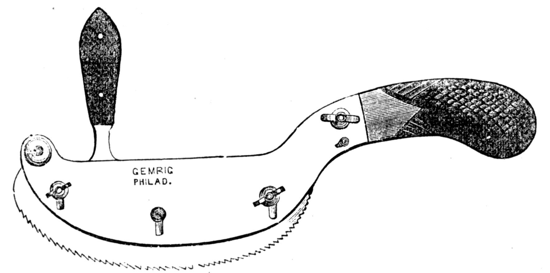
the kindness of Mr. J. H. Gemrig, in the loan of the
electrotype plates which embellish the preliminary chapter.
Conscious that the work is not without defects, it is
still presented to the profession, with the hope that it
may be found to answer the purpose for which it was
designed.
Before entering upon the study of any subject, it is of
moment that the student be thoroughly convinced of the
importance of the knowledge which he is about seeking to
acquire, since his zeal in its pursuit will, in most cases, be in
proportion to this conviction. We will, therefore, before
entering upon the subject proper of this book, present in few
words, some considerations on the importance of a study of
morbid anatomy as revealed by post-mortem examinations.
The necessity of a study of anatomy and physiology by a
medical student, is now so universally recognized, that an
attempt to prove its importance would seem deserving only
of ridicule; but the ignorance of many practitioners on the
subject of morbid anatomy, shows that this study has yet
to vindicate its claim as a necessary branch of a medical
education.
From a purely theoretical standpoint, the educated physician—one
whose motto is Esse, non videri—after combating
a disease in vain, should not feel content to remain in
ignorance of its real nature, so far as discoverable by anatomical
changes, capable of being recognized after death, even
had he no prospect of adding thereby one jot to his practical
acquaintance with disease or to his power to combat it; yet
his scientific conscience (if we may be allowed the expression)
ought not to rest satisfied until, in all doubtful cases, his
ante-mortem diagnosis be confirmed or overthrown, and his
conception of the case completed in all its details by a post-mortem
examination.
18Besides this purely individual scientific interest, there are
weightier practical reasons for an acquaintance with this
branch of medical science by the physician as practitioner.
Among the almost innumerable questions upon medical,
theological, and miscellaneous subjects which the American
public feels at liberty to propound to its medical advisers,
none are of more frequent occurrence and none are more
justifiable than the two: “What is the matter with the
patient?” and “Will he, or can he, recover?” and to none
is an answer more imperatively demanded. The public very
naturally, and with reason, requires on the part of a physician
the ability to make a diagnosis and a prognosis. It
will not be satisfied with being told that the name of the disease
is of no importance, that the doctor only wants to hear
the symptoms; that he does not cure diseases, but removes
the symptoms of disease, &c. Only an exceedingly well-trained
public will accept these truisms as an equivalent for
diagnostic skill. Hence, it is the physician’s interest, as
well as his duty, as we shall see, to seek, in all cases, to
make a diagnosis, no matter how difficult the task may
prove to be. The question how far his treatment will be
modified by his diagnosis is a question of therapeutics, and
does not belong here; but certain it is, that a mere combatal
of the symptoms as isolated phenomena cannot be
regarded as fulfilling the whole duty of a conscientious
physician.
The ability to make a diagnosis, and consequently prognosis,
depends upon a knowledge of pathology, with a
knowledge of symptoms as signs of pathological states and
changes; and as such they must be critically examined and
their true import discovered, if possible. Thus treated, we
arrive, by various processes of reasoning, at a diagnosis,
under which the symptoms fall into their natural order of
importance, and we run but little risk of contending with
remote subjective symptoms (of great importance in differential
19therapeutics) to the neglect of more important, though
perhaps less prominent ones.
Besides this, we are, in a measure, prepared to foretell the
probable course of a disease, and can, therefore, in many
cases, adopt anticipatory measures, while in all we will be
guarded against the error, so often committed, of ascribing
to the remedy used the so-called “aggravations,” which are
often only natural symptoms of the unchecked and, perhaps,
entirely uninfluenced morbid process. Such knowledge
serves thus, by purifying our experience, to guard us against
self-deception, and to prevent us from misleading others by
reports of cures of diseases existing only by virtue of a false
diagnosis.
A knowledge of pathology, furthermore, places in our
hands a thread which can guide us through the labyrinth of
our vast materia medica, and which enables us, from the
myriad of symptoms, to eliminate the non-important ones.
It shows us the “bearings” of the medicines and their
various specific ranges, thus materially facilitating the choice
of a remedy.
Again, medical science is virtually based upon pathology,
and we see, therefore, how important, nay, how absolutely
necessary, to the progress of the former is the study of the
latter. The practice of medicine as an art can never be
advanced knowingly by those who neglect its cultivation as
a science. While each one may practice the art according
to his own convictions, true medical science stands above all
the belittling, bigoted prejudices of the schools. Here,
every one claiming the name of an educated physician can
and ought to work.
The wild vagaries of former ages, when philosophy set up
purely theoretical views, under which observed phenomena
were compelled to arrange themselves, have warned the
present age to be guided solely by sober and exact observations
and investigations; and it needs no proof that, in the
20advancement of our knowledge of disease, these are best
accomplished by frequent post-mortem examinations, which
thus become a necessary adjunct to a proper study of
pathology.
In all cases of interest, therefore, the physician should
feel it a duty which he owes to himself and the profession
at large, to seek permission to make a post-mortem examination;
but in order that the fullest benefit may be derived
from the same, he must know how to look for what he is in
search of, and how to recognize it when found.
To furnish this knowledge is the object of the following
pages, to which we herewith introduce the reader.
PRELIMINARY CHAPTER. — INSTRUMENTS AND GENERAL DIRECTIONS. — PRELIMINARY PREPARATIONS.
21
The Post-mortem Case, as prepared by the instrument
makers, will be found to contain, usually, the following
instruments:
1. A set of ordinary dissecting scalpels, four or five in
number, and of graduated sizes, including one heavy cartilage
knife.
2. A brain knife with a long blade, for slicing the brain.
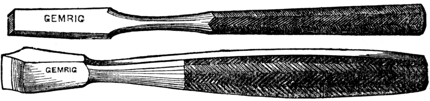
3. Chisels, of one or both the accompanying forms, for use
in opening the head or spine.
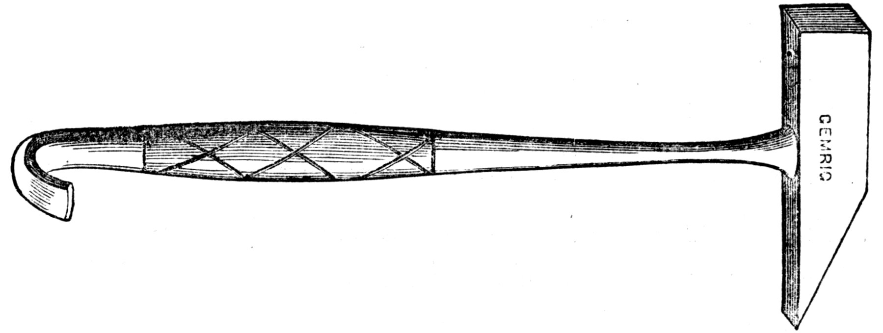
4. An iron mallet or hammer, with a hook on the end of
the handle for tearing off the culvarium.
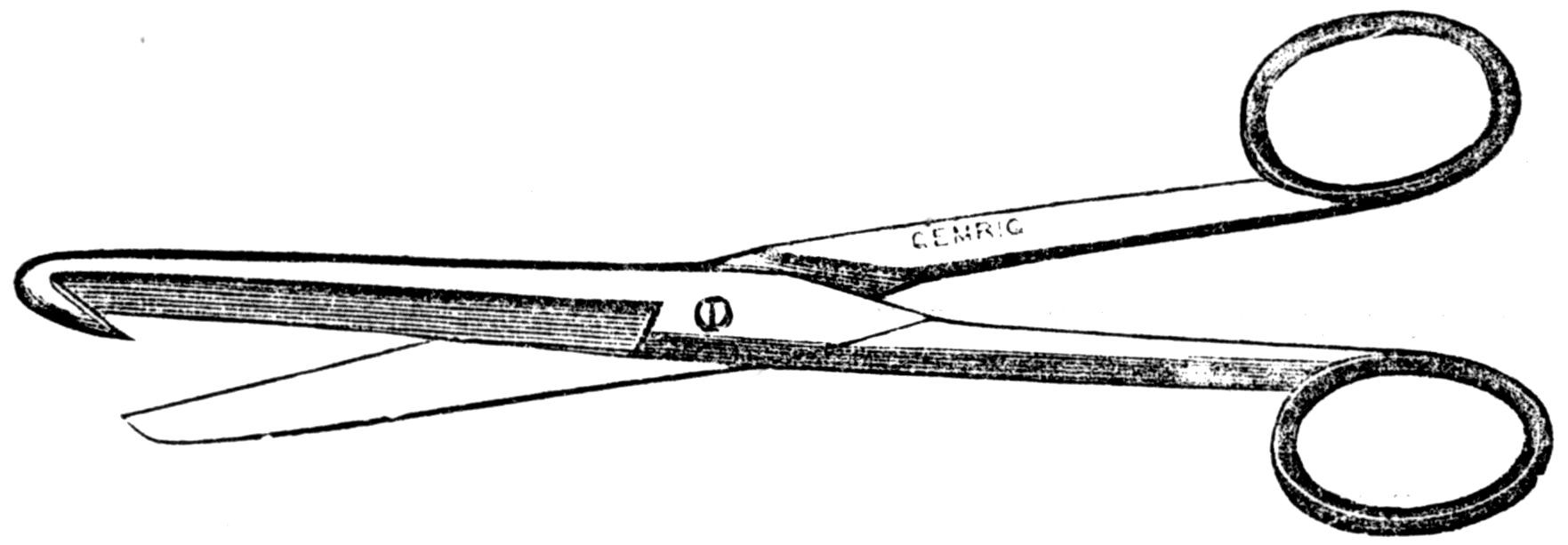
22
5. An enterotome, or scissors with blunt, hooked point,
for splitting open the intestinal canal.
6. A saw with movable back; this arrangement permitting
of a deeper cut, in dividing large bones.
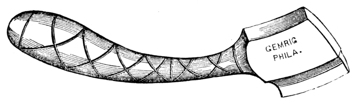
7. The rachitome, a chisel-like instrument, to be used
with the hammer in opening the spinal canal.
8. The double saw, used for dividing the laminæ of the
vertebræ. A side view of the instrument being given in the
cut, one blade only is seen. The two are attached to one
handle, placed parallel with one another, and about one and
one-fourth inches apart. After the soft tissues have been
23removed, this saw is used by passing the spinous processes
between the two blades, and thus dividing both laminæ at
once.
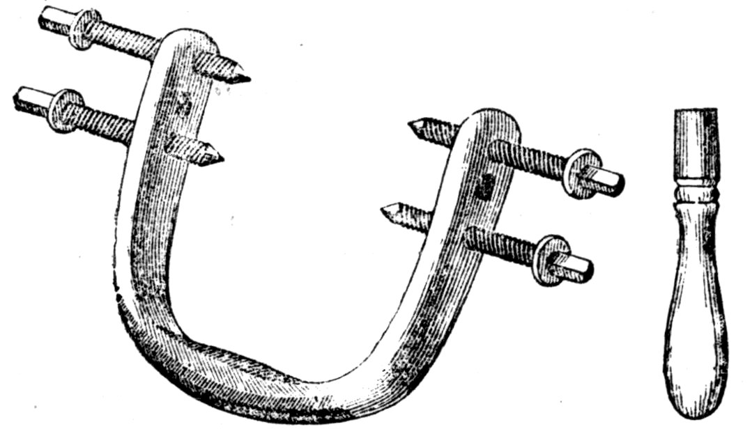
9. A skull clamp, for steadying the head while removing
the calvarium. After the removal of the scalp, this instrument
may be employed, and be of much service for the
above object. It is applied by placing the open end of the
instrument over the crown of the head, turning down the
screws, and thus fastening it just above the line of division
of the bone. The arched end of the instrument now serves
as a handle for turning or steadying the head.
10. Rib-shears, for dividing the ribs where that operation
is found desirable.
11. A tube for inflating the lungs, and an ordinary blowpipe.
12. Dissecting forceps, tenacula in handle and with chain,
grooved director, and assorted needles, straight and curved.
While a post-mortem case with all of the above instruments
is very convenient, and important even, where there
is frequent occasion for its use, still its absence should never
deter the physician from making an examination where the
ordinary dissecting case may be had; and with the country
physician generally, this case is all that is absolutely essential,
as a common carpenter’s saw and chisel may at any
time be found, in cases where the head is to be opened;
while for opening the chest and abdomen, the dissecting case
contains everything that is essential.
24Instruments used in post-mortem examinations should
never be employed for operating upon the living, without
first being repolished by the instrument maker, and the
handles disinfected by careful cleaning in a solution of
permanganate of potash.
The Preliminary Preparations at the place of the
operation should consist in providing a sponge for absorbing
fluids; newspapers and old cloths for filling cavities or
wrapping up any morbid specimen that it may be desirable
to preserve; a couple of quarts of clean sawdust or wheaten
bran for throwing into the cavities before closing them up,
and thus absorbing any excess of fluids; stout thread or
twine for tying intestines and closing the cavities; lard or
sweet oil for oiling the hands; a couple of empty slopbuckets
for receiving the fluids, bloody water, &c., and
plenty of water, hot and cold, with towels and soap. If
the floor of the room is carpeted, to protect it from accident,
a piece of old carpet, or quilt, or oilcloth should be spread
alongside the table or box where the examination is made.
Care should be observed to have all these matters provided
before the operation is commenced, when the door should
be fastened, that there may be no intrusion, by accident or
otherwise.
In preparing the body for examination, if the head is to
be opened and the body is in an ice-box, it will be absolutely
necessary that it be removed. Placing the cover of the box
on the floor, the body may be lifted out and placed on the
same, when both may be again placed on the top of the box
or on a table, for the examination. If only the chest and
abdomen are to be examined, it will hardly be necessary to
lift the body from the box. In all cases, the sheet in which
the body is placed, with the underclothing, (the latter having
25been split down the centre,) should be carefully turned aside,
and care observed during the operation to avoid as much as
possible soiling them with blood-stains.
Precautionary Measures. That there is a certain
amount of danger from absorption of virus in case of cuts or
scratches received while conducting a post-mortem examination,
cannot be denied; yet this danger is by no means
common to every case. In the great majority of instances,
probably no mischief whatever would follow such an accident,
the danger being confined almost wholly to cases of
peritoneal inflammation, erysipelas, and certain malignant
forms of disease. In all cases, however, it will be well to
look for any scratch, cut, or abraded point on the hands or
fingers, and first touching them with nitrate of silver, cover
them finally with collodian. Smearing the hands well with
lard or olive oil will also aid much in preventing absorption.
Should an accidental cut be received during the operation,
it will be prudent, in all cases, to wash the hands at once,
squeeze and suck the part, to favor bleeding, and, finally,
touch with a crystal of nitrate of silver. Punctures with a
needle or slight scratches which do not bleed are, probably,
more dangerous than a free cut; hence, in cases of suspected
danger, when a puncture has been received, it would be
safer to make a free incision with a knife, thus inducing
bleeding, which will favor the washing out of any virus, and
finally, use the caustic.
Time for Making the Examination. As a rule,
post-mortem examinations should be made as soon after
death as a due regard to the feelings of friends will permit,
say within twelve to twenty-four hours; a longer delay
would give time for such decomposition as not only to make
the examination extremely disagreeable, but, from change of
structure in tissues and organs, much less satisfactory. In
cold weather, however, or where the body has been placed
26in ice soon after death, as is the custom in most cities, the
examination may be postponed to any convenient time
before the burial.
Notes. In all important cases, notes should be taken
down by an assistant, on the spot, as dictated by the operator;
heading these by the name and age of the patient and
a brief sketch of the disease. In medico-legal cases, this is
to be conducted with particular care, as will be pointed out
hereafter.
Consent of Friends. Much difficulty will sometimes
be met in obtaining the consent of friends for a post-mortem
examination, the idea of the mutilation of the body of the
deceased striking many with particular dread. With a little
tact and management, however, consent may, in the majority
of instances, be obtained. Instead of speaking to a single
member of the family and leaving him or her to bring the
subject before others, in all doubtful cases it will be better
for the physician to see all the interested parties himself,
either together or separately, and endeavor to interest them
in the case, by pointing out the peculiar character of the
disease, the satisfaction which the friends will themselves
derive from a verification of the diagnosis of their physician,
while, when too late, they may regret having withheld their
consent; also the scientific interest which attaches to the case,
and the benefit which will no doubt accrue to medical science
through a post-mortem examination, and the possibility that
the lives of others may depend upon a knowledge of the true
state of this case. These, with like considerations, adapted
with ready tact to the class of persons to be persuaded, will,
in the majority of instances, be enough to overcome all
scruples, especially when joined to the assurance that the
examination will be so conducted as to leave no visible trace
of the operation with which to embitter the remembrance
of the well-known countenance.
PART I. THE HEAD AND SPINE. — CHAPTER I. THE OPERATION. — THE SPINAL CORD. — CHAPTER II. PATHOLOGICAL CONDITIONS. — Section I. OF THE SKULL. — Section II. THE MEMBRANES OF THE BRAIN. — Section III. OF THE BRAIN. — Section IV. OF THE SPINAL CORD.
27
In opening the head for a post-mortem examination, let
a thick block be placed beneath the occiput, when, after
having carefully parted and turned aside the hair, an incision
may be made through the scalp, over the top of the head,
from ear to ear. The back of the scalpel being placed next
the head, the point may be pushed in advance, thus dividing
the tissues without danger of injuring the edge of the instrument
by bringing it in contact with the bone.
From the loose connection of the tendon of the occipitofrontalis
muscle to the periosteum, by the use of the handle
of the scalpel, the flaps of the scalp may readily be turned
aside, the anterior over the face, the posterior on the back
of the neck. The skull clamp may now be applied, which
will be of great service in steadying the head while using the
saw. Instead of dividing the skull in a circular manner, as
is usually done in the dissecting-room, it will be better to
start the saw above the frontal eminences and run obliquely
down towards the mastoid process, meeting this line by
another commencing at the occipital protuberance and carried
horizontally forwards, thus giving a wedged form to the portion
removed. The advantage of this method consists in our
28being better able to hold the parts in position as they are
replaced, as in the old method, from the readiness with which
the parts move upon one another, the calvarium is liable to
slide forwards or backwards from its position, thus producing
an unsightly ridge across the forehead, which will more
or less plainly show through the integument. With young
children, a strong pair of scissors may be used instead of
the saw in dividing the calvarium.
In using the saw, care should be observed not to injure
the dura mater; hence it will be better not to attempt to
divide both tables with that instrument, but rather depend
upon the chisel or rachitome for breaking away the inner
table. To avoid the sound which would attend the use of
the iron hammer, which, if heard by members of the family,
might excite unpleasant feelings, a billet of wood or wooden
mallet may be used; or, these not being at hand, the head of
the iron hammer may be muffled with a towel, so as materially
to deaden the sounds. The inner table having been
divided, the calvarium may be pried off with the chisel, or
torn away with the hook on the handle of the hammer.
This, from the close adhesion of the dura mater to the bones,
will generally require considerable force.
The calvarium having been removed, and the superior
longitudinal sinus opened, and the condition of its contents
noted, the dura mater, after its careful examination, may
be cut through with the knife or scissors in the line of the
division of the bone. The membrane may now be lifted up,
when adhesions will be found between its under surface and
the pia mater, along either side of the falx cerebri. These
should not be mistaken for results of inflammation, as they
sometimes are, they being simply the points of entrance of
the veins of the pia mater into the superior longitudinal
sinus. These having been divided, with the falx near its
attachment to the crista galli, the whole may be turned
back, exposing the brain.
29To remove the latter, take away the block from beneath
the head, and lifting the anterior lobes of the brain, divide
successively the several pairs of nerves as they appear in
sight, with also the tentorium on either side, and as the brain
falls back into the left hand, the knife may be passed down
into the foramen magnum, and the medulla oblongata and
vertebral arteries divided, when the whole may be lifted
from its position. More or less blood with the cerebro-spinal
fluid will necessarily flow off during this operation,
to secure which, a bucket should be placed beneath the edge
of the table, over which the head should slightly project.
In dropsy of the brain, although the fluid is mainly in the
ventricles, it may escape during the operation and be caught
in the same manner.
The external surface of the brain having been carefully
examined, the hemispheres may be sliced off to a level with
the corpus callosum, when, by removing the latter, the
cavities of the lateral ventricles will be exposed. After a
careful inspection of the several objects seen here, the fornix
and velum interpositum may be removed, opening thus into
the third ventricle. Slices may now be taken off the corpora
striata and optic thalami, with deeper portions of the hemispheres,
and sections made of the crura cerebri cerebellum
and medulla oblongata, thus giving an opportunity of judging
of the condition of all those parts.
After the removal and examination of the brain, attention
should be given to the base of the skull. The lateral and
other sinuses should be laid open, the dura mater, as far
as possible, torn away, and the bones examined for fractures,
caries, abnormal growths, etc.
“The simplest method for removing the ear for the sake of
dissection is, after the removal of the calvarium in the usual
way, to take out both the petrous bones together by means
of two transverse vertical sections, one in front of the two
30petrous bones and the other posterior to them. The anterior
of these sections should pass in a line a little anterior to the
anterior clinoid processes, and the posterior in a line through
the posterior third of each mastoid process. By means of
these two sections, the trumpet-shaped extremity of each
Eustachian tube, a portion of the mucous membrane of the
fauces, and the whole of each petrous bone, together with the
mastoid processes, can be taken out.
“The disadvantage of this procedure is the disfigurement
which is apt to ensue from the falling in of the face. To
avoid this disadvantage, another mode of removing the ear
may be resorted to. This consists in taking out each petrous
bone separately in the following manner: The calvarium having
been sawn off, an anterior section is to be made in each
side in the same line as in the above plan, but extending
only as far as the outer part of the body of the sphenoid
bone; a posterior section in each side is then to be made, as
in the first plan, but not extending farther inwards than the
basilar process of the occipital bone. These two sections are
to be made with a saw or with a chisel and hammer. The
apex of each petrous bone is then to be separated from the
sphenoid and occipital bones, and each petrous bone (the
outer ear and integument being detached and reflected downwards)
is to be drawn outwards, taking care, by inserting
the scalpel deeply, to remove as much of the soft parts as
possible.
“The organ of hearing having been removed, the dissection
may be conducted in the following manner: The auditory
nerve in its meatus should be first carefully examined, presuming
that a previous inspection has been made of the
portion of the brain to which the portio mollis and portio
dura nerves are attached. The size of the external meatus
having been ascertained by allowing a strong light to fall
into it, its anterior wall is to be removed by the cutting
forceps.
31“The state of the epidermis, the ceruminous glands and
secretion, the dermis, periosteum and bone is to be noticed.
The outer surface of the membrani tympani is then to be
examined; also the state of its epidermoid and dermoid
laminæ, its degree of tension, and the amount of motion
possessed by the malleus when pressed upon by a fine point.
The next step is to ascertain the condition of the guttural
portion of the Eustachian tube, to lay open the cartilaginous
tube with the scissors, and then to expose the cavity of the
osseous portion by means of the cutting forceps. In doing
this, the tensor tympani muscle is exposed; its structure
should be examined, and if it has not a healthy appearance,
portions of it should be submitted to microscopic inspection.
The upper wall of the tympanum is next to be cut away by
means of the cutting forceps. In doing this, great care must
be taken not to disturb or disconnect the malleus and incus,
which lie immediately beneath it. After the tympanic cavity
has been exposed, the first step is to pull the tensor tympani
muscle and ascertain how far it causes a movement of the
membrana tympani and ossicles. The incus and stapes are
now to be touched with a fine point, so as to ascertain their
degree of mobility; the tendon of the stapedius muscle is
also to be pressed upon. The condition of the mucous membrane
of the tympanum and of the mastoid cells is then to
be ascertained, and any peculiarity of the cavity, the existence
of bands of adhesion, etc., to be noted.
“The most delicate parts of the dissection, viz., that of the
internal ear, must now be undertaken. The cavities of the
vestibule and cochlea are to be exposed by removing a small
portion of the upper wall of each. Before reaching the vestibule,
the superior semi-circular canal will be cut through
and removed; the membranous canal should be drawn out
and inspected. As the cavities of the vestibule and cochlea
are laid bare, it is desirable to see that the quantity of perilymph
is natural, as well as its color and consistence. The
32outer surface of the membranous labyrinth having been
observed, it should be opened so as to expose the endolymph
and otoliths, portions of all which parts should be removed
for microscopic inspection. This having been effected, the
remaining membranous semi-circular canals are to be exposed,
and the connection of the base of the stapes to the
fenestra ovalis carefully examined. The last stage of the
dissection consists in removing parts of the lamina spiralis,
in examining them microscopically, and in exposing from
within, by following the course of the scala tympani, the
membrane of the fenestra rotunda.
“The only part which now remains unexamined is the
stapedius muscle; in order to expose it, the course of the
aquæductus Fallopii, beginning at the stylo-mastoid foramen,
should be followed until the base of the pyramidal eminence
containing the muscle is reached.”[1]
The Eyes, with the optic nerves, may be most conveniently
removed for examination, by breaking up the roof of
the orbit with the hammer, and, after the removal of the
fragments of bone, dissecting away the fat and muscles
until the ball is exposed, when, with the scissors, the tunics
of the latter may be divided just behind the conjunctival
attachment, and the ball removed with the optic nerve.
The front portion of the eye being left in position, by filling
the cavity behind with a little cotton or paper, any flattening
may be prevented, and thus any appearance of loss of parts
avoided.
The examination having been completed, the brain may
be replaced, the calvarium put in position, the scalp brought
over the same and united with stitches, the arranging of the
hair effacing all traces of the operation.
33
For the removal of the spinal cord, place the body in a
prone position, and make an incision the whole length of the
back, over the spinous processes of the vertebræ. Then
raise up and turn aside all the muscles with the integument,
exposing the laminæ of the vertebræ. The latter may now
be divided with the single or double saw, the rachitome or
chisel being used to complete the operation, when the laminæ,
with the spinous processes, may be removed in one strip,
exposing the cord enclosed in its membranes.
The roots of the nerves may now be divided, and the cord,
enclosed in its sheath, removed; care being observed not to
handle the parts roughly while so doing. The dura mater
may now be split open for the examination of the cord.
Should it be desired to preserve any portion for microscopic
examination, it may be suspended in a solution of bichromate
of potass, xx to xxx grs. to the ounce of water, and in a few
days transferred to a solution of chromic acid, ij grs. to the
ounce of water, where it should remain until sufficiently
hardened to be cut into thin sections.
The examination completed, the parts may be replaced
and the incision sewed up.
34
[Notice in examination, condition of scalp; cuts, bruises, extravasation
of blood, &c. Cranium—Color, smoothness or roughness of
exposed surface; fractures, their position and relation to injuries of
scalp. Removal of Calvarium—Adhesion of dura mater; inner surface
of; smooth or rough; seat of same; deposit of new bone; depression
of inner table; thickening or thinning of. State of fontanelles in
children; condition of frontal sinuses; condition of base of skull noted
after removal of brain; fractures; condition of petrous portions of
temporal bones, &c.]
Fracture. In a post-mortem examination of the head,
after death from a blow or fall upon that part, a fracture
may be disclosed of which there was no external trace.
Owing to the greater thinness and brittleness of the inner
table, it is possible for this to be fractured without any corresponding
injury to the external; such fracture may possibly
rupture some of the branches of the meningeal arteries,
which will be followed by the formation of a clot between
the bone and dura mater, the pressure of which may be sufficient
to produce death. Owing to the same cause—greater
brittleness of the inner table—when fracture of the outer
exists, that of the inner table will be likely to be more
extensive, accompanied perhaps with depression, of which
there is no external evidence.
Again, it will sometimes be discovered that fracture exists
at a point opposite to that upon which the blow was received,
on the principle of the “contre coup” of the French; thus,
the blow having been received on the occiput, the fracture
may be found in the frontal region; or, received on the top
of the skull, fracture may result at the base. In falls from
a height, upon the top of the head, the weight of the body,
35acting through the cervical vertebræ as a propelling force, is
very likely to produce fracture of the occipital bone.
Caries may affect any of the bones of the head, but in
the majority of cases it will be the result of syphilitic or mercurial
poisoning. Following an attack of periostitis, the
inflammation extends to the bone and gradually develops
the carious ulceration. While the condition is usually confined
to, or at least, most strongly developed upon the outer
table, it may involve the entire thickness. In syphilitic
ulceration of the bones of the nasal cavity, the disease may
destroy the cribriform plate of the ethmoid bone, and thus
extend to the membranes of the brain.[2] So also, in
caries of the petrous and mastoid portions of the temporal
bone, which sometimes results from scarlet fever, the disease
may extend to the inner surface, resulting in the accumulation
of pus within the cranial cavity.
Along either side of the central portion of the inner surface
of the calvarium may be usually seen, at least in the heads
of aged persons, a number of irregular, rough pits, varying
in size and depth, which are not to be mistaken for disease
of the bone, they being simply impressions of the Pacchionian
glands.
Thinning of the bones of the head will always indicate
increased pressure from within, induced by hypertrophy
of the brain, or, as is more frequent, hydrocephalus. In the
latter case, not only thinning, but complete absorption of
the parietal and other bones may result.[3]
Increased Thickness will also sometimes be found.
This condition indicates diminished pressure from within,
as in atrophy of the brain. The thickening results from a
36gradual remodeling of the inner table and diploe, so that
while the exterior of the skull may retain its normal size
and form, the inner table following the retiring and shrinking
brain, the interval between the two becomes filled with
the thickened diploe. It has been observed that this hypertrophy
is greatest at those parts of the bones where ossification
first commences, as at the parietal and frontal eminences.
It is not confined to old persons, though perhaps more frequent
with them.[4]
1. The Dura Mater.
[Notice color and general character of surface; blood between it
and bone; position of; quantity; coagulated or not. Condition of
bone—necrosed or fractured; pus between dura mater and bone.
Tumors—their position, size, &c. Wounds—their position, extent, &c.
Open longitudinal sinus and note contents.]
This membrane, serving both as a periosteum to the inner
surface of the cranial bones and as a support to a serous
membrane—the reflected layer of the arachnoid—is subject
to affections of a two-fold character, those peculiar to the
fibrous and serous portions.
Inflammation of this membrane, may involve either the
outer fibrous, or the inner serous layer. In the former case,
the membrane appears congested, red and more or less softened.
The inflammatory process may result in the formation
of pus between the bone and dura mater, and even in
gangrene. The disease may also extend to the adjacent
portions of the pia mater and brain substance. External
injuries, fractures of the bones of the skull, inflammation of
the periosteum, otitis, resulting in caries of the temporal
37bone, may all be causes of inflammation of the outer portion
of the dura mater.
Inflammation of the inner surface of this membrane, is
marked by the presence of a net-work of delicate red vessels,
while the surface is covered by a soft, grayish or yellow
semi-purulent matter, and may attend cases of pyæmic
poisoning, puerperal peritonitis, or some of the exanthemata.
Thickening of the fibrous portion of the dura mater may
be found as a result of chronic inflammation, either spontaneous
or as the result of external injury. From the identity
of structure between this and other fibrous tissues of the
body, it is not unlikely that this thickening is often the
result of a rheumatic form of inflammation.
External violence is, however, most commonly the cause
of the change. In one case, the patient fell down stairs in
a state of intoxication, striking the head on the steps. He
continued in a state of insensibility for nine days, when he
began to show signs of returning consciousness, taking food
and drink, but memory, judgment, and all the mental faculties
were gone. Death ensued in about two years. The
dura mater of the left hemisphere was found greatly thickened.
The pia mater infiltrated with a large amount of
serous fluid. The convolutions were atrophied, and about
four ounces of serous fluid found in the ventricles. The
fornix was softened and the septum lucidum entirely
destroyed.
Fibrinous Clots, or Thrombi, will be occasionally
found within the sinuses of the dura mater, where they may
have given rise to congestion of the brain, with apoplectic
effusions, paralysis, convulsions, coma, etc. They may
originate from injuries of the head, inflammation of the dura
mater, pulmonary disease, etc.
38Tubercular Deposits are also sometimes found in this
membrane, appearing, however, mainly upon the arachnoid
surface. They present the usual character of tubercle, having
a whitish or grayish appearance, with the consistence of
cheese, and scattered in small particles upon the surface.
They are found in tubercular meningitis, (a disease common
with children but rare with adults,) and in most of those
cases of so-called acute hydrocephalus.
Tumors of various kinds are occasionally found in the
dura mater, including cystic, fibrous, fatty, osseous and cancerous
growths. The latter may cause such absorption of
the bones of the skull, as to appear on the exterior of the
head.
In one case which came into the dissecting-room some
years ago, thin bony formations of the size of a silver
quarter dollar were found in the tentorium, and smaller
ones in the falx cerebri. Nothing could be learned of the
previous history of the case.
2. Arachnoid and Pia Mater.
[Notice in examination, contents of cavity of arachnoid; serum or
blood; if former, amount, color, odor; if blood, situation, quantity,
fluid or coagulated; adhesion of surfaces of arachnoid; tubercles,
their position, &c.; color of membrane; vascularity, transparency,
or opacity. Sub-arachnoid fluid—quantity, position, color, &c. Pia
mater—vascularity, in points and entire; serous effusion into substance
of; blood, &c.; position, size, &c., of clots; granulations, (tubercles,)
number, position, &c.; Tumors—size, position, and character.]
As morbid conditions of the arachnoid membrane are
more common with its visceral layer, and as these conditions
usually involve the pia mater, the two membranes are here
noticed together. They will be found presenting various
39degrees of congestion after death, which will not necessarily
be a positive indication of the extent of congestion during
life.
Pacchionian Bodies. Along either side of the great
fissure may be noticed within the pia mater of adults, several
small white bodies, varying in number and size—the Pacchionian
bodies. They may cause absorption and perforation
of the dura mater, and even of the bones of the skull.
Being looked upon as the result of mere senile changes,
they do not indicate the presence of disease, though
repeated congestions of the brain appear to favor their more
rapid development.
Inflammation of these membranes, or meningitis, is a
very common affection, and is accompanied with an accumulation
within its substance or beneath its layers, of
serum, lymph, or fibrine in various proportions. It may be
confined to circumscribed portions, or involve a large portion
of the membrane, and even extend to the spinal cord.
Adhesions between the two surfaces of the arachnoid sometimes
result from this form of inflammation.
Insanity in its various forms is most frequently accompanied
with some morbid condition of these membranes.
In twenty-two cases of insane persons whose brains were
inspected by Dr. Marshall, in twenty-one, serous fluid, varying
in amount from one to twelve ounces, was found in the
ventricles, and in seventeen of these twenty-one cases, similar
effusion was found in the sub-arachnoid space, or within the
substance of the pia mater. While red injection of the
membrane was found only in four cases, yet other conditions—the
effusions, &c.—were evidently the result of previous
inflammation. In nine cases were the arteries of the brain
opaque, thickened, steatomatous, or ossified; conditions
40highly favorable for deranging the capillary circulation of
the membranes or of the brain.[5]
The following statement gives the principal morbid changes
of these membranes which have been found in cases of
insanity:
1. Injection, more or less intense, of the pia mater, giving
a red or scarlet appearance; or, where infiltrated with serous
fluid, presenting a pale gray color and increased in thickness.
2. The arachnoid (the visceral layer) becomes opaque and
thickened, resembling the dura mater or macerated parchment.
3. The meningeal injection may terminate in serous effusion,
either from the free surface of the arachnoid into the
sub-arachnoid tissue, (pia mater,) or from the choroid plexus
into the ventricles.
4. Albuminous exudations may be found upon the free
surface of the arachnoid of the dura mater, covering its
whole extent, or confined to definite portions.
5. Adhesions of the two surfaces of the arachnoid may
rarely be found. It is most common in the great fissure,
and has been found in the ventricles.
6. Blood may be effused upon the surface of the arachnoid
or in the substance of the pia mater.
Serous Effusion. As has been already intimated, this
may be found either in the cavity of the arachnoid—between
the reflected and visceral layers—or within the ventricles. In
the former position, the quantity is never large, while in the
latter, it may amount to twelve or sixteen ounces, and be
present for many years. When in such large quantity, there
41will be great distension of the ventricles and thinning of the
corpus callosum and fornix, with destruction of the septum
lucidum, as well as more or less separation of the cranial
bones. It is only in children and before the bones of the
head have become united, that such large accumulations are
possible, as at a later period, from the unyielding condition
of the walls of the skull, the presence of a single ounce, particularly
if suddenly formed, would produce death. In all
cases, the danger to life will be in proportion to the rapidity
of the formation; a very slow and gradual accumulation
permitting either of an expansion of the cranial bones, or a
gradual absorption of brain substance, thus preserving an
approximation to the normal pressure on the brain tissue.
Serous effusions, like sanguineous, are generally the result
of over distension of the cerebral vessels, either from mechanical
obstruction, or a weakened condition of the coats of
the vessels, with increased force in the action of the heart.
It generally attends tubercular meningitis, and may be
favored by an anæmic condition of the system. The symptoms
during life, attending a rapid effusion of serum, are not
so readily distinguished from those of sanguineous effusion,
as to enable us to pronounce with certainty in any given
case as to the cause of the cerebral pressure.
Sanguineous Effusion—Apoplexy. Effusions of
blood may be found between the bones and dura mater; between
the two layers of the arachnoid; within the substance
of the pia mater; within the ventricles, or within the brain
substance. Blood clots will seldom be found between the
bone and dura mater, except as a result of mechanical injury,
and in the majority of cases as an attendant upon fracture.
If a fragment of the bone at the same time, be driven through
the dura mater, then a clot may be found in the arachnoid
cavity. But in another class of cases, where death has
42resulted from blows upon the head without producing fracture
of the bones, the whole surface of the brain in the
region of the injury, and not unfrequently in distant parts,
after the removal of the dura mater, is found covered with a
layer of blood, which at first sight appears to be outside of
the membranes; but on close examination, it is found that the
blood is effused or infiltrated into the sub-arachnoid tissue,
and that it has escaped from the lacerated vessels in the
pia mater. The thickness of the layer varies. It is generally
in greater quantities at the sides and base of the brain,
and the inferior lobes and cerebellum may be covered by it.
It is usually thickest over the crura, the pons Varolii and
medulla oblongata.
In the same class of cases the blood may be also effused
into the ventricles. These appearances are so uniformly the
result of violence, as to form a valuable piece of evidence in
medico-legal inquiries, to prove that such hæmorrhage and
death could not be the result of internal causes.
In the greater number of still-born children, an examination
of the head will show a similar condition of things.
The surface of the cerebrum generally, with sometimes that
of the cerebellum, will be found covered with a layer of coagulated
blood effused into the pia mater, while the ventricles
will often be filled with clots. Where the history of the
case is not known, it might at first be suspected that death
was the result of violence inflicted after birth. Violence has,
to be sure, been the cause of death, but it is such violence as
attends a protracted case of labor, with, perhaps, a large head,
and a contracted pelvis of the mother. The wonder is, that
from the great pressure to which the head is subjected during
labor, that so few children are still-born or do not die soon
after birth, from rupture of the cerebral vessels.
Effusions into the ventricles, may also be frequently the
43result of external violence. The pia mater, the vascular
membrane of the brain, we find carried into these cavities
by means of the velum interpositum, which forms the roof
to the third ventricle, while its borders extend into the lateral,
forming the choroid plexuses. The effused blood may, therefore,
extend along this membrane into the cavity of the
ventricles.
Again, blood may be effused in any portion of the brain
substance, constituting true sanguineous apoplexy. Certain
parts are much more frequently the seat of these effusions
than others. They are more common in the striated bodies
or optic thalami—probably from the greater vascularity of
those parts—but may be found in the corpora quadrigemina,
the pons Varolii, the crura cerebri, or in the cerebral
hemispheres, and occasionally in the cerebellum. The symptoms
during life will vary according to the location of the
effusion; when in the pia mater, or in other words, outside
of the brain, paralysis will seldom attend, though the coma
may be profound, with relaxation of the muscular system and
sometimes convulsions. When the hæmorrhage is in the
optic bed or striated body of one side, from the decussation of
fibres in the medulla oblongata, paralysis of the opposite side
of the body will follow; while if the effusion has taken
place in both hemispheres, the palsy will be double-sided,
though probably more complete on one side than on the
other.
Effusions of blood into the corpora quadrigemina, will
most frequently be attended with muscular tremblings or
convulsions, and probably impaired sight, with some change
in the pupil. When in the medulla oblongata, convulsions,
followed by palsy, deep coma, and early death; greater fatality
attending effusions at this point, probably, than at any
other. When the effusion takes place in the cerebellum,
the loss of consciousness will be very temporary; there may
44be relaxation of muscles without palsy or loss of sensibility,
and, it is said, frequent vomiting.
The amount of blood effused is subject to the greatest
variation. Clots may be found as large as a hen’s egg, or
smaller than a pea. Indeed, violent apoplectic attacks,
ending in death, may occur, where the most careful examination
will fail to detect any effused blood, death being the
result of extreme congestion of the membranes. On the
other hand, the presence of a clot is not necessarily fatal,
evidence being abundant that they may be so far reabsorbed
as to be followed by at least partial recovery.
The following cases will serve to illustrate these several
conditions:
Case I.—Sudden Death from Cerebral Congestion.
Mr. M——, aged thirty-five years, had always enjoyed good
health. For some months previous to death, had been working very
hard, with a good deal of anxiety, in arranging the affairs of a company
of which he was secretary. On the evening previous to his
attack, he retired at eleven o’clock, well and in good spirits. At
three o’clock A. M., his wife was wakened by his heavy, stertorous
breathing, attended with slight convulsive movements of the limbs.
In less than a half hour he was dead. The post-mortem showed the
heart, lungs, and abdominal organs to be in a perfectly healthy condition,
while the most careful examination of every portion of the
brain failed to expose the least effusion of blood. The ventricles and
sub-arachnoid space contained a moderate amount of serum, while the
vessels of the pia mater were strongly engorged with blood.
Case II.—Apoplectic Attack, followed by Death in Three Days; Clot found in Thalamus.
Mr. J——, aged sixty-one, a butcher, abstemious in his habits
but plethoric in temperament, was suddenly stricken down with an
attack of apoplexy, which left him with paralysis of the right side of
the body, with impaired speech and memory. He gradually and
almost entirely recovered. Ten months after, while in the street, he
45fell with another attack. He partially recovered consciousness, but
had complete palsy of the left side, and died in three days. Here, the
post-mortem revealed excessive congestion of the pia mater, and a
large clot, the size of a hickory nut, in the right optic thalamus. In
the left thalamus was a distinct trace of a clot, (nearly absorbed,
however,) which had undoubtedly been effused in the attack ten
months previously.
Case III.—Apoplectic attack, followed by Hemiplegia, and Death in Five Years.
Charles E. W——, at the age of forty-seven had a sudden apoplectic
stroke, in January, 1867, followed by partial paralysis of right
side. In May following, he had a second attack, which greatly
increased the hemiplegia, impaired the articulation, and weakened the
memory and intellect. His general health and strength gradually failing,
he died August, 1872, five years after the original attack.
Post-mortem revealed a greatly thinned and dilated condition of the
walls of the arteries of the base of the brain and the remnants of the
original clot imbedded in the left optic thalamus, the brain substance
around being of a dark color and much softened. From the appearance
of the dark, ragged remnant of the clot, it must have been
originally of about the size of a robin’s egg.
Case IV.—Death from Congestion, with Serous Effusion and Softening.
Mr. F. H——, aged thirty, had an attack of brain fever at twenty-three,
followed by attacks of severe pain in the head, recurring every
few days, weeks or months. These attacks were accompanied with
slight convulsive symptoms and delirium, the pain being of a most excruciating
character, and lasting from a few hours to two or three days.
The attacks were gradually increasing in severity, but without any
impairment of the faculties of the mind. On a Saturday night, he was
brought home with one of his worst attacks. When I first saw the
patient on Sunday evening, I found him in a half-delirious, stupid condition,
yet when roused up, giving satisfactory answers to questions,
but immediately sinking into his former condition, and every one to five
minutes starting suddenly up and, with staring eyes and distorted face,
uttering piercing shrieks and screams, and calling to those around to
shoot him, split open his head with a hatchet—anything to release him
from his sufferings. These paroxysms would sometimes be followed by
46a convulsive action of the diaphragm and abdominal muscles, ending in
his sinking into the same dull, stupid condition as before. In the interval
of calm, the respiration was remarkably slow and feeble, with a
slow irregular pulse of forty beats to the minute. These symptoms
becoming gradually worse, he sank into a comatose condition early
Tuesday morning, expiring at daylight, sixty hours after the commencement
of the attack.
The post-mortem revealed greatly enlarged Pacchionian glands, with
extreme thinning of the skull over the same. The pia mater was considerably
congested. Upon turning the brain back for removal from
the base of the skull, there was a sudden gush of water from the ventricles,
sufficient of which was secured to show that the quantity could
not have been less than two ounces. Upon opening the lateral ventricles,
these cavities were found unusually large from distension by the
fluid, while the septum lucidum and fornix were found in a soft, pulpy
condition, the former being completely broken down and detached
from the corpus callosum above. The gray portions of the corpora
striata were also softer and more easily broken up than was natural.
The appearance presented by a clot, as well as the brain
substance immediately around it, will vary according to the
time the patient survives the attack. When death follows
in a few days, the clot will have a soft, blackish appearance;
after a month or six weeks, it becomes firm, and assumes a
deep brown color, and at a still later period, it becomes still
more firm, and of a pale red tint; lastly, it may become
entirely absorbed. Peculiar changes also take place in the
brain substance immediately around the clot, which vary
according to the time intervening between extravasation and
death. The portion immediately in contact with the clot is
generally of a dark red or wine color, or, at a later period,
of a chocolate brown, and of a soft, pulpy consistency. Exterior
to this, the color is paler and of an orange tint, and
still further on, of a bluish-white or yellow. The change in
structure and consistence of the brain, immediately around
the clot, constitutes one form of softening soon to be noticed.
47
[Noticebefore removal—size; form; symmetry; space between
surface of brain and calvarium. After removal—parts at base, their
size, symmetry, color on surface, infiltration with serum or blood. Removal
of pia mater—degree of adhesion; appearance of convolutions;
color; consistence; effect of stream of water. Ulcers—condition of
brain around. Sloughs—relation to membranes, condition of brain
around. Deposits; tumors, wounds, etc. After section, notice breadth
and character of gray portions of convolutions; color, vascularity, etc.
Consistency—softened or hardened; of white substance; color. Blood-vessels—number
and size of red points in different portions. Extravasation
of blood—its situation; fluid or coagulated; amount or size of
coagulum; color, etc. Cavities in brain substance; their number, shape
and contents. Blood, purulent, or fluid; its quantity and color. Condition
of brain around; cicatrices; wounds; adventitious substances, as tumors,
calcareous masses, tubercle cancer, etc. Lateral ventricles—note any
difference in the two; contents; amount of serum or blood. Choroid
plexus—pale or congested; cysts; calcareous bodies; their size and
situation. Lining membrane of ventricles; its vascularity, roughness,
opacity, etc. Septum lucidum—entire or lacerated; consistence.
Fifth ventricle—size, contents, etc. Third ventricle; contents. Commissures—their
condition; middle, broken or double. Optic thalami and
corpora striati—size, symmetry; character of surface and interior; if
softened, extent; extravasation of blood, etc. Condition of pineal gland;
corpora quadrigemina; valve of Vieussens, etc.Medulla oblongata—degree
of adhesion of membranes; softening, exact locality of; condition
about origin of nerves; appearance upon section. Fourth ventricle—contents;
condition of floor, etc. Cerebellum—examine with same care,
and note same points as in cerebrum.]
Inflammation. Acute inflammation of the brain substance
is a rare disease, except as the result of mechanical
injury. In such cases, the disease is generally quite circumscribed,
being confined to the immediate region of the
injury. The brain becomes very vascular, acquiring a red
color, which, at a later period, changes to a brown or greenish
hue, and becomes much softer than natural.
If a foreign body be lodged in the brain, as a piece of
bone, or a bullet, then the inflammation is likely to result
in an abscess, which will give rise to head-ache, delirium
with intolerance of light, succeeded by convulsions, coma
and death.
Subacute, or chronic inflammation of the brain, is much
48more common, yet is accompanied with pathological changes
similar to those of the acute form. At first, the inflamed
portion becomes red and congested, the color gradually
changing to a crimson, purple, brown, or claret color, with
more or less change of consistence.
The symptoms attending inflammation of the brain substance,
or cerebritis, are not readily distinguishable from
those of meningitis; indeed, in most instances the two diseases
are, to a certain extent, combined; as, from the vascular
connection between the brain and pia mater, there
might be a ready extension of inflammation from one to the
other. When, however, there is any sudden perversion of
the sense of vision or hearing; or if there are convulsions,
affecting mainly one side of the body; or if coma succeeds
the convulsions and is accompanied by one-sided paralysis,
we may expect to find evidence of inflammation of the cerebral
substance, with possibly that of the membranes also.
It is a fact to be borne in mind, however, that while in
typhus and typhoid, and perhaps some other forms of fever,
we may have symptoms strongly resembling those of idiopathic
inflammation of the brain or its membranes, still the
post-mortem will reveal no trace of any such morbid condition.
Softening. To be able to recognize readily any change
in the consistency of the brain, clear ideas must first be had
of the normal density of this organ. This may be obtained
from an examination of the brain of some of the lower
animals—as the sheep or ox—that has been killed in a state
of health. We shall then find that the brain presents sufficient
firmness to permit of its being handled without rupture
of its substance, and to allow of its being sliced into thin
sections which will support their own weight. The gray
portion is somewhat softer than the white, yet the fibrous
character of the latter, becomes apparent only after hardening
49in alcohol. If put into pure water, it continues
unchanged for eight or ten hours, and without any portion
becoming dissolved, or rendering the water any degree turbid.
The consistence of the brain varies, normally, at different
periods of life. In the fœtus and at birth, its softness
approaches to semi-fluidity. From this to the fifteenth or
twentieth year, it gradually acquires the firmness of the brain
of the adult.
Softening, one of the most common variations from
the normal condition, is generally a mere result of inflammation
of a chronic or sub-acute form, accompanied
with a fatty degeneration of nerve substance. Under the
microscope, the change is seen to consist in a disintegration
of the nerve fibres, the medullary substance breaking up
into large masses, and undergoing fatty metamorphosis. It
may take place either on the external surface of the organ,
or in the septum lucidum and fornix, the corpora striata, or
optic thalami of the ventricle, the central parts of the
hemispheres, the cerebellum, or the crura cerebri, in the
order mentioned.
While this change is usually the result of inflammation,
as already mentioned, it may accompany or succeed the following
morbid conditions of the organ: 1. It may be the
result of congestion of the vessels of some portion of the
brain. In this case, the softened portion is reddish, crimson,
or brown in color. 2. It may follow the effusion of blood
in apoplexy. The softened portion is then of a brownish
color, or, if considerable time has elapsed, it may be of a
dirty ash color, tending to green. 3. It may accompany or
follow the process which terminates in serous effusions. It
is then of a milky-white color. 4. It may take place in the
brain substance immediately around tumors, when its color
may present a variety of tints.
Among the causes tending to induce this condition of
50the brain, may be mentioned a diseased state of its blood
vessels, or their obstruction by fibrinous clots; constitutional
syphilis; excessive mental labor, or frequently repeated
epileptic convulsions. The consistency will be found to
vary from a slight change from the normal condition, to that
of a soft, pulpy, or even cream-like condition.
The symptoms accompanying softening of the brain, present
a considerable variation. Among the more prominent,
may be mentioned an unsteady or tottering gait, partial
palsy, thick and inarticulate speech, feeble memory, disordered
intellect, dull pain or heaviness in the head, frequent
drowsiness, formication, numbness or rigidity, or occasional
involuntary contraction of the muscles of the upper extremities.
Abscess of the brain, differs from softening, to which it
may have some points of resemblance, in the purulent matter
being contained in an irregular cavity, lined with a more
or less distinct membranous cyst. Flakes of lymph are
frequently found in these abscesses, giving the matter much
the appearance of that contained in scrofulous abscesses, and
they are most commonly found in subjects who present the
usual symptoms of the strumous diathesis.
While these collections are unquestionably often the
result of inflammation, yet it has been claimed that they
may result from previous disease of a suppurative character
in the lungs, and perhaps other organs, the purulent matter
being taken up by the veins and carried into the general
circulation, and finally deposited in the substance of the
brain. Abscesses may be found in any portion of the brain
substance, or the purulent accumulation may be found
between the bone and dura mater, or between the two layers
of the arachnoid. In the latter cases, the disease may
generally be traced to caries of some of the bones of the
head, as of the petrous or mastoid portions of the temporal,
51or of the bones of the nasal cavity, involving the cribriform
portion of the ethmoid.
Again, abscesses in the brain may result from external
violence, as from a blow or fall upon the head. It is
remarkable, that in these cases, some months may elapse
between the date of the injury and death.
Hardening. Induration of the cerebral substance, is a
condition not readily distinguished during life from that of
softening, the two states being accompanied by symptoms
of a similar character. Dr. Jones, who has charge of the
male department of the Pennsylvania Hospital for the Insane,
and has had many opportunities for examining the
brains of insane patients, tells me that he found hardening,
nearly as frequently as softening, in these cases, and never
could tell beforehand, with certainty, which condition might
be present.
In hardening of this organ, its density may be increased
to that of boiled white of egg, or it may approach in consistence
to that of a brain that has been hardened in alcohol,
losing much of the sticky, adhesive character when broken
up by the fingers, which marks the brain substance when in
its normal state. The cause of the change is not well known,
yet it is conjectured to be one of the results of inflammation,
inasmuch as it is generally found with its capillaries greatly
loaded with blood, while more or less fluid is found beneath
the arachnoid and in the ventricles. The induration may
affect the greater part or even the whole of the cerebral
mass, or may be confined to particular portions or regions.
In a case recently examined for Dr. Toothaker, of this
city, where insanity of some years’ duration, and finally
death, had followed a severe injury of the head, the brain
was found so hard as to permit of fracture in the direction
of the fibres, thus readily tracing their course.
52The symptoms that have most frequently been observed
to accompany this change are, defect and gradual loss of
memory, apathetic indifference, slight difficulty of articulation,
followed by loss of sexual desire, partial palsy, fatuity,
wasting and death.
Extreme induration is often found in the brains of idiots.
The whole organ may be found resembling in color and
density boiled white of egg, or even cheese. The cerebral
substance is shrunken, dense, and apparently quite void of
vessels.
Hypertrophy of the brain, is a condition in which there
is usually increased hardness as well as increased volume,
and is distinguished by flattening of the convolutions, narrowing
of the ventricles, and a remarkable dryness of the
whole organ with its membranes, the change involving both
the cerebrum and cerebellum, and accompanied with an
increase of weight, all indicating increased nutrition, or the
deposit of new matter in the tissue of the brain. Thinning
of the cranial bones may attend this condition, increased
internal pressure resulting in their partial absorption.
The causes of this form of disease are not generally understood.
The symptoms, though always present, are not
uniform. Among them may be mentioned intense head-aches,
a weakened or perverted state of the intellectual
faculties, fits of giddiness, accompanied with stupor; finally,
convulsions, with perhaps loss of sensation and motion, the
patient being unexpectedly cut off by an epileptic attack.
The disease has not been observed in persons over fifty. In
most cases, the patients were between twenty and thirty.
In one instance, however, it was developed in a young
girl of thirteen. Lead poisoning would seem to be
an exciting cause, it having, in several instances, been developed
in painters and manufacturers of white lead.
53Atrophy. In this condition, there is found a general
diminution of the volume of the brain, and especially of the
convolutions. The latter are shrunk, narrow, and sometimes
softened, while the sulci are large and open, the brain
receding from the skull, and the pia mater greatly injected
with serous fluid, giving the appearance of a jelly-like
investment to the whole brain.
The brain substance is at the same time soft, the ventricles
enlarged and filled with fluid, while a large amount of
serum will flow from the sub-arachnoid space at the base of
the brain, and from the spinal canal.
While this condition of the brain might be looked upon as
a result of pressure from the accumulated serum, the absence
of the usual symptoms of hydrocephalus, with the known
history of these cases, renders it quite probable that the
change commences as an actual loss of brain substance, the
place of the latter being supplied with serous fluid.
Resulting from atrophy of the brain, there will often
be found an increased thickness of the cranial bones, this
being another conservative effort of nature to preserve the
normal support and pressure upon the brain.
Atrophy of the brain is sometimes found in old age and in
various enfeebling diseases, where all the organs suffer more
or less waste from improper nutrition, but it is so frequently
found with drunkards, especially those who have died with
delirium tremens, that this condition may, in the large
majority of cases, be considered as the effect of intemperance.
Several forms of atrophy may also be observed. It may
be confined to some portion of the convoluted surface, or to
one of the striated bodies or optic beds. Atrophy of the
optic tracts, or nerves of one or both eyes, is not unfrequently
associated with loss of sight from amaurosis.
54
Tumors of the Brain.
The brain, like other parts of the body, is subject to morbid
growths of a great variety of forms. The symptoms of
tumors in the brain, in many points, present such resemblances
to other forms of disease, that it is usually the post-mortem
alone, that will determine the fact of their presence
or character. It may be said in general, however, that the
effects of morbid growths in the brain will vary according:
1. To the changes in the surrounding cerebral substance;
2. To the size of the growth; and 3. To the position of the
brain in which it may be developed.
1. The changes induced in the brain substance surrounding
tumors are usually, first, derangement of the circulation,
and second, as a result of this, effusion of serum, or finally,
softening of a greater or lesser amount of contiguous cerebral
substance. Head-ache, with epileptic attacks, loss of
memory, irregular contraction of the muscles and partial
paralysis may accompany the vascular derangement, while
as softening or pulpy destruction supervenes, a general
aggravation of all the symptoms will follow, ending in death,
either with coma or by a sudden apoplectic attack.
2. Tumors of the brain of a small size, may be found
after death from other causes, that have evidently produced
no symptoms during life; and in other instances, they have
induced no change until a few days before death, in which
cases, the convulsions, paralysis and coma which precedes
this result, must be attributed to the vascular disturbance
in the surrounding brain tissue.
3. The position of the tumor, will modify to some extent
the character of the symptoms manifested. When in the
anterior lobes, loss or impairment of speech is said to attend.
While, when in the corpus striatum, the motions of the legs
and arms are disordered.
55The following are the more commonly recognized tumors
of the brain:
Adenoid, or Glandular-like Tumors. These are
generally described as resembling an enlarged lymphatic
gland, both in color and density. They may vary in size
from a filbert to that of an orange. There may be a single
growth of this kind, or several, and they may be found in
any part of the brain.
Tubercular or Scrofulous Tumors. Under this
head may be placed certain bodies of a white or pale yellow
color, firm, like soft cheese, sometimes granular and friable,
and consisting chiefly of a large proportion of albuminous
matter. They may be found first, as one or more individual
masses of considerable size; or second, sometimes as
many minute rounded bodies, distinct and separate from
one another.
In examining the head of a hydrocephalic child of four
years, with Dr. von Tagan, we found attached to the underside
of the middle lobe of the left hemisphere, a body of a
white cheesy consistence, and of the size of half a hen’s
egg. Connected with the cerebellum, was another tumor of
the same character, but smaller in size.[6]
Tubercular masses of this kind, may be found on the
surface, or imbedded in the substance of any portion of the
cerebrum or cerebellum.
The second form of tubercular deposits, are confined almost
wholly to the gray matter of the surface. In one case,
over two hundred of these bodies were found scattered
through the gray matter of the cerebrum and cerebellum,
of the size of a pea or bean, and of a pale yellow or bluish
color. When cut open, the interior of the bodies was found
to resemble boiled potatoes in consistency.
56This form of disease is confined principally to children.
Of thirty cases collected by Dr. P. H. Green, of London,
all were between the ages of nineteen months and twelve
years.
Adipose Tumors. Under this name, has been described
a peculiar and quite rare form of disease of the brain, in
which either some portion of the brain itself, or growths
attached to or imbedded within the cerebral substance, present
a fatty appearance, which by some has been denominated
lardaceous degeneration. The exterior of the tumor
is smooth, of a yellow color, and the interior composed of
adipose matter of ash color, and semi-solid consistency.
Cholesteroma. This is another rare form of tumor of
the brain, consisting of white pearl-like, glistening bodies,
varying in size from that of a pea, to a walnut or small
orange. They are found mostly at the back of the brain,
and in the sub-arachnoid tissue. When examined chemically,
the substance of these tumors is found to consist almost
wholly of cholesterin.
Cartilaginous Tumors. These are often spoken of as
scirrhus in their nature. They may be described as irregular
in shape, sometimes lobulated, the interior yellowish
in color, of a cartilaginous hardness, and arranged sometimes
in streaks or bands, in other cases, in rounded masses.
At a more advanced stage, from softening of the interior,
cavities begin to form, which are filled with a semi-fluid or
jelly-like substance. Death will generally ensue before this
process is far advanced.
Calcareous or Bony Deposits.Osseous formations
are not unfrequently found connected with the membranes
of the brain, but rarely, if ever, with the brain substance.
57Calcareous deposits, on the other hand, have been found in
almost every part of the brain. In the brain of an idiot
of sixteen, the pons Varolii, crura cerebri and cerebellum,
contained so much earthy matter, as to give difficulty in
cutting with a knife (Sir E. Home.) In the brain of a
man who had long suffered from acute pain in the head, a
hard plaster-like concretion was found as large as a filbert.
These bodies are, therefore, not to be looked upon as
genuine bony formations, but rather as an infiltration of
chalky substance into the brain tissue. Of the same nature,
are the calcareous deposits found in the pineal gland, which,
although very constantly present after the age of eight or
ten years, can hardly be considered as normal products,
although it is well established that they exert no influence
on the functions of the brain.
Encysted Tumors, Hydatids. A variety of tumors
of the encysted form have been found in the brain by different
observers, varying in size from a pea, to that of an egg
or orange. Their contents also, have presented a great
degree of variation. While some have been filled with the
cheesy substance of the ordinary steatome, others have contained
blood, a jelly-like, or even a limpid watery fluid.
It has been claimed by some, doubted by others, that the
animal hydatid, the cysticercus, has been found in the
human brain. Having unquestionably been observed in the
eye, the heart, and other parts of the body, it may also,
possibly, sometimes be found in the brain. This curious
animal is now known to be but the larval form of one of
the cestoid entozoa, the Tænia solium. Its position in the
brain, can only be accounted for, by supposing that the
embryo, which in its first stage is very minute, by piercing
the coats of the stomach, into which the egg has been taken
with the food, enters a blood-vessel, and being carried into
the brain, lodges in some of the capillaries, the walls of
58which it penetrates. Entering thus the brain substance,
it develops into the second larval form, which consists of a
small bag or cyst, filled with a limpid fluid. Developed
within the cyst, yet capable of being thrust out, is the
head, which presents four sucker-like processes, surrounded
by a circle of minute hooks, which give it the power of
active migration through the tissues. In the pig, the
presence of these cysticerci constitutes measly pork, which
if taken into the stomach in a raw state, the contained
larvæ at once develop into the perfect worm, the Tænia
solium.
Blood Cysts, though not common, have been found in
the brain. They consist of a membranous cyst, which may
be lobulated or contain smaller cysts, the inner surfaces of
which are lined with a vascular membrane, from which
escapes a bloody fluid.
Cancerous Tumors may be found in any portion of
the brain, and may present the several varieties of these
malignant growths, including fungus hæmatodes. While
they are generally secondary with similar tumors in other
parts of the body, they may be primary in their origin.
They may acquire such a development as to cause absorption
of both the dura mater and skull, and thus appear upon the
outside of the head. In fungus hæmatodes, the enclosed
substance consists of soft spongy matter, of a brain-like
consistency, divided into lobular masses, of a reddish shining
aspect. They are mainly found in young subjects, sometimes
in adults.
Melanosis. This form of morbid growth is occasionally
found in the human brain. The middle lobe of the left
hemisphere of the brain of a subject in the dissecting-room,
was found to be attached to the dura mater by a melanotic
59mass, resembling the dark bodies frequently found around
the bronchial tubes. Its attachments were such as to render
it doubtful whether it had its origin in the brain or in its
membranes. The gray substance of the brain of persons
who have suffered malarial diseases, is sometimes found
presenting a blackish appearance from the dark pigment
within its substance, or within the pia mater.
Syphilitic Tumors are occasionally found in the brain,
situated near its surface. Their characters are not such as
to permit of their ready recognition, except when associated
with syphilitic growths in other parts of the body. They
may vary in size from a pin’s head to a cherry. They present
a rounded or irregular form, a yellow color, and are
composed of spindle-shaped or round cells, which may
undergo a cheesy degeneration.
Obstruction of Cerebral Arteries. Patients suffering
from endocardial inflammations, or from aneurism of the
arch of the aorta, or from any cause having fibrinous
deposits forming in the heart, are liable to have the same
washed along the carotids, and thus carried into the vessels
of the brain, thereby deranging the circulation through
this organ. The symptoms resulting, are generally those of
apoplexy. Hemiplegia, with or without loss of consciousness,
follows. Softening of the cerebral substance follows as a
result of the accident, and it is not impossible but that the
presence of these bodies is the general cause of this structural
change of the brain.
The position of the embolus, in fatal cases, is usually
in one of the middle cerebral arteries; these vessels being
in a more direct line with the internal carotids. They may
lodge, however, in the vertebrals or basilar, yet through the
circle of Willis, the several parts of the brain will still receive
a partial supply of blood.
60Atheromatous Degeneration, and Calcification
of Cerebral Arteries. The coats of the arteries of the
brain in old people, are liable to become infiltrated with
atheromatous and calcareous matter to such a degree, as to
render them rigid, and inelastic.
While the arteries of the base of the brain, as the basilar,
the cerebrals, or the communicating of the circle of Willis,
are more liable to this change, it may involve the smaller
branches as well. The increased thickness of the coats of
the vessels, results in a diminution of the calibre, while they
are enlarged in external circumference.
Resulting from this state of the vessels, we shall have
disturbed circulation, followed by effusion, and in some
cases by atrophy of the brain. Attending this condition,
are frequent attacks of stupor and insensibility, lasting
for several hours or even days.
Again, from the weakened condition of the coats of the
vessels in these cases, we may have an aneurism resulting,
the walls of which suddenly giving away, death from
hæmorrhage speedily follows.
[Notice in examination. 1. Vertebræ.—Condition of several parts;
caries, etc. 2. Vertebral Canal.—Proportion to cord; contained fluid;
serum, pus or blood; amount, etc.; condition of spinal veins. 3. Membranes
of cord.—Bulging of any part; thickening; congestion; morbid
growths, etc.; fluids within; amount, color, etc. 4. Spinal cord.—Weight;
size; condition of fissures; of interior, as seen on section;
softening; exact point of; roots of nerves; pressure upon, etc.]
From the continuity and identity of structures, the spinal
cord is subject to essentially the same diseases as those of
the brain.
1. The Membranes.
Inflammation of the latter may exist alone, or in connection
with that of the membranes of the brain, constituting
61cerebro-spinal meningitis. Inflammation of the dura mater
is an uncommon occurrence. The inflammation in spinal
meningitis is almost wholly confined to the pia mater, this
being the more vascular of the membranes. A pale reddish,
or sometimes purple color, with a bloody jelly-like infiltration,
characterizes the earlier stage, while at a later period the
membrane presents a greyish or dirty yellow appearance,
from the presence of a thick pus-like substance covering
the surface. Such inflammations may be either idiopathic,
rheumatic or traumatic in their origin.
In spotted fever the inflammation extends to the pia mater
of the base of the brain, and is remarkable from the epidemic
form which it sometimes assumes.
Tubercular deposits, as well as tumors of various kinds,
may be found connected with the spinal membranes, so similar
to those described with the cerebral membranes, as not to
require separate notice.
Serous effusion may be found in the spinal canal,
in the same cases when it exists in the cranial cavity, and
may be either a diffusion of fluid through the sub-arachnoid
space, that has been effused in the brain, or it may originate
from the membranes of the cord itself. The fluid may be
either between the vertebræ and dura mater, or between
the latter and the pia mater.
A peculiar form of dropsy of the spine is sometimes found,
congenital in its nature, and accompanied with a deficiency
in the spinal column, by means of which a cleft remains in the
arch of one or more of the vertebræ; hence the name, Spina
bifida, as applied to this disease. The effusion in these
cases, taking place before the vertebræ are fully developed, the
pressure from within prevents the final closure of the canal
posteriorly; when, from want of support at that point, the
membranes and covering tissues, yielding to the pressure from
62the accumulating fluid, gradually protrude at that point,
producing a rounded fluctuating tumor. The disease is more
frequently found in the lumbar region, although it may occur
in the dorsal or cervical, and in some cases may involve the
whole spinal column.
2. Spinal Marrow.
Inflammation of this structure, (myelitis,) presents the
same character, and may be followed by the same results as
inflammation of the brain. From the distension and engorgement
of the capillaries of the inflamed part, a bright red
color may be presented, not only on the surface, but in the
substance of the cord when exposed by division. Blood
may also be found effused in the substance of the cord, or
between its membranes, or between the bony walls and the
dura mater.
Softening of the spinal cord, may arise either spontaneously,
or as a consequence of injury. When spontaneously
taking place, a large portion of the cord may
be involved. The appearance presented, is most usually
that of a soft pulpy mass, which easily breaks down upon
the opening of the membranes. When the result of injury,
the softening is generally confined to the portion involved
in the original violence.
Hardening of the spinal marrow, (sclerosis,) may be one
of the results of inflammation, yet the accompanying symptoms
may vary so slightly from those of other forms of spinal
disease, that the post-mortem alone, will disclose the true
nature of the difficulty. In locomotor ataxy, the posterior
columns of the cord are usually affected with this change.
63Atrophy of the spinal cord, differs from hardening, in
the shrinking which attends the progress of the disease,
although there may be increased density, as in the former
case. The essential peculiarity of atrophy, consists in an
increased development of the fibrous elements of the cord,
which by pressure, gradually destroys the elementary nerve
constituents. This disease is confined chiefly to men of a
middle age, and is generally the result of venereal excesses;
muscular over-exertion, exposure to cold, &c., may also be
exciting causes.
Morbid Growths, of various kinds, and closely resembling
those found in the brain, as well as animal parasites,
are sometimes found in the spinal cord.
PART II. THE NECK AND CHEST. — CHAPTER I. OPERATION ON THE NECK. — OPERATION ON THE CHEST. — CHAPTER II. PATHOLOGICAL CONDITIONS. — Section I. OF THE TONGUE. — Section II. OF THE LARYNX AND TRACHEA. — Section III. OF THE PHARYNX AND ŒSOPHAGUS. — Section IV. OF THE PERICARDIUM. — Section V. OF THE HEART. — Section VI. THE AORTA AND ARTERIES GENERALLY. — Section VII. THE PLEURA. — Section VIII. THE LUNGS AND BRONCHIAL TUBES.
64
The parts which we may wish to examine in the region
of the neck in a post-mortem examination, include the tongue,
the larynx, the trachea and the œsophagus. These may be
removed together, by first making a single, straight incision
from the chin, down the central line of the neck to the
sternum. Next turn aside the integument with the superficial
structures, separate the muscles of the tongue from their
attachment to the jaw, and divide the mucous membrane of
the floor of the mouth on either side of the tongue, when,
with the tenaculum, the latter may be drawn down beneath
the jaw. The neck being well extended and the tongue
forcibly drawn down, the knife may be carried back on
either side of that organ, dividing the muscles and mucous
membranes, including the palatine arches and tonsils, when
the tongue may be so drawn down as to permit the knife to
reach the posterior walls of the pharynx. This being divided,
from the slight adhesion to the spinal column, the whole
may now be drawn down and removed together, the trachea
and œsophagus being divided at the upper end of the
sternum.
65By the use of the enterotome, the pharynx, larynx and
trachea may be laid open from behind, which will fully
expose their interior for careful examination.
After the examination of the parts is completed, if it is
desired to preserve the specimen, the cavity remaining may
be so filled with paper or rags, as, upon the closing of the
part, to leave little or no evidence of the absence of any
portion.
Through the mouth, the buccal cavity may be examined,
when there may be noted the condition of the teeth, gums,
tonsils, palate, etc.; the presence or absence of ulcers on
any of these parts, or of food or other foreign substances
within the cavity.
In opening the thoracic cavity for an examination of its
contents, a straight incision may be made along the central
line, through the skin and superficial tissues, from the upper
end of the sternum, to near the umbilicus. If it is desired
to examine the abdomen at the same time, the incision may
be carried on to the pubis; otherwise, ending just above the
umbilicus, a transverse cut may be carried from its lower
end, to the border of the chest upon either side. These
incisions being carefully carried through the muscles and
peritoneum, the flap thus formed may be lifted and turned
up upon the chest, and the peritoneum divided along the
cartilages of the ribs. The integument with the pectoral
muscles may now be dissected up together, and turned back
as far as the union of the cartilages with the ends of the
ribs. With the heavy cartilage knife, the former may now
be divided near their union with the ribs; care being
observed not to permit the knife to pass into the chest, and
thus injure the lungs. In aged persons, and sometimes in
the middle aged, the cartilages will be found so ossified, as
66to require the use of the saw or chisel in the place of the
knife; this being more frequently required with the first
ribs, than with any other. The ligaments uniting the
clavicle with the sternum, having been divided, the latter
may be removed by commencing at the lower end, and
separating the diaphragm from its connections to the
sternum and costal cartilages. The sternum being now
lifted from below, the mediastinum may be divided, this
forming the only bond of union to the parts beneath,
except in cases, where, from pleuritic inflammation, adhesions
may exist between the lungs and costal cartilages.
The sternum having thus been removed, the lungs will be
found more or less collapsed from atmospheric pressure,
unless adhesions be so extensive as to prevent.
The attention may now be given to the pericardium and
heart. By making a small opening into the cavity of the
former, any dropsical fluid present may be removed and
measured. This may be accomplished, by introducing the
point of a syringe into the opening, and carefully drawing
the fluid into the same, and then forcing it into some vessel.
Or, it may be carefully absorbed by a sponge, and then
squeezed out and measured.
The heart may be removed for examination, by dividing
the large vessels springing from its base. In examining
the cavities, valves, &c., the right auricles should first be
opened, by an incision along its base, another meeting this
at right angles, thus making two angular flaps, which may
be turned aside, exposing the interior. In opening the
ventricle, let one incision be made parallel with, and about
one-third of an inch from the groove in the anterior surface
dividing the right from the left ventricle, commencing at
the base and extending to the apex; and another along
the posterior groove, meeting the former at the apex, thus
making a triangular flap, which may be lifted, exposing
the interior of the cavity, without injury to the tendonous
67cords. The semilunar valves of the pulmonary artery
may readily be exposed, by splitting open that vessel, and
turning aside its walls. The left side of the heart may
be examined in the same manner.
The examination of the lungs may often be made in situ.
Deep incisions may be made into their substance at various
points, or portions may be removed for examination. If a
more careful inspection is desired, or if we wish to examine
the bronchial tubes or aorta, the whole thoracic viscera
may be removed together. To accomplish this, divide the
trachea and œsophagus, as they enter the chest, with also
the branches arising from the arch of the aorta. Carry
the hands around either lung, breaking up any adhesions
which may be found, and then by grasping the trachea and
arch of aorta, and dragging down upon the same, the posterior
mediastinum may be divided from above downwards,
the aorta and œsophagus divided as they pass the diaphragm,
and the whole of the contents of the chest thus
removed en masse. Placed upon a large tray, they may
now be examined in detail. The bronchial tubes may be
best inspected from behind, laying open the passages with
the scissors at their posterior walls. By means of deep
incisions, extending from apex to base upon the anterior
surface, the condition of the interior of the organs may
be carefully noted.
The examination completed, all blood should be sponged
from the cavity, the organs replaced, and any remaining
vacancy filled with bran or sawdust. The sternum now
being placed in position, the incision may be sewed up.
68
[Notice in examination: size; form; surface coated or clean; furrowed
or fissured; marks of bites, stains; color generally; vesicles;
ulcers; sloughs; tumors, wounds, etc.]
The diseases of the tongue, the appearance of which we
may wish to examine after death, include cancer, syphilitic
ulcers or tubercle, tumors, hypertrophy, etc.
Cancer is the only disease of this organ likely to result
in death. It is said to occur more frequently in females
than males. It may assume the various forms of this disease,
but is more frequently epithelial or scirrhus, than
medullary.
Syphilitic Ulcers or Tubercle, may be confounded
with cancer, and in some instances, only a previous knowledge
of the history of the case, assisted by a microscopic
examination, would positively determine the diagnosis.
Tumors of various kinds—encysted, fatty, fibrous and
erectile—may be found in the substance of the tongue, or
underneath it in the floor of the mouth.
Ranula is a peculiar form of tumor found under the
tongue, often attaining the size of a pigeon’s egg, and filled
with a watery or albuminous, or sometimes cretaceous matter.
The tumor is usually considered as arising from a
dilatation of Wharton’s duct, but of this there is doubt
in some cases.
69Hypertrophy of the tongue is sometimes found, where,
from an increase of the connective or areolar tissue, without
any change in the muscular fibres, the organ has become
greatly enlarged, so as to cause deformity of the mouth, or
even of the whole lower part of the face. This condition
may involve the whole or a portion only of the tongue.
[Notice in examination: 1. Contents—mucus; lymph; pus or blood;
amount; foreign bodies; false membranes, etc. 2. Larynx—condition of
epiglottis; œdema, ulcers, sloughs, polypi; same of superior opening
to cavity. Ventricles—condition of mucous membranes, etc. Vocal
Cords—thickness, color, œdema, ulcers, etc. Cartilages—condition of;
ossification; caries, etc. 3. Trachea—contents; mucous membrane;
cartilaginous rings; ossification of; caries; denuded of mucous membrane,
etc.]
Inflammation, ulceration, œdema, necrosis of the cartilages,
tumors and false membranes are the more usual pathological
conditions of these parts that may claim the attention in a
post-mortem examination.
Inflammation of the mucous membrane of the larynx
(laryngitis) and trachea, will appear as a diffused redness,
with some thickening of the membrane, and within which
may be traced many small, congested blood-vessels. It may
be important to distinguish the redness of inflammation, from
that attending the early stage of decomposition, which, it is
an interesting fact to know, first appears in these parts.
Immediately after death, the mucous membrane is pale,
except in death from suffocation or laryngitis. In a day or
two it becomes of a dusky red, which is distinguished from
that of inflammation by the absence of congested vessels;
the redness of decomposition, also, having a superficial filmy
appearance, as if washed with dirty wine.
70Ulceration of the larynx commences in the mucous
membrane, but may extend to the deeper parts. The more
usual location is upon the epiglottis, or on the margin of the
glottis and vocal cords. Impairment of the voice to a
greater or less degree, will have attended either inflammation
or ulceration of the vocal cords. It is to be borne in mind,
however, that aphonia may arise from some impairment of
the nerves of the larynx, in which case, a post-mortem
examination will fail to reveal any morbid condition of the
mucous membrane or vocal cords.
Pulmonary consumption we sometimes find accompanied
with the presence of ulcers upon the posterior walls of the
larynx, with evidence of inflammation in the surrounding
parts. Whether these arise from tubercles in the mucous
membrane, or ulceration of the mucous follicles, is uncertain.
Œdema of the larynx, is attended with great swelling
of the mucous membrane, from serous effusion into the
submucous tissues, and frequently attends chronic inflammation
of the parts, with ulceration. It may, however, be of
an erysipelatous character, occurring as the result of exposure
to infection. The surface appears red, pulpy and swollen,
from infiltration of the submucous tissue. Œdema of the
larynx, is confined to the parts around the epiglottis, and
margins of the glottis, never descending below the true vocal
cords, owing to the close adhesion of the mucous membrane
to the fibrous structure of the cord, without any intervening
areolar tissue.
Necrosis of the Cartilages of the larynx, may occur
in the advanced stage of laryngitis with ulceration. In this
manner the epiglottis, with the arytenoid, the cricoid, and
even thyroid cartilages may, to a greater or less extent, be
destroyed.
71Abscesses may also form, where the cartilages are so
much involved, these in some cases breaking upon the
outside, and establishing fistulous communications through
which air may escape during respiration.
Tumors of the larynx, may be found first, outside of
the cavity, imbedded in some portion of its tissues; and
may include encysted, fatty or fibrous tumors; or, second,
they may be found in the interior, springing from the mucous
membrane, and resembling polypi in their form and
structure. They are sometimes granular or cauliflower-like
in appearance, and vary in size from a pea to a hazel-nut.
False Membranes may be found in the larynx and
trachea in fatal cases of croup or diphtheria. In croup, the
membrane adheres but slightly to the mucous structure
beneath, which will be found red and congested, but may
form a complete tubular lining to both larynx and trachea,
extending also into the bronchial tubes.[7] It is usually
tougher in the larynx and upper portion of the trachea, becoming
softer and more gelatinous in the lower portion of the
trachea and bronchial tubes. Much difference of opinion
has been entertained as to the nature of this substance. It
may, however, be considered as a morbid secretion from the
inflamed mucous surfaces, in a semi-fluid form, which, in
consequence of the presence of albuminous matter, coagulates
upon exposure to air.
The false membrane of croup may generally be distinguished
from that of diphtheria by the fact, First, That the
latter is usually confined to the fauces, sometimes extending
to the larynx, rarely to the trachea. Second, That diphtheritic
membrane occurs more in patches, is tougher, is more
directly incorporated with the mucous surface beneath, and
72is removed with more difficulty. Third, A microscopic examination
shows the membrane to be composed mainly of
fibrine in a fibrillated condition, with granular corpuscles
and pus cells.
[Notice in examination: displacements; their cause, as by tumors,
etc.; dilatation; contraction or stricture, seat of; calibre at stricture;
above, below; condition of mucous membrane; ulcers, etc. Contents of
œsophagus; food, foreign bodies, wounds.]
These parts are liable to inflammation, ulceration, stricture,
dilatation and new growths.
Inflammation of these passages, rarely occurs, except as
the result of mechanical injury, as from the lodgment of a
foreign body, or from swallowing some caustic or highly
irritating substance. Catarrhal or croupous inflammation of
the mouth and tonsils, may, however, involve a portion of
the walls of the pharynx.
Ulceration may result from the same causes, and hence
may be confined to a small portion or may involve the
greater part of the tube.
Stricture of the œsophagus may be either spasmodic or
organic. The former usually occurs in hysterical women,
and may result from the irritation following the removal of
some foreign body lodged in the canal. Never proving fatal
of itself, we seldom have the opportunity of examining a
case of this kind.
Organic stricture is found usually at the commencement
of the canal, sometimes at its lower end. It may result from
contractions attending the healing of an ulcer, but is more
frequently induced by a cancerous affection of the walls, or
73by the projection of some morbid growth into its interior,
as from aneurism of the aorta, or tumors, abscesses, &c., in
the lungs or left lobe of the liver.
Dilatation of the œsophagus may result from the presence
of stricture. When the latter is near the cardiac orifice
of the stomach, the entire length of the tube may be involved.
The walls in these cases may be either thickened or thinned.
From the accumulation of food above the point of stricture,
the walls gradually yield to the distending force, until a degree
of dilatation is attained that is quite remarkable.
Tumors of various kinds are occasionally found within
the walls of the pharynx or œsophagus including cystic,
fatty and fibrinous tumors of a polypoid form.
Malignant or cancerous growths may appear in any part
of the tube, the epithelial form being more common. Beginning
in the submucous tissue, the disease will soon involve
the whole circumference of the tube, to the extent of from
one to three or four inches, producing a hardness of the tissues
with more or less contraction. Ulceration ultimately
taking place, a dilated cavity may take the place of the
original stricture.
[Notice: 1. External characters—shape; measurement; amount of
fat, etc. 2. Contents—serum; quantity; color; how affected by heat;
blood; quantity; character; source. 3. Internal surface—adhesions;
their position, extent and character; as firm, soft, etc.]
The pericardium is subject to the same affections as other
serous membranes, including inflammation, adhesions, effusions
or morbid growths.
Inflammation of the pericardium or pericarditis, is
characterized by an unusual dryness of the surface, with
74injection of the vessels in the early stage, while at a later
period a layer of plastic lymph will be found adhering to the
surfaces. The deposit may be limited to some small portion,
or be distributed over the whole inner surface of the membrane
and upon the exterior of the heart, giving them the
appearance of having been smeared over with some sticky
substance. Often the surfaces are rough or villous in appearance,
like the mucous membranes of the intestines.
Irregular calcareous patches are sometimes found in old
chronic cases, developed within the thickened portions of the
membrane.
Adhesions may also form between the surfaces in contact.
The whole cavity may in this manner become obliterated,
or bands of adhesion may be found here and there.
Effusions may be found in the pericardium, as in other
serous cavities. When the result of pericarditis, the fluid
will often contain floating shreds of lymph. Sometimes the
fluid will be found highly albuminous, and again bloody or
mixed with pus. This effusion may have taken place so
early as to have prevented any adhesion of parts; or it may
not have commenced until union had taken place at certain
parts, when, from the distension of the sac, these bands may
be found greatly elongated, and stretching across the cavity
in various directions. When the effusion is but part of a
general dropsy, it will be clear, while the surfaces of the
membrane will be smooth and destitute of evidences of inflammation.
In quantity, the fluid found in these cases may
vary from a few ounces to a pint or more.
Blood may sometimes be found filling the pericardium,
either from a wound of its walls, rupture of the heart, or
from the bursting of an aneurism. In scurvy, purpura, etc.,
small patches of extravasated blood may be found in its
walls.
75Morbid Growths. The more common of these, found
in connection with the pericardium, are cancers. They are
usually secondary in their appearance, and generally will
have first developed in the mediastinum.
Fibrous or cystic tumors have been noticed in a few rare
cases, while tubercles of the miliary form, are not uncommon
within this membrane.
[Notice: 1. In situ—exact position and relation to surrounding
organs; shape; size; degree of firmness or flabbiness. 2. After removal—amount
and kind of blood discharged from divided vessels; shape
of heart; round or elongated; apex formed by which ventricle; amount
of fat on surface; relative size of each cavity. 3. Right auricle—character
and quantity of blood contained; fibrinous clot. Lining membrane;
general condition of; foramen ovale; open or closed; thickness,
consistence, etc., of muscular walls. Auriculo-ventricular opening; its
shape; estimate of size by introducing fingers; rough or smooth; fibrous
or calcareous deposits; circumference. 4. Right ventricle—nature and
quantity of contents; fluid blood; blood clot; fibrinous clot; size;
attachments of same to muscular columns; firm or soft; size of cavity;
thickness and condition of muscular walls; color, firmness, etc. 5. Tricuspid
valves—natural, thickened, thinned or contracted; granulations;
patches of calcareous matter; degree of flexibility; can they close the
opening? Condition of chordæ tendineæ; length, thickness, flexibility,
rupture, etc. 6. Pulmonary opening—size and shape; smooth or
rough. Semilunar valves; thin and smooth, or thick, rough and inflexible;
power of closing the opening; will water pass through when
poured in from above? 7. Left side of heart—observe same as on right.
8. Generalities—weight after removal of blood; wounds; morbid
growths; abscesses; malformations; aneurism; rupture, etc.]
The several morbid conditions of the heart may be
classified as follows:
1. Inflammation
of muscular walls.
Carditis.
of serous membranes.
Pericarditis.
Endocarditis.
2. Diseases of valves.
Thickening.
Ossification.
Atrophy.
76
3. Changes in the walls of the heart, influencing the size of the cavities.
Hypertrophy.
Dilation.
Atrophy.
4. Morbid conditions of the walls alone.
Fatty degeneration.
Morbid growths.
Ossification of coronary arteries.
Malformations.
Abscess.
Aneurism.
Rupture.
5. Displacements.
Congenital.
Ectopia cordis.
Pathological.
Transposition.
6. Contents of cavities.
Heart clots.
1. Inflammatory Affections.
Carditis. Inflammation of the muscular substance of
the heart is by no means a common disease, and when
present, is probably always associated either with endocarditis,
or more frequently pericarditis. We may recognize
this condition after death, by the light yellow color of the
heart; with a relaxed, flabby, and in some instances a softened
condition of the walls. Upon cutting into the muscular
walls, there will be found exuding a semi-purulent fluid and
often small cavities, varying in size from a pin’s head to a
small pea, will be found filled with pus. This condition
may involve the whole heart, or may be confined to one or
more portions. Inflammation of the muscular substance of
the heart, may, undoubtedly, be one of the primary causes
leading to aneurism or even to rupture. The symptoms of
this disease are not readily recognized during life, being
always combined with inflammation of the peri- or endocardium.
Pericarditis. Affections of the pericardium have been
noticed in the previous section.
77Endocarditis. Inflammation of the lining membrane
of the heart (endocardium) most frequently occurs in connection
with an attack of articular rheumatism. It may,
however, result from blows or injuries of the chest, and has
been induced by violent muscular efforts. It is said also, to
be often connected with some vitiated condition of the blood,
as in pyæmia or Bright’s disease of the kidneys, and has been
noticed also in cases of measles, typhus and puerperal fever.
Endocarditis more frequently attacks the left, than the
right side of the heart. The anatomical appearances, are a
loss of smoothness and transparency of the membrane, with
an injected condition of its vessels. Deposits of lymph may
be found adhering to the free surface at various points, or to
the tendinous cords or valves, giving a roughened, or even
warty appearance to all those parts. These may become
detached, and swept on with the blood, and finally lodging
in some of the arteries of the head or extremities, where they
are known as emboli, they may become a source of serious
trouble. The inflammation may extend to the muscular
structure, resulting in softening or the formation of purulent
cysts.
It is, however, upon those folds of the lining membrane
constituting the valves, and particularly upon the left side
of the heart, that the effects of endocardial inflammations
are especially manifested. In this way originates most of
the so-called
2. Valvular Affections of the Heart.
One of the most common of the results of inflammation
extending to the valves, is
Thickening. This may depend either upon a deposit
of lymph beneath or between the layers of membrane constituting
the valves, thus rendering them thick and inflexible
78while the surface is left smooth, or, at the same time, a
deposit upon the exterior may be found, rendering them
rough and even warty in appearance, and so stiffened and
irregular upon their borders as to greatly interfere with the
performance of their functions, and thus permitting regurgitation
to take place at the imperfectly closed opening.[8]
The tendinous cords, at the same time, may be found
thickened, hardened, and contracted, or even ruptured,
while the auriculo-ventricular openings may also be found
greatly contracted from the thickening of the base of the
valves and the fibrous tissues forming the borders of the
openings. In one case the contraction on the left side of the
heart was so great as scarcely to admit the little finger, while
the thumb should readily pass that opening.
The semilunar valves of the aorta are liable to the same
changes, their thickened condition preventing their folding
back completely into the sinuses of the artery during the
systolic action of the heart, or of completely closing the
vessel upon pressure from above.
Calcification. This condition of the valves may result
from a progressive change from simple thickening with
fibrinous deposits, to a cartilaginous state, accompanied
with so-called bony, or more properly calcareous patches,
which may involve large portions of the valves. Ossification
of the valves upon the right side of the heart, is but
rarely found; upon the left, both the mitral and aortic
valves are liable to this affection.
Atrophy. The aortic, pulmonary, and sometimes mitral
valves, are occasionally found greatly thinned; and this
condition may result either in a gradual stretching of the
central portion of the valve from pressure of the blood,
79giving rise to aneurism of the valves, or, it may become perforated
with small irregular openings, or, from its weakened
condition, rupture may take place, producing sudden death.
This condition appears to consist in a gradual wearing away
of the substance of the valves, from unusual brittleness of
their structure, the result probably of chronic inflammation.
Again, the valves may be found greatly contracted,
(stenosis) hard and rigid, which will be attended with
imperfect closure and consequent regurgitation. Dilatation
of the orifices without any change in the valves, may
also be found, resulting in the same imperfect closure.
Disease of the valves of the heart, by obstructing the
orifices, is likely to result in
3. Changes Affecting the Size of the Cavities.
Hypertrophy. Hypertrophy of the heart, is a condition
in which there is an increased thickness of its walls,
and generally also enlargement of its cavities. Yet there
may be thickening—very rarely however—with a diminution
in the size of the cavities. It may affect both sides of the
heart, but is frequently confined to the left ventricle.
The main cause of hypertrophy of the heart, is the
existence of some obstruction to the circulation, either in
the heart, or some portion of the arterial system, as by
aneurism, pressure of tumors, etc., or by disease of the
kidneys. It is most frequently, however, associated with
disease of the valves or large arteries. It may sometimes
result from continued functional excitement, and generally
accompanies cases of partial adhesion of the surfaces of
the pericardium, while complete adhesion is more likely
to be followed by dilatation or atrophy. On the right side
of the heart, hypertrophy is usually the result of some
obstruction to the circulation through the lungs, as in an
emphysematous condition of that organ.
80The following measurements, &c., of the normal heart,
will serve as a guide in judging of cases of enlargement:
Size. Lænnec has stated, that the heart in its normal
condition, is about the size of the closed fist of the individual.
This comparison, however, is not very satisfactory.
It will be usually found to measure about 5 inches in
length, 3½ in its greatest width, and 2½ in its extreme
thickness, from its anterior to its posterior surface.
Weight. From an examination of four hundred cases, the
average weight was found to be 9½ ounces in the male, and
8¼ ounces in the female. In a robust, muscular male, the
heart may, however, be found to weigh as much as 12
ounces, and still be normal in all its parts.
Thickness of Walls. Right auricle, 1 line; left, 1½
lines. Right ventricle, 1½ lines, and left, a little over 5
lines, or half an inch, at its middle, being a little thinner
both at the base and apex.
Size of Orifices. Circumference of auriculo-ventricular
opening of the right side, nearly 4 inches; of left side,
3½ inches; of the pulmonary artery, 2⅝ inches; of aorta,
2⅜ inches.
When enlarged, the heart may be found measuring
6 to 7 inches in length, as much in breadth, and 12 to 16
inches in circumference. The weight may also be increased
to 15, 20 or 25 ounces, and the walls may increase in
thickness to nearly a-half inch, and upon the left to over
an inch.
Hypertrophy of the heart, has been divided into three
forms:—1st, simple hypertrophy; 2d, eccentric; and 3d,
concentric hypertrophy.
In the first form, the walls are thickened, while the cavities
remain unchanged. (Simple hypertrophy.)
In the second form, the thickening of the walls is attended
81with an enlargement or dilatation of the cavities. (Eccentric
hypertrophy.[9])
In the third form, the thickening is attended with a
diminution in the capacity of the cavities. (Concentric
hypertrophy.)
It has been observed in cases where an examination has
been made very soon after a sudden, violent death, attended
with loss of blood, as in decapitation, etc., that the cavities
have been nearly obliterated, while the walls were greatly
thickened. By maceration for a few days, the ventricles
have become relaxed to their natural size and capacity.
This state of the heart has been observed in persons in
whom, during life, none of the symptoms of disease of the
heart had been manifested, and hence the condition is to be
considered as the immediate effect of the peculiar character
of the cause of death.
Dilatation. Dilatation of the cavities of the heart, is
a condition which may also result from the presence of
obstacles or impediments to the circulation, as from ossification
of the valves; narrowing of the pulmonary or aortic
orifices; employments requiring powerful muscular efforts;
and in consolidation, tubercular induration, emphysematous
condition of the lungs, or fatty degeneration.
The muscular substance is usually soft and flaccid, sometimes
of a violet color, again pale and yellowish. The thinning
may be so great, as to reduce the thickest part of the
left ventricle to two lines or even less, when the walls will
appear to be composed of but little more than a thin layer
of fat covered with the pericardium.
Three forms of dilatation are recognized: active, simple
and passive.
Active dilatation is associated with hypertrophy of walls,
constituting eccentric hypertrophy.
82In simple hypertrophy, the walls retain their normal
thickness, while the shape may be changed according to
the cavity affected.
Passive dilatation, on the other hand, is accompanied with
thinning of the walls, and usually results from fatty degeneration,
atrophy, or some other change in the muscular fibre.
Atrophy. In this condition there is a uniform decrease
in the size of the heart. Its cavities becomes small, and its
walls thin. It usually attends diseases accompanied with
great impoverishment of the blood, as in cancer, diabetes,
etc., or may result from obstruction of the coronary arteries
from calcification, atheroma or thrombi.
Paget mentions a case where the heart of a cancerous
man, fifty years old, weighed only five ounces, four drachms;
and that of a diabetic woman, which weighed only five
ounces, one drachm. It is usually accompanied with a general
wasting of the tissues and organs of the body, and frequently
will be found associated with fatty degeneration,
which will now be noticed.
4. Morbid Condition of the Walls Alone.
Fatty Degeneration. Two forms of fatty diseases of
the heart have been recognized. In the first, which should
be known as “fatty growth,” to distinguish from “fatty
degeneration,” there is an unusual quantity of adipose matter
in those parts of the heart where more or less is usually
found, viz.:—Along the furrows through which the vessels
run, and particularly about the base of the heart. The
fatty masses may dip more or less into the substance of the
walls, displacing the muscular fibres, although the latter are
generally normal in color and density, even when imbedded
in masses of fat. This condition may be found in persons
who are otherwise thin, as well as in the obese.
83But the more frequent form of fatty disease, is that known
as fatty degeneration. In this, we find upon opening the
heart, that it has lost the reddish-brown color characteristic
of the muscular fibre in its normal condition, and is pale, soft
and flabby. The whole organ feels soft, doughy and inelastic,
much like a heart beginning to decompose. If the wall
of the left ventricle be partly cut through, the remainder is
easily torn, and the surfaces have a granulated appearance.
Upon the inner surface, beneath the endocardium, numerous
small thickly set spots, or sometimes wavy lines, of a
pale buff, or light yellow color, may be noticed. This
appearance does not depend upon a deposit of fat among the
muscular fibres, but rather upon a change in that tissue;
and an examination with the microscope, will show fatty
degeneration of the fibre.
This condition of the heart, may involve the whole organ,
or may be confined to one or more portions. It is much
less common in the auricles, than in the ventricles, and
more frequent in the left ventricle than in the right. It
will be generally found more advanced in the upper portion
of the septum of the ventricles, and in the large, fleshy
columns of the left side; or it may be found in these columns
alone, which accounts for the occasional rupture of the
latter.
Fatty Degeneration may be associated with fatty growth,
or with hypertrophy, or thinning and dilatation, and may
be the cause of rupture. The general character of softness,
paleness, and mottled color, should lead to suspicion of the
existence of this disease, when a microscopic examination
being resorted to, the conclusion would be decisive. A
small portion of fibre, examined with a power of 300 or 400
diameters, will present, in fatty degeneration, instead of the
striated appearance of the normal fibre, a granular appearance,
with numerous minute oil globules scattered through
the fibre. In the palest part of the heart, the disease will
84be generally most advanced; but even here, the microscope
will show some fibres in a healthy condition, while others
around them are rendered completely granular.
Exhausting diseases of various kinds, typhus and other
severe fevers, phosphorus poisoning, etc., may result in this
peculiar condition of the heart.
5. Morbid Growths.
Under this head may be placed tumors, cancers, melanosis,
and hydatids.
Tumors of various kinds are occasionally found in connection
with the heart. Fibrous tumors of a small size may
develop within the muscular walls, while syphilitic growths,
cysts, and tubercular deposits may also, in rare cases, be
discovered.
Cancer. Cancer of the heart has been noticed in two
forms—epithelial and medullary. A man, fifty-eight years
old, had an epithelial cancer of the eye, which was removed.
Two years after, the man died with a large cancerous tumor
over the parotid gland. A post-mortem examination revealed
a cancerous mass, about an inch and a-half in diameter,
imbedded in the apex of the right ventricle and septum
of the heart. A microscopic examination revealed its epithelial
character.[10]
Medullary cancer of the heart has been usually found
associated with the same disease in the lungs and liver, and
forms an investing mass which may involve the whole organ.
Melanosis of the heart presents the same character as
in other parts of the body, and is considered but a variety
of the medullary cancer, pigmentary matter being added.
85It may be developed upon the surface, or may infiltrate the
substance of the whole organ.
Hydatids have been occasionally found in the heart,
the most of which have probably been animal in their character,
(acephalocysts.) A female, forty years old, who had
been suffering pain in the region of the heart for some
months, suddenly died, after running rapidly up stairs. One
ounce of fluid was found in the pericardium. A considerable
tumor was found at the apex of the heart, which slightly
fluctuated. This tumor was about three inches in diameter,
globular in form, and encroached considerably upon the
cavity of the right ventricle. When laid open, it was found
to contain a large number of small cysts or hydatids, varying
in size from that of a small pea to that of a pigeon’s egg,
the space between which was filled with a soft curdlike substance,
of a yellow color.[11]
Ossification of Coronary Arteries. That condition
of the arteries of the heart usually termed ossification is more
properly one of calcification, consisting of a deposit of hard,
gritty, calcareous matter, in which there is none of the true
character of real bone, no trace of bone-corpuscle or vascular
canals ever being discovered in them. Chemical analysis
has shown the deposits to be composed of 50 parts of animal
matter, with 47½ of the phosphate and 2 of the carbonate of
lime in every 100. This matter being deposited in circular
layers, the artery becomes gradually converted into a hard,
bony-like tube, which may be traced with the finger along
the grooves of the heart through which the coronary artery
runs. This is peculiarly a disease of old people, and may
accompany a similar condition of the arch of the aorta or of
the semilunar valves, and by interfering with the proper
86nutrition of the heart may result in other forms of disease,
as fatty degeneration, dilatation, &c.
Abscess. Abscess of the heart, may unquestionably
follow an attack of carditis, or inflammation of the muscular
substance. The cut surface of the heart in these cases, not
unfrequently shows small cavities containing a purulent fluid,
and in some instances a distinct abscess is found.
A man, sixty years old, was suddenly attacked, while at
work, with coma and great feebleness, followed by death on
the third day. The left ventricle being opened, an abscess
was discovered near the apex, irregular in form, and containing
a bloody, purulent-looking fluid. The coronary
arteries were much ossified.
Malformations of the heart, are extremely rare in adults,
or even in children that have passed the early days of infancy.
They consist frequently in arrest of development of
the auricular septum, or, in other words, of a patulous foramen
ovale, which, by permitting of a mixture of arterial and
venous blood upon the left side of the heart, results in early
death. I have in several instances found this foramen imperfectly
closed in the adult heart, but the opening has been
so small—barely sufficient to permit the passage of a probe—as
to offer little or no obstacle to the proper performance
to the heart’s function. Malformation of the heart is undoubtedly
one of the many causes of death in utero, which
might be demonstrated by a post-mortem examination.
Aneurism. Partial aneurism, or false aneurism of the
heart, consists in the formation of a sac or pouch, in some
portion of the walls of the organ, communicating with the
cavity of the chamber, in the walls of which it has been
formed. They may form in any part of the muscular walls
of the heart, but are more frequent in the left ventricle.
87They would seem to result from a separation of some of the
muscular fibres, when, by their retraction, a cavity or pouch,
of a rounded or oval form results, which, in some instances,
has its walls composed of the pericardium alone, there
being a complete destruction of the muscular fibres.
The interior of these pouches, may be found filled with
layers of coagulated fibrinous deposits, as in the case of
aneurism of arteries; or, if they communicate with the
ventricular cavity by a large opening, they may be filled
with a simple soft clot of blood.
The size of these aneurismal pouches, vary from that of a
cherry, to a pigeon’s egg, or larger, when they change much
the usual figure of the heart, by their projection upon the
external surface.
The following conditions have been supposed to favor the
formation of these aneurismal sacs:—1st, softening of the
muscular tissue of the heart; 2d, ulceration of the lining
membrane; and 3d, rupture of the muscular fibres.
Rupture of the walls of the heart sometimes happens,
producing sudden death. It may result from severe contusions
of the chest, in which case, the auricles are more likely
to give way. More frequently, rupture will result from softening
of the walls in fatty degeneration or ulceration, or
from the bursting of an aneurism of the heart, or from
stenosis of the aorta. In the latter cases, the left ventricle
will be usually the seat of the rupture.
6. Displacements.
Changes of Position of the heart, are by no means unfrequent.
They may be congenital in their origin, or the result
of disease in the surrounding organs. Of congenital displacement,
we may mention first:
88Ectopia Cordis, where, from some arrest of development,
in the inclosing parts, the heart may be found in some
position other than its normal one. From deficiency of
the sternum and ribs, the heart has been found protruding
from the chest, (ectopia pectoralis,) or from absence or
deficiency of the diaphragm, it may be found in the
abdomen with the abdominal viscera. Such cases live but
a short time after birth.
Transposition of the heart is found in those cases where
all the viscera, abdominal and thoracic, are exactly reversed
in position. A case of this kind was discovered in the dissecting-room
a few years ago, by Dr. R. B. Weaver, demonstrator
of anatomy, in the Hahnemann Medical College. The heart
was here upon the right side, the aorta curving to the left;
the liver upon the left; the stomach with its pylorus to the
left; the colon commencing in the left iliac fossa; in short,
everything completely reversed in position. The subject
was a female of about thirty years, and undoubtedly suffered
no inconvenience from the abnormal positions.
The more frequent displacements of the heart, however,
are those resulting from disease in the surrounding structures.
It may be crowded from its normal position, by
pleuritic effusions, accumulations of air (pneumothorax), or
even by a highly emphysematous lung. Displacements may
also result from the presence of aneurismal or other tumors
in the chest; curvatures of the spine; or from a hernial
protrusion of some of the abdominal viscera through an
opening in the diaphragm; or the presence of tumors, enlarged
viscera, dropsies, etc., within the abdomen.
7. Contents of the Cavities.
Heart Clots. This subject is one that has until recently,
been but imperfectly understood; and now even, our
knowledge relating to it, is by no means complete. Enough
89is known, however, to convince us that heart clots, are a
more frequent cause of sudden death, than has been heretofore
supposed.
Having in a recent paper, read before the Philadelphia
County Homœopathic Medical Society, quite fully treated
of this subject, I shall here merely transcribe the leading
points of the same.[12]
Fibrinous heart clots, polypus of the heart, or fatty deposits,
as they are sometimes called, differ from ordinary clots
of blood, in the absence of the blood corpuscles, and hence,
presenting the buff color characteristic of the coagulated
fibrin of the blood. Ordinary blood clots, with the corpuscles
entangled with the fibrin—and hence presenting the red
color of blood—are usually found in the cavities of the heart
and large blood-vessels after death, but in greater quantity
upon the right side.
Color. The shade of color presented by the fibrinous clot
varies in different cases. While buff is the prevailing color,
the shade varies from a light drab to a decided yellow.
Consistency. In this respect a good deal of variation is
also found; the difference depending, probably, in part upon
the character of the disease, and in part upon the rapidity
or slowness of the formation; those of a rapid or very recent
formation, having a soft, fatty, or jelly-like character;[13]
while, on the other hand, those of a more gradual formation,
and with more sthenic forms of disease, acquire a considerable
degree of density, the surface presenting a smooth
appearance, as if acted upon by a current of blood, and in all
respects resembling the dense fibrinous masses, found blocking
up the cavities of aneurismal tumors.
Position. In every case reported below, the clot has
been on the right side of the heart, although in some, a
90small, soft clot has been found on the left. I am not sure
that I have ever found one of these clots on the left side of
the heart of such a size and consistency, or under such circumstances,
as to have led me to suppose that it might have
been a cause of death.
The body of the clot is usually found in the ventricle,
extending from this, either up into the pulmonary artery, or
through the opening into the auricle. In all cases, the clot
has been more or less entangled with the tendinous cords of
the valves and muscular columns of the heart, requiring, in
some instances, considerable force to tear it away from its
attachments.
Time of formation. An important question to be decided
in regard to these heart clots, is the time of their formation.
Are they ante or post-mortem in their origin? And upon
the solution of this query depends the conclusion as to
whether they are the cause or the result of death in the
cases where found. That a fibrinous clot may sometimes be
formed in the coagulation of the blood outside of the body,
is a fact well known; as in the blood drawn from patients
suffering from acute inflammatory affections, where, from
the retarding of the coagulation, the blood corpuscles, from
their greater specific gravity, have time to fall towards the
bottom of the vessel, thus giving the “buffy coat” to the
upper portion of the clot. The same cause—retarded coagulation—unquestionably
may give rise to a clot in the heart
after death, presenting the same character, viz., with the
upper portion, of the buff, fibrinous character, while the
lower portion, from the presence of corpuscles, will present
the appearance of an ordinary blood clot. Such clots are
not unfrequently found after death.
But have we any evidence that the fibrin of the blood
may be deposited, forming clots within the vessels during
life? In proof of this, we have only to refer to the result
of the application of a ligature to an artery; where the
91interval between the point of application and the first
vessel coming off above, will be filled with a fibrinous clot,
which performs an important part in the closing up of
the vessel; or, to the well-known deposits of fibrinous
layers within aneurismal tumors, sufficient, in many cases,
to so fill up the sac as to result in a cure; therefore, the
favoring conditions being present, it is not unreasonable to
claim that fibrinous clots may form within the heart, of such
size and in such positions as to be an immediate cause of
death.
Causes. In looking for the causes, or conditions promoting
the formation of these fibrinous deposits, we have
to consider, first, variations in the character of the blood
itself; and secondly, peculiarities in its circulation and in
the circulatory apparatus. Fibrin, one of the normal constituents
of the blood, is estimated by physiologists as
forming from 2 to 3 parts in 1000, while it may fall as low
as 1, or rise to 7½ parts. We find it reduced to the minimum
quantity in all diseases which present a hæmorrhagic
tendency, as in true typhus, yellow fever, certain malignant
forms of disease, and as the effect of many poisons, both
animal and vegetable. In these cases, the loss of fibrin
results in the effusion of blood into the tissues, producing
petechial spots, or upon the mucous surfaces, giving rise to
epistaxis, black vomit, hæmaturia, etc., while very feeble, if
any, coagulation of the blood will be found after death.
In scurvy, however, where we have a condition of the
blood not unlike that above referred to, there appears to be
a marked tendency to the formation of clots, as has been
noticed by many observers, and as has been verified by Dr.
J. C. Morgan, in several cases which came under his notice
while in the army.
But it is in cases where there is at least a relative increase
of fibrin, that heart clots are more liable to form. Such a
condition we find in cases where, while the fibrin remains
92normal in quantity, the water of the blood, the menstruum
in which the fibrin is held in solution, is below the normal
standard, this reduction favoring the tendency to deposit.
Thus, in all cases where there has been an exhausting and
rapid flux from the bowels, as in cholera, or excessive
purging from drastic cathartics, or from the colliquative
sweating of phthisis, we have the favoring condition, and
death may be the immediate result of a fibrinous clot in the
heart.
The most favorable condition, however, for the formation
of heart clots, is undoubtedly that in which there is an absolute
increase of fibrin, and this we find in a large number of
diseases marked by acute inflammatory symptoms; as in
pleurisy, pneumonia, diphtheria, croup, acute rheumatism,
erysipelas, puerperal fever, etc. In a large number of
deaths from these diseases, a post-mortem examination
would undoubtedly bring to light a heart clot, which has at
least served to hasten, if it has not been the immediate
cause of the fatal termination.
Again, the formation of heart clots is evidently promoted
by any circumstances or conditions, resulting in great feebleness
or languor of the circulation, independent of variations
in the amount of fibrin. A complete stasis of the blood is
certain to be followed by coagulation; as in employment of
pressure in the treatment of aneurism; so in cases of great
prostration of the powers of life, attended with extreme
feebleness of the circulation, as 1st. In cases of shock,
where life is not immediately destroyed; 2d. In certain
cases of poisoning, as by opium, where the action of the
heart is greatly depressed; and 3d. In syncope, either with
or without loss of blood. In all of these cases the danger
of the formation of the heart clot is very great, and probably
in a large number this is the immediate cause of death.
It is a fact well established, that loss of blood, either by
hæmorrhages or venesection, is followed by an increased
93coagulability of that fluid; hence, the fearful hæmorrhages
which sometimes attend parturition, if accompanied with
syncope, are in great danger of being followed by the formation
of the heart clot, and thus ending in death.[14]
Another circumstance tending to promote the formation
of fibrinous clots in the heart, is evidently to be found in
the peculiar formation of the valves guarding the auriculo-ventricular
openings. It is well known how fibrin may be
collected from fresh blood, by beating the same with a
bundle of twigs, the latter soon becoming coated with shreds
of fibrin; so the chordæ tendineæ and fleshy columns of the
heart, between which the blood is continually being driven,
affords convenient points for collecting the same from the
feebly circulating or overcharged blood; and from the close
intermingling of the tendinous cords, with the substance of
the clot, it is probably upon these that the deposit first
begins to form.
In explanation of the fact that fibrinous clots are almost
universally found on the right or venous side of the heart,
notwithstanding that arterial blood is richer in fibrin than
venous, the following has been suggested to my mind:
First. While venous blood contains a smaller proportion of
fibrin, may not its deoxydized condition favor the more ready
deposit of this substance, than by the more highly vitalized
arterial blood? Second. The feebler muscular power of the
right side of the heart would necessarily be attended with a
slower circulation through its cavities, the partial stasis of
the blood giving another condition favorable for the formation
of a clot; and Third. The valves of the right side of
the heart, present three flaps or folds, instead of two as on
the left, and hence, with their numerous tendinous cords,
offer an increased number of obstructing points, around
which the deposit may be made. These several circumstances
94would seem sufficient to account for the admitted
fact.
Symptoms. The symptoms attending the formation of
fibrinous clots in the heart, are usually sudden in their accession,
frequently attended with a chill, and marked by great
oppression in breathing, coldness of surface, and pallor of
face and lips, the latter symptom distinguishing from the
dyspnœa attending croup, asthma, pneumonia, etc., where the
face is livid from venous congestion. The pulse is usually
rapid and feeble; the action of the heart labored, palpitating,
and sometimes intermitting, while auscultation will
reveal a tumultuous churning-like action, the normal sounds
being quite undistinguishable.
Pulsation of the jugulars will be present in most cases,
and where the clot greatly obstructs the play of the tricuspid
valves, a double pulsation will be likely to be noticed. In
the last stage, a copious cold perspiration appears upon the
whole surface of the body.
As might be anticipated, fibrinous formations, while of
small size, are sometimes washed away from their attachments
and swept on with the current of blood into the
arteries and carried to distant parts of the body, as is sometimes
the case also in aneurism, thus producing the embolic
masses often found blocking up arteries in different parts of
the body. When upon the right side, the embolus would
be carried into the pulmonary artery, obstructing the circulation
through the lungs, and producing symptoms more or
less grave, according to the size of the clot. Upon the left
side of the heart, from the greater force of the circulation,
these bodies are probably more frequently swept away from
their attachments and carried into the aorta, and thus on,
perhaps, through the carotids to the head, or into the subclavian,
or down the aorta, finally lodging in some of the
branches of the lower extremities. Convulsions, paralysis,
etc., are not unfrequently produced by the lodgment of
95emboli in some of the arteries of the brain, while, when
carried into the arteries of the extremities, pain, falling of
temperature, impairment of sensation, contraction of muscles,
atrophy, and even gangrene may result.
The following examples will serve to illustrate the class
of cases to which I refer:
Case I.—Death from Heart Clot in Anæmia.
A lad, 11 years old, and very anæmic, went to school in the morning
in his usual health; while there was taken with a chill. On his way
home vomited freely. The chill lasted for a long time, and was accompanied
with an oppression in breathing, which gradually increased
through the day and night, and until the time of my first visit at
11 o’clock A. M. the next day. I then found him extremely pallid,
lips bloodless, perspiring freely, suffering from great restlessness and
distress, with extreme dyspnœa; mind wandering, pulse irregular and
feeble; action of the heart very tumultuous, the normal sounds being
unrecognizable. In the neck noticed a rapid rolling pulsation of the
jugulars, which presented two beats to one of the artery at the wrist.
At my second visit, made at 4 o’clock P. M., the patient had
just expired.
The autopsy, made twenty hours after death, gave the following
results: Upon opening the abdomen found the liver presenting a dark
mottled appearance, and highly congested; other abdominal organs
natural; pericardium contained about one ounce of serum. Upon
opening the right auricle of the heart, found a firm fibrinous mass, extending
downwards through the ventricular opening, and which, upon
the latter cavity being opened, was found firmly attached to the tricuspid
valves, and entangled with the fleshy columns and tendinous
cords.
The presence of such a body in this position, and with such attachments,
it was evident, must have so interfered with the passage of the
blood from the auricle to the ventricle, as, upon the contraction of the
former cavity, to have caused a backward pressure into the veins, and
thus have produced the first of the double pulsations of the jugulars.
Again, the position of this clot, preventing the closure of the valves
upon the contraction of the ventricle, there would have been a regurgitation
into the auricle, and the same backward flow into the veins,
thus producing the second pulsation of the jugulars seen during life.
96
Case II.—Death from Heart Clot in Pregnancy.
A lady, 28 or 30 years old, also very anæmic, and three months
pregnant, had been suffering occasional fainting spells. For some days
before her decease she had suffered from dyspnœa, and on that day,
after ascending a flight of stairs, fell upon the floor, and before a
physician could be obtained breathed her last. A post-mortem showed
all the thoracic and abdominal viscera in a healthy condition, while
the right side of the heart contained a large fibrinous clot, with attachments
similar to those found in Case I.
Case III.—Death from Heart Clot in Diphtheria.
A boy, 3 years old, had an attack of diphtheria. The case presented
no unfavorable symptoms until about the fourth day, when he was
taken with great restlessness and oppression in breathing, and while
sitting on the chamber at stool, suddenly died. The autopsy here
again revealed the heart clot, as in the other cases.
Case IV.—Death from Heart Clot in Consumption.
A man, 30 years of age, a furrier by trade, was suffering from
tubercular disease of the lungs. He had never given up his work,
though he was much reduced in flesh, had a bad cough, diarrhœa, and
night sweats. While at his employment, he was one day taken with
great oppression, increased cough, etc., and in twenty-four hours expired.
The post-mortem showed that, while the upper portion of both lungs
contained large deposits of tubercles, there were no abscesses, and the
lower portions presented sufficient sound lung tissue to have maintained
life. Upon opening the heart the usual fibrinous clot was found in
the right ventricle, extending upwards into the auricle.
Case V.—Death from Heart Clot in Rheumatism.
A strong colored man, of 25 years, had an attack of inflammatory
rheumatism. The disease presented the usual characters, the inflammation
wandering from joint to joint. During the second week he
was suddenly attacked with great difficulty in breathing, violent and
irregular action of the heart, and great distress, followed by rapid
prostration and death. The post-mortem examination revealed thickened
tricuspid valves, with a firm clot of large size adhering to the
same.
Case VI.—Death from Heart Clot in Debility.
A gentleman, 55 years old, who had been for some time in feeble
health, was taken, upon rising in the morning, with oppression and
97distress in the region of the heart, dying in twelve hours. The heart
clot was found here, as in the other cases, upon the right side of the
heart.
Case VII.—Heart Clot in Death from Over-dose of Morphia.
A gentleman, of about 50 years, a physician, was found one morning
dead in his bed. His health had been previously good, excepting that
he was troubled with neuralgia which gave him sleepless nights, and
for which he sometimes took morphia. On the night previous to his
death he came home and retired at a late hour. An open bottle of
morphia and small spatula were found on his desk next morning, the
spatula showing evidence of having been thrust deeply into the morphia,
and probably a large and over-dose carelessly removed and
taken. The autopsy here again revealed a large and firm fibrinous
heart clot.
Case VIII.—Death from Heart Clot in Acute Gastritis.
A lady of about 60 years, had an attack of acute gastritis, but was
considered convalescing, and her physician (Dr. Martin) made his last
visit in the evening. The next morning she was found dead in her
bed. The post-mortem showed the spleen somewhat enlarged, and its
capsule greatly thickened.[15] Other organs healthy, while the heart
contained an unusually large fibrinous clot, which has been preserved
in the College Museum.[16]
The above include the more marked cases of death that have come
under my notice, where that result could, in my mind, be fairly attributed
to the formation of fibrinous clots in the heart.
The following inferences may, we think, be fairly deduced
from the several cases reported:
First. In some instances the fibrinous clots are apparently
the sole cause of death. (Cases 1, 2, and 3.)
Second. In other, and a larger number of diseases, as in
acute rheumatism, pneumonia, croup, etc., which otherwise
would recover, a fatal termination results from the formation
of heart clots. (Cases 3 and 5.)
Third. In still other diseases, which are of themselves
necessarily fatal, as in phthisis, cholera, etc., death is often
hastened by these formations. (Case 4.)
98
[Notice: 1. Before Opening Vessel—size; course; condition of external
coat and surrounding tissues. 2. After Opening Vessel—character
of blood within; coagulated or not; size, color, consistence, etc.,
of clot; size of canal; thickness of walls; rigid or flexible. Lining
Membrane—smoothness; transparency, etc.; readiness of detachment;
thickness, etc.; if rough, the apparent cause; fibrinous, atheromatous
or calcareous deposits. Middle Coat—its thickness; color; deposits
within, and their character. External Coat—general condition.
Aneurisms:—1. External Characters—size; shape; is the dilatation
lateral or general in its relation to the vessel? Openings as seen externally;
size, position, etc.; blood effused; quantity, etc. After opening,
notice—contents; blood fluid or coagulated; fibrinous contents; laminations;
their thickness; number; density; dryness; difference between
outer and inner layers. Channel for blood: size; character of inner
surface; how formed? Walls of aneurism: how formed; by all, or one
coat of artery. Size of artery above and below aneurism.]
The diseases of the aorta, and of arteries generally, which
may claim attention in a post-mortem examination, are
inflammation, fatty degeneration, calcification, aneurism,
and rupture.
Inflammation. This process may be found involving
either the outer or inner coats of arteries. In the former
case the walls appear thickened and infiltrated with a soft,
jelly-like substance, which appears, at a more advanced
stage in some cases, to degenerate into a purulent condition,
while in others, great thickening of the coats, or even obliteration
results. Inflammation of the inner coat generally
precedes atheromatous or calcareous deposits, and is mostly
confined to old persons. The roughened inner surface thus
produced, may serve to collect fibrinous shreds from the blood,
and thus be the occasion of the formation of emboli.
Inflammation of arteries may result from injuries, from
the presence of emboli, or may be spontaneous in its origin.
While the more frequent seat of the disease is in the aorta,
it may occur in any other artery.
99Fatty Degeneration, or atheromatous disease of arteries,
is an important affection of those vessels, and is usually
associated with aneurism. It is seen more frequently in the
arch of the aorta, and consists in the presence of fine white
streaks, situated in the substance of the lining membrane.
The disease may be found in children as young as from three
to seven years of age, but is more common with adults.
As the disease advances, the middle coat becomes involved.
The streaks gradually change into large, white, opaque
patches. The middle coat becomes thinned, loses its elasticity,
assumes a gray, semi-transparent appearance, and, at
a later stage, becomes soft and cheesy, and sometimes even
undergoes a form of liquification into a creamy fluid resembling
pus, but dependent upon the abundant formation of
fat globules, with scales of cholesterine and granular matter.
While these destructive changes are going on in the inner
and middle coats, and tending to their rupture, by a conservative
process, the outer coat, upon which the strength
of the vessel mainly depends, becomes thickened and
strengthened by the accumulation of plastic matter.
Ossification. Ossification of the aorta, like that of the
coronary and other arteries, consists rather in a process of
calcification. The deposits are largely confined to the arch,
and consist mainly of patches of calcareous matter of various
sizes. We seldom find the whole circumference of the vessel
involved, as in the case of smaller arteries. The aortic
valves will generally be found more or less loaded with the
same deposits.[17]
Aneurism. This disease is said to occur more frequently
in the aorta, than in any other artery. It may be developed
in any portion of this vessel or its principal branches,
100but is more commonly found in the arch. The walls of the
vessel being weakened by fatty degeneration, they become
less and less able to resist the pressure of the contained
blood, and gradually yielding to the systolic force of the
heart, become more and more distended, until the complete
aneurismal sac is formed.
Aneurism may be either true, in which there is a
dilatation of all the coats of the vessel, or false, where there
is rupture of the inner, and perhaps also of the middle, and
dilatation of the outer coat alone. The latter form, when
developed upon the aorta, may become very large, and by
pressure, cause absorption of the sternum, costal cartilages
and ribs, and even of the clavicle.
In some cases, the inner and middle coat having ruptured,
the blood instead of being confined in a sac formed by the
outer coat, becomes diffused between the middle and outer,
or between the layers of the middle coat, thus constituting
what is known as dissecting aneurism. In these cases, the
blood may extend the whole length of the aorta, and even
upwards upon the carotids to their bifurcation.
In an examination of an aneurismal sac, the true aneurism
will be recognized by the walls presenting all the coats
of the artery, and generally by the indication of the presence
of atheromatous and calcareous deposits, which are confined
to the inner and middle coats. In these cases also, the
communication between the sac and the aorta is large and
free. If the aneurism be false, however, there will be an
absence of those deposits, the opening into the artery will
be comparatively small, and the inner and middle coats will
terminate abruptly at its margin.
The interior of aneurismal sacs will usually be found containing
a quantity of fibrine deposited from the blood, and
arranged in concentric layers. In color, these fibrinous
layers are of a light buff, the outer layers being dry and
firm, while the inner ones are softer and more moist, and the
101central portion, at the same time, filled with a dark mass of
coagulated blood. A spontaneous cure will sometimes be
effected by a complete blocking up of the sac with fibrinous
deposits, thus preventing further dilatation or danger of
rupture.
The following case, which has been before reported,[18]
shows a combination of both true and false aneurism, with
spontaneous cure of the latter:
Case.—Spontaneous cure of aneurism of ascending portion of arch of aorta, with death from bursting of aneurism of descending portion into the œsophagus.
Mr. H——, of this city, aged sixty years, while walking in his
yard one day after a hearty dinner, was taken with a sudden sensation
of faintness and nausea, which was soon followed by vomiting the contents
of his stomach, with a considerable quantity of blood. After
entering his house, the vomiting was frequently repeated, and at each
effort large quantities of pale blood ejected. Sinking rapidly, in an
hour he was dead.
Twenty-four hours after, I made a post-mortem examination. Upon
exposing the chest, found at the right border of the sternum, just below
the clavicle, a hard, inelastic tumor beneath the skin, of the size of a
small orange. While not adherent to the integument, it appeared
firmly attached to the walls of the chest beneath. Upon turning aside
the integument and pectoral muscles, the tumor was found connected
by a long pedicle to parts within the chest; absorption of considerable
portions of the sternal ends of the first and second ribs, with the side
of the sternum, having resulted from pressure of the tumor upon those
parts, and finally permitting its appearance beneath the skin.
The removal of the sternum at once demonstrated the aneurismal
character of the tumor by showing its connection with the ascending
portion of the arch of the aorta, while a section of the same, exhibited
the interior filled with dense concentric layers of fibrinous matter,
separable from one another, the outer layers being dry and hard,
while the inner portion was less firm and moist. This aneurism was
plainly of the false variety. The neck of the tumor was not much
102larger than the thumb, and of sufficient length to reach from the arch
of the aorta to the dilated sac beneath the skin, outside the chest.
A further examination of the aorta brought to light a second and
true aneurism of the descending portion of the arch, the dilatation involving
all the coats of the vessels, and which, having burst into the
œsophagus, explained at once the cause of the hæmorrhage and sudden
death.
Upon inquiring of the family, I learned that the tumor upon the
chest had been known to have existed for fifteen or twenty years; that
for some years the pulsations of the tumor were strong, but that for
many years all beating had ceased; that he had never discontinued his
work, that of a carpenter, and in fact, it had given him so little trouble
that he had never consulted a physician in regard to it. For a year or
so previous to death, he had been troubled with a cough, particularly
upon exercising, but otherwise had been in good health.
A remarkable and interesting feature of this case was, the little inconvenience
experienced by the patient from so grave a malady, and one
usually attended with great suffering.
Rupture. Spontaneous rupture of the aorta is a very
rare occurrence, and probably never happens except the
coats have first been weakened by disease. Hence, in all
of these cases, there will be found atheromatous softening,
and generally thinning of the walls by dilatation. In this
condition, some violent muscular effort may result in rupture
at the most weakened point, and this will be generally at
that portion of the aorta within the pericardium, the external
coat being weaker here than at any other point.
Where the dilatation of the diseased and weakened vessel
has resulted in the formation of an aneurism, rupture of this
will be the more frequent termination. This may take place
into the œsophagus, as in the case reported, or into the
trachea, the pericardium, either pleural cavity, or upon the
surface of the body.
Rupture of the coronary arteries, of the arteries of the
brain, various branches of the abdominal aorta, and arteries
of the extremities, have occasionally been found, when
softened by fatty degeneration or atheromatous disease.
103
[Notice: Condition of membrane—inflamed; extent and position
of; thickened; transparent or opaque; rough or smooth. Contents—blood,
gas, serum, pus, amount and probable source of. Adhesions—general
or local; firmness of, etc.]
Like other serous membranes, the pleura is liable to inflammation,
both acute and chronic, with their results—plastic,
serous, or purulent effusions, adhesions, etc.
Inflammation. The first change which is observed in
inflammation of the pleura (pleurisy) is a loss of the shining,
transparent appearance of that membrane, it becoming dull
and opaque. Red injected vessels, in minute ramifications,
sometimes radiating from single points, in others more uniformly
diffused, will be noticed. Often the surface will
present a red mottled appearance, with here and there
small points of extravasation. This condition having existed
for from six to twenty-four hours, certain results follow—at
least in the acute form—which give rise to what is
known as
Plastic Effusion. Soon after the inflammatory process
is fully established, there will appear upon the surface a
small quantity of clear fluid, which, as it increases in quantity,
undergoes coagulation, and thus gradually covers the
surface with a jelly-like layer of variable thickness and
honey-comb surface. A thin fluid of a straw color, will be
found oozing from the surface, which is increased as the
coagulated membrane is cut or torn. This condition may
be extended over the whole surface of both the costal and
pulmonary pleura, or may be confined to a limited portion.
Adhesions. The two layers of the pleura being in immediate
contact, the consequence of this effusion of coagulated
104lymph will be an early adhesion of the applied surfaces.
This is accomplished by a blending of the layers of coagulated
matter in contact, and a gradual organization of the same by
an extension of blood-vessels from the pleura into the new
formation. At the same time that these changes are progressing,
the watery part of the exudation trickles down to
the most dependent portion of the cavity, and there forms a
serous or sero-purulent accumulation. Adhesions are more
frequent at the upper portion of the lungs, but may be found
at any point, as between the inner surface and the mediastinum,
or the lower surface and diaphragm; or, from repeated
attacks of pleuritis, involving different portions of the
serous membrane, the whole of the exterior of the lung may
become united to the adjoining surfaces.
The strength of the adhesions will be somewhat in proportion
to their age, those of long standing requiring considerable
force to break them up, and in many instances the
lung tissues becoming lacerated before the attachments can
be torn away.
Unusual thickness of the pleura is often found at points
where no adhesions exist, this being unquestionably the
result of the effusion of plastic matter into the subserous
tissue during an attack of inflammation.
Serous Effusion. While in the majority of cases of
pleuritic inflammation, we shall find plastic effusions followed
by adhesions of the inflamed with the adjoining surface,
in some instances a serous or watery fluid is rapidly poured
out, and, accumulating in the pleural sac, constitutes hydrothorax
or dropsy of the chest. The fluid in these cases may
present a variety of shades of color, from a pinkish or light
straw color, to a dark brownish shade. It may be transparent
or opaque, and generally will be more or less albuminous.
The quantity may vary from a few ounces to three, four, or
five pints, or more. When in large quantity, the lung will
105be found more or less collapsed, shrunken, and pressed against
the posterior walls of the chest and spinal column.
In the general dropsy attending diseases of the heart,
kidneys or liver, effusions may take place into the pleural
cavities, to such an extent as to give rise to great dyspnœa
from compression of the lungs.
Sero-Purulent or Puriform Effusions, consist in the
presence of a quantity of granular particles with albuminous
matter, which subside to the bottom of the vessel when
drawn off, and always contains floating flakes of lymph. It
may be found in cases of both acute and chronic pleurisy,
and, like serous effusions, may be found in large quantity.
Purulent Fluid, as found in the cavity of the chest,
consists of a white or cream-colored, opaque, and homogeneous
fluid, combined with more or less albuminous matter, in the
form of shreds and flakes, yet destitute of the granular matter
of the sero-purulent fluids, and not separating into a fluid
and solid portion when at rest, as is the case with the latter.
It is a fact well established, that genuine purulent matter
may be formed in the pleural cavity, as well as in other
serous cavities, without ulceration of any portion of the surface,
or discharge of an abscess into the same, it being the
result of a more advanced stage of the process which gives
rise to the serous or plastic effusions. It may be secreted
directly from the capillaries of the inflamed surface, or, in
some instances, it would appear to be derived from the
organized false membranes, which have taken on a suppurative
action.
Pneumothorax. Air may enter the pleural cavity,
by perforation of the walls of the chest from external injury,
or, as is more common, by the destruction of the pulmonary
portion of the membrane, from the bursting of a distended
106air-cell, or from softening of tubercular deposits, or bursting
of an abscess. If there are but few or no adhesions, the
accumulation of air in the cavity may be accompanied by a
more or less complete collapse of the lung, as in hydrothorax.
This condition, during life, is not readily distinguished from
emphysema, both being accompanied with similar oppression
in breathing, distension of the chest, and displacements of the
heart, and with increased clearness on percussion. Serous
or sero-purulent effusions will frequently accompany the
presence of air in the cavity, and thus give rise to many of
the peculiar physical signs which may have been noticed
during life, as metallic tinkling, a splashing sound on shaking
the chest, etc.
[Notice: 1. While in situ—degree of collapse; adhesions; position
and character of; wounds, etc. 2. After removal—external character;
color; peculiarity of shape; adhesion of lobes; puckerings at apex;
solid or compressible; crepitation; where most noticeable; effect of
inflation on color and size. Tubercular deposits—size; location. 3.
Substance of lung—solid or porous when cut; extent of solidified portions;
fluids escaping; character and quantity of. Abscesses—position;
number; size; character of contents; color; odor, etc.; condition of
lung around cavities; character of walls; thick or thin; smooth or
rough; crossed by bands; communication with bronchial tubes.
Gangrene—location and extent of. Bronchial tubes—contents; contraction
or dilatation; measurements at, above and below these points;
condition of mucous membrane; congested or ulcerated; walls of tubes;
thicker or thinner than natural. Extravasation of blood—(apoplexy of
lung;) portion and extent of lung involved; condition of surrounding
tissue; blood infiltrated or encysted. Adventitious deposits—cretaceous
bodies; situation; size; density; condition of surrounding tissues.
Tubercles—seat; size; number; color; density, etc.; condition of surrounding
tissue. Cancerous masses—size; location, etc. Carbonaceous
deposits—around bronchial tubes; beneath pleura. Result of placing
entire lungs in water—do they sink or float? If they sink, is it rapidly
or slowly? If they float, is it above, below, or at the surface of
the water? Results with portions of each lung.]
The pathological conditions of the lungs, may be arranged
as follows:—Inflammation and its results, hepatization,
107suppuration, abscess, gangrene, hæmorrhage, pulmonary
apoplexy, emphysema, tubercular disease, morbid growths,
and parasitical animals.
When in a healthy condition, the lungs will present the
following appearance:—Upon opening the chest, there will
be a more or less complete collapse of both organs, partly
from atmospheric pressure, and partly from the elasticity of
the lung tissue. They will then have a shrunken, shrivelled
appearance, crepitating under pressure, and have an ashen
gray color. If inflated, the surface becomes smooth and
shining, showing an indistinct outline of the lobules, which,
with the dark pigmentary matter seen here and there, gives
the surface more or less of a mottled appearance.
Where pleurisy had previously existed, there may be
adhesions preventing the collapse of the lungs, until these
have been broken up. When cut into, healthy lung tissue
has a soft, spongy character, the upper portions will be quite
destitute of blood, while the posterior portions may be more
or less filled with that fluid from gravitation, giving them a
dark congested appearance.
Inflammation and its Results.
Pneumonia, or inflammation of the lungs, may affect
both the air cells, when the latter become filled with
fibrinous exudations, and the connective areolar tissue,
which then become increased in quantity.
The following characters present themselves, corresponding
to the three recognized stages of the disease:—First,
congestion; second, red hepatization; third, gray hepatization
or softening.
Congestion. From the peculiar structure of the lungs,
in connection with the free circulation through the same,
108these organs are peculiarly liable to the several forms of
congestion. In many cases of death, without any original
disease of the lungs, there will be a tendency for these
organs to become loaded with blood, giving rise to post-mortem
appearances, often with difficulty distinguished from
those of a pathological origin. In this post-mortem, or, as it
has been called hypostatic congestion, the posterior and
inferior portions of the lungs are chiefly affected, as the
blood after death, obeying the law of gravitation, sinks to
the lowest point. The congested portion presents a dark
red color, and though firmer than other portions, crepitates
under the finger and floats in water, the latter circumstance
serving to distinguish this form of congestion from that of an
inflammatory origin. If the congestion be confined to one
lung, or to the anterior parts of either, we may safely
attribute it to a pathological cause.
In all cases of congestion, upon opening the chest, although
there may be no adhesions, the lung does not collapse,
or does so feebly. When cut, it is found to be loaded with
blood, and upon pressure, much bloody serum escapes, while
the divided bronchial tubes will be found filled with frothy
mucus.
Red Hepatization. This condition of the lungs soon
follows that of congestion. The change is a gradual one,
and is first marked by an effusion of serum and coagulable
lymph into the connective tissue and air cells, thus rendering
the lungs more solid, while as the change becomes complete,
the blood itself, which had during the congestive stage
been confined to the vessels, is now found extravasated into
the interstices of the tissues. The portion of the lung thus
affected is not only of a dark red or violet color, but solid,
firm, does not crepitate, sinks when thrown into water, and
when cut and washed, the section shows patches of a rough,
granular aspect, totally different from that of healthy lung
109tissue. The pleura in this condition may be wholly unchanged,
even though the solidification may have been of
long standing.
Gray Hepatization, which characterizes the third
stage of pneumonia, is known by the lung presenting a firm,
semi-solid, inelastic, and more or less incompressible character.
Failing to collapse, the lung is found more or less
completely filling the chest. The pleura will generally present
evidences of inflammation in the presence of patches of
lymph and more or less points of adhesions. The upper lobe
may be soft and compressible, while the lower is solid from
hepatization. When divided with the knife, the substance
is found of a gray, red, or dirty yellow color; compact, but
friable and easily broken down with the fingers, while the
smaller bronchial tubes are filled with fibrinous plugs.
Bloody purulent matter, with much turbid serous fluid, will
ooze from the cut surfaces. Pus globules will be detected
in the escaping fluids by a microscopic examination.
Resolution of a hepatized lung, consists in the gradual
softening of the effused substances within the smaller bronchial
tubes and air cells, and the discharge of the same by
cough and expectoration.
Inflammation of the lung usually commences in the lower
lobe, and while the disease here may extend to complete
hepatization, the middle lobe may be found merely congested,
while the upper is quite healthy. Inflammation may
attack one or both lungs. In the former case it is known
as single, and in the latter as double pneumonia. From an
examination of a large number of cases, it has been ascertained
that inflammation of the right lung is more frequent
than that of the left in the proportion of about three to one,
and that single pneumonia is more common than double
pneumonia in the ratio of six to one.
Pneumonia is sometimes divided into Catarrhal and
110Croupous. In the former, the exudation contains little or
no fibrinous matter, while the bronchial mucous membranes
are also involved, the disease at the same time being confined
mostly to the lobules of the lungs. In the croupous
form, the exudation contains a large proportion of fibrine,
and the disease usually involves the greater part of a lobe,
or may extend to the whole of one or both lungs.
Both forms of the disease may run through the three
stages of congestion, red and gray hepatization.
A peculiar form of inflammation of the lungs, found mostly
in children and young persons, and usually chronic in character,
has been described as
Lobular Pneumonia. The inflammation here being
confined to the lobules, these, after the disease is perfectly
developed, present the appearance of a multitude of rounded
nodules, of the size of small nuts, scattered through the substance
of the lungs. The exterior of these is reddish, firm,
and vascular, while the interior is of a grayish color, containing
effused lymph, with more or less purulent matter.
This form of pneumonia being frequently associated with
diseases of the joints and bones, as well as with inflammation
and ulceration of the glands of the intestines, it has been
considered as depending upon a strumous diathesis, and as,
in fact, but the early stage of tubercular consumption. Seldom
proving fatal in the early stage, or before the disease
has extended to the whole substance of the lung, and perhaps
resulted in the formation of cavities, we much less frequently
meet with this form of pneumonia in post-mortem examinations.
Suppuration and Abscess. Gray hepatization must
be looked upon as a form of suppuration of the lungs; as
the purulent-looking fluid found infiltrating the tissues, filling
the air cells and smaller bronchial tubes, upon a microscopic
examination, is found containing undoubted pus globules.
111This, however, is not an abscess, the matter not being confined
within a cavity, but diffused through the tissues of the
part. That a distinct abscess of the lungs may form, as a
result of pneumonia, is generally admitted, though they are
usually small and confined to the lower lobes. From softening
of tubercular masses, abscesses not unfrequently form
in any portion of the lungs. That they do not occur more
frequently in pneumonia, may result from the fact that the
disease often proves fatal by suffocation, before there has
been time for it to have reached the suppurative stage.
The pleura over the seat of the abscess will generally be
found much thickened, and frequently adherent to the opposite
walls of the chest. Pulmonary abscess may be wholly
discharged by expectoration—the cavity communicating
with the bronchial tubes—or it may discharge into the
pleural cavity, or when adhesions have first formed, it may
work its way between the ribs, and the matter escape upon
the surface of the body.
Metastatic or Secondary Abscess. This form of
abscess in the lungs, is well understood to be the result of
suppuration in some distant part or organ, which, being
attended with phlebitis of the part, purulent matter is introduced
into the circulation, and thus conveyed to the lungs
or perhaps the liver. This original suppuration may be at
the uterus after delivery, or from a fistulo in ano, psoas
abscess, or any other similar affection.
From the fact, probably, that the whole volume of the
blood flows through the lungs, at each round of the circulation,
these organs are more frequently affected with this form
of abscess than any other, it occurring next in frequency in
the liver. These abscesses may be recognized as spots of
yellow pus, varying in size from a pin’s head to a walnut,
generally situated near the surface of the organ and surrounded
by a dark, well defined layer of congested tissue,
112while beyond this, the structure is in a healthy condition.
Several of such abscesses may be found in various parts of
the lungs.
Gangrene. Gangrene of the lungs, rarely results from
an attack of ordinary pneumonia, but appears more frequently
to take place either as a concomitant of pestilential
fevers in general, or as an accompaniment of certain cases of
tubercular vomicæ of the lungs, or as a primary and peculiar
species of inflammatory affection of those organs.
In the first instance, a patient suffering a severe form of
typhoid fever, presents symptoms of pulmonary disorder, as
hurried respiration, livid face, cough, first dry and soon
moist, with thick orange-colored and finally dark or bloody
and extremely offensive expectoration, and fetid breath.
With these symptoms are generally associated great feebleness,
delirium, with tendency to gangrene of the extremities
and prominent points of the hips, sacrum, etc.; and finally,
with increased difficulty in respiration and fetor of breath,
death ensues.
In the second case, a patient suffering from clearly recognized
tubercular disease of the lungs, which has passed on to
softening of tubercular masses, and the formation of vomicæ,
has an aggravation of all his symptoms, accompanied with
the expectoration of a highly offensive dark matter, plainly
resulting from a gangrenous condition of the interior of a
tubercular cavity.
In the third case, the disease comes on at first as an
affection of the lungs. The attack commences either as
pulmonary inflammation, or bronchial disease, or with spitting
of blood with more or less pain in the chest. The patient
becomes rapidly worse, the cough increasing, with reddish-brown
or bloody sputa, and offensive breath. The countenance
is anxious and livid, the eye heavy, sometimes
wild and glaring. The fetid breath is not always an early
113symptom, but when it does appear, the disease in general
tends rapidly to a fatal termination, although recovery
sometimes takes place.[19]
The appearances after death, in cases of gangrene of the
lungs, are of two kinds, according as the disease is diffuse
or circumscribed.
“In the first case, a mass of lung, two and a-half or three
inches wide, but irregular in figure and outline, is converted
into a soft, pulpy, dark, ash-colored substance, which, when it
is handled or pressed by the fingers, falls down into a loose,
moist mass, emitting a fetid, offensive odor, without trace of
the usual structure of the lungs, except a few bronchial tubes,
and blood-vessels, and shreds of filamentous tissue. This
mass is generally bounded by, but it does not terminate
abruptly in, healthy lung. It is soft, dingy, and infiltrated
with a dark, ash-colored, dirty, serous liquor. Occasionally
the surrounding portion of the lung is hepatized or infiltrated
with blood or bloody serum; the bronchial tubes always
contain much bloody, viscid mucus; and sometimes the
pleura is reddened, covered with lymph or adhesions, and
contains fluid in its cavity.”
The portion of the lung thus affected is usually within
the lower or middle lobe, the upper portion being rarely involved.
In the second, or circumscribed form, a portion of the lung,
generally near the surface, presents a dark-colored, hard
patch, varying in size from a quarter, to a half-dollar piece or
more, often quite circular, and bounded all round by healthy
lung. This circular hard patch, which resembles closely an
eschar produced by caustic potash, may adhere or be easily
detached. In the latter case, it generally leaves a cup-like
cavity, the surface of which is firm, granular, with the blood-vessels
114and bronchial tubes closed, and with the surrounding
lung more softened, but generally presenting marks of pleurisy,
pneumonia, and bronchitis all combined, which may be
looked upon as an effort of nature to isolate and detach the
diseased mass.
Pulmonary Hæmorrhage—Hæmoptysis. Discharge
of blood from the lungs by coughing, may result from a
variety of causes, among which may be mentioned: 1, mechanical
shock or injury, as in falls or blows upon the chest;
2, inflammatory action within the lungs; 3, disease of the
heart; 4, disease of the arteries; 5, tubercular deposition;
6, tubercular destruction, with ulceration of vessels.
In the first instance the expectorated blood may be copious
or slight, according to the severity of the injury. If death
soon results, an examination of the lungs will disclose one or
more of the bronchial tubes filled with blood, which has
plainly arisen from a rupture of some of the capillaries of the
bronchial mucous membrane. The blood discharged in many
cases of the early stage of consumption, and in young females
after the suppression or retention of the menstrual flow, is
from the same source.
Hæmorrhage from the lungs may also take place as a result
of tubercular deposits. The presence of tubercular masses
must necessarily produce more or less pressure on the adjoining
vessels, interfering with the flow of the blood through the
same, and thus inducing congestion, and even rupture of
some of the capillary branches. Again, where tubercular
masses have progressed to softening, and a cavity has been
formed, the ulcerative process may open a large vessel, and
death result in a few minutes from excessive hæmorrhage.
A post-mortem will here show the cavity, as well as the
bronchial tubes and trachea, filled with coagulated blood.
The blood expectorated during the early stage of an attack
of pneumonia, is never copious, consisting mainly of streaks
115of blood through the saliva, while at a later stage, from being
more uniformly diffused, it gives the peculiar rusty sputa
characteristic of this disease. The post-mortem appearances
have already been given under the head of pneumonia.
Certain forms of disease of the heart, as ossification of the
mitral valves, with contraction of the orifice, or in hypertrophy
of the left ventricle, with disease of the aortic valves,
are frequently attended with hæmoptysis. In either case,
the obstruction to the free circulation through the left side of
the heart, must induce an over distension of the pulmonary
veins, which, upon some unusual exertion, may readily result
in extravasation through the bronchial mucous membrane,
causing the bloody expectoration which takes place during
life, or into the pulmonary connective tissue, giving origin
thus to what is known as
Pulmonary Apoplexy. The post-mortem appearances
in these cases, are as follows:—The portion of the lung
involved, fails to collapse on opening the chest. It is firm,
and of a dark red color; and when cut into, thick blood
issues from the cut surfaces. The portion involved may
include from one to four cubic inches. It will be found
circumscribed with healthy lung tissue, and looks not unlike
a clot of venous blood; these circumstances serving to distinguish
it from hepatization, which terminates more or less
gradually in sound lung.
While these hæmorrhagic effusions may, in many cases,
cause early death by their size and number, in others, the
clot may soften, the lung around become inflamed, or even
gangrenous, resulting in the formation of an irregular cavity
filled with dark, offensive, semi-fluid contents. In still
other cases, where the clot is small, and in part within the
air cells, it may soften and become absorbed or coughed up,
and the air again enter the cells, or these may contract
into a fibrous indurated mass.
116Emphysema. Emphysema of the lungs, is usually
described as of two forms—vesicular and interlobular.
Vesicular emphysema, consists essentially in a dilatation
or over-distension of a greater or less number of air cells,
resulting in giving the portion involved greater buoyancy in
water, from diminished specific gravity, lessening the crepitation
on pressure, preventing collapse on the opening of the
chest, and rendering the affected portion more or less dry
and bloodless. From the loss of elasticity, there will be
during life, a difficulty in the lungs emptying themselves of
air as they should, hence the patient will be subject to
severe attacks of oppression upon the slightest aggravating
cause. If one lung only is affected, the corresponding side
becomes enlarged and less movable than the other; the
adjoining viscera, as the heart or abdominal organs, are
more or less displaced, the intercostal spaces swell out, and
the ribs becoming more horizontal, give a barrel-shape to
the chest, which is quite characteristic of emphysema.
This distension of the air cells, is more marked along
the edges of the lungs, the vesicles at these parts being
probably the least supported. Patches of dilated cells may
be found, however, at other parts, which, if superficial, will
project beyond the surface of the surrounding healthy portions,
and appear like large bladders, from the coalescing of
several vesicles.
This form of emphysema may be induced by any cause
interfering with the ready escape of the air from any portion
of the lungs, especially if accompanied with severe cough, as
in many forms of bronchial disease, enlargement of the bronchial
glands, etc.
Interlobular Emphysema, consists in an effusion of air into
the connective or areolar tissue of the lungs, from a rupture
of air cells or smaller bronchial tubes, or from the laceration
of the lungs from a broken rib, when the air may accumulate
in the pleural cavity, constituting pneumothorax,
117and may also be accompanied with emphysema of the
chest, neck and head, from an escape of the air at the point
of injury into the tissues of those parts. This form of emphysema
may involve a large part or the whole of the
lung, while the vesicular form is generally limited to definite
portions. By disturbing the circulation through the
lungs, emphysema is liable to induce dilatation of the right
side of the heart.
From an evolution of gases within the lungs after death,
we may have similar appearances to that above described,
requiring some care to distinguish between the two. In the
latter case, the general indications of decomposition, with the
ease with which these distended vesicles may be emptied by
pressure, will aid in determining the character of the case.
Tubercular Disease of the Lungs.
I shall not attempt to present here the various theories
that have been promulgated as to the nature and origin of
tubercle, contenting myself by giving a description of their
anatomical characters, as presented in the several stages of
tubercular disease.
Tubercle, or tubercular matter, may be described as consisting
of a yellowish-white substance, opaque, friable and
unorganized. It may be deposited in most of the tissues or
organs of the body, but its more common seat is the free
surfaces of mucous membranes, though often found in connection
with the serous.
Tubercular deposits in the lungs, are not uniformly distributed
through all parts of those organs, being in the large
majority of cases confined to the upper and back part of the
upper lobes, and in those cases where they are more or less
distributed through the whole lung, they will be found more
numerous and larger in those parts.
Tubercles may exist as fine points, not larger than a pin’s
118head, (miliary tubercle,) or the matter may accumulate in
masses of the size of a kernel of corn, of a cherry, or of a
robin’s egg. In other cases, the pulmonic exudation in some
portion of the lung attending an attack of pneumonia, may
become transformed into tubercular matter, having an irregular
outline and no distinct boundary, (infiltrated tubercle.)
Tubercular matter is undoubtedly, in most instances,
deposited within the air cells, so filling these, as to more or
less interfere with the admission of the air, and giving greater
density to the portion of lung involved. While the secreted
matter is at first soft, or semi-fluid and partially translucent,
it gradually acquires greater density, becomes opaque and
cheesy in its character, and in all respects acting as a foreign
body within the lungs. Sooner or later, the presence of
tubercles will excite inflammation in the surrounding tissues.
In this manner these bodies may become softened and their
substance expectorated. If large numbers be aggregated
together, the ulcerative process may completely destroy the
tissues between, an abscess or vomica resulting.
In the early stage of the disease, before the inflammatory
and ulcerative processes have been set up, the presence of
tubercular matter, by interfering with the capillary circulation,
may give rise to a hæmorrhage into the bronchial
tubes, constituting the hæmoptysis so frequently present in
this disease; while at a later period, from a destruction of
some of the larger vessels from ulceration, a profuse and
even fatal hæmorrhage may result.
Post-mortem Appearances.
In examining the lungs of those who have died after
suffering the usual symptoms of pulmonary consumption, we
shall find the upper portion of one or both lungs, more or
less indurated, and occupied by one or more irregular shaped
cavities, containing either air, or air and a quantity of viscid,
119puriform, dirty-looking fluid. Generally the apex of the
affected lung, will be found firmly attached to the inner
surface of the chest, by means of a thick, firm, false membrane,
which unites the two layers of the pleura. In some
instances, nearly or quite the whole surface of the lung will
be found thus adhered, while the lobes will also be united by
an interlobular false membrane. When the adhesions are
confined to the upper portions of the lungs, the pleura
covering the lower portion will frequently be found more or
less rough from albuminous exudation, while a quantity of
sero-purulent fluid will be found in the posterior part of the
thoracic cavity.
The greater part of the upper lobe, may be found converted
into one irregular cavity; more frequently, the upper
lobe presents two or three, either isolated or communicating.
The largest, when several are present, is most commonly in
the upper portion of the lobe. When entirely or partially
filled with matter, such cavities are usually termed vomicæ
or abscesses, while when empty, they are generally called
tubercular cavities or excavations.
In the lower part of the upper lobes, the cavities are few
and small. The middle lobe of the right lung, rarely presents
cavities, while the lower lobes of both lungs are entirely
free. The whole of these parts, however, may be more or
less indurated by the presence of hard, irregular shaped
masses, the result, probably, of inflammatory action.
Tubercular cavities present a considerable variety, both in
size and shape. They may not be larger than a pea, or
bean, or may reach the size of an egg, or even of an orange.
Always of an irregular shape, they often consist of one large
cavity, communicating with two or three smaller ones. The
interior will be found traversed by bands, or cords, passing in
various directions, but generally taking a longitudinal course,
and probably the remnants of blood-vessels and bronchial
tubes.
120The tissues immediately around a cavity, and forming its
walls, will be found firm, inelastic, almost cartilaginous in
character, and of a dark red, or brown color. The density of
the structures is caused partly by tubercular deposits in
the lung, and partly by inflammatory induration.
While tubercular disease of the lungs is almost universally
fatal, there is reason to believe that, in a very small proportion
of cases recovery has taken place, and the post-mortem
appearances of the lungs have accorded with this view.
These appearances may be described as follows:
We sometimes observe in examining the lungs of individuals
who may have died from diseases of other organs,
that the pleura covering the upper lobe of the lung, presents,
at a certain point, a puckered, shrivelled appearance, with a
leather-like feel, and with a rounded, firm mass beneath.
Upon dividing the latter with the knife, the interior is found
composed either of a soft substance like putty, or more frequently
of a chalky nature. This is looked upon as a cicatrized
or contracted vomica, the putty or chalk-like contents
being the residuary matter of the softened tubercle, the
thinner portion having been expectorated or removed by
absorption. In some instances, these bodies are of almost a
stony hardness, grating against the knife.
In other cases, cavities lined with a smooth, semi-cartilaginous
false membrane are found, containing air only, and
with dilated bronchial tubes opening into the same, no appearance
of ulceration being visible, everything indicating
that a tubercular mass had once occupied the cavity, its softening
and expectoration having been followed by a healing
of the inner surface.
Morbid Growths.
Cancer. Malignant disease of the lungs is by no means
frequent, yet we have abundant evidence that cancer in its
121several forms may be developed in these organs. Colloid
cancer, has been usually found more or less infiltrated through
the substance of the lungs, while other forms appear in
nodules or isolated tumors.
It is seldom, perhaps, that cancer exhibits itself as a primary
affection of the lungs, the disease first appearing in
some other part, and more frequently, it is said, in the bones
or testicles; operation for the removal of cancer in these
parts being very liable to be followed by an early development
of the disease in the lungs or other internal organs.
On the other hand, where the cancer is connected with any
organ whose veins form a part of the portal system, as the
stomach, spleen, pancreas, intestines, etc., the disease does
not so frequently extend to the lungs, while in those cases
the liver is more liable to become affected.
The encephaloid form of cancer, is that more frequently met.
It may be connected either with the bronchial glands, when
the diseased mass will be mainly confined to the mediastinum,
and may consist of bodies varying in size from that of a
cherry to that of a large apple, or, the disease may commence
directly in the substance of the lungs, the tumor rapidly increasing
in size, and crowding the lungs from their normal
position. After death, the encephaloid mass may be found
compressing the lungs into a very small space. The tumor
presents the usual character of this disease, some of the
lobules being soft and pulpy, or brain-like, others of a more
firm, cheese-like consistence.
Melanosis. Two forms of melanotic deposits are observed
in the lungs: one, true melanosis, and frequently
associated with encephaloid disease; the other a deposit of
carbonaceous matter from coal dust, smoke, etc., which has
been inhaled during life, and distinguished as spurious melanosis.
True melanosis consists in a deposit of a dark pigmentary
122matter in the substance of the bronchial glands, found at the
bifurcation of the trachea, and along the main bronchi. The
glands are at the same time enlarged. The coloring matter
may be solid, or slightly fluid, or pasty. At the same time
the melanotic matter may be infiltrated to some extent into
the substance of the lungs, or deposited in cysts within the same.
In spurious melanosis, the dark carbonaceous matter is
diffused more or less through the whole lung, and may be
seen distinctly through the pleura. The bronchial mucous
membrane is more or less tinged with the same substance,
and generally a quantity of black-colored fluid may be expressed
from the cut surfaces.
Hydatids. Acephalocysts or animal hydatids, have not
unfrequently been found in the lungs, and in several instances
they have been discharged by expectoration.
These cysts vary in size from a cherry to an egg, and
consist of a double membrane containing a limpid fluid
within which other hydatids may be found, of the same
character as the parent cyst. They may excite inflammation
and suppuration in the tissues around, and thus become
discharged into the bronchial tubes, the pleural cavity, or
through the diaphragm into the abdominal cavity.
Cystic, Fibrous, Cartilaginous, and other forms of tumors,
are occasionally found in the lungs, and, while they are
generally small, they may acquire such size as to become a
source of trouble during life.
The Bronchial Tubes.
The examination of the trachea and bronchial tubes in
post-mortem examinations, is too frequently omitted. The
lungs having been removed from the chest, they may be
readily opened along their posterior aspect, and the bronchial
tubes traced into the substance of the lungs. The
123pathological conditions of the bronchial tubes which may
claim our attention, are inflammation in its various forms,
obliteration, and dilatation.
Bronchitis. Bronchial inflammation has been divided
into two varieties, according to the portion of the tubes
affected. In one case the disease may be confined to
the large and medium sized tubes; it is then known as
tubular bronchitis. In the other, it is seated principally in
the terminal ends, where the lining membrane is more delicate,
and the tubes much smaller, and from this, extending
to the air cells, forms what has been called vesicular
bronchitis. The latter form is closely allied to pneumonia;
in fact the two diseases pass into each other, and in most
cases probably coexist.
Ordinary, or tubular bronchitis, is not often a fatal
disease, hence we cannot speak accurately of its anatomical
characters; yet, being frequently associated with other forms
of fatal disease, we have opportunities of examining it under
those circumstances. The lining membrane is then found
thickened, rough, of a dark red or brown color, with more
or less contraction of the calibre of the tube, and covered
with a viscid, jelly-like mucus, often streaked with blood,
and in some cases of a puriform character. This form of
bronchitis may occur as a primary disease, or it may accompany
tubercular consumption; is frequent in cases of heart
disease, and may arise in the course of typhoid fever, measles,
scarlet fever, and small-pox.
Vesicular bronchitis, from its involving the smaller tubes
and air cells, is much more frequently fatal than the tubular
form of the disease, although in fatal cases the two forms
will usually coexist. In a post-mortem examination of these
cases, we find the bronchial membrane red and injected, pulpy
and thickened. In a more advanced stage, the air cells and
smaller tubes are filled with a viscid, puriform mucus, which
prevents the air from reaching the vesicles during life, and the
124lungs from collapsing upon opening the chest after death.
Minute ulcers are not uncommon upon the mucous membrane,
the effect of these, being that of changing the character
of the secretion from a transparent mucoid, to an opaque
purulent form.
Bronchial inflammation, as has been stated in another
place, may result in emphysema of the lungs. In these
cases, a valvular-like obstruction is produced in some of the
bronchial tubes, which, offering little impediment to the
entrance of the air, interferes with its escape, and thus by
producing increased pressure upon the air cells supplied by
the obstructed tube, a gradual dilatation or rupture ensues,
resulting in the former case in vesicular, and in the latter,
in interlobular emphysema.
Disease of the heart may also result from chronic bronchial
inflammation. Not only respiration, but the circulation
may be so impeded as to exert a direct influence upon
the heart. From the difficulty which the blood encounters
in flowing through the branches of the pulmonary artery, the
main trunk of that vessel becomes permanently dilated,
while the right ventricle, from the increased force required
to overcome the obstruction in the lungs, becomes gradually
dilated, and at the same time, perhaps, hypertrophied.
From the union of the two ventricles, the excessive action
of the right may induce a similar action in the left, and thus
in time result in that hypertrophy of both ventricles, which
is sometimes found in persons who have suffered from
chronic bronchitis.
Narrowing or Obliteration of Bronchial Tubes.
In some cases, in carefully tracing the bronchial tubes, we
may find either a remarkable narrowness of the vessel, or a
complete closure of the same. In the former cases, there is
a distinct thickening of the walls of the tube, by an effusion
of lymph, or blood and lymph, into the submucous tissues;
125or, from induration of the lung tissue around the smaller
bronchial tubes, from tubercular or other deposits, a similar
narrowing may result from external pressure.
Complete closure may be found in any portion of the
tubes, in the large trunks, arising from the main branches, as
well as in the smaller branches. They may be detected by
passing a blunt probe into the tubes. The branches will frequently
be found continuing from the points of closure, as a
fibrous cord. The most common seat of these closures is
in the upper lobe of the lung, yet they have been found
in the lower lobes.
The causes of obliteration of the bronchial tubes is not
well understood, yet, they are more frequently observed in
persons who have suffered repeated attacks of bronchitis, or
of chronic pneumonia.
Dilatation of the Bronchial Tubes. This condition
of the bronchial tubes is more frequent in its occurrence
than obliteration. It takes place in two forms, either several
tubes are uniformly dilated, like the fingers of a glove,
or a single tube may form a cavity, by undergoing a sacular
enlargement. Some mechanical obstruction, by interfering
with the free passage of air through the tubes, will usually
have caused the difficulty, as an enlarged bronchial gland,
pressing one of the bronchi. Here the free exit of the
respired atmosphere being prevented, an accumulation of air
takes place behind the narrowed point. Any impediment
to the entrance or exit of the air into the lungs will produce
irregular and forcible breathing, and throw a greater strain
upon those parts especially which are in the vicinity of the
obstacle. If, at the same time, the patient suffers an attack
of asthma, bronchial catarrh, or whooping-cough, the violence
of the cough materially aids in developing the dilatation.
The degree of dilatation is greatly variable. Tubes which,
in their natural state, are not larger than a crow-quill, may,
126especially in the lower and middle lobes, reach the size of the
finger, while at various points, sacular dilatations may occur,
which at first sight may appear as vomicæ, but which upon
more careful inspection, prove to be dilated portions of the
bronchial tubes. The tubes in this state are usually filled
with a puriform fluid, upon the removal of which the lining
membrane is seen to be reddened and softened, or perhaps
ulcerated.
This condition of the bronchial tubes may frequently be
detected during life. The voice is hoarse, like a person in
croup. The cough is also hoarse and brazen, while the
breathing is difficult, and mucus rattling is heard in the
middle or lower portion of the lung.
The post-mortem appearances in cases of foreign bodies
in the bronchial tubes, may be readily anticipated and easily
recognized.
The Mediastinum.
Inflammation may arise in the anterior mediastinum,
from fracture or caries of the sternum; and in the posterior,
from injury, inflammation, caries, or necrosis of the vertebræ.
This inflammation may also result in the formation of an
Abscess; or, ulceration and perforation of the œsophagus,
or inflammation of the lymphatic glands may lead to the
same results. These abscesses may reach large size, resulting
in displacement of the heart, and may rupture into the
pleural cavity, the trachea or œsophagus.
Tumors of various kinds, may also develop within this
space, including the several forms of cancerous growths.
The latter will frequently have their origin in the bronchial
or lymphatic glands, or, perhaps, in the remnant of the
thymus gland.
PART III. THE ABDOMEN AND PELVIS. — CHAPTER I. THE OPERATION. — The Pelvic Viscera. — CHAPTER II. PATHOLOGICAL CONDITIONS. — Section I. OF THE PERITONEUM. — Section II. OF THE STOMACH. — Section 2. THE INTESTINES. — Section IV. THE PANCREAS. — Section V. THE SPLEEN. — Section VI. OF THE LIVER. — CHAPTER II. THE URINARY APPARATUS. — Section I. THE KIDNEYS. — Section II. THE URINARY BLADDER. — CHAPTER III. THE MALE GENERATIVE ORGANS. — Section I. OF THE PENIS. — Section II. OF THE SCROTUM. — Section III. OF THE TESTICLES. — Section IV. THE SEMINAL VESICLES AND PROSTATE. — CHAPTER IV. THE FEMALE GENERATIVE ORGANS. — Section I. THE PUDENDA AND VAGINA. — Section II. OF THE UTERUS. — Section III. THE OVARIES AND FALLOPIAN TUBES. — Section III. OF THE MAMMÆ.
127
The cavity of the abdomen, may be opened without disturbing
that of the chest. An incision from sternum to
pubes, down the central line, and through the superficial
structures, should be followed by a careful division of the
tendinous portions of the muscles and peritoneum, for a
sufficient space to admit two fingers, when, by introducing
the same, the remaining portion may be divided without risk
of injury to the intestines. A cross incision having been
made at the umbilicus, the angular flaps may be turned
aside, fully exposing the abdominal contents. Where the
chest is opened at the same time, the transverse incision
will not be required. The peritoneum, with any serous or
other contents having been examined, the attention may be
given to any special organ or part that may be involved, or
each may be taken up seriatim.
In many instances there will be no occasion for removing
any of the viscera, while in others, one or all of the organs
may require so careful an examination, as to necessitate
an entire removal from the body.
The small intestines may be removed en masse, or in sections.
128After applying double ligatures at the lower end of
the ileum, and just below the duodenum, the bowel may
be divided between these, when, by dividing the mesentery
near its intestinal border, with either the knife or scissors,
the whole mass may be removed. By means of the enterotome,
they may now be rapidly laid open through their
entire length, the contents removed, and the surface cleansed
if desired for more careful inspection. Occasionally, portions
only of the small intestines will require examination. By
applying double ligatures, above and below the portion to
be examined, the removal is effected without escape of the
contents into the abdominal cavity.
In the removal of the colon, either in sections, or as a
whole, the same care should be observed in the application
of the ligatures. The rectum having been divided, it may
be lifted and rendered tense, its attachments, with those of
the ascending transverse and descending colon, being successively
divided with the knife, and thus the whole gut
removed and afterwards split open with the enterotome.
The removal of the rectum, for the examination of its
whole length, will usually require the removal of the other
pelvic viscera, directions for which will be given further on.
In all cases where the stomach is to be examined, it will
be better first to remove it from the body. To accomplish
this, both omenta should be detached from the curves of the
stomach, which may be done either with the fingers or scissors.
The hand may now be carried down to the cardiac
end of the stomach and the fingers forced around the œsophagus
without the use of the knife, and a ligature placed upon
that tube. A ligature should also be placed just below the
pyloric orifice, and another an inch below this. The knife
or scissors may be used to divide the œsophagus close to
the diaphragm, and the duodenum between the two ligatures;
the stomach may then be lifted from its position without loss
of any of its contents.
129If the object is merely to make a chemical analysis of
the contents, the stomach should be placed immediately
in the vessel prepared for its reception, and carefully
sealed and labelled. If, on the other hand, we may wish to
examine the inner surface of the organ, it may be freely
opened along one of the curves with the scissors, the
contents removed, and the mucous surface cleansed with a
stream of water, for more satisfactory inspection. Both the
contents and the stomach, may still be preserved for chemical
examination, should the circumstances of the case seem
to require it.
From the manner in which the duodenum is bound
down to the posterior abdominal walls by the peritoneum,
some little care will be required in its removal. Ligatures
should be applied for retaining the contents, as directed with
the stomach.
The kidneys, with the suprarenal capsules, may be
reached by lifting the intestines, and tearing open the
peritoneum with the fingers. The gland may then be
readily lifted from its position, and the vessels divided
with the knife. To examine the interior, the gland may
be split open longitudinally along its convex border,
which will give a view of the cortical and pyramidal portions,
with the interior of the sinus and pelvis. For
microscopic examination, portions should be hardened in
alcohol or solution of bichromite of potassa.
The spleen may be easily removed from its position, by
dragging it from its bed, in the left hypochondriac region, and
dividing its vessels and omental attachments to the stomach.
The pancreas may be brought into view, by tearing open
the great omentum just beneath the stomach, when the
gland may be seen behind the peritoneum, extending transversely
across in front of the aorta. To remove it from its
position will require some care, owing to its being bound
130down to the posterior walls by the peritoneum, and closely
attached to the duodenum by its right extremity or head.
The liver may be generally examined in situ. The
condition and contents of the gall-bladder, the size, color,
density, etc., of the gland, may all be noted without removal.
Where, however, we may desire to ascertain the weight
of the gland, or to examine its posterior and upper surface,
its removal will be required. Where the chest has
previously been opened, this will not be a difficult operation.
In other cases, the cartilages and ribs, forming
the lower boundary of the chest, should be strongly
elevated by an assistant; the operator then, by dragging
down the liver, having first divided the suspensory ligament,
may expose the coronary and lateral ligaments, which will
require care in their division, to avoid opening through the
diaphragm into the chest. The fingers should be now freely
used, to peel the gland from the diaphragm. From the
close connection of the liver to the ascending vena cava, this
vessel will require to be divided at the upper border of the
liver, close to the diaphragm, and again, after the gland has
been rolled from its bed, at its lower border, with also the
portal vessels, hepatic artery and duct, which reach the
transverse fissure through the border of the lesser omentum.
The liver may now be lifted from the body, and placed
in any convenient vessel for a more detailed inspection.
The whole pelvic viscera, with the external organs of
generation, in either the male or female, may be removed
together, in the following manner:—Apply a double ligature
to the upper portion of the rectum, and divide the gut
between. The peritoneum may now be divided around the
border of the pelvis, in the female, at the same time, cutting
131across the round and broad ligaments of the uterus, when,
with the hand, the bladder may be stripped down from the
inner side of the pubes, the rectum torn from the hollow of
the sacrum, and in the same manner the parts torn off from
the sides of the pelvis, using the knife only for dividing the
more closely adhering points.
Now, after flexing the thighs upon the abdomen, an
incision may be made through the skin of the mons
veneris just over the anterior commissure of the vulva
of the female, and over the penis of the male, and then
carried back upon either side of the genital organs,
meeting behind the anus, near the point of the coccyx.
This incision may be carried through the superficial tissues,
down to the pubic arch, when the crura of the penis, or
clitoris, may be detached from the rami of the pubes, by
carrying the knife close to the bone. The finger may now—after
a slight use of the knife—be pushed beneath the arch
of the pubis, and made to appear in the pelvis. Taking
this as a guide, the knife may be introduced at this
opening, and carried deeply along the ramus of the ischium
and pubis of either side, dividing the levator ani muscle
and pelvic fascia. The bladder may now be drawn forward
beneath the arch, this followed by the rectum, deep incisions
being carried back to the tuberosities of the ischia and
point of coccyx, and thus the whole mass removed entire.
The external parts may afterwards be so drawn together by
stitches, as to make the absence of the external organs
scarcely noticeable, while a bundle of rags crowded into the
pelvis from above, will prevent the possible escape of any of
the abdominal contents.
Where the internal organs only, are required to be removed;
after they have been detached upon all sides as
before directed, the knife may be carried down beneath the
pubic arch, and the urethra divided, in the male, just in advance
of the prostate. Incisions may now be carried back
132upon either side of the bladder and rectum, dividing the
levator ani muscle, when, by drawing upwards upon the
mass, the rectum, and in the female, the rectum and vagina,
may be divided near their lower ends, and the whole
removed together.
In many cases it may be desired to remove the uterus of
the female alone. This may be done by dividing the broad
and round ligaments upon either side, when, by dragging
the uterus forcibly upwards, the vagina may be cut across
about an inch below the cervix, and thus the organ removed.
In all cases, a few rags should be crowded into the pelvis
for the purpose of preventing the escape of any fluids.
In hospital cases, where parts are to be exhibited to a
class, and especially if several organs are involved in the disease,
the whole thoracic and abdominal viscera may be removed
together and brought before the class on a large tray. This
may be effected in the following manner:—A single incision
may be carried from the upper end of the sternum to the
pubes, and the sternum removed in the usual manner. The
trachea and œsophagus, with the large vessels of the arch of
the aorta, may now be divided at the root of the neck.
Grasping the arch of the aorta and the trachea, the whole
thoracic contents may be stripped from the spinal column.
The diaphragm being now separated from its attachment to
the ribs on either side and the spinal column, the whole abdominal
contents may, in the same manner, be dragged from
above downwards, the rectum tied and divided, and the contents
of the two great cavities removed entire and with little
disturbance of the relation of parts.
In closing up the cavity after the examination is completed,
the viscera having been replaced, a sufficient quantity
of wheaten bran or clean sawdust should be thrown in to
absorb any remaining fluids, thus preventing their escape
after the sewing up of the incisions.
133
[Notice in examination:—1. Contents of cavity—serum; amount,
color, coagulable or not; pus—amount, consistence, odor, source;
blood—amount, source; foreign bodies; gall-stones; worms. 2. Condition
of membrane—color, transparency, rough or smooth, moist or
dry, thickness; adhesions—position and strength of. Vascularity;
ulcers; perforations; tubercles; tumors; wounds, etc.]
This membrane we find liable to congestion, inflammation,
gangrene, effusions, and morbid growths.
Congestion of the peritoneum, may result from obstructed
circulation through the liver, or ascending vena cava,
or from inflammatory action; and may terminate in serous
effusions into the abdominal cavity, or thickening of the
membrane. The redness of congestion, may be distinguished
from that of inflammation, by the larger vessels appearing
more involved, and by the absence of any plastic effusions.
Inflammation of this membrane, (Peritonitis,) may be
either acute or chronic.
Acute peritonitis, in most instances, commences at some
one or more points, and from this gradually diffuses itself
over the membrane until it becomes general. Such point of
inflammation may commence immediately over some inflamed,
or ulcerated, or perforated spot in the intestines, or
in the peritoneal covering of an inflamed uterus, liver, etc.,
or as the result of external injury.
In the early stage of peritonitis, the injected vessels give
the membrane a more or less red appearance, which will be
134more marked in streaks and patches. From the readiness,
however, with which fibrinous exudation takes place from
this membrane, this redness is seldom very strongly marked,
and in some instances will scarcely be noticed, unless
the surface is carefully scraped, thus removing the exudation.
Small extravasations of blood are occasionally found in
the substance of the membrane. The muscular coat of the
intestines, where the peritoneal covering is involved, may
become infiltrated with serum, the fibres relaxed and paralyzed,
thus permitting of the great tympanitic distension
found in these cases.
Chronic peritonitis, is not a very common occurrence.
It may, however, follow an attack of acute peritonitis,
and is sometimes found in connection with ascites, or
tubercular deposits in the peritoneum. In examining the
body of a colored woman who had died of heart disease,
accompanied with general dropsy, and who had suffered
abdominal pain and tenderness for a number of weeks
previous to death, a large portion of the peritoneum, particularly
that reflected upon the abdominal walls, was found
intensely red, the blood-vessels having an arborescent
arrangement, and being beautifully injected. No plastic
matter was found effused upon the surfaces. The cavity
contained some twelve quarts of serum.
Fibrinous Exudation, as already observed, readily
follows inflammation of this membrane. It will often be
found as a uniform layer covering the whole surface of the
peritoneum, rendered more apparent, however, by separating
parts, when it appears as delicate bands or filaments, stretching
across the interspace. In cases of acute inflammation,
this plastic effusion is often very great, and frequently
intermixed with purulent matter, while the serous fluid,
which is poured out in considerable quantities in these cases,
is rendered turbid by the presence of numerous flakes of
135fibrin, and quantities of pus cells diffused through the same.
More or less extensive and firm adhesion of parts may thus
be induced, the plastic matter effused becoming more and
more firm, and finally converted into dense bands of fibrous
tissue.
Mechanical obstruction and strangulation of the bowels,
may be induced by the presence of these bands, stretching
between parts, and forming thus an opening through which
the bowel passes, and finally becomes incarcerated.
Suppuration is not an unfrequent result of acute peritonitis;
the matter being found uniformly smeared over the
whole surface, or, in some cases, confined to a single part,
thus forming a circumscribed abscess. Adhesions having
taken place around the boundaries of the suppurating surfaces,
in this manner the diffusion of the matter is prevented,
and its discharge into the intestinal canal, or in some
instances upon the surface of the body, is promoted.
Gangrene of the peritoneum may result from intussusception
or hernial incarceration of some portion of the bowel,
when the part will appear as a softened, dark, offensive mass,
limited by a band of highly congested tissue.
Ascites. Dropsical accumulations in the abdominal
cavity, may result from obstructed circulation, caused by
disease of the liver, kidneys, heart or lungs; or from pressure
upon the vena cava, or portal vein, by some abnormal
growth.
The fluid effused may be nearly colorless, or present
various shades of yellow, red or green, and usually coagulates
on the application of heat. The peritoneum may
appear unchanged, or it may present a thickened, opaque,
white or macerated appearance, in chronic cases.
Blood may be found in the peritoneal cavity, as a result
136of wounds, rupture of some of the abdominal or pelvic
organs, or bursting of an aneurism.
Morbid Growths.
Tubercular Deposits, of the miliary form, are not unfrequent
in the peritoneum. They may be diffused over the
whole membrane, as semi-transparent, gray granules, but
more frequently are found on the under surface of the diaphragm,
in the neighborhood of the spleen, and on the viscera
generally, while the parietal layer is more free. The tubercles,
acting as foreign bodies, give rise to inflammation,
usually of a chronic form, but sufficient to result in
exudation of lymph, and the formation of adhesions between
the adjoining surfaces. Softening of the tubercular
deposits sometimes takes place, and perforation of the
intestinal wall results, leading to an effusion of the intestinal
contents into the peritoneal cavity.
Cancer of the peritoneum, is sometimes seen as a primary
affection, yet it more frequently extends to this membrane
from some of the deeper parts. The encephaloid variety
may be met with, but the colloid form is that most frequently
seen. The omentum is the occasional seat of this form of
cancer, the membrane in such cases becoming enormously
increased in size.
Tumors of various kinds, including fibrous, fatty, and
cystic, may be found in the peritoneal cavity, generally
having had their origin, however, in the sub-peritoneal
tissues. Fatty tumors may originate within the substance
of the omentum or mesentery, while cystic tumors may be
found within the broad ligaments of the uterus or ovaries.
137
[Notice in examination:—1. External characters—position; size;
form; adhesions. 2. Contents—quantity, color, odor, reaction. Food—its
nature, degree of digestion. Blood—pure or mixed with food;
probable source. Foreign substances—powders, metallic particles, spirits,
fœcal matter, bile, pus, worms. 3. Mucous membrane—general condition
of; color, soft or firm, rugæ present or absent; thickness at various
points; ulcers; their position, size, etc. 4. Muscular coat—thickness;
visibility of fibres. 5. Entire walls—transparency; wounds; perforations;
ruptures; weight. 6. Condition of orifices—constricted; dilated.
Tumors—position, size, character, etc.]
Few organs of the body are subject to such a variety, or
to such early post-mortem changes as the stomach, many of
which, being closely simulative of the effects of disease,
render a satisfactory examination of this organ, in many
instances, very difficult. Therefore, before entering upon an
account of the morbid anatomy of the organ, I shall briefly
notice those changes which are post-mortem in their origin.
The ordinary interval which intervenes between death and
a post-mortem examination, is, in most instances, sufficient
to seriously change the appearance even of the healthy
stomach. Hence our knowledge of the healthy appearance
of that organ, at least, previous to the experiments of Dr.
Beaumont upon the stomach of Alexis St. Martin, was quite
imperfect.
Post-Mortem Changes.
Among those changes taking place after death, which are
no evidence of disease during life, may be mentioned:
First.Appearances of Congestion. Very soon after
death, or at least within ten or twelve hours, by gravitation
of the blood, the same hypostatic congestion will be found in
the most dependent portion of the stomach, that is seen in a
more marked degree in the lungs, or in the subcutaneous
tissues.
138Second.Coloring of Tissues. Not unfrequently, the
tissue of the stomach will be found strongly tinged by coloring
matter of food or medicine, such as the red color of wine
or logwood, or the black color of the metallic sulphurets, etc.
Third.Change of Shape and Size. Variations in the
shape and size of the stomach from the normal standard, are
not unfrequently found after death. It is sometimes found
unusually small, apparently from the influence of the rigor
mortis, the contraction necessarily resulting in increased
thickness of the walls. What is known as hour-glass contraction,
although sometimes congenital in its origin, is frequently
but a manifestation of the rigor mortis, when it may
be distinguished from the former by inflation. Extreme
dilatation, with thinning of the walls, is also sometimes seen,
this condition resulting apparently from an absence of the
post-mortem contraction.
Fourth.Exfoliation of Epithelium. The stomach of
young adults, dying of some acute disease, not unfrequently
is found to have thrown off the epithelial layer of its
mucous lining, even when the examination is made soon after
death, and in cold weather. In many of the healthiest animals
slaughtered for food, the same change has been noticed
as early as two hours after death. The detached cells are
found floating in a thick mucus, the microscope also showing
that the gastric follicles have thrown off their epithelial
lining, with their pepsinous contents. With this change
commences the post-mortem digestion of the stomach, to be
soon noticed.
The younger and healthier the subject, and the more acute
the disease causing death, as a general rule, the more rapidly
and effectively does this exfoliation take place. It may
affect only the summit of the folds into which the mucous
membrane is thrown by the contraction of the muscular
coat, or it may uniformly involve the whole mucous surface.
Fifth.Softening and Perforation. It is an interesting
139fact, that while the tissues of the stomach during life are
unaffected by the gastric juice—the vitality of the tissues
enabling them to resist its solvent power—after death, they
immediately yield to its influence, and hence results a greater
or less degree of softening of the coats, or even in some
instances, complete perforation of the walls, the extent of the
change depending upon the quantity of gastric fluid in the
stomach at the time of death. In these cases there is, of
course, no evidence of inflammation, while the tissues present
a pulpy, gelatinous appearance, the walls being greatly
thinned, and breaking down under the slightest force. In
most instances, probably the actual perforation is the result
of the force employed in lifting the stomach from its position.
The opening in these cases is an irregular ragged hole, with
soft, pulpy margins, and will more frequently be found at
the large or cardiac extremity of the organ.
In some extreme cases, the process of softening has not
been confined to the walls of the stomach, but has extended
to the adjoining organs, as the spleen, liver, or diaphragm.
The whitish-gray and gelatinous appearances of these
cases, will enable us to distinguish them from ordinary
cases of softening and perforation from ulceration.
This form of softening is especially observed in cases of
sudden death immediately after a meal, while the stomach
contains a large quantity of gastric juice. It is also seen
much more frequently in children and young persons than
in the aged, or those dying from chronic forms of disease.
It has often been noticed in cases of consumption, however,
which is to be accounted for upon the fact that many of
these patients retain a good appetite to the last.
Brinton, is of the opinion, that the solvent action of the
gastric fluids upon the walls of the stomach, is promoted by
the presence of vegetable or starchy food:—(1) by offering
little substance upon which the fluids can expend themselves;
and (2) by producing by its decomposition, an
140amount of acid, favoring an energetic action of the gastric
fluids; while on the other hand, the action of those fluids
is retarded, (1) by the presence of alkaline saliva, or bile in
any quantity; and (2) by the presence of animal food upon
which the juices may act.
Case.—Perforation of the Stomach in a child two years of age—death from Hydrocephalus.
A child of Mr. T——, in its second summer, had an attack of hydrocephalus,
finally dying in convulsions. The autopsy, made twenty-four
hours after death, disclosed great congestion of the membranes of
the brain, with two ounces of serum in the ventricles. Upon opening
the abdominal cavity, all the viscera appeared healthy. In lifting the
stomach from its position, a gush of colored fluid appeared from behind
it, which at once led to the suspicion of a rupture. The whole organ was
then carefully removed, when a ragged rent, through which the thumb
could readily be passed, was discovered at the posterior portion of the
cardiac end. The walls of the stomach at this point were extremely
thin, soft and jelly-like; this condition being plainly the result of the
post-mortem action of the gastric juice, while the rupture was the
immediate consequence of lifting the organ from its position.
Pathological States of the Stomach.
Gastritis.Acute inflammation of the stomach rarely
occurs, except as a result of some chemical or mechanical
irritation. From the experiments of Dr. Beaumont, however,
we learn that the stomach is extremely liable to
various grades of inflammatory action, which passing rapidly
through their several stages, end finally in recovery. By
watching the effects of excesses in the use of alcoholic stimulants,
food, condiments, and of exercise after meals, etc., he
observed that the pale, pink color, natural to the mucous
membrane of the healthy stomach, was exchanged for a
somewhat livid erythematous redness, which was distributed
throughout the organ in irregular patches of various sizes,
and in its most intense form, amounted to a kind of
ecchymosis.
141Again, he noticed an excessive growth of epithelium, forming
patches of false membrane like, which at various points
appeared distended by an accumulation of a puriform fluid
beneath, giving the appearance of little pustules.
The following forms of gastritis are generally recognized:
1. Catarrhal Gastritis. This, in its acute form, is seldom
seen in post-mortem examinations. Chronic Catarrhal
Gastritis, however, is by no means uncommon, and may be
a result of the use of alcoholic drinks, the presence of
various irritating substances taken either as food or medicine,
and may attend many forms of chronic disease of other
organs, or may be caused by obstruction to the circulation
from disease of the heart, liver or lungs.
The post-mortem appearances are neither very marked,
nor constant. The mucous membrane may be found red, or
of a dark color, thickened and sometimes roughened. The
submucous and muscular coats may also be thickened, while
less frequently, small ulcers may be found.
2. Croupous Gastritis. This form is very rare, and
seldom diagnosticated during life, but may be found with
children who have died with croupous inflammation of the
air passages, when small patches of false membrane may be
found adhering to the mucous surfaces. It may be found in
adults also, as an attendant of certain grave forms of disease,
as typhus, puerperal fever, cholera, dysentery, or in death
from irritating poisons.
3. Phlegmonous Gastritis, is another very rare form of
inflammation of the stomach, in which the disease involves
all the coats, although originating in the submucous, and
may destroy the patient in a few days with symptoms of
peritonitis. The submucous tissues will be found filled
with an exudation of a sero-plastic, yellowish substance,
which produces thickening of the walls, and which may be
confined to a portion or involve the whole organ.
142Effects of Poisons. The effects of caustic and other
irritant poisons upon the stomach, as exposed by a post-mortem
examination, will vary according to the nature of
the substance, and the time it may have remained in the
stomach. Redness in various degrees, and of various shades,
ulceration, softening and perforation, may one or all, be
detected in different cases.
In large quantities, the mineral acids may leave the
mucous membrane black, and of a soft, tarry consistence,
readily breaking down upon handling the stomach.
The peculiar action of the several poisons will be noticed
in another place. (See Part IV.)
Gastric Ulcer. Ulceration of the mucous membrane
of the stomach, is much less frequent than of other portions
of the intestinal canal, except as a result of the corrosive
action of poisons.[20] A peculiar kind of ulcer, however—rare
in this country, but said to be common on the Continent of
Europe and in England—is sometimes found, which is of interest,
from its occurring in tissues otherwise healthy, and
often leading to a rapidly fatal termination. Rokitansky
terms it the perforating gastric ulcer, from its marked tendency
to perforate the walls of the stomach. It is situated
in the region of the pylorus, and more frequently at the posterior
surface and near the lesser curve. It is of a circular
form, of three to six lines in diameter, and with as sharp
edges as if a round piece of the walls had been punched out;
the edges being bevelled off, however, from within, leaving
the peritoneal opening less than that in the muscular or
mucous coats. Being usually situated near the lesser curve
of the stomach, some of the larger blood-vessels are liable to
become involved, giving rise to hæmorrhage more or less
143severe. While but a single ulcer of this description is generally
found, two, three or more, may be present.
A peculiarity of this form of ulcer, consists in its not being
dependent upon irritation or inflammation, but rather upon
a loss of vital assimulative power in the part affected.
This form of ulcer may heal at any time previous to perforation,
and it is not uncommon to find a cicatrix in the
mucous membrane of the stomach which has probably arisen
in that way.
Gastric ulcer is much more frequent in females than
males, and is mainly a disease of middle and advanced
life.
Hæmorrhagic Erosions. The appearance of the
stomach, where there has been frequent vomiting of blood
from this cause, is thus described by Rokitansky: “There
are several roundish spots of the size of a pin’s head or pea,
or narrow elongated streaks at which the mucous membrane
appears dark red, lax, soft and bleeding, and presenting a
depression in consequence of loss of substance or slight
erosion. This condition is invariably accompanied by hæmorrhage,
the effused blood being mixed, in a more or less
altered state, with gastric mucus. The erosions are often
very numerous, studding, perhaps, every part of the stomach
except the fundus, the pylorus being their chief seat.”
This condition of the stomach is not peculiar to any form
of disease, or age, but is frequently associated with intemperance.
It is rarely fatal, except by inducing some other
lesion of the stomach, or by being united with some more
general malady.
Softening of the Stomach. We have already referred
to that form of softening of the stomach, which is
attributed to the action of the gastric juice after death.
Another form is sometimes met with, which evidently takes
144place during life, and in most instances is attributable to a
chronic form of inflammation. It is not always easy to distinguish
the two forms of softening without a knowledge of
the previous history of the case. The distinction may, however,
generally be made by attending to the following
points:—1. The presence during life, of symptoms of disease
of the stomach. 2. Appearances of congestion or inflammation,
as well as softening, after death. 3. Extension of
the morbid change to other portions than that affected by
post-mortem softening, the latter being usually confined to
the posterior portion of the cardiac end.
Cirrhosis of the Stomach. In some obscure cases of
gastric disease, upon opening the abdominal cavity in a
post-mortem examination, we may at once notice a marked
change in the appearance of the stomach. It presents a
peculiar whiteness and opacity, an appearance which is
partially due to a dulness of the peritoneal coat, in marked
contrast with its usual brilliancy; at the same time the
organ may be either larger or smaller than the average size.
Upon removing the organ, we find it greatly increased in
weight and density, and presenting a hard, gristly feel, and
with so much elasticity as to fail to collapse. An incision
shows the walls uniformly thickened, to the extent of six or
eight times their normal condition; the whole organ is comparatively
bloodless, a condition strongly in contrast with
the usual appearance after death.
A close inspection of such a specimen, shows the several
coats—muscular, mucous and fibrous—to be remarkably
alike, the thickening and increased density, resulting from
the presence of a generally diffused imperfect fibrous structure,
similar to that found in common fibrous tumors. The
several coats of the stomach will be found unequally affected
by this deposit. The submucous structure, as seen in a
vertical section, being increased from ten to twenty fold,
145while the serous with the subserous may be increased seven
to ten fold. The muscular tunic may be found from five to
eight times its normal thickness, while the mucous membrane
proper, is seldom more than double.
Notwithstanding the bloodless character of the walls of
the stomach in this disease, the abnormal condition is
unquestionably the result of a chronic form of inflammation.
The symptoms during life are usually obscure, and although
the hard contracted stomach may form a sort of epigastric
tumor, noticeable upon the surface, the absence of acute
symptoms, with the age at which the disease makes its
appearance—usually between twenty and thirty—permits
of a ready distinction being made between this disease and
cancer, with which it might otherwise be confounded.
Atrophy of the Stomach. This condition of the
stomach can hardly be looked upon as an independent
malady, being rather an attendant of the general wasting of
certain diseases, particularly of pulmonary consumption,
marasmus, and starvation. The organ, in these cases, may
be reduced to less than half its normal proportions, while its
walls may be thinned and frequently softened.
Dilatation of the Stomach, is another condition that
can scarcely be considered as a primary affection. A great
variation in the size of this organ is evidently compatible
with health, large eaters having necessarily large stomachs,
yet as the result of certain other morbid conditions, dilatation
to an enormous extent may be induced.
The following conditions may result in dilatation:
1. Obstruction of the pylorus, as in scirrhus of that portion
of the stomach.
2. Destruction of a segment of the muscular coat by
ulceration, or by becoming involved in a cancerous growth.
Here the loss of contracting power, permits of a gradual
146dilatation, from the inability of the segment involved to
aid in carrying on the contents, their accumulation above
this point aiding in the distension.
3. An acute form of dilatation is sometimes met with, which
can only be attributed to a paralysis of the muscular and
secreting structures of the organ. It occasionally happens
to a patient recovering from a fever. He has perhaps overindulged
in eating, as is not unfrequently the case with
convalescents, and is suddenly seized with intense pain in
the stomach, followed by rapid and great distension, and
finally death. The autopsy discloses the stomach enormously
distended, and its contents, including matters, in
some cases, which were ingested many days before. The
mucous membrane appears but little changed, while the
muscular coat is so thinned and stretched, as to appear like
a scattered net-work of fibres.
Morbid Growths.
Cancer. This formidable disease occurs more frequently
in the stomach, than in any other organ of the body, excepting
the uterus of the female. The disease is usually primary
in this organ, but frequently springs up secondarily in other
parts.
The disease may occur in the three following forms—the
scirrhus, medullary, and colloid; while Dr. Brinton adds a
fourth, the villous cancer of the mucous membrane. The
usual seat of the disease is at the pylorus. It may involve
a portion or the whole circumference of this opening, and
from this extend along the lesser curve. In some cases, it
commences at the cardiac orifice, and very rarely involves
the whole organ, the fundus usually remaining free. The
walls of the stomach may become greatly thickened in this
disease, the inner surface tuberculated and roughened, and
the cavity much diminished in size. When situated at the
147pyloric end, the disease seldom or never extends into the
duodenum, but when at the cardiac, it generally involves
the lower portion of the œsophagus.
The fibrous or scirrhus form of the disease, is by far more
commonly met with than any other, although it may be
found occasionally combined with the medullary, or both
these with colloid. Indeed, it is not improbable, but that in
many cases, a growth originally scirrhus, becomes gradually
converted into one of the other forms.
In almost all cases, cancer commences in the submucous
tissue, in the form of a dense mass, of a white color. When
cut, the surface presents a whitish-gray appearance, contrasting
strongly with the vascular mucous membrane of the
stomach, and presenting a distinctly striated appearance. A
small portion under the microscope, or the juice scraped
from the cut surface, will show the peculiar cancer cell, with
granular matter.
Encephaloid or medullary cancer, may be developed upon
or within the fibrous form, or it may occur primarily as
knotty tumors projecting through the mucous membrane.
The microscopic appearance is much the same as in the
fibrous variety, except that the cells are not so closely
packed, but are loosely held together by an abundant, soft,
or liquid substance.
The colloid form of the disease, may originate either in
the mucous membrane itself, or in the submucous tissue.
It is known by its presenting a tough, fibrous-looking, white
tissue, which, arranged in intersecting bands, incloses irregular
spaces, which are filled with a clear, soft, or semi-liquid
material, the proper colloid substance.
The villous cancer, Dr. Brinton describes as arising in the
basement membrane of the mucous coat, and as but a modification
of the epithelial cancer of other parts of the body.
The mucous membrane covering cancerous growths, is
subject to a variety of changes. It may become converted
148into a sort of fungoid growth, which at points suppurates,
showing the submucous scirrhus tissue; or it gradually
softens, giving rise to hæmorrhages.
The cancerous mass itself, may also soften or suppurate,
resulting perhaps in perforation and peritonitis; or adhesions
may take place, followed by extension of the disease to the
liver, spleen, pancreas, kidneys, etc.
Cancer of the stomach, in the great majority of cases,
occurs in persons between fifty and sixty years, although it
may appear as early as forty, or as late as sixty. Males
appear to be more subject to the disease than females, in the
proportion of four to three.
The obstruction which the presence of cancer of the
stomach is liable to produce, may result in one or more of
the following conditions:
First, hypertrophy of the muscular coat. From increased
nutrition, the muscular fibres of the stomach may become
considerably increased in size and darker in color, thus
better enabling them to overcome the obstruction, which in
some cases, amounts almost to occlusion.
Second, dilatation. This condition frequently attends the
former, and indeed is seldom seen alone. It is confined to
those cases where the cancer is at the pylorus, and is more
noticeable at the fundus of the organ.
Third, contraction. This is seldom seen in connection
with hypertrophy, and is far less common than dilatation.
Generally found in connection with cancer at the cardiac
orifice, it may be looked upon as the result of the constant
regurgitation which the obstruction produces, preventing thus
the cavity of the organ from undergoing its normal distension,
by the presence of any quantity of food.
Tumors. With the exception of cancerous growths,
tumors of the stomach are by no means common.
Fatty tumors are sometimes met with, originating in the
149submucous tissues, and as they increase in size, they may
crowd either inwards towards the gastric cavity, or outwards
towards the peritoneum.
Fibroid tumors, are also occasionally met with in the
submucous tissues, generally in the line of the lesser curve,
and about the cardiac orifice.
Polypoid growths may also be found springing from the
mucous surface, presenting the character of those formations
usually.
[Notice in examination:—1. External characters—displacements,
as in hernia; amount and condition of involved bowel. Invaginations—number,
position and size; dilatations or contraction of intestines;
apparent cause of. Peritoneal coat—inflamed or not; adhesions; their
position, strength; perforations, etc. 2. Contents—gas; mucus; blood;
pus; fæcal matter; foreign substances, etc.; particulars in regard to
each. Entozoa—number and character. 3. Mucous membrane—general
condition; congested, inflamed, ulcerated. Orifice of bile duct.
Brunner’s glands—inflamed, enlarged or ulcerated. Peyer’s patches—number,
situation, general condition, ulceration, etc. 4. Cæcum
with appendix vermiformis—length, contents, ulcers, perforations, etc.
5. Rectum—prolapsus, hæmorrhoids, fistulæ.]
Malformations. The intestine is sometimes defective
in some part of its course, most usually near its lower extremity,
and generally accompanied with an imperforate condition
of the anus, (atresia ani.) This latter may be of
various degrees, consisting sometimes in a simple closure of
the anus by a continuation of the integument over it; in
other cases the rectum terminates in a blind pouch at a greater
or less distance from the anus.
Sometimes the intestine is unusually short, without any
distinction as to size between the large and small intestines.
It may terminate at the umbilicus, or in a cloaca common
to it and the genito-urinary organs.
Finally, it may consist of several detached cœcal portions.
Andral notes the following malformations:—A single
150straight canal from the termination of the œsophagus to the
commencement of the rectum; a double duodenum; two
colons; an unusually large, and at same time, double appendix
vermiformis.
Diverticula are not unfrequent. They are cœcal appendages,
resembling the finger of a glove, one or more in number,
varying in length from a few lines to several inches, and
giving off at various points. Like the appendix vermiformis,
they may become a source of danger by affording a lodgement
for indigestible matters.
In very rare instances the position of the intestines has
been found completely transposed, with a corresponding
transposition of all the abdominal viscera, or of only one
organ.
Inflammation. Vascular injection by itself cannot be
taken as a decisive proof of the existence of inflammation.
Obstruction to the free return of blood by the veins, during
life, and the gravitation of blood to the most dependent
parts, after death, especially after fevers, can and do produce
this very marked injection. In general, however, the
smaller and more isolated the patch of injection is, the more
likely it is to be inflammatory in its origin.
Catarrhal inflammation may be either acute or chronic,
and may either attack the mucous membrane uniformly, or
be developed mainly in the villi and follicles.
In the acute form: “There is more or less intense redness
and injection of the mucous membrane, affecting its
entire surface, or appearing as punctiform reddening from
affection of the villi, or as a vascular halo surrounding the
follicles; relaxation of the tissue, and intumescence of the
mucous membrane, equally affecting the entire surface, or
only the villi and follicles; opacity of the mucous membrane
and its epithelium, from infiltration of the former and softening
of the latter; friability and softening of the mucous
151membrane. The submucous cellular tissue is injected, relaxed
and infiltrated with a watery opaque fluid; the secretion
is at first copious and serous; as the affection increases
in intensity it becomes opaque, viscid and puriform.”
In the chronic form, besides the above signs, we have also
a dark, rusty, livid discoloration, sometimes pervading the
entire mucous membrane; the mucous membrane and its
follicles are swollen, the tissue has become more dense, and
the surface covered with an opaque, grayish-white, or puriform
mucus. Polypoid excrescences are sometimes found
upon the mucous membrane.
Both the large and the small intestines may be affected
by catarrhal inflammation, although the chronic form seems
to occur more frequently in the large.
The muscular coat of the intestines is also sometimes the
seat of inflammation, rarely if ever, however, as a primary
disease, but by extension from the serous covering or mucous
lining.
Croupous Inflammation. The mucous membrane is also
subject to a chronic or sub-acute form of inflammation resulting
in the production of an exudation much resembling
that of croup. Sometimes it forms in a layer of some
thickness, pretty uniformly over the surface, or appearing in
the stools as tubular casts of the intestines; sometimes it is
very thin, or consists of mere shreds. The anatomical
changes observed will be similar to those just noticed.
Perityphlitis is an inflammation of the loose areolar tissue
around the cœcum, occurring primarily or in consequence of
typhlitis. If not checked, it ends in the formation of abscess
in the right iliac fossa, which may discharge either into the
neighboring viscera, or externally through the abdominal
walls, mostly near Poupart’s ligament.
Peripractitis is an inflammation of the areolar tissue
around the rectum. The resulting abscess discharges either
externally, back of the anus, or in the perineal region, or
152internally into the rectum, or more rarely into the bladder,
the vagina, the uterus, or into some other part of the
intestines. Fistula in ano, frequently originates in this
manner.
Ulceration. Ulceration may occur as the result of inflammations
both catarrhal and croupous, and whether commencing
in the mucous or the muscular layer, the ulcers may
perforate the intestinal walls and give rise to an escape of
the contents; or the ulcers may cicatrize with the formation
of the usual fibroid tissue, which, by subsequent contraction,
may give rise to puckering or obstruction.
In follicular ulceration of the colon, after lientery or
tedious diarrhœa, the follicles are at first tumefied, and project
as smaller or larger, round, conical nodules on the internal
surface of the intestine, surrounded by a dark red
vascular halo. Ulceration takes place in their interior; an
abscess with red, spongy walls appears; the follicle is eaten
away, and an ulcer of the size of a pea or lentil is formed.
The mucous membrane is extensively destroyed, and with
great rapidity. The disease is always confined to the colon,
but when it runs a very rapid course, it may be accompanied
with catarrhal inflammation of the small intestines.
Typhus ulcers. In continued fevers where the disease
especially attacks the intestines, we find an ulceration of
Peyer’s patches and the solitary glands, which is called
typhus ulceration by Rokitansky, and is thus described by
him:—“After a preceding hyperæmia around the solitary
follicles, and in and around Peyer’s patches, there is an
enlargement of the glandular structures, followed by a
softening and breaking down of the glandular mass. The
cavity remaining on the mucous membrane after the discharge
of this mass constitutes the typhus ulcer. Its form
is elliptical, if a large patch has been destroyed; round, if
a smaller patch or a solitary gland has been destroyed.
153Partial destruction of a patch will produce an ulcer of
irregular shape. The size varies also, according to the
amount of ulceration.”
The margin of the ulcer is invariably formed by a well defined
fringe of mucous membrane, which is a line or more
wide, detached, freely movable, of a bluish-red, and subsequently
of a slaty or blackish-blue color. The base of the
ulcer is formed by a delicate layer of submucous tissue,
which covers the muscular coat. The lower third of the
small intestine is most liable to be involved in the ulcerative
process, the number and size of the ulcers increasing
as they advance toward the ileo-cœcal valve.
Dysentery may also produce extensive ulceration of the
colon, with considerable loss of substance. This loss may be
repaired by cicatrization. In some cases, the cicatrix tissue,
condensed into fibrous bands, forms projections into the cavity
of the intestine, and not unfrequently encroaches upon its
calibre in the shape of valvular or annular folds, giving rise
to stricture of the colon.
Dilatation. Disease of the nervous centres, inflammation
of its serous tissue, or simple atony of the muscular
fibres, may be the cause of inaction of the intestine and
consequent distension. Stricture will also produce distension
above itself, by an accumulation of the contents of the
intestine. In these latter cases, the dilatation is often
enormous.
Contraction of the intestines may occur throughout a
considerable extent, or in a very small part.
In the former case, it results from the canal having been
for some time empty, and is most likely to occur below a
stricture. It can hardly be considered in itself a morbid
condition.
The second kind of contraction or constriction, is generally
154morbid, and may result either from external pressure by
tumor or otherwise, or from a disease of the tissue itself.
The cicatrices of ulcers which have assumed an annular
shape, are the most frequent causes of stricture originating
in the intestine itself.
Displacements. The most common of these constitute
the various forms of hernia.
1. Inguinal Hernia. Here the intestines escape by the
inguinal canal, and it is Scrotal in man, when they descend
into the scrotum, and Pudendal or Vulvar in woman, when
into the labia majora.
2. Crural or Femoral Hernia; when the intestines
escape by the crural canal.
3. Hernia at the Foramen Ovalis; when the viscera
escape through the opening which gives passage to the
obturator vessels.
4. Ischiatic or Sciatic Hernia; when it takes place
through the sacro-sciatic notch.
5. Umbilical Hernia; when it occurs at or near the
umbilicus.
6. Epigastric Hernia; occurring through the linea alba,
above the umbilicus.
7. Hypogastric Hernia; when it occurs through the
linea alba, below the umbilicus.
8. Perineal Hernia; when it occurs through the levatorani
and appears at the perineum.
9. Vaginal Hernia; occurring through the parietes of
the vagina.
10. Diaphragmatic Hernia; when it passes through the
diaphragm.
A more detailed description of hernia belongs to works
on surgery.
A hernia, if not reducible, may, by becoming strangulated,
give rise to constipation, hiccough, vomiting, and all
155the signs of violent inflammation. Gangrene supervenes,
with alteration of the features, small pulse, cold extremities,
and death.
Incarceration, is a form of mechanical obstruction of
the bowels, differing from hernia, in there being no escape of
the intestine from the abdominal cavity, as in the latter case.
It may arise in various ways, but the most frequent form is
that in which a portion of intestine becomes constricted by
means of fibrous bands which have formed as a result of
peritoneal inflammation. Passing from one portion of the
intestines to another, or from the intestines to the abdominal
walls, a loop of bowel may slip beneath or between
these bands, and become so compressed, as to interfere with
the passage of the contents, and result in great dilatation of
the gut above the point of stricture. Complete strangulation
may finally result, and the patient die with symptoms of
mechanical obstruction.
Another, but less frequent form of incarceration, is where
a portion of intestine slips through the foramen of Winslow,
or through a congenital opening in the mesentery, as in the
following
Case:—Death from Strangulation of the Bowel, from becoming Incarcerated in an opening in the omentum.
Mary H——, aged five years, was taken suddenly with great pain
in the bowels at 2 o’clock A. M., having retired the night before
in perfect health. Vomiting soon set in, accompanied with great
thirst, and the whole body became bathed in a profuse cold perspiration.
The severe pain continued, and the vomited matter became
stercoraceous. I saw the case at 10 o’clock A. M., and then found the
child in a moribund condition. The breathing was rapid; pulse
very small and frequent; skin pale, damp and cold; eyes sunken and
nose pinched. Rapidly sinking, she died at 12 M.
Autopsy twenty-four hours after death. Upon opening the abdominal
cavity, a large portion of the intestines was found of a dark purple
or black color, while the remainder was perfectly natural in color.
156Upon lifting the bowels and exposing the mesentery, there was found
an opening in the latter, of sufficient size to receive the thumb, and
through which a large portion of the small intestines had become
crowded, producing such a twist in the border of the mesentery as to
have produced complete strangulation of the bowel, which had rapidly
passed into a gangrenous state, resulting in violent shock and death in
less than twelve hours.
The opening was situated at about one inch from the intestinal
border of the mesentery, and was plainly congenital in its origin, as
indicated by its smooth and rounded edges.[21]
Another form of obstruction is sometimes found, and
known as
Volvulus, in which a loop of bowel, generally of the
small intestines, becomes twisted upon itself, the constriction
at the base of the loop, finally resulting in complete
closure.
Intussusception, or invagination of the bowels, consists
in the slipping of a portion of intestine into itself, and generally
from above downwards. Either the large or small
intestines may be found in this condition, but it is much
more frequent in the lower portion of the small bowels.
From a few inches to a foot or more of the bowel may thus
become slipped into itself, and it may be found at more than
one point.
From the constriction which must necessarily attend such
a displacement, congestion with hæmorrhage may result, or
peritoneal inflammation, gangrene, and death, with symptoms
of mechanical obstruction. In some rare cases, the
inner or invaginated portion of the bowel sloughs off, adhesion
takes place at the point of commencement of the
intussusception, and the patient recovers.
This form of displacement may be found in both children
157and adults, where the appearance of the parts are such as to
render it apparent that it had not been a source of trouble
during life.
Rupture of the intestines may result from severe injury
by blows, or from a crushing force applied to the abdominal
walls.
Penetrating wounds of the bowels may be followed by
escape of the intestinal contents into the peritoneal cavity,
acute peritonitis, and death. If the bowel be empty at the
time, adhesions may form between the adjoining parts, the
wound thus closed, and recovery follow.
Prolapsus of the rectum consists in a protrusion of the
mucous membrane or entire walls from the anus. The
only post-mortem change that may be detected is a relaxed
condition of the coats of the bowel, with congestion of the
mucous membrane.
Diseases of the Anus. These include ulcer and
fissure of the anus, fistula in ano, and hæmorrhoids.
Ulcer, and fissure of the anus, usually accompany each
other, though either may exist alone. The ulcer, when
present, is found just within the anus, while the fissure
extends from this across the edge of the sphincter. While
these affections are trifling in their post-mortem appearance,
they are of great importance from the local trouble and
constitutional irritation which they may produce during life.
Fistula in ano, consists in the presence of a false passage
along side the rectum, usually the result of a small abscess
in the ischio-rectal fossa. It is said to be complete when
it opens at one end into the bowel, and at the other
through the integument near the anus; and incomplete,
when it has but one opening, whether that be on the surface
or in the rectum.
Hæmorrhoids will be noticed under the head of
158
Morbid Growths.
Cancer, in its various forms, may be found in connection
with the intestines, where it is usually primary in its
origin. The scirrhus form is more frequently met with in
the rectum, and is likely to involve the whole circumference
of the passage. From the tendency which this form
has to contract the parts, stricture of the rectum is likely to
result, which may become a source of great suffering, and
finally of death. Other forms of cancer may be found in
any portion of the intestines; the colon and rectum, however,
being their more frequent location.
Cancer of the intestines is very liable to extend to the
surrounding tissues and organs, and in many cases, perforations
of the bowel, or fistulous communications between the
rectum and bladder in the male, or uterus or vagina in the
female, may result.
Tubercles, generally of the miliary form, may be found
within the coats of the intestines, principally confined to
the peritoneal covering however. They may occasionally
be found in the mucous coat, and in the walls of follicular
ulcers of that membrane.
Tumors of various kinds may be found in connection
with the intestine.
Fatty tumors may originate within the mucous membrane,
and project as polypoid growths into the cavity of
the bowel; or they may commence in the appendices
epiploicæ, and degenerate into a cystic tumor with fluid
contents, or become infiltrated with calcareous matter; or
the pedicle may become atrophied and the tumor detached,
and found free in the peritoneal cavity.
Adenoid tumors may result from hypertrophy of the
several forms of glands of the intestines, and appear as soft,
159rounded, and perhaps pedunculated tumors, which are
liable to become ulcerated.
Fibroid tumors of small size and polypoid form, may be
found in any part of the intestines, and are generally considered
as a result of chronic inflammation.
Hæmorrhoids or piles, consist in a dilatation of the veins
of the lower portion of the rectum, with a thickening of
their walls, and increase of surrounding fibrous tissue.
They may be internal or external. The contained blood
may coagulate forming a thrombus. The walls may rupture,
giving rise to hæmorrhages, or become inflamed and
suppurate.
Abnormal contents. The normal contents of the bowels
may be found mixed with the various products of inflammation,
including mucus, serum, blood and pus.
Biliary calculi and foreign bodies of various kinds, may
be found, which may have produced no effects, or they
may have served as nuclei, around which the salts of
lime, bile, mucus, fæcal matter, etc., may have accumulated,
producing intestinal concretions.
Parasites. The intestinal canal is infested by several
forms of entozoa, among which may be found the following:
Ascaris lumbricoides; the common round worm, six to ten
inches in length. It may be single or in large numbers.
Oxyuris vermicularis; a small white worm, measuring
from two to four lines in length, and found only in the large
intestines, and mainly in the lower part of the rectum,
where they may be present in large numbers.
Trichina spiralis. This parasite is found in the small
intestine, and only in its adult form. It measures from less
than a line, to two lines in length. The embryos penetrate
the walls of the intestine, and finally locate in the muscles,
where they remain encapsulated. If a portion of this muscle
is eaten by another animal, the larvæ again become active,
160and acquiring the mature sexual form, reproduce, the young
embryos again migrating to the muscles.
Tricocephalus dispar. Found only in the head of the
colon, and measures one and a-half to two inches in length;
neck long, and body of male covered with wart-like appendages
on one side.
Distoma lanceolatum. Flat, lancet-shaped, and transparent,
a-half inch long, one-quarter wide. Rarely found in
upper portion of small intestines, natural habitat appearing
to be in the bile passages.
Of tapeworms, the following varieties may be found:
Tænia solium. Head about the size of a pin-head, and
furnished with sucking disk and double row of hooks; neck
long and narrow; body flat and jointed, each segment
about a-half inch in length; body may be from ten to fifty or
more feet in length.
Tænia mediocanellata. Head truncated and destitute of
hooks; body jointed and of great length.
Tænia flavopuncta. Very rare. Yellow spot at the
middle of each joint.
Bothriocephalus latus, (broad tapeworm.) Head long,
unarmed; neck inconspicuous; body composed of about
two thousand joints; mature joints broader than long.
[Notice in examination:—1. External characters—malformations;
position; size; form; adhesions. 2. Substance—color; consistence;
wounds; abscess; tubercular deposits; cancer; cysts. 3. Ducts—calibre;
contents; pus or blood, etc.]
Anomalies of the pancreas are not common. It is
wanting only in very imperfect monstrosities. Excess of
development is very rare.
Sometimes the duct is double, up to the point of its
entrance into the duodenum.
161Hypertrophy and Atrophy. The former, when it
does occur, which is rarely the case, affects chiefly the
cellular tissue which is interwoven with the glandular
tissue.
Atrophy often occurs spontaneously in old age, or it may
result from chronic inflammation or fatty degeneration.
The organ may be soft, or of a leathery consistence.
Inflammation. The acute form rarely occurs, and is
marked by the same signs of inflammation as are observed
in inflammation of similar organs.
“Chronic inflammation induces condensation, induration
of the cellular tissue, obliteration of the acini, and either
permanent enlargement, or subsequent atrophy of the
gland.”
Fatty Degeneration is of frequent occurrence in
drunkards, associated with fatty liver, but due to the
intrusion of the surrounding adipose tissue on the wasting
organ.
Dilatation of the ducts of the pancreas occurs from an
obstruction of its outlets by pressure of a tumor, or by the
presence of calcareous concretions. The dilatation may be
uniform or saculated, forming cysts which may attain a
considerable size.
Cancer, only in the forms of scirrhus and encephaloid,
affects generally the head of the pancreas. It may occur
primarily or secondarily. The ductus choledochus is frequently
obstructed by the pressure of the tumor, and
jaundice produced. The disease may extend to the duodenum,
the omentum, the mesentery, liver, and even the
suprarenal capsules and kidneys. As secondary cancer,
it is most frequently an extension from a scirrhus pylorus.
162
[Notice in examination: 1. External characters—color, size,
weight, form, adhesions; surface smooth or rough; capsule thickened,
etc. 2. Substance—color; consistence; wounds; rupture; abscesses;
tubercle; cancer; degenerations; tumors, etc.]
Congenital Anomalies. In acephalous monsters the
spleen is generally absent. Occasionally in subjects otherwise
well developed, it is wanting, together with the stomach
or the fundus of the stomach.
Congenital displacements have been met with.
Supernumerary spleens, varying in number and small in
size, are frequently met with.
Hypertrophy and Atrophy. Probably no organ of
the body is as liable to such great variations in size as the
spleen. The normal spleen in the adult, in whom it attains
its greatest size, is usually about five inches in length, three
to four in breadth, and an inch or an inch and a-half in
thickness, and weighs about seven ounces. Its size is increased
during and after digestion, and varies considerably
according to the state of nutrition of the body. In typhus
the spleen is enlarged, the parenchyma exceedingly soft, its
color a dirty red, of different shades. In leukæmia it is also
found greatly enlarged, but of a denser consistence. Rokitansky
states that the spleen not unfrequently measures
sixteen inches in length, seven in breadth, and four inches
in thickness, and its weight may amount to twelve or
fourteen pounds, or, according to others, to twenty or even
forty pounds. (Huschke.)
Most of the hypertrophies of the spleen are accompanied
not only by an engorgement of the very numerous vessels, but
by an alteration and increase of the red, pulpy parenchyma.
Atrophy may reduce the spleen to the size of a hen’s egg
or a walnut. It takes place normally in advanced age.
163Displacements. Some of these are congenital: thus it
has been found by the side of the bladder; in the right side
of the thorax; in the left thoracic cavity when the diaphragm
was absent; and external to the abdomen in large
umbilical herniæ, or where the abdominal walls had not
closed.
Other displacements are the result of disease. The
enlargement or distension of adjacent parts, or increase in
its own size with laxity of its ligaments, causes it frequently
to be displaced, and even to descend into the pelvis.
Rupture occasionally happens as the result of injuries.
Spontaneous rupture in intense congestions during typhus,
cholera, and the cold stage of ague, has occurred. This
always proves fatal.
Inflammation.Primary inflammation of the spleen is
comparatively rare. Unless it ends in resolution, it gives
rise to the formation of laudable pus or fibrin, which may
either be contained in a circumscribed abscess, and thence
become obsolete, or the cavity may enlarge until the pus
penetrates into the left thoracic cavity, the stomach, the
transverse colon, or the peritoneum.
Secondary splenitis is regarded as identical with pyæmic
deposits. The deposits are well defined, always at the
periphery, cuneiform in shape, the apex directed inwards.
Their color is darker than the surrounding tissue, and their
consistence firmer. They are either converted into a cellulo-fibrous
callus, which contracts and causes a cicatrix in
the surface; or “into a puriform, creamy mass; or into a
sanious greenish, greenish-brown, or chocolate-colored pulp.”
There are also fibrinous deposits frequently found in the
parenchyma of the spleen, classed by some among the
phenomena of secondary splenitis, but regarded by others as a
simple exudation of fibrin, from an excess of this in the blood.
164These deposits appear as a circumscribed yellowish mass,
with a margin of darker or lighter red congestion of increased
consistence, easily recognized when handling the
part, “and showing under the microscope a confused mass of
granular with more or less oily matter infiltrated among the
remains of the parenchyma. They very commonly undergo
fatty degeneration.”
Chronic Thickening of the Capsule of the spleen is
of frequent occurrence. It seems to take place at the
expense of the parenchyma of the organ, and may proceed
to a very great extent. It is usually pretty uniform. Ossification
of the thickened fibroid layers is rare, except in old
persons.
Amyloid Degeneration of the Spleen. The disease
may be limited to the Malpighian corpuscles, constituting
the so-called “sago spleen,” or it may extend and
implicate the pulpy parenchyma between the corpuscles.
The sago spleen is more or less enlarged; its weight and
density are increased. On section, the surface appears
smooth, dry, and studded with glistening sago-like bodies,
varying in size. An iodine solution gives them a reddish-brown
color.
In the more advanced stage, where the pulp is infiltrated
with the new material, the organ generally is much larger
than in the sago spleen. It is hard and firm; the capsule
tense and transparent. The cut surface is dry, homogeneous,
translucent, bloodless, of a uniform dark, reddish-brown
color. The organ can be cut like wax. The
corpuscles are obscured by the surrounding pulp.
Morbid Growths.
Tuberculous matter is commonly deposited in the spleen,
only in connection with general tuberculosis. It appears in
165the form of gray granulations, miliary crude tubercles, or
yellowish cheesy masses of various sizes.
Cysts have been observed in the capsule of the spleen.
They are small, of conical shape, and lightish red color,
containing numerous granular cells, floating in a transparent
liquid.
Hydatid cysts may be found in the spleen alone, or at
the same time with one in the liver.
Cancer is rare. The encephaloid is the only form met
with, and generally only with similar disease in the liver,
stomach or omentum.
[Notice: 1. External characters—relation to other organs and extent
uncovered by cartilages of ribs; adhesions—their extent, position,
firmness, etc. 2. After removal—weight; measurements; form; color;
puckerings; rough or smooth; granulations; tubercles; cysts, etc. Capsule—thickness,
transparency; facility of removal; appearance of liver
substance beneath. 3. Internal structure—appearance of cut and fractured
surfaces; fluids expressible; appearance of lobules; abscesses;
fistulæ; calcareous deposits; tubercles; growths; cysts; wounds;
rupture, etc. 4. Gall-bladder—absent; size; shape; adhesions.
Cavity—obliterated. Contents—bile; quantity, color, consistence;
mucus; pus, etc. Gall-stones—number, size, form, color, internal
character. Walls—thickness; deposits; adipose or calcareous; abscess;
tubercle; cancer; wounds; rupture. Ducts—calibre; dilated or contracted;
impervious; from what cause? contents; condition of walls
and mucous membrane.]
The liver, in its normal state in the adult, will measure
from ten to twelve inches in its transverse diameter, from
six to seven in breadth at its widest part, and about three
inches thick at the posterior border of the right lobe; its
weight being from three to four pounds. The gland is
much larger in infants in proportion to the size of the body.
In the adult, the average weight of the liver is but one-fortieth
of that of the entire body, while in infancy, it may
be as much as one-thirtieth or even one-twentieth.
166The natural color of the liver may be described as a
reddish-brown or mahogany color, yet the shade may vary
to a considerable degree in different cases.
In studying the morbid anatomy of this organ, we shall
notice first, changes peculiar to the liver itself, and secondly,
those connected with the gall-bladder and gall-ducts.
1. Diseases of the Liver.
Congestion. From the large size and extensive distribution
of vessels through the liver, this gland is capable of
containing a large amount of blood, and in cases of retarded
circulation in other parts, as in the recession of blood from
the cutaneous vessels in a chill, the vessels of the liver may
become greatly distended, constituting what is known as
congestion. Although the gland is closely invested with
the capsule of Glisson, yet, the elasticity of this membrane
will admit of considerable distension, and hence the great
enlargement attending this condition of the gland. Congestion
of the liver may be partial, confined to one or more
lobes; or general, involving the whole gland. It may also
be active or passive.
Active congestion of the liver may result from blows or
injuries over the region of the gland, from suppression of
hæmorrhoid discharges, or suppression of the menses in the
female. It will then be found presenting a deep red color,
may be greatly increased in size, is more firm, and before
opening the body, may be frequently felt below the margin
of the ribs on the right side.
One of the most frequent causes of passive congestion of
the liver, is organic disease of the heart, accompanied with
obstruction in the circulation through the lungs, giving rise
thus to difficulty in emptying of the right side of the
heart, and of the venous system generally. A chronic
form of congestion of the liver may result from emphysema
167of the lungs, large pleuritic effusions, or tumors within the
chest, and is frequently found in persons of sedentary habits
who have been “high livers,” or in those who have used
large quantities of alcoholic or fermented liquors, or in the
residents of hot climates or malarial districts.
Temporary congestion of the liver, although very extreme,
does not result in structural change; but when arising from
a permanent cause, as disease of the heart, etc., it produces
the following effects:—“The distended capillaries of the
portal-hepatic plexus press on the intervening cells; these
become in part atrophied or stunted; in extreme cases
almost destroyed; in part they are gorged with yellow
matter to such a degree that they appear as opaque masses.
The quantity of yellow matter thus formed is far greater
than any that exists in a healthy state of the organ, and as
some of it is doubtless absorbed and carried into the blood,
we find in this circumstance some explanation of the icteric
hue which is so often observed in such patients. Whether
long-continued congestion produces still further changes is
not yet made out clearly.”[22]
Extreme congestion of the liver may sometimes result in
Hæmorrhagic Effusion, the blood being either poured
out near the surface, and dissecting up the capsule, or more
deeply in the substance of the gland; or, rupturing the
capsule, it may escape into the peritoneal cavity. Such
effusions may be found in new-born children after protracted
and difficult labors, or as a result of external violence, and
sometimes attend malignant fevers, scurvy, and purpura.
Perihepatitis. The peritoneal covering of the liver,
very frequently in post-mortem examinations, presents
appearances of having been attacked by inflammation, in
168the presence of bands of adhesion connecting different portions
of the surface of the gland to adjoining organs. The
whole of the upper surface will sometimes be found closely
united in this manner, to the diaphragm. Such appearances
are sure indications of the existence at one time, of an attack
of peritoneal inflammation. In a cirrhosed condition of the
gland, and over the seat of abscesses of the liver, such adhesions
almost universally form.
Inflammation of the peritoneal covering of the under
surface of the liver, may result from an extension of the
disease from an inflamed stomach or duodenum, or from
the presence of a biliary calculus impacted in some of the
ducts; such inflammation resulting in more or less extensive
adhesion of the parts in contact. The presence of
hydatid or cancerous masses, are not usually attended with
these evidences of inflammation.
Scar-like Marks, are not uncommonly found on the
surface of the liver. The peritoneum at these points seems
drawn into the substance of the gland, at the centre of the
mark, while radiating ridges extend in various directions, to
the distance of a-half to three-fourths of an inch. The cause
of these appearances is evidently inflammation of the peritoneum,
extending to the subserous tissue, and perhaps to
the liver substance.
Hepatitis. Inflammation of the liver may be acute or
chronic.
Acute inflammation of the liver, though a frequent occurrence
in hot climates, is seldom met with in cold or temperate.
The gland in this condition, is found more or less
swollen and enlarged, and the tissues somewhat softened.
This condition may be confined to one or more lobes, or
involve the whole gland. Where a section is made, the
turgid swollen tissue rises above the peritoneal covering,
169along the edges of the incision. As the disease advances to
a later stage, the deep red color changes to a brownish or
grayish-red, patches of these being mingled with others of
a yellowish-red or pale yellow.
Acute inflammation of the liver may terminate in resolution,
when there will be a gradual restoration to a normal
condition, or in suppuration, and the formation of an
abscess; the latter result being much more common.
Abscess of the liver may involve the greater portion
of the right or left lobe. The substance of the gland
immediately around the abscess, will appear unusually red,
and perhaps a little hardened, while other portions may
present the appearance of health. In some instances, from
a complete destruction of the hepatic tissue, the peritoneal
covering will form the only protection to the contained
matter. The quantity of purulent matter contained in these
abscesses may vary from half a pint or less, to one or two
quarts.
The inflammation attending the formation of an hepatic
abscess, will usually extend to the peritoneum, resulting in
the formation of adhesions to the adjoining organs, and thus
preventing the abscess, in many instances, from discharging
into the peritoneal cavity, as it otherwise would be likely to
do. Abscesses thus formed, may discharge: 1st, by the
adhesive process through the diaphragm into the chest, and
if adhesions had previously taken place between the diaphragm
and lung, by an extension of the ulcerative process,
the matter may find its way into the bronchial tubes, and
thus be discharged by expectoration; 2d, by a similar
process into the stomach, duodenum or colon; 3d, upon the
surface of the body; and 4th, within the peritoneal cavity.
In the latter case, death is inevitable; in the others recovery
is possible. The following case illustrates discharge and
recovery by the first method:
170
Case.—Abscess of the Left Lobe of the Liver, discharging through the diaphragm, a portion of the matter expectorated, the balance discharged upon the surface of the body—Recovery.
In the winter of 1856–7, I was called to see Mrs. B——, aged 40,
of Broad street. She had been given up by the physicians previously
in attendance. Found her greatly emaciated, suffering from a terrible
cough, and expectorating great quantities of excessively offensive matter.
Diarrhœa, hectic fever, night sweats, with occasional chills, completed
the picture, and appeared to render the case perfectly hopeless. Upon
inquiry, learned that she had been taken ill some months previously,
with what the attending physician had pronounced as acute hepatitis.
After the usual acute symptoms, the formation of an abscess became
evident from the fulness in the region of the left lobe, accompanied
with chills, hectic, etc. A violent cough, with evidences of inflammation
in the left lung, accompanying the other symptoms, the case was
supposed to be complicated with tuberculosis. The expectoration
finally became greenish, thick and exceedingly offensive, indicating
that the abscess had worked its way into the bronchial tubes. In
examining the chest, after the case had been under my care a few
days, I noticed between the ninth and tenth ribs a fulness and slight
redness. After the application of poultices for a few days, a distinct
“pointing” appeared, from which, after the use of the lance, came a
most copious discharge of the same green, offensive matter, as was
being discharged by expectoration. From this time, a slight improvement
was noticed in the patient. The external opening was carefully
kept from closing. The cough gradually improved. Little or none of
the offensive matter was raised after the establishment of the external
opening. In six months the health was fully restored, and now,
fifteen years after, she is a stout, healthy woman.
In the progress of formation of an abscess in the liver, as
branches of the portal or hepatic veins are reached, inflammation
of their coats is excited, which results in their obliteration,
thus generally preventing the admission of pus into the
venous system. But as the enlarging abscess encroaches
upon the hepatic ducts, instead of these becoming closed by
inflammation, they ulcerate through, and thus establish a
communication between these vessels and the cavity of the
abscess. Hence the pus contained in these abscesses, is
very likely to be mingled with more or less bile; while at
171the same time, a portion of the contents may be discharged
through the common duct into the bowels.
Abscess of the liver may result from inflammation and
ulceration of the bile ducts, from the irritation of impacted
calculi, or from the presence of intestinal worms that have
entered by the ductus communis; or from the lodgment of
emboli in some branch of the portal vein or hepatic artery.
Secondary, Pyæmic or Metastatic Abscess. The
liver is occasionally the seat of abscesses forming as a result
of pyæmia, induced by absorption of pus from some wound
of a joint, vein, or bone; or from a diffused abscess or
erysipelas of the skin. These abscesses usually contain a
somewhat thin and oily-looking pus. They also differ from
ordinary abscesses in the rapidity with which they form, a
few days generally sufficing to give them a large size. The
insidious manner in which they form—the tissues breaking
down, as it were, without any inflammation—constitutes a
distinguishing feature of these collections. Pyæmic abscesses
of the liver are usually many in number, and varying in
size from a pea to that of a walnut. The gland is usually
enlarged at the same time, and in some cases to such an
extent as to reach quite to the umbilicus. This form of
abscess is not confined to the liver, but may be found in the
lungs, spleen, in the joints, or in the serous cavities, and
sometimes diffused through the connective tissues and
muscles of the limbs or trunk.
Degenerations of the Liver.
Waxy, Lardaceous, or Amaloyd Liver. In this
form of disease, the liver undergoes greater enlargement
than in any other disease excepting cancer. The enlargement
is uniform in every direction, so that the form of the
gland is unchanged. Pain and tenderness are never prominent
172symptoms of this disease, hence the liver may be
manipulated during life with impunity, the patient complaining
only of weight and tightness in the right hypochondrium.
The progress of the disease is usually slow, extending, in
most cases, over several years. The spleen, kidneys and
intestines will frequently be found presenting this change at
the same time.
The tissue of the gland in these cases is very firm, so that
the organ generally retains its form when laid with its convex
surface on the table. The external surface is smooth
and free from adhesions. When cut, a peculiar translucent
substance is found infiltrated through the tissues, giving it a
firm, glistening appearance, known as waxy, lardaceous,
amaloyd, albuminous, or sometimes scrofulous liver. This
substance is stained a deep red by the action of a weak
solution of iodine.
The change appears to commence first, in the small blood-vessels,
finally extending to the lobules, appearing first in
the centre, and ultimately involving the whole lobule.
The disease is more common in males than females, and
is frequently caused by constitutional syphilis. In some
instances, it would appear to be produced by a tubercular
diathesis, and coexists with some local form of scrofulous
disease, or by a long exposure to malarial influences.
Fatty Liver. This form of disease we find in drunkards;
in persons who have been large eaters and sedentary
in habit; in several wasting diseases, as in chronic diarrhœa,
and especially in phthisis pulmonalis. There is a moderate
degree of enlargement which affects all portions of the gland.
The consistency is softer, and the resistance less than in
waxy liver, giving it a doughy feel. The color varies, but is
usually lighter than normal, approaching a yellow, and more
or less mottled. When cut, the substance presents a decidedly
173oily appearance, both to the feel and sight. The
disease is unaccompanied with pain from first to last;
neither is its function materially interfered with, hence
jaundice is not usually a symptom of the disease.
A microscopic examination shows the lobules of the liver
filled with fat globules, which appear to have originated in
the hepatic cells. The change appears to commence at the
circumference of the lobule, the centre remaining normal in
color, thus giving a mottled appearance to the cut surface.
Other organs are very liable to be affected at the same
time by this form of disease, as the heart, kidneys, etc., the
symptoms which the case presents, such as albuminous
urine, tendency to dropsy, dyspnœa, etc., arising from these
organs, rather than from the fatty liver.
Pigmentary Degeneration. In cases of malarial
poisoning, we sometimes find the liver with other organs of
the body, as the spleen, lungs, brain, kidneys, etc., presenting
a peculiar dark color, the result of the presence of a
black or brown pigment in the blood, filling the vessels of
these organs. The pigment appears to be formed of small
granules either free or contained in irregular cells. In the
liver, this pigment is found most abundant in the blood of
the portal vein, but may be present in the hepatic artery,
and in all the venous capillaries.
The liver may be normal in size, or it may be atrophied or
hypertrophied, or may have undergone fatty or waxy degenerations.
Granular Degeneration. A peculiar change in the
liver substance is sometimes found after death from various
acute or infectious diseases, as the exanthemata, pyæmia,
septicæmia, erysipelas, typhus, typhoid, and yellow fever,
etc.; or from thrombosis of the portal vein, abscesses or
174cirrhosis, as well as in poisoning by arsenic, phosphorus or
antimony.
The change in the early stage consists in an accumulation
in the liver cells, of a fine granular substance, soluble in
alkalies, and apparently of an albuminous nature; and, at a
later stage, of coarser shining particles of a fatty character,
and soluble in æther or alcohol.
Atrophy of the liver may be divided into the following
forms:
I.
Simple Atrophy.
II.
Acute or Yellow Atrophy.
III.
Chronic Atrophy, or Cirrhosis.
Simple Atrophy. By this, we understand a diminution
in the size of the liver, without any alteration in its
structure. In this state, the liver may be reduced to one-half
its normal weight and bulk. It is found to occur:
1. Old age. Hence this form of atrophy is sometimes
called “senile atrophy.” With the loss of adipose tissue in
advancing years, there is also a tendency either to degeneration
or wasting (atrophy) of many of the organs, and
especially of the liver. In this manner, the liver may be
reduced to one-half its normal size and weight without any
change of structure.
2. Inanition, arising either from an insufficient supply of
food, or from diseases which interfere with the assimilation
of food, may result in simple atrophy of the liver.
3. External pressure may also produce the same result,
as from tight lacing, pleuritic, pericardial, or peritoneal
effusions, or from enlargement of organs, or presence of
tumors near the liver.
Simple atrophy is rarely attended with jaundice unless
pressure upon the bile ducts has been such as to obstruct
the flow of that fluid.
175Acute, or Yellow Atrophy. In this somewhat rare
form of disease, the liver becomes rapidly atrophied, accompanied
with jaundice and cerebral symptoms. After death,
the organ is found greatly reduced in size, extremely soft
and yellow, with no appearance of lobules, and upon microscopic
examination, the secreting cells found more or less
changed into granular matter and oil globules. The weight
of the gland in these cases, may be reduced from three to
four pounds, the average normal weight, to less than two
pounds.
This form of disease is frequently attended with hæmorrhages,
particularly of the stomach and bowels, and in some
instances, from the uterus or nose.
Pregnant females suffering from this affection usually
abort.
Among the causes of this form of disease of the liver may
be mentioned pregnancy, dissipation, constitutional syphilis,
malaria, and the blood-poisoning of typhus fever.
Females appear much more liable to the disease than
males, and most persons attacked are under middle age.
Chronic Atrophy, Cirrhosis or Hob-nail Liver.
The form of atrophy of the liver which we now have to
consider, is slow in its progress, and is usually associated
with abdominal dropsy. The appearance and density of the
gland varies to a considerable extent in different cases of
chronic atrophy, yet the usual appearance is that seen in
what is known as cirrhosis,[23] or “hob-nail liver,” also sometimes
called “gin-drinker’s liver.” Here the liver has
176become reduced in size, from a slow destruction of the
secreting tissue, while, at the same time, the fibrous tissue
of Glisson’s capsule has become thickened and hardened,
from a chronic inflammatory action, often due to the use of
spirituous liquors. The outer surface presents a granular or
nodulated character, which has given rise to the term “hob-nail,”
as applied to this disease; while, upon section, the
interior presents firm fibrous bands, surrounding yellow
patches of secreting tissue. While, in the majority of cases,
this disease is plainly owing to an abuse of spirituous
liquors, in others, it is found associated with disease of the
heart, or with constitutional syphilis, where the patient has
been strictly temperate.
The increased density, in connection with the diminution
of size, and granulated character of the surface, renders the
disease readily recognizable in a post-mortem examination.
The early stage of this disease appears to be accompanied
with a degree of enlargement of the gland, resulting from
the congestion attending the inflammation of the fibrous
structure. As this structure increases in density, by pressure
it causes a gradual absorption of the secreting lobules,
and thus results in a reduction in the size and weight of the
organ.
The secreting cells of the lobules of the liver, may
undergo fatty change, or they may become entirely destroyed.
Thrombi may be found in the portal vein. The
hepatic artery and its branches become increased in size,
while the interlobular hepatic veins become quite destroyed.
The obstruction to the circulation through the liver, resulting
from these changes, gives rise to the dropsical effusions
usually found in this disease.
Prominent among the symptoms attending this disease,
during life, may be mentioned:—1st. Diminished area of
hepatic dulness. 2d. Ascites, particularly in advanced
stages of the disease, although patients may die before
177dropsy sets in. 3d. Enlargement of the spleen—this being
present at least in about one-half the cases. 4th. Enlargements
of the superficial veins of the abdomen, from obstructed
flow of the portal blood, or from pressure upon the
vena cava, from abdominal distension. 5th. Hæmorrhoids,
epistaxis, hæmatemesis, etc. 6th. The rare occurrence of
decided jaundice. 7th. In all cases, the advance of the
disease is marked by progressive emaciation and debility,
the patient usually dying of exhaustion, although in some
cases death is due to an attack of pneumonia, œdema of the
lungs, or acute peritonitis.
Hypertrophy. We sometimes find an evident increase
in the size of the liver, without any alteration of structure,
or the presence of any prominent symptoms. Such cases
may be considered as instances of simple hypertrophy.
This condition has been observed in cases of leukæmia, and
in some exceptional cases of saccharine diabetes, heart disease
and phthisis.
Morbid Growths.
Cancer of the Liver. Every variety of cancer may
be found in the liver, though the scirrhus or medullary
forms are more common. The disease is invariably accompanied
with enlargement, and in some instances the increase
is enormous. The progress of the disease is rapid, a few
weeks in many instances, a few months at the longest,
being required to fully develop the disease. The enlargement
is not uniform. The surface becomes irregular and
uneven, nodules of various size are found projecting from its
surface and borders, which are usually harder than those of
the surrounding portions. The disease is nearly always
accompanied with pain, and considerable tenderness is felt
upon touch.
178Jaundice is present in many cases, but in ninety-one cases
collected by Frerichs, fifty-two showed no symptoms of
jaundice. Abdominal dropsy to any considerable extent,
rarely attends the disease, although usually a small quantity
of fluid will be found in the peritoneal cavity. These characters
will usually enable us to make a correct diagnosis of
these cases during life.
The post-mortem examination, discloses, in the majority
of cases, a greater or less number of irregular, rounded
masses, projecting from the surface of the liver, and varying
in size from a kernel of corn to an orange. Through
the peritoneum, these bodies present a light, straw-colored
appearance, and when divided, the interior is found of a
whitish-gray color, and of the consistence of tallow or
cheese. Examined very carefully, the substance has the
appearance of infinitely minute granules, aggregated together.
These masses may be confined to one of the lobes, or
involve the whole organ; they are of an irregularly rounded
or globular form, and in some cases two or three appear to
have coalesced into one mass.
In other, and more rare cases, the cancerous matter,
instead of being collected in masses, is found more or less
infiltrated through the liver substance, as in Case I.
They may soften, and form cysts filled with a thin serous
fluid, or they may undergo a form of fatty degeneration.
In such cases the disease is liable to be mistaken for
waxy degeneration. In both, there is a uniform hard
enlargement, but in the waxy enlargement, the progress is
slow, and without pain, and there is usually enlargement
of the spleen, with albuminuria, and a syphilitic taint; while
in cancer there is no enlargement of the spleen, or albuminuria,
and the course of the disease is rapid; there is pain,
cachexia, and often signs of cancer elsewhere.
Cancer of the liver, in the majority of instances, is
179secondary to cancer of some other part, as of the stomach,
rectum, or female breast. In more than one-third of the
cases, it is said to be secondary to cancer of the stomach.
Cases are rare where the liver is primarily affected with
cancer. Before thirty-five or forty years of age, secondary
cancer seldom occurs.
The following cases will serve to illustrate the two forms
of cancer of the liver:
Case I.—Primary Cancer of the Liver, with great enlargement—Rupture of Stomach from post-mortem softening.
Mrs. K——, aged 38, light complexion, short and fleshy, commenced
complaining about New Year’s, 1868, of pain in the “stomach,” as she
expressed it, with loss of appetite, restlessness at night, accompanied
with weakness and prostration. These symptoms continued for a couple
of weeks, when she commenced to complain of soreness in stooping, and
inability to wear her clothes tight. This led to an inspection of the
abdomen in bed. I then found projecting below the margin of the
chest on the right side, a hard rounded tumor, nearly of the size of the
fist, somewhat sensitive to the touch, and evidently springing from the
liver. The pain daily increased in severity, and coming on as it did,
in paroxysms, resembled much the pain attending the passage of
biliary calculi.
After she took to her bed, which was in the latter part of January,
there was a rapid increase in the size of the liver, with a marked
aggravation of all the symptoms. The pain was most agonizing;
slight chills occurred from day to day; the flesh rapidly wasted, and
the outline of the lower border of the liver could be distinctly traced
through the abdominal walls. There were no symptoms of jaundice.
The skin was pale and waxen in hue. In the latter part of February,
frequent epistaxis, and bleeding of the gums set in, while from
the pressure upwards upon the diaphragm, the lungs were so embarrassed
as to give rise to great dyspnœa. Rapidly sinking, she died on
the first of March.
Autopsy, made thirteen hours after death. Anterior portion of the
body pale; posterior dark from gravitation of the blood. Rigor
mortis scarcely noticeable. Upon opening the abdomen, found four to
six ounces of serum in the peritoneal cavity.
The liver was enormously enlarged, filling a great portion of the
abdominal cavity, pushing the diaphragm high up into the chest, and
180giving the lungs less than half their normal amount of room for expansion.
The upper surface of the right lobe was found adhered to the
under surface of the diaphragm, and to the anterior abdominal walls,
and the under surface to the stomach, duodenum and transverse colon.
The surface of the liver was dark, mottled, and somewhat nodulated.
The whole gland was quite firm, yet evidently just entering upon
a softened stage at numerous points. No trace of an abscess forming
at any point.
Upon lifting the left lobe of the liver, a dark, brownish fluid
appeared behind the stomach, the origin of which was not at first
apparent. The removal of the liver, however, completely exposing
the stomach, showed the posterior wall at the large end, softened and
ruptured. This softening was evidently a post-mortem action of
the gastric juice; the rupture resulting from the tension upon the
same, in tearing away the adhesions between the stomach and liver.
Upon the removal of the liver, found it to weigh eighteen pounds.
The gall-bladder was empty and contracted. The only portion of the
gland not involved in the disease, was one of the small lobes, the lobus
Spigelii, and a portion of the left lobe. Incisions, showed the interior
presenting a similar mottled appearance as the surface; dark, almost
black spots, intermixed with spots of brown and gray. The blood-vessels
of the liver were enlarged, and filled with dark defibrinated
blood. No trace of coagulated blood, in any of the blood-vessels of
the body.
Microscopic examination. An examination of a small portion taken
from the right lobe, with a power of 350 diameters, showed innumerable
cells of an irregular outline, and varying in size; oil globules, and
granular matter. The action of acetic acid, rendered the nuclei of
the cells faintly visible. Many cells of a large size were found filled
with a growth of smaller ones. All other organs of the body normal.[24]
Case II.—Cancer of the Liver, secondary to Cancer of the Rectum, with diffused Abscess in the Neck.
Mr. A——, of Doylestown, Pa., aged 60, had been suffering for
some months with symptoms of disease of the rectum, with also inflammation
of the bladder. His passages were painful, and accompanied
with more or less bloody, purulent matter. His urine was thick, at
first from presence of large quantities of mucus, later of pus. His
appetite and digestion were poor; his color pale and cachectic.
Some six months previous to death, he commenced passing with his
181urine, small quantities of seeds of berries, tomatoes, etc. These gradually
increased in quantity, until, for some weeks previous to death,
there was a free discharge of feculent matter from the bladder, and at
the same time much urine passed per rectum. A few days before
death, there appeared a diffused swelling upon the front of the neck,
extending from the clavicles as high as the upper portion of the
larynx. There was no discoloration of the surface. The swelling presented
a boggy feel, without any positive fluctuation.
On the 15th of July, 1871, I was called by Dr. George Wright, who
had been treating the case for the past year, and from whom I learned
the above facts, to make a post-mortem examination, the patient
having died the day before.
Found the body very thin, surface pale. No serum in the peritoneal
cavity. Upon lifting the small intestines from the cavity of the
pelvis, found the rectum closely adhered to the posterior surface of the
bladder, completely obliterating the recto-vesical cul-de-sac. Considerable
dense scirrhus matter, was found upon either side of the rectum
and bladder.
Upon removing the rectum and bladder, a large opening (one inch
in diameter) was found communicating between the two. The edges
of this opening were thick and ragged. The walls of the bladder and
rectum generally, were thick and hard from scirrhus deposits. The
bladder contained considerable purulent and feculent matter, with also
a cherry-stone. Upon examining the liver, found upon the under
side of the left lobe, a large cancerous mass, imbedded in the substance,
but projecting from the surface, and quite as large as a goose egg.
Other portions of the liver healthy.[25]
The lungs were healthy. The muscular walls of the heart were pale
and soft, while each of the cavities contained soft, imperfectly formed
fibrinous clots.
The right pleural cavity contained nearly a pint of serum, while the
pleura presented a red, inflamed appearance.
In removing the sternum from its position, noticed purulent matter
beneath the upper end, which appeared to come down from the neck.
Upon carrying an incision upwards to the hyoid bone, found the whole
region of the neck infiltrated with pus, without being confined by any
limiting membrane or sac, and evidently metastatic in its origin.
Tubercles. The peritoneum covering the liver, like
other portions of this membrane, will sometimes (more
182frequently with children) be found filled with numerous
minute tubercular particles, the presence of which are liable
to give rise to appearances of inflammation, such as redness,
roughness, and perhaps adhesions.
Fibroid and Cartilaginous Tumors are of extreme
rarity in the liver. When present, they exhibit the characters
of those growths in other parts.
Adenoid Tumors have been detected in this gland.
They vary in size and number, but are usually enclosed in
a fibrous capsule, and appear to be made up of glandular
cells, resembling the hepatic cells, but of larger size and
greater density.
Vascular Tumors are sometimes found in the liver,
consisting apparently of a compact, irregular net-work of
dilated veins, held together by connective tissues. Of a
dark, almost black color, they vary in size from a few lines
to two or three inches in diameter, and are very irregular
in their outline.
Cysts of small size are occasionally found, developed
either in the connective tissues, or from a dilatation of bile
ducts. They may be found filled with serum, or colored
mucus and epithelial cells.
Syphilitic Tumors, from the size of a pin’s head to that
of the fist, may be found in the liver. They are of a gray,
whitish or yellow color, made up of cells of an irregular
form, which show a tendency to cheesy degeneration, or to
such softening as to give the appearance of an abscess.
Blood-vessels of the Liver.
The hepatic artery is sometimes found with aneurismal
enlargements, and rarely contains an embolus.
183The portal vein is frequently found containing a fibrinous
clot, constituting thrombosis. Such clots may result from
pressure on the vein from the presence of some morbid
growth in the liver, or from a tumor in the mesentery or
some other part, obstructing the portal vein below the liver;
or from suppurative disease of ulceration of the several
organs from which the portal vein arises. They may be a
cause, or result, of phlebitis, and may give rise to jaundice,
and sometimes to abscess of the liver.
Dilatation of the portal vein may result from obstruction
of the capillaries of the liver in chronic atrophy or cirrhosis;
or from the presence of thrombi, or pressure by various
morbid growths.
Calcification, not only of the portal vein within the liver,
but of its various branches of origin within the mesentery,
omentum, etc., may occasionally be met with.[26]
The hepatic veins may be found presenting the same
abnormal conditions as the portal vessels.
Animal Parasites.
The liver has long been known as a favorite resort for
different parasitical animals, the most common of which, is
that of the larval form of one of the tapeworms—the Tænia
echinococus—constituting when developed in the liver, what
is known as an acephalocyst or hydatid.
Hydatid Tumors of the liver, arise in the following
manner:—The several tapeworms pass through three stages
of development, these never being completed however in the
same animal. The Tænia echinococus, acquires its adult
184form, only in the intestines of the dog or wolf. The mature
segments, each of which are filled with vast numbers of
eggs, are voided with the fæces, into which the eggs are
discharged. These soon develop into a minute embryo,
with one extremity provided with numerous little hooks. If
taken into the stomach of an herbivorous animal or man,
this embryo pierces the walls of the intestines, enters a
blood-vessel, and finally lodges in some of the tissues or
organs, more frequently the liver, where it develops into
a sac-like body, known as a cysticercus or hydatid, the
second larval form. When the embryo is taken into the
stomach of other animals, no further development takes
place.
While in the hydatid, or second larval form, by a peculiar
process known as alternate generation, there may be a
reproduction of cysts to an almost endless extent within the
parent cyst, or secondarily in other parts.
The adult or third stage of development, can only be
attained within the intestinal canal of the dog or wolf.
These animals devouring a sheep or other ruminant, or
in some rare case perhaps a human being, within which
the hydatid has been formed, the cysts thus taken into the
stomach, develop into the perfect worm, from which segments
containing the eggs are again discharged.
It would appear almost impossible for the embryo from
these eggs, to ever enter the human stomach, but it is not
difficult to understand that the fæces of the dog containing
the ova, may enter a spring or stream, from which the
minute embryo may be taken with the water, by either man
or a lower animal; or, by attaching themselves to watercresses,
etc., they may be eaten with these by the same.
Hydatid tumors of the liver, may vary, greatly, in size,
according to their age. From an extremely minute cyst, they
may acquire such a size as to fill and distend the abdominal
cavity, crowding the several viscera from their position.
185There may be one or several. They may be confined to the
liver, or secondary cysts may appear in other organs.
When opened, the interior is generally filled with numerous
smaller cysts of various sizes, each filled with a gelatinous
fluid of varying degrees of density and color, and
within which, by a careful microscopic examination, may be
detected—many times, not always—numerous hooklets,
which have been detached from the minute heads. The
walls of the parent cyst may become greatly thickened, or
even calcified. The contents may degenerate into a purulent
mass, with which may be mingled blood or bile.
The development of these tumors is usually slow, and
unattended with pain, or functional disturbance of the liver.
After they have acquired a large size, they may induce
peritonitis, resulting in extensive adhesions.
Rupture of these hydatids sometimes take place, this
accident being followed by death, or recovery, according to
the point at which the rupture takes place. They may
burst in the following directions:
1. Through the diaphragm into the pleural cavity, or
into the substance of the lungs.
2. Rarely into the pericardium.
3. Into the peritoneal cavity, resulting in acute peritonitis.
4. Through the abdominal walls, when recovery is
possible.
5. Into the stomach or intestines; this being the most
favorable point of rupture. In fifteen cases of rupture into
the intestines, fourteen recovered.
6. Into the biliary passages or large blood-vessels.
Case.—Large Hydatid Tumor of the Liver—Death from Exhaustion.
Mr. O——, of this city, aged 75 years, noticed some two years previous
to his death that his abdomen was enlarging. An examination
disclosed the presence of a large tumor descending from the region of
186the liver. It was slightly fluctuating, and unattended with pain or
soreness. He complained of nothing but weakness with vertigo. All
the functions of the body were natural.
A gradual increase of size took place, with occasional attacks of
inflammation, until the abdomen acquired the dimensions of that of a
woman at full term. The oppression now became so great, from the
crowding of the lungs, that I decided to resort to paracentisis. Some
two quarts of a thick, gelatinous fluid were drawn off from the tumor,
with several quarts of ascitic fluid from the peritoneal cavity. The
operation was followed by great relief. Rapidly refilling, the oppression
again became severe, and the operation was repeated a few months
later with similar results. Death finally followed from exhaustion.
The post-mortem revealed an immense tumor, filling the abdominal
cavity with extensive, firm adhesions, and crowding the lungs into the
upper part of the chest. The walls of the sac were thick and semi-cartilaginous,
and the interior divided into numerous compartments by
septa passing in various directions. These compartments were filled
with a gelatinous fluid, in which were innumerable cysts of various
sizes, each filled with a similar fluid as that with which they were
surrounded. The microscope showed the presence of numerous cholesterine
scales in this fluid.
The weight of the entire tumor was over fifty pounds. A portion
was preserved and deposited in the College Museum.[27]
The following parasites are also sometimes found in the
human liver, or its ducts:
Distoma hepaticum, or liver fluke. Common in the bile
passages of lower animals, rare in that of man. It is flat,
oval, from two to four lines long, and a-half to one line
broad. Its presence in the hepatic ducts, may give rise to
enlargement, some degree of obstruction, or calcification.
Distoma lanceolatum. This parasite, something smaller
than the above, is still more rare in the bile passages of
man.
Pentastoma denticulatum. This animal is found as a
small cyst, with calcified walls, and containing fatty and
calcareous matter, with the remains of the dead parasite.
187It is considered as the larval form of a worm sometimes
found in the nasal cavity of the dog, and some other animals.
The Ascaris lumbricoides may be found in some of the
bile passages, it having entered by the opening of the
common duct into the intestine.
2. Affections of the Gall-bladder and Ducts.
The gall-bladder is sometimes wanting, this being the
normal condition in the horse and some other animals.
When thus absent, the hepatic ducts are so increased in
size, as to be able to contain the accumulating bile in the
intervals of digestion.
Inflammation of the gall-bladder and common duct,
involving their mucous lining, is not uncommon. Such
inflammation may be either catarrhal or suppurative.
Catarrhal inflammation may result in thickening or
calcification of the lining membrane of the ducts and bladder,
with the accumulation of such quantities of thick
tenacious mucus, as to become a source of impediment to
the flow of the bile, and thus give rise to jaundice. Such
inflammation becoming chronic in the common duct, may
result in great dilatation of the gall-bladder, from the accumulation
of bile.
In many cases, this form of inflammation would seem to
have originated in the duodenum, reaching the biliary passages
by extension through the opening of the duct into the
intestine. It may also result from the presence of calculi
or parasites in the passages, or from inflammation of the
liver.
Suppurative inflammation may attend different forms of
fevers, or result from the presence of calculi. The gall-bladder,
with the bile ducts, in such cases, may be filled
188with a purulent fluid, or the same may be found infiltrated
through their walls.
Perforation of the walls of the gall-bladder may result
from this form of inflammation, with escape of contents
into the peritoneal cavity, inducing thus fatal peritonitis.
Fistulous communication may also form between the bladder
and colon, duodenum or stomach, or through the abdominal
walls, adhesions having first taken place between these parts.
Dilatation, both of the bladder and ducts, may occur
as a result of obstruction of the gall-ducts. That of the
former may be very great, giving rise to a tumor that may
be plainly felt through the abdominal walls. Dilatation of
the ducts may involve either the common, or large hepatic
ducts, or the smaller branches within the liver. Such
dilatations may be sacculated in form, or general, involving
the whole tube.
Morbid Growths.
Cancer of the walls of the gall-bladder is not unfrequent,
and may be either primary or secondary. The cavity of
the bladder may in this way become obliterated, and the
common duct obstructed, thus inducing jaundice.
In a case examined for the Drs. Pettingill, the gall-bladder
was as large as the fist, from scirrhus cancer; its
cavity obliterated; adhered to the pylorus to which the
disease had extended; and the bile ducts greatly dilated
and filled with a large quantity of puriform fluid. The
patient had been for many months extremely jaundiced.
Fibroid tumors are very rarely observed in connection
with the gall-bladder.
Tubercular deposits may be found beneath its peritoneal
covering.
Biliary Calculi. The presence of biliary calculi, or gall-stones
189in the gall-bladder, or some of the ducts, is a very
common occurrence. These bodies are composed of the elements
of the bile, largely however of cholesterine, sometimes
in an almost pure state, in others, more or less mixed
with inspissated bile. In many cases, a nucleus of nearly
pure cholesterine will be surrounded by a deposit of biliary
matter mixed also with scales of cholesterine.
Cholesterine is a peculiar spermaceti, or fatty-like substance,
found not only in the bile, but also in the nervous
tissues, insoluble in water, but soluble in æther or boiling
alcohol. When found in a pure state, it is of a yellowish-white
color, with the particles arranged in the form of shining
thrombic scales.
Gall-stones may occur of all sizes, from a pin’s head to that
of a hen’s egg. When small, they are generally numerous;
in some instances fifty to one hundred being found in the
gall-bladder at one time. When several are present in the
bladder, they will be more or less angular or polyhedral in
form from contact and attrition with one another. Where
there are but one or two, the size may be considerable, while
the form will be rounded, oblong or pear-shaped, and more
or less regular.
These bodies, when first removed, are usually heavier than
water, but, after being dried, become considerably lighter.
They are inflammable, and may be reduced to almost pure
charcoal by burning. After having being exposed to the air
for some time, they are very liable to crumble more or less
completely.
Position. 1. Gall-stones may be confined to the gall-bladder.
This is the position in which they are more frequently
found. There is every reason to believe that they
may remain there for a long time without giving rise to any
uncomfortable symptoms. We frequently find them after
death in the gall-bladder of persons who, during life, exhibited
no symptoms of their presence. They are liable,
190however, when present, and particularly if numerous or
large, to give rise to a sense of weight and dragging in the
part, and to occasional attacks of pain, derangement of the
stomach and vomiting; and may also excite inflammation
and ulceration of the walls of the bladder.
The presence of gall-stones, when in large numbers, may
frequently be detected through the abdominal walls, as a
hard resisting tumor, which, by grasping, may be made to
elicit a rattling sensation, like pebbles in a bag.
2. Gall-stones may become impacted in the neck, or cystic
duct of the gall-bladder. In this case, it is likely to give
rise to an attack of biliary colic, with vomiting, etc. As
long as it remains in this position there will be no jaundice.
Their presence, however, may excite inflammation; yet we
sometimes, in post-mortem examinations, find the neck of
the gall-bladder blocked up by a calculus, when no symptoms
of such an obstruction existed during life.
3. Gall-stones may form in some of the branches of the
hepatic ducts within the liver. This is not a common point
for the formation of these bodies. They are sometimes
found, however, in cases of obstruction of the ductus communis.
The concretions may be small and rounded, or
branching casts of the tubes, resembling pieces of coral.
4. Gall-stones may be lodged in the ductus communis
choledochus. This is one of the most common situations
for these bodies, and they may reach the point, either from
the gall-bladder, or from the ducts from the liver. While
in this position, the calculus is likely to give rise to jaundice
with paroxysms of severe pain, which will be repeated from
time to time, until it passes into the intestines.
Effects. As already intimated, gall-stones may remain
for an indefinite period in the gall-bladder or ducts, without
giving rise to any symptoms. In many instances, also, they
may undoubtedly pass the ductus communis when small,
and be discharged by the bowels, without the knowledge of
191the patient. More frequently, however, the passage of these
bodies is accompanied with paroxysms of severe pain, the
location and character of which will usually serve to indicate
the true cause. Where, however, the body is too large
to pass, its presence in the gall-bladder or any of the gall-ducts,
may excite inflammation and ulceration in those parts,
and thus lead to perforation and discharge of contents into
the peritoneal cavity.
Case.—Rupture of the Gall-bladder, with discharge of contents into the peritoneal cavity, followed by peritonitis and death.
Mr. De K. T——, of this city, aged about 60, had suffered from
several attacks of severe pain in the region of the gall-bladder, which
I had diagnosed as biliary colic, induced by the presence of a calculus.
One morning in June, 1863, while working in his garden, he felt a
sensation of something giving away in his side, which was immediately
followed by an attack of severe pain. A chill and fever soon succeeded;
and, in twenty-four hours, a violent peritonitis set in, resulting in
death in four days.
Autopsy, thirty-six hours after death. Upon opening the cavity of
the abdomen, the peritoneal membrane was found intensely inflamed
at all points, and containing nearly a quart of greenish sero-purulent
fluid. Slight plastic adhesions were found at various points, uniting
the intestines to the abdominal walls, while old, firm and extensive
adhesions were found between the same and the liver and gall-bladder.
There were evidences that the latter had been largely distended,
yet, through a distinct opening, the contents had escaped into
the peritoneal cavity, leaving behind a single calculus of a regular
oval form, and one inch of its long diameter.[28]
From the appearance of the part, it would seem that the calculus
had excited inflammation and ulceration in the walls of the gall-bladder,
destroying the latter so completely, that the fluid contents
were kept from escaping into the abdominal cavity, only by the adhesions
that had formed, and that these had been probably torn away
by the exercise of digging with the spade.
In many cases, the adhesions which are induced by the
192inflammation, will secure a more fortunate result, favoring
the working of the calculus, by the ulcerative process, either
into the duodenum or colon, and thus, in most cases, securing
its passage per anum. In such instances, should an opportunity
be had of examining the parts after death, traces will
be found remaining, sufficient to indicate the point at which
the escape into the bowel was effected.
In some rare instances, gall-stones have been vomited
from the stomach. While it might be possible for such
bodies to be carried from the duodenum into the stomach by
a reversed peristaltic action, it is more probable that, in such
cases, the calculus has found its way into the stomach by
a direct fistulous communication with the gall-bladder.
Fistulous communications of a permanent character are
sometimes left, after an ulcerative discharge of a gall-stone
into some portion of the intestinal canal.
Gall-stones, after entering the intestinal canal, may
become impacted, thus producing intestinal obstruction.
Many fatal cases of this character have been reported.
An interesting case of intestinal obstruction from this
cause, was reported by Dr. Frieze, of Harrisburg, at the
meeting of the American Institute of Homœopathy, in
Philadelphia, in June, 1871. A lady sixty-five years of
age, had been suffering severe pain in the bowels for over
a week, with symptoms of obstruction, when, after a persevering
use of injections, she passed a calculus of a cylindrical
form, one and three-quarter inches in length, and four and
one-half inches in circumference, and weighing four hundred
and thirty-seven and one-half grains.[29]
Gall-stones have, in a number of cases, been discharged
upon the surface of the body, while in some rarer instances,
by the ulcerative process they have worked into the ureter,
and even into the vena cava and portal vein.
193
[Notice:—Absence of either kidney, or other abnormalities. Of
each kidney, note 1. Form, size, weight, wounds, etc. 2. Capsule—thickness;
transparency; facility of removal. 3. Surface of kidney
after removal of capsule—color; smooth or lobulated, size of lobules;
puckerings; granulations; cysts, etc. 4. Substance of kidney—consistence;
flaccidity, etc.; fracture, granular or not? wounds, rupture.
5. Cut surface—color of pyramidal and cortical portions; proportion
of each; amount of blood exuding from; thickness of cortical portion;
color; Malpighian corpuscles; their degree of visibility, color, etc.;
appearance of striæ in pyramidal portion, color, etc. 6. Abnormal
growths and deposits—cysts; fibrinous masses; tubercle; cancer; chalky
masses; abscesses, etc. 7. Pelvis of kidney—peculiarity of form; contents;
fluid, quantity, quality, purulent, etc. Calculi—their size,
position, etc. Walls of pelvis—their thickness; transparency; fistulous
openings; wounds, etc. 8. Ureters—size, contents, etc. 9. Microscopic
examination—make section with Valentine knife, from convex border
through cortical portion, and from base to apex of cone, parallel
with tubules; place on slide and examine with varying powers, from
100 to 500 diameters. Note condition of tubules—contents; blood, oily
particles, fibrinous, waxy, epithelial or other casts; or denuded of
epithelium and empty. Malpighian tufts—gorged, ruptured, filled
with granular or oily matter, or obliterated. Are crystals of any
kind present, as uric acid, oxalate of lime, etc., minute cysts, purulent
infiltration, tubercular, cancerous or other deposits?]
In the normal state, in the adult, each kidney will be
found to be about four inches in length, two inches in
breadth, and one inch in thickness, of a firm consistency,
and of a deep, red color. The weight of the kidney varies
from four ounces to six ounces, being somewhat lighter in
the female than in the male. The left kidney is generally
somewhat longer, thinner and heavier than the right. The
fibrous capsule in which each kidney is enveloped is thin,
smooth, and in a state of health is easily removed from the
surface of the gland.
194Congenital Anomalies. Although the absence of
both kidneys is of rare occurrence, it is not uncommon
to find only one. This may occupy its usual position, and
differ from the natural kidney only in being larger—the
unsymmetrical kidney of Rokitansky. In other cases, we
find a more or less complete fusion of the two organs
together—the solitary kidney.[30] Either the lower parts of
each are connected by a band of renal substance passing across
the vertebral column, constituting the horse-shoe kidney; or
there is only a single disk-like gland, lying in the median
line, and situated much lower down, even as far as the
concavity of the sacrum.
Congestion. This, and its consequences, are the main
features of hyperæmia of the kidneys, which is of frequent
occurrence. This condition is almost always the result of
some prior general affection, such as the scarlatinal poison,
the suppression of perspiration, or obstructive diseases of
the heart.
In a simple congestion, with perfect integrity of the renal
tissue, we find the kidney enlarged, and its weight often
doubled; of a dark, red color, and dripping with blood when
cut into. The cortical substance is somewhat softened, of a
dark, red color, presenting in many cases small dark, red
spots, the result of hæmorrhagic effusion into and between
the tubercles. The Malpighian tufts are distinctly visible on
the cut surface, as minute, reddish, semi-transparent grains.
In the medullary cones, the congested vessels form long
dark red streaks. If the congestions have occurred in an
otherwise healthy kidney, the capsule can be readily peeled
off.
A microscopic examination shows the Malpighian and
other capillaries loaded with blood, extravasation sometimes
195taking place into the capsule of the latter, and often into
the channel of the fibres.
In extreme cases of hyperæmia and congestion, a fibrinous
exudation takes place, which will be found coagulated in the
tubes, forming casts of their interior, and consisting of a
granular or homogeneous material, entangling blood-globules,
and often some particles of detached epithelium.
Hæmorrhage. As a result of acute congestion, or
from injury from falls, blows, or wounds of the kidneys,
blood may be effused, either beneath its capsule, or within
the sinus, constituting hæmorrhage of the kidney.
Nephritis. This differs in no material respect from
common inflammation of other parts, and like it, often
passes into suppuration.
Its most common causes are:—Excess in the use of
irritating and alcoholic drinks; abuse of diuretics; blows
or falls on the loins; the presence of renal calculi; and,
according to some authors, a peculiar morbid state of the
blood, such as gives rise to carbuncles.
This disease can be distinguished from the inflammatory
form of Bright’s Disease, during life, by its generally affecting
only one kidney, by the much greater pain and tenderness
in the lumbar region, by the retracting of the testicles,
and the higher degrees of febrile excitement. Then, too,
the deeply-colored urine which is voided, contains little or
no albumen.
In a case of nephritis, unattended with the formation of
pus, a post-mortem would probably fail to distinguish it
from the condition of congestion just considered. Where
suppuration was about taking place, the microscope shows
the cortical tubes so distended and crowded together by
infarcted epithelium, as to be scarcely distinguishable; in
some parts the basement membrane gone, and their contents
196a uniform mass of nuclei and granular matter. The medullary
tubes are also infarcted and opaque.
Pyelitis. Inflammation of the walls of the sinus of the
kidney, is thus designated. It may exist alone, or in company
with inflammation of the kidney, constituting Pyelonephritis.
It appears to originate, in many cases at least,
secondarily to an attack of cystitis, and would seem metastatic
in its nature, the ureter connecting the two inflamed
organs, escaping the disease. It is a very serious, and often
rapidly fatal disease.
Where the inflammation extends to the kidney tissue,
suppuration is likely to follow, resulting thus in the formation
of an abscess.
Abscesses. Renal abscesses are found bordered by a
red injected halo, which gives rise to a friable product, thus
leading to an extension of the abscess. The mucous membrane
of the calices and the pelvis, especially when a calculus
is present, is softened and inflamed, and secretes a
purulent fluid.
The process of suppuration may continue until the whole
organ is converted into a pouch of pus. Then, or even
before the organ is quite destroyed, the abscess may make
its way by the usual process of absorption, and discharge
its contents into the calices, to be carried off by the urinary
passages; into the ascending or descending colon, or the
duodenum, to be passed with the fæcal evacuations; or,
after perforating the diaphragm, into the bronchi, whence
they are removed by coughing; or through the lumbar
muscles; or it may burst into the peritoneal cavity and
cause rapid death.
This disease rarely attacks more than one kidney, and
the other healthy kidney generally enlarges and becomes
capable of performing a double amount of work.
197It is well to remember, that a mass of softened fibrinous
exudation, bordered by a red halo, may sometimes so far
simulate an abscess, that only the microscope can distinguish
the one from the other.
Inflammation of the Capsule may take place and
cause fibroid thickening, more or less induration, atrophy,
and obliteration of the organ. The cortical substance generally
suffers most, and the surface is sometimes overspread
with purulent matter, while the tissue itself becomes
sloughy or gangrenous, or is only congested and softened.
Morbus Brightii.Bright’s Disease.Degenerative
disease of the kidneys.Desquamative and un-desquamative
nephritis.
In view of the impossibility of accurately defining the
term Bright’s Disease, we will describe it in general
as including those diseases of the kidneys which, in some
stage or other of their course, are accompanied by albuminuria,
or dropsy, or both. And as it would be foreign
to the object of this work, to enter into an examination
of the respective merits of the theories, in reference to
the nature and course of this disease, we will adopt that
classification which seems best adapted to our purpose, and
proceed to consider the morbid anatomy of Bright’s Disease
in its various forms and stages as first suggested by Virchow
in his Cellular Pathology, and adopted and developed
by Stewart.[31]
He distinguishes three forms—(1) the inflammatory, (2)
the waxy, (3) the cirrhotic or contracting; the first originating
in the tubules, the second in the vessels, and the
third in the connective tissue of the organ.
1. The Inflammatory Form. This has three stages: that
198of inflammation, that of fatty transformation, and that of
atrophy. The disease may prove fatal at any stage of its
course. In the first stage, an exudation is poured out, and
a destruction of the epithelium takes place. This exudation,
affecting a large number of tubules, leads to enlargement of
the organ, and also to fatty degeneration of the epithelium;
its absorption or removal, leads to ultimate atrophy. There
is also a fatty degeneration, to which we will refer later,
which is unattended either with albuminuria or with dropsy,
and which does not, therefore, belong to this category.
In this stage of inflammation, the organ is of the natural
size, or slightly enlarged; its capsule is unaltered, and can be
peeled off readily; its surface is smooth, more or less congested,
often pink, sometimes of a dark purplish color, sometimes
mottled, pale and purple. On section, the cortical
substance is found relatively enlarged, and often congested.
The Malpighian bodies stand out prominently from the surrounding
tissues, the congested vessels, separated by a
varying amount of white (somewhat opaque) deposit, composed
of the altered tubules. The vascular spaces between
the cones and the cortical substance are uniformly distended
with blood. The cones are usually redder than the cortical
substances, and from the engorgement of their vessels and
the altered condition of their tubules, they present a series
of alternating red and white lines, converging to the apex of
the cone, at which point the white distinctly predominates.
The pelvis of the kidney is natural.
Examination with the higher powers of the microscope
shows the Malpighian bodies dense and granular. The
tubules are more bulky than natural, and their epithelium
is swollen, granular and dense, while within them is frequently
seen a transparent homogeneous exuded material,
binding into one mass, the epithelium of the tubules. Blood
corpuscles are frequently found incorporated in this exuded
material.
199In the stage of fatty transformation, the organ is enlarged;
its capsule natural; its surface smooth or slightly lobulated.
It is pale and fatty in color, and on its surface stellate
vessels are frequently seen. On section, the cortical substance
is pale, of a yellowish-white color, and increased in
volume, while the cones are pink, and of natural color and
size. The Malpighian bodies do not project prominently as
in the first stage.
Under the microscope we find the tubules to be irregularly
distended with fatty granules, contained for the most part
within the walls of the epithelial cells, which again are
imbedded in a material that blocks up the tubules. In the
Malpighian bodies, oil globules and fatty cells are of frequent
occurrence, but the capillary tuft is natural.
In the last stage of this form, that of atrophy, both the
bulk and weight of the organ are diminished, its capsule
although natural, is less easily torn off than in health, and
on its removal the surface of the gland is found to be
uneven, with numerous depressions and elevations. The
color is, as in the second stage, mottled. On section we
find that, while the cones have remained nearly of their
natural size, the cortical substance is small and atrophied,
and that which intervenes between the cones is greatly
diminished. In the cortical substance, the Malpighian
bodies are not prominent, while the vessels, and especially
the arteries, are thicker and more prominent.
The pathological distinction between this stage of the
inflammatory form, and the cirrhotic or contracting kidney,
depends mainly upon the condition of the tubules and the
relative amount of connective tissue. When the atrophy is
a consequence of inflammation, many of the tubules show
evidences of inflammatory action, being blocked up with
exudation and epithelium in process of fatty degeneration,
while in the cirrhotic there is little or none of this. Again,
in the cirrhotic the fibrous stroma is very greatly increased,
200which is not the case in the inflammatory form. In the
latter, too, the capsule is more easily stripped off, and the
occurrence of cysts is less frequent than in the former.
The form of Bright’s Disease, of which we are treating, is
often complicated with hypertrophy of the heart, affections
of the lungs and bronchi, inflammation of serous membranes,
derangements of the alimentary tract, diseases of the brain,
affections of the eye, liver and spleen.
2. The Waxy or Amyloid Form. This also has three
stages: that of simple degeneration of the vessels; that in
which a secondary alteration of the tubules is suspended;
and that of atrophy. An increased secretion of urine characterizes
this form from its earliest stages. In all stages
the vessels present to the naked eye more or less distinctly
the appearance of boiled starch or sago, while a little of the
liquor iodi poured over the surface, produces everywhere a
yellowish color, but the degenerated parts assume a reddish-brown,
mahogany-red, or orange-red hue, and stand out very
conspicuously.
In the first stage, the organs are of normal size, weight
and color, the latter being however, in some cases, a little
paler than usual. Their capsule is easily stripped off, and
their surface is smooth. The waxy degeneration begins in
the capillary tufts of the Malpighian bodies, and in the
transverse fibres of the middle coat of the small arteries.
On these there are thickenings here and there, presenting
the same sago-like translucency as is seen in the tufts.
In the second stage, the kidneys are increased in bulk and
weight; the capsule is easily stripped off, and the surface
smooth and pale. The cortex is thick and white, and
presents much the appearance of white beeswax. Under
the microscope, we see the Malpighian bodies and arteries
degenerated as just described, and in addition many of the
tubules full of matter, not dense and opaque as in the
inflammatory form, but tolerably transparent, consisting of
201hyaline tube casts. Their epithelium is swollen, and their
basement membrane may also be waxy. It is to this form
of disease that the term “waxy kidney” is most applicable.
In the third stage, that of atrophy, the organ is reduced
in bulk, from about the natural size to a fourth or even less.
Its weight is also diminished. The capsule may be torn off
without much difficulty. The surface is rough, granular,
and of a pale, waxy color. On section the cortical substance
is found much diminished, while the cones are nearly natural.
The Malpighian bodies are large and closely grouped together;
the smaller arteries dilated and their walls thickened. A
few tubules remain distended, but most are collapsed, and
are represented only by fibrous tissues.
3. The Cirrhotic or Contracting Form. This consists of
an hypertrophy of the connective tissue of the organ, and a
consequent atrophy of all the other structures. It has been
termed also “gouty kidney,” “intertubular or interstitial
nephritis,” and “granular kidney.”
In the commencement of the process there is but little
diminution in the size of the organ, but the capsule is thickened
and more adherent than natural, and the surface is
rough and granular. The color is pale or reddish. On section,
the cortical substance is found relatively diminished, the
diminution being most marked towards the surface. The
arteries are prominent, their walls thickened and their cavities
often dilated. Even to the naked eye, and to the touch,
the increased density and fibrousness of structure are evident.
On the surface, and in the substance, cysts are frequently seen.
Some are produced by dilatation of the Malpighian capsules,
some by dilatation of the tubes, and others from morbid growth
of epithelial elements. The tubes are compressed and atrophied
by the new fibrous tissue. They contain little of the
opaque matter found in inflammatory cases, but translucent
hyaline matter is common. All these characteristics become
more marked as the disease progresses. In the more advanced
202stages, both the kidneys are much reduced in size,
but one may be more atrophied than the other. Throughout
the whole course of the disease the cones are but little
affected.
In gouty cases a deposit of chalk-like substance is occasionally
found, composed of needle-like crystals of urate of
soda, situated in the stroma of the organ, as well as in the
tubules.
In distinguishing a cirrhotic kidney from one in the third
stage of the inflammatory or of the waxy disease, besides
the characteristic iodine test in the one case, the following
points of comparison will be useful:—In the cirrhotic, the
capsule is more thickened and more adherent than in the
other two forms. In the cirrhotic, the surface is very
uneven, frequently studded with cysts, and presents little or
no sebaceous-looking material; in the inflammatory and the
waxy, the surface is less uneven, cysts are much less
common, and in both, particularly the inflammatory, sebaceous-looking
material is very abundant. In the cirrhotic,
the stroma is greatly increased, especially towards the
surface; in the inflammatory and waxy, the stroma, although
increased relatively to the other tissues, is not absolutely
above the normal amount.
It must also be borne in mind, that both the waxy and
contracting forms may be secondarily affected with the
inflammatory disease.
Simple Fatty Degeneration of the Kidneys. We
occasionally find, along with fatty degeneration of the liver
and of the muscular substance of the heart, a fatty degeneration
of the kidney, without any trace of inflammation. The
kidneys are of about the normal size; the surface smooth,
and the capsule not adherent. The organ is more soft and
flexible than natural, and the surface is pale, and mottled
with sebaceous-looking deposits. On section, we find the
203abundant deposition of sebaceous-looking material to be
mainly in the tubules of the cortical substance, but also to
be found in those of the cones. The microscope shows that
the deposit is not in the free cavity of the tubes, but within
the epithelial cells.
We may have the simple fatty degeneration in connection
with exhausting disease, old age, or with excess of fatty food.
The adipose tissue in which the kidney lies embedded,
may increase to such a degree as to penetrate by the hilus
into the substance of the organ, impede its nutrition, and
induce a kind of atrophy. Rokitansky states that, in the
highest degree of this change, the kidney presents the
appearance of a mere mass of fat, without the slightest trace
of renal organization; the urinary passages at the same
time being atrophied and obliterated.
Dislocated Kidney. As a result of over-exertion,
tight lacing, or perhaps pregnancy, the kidney sometimes
becomes detached from its connections to the surrounding
structures, permitting of a change of position, and constituting
what is known as movable or dislocated kidney.
The right kidney is said to be more frequently affected
in this manner, and the condition is more common with
females than males.
Morbid Growths.
Tubercular Disease, though not of frequent occurrence,
does sometimes occur in the kidneys. In most cases
we find a deposit of tubercle in other organs, especially in
the lungs, and often in various parts of the genito-urinary
apparatus.
This disease is most liable to occur in the middle period of
life. It is found sometimes in the miliary form, sometimes
in larger masses.
204In a very decided tubercular dyscrasia, we find associated
with the miliary granulations a considerable amount of
hyperæmia of the organ; but where the deposit is more
chronic, the surrounding tissue is quite pale. The large
masses are remarkably bloodless. When the tubercular
deposits extend to the renal tissue from the mucous membrane
of the calices and the pelvis, these cavities become
remarkably enlarged, and the whole organ is increased in
size, and appears rather pale. The epithelial lining of the
tubes is more or less opaque and granular, or of an oily aspect.
By the softening and breaking down of the tuberculous
deposits, large cavities are formed, containing a mixture of
tuberculous detritus and pus. Fibrinous casts are sometimes
found in great numbers in the tubes.
Cancer. Secondary, is of more frequent occurrence than
primary cancer. The scirrhus and colloid varieties are
rarely, if ever, found. Encephaloid growths, especially in
children, attain in the kidneys an enormous size.
Cancer of the liver and right kidney, or of the adjacent
parts of the stomach, or descending colon and left kidney,
frequently coexist according to the observations of M. Rayer
and Dr. Walshe. The period of life between fifty and
seventy is most liable to cancer of the kidneys.
The natural character of the urine excreted by cancerous
kidneys is seldom changed until the encephaloid growth
softens and breaks down, when blood, puriform matter and
detritus may appear.
Cystic Tumors, supposed to originate from a dilatation
of the Malpighian capsules, are sometimes found. The cysts
vary in size from a pin’s head to a small bird’s egg. They
may be few or many in number, and are filled either with a
clear watery fluid, or with a gelatinous or pigmentary substance.
The walls of the cysts are thin and smooth, partly
205divided into compartments by imperfect septa. They are
confined to the cortical portion of the gland, and may be
imbedded in that substance, or project from the surface.
The kidney may be unchanged in size, or considerably
enlarged. The clinical symptoms of these cysts are very
obscure, and of the cause of their formation, little is known.[32]
Cysts of a congenital origin, are sometimes found in the
kidney at birth. They may be of great size, and vast
number, and appear to result from a dilatation of the
uriniferous tubes, and Malpighian capsules.
Fibroid and Adenoid tumors of small size are rarely
found in the kidneys; the former within the tubular, the
latter within the cortical portions.
Parasites.
Entozoa are occasionally found within the kidneys,
among which may be mentioned the hydatid, or larval
form of the Tænia echinococus.
Cysticercus cellulosæ. The larval form of the Tænia solium.
Eustrongylus gigas. A small cylindrical worm, with the
body tinged with red. Male—ten to fourteen inches long,
three lines wide. Female—three feet long, six lines wide.
Pentastoma denticulatum. Supposed to be the larval form
of a worm found in the nasal cavities of some animals, and
consisting of a small sac, with calcified walls.
The Ureters.
As a congenital defect, we find the ureter terminating in a
cul-de-sac, either in the vicinity of the kidney, or of the
bladder. Sometimes they are double or even triple, but
they generally unite before their vesical termination.
206Dilatation. When the opening into or from the bladder
has, from some cause or other, become greatly narrowed or
obliterated, the obstacle to the passage of the urine causes a
dilatation of the ureters.[33] The sinus and calices at the
same time, dilate at the expense of the renal tissue, so
that we frequently find but a thin layer of cortical substance
compressed against the investing capsule, and the kidney
converted into a number of pouches, separated by the remains
of the medullary cones. The surface of the kidney
is markedly lobulated.
The distention of the ureters may reach such a degree,
that they resemble a portion of small intestines, their walls
being at the same time somewhat thickened.
From an increase in length sometimes met with, the
ureters no longer lie straight, but are thrown into coils or
flexures. With dilatation of the ureters, we not unfrequently
find coexisting a state of
Inflammation. The mucous membrane is then found
swollen and injected, of a villous aspect, and covered with a
muco-purulent fluid. Sloughing may ensue with consequent
perforation of the ureters and infiltration of the urine
into the adjacent tissues, producing an extension of the
sloughing process or circumscribed abscesses. The inflammation
rarely exists as a primary disease; its most frequent
causes, are the irritation from calculi, or the extension of
vesical disease. It may extend to the sinus of the kidney,
constituting pyelitis.
Morbid Growths.
Cancer of the urinary passages but seldom occurs, and
only when found elsewhere at the same time.
207Tubercles may occur in the ureters, even when the
kidneys are healthy, but most frequently where they are
involved at the same time. These usually coexist with
tuberculosis of some important organ. “The deposit takes
place in the submucous tissue, and forms, when its progress
is chronic, gray granulations, which become yellow, soften,
and give rise to small circular ulcers. When the disease is
more acute, larger patches of deposit are formed, or the
mucous membrane becomes infiltrated throughout with the
tubercular product of inflammation, which is at once detached
as a cheesy, purulent mass.”
Cysts, containing a glutinous or hard matter, about the
size of millet-seeds or peas, are occasionally found developed
under the mucous membrane of the urinary passages.
The Suprarenal Capsules.
These bodies are sometimes entirely absent. Where one
of the kidneys is absent or displaced, the capsule may still
be found in its normal position.
Inflammation and Degeneration. Inflammation of
the bodies, either acute or chronic, appears to result in the
following changes:
First, the organs become slightly enlarged and infiltrated
with a semi-translucent material, of a grayish color, soft,
homogeneous, or slightly fibrillated, or containing a few imperfect
cells. The substance resembles what is often seen
in scrofulous disease of the lymphatic glands.
At a later period, this substance gradually changes into a
soft, putty-like substance, or into chalky concretions scattered
through the body. The whole substance of the organ
208may thus be destroyed. It may, at the same time, be
found more closely adherent to the surrounding organs.
Dr. Addison has associated with these changes in the
suprarenal capsules, a peculiar bronzed condition of the shin
sometimes seen, and named from him “Addison’s Disease.”
Hæmorrhage occasionally occurs within the substance
of the capsule, forming a kind of cyst filled with blood. It
is more frequent with young children, but is sometimes seen
in adults.
Morbid Growths.
Cancer. Primary cancer of these bodies is rare; the
secondary form may appear in connection with the same
disease in the kidneys, stomach or liver.
Tubercles of the miliary form are rarely seen.
Cysts, both single and multiple, and with varying contents,
may be found, generally connected with the enclosing
membrane.
[Notice: 1. External Characters—malformations; adhesions; size;
Wounds, etc. 2. Walls—their thickness; condition of several coats;
morbid growths, cancer, tumors, tubercles, perforations; sinuses; rupture;
wounds. 3. Contents—urine, its quantity and characters; blood,
its amount and source; pus; calculi, number, size, position, etc.]
Malformations. Among the most common of these,
may be mentioned extroversion, where there is an absence of
the anterior walls of the bladder, with a deficiency in the
corresponding portion of the abdominal parietes. From the
pressure of the abdominal viscera, the posterior walls of the
bladder will be crowded forward, and protrude as a rounded
tumor, covered by a vascular mucous membrane, while near
the lower portion may be seen the orifices of the ureters,
209through which the urine will be more or less constantly
flowing.
Malformations of the external organs of generation, are
liable to accompany those of the bladder.
The Urachus sometimes fails to close before birth, leaving
thus an open passage from the fundus of the bladder to the
umbilicus, through which the urine may be noticed flowing
after division of the cord.
Dilatation. This is of not unfrequent occurrence, and
is the result either of a paralysis of the muscular coat, or
of some obstacle to the outflow of the urine. The dilatation
may be uniform, or we may find diverticula, formed by a
protrusion of the mucous membrane between the fasciculi
of muscular fibres. Such partial distensions occur most frequently
in the lateral portions, the posterior surface, or the
neighborhood of the fundus, and as we should be led to
expect from the manner of their formation, are generally
destitute of a muscular tunic, or have but a few scattered
fibres. Calculi are sometimes found in these pouches after
death, the presence of which had escaped notice during life
from their concealed position.
Hypertrophy of the muscular coat of the bladder, will
generally be found attending cases of obstruction to the
escape of the urine, either from an enlarged prostate,
stricture of the urethra, or from the presence of a calculus.
The muscular coat in these cases is greatly thickened, the
interlacing bundles of fibres appearing with great distinctness
upon the inner surface.
As a result of this condition, we usually find the bladder
greatly contracted, its capacity in some cases being reduced
to one or two ounces. Inflammation of the mucous coat,
with dilatation of the ureters, will also generally attend,
hypertrophy of the muscular walls.
210Contraction of the bladder is met with, as the result
either of irritation of the mucous membrane, or hypertrophy
of the muscular coat.
Inflammation of the bladder is generally seen in its
chronic form.
The appearances in acute cystitis are “strong vascular
injection of the mucous lining, with brownish patches in the
vicinity of the neck and fundus; more or less thickening of
the membrane, with exudation of fibrin or pus on the surface,
or foci of the latter in its substance. The mucous tissue
may be ulcerated at several points, softened or affected
by commencing gangrene. Abscesses may form in the substance
of the parietes, and open either into the cavity of
the bladder, or upon its external surface. Sometimes the
mucous membrane is almost completely destroyed, a few
shreds or filaments being the only traces remaining, while
the muscular tunic is left as if cleanly dissected. This is
probably the result of phagedenic ulceration.” The inflammation
may spread from the mucous membrane to the
muscular coat, but it very rarely reaches the peritoneal
covering. In some cases it extends back along the ureters,
and even to the kidneys. The morbid action is not often of
idiopathic origin, it is more frequently due to the extension
of an attack of gonorrhœa, to disease of the prostate, to
traumatic causes, to protracted retention of urine, or to
the irritation produced by medicines or stimulating drinks.
It is sometimes owing to the constitutional poison of rheumatism
or gout. It is met with oftener in men than
women, and in adults than in children.
Chronic cystitis, called also catarrh of the bladder, is very
common in advanced age. The morbid process is excited by
some obstacle to the emission of the urine, either paralysis
of the viscus, or a stricture, or by presence of a stone in the
bladder, or by enlargement of the prostate gland. It may
211also result from successive attacks of the acute form, or from
extension of urethral inflammation.
Various degrees of vascular injection are presented, with
dark-reddish, slate-colored or bluish-black discoloration,
more or less thickened induration of the parietes, which
assume an homogeneous, lardaceous appearance. An acute
attack may supervene upon a state of chronic inflammation,
leading to ulceration, suppuration, perforation and extravasation
of urine, as in the case of primary acute cystitis.
Chronic or sub-acute inflammation is often attendant upon
paraplegia, and proves the immediate cause of death.
Morbid Growths.
Cancer as a primary disease, is but rarely met with.
Encephaloid, forming nodulated prominences or cauliflower-like
excrescences, is the form which vesical cancer usually
assumes. They are developed in the submucous tissue, but
as they grow, the mucous membrane is also destroyed, and
either an ulcer is produced or a soft luxuriant fungous mass.
Tubercle in the form of separate granulations are sometimes
met with about the fundus and neck of the bladder
in the male, and usually are accompanied with similar
deposits in the testes, prostate, kidneys, etc. They are
surrounded by more or less hyperæmia, and by softening
give rise to circular ulcers of the mucous membrane covering
them.[34]
Tumors.Polypoid growths, both of a fibrous and
adenoid character, may be found in the neck of the bladder,
both in children and adults. They vary in size from that
of a pea to a cherry.
212Cystic tumors of small size are sometimes found within
the mucous membrane.
Parasites.
The Sarcina ventriculus, a vegetable parasite, is sometimes
found in the bladder, in cases of chronic cystitis.
Of animal parasites, the Eustrongylus, Echinococus, and
Ascarides, have found their way into this organ from other
parts.
Of the Urethra.
Malformations. As congenital malformation, we need
mention only Epispadias, fissure on the upper, and Hypospadias,
on the lower surface, from arrest of development,
and complete closure of the opening, Atresia urethræ.
Inflammation of the urethra, of the catarrhal kind, the
so-called gonorrhœa, commencing at the anterior extremity,
may, in severe cases, extend backwards, even into the bladder.
The lining membrane becomes swollen, injected, and covered
with mucus or muco-purulent secretion, at first thin, then
thicker, and then, as the inflammation subsides, thin and pale
again. When a chancre coexists with gonorrhœa, “the discharge
has usually a grayish or reddish tint, or sanious
aspect.” From an extension of the inflammation deeper
into the fibrous structure of the corpus spungiosum, results,
sometimes, an exudation of fibrin in the venous cells, suppuration
and abscess. Cowper’s gland, the prostate, the
vesiculæ seminales, and the testicles, may also be affected
by an extension of the inflammation along the continuous
mucous lining.
The contact of unhealthy vaginal secretions, whether
specific or not, is the most frequent cause of urethritis.
213Dilatation and Contraction.Dilatation is most frequently
the consequence of obstruction to the flow of urine.
It occurs generally in the membranous portion, which is expanded
into a pouch, occasionally as large as a small orange.
The mucous lining of these pouches appears “injected and
thickened, presenting fungous vegetations, and occasionally
coated with lymph.”
Contraction may result from inflammation of the mucous
membrane, and finally end in stricture.
Stricture is a very frequent result of inflammation of
the urethra. It usually is found in the anterior part of the
membranous portion. Contusions and wounds also, often
produce stricture.
The simplest form of stricture is, where the canal is
partially closed by a fold of membrane passing across it,
leaving either a crescentric, or annular opening. In the
most common kind of stricture the urethra is narrowed in a
much greater extent of its course, sometimes for an inch, or
more. When the obstruction occasioned by a stricture is very
great, the urethra behind is dilated, often inflamed and
sometimes ulcerated, so as to give rise to urinary fistula or
effusion of urine.
Rupture of the urethra may result from severe contusions,
or fracture of the bones of the pelvis, and being followed
by extravasation of blood and urine, inflammation,
suppuration or gangrene may supervene, or fistulous openings
may be thus established.
Morbid Growths.
Warty growths sometimes appear within the urethra near
the meatus. They are generally quite vascular, and may
cause considerable obstruction.
214Tubercles are of rare occurrence in the urethra.
Cancer occurs only as an extension of the disease from
the penis, prostate gland or bladder.
Urinary Calculi. Calculi of different size, form, and
chemical composition, may be found in the urinary bladder,
ureters, or sinus of the kidneys.
Uric, or lithic acid calculi, are the most frequent in their
presence. They may vary in size, from a pea to that of a
hen’s egg. In color, also, they may vary from a fawn or
light yellow, to a dark, almost mahogany tint. The surface
may be slightly tuberculated, or smooth, and the interior,
where a section is made, has a concentric arrangement of
layers around a central nucleus.
Oxalate of lime calculi are next in frequency. From the
strongly tuberculated character of the surface, they are
frequently known as mulberry calculi. They are of an
irregular, spherical form, and usually single. In color, they
are usually of a dark olive or brown, but may be light and
almost white. They seldom acquire so large a size as the
lithic acid variety, are very hard, and permit of a high polish.
Phosphatic calculi are characterized by their softness,
which permits of their being readily crushed. They are of
a grayish-white color, and frequently composed of alternate
layers of other deposits. They may be composed wholly of
phosphate of lime, or of a triple phosphate—ammonio-magnesian
phosphate—or of a combination of the two.
Cystine calculi are very rare; they are yellowish in
color, of a waxy appearance, and soluble in aqua-ammonia.
Uric oxide calculi are extremely rare; they resemble uric
acid calculi, but present a waxy appearance when polished.
215
[Notice: Malformations; size; condition of prepuce and glans;
chancres, warts, etc. Split open urethra and notice ulcers, strictures,
etc.]
Congenital Anomalies. The penis may be very imperfectly
developed, even with a normal development of
the other organs of generation, although it more frequently
occurs when the latter are themselves imperfect.
It occasionally happens that, from an arrest of union in
the median line of the penis, a slit or fissure is left communicating
with the urethra. This commonly occurs in the
under surface, constituting Hypospadias; less frequently on
the upper surface, (Epispadias,) and only in cases of extroversion
of the bladder.
The prepuce may be wanting.
Congenital phymosis occasionally occurs, usually associated
with atrophy of the penis. It is supposed by some, to be
a predisposing cause of cancer of the penis.
Hypertrophy and Atrophy. In consequence of long-continued
onanism, the penis may become hypertrophied, or
as the result of chronic irritation and disease, we may have
an hypertrophy of the prepuce and of the body of the penis,
sometimes attaining an enormous size. Vidal has related
and figured a case where the organ reached to below the
knees, and was as large as a thigh. Atrophy of the penis,
accompanied with obliteration of the cavernous textures,
occurs with atrophy of the testicles.
216Fracture of the Penis has occurred from the giving
way of the erectile tissue during coition, in consequence of
the state of hyperæmia of the penis. The organ appears
broken, and cannot assume the erect condition beyond the
part injured.
Paraphymosis, by the strangulation of the glans in front
of a tight prepuce, may, if not relieved, lead to inflammation,
and even gangrene.
Balanitis commonly occurs as the result of local irritation,
not unfrequently set up by a gonorrhœa. The prepuce
is much swollen, infiltrated and reddened. The inflammation
is generally complicated with inflammation of the internal
lamina of the foreskin, and the mucous membrane of
the glans (posthitis), giving rise to excoriation, exudation of
coagulable lymph, adhesion of the prepuce to the glans, suppuration
and ulceration. “When chronic, it induces exuberant
formation of epidermis; and if the deeper parts of
the parenchyma of the glans are involved, obliteration,
cartilaginous induration and atrophy follow.”
Herpes of the Glans and Prepuce, is characterized
by the formation of small vesicles or excoriated points upon
the mucous membrane of this region, chiefly occurring in
persons of a gouty habit of body, with an irritable mucous
membrane.
Psoriasis of the prepuce, produces a red, thickened, and
fissured condition of the part. Phymosis is apt to occur as
a consequence.
Chancres. These specific ulcerations form usually upon
the glans, although they may be found in the internal
surface of the prepuce, the frænum, and near the meatus
within the urethra.
217The Hunterian or hard chancre, is nearly circular, deep
and excavated; base and edges are as hard as cartilage, but
the hardness is circumscribed; its color is livid or tawny. It
may occur upon the integument, the glans, or the body of
the penis.
The non-indurated or soft chancre is more frequently
found on the inner surface of the prepuce. It appears as a
foul, yellowish, or tawny sore. Indolent fungous granulations
are subsequently thrown out, unless it be situated upon
the glans.
Phagedenic chancres are of irregular shape, their edges
ragged or undermined, their surface yellow and dotted with
red streaks. The surrounding margin of skin usually looks
pulpy and œdematous; but is sometimes firm and of a
vivid red.
The cicatrices left by chancres which have healed, are
whitish, more or less hard, striated and depressed.
Morbid Growths.
Warts, belonging to the class of epithelial tumors, sometimes
form on the glans, or on the inside of the prepuce.
They are commonly the result of repeated inflammations.
Cancer of the penis is of two distinct kinds, occurring as
scirrhus or as epithelioma. According to Dr. Walshe, the
disease may originate as a warty excrescence, or as a pimple,
which discharges an excoriating fluid, scabs, and breaks out
afresh, while induration, followed by ulceration, advances at
its base. Or it may infiltrate the glans, so as to convert
that part into an indurated mass; or venereal ulcers may
take on cancerous action.
When of the scirrhus form, it usually springs from the
ulcers behind the glans, and may thence invade the neighboring
parts of the organ.
Epithelioma, commencing as a tubercle in the prepuce,
218may after a time give rise to a large, irregular, and sprouting
mass, having a granular fungous appearance. In other
cases, it commences as a hard scirrhus mass, of a pale,
reddish-white color, situated on the glans, or between the
prepuce and the glans. This increases in size, cracks, and
allows the exudation of a serous fetid discharge. Ulceration
then rapidly takes place.
Secondary cancers, except in the adjacent glands, are not
a common occurrence. Phymosis and the irritation caused
by the retained secretion, seem to act as an exciting, and
advancing age as a predisposing cause of cancer.
Encysted tumors, nævus and fibro-plastic tumors, situated
about the prepuce, may also occur.
[Notice: General condition; relaxed or contracted; œdema; cancer;
tumors, etc.]
Hypertrophy.Common hypertrophy of the integument
of the corium sometimes occurs; in this there is no alteration
of the subcutaneous tissue.
In Elephantiasis of the scrotum, however, the epidermis,
the corium, and the subcutaneous areolar tissue, are all,
especially the latter, greatly hypertrophied. The areolar
tissue is converted into a large mass of fibrous material,
infiltrated with an albuminous and fibrinous fluid. “When
the disease is confined to the scrotum, and the enlargement
becomes great, the penis becomes drawn in and ultimately
disappears, while the elongated prepuce is continuous at a
navel-like opening in the skin of the surface of the tumor.”
The enlargement sometimes is enormous, such a mass
having been known to weigh two hundred pounds, more
than the weight of the rest of the body.
219Inflammatory Œdema of the scrotum is an erysipelatous
inflammation of this region, giving rise to great effusion
into and swelling of the areolar tissue, with a tendency to
the rapid formation of a slough, by which the integument
may become so affected as to leave the testes and cords
entirely denuded. A peculiar form of this disease occurs as
a sequence of small-pox and scarlet fever. Here there is a
tendency to speedy gangrenous disorganization of the areolar
tissue, and of the covering of the generative organs.
Morbid Growths.
Cancer.Epithelial cancer is the common form under
which it attacks the scrotum. This disease has appropriately
been called chimney-sweeper’s cancer, as it appears to
arise from the irritation of the soot lodging in the folds of the
scrotum. It commonly commences as a tubercle or wart,
which, after a time, cracks or ulcerates. It spreads rapidly,
involving at last the greater part of the scrotum, and sometimes
invading the testes, even extending to the groin and
thigh, destroying life by perforating the coats of some of the
large vessels. The glands of the groin are not always
affected.
Melanotic cancer of the scrotum has been observed.
Fibrous tumors are sometimes developed in this part, and
may form a large mass when several are grouped together.
[Notice: Malformations; position, in scrotum, inguinal canals, or
abdomen. Size; consistence; condition of coats. Tunica vaginalis—contents;
serum, blood; adhesions. Abscesses; cysts; tumors; cancer;
tubercle, etc.]
Congenital Anomalies. There is no sufficient evidence
of the presence of more than two testicles. They are
220both absent when the entire sexual apparatus is wanting,
and in some rare cases they are imperfectly developed, or
only one may exist.
It not unfrequently happens that at birth there is an apparent
absence of one or both glands from an arrest or
delay in their descent, so that they lie in the groin, the
inguinal canal, or the lower part of the abdomen. Sometimes
they wander into other situations, e. g., into the perineum
close by the anus, and through the crural canal. If the
descent does not take place within twelve months after birth
it is rarely perfectly completed afterwards without being
accompanied by hernia.
The organ is sometimes retroverted, so that the epididymis
is placed in front.
The vas deferens may be absent to a greater or less
extent, and even the epididymis has been found in great
part deficient.
The vas deferens frequently terminates in a blind extremity
before reaching the vesicula seminalis.
Hypertrophy and Atrophy. True hypertrophy of
the testicles does not occur but when attacked with inflammation,
or when the seat of morbid growths the glands may
become greatly enlarged. Atrophy, congenital or acquired,
is not unfrequent. The effect of old age is very gradual, the
gland being often but very little diminished in size.
“The testicle atrophied from disease is not only of diminished
size and weight, but is altered in shape, being uneven
and irregular, and sometimes of an elongated form. There
is little or no trace of the proper glandular structure, the
organ being converted into fibrous tissue of a firm texture.
“The testicle in an advanced stage of wasting, not arising
from disease of the gland, usually preserves its shape, but feels
soft, having lost its elasticity and firmness. Its texture is
pale, and exhibits few blood-vessels; the lobuli and septa
221dividing the lobes are indistinct, and the former cannot be
so readily drawn out into shreds as before. The epididymis
does not usually waste so soon, nor in the same degree as
the body of the testicle. Fatty matter is also found in the
glandular substance of atrophied testicles.”
Inflammation. The serous covering of the testes, the
tunica vaginalis, is liable to acute inflammation, and is then
affected as other serous membranes. It becomes thickened
and injected with blood, and is coated with a variable
quantity of fibrinous exudation. Serum is, at same time,
effused into the cavity, and rendered turbid by flakes of fibrin.
Adhesions between the opposing surfaces commonly form.
The epididymis is apt to partake of the inflammation of the
tunica vaginalis, and vice versa.
Orchitis and Epididymitis, may be acute or chronic,
primary or secondary. In acute cases, the testis is congested,
and of a darker hue than natural, although not much
enlarged. The epididymis, especially its lower part, is
much enlarged, and feels thick, firm and indurated. “The
coats of the vas deferens are thickened, and the adjacent
vessels injected. The tunica vaginalis is inflamed, and its
cavity contains the usual effusions.”
Suppuration may occur more frequently in primary
orchitis, rarely in the secondary form. The pus is liable
to burrow and disorganize the tissue of the gland. By a
subsequent absorption of the fluid part of the pus, there is
often left a whitish mass resembling tubercular deposit, but
distinguished from this by being contained in a cyst, and by
the altered condition of the adjacent gland tissue. The
epididymis not unfrequently remains enlarged, presenting a
hard, knotty swelling at its lower part. “In old cases the
epididymis acquires the density and consistence of cartilage,
and sometimes even of bone.”
Atrophy of the gland is a frequent result of inflammation.
222Chronic orchitis is characterized by the effusion of a
yellowish, homogeneous-looking matter, in the substance of
the testicle, within the tubuli. This deposit may shrink
and contract, inducing gradual atrophy of the testis, or, by
adhesions and ulcerative absorption, a fungous protrusion of
the affected tissue may take place.
Hydrocele.Simple Hydrocele is a dropsy of the tunica
vaginalis. The fluid is usually clear, and of a straw color,
sometimes turbid, with albuminous flocculi, and not unfrequently
contains shining particles of cholesterine. In old or
very large hydrocele, it is often dark-brown or chocolate
colored from disintegrated blood. Its quantity is sometimes
very considerable. The position of the testicle may be
altered by adhesions formed between the two layers of the
tunica vaginalis; these latter may also produce a multilocular
hydrocele. Simple hydrocele may occur with some of
the other varieties to be mentioned, and also with inguinal
hernia.
When serous effusion in the tunica vaginalis, is associated
with chronic orchitis or other diseases of the gland,
we have hydro-sarcocele.
In congenital hydrocele, the dropsical tunica vaginalis
retains its fœtal communication with the peritoneal cavity.
Encysted Hydrocele. In this variety the fluid is contained
in cysts, which may be situated (1) beneath the visceral
portion of the tunica vaginalis, investing the epididymis; (2)
between the testicular portion of the tunica vaginalis and the
tunica albuginea, which are thus separated from each other;
(3) between the layers of the loose or reflected portion of the
tunica vaginalis. The two last mentioned varieties are of
rare occurrence. The cysts have thin fibrous walls, a
lining of tessellated epithelium, and contents usually clear,
although sometimes mixed with various exudations of fibrin
or even blood. Spermatozoa are very frequently found in
223the fluid of these cysts. Their presence is undoubtedly due,
as pointed out by Cushing, to the rupture of a neighboring
seminal duct. They are but rarely found in the fluid of
common hydrocele.
Diffused Hydrocele of the Cord, gives rise to an oval or
oblong, irregular, circumscribed tumor, extending below and
into the inguinal canal. “It consists in the enlargement of
the cells of the areolar tissue, and their distension with a
white or yellowish serous fluid. The inclosing fascial
sheath is condensed and thickened, and at the lower part of
the swelling, which is always the largest, separates it completely
from the tunica vaginalis.”
Encysted Hydrocele of the Cord forms a tumor of oval
shape, loosely attached to the vessels of the cord which pass
behind it. Instead of a single cyst, there may be a number,
forming a series along the cord.
Hæmatocele, is a tumor formed by an effusion of blood
from the vessels of the testis or of the spermatic cord, into
the cavity of the tunica vaginalis. It may be traumatic or
spontaneous, and may attain a large size. Coagula are
formed either in separate masses, or in firm layers, as in
aneurism. Inflammation may be set up, leading to fibrinous
and serous effusion, and to suppuration, or the blood may
putrify and gangrene result. The tunica vaginalis is commonly
thickened, the testicle unaffected, or in old cases
atrophied from pressure.
Diffused Hæmatocele of the Cord results from the rupture
of some vessels of the cord, in consequence of which
blood is effused within the spermatic fascia. A tumor of
enormous size may be formed should the bleeding continue,
or recur after having been arrested. The usual cause is
some strain or violent exercise.
Varicocele is a morbid dilatation of the spermatic veins.
224“The enlarged veins hang down below the testicle, and reach
upwards into the inguinal canal; and when very voluminous,
conceal the gland, encroach on the septum, and
extend to the other side of the scrotum.” The left veins are
more frequently affected than the right. In an advanced
stage of the disease, the coats of the veins are thickened,
and do not collapse when cut across. In cases of slight
varicocele, the nutrition of the testis is not interfered with,
but when large, it produces marked atrophy.
Morbid Growths.
Cancer is most frequently primary, and generally attacks
the body of the testis in the first instance, the epididymis
remaining for some time unaffected.
The scirrhus variety, characterized by its great induration,
is rarely met with.
Encephaloid is the ordinary form; it commences as one
or two masses among the tubuli, which it gradually destroys.
The tunica albuginea is absorbed by degrees, gives way, and
allows the growth to project into the scrotum and there
freely vegetate. The scrotum is slow to be involved in the
disease, but at first becomes distended, sometimes to the size
of a cocoanut, and then gradually ulcerates. The spermatic
artery and the accompanying veins become greatly enlarged.
The cord may also be attacked with the disease, while secondary
cancers spring up in various places. The lymphatic
glands in the neighborhood become enlarged, especially
those in the iliac fossa. The inguinal glands do not generally
become affected until the skin has become involved in
the disease.
Intermixed with the encephaloid are commonly found
masses of a bright yellow color, supposed by some to be deposits
of tuberculous matter, but by others, merely plastic
matter undergoing fatty degeneration.
225Colloid and melanotic cancers have rarely been observed
in the testes.
The tunica vaginalis is said to have been attacked with
cancerous disease, the testis remaining healthy.
Cystic Disease of the Testis. The cysts may be
but few, or very numerous. The testis is proportionately
enlarged, indurated, of a yellowish-white and opaque appearance,
and studded with cysts varying in size. The contents
are, in the younger cysts, a clear, amber-colored fluid; in the
older ones, more thick, viscid, highly albuminous, and of a
brownish color. The cysts are sometimes imbedded in solid
stroma, probably of fibroid tissue; sometimes small masses of
enchondroma are found between them. When of an innocent
character, the cystic disease is characterized by the
presence of tessellated epithelium in the cysts; when malignant,
by the presence of nucleated cancer cells.
“Occasionally cystic tumors of the testicle are met with,
in which the substance of the organ is atrophied or absorbed,
and its place occupied by one or more large thin-walled
sacculi containing fluids of different color and consistence,
dark or fatty.”
Tubercles are not very unfrequent, and appear sometimes
in the body of the gland, but oftener in the epididymis,
whence they may spread to the testis. They occur as gray
granulations, infiltrated or encysted, and varying in size
from a pin’s head to a plum-stone. They are commonly
found in all stages of development and disintegration in the
same organ. Their presence in and between the tubuli
produces inflammation, suppuration and disorganization of
the structure of the testis, with which they become mixed,
so as to form a cheesy mass of a dirty buff color. This may,
by ulceration, perforate the scrotum, and protrude as a
fungous growth of a pale, reddish-yellow granular mass.
226This disease of the testis is frequently found in connection
with pulmonary tubercle, or general tuberculosis.
Tubercular syphilitic sarcocele, described by Hamilton, of
Dublin, is a variety occurring in an advanced stage of constitutional
syphilis.
“Cretaceous matter is occasionally met with in the testis,
doubtless the residue of tuberculous deposit which has
softened and undergone calcareous change.”
Tumors.Fibroid tumors of small size are sometimes
found developed within the visceral layer of the tunica vaginalis,
or within the substance of the cord.
Fatty tumors may be found, which have originated within
the tunica albuginea, the dartos, or within the fibrous connective
tissue of the cord.
Cartilaginous tumors of small size, originate with the
substance of the gland, and are frequently associated with
cystic disease or cancer.
[Notice: 1. Seminal vesicles—present or absent; size; distended
or empty; contents. Condition of mucous lining; inflamed, thickened,
ulcerated, perforated; tubercular deposits, etc. 2. Prostate gland—abnormalities;
size; density; enlargement of middle or lateral lobes;
color of section; appearance of inflammation; abscess; tumors; cancer;
tubercle. Contents of ducts—calculi: their position, size, etc.]
Congenital Anomalies. The vesiculæ seminales participate
in the defective development of the testes, being
absent or imperfect when their related glands are so.
Inflammation. It is not uncommon for these bodies
to be attacked with chronic catarrhal inflammation, which
causes a swelling of their mucous membrane, the secretion
227of unhealthy mucus, dilatation of the cavity, and thickening
of its walls. Ulceration, perforation, and the formation of
abscess in adjacent parts, may result.
Tubercular Deposits are occasionally met with, chiefly
in cases of extensive tuberculosis. “It appears as a thick,
yellow, cheesy, lardaceous, fissured, purulent layer, replacing
the mucous membrane.” It never occurs before puberty.
The Prostate Gland.
Congenital Anomalies. When the organs of generation
are imperfectly developed, the prostate gland is
generally found to be so too.
Hypertrophy and Atrophy.Hypertrophy is of frequent
occurrence, especially in connection with old age. All
the lobes may be enlarged equally, or nearly so, or one or the
other of the lateral lobes alone, or the middle lobe, without
any corresponding hypertrophy of the lateral. Hypertrophy
of the middle lobe, when considerable, throws the
neck of the bladder forward, and increases the depth of its
lower region, so that calculi may lodge behind and below the
prostate in its cavity. The canal of the urethra becomes
lengthened in its prostatic portion, and may be narrowed by
compression, or considerably dilated, so that the prostatic
sinus may contain two or three ounces of urine. The
retained urine decomposing, may cause irritation and inflammation
of the bladder.
The texture of the enlarged gland is generally indurated,
though sometimes it is found to be looser and softer than
natural. On section, the cut surface bulges above the
level, and the shades of color are more strongly marked
than in health. Frequently single gland-lobules are found
hypertrophied. Small cavities, dilatations of the gland-follicles,
228are occasionally met with, sometimes empty and
sometimes containing a yellow, pus-like fluid, the prostatic
secretion in a thickened state.
Atrophy, with consolidated texture, is found with atrophy
of the testes.
“Eccentric atrophy is occasionally met with; the cavities
are dilated and the walls thinned, in consequence of the increase
in size of calculous concretions in its follicles. Cases
sometimes occur, in which the whole of one lobe, or even
the entire organ, is converted into a thin fibrous capsule, the
proper substance of the gland being almost wasted.”
Inflammation. As a result of suppressed gonorrhœal
discharge, the prostate may be attacked with acute inflammation,
followed by suppuration, or chronic enlargement,
or an irritable state of the gland, with increased secretion.
Abscesses, single or multiple, may occur, and open into
the bladder, into the prostatic sinus of the urethra, into the
rectum, or, externally through the perineum.
Ulceration rarely occurs.
Morbid Growths.
Cancer of the prostate is rare; encephaloid is almost the
only form that occurs. The gland is enlarged and the
growth may perforate the mucous membrane of the bladder,
and vegetate in its cavity.
Tubercles occasionally occur. Their softening and disintegration
give rise to abscesses, which pursue the same
course as inflammatory abscesses.
Fibrous tumors, varying in size from that of a pea to
a nut, are of frequent occurrence; sometimes but loosely
attached to the hypertrophied gland.
Cysts are of extremely rare occurrence, commonly resulting
from closure and dilatation of the gland-follicles.
229Concretions. “In greater or less numbers, they are of
almost constant occurrence in the prostatic cavities; they
may often be seen on making a section of the gland, as
reddish-yellow grains. Their form varies very much; in
the smaller it approaches the oval or circular; in the larger
it is more polygonal or triangular. They are not unfrequently
pale or colorless. The contents of these semiorganized
formations appear to be earthy matter (phosphate,
with a little carbonate of lime), tinged by the ordinary
yellow pigment which is so often derived from the blood.
It is most probable that, in ordinary healthy states, these
concretions undergo solution at an early period of their
existence, yielding up their contents to form part of the
secretion of the gland. But, if this does not occur, and
they go on increasing in size, they become the nuclei, or are
developed into prostatic calculi. These are not unfrequently
very numerous; as many as fifty or sixty have been found
in an atrophied, dilated prostate. The calculi sometimes
cohere, and form a large mass, projecting into the membranous
portion of the urethra, which becomes in consequence
much dilated. The smaller calculi often escape into
the bladder through the dilated prostatic ducts; if they
remain there, they excite irritation of the mucous membrane
and deposition of phosphates upon their own surface.”
230
[Notice: 1. Of the pudenda—malformations; condition of the
labia and clitoris; size; color; abrasions; ulcers; eruptions; tumors;
marks of violence, etc. Orifice of urethra—growths around, their
number and size. Hymen—present or absent; entire or lacerated;
imperforate.]
1. The Pudenda.
Congenital Anomalies. In rare cases the external
organs are entirely absent, more frequently but partially
developed.
The nymphæ may be found abnormally enlarged.
The clitoris may be abnormally long, perforated, or cleft.
Many of the cases of so-called hermaphroditism are instances
of an undue congenital development of the clitoris,
with an irregular development of the other organs of generation,
either external or internal, or both.
Hypertrophy. We may find an hypertrophy of the
labia, due to a kind of solid œdema, perhaps originally
dependent upon a fissure or ulcer of the part. The nymphæ
are often abnormally enlarged, not necessarily as the
result of an abuse of sexual indulgence. In new-born
infants, they normally project beyond the labia majora.
The clitoris occasionally is enlarged, elongated and pendulous,
and, in some cases, attains an enormous size. A
specimen preserved in the Museum of the University of
Bonn, is fourteen inches in circumference and weighs eight
pounds. There is no necessary connection between an
habitual sexual indulgence and an enlarged clitoris.
231Varicose swellings of the labia may reach a considerable
size, and, although not generally interfering with parturition,
have been known to be lacerated at that time with fatal issue.
As the result of external violence, or during parturition,
suggillations often occur in the labia, and may give rise to
considerable swelling. The tumor presents a tense, smooth
surface, of a livid color, thus distinguished from a varicose
swelling, with the peculiar vermicular character of its
contents.
Inflammation. The cutaneous covering, the mucous
lining, the cellular tissue, and the sebaceous and mucous
follicles, may be the seat of inflammation, resulting from
external or internal causes.
Eczematous and apthous inflammations may result from
derangement of the digestive organs, from pregnancy, from
a want of cleanliness, or from excessive sexual indulgence,
and are of frequent occurrence.
The loose cellular tissue is especially favorable to œdematous
swelling, and when the inflammation has a phlegmonous
character, extensive sloughing may result. It occasionally
occurs as an epidemic among those in early life.
The vulvo-vaginal glands are also liable to inflammation
of a catarrhal, herpetic or syphilitic character, resulting in
chronic ulceration, or tedious discharges. Young children
are frequently liable to a benignant inflammatory affection
of these parts, giving rise to much irritation and a muco-purulent
secretion.
Morbid Growths.
Warty excrescences, arising from a syphilitic taint, may
affect the labia, the entrance of the vagina, and the clitoris.
They consist of groups of small pedunculated tumors, producing
a sort of mushroom appearance.
232Syphilitic mucous tubercles are described as round, flattened
tubercles, raised above the surrounding tissues, sometimes
becoming elongated, of a reddish-blue color, and
frequently ulcerated on the surface.
Cystic tumors are also met with in the labia. They consist
of a membranous envelope, containing a transparent,
glairy fluid, and often attain a large size.
The mucous membrane surrounding the orifice of the
urethra, is liable to an hypertrophy of development, giving
rise to small vascular, generally pedunculated tumors, extremely
sensitive during life, and liable to become abraded.
Elephantiasis may attack the labia majora, the nymphæ,
or the clitoris, and may attain to a great size. It consists of
the loose connective tissue of the part, infiltrated with serum,
and covered either with the smooth skin, or one which has
become roughened by hypertrophy of the papillæ. It may
appear as a diffused hypertrophy, or be furnished with a
pedicle, and resemble a polypus.
Fibrous, fatty and scirrhus tumors are also met with
in this part of the system.
2. The Vagina.
Congenital Anomalies. The valvular fold of membrane
which protects the virgin vagina, the hymen, may be
imperforate or much indurated, and of a cartilaginous consistency.
It may thus entirely close the vagina. Besides
this, we may find the vagina terminating in a cul-de-sac,
either with the uterus present or absent, and the
ovaries normal or abnormal. The vagina may also be
duplicated, by a septum extending the entire length of the
canal, or only partially dividing it. There may be at the
same time a double uterus. Entire absence of the vagina is
also met with, the internal organs of generation being also
absent, or but imperfectly developed.
233Morbid States. Occlusion or stricture of the vagina
sometimes occurs as the result of external injury, or of
cicatrization of ulcers.
Dilatation, or lengthening of the vagina, also occurs.
The rigidity or laxness of the walls of the vagina, varies
much in different individuals, according to constitution, age,
and the effects of cohabitation and child-birth. Prolonged
uterine or vesical disease, often produces a very lax condition
of the mucous membrane of the vagina. In old women
we often meet with this relaxed state, which may amount
to a complete prolapsus. The anterior wall is particularly
liable to be thus affected.
Laceration and Rupture. External mechanical injuries
may produce laceration of the vagina. During parturition,
either from unusual rigidity, or from want of care
on the part of the attendant, the lower portion of the canal
is apt to give way when the labor pains are at their height.
The lesion may vary from a mere laceration of the fourchette,
to a rupture of the entire perineum, from the vagina to the
anus. Lacerations of the upper portions of the vagina also
occur with rupture of the uterus, or even independently of it.
Lacerations of the vagina are not necessarily fatal, but
may result in vesico-vaginal fistula, where a communication
is established between the bladder or urethra, and the
vagina; or in recto-vaginal fistula, where the fistula opens
into the rectum.
Inflammation. The mucous membrane of the vagina
is frequently the seat of inflammation. The commonest
form is the catarrhal, which may be acute or chronic. In
the first stage, the passage is reddened, heated and dry.
This is followed by an abundant secretion of white, creamy
mucus; or of a more purulent discharge, if the inflammation
has anything of a specific character.
234Croupous inflammations, in connection with general disease,
or a similar disease of the uterus, may occur. They
produce a solution of the mucous membrane and the submucous
tissue, varying in shape and depth, and not unfrequently
resembling gangrenous destruction. (Rokitansky.)
A chronic thickening of the mucous membrane, as the
result of inflammation, is occasionally met with.
The follicular, syphilitic and carcinomatous ulcer also
affect this part.
Suppurative inflammation may result from injuries, ending
in the formation of an abscess in the fibrous structures,
which may burrow within the pelvic areolar tissue, or extend
into the labia.
Gangrene sometimes results from injuries received during
parturition, or from a degeneration of croupous inflammation
in a vagina affected with blenorrhœa of a gonorrhœal
or syphilitic origin.
Morbid Growths.
Polypi and cysts are the varieties most frequently met
with in this situation. The polypi may be either fibro-vesicular,
or cellulo-vascular, varying greatly in size. The
encysted tumors, originate in an obstruction of the follicles
of the part, and contain a glairy, transparent, greenish, or
dirty-brown albuminous fluid.
Myomatous tumors may be found developed within the
muscular coat of the vagina, the posterior wall being their
usual position.
Carcinoma may occur primarily, or by an extension of
the disease from the cervix uteri.
The form in which it appears is the encephaloid kind, the
appearance of which is described in connection with the
uterus.
Malignant epithelial growths are not met with in the
vagina.
235
[Notice: 1. In situ—absence or malformations. Size, and relation
to surrounding organs and walls of pelvis; high or low in pelvis;
versions; flexions; adhesions, etc. 2. After removal—os; size and
shape, round, oval, irregular, etc. Lips—size; form; color; condition
of surface; soft or firm; rough or smooth; abrasions; granulations;
ulcers; tumors, etc. External characters of body—size; measurements;
weight; tumors; rupture; consistence, hard or soft. After
section—thickness of walls; density; condition of blood-vessels; abscesses;
tumors, etc. Uterine cavity—size and form. Contents; serum;
size and condition; blood; mucus; pus; tumors. Condition of mucous
lining, cancerous growths, etc.]
According to the measurements of Kilian, the uterus
in the virgin adult, varies in length from twenty-four to
twenty-six lines; the greatest breadth is eighteen lines;
the thickness nine lines; the cervix is from ten to twelve
lines long; its breadth from six to eight; its thickness
from five to six lines. The length of the uterine cavity
is twelve lines, and its breadth nine lines. After one or
more births all these measurements increase from one-fifth to
one-quarter. The weight of the uterus varies from eight to
twelve drachms, and may, after several pregnancies, amount
to two ounces.
Congenital Anomalies. The entire absence of the
uterus is an exceedingly rare occurrence, and need not affect
the health of the individual. A seeming multiplication of the
organ is occasionally met in the bilocular and horned uterus.
In the former, a more or less perfect septum extends through
the organ in the median line, while in the latter, the uterus
is divided into two lateral portions, by a prolongation of the
angles or cornua, giving a resemblance thus to a permanent
form seen in many of the lower animals.[35] We may also find
the so-called uterus unicornis, where only one of the two
236rudimentary bodies from which the normal uterus is developed
arrives at maturity.
All these kinds of uteri are capable of becoming impregnated,
but parturition, although not necessarily fatal,
seriously endangers the life of the patient; owing, according
to Rokitansky, partly to the want of the necessary dimensions
of the part that undertakes the functions of the entire
organ, and partly to the obstacle opposed to the uniform
development of the impregnated half by the unimpregnated
half. These circumstances favor rupture of the uterine walls.
Hypertrophy and Atrophy. These are in part normal
at the periods of puberty and change of life; as a morbid
state, the first is of more frequent occurrence than the last.
Either may affect the entire organ, or only a part. After
the climacteric period the cervix often disappears entirely.
Hydrometra. As the result of inflammatory processes,
the os internum or the os externum may become occluded,
causing a retention of the secretions from the diseased
mucous membrane of the uterus. This secretion gradually
changes into a sort of thin serum. The uterus becomes
dilated, and we have hydrometra.
Hæmatometra is a condition where the uterus is dilated
with serum mixed with blood, or exclusively with retained
menstrual blood. This latter state is more frequently the
result of congenital than of acquired atresia.
The amount of dilatation may vary greatly.
Malpositions of the Uterus. These may be of two
kinds: (1) where the direction of the axis is changed; or (2)
the organ becomes altogether displaced, so that its relation
to all the pelvic viscera is altered. Of the former class, are
ante- and retro-versions, with flexions, and lateral obliquities;
of the latter, prolapsus procidentia and inversion.
237Inversion may occur spontaneously or as the result of
manual interference in the removal of the after birth. The
fundus may pass but a short distance into the cavity of the
organ, or the uterus may be turned completely inside out.
Inversion may also result in an unimpregnated uterus from
the presence of fibrous polypi, growing from the inner surface
of the fundus. These growths, when complicating
pregnancy, favor inversion by disturbing the regular expulsive
contractions.
Hæmorrhages. An effusion of blood into the cavity
of the uterus, occurs normally at every period of menstruation;
from some morbid condition of the vessels of the
uterus, it may at times amount to an hæmorrhage. Attending
parturition, it may be due to placenta prævia; or following,
to atony or defective contraction of the uterine
walls, where we find the uterus maintaining its dilated condition
with flabby and soft walls; or to spasm or irregular
contraction, to which the term, “hour-glass contraction,”
has been applied.
The presence of polypoid tumors is frequently attended by
hæmorrhages.
Peri- or Retro-uterine Hæmatocele, is an accumulation
of menstrual blood, generally in the utero-rectal cul-de-sac.
It may arise from rupture of a blood-vessel, from
defect in the excretion of the menses, or from a morbidly
profuse exhalation of blood from the genital organs. The
extravasation may be reabsorbed, or may by perforation be
discharged by the rectum or vagina, or may lead to suppuration
and the formation of an abscess.
Inflammations. The traces of acute catarrhal inflammation
are but seldom to be discovered. They present
the same features as catarrhal inflammations of other mucous
238membranes, congestion and swelling, with a more or
less abundant secretion of muco-pus.
In chronic catarrhal inflammations, the membrane is
found thickened, of a brownish or slate-gray color, with a
more or less purulent secretion, often blood-streaked. The
walls of the uterus may be atrophied or hypertrophied.
Catarrhal erosions, and follicular ulcers, the result of the
bursting or suppuration of the stopped-up follicles, usually
accompany catarrhal inflammations.
Acute Metritis. Here we find the organ swollen and
congested, and its substance of a darker color. The mucous
membrane shows symptoms of a catarrh, and the peritoneal
covering is also congested. Occasionally extravasations of
blood are found in the substance or cavity of the uterus.
The inflammation may lead to the formation of abscesses
within the uterine walls.
In Chronic Metritis the organ is generally much
enlarged. The walls are remarkably pale and dry, thick
and hard. The mucous membrane almost always presents
the appearances described under chronic catarrhal
inflammation, while the peritoneal covering frequently
shows numerous adhesions to the neighboring organs.
Ulcerations may be catarrhal, with superficial erosions
or follicular ulcers; or syphilitic, in the form of the hard
and soft chancre; or we may, in rare cases, have the corroding
ulcer, described by Dr. John Clarke, and differing from
genuine carcinoma only in the absence of an indurated
deposit.
Morbid Growths.
Fibroid tumors are of most frequent occurrence. They
are found either imbedded in the texture of the uterus, or
protruding from its inner surface into the cavity, or from
some part of its external surface.
239Those projecting into the cavity of the uterus, called also
fibrous polypi or submucous tumors, are most frequently
met with. Their pedicles are generally situated just below
the openings of the Fallopian tubes, although they spring
also from the posterior wall and from the fundus, less
frequently from the anterior wall, and still more rarely from
the cervix uteri.
Recent investigations go to show that these tumors are to
be classed with the homologous, rather than heterologous
productions, and that they are developments of true muscular
tissue. To the naked eye this structure varies in some
respects; at times they present a concentric disposition of
fibres, but more commonly an irregular, wavy appearance,
without any uniformity of arrangement, and in the latter
case, frequently with cavities containing blood, a dark-colored
gelatinous fluid, or a clear serum. Under the
microscope, a fibrous structure is scarcely perceptible, but
elongated nuclei are seen, imbedded in an amorphous
stroma.
The vascularity of fibrous tumors varies. The majority
are but scantily provided with vessels. The tumors
imbedded in the uterine tissue form globular, white, glistening,
dense tumors. There may be only one, or they may be
numerous, and may vary in size from that of a pin’s head to
that of a melon. These growths are subject also to secondary
changes; thus we may find abscesses in the very
centre of fibroid growths, or they may contain encysted
melanotic tumors, or a species of calcification may be
developed.
Fibrous tumors have not been observed before puberty,
but occur, according to Lee and Bayle, most frequently in
virgins.
Polypi and Polypoid Growths. These growths—not
to be confounded with the fibrous tumors, as is frequently
240done—are soft and succulent, and project into the
cavity of the uterus, or hang into the vagina. They are
attached by a pedicle of greater or less width, to the
surface from which they spring, and are covered with
the mucous membrane of the part. They are essentially
a morbid condition of the structures of the surface, the
mucous membrane, the follicles, or sebaceous crypts of the
different parts of the uterus.[36]
Polypoid tumors may give rise to hæmorrhages. They
may become inflamed, suppuration or even gangrene supervening.
In this way the pedicle may be destroyed, and the
tumor be expelled.
Cysts and Tubercular Deposits are extremely rare
in the uterus. The latter affect primarily the lining membrane,
where it occurs in the miliary form, or accumulated
in masses, aggregated into nodules, or forming a cheesy layer
over the entire surface. The uterine tissue may be secondarily
affected, and is then liable to become infiltrated with
the morbid product. Traces of the disease are found also in
the vagina as spots of ulceration, and in the Fallopian tubes.
Cancer. Carcinoma of the uterus is of frequent occurrence.
The period of life in which it is most frequently
met with, is that between the fortieth and fiftieth years.
Although met with in single women, it is found most frequently
among the married.
In general, this disease attacks the cervix first, whereby
it is distinguished from fibroid growths.
Many instances of supposed cancers, prove on microscopic
examination, to be nothing more than an irregular thickening
and induration of the cervix, consequent upon chronic
inflammation.
241According to Rokitansky, the prevailing form of uterine
cancer is the medullary carcinoma, appearing as an infiltration
of a white lardaceo-cartilaginous, or loose encephaloid
matter, in which the uterine tissue is lost, and giving rise to
the characteristic nodulated surface of the cervical portion of
the organ.
Of rarer occurrence is the fibrous cancer, consisting of
dense, whitish, reticulated fibres, containing in their meshes
a pale-yellowish translucent substance. Its limits are not
sharply defined, but are lost in the uterine tissue.
Nowhere does the destructive character of the cancerous
disease manifest such virulence, as when attacking the
uterus. The degeneration spreads more or less rapidly to
the adjoining parts, and, in extreme cases, the whole contents
of the abdomen are matted together, and present a
frightful spectacle of disorganization and destruction.
Cauliflower Excrescence of the cervix, is regarded by
both Rokitansky and Renaud, as a modification of encephaloid
growth. It appears as an irregular projection, with a
base as broad as any other part of it, attached to some part
of the cervix. The surface has a granulated feel. On
removal from the body it collapses, owing to its vascular
character.[37]
Morbid Conditions following Parturition.
Rupture of the Uterus is not unfrequent as a concomitant
of pregnancy in the horned or bilocular malformation
of the organ. It is also met with in the normal uterus.[38] A
laceration of the os tincæ occurs at every birth, and so long
as it does not extend beyond the circular fibres of the cervix
242is not dangerous. The result is generally more disastrous
when the rupture extends beyond this point. It may penetrate
the entire thickness of the organ, so as to allow of the
escape of the fœtus into the abdominal cavity, or only one
layer of the walls may give way, or only the peritoneal
investment may be lacerated, while the uterus itself remains
uninjured. Rupture of the uterus may also result from
external injury before parturition. It is not necessarily
fatal. Primiparæ are more liable to this accident than
multiparæ.
Puerperal Inflammations. Where the uterus has
itself been the main seat of inflammation, we find that an
exudative process has given rise to the formation of a yellowish
or greenish, more or less, gelatinoid lining on its internal
surface, causing a ragged, patchy appearance. This exudation
may be easily detached from the subjacent mucous
membrane, which, according to the intensity of the disease,
is more or less reddened, tumefied and softened. This condition
may penetrate to the deeper tissues, and involve the
entire thickness of the uterus, which will then, also, be
more or less softened and discolored, infiltrated with a thin
sanious product, and even converted into a mere pulp.
The dirty-colored, brownish, flocculent matter that is
found investing the inner surface of the uterus soon after
delivery, and which is merely the residue of the decidua,
must not be mistaken for the product of disease. The
ragged appearance of the part to which the placenta was
attached, due, according to Dr. John Clarke, to the remains
of the maternal portion of the placenta and the coagula of
blood left after its separation, is also liable to be the source of
error. In both cases, however, if the apparent exudation
be scraped off, which can easily be done, we find the
healthy surface underneath.
In putrescence, the lowest form of uterine inflammation,
243we find the internal layer of the organ covered with a thin,
opaque, or more dense product, varying in color from pale
green to dark brown, beneath which the tissue to a greater
or less depth is converted into a similar pulp. We sometimes
find small abscesses within the muscular tissue without
any perceptible change in the surrounding parts; in most
cases, however, the structure of the muscular fibre is entirely
destroyed.
Metrophlebitis. Inflammation of the venous channels
and lymphatics of the uterus, is a very frequent cause of
the fatal termination of cases of puerperal fever. Tonnellé
found it present in one hundred and thirty-two cases out of
two hundred and twenty-two. Besides the appearance of
the vessels common to ordinary phlebitis, we find the uterus
studded with small abscesses which may be traced to the
vessels. The lymphatics may be primarily and coincidently
affected, or they may be attacked separately and secondarily;
the former is the more frequent. They present the same varicose
appearance as the veins, and are thickened and distended
with the purulent or sanious products of the inflammation.
Puerperal Peritonitis, is the lesion most commonly associated
with puerperal fever. It may be confined to the
surface of the organ, particularly to the part surrounding
the neck, or may involve more or less entirely the whole
sac. In the sthenic forms, the appearances presented, resemble
those of ordinary peritonitis. In the low typhoid forms,
there is a peculiar absence of congestion and redness. The
ordinary character of the exudation, is a copious effusion of
an aplastic character, of a dirty-yellow, greenish, or brownish
hue, in which flocculent particles of lymph are found
floating, while but small patches of a thin, non-coherent
exudation, are observed in the peritoneal sac. The smell of
the fluid is distinctive, differing from anything found in the
human body in health or disease, and after having been
once noticed, cannot fail to be recognized.
244
Extra-uterine Pregnancy.
This species of gestation may be considered under the
following varieties, receiving their names according to the
part of the passage where the ovule becomes fixed:
1.
Abdominal Pregnancy.
2.
Tubo-abdominal Pregnancy.
3.
Tubal Pregnancy.
4.
Interstitial Tubo-uterine Pregnancy.
5.
Utero-tubal Pregnancy.
1. Abdominal Pregnancy. This includes all cases in
which the fecundated ovule fails to engage in the tube.
Three varieties may occur. The ovule may remain in the
ruptured ovisac and there be developed, giving rise to an
internal ovarian pregnancy. Should it after escaping from
the Graafian vesicle adhere to the surface of the ovary, we
have an external ovarian pregnancy. Finally, if the ovule,
escaping from the ovary, fall into the peritoneal cavity,
and there undergo development, a peritoneal pregnancy
results. In the last class, the points to which the ovule
may attach itself are exceedingly numerous. The placenta
has been found attached to the peritoneum covering the
right or left iliac fossa, sometimes to a part of the small or
large intestine, and sometimes to the anterior wall of the
abdomen.
2. Tubo-abdominal Pregnancy. This name is applied to
those cases where the ovule having but just entered the tube,
is arrested by an obliteration or constriction of the canal, and
there undergoes development. The placenta is attached in
the interior of the tube, and the fœtus developed in the
abdominal cavity, and both are surrounded by a cyst, the
walls of which are partly made up by the walls of the
dilated tube. This includes also what has been described as
tubo-ovarian pregnancy.
2453. Tubal Pregnancy is the most frequent of all varieties
of extra-uterine pregnancy. The ovule is here arrested and
developed at some spot within the tube, between its abdominal
extremity and the point where it enters the uterine
walls. The fibres of the enormously distended tube constitute
the envelope of the fœtal cyst.
4. Interstitial Tubo-uterine Pregnancy. Here the ovule
is arrested in that part of the tube that traverses the thickness
of the uterine walls. It may remain, during its development,
enclosed by the tube, or it may make its way
through these and be developed within the muscular fibres
of the womb itself.
5. Utero-tubal Pregnancy is a very rare but possible form
of extra-uterine pregnancy. The ovule may ingraft itself
just at the internal orifice of the canal. “In this variety,
the fœtus is found in the abdominal cavity; the cord leaving
the umbilicus enters the Fallopian tube, traverses its whole
length, and is inserted in the placenta, which is itself
attached to the internal surface of the uterus.” The tube
has evidently been ruptured, allowing the passage of the
fœtus into the peritoneum, while the placenta remained in
the uterus.
In all these pregnancies, the ovule has originally its
proper membranes, the chorion and the amnion. The
structure of the walls of the enclosing cyst varies according
to the species of extra-uterine pregnancy. As a general
rule, the fœtus exhibits nothing peculiar in its development.
The most common of the numerous alterations which it may
undergo, are putrescent dissolution of its soft parts, and the
separation of the various pieces of its skeleton; a complete
drying-up or mummification; and transformation of all its
tissues into an osseous or cretaceous substance.[39]
In the tissues of the mother, new or increased vascularity
246of those parts where the ovule is attached will be noticed,
while the womb will be found to have sympathized with the
development of the fœtus by an hypertrophy of its mucous
membrane, which, however, does not last more than a few
months. A gelatinous substance, a kind of thick, ropy
mucus, is also frequently found in the neck of the uterus.
These appearances are generally wanting in the womb,
where the pregnancy has advanced beyond term.
Extra-uterine pregnancy generally terminates fatally. In
the abdominal form, the pregnancy may progress to the
later months of gestation, when, losing its vitality, the
fœtus may decompose, producing peritonitis and death, or
it may become encapsulated and gradually absorbed; or by
the ulcerative process, the remains may be discharged into
the intestinal canal, or through the abdominal walls. Where
the case has been diagnosed before death, the dead fœtus
has been successfully removed by abdominal section.
In the varieties of tubal pregnancy, rupture of the tube,
and death from hæmorrhage usually takes place in the early
months, as in the following case:
Case.—Tubal Pregnancy, with Rupture of the Fallopian Tube—Hæmorrhage and Death.
Mrs. C——, aged thirty-three years, four years married, but childless,
had been indisposed for two weeks. Early in the morning of
July 9th, she was taken with severe pain in the lower abdomen,
nausea and vomiting, rapid prostration, increasing tumidity of the
abdomen, and death at 7 o’clock P. M.
Thirty-six hours after death, assisted by the attending physicians,
Drs. H. J. Sartain and E. Calvin, I made a post-mortem examination.
“A quart of bloody serum was sponged out of the abdominal cavity,
then a pint and a-half of black coagula was removed, when the pelvic
viscera were exposed. The right Fallopian tube was found enlarged
and ruptured, within an inch of its connection with the uterus. The
ovule had lodged in the tube, about half an inch from its outlet, and
there formed its attachments. The oozing blood from the ruptured
arterioles and venules of the tube, had destroyed the outline of the
247embryo, leaving a sort of granular debris lying in the fragments of the
membranes, which were detached from the inner surface of the tube.
The nidus measured externally about an inch in length, and three-quarters
of an inch in transverse diameter. The walls of the uterus
were slightly softened, and the decidua had formed.”[40]
[Notice: 1. External Characters of ovaries; size; color; consistence;
soft and boggy, or firm and hard; surface smooth, or rough,
irregular and fissured; cysts beneath the surface, or projecting from
same. Characters on section—color, density; condition of stroma,
consistence, etc. Corpora lutea: number, size, situation; cysts; tumors;
abscesses; tubercles; cancer, etc. 2. Fallopian tubes—absent
or malformed; length; size of canal; thickness of walls; condition of
fimbriated extremity; tumors, tubercle, cancer, etc.]
1. The Ovaries.
Malformations and Malpositions. The absence or
arrest of development of one or both ovaries is occasionally
met with.
The ovaries may be found in the labia majora as a congenital
defect, or in the inguinal or crural canal, or in the
foramen ovale, as congenital or acquired herniæ.
Inflammation is but rarely met with in post-mortem
examinations in an isolated form. It generally is associated
with affections of the uterus or its appendages, in
connection with the puerperal condition. It does, however,
occur as an idiopathic disease, and then generally attacks
but one ovary. In the congestive stage there is more or
less engorgement with blood, even amounting to extravasation,
enlargement and softening of the organ.
Abscesses. As a result of acute inflammation, abscesses
248may form in the substance of the ovary. These may reach
considerable size, and may burst into the peritoneal cavity,
resulting in death; or they may discharge into the rectum,
vagina or bladder, and end in recovery.
Morbid Growths.
Ovarian Tumors or Ovarian Dropsy, are generic terms
for a class of affections characterized by the formation of
cysts, which have a tendency to excessive development.
The disease affects married females more frequently than
the single, and the age from thirty to forty years is that most
subject to it. According to statistics, the right ovary is
more frequently the seat of the malady. Various forms of
the disease are met with. The cysts may be simple or
unilocular, compound or multilocular, or cancerous.
Simple cysts have but a single, undivided cavity, containing
fluid, and enclosed within the ovary or external to it. We
may find one or more, varying greatly in size, some being no
larger than a pin’s head or pea, while others contain several
gallons of fluid. The contained fluid also, presents great
varieties; it may be clear, straw-colored, highly albuminous,
or present a viscid, glairy, more or less opaque character;
or we may find it of a coffee color, or greenish, with a large
quantity of oily matter floating on the surface. In the latter
cases the appearance is due, as shown by the microscope, to
the presence of blood corpuscles and cholesterine plates.
These simple cysts may acquire an enormous size, filling
the abdominal cavity, and crowding the viscera from their
position. In a case examined for Dr. B. Berens, in 1855,
a free incision was made through what appeared to be the
abdominal walls alone, when the cavity was found filled
with a straw-colored, slightly gelatinous fluid, of which
several gallons were removed.
Upon extending the incisions and looking into the cavity,
249it presented the appearance of an entire absence of all the
abdominal viscera; the spinal column projected at the posterior
portion, while above was seen what appeared to be the
concave, under surface of the diaphragm, with no trace of
liver, stomach, or other viscus. A careful examination of
the edges of the incision disclosed the divided walls of the
cyst, closely adhered to the abdominal parietes at all points.
With a little care, these were gradually torn away, when,
behind the tumor, was found the atrophied viscera, crowded
and displaced upwards and backward into the smallest possible
space.
Pilo-cystic or Dermoid cysts may be found containing
hair and fatty matters. These appear, in many cases, to
be the remains of blighted ova enclosed in the body. They
are congenital in their origin, and usually contain some fœtal
debris, such as portions of bone, teeth, etc.
Case.—Ovarian Cystic Disease—fatal termination. Autopsy revealing presence of bone and teeth in small cyst.
The following interesting case occurred in the practice of Dr. William
A. Read, of this city, from whom the appended statement has been
received:
Miss ——, aged 42 years, after having been treated by several physicians,
came under the care of Dr. Read for the treatment of what was
diagnosed as an ovarian tumor. Paracentesis was resorted to, with the
result of drawing off a considerable quantity of gelatinous fluid, but
without any permanent benefit. The disease pursued the usual course,
and the patient finally died of exhaustion.
The autopsy revealed a large multilocular tumor, filling a large
portion of the abdominal cavity. Upon removing the same from its
pelvic attachments, and opening one of the smaller cysts within the
broad ligament, the latter was found filled with a quantity of highly
offensive fluid, and containing one large, irregular mass of bone, in which
were imbedded two well-formed teeth, a smaller piece with one tooth,
and nine detached teeth found in the same sac, making twelve in all.[41]
The first impression upon the discovery of such remains would naturally
250be, that the case was one of extra-uterine pregnancy; but in this
instance, the well known character of the lady was such as to preclude
such a theory; while the presence of the unbroken hymen was further
evidence of virginity. From the impossibility of conception having
been the source of the bony and dental remains found in this and similar
cases, the problem can only be solved, by supposing that, two ova
had been impregnated when this woman was conceived, one of which,
in some manner, became imbedded within the other, so that at her
birth this lady had within her abdomen the remains of her undeveloped
twin. These became encapsulated within the pelvis, and finally induced
the local disease, which resulted in death.
That this is the correct explanation of such cases, is confirmed by the
fact that similar remains have been found within the bodies of males.
Multilocular cysts disclose, instead of a single cavity,
numerous chambers, containing secondary, and even tertiary
cystic growths, either sessile or pedunculated, and with
varying contents.
By the complicated form, we understand that in which, to
some other diseased state of the organ—as hypertrophy,
fibrous tumors, or carcinomatous growths—the cyst formation
is superadded.
Fibrous growths. These are developed in the tissue of
the ovary, and present a globular form, with well defined
outline. They may attain an enormous size; the largest one
on record, occurred in the practice of Dr. Simpson, and
weighed fifty-six pounds. We occasionally meet with proofs
of a tendency to so-called ossification, in the presence of calcareous
matter, into which a portion of the tissue has been
converted.
Malignant disease of the ovary, is by no means a rare
affection. It is generally limited to one side, and appears
as scirrhus, encephaloid, hæmatoid, melanotic, or alveolar
cancer, either as an isolated growth, or in the infiltrated
form, and generally as an addition to some other morbid
formation. It runs a rapid course, although it has been
met with even before puberty; forty-one years was the
251average age at death according to the statistics collected by
Dr. Walshe.
Cartilaginous tumors are extremely rare in the ovaries.
Tubercles are occasionally found as small, cheesy deposits.
2. The Fallopian Tubes.
Congenital Anomalies. One or both of the tubes
may be imperfectly developed, in connection with an unsymmetrical
development or total absence of the uterus. The
tubes may be occluded by the closure of one or both ends,
and the point of insertion into the uterus may be abnormal.
Inflammation. Catarrhal inflammation is of not unfrequent
occurrence, and may lead to partial or total, temporary
or permanent closure of the channel of the tubes.
Thus the fimbriated extremities may become agglutinated
to the ovaries, the broad ligament, or the uterus itself; or
obliteration may occur at one or more points within the
passage.
The continued accumulation of the secretion of the
mucous membrane will cause distension, either simulating
a cyst formation, or presenting the appearance of several
saccular dilatations. The dilatations, containing mucus
matter of a more or less purulent character, or fluid of
an heterogeneous constitution, are rarely of large size,
although an instance is on record in which the distension
amounted to five inches in diameter. The morbid contents
may be poured into the uterus, or in less favorable cases the
sac is ruptured, and the contents are effused into the abdominal
cavity.
Morbid Growths.
Cysts of small size frequently affect the fimbriated extremities
of the tubes.
252We may also find fibroid growths, carcinoma and tubercle;
the two latter commonly, although not invariably, secondary
to similar diseases of the uterus.
[Notice: 1. External Characters—abnormalities; silvery lines on
integument, indicating previous enlargement; sinuses; firm, or soft and
flabby. Nipple—its size, color, retracted, ulcers; excoriations, etc.
Areola—size and color. 2. Appearance on section—color of substance;
consistence of gland and fluids exuding; abscesses; tumors; cysts;
cancer, etc.]
Anomalies. Supernumerary mammæ, with the power
of secreting milk during lactation, have been observed in a
number of instances.
The cases of absence of one or both mammæ are rather to
be classed as the result of arrest of development or atrophy.
A too early development of the glands in young children
is occasionally met with, where there is a precocious development
of the organs of generation.
While the mammary glands in the male usually remain
in a rudimentary state during life, cases have occurred
where they have acquired an increased size, and have been
stimulated to such a functional activity as to permit of the
suckling of an infant.
Hypertrophy and Atrophy. When puberty occurs,
the breasts naturally enlarge and often become tender; and
such a temporary enlargement very commonly accompanies
menstruation.
An increase of size, such as normally takes place during
pregnancy, between the fourth and ninth months, will occasionally
commence at puberty, and go on until the organ
attains an enormous size. In some cases the breast has
been found, after death, to weigh as much as twenty pounds,
the tissue being perfectly normal.
253Both breasts are usually affected, although one is commonly
more so than the other.
After the cessation of the menses, the breasts normally
begin to atrophy.
We may also have an atrophy of the breast following
upon lobular hypertrophy, as described by Sir A. Cooper.
Inflammation and Abscess.Inflammation of the
Nipple and Areola, preceding or following a fissured state
of the nipple, usually occurs at an early period of lactation,
and especially with the first child. Abscess of the areola is
often a consequence.
Inflammation of the Breast, generally terminating in suppuration,
may occur in three positions: either in the subcutaneous
areolar tissue, supramammary abscess; or in the
areolar tissue, in which the gland is imbedded, submammary
abscess; or in the gland itself, mammary abscess.
Chronic Abscess of the Breast may be of two kinds: the
diffused and the circumscribed or encysted. The former
may occur at all ages, and in the single as well as in the
married. It usually appears in the submammary areolar
tissue, and may acquire a very large size; and by pushing
the mammary gland before it, gives the breast a pointed,
conical shape.
Chronic encysted abscess, so closely simulates various
tumors in this situation, as to render a diagnosis in
some cases very difficult during life. It usually commences
as a result of pregnancy; sometimes as a consequence
of lacteal inflammation; but usually without any
injury or other direct local cause. An indolent, indurated
swelling forms, and this may gradually soften in the centre,
although fluctuation may for a long time be very indistinct,
and even absent, owing to the thick wall of plastic matter
that is thrown around the collection of pus. It is not unfrequently
attended with retraction of the nipple.
254Syphilitic ulcers are also found affecting the nipple; while
eczematous and erysipelatous inflammation in this situation
are of frequent occurrence.
Morbid Growths.
The mammæ are frequently the seat of adventitious
growths, presenting the characters of non-malignant and
malignant formations. The most common of the benignant
tumors is, perhaps, the
Adenoid Tumor or Adenocele. This is most frequently
met with in young women under thirty years of age,
seldom commencing at a later period than forty. It may
remain stationary for years, or it may slowly increase or grow
very rapidly to a great size. It has frequently been mistaken
for cancer, but the otherwise good health of the patient, the
mobility of the mass, the absence of all implication of the
skin or glands, the want of hardness and its circumscribed
character, are points of diagnostic value.
On removal, it appears irregularly lobulated, is encapsulated,
and its cut surface has a bluish or grayish-white color,
which, on exposure to the air, assumes a rosy tint. On
pressure, drops of thick, creamy fluid will often exude. According
to Birkett, the microscope shows it to consist of
imperfectly developed hypertrophy of the glandular tissue,
the terminal cells of which are filled with epithelial scales.
This tumor sometimes simulates malignant disease by its
extreme rapidity of growth, especially where it developes
later in life. It then, after section, presents a lobulated,
glistening appearance, somewhat resembling a mass of rice
or sago jelly, often having cysts interspersed throughout its
substance containing fluid or semi-solid glandular tissue.
In rare cases, the adenocele may return, even after extirpation
of the entire mammary gland.
255Cystic Tumors. These may occur as the unilocular
cyst, or as the cysto-sarcomatous tumor.
Unilocular cysts usually occur as a small, thin sac, of about
the size of a filbert, containing a clear, serous fluid, imbedded
in the glandular structure of the breast, and movable under
the skin. As they increase in size or become multiple, their
contents may assume a greenish-brown or blackish tinge
from effused blood. According to Brodie, they are originally
formed by a dilatation of the lactiferous tubes.
Unilocular cysts occasionally attain an immense size at the
same time that their walls remain thin and supple. In some
of these instances, the fluid continues to the last, of a truly
serous character; while in others, it becomes more or less
glairy or mucilaginous.
Sometimes the walls of the cysts have been found to have
undergone calcareous degeneration.
The cysto-sarcoma, occurs as an isolated, globular or oval,
and more or less movable cyst; or there are numerous
growths of this kind, varying in size from a pin’s head to a
hen’s egg. The inner surface is smooth, or it presents a
broad-based, lobulated, cauliflower growth or warty excrescences,
and the substance of the surrounding gland is indurated
and atrophied. A retraction of the nipple may also be
observed. A transverse section shows a double sheath: one
proper to the cyst, and the other the result of condensation
of the adjoining textures. The contents are either fluid, of a
limpid, opalescent, non-albuminous, or a grumous, brownish,
highly albuminous character; or solid, approaching the character
of a fibroid deposit, composed of a pale, compact substance,
traversed by undulating fibrous lines, which imperfectly
divide it into lobes of various sizes and shapes.
Hydatid cysts, containing the echinococus, occur in the
female breast. The tumor is firm to the touch, and contains
a clear fluid, in which the microscope detects the tenacula of
256the echinococus, the animalculum itself being attached to the
internal wall of the cavity.
Fibrous, cartilaginous and osseous tumors, are of doubtful
or very rare occurrence.
Carcinoma. Cancer affects the mammæ more frequently
than any other organ of the body. The age from forty to
fifty years seems most liable to its occurrence. According to
Dr. Walshe, the left side is more frequently affected than
the right, and both are but rarely involved.
All varieties of carcinoma have been met with in the
breast; but scirrhus is by far the most frequent form in
which it occurs primarily. The encephaloid variety is
generally engrafted upon the scirrhus, although it may also
be primary. The colloid form is the most rare.
Scirrhus appears as a hard, lobulated tumor, imbedded in
the adipose tissue of the gland, causing adhesion to the skin
and retraction of the nipple. Although at first movable, it
soon becomes firmly adherent to the subadjacent parts, and
involves more or less the gland tissue, the muscles of the
thorax, and the adjoining glands. Instead of an isolated
tumor, there may be an infiltration of the various structures
of the part from the commencement. It will then have an
ill-defined outline, sending out branches into the adjacent tissues,
and involving in its mass the lacteal tubes and lymphatics.
These become contracted and flattened into many
bands, giving a peculiar appearance to this form of mammary
cancer not observed in any other.
Ulceration of the skin gradually follows near the nipple;
the edges of the sore are raised, everted and puckered. The
surface is of a bluish-red color. A purulent, ichorous fluid,
of a faint, fetid odor, is secreted; hæmorrhage may ensue,
and the patient sinks from exhaustion.
The average time occupied by a scirrhus in reaching its
full development is from two to three years. When the
257ulcerative stage has once begun, the system is soon broken,
and the disease proves fatal in from six months to two years.
The older the individual is at the first appearance of
scirrhus, the more slowly does it pass through the various
stages of its growth.
The axillary lymphatic glands are also in most cases found
swollen, hard, and infiltrated with cancerous matter.
The pectoral muscles, ribs and costal cartilages are also
found more or less involved; and a secondary affection of
the pleura and lung is not unfrequent. We may also look
for œdema of the extremity on the affected side, caused
towards the termination of the disease by direct interference
with the venous circulation.
The encephaloid form occurs earlier in life, and commonly
runs a more rapid course. Its margin is less defined, the
base of the tumor being diffused among the healthy cellular
membrane, or other parts where it may be situated. It
differs from scirrhus also in this: that the disease may advance
to ulceration without any affection of the glands of
the axilla.
The Male Mammæ.
The structure of the male mammæ resembles that of the
female gland, though in a rudimentary state; hence we may
find anomalies and morbid conditions in them similar to
those found in the latter.
An increased number of mammæ have been met with.
Hypertrophy sometimes occurs.
There have been well authenticated instances of the secretion
of milk by men.
The male breast may be the seat of non-malignant and
malignant growths. Cancers, simple cysts, compound cysts,
and other tumors occur, but exceptionally.
PART IV. MISCELLANEOUS SUBJECTS. — CHAPTER I. OF THE PERIOSTEUM AND BONES. — Section I. OF THE PERIOSTEUM. — Section II. OF THE BONES. — CHAPTER II. DISEASES OF THE JOINTS. — CHAPTER III. OF TUMORS. — I. Benign or Non-Malignant Tumors. — II. Malignant Tumors. — CHAPTER IV. POST-MORTEM APPEARANCES IN DEATH FROM UNNATURAL CAUSES. — 1. Death from Poisoning. — 2. Death from Suffocation. — 3. Death from Hanging, Throttling and Strangling. — 4. Death from Drowning. — CHAPTER V. MEDICO-LEGAL QUESTIONS. — 1. Method of Conducting a Medico-Legal Autopsy. — 2. Questions relating to New-Born Children. — 3. Supposed Period of Death. — 4. The Probable Cause of Death. — CHAPTER VI. ON EMBALMING THE DEAD. — CHAPTER VII. PRESERVATION OF SPECIMENS OF MORBID ANATOMY. — Preparation of Bones.
258
[Notice: degree of vascularity; thickness; density; detached or
adhered; effusions beneath; serum or pus; ulcerative destruction of;
condition of bone beneath, etc.]
Inflammation of the periosteum occurs in the vicinity
of chronic ulcers; as essential to the reproduction of bone
after fractures; in consequence of syphilis or its mercurial
treatment; in rheumatism; and as a manifestation of a scrofulous
cachexia.
In incipient inflammation, the membrane has a reddish
tinge, a humid, succulent appearance, and there is more or
less of a serous effusion, causing a slight separation from the
bone. As the inflammation advances, the connection between
the membrane and the bone becomes more lax, and
the effusion assumes a purulent character.
Syphilitic inflammation of the periosteum is apt to appear
in detached spots, causing swelling, induration, the formation
of new osseous matter and necrosis. The periosteum of the
skull, sternum and tibia are most frequently attacked.
A malignant disease of the periosteum, the consequence
of long-continued or repeated attacks of inflammation, is
described by Stanley. It occurs on the bones of the hips,
259and gives rise to the growth of a fungous excrescence upon
the membrane. “This is sometimes soft and flocculent on its
surface, with a firm, grayish, gelatinous base; at others it
consists throughout of a firm, gelatinous substance.”
[Notice: 1. Surface of bone—smooth or rough, firm and hard or
soft; periosteum present or destroyed; caries; necrosis; tumors, etc.
2. Whole bone—weight and size increased, or diminished? bent or
fractured? 3. Appearance on section—density of different portions;
condition of cancellated portion; destroyed, or softened; abscesses;
caries; necrosis; tumors, etc. 4. Medulla—density, color, vascularity,
morbid growths.]
Inflammation and Abscess. Acute inflammation
rarely takes place except in connection with mechanical
injury.
Inflammatory processes in bone, give rise for the most
part, to an increase of medullary tissue, and to softening of
the osseous structure. Haversian canals and medullary
spaces increase in size, and ultimately become confluent by
the gradual absorption of the surrounding osseous lamellæ.
The results of progressive inflammation are congestion, exudation,
suppuration, caries and necrosis. An enlargement
of the affected portion is invariably met with.
In case the exudation be absorbed, or the inflammatory
process be arrested, the parts may return to their normal
condition, or the bone retains a permanently disorganized
condition, which may present either an increased condensation
and induration, as in gouty bone, or an abnormal rarefaction
of the bone, as in the bones of rickety individuals.
The same state of rarefaction, or osteoporosis, according
to Lobstein, is occasionally met with in advanced life, as
an effect of mal-nutrition.
Suppuration, with the formation of abscess, may be diffused
260or circumscribed. In circumscribed abscess, we find
a cavity generally in or near the epiphyses, lined with a
vascular membrane, and thickening of the adjoining periosteum,
and of the surrounding cellular tissue.
Caries. Caries, a process of molecular disintegration,
may occur in all bones, and in every part of their structure,
though it generally affects the cancellous tissue. The
carious bone is porous and fragile, of a gray, brown, or
blackish color, partly broken down in softened masses, and
partly hollowed out into cells, which contain a reddish-brown
and oily fluid. Small portions of dead bone lie
detached in the carious cavity. The periosteal and medullary
membranes, and the bone around the carious portion,
will be found extremely vascular, and in many cases,
compact masses of osseous tissue are deposited around the
carious cavity.
Caries of bone occurs as a result of inflammation, and
corresponds to ulceration of the soft tissues. It frequently
results from chronic suppurative arthritis, when, from destruction
of the articular cartilages, the disease attacks the
cancellated structure of the extremity of the bone.
Necrosis. The death of a portion of osseous tissue, or
necrosis, although frequently accompanied with caries, is
entirely distinct from it. It attacks principally the compact
tissue, and is met with, therefore, most frequently in
the shafts of long bones. The necrosed portion is of a
dirty, yellowish-white color, and has a dull, opaque look;
after exposure to the air, it gradually becomes of a green,
deep brown, or black tint. Its boundaries are usually distinct,
but sometimes are so imperceptibly lost in the healthy
tissue, that it becomes difficult in the dead body to determine
its exact limits.
Necrosis results from causes which interfere with the nutrition
261of bone, as from suppurative periostitis, traumatic
destruction of the periosteum, or osteitis. Ulcerative destruction
of the surrounding soft parts, or the diminished
vitality attending certain general diseases, as typhus, etc.,
may also result in necrosis.
The death of a portion of bone is followed by inflammation
at the dividing line, which finally results in the separation
of the dead portion or sequestrum. This change is soon
followed by the production of new bone, in which process
the periosteum and medulla may take part.
Rachitis or Rickets, is essentially a disease of mal-nutrition,
most frequently affecting children between the
first and third years, although it does also occasionally
occur later. The lower extremities are the first to show the
effects of the disease, by a curvature commonly referred to
too early attempts at walking. A contortion of the bones of
the pelvis, of the spine, the thorax, the upper extremities,
and malformations of the skull, may follow in the course of
the disease.
The bone on analysis, shows a decided diminution in the
quantity of phosphate of lime, and a uniform increase of
fatty matter; fluoride of calcium always present in healthy
born, is also wanting.
The joints are usually swollen, and the epiphyses of the
bones enlarged by the exudation of a reddish serum into
the enlarged cancelli and canals, the osseous corpuscles, at
the same time, showing a deficiency or entire absence of
earthy matter. The periosteum is pulpy and thickened,
and more than usually adherent to the bone.
If a reparative process have been set up, the deformity
may have been greatly diminished, or even entirely removed;
or a new deposit of bone taken place, so as to
afford a useful limb during life. “This supplementary ossification
is found, on vertical section of a long bone, chiefly on
262the concave side, so that this part of the shaft may present
double and treble the thickness of the opposite side. The
structure, at the same time, is very dense, and of ivory
texture.”
In flat bones, as in those of the skull—which is commonly
unduly large in rickety subjects—there is a uniform thickening.
In some cases the thickening affects the capacity of
the foramina.
In a peculiar form of disease of the cranium described by
Elsasser, the bone is atrophied, soft and porous; numerous
openings are found along the lambdoidal suture, and in the
body of the bone, with the exception of the occipital protuberance.
The perforations are filled up only by the dura
mater and pericranium, which are adherent to one another.
This disease is commonly met with between the third and
sixth months of infant life.
Mollities Ossium, or Osteomalacia, is regarded by
some as a form of atrophy, by others as identical with
rachitis, except that it attacks adults instead of children,
and by others as an essentially distinct osseous disease.
It is of rare occurrence, and consists in perverted nutrition
of the skeleton, whereby the earthy phosphates are
eliminated from the system by the kidneys, while a deposit
of fat takes place in the cartilaginous matrix.
As the bones of the trunk are especially liable to be
attacked, the individual affected becomes reduced in size by
the collapse of the vertebral column.
It attacks females more frequently than males, and the
former chiefly after they have commenced child-bearing.
The disease presents two varieties—the waxy, in which
the bones, especially those of the pelvis, present a dirty,
dark-yellow color, and remain greasy after drying; and the
fragile, where the bones are of a snowy whiteness, and of
263a light, transparent, open texture, and so fragile that they
give way under the mere pressure of the finger.
Under the microscope, we find the corpuscles and their
canaliculi empty and transparent, and only faintly visible,
and the Haversian canals unnaturally enlarged.
Morbid Growths.
Enchondromatous Tumors, are usually found in
connection with some of the short bones, more particularly
those of the fingers and toes, though the ribs, vertebræ,
sternum, tibia and femur are sometimes attacked.
They may originate on the surface of the bone, or within
the cancellous tissue. In the former case, they exhibit a
lobulated arrangement, and are surrounded by a fibrous
sheath; in the latter, the bone gradually expands with the
development of the tumor. The rapidity and extent of
their growth vary. In their microscopic characters, the
enchondroma resembles normal cartilage.
The central variety presents a semi-elastic feel, and, on
section, the knife passes through a thin, crackling shell of
bone, and then exhibits a white, cartilaginous mass, which is
occasionally found to contain some small cells; while in
some tumors there is an interlacement of fibrous tissue, in
which cartilage is imbedded.
The superficial variety is microscopically and chemically
identical with the central form, but has no osseous shell. It
is met with chiefly in the pelvis, on the cranium, and on the
ribs.
There may be a partial ossification.
The disease is chiefly met with in early life.
Osseous Growths, consisting of true bone, are divided
into exostoses and osteophytes. The surface of the former is
smooth; their outline generally a segment of a circle or of
264an ellipse; their cause: an idiosyncracy of the individual,
not referable to any definite constitutional taint. Of the
latter, the surface is rough; they do not form any well defined
local, circumscribed tumor; are referable to rheumatic
or gouty inflammation, to syphilis or other causes.
Exostoses are of two kinds: the one, hard and compact;
the other, softer and more spongy. The hard or ivory exostosis
is extremely dense, and whiter than the bone from
which it springs, but possesses a true bony structure. It
generally grows from flat bones, and is of small size. It has
been known to necrose and to slough away from the parts
on which it has been situated.
Spongy exostoses often attain a considerable size, and are
very commonly multiple. They differ from the compact
variety in being composed of cancelli, containing medullary
matter, and surrounded by a shell of bone. They spring
from the cancellous, or compact tissue of the bone, and their
surface is continuous with that of the latter. In some cases
the cavity of the exostosis communicates directly, or is continuous,
with the medullary cavity of the bone. Their most
common seats are the tibia, fibula and humerus.
The osteophyte chiefly affects the more vascular portions
of bones, as their articular ends, their rough lines, or, in the
skull, the sutural cartilages; being generally the product of
an inflammatory process in the superficial part of the bone,
and in the periosteum.
Fibrous Growths always develop in the cancellous
structure. All the long bones and many of the flat bones
are liable to this disease. They present more or less elasticity,
are of a gray and opaque appearance, and yield gelatin
on boiling. They may attain an enormous size.
Cystic Tumors are of rare occurrence, and are generally
met with in adults.
265They may be unilocular, usually filled with a solid mass
of a fibro-cellular or fibro-cartilaginous character; or multilocular,
with thin and serous, sero-sanguinolent, viscid or
dark-colored contents, often associated with central, fibrous
growths.
Hydatid cysts have been met with. According to Stanley,
both the acephalocyst and the cysticercus cellulosæ
have been found, but more frequently the former.
Tubercles are occasionally present in bone, as the
yellow, opaque tubercles, deposited chiefly in the spongy
bones and the cancellous portions of long bones. They may
soften or become cretified.
Vascular Tumors are of not very frequent occurrence.
They are met with most commonly in the cancellous articular
ends of the long bones; although they have also
been found in the pelvic bones, the bones of the skull, and
in the ribs.
In the most frequent class of cases, a new tissue is developed
in the osseous structure, and the tumor partakes of
an encephaloid character. A creamy, curdy or brain-like,
soft, and very vascular mass, is formed as the essential constituent.
This will be found to present every shade of transition,
from a purely vascular tissue, of an erectile character,
to true encephaloid cancer.
In a second, more rare form of disease, there is developed
in the bone a vascular, erectile growth, closely resembling
capillary nævus in its structure, composed of an infinity of
blood-vessels, interlacing in every possible way, so as to
form a soft, reddish-yellow tumor.
In a third form, a hollow cavity is formed in the bone,
scooped out of the cancellous structure and filled with blood,
partly liquid and partly coagulated, and having arterial
branches freely opening into it. According to the stage of
266the disease, the blood is found in cells, intersected by fibres,
or laminæ and fibres, the remains of the original osseous
structure; or in a more advanced stage, in a single cavity.
The shell of bone surrounding the cavity is very thin and
expanded, being usually absorbed at one point, where it
often becomes at last perforated. This last class constitutes
true aneurism of bone.
Cephalohæmatoma, met with in infancy on the cranial
bones, as a result of pressure during parturition, is an effusion
of blood between the pericranium and the bone, commonly
occurring on one of the parietal bones, most frequently on
the right side. Rare cases of internal cephalohæmatoma
have been recorded in which the effusion took place between
the dura mater and the bones.
Cancer of bone most frequently occurs in the head of
the tibia and the lower end of the thigh bone, occasionally
in the humerus and in the jaws, more especially about the
antrum.
The encephaloid variety is the most frequent. It is of
two distinct forms: in the one, the morbid growth is central,
springing from the medullary canal; in the other, it is
peripheral, being attached to the compact osseous substance.
In the central form, it is usually situated at or about the
articular ends, but always affects the whole of the bone by
infiltration.
In the peripheral, the more common form of cancer of
the bone, the osseous tissue is not so completely invaded;
for although the disease may be located upon, or in intimate
contact with the outer layers of the bone, which are incorporated
in it, it does not extend into the cancellous tissue or
the medullary canal. In this form, the muscles attached to
the affected portion of bone will often be found extensively
infiltrated with cancer cells.
Encephaloid of bone is harder and more fibrous looking
267than the same affection elsewhere. The cancer cell also is
not so well marked, and may indeed be absent altogether.
Occasionally some colloid, and more rarely melanotic matter,
is intermixed, but scirrhus is never found in bone.
Of the Medulla.
“It is yet to be determined in how far the medulla is
liable to be primarily affected. It varies in consistency according
to the vigor of the individual; while in dropsical
and phthisical cases we find it thin and serous, or yellow in
icterus, or very scanty in ivory condensation of a bone. It
exhibits greater firmness, and a richer pink hue, in habits
tending to an inflammatory character. The real seat of
inflammation in bone is the membrane that lines its cavities.
It is, therefore, fair to infer that, in all diseases dependent
upon the state of the vascular system, whether of an
ordinary or of a malignant character, the medulla is affected
coincidently with, if not previously to, the bony tissue
itself.”
Morbid growths, of various kinds, may be found within
the medulla.
The Cysticercus cellulosæ, and Echinococcus, are said to
have been detected within the medulla and periosteum.
268
Malformations. Cases of congenital anchylosis, the
joints being absent, have been met with; also imperfectly
developed joints, with a partial or total absence of the ligaments.
Supernumerary joints also occur, either with the
normal, or with an excessive number of bones.
Morbid Conditions of the Synovial Membrane.Inflammation may be acute, sub-acute or chronic. It usually
results from exposure to cold, especially in rheumatic or
syphilitic constitutions.
There is at first a congestion and increased vascularity of
the membrane, and a loss of its satiny polish; the synovia
is increased in quantity, but becomes thin and serous, and
at a later period, mixed with plastic material. If the disease
progress, the vascularity and swelling of the membrane
increase, and it becomes turgid and distended with blood
and effused fluids; a thin, purulent-looking fluid, composed
of granular corpuscles, floating in a serous liquid, is poured
out, and disintegration, with thinning and erosion of the
cartilage, ensues; or granulations are thrown out on the
looser portions of the membrane, and, becoming injected with
blood-vessels, form fringed membranous expansions, in contact
with the ulcerating part of the cartilage.
In chronic synovitis, the swelling from the accumulated
serous fluid may become so considerable as to constitute a
true dropsy of the joints—hydrarthrosis. This same accumulation
may, however, take place without any evidence of
preceding inflammation.
Pulpy Degeneration of the Synovial Membrane is peculiar
to the articular lining membranes, nothing analogous having
269been found in the serous sacs. The reflected portions of the
synovial membrane are first attacked, and converted into a
light-brown, pulpy substance, from a quarter to a half, or
even a whole inch in thickness, intersected with white membranous
lines and red spots, formed by small injected vessels.
The membrane of the cartilages are then invaded, ulceration
in the cartilages going on at same time, till the ulcerating
surfaces of the bone are exposed.
The disease almost always occurs before the middle period
of life, frequently can be traced to no cause, but is occasionally
the consequence of repeated attacks of inflammation.
It generally occurs in the knee, but has been met with in
the ankle and in a joint of the finger.
A growth of large villous processes, presenting a shaggy
appearance, is sometimes observed. “They have sometimes
the form of simple threads or flattened shreds, or their free
extremities are split into filaments or have a club shape, or
resemble melon-seeds hanging singly, or in clusters from
each stalk. In many cases, the healthy texture of the
articulation is not materially affected.”
Morbid Conditions of Bursæ. “These small synovial
sacs are liable to be affected much in the same way as
larger ones. They may be attacked by inflammation more
or less acute or quite chronic, resulting from rheumatism, the
abuse of mercury, or some other constitutional affection; or
excited by violence or long-continued pressure. The effusion
which takes place may, in cases of a chronic kind, be a simple
synovial or serous fluid; but when the inflammation is
more acute, it is either a turbid serum, with flakes of fibrinous
matter floating in it, or actual pus.” The walls of an
inflamed bursa sometimes become very much thickened by
the organization of layers of fibrinous effusion.
In cases of long-standing inflammation, flat oval bodies,
resembling melon-seeds, of a light-brown color, are not unfrequently
270met with. Their origin is no doubt to be traced to
the coagulated lymph effused in the beginning of the disease.
In the synovial sheaths surrounding the flexor tendons of
the fingers, as they pass under the annular ligament, small
bodies, resembling grains of boiled rice, are also occasionally
found.
The so-called ganglions are small collections of fluid in
bursal cavities of new formation, and occur principally on
the back of the wrist and forearm. In the sheaths of the
tendons of the hand, these synovial accumulations may become
so excessive, as to greatly damage the usefulness of
the member.
Morbid Conditions of Cartilage. The thickness of
cartilage may be greatly increased, while the tissue becomes
soft and yielding. In advanced age, the articular cartilages
become considerably thinned; ossification of the cartilage
occurs, sometimes gradually with advancing years, at
others in connection with chronic rheumatic arthritis. In
joints apparently not diseased, we sometimes find the
cartilages more or less deficient at one or more points, due
to pressure and consequent partial atrophy. Sometimes
its place is taken by a hard, semi-transparent substance of a
gray color, with an irregular granulated surface, the result
of a fibrinous exudation.
The free surface of cartilages is occasionally found covered
with a thin layer of lithate of soda, as the result of gout.
Loose cartilages may be found in the knee and other large
joints; they never contain any of the characteristic cells of
cartilage, and appear to consist solely of compressed fibrillating
exudation. They vary in size and number, are more or less
oval and flattened, with a smooth surface, and are sometimes
attached to the synovial membrane by a pedicle of varying
length. In the latter case, they are invested by a serous covering.
Calcareous deposits are occasionally met with in them.
271Ulceration of Cartilage may occur as an acute or
sub-acute affection. The cartilage corpuscles, instead of
being of their usual form, will be found larger, rounded or
oviform; and instead of two or three nucleated cells in their
interior, they contain a mass of them. The cavities of the
enlarged corpuscles open on the ulcerated surface, by orifices
of various sizes. The texture of the ulcerating cartilage,
shows no trace of vascularity. In most cases, a vascular
false membrane is found in opposition to the diseased part.
The membrane generally adheres with some firmness to the
ulcerating surface, in other instances it is loosely applied to
it; but in all cases the two surfaces are accurately moulded
to each other. If a portion of the false membrane be torn
slowly off, the cartilage will be found to be rough and
honeycombed, and into each depression on its surface, a
nipple-like projection of the vascular membrane will be seen
to have penetrated.
Chronic Rheumatic Arthritis is very frequent in the
hip, the shoulder, the knee, and the articulations of the hand.
The process consists essentially, first, in an hypertrophy of
the articular cartilage, generally at the margin, and principally
near to the articular surface. Secondly, in the development
of true osseous tissue in the hypertrophied cartilage.
We will, therefore, find irregular enlargement of the articulating
head of the bone; an absence of the articular cartilages,
or new osseous growths surrounding their margins;
and the synovial sacs presenting evidences of having been
the seat of chronic inflammation.
Scrofulous Arthritis, or White Swelling, attacks primarily
the articular extremities of the bones. They become
very vascular and softened, so that they can be readily cut
with a knife, while a characteristic transparent and afterwards
a yellow, cheesy substance is deposited in their cancelli. As
272the disease advances, the cartilage ulcerates, and the osseous
tissue gradually wastes and undergoes a true caries. Abscess
forms in the joint, and finds its way by ulceration to the
external surface, causing numerous and circuitous sinuses in
the neighboring soft parts.
In some cases, the disease may commence in the synovial
membrane, extending finally to the cartilages and ultimately
to the bone.
The disease affects principally the joints of children, and
rarely occurs after the age of thirty. The existence of scrofulous
disease in other parts, and the deposition of the yellow,
cheesy matter within the cancelli, will serve to distinguish
this disease from simple caries, resulting from inflammation.
Disease of the Spinal Column. The joints of the
vertebræ are liable to nearly the same affections as more
perfectly developed articulations.
The scrofulous disease just described may attack the cancellous
tissue, causing caries and the deposition of cheesy
matter. The first effects are generally perceptible where
the intervertebral cartilage is connected with the bone, or in
the intervertebral cartilage itself, although ulceration may
commence on any part of the surface, or even in the centre
of the bone. In some cases, of rarer occurrence, the bodies
of the vertebræ are affected with chronic inflammation, with
ulceration of the intervertebral cartilages as the consequence.
If not checked, the disease proceeds to the destruction of
the bodies of the vertebræ and of the intervertebral cartilages,
leaving the posterior parts of the vertebræ unaffected.
The necessary consequence is a curvature of the spine forward,
and a projection of the spinous processes posteriorly.
Chronic inflammation of the bones sometimes extends
to the membranes of the spinal cord; and when the curvature
is very great, the cord may be so compressed that
it cannot properly discharge its functions. Suppuration
273may take place at different stages of the disease, sometimes
earlier, sometimes later. “The soft parts in the neighborhood
of the abscess become thickened and consolidated,
forming a thick capsule, in which the abscess is sometimes
retained for several successive years; but from which it
ultimately makes its way to the surface, presenting itself in
one or another situation, according to circumstances. In the
advanced stages of the disease, new bone is often deposited
in irregular masses on the surface of the bodies of the neighboring
vertebræ; and where recovery takes place, the carious
surface of the vertebræ above, coming in contact with
that of the vertebræ below, they become united with each
other, at first by soft substance, afterwards by bony anchylosis.”
Where the bones are affected by scrofula, bony anchylosis
does not so readily take place as where they
retain their natural texture and hardness. Occasionally,
portions of the ulcerated or carious bone lose their vitality,
and having become detached, are found lying loose in the
cavity of the abscess. The pressure of a large abscess on
the surfaces of the contiguous vertebræ may cause an extensive
caries far beyond the limits of the original disease.
274
In giving a brief description of tumors, the following
classification has been adopted from Gross, as presenting the
most practical and convenient arrangement of the subject:
I. Benign or Non-Malignant Tumors.
1.
Cystic Tumors.
a.
Simple Cysts; including Serous, Mucous, Synovial, Colloid, Sanguineous, Salivary, Milk,
Oil, Seminal, and Dermoid Cysts.
b.
Compound or Proliferous Cysts.
2.
Hydatid Tumors.
3.
Myxomatous „
4.
Lipomatous „
5.
Fibrous „
6.
Cartilaginous „
7.
Osseous „
8.
Papillary „
9.
Polypoid Tumors.
10.
Myomatous „
11.
Vascular „
12.
Neuromatous „
13.
Adenoid „
14.
Lymphatic „
II. Malignant Tumors.
1.
Sarcomatous Tumors.
a.
The Round Celled.
b.
The Spindle Celled.
c.
The Giant Celled.
2.
Carcinomatous Tumors.
a.
Scirrhus Tumors.
b.
Encephaloid „
c.
Melanotic „
d.
Colloid Tumors.
e.
Epithelial „
1. Cystic Tumors. These are of very frequent occurrence,
and may acquire an extraordinary size. Their structure
275may be simple or very complex, and we may accordingly
divide them into simple or barren, and compound or
proliferous cysts. They may be new formations or, as in
most cases, merely hypertrophies. They occur in nearly
every organ and tissue of the body, but are most frequently
met with in the skin and mucous membranes, the glandular
organs, and in the subcutaneous cellular tissue.
A.Simple Cysts generally consist of a thin sac or cyst
filled with contents, varying according to the structure and
function of the affected part. The cyst itself may be solitary
or multiple, generally composed of a single layer, its external
surface being rough and adherent to the surrounding
tissues, while the internal surface is shining or glossy, and in
immediate but loose contact with the contents of the tumor.
According to the nature of their contents, we may find
the following varieties of simple cysts:
Serous Cysts. Their contents are generally of a thin,
watery character, slightly saline in taste, and consisting
largely of albuminous material. The walls are thin, and at
first translucent; as they grow older they become thicker
and denser.
Mucous Cysts generally contain a thick, ropy, glutinous
material, intermixed with epithelial matter. Sometimes the
contents are thin and clear; occasionally they resemble the
fluid contained in a synovial bursa. Such tumors occur
chiefly in connection with the mucous membranes, are generally
spherical or pyriform in shape, and may attain the size
of a fœtal head.
Synovial Cysts are generally small in size, rounded, globular,
or hemi-spherical in shape, with contents of a serous,
mucus, glutinous, colloid, or of a jelly-like consistence, and
of a whitish, opaque appearance. They occur generally in
the synovial sheaths of the tendons of the wrist, and on the
front of the patella. The most characteristic types of synovial
cysts are those known as ganglions and bursæ.
276Colloid Cysts are rarely met with as independent structures;
they usually occur as accidental constituents of
various kinds of morbid growths. Their contents vary in
consistence between mucus and the thickest jelly, “their
color being generally whitish or pearl-like, not unfrequently
blended with shades of pink, yellowish-brown, or olive-green.”
Sanguineous Cysts, or hæmatomata, may be entirely new
formations, or may occur in a normal cavity. Their contents
may consist either of pure blood or of blood mingled
with serum and other substances. “The cyst wall is usually
very thin and smooth; but in some cases the inner surface
has a peculiar fasciculated appearance, not unlike that of the
right auricle of the heart.” The cyst is commonly small,
and of a rounded or hemi-spherical shape.
Salivary Cysts are most frequent in connection with the
sublingual gland, constituting the so-called ranula. The
contents of these cysts are thick and ropy, like the white of
egg, and consist essentially of saliva, mixed with mucus and
epithelial matter.
Milk Cysts are liable to be formed in the mammary
gland during lactation. Their size varies. Their contents
may be pure milk and perfectly liquid, or mixed with caseous
and epithelial substances.
Oil Cysts are of rare occurrence, and usually of quite
small size. They occur most frequently in the skin or in
some glandular organ, especially the breast. Their contents
are generally fatty matter, with epithelial and other substances.
Seminal Cysts are never of independent growth. They
contain a fluid, mostly serum, with the characteristic spermatozoa
held in suspension, and only to be detected by
the microscope. The only true tumors of this kind are
hydroceles of the spermatic cord.
Dermoid Cysts are usually congenital, and, in the majority
277of cases, contain the debris of a blighted ovum, such
as hairs, teeth and bone. Sebaceous tumors are a variety
of dermoid cysts, and contain sebaceous matter, combined
with epithelium and even hair, oil or fat.
B.Compound or Proliferous Cysts are “characterized by
the existence of subordinate cysts, occupied by different organized
substances, and giving rise to that peculiar arrangement
known as multilocular or polycystic, generally so conspicuous
in this class of tumors.” When a number of cysts are
crowded together, their walls are frequently absorbed, and
irregular cavities, varying in size and shape, are thus formed.
Their contents are of the most diversified character. The
cyst wall is thin at first, but becomes thicker and firmer in
a later stage.
Proliferous cysts sometimes take on a malignant character,
of the encephaloid or epithelial type, years after their origin.
They occur most frequently in the ovary, the mammæ, and
the thyroid gland. In the first situation, they not unfrequently
attain an enormous size.
2. Hydatid Tumors. These tumors occur most frequently
in the liver, ovary and uterus. They consist of a
distinct sac, enclosing an entozoon, parasite or vesicular
worm, “varying in volume between a mustard-seed and a
small orange. The entozoon is of a globular figure, of a
whitish, semi-opaque appearance, and composed of a vesicle
or bladder, filled with serous fluid, and surrounded by a cellulo-fibrous
capsule.” Generally a number of them are found
in a common cyst. “The contents of the animal are of a
clear, limpid character, remarkably saline to the taste, but
destitute of odor and coagulability. Between the cyst and
the parasite there is commonly a soft, pulpy, dirty-looking
substance, the precise nature of which is undetermined.
Large hydatids sometimes contain several smaller ones, one
within the other.”
278“The inner surface of the parasite is studded with numerous
little bodies, resembling diminutive fish-spawn, hardly
as large as a grain of sand, of a spherical shape and of a
grayish color, each consisting of a delicate cyst, filled with
echinococci.”
“Each echinococcus consists of a body and a head, the
latter being encircled by a row of teeth, naturally concealed
in a narrow cleft, but capable of projecting itself. The body,
composed of solid, granular matter, has a curiously speckled
appearance, due to the presence of numerous ovoid spots
immediately beneath its outer coat. The teeth, or hooklets,
are spinous, sharp, and perfectly characteristic.”
3. Myxomatous Tumors. The Myxomata or mucous
tumor, consists of mucous tissue, a translucent and succulent
connective tissue, the intercellular substance of which yields
mucin. Their characteristic features are elasticity and softness;
the older growths, however, are harder than the more
recent ones. They are of a pale, greyish or reddish-white
color. They consist of a basement structure, the proper
stroma of the tumor, and an intercellular substance, pervaded
by distinctly visible blood-vessels. On being cut, they yield,
on pressure, the tenacious, mucilaginous, intercellular liquid,
in which may be seen the cellular elements of the growth.
The majority of the cells, under the microscope, are found to
be angular and stellate, with long, anastomosing prolongations
and trabeculæ. Others are isolated and fusiform, oval
or spherical in shape. They usually possess one, in some
cases two distinct nuclei. Fat cells, fibrous tissue, both
white and elastic, and cartilage in varying proportions, are
often met with in the morbid mass.
They occur chiefly in the subcutaneous and intermuscular
cellular tissues, in the mucous cavities, in the hilus of the
kidney, and in the nerves and bones. When situated in
superficial parts they may become pedunculated. In the
279submucous tissue of the nose they constitute the gelatinoid
polypus. Other perfect types of mucous tumors are seen in
the polypi of the ear and the uterus.
4. Lipomatous Tumors. The Lipomata or fatty tumors
are very common, and may occur in any part of the body.
There may be but one, or they may appear in very large
numbers in different parts of the body. They sometimes
attain an enormous size. They are lobulated, and are
usually surrounded by a fibrous capsule, which separates
them from the adjacent structures. Their consistence varies
according to the amount of fibrous tissue that enters into
their formation. They frequently become pedunculated, or
assume a pyriform shape, no doubt by reason of their
weight, by which they are gradually dragged out of their
original shape, as well as position. They resemble in
structure, as also in appearance on section, adipose tissue.
They consist of more or less round or polygonal cells, distended
with fluid fat, and united into masses or lobules of various
sizes by connective tissue, which also forms a sort of capsule
around the tumor, and connects it more or less firmly to the
parts around.
Inflammation, suppuration, ulceration, and even gangrene
may occur in these growths. They may also undergo at
certain points, fibrous, cartilaginous or osseous degeneration.
Cysts filled with various kinds of substances may also
occasionally occur within them.
5. Fibrous Tumors. The Fibromata appear in very
different parts of the body, commonly in those which normally
contain much fibrous tissue. Several may exist in
the same organ, more particularly in the uterus, rarely do
they coexist in separate organs. Their form is mostly
spherical, generally with a smooth, even surface, although
not unfrequently it is lobulated, or marked by numerous
280elevations and depressions. They feel heavy and incompressible.
Near a free surface they are prone to become
pedunculated. They may attain a very great size. Their
vascularity is in proportion to the density of their structure,
some having but few vessels, while others are highly vascular.
A distinct capsule is but seldom met with, although
the tissues around the tumors will usually be found a great
deal condensed and thickened.
They consist essentially of fibres, resembling those of
areolar tissue. “Sometimes the fibres are tolerably distinct
and separate, more often so interlaced and blended together,
or so imperfectly evolved that they cannot be made out as
such. Yellow elastic fibres are not unfrequently mingled
with the white.”
Growths of this kind are not, in general, liable to any
great degree of change. Inflammation, with injection and
softening of the part may take place. Cretification may
occur, by which either the whole tumor may be converted
into a calcareous mass, or only the outer stratum surrounding
the rest as a kind of shell.
6. Cartilaginous Tumors. The Enchondromata, histologically
resemble cartilage, and like it consist of cells and
an intercellular substance, presenting all the variations
observed in the normal tissue. The intercellular substance
may be hyaline, fibrous, or mucoid, or as most frequently is
the case, all combined. The cells are round, oval, spindle-shaped
or stellate, and may be very numerous, or few in
proportion to the matrix. They enclose one or more nuclei,
and slightly granular contents; sometimes a cell-wall cannot
be distinguished.
In addition to the intercellular tissue, the growth is
usually divided into several lobes by bands of fibrous tissue.
The fibrous tissue in most cases, forms a capsule around the
tumor, and separates it from the surrounding structures.
281The enchondroma is met with most frequently in early life,
and occurs chiefly in connection with the osseous system,
principally the metacarpal bones, and phalanges of the
fingers, where it may grow either from the periosteum or
from the medulla. It is met with also in the parotid and
submaxillary glands, in the testicle, mammæ and ovary, and
occasionally in the subcutaneous and intermuscular cellular
tissue.
They may attain an enormous size. “To the hand, it
imparts the sensation of unusual firmness and solidity; it is
destitute of elasticity, is generally distinctly circumscribed,
and is nearly always strongly adherent to the tissues from
which it springs.”
Calcification and ossification of these tumors may occur.
In rare cases, the skin covering the tumor ulcerates, and a
fungating mass protrudes.
Although in general an innocent growth, the enchondroma
in some instances, assumes a malignant form, and recurs
after extirpation.
7. Osseous Tumors. The Osteomata are tumors consisting
of osseous tissue, met with chiefly as outgrowths of
the skeleton, especially of the external and internal surfaces
of the skull, and of the thigh bone.
There are three classes, the soft, spongy or cancellous,
the compact, and the eburnated osteoma.
The spongy, which is the most common, consists of cancellous
osseous tissue. The medullary spaces may contain
embryonic tissue, a fibrillated tissue, or fat. “In its earlier
stages, it is often invested by a layer of cartilage, of a greenish,
whitish, slightly bluish, or pearly aspect, and of a
hyaline character. Sometimes it is enclosed by a thin,
fibrous, or fibro-cellular capsule, a form of synovial bursa,
lubricated by serous or sero-oleaginous fluid.”
The compact osteoma is generally more or less rounded,
282with a nodulated surface, and a broad base. It is of a firm,
bony consistence, and resembles, as nearly as possible in
structure, the compact tissue of the long bones, differing only
in the arrangement of the Haversian canals and canaliculi,
which is less regular than in normal bone.
The eburnated osteoma consists of dense osseous tissue.
The lamellæ are arranged concentrically and parallel to
the surface of the tumor. Blood-vessels and cancellous
tissue are both absent. The tumor is of small size, rounded,
globular or hemi-spherical in shape, and generally smooth,
or slightly nodulated.
8. Papillary Tumors. The Papillomata resemble in
structure ordinary papillæ, and like these grow from cutaneous
and mucous surfaces.
They consist of a basis of connective tissue, supporting
blood-vessels, which terminate in a capillary net-work, or
in a single capillary loop, the whole being enveloped in a
covering of epithelium, varying in character according to the
surface from which the new formation springs. These
growths are sometimes very vascular.
On the skin we may have these growths as warts and horny
growths. These are commonly firm, with a dense epithelial
covering, and are less liable to ulceration and hæmorrhage,
than those growing on other parts. But we have, in the
condylomata and venereal warts, occurring around the anus
and upon the external male and female genital organs, instances
of larger and more vascular forms on cutaneous
surfaces.
On the mucous membranes, the papillomata are softer
and more vascular, and have a less dense epithelial covering.
Many of them constitute so-called mucous polypi.
They are met with on the tongue, in the larynx and nose,
in the gastro-intestinal mucous membrane, on the cervix
uteri, and in the bladder.
2839. Polypoid Tumors. These growths occur exclusively
in the mucous cavities of the body, and may attain a large
size.
They occur most frequently in the nose and the uterus,
but are also met with in the ear, maxillary sinus, vagina
and rectum, while their presence in the larynx and throat
is exceedingly rare.
They are usually solitary, varying in size and shape
according to the locality which they occupy.
Four varieties are met with, differing essentially in their
structure.
The gelatinoid polypus, the most common of all, occurs
almost exclusively in the nose. It is of a jelly-like appearance,
irregularly pyriform in shape, with a narrow pedicle,
sometimes nearly an inch in length. It is nourished by a few
straggling vessels, which are often of considerable length
and thickness.
The structure of the fibrous polypus is exceedingly dense
and composed of fibres, interlacing with each other in all
directions. It is tough, hard and incompressible; of a reddish,
purple or livid hue. It is nearly always solitary, may
attain a large size, and is usually attached firmly by a broad
base, and not by a pedicle. The uterus, nose and maxillary
sinus, are its most common sites.
The granular polypus, is of rare occurrence. It is met
with chiefly in the uterus and in the ear. It is generally
small, of a pale, greyish, or whitish color, soft and fragile
in consistence, and globular, conical, or ovoidal in shape.
Its structure is granular, homogeneous and inelastic.
The vascular polypus, attached usually by a narrow base,
is of a florid color, of a soft consistence, and not very large
in size. On section we find numerous vessels interspersed
throughout a fibro-cellular tissue.
Carcinomatous disease is more liable to supervene in the
case of the fibrous growths, than in the other of these
formations.
28410. Myomatous Tumors. The Myomata are tumors
consisting of muscular tissue, either of the striated, or nonstriated
variety. The former are exceedingly rare and generally
congenital, the latter are quite frequent and are never
congenital; but occur principally in elderly subjects.
They are met with most frequently in the uterus, where
they sometimes attain an immense size. They form either
distinctly circumscribed tumors of a globular, conical, or
pyriform shape, or ill-defined masses in the uterine walls.
When projecting into the cavity of the uterus, or into the
abdominal cavity, they assume the shape of polypi, with a
narrow pedicle.
Myomata may also occur in the prostate gland, in the
œsophagus, stomach and intestines.
“In structure they consist of elongated spindle-shaped cells,
more or less isolated, or grouped into fasciculi of various
sizes, with a varying amount of connective tissue.”
Maceration with dilute nitric acid, is often necessary in
order to isolate and display the muscular elements.
11. Vascular Tumors. The Angiomata are tumors
consisting of blood-vessels, held together by a small amount
of connective tissue. They include the various forms of
nævi, the erectile tumors, and aneurism by anastomosis.
They are generally met with as congenital affections.
Their ordinary sites are the skin and mucous membranes,
especially about the head, face and tongue.
They are soft and spongy, easily compressible, and very
elastic, varying in color according to the nature of their contents,
whether venous or arterial, or both combined.
12. Neuromatous Tumors. True Neuromata are
tumors consisting almost entirely of nerve tissue. The
term has also been applied to growths of other kinds, found
in connection with nerves; these are false or spurious neuromata.
285True neuromata are of very rare occurrence. They
consist mainly of a new growth of nerve fibres. “They
resemble in structure the cerebro-spinal nerves, consisting of
tubular fibres, with a varying quantity of intertubular
connective tissue, and in some cases a few gray, gelatinous
fibres.” They usually exist as small, single nodules, solid
to the touch, firm, inelastic, and developed within the neurilemma
of the affected nerve.
The most frequent seat of these growths is the extremities
of divided nerves, where they sometimes occur after
amputation. They may also exist in the course of the
nerves, in any situation, singly, or in great numbers.
13. Adenoid Tumors. The Adenomata are new formations
of gland tissue, resembling in structure the racemose,
or tubular glands. “They consist of numerous small saccules
or tubes filled with squamous or cylindrical epithelial cells.
These are grouped together, being merely separated by a
small, though varying amount of connective tissue, in which
are contained the blood-vessels.” They are essentially local
hyperplasias. The new growth may remain in intimate
relation with the adjacent gland, or it may gradually become
separated from it by the formation of a fibrous capsule.
The adenoma is usually a solitary tumor, of a firm, dense,
inelastic consistence, of a whitish, grayish, or pale straw
color, seldom larger than a hickory nut. On section, the cut
surface has a glistening appearance, and in recent cases
never yields any fluid on pressure.
It occurs in the mammary, thyroid, prostate, and parotid
glands, and in the mucous follicles. In mucous surfaces, it
gradually projects above the surface of the membrane, so as
to form a polypus, and thus constitute the most common
form of mucous polypus.
14. Lymphatic Tumors. “The Lymphomata are new
286formations consisting of lymphatic, or as it is more commonly
called, adenoid tissue. This tissue consists essentially
of a delicate reticulum, within the meshes of which
are contained the so-called lymph corpuscles. The reticulum
is made up of very fine fibrils, which form a close
net-work, the meshes of which are only sufficiently large
to enclose a few, or even a single corpuscle in each. The
fibrils usually present a more or less homogeneous appearance,
and amongst them there are a few scattered nuclei.”
The lymphatic tumor is most frequently met with in the
lymphatic glands of the neck, axilla, groin, and mesentery,
and usually consists of several enlarged glands, fused into
one common mass of variable size, shape, and consistence.
The sectional surface is of a grayish, light pink, or reddish-yellow
color, and yields on pressure a whitish, lactescent
juice, not unlike that of certain forms of carcinoma, containing
cells with one or more nuclei.
1. Sarcomatous Tumors. The Sarcomata are tumors
consisting of embryonic connective tissue. This differs from
the fully developed tissue, in consisting almost entirely of
cells, which are also larger and rounder than those of
mature tissue. Its intercellular substance, instead of being
fibrous, is soft and amorphous or only obscurely fibrillated.
The cells of sarcoma are round, spindle-shaped or stellate,
and exist either separately or in conjunction in the same
tumor. The latter is most frequently the case; but one
form generally predominates, and according to the preponderance
of one or the other kind of cell, these tumors can be
most conveniently classified as round-celled, spindle-celled,
and giant-celled sarcomata.
Round cells are found in all sarcomata, and are often very
small, scarcely distinguishable from lymphatic cells, or white
287blood corpuscles. Others are larger, and contain an indistinct
nucleus, with one or more bright nuclei.
The fusiform or spindle-shaped cells are the so-called
fibro-plastic cells. They are long, dimly granular, pale
bodies, terminating at each end in a fine prolongation.
They are slightly granular, and enclose a long, oval nucleus,
with or without nucleoli. In size they vary.
The giant or mother cells are the largest of human cells,
irregular in shape, though usually more or less spherical.
They are finely granular, and contain numerous round or
oval nuclei, each with one or more bright nucleoli.
The intercellular substance exists usually in small quantities.
“It may be perfectly fluid and homogeneous, or
firmer and granular, or, less frequently, more or less fibrillated.
Chemically, it yields albumen, gelatin and mucin.”
These growths may occur at any period of life, but are
most frequently met with between the twentieth and fortieth
year. It is most common in the skin and subcutaneous and
intermuscular connective tissues of the extremities. The
periosteum and bones, particularly the epiphyses of the long
bones and the maxillæ, the female breast, the testicles, and
the eye, are also liable to be attacked.
They usually arise as nodules, single or multiple, firm or
soft, and often attain an enormous size by their characteristic
rapidity of growth. They are liable to fatty degeneration,
with the production of cyst-like cavities. Calcification,
ossification and mucoid degeneration are also common.
Sarcomata are decidedly malignant, and are characterized
by their rapid growth, their great tendency to extend locally,
and to recur after removal, and by their power of reproducing
themselves in internal organs.
The Round-celled Sarcoma, called also from its resemblance
in many cases to encephaloid, medullary, encephaloid
or soft sarcoma, is of a uniform, soft, brain-like consistence,
and of a somewhat translucent, greyish, or reddish-white
288color. The sectional surface, on being scraped, yields a juice
rich in cells. It is exceedingly vascular, with the vessels
often dilated and varicose. “It can be distinguished from
encephaloid cancer by the absence of a fibrous stroma, by
the uniformity in the character of its cells, and by the
absence of any invasion of the surrounding structure in their
growth other than the connective tissue from which they
grow.”
The Spindle-celled Sarcoma, called also the fibro-plastic
and recurring fibroid, is most closely allied to the fibroma.
It consists essentially of fusiform cells, with well-marked
nuclei and thin processes, sometimes split at the end. They
are nearly in close contact, there being but little intercellular
substance. The cells are parallel and arranged in bundles,
which pass in all directions through the growth. “When
cut, this sarcoma grates under the knife, and the surface
exhibits a firm, tough, greyish or pale-yellowish appearance,
similar to that of ordinary fibrous growth. After removal,
they are softer and more succulent when they recur.”
They grow from the periosteum, the fasciæ, and from the
connective tissue in other parts. They are more frequently
enclosed in a capsule than the other varieties.
The Giant-celled Sarcoma, called also the myeloid sarcoma,
most frequently occurs in connection with bone. They consist
of large, many nucleated cells, mingled with round or spindle
forms, nearly in contact, there being but a sparse intercellular
substance. On section, the surface appears smooth,
compact, shining, greyish-white or greenish, with blotches
of a dark crimson, brownish or pink hue.
The giant-celled sarcoma is not a benign affection, but is
the least malignant of the sarcomas.
2. Carcinomatous Tumors. “The Carcinomata are
new formations, consisting of cells of an epithelial type,
without any intercellular substance, grouped together irregularly
289within the alveoli of a fibrous stroma. The cells
are characterized by their large size, by the diversity of
their forms, and by the magnitude and prominence of
their nuclei and nucleoli. In size they vary from ¹⁄₆₀₀ to
¹⁄₁₅₀₀ of an inch in diameter, the majority being about five
times as large as a red blood-corpuscle. They are round,
oval, fusiform, caudate, polygonal—exhibiting, in short,
every diversity of outline. The nuclei, which are large and
prominent, are round or oval in shape, and contain one or
more bright nucleoli.” There is generally but a single
nucleus, two, however, are often met with, and they are
even still more numerous in the soft varieties of cancer.
The stroma varies in amount. It consists of a fibrillated
tissue, forming by their peculiar arrangement, alveoli of
various sizes and shapes, in which the cells are grouped.
The stroma varies in character according to the rapidity of
its growth. Where its growth is rapid, it will contain
numerous round and spindle-shaped cells; where it is slow,
or has ceased altogether, the tissue will contain but few cells,
and will be dense and fibrous in character. The latter condition
is most frequently met with.
The blood-vessels are sometimes very numerous, and are
always limited to the stroma, and never encroach upon the
alveoli. This serves to distinguish the carcinomata from
the sarcomata, since in the latter the blood-vessels ramify
amongst the cells of the growth.
The carcinomata also possess lymphatics, and it is owing
to this that the lymphatic glands are so constantly involved
in the disease.
Cancers very rarely become encapsuled, but generally
invade the surrounding structures. The epithelial elements
are found infiltrating the tissues for some distance around
the tumor, so that there is no line of demarcation between
it and the normal structures.
290Carcinomatous tumors are liable to certain alterations and
transformations, like other morbid growths.
Fatty degeneration is the most common of these. This
occurs to a greater or less extent in all the varieties of
cancer. It produces softening of the growth, which is often
reduced to a pulpy, cream-like consistence.
Calcareous degeneration has been occasionally met with
in encephaloid, and in carcinoma invading bone.
Inflammation, softening, and consequent ulceration, are
not unfrequent.
The varieties of carcinomata are:
(a.) Scirrhus. Scirrhus, fibrous, hard or chronic cancer,
seldom occurs before middle age, and more frequently in the
female than male.
The liver, mammæ and uterus, are particularly liable to
be attacked. It also occurs in the alimentary canal, and in
the skin.
Scirrhus is characterized by the large amount of its
stroma, and its slow growth. The tumor is firm, hard and
inelastic, of variable shape, and is often depressed in the
centre, owing to the contraction of the cicatricial tissue.
On section, especially in the more matured stages, the
tumor exhibits a whitish, glistening aspect, intersected with
fibrous bands, the remains of normal tissue, changed by
disease. It yields on being scraped, a peculiar fluid, the
so-called cancer juice, generally of a pale, grayish, turbid
appearance, rich in cells, nuclei and granules, sometimes of
a whitish, creamy hue. This juice is evidently the result of
disintegration, and is hence but sparingly present in recent
specimens. It readily mixes with water, and often contains
a quantity of free oil.
Scirrhus has but few blood-vessels. Nerves and lymphatics
also exist. It has a tendency to ulcerate, and to
contract adhesions with the structures surrounding it. The
lymphatic glands are also liable to be infected, by the conveying
291of cancer elements to them through the lymphatic
vessels situated in the scirrhus mass.
(b.) Encephaloid. Encephaloid, medullary, soft or acute
cancer, differs from the preceding, in the small amount of
stroma, the consequent softness of consistence, and its rapid
growth.
It is most common in the mammæ, eye, testicle, uterus,
liver, lymphatic glands, periosteum and bones. The greatest
number of cases occur between the twentieth and fiftieth
years.
The cells are exactly similar to those of scirrhus, but
far more numerous, while the stroma is not so well marked,
and much less fibrous, and does not undergo a similar cicatricial
contraction.
Blood-vessels are very abundant.
The tumor varies in size from a pea to that of an adult’s
head, its shape being generally ovoidal, and its surface more
or less lobulated. “It is of a soft, brain-like consistence,
the central portions; where fatty degeneration is most
advanced, often being completely diffluent. On section, it
presents a white, pulpy mass, much resembling brain substance,
which is often irregularly stained with extravasated
blood.”
Occasionally these tumors contain serous cysts of small
size, as well as other adventitious products. When ulceration
takes place; the sore is characteristic. “Its edges
are thin, undermined, jagged, or irregular, while its bottom
has a foul, bloody, fungous appearance. The parts around
are of a deep red, livid, or purple color.”
(c.) Melanosis. Melanotic, or black cancer, is probably
merely the result of a pigmentation of the encephaloid. It
occurs most frequently in the eye and skin, and is occasionally
met with in the viscera. The melanotic matter
occurs in small masses, of a rounded, ovoidal, or irregular
shape, with or without a cyst, from the size of a pin’s head
292to that of a walnut; of a dull, sooty, brownish, or black
color. They are generally invested by a distinct capsule,
formed out of the cellular tissue in their immediate vicinity.
“Under the microscope, it is seen to consist of a fibrous
net-work, including numerous alveoli, filled with free, unadherent
pigment cells, occupied by colored granules, a few of
the larger or older ones containing sometimes a nucleus with
its nucleolus. Free pigment granules are also found in
great abundance.”
(d.) Colloid. The colloid, alveolar, gelatiniform cancer,
is the most uncommon form of heterologous formations. It
is regarded by some, as simply one of the preceding forms
which has undergone mucoid or colloid change.
It is most frequently met with in the stomach, the intestines,
and the periosteum; and can appear at any age, but
is most common between the thirty-fifth and fiftieth year.
The tumor varies in size, from the size of a marble to an
adult head, is globular or irregular in shape, of a firm, dense
consistence, with a rough, knobby, or distinctly lobulated
surface.
The stroma, of a fibrous character, of a dull, whitish,
grayish, or pale-yellowish color, and great density, is so arranged
as to form numerous alveoli of various sizes and
shapes, communicating with each other. Within these
cavities is contained the gelatinous or colloid material,
which is a glistening, whitish, greenish, or yellowish color,
and of the consistence of thin mucilage or ordinary jelly.
In the older cells it becomes more firm and opaque. “In
the main, it is perfectly structureless; within it, however,
are imbedded a varying number of spherical cells, which
also contain the same gelatinous substance. These cells
present a peculiar appearance; they are large and spherical
in shape, and are distended with drops of the same gelatinous
material as that in which they are imbedded. Many of
293them display a lamellar surface, their boundary consisting
of concentric lines.”
(e.) Epithelioma. Epithelioma, cancroid, or epithelial
cancer, grows in connection with cutaneous and mucous
surfaces.
The cells, usually containing a single nucleus, resemble
very closely those met with in the cutaneous surfaces, and
in the mucous membrane of the mouth, only that they are
larger, their average being about ¹⁄₇₀₀ of an inch. In shape
they are either rounded, oval, angular, or elongated, according
to the pressure to which, in their growth, they are
subjected. They are closely packed together into nests,
assuming a concentric arrangement, like the layers of an
onion.
Mixed up with the cells, especially if the growth have
made much progress, are great numbers of free nuclei and
granules, and sometimes also crystals of cholesterine, pigment
cells and blood corpuscles.
“The tumor itself is firm in consistence, more or less friable,
and on section presents a grayish-white granular surface,
intersected with lines of fibrous tissue. The cut surface
yields on pressure, a small quantity of turbid fluid, and in
most cases also a peculiar thick, crumbling, curdy material,
can be expressed, which comes out in a worm-like shape,
like the sebaceous matter from the glands of the skin.”
The ulcer formed has a foul, fungating appearance, with
irregular granulated edges, and a hard rough base.
The disease is more common in men than women, and
seldom occurs before the age of thirty-five or forty. It is
most frequently met with in the lower lip, in the tongue,
prepuce, scrotum, (“chimney-sweep’s cancer,”) labia, eyelids,
cheeks, in the uterus and bladder. As it extends it
may involve any tissue.
294
In speaking of the lesions produced by poisoning, we will
confine our attention to the more common poisonous substances
which are given, or taken, either intentionally or by
accident, and which may result in death.
Sulphuric Acid. Death from poisoning with this acid,
commonly results in from twelve hours to three days, but
sometimes life is prolonged for a week or a fortnight, or for
months, and sometimes death may take place in an hour.
The morbid appearances met with will vary according to
the quantity of the acid taken, and the manner of its administration.
The appearances are in general as follows:
On the lips, fingers, or other parts of the skin, spots and
streaks of a brownish, or yellowish-brown color are met
with, where the acid has disorganized the cuticle.
The mucous membrane of the tongue and fauces is white;
the pharynx is only in the rarest cases carbonized like the
stomach, is generally hard to cut, as if tanned, and of a gray
color; the vascular injections of its mucous membrane may
be recognized.
The rima glottidis is sometimes contracted, the epiglottis
swelled, and the commencement of the larynx inflamed.
The œsophageal membrane is often completely detached, or
comes off in shreds; and the passage shows traces of the
corroding effects of the poison.
The outer surface of the abdominal viscera is commonly
either very vascular or livid.
295The stomach, if not perforated, is commonly distended
with gases, and contains a quantity of yellowish-brown or
black matter, and is sometimes lined with a thick paste,
composed of disorganized tissue, blood and mucus. The
pylorus is contracted. If the acid has been taken diluted,
the mucous membrane is merely excessively injected, with
blackness of the vessels, and usually a softening of the rugæ,
or actual removal of the villous coat.
If the stomach be perforated, the holes are commonly
roundish, with thin, colored and disintegrated margins, and
surrounded by vascularity and black extravasations. The
inner coat of the duodenum often presents appearances
closely resembling those noticed in the stomach. Sometimes,
especially in cases which are rapidly fatal, it is not at
all affected, probably owing to the spasmodic contraction of
the pyloric orifice.
The urinary bladder is commonly empty.
The blood is thickened, and of an acid reaction.
Nitric Acid. The appearances observed in cases of
poisoning by this acid, are similar to those noticed under
sulphuric acid. A difference of tint in the color produced
by nitric acid on the skin, lips and mucous lining of the
mouth and œsophagus, being the only distinguishing mark.
Whereas in the case of sulphuric acid the color is brownish,
in the case of nitric acid, it is most frequently yellow.
Oxalic Acid. This is a poison of great energy, and so
more frequently used for committing suicide than for purposes
of murder. It has often been taken by accident for
Epsom salt, (sulphate of magnesia,) which it greatly resembles
in general appearance.
It is the most rapid and unerring of all the common
poisons, and produces death generally within an hour,
although a large dose may prove fatal in two or three
296minutes, and a smaller one may be survived for as long
as twenty-three days.
The mucous membrane of the throat and œsophagus look
as if scalded, and can easily be scraped off. The stomach
contains a thick fluid, commonly dark, like coffee-grounds.
The inner coat of the stomach is pulpy, in some points
black, in others red.
The mucous membrane of the intestines is usually similarly,
but less violently affected. In some cases, the stomach
and intestines have been found healthy.
Phosphorus. This substance has frequently, in the
form of heads of lucifer matches, been the cause of death;
more frequently taken by accident, or with the intention of
committing suicide, than with the design of destroying the
lives of others.
The symptoms of phosphorus poisoning vary during life,
and after death the morbid appearances are not constant,
depending much upon the length of time that has elapsed
before death.
In cases which have proved rapidly fatal, the main
appearances are those of irritation, somewhat similar to
those already described, and due to the direct action of the
poison. In more protracted cases there is generally jaundice,
the blood is found in a state of complete fluidity, non-coagulable,
and with very few corpuscles, while ecchymoses and
sanguineous effusions appear everywhere.
Sometimes the stomach is distended with gas, which
stinks of garlic. The mucous membrane is partly ash-colored,
partly of a dark, purplish-red, and exhibits gangrenous
ulcers which penetrate deeply into the muscular
coat. In recent cases, the whole contents of the stomach
shine in the dark, especially when gently warmed.
The liver is greatly altered, an acute fatty degeneration
is found to have taken place in its secreting structure. The
297acini are sometimes found filled with fat, even to bursting;
but more commonly they are wholly destroyed, and oil and
fat globules fill their place.
The secreting structures of the kidneys are also found in
a state of fatty degeneration, and the ducts are sometimes
filled with exudation matter. The heart and the muscles
generally also show signs of the same fatty degeneration.
Arsenic. This is the poison most frequently chosen for
the purpose of committing both suicide and murder.
Arsenic produces two classes of phenomena, one is purely
irritant, by virtue of which it induces inflammation in the
alimentary canal and elsewhere; and the other consists in a
disorder of parts of organs remote from the seat of its
application. It is absorbed by the blood, which in most
cases of acute poisoning, is found in a remarkable state
of fluidity, and can be detected in the liver, the spleen, and
in the urine. It acts with nearly the same energy, whatever
be the organ or tissue to which it is applied.
From two to three grains have proved fatal, but an
instance is recorded of recovery after a dose of sixty grains.
Death may ensue in half an hour, or may be delayed for
nearly three weeks; the usual time is perhaps from twelve
to forty-eight hours. There are some cases in which little
or no morbid appearances are to be seen.
Usually, however, traces of irritation will be discoverable.
In the mouth and throat they are often wanting. The
inner surface of the stomach may be red and inflamed, or
blackish from the extravasation of blood, or softened, or
in some cases thickened, with the rugæ raised and corrugated.
Ulceration of the coats of the stomach is but
rarely met with, unless the patient have survived nearly
two days. The mucous secretion is generally increased in
quantity, sometimes thin, and viscid, as in its natural state,
but sometimes solid, as if coagulated. In the latter case, it
298forms either a uniformly attached pedicle, or loose shreds
floating among the contents.
A very common appearance, is the presence of a sanguinolent
fluid, or even actual blood in the cavity of the stomach.
The poison itself may also be found within the stomach.
The intestines may be congested and inflamed throughout
their whole length, but most frequently only in the duodenum
and rectum.
Within the chest, redness of the pleura, redness and congestion
of the lungs, have been noticed.
In general, arsenic retards the process of putrefaction
after death.
The forms in which arsenic is most frequently used for
the purpose of poisoning, are arsenious acid, and the
arsenite of copper, Scheele’s green.
Corrosive Sublimate. The appearances observed in
the bodies of persons killed by this poison, are very similar
to those excited by the irritant poisons already noticed.
The mouth and throat are more frequently affected than
by arsenic. The tongue is often shrivelled, and the papillæ
at its root greatly enlarged. The mucous membrane is
swollen and whitened. The same appearances are generally
noticed in the œsophagus. The coats of the stomach and
intestines, more particularly the colon and rectum, have
been found congested and inflamed, and sometimes destroyed,
either by a chemical decomposition of the tissues, or by
ulceration.
The bladder is often excessively contracted; the kidneys
usually much congested and inflamed. Inflammation of the
peritoneum, and effusion into its sac are frequent results of
poisoning with corrosive sublimate.
Hydrocyanic Acid. The poisons whose energy depends
upon the presence of this acid, surpass almost all others in
299rapidity of action, and the minuteness of the quantity in
which they operate.
The lesions produced are uncertain. The spine and neck
are stiff, the abdomen retracted, the skin usually livid.
The body, generally the blood, serous cavities, stomach
and the various tissues usually exhale, for some time after
death, the characteristic odor of the acid. Turgescence of
the venous system, and emptiness of the arterial system, are
commonly remarked throughout the body. The stomach
and intestines are congested and red. The liver and lungs
are gorged with blood.
Strychnia—Nux vomica. In poisoning with this
substance, the rigidity of the body which exists during life,
is frequently retained for hours after death. There is congestion
of the membranes of the brain and spinal cord.
The stomach is frequently quite natural in appearance, as
also the intestines, although occasionally signs of irritation
are noticed in both.
Alcohol. In death from alcohol, the body is slow to
putrify, and the internal organs exhale no cadaveric odor,
but rather that of recent flesh, or in some cases a faint odor
of brandy. The appearances constantly found are, hyperæmia
of the brain, sometimes cerebral hæmorrhage; hyperæmia
of the large abdominal veins, or hyperæmia of the
lungs and heart, and always visible fluidity and dark color of
the blood.
Carbonic Oxide. This is the poisonous ingredient of
illuminating gas, and is generated by burning charcoal.
It is usually in one of these forms that it is the cause of
death.
The most characteristic appearance after death, is the
bright cherry-red color of the heart. There is, also, hyperæmia
of the lungs and of the right side of the heart.
300Opium. In cases of death from this substance, lividity
of the skin is usually present.
Turgescence of the vessels of the brain, and watery
effusion into the ventricles are generally met with.
The lungs are sometimes found gorged with blood. The
stomach is occasionally red, but decided inflammation is
rare. The blood is always fluid, and the body is apt to
pass rapidly into putrefaction.
By suffocation is meant that condition in which the system
is prevented from receiving the necessary amount of
oxygen through the lungs. The term is generally restricted
to a condition arising from the obstruction of the air passages,
either internally, or from without, or from the breathing of
irrespirable gases. (The latter form being attended also by
blood-poisoning, which we have already noticed.)
The appearances noticed are the following:
The face may be more or less bluish-red, swollen, with
protruding eyes, or differing in no respect from that observed
after other kinds of death. Froth is often observed coming
out of the mouth.
There is a universal and unusual fluidity and dark color
of the blood. Hyperæmia of the right side of the heart,
while the left is either entirely empty or contains only a
few drachms of blood, hyperæmia of the lungs and congestion
of the pulmonary artery, are seldom wanting.
In the case of new-born children, Caspar has noticed
capillary ecchymoses, resembling petechiæ, beneath the pulmonary
pleura, upon the aorta, or the surface of the heart, and
even upon the diaphragm, which gives the parts a spotted
appearance.
The mucous membrane of the larynx and trachea are
301more or less injected, of a cinnabar-red, either in patches or
uniformly over the whole surface. A deposit of soot upon
the tracheal membrane, points to suffocation in smoke.
There is usually present in the trachea a greater or less
amount of fluid, consisting of a mixture of air, mucus, and
blood, in the form of frothy vesicles, or colorless, or bloody
foam. The more gradual the suffocation has been, the
greater the quantity of this fluid. It may exist also in the
bronchial tubes, and can be forced out by careful pressure
on the lungs. Foreign bodies of every kind may be found
in the trachea.
We find also as secondary results of the foregoing, hyperæmia
of the abdominal and cranial organs.
In these cases, death may result from simple cerebral congestion
(apoplexy), from simple congestion of the thoracic
organs (cardiac or pulmonary apoplexy), from a combination
of the two (apoplexy and asphyxia), or, as is very frequently
the case, from neuro-paralysis (nervous apoplexy).
The internal appearances will therefore vary, or, as in the
last case, no lesions can be detected.
The face may in some cases be livid, with protruding eyes
and tongue; but in many cases the countenance is like that
of any other corpse.
Turgescence of the male and even of the female genitals
has been noticed in some cases.
The mark of the cord about the neck may be wanting,
and is nearly always more or less interrupted, and presents
many varieties of appearance. It maybe of a dirty, yellowish-brown
color, cutting hard and leathery; or of a bright
blue or dirty-reddish color, soft to cut; or it may have little
or no color, and also soft to cut. Patches of excoriation are
302also sometimes visible, if the cord have been hard and
rough. A similar mark of the cord may be produced after
death.
In cases of throttling, the marks of fingers may often be
recognized, as round or semi-circular, or perfectly irregular
patches, of a dirty, brownish-yellow color, hard to cut,
and not ecchymosed. Rarely they are of a dirty-bluish
color and ecchymosed.
Physiologically considered, death from drowning is to be
regarded as identical with death from asphyxia or strangling,
and hence the results of the dissection, do not differ
from those just mentioned. Those drowned may die from
cerebral hyperæmia, the rarest form; from pulmonary
hyperæmia; from both combined; or from neuro-paralysis.
Death from hyperæmia of the thoracic organs, and death
from paralysis, are of almost equal frequency in cases of
drowning. The countenance is pale, in most cases not
swollen, the eyes shut, and when asphyxia has been the
cause of death, there is commonly froth over the mouth. If
the body has been in the water for two or three days in
summer, or eight to ten in winter, the face is rather reddish,
or bluish-red—the commencement of putrefaction, which in
bodies in water, begins in the head and extends from above
downwards, and not in the abdominal coverings.
An almost constant appearance is the cutis anserina, a
phenomenon entirely independent of the temperature of the
water in which the person has been drowned.
The hands and feet have a livid, grayish-blue color, and
the skin is corrugated in longitudinal folds, provided the
body has not been taken out of the water within about
303eight hours after death. Sand, gravel, mud, etc., are often
found under the finger-nails.
Contraction of the penis and scrotum in men who have
fallen into the water alive, is an almost constant appearance.
The lungs are greatly increased in volume, completely fill
and distend the cavity of the chest, and are not firm and
crepitating like healthy lungs, but feel like sponge. The
trachea and large bronchial tubes are frequently filled with
frothy mucus. The spasmodic closure of the glottis, will
prevent the entrance of water into the lungs while life
continues, but after death it may enter in small quantities.
In the stomach is often found some of the fluid in which
the drowning has taken place.
304
In making medico-legal examinations of human bodies,
the greatest care must be taken not to omit the examination
and recording of any of the appearances presented; since
a point trifling in itself, may, in the course of the subsequent
legal process, prove of great importance.
It may sometimes be necessary for the physician to examine
the exact spot and the locality in which the body
has been found, to ascertain the position in which it was
discovered, etc., and also to inspect the clothing.
In cases of suspected poisoning, the utmost precautions
are to be used in making the autopsy. All the viscera are
to be carefully examined, and the stomach and intestines,
with their contents, are to be removed in the manner
already described. They are then to be opened, examined
in separate vessels, either entirely new, or thoroughly
cleansed immediately before being made use of. After a
careful examination, they are to be placed in perfectly clean
or new glass jars, without the addition of any foreign substance;
the jars are then to be securely corked and labelled,
and handed over to the chemist for analysis. Should the
jars have to remain any time in the hands of the physician,
they are to be kept under lock and key, in some place to
which none but himself has access. Portions of other
organs, especially of the liver, spleen, kidneys and brain,
should also be preserved with the same care for future
analysis.
The results of the examination are to be taken down on
the spot, by an assistant, in ink, and after having been read
305through by the physician at the close of the examination,
are to be signed by him.
All these precautions will be found of great value, in
saving from innumerable petty annoyances, at the hands
of the “learned members of the bar,” if the case should be
brought before court.
In the external inspection of the body, we should notice:
1. The Sex. Even after the external parts of generation
have been completely destroyed, the sex may still be ascertained
by a reference to the growth of hair around these
parts. A circumscribed arc of hair on the mons veneris, is
distinctive of the female, while its prolongation, however
slight, from this point towards the umbilicus, marks the male.
2. The Age. In the case of known bodies this is not of
any importance, in unknown bodies it is, however, necessary.
The physician can only conjecture from appearances,
which, even in the living body, are very deceptive, and he
will do well, therefore, to allow tolerably wide limits to this
conjecture.
3. The Size. The length of the body must be ascertained
by actual measurement in a straight line, from the crown of
the head to the sole of the heel.
4. The General Condition of the Body. Lean or fat, etc.
5. Color and Condition of the Hair.
6. Color of the Eyes, if still recognizable.
7. Number and Condition of the Teeth. In the case of
unknown bodies, an accurate description is always advisable,
with a view to future identification.
8. Special Marks or Deformities. Scars, tattoo-marks,
excess or defects of limbs, marks of disease, as ulcers, etc.,
should all be accurately noted.
9. Injuries or Wounds, which appear to have been the
cause of death, should be carefully described. In the case
306of wounds, their position and direction with reference to the
neighboring fixed points of the body, and their exact length
and breadth, must be recorded.
10. Of the Body itself, the parts deserving of particular
examination are the natural openings of the ears, nose,
mouth, anus and female genitals; the neck and the hands.
In the internal examination or dissection, the three great
cavities—the head, thorax and abdomen—should all be
opened. In some cases, it may be important to open also
the spinal canal.
The first thing to be observed on opening each of these
cavities, is the position of the organs they contain; next,
whether there be any fluid effusions present; and lastly,
the external and internal appearance of each separate organ.
In every case, that cavity should be opened first, in which
there is the greatest probability of finding the cause of death.
In the case of new-born children, however, the abdomen
must be first opened in order that the natural position of
the diaphragm may be observed undisturbed.
In examining the base of the skull for injuries, we must
not omit to remove the periosteum, which might otherwise
conceal small fissures.
In examining the thoracic organs, if it be particularly
desired to observe the amount of blood contained in them,
and we do not wish to apply ligatures, we examine the
heart first, leaving it in its natural horizontal position, and
opening it by a lateral longitudinal incision on both sides.
This gives us a distinct idea of the actual amount of blood
in all the cardiac cavities. The lungs are next cut into, and
last of all the large blood-vessels. This procedure is to be
followed, e. g., in cases of suffocation, where it is of particular
importance to determine the amount of blood in these
organs, and where the blood is peculiarly fluid.
307In determining the amount of blood in the venous trunks,
it will be sufficient to examine the vena cava ascendens.
In penetrating wounds, the wound is of course to be examined
as far as possible before disturbing any of the
organs.
The result of the external and internal examinations,
thus thoroughly conducted, are to be noted down at the
time, and are not to be trusted to memory. It is of the
utmost importance that this rule be observed.
In presenting a written or verbal report before court, the
physician should be careful to furnish merely a description
of the post-mortem appearances, and not to give an opinion
as to their probable or possible cause or causes, unless called
upon to do so. He should also avoid prolixity and, as much
as possible, the use of technical terms, unintelligible to nonprofessionals.
His answers to direct questions should be concise and
decided if possible, but where this is not possible, he should
not hesitate to state that the dissection has not afforded him
any facts which could enable him to give a positive answer.
The body of a dead infant is found, and the physician
may be called upon to answer the following questions, one
or all:—Was the child mature? Was it born alive? If so,
what was the cause of its death?
Was the child mature? Among the various signs of
fœtal maturity, such as the firm, tense skin, of the usual pale
corpse-color, the hair upon the head, the weight and length
of the body, the diameters of the head, shoulders and hips,
the horny nails reaching to the tips of the fingers, the
308absence of the pupillary membrane, etc., the most infallible,
is the presence of the centre of ossification of the inferior
femoral epiphysis. “The easiest way to find this, is to make
a horizontal incision through the skin and superficial tissues
over the knee-joint down to the cartilages. The patella
is then to be removed, and the end of the femur made
to protrude through the incision. Thin horizontal sections
are then to be removed from the cartilaginous epiphysis,
at first more boldly, but so soon as a colored point is observed
in the last section, then very carefully, layer by
layer, till the greatest diameter of the osseous nucleus is
attained. This appears to the naked eye as a more or less
circular bright blood-red spot in the midst of the milk white
cartilage, in which vascular convolutions can be distinctly
recognized.” When there is no visible trace of this centre of
ossification, the fœtus can be no more than from thirty-six
to thirty-seven weeks old.
In still-born children, the commencement of this nucleus
indicates a fœtal age of thirty-seven to thirty-eight weeks;
when it possesses a diameter of from three-quarters to three
lines, it shows the fœtus must have attained a uterine age
of forty weeks. When the osseous nucleus measures more
than three lines, we may conclude that the child has lived
after birth.
Isolated exceptions are occasionally met with, when,
however, concomitant appearances, such as, in the one case,
defective ossification of the skull, or in the other, peculiarly
advanced development, will guard us against mistakes.
Was the child born alive? or, Did it live during and
after its birth? and, If so, how long?
These questions are intimately connected, and in order to
be able to answer them, we must in our examination note
the following points:
The position of the diaphragm, is a good diagnostic sign.
309The diaphragm will necessarily be higher where there has
been no respiration, natural or artificial, than where the
child has actually breathed. “Its position is most easily
ascertained by making a longitudinal incision through the
skin and superficial cellular tissue, from the chin to the
pubis, in the mesial line, dissecting these from the thorax
on both sides, next carefully opening the abdominal cavity,
introducing the finger of one hand into it, and pressing it
up to the highest point of the concavity of the diaphragm,
and then with one finger of the other hand reckoning off the
intercostal spaces from above downwards till both fingers
correspond. The rule is, that the highest point of the concavity
of the diaphragm in children born dead, is between
the fourth and fifth ribs, and in those born alive, between
the fifth and sixth.” Where respiration has been but
transitory, the diaphragm will remain very nearly in its
fœtal position.
The lungs, from lying quite posteriorly in the fœtus, come
to fill the cavity of the chest, the more perfectly respiration
has been established. In the fœtus, the left lung is never
found even partially covering the heart. Where respiration
has been but transitory and imperfect, the volume of the
lungs will not be much increased.
The presence of dark bluish-red, insular patches in the
lungs, no matter what may be their ground color, proves
that respiration has taken place.
The crepitant spongy consistence of the lungs of a live-born
child, is readily distinguished from the compact, resistent
liver-like lungs of one still-born.
The hydrostatic test for the presence of air in the lungs, is
of all, the surest for deciding whether respiration has taken
place. The vessel used should be at least one foot in
depth, eight or ten inches in diameter, and filled with pure
cold water. The buoyancy of the lungs depends upon the
greater or less completeness with which the pulmonary
310tissue is permeated by the air. Only one lung may float,
generally the right one, or only single lobes, or only a few
pieces into which the lung has been and must be divided, in
order accurately to apply the test. Artificially inflated
fœtal lungs, may be distinguished from those lungs which
have respired, by the presence, in the case of the latter, of
the bluish-red mottling above referred to, and the escape of
bloody froth when the substance of the lungs is cut into,
and slight pressure applied.
The general appearance of putrescence in the lungs, will
serve to distinguish the buoyancy arising from the gaseous
products of putrefaction, from that due to respiration.
Careful attention to the foregoing points, will enable us to
answer with certainty whether the child was born alive.
How long did the child live after its birth? The question
can be approximately answered with reference to the first
few days, by attention to the following points:
If there are no traces of blood, or of that peculiar
unctious substance, the vernix caseosa, on the body, sufficient
time must have elapsed since its birth, to have
afforded leisure and opportunity for cleansing it.
The contraction of the umbilical arteries in living children,
does not occur sooner than after eight or ten hours.
The mummification of the cord commences after two, three,
or even four days, and the putrefaction only after a much
longer time. Mummification of the cord takes place as well
after death as before, but not in water, nor in the liquor
amnii. If the umbilicus has already cicatrized, the child
must be at least five days old.
The stomach immediately after birth, contains a small
quantity of quite white, transparent, seldom somewhat
bloody, inodorous mucus, very tough, or a trifling quantity
of the colorless liquor amnii. If milk be found, it shows
that some time must have elapsed since the birth.
311In the large intestines meconium is still to be found,
two, three, or even four days after birth.
The age of a child, evidently older than five or six days,
can only be determined by general appearances.
What was the cause of death? We will here refer only
to those injuries and kinds of death as can only occur in
new-born children, and to those post-mortem appearances
which might lead to error in regard to them.
During labor, death to the child may result from cerebral
hyperæmia, or from injuries to the cranium, which are
then unattended by traces of violence on the body, and
are frequently due to imperfect ossification of the bones,
detected by holding the bones up to the light after removal
of the periosteum.
Prolapse and pressure of the cord, and coiling of the cord
around the neck, may produce all the appearances of death
by suffocation.
The mark of the cord runs uninterruptedly round the
neck, is broad, circularly depressed, grooved, never excoriated,
and everywhere quite soft. A mummified, parchmentlike,
unecchymosed depression, with or without excoriations,
points to intentional strangulation.
Subsequent to birth, the child may have been killed by
falling on its head on the floor, if the birth has been very
rapid. The probable results of such a fall, would be rupture
of the cord; premature separation of the placenta, with its
consequences; concussion of the brain and hyperæmia, or
actual hæmorrhage within the skull; and fracture of the
skull bones. The fractures are almost exclusively confined
to the parietal bones, one or both, chiefly in the region
of the vertex.
Comminuted fracture of several bones of the skull, speak
against an accidental death by a fall. The absence of any
signs of violent usage on other parts of the body, or about
312the head, with simple fracture of the skull, speaks for an
accidental death.
The child may also have been suffocated by the mother
in violent attempts at self-delivery. The visible signs
of this will consist merely of scratches and nail-marks upon
the face or neck. Very severe injuries are never produced
in this way.
We must be careful not to mistake the common blood-coagulum
usually found under the aponeurosis on the child’s
head for the result of violence; nor the folds of the skin,
in fat children particularly in winter, produced by the movements
of the head, and which remain strongly marked in
the solidified fat, for the mark left by the cord in a case of
actual strangulation.
The answer to the question as to the probable time of
death, is often of the utmost importance. To be able to
determine this, we must have regard to the various appearances
following death, previous to putrefaction, and to the
chronological succession of the phenomena of external and
internal putrescence.
A.Signs of Death previous to Putrefaction.
Respiration and circulation have entirely ceased.
The eyes have lost their lustre.
There is no vital reaction to stimulants.
The body grows ashy-white. A particularly florid complexion
may retain its color for some days after death.
Neither the red or livid edges of ulcers, nor red, black or
blue tattoo-marks disappear after death.
An icteric hue existing at death never becomes white, and
313ecchymoses retain in every case, the hue they had at the
time of death.
Most bodies become quite cold in from eight to twelve
hours. Fat bodies and those of persons killed by lightning,
or by suffocation, retain the heat longer than others; in
water, bodies cool rapidly.
A general relaxation of the muscular system occurs immediately
after death.
A body presenting only the above signs, has been dead
from eight to twelve hours at the longest.
In from twelve to eighteen hours the eye-balls become
soft and inelastic, and feel flaccid.
The muscles on those parts of the body on which it lies,
become flattened by the weight of the body.
In from eight to twelve hours after death, hypostases resulting
from the gravitation of the blood in the capillaries,
begin to form on all the depending parts of the body.
The most important are the external hypostases—for
they are liable to be confounded with ecchymoses, and consequently
with traces of violence committed previous to
death. An incision into the discolored spot should always
be made, when, if it be an hypostasis—a post-mortem
stain—there will be no escape of effused fluid or coagulated
blood, as there will be if it be an ecchymosis, the result of
violence previous to death.
The color of these post-mortem stains varies from a livid
or coppery-red to a reddish blue. They are extremely
irregular in form, and are never elevated above the surrounding
skin. They are formed after every kind of death.
Internal hypostases occur in the brain, in the sinuses and
veins of the pia mater of the posterior hemispheres, even
after death from hæmorrhage. They must not be confounded
with cerebral hyperæmia.
In the lungs, hypostases are of constant occurrence, and
314are carefully to be distinguished from the signs of ante-mortem
pulmonary congestion and pneumonia.
In the intestines and kidneys, hypostases are also noticed.
In the heart we find no hypostasis, but clots, or “cardiac
polypi,” which are the coagulated fibrine of the blood,
formed in most cases after death. Their presence proves
that coagulation of the blood may take place after death.
The last sign of the earliest stage of death is the rigor
mortis. It passes from above downwards, beginning on the
back of the neck and lower jaw, passing on into the facial
muscles, the front of the neck, the chest, the upper extremities,
and last of all, the lower extremities. It begins
variously after eight, ten, or twenty hours, and may last
from one to nine days. In the mature new-born infant
it is feeble and very transitory. A low temperature, and
the existence of alcoholization, favor the long duration of
cadaveric rigidity. A frozen body is stiff as a board from
head to foot, whereas in rigor mortis the extremities, particularly
at the elbows and knees, preserves a certain
amount of mobility. A body in which only the signs thus
far mentioned are present, may be presumed to have been
dead from two to three days at the longest.
B.The Process of Putrefaction. The progress of putrefaction
is modified by the following conditions:
By age. The bodies of new-born children putrefy more
rapidly than others; those of very aged persons much more
slowly.
By the condition of the body. Fat, flabby and lymphatic
corpses putrefy more quickly than lean ones, for an abundance
of fluid is very favorable to decomposition.
By the kind of death. The process is rapid after death
from exhausting diseases, from injuries attended with much
mutilation, from suffocation, from narcotic poisons. It is
315slower after sudden death in healthy persons, after death
from poisoning with phosphorus, sulphuric acid and alcohol.
By the access of atmospheric air. Whatever prevents
this, retards decomposition. Thus, bodies buried in the
earth, or lying in water, or clothed, putrefy less rapidly
than those exposed to the direct influence of the air.
By the quantity of moisture, which in addition to its own,
can and does reach the body from without. The more
moisture, the more rapid the process.
By the temperature of the air, or of the water in which
the body is lying. “At a tolerably similar average of temperature,
the degree of putrefaction present in a body after
lying in the open air, for one week (or month), corresponds
to that found in a body after lying in the water for two
weeks (or months), or after lying in the earth in the usual
manner for eight weeks (or months.”) (Caspar.)
In bodies lying in the air, external putrefaction begins
with a greenish coloration of the abdominal coverings, in
from twenty-four to seventy-two hours after death, according
to the modifying conditions just noticed. (In bodies
lying in water, the process of putrefaction begins in the
face, head as far as the ears, and the upper part of the
neck, with a livid, bluish tinge, rapidly becoming a brick-red,
and proceeds downwards in the same relative manner
as about to be described.) Within the same period, the
eye-ball becomes soft, yielding to the pressure of the finger.
After three to five days from the period of death, the
discoloration has spread over the whole abdomen and
external genitals, and spots make their appearance on other
parts.
In from eight to ten days the discoloration has spread
over the whole body, and the peculiar odor is developed.
The abdomen is distended with gas; the cornea has fallen
in and become concave. The nails are still firm.
In fourteen to twenty days after death, the whole body is
316of a bright-green, mixed with red and brown. The epidermis
is raised here and there in blisters, and in other
parts patches of it are quite stripped off. Maggots cover the
body. From the continued development of gas, the whole
body is bloated, and has a gigantic appearance. The nails
are detached at their roots and lie loose, and are easily
separable. The hair is loose and easily pulled out.
Since this stage may continue many weeks or even
months, we cannot distinguish a body in this state after one
month, from one in the same condition after from three to
five months.
After from four to six months, or sooner in the case of
bodies that have lain in warm and moist media, the cavities
of the body are opened by the continued development
of gas; the skull has separated from the neck, and the
brain has run out; the orbital cavities are empty; all the soft
parts have commenced to break down into a soft pulp, or are
partly already broken down and dissolved, leaving entire
bones exposed. The bones of the extremities are often
separated by the destruction of the fasciæ and ligaments.
No trace of a physiognomy is discernible. The doubtful sex
of the deceased can only be determined from the external
peculiarities of form, or the hair about the pubis, or by the
presence of a uterus, which withstands decomposition longer
than any other soft organ of the body.
Internally, the process of putrefaction begins in the
trachea and larynx. The brain in children up to the end of
the first year, is next attacked. Next the stomach, intestines
and spleen, and then the omentum and mesentery. The
liver is usually compact and firm, even some weeks after
death. Putrefaction commences on its convex surface. The
brain of the adult follows next in the succession of putrefying
organs. Several months usually elapse before putrefaction
of the heart has advanced very far. In the lungs
decomposition begins about the same time as in the heart,
317sometimes earlier. The kidneys, urinary bladder, œsophagus,
pancreas, diaphragm and larger arterial trunks then
succumb, and last of all the uterus.
Although in general, a careful examination of a body
found dead, will readily reveal the true cause of death,
yet instances frequently occur where attempts at concealing
it, or peculiar circumstances in connection with the
death, may render the task more difficult.
We confine ourselves here to a brief notice of some of the
more important points to be kept in view, in rightly estimating
the relative value of post-mortem appearances, and in
guarding against possible mistakes.
Rough handling, falls and blows, occurring a short time
after death, may produce excoriations and pseudo-ecchymoses,
which cannot by sight be distinguished from such as
are the result of violence inflicted during life.
Wounds inflicted during life are distinguished from those
inflicted after death, by the entire absence in the case of
the latter, of any signs of vital reaction, as inflammation,
hæmorrhage, suppuration, swelling or cicatrization of the
edges of the wound, etc. But in the case of very sudden
death from wounding of an important organ, these traces of
vital reaction may also be wholly wanting. Again, injuries
are often produced on dead bodies, by the instruments used
to recover them from the places in which they have been
discovered.
Contused wounds seldom represent the exact dimensions
of the weapon employed.
Blunt weapons may merely contuse and disfigure, or
lacerate, or fracture bones, or produce rupture of internal
318organs. Healthy organs never rupture spontaneously, and
can only be ruptured by external violence.
The inspection of the position, direction, depth, breadth,
and number of wounds, compared with the weapon with
which they have been inflicted, often furnish the means of
approximately judging of the position of the perpetrator
when he committed the deed, and even his object and
bodily strength.
In judging whether fractures have been produced before
or after death, we must remember that it is very difficult to
fracture the bones of a dead body. Hence, for example,
considerable injuries of the cranial bones, particularly of
the base of the skull, have most probably been produced
during life. A fracture of the ribs in the dead body is never
splintered.
In deciding whether a case is one of suicide or homicide,
besides the previous state of mind of the deceased, the
posture and position of the body, hands, etc., the appearance
of the clothing, and the character of the wounds or injuries,
are the points to be particularly noted, as well as the
absence or presence of evidences of robbery.
Gunshot wounds produced upon dead bodies, are never as
deep as similar ones would be in a living body; the track
of the bullet can be distinctly traced; and the edges of the
wound show no appearance of vital reaction. Hence they
can readily be distinguished from such as have been
produced during life.
Burning of a dead body does not in general produce
vesication; by exceedingly intense heat it may, however, be
produced. The bullæ, however, last but a few minutes,
never contain serum, but only watery vapor, and never
exhibit any trace of the bounding line of redness, nor any
trace of color on their basis. They are, therefore, easily
distinguished from burns inflicted during life.
319In distinguishing between spontaneous apoplexy and cerebral
hæmorrhage the result of injuries, it will be sufficient
to note that, in the former case, but a very small amount
of blood is effused, so that the discovery of very extensive
and considerable extravasation of blood within the cranial
cavity, can be regarded as a proof of the application of
external violence.
In the case of a dead body found hanging, it is in most
instances to be regarded as a case of suicide, unless the
examination of the body should show external marks of
violence, or internal signs of death from another cause.
Where a body has been found in the water, the question
may arise whether it was alive or dead when it entered the
water. The investigation of the body will reveal the cause
of death. The surest sign that the body was alive when it
was thrown or fell into the water, is the presence in the
stomach of some of the fluid in which it was lying, if this
fluid be such as is never voluntarily drank. Whether the
drowning was a case of suicide or homicide, it is sometimes
impossible to determine. All the various circumstances of
time and place, and concomitant appearances, must be
minutely investigated, in order to hope to arrive at a
probable conclusion.
In the case of supposed poisoning, where the fact of the
administration of poison has been proved, and the person
has died with symptoms attributable to poisoning, and the
post-mortem appearances reveal no other cause of death—then
the death is to be regarded as the actual results of the
poison, whether its existence in the body can be proved by
chemical analysis or not. Only such poisons are used by
suicides as a general thing, as are known to be certain
poisons, and such as have a very disagreeable taste are, from
this fact, hardly ever used for the purpose of murder, except
in the case of very small children, or persons rendered
insensible by any means. (Caspar.)
320
From the earliest times, the attention of mankind has
been given to the preservation of the bodies of the dead.
With the ancient Egyptians, the art was carried to its highest
degree of perfection. The motive which led these people
to devote so much care to this object, is still conjectural;
yet it would seem to have originated, in part at least, as a
sanitary measure, for preserving the purity and healthfulness
of the atmosphere.
While so little is known of the method by which they
produced such wonderful results, as to have placed the
process among what have been termed the “Lost Arts,”
yet, from a careful examination of a large number of mummies,
it would appear that the operation consisted:
First. In the removal of the abdominal and thoracic
viscera, through an opening in the left side beneath the
ribs; and of the brain through the nostrils, by breaking
through the crebriform plate of the ethmoid bone; or
through the mouth, by boring through the basilar process
of the occipital bone.
Second. Subjecting the body for a long time to the action
of an alkali, (natron,) after having filled the cavities
with various gums and spices, etc., and thus removing the
fatty portions.
Third. A rapid desiccation, after further additions of
balsams, resins, etc.
Fourth. Enveloping the whole body in numerous bandages
saturated with gum or bitumen.
Modern nations, have long practiced evisceration, in connection
with the use of various substances for preserving
bodies. In the middle ages, the art of embalming consisted
321in mixing aromatic substances with salt, and filling
the bodies with the same. Henry I. of England, is said to
have been thus embalmed in 1135. Long and deep
incisions were made in various parts of the body, these
filled with the composition, and then carefully sewed up,
the body afterwards enveloped in a beef’s skin, and enclosed
in a coffin.
Louis C. Bils, a nobleman of Holland, and Ruysch, a
Dutch physician, in the latter part of the seventeenth and
beginning of the eighteenth centuries, acquired great celebrity
from their success in embalming bodies. Both died,
however, without imparting their secrets to others.
The discovery of the preservative properties of corrosive
sublimate in 1762, was soon followed by its employment by
Chaussier, Beclard and Larrey, in the preserving of bodies.
Dr. Franchina of Naples, employed arsenic also, with considerable
success; but from the danger to dissectors, where
these poisons had been employed, their use after the discovery
of the preservative properties of chloride of zinc, was
quite discontinued.
The latter substance, with carbolic acid, constitutes the
best antiseptis known at the present time, and these are
more generally employed than any other for preserving
dead bodies.
The objects of the process of embalming at the present
day, are threefold: 1st. It permits the delay of burial where
this is desirable from the absence of friends; or of the ready
transportation of the bodies of those who may have died a
long distance from home, and that free from decay and post-mortem
change. 2d. As a sanitary measure in large towns
and cities, where many bodies are placed in family vaults,
instead of being buried in the ground. 3d. For the preservation
of bodies for the purpose of anatomical study.
For either of these objects, it is desirable that the process
be simple, easy, quick and inexpensive.
322Frequently in the large cities, and occasionally in the
country, the physician will be called upon to perform this
operation, but without some information upon the subject,
he will be unable to comply with the request.
The operation at the present time, consists simply in
filling the vascular system with a preserving fluid, by injecting
the same into some one or more of the arteries; the
substance employed being either carbolic acid or chloride of
zinc in solution; the latter, from possessing greater preserving
power without the disagreeable odor of the carbolic acid,
being much more frequently employed.
Preparation of the Zinc. In the large cities, the chloride
of zinc may be obtained of the manufacturing chemists
ready made; but in the country, the physician may manufacture
the solution himself, by the following process:
Place in an open stone jar, ten pounds of muriatic acid,
and add to the same, old scrap sheet zinc, ad libitum. It
should be frequently stirred, and allowed to stand in the open
air for from twelve to twenty-four hours, or until the acid
has so acted upon the zinc as to have become thoroughly
neutralized, forming thus a saturated solution of the chloride
of zinc.
The quantity required of this solution to preserve a body,
will depend upon the state of the weather, in connection
with the size of the body, cause of death, etc.; more being
required in hot weather, or in a large body, or in such
diseases as are followed by a tendency to rapid putrefaction.
In all cases, however, the rule should be to throw in as much
as the vessels will hold, varying the strength according to the
weather, character of the body, etc. With young, thin
subjects, and cool weather, the chloride may be diluted one-half
with water; but in warmer weather, and with a large
body full of fluids, a proportion of two-thirds zinc, or even
stronger may be required. It will be well to commence by
323throwing in a weaker solution, which, by not constringing
the small vessels, will pass more readily into the extremities,
and follow by a stronger for filling the large vessels
of the trunk.
Dr. Vivodtsef of St. Petersburg, employs a mixture
of carbolic acid and alcohol. Taking alcohol of about
ninety degrees, he adds one-fifth its weight of carbolic acid;
there being required of this mixture, a quantity equal to
about one-half the weight of the body to be injected. It
may be thrown into the vessels in the same manner as the
solution of the chloride of zinc. The high price of alcohol
in this country, would constitute an objection to the use of
this substance, even if it afford advantages over the zinc,
which it probably does not.
Instruments Required. For the satisfactory performance
of this operation, there will be required the anatomical
syringe. This instrument consists of a brass cylinder and
piston, of a capacity of twelve to sixteen ounces, with a
movable stop-cock, and series of graduated pipes for arteries
of different size. Each pipe has a small rim or shoulder at
the point to be introduced into the vessel, above which the
ligature is applied, which thus holds the pipe more securely
in its position.
The absence of the anatomical syringe should not, however,
deter the physician from attempting this operation,
as, by a little preparation, the common gum-elastic family
syringe, of the Mattson or a similar pattern, may be made to
answer the purpose. The point of the small or child’s rectum
pipe may be reduced with a file, and a shoulder prepared,
about one-fourth of an inch from the end, for securely holding
it when tied in the vessel.
An apparatus of a complicated character, and working by
atmospheric pressure, has been devised for this purpose; but
324while it possesses very few advantages over the syringe
described, it is expensive and not easily managed.
The Operation. In the dissecting-room, it is usual to open
the chest and inject through the arch of the aorta; but in
other cases, one of the common carotids, and better the
right, may be selected. Extend the head, placing thus the
muscles upon the stretch, and along the inner border of the
right sterno-cleido-mastoid muscle, make an incision two
inches in length, through which may be exposed the upper
portion of the common carotid artery. After detaching the
vessel from the surrounding structures, it should be raised
upon the handle of a scalpel and two ligatures passed around,
one of which is to be tightly tied, high up, upon the vessel.
Below this, a longitudinal slit, one-half inch in length, should
be made in the vessel, for receiving the pipe of the syringe,
which, when in position, should be securely tied with the
second ligature, just above the rim. The stop-cock joint
may now be fitted to the pipe.
To permit of the escape of as much of the blood as possible,
an opening may be made into the jugular vein, ligatures
being passed beneath the vessel—one above and one
below the opening—that it may be closed at the proper
time.
The antiseptic fluid having been prepared in a convenient
vessel, the syringe, after having been carefully examined
and found in good working order, should be slowly filled,
adjusted to the stop-cock joint, the cock opened, and the
syringe slowly discharged. Shutting off the cock, to prevent
escape of the fluid, the syringe may now be detached, refilled
and again discharged, the process being repeated until the
vessels are filled with the fluid, which may be known from
the resistance offered to its introduction. After a few
syringefuls have been thrown into the vessels, the blood will
325be seen flowing more or less freely from the jugular, and
which will gradually change to the clear antiseptic fluid;
then the vessel may be tied, to prevent any further loss.
To facilitate the flow of the fluid into the vessels of the extremities,
free flexion and extension of all the limbs should
be made; and after some minutes, another effort made to
force in more of the fluid. A successful filling of all the
vessels may be known by the distension of the superficial
veins of the chest and arms, accompanied with an escape of
a dark fluid from the nostrils and, perhaps, mouth, from an
oozing of the fluid through the capillary vessels of the
mucous membranes of the head.
If the extemporized gum syringe be used, its pipe should
be tied in the vessel as in the other case, the opposite end of
the tube placed in the vessel of fluid, and the same thrown
in by pressure of the ball. The operation being completed,
a ligature should be tightly placed around the artery below
the pipe, the latter removed and the incision sewed up.
Changes Resulting from the Operation. As the vessels
become filled during the operation, the abdomen will gradually
swell, the chest become more round and full, the face
and eyelids become puffy, while the superficial veins will
appear full and dark from the contained blood. In a few
hours the skin assumes a pale, ashen hue, with a parchmentlike
feel, which will be a sure indication of the success of
the operation. In a few days the puffed appearance of the
face will have disappeared, when no further change will be
noticed, until after several weeks the ears, with the tip of
the nose, and ends of the fingers, begin to shrivel and dry
up.
326
Many valuable and interesting pathological specimens are
continually being lost, from a want of knowledge, or a
careless neglect on the part of the physician. That the
fullest benefit may result from a post-mortem examination,
any rare or interesting specimen should be carefully preserved.
Should the physician feel no interest in forming a
cabinet of morbid anatomy for himself, he should then
deposit the specimen in the anatomical museum of some
medical school, where it may be permanently preserved,
and the profession thus benefited.
All specimens of soft tissues designed for preservation,
should first be soaked in water which is changed daily,
until all blood is removed. If the weather be warm, it
will be necessary to add to the water each time, either a
small proportion of alcohol, or some antiseptic, as carbolic
acid, chloride of zinc, corrosive sublimate, or common salt.
After all blood is removed, the specimen may be put into
alcohol of a strength proportionate to the size of the specimen,
and then allowed to remain until thoroughly “cured.”
Small specimens are dried up, shrunken and nearly spoiled
in many cases, by the use of too strong alcohol, while on
the other hand, large ones, by diluting the alcohol with the
large amount of water contained, would be likely to spoil in
a weak preparation. Large specimens, particularly in warm
weather, should have deep incisions made at a few points,
for the better penetration of the preserving fluid.
Before being permanently put up, the specimen should be
carefully trimmed, and everything interfering with the best
display of the essential point removed.
327While either of the antiseptics mentioned above will
answer for “curing” a specimen, for permanent suspension,
there is no substitute for alcohol. Other fluids may preserve,
but they will also become turbid, throw down a precipitate,
and in cold weather are liable to freeze, while from their
high specific gravity, many light specimens will float on or
near the surface. If the specimen has been thoroughly
“cured” before suspension, very dilute alcohol will answer
for that purpose: equal parts of pure water and alcohol of
ninety-five degrees, in all except the larger specimens, being
abundantly strong. Delicate, light specimens will sometimes
float in such a dilution, in which case it should be
made stronger.
The best form of anatomical jar is made with ground glass
stoppers, with a hook on the under side of the latter for suspending
the specimen. Although expensive, these jars, by
effectually securing the alcohol from evaporation, as well as
from the greater facility with which the spirits may be
changed, which will sometimes be required, will give better
satisfaction than any other.
Where the common jar without a stopper is used, great
care will be required in closing up, to prevent loss by evaporation.
There should first be prepared a circular plate of
thick sheet lead, to rest on the top of the neck of the jar.
The specimen having been suspended by a string, carried
through by a large needle, and at two different points, is
to be fastened to the disk of lead by means of two holes,
punched by an awl, about half an inch apart, near the centre.
The portion of string above the lead, with the holes also,
should now be carefully covered with sealing-wax, otherwise,
by capillary attraction, the alcohol will keep the bladder
covering continually wet, and finally rot it out. Over the
lead is now to be stretched a piece of moistened bladder,
and wound with twine around the neck of the jar. When
dry, this is to be covered with a coat of black varnish; this
328again when dry, being followed by a second layer of bladder
and varnish.
Wet preparations should be kept where they may have a
free exposure to sunlight. If placed in a dark closet, they
become damaged in texture, and acquire a dark and unpleasant
color; this is particularly the case with the ligamentous and
nervous tissues.
Dr. Brunetti of Padua, who has invented a new process
for preserving certain kinds of specimen, and who received
a gold medal some years ago at the Paris Exposition, gives
the following particulars of his method:
“The process comprises four several operations, viz.:
1, the washing of the piece to be preserved; 2, the degraissage,
or eating away of the fatty matter; 3, the tanning,
and 4, the desiccation.
“1. To wash the piece M. Brunetti passes a current of
pure water through the blood-vessels and the various excretory
canals, and then he washes the water out by a current
of alcohol.
“2. For destroying the fat he follows the alcohol with
ether, which he pushes, of course, through the same blood-vessels
and excretory ducts; this part of the operation lasts
some hours. The ether penetrates the interstices of the
flesh and dissolves all the fat. The piece, at this point of
the process, may be preserved any length of time desired,
plunging it in ether, before proceeding to the final operations.
“3. For the tanning process M. Brunetti dissolves tannin
in boiling distilled water, and then, after washing the ether
out of the vessels with distilled water, he throws this solution
in.
“4. For the drying process Dr. Brunetti places the pieces
in a vase with a double bottom filled with boiling water,
and he fills the places of the preceding liquids with warm,
dry air. By the aid of a reservoir, in which air is compressed
to about two atmospheres, and which communicates
329by a stop-cock and a system of tubes, first to a vase containing
chloride of calcium, then with another heated, then
with the vessels and excretory ducts of the anatomical piece
in course of preparation, he establishes a gaseous current
which expels in a very little time all the fluids. The operation
is now finished.
“The piece remains supple, light, preserves its size, its
normal relations, its solid elements, for there are no longer
any fluids in it. It may be handled without fear, and will
last indefinitely.”
For the preservation and hardening of tissues for microscopic
examination—as sections of tumors, glands, membranes,
spinal cord or brain, etc.—Müller’s fluid may be
employed, which is prepared as follows: bichromate of potassa,
75 grains; sulphate of soda, 35 grains; dissolved in
six ounces of water. The specimen should be suspended in
this fluid until sufficiently hardened to permit of a satisfactory
examination.
Pathological specimens of bones, may be prepared either
by boiling or maceration. The method by boiling should be
employed, however, only where the texture of the specimen
is firm and solid, as in cases of united fracture, etc.; and in
these cases, care will be required to avoid injuring the
specimen by too long exposure to the process. Maceration,
however, is the better method for all cases, and the only
one to be employed in cases where the bone is softened by
caries or necroses.
The specimen having been roughly stripped of the soft
tissues, is to be thrown into a vessel of water, the latter
being changed every day, as long as it becomes colored by
the blood. It is now to be left until the putrefactive process
330has so softened the tissues that they may be easily
removed, when, after being thoroughly washed in soda water,
it may be dried and mounted. If the weather be cold, it
will be absolutely important that the macerating be conducted
in a warm room, else the process will be so slow
as to convert the tissues into a form of adipocere, quite
arresting the putrefactive process, and greatly damaging the
specimen in its beauty and value.
In this connection, it may be observed that bones from a
subject that has been injected with chloride of zinc, can
never be successfully macerated, the zinc having a sort of
tanning effect upon the tissues, which enables them to resist
the putrefactive process. Boiling is the only method by
which such specimens can be cleansed.
Where a bone has been properly macerated and dried, and
especially if from a young subject, it will be found white and
quite free from grease; but when taken from old subjects, or
when prepared by boiling, it may be required to be subjected
to a bleaching process for removing the grease and improving
the color. By exposure to the sun, with frequent washing
in chlorine and soda water, the appearance of the specimen
may be greatly improved, or what is still better, by covering
the specimen for a few days in sulphuric ether, which
will effectually dissolve out all the grease, and then washing
and exposing to the sun for a few days, it will be left
beautifully white and clean.
For permanent preservation, the specimen should finally
be mounted on a block or board, in such a manner as best to
expose its interesting points.
23. Cirrhosis, as applied to this condition of the liver, has reference to
the yellow color, due to the presence of large quantities of yellow
pigment contained in the secreting cells; hence the application of the
term to diseases of the lungs, kidneys, etc., which resemble cirrhosis of
the liver, not in color, but in density of the tissues, is obviously
inappropriate.
26. See report of case of “Ossification of Veins,” by James Kitchen,
M. D., in American Journal of Homœopathic Materia Medica for
December, 1871, page 143.