In ordinary surgical language the name Amputation is applied to all
cases of removal of limbs, or portions of limbs, by the knife, though in
strict accuracy it should be restricted to those cases in which a limb
is removed in the continuity of a bone, its removal at a joint being
called a Disarticulation.
The briefest outline of a history of amputation would fill a work much
larger than the present. I may be allowed in a few sentences to attempt
to show the principle on which such a sketch should be written, in
describing the three great eras of progress in improvement of the
methods of amputating.[24]
I. Prior to the invention, or at least prior to the general
introduction, of the ligature and the tourniquet, the great barrier to
all improvement in operating was the impossibility of checking
hæmorrhage during an operation, and after its conclusion. Many surgeons
would not amputate at all, others only through gangrenous parts; others
more bold, only at the confines of parts in which gangrene had been
artificially induced by tight ligatures.
With the exception of Celsus, who in one place recommends a flap to be
dissected up, and the bone thus divided at a higher level, all were in
too great a hurry to get the operation completed to think of flaps. Cut
through all the parts at the same level with a red-hot knife, if you
will, like Fabricius Hildanus; by a single blow with a chisel and
mallet, like Scultetus; or by a crushing guillotine, like Purmannus: or
by two butchers' chopping-knives fixed in heavy blocks of wood, one
fixed, the other falling in a grove, like Botal; and then try to check
the bleeding by tying a pig's bladder over the face of the stump, like
Hans de Gersdorf; or tying it up in the inside of a hen newly killed; or
by plunging it at once into boiling pitch.
We are the less surprised to read of Celsus's description of a flap
operation, when we remember that it is almost certain that Celsus was
acquainted with the ligature as a means of checking hæmorrhage.[25]
II. A new era was ushered in when, about 1560, Ambrose Paré invented, or
re-introduced, the ligature as a means of arresting hæmorrhage, but not
for more than a century after this did the full benefit of his discovery
begin to be felt, when the tourniquet was introduced by Morel at
Besançon in 1674, and James Young of Plymouth in 1678, and improved by
Petit in 1708-10.
Now surgeons had time to look about them during an amputation, and to
try to get a good covering for the bone, so that the stump might heal
more rapidly and bear pressure better. Great improvements were rapidly
made, and any history of these improvements would need to trace two
great parallel lines, one the circular method, the other the flap
operation.
1. The old method in which the limb was lopped off by one sweep, all the
tissues being divided at the same level, might be called the true
circular. This, however, was soon improved—
A. By Cheselden and Petit, who invented the double circular incision,
in which first the skin and fat were cut and retracted, and then the
muscle and bone were divided as high as exposed.
B. By Louis, who improved this by making the first incision include
the muscles also, the bone alone being divided at the higher level.
C. By Mynors of Birmingham, who dissected the skin back like the
sleeve of a coat, and thus gained more covering.
D. Then comes the great improvement of Alanson, who first cut through
skin and fat, and allowing them to retract, next exposed the bone still
further up by cutting the muscles obliquely so as to leave the cut end
of the bone in the apex of a conical cavity.
E. An easier mode, fulfilling the same indications, is found in the
triple incision of Benjamin Bell of Edinburgh, who in 1792 taught that
first the skin and fat should be divided and retracted, next the
muscles, and lastly the bone.
F. A slight improvement on E, made by Hey of Leeds, who advised that
the posterior muscles of the limb should be divided at a lower level
than the anterior, to compensate for their greater range of contraction.
2. In the progress of the flap operation fewer stages can be defined.
Made by cutting from within outwards, after transfixion of the limb, the
flaps varied in shape, size, position, and numbers, from the single
posterior one of Verduyn of Amsterdam, to the two equal lateral ones of
Vermale, and the equal anterior and posterior ones of the Edinburgh
school.
Then came the battle of the schools: flap or circular.
Flap.—Speedy, easy, and less painful; apt to retract, and that
unequally.
Circular.—Leaving a smaller wound, but more slow in performance, and
apt to leave a central adherent cicatrix.
3. The last era in amputation began after the introduction of
anæsthetics. Now speed in amputation is no object, and the surgeon has
full time to shape and carve his flaps into the curves most suited for
accurate apposition, and suitable relation of the cicatrix to the bone.
It has also been brought clearly out that different methods of operating
are suitable for different positions, and also that even in the same
operation it is possible to unite the advantages of both the flap and
the circular method.
In the modified circular, which is best suited for amputation below the
knee, in the long anterior flaps of Teale, Spence, and Carden, we have
illustrations of the manner in which the advantages of both the flap and
circular methods have been secured, without the disadvantages of either.
The long anterior flap, not like Teale's to fold upon itself, but like
Spence's and Carden's to hang over and shield the end of the bones, and
the face of a transversely-cut short posterior flap, seems to be now the
typical method for successful amputations. There may be exceptions, as
when the anterior skin is more injured than the posterior, or where an
anterior flap would demand too great sacrifice of length of limb, but as
a rule it will be found the best method for the patient.
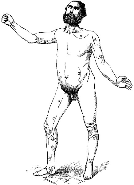
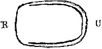
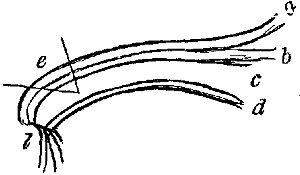
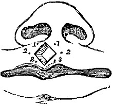
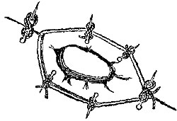
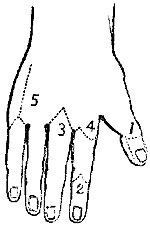 Fig. i.
Amputation of the Upper Extremity.—The extreme importance of the human
hand, its tactile sensibility, its grasping power, and the irreparable
loss sustained by its removal, render the greatest caution necessary,
lest we should remove a single digit or portion of one that might be
saved. In cases of severe smashing injuries involving the fingers, it is
the surgeon's bounden duty not recklessly to amputate the limb with neat
flaps at the wrist-joint, but carefully to endeavour to save even a
single finger from the wreck, though at the risk of a longer
convalescence, or even of a profuse suppuration. While a toe or two, or
a small longitudinal segment of the foot, may be comparatively useless,
and a good artificial foot, with an ankle-joint stump, certainly
preferable, a single finger, provided its motions are tolerably intact,
will prove much more valuable to its possessor than the most ingeniously
contrived artificial hand.
However, while in cases of extensive smash we endeavour to save anything
we can, the case is very much altered when it is only one or two fingers
that are injured. Here we find another principle brought into play, and
our conservative surgery must be limited by the following consideration.
In endeavouring to save a portion of the injured finger or fingers, will
the saved portion interfere with the important movements of the
uninjured ones? These two principles—1. Generally to save as much as we
can; 2. Not to save anything which may be detrimental or in the
way,—will guide us in describing the amputations of the upper
extremity.
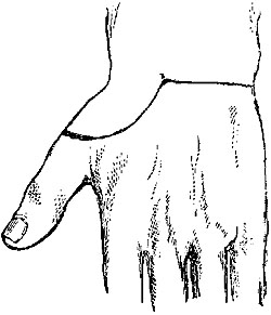
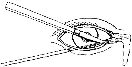
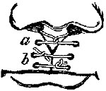
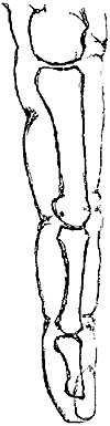 Fig. ii.
Amputation of a distal phalanx.—This small operation is not very
often required. In cases of whitlow in which the distal phalanx alone
has necrosed, removal of the necrosed bone by forceps is generally all
that is necessary. In cases of injury, however, in which nail and distal
phalanx are both reduced to pulp, it will hasten recovery much to remove
the extremity. There is no choice as to flap, the nail preventing an
anterior one, so a flap long enough to fold over must be cut from the
pulp of the finger in either of two ways (Fig. i. 1):—1. Holding the
fragment to be removed in the left hand, and bending the joint, the
surgeon makes a transverse cut across the back of the finger, right into
and through the joint, cutting a long palmar flap from within outwards
as he withdraws the knife.
Note.—Some difficulty is often felt in making the dorsal
incision so as exactly and at once to hit the joint; the most
common mistake being, that the transverse incision is made too
high, and the knife, instead of striking the joint, only saws
fruitlessly at the neck of the bone above. To avoid this, the
surgeon should take as a guide to the joint, not the well-marked
and tempting-looking dorsal fold in the skin, but the palmar
one, which exactly corresponds with the joint between the proximal
and middle phalanges, and is only about a line above the distal
articulation.—(Fig. ii.)
2. Making the long flap by transfixion, it may be held back by an
assistant, and the joint cut into.
Amputation through the second phalanx.—If the distal phalanx be so
much crushed that a flap cannot be obtained, two short semilunar lateral
flaps may be dissected (Fig. i. 2) from the sides of the second phalanx,
which may then be divided by the bone-pliers at the spot required.
In cases of injury which do not admit of either of the preceding
operations, it is quite possible to amputate either at the first joint,
or even through the proximal phalanx. Patients are sometimes anxious for
such operations in preference to amputation of the whole finger. The
surgeon should, however, never amputate through a finger higher up than
the distal end of the second phalanx, unless absolutely compelled by the
patient, for the resulting stump, being no longer commanded by the
tendons, will prove merely an incumbrance, and may possibly require a
secondary operation at no distant date for its removal.
This rule is applicable in cases in which a single finger is injured,
and two or three complete ones are left; in cases where all the fingers
have been mutilated every morsel should be left, and may be of use.
Amputation of a whole finger.—(Fig. i. 3)—This is an operation of
great importance, from its frequency.
If the third or fourth digits require amputation, it should be performed
as follows:—The vessels of the arm being commanded, an assistant holds
the hand, separating the fingers at each side of the one to be removed.
The surgeon holding the finger to be removed, enters the point of a long
straight bistoury exactly (some authorities say half an inch) above the
metacarpo-phalangeal joint, and cuts from the prominence of the knuckle
right into the angle of the web, then, turning inwards there, cuts
obliquely into the palm to a point nearly opposite the one at which he
set out.
Note.—While most authorities agree with the direction in the
text regarding the palmar termination of the incision, I believe,
in most cases, it is not necessary to go so far, and that the
incisions may fitly meet in the palm at a point midway between a
point opposite to the knuckle, and the centre of the well-marked
"sulcus of flexion."
He then repeats this incision on the other side, makes tense the
ligaments, first at one side and then at the other, by drawing the
finger to the opposite side, and cuts them. The tendons being cut, the
finger is detached. The vessels being tied, one point of suture is put
in on the dorsal aspect, and the fingers on each side tied together at
their extremities, with a pad of lint between them.
Modification.—Lisfranc's method is too long in its minute
description to give in detail. The principle is to make a semilunar
flap at one side (the one opposite the operator's right hand), by
cutting from without inwards, then to open the joint from this cut,
and, still keeping the edge of the knife close to the head of the
phalanx, cutting the other flap from within outwards. This can be
very rapidly done, but the last flap is apt to be irregular and
deficient, especially in those common cases, in which, after
whitlow or the like, the tissues are hard and brawny, and the skin
does not play freely.
It is quite unnecessary to remove the head of the metacarpal, either for
the sake of appearance, or to render healing more rapid, and its removal
weakens the arch of the hand; where the cartilage is eroded by disease,
the cartilage-covered portion can be scooped off by a gouge or removed
entire by pliers, without interfering with the broad end to which the
transverse ligament of the palm is attached. If required either for
injury or disease, the metacarpal head may be easily removed by a single
straight incision from the knuckle upwards, as far as the point at which
it may be deemed necessary to saw it through, or better still, divide it
with the bone-pliers. This incision should be made as a first step in
the first incision for amputation of the finger, and the finger should
not be disarticulated, but kept on, to aid by its leverage in separating
the metacarpal head.
Amputation of the index or little fingers.—This operation differs
from the preceding only in this, that care must be taken to make a good
large flap on the free side of each; making the incision, which begins
at the knuckle (Fig. i. 4), enclose a well-rounded flap, and not
allowing it to enter the palm till it reaches the level of the web
between the fingers. The metacarpal heads may here be cut obliquely with
the bone-pliers, to prevent undue projection.
Amputation of one or more metacarpals.—These operations may be
rendered necessary by disease or injury. If the latter demands their
performance, no rules can be given for incisions or flaps, they must
just be obtained where and how they can best be got. If for disease, a
single dorsal incision (Fig. i. 5) over the bone will allow it to be
dissected out of the hand.
N.B.—In no case, except that of the thumb, should any attempt be
made to save a finger while its metacarpal is removed. (See Excisions
of Bones.)
Amputation of first and fifth metacarpals.—Various special operations
have been devised for speedy and elegant removal of these bones. Their
disadvantages, etc., are fully detailed under Amputations of the Foot.
The vascularity and consequent vitality of the tissues of the hand and
arm sometimes afford very encouraging and satisfactory results in
conservative operations.
The following is an instance of what may be accomplished in a young
healthy subject.
A. A., æt. 18, ploughman, was harnessing a vicious horse, when it caught
his right hand between its teeth, and gave a severe bite. On admission,
I found the middle and ring fingers completely separated at the
metacarpal joints, but each hanging on by a portion of skin, the middle
by the skin on its radial side, the ring by that on its ulnar. The back
and the palm were both stripped of skin up to the middle of the third
and fourth metacarpal bones, which were exposed, but not fractured. As
it was important for him to maintain the transverse arch of the hand
intact, I determined to make an attempt to save the metacarpals, and
finding that the skin on the radial side of the middle, and ulnar side
of the ring fingers, was still warm, and apparently alive, I carefully
dissected as long a flap as possible from each, and then folded them
down, one at the front, the other at the back of the hand. The flaps
survived, and the result was admirable, the patient being able in a very
few weeks to guide the plough. The sensation in his new palm and back of
the hand is very peculiar, they being still the fingers, so far as
nervous supply is concerned.
In amputations involving the metacarpals for injury, it is always
important to avoid entering the carpo-metacarpal joint, hence if it can
be done it is best to saw through the bones at the required level,
rather than disarticulate. This rule should be observed even in those
cases in which the thumb alone can be saved, for notwithstanding the
isolation of the joint between the first metacarpal and the trapezium,
it is very important for the future use of this one digit that the
motions both of the wrist and carpal joints should be preserved entire.
No exact rules can be given for the performance of these operations, as
the size and positions of the flaps must be determined by the nature of
the accident and the amount of skin left uninjured.
In the rare condition where the greater part of the metacarpus is
destroyed, and yet carpal joints are uninjured, a most useful artificial
band, preserving the movements of the wrist, may be fitted on; and as
much as possible should be saved, but in cases of injury, where the
carpus is opened and the hand irreparably destroyed, the question
arises, Where ought amputation to be performed? To this we answer that
there appears no conceivable advantage to be gained by leaving all or
any of the carpal bones. If successful, it would result only in the
retention of a flapping joint, unless from there being no tendons to act
upon it, except the tendon of the flexor carpi ulnaris attached to the
pisiform, and there are several risks it would run in the inflammation
of all the carpal joints, and the almost certain spread of this
inflammation to the bursa underneath the flexor tendons, beyond the
annular ligament, and up the arm among the muscles.
Amputation at the Wrist-Joint.—This is an operation by no means
frequent, and it has the advantages of preserving a long stump, and
retaining the full movements of pronation and supination, in cases where
the radio-ulnar joint is sound and uninjured, but in practice it is
often found that fibrous adhesions limit to a great extent the motions
of the two bones on each other, specially in those cases where the
radio-ulnar joint has been diseased or injured.
Another advantage is the extreme ease with which disarticulation may be
performed on emergency, no saw being required, and the ordinary bistoury
of the pocket-case being quite sufficient for cutting the flaps.
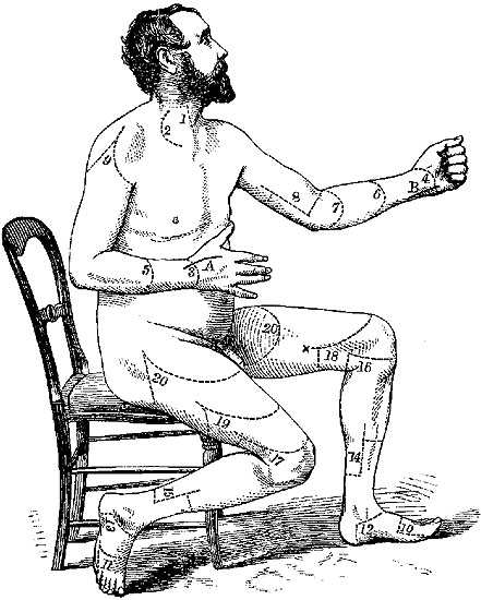
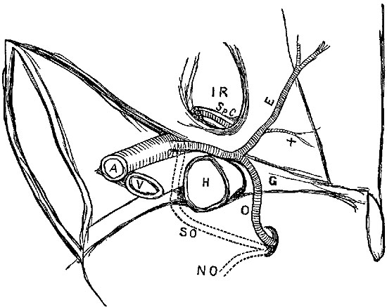
Operation.—By double flap. An incision (Plate IV. fig. 3) on the
dorsal surface, extending in a semilunar direction from one styloid
process to the other, will define a flap of skin only, which must be
raised; the joint must then be opened by a transverse incision, and a
long semilunar flap of skin and fascia should be shaped (Plate IV. fig.
4) from the palm. Disarticulation is facilitated by the surgeon forcibly
bending the wrist when he makes the transverse cut, and it will be found
easier to shape the palmar flap from the outside by dissection, than to
do it by transfixion after disarticulation, on account of the prominence
of the pisiform on the inner side of the palm.
In the thin wasted wrists of the aged, or in any case where the
skin is very lax, this amputation may be very easily performed by
the circular method. While an assistant draws up the skin as much
as possible, the surgeon makes an accurate circular incision
through the skin, about an inch below the styloid processes, just
grazing the thenar and hypothenar eminences. Another circular sweep
just above the pisiform and unciform bones divides all the soft
textures, after which the joint may be opened, and, if necessary,
the styloid processes cut away with saw or pliers.
Amputation by a long single flap, either dorsal or palmar, may be
rendered necessary by accident. The palmar one of the two is
preferable; indeed, rather than trust for a covering to the thin
skin of the back of the hand, with its numerous tendons, it is
better to amputate an inch or two higher up through the fore arm.
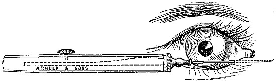
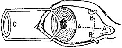
The following amputation by external flap has been described (so
far as I can discover, for the first time) by Dr. Dubrueil, in his
work on operative Surgery:[26]—"Commencing just below the level of
the articulation, while the hand is pronated, the surgeon makes a
convex incision, beginning at the junction of the outer and middle
thirds of the arm behind, reaching at its summit the middle of the
dorsal surface of the first metacarpal, and terminating in front
just below the palmar surface of the joint, again at the junction
of the outer and middle thirds of the breadth of the arm. This flap
being raised, the wrist is disarticulated, beginning at the radial
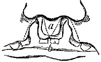
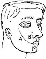
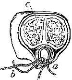
side. A circular incision finishes the cutting of the skin." (Figs.
iii. and iv.)
Amputation through the Fore-arm.—The method of operating must, in the
fore-arm, depend a good deal upon the part of the arm where you require
to amputate, the muscularity of the limb, and the condition of the skin
and subcutaneous cellular tissue.
It must be remembered that a section of the fore-arm involves two bones,
not, like the tibia and fibula, on a constant permanent relation in
position to each other, but which rotate one upon another to an amount
which varies with the part of the limb divided, and which rotation is a
very important element in the future usefulness of the stump; again,
that two sets of muscles occupy, one the back, the other the front of
the limb, that these two are unequal in size, and that the outer sides
or rather edges of each bone are subcutaneous; again, that these sets of
muscles are comparatively fleshy in the upper two-thirds of the limb,
and almost entirely tendinous in the lower third.
Remembering these points, we find that certain things require our
attention, and certain difficulties are present in amputation of the
fore-arm, from which amputation of the arm, with its single bone and
copious muscular covering on all sides, is completely free.
Thus our flaps in the fore-arm must be antero-posterior; lateral flaps
are an impossibility. Great care is requisite to cut them at all equal,
from the inequality of the muscles on the two sides. In the lower third
we cannot obtain available muscular flaps. Lastly, care must be taken
lest, from the ever-varying relations of the two bones to each other in
the varying positions of the limb, the surgeon mistake their position
and pass his knife between them.
The next question that arises is, Where are we to operate? In cases
where we have a choice, is there here, as in the leg, any "point of
election"? No. As a rule in the fore-arm, the surgeon should endeavour
to save as much as possible; especially when nearing the middle of the
fore-arm, he should try to save the insertion of the pronator teres, so
important in its function of pronating the radius.
Amputation in Lower Third of the Fore-arm.—By two flaps. These
antero-posterior flaps must consist of skin only, as the tendons are
only in the way, and thus should be made by dissection from without.[28]
Making the dorsal one first, the surgeon should enter his knife at the
palmar edge of the bone that is further from him, and cut a semilunar
flap of skin only, finishing the incision quite on the palmar edge of
the inner bone. The two ends of this incision must then be united by a
similar semilunar flap of skin on the palmar side. The two flaps having
been dissected back, he then clears the bones by a circular incision
through tendons and muscles, not forgetting to pass the knife between
the bones, and retracting all the soft parts, saws through the bones, at
least half or probably three-quarters of an inch higher up. It is
generally easiest to saw through both bones at once.
Long Dorsal Flap.—Where it is possible from laxity of the soft parts
and the wrist not being much destroyed, to get a long flap from the back
of the arm after Mr. Teale's method, a very good stump will result. This
rule is, "In tracing the long flap a longitudinal line is drawn over the
radius, so as to leave the radial vessels for the short flap (Plate II.
fig. 1). At a distance equal to half the circumference of the limb,
another line parallel to the former is drawn along the ulna. These are
then joined at their lower ends, across the dorsal aspect of the wrist
or fore-arm, by a transverse line equal in length to half the
circumference of the fore-arm. The short flap is marked by a transverse
line on the palmar aspect, uniting the long ones at their upper fourth.
"The operator, in forming the long flap, makes the two longitudinal
incisions merely through the integuments, but the transverse one is
carried directly down to the bones. In dissecting the long flap from
below upwards, the tissues of which it is composed must be separated
close to the periosteum and interosseous membrane. The short flap is
made by a transverse incision through all the structures down to the
bones, care being taken to separate the parts upwards close to the
periosteum and membrane." The stump must be placed in the prone
position, "to allow the long dorsal flap to be the superior when the
patient is recumbent, and thus fall over the ends of the bones."[29]
The principal objection to the long dorsal rectangular flap (which
makes an excellent covering) is, that unless it can be obtained from
over the wrist-joint it requires the bones to be sawn so very high up.
This may be avoided, to some extent, by making it shorter and rounded
off, as in Carden's Amputation, q.v.
Amputation in Upper Two-Thirds.—Where the fore-arm is very fat or
fleshy, this amputation can be very easily performed by two equal
antero-posterior flaps made by transfixion. In most cases, however, from
the comparative leanness of the dorsal aspect of the limb, the following
method will have the best result. The surgeon must, as in the former
case, shape a rounded dorsal flap by dissection from without (Plate IV.
fig. 5), embracing the whole breadth of the limb down to the palmar edge
of both bones. Then at once he transfixes the two points of this dorsal
flap, and cuts out an equal one from the anterior aspect of the limb
(Plate IV. fig. 6). Dissecting up the dorsal flap he clears the bones at
least half an inch above as before, and applies the saw.
N.B.—This operation should be performed even in cases where only an
inch of radius can be retained, as the attachment of the biceps makes a
very small stump of fore-arm wonderfully useful.
Amputation at Elbow-Joint.—In cases where it is found impossible to
save any portion of the fore-arm, disarticulation at the elbow-joint may
be easily performed. This operation was proposed and performed so long
ago as the days of Ambrose Paré,[30] was much approved by Dupuytren,
Baudens, and Velpeau, had fallen into disuse for a time, but is now
again recommended by some excellent surgeons, especially by Gross[31]
and Ashhurst,[32] both of Philadelphia.
It is tolerably easy to perform, and does not involve any sawing of
bones, but the flaps are apt to be cut too short, unless care be taken,
from the manner in which the trochlea projects downwards beyond the line
of the condyles, so that if the base of an ordinary-shaped flap be made
on a level with the condyles, it will prove insufficient to cover the
bone. It may be performed either by the circular method (Velpeau), oval
(Baudens), or by a long anterior and short posterior flap (Textor and
Dupuytren). Probably the best method is by a long anterior flap when it
can be obtained, thus:—The arm being placed in a slightly flexed
position, the surgeon transfixes in front of the joint, in a line
extending from the level of the external condyle to a point one inch
below the internal condyle (Plate IV. fig. 7); the tissue should be held
well forward at the moment of transfixion. The flap should be at least
two and a half inches deep at its apex, which must be rounded off. The
two ends of this flap may then be united behind by a semilunar incision
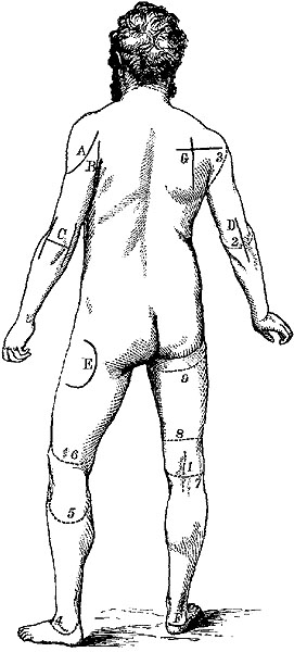
(Plate III. fig. 2), which will separate the radial attachments. The
ulna must then be cleared, and the triceps divided at its insertion.
Modifications.—Dupuytren used to saw through the ulna, leaving
the olecranon attached. Velpeau opposed this, but it is again
recommended by Gross, who leaves the olecranon, and at the same
time improves the shape of the stump by sawing off the "inner
trochlea" on a level with the general surface.
Amputation of the Arm.—This amputation is best performed by double
flap, and is the typical instance which exhibits all the advantages of
two equal flaps made by transfixion, without any of the disadvantages of
that method. These advantages are, easiness of performance, rapidity,
excellent covering for the bone, with as little sacrifice of tissue as
is possible, while the fact that the cicatrix is opposite the end of the
bone is hardly a disadvantage in the arm (as it certainly is in the
leg), as no weight has to be borne on it. When they can be obtained,
anterior and posterior flaps are generally considered most satisfactory,
but Mr. Spence prefers lateral ones, lest the line of union should be
interfered with by the deltoid raising the bone. If the right arm has to
be amputated, the operator standing at the inner side raises the
anterior muscles with his left hand, and enters the knife just in front
of the brachial vessels (Plate I. fig. 12); keeping as close as possible
to the bone, he brings out the knife at a point exactly opposite, then
with a brisk sawing motion, cuts a semicircular flap, taking care to
bring out the knife more suddenly just at the end, in order to cut
through the skin as perpendicularly to the arm as possible. The knife is
again entered at the same point, carried behind the bone, and brought
out at the same angle, and an exactly corresponding flap cut from the
other side of the limb, the flaps are then retracted, the bone cleared
by circular incision and sawn through as high up as it is exposed. In
primary cases, where the muscles are firm and developed, the flaps
should be cut a little concave.
Modifications and Varieties.—Teale's method may of course be
used here as elsewhere. The internal line of incision (Plate IV.
fig. 8) should be made just in front of the brachial vessels. This
method requires the amputation to be performed higher up than would
otherwise be necessary (from the length of the anterior flap), and
this disadvantage is not counterbalanced by any special advantage
in the posterior retraction of the cicatrix.
In feeble flabby arms, the true circular operation is very easily
performed, and with good results. A circular sweep of the knife is
made through the skin alone, which is drawn up by an assistant,
while the surgeon separates it from the fascia; another circular
cut through fascia and muscles exposes the bone, which must then be
cleared and cut through at a still higher level.
Amputation at the Shoulder-Joint.—This operation, like that at
the hip joint, can, from the nature of the joint to be covered, and
the abundant soft parts in the normal state of the tissues, be
performed on the dead in very various ways, by single, double, or
triple flaps, by transfixion or dissection, rapidly or slowly.
Hence manuals of operative surgery might collect at least twenty
different methods, most of which have some recommendation, and all
of which are practicable enough.
When, however, we reflect that in the living body, in cases where
amputation at the shoulder-joint is required at all, the severity
of the accident, or the urgency of the disease, will, in general,
leave no room for selection, we shall see how utterly valueless is
any knowledge of mere methods of operating, and of how much greater
importance it is that we should be simply thoroughly familiar with
the anatomy of the joint.
For example, an accident which necessitates amputation so high up
has, in all probability, opened into the joint and destroyed the
soft parts on at least one aspect; in such a case the flaps must be
cut from the uninjured soft parts only. If an aneurism has rendered
amputation through it and through the joint a last resource, the
flap must be gained chiefly at least from the outside; a malignant
tumour of the humerus will almost certainly prevent any
transfixion, and require flaps to be made by dissection, wherever
the skin is least likely to be involved. Again, some of the most
vaunted and most rapid operations almost require for their success
the integrity of the humerus, which has to make itself useful as a
lever in disarticulation, while in most cases of accident we are
amputating for compound injury of the humerus, almost certainly
implying fracture with comminution.
From its proximity to the trunk, hæmorrhage is one of the chief
dangers to be apprehended during this operation, especially from
the axillary artery. As far as possible to obviate this danger,
most plans of operating are based on the principle that the
vessels and nerves should be the last tissues to be cut; in some
they are not divided till after disarticulation.
While a good assistant, to make pressure on the subclavian above
the clavicle, is a most advisable precaution, too much must not be
trusted to this pressure above, as the struggles of the patient and
the spasmodic movements of the limb, which are so apt to occur
under the stimulus of the knife, are apt to render futile the best
efforts at compression.
The operator should trust rather to making the incisions in such a
manner that the great vessel be not divided till the hand of an
assistant, or in default of a suitable one, his own left hand, is
able to follow the knife and grasp the flap.
The bleeding from the circumflex, subscapular, and posterior
scapular arteries can easily be arrested by a dossil of lint till
the great vessel is tied, and they can be secured.
In cases where proper assistants cannot be had, temporary closure
of the axillary vessel could easily be made by carrying a strong
silver wire or silk ligature completely round the vessel by a
curved needle before the incisions are commenced, and by tying this
firmly over a pad of lint.
Pressure on the artery above the clavicle is best made by the thumb
of a strong assistant, who endeavours to compress it against the
first rib; where the parts are deep and muscular, the padded handle
of the tourniquet, or of a large door-key, will do as the agent of
pressure.
A brief notice of three of the best methods of operating will be
quite sufficient to show what should be aimed at in shoulder-joint
amputations:—
1. In cases where the surgeon can choose his flaps, the following
method will be found the most satisfactory, as resulting in the
smallest possible wound, in having less risk of hæmorrhage during
the operation than any other method, and in providing excellent
flaps.
It is Larrey's method slightly modified.
Operation.—With a moderate-sized amputating knife an incision of
about two inches in length, extending through all the tissues down
to the bone, should be made from the edge of the acromion process
to a point about one inch below the top of the humerus; from this
latter point a curved incision, enclosing a semilunar flap, should
be made on each side of the limb to the anterior and posterior
folds of the axilla respectively (Plate IV. fig. 9, and Plate III.
fig. 3). These flaps should then be dissected back, including the
muscles and exposing the joint. When thoroughly exposed, the joint
must then be opened from above, and the bone separated. One small
portion of skin lying above the artery, vein, and nerves still
remains to be divided (Plate I. fig. 13). This may be done by an
oblique cut from within outwards, in such a direction as to form
part of the anterior or internal incision, and with the precaution
of having an assistant to command the vessels before they are
divided. The resulting wound is almost perfectly ovoid, the flaps
come together with great ease in a straight vertical line, which
admits of easy and thorough drainage. Union is generally rapid.
Larrey's success by this method was very remarkable: ninety out of
a hundred cases in military practice were saved, notwithstanding
the well-known risks of such operations.
2. As good as the former, and nearly as universally applicable, is
the method devised by Professor Spence, and practised by him in
nearly every case:—"With a broad strong bistoury I cut down upon
the inner aspect of the head of the humerus, immediately external
to the coracoid process, and carry the incision down through the
clavicular fibres of the deltoid and pectoralis major muscles till
I reach the humeral attachment of the latter muscle, which I
divide. I then with a gentle curve carry my incision across and
fairly through the lower fibres of the deltoid towards, but not
through, the posterior border of the axilla. Unless the textures be
much torn, I next mark out the line of the lower part of the inner
section by carrying an incision through the skin and fat only,
from the point where my straight incision terminated, across the
inside of the arm to meet the incision at the outer part. This
insures accuracy in the line of union, but is not essential. If the
fibres of the deltoid have been thoroughly divided in the line of
incision, the flap so marked out, along with the posterior
circumflex trunk, which enters its deep surface, can be easily
separated from the bone and joint, and drawn upwards and backwards
so as to expose the head and tuberosities, by the point of the
finger without further use of the knife. The tendinous insertions
of the capsular muscles, the long head of the biceps, and the
capsule, are next divided by cutting directly upon the tuberosities
and head of the bone; and the broad subscapular tendon especially,
being very fully exposed by the incision, can be much more easily
and completely divided than in the double-flap method. By keeping
the large posterior flap out of the way by a broad copper spatula
or the fingers of an assistant, and taking care to keep the edge of
the knife close to the bone, the trunk of the posterior circumflex
is protected. In regard to the axillary vessels, they can either be
compressed by an assistant before completing the division of the
soft parts on the axillary aspect, or to avoid all risk, the
axillary artery may be exposed, tied, and divided between two
ligatures so as to allow it to retract before dividing the other
textures."[33]
Another, but not so good method of making an external flap, is the
following:—(a.) For the right arm.—The patient lying well over
on his left side, the surgeon stands to the inside of the arm to be
removed. Seizing the deltoid in the left, with the right he passes
an amputating knife, seven or eight inches in length, from a point
a little nearer the clavicle than the middle space between the
acromion and coracoid processes; then, transfixing the base of the
deltoid, and just grazing the posterior surface of the humerus,
thrusts the knife downwards and backwards till it protrudes at the
posterior margin of the axilla. When doing this, it is important
that the arm be held outwards and backwards, and even upwards, as
far as possible to relax the deltoid; without this it will be
impossible to make the flap of the full size. The flap must then be
cut of as full length as can be obtained, four or five inches at
least. An assistant then holds it upwards, while the surgeon, or
(if the arm is very muscular) another assistant, brings the arm
forwards well across the patient's chest, thus exposing the
posterior aspect of the joint. This may have very possibly been
already opened during the transfixion; the attachments of muscles
must now be divided, the knife passed behind the head of the bone,
which is dislocated forwards, and a suitable flap of the tissues in
front cut from within outwards. The assistant is to follow the
knife with his finger and compress the vessels.
(b.) If the left shoulder is to be amputated, the patient lying
on his right side, the surgeon stands behind him, and raising the
elbow of the limb to be removed from the side, and pulling it
slightly backwards, enters the knife at the posterior fold of the
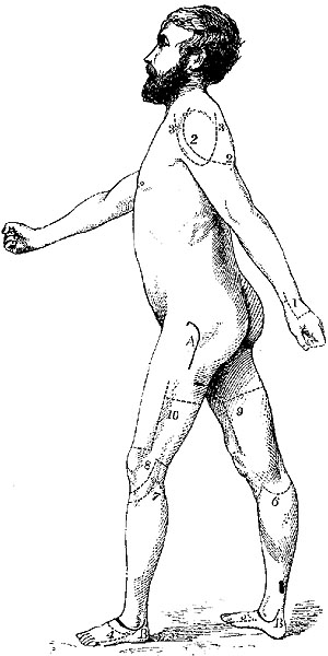
axilla (Plate II. fig. 2), and passing the posterior aspect of the
head of the humerus, endeavours to protrude it as near the acromion
as possible; the flaps must be cut and the rest of the operation
performed in the manner we have just described for the other arm.
3. Where the destruction of tissue has been chiefly below the joint, a
very good flap may be obtained from above, composed chiefly of the
deltoid muscle, and the skin over it. This may be made by transfixion at
its base, but is better obtained by dissection from without.
The surgeon cuts (Plate II. figs. 3, 3) in a semilunar direction (with
the convexity downwards) from one side of the deltoid to the other,
viz., from the root of the acromion to near the coracoid process; he
then raises the large flap upwards and throws it back, opens the joint,
disarticulates, passes the knife behind the head of the bone, and cuts
out without attempting to save any flaps below, in a transverse
direction. By this means the artery is still almost the last structure
to be divided, and can be secured by a ready assistant. In cases where
much injury has been done to the floor of the axilla and wall of chest,
the deltoid flap must be made large in proportion, and triangular rather
than semilunar in shape.
N.B.—The statistics of amputation at the shoulder-joint bring out
some interesting facts: 1. That the primary amputations here are far
more successful than secondary ones. Guthrie records nineteen cases of
the former out of which only one died, while out of a similar number in
which the amputation was secondary, fifteen died. In the Crimea, British
surgeons had thirty-nine cases, with thirteen deaths; of thirty-three
primary, nine died; and of six secondary, four were fatal.
S.W. Gross's[34] statistics confirm this: of one hundred and
seventy-eight primary, forty-six died—25.8 per cent.; ninety-five
secondary, sixty-one died—64.2 per cent.
Amputations above the Shoulder-Joint.—Under this head we may group the
comparatively rare cases in which, from accident or disease, the removal
of portions of the scapula and clavicle, or even the entire bones, is
rendered necessary. That it is quite possible to survive such injuries
has been frequently shown in cases of accident when the scapula along
with the arm has been torn off, and yet the patient recovered.
Encouraged by such cases, Gaetani Bey of Cairo removed the whole of
scapula and part of the clavicle in a case where he had amputated at the
shoulder for smash. The patient recovered. Heron Watson has had a
similar case. Dr. George M'Lellan amputated arm and scapula in a youth
of seventeen for an enormous encephaloid tumour. Fifty-one such cases
are now on record.
Syme amputated with success the arm along with the scapula and outer
half of clavicle, in a case in which he had previously excised the head
of the humerus for a tumour.[35]
Gilbert, Mussey, Rigaud, Fergusson, and others have performed similar
operations, secondary to amputation at the shoulder-joint, for cases of
caries and malignant tumour. It is impossible to give any exact
directions for the incisions which must be planned for individual cases,
with two chief aims, to avoid hæmorrhage as far as possible, and to
leave abundance of skin. In operations on the scapula, it should be
freely exposed by large enough incisions. (See Excisions.)
Amputations of Lower Extremity.—Commencing with the most distal, and
gradually working our way upwards, we find that partial amputations of
the toes are extremely rare. Only in the case of the great toe is such
an operation ever admissible, for the other toes are so short, and the
stumps left by amputation are at once so useless from their shortness,
and so detrimental from the manner in which they project upwards and rub
against the shoe, that any injury requiring partial amputation of a
lesser toe is treated by its complete removal.
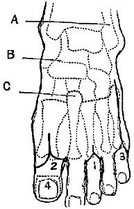 Fig. v.
Amputation of Distal Phalanx of Great Toe.—This is comparatively rarely
required now. It used to be thought necessary for the cure of those not
uncommon cases of exostosis of the distal phalanx, but it is now found
that most of these can be cured by simply clipping off the exostosis.
When necessary, however, and when the choice of flaps is possible, the
best plan is by a long flap from the plantar surface (Fig. v. 4), as in
the similar operation on the thumb; laying the edge of the knife over
the dorsal aspect of the joint, cutting through it, and turning the edge
of the knife round close to the bone, so as to cut out a large flap from
the ball of the toe.
Amputation of a Single Lesser Toe—second, third, or fourth.—This
operation is on exactly the same principle as that described for the
corresponding finger; but it must be remembered that the
metatarso-phalangeal joint is more deeply situated in the soft parts
than is the metacarpo-phalangeal; and thus the commencement of the
elliptical incision which is to surround the base of the toe must be
proportionally higher up (Fig. v. 1). On the other hand, as it is very
important to avoid as much as possible any cicatrix in the sole of the
foot, the plantar end of the incision need not be carried to a point
exactly opposite the one from which it set out, but it will be
sufficient if it reaches the groove between the toe and sole. A little
more care may thus be required in dissecting out the head of the first
phalanx, but this is quite repaid by the cicatrix in the sole being
avoided. Early division of flexor tendons renders disarticulation easy.
Amputation of the First and Fifth Toes.—The incisions are conducted on
the same principle as in the other operations, the operator being
careful to preserve as much as possible (Fig. v. 2) of the hard useful
pad of the inner and outer sides respectively.
Most surgeons are now agreed that in these toes it is best not to remove
the head of the metatarsal bone with the toe. Cutting off the large
cartilaginous head obliquely with a pair of bone-pliers may prevent an
awkward unseemly projection, but it does diminish the strength of the
transverse arch of the foot.
Amputation of one or more Toes with their Metatarsals.—It is not
necessary to give very particular details regarding such operations, as
the surgeon must be guided in the individual cases by the specialties of
accident or disease.
One or two guiding principles are important:—
1. Having made up your mind at what point you are to cut the metatarsal,
if the amputation be a partial one, or as to the exact position of the
joint, if you intend to disarticulate, commence your dorsal incision
(Fig. v. 3) at a point fully half an inch higher up than the selected
spot, as free access is of the very last importance.
2. Whenever it is possible, cut the bone through its continuity rather
than disarticulate. Specially is this important in the case of the
metatarsal bone of the great toe, that the insertion of the tendon of
the peroneus longus may be saved. If, however, the terminal branch of
the dorsalis pedis artery be wounded, it may be necessary to
disarticulate the first metatarsal to secure it rather than trust to
compression to stop the bleeding.
3. In cutting through the first and fifth metatarsals, remember to apply
the bone-pliers obliquely, not transversely, so as to avoid unseemly
projection.
4. As far as possible avoid cutting into the sole at all.
The plantar cicatrix is almost a fatal objection to a plan of removing
the first and fifth toes and their metatarsals which has much otherwise
in rapidity and elegance to recommend it. In the great toe, for example,
it is performed as follows:—Seizing the soft parts of the inner edge of
the foot in his left hand, the surgeon draws them inwards, transfixes
just at the tarso-metatarsal joint, and, keeping as close as possible to
the inner edge of the metatarsal bone, cuts the flap as long as to the
middle of the first phalanx; then the soft parts of the foot being drawn
as far outwards as possible by an assistant, the surgeon enters his
knife between the first and second toes, and succeeds in entering his
former incision so as to separate the metatarsal bone without removing
any skin. All that remains is to open the tarso-metatarsal joint. It is
a very neat-looking operation, leaves a very good covering for the
parts, and is performed with extreme rapidity. This last is not so much
required in these days of anæsthetics, and the cicatrix in the sole is a
very formidable objection to it.
The simplest and shortest rule that can be given for the amputation of a
toe, with the part or whole of its metatarsal, is to make one dorsal
incision, commencing about a quarter of an inch above the spot at which
you intend to divide the bone or to disarticulate, extending downwards
in a straight line to the metatarso-phalangeal articulation, and then
bifurcating so as to surround the base of the toe at the normal fold of
the skin. The soft parts are then to be cleared from the
metatarso-phalangeal joint, and the toe still being retained on the
metatarsal bone, it should be carefully dissected up, avoiding any
pricking of the soft parts below, till the joint is reached, or the spot
at which the bone-pliers are to be applied is fully cleared.
Amputation of the anterior portion of the Foot at the Tarso-metatarsal
Joint—Hey's Operation.—This operation, which is now comparatively
rarely performed, has been invested with a halo of difficulty and
complexity which is to a great extent unnecessary.
There is no doubt that the anatomical conformation of the joints
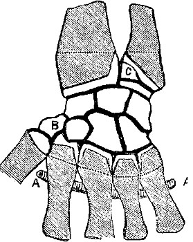
involved, especially the manner in which the head of the second
metatarsal (Fig. v. C) projects upwards into the tarsus, and is locked
between the cuneiform bones, renders disarticulation in the healthy foot
rather difficult; but it must be remembered that in cases where for
accident we have to deal with previously healthy tissues, it is quite
unnecessary to disarticulate, a better result being attained by simply
sawing the foot across in the line of the articulation; and again, where
we have to operate for disease, the tissues are so matted, and the
bones so soft, that complete removal of the metatarsus is much easier
than it appears when practising on the dead subject.
Very various plans of incision have been proposed. Mr. Hey's original
procedure has not been much improved upon. His short account of it has
at once surgical value and historical interest:—
"I made a mark across the upper part of the foot, to point out as
exactly as I could the place where the metatarsal bones were joined to
those of the tarsus. About half an inch from this mark, nearer the toes,
I made a transverse incision through the integuments and muscles
covering the metatarsal bones (Plate IV. figs. 10, 11). From each
extremity of this wound I made an incision (along the inner and outer
side of the foot) to the toes. I removed all the toes at their junction
with the metatarsal bones, and then separated the integuments and
muscles forming the sole of the foot from the inferior part of the
metatarsal bones, keeping the edge of my scalpel as near the bones as I
could, that I might both expedite the operation and preserve as much
muscular flesh in the flap as possible. I then separated with the
scalpel the four smaller metatarsal bones at their junction with the
tarsus, which was easily effected, as the joints lie in a straight line
across the foot. The projecting part of the first cuneiform bone which
supports the great toe I was obliged to divide with a saw. The arteries,
which required a ligature, being tied, I applied the flap which had
formed the sole of the foot to the integuments which remained on the
upper part, and retained them in contact by sutures....
"The patient could walk with firmness and ease; she was in no danger of
hurting the cicatrix by striking the place where the toes had been
against any hard substance, for this part was covered with the strong
integuments which had before constituted the sole of the foot. The
cicatrix was situated upon the upper part of the foot, and had very
little breadth, as the divided parts had been kept united after being
brought into close contact."[36]
Lisfranc's method has, briefly, the following modifications.—Having
fixed the position of the articulations of the first and fifth
metatarsals with the tarsus, the operator unites them by a curved
incision across the dorsum of the foot, with its convexity downwards. He
then divides the dorsal ligaments over the articulations, opens the
first from the inside, the fifth, fourth, and third from the outside, he
then with a strong narrow-bladed knife divides the interosseous
ligaments between the sides and end of the head of the second metatarsal
and the cuneiforms, thus completing the disarticulation; bending the
fore part of the foot downwards, he then keeps the edge of the knife
close to the lower surface of the bones, separating the plantar
ligaments, and cutting out a long plantar flap of skin and muscles.
In every case it must be remembered that the upper end of the fifth
metatarsal projects far up along the outer edge of the foot. Allowance
must be made for this projection in commencing the incision. A rule
given by Mr. Syme to guide the disarticulation of the three outer
metatarsals will often be of service; it is this: "Having once entered
the joint of the fifth, the knife must be drawn along in a direction of
a line drawn towards the distal end of the first metatarsal; for the
fourth, the direction must be changed to the middle of the same bone;
and to open the third it will be necessary to come across the dorsum of
the foot as if intending to reach the proximal end."
To avoid the difficulties of disarticulation, Skey recommends cutting
off the head of the second metatarsal with a pair of pliers. Baudens,
Guérin, and others approve of sawing all the bones across in the line
desired.
Most surgeons are now agreed that in this operation it is better to make
both flaps by cutting from without, in preference to transfixion of the
plantar one from within. In cases where, from injury and disease, the
plantar flap is deficient in size, it may be necessary to make the
dorsal flap longer. However, the long plantar is preferable both from
its superior hardness, and also because from its length it permits the
cicatrix to be well on the dorsum of the foot, and therefore less likely
to be injured by the pressure of the boot in front.
Amputations through the Tarsus.—Various plans of amputating through the
tarsus have been devised and described at great length. The most
important of these is the operation of removal of the anterior portion
of the foot, at the joints between the astragalus and scaphoid, and os
calcis and cuboid, well known to the profession by the name of its first
describer, Chopart.
It has been so completely superseded by the infinitely preferable
amputation at the ankle-joint of Mr. Syme, as rarely, if ever, to be
practised in this country. Indeed, amputation at the ankle-joint may be
said to have taken the place of all these amputations through the
tarsus; for though cases are occasionally met with in which the
limitation of the disease or injury may render Chopart's possible, and
though at first sight it appears to have an advantage in removing less
of the body, still the following objections are nearly fatal to its
chance of being selected:—1. In cases of injury, through leaving a long
stump, and, at first sight, a useful one, experience shows that the
tendo Achillis sooner or later (being unopposed by the extensors of the
toes) draws up the heel so as to make the end of the stump point, and
the cicatrix press on the ground, rendering it unable to bear any
weight. 2. In cases of removal for disease of the tarsus, the bones left
behind, though apparently sound at the time, are almost sure to become
eventually diseased.
As it has an historical interest, and as this operation (defective as it
is) had been the means of saving many legs prior to the invention of
amputation at the ankle-joint, a brief description may be appended:—
Chopart's own manner of operation was briefly somewhat as follows:—
The tourniquet having been applied, the surgeon is to make a transverse
incision through the skin which covers the instep, two inches from the
ankle-joint. He is to divide the skin, and the extensor tendons, and the
muscles in that situation, so as to expose the convexity of the tarsus.
He is next to make on each side a small longitudinal incision, which is
to begin below and a little in front of the malleolus, and is to end at
one of the extremities of the first incision. After having formed in
this way a flap of integuments, he is to let it be drawn upwards by the
assistant who holds the leg. There is no occasion to dissect and reflect
the flap, for the cellular substance connecting the skin with the
subjacent aponeurosis is so loose, that it can easily be drawn up above
the place where the joint of the calcaneum with the cuboides and that
between the astragalus and scaphoides ought to be opened. The surgeon
will penetrate the last the most easily, particularly by taking for his
guide the eminence which indicates the attachment of the tibialis
anticus muscle to the inside of the os naviculare. The joint of the os
cuboides and os calcis lies pretty nearly in the same transverse line,
but rather obliquely forwards. The ligaments having been cut, the foot
falls back. The bistoury is then to be put down, and the straight knife
used, with which a flap of the soft parts is to be formed under the
tarsus and metatarsus, long enough to admit of being applied to the
naked bones, so as entirely to cover them. It is to be maintained in
position with three or four straps of adhesive plaster, etc.[37]
Chopart's amputation, after an interval of comparative neglect, was
introduced into this country by Mr. Syme in 1829. His method of
performance is simpler and easier than Chopart's. He thus describes
it:—"The blade of the knife employed should be about six inches long,
and half an inch broad, sharp at the point and blunt on the back. The
tourniquet ought to be applied immediately above the ankle, having
compresses placed over the posterior and anterior tibial arteries. The
surgeon should measure with his eye the middle distance between the
malleolus externus and the head of the metatarsal bone of the little
toe, which is the situation of the articulation between the os cuboides
and os calcis. Placing his forefinger here, he ought to place his thumb
on the other side of the foot directly opposite, which will show him
where the os naviculare and astragalus are connected. An incision (Plate
II. figs. 4 and 5) somewhat curved, with its convexity forward, is then
to be made from one of these points to the other, when, instead of
proceeding to disarticulate, the operator should transfix the sole of
the foot from side to side at the extremities of the first incision, and
carry the knife forwards so as to detach a sufficient flap, which must
extend the whole length of the metatarsus to the balls of the toes. The
disarticulation may finally be completed with great ease, as the shape
of the articular surfaces concerned is very simple, and nearly
transverse."[38] Regarding the method of disarticulating at the
astragalo-calcaneal joint, and removing all the foot except the
astragalus, no detail need be given. Malgaigne advises an internal flap,
thus sacrificing the valuable pad of the heel. Roux, Verneuil, and
others endeavour to save the pad. This operation, however, has now
fallen almost completely into disuse.
Subastragaloid Amputation has been highly recommended. In it the flap is
made as in Syme's, then anterior bones removed as in Chopart's, and os
calcis grasped by lion forceps and twisted off, its attachment and the
insertion of tendo Achillis being cautiously avoided. If flaps are
scanty, head of astragulus may be cut off with a small saw.—Hancock and
Ashurst.
Tripier's Amputation[39] is a modification of above, the skin incisions
being made as in Chopart's amputation, and then the calcaneum is sawn
through on a level with the sustentaculum tali on a plane at right
angles to the axis of the leg.
Amputation at the Ankle-joint, or Syme's Amputation.—This operation is
one of much interest and great practical importance. In our cold
variable climate caries of the bones of the tarsus, and strumous disease
of the ankle-joint, are very common and very intractable maladies, and
for both of these, when far advanced, Syme's amputation is the only
justifiable procedure. When properly done, according to the exact plan
of its proposer, it removes the whole of the diseased parts and not an
inch more, is an operation of very slight danger to life, and results
almost invariably in a thoroughly useful comfortable stump. Much of its
success depends on the manner in which it is performed, and as many
surgical manuals are not sufficiently full, some positively in error
regarding this point, and as very many modifications have been devised
diminishing in value and applicability very much in proportion as they
diverge from the original description, I think it advisable to describe
the operation minutely, and point out in detail the parts of it which
seem absolutely essential to success.
Operation.—The foot being held at a right angle to the leg, the point
of a straight bistoury, with a pretty strong blade, should be entered
just below the centre of the external malleolus (Plate IV. figs. 12,
13), (1.) and then carried right across the integuments of the sole, in
a straight line (or in the case of a prominent heel, slightly
backwards), (2.) to a point at the same level on the opposite side. (3.)
This incision should reach boldly through all the tissues down to the
bone. Holding the heel in the fingers of his left hand, the operator
then inserts his left thumb-nail into the incision, and pushes the flap
downwards, as with the knife kept close to the bone, and cutting on it,
he frees the flap from its attachments. The thumb-nail guards the knife
from in any way scoring the flap. (4.) This process is continued till
the tuberosity of the os calcis is fairly turned, and the tendo Achillis
nearly reached. Shifting his left hand he then extends the foot, and
joins the extremities of the first incision by a transverse one right
across the instep. (5.) Thus he opens the joint between the astragalus
and tibia, (6.) divides the lateral ligaments, disarticulates, and still
keeping close to the bone, removes the foot by the division of the tendo
Achillis.
The lower ends of the tibia and fibula are then to be isolated from the
soft parts, and a thin slice, including both malleoli, to be removed. If
the disease of the joint has affected the lower end of the bone, slice
after slice may be removed, till a healthy surface of cancellated
texture is obtained. The vessels are then secured.
Dressing of the Stump.—From its peculiar shape and position, the
escape of any blood into the stump is much to be deprecated, for as it
cannot easily get out, on the one hand it gives pain, and may cause
sloughing from its pressure, and on the other it is sure eventually to
cause suppuration, and delay union. To avoid such results care must be
taken to secure every vessel that can be seen; if there is any general
oozing it is best merely to pass the sutures through the edges of the
flaps, but not bring them together, thus leaving the stump open for some
hours; then apply cold, and when the surfaces are fairly glazed over,
remove any clots and bring the flaps together.[40]
Another plan introduced by Mr. Syme was to make a longitudinal slit in
the flap, through which all the ligatures are to be drawn; these give a
dependent drain to any pus that may be formed, and by their presence
greatly expedite the healing of the wound. Again, in cases where from
the amount of disease existing before the operation, and the gelatinous
thickening of the flap and neighbouring parts, much suppuration may be
looked for, probably it will be found best to keep the flaps quite apart
for some days, by stuffing the wound with lint, and aiming only at
secondary union by granulations.
A drainage tube passed through the breadth of the flap, and brought out
at the angles, and retained for a few days, will do admirably.
Notes.—(1.) If commenced further forward, as in Pirogoff's
modification, it will be found difficult to turn the corner of the
heel; if further back, the nutrition of the flap is endangered.
(2.) This is very important. In several well-known text-books, even
in the last edition of Gross's Surgery, the incision is figured
passing obliquely forwards. This is a fatal error, for besides
making a flap far too long, it forces the operator to cut fairly
into the hollow of the sole, quite off the prominence of the os
calcis, and he finds that it is utterly impossible to free his flap
without using great force, and inevitably scoring it in all
directions. Sloughing is almost inevitably the result.
(3.) The incision is to stop at least half-an-inch below the
internal malleolus. Most surgical manuals, even when they profess
to describe Mr. Syme's own method of operating, say that the
incision should extend from malleolus to malleolus. If this is
done, the flap becomes unsymmetrical, too long, and also the
posterior tibial artery, on which much of the vascular supply of
the flap depends, is cut. When the incision is properly made, the
vessel is not cut till after its division into the plantar
arteries.
(4.) Scoring the flap. Some may ask, Why do you object to a little
scoring, the tissues are thick enough, and besides, don't you
advise a slit in the flap yourself? Yes. One look at an injected
preparation will show that the vessels supplying this thick flap
come to it from its inner surface, and are inevitably cut across in
any scoring of it, and also, that scoring cuts across the vessels,
and must divide dozens of them; the slit we make is parallel with
their course, and may not divide one.
(5.) Across the instep. Some authors recommend a semilunar anterior
flap; this is quite unnecessary, increases bagging and delays
union. It can be required only in cases where the heel flap has
been destroyed or lessened by disease, or by operators in whose
hands the heel flaps occasionally slough.
(6.) It is not impossible that a careless operator may (by cutting
a little too low) miss the joint and get into the hollow of the
neck of the astragalus, where he may cut away for a long time
without making much progress.
Advantages.—1. It is wonderfully free of danger to life. It is very
hard to obtain exact statistical information, but my experience is that
the mortality is certainly not more than about 10 per cent., a very
remarkable result when compared with that of amputations through the
leg, the operation which used to be required for those cases which now
require only amputation at the ankle-joint.
In the Statistical Report by the Surgeon-General of the United States,
9705 cases of amputation resulted in death, the proportions being as
follows:—
| Amputation of | hip, | 85 | per cent. died. |
| " | thigh, | 64 | " |
| " | knee, | 55 | " |
| " | leg, | 26 | " |
| Amputation of | ankle-joint, | 13 | per cent. died. |
| " | shoulder, | 39 | " |
| " | arm, | 21 | " |
| " | fore-arm, | 16 | " |
2. It is the most perfect stump that can be made, in fact the only one
in the lower extremity which can bear pressure enough to support the
weight of the body; all the others require the weight to be distributed
over the general surface of the limb by means of apparatus. A good
ankle-joint stump can bear the whole weight of the body, as when the
patient hops on it without any artificial aid, or without even the
interposition of a stocking between the stump and a stone floor. More
than this, I have seen a patient who had both his feet amputated at the
ankle-joint run without shoes or stockings on the stone passages,
without even the aid of a stick, and with very great swiftness.
The reason of this may be found in the nature of the flap itself,
originally intended to bear the weight of the body, there being no
cicatrix at the part on which pressure is borne. I have noticed that
perfection in walking on an ankle-joint stump has a certain relation to
the freedom of movement which the pad has over the face of the bone.
This ought to be pretty considerable. It is explained by the new
attachments formed by the tendons, and is under the control of the
patient, being elicited when he is told to move his toes.
It has been objected to this operation that the flap is apt to slough.
When improperly performed, as when the flap is scored transversely in
its separation, and especially when the flap is cut too long (as has
been already noticed), this may occur; but that there is nothing
whatever in the position or condition of the flap itself that at all
necessitates its sloughing, is thoroughly proved by the following
remarkable case, given by Mr. Syme in his volume of Observations in
Clinical Surgery. I quote it entire:—
"P.C., aged thirty-three, was admitted into the hospital on the 25th
July 1860, in the following state:—He had been treated in the
Manchester Infirmary for popliteal aneurism by pressure, so decidedly
applied that it had caused an ulcer, of which the cicatrix remained; but
without producing the effect desired. The femoral artery was then tied
with success, in so far as the aneurism was concerned, but with the
unpleasant sequel, some months afterwards, of mortification in the foot,
which was thrown off, with the exception of the astragalus and os calcis
with their integuments, a large raw surface being presented in front
where the bone was bare. Although the patient was extremely weak, and
the parts concerned might be supposed more than usually disposed to
slough, I did not hesitate to perform the operation, with the speedy
result of a most excellent stump and complete restoration to
health."—Pp. 49, 50.
The modifications of Mr. Syme's original operation have been very
various. It will be unnecessary even to name them all. One or two may
require notice. Retaining Mr. Syme's incisions in their integrity, some
operators prefer not to disarticulate the foot, but remove it by sawing
through the tibia and fibula at once, while still in connection with the
foot. That most excellent surgeon and first-rate operator, Dr. Johnston
of Montrose, used to prefer this method.
In cases where the pad of the heel has been destroyed by disease or
accident, so as to be partially or entirely unavailable for the flap,
the late Dr. Richard Mackenzie[41] practised the following operation by
internal flap:—With the foot and ankle projecting from the table with
their internal aspect upwards, he entered the point of the knife (Plate
I. fig. 14) in the mesial line of the posterior aspect of the ankle, on
a level with the articulation, carried it down obliquely across the
tendo Achillis towards the external border of the plantar aspect of the
heel, along which it is continued in a semilunar direction. The incision
is then curved across the sole of the foot, and terminates on the inner
side of the tendon of the tibialis anticus, about an inch in front of
the inner malleolus. The second incision (Plate III. fig. 4) is carried
across the outer aspect of the ankle in a semilunar direction, between
the extremities of the first incisions, the convexity of the incision
downwards, and passing half an inch below the external malleolus.
Precisely the same principle might supply the flap from the outer side
in cases where the internal flap as well as the heel was deficient, but
probably the nutrition of the external flap would be more doubtful.
Neither the one nor the other is nearly so good as the true heel flap,
and they are both only very poor substitutes for it when it cannot be
had.
The modification devised by Dr. Handyside does not seem to have any
advantages over the original operation, and has not been adopted.
The modification invented by Professor Pirogoff involves a much more
important principle than any of the preceding. Instead of dissecting the
flap from the posterior projecting portion of the os calcis, and
removing the tarsus entire, he sawed off the posterior portion of the os
calcis obliquely, leaving it in contact with the pad of skin, which is
retained. Immediately after making the cut which defines the posterior
flap and divides the tissues down to the bone, he opens the joint in
front, disarticulates, and then putting on a narrow saw immediately
behind the astragalus and over the sustentaculum tali, he saws the os
calcis obliquely downwards and forwards till he reaches the first
incision; then removes the ends of the tibia and fibula and brings up
the slice of os calcis into contact with them.
Advantages.—It is easy of performance, saving the dissection from the
heel, which some find so hard. It leaves a longer limb. It is said to
bear pressure better, and there is certainly not so much chance of
bagging of pus, and the mortality is exceedingly small, Hancock's
collected cases giving only 8.6 per cent.; in cases of injury it is
quite a warrantable operation.
Disadvantages.—It is contrary to sound principle in cases of disease,
for it wilfully leaves a portion of the tarsus, in which disease is
almost certain to return. It leaves too long a limb, for it is found
that the shortening in Mr Syme's method is just sufficient to admit of a
properly constructed spring being placed in the boot to make up for the
loss of the elastic arch of the foot. It brings the firm pad of the heel
too much forward, thus tending to lean the weight of the body on the
softer tissues behind the heel. It takes much longer to unite and
consolidate.
The author has now, in a large number of cases of Syme's amputation for
disease, found advantage in leaving the periosteum in the heel flap,
i.e. he cuts fairly into the os calcis when dividing the skin of heel,
and then using a periosteum scraper instead of the knife, it is quite
easy to remove the whole of the periosteum from the bone; this results
in a large and more rounded pad of great strength and thickness.
In cases where from disease or injury it is impossible to obtain either
a heel flap or a substitute lateral one, the question is, Where should
amputation be performed?
It was for a long time the opinion of nearly all the best surgeons, and
still is the opinion of many, that amputation of the leg should be
performed at what was known as the "seat of election," just below the
knee, even in cases where abundance of soft parts could be obtained for
an amputation much lower down. The rule in surgery, to save as much of
the body as possible in every amputation, was in the leg believed to be
set aside by objections which militated strongly against all the other
operations in the leg except the one performed just below the knee. Very
briefly, these were somewhat as follows:—1. Just above the ankle you
have large bones with nothing to cover them except skin and tendons. 2.
Higher up in the calf you have plenty of muscle, but it is all on one
side, and that the wrong one; it is very heavy, very difficult to dress
and keep in position, and then when you have succeeded with it, the
muscle wastes away and the stump is flabby. 3. And chiefly, as in all
the amputations of the leg, the cicatrices are so much in the way, and
the bones are so ill covered, that the patient can never rest his leg on
the stump itself, but has either to rest his weight on his patella
impinging on the top of a bottle-shaped leg, or just to stick out his
stump behind him and kneel on the top of his wooden leg; therefore it is
no use to have a stump longer than a few inches; in fact, the longer the
stump is the more it is in the way. And more than this, many of the
stumps made near the ankle, or through the calf, are not only useless,
but positively painful. The skin becomes attached to the bones, the
cicatrix never properly firms at all, the patient can hardly bear the
pressure of a stocking, far less can he make use of the limb. For these
reasons, secondary amputations below the knee are of very common
occurrence.
Now, this idea has been much modified, and a few isolated cases in the
past, and series of cases considerably more numerous in the present day,
show that under certain conditions, and as a result of certain
precautions in their performance, such operations are both warrantable
and successful.
In the past, as we find in an erudite note in South's Chelius, Dionis,
White, and Bromfield had each of them many successful cases of
amputation just above the ankle, successful in so far that artificial
limbs could be used which preserved the motion of the knee, and gave
the patient much more command of the limb than is possible with the
short stump below the knee.
A still more important point to be remembered is, that amputation just
above the ankle is a much less fatal amputation than that just below the
knee (Lister in Holmes's Surgery, 3d ed. vol. iii. p. 716; Gross, 6th
ed. vol. ii. p. 1113; Ben. Bell, 6th edit. vol. vii. p. 312).
There is little doubt, however, that the principle so much in vogue in
the present day, of one long anterior or posterior flap, instead of two
equal flaps, or of circular amputations, has done very much to make
amputations at the ankle or through the calf justifiable and useful in
bearing the weight of the body.
Amputation just above the Ankle.—Cases admitting of this operation must
always be rare, for disease of the tarsus or ankle-joint hardly ever
goes so far as to contra-indicate the performance of Mr. Syme's greatly
preferable operation; and an accident which would require this operation
from injury to the ankle would in most cases require an amputation a
good deal higher up from the splintering of the tibia so apt to occur.
In a suitable case the plan of the operation should be as follows:—A
long anterior flap slightly rounded at the end should be cut (Plate I.
figs. 15, 16)—from the outside, not by transfixion,—and the anterior
muscles dissected up along with it. It should be long enough to fall
down over the face of the bones at the point of section, and easily
cover the point of the posterior flap, which is to be made by cutting
through all the tissues with one bold transverse stroke of the knife.
This operation, which is the plan of Mr. Teale of Leeds very slightly
modified, is equally applicable at any point of the leg, with this
difference only, that the length of the anterior flap must always be
carefully proportioned to the mass of the muscular flap behind it has to
cover in.
This operation provides a skin covering, without any danger of the
cicatrix being pressed on or becoming adherent.
The author has within the last few years operated nine times in
this manner, in cases of accident in which the heel flaps had been
completely destroyed; and seen a tenth case in which Mr. Syme did
so. All ten cases recovered completely and rapidly, and walked on
useful limbs, with the free movement of the knee-joint.
Where from injury in a muscular patient a long anterior flap cannot be
had, recourse should be had at once to the operation at the seat of
election, rather than run the risk of pressure on the cicatrix by using
a double flap operation, or trust that broken reed, the long posterior
flap from the great muscles of the calf.
In June 1865, Mr. Henry Lee described a method of operating which he
hoped would unite the benefits of Mr. Teale's method to the ease of
performance of the old flap from the calf. I append a short account of
his method. From its position, however, it has the great disadvantage of
retaining the discharges, and by its weight straining the stitches and
weighing down the cicatrix:—
Lee's Amputation of the Leg by a long rectangular flap from the
Calf.—The operation described was performed according to Mr. Teale's
method, as far as the external incisions were concerned, but the long
flap was made from the back instead of from the front of the limb (Plate
IV. figs. 14, 15). Two parallel incisions were made along the sides of
the leg, these were met by a third transverse incision behind, which
joined the lower extremities of the first two. These incisions, which
formed the three sides of the square, extended through the skin and
cellular tissue only. A fourth incision was made transversely through
the skin in front of the leg so as to form a flap in this situation,
one-fourth only of the length of the posterior flap. When the skin had
somewhat retracted by its natural elasticity, an incision was made
through the parts situated in front of the bones, which were reflected
upwards to a level with the upper extremities of the first longitudinal
incisions. The deeper structures at the back of the leg were then freely
divided in the situation of the lower transverse incision. The conjoined
gastrocnemius and soleus muscles were separated from the subjacent
parts, and reflected as high as the anterior flap. The deeper layer of
muscles, together with the large vessels and nerves, were divided as
high as the incision would permit, and the bones sawn through in the
usual way. The flaps were then adjusted in the manner recommended by Mr.
Teale.[42]
The patients were able to bear the weight of the body on the end of the
stump.
In cases of chronic disease, where the muscles are atrophied and
condensed, the following posterior flap method may be used with
advantage. It is approved of by Mr. Spence. An incision is made across
the front of the leg from the posterior edge of the fibula to the
posterior edge of the tibia, or vice versâ, according to the limb.
The limb is then transfixed behind the bones from the same points, and a
long and gently rounded posterior flap cut. The bones are then cleaned,
and cut through at a little higher level.
Amputation immediately below the Knee at the "true seat of
election."—The principles on which this operation is founded are—1.
That a muscular flap is not necessary, skin being perfectly sufficient;
2. That as the muscles retract they must be cut at a lower level than
the bones, and as they retract unequally from their varying length, the
cuts must be made with due reference to that inequality; 3. That no more
of the tibia need be retained than what is just sufficient to retain
the attachment of the ligamentum patellæ, and to insure its vitality; 4.
That the head of the fibula must be retained in every case, as in a
certain proportion the tibio-fibular articulation communicates with the
knee-joint.
Operation.—Two equal semilunar flaps of skin must be cut—from the
outside, not by transfixion,—one anterior and external, the other
posterior and internal, their extremities meeting at points about two
inches below the tuberosity of the tibia on either side (Plate I. figs.
17, 18). These must be reflected up, and with them a further extent of
skin, embracing the whole circumference of the limb, must be dissected
up (as if pulling off the fingers of a glove), so as to expose the bone
one inch below the tuberosity. The anterior muscles being very close to
their origin, and consequently being able to retract very slightly, must
be cut as high as exposed, and the posterior ones about the middle of
their exposed surface.
The bones must then be sawn as high as exposed, with the following
precautions:—1. In order to prevent splintering of the fibula,
endeavour to saw it along with the tibia, so as to finish it first; 2.
To prevent projection of a sharp prominence of the edge of the tibia,
enter the saw obliquely a little higher up than where you intend to
divide the bone, then withdraw it, and enter the saw again at right
angles to the bone, and a line or two lower down. Some surgeons prefer
to make this section afterwards with a finer saw or the bone-pliers.
This operation is very frequently required to remedy painful and
unhealed stumps, the result of amputations lower down, specially those
in which the long posterior flap from the muscles of the calf has been
used. In the above amputation the patient will not be able to rest the
weight of his body on the face of the stump, but by putting the limb
into a well-padded case with soft rounded edges, the weight might be
borne partly on the sides of the stump, and partly on the lower edge of
the patella; and the patient will be able to walk with great comfort,
preserving the use of his knee-joint.
Amputation at the Knee-joint.—This "relic of ancient surgery," as Mr.
Skey calls it, has been revived only of late years, and seems in certain
cases to be a justifiable and successful operation.
Practised by Fabricius Hildanus and Guillemeau in the sixteenth and
seventeenth centuries, it had fallen into disuse till revived by Hoin,
Velpeau, and Baudens, on the Continent, Professor Nathan Smith in
America, and Mr. Lane in London.
It is not possible that this operation can be at all frequent, since the
cases in which it is applicable are comparatively rare; for, to be
successful, the following conditions are essential:—1. That there be
abundant skin in front of the knee-joint to make a long anterior flap;
2. That the patella and articular surface of the femur are healthy.
These conditions at once exclude nearly every case of disease or
accident. If the joint is diseased some amputation through the thigh
must be attempted; if injured, and the front of the knee is safe, it may
very likely be possible to amputate below the knee. Hence this operation
may be useful in cases where, for malignant disease, the whole tibia
requires removal, and yet the knee-joint is sound, or for gunshot
injuries, in which the tibia is splintered but the soft tissues
comparatively uninjured.
Operation.—A long anterior flap should be cut with a semilunar end
(Plate II. figs. 6, 7), extending as far as the insertion of the
ligamentum patellæ. This flap, including the patella, should be thrown
up, the joint cut into, and a short posterior flap made by transfixion.
It is important to retain the patella, if possible, as it fills up the
hollow between the condyles; it sometimes becomes anchylosed, but in
other cases it remains freely mobile, and adds to the value of the
stump.
Professor Pancoast has practised an amputation at the knee-joint by
three flaps, performed entirely by the scalpel, which, he says, results
in a good stump. One flap, the anterior one, is longest and semilunar in
shape, its convexity passing three inches below the tuberosity of the
tibia; the other two are much smaller, and postero-lateral.[43]
Advantages.—The bone is not cut into at all, there is a free drain
for matter, no tendency to retraction of the flaps, and the weight of
the body is borne on skin previously habituated to pressure.
The statistics seem to be favourable: out of 55 cases, Continental,
American, and English, 21 died, a mortality of 38 per cent., while
in a table of 1055 cases of amputation of the thigh, 464 died,
being a mortality of 44 per cent. In some of the American cases the
articulating extremity of the femur seems to have been removed, as
in the following operation:—
Amputation through the Condyles of the Femur.—In the London and
Edinburgh Journal of Medical Science for 1845, Mr. Syme advocated a
method of amputation through the condyles of the femur as specially
suitable in case of diseased knee-joint. Amputation at this spot has
certain advantages:—1. The shaft of the bone being untouched, there is
no injury of the medullary cavity, and hence no fear of inflammation of
its lining membrane. 2. There is less risk of exfoliation, the
cancellated texture of the epiphysis not being liable to it. 3. Being
close to the joint, the muscles are cut through where they are
tendinous, thus very much diminishing the risk of retraction and
consequent protrusion of the bone. 4. A large broad surface of bone is
left to bear the weight of the body, and one which, like the ankle-joint
stump, will round off and afford a comfortable pad over which the skin
of the flap will freely play.
One objection used to be urged against this mode of operating, the fear
lest the thickened, brawny, and often ulcerated textures in the
neighbourhood of a diseased knee-joint, would not make a good covering.
This, however, is no longer a bugbear, as we see in cases of resection,
where the diseased joint alone is taken away, how very soon all swelling
and disease departs, once its cause is removed.
Mr. Syme's original operation was briefly as follows:—With an ordinary
amputating-knife make a lunated incision (Plate I. fig. 19) from one
condyle to the other, across the front of the joint, on a level with the
middle of the patella, divide the tissues down to the bones, and then
draw the flap upwards, then cut the quadriceps extensor immediately
above the patella. The point of the blade should then be pushed in at
one end of the wound, thrust behind the femur, and made to appear at the
other end; it should then be carried downwards (Plate III. fig. 5), so
as to make a flap from the calf of the leg, about six or eight inches in
length, in proportion to the thickness of the limb; the flap should then
be slightly retracted, and the knife carried round the bone a little
above the condyles to clear a way for the saw, which should be applied
so as to leave the section as horizontal as possible.
This method is now hardly ever used, as the following seems a much
better one:—
Gritti's[44] Amputation.—In this two flaps are formed—an anterior long
one rectangular and a posterior short one. The condyles of the femur are
divided through their base, and the lower surface of patella is removed
by a small saw, and then the surfaces of bone approximated.
Stokes's[45] modification of Gritti's amputation.—In this
"supracondyloid" amputation, the femur is sawn just above the condyles,
without going into the medullary canal. The anterior flap is oval, twice
as long as posterior, and the patella is brought up after denudation
against end of femur.
Carden's Amputation at the Condyles of the Femur.[46]—The operation
consists in reflecting a rounded or semi-oval flap of skin and fat from
the front of the knee-joint, dividing everything else straight down to
the bone, and sawing the bone slightly above the plane of the muscles,
thus forming a flat-faced stump, with a bonnet of integument to fall
over it.
The operator standing on the right side of the limb, seizes it between
his left forefinger and thumb at the spot selected for the base of the
flap, and enters (Plate II. fig. 8) the point of the knife close to his
finger, bringing it round through skin and fat below the patella to the
spot pressed by his thumb; then turning the edge downwards at a right
angle with the line of the limb, he passes it through to the spot where
it first entered, cutting outwards through everything behind the bone
(Plate IV. fig. 16). The flap is then reflected, and the remainder of
the soft parts divided straight down to the bone; the muscles are then
slightly cleared upwards, and I saw it applied.
I have ventured to make a slight change in the method of performing this
most excellent operation, for having found the posterior flap, as cut in
the method above described, rather scanty in the earlier cases in which
I have had occasion to perform it, after dissecting back the anterior
flap and cutting into the knee-joint, I shape a slightly convex
posterior flap of skin only, at least one and a half inches in length
in adult, and allow it to retract before dividing the muscles by a
circular cut to the bone, and have had every reason to be satisfied with
the change.
Amputation of the Thigh.—Amputation of the thigh has been the favourite
battle-ground where flap and circular, antero-posterior and lateral,
long and short flaps, double, triple, and conical incisions, have
striven with each other; so were I to attempt to describe one quarter of
the various methods employed, I should need to rewrite the history of
Amputation.
It will suffice merely to describe the best modes of amputating the
thigh through its lower, middle, and upper thirds respectively, and at
the hip-joint.
In one word, it may be stated that, with the exception of those
amputations performed through the lower third of the bone, the flap
method is to be preferred, and the flaps should in almost every case be
made by transfixion.
In the lower third, however, the flap method, though exceedingly easy,
and capable of very rapid performance, has certain defects; the chief of
these being the tendency which the muscular flaps (the necessary result
of transfixion) have to cause undue retraction, and hence protrusion of
the bone. This is seen specially in the hamstrings, which from the great
distance of their origin, and the purely longitudinal direction of their
fibres, retract to a very great extent, much more than the anterior
muscles can do from the pennate direction of their fibres, and the
manner in which they are mutually bound down to each other and to the
bone.
Even in this one position, the lower third of the thigh, the methods
that may be needed are various, and require separate notice;—for
operations here are extremely frequent from the frequency of strumous
disease of the knee-joint in our variable climate, and from the fact
that compound fractures or dislocations of the knee-joint so very often
necessitate amputation.
In cases where the skin over the patella is uninjured and available, the
operation by long anterior flap (either by Teale's method, or by Mr.
Spence's modification of it, which curiously is almost exactly similar
to the amputation of Benjamin Bell by a single flap) is suitable enough.
But, I believe, preferable to either of these is the operation of Mr.
Carden, already described. In cases where the knee-joint is injured, and
the skin over the patella unavailable, and yet where it is not necessary
to go higher up the limb, the modified circular amputation of Mr. Syme
will be found very suitable.
As it is in this lower third of the thigh that a very large proportion
of the cases requiring a long anterior flap is to be found, it affords
the best opportunity for comparing in their detail the three almost
similar plans of B. Bell, Teale, and Spence—after which Mr. Syme's
modified circular may be described.
Benjamin Bell's Flap Operation above the Knee (reported in his own
words, slightly abbreviated).—"When this operation is to be performed
above the knee, it may be done either with one or two flaps, but it will
commonly succeed best with one. The flap answers best on the fore part
of the thigh, for here there is a sufficiency of the parts for covering
the bones, and the matter passes more freely off than when the flap is
formed behind.... The extreme point of the flap should reach to the end
of the limb, unless the teguments are in any part diseased, in which
case it must terminate where the disease begins, and its base should be
where the bone is to be sawn. This will determine the length of the
flap, and we should be directed with respect to the breadth of it by the
circumference of the limb, for the diameter of a circle being somewhat
less than a third of its circumference, although a limb may not be
exactly circular, yet by attention to this we may ascertain with
sufficient exactness the size of a flap for covering a stump (Plate IV.
fig. 17). Thus a flap of four inches and a quarter in length will reach
completely across a stump whose circumference is twelve inches; but as
some allowance must be made for the quantity of skin and muscles that
may be saved on the opposite side of the limb, by cutting them in the
manner I have directed, and drawing them up before sawing the bone, and
as it is a point of importance to leave the limb as long as possible,
instead of four inches and a quarter, a limb of this size, when the
first incision is managed in this manner, will not require a flap longer
than three inches and a quarter, and so in proportion, according to the
size of the limb. The flap at its base should be as broad as the breadth
of the limb will permit, and should be continued nearly, although not
altogether, of the same breadth till within a little of its termination,
where it should be rounded off so as to correspond as exactly as may be
with the figure of the sore on the back part of the limb. This being
marked out, the surgeon, standing on the outside of the limb, should
push a straight double-edged knife with a sharp point to the depth of
the bone, by entering the point of it at the outside of the base of the
intended flap; and carrying the point close to the bone, it must here be
pushed through the teguments at the mark on the opposite side. The edge
of the knife must now be carried downwards in such a direction as to
form the flap, according to the figure marked out; and as it draws
toward the end, the edge of it should be somewhat raised from the bone,
so as to make the extremity of the flap thinner than the base, by which
it will apply with more neatness to the surface of the sore. The flap
being supported by an assistant, the teguments and muscles of the other
parts of the limb should, by one stroke of the knife, be cut down to
the bone, about an inch beneath where the bone is to be sawn; and the
muscles being separated to this height from the bone with the point of a
knife, the soft parts must all be supported with the leather retractors
till the bone is sawn," etc., arteries tied, and dressings applied.[47]
Amputation of Thigh by Rectangular Flap—(Teale's).—I take the
opportunity here of describing fully, and as far as possible in his own
words, Mr. Teale's method of amputating, this being the situation where
his method is most frequently available. The same principle may be
applied to amputations at almost any other part of the body.
After advising the surgeon to mark out the proposed line of incision
with ink before the operation, he gives the following directions for
fixing the exact size of the flap:—"Supposing the amputation to take
place (Plate II. figs. 9, 10) at the lower part of the middle third of
the thigh, the circumference of the limb is to be measured at the point
where the bone is to be divided.[48] Assuming this to be sixteen inches,
the long flap is to have its length and breadth each equal to half the
circumference, namely, eight inches. Two longitudinal lines of this
extent are then traced on the limb, and are met at their lower points by
a transverse line of the same length. The inner longitudinal line should
be first traced in ink as near as practicable to the femoral vessels,
without including them within the range of the long flap. The outer
longitudinal line, which is somewhat posterior, is next marked eight
inches distant from the former and parallel to it. These two lines are
then joined by a transverse line of the same extent, which falls upon
the upper border of the patella, or upon some lower portion of this
bone. The short flap is indicated by a transverse line passing behind
the thigh, the length of this flap being one-fourth that of the long
one; or, assuming the circumference of the limb to be sixteen inches,
and the length of the long flap eight inches, the length of the short
flap is two inches. The operator begins by making the two lateral
incisions of the long flap through the integuments only. The
transverse incision of this flap, supposing it to run along the upper
edge of the patella, is made by a free sweep of the knife through the
skin and tendinous structures down to the femur. Should the lower
transverse line of the flap fall across the middle or lower part of the
patella, the transverse incision can extend through the skin only, which
must be dissected up as far as the upper border of the patella, at which
place the tendinous structures are to be cut direct to the thigh-bone.
The flap is completed by cutting the fleshy structures from below
upwards close to the bone. The posterior short flap, containing the
large vessels and nerves, is made by one sweep of the knife down to
the bone, the soft parts being afterwards separated from the bone close
to the periosteum, as far upwards as the intended place of sawing.... In
adjusting the flaps, the long one is folded over the end of the bone,
and brought, by its transverse line, into union with the short flap, the
two corresponding free angles of each being first united by suture. One
or two additional stitches complete the transverse line of union. Care
is now required in arranging the two lateral lines of union. As the long
flap is folded upon itself so as to form a kind of pouch for the end of
the bone, it is requisite that it should be held in its folded state by
a point of suture on each side. Another stitch on each side secures the
lateral line of the short flap to the corresponding part of the long
one. A longitudinal line of union thus passes at right angles each end
of the transverse line."[49]
Mr. Teale's account of the resulting stumps is too long to quote entire,
but in a few words, we find that by retraction of the short posterior
flap, the cicatrix is drawn up quite behind and out of the way of the
bone, that a soft mass without any large nerves or vessels is the result
of the partial atrophy of the long flap, and that the patient is able to
bear one-half, two-thirds, or even in some cases the entire weight of
his body on the face of the stump. Such a power of support is to be
found in no other flap except in Mr. Syme's amputation at the
ankle-joint.
Spence's Amputation by a long Anterior Flap.[50]—The method used by Mr.
Spence in amputations just above the knee-joint obtains the advantages
of Teale's method, and avoids many of its disadvantages. He makes two
flaps. The anterior one, which is to fall loosely over and cover in the
posterior segment of the stump, must have a breadth fully equal to
one-half of the circumference of the limb, and must be gently rounded at
its extremity, so as to adjust itself readily to the curve of the cut
margin of the posterior half of the stump. He begins the anterior
incision below, or on a level with, the lower margin of the patella, and
when the skin is retracted to a little above the patella, cuts down
obliquely to the bone, so as to divide the soft parts up to the base
of the flap. For the posterior incision, he begins about two
fingers'-breadth below the base of the anterior flap, and the assistant
retracting the skin, the edge of the knife is carried obliquely up to
the bone (in Alanson's manner) and the posterior soft parts divided, the
bone is sawn through—or immediately above—the condyloid portion. Mr.
Spence does not advise or practise this method high up. The results are
good, for by these means the end of the bone has a thick covering,
including muscular fibres, over it, and the cicatrix is not pressed
upon in walking. The stump remains full, mobile, and fleshy, as in Mr.
Teale's method, without the disadvantage which it has, in requiring the
bone to be divided so far above the seat of injury or disease. This is
an exceedingly good method of operating in the lower third of the thigh,
in muscular patients the very best, and in all cases only equalled in
value by Carden's method.
The next is now hardly ever used here, except in cases where the skin
over the patella is destroyed.
Modified Circular at Lower Third of Thigh (Syme's).—Two equal semilunar
flaps of skin should be cut (Plate I. fig. 20, Plate III. fig. 6), one
anterior, the other posterior, their convexities being towards the knee.
The skin and subcutaneous cellular tissue should be raised from the
fascia, and then retracted to a further distance of at least two inches;
the muscles should then be divided right down to the bone, on a level as
high as they are exposed in front, and as low as they are exposed
behind. This allows for the different amount of retraction at the two
sides of the limb, and leaves the muscles cut on a level; the whole mass
of muscles should then be drawn well up, and the bone exposed, and sawn
through at a level about two inches higher than where it was first
exposed by the anterior incision through the muscles.
In very weak thin flabby limbs this process may be simplified by just at
once including the muscles in the skin flaps, and carefully exposing the
bone higher up. In performing the retraction the assistant should be
cautioned not to overdo it, lest he strip the periosteum from the bone
higher than is necessary. This is very easy to do in the weak limbs of
strumous patients, and may cause exfoliation, and greatly delay cure.
Amputation in the middle third of the Thigh.—A very short notice will
suffice here. The exact position, shape, and size of the flaps must in
every case be modified by the nature of the injury for which the
operation is performed, taking the flaps where they can be obtained. As
a general rule, a long anterior flap with a short posterior, on the
principle described above, should be preferred. In cases where the long
anterior cannot be obtained, two equal flaps should be made by
transfixion. The flaps should always be antero-posterior, the lateral
flaps introduced by Vermale, and indorsed by Chelius and Erichsen,
having the great disadvantage of allowing the bone, which is drawn up by
the psoas and iliacus, to project at the upper angle.
Supposing the right thigh is to be amputated, the surgeon, standing on
the inside of the leg, should raise the skin and muscles of the front of
the limb in his left hand, and entering the knife just in front of the
vessels, should transfix the limb, the knife passing in front of the
bone, and including as nearly as possible an exact half of the limb
(Plate IV. fig. 19); having by a sawing motion brought out the knife and
cut a flap of the required length, the knife is re-entered at the same
place, and passing behind the bone, the point must be brought out at the
angle on the other side. Both flaps being then held back by an
assistant, the bone is cleared by a circular turn of the knife, and the
saw applied, the vessels are found cut high up in the inner angle of the
posterior flap.
In muscular patients it is often better to make the incision through the
skin first and allow it to retract before transfixing; this is slower
and not so brilliant looking, but avoids redundancy of muscle.
Amputation at the Hip-Joint.—This operation, exceedingly dangerous from
the amount of the body removed, the great hæmorrhage, and the risk of
pyæmia, is of comparatively modern invention. Though the proportion of
recoveries is at present to that of deaths about one to two or two and a
half, it is still a perfectly justifiable operation in many cases of
disease and injury.
Like amputation at the shoulder, amputation at the hip has given rise to
very many various methods of performance. Under the heads of single
flap, double flap, oval, circular, and mixed flap and circular, at least
twenty distinct methods have been put on record, and, including
modifications, there are thirty-seven or thirty-eight different surgeons
who have each their own plan of operation.
The reason of this fearful complexity in its literature depends on this
fact, that this amputation has generally been performed for cases of
such severe injury of the limb, that no milder amputation was possible,
and thus the flaps had to be taken just where the surgeon could get them
best. And this will have to be the guiding principle in most amputations
at this joint; the surgeon must just cut his coat according to his
cloth—get his flaps where and how he can.
In cases, however, where it is possible to have a choice, and to select
the flaps, the following is, I believe, both the best and quickest
method:—
This is one of the very few operations in which quickness of performance
is a desideratum; the use of anæsthetics has, in most other cases, given
time for elaboration of flaps, and careful dissection; here the risk of
loss of blood, specially from the posterior flap, renders rapid
disarticulation imperative.
Amputation by double flap, anterior the longer.—In hip-joint
amputations, besides the ordinary sponge-squeezers, two assistants are
necessary, whose duties are exceedingly important.
The first is to check hæmorrhage. Pressing with a firm pad on the
external iliac just as it passes the bone, he must be prepared, the
instant the anterior flap is cut, to follow the knife and seize flap and
artery in his hand, and he is to hold it there till all the vessels in
the posterior flap are first tied.
The second has to manage the limb, and on the manner in which he
performs his duty much of the success and nearly all the celerity of the
operation depend. While the surgeon is transfixing the anterior flap,
this assistant is to support the limb in a slightly flexed position, so
as to relax the muscles; the instant the flap is cut he is to extend the
limb forcibly, and at the same time be careful not to abduct it in the
least, but to turn the toes inward so as to bring the great trochanter
well forwards on a level with the joint; if this precaution is
neglected, the operator in making the posterior flap is almost certain
to lock his knife in the hollow between the head of the bone and the
great trochanter.
If it is the left side, the operator, standing on the outside of the
limb, enters the point of a long straight knife midway between the
anterior superior spinous process of the ilium and the great trochanter,
and passes it as close to the front of the joint as possible, making the
point emerge close to the tuberosity of the ischium (Plate IV. fig.
20-20). With a rapid sawing movement he then cuts a long anterior flap,
avoiding any pointing of it, and endeavouring to make the curve equal.
The fingers of the assistant must be inserted so as to follow the knife
and seize the vessel even before it is divided. The flap being raised
out of the way, the surgeon, without changing his knife (as used to be
advised), opens the joint, divides the ligaments as they start up on the
limb being extended and adducted, the round ligament, and the posterior
part of the capsule; and then getting the knife fairly behind both the
head of the bone and the trochanter, cuts the posterior flap as rapidly
as possible. Instantly on the limb being separated, assistants should
be ready with large dry sponges or pads of dry lint to press against the
surface of the posterior flap, till the large branches, chiefly of the
internal iliac, which are cut in it, are tied one by one.
The lever invented by Mr. Richard Davy, by which the common iliac is
compressed from the rectum, has in many cases proved of great service in
preventing hæmorrhage, but has dangers of its own in cases of abnormal
position of rectum, or even in sudden movements of the patient.
In every case the abdominal tourniquet will be found of great service in
checking hæmorrhage, during the operation of amputation at the
hip-joint. It consists of an arch of steel fitted with a pad behind,
which rests against the vertebral column, and a pad in front playing on
a very fine and long screw, through an opening in the arch. When screwed
down tightly on the aorta just before the incisions are commenced, it
checks hæmorrhage admirably without injuring the viscera. When this is
applied, a method of amputation once practised by Mr. Syme, though not
so rapid as the double-flap method by transfixion, will be found very
easy, and to result in most excellent flaps. He cut an anterior flap in
the usual manner by transfixion, then made a straight incision from its
outer edge down to about two inches below the great trochanter, thus
exposing it fully, and from the lower end of this incision transfixed
again, cutting a posterior flap nearly equal in size to the anterior; a
few strokes of the knife round the joint finished the disarticulation.
The resulting flaps came together with great accuracy, and were not
burdened with the great unequal masses of muscles so often noticed in
the posterior flaps which are made by cutting from within outwards
after disarticulation.
In some cases of amputation where the femur has been badly shattered, it
is a good plan to amputate through the upper third of thigh, tie all the
vessels, and then, aided by an incision at outer side, dissect out the
head of the bone.
Mr. Furneaux Jordan of Birmingham carries out this principle by first
dividing the soft parts in circular direction low down the thigh, and
then dissecting out the head of the bone from the muscles by a long
incision on the outer aspect of the limb.
Note.—In severe cases of smash when both lower limbs have
required amputation, the author has derived much assistance from
the method of managing the operation detailed below:—
Double Primary Amputation of (both) Thighs from railway
smash—Rapid recovery.—G., a healthy-looking man, aged
twenty-seven, but looking much older, while driving a horse near
Granton, caught his foot on the edge of a rail at a point, fell,
and both his legs were run over by several loaded wagons. A special
engine was procured, his thighs tightly tied up, and he was sent up
to hospital at once.
I was in hospital at the time, so with as little delay as possible
he was placed on the operating table, and the necessity for
amputation being too evident, I obtained his leave to remove both
his legs above the knee; but his pulse was very feeble, and he was
intensely nervous, throwing his arms wildly about, panting for
breath, and looking very ill, cold, and exhausted.
I determined that by great rapidity he might be got off the table
alive, so operated in the following manner:—Fixing the tourniquet
firmly near both groins, I first amputated the right leg by
Carden's method, and tied the femoral only, wrapped up the stump in
a towel wrung out of carbolic solution 1-20, then took off the
other limb by Mr. Spence's method,—it had been injured higher than
the right, so that I could not save the condyles of the
femur,—then tied the femoral there, and fixed it up with another
towel; then returning to the first, I tied one or two large
branches which spouted, and rolled it up again, then back to the
left one, doing the same, and getting the tourniquet off both
limbs. On going back to the right the surface was nearly dry and
glazed, so, asking Dr. Maclaren, who assisted me, to stitch it up
and insert a drainage-tube, I did the same for the left, so rapidly
that the patient was in his bed with his limbs dressed and bandaged
in 24½ minutes from the time he entered the hospital gate.
The strictest antiseptic precautions were observed, two engines
being used to furnish spray. Of course this great rapidity was due
to the fact that everything was ready, the assistants all in
hospital, admirably disciplined, and steam had been up in the spray
engines. Shock was comparatively trivial; his temperature once, and
only once, reached 100°. His stumps healed by first intention, and
he was in the garden on the seventh day after the operation.
I have now in three cases found the benefit of this mode of dealing
with double primary amputation in avoiding shock, lessening the
time needed, and greatly diminishing the number of vessels
requiring to be tied. In a previous case of double amputation for
railway smash at the knees, the patient was almost pulseless, and
had he been kept many minutes more on the table would not have left
it alive. He also rapidly recovered.
The case is interesting also as showing that, when the assistants
know their work, the strictest adherence to antiseptic precautions
need not in itself make either the operation or the dressing
tedious, though it can easily be made an excuse for much fussing
and many delays.[51]